Embed Size (px)
Citation preview

Microorganisms 2022, 10, 417. https://doi.org/10.3390/microorganisms10020417 www.mdpi.com/journal/microorganisms
Review
Recent Antimicrobial Responses of Halophilic Microbes in
Clinical Pathogens
Henciya Santhaseelan 1, Vengateshwaran Thasu Dinakaran 1, Hans‐Uwe Dahms 2, Johnthini Munir Ahamed 2,
Santhosh Gokul Murugaiah 1, Muthukumar Krishnan 3, Jiang‐Shiou Hwang 4,5,6,* and Arthur James Rathinam 1,*
1 Department of Marine Science, Bharathidasan University, Tiruchirappalli 620024, India;
[email protected] (H.S.); [email protected] (V.T.D.); [email protected] (S.G.M.) 2 Department of Biomedical Science and Environmental Biology, Kaohsiung Medical University,
Kaohsiung 807, Taiwan; [email protected] (H.‐U.D.); [email protected] (J.M.A.) 3 Department of Physics, National Institute of Technology, Tiruchirappalli 620015, India;
[email protected] 4 Institute of Marine Biology, National Taiwan Ocean University, Keelung 20224, Taiwan 5 Center of Excellence for Ocean Engineering, National Taiwan Ocean University, Keelung 20224, Taiwan 6 Center of Excellence for the Oceans, National Taiwan Ocean University, Keelung 20224, Taiwan
* Correspondence: [email protected] (J.‐S.H.); [email protected] (A.J.R.)
Abstract: Microbial pathogens that cause severe infections and are resistant to drugs are simultane‐
ously becoming more active. This urgently calls for novel effective antibiotics. Organisms from ex‐
treme environments are known to synthesize novel bioprospecting molecules for biomedical appli‐
cations due to their peculiar characteristics of growth and physiological conditions. Antimicrobial
developments from hypersaline environments, such as lagoons, estuaries, and salterns, accommo‐
date several halophilic microbes. Salinity is a distinctive environmental factor that continuously
promotes the metabolic adaptation and flexibility of halophilic microbes for their survival at mini‐
mum nutritional requirements. A genetic adaptation to extreme solar radiation, ionic strength, and
desiccation makes them promising candidates for drug discovery. More microbiota identified via
sequencing and ‘omics’ approaches signify the hypersaline environments where compounds are
produced. Microbial genera such as Bacillus, Actinobacteria, Halorubrum and Aspergillus are produc‐
ing a substantial number of antimicrobial compounds. Several strategies were applied for produc‐
ing novel antimicrobials from halophiles including a consortia approach. Promising results indicate
that halophilic microbes can be utilised as prolific sources of bioactive metabolites with pharmaceu‐
tical potentialto expand natural product research towards diverse phylogenetic microbial groups
which inhabit salterns. The present study reviews interesting antimicrobial compounds retrieved
from microbial sources of various saltern environments, with a discussion of their potency in
providing novel drugs against clinically drug‐resistant microbes.
Keywords: antibiotic resistance; salinity; halophilic; bioactive compound; pharmaceutical
1. Introduction
Clinical sectors have been confronted with health risk challenges provided by anti‐
biotic resistance (ABR). This phenomenon warrants the development of effective antibi‐
otics, particularly against human pathogens that cause serious threads [1]. The usage of
large antibiotics for human therapy as well as for animals, such as those important for
agriculture andaquaculture, results in a selection of pathogenic microbes that are resistant
to multiple drugs.Regional surveillance of ABR, including Africa, America, Europe, the
eastern Mediterranean and the southeastern and western Pacific,was highlighted by the
WHO with respect to specific pathogens, such as E. coli (third‐generation Cephelosporine
resistance), K. pneumoniae (Carbapenems and third‐generation Cephelosporineresistance),
Citation: Santhaseelan, H.;
Dinakaran, V.T.; Dahms, H.‐U.;
Ahamed, J.M.; Murugaiah, S.G.;
Krishnan, M.; Hwang, J.‐S.;
Rathinam, A.J. Recent Antimicrobial
Responses of Halophilic Microbes in
Clinical Pathogens. Microorganisms
2022, 10, 417. https://doi.org/10.3390/
microorganisms10020417
Academic Editor: Cristina
Sánchez‐Porro
Received: 28 December 2021
Accepted: 8 February 2022
Published: 11 February 2022
Publisher’s Note: MDPI stays neu‐
tral with regard to jurisdictional
claims in published maps and institu‐
tional affiliations.
Copyright: © 2022 by the authors. Li‐
censee MDPI, Basel, Switzerland.
This article is an open access article
distributed under the terms and con‐
ditions of the Creative Commons At‐
tribution (CC BY) license (https://cre‐
ativecommons.org/licenses/by/4.0/).

Microorganisms 2022, 10, 417 2 of 21
S. aureus (methicilllin resistance), Shigella sp. (resistant to fluoroquinolones) and Neisseria
gonorrhoeae (susceptibility decreasing to 3rd generation Cephelosporines) [2]. Every year,
at least 2.8 million people in the United States become sick with antibiotic‐resistant bacte‐
ria or fungus, and over 35,000 people die as a result [3]. Recently, due to the emergence of
ABR, progress reports were prepared by the CDC for the years 2016–2020, and various
health practices, including usage of drugs for human and veterinary health, were imple‐
mented and signed by the respective partner countries [4]. ABR is also known to cause
infections associated with healthcare facilities and is likely to betransferred between
healthcare facilities. Emerging technologies are currently being used to eradicate drug‐
resistant strains by utilising diverse medicines and functionalised biomaterials. However,
not only in the human sector but also in the animal and environmental sectors is the phe‐
nomenon of drug resistance difficult to overcome [5]. As for the cell walls of bacteria, se‐
lective mechanisms emerged, such as membrane permeability, efflux pumps, and the al‐
teration of target molecules modifyingcell wall precursors, resulting in drug resistance.
Biomolecule discovery necessitates the development of novel drugs to address these dif‐
ficulties [6]. Microorganisms from extreme habitats have recently attracted a lot of atten‐
tion. This is mostly related to the evolution of molecular components in their living sys‐
tems, as well as the stability of macromolecules [7]. Halophilic microorganisms are salt‐
tolerant extremophiles that thrive at high salt concentrations. Recent research indicates
that halophilic or halotolerant bacteria and fungi from high saline environments provide
a suitable source of biosurfactants, enzymes, and aromatic chemical degraders [8–10].
Hypersaline regions offer several possibilities for the synthesis of secondary metabolites
withbioactivities of industrial interest [11]. Several haline lakes, salterns accommodating
microbes such as Pisibacillus and Nocardiopsis possess antibacterial activity by excreting its
potential extract and compounds, such as pyrrolo (1,1‐A(pyrazine‐1,4‐dione,hexahydro‐
3‐(2‐methylpropyl)‐) [12,13]. Cold environments, such as the Antarctic Casey station, also
support halophilic bacteria that create lipopeptides with enzymatic and antibiotic prop‐
erties of applied interest [14]. Arctic subsea sediments of the Barents Sea harbour A. pro‐
tuberus, which produces the antifungal compound Bisvertinole [15]. The halophilic bacte‐
rium Vibrio azureus MML1960 was found to have antifungal action against fluconazole‐
resistant Candida albicans [16]. A metabolite secreted by the halophilic Pseudomonas aeru‐
ginosa developed an antibiotic against methicillin‐resistant S. aureus. [17]. Certain archaea,
such as Haloquadratum walsbyi and the bacterium Salinibacter, have different anaerobic
growth, produce gas vesicles, and deliver halocins able to kill other archaea, and certain
strains produce pigments such as carotenoids with various strong bioactivities, including
antioxidants [18–21]. Some enzymes and proteins generated by halophiles were tested for
antibacterial activities against plant diseases, including L‐asparaginase, amylase, prote‐
ase, lipase, cellulose, and glycoproteins from Halomonas and Bacilluss pp [22,23]. The bio‐
activity of halophilic microbes from diverse saline environments against various patho‐
gens has increased interest in biomolecule applications in the pharmaceutical industry.
Moreover, many prospective bioactivities of halobacteria, halofungi, haloarchaea, and
halo‐diatoms remain unexplored [24]. More attention should be paid to halo‐microbial
communities as a reliable source of novel drugs against drug‐resistant bacteria. Conse‐
quently, the current review emphasises the recent antimicrobials produced by halophilic
microbes against clinical drug‐resistant strains and discusses the adaptation strategies of
halophiles for extreme environments.
2. Halophiles: A Potential Source of Antimicrobials
There are more examples of hypersaline locations throughout the world, such as
coastal lagoons, soda and salt lakes, hypersaline human‐made ponds for salt production
(salterns), deep sea brine pools (formed by salt dissolution during seafloor tectonic activ‐
ity), brine channels in sea ice, and brine pickling solutions. Both halophiles and halotoler‐
ants produce antimicrobials at optimal culture conditions, such as the halophilic Actino‐
mycetes sp., halophilic Kocuria sp., and halotolerant Micromonospora sp., which secrete

Microorganisms 2022, 10, 417 3 of 21
antibacterial compounds against Staphylococcus citreus, Staphylococcus aureus, and Vibrio
cholera [25]. Even some antifungal activities were provided by hypersaline actinomycete
genera against Aspergillus niger, Cryptococcus sp., and Fusarium solani [26]. Antimicrobials
derived from Microbacterium oxydans and Streptomyces fradiae of foreshore soils showed
broad‐spectrum action against P. aeruginosa, S. typhi, Micrococcus luteus, C. albicans, and
Colletotrichum gloeosporioide [27]. Ultimately, the principal phylum responsible for the in‐
hibition of clinical pathogens are Actinobacteria, which are available as frequent isolates
from solar salterns, sea floor sediments, and mangroves. The predominant genera here
are Streptomyces and Nocardiopsis [26,28,29]. Aside from the Actinomycetes, other genera,
such as Bacillus (Bacillus sp. BS3) and Vibrio (Vibrio parahaemolyticus), have previously been
identified as antimicrobial producers against human pathogens, such as E. coli, P. aeru‐
ginosa, S. aureus, and B. subtilis, as well as S. albus [30,31] (Table 1). Furthermore, an ethyl
acetate extract of the solar saltern Halomonas salifodinae bacteria exhibits antibacterial ac‐
tion against aquatic pathogens, such as Vibrio parahaemolyticus, Vibrio harveyi, Aeromonas
hydrophila, and Pseudomonas aeruginosa, isolated from fish and shrimp [32]. The purified
fraction of the aforementioned metabolites has antiviral efficacy and contains compounds,
such as Perfluorotributylamine, Cyclopentane, 1‐butyl‐2‐ethyl and 1,1′‐Biphenyl]‐3‐
amine, Pyridine, 4‐(phenylmethyl)‐Hexadecane, 2‐methyl‐, and Nonandecane, which
suppresses the replication of white spot syndrome virus (WSSV) in Fenneropenaeus indicus.
The domain Archaea contains 56 genera and 216 species of procaryotes that produce
halocin (antimicrobial peptides) [33]. The archaea HaloferaxlarseniiHA3 has cross‐domain
antibacterial action and inhibits the growth of H. larsenii HA10 [34,35]. Furthermore, the
supernatant of the halocin‐synthesising strain HaloferaxmediterraneiDF50‐EPS (incapable
of making EPS (Exopolysaccarides)) induces DNA uptake, as evidenced by the uptake of
the pWL502 plasmid. [36]. The Halocin C8 peptide (7.4 kDa) generated by Natrinemasp.
AS7092 strongly inhibits Halorubrum chaoviator [37]. However, there is no potential evi‐
dence that halocins are effective against human pathogenic microorganisms. Chemical
molecules from halophilic microorganisms, such as indole derivatives, alkaloids, tripe‐
noids, and peptides, showed some bioactivity against certain pathogens. [38]. Several bac‐
terial genera isolated from halophilic ecological environments produce antibiotic com‐
pounds that are effective against various pathogens. Figure 1 depicts the phylogenetic
representation of antimicrobial agents producing halophilic bacterial strains generated
from recent literature using MEGA –X Software [39].Other than bacteria, a halophilic fun‐
gus Aspergillus protuberus MUT 3638, isolated from Arctic Ocean abyssal marine sedi‐
ments, has antibacterial effectiveness against A. baumanii, B. metallica, S. aureus, and K.
pneumoniae [15]. Antibacterial and antioxidant capabilities are found in Aspergillus gracilis,
Aspergillus penicillioides, and Aspergillus flavus [40]. The marine diatoms Chaetoceros pseu‐
docurvisetus and Skeletonemacostatum have been studied lately for their anti‐tuberculosis
action, particularly under phosphate‐depleted circumstances, with non‐toxic effects on
human cell lines [41]. The Amberlite resin extract of Chaetoceros pseudocurvisetus at 800
g/mL inhibited the growth of Mycobacterium tuberculosis by 99%. Skeletonema costatum also
demonstrated antifungal and antibacterial activity against Fusarium moniliforme and Strep‐
tococcus pyogenes with 18 mm diameter of inhibition zones via methanol and ethanol ex‐
tracts, and other diatoms, including Chroococcusturgidus, revealed a significant inhibition
zone against E. coli with 21.4 mm diameter via methanol and ethanol extracts [42].

Microorganisms 2022, 10, 417 4 of 21
Table 1. Produced antimicrobials from halophilic microbes against different clinical pathogens.
S.No Organism Isolation Source Compound Activity Reference
1. Bacillus sp.
Condenser water, solar salt
works in Thamaraikulam,
Kanyakumari district,
Tamil Nadu, India
13‐Docosenamide, 9‐Octa‐
decenamide, Cylohex‐
1,4,5‐triol‐3‐one‐1‐carbo
Antibacterial and
Antifungal [31]
2. Halomonassali‐
fodinae
Solar salt condenser, Tha‐
maraikulam solar astern,
Kanyakumari district,
Tamil Nadu, India
Perfluorotributylamine,
Pyridine, 4‐(phenylme‐
thyl), Nonadecane
Antibacterial [32]
3. Pseudonocardi‐
aendophytica
Sediments of mangrove
Nizampatnam, Bay of Ben‐
gal, Andhra Pradesh, India
3‐((1H‐indol‐6‐yl) methyl)
hexahydropyrrolo [1,2‐a]
pyrazine‐1,4‐dione
Antibacterial [43]
4. Piscibacillus sp. Sambhar Lake in India Crude extract Antibacterial and
anticancer [12]
5. Nocardiopsis sp. Saline soil of Kovalam so‐
lar salterns India
Pyrrolo (1,2‐A (pyrazine‐
1,4‐dione, hexahydro‐3‐(2‐
methylpropyl)‐)
Antibacterial [13]
6. Nocardioides sp. Antarctic Casey Station,
Wilkes Land,
Glycolipids and/or
lipopeptides
Enzymatic and anti‐
microbial activities [14]
7. Aspergillus floc‐
culosus
Putian saltern of Fujian,
China
6‐(1H‐pyrrol‐2‐yl) hexa‐
1,3,5‐trienyl‐4‐methoxy‐
2H‐pyran‐2‐one
Antibacterial [44]
8.
Bacillus subtilis,
Bacillus licheni‐
formis
Halophilic MaharluSalt
Lake—Iran glycoprotein
Antifungal, Antibac‐
terial [22]
9.
Virgibacillusma‐
rismortu,
Terribacillushal‐
ophilus
Halophilic Tunisian Seb‐
kha
Glucanase, thermotolerant
chitinases
Antimicrobial activ‐
ity, Antifungal en‐
zymes
[45]
10. Nocardiopsis ter‐
rae
Saline soil, Qaidam Basin,
north‐west China
Quinoloid alkaloid 4‐oxo‐
1,4‐dihydroquinoline‐3‐
carboxamide, Indole‐3‐car‐
boxylic acid
Antibacterial and
anticancer [46]
11.
Aspergillus fla‐
vus,
Aspergillus gra‐
cilis
Solar saltern, Phetchaburi,
Thailand
Crude extracellular com‐
pounds
Antibacterial and
antioxidant [40]
12. Halomonas sp. Halophilic bacteria Yun‐
cheng Salt Lake, China
Amylase, protease, lipase,
cellulase, pectinase and
DNAase
Antimicrobial activ‐
ity, hydrolytic activ‐
ities.
[23]
13. Streptomonospo‐
raalba
Soil sample, Xinjiang Prov‐
ince, China Streptomonomicin Antibacterial [47]
14. Salinisporaare‐
nicola
Great Barrier Reef (GBR)
sponges, Queensland,
Australia
Rifamycin B, S and W Antifungal [48]
15. Nocardiopsis
lucentensis
Salt marsh soil, Alicante,
Spain Nocarbenzoxazole G
Antibacterial and
anticancer [49]
16. Buttiauxellasp.
Halophilic, marine bacteria
mangrove forest, Qeshm
Island, south of Iran
Glycolipid biosurfactant Antimicrobial activ‐
ity [50]

Microorganisms 2022, 10, 417 5 of 21
17. Actinomyces sp.
Halophilic AranBidgo‐
landMaharlu Lakes in cen‐
ter and south of Iran
Chloroacetate, ethylcholo‐
roacetate and 4‐chloro‐
3hydroxybutyronitrite
groups
Antimicrobial activi‐
ties [51]
18. Paludifilumhal‐
ophilum Sfax solar saltern, Tunisia
Gramicidin S, Cyclo(l ‐4‐
OH‐Pro‐ l ‐Leu), Cyclo(l ‐
Leu‐ l ‐Pro)
Antibacterial [52]
19. Vibrio sp.
Brine and sediments from
Manaure solar saltern. La
Guajira, Colombia
13‐cis‐docosenamide Antibacterial [53]
20. Nocardiopsis sp. Salt lake soil, Algerian Sa‐
hara. Algeria
Compound 1:(−)‐8‐O‐me‐
thyltetrangomycin Anticancer [54]
21. A. protuberus Arctic sub‐sea sediments
from the Barents Sea Bisvertinolone Antifungal [15]
22. Bacillus sp. Halophilic
carotenoids, polyhydroxy
alkanoates, ectoine, bio‐
plastics and enzyme
Antibacterial Activ‐
ity [55]
23.
Halomonas elon‐
gate,
Halobacil‐
luskarajiensis,
Alkalibacillus
almallahensis
Halophilic extreme saline
soil samples of Khewra
Salt Mines, Pakistan
Peptide furanomycin, bio‐
surfactants
Radical scavenging
activity, antioxidant
potential, antimicro‐
bial activity
[9]
24. Halomonaselon‐
gata Halophilic Ectoine
Antimicrobial activ‐
ity [56]
25. Coccomyxaonu‐
bensis Tinto river, Spain
Palmitic acid, oleic acid,
linoleic acid
Antibacterial and
Antifungal [57]

Microorganisms 2022, 10, 417 6 of 21
Figure 1. Phylogenetic representation of halophilic bacterial genera producing antimicrobial metab‐
olites, as computed from recent literature (after 2010).
3. Biopotency of Halophiles as Antibacterials for Clinical Drug‐Resistant Pathogens.
Drug resistance in clinical strains is updated against antibiotics in both Gram‐posi‐
tive and Gram‐negative strains, such as Enterobacteriaceae (Cephalosporines‐ and Car‐
bapenem‐resistant), Pseudomonas aeruginosa, and Neisseria gonorrhoeae (Aminoglycosides‐
and quinolone‐resistant) Helicobacter pylori (Clarithromycin), Haemophilus influenza (ampi‐
cillin), and Staphylococcus aureus, a highly infectious strain to humans with resistance to
methicillin (MRSA) and intermediate to vancomycin, Enterococcus faecium (vancomycin‐
and cephalosporin‐resistant), and Streptococcus pneumoniae(penicillin‐resistant) [58]. Sur‐
prisingly, the quorum sensing (QS) of P. aeruginosa caused fluconazole resistance in Can‐
dida albicans by generating QS component N‐(3‐Oxododecanoyl)‐L‐homoserine lactone
via the reverse pathway of ergosterol production [59]. To address these concerns, the use

Microorganisms 2022, 10, 417 7 of 21
of halophilic biomolecules against drug‐resistant bacteria has recently gained attention,
particularly since novel anti‐MRSA drugs were discovered (Figure 2). The halophilic bac‐
terium Vibrio azureus MML1960 from saltpan sediments (Kelambakkam saltpan) at‐
tributed anti‐candidal activity on fluconazole‐resistant Candida albicans, with 0.375 mg/mL
of its crude extract with a maximum inhibition zone of a 26 mm diameter [16]. Further‐
more, Vibrio sp. A1SM3‐36‐8 was found in Colombian solar salterns to be the producer of
13‐cis‐docosenamide, a unique antibacterial agent against MRSA [53]. The halophilic Ba‐
cillus provides a significant amount of bioactive molecules. However, the majority of them
were thought to be anticancer agents rather than antimicrobials [38]. Bacillus firmisVE2, a
halophilic bacterium isolated from Vedaranyam sediments, produced Subtilisin ‘A’, a
protein with antifungal activity against C. albicans and C. parapsilosiswith 15 mm diameter
of inhibition zones, as well as S. aureus with 16 mm [60]. The Batim and Ribandar saltpans
with Bacillus and Virgibacillus spp. produced metabolites against both MRSA and MSSA
(methicillin‐sensitive S. aureus) with more than 20 and 18 mm diameter inhibition zones
[61]. Halophilic P. aeruginosa shows antibacterial activity against MRSA with MIC (Mini‐
mum Inhibition Concentration) at 250 μg/mL [62,63]. The ethyl acetate extract of halo‐
philic P. aeruginosa isolated from coastal saltpan sediments exhibits broad antibacterial
activity against Norfloxacin and Ciprofloxacin‐resistant Klebsiella quasivariicola, vancomy‐
cin‐intermediate E. coli, and methicillin, as well as Norfloxacin‐resistantS. argenteus iso‐
lates from diabetic foot infections, with inhibition zones with diameters of 24, 21, and 22
mm [64]. The halophilic actinomycete, Nocardiopsis sp. HR‐4, recovered from the soil of a
Salt Lake in the Algerian Sahara, offers greater antimicrobials against drug‐resistant bac‐
teria. It produces a novel natural product,7‐deoxy‐8‐O‐methyltetrangomycin, which is ef‐
fective against MRSA (ATCC 43300) [54]. Nocardiopsis sp. JAJ16 isolated from Crystallizer
Pond and Nonomuraea sp. JAJ18 from Indian coastal solar salterns also provide antibacte‐
rial activity against MRSA [65,66]. Marinispora sp. NPS12745, isolated from marine sedi‐
ments in Mission Bay, southern California, produces Lynamicin E, which has antibacterial
action against penicillin‐resistant Streptococcus pneumoniae ATCC 51915, vancomycin‐sen‐
sitive E. faecalis ATCC 29212, and vancomycin‐resistant E. faecium [67], and Streptomyces
sp. CNQ‐418 from marine sediments of La Jolla, California, produces the compounds
Marinopyrroles A and Marinopyrroles B, which were also active against MRSA [68]. Sub‐
stantially, the endophytic Streptomyces SUK‐25‐derived compound DKPs cyclo‐(l‐Val‐l‐
Pro), cyclo‐(l‐Leu‐l‐Pro) and cyclo‐(l‐Phe‐l‐Pro) provoked bioactivity against MRSA and
Enterococcus raffinosus [69]. Isolates from the coasts of Papua New Guinea Bismarck and
the Solomon Sea, such as Micromonospora nigra DSM 43818, Micromonospora rhodorangea,
and Micromonospora halophytica DSM 43171, demonstrated bioactivity against several
Gram‐positive MDR strains, vancomycin‐resistant enterococci, and MRSA [70]. C. albicans
was also inhibited by halophilic actinobacterial strain H262 from Algerian arid habitats of
the Sahara desert with a 17 mm inhibition zone, 19 mm for Penicillium expansum fungi PE1,
31 mm for the bacterium B. subtilis, and 37 mm for MRSA [71]. Moreover, Gohel et al.
(2015) [72] provided a thorough description of the antibacterial activity of haloalkaliphilic
actinobacteria. Extensively, halophilic Proteobacteria have already been shown to synthe‐
sise a variety of natural compounds [73]. The marine alpha Proteobacteria Labrenzia spp.
synthesised cyclopropane fatty acids with broad antimicrobial action against MRSA and
the fungus Eurotium rubrum DSM 62631 [74]. A study conducted in Yuncheng Salt Lake,
China, investigated potential halophilic strains, such as 3, 6, 15, 12, 15, and 16, belonging
to different families, such as the Clostridiaceae, Staphylococcaceae, and Bacillaceae, that
inhibit the growth of S. aureus, E. coli, C. albicans, F. moniliforme, F. semitectum, and F.
xysporum [23]. These reports also state that by using halo microbial compounds, most
drug‐resistant strains are rendered less virulent.

Microorganisms 2022, 10, 417 8 of 21
Figure 2. Representation of diverse halophilic ecological metabolites reported against drug‐resistant
pathogens.
4. Recent Activity Findings from Halophiles—Against Clinically Important Pathogens
4.1. Halophilic Bacillus sp.
Bacillus and Virgibacillus were frequently isolated from saline systems with antimi‐
crobial potential [75]. Bacillus pumilus NKCM 8905 Bacillus pumilus AB211228 isolates of
coastal soil, Arabian Sea, Mumbai, produced antibiotics against E. coli, S. aureus, B. subtilis
and A. niger [76]. Phospholipid compounds produced from halophilic B. subtilis had a bet‐
ter antimicrobial activity than alkaliphilic B. subtilis on S. aureus with a maximum of 26
mm diameter inhibition zone, whereas alkalic Bacillus sp. showed 21 mm [77].B. subtilis
derived from Haj Aligholi Salt Deserts and Dagh Biarjmand, Iran, revealed antimicrobial
activity against pathogenic fungi and bacteria with MIC ranges from 12.5 to 25 μg/mL,
fungus includes A. flavus, F. oxysporum, C. albicans, and the bacterium includes B. cinerea,
and N. crassawith inhibition zones with diameters of 14, 11, 8, 39, and 13 mm [75]. B. sub‐
tilis isolated from Kovalam Beach waters, Chennai in India, shows activity against clinical
pathogens P. aeruginosa, Proteus mirabilis, K. pneumonia, Salmonella typhi and S. typhi B. The
chloroform crude extract of this bacterium containing compound Pyrrolo (1, 2‐a) pyra‐
zine‐1, 4‐dione might be responsible for the reduction in OD (optical density) compared
to the control for the aforementioned bacterial species [78]. Bacillus persicus 24‐
DSMisolated from Dead Sea mud provided activity against B. subtilis and E. coli [79]. An‐
other discovery revealed that the Bacillus species DSM2 from the same location has activ‐
ity against pathogenic fungi, including C. albicans ATCC 10231 and A. brasiliensis ATCC
16404 (Maher 2017) [80].
4.2. Halophilic Actinomycetes
Due to the wide range of biopharmaceutical applications of Actinobacteria, there is a
great diversity of halophilic strains being studied [81]. Nocardiopsis dassonvillei halophilic
actinomycetes showed antibacterial efficacy against human pathogens, such as S. aureus,

Microorganisms 2022, 10, 417 9 of 21
E. coli, B. cereus, and P. aeruginosa [82]. The ethyl acetate extracts of Kocuria sp. strain rsk4
inhibitS. aureus at the lowest MIC of 30 g/mL by secreting an antibacterial unknown com‐
pound with a molecular mass of 473 g/mol [83]. The phenolic extracts of the halophilic
actinomycetes isolate GD3007 provided activity at 50 μL/g against different pathogens
such as E. coli, S. aureus, Vibrio sp., P. aeruginosa, and K. pneumonia with inhibition zone
diameters of 30, 27, 24, 25, and 26 mm [84]. Corum salterns actinomycetes were found to
be active against B. subtilis, E. coli, and A. niger. The most significant activity was obtained
from strains belonging to Streptomyces providing gene clusters including PKS‐I, PKS‐II,
and NRPS, which were also tested for antibacterial efficacy using similar primers [85].
Streptomyces sp. MA05, which was isolated from a salt lake in Chennai, showed antibacte‐
rial activity against S. aureus with an inhibition zone larger than 15 mm [86]. Streptomyces
spp. AJ8 was isolated from the Kovalam solar saltern in India, with a single gene fragment
of NRPS length and was found to have antagonistic properties against bacterial and fun‐
gal pathogens, such as V. harveyi (9.2 mm inhibition zone), A. niger (9.8 mm), and C. albi‐
cans (5 mm) [87].
4.3. Other Halophilic Bacterial Species
Other Halomonas taxa isolated from the salty habitat of Northeastern Algeria
showed broad antifungal activity against Fusarium oxyporum, Botrytis cinerea, Phytophthora
capsici, and F. verticillioides [88]. Gamma Proteobacteria from coastal solar salterns, such as
Halomonas smyrnensis and Halomonas variabilis, were found to have antibacterial properties
against S. pasteuri and E. coli. Salinicoccus roseus and Virgibacillus salaries exhibited activity
against M. luteus, A. johnsonii, X. oryzae, C. lipolytica, S. cerevisiae, and M. luteus, X. oryzae,
C. lipolytica, S. cerevisiae [89]. The cell supernatants of Nocardioides sp. of halo‐Antarctic
soils containing glycolipids and/or lipopeptides provided antimicrobial activity against S.
aureus and X. oryzae, whereas its salt medium supplemented with various carbon sources
provided enzymatic activity [14].
4.4. Halophilic Microalgae
Dunaliella salina alone produced several compounds with antimicrobial potencies
against several pathogens. Hexane extract of the microalga Dunaliella salina at 97.0 mg
mL−1 concentration showed an inhibition zone with a diameter of 20 mm against B. subtilis,
and ethanolic extract at 214.0 mg mL−1 showed 21 mm against B. subtilis [90]. The methanol
and chloroform extract of Dunaliella salina possesses antibacterial activity on Vibrio cholerae
at a maximum 10.4 mm inhibition zone due to the unique compounds such asn‐Hexade‐
cane (M.W. 226.2) and 3, 3, 5‐Trimethylheptane (M.W. 142.2) [91]. A mixed culture tech‐
nique using marine and freshwater microalgae, such as Coelastrumsp, Scenedesmus quadri‐
cauda, and Selenastrum sp., exhibited growth inhibition on S. epidermidis, S. marcescens, and
P. fluorescens via their methanol and hexane extracts [92]. Jafari et al. 2018 [93] proved the
antibacterial efficacy of D. salina by suppressing the growth of S. mutants at 6250 g mL−1
using methanol, chloroform, and acetone extracts.
5. Novel Antimicrobials and Their Producing Strains from Halophiles
Interestingly, the novel bacterium Paenibacillus sambharensis isolated from a salt lake
suppressed the growth of S. aureus by producing the compound bacitracin A, with a mo‐
lecular mass of 1421.749 Da [94]. WT6 and R4A19 antimicrobials generating strains were
recently retrieved from an Iranian Salt Lake, producing activities against E. coli and B.
cereus [95]. The novel halophilic isolates AH35 and AH10 of the Algerian Sahara showed
antibacterial activity (13–45 mm) against K. pneumoniae, Pseudomonas syringae, and Agro‐
bacterium tumefaciens, and AH35 was active againstSalmonella enterica (13 mm). The phy‐
logenetic clades of these potential strains represent the species Saccharomonospora
paurometabolica, Saccharomonospora halophila, and Actinopolysporairaqiensis [96].

Microorganisms 2022, 10, 417 10 of 21
The unexplored deep‐sea habitats of the Andaman and Nicobar Islands provided a
source of novel halophilic species, including Bacilli, Alpha‐, and Gamma‐Proteobacteria,
with antibacterial activity against Gram‐positive and Gram‐negative strains, including P.
mirabilis MTCC1429, V. cholerae MTCC3904, K. pneumonia MTCC109, E. coli MTCC443, and
S. pneumoniae MTCC1935 [97]. The partially purified biosurfactants produced from halo‐
philic strains (Khewra Salt Mines, Pakistan) Halobacilluskarajiensis and Alkalibacil‐
lusalmallahensis suppressed the growth of K. pneumoniae (94%) and A. flavus (80.4%) [9]. A
novel p‐terphenyl 1 and a novel p‐terphenyl derivative 3 providing a benzothiazole moi‐
ety were discovered from halophilic Nocardiopsisgilva YIM 90087, thus p‐terphenyl 1 sig‐
nifies its activity against F. avenaceum, F. graminearum, and F. culmorum with 8, 6, and 128
μg/mL MICs. Compound 1 exhibits antifungal activity with MIC 32 μg/mL against C. al‐
bicans, B. subtilis with 64 μg/mL, Novobiocin 4 showed antibacterial efficacy against B.
subtilis with 16 μg/mL MICs and S. aureus with 64 μg/mL MICs [98]. Despite the fact that
the saline environment produces antimicrobials, some saline niches still remain unex‐
plored and warrant urgent study for the discovery of novel antimicrobials and other bio‐
activities of applied interest.
6. Halo‐Microbial Derived Products as Antimicrobials
6.1. Pigments
A type of carotenoids, bacterioruberin, was retrieved from the halophilic bacterial
species Salinicoccussesuvii MB597, Aquisalibacillus elongatus MB592, and Halomonasaquama‐
rina MB598, which were isolated from the salt range of Khewra, Pakistan, and provided
antimicrobial activity against some pathogenic bacteria. Here, Enterococcus faecium was
suppressed by a maximum inhibition zone diameter of 23 mm, besides wide antifungal
activity attained from a pigment derived from Halomonasaquamarina MB598 with 98%
growth inhibition on Aspergillus fumigatus and pigments derived from Aquisalibacillus elon‐
gatus MB592 showing 96% growth inhibition against the same fungus. Pigment derived
from Salinicoccussesuvii MB597 gave 96.7% growth inhibition against Mucor spp. [9]. Red
pigment produced by the bacterium Candidatus chryseobacterium massiliae isolated from
Arabian seawater samples showed higher antibacterial activity among the isolated strains
against B. cerus (8 mm), S. aureus (6 mm), B. megaterium (7 mm), B. subtilis (6 mm), and V.
cholerae (8 mm) [99]. A crude extract of bright yellow pigment produced from marine
Brevibacterium showed antibacterial activity against S. aureus (29 mm), E. coli (17mm), P.
aeruginosa (27 mm), and B. subtilis (28 mm) [100]. Salinococus sp. isolated from the Nellore
sea coast produced a pinkish orange pigment, and its crude extract revealed antimicrobial
activity against K. pneumoniae, P. aeruginosa, and S. aureus with the respective inhibition
zone diameters: 16 mm, 14 mm, and 24 mm [101]. In addition, an interesting study says
the prodigiosin pigment extracted from marine Serratia rubidaea RAM Alex strain with
textile fabric coating showed antibacterial activity against S. aureus and E. coli, which sig‐
nificantly decreased the hospital‐acquired infections (HAI) [102]. Marine P. aeruginosa
producing pyocyanin was shown to act as an anti‐chlamydial agent at a concentration of
0.02 μM [103]. Nanomelanin derived from P. aeruginosa obtained from the marine sponge
T. citrine had antibacterial activity against B. subtilis, S. aureus, and E.coli [104]. Marine‐
derived V. ruber DSM 14379 producing prodigiosin showed strong killing efficiency on B.
subtilis [105]. Marine Streptomyces sp. 182SMLY producing quinones exhibited strong an‐
tibacterial activity against MRSA [106]. Medermycin‐type naphthoquinone‐strep‐
toxepinmycin A to D derived from the marine Streptomyces sp. XMA39 displayed antibac‐
terial and antifungal activities against S. aureus, E. coli, and C. albicans [107]. As a result of
these findings, it appears that marine bacteria create relatively more significant pigments
with antimicrobial properties [108]. Dunaliella spp. is well‐known for creating bioactive
pigments from their methanol and chloroform extracts against pathogens, such as B. sub‐
tilis and E. coli, with inhibition zones measuring 20, 19, 18, and 22 mm, respectively.
Through GC‐MS and HPLC‐DAD analyses, the chloroform extract of Dunaliellasp. 2

Microorganisms 2022, 10, 417 11 of 21
containing active pigments, such as luetin, carotene, and Zeaxanthin, was proven to have
the aforementioned activity [109]. Dunaliella sp., which produces orange‐red pigments,
showed antibacterial and antiviral properties. [110].
6.2. Biosurfactants
The partially purified biosurfactants containing compound 1, 2‐Ethanediamine N, N,
N′, N′ ‐tetra, 8‐Methyl‐6‐nonenamide, (Z)‐9‐octadecenamide, and fatty acid derivatives
retrieved from Halomonas sp. BS4 showed activity against human pathogens, including S.
aureus (15 mm), K. pneumoniae (15 mm), andS. typhi (17 mm), and growth inhibition on the
fungus A. niger [31]. The same team discovered halophilic Bacilllus sp. BS3 in Kaniya‐
kumari, India, which produced a lipopeptide biosurfactant comprising compounds such
as 13‐Docosenamide., (z); Mannosamine,9‐; and N,N,N′,N′‐Tetramethyl and showed an‐
tiviral activity against the White spot syndrome virus (WSSV) by suppressing viral repli‐
cation at their higher concentrations of 50%, 75% and 100%, respectively. The aforemen‐
tioned purified biosurfactants were found to have antibacterial activity against E. coli and
S. aureus at 20 μL concentrations, with inhibition zone diameters of 16.0 and 14.06 mm,
respectively. Alvionita and Hertadi (2019) [111] conducted an intriguing investigation em‐
ploying Halomonaselongata BK‐AG18 to bioconvert glycerol into a biosurfactant in a nutri‐
tional medium with glycerol as the sole carbon source at an optimal pH 6. The growth
inhibition efficacy of a purified biosurfactant was observed against S. aureus at 1000 mg/L
by reducing its optical density (OD600). The biosurfactants produced from halophilic bac‐
teria, such as Halomonaselongata, Halobacilluskarajiensis, and Alkalibacillusalmallahensis,
proved its antimicrobial activity at a 100 μg/mL concentration by reducing the OD value
on S. aureus (97.75%), Enterococcus faecalis (97.6%), and B. subtilis (97%) [9]. Antimicrobial
glycolipid biosurfactants were recovered from the halophilic bacterium Buttiauxellasp,
isolated from soils of the Qeshm Island mangrove forest, southern Iran. The antimicrobial
activity of the produced biosurfactants was confirmed against the pathogens B. cereus (250
μg/mL), E. coli (200 μg/mL), S. enterica (250 μg/mL), B. subtilis (300 μg/mL), A. niger (100
μg/mL), and C. albicans (150 μg/mL) [49]. Pseudomonas sp., isolated from a polluted salt‐
pan, Puthalam district, Kanyakumari, developed biosurfactants with high antibacterial
activity to Gram‐negative strains E. coli (15 mm), K. pneumoniae (13 mm), and V. cholerae
(10 mm) [112]. An interesting report says the anti‐biofilm activity of a biosurfactant pro‐
duced from Halomonas sp. isolated from the sediments of the Bay of Bengal showed 99.8%
growth inhibition on S. typhi and 99.5% on V. cholerae at 125 g/mL Con [113]. A new bio‐
surfactant named leu/ile‐7 C15 surfactin [M + Na]+ derived from the moderate halophilic
bacterium B. tequilensis ZSB10 isolated from Crystal salt pond, Las Ventas, showed anti‐
fungal action by growth inhibition of Helminthosporium sp. at 79.3% and also an IC50 at
1.37 mg/disc [114]. The biosurfactant produced from Halobacterium salinarum showed an‐
timicrobial activity against Bacillus spp., Streptococcus spp., E. coli, Pseudomonas spp., S.
aureus, C. albicans, and A. niger [115].
6.3. Exopolysaccharides
Marine bacteria produceexopolysaccharides (EPS) with various sugar and non‐sugar
compounds such as arabinose, xylose, glucose, acetic acid, and succinic acid from Bacillus,
Alteromonas, Pseudoalteromonas, and Vibrio species that possess several pharmacological
properties, including antimicrobial responses [116]. Several marine bacterial supernatants
were shown to exhibit anti‐biofilm activity by generating active chemicals ranging from
furanones to multifunctional polysaccharides that were shown to be QS (Quorum sens‐
ing) inhibitors [117]. The marine Bacillus altitudinis MSH2014 isolated from mangrove sed‐
iments in Ras Mohamed, Red Sea Coast, Egypt, was able to produce mannuronic acid,
glucose, and sulphate‐containing heteropolysaccharide that gave an antimicrobial re‐
sponse against B. subtilis (17.8 mm), S. aureus (18.8 mm), E.coli (24.9 mm), P. aeruginosa
(15.6 mm), and yeast, as well as fungi, including S. cerevisiae (17.6 mm), C. albicans (17.3
mm), A. niger (20 mm), and F. oxysporum (10.5 mm) at 200 μg/disc [118].

Microorganisms 2022, 10, 417 12 of 21
Halomonassaccharevitans AB32 were able to produce EPS at the optimum temperature of
25 °C and pH 9 using lactose and malt extract as their carbon and nitrogen sources with
maximum EPS yields at 138 gL−1. The antimicrobial activity of the produced EPS was ex‐
amined against the pathogenic bacteria V. fluvialis and the fungus A. niger by growth in‐
hibition at the maximum absolute units of 14.1 and 25.1 [119]. Raffinose carbohydrate was
significantly present in the HPLC analysis for the aforementioned EPS with a significant
peak at a retention time of 3.910. Halophilic species such as Bacillus, Halomonas, Psychro‐
bacter, and Alcaligenes produced eight EPS compounds with antimicrobial efficacies, and
E15 strains were reported to be more active against B. cereus, S. aureus, S. saprophyticus,
Enterobacter cloacae, Proteus mirabilis, MRSA, Enterococcus faecalis, Streptococcus pneumonia,
Acinetobacter sp, and Campylobacter jejuni with MICs ranging between 250 and 500 μg/mL
[120]. E37 also exhibited a wide antimicrobial activity with 250, 62.5, 125, and 500 μg/mL
MICs, respectively, against the same pathogens mentioned above. Generally, EPS pro‐
duced from halophilic isolates displayed more antibacterial action from the genera Halo‐
monas, Chromohalobacter, Salinivibrio, Nesiotobacter, Brevibacterium, Virgibacillus, and Salini‐
coccusagainst E. coli, S. pasteuri, B. cereus, P. aeruginosa, M. luteus, and S. cerevisiae [89]. Ac‐
cording to the literature, a large number ofEPS were produced in saline areas, but only
moderate antibacterial activity against microbial pathogens was identified.
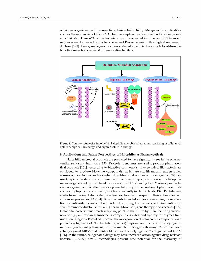
7. Strategies Behind Halophiles for Bimolecular Adaptation to Extreme Habitats
Microbial metabolite secretions at challenging habitats, such as saline/hypersaline
ecosystems, could promote adaptations through specific pathways [121]. Moreover,
hypersaline environments denoting salinities of more than ≈35‰, where seawater might
even show an oversaturation of salts [122,123]. Halophilic bacteria and eukaryotes exploit
the salt‐out strategy that excretes salts from the cytoplasm, and they either synthesise or
accrue the de novo attuned solutes, including glycine betaine, and some zwitterionic com‐
pounds in bacteria, such as glycerol, and certain polyols in eukaryotes [123]. Halophiles
adopt common strategies to avoid an excessive loss of water due to NaCl saturation. These
include cellular adaptations, high salt‐instrategy or low salt/solute‐instrategy (Figure 3).
The first one produces osmoprotectants that increase osmotic‐cytoplasm activity to adjust
to the external environment or reach the equilibrium state by increasing high salt concen‐
trations so that their cytoplasm matches with high environmental salt concentrations. The
high salt‐in strategy performs the protection of halophiles through the accumulation of
inorganic solutes intracellularly to balance the salt concentration of the external environ‐
mentthrough the uniport and symport system in the presence and absence of light. In the
third strategy, osmolytes from the external environment protect the cell protein from de‐
naturation [124]. The adaptations of halophilic biomolecules are documented through var‐
ious mechanisms. Especially in fungi, the glycerol signalling pathway with high osmolar‐
ity to increase the salt level was screened between the fungus W. ichthyophaga and H. wer‐
neckiias halotolerant/halophilic fungi [125]. Even some halophilic protists express high
gene proportions in duplicated genes at high salt concentrations that were expressing dif‐
ferent levels in H. seosinensis, which has its acquisition from bacteria that could evidence
the evolutionary process that might facilitate high salt adaptation [126]. Halophilic me‐
tabolite production could depend on salinity, as evidenced for Bacillus VITPS3, which pro‐
duced 3.18‐fold more metabolites in the presence of 10% (w/v) NaCl from various tested
concentrations [127]. Moreover, in media enrichment apart from salinity, the source of
carbon has its potential towards antimicrobial production in the culture media [14]. The
role of salinity in halophilic and halotolerant microbes might vary since halotolerants can
grow in the presence but also in the absence of higher salt concentrations, which was re‐
cently shown for Exiguobacterium sp. SH31, which can grow in up to 50 g/L of NaCl [128].
In order to produce potential metabolites from complex halophiles due to various salinity
gradients, recent strategies such asthe mixed culture approach were developed by Conde‐
Martinez et al. (2017) [53]. It is used to isolate potential strains from different ecosystems,
including brine and sediment samples via inoculation into different media, and to finally
Microorganisms 2022, 10, 417 13 of 21
obtain an organic extract to screen for antimicrobial activity. Metagenomic applications
such as the sequencing of 16s rRNA illumine amplicon were applied in Karak mine salt‐
erns, Pakistan. Here, 66% of the bacterial consortia occurred in brine, and 72% from salt
regions were dominated by Bacteroidetes and Proteobacteria with a high abundance of
Archaea [129]. Hence, metagenomics demonstrated an efficient approach to address the
bioactive microbial species at different saline habitats.
Figure 3. Common strategies involved in halophilic microbial adaptations consisting of cellular ad‐
aptation, high salt‐in energy, and organic solute‐in energy.
8. Applications and Future Perspectives of Halophiles as Pharmaceuticals
Halophilic microbial products are predicted to have significant uses in the pharma‐
ceutical sector and healthcare [130]. Proteolytic enzymes are used to produce pharmaceu‐
tical products [131]. According to bioactive compounds, diverse halophilic bacteria are
employed to produce bioactive compounds, which are significant and understudied
sources of bioactivities, such as antiviral, antibacterial, and anti‐tumour agents. [38]. Fig‐
ure 4 depicts the structure of different antimicrobial compounds produced by halophilic
microbes generated by the ChemDraw (Version 20.1.1) drawing tool. Marine cyanobacte‐
ria have gained a lot of attention as a powerful group in the creation of pharmaceuticals
such ascryptophycin and curacin, which are currently in clinical trials [132]. Peptide mol‐
ecules from marine diatoms also have been explored with respect to their antioxidant and
anticancer properties [133,134]. Biosurfactants from halophiles are receiving more atten‐
tion for antioxidants, antiviral antibacterial, antifungal, anticancer, antiviral, anti‐adhe‐
sive, immunomodulator, stimulating dermal fibroblasts, gene therapy, and vaccines [135].
Halophilic bacteria must reach a tipping point in the future by manufacturing various
novel drugs, antioxidants, sunscreens, compatible solutes, and hydrolytic enzymes from
unexplored regions. Recent advances in the incorporation of halogenated compounds into
peptoids (oligomers of N‐substituted glycines) improve antimicrobial efficacy against
multi‐drug‐resistant pathogens, with brominated analogues showing 32‐fold increased
activity against MRSA and 16‐64‐fold increased activity against P. aeruginosa and E. coli.
[136]. In the future, halogenated drugs may have increased action against drug‐resistant
bacteria. [136,137]. OMIC technologies present new potential for the discovery of

Microorganisms 2022, 10, 417 14 of 21
exclusive properties and/or novel biomolecules derived from halophiles in the future
[138,139]as a result of recent findings of halophilic bacteria, even from terrestrial environ‐
ments [140].
Figure 4. The structure of different antimicrobial compounds produced by halophilic microbes.
Further research is needed to report on how halophilic microorganisms evolved dur‐
ing the early phases of evolution of life on earth, as well as how they diversified and
spread around the world. Their biotechnological potency for generating compatible so‐
lutes, biopolymers, and other molecules is of industrial interest. To fully realise their clin‐
ical potential, additional research must focus on their physical organisation and modes of
action, allowing physicians to forecast which molecule could produce the desired medic‐
inal effect.
9. Conclusions
Researchers focusing on halophilic ecosystems in their search for novel biomolecules
are mostly motivatedby the threat of drug‐resistant human pathogens. This review high‐
lights that no Haloarchaeon has been found to show antibacterial action. More new com‐
pound extraction from more halophilic microbial genera is needed to combat human path‐
ogenic drug‐resistant microbes. Halophilic representatives of Bacillus and the dominating
actinomycete biomolecules have already been demonstrated to be effective against human
drug‐resistant infections. There is no benign report yet for the enzymes from halophilic
microbes against human pathogens. However, clinical trials should focus more on anti‐
microbials produced from halophiles because knowledge on the mode of action of halo‐
antimicrobials against drug‐resistant organisms is lacking. Overall, this short review sum‐
marises the risk of clinical drug‐resistant strains and signifies its control using halo‐de‐
rived compounds as a more promising strategy.

Microorganisms 2022, 10, 417 15 of 21
Author Contributions: Conceptualisation, H.S. and A.J.R.; methodology, H.S.; software, V.T.D.; val‐
idation, S.G.M. and M.K.; formal analysis, H.‐U.D. and J.M.A.; investigation, H.‐U.D. and J.‐S.H.;
writing—original draft preparation, H.S.; writing—review and editing, H.S., A.J.R. and J.‐S.H.; su‐
pervision, A.J.R., H.‐U.D. and J.‐S.H. All authors have read and agreed to the published version of
the manuscript.
Funding: The financial support from the Ministry of Science and Technology of Taiwan (Grant Nos.
MOST 107‐2621‐M‐019‐001, MOST 108‐2621‐M‐019‐003, MOST 109‐2621‐M‐019‐002, and MOST 110‐
2621‐M‐019‐001) and the Center of Excellence for Ocean Engineering (Grant No. 109J13801‐51,
110J13801‐51) to J.‐S. Hwang.
Institutional Review Board Statement: Not Applicable.
Informed Consent Statement: Not applicable.
Data Availability Statement: Not applicable.
Acknowledgments: We acknowledge RUSA 2.0—Biological Sciences, Bharathidasan University,
and the National Centre for Coastal Research (NCCR), MoES grant on academic support.
Conflicts of Interest: The authors declare no conflict of interest.
References
1. Peterson, E.; Kaur, P. Antibiotic resistance mechanisms in bacteria: Relationships between resistance determinants of antibiotic
producers, environmental bacteria, and clinical pathogens. Front. Microbiol. 2018, 9, 1–21.
https://doi.org/10.3389/fmicb.2018.02928.
2. Antimicrobial Resistance Global Report on Surveilance; World Health Organization (WHO): Geneva, Switzerland, 2014.
3. CDC. Available online: https://www.cdc.gov/drugresistance/about/where‐resistance‐spreads.html (accessed on 13 December
2021).
4. TATFAR Progress Report 2016–2020; U.S. Centers for Disease Control and Prevention (CDC): Atlanta, GA, USA, 2021.
5. Zhao, Y.; Guo, Q.; Dai, X.; Wei, X.; Yu, Y.; Chen, X.; Li, C.; Cao, Z.; Zhang, X. A Biomimetic Non‐Antibiotic Approach to Eradicate
Drug‐Resistant Infections. Adv. Mater. 2019, 31, 1–12. https://doi.org/10.1002/adma.201806024.
6. Kapoor, G.; Saigal, S.; Elongavan, A. Action and resistance mechanisms of antibiotics: A guide for clinicians. J. Anaesthesiol. Clin.
Pharmacol. 2017, 33, 300–305. https://doi.org/10.4103/joacp.JOACP_349_15.
7. Vasavada, S.H.; Thumar, J.T.; Singh, S.P. Secretion of a potent antibiotic by salt‐tolerant and alkaliphilic actinomycete Strepto‐
myces sannanensis strain RJT‐1. Curr. Sci. 2006, 91, 1393–1397.
8. Chamekh, R.; Deniel, F.; Donot, C.; Jany, J.L.; Nodet, P.; Belabid, L. Isolation, Identification and Enzymatic Activity of Halotol‐
erant and Halophilic Fungi from the Great Sebkha of Oran in Northwestern of Algeria. Mycobiology 2019, 47, 230–241.
https://doi.org/10.1080/12298093.2019.1623979.
9. Fariq, A.; Yasmin, A.; Jamil, M. Production, characterization and antimicrobial activities of bio‐pigments by Aquisalibacillus
elongatus MB592, Salinicoccus sesuvii MB597, and Halomonas aquamarina MB598 isolated from Khewra Salt Range, Pakistan. Ex‐
tremophiles 2019, 23, 435–449. https://doi.org/10.1007/s00792‐019‐01095‐7.
10. Mainka, T.; Weirathmüller, D.; Herwig, C.; Pflügl, S. Potential applications of halophilic microorganisms for biological treat‐
ment of industrial process brines contaminated with aromatics. J. Ind. Microbiol. Biotechnol. 2021, 48.
https://doi.org/10.1093/jimb/kuab015.
11. Manikandan, P.; Senthilkumar, P.K. An Overview of Saltpan Halophilic Bacterium. J. Antimicrob. Agents 2017, 3, 151.
https://doi.org/10.4172/2472‐1212.1000151.
12. Neelam, D.K.; Agrawal, A.; Tomer, A.K.; Bandyopadhayaya, S.; Sharma, A.; Jagannadham, M.V.; Mandal, C.C.; Dadheech, P.K.
A Piscibacillus sp. Isolated from a soda lake exhibits anticancer activity against breast cancer mda‐mb‐231 cells. Microorganisms
2019, 7, 34. https://doi.org/10.3390/microorganisms7020034.
13. Adlin Jenifer, J.S.C.; Michaelbabu, M.; Eswaramoorthy Thirumalaikumar, C.L.; Jeraldin Nisha, S.R.; Uma, G.; Citarasu, T. Anti‐
microbial potential of haloalkaliphilic Nocardiopsis sp. AJ1 isolated from solar salterns in India. J. Basic Microbiol. 2019, 59, 288–
301. https://doi.org/10.1002/jobm.201800252.
14. Gesheva, V.; Vasileva‐Tonkova, E. Production of enzymes and antimicrobial compounds by halophilic Antarctic Nocardioides
sp. grown on different carbon sources. World J. Microbiol. Biotechnol. 2012, 28, 2069–2076. https://doi.org/10.1007/s11274‐012‐
1009‐2.
15. Corral, P.; Esposito, F.P.; Tedesco, P.; Falco, A.; Tortorella, E.; Tartaglione, L.; Festa, C.; D’Auria, M.V.; Gnavi, G.; Varese, G.C.;
et al. Identification of a Sorbicillinoid‐Producing Aspergillus Strain with Antimicrobial Activity Against Staphylococcus aureus: A
New Polyextremophilic Marine Fungus from Barents Sea. Mar. Biotechnol. 2018, 20, 502–511. https://doi.org/10.1007/s10126‐018‐
9821‐9.
16. Abirami, G.; Ramprasath, C.; Arthi, M.; Mathivanan, N. Anticandidal activity of halophilic bacterium Vibrio azureus MML1960
isolated from Kelambakkam Saltpan, Tamil Nadu, India. Res. J. Biotechnol. 2018, 13, 8–20.

Microorganisms 2022, 10, 417 16 of 21
17. Lee, D.S.; Eom, S.H.; Jeong, S.Y. Anti‐methicillin‐resistant Staphylococcus aureus (MRSA) substance from the marine bacterium
Pseudomonas UJ‐6. Environ. Toxicol. Pharmacol. 2013, 35, 171–177.
18. Alvarado, C.; Alvarez, P.; Jiménez, L.; De la Fuente, M. Improvement of leukocyte functions in young prematurely aging mice
after a 5‐week ingestion of a diet supplemented with biscuits enriched in antioxidants. Antioxid. Redox Signal. 2005, 7, 1203–
1210. https://doi.org/10.1089/ars.2005.7.1203.
19. Hosseini Tafreshi, A.; Shariati, M. Dunaliella biotechnology: Methods and applications. J. Appl. Microbiol. 2009, 107, 14–35.
https://doi.org/10.1111/j.1365‐2672.2009.04153.x.
20. Ohyanagi, N.; Ishido, M.; Suzuki, F.; Kaneko, K.; Kubota, T.; Miyasaka, N.; Nanki, T. Retinoid ameliorates experimental auto‐
immune myositis, with modulation of the cell differentiation and antibody production in vivo. Arthritis Rheum. 2009, 60, 3118–
3127. https://doi.org/10.1002/art.24930.
21. Oren, A.; Hallsworth, J.E. Microbial weeds in hypersaline habitats: The enigma of the weed‐like Haloferax mediterranei. FEMS
Microbiol. Lett. 2014, 359, 134–142. https://doi.org/10.1111/1574‐6968.12571.
22. Hashemi, T.; Baserisalehi, M.; Bahador, N. Isolation of Halophilic Bacteria from Maharlu salt Lake—Iran and their evaluation
for the production of bioactive compounds. Int. J. Mol. Clin. Microbiol. 2014, 1, 365–370.
23. Li, X.; Yu, Y.H. Biodiversity and screening of halophilic bacteria with hydrolytic and antimicrobial activities from Yuncheng
Salt Lake, China. Biologia 2015, 70, 151–156. https://doi.org/10.1515/biolog‐2015‐0033.
24. Gaffney, E.M.; Simoska, O.; Minteer, S.D. The Use of Electroactive Halophilic Bacteria for Improvements and Advancements in
Environmental High Saline Biosensing. Biosensors 2021, 11, 48. https://doi.org/10.3390/bios11020048.
25. Ballav, S.; Kerkar, S.; Thomas, S.; Augustine, N. Halophilic and halotolerant actinomycetes from a marine saltern of Goa, India
producing anti‐bacterial metabolites. J. Biosci. Bioeng. 2015, 119, 323–330. https://doi.org/10.1016/j.jbiosc.2014.08.017.
26. Selim, M.S.M.; Abdelhamid, S.A.; Mohamed, S.S. Secondary metabolites and biodiversity of actinomycetes. J. Genet. Eng. Bio‐
technol. 2021, 19. https://doi.org/10.1186/s43141‐021‐00156‐9.
27. Irshad, A.; Ahmad, I.; Bum Kim, S. Isolation, characterization and antimicrobial activity of halophilic bacteria in forshore soils.
Afr. J. Microbiol. Res. 2013, 7, 164–173. https://doi.org/10.5897/AJMR12.1004.
28. Hamedi, J.; Mohammadipanah, F.; Ventosa, A. Systematic and biotechnological aspects of halophilic and halotolerant actino‐
mycetes. Extremophiles 2013, 17, 1–13. https://doi.org/10.1007/s00792‐012‐0493‐5.
29. Manteca, A.; Yagüe, P. Streptomyces as a Source of Antimicrobials: Novel Approaches to Activate Cryptic Secondary Metabolite
Pathways. In Antimicrobials, Antibiotic Resistance, Antibiofilm Strategies and Activity Methods; InTech Open: Rijeka, Croatia, 2019.
https://doi.org/10.5772/intechopen.81812.
30. Bell, R.; Carmeli, S.; Sar, N. Vibrindole a, a metabolite of the marine bacterium, Vibrio parahaemolyticus, isolated from the toxic
mucus of the boxfish Ostracion cubicus. J. Nat. Prod. 1994, 57, 1587–1590. https://doi.org/10.1021/np50113a022.
31. Donio, M.B.S.; Ronica, S.F.A.; Viji, V.T.; Velmurugan, S.; Jenifer, J.A.; Michaelbabu, M.; Citarasu, T. Isolation and characteriza‐
tion of halophilic Bacillus sp. BS3 able to produce pharmacologically important biosurfactants. Asian Pac. J. Trop. Med. 2013, 6,
876–883. https://doi.org/10.1016/S1995‐7645(13)60156‐X.
32. Velmurugan, S.; Raman, K.; Thanga Viji, V.; Donio, M.B.S.; Adlin Jenifer, J.; Babu, M.M.; Citarasu, T. Screening and characteri‐
zation of antimicrobial secondary metabolites from Halomonas salifodinae MPM‐TC and its in vivo antiviral influence on Indian
white shrimp Fenneropenaeus indicus against WSSV challenge. J. King Saud. Univ. Sci. 2013, 25, 181–190.
https://doi.org/10.1016/j.jksus.2013.03.002.
33. Najjari, A.; Mejri, H.; Jabbari, M.; Sghaier, H.; Cherif, A.; Ouzari, H.I. Halocins, Bacteriocin‐Like Antimicrobials Produced by
the Archaeal Domain: Occurrence and Phylogenetic Diversity in Halobacteriales. In Extremophilic Microbes and Metabolites—
Diversity, Bioprespecting, and Biotechnological Applications; Intech Open: London, UK; Rijeka, Croatia, 2021.
https://doi.org/10.5772/intechopen.94765.
34. Ghanmi, F.; Carré‐Mlouka, A.; Vandervennet, M.; Boujelben, I.; Frikha, D.; Ayadi, H.; Peduzzi, J.; Rebuffat, S.; Maalej, S. Antag‐
onistic interactions and production of halocin antimicrobial peptides among extremely halophilic prokaryotes isolated from the
solar saltern of Sfax, Tunisia. Extremophiles 2016, 20, 363–374. https://doi.org/10.1007/s00792‐016‐0827‐9.
35. Kumar, V.; Tiwari, S.K. Activity‐guided separation and characterization of new halocin HA3 from fermented broth of Haloferax
larsenii HA3. Extremophiles 2017, 21, 609–621. https://doi.org/10.1007/s00792‐017‐0930‐6.
36. Chen, S.; Sun, S.; Korfanty, G.A.; Liu, J.; Xiang, H. A Halocin Promotes DNA Uptake in Haloferax mediterranei. Front. Microbiol.
2019, 10, 1960. https://doi.org/10.3389/fmicb.2019.01960.
37. Besse, A.; Vandervennet, M.; Goulard, C.; Peduzzi, J.; Isaac, S.; Rebuffat, S.; Carré‐Mlouka, A. Halocin C8: An antimicrobial
peptide distributed among four halophilic archaeal genera: Natrinema, Haloterrigena, Haloferax, and Halobacterium. Extremophiles
2017, 21, 623–638. https://doi.org/10.1007/s00792‐017‐0931‐5.
38. Corral, P.; Amoozegar, M.A.; Ventosa, A. Halophiles and their biomolecules: Recent advances and future applications in bio‐
medicine. Mar. Drugs 2020, 18, 33. https://doi.org/10.3390/md18010033.
39. Kumar, S.; Stecher, G.; Li, M.; Knyaz, C.; Tamura, K. MEGAX: Molecular Evolutionary Genetics Analysis across Computing
Platforms. Mol. Biol. Evol. 2018, 35, 1547–1549.
40. Ali, I.; Siwarungson, N.; Punnapayak, H.; Lotrakul, P.; Prasongsuk, S.; Bankeeree, W.; Rakshit, S.K. Screening of potential bio‐
technological applications from obligate halophilic fungi, isolated from a man‐made solar saltern located in Phetchaburi Prov‐
ince, Thailand. Pakistan J. Bot. 2014, 46, 983–988.

Microorganisms 2022, 10, 417 17 of 21
41. Lauritano, C.; Martín, J.; De La Cruz, M.; Reyes, F.; Romano, G.; Ianora, A. First identification of marine diatoms with anti‐
tuberculosis activity. Sci. Rep. 2018, 8, 2284. https://doi.org/10.1038/s41598‐018‐20611‐x.
42. Sushanth, V.R.; Rajashekhar, M. Antioxidant and antimicrobial activities in the four species of marine microalgae isolated from
Arabian Sea of Karnataka Coast. Indian J. Geo‐Marine Sci. 2015, 44, 69–75.
43. Mangamuri, U.K.; Vijayalakshmi, M.; Poda, S.; Manavathi, B.; Chitturi, B.; Yenamandra, V. Isolation and biological evaluation
of N‐(4‐aminocyclooctyl)‐3, 5‐dinitrobenzamide, a new semisynthetic derivative from the Mangrove‐associated actinomycete
Pseudonocardia endophytica VUK‐10. 3 Biotech 2016, 6, 158. https://doi.org/10.1007/s13205‐016‐0472‐0.
44. Zheng, J.; Wang, Y.; Wang, J.; Liu, P.; Li, J.; Zhu, W. Antimicrobial ergosteroids and pyrrole derivatives from halotolerant As‐
pergillus flocculosus PT05‐1 cultured in a hypersaline medium. Extremophiles 2013, 17, 963–971. https://doi.org/10.1007/s00792‐
013‐0578‐9.
45. Essghaier, B. Antimicrobial Behavior of Intracellular Proteins from Two Moderately Halophilic Bacteria: Strain J31 of Terribacil‐
lus halophilus and Strain M3‐ 23 of Virgibacillus marismortui. J. Plant. Pathol. Microbiol. 2014, 5. https://doi.org/10.4172/2157‐
7471.1000214.
46. Tian, S.; Yang, Y.; Liu, K.; Xiong, Z.; Xu, L.; Zhao, L. Antimicrobial metabolites from a novel halophilic actinomycete Nocardiopsis
terrae YIM 90022. Nat. Prod. Res. 2014, 28, 344–346. https://doi.org/10.1080/14786419.2013.858341.
47. Metelev, M.; Tietz, J.I.; Melby, J.O.; Blair, P.M.; Zhu, L.; Livnat, I.; Severinov, K.; Mitchell, D.A. Structure, bioactivity, and re‐
sistance mechanism of streptomonomicin, an unusual lasso peptide from an understudied halophilic actinomycete. Chem. Biol.
2015, 22, 241–250. https://doi.org/10.1016/j.chembiol.2014.11.017.
48. Bose, U.; Hewavitharana, A.K.; Ng, Y.K.; Shaw, P.N.; Fuerst, J.A.; Hodson, M.P. LC‐MS‐based metabolomics study of marine
bacterial secondary metabolites and antibiotic production in Salinispora arenicola. Mar. Drugs 2015, 13, 249–266.
https://doi.org/10.3390/md13010249.
49. Sun, M.; Zhang, X.; Hao, H.; Li, W.; Lu, C. Nocarbenzoxazoles A‐G, Benzoxazoles Produced by Halophilic Nocardiopsis lucen‐
tensis DSM 44048. J. Nat. Prod. 2015, 78, 2123–2127. https://doi.org/10.1021/acs.jnatprod.5b00031.
50. Marzban, A.; Ebrahimipour, G.; Danesh, A. Bioactivity of a novel glycolipid produced by a halophilic Buttiauxella sp. and im‐
proving submerged fermentation using a response surface method. Molecules 2016, 21, 1256. https://doi.org/10.3390/mole‐
cules21101256.
51. Nasri, M.R.; Baserisalehi, M.; Kurdtabar, M. solation and Identification of Halophilic Actinomyces with Antimicrobial Activity
and Partial Characterization of their Bioactive Compounds. Electron. J. Biol. 2017, 13, 383–390.
52. Frikha Dammak, D.; Zarai, Z.; Najah, S.; Abdennabi, R.; Belbahri, L.; Rateb, M.E.; Mejdoub, H.; Maalej, S. Antagonistic proper‐
ties of some halophilic thermoactinomycetes isolated from superficial sediments of a solar saltern and production of cyclic
antimicrobial peptides by the novel isolate Paludifilum halophilum. Biomed. Res. Int. 2017, 2017, 1205258.
https://doi.org/10.1155/2017/1205258.
53. Conde‐Martinez, N.; Acosta‐González, A.; Díaz, L.E.; Tello, E. Use of a mixed culture strategy to isolate halophilic bacteria with
antibacterial and cytotoxic activity from the Manaure solar saltern in Colombia. BMC Microbio. 2017, 17, 230.
https://doi.org/10.1186/s12866‐017‐1136‐x.
54. Hadj Rabia‐Boukhalfa, Y.; Eveno, Y.; Karama, S.; Selama, O.; Lauga, B.; Duran, R.; Hacène, H.; Eparvier, V. Isolation, purification
and chemical characterization of a new angucyclinone compound produced by a new halotolerant Nocardiopsis sp. HR‐4 strain.
World J. Microbiol. Biotechnol. 2017, 33, 126. https://doi.org/10.1007/s11274‐017‐2292‐8.
55. Latif, A.; Nasir, K.; Fatima, N.; Jamil, N. Characterization of Halophilic Isolates Producing Bioactive Metabolites Against Path‐
ogens. LGU J. Life Science 2019, 3, 92–97.
56. Oren, A. Halophilic microbial communities and their environments. Curr. Opin. Biotechnol. 2015, 33, 119–124.
https://doi.org/10.1016/j.copbio.2015.02.005.
57. Navarro, F.; Forján, E.; Vázquez, M.; Toimil, A.; Montero, Z.; Ruiz‐Domínguez, M.d.C.; Garbayo, I.; Castaño, M.; Vílchez, C.;
Vega, J.M. Antimicrobial activity of the acidophilic eukaryotic microalga Coccomyxa onubensis. Phycol. Res. 2017, 65, 38–43.
https://doi.org/10.1111/pre.12158.
58. Breijyeh, Z.; Jubeh, B.; Karaman, R. Resistance of Gram‐Negative bacteria to current antibacterial agents and overcoming ap‐
proaches. Molecules 2020, 25, 1340. https://doi.org/ 10.3390/molecules25061340.
59. Bandara, H.M.H.N.; Wood, D.L.A.; Vanwonterghem, I.; Hugenholtz, P.; Cheung, B.P.K.; Samaranayake, L.P. Fluconazole re‐
sistance in Candida albicans is induced by Pseudomonas aeruginosa quorum sensing. Sci. Rep. 2020, 10, 7769.
https://doi.org/10.1038/s41598‐020‐64761‐3.
60. Manikandan, P.; Moopantakath, J.; Imchen, M.; Kumavath, R.; SenthilKumar, P.K. Identification of Multi‐Potent Protein Subtil‐
isin A from halophilic bacterium Bacillus firmus VE2. Microb. Pathog. 2021, 157, 105007.
https://doi.org/10.1016/j.micpath.2021.105007.
61. Kamat, T.; Kerkar, S. Bacteria from Salt Pans: A Potential Resource of Antibacterial Metabolites. Recent Res. Sci. Technol. 2011, 3,
46–52.
62. Cardozo, V.F.; Oliveira, A.G.; Nishio, E.K.; Perugini, M.R.E.; Andrade, C.G.T.J.; Silveira, W.D.; Durán, N.; Andrade, G.; Koba‐
yashi, R.K.T.; Nakazato, G. Antibacterial activity of extracellular compounds produced by a Pseudomonas strain against methi‐
cillin‐resistant Staphylococcus aureus (MRSA) strains. Ann. Clin. Microbiol. Antimicrob. 2013, 12, 1–8. https://doi.org/10.1186/1476‐
0711‐12‐12.

Microorganisms 2022, 10, 417 18 of 21
63. Xu, L.Q.; Zeng, J.W.; Jiang, C.H.; Wang, H.; Li, Y.Z.; Wen, W.H.; Li, J.H.; Wang, F.; Ting, W.J.; Sun, Z.Y.; et al. Isolation and
determination of four potential antimicrobial components from Pseudomonas aeruginosa extracts. Int. J. Med. Sci. 2017, 14, 1368–
1374. https://doi.org/10.7150/ijms.18896.
64. Henciya, S.; Vengateshwaran, T.D.; Gokul, M.S.; Dahms, H.U.; James, R.A. Antibacterial Activity of Halophilic Bacteria Against
Drug‐Resistant Microbes Associated with Diabetic Foot Infections. Curr. Microbiol. 2020, 77, 3711–3723.
https://doi.org/10.1007/s00284‐020‐02190‐1.
65. Ray, L.; Suar, M.; Pattnaik, A.K.; Raina, V. Streptomyces chilikensis sp. nov. a halophilic streptomycete isolated from brackish
water sediment. Int. J. Syst. Evol. Microbiol. 2013, 63, 2757–2764. https://doi.org/10.1099/ijs.0.046284‐0.
66. Jose, A.; Santhi, S.; Solomon, R.D.J. In Vitro Antimicrobial potential and growth characteristics of Nocardiopsis sp. JAJ16 isolated
from crystallizer pond. Int. J. Cur. Res. 2010, 3, 24–26.
67. McArthur, K.A.; Mitchell, S.S.; Tsueng, G.; Rheingold, A.; White, D.J.; Grodberg, J.; Lam, K.S.; Potts, B.C. Lynamicins A‐E, chlo‐
rinated bisindole pyrrole antibiotics from a novel marine actinomycete. J. Nat. Prod. 2008, 71, 1732–1737.
https://doi.org/10.1021/np800286d.
68. Hughes, C.C.; Prieto‐Davo, A.; Jensen, P.R.; Fenical, W. ChemInform Abstract: The Marinopyrroles, Antibiotics of an Unprece‐
dented Structure Class from a Marine Streptomyces sp. ChemInform 2008, 39, 4–6. https://doi.org/10.1002/chin.200830209.
69. Alshaibani, M.M.; Mohamadzin, N.; Jalil, J.; Sidik, N.M.; Ahmad, S.J.; Kamal, N.; Edrada‐Ebel, R. Isolation, purification, and
characterization of five active diketopiperazine derivatives from endophytic Streptomyces SUK 25 with antimicrobial and cyto‐
toxic activities. J. Microbiol. Biotechnol. 2017, 27, 1249–1256. https://doi.org/10.4014/jmb.1608.08032.
70. Magarvey, N.A.; Keller, J.M.; Bernan, V.; Dworkin, M.; Sherman, D.H. Isolation and characterization of novel marine‐derived
actinomycete taxa rich in bioactive metabolites. Appl. Environ. Microbiol. 2004, 70, 7520–7529.
https://doi.org/10.1128/AEM.70.12.7520‐7529.2004.
71. Saker, R.; Meklat, A.; Bouras, N.; Zitouni, A.; Mathieu, F.; Spröer, C.; Klenk, H.P.; Sabaou, N. Diversity and antagonistic prop‐
erties of culturable halophilic actinobacteria in soils of two arid regions of septentrional Sahara: M’zab and Zibans. Ann. Micro‐
biol. 2015, 65, 2241–2253. https://doi.org/10.1007/s13213‐015‐1065‐6.
72. Gohel, S.D.; Sharma, A.K.; Dangar, K.G.; Thakrar, F.; Singh, S.P. Antimicrobial and biocatalytic potential of haloalkaliphilic
actinobacteria. In Halophiles; Maheshwari, D.K. Saraf, M. Eds.; Springer: Heidelberg, Germany, 2015; Volume 6, pp. 29–55.
https://doi.org/10.1007/978‐3‐319‐14595‐2_2.
73. Buijs, Y.; Bech, P.K.; Vazquez‐Albacete, D.; Bentzon‐Tilia, M.; Sonnenschein, E.C.; Gram, L.; Zhang, S. Da Marine Proteobacteria
as a source of natural products: Advances in molecular tools and strategies. Nat. Prod. Rep. 2019, 36, 1333–1350.
https://doi.org/10.1039/c9np00020h.
74. Moghaddam, J.A.; Dávila‐Céspedes, A.; Kehraus, S.; Crüsemann, M.; Köse, M.; Müller, C.E.; König, G.M. Cyclopropane‐con‐
taining fatty acids from the marine bacterium Labrenzia sp. 011 with antimicrobial and GPR84 activity. Mar. Drugs 2018, 16, 369.
https://doi.org/10.3390/md16100369.
75. Elyasifar, B.; Jafari, S.; Hallaj‐Nezhadi, S.; Chapeland‐Leclerc, F.; Ruprich‐Robert, G.; Dilmaghani, A. Isolation and identification
of antibiotic‐producing halophilic bacteria from Dagh Biarjmand and Haj aligholi salt deserts, Iran. Pharm. Sci. 2019, 25, 70–77.
https://doi.org/10.15171/PS.2019.11.
76. Sawale, A.; Kadam, T.A.; Karale, M.A. Antimicrobial activity of Secondary metabolites from Halophilic Bacillus pumilus sp. Int.
J. Curr. Microbiol. Appl. Sci. 2014, 3, 506–512.
77. Shinde, V.A.; Patil, R.B.; Pawar, P.V. Comparative study of antimicrobial potentials of phospholipid compounds produced by
halophilic and alkaliphiles Bacillus subtilis isolated from alkaline meteorite crater Lonar lake, India. Int. J. Life Sci. 2017, 5, 420–
424.
78. Thacharodi, A.; Reghu, A.P.; Priyadharshini, R.; Kathikeyan, G.; Thacharodi, D. Bioprospecting of Halotolerant Bacillus subtilis:
A Study depicting its Potential Antimicrobial Activity against Clinically Important Pathogens. Indian J. Sci. Technol. 2019, 12, 1–
7. https://doi.org/10.17485/ijst/2019/v12i22/144660.
79. Al‐Karabli, N. Antimicrobial Activity of Bacillus persicus 24‐DSM Isolated from Dead Sea Mud. Open Microbiol. J. 2018, 11, 372–
383. https://doi.org/10.2174/1874285801711010372.
80. Maher, O. Isolation and characterization of extremely halotolerant Bacillus species from Dead Sea black mud and determination
of their antimicrobial and hydrolytic activities. Afr. J. Microbiol. Res. 2017, 11, 1303–1314.
https://doi.org/10.5897/ajmr2017.8608Mishra.
81. Mishra, I.; Sapre, S.; Tiwari, S. Diversity of halophilic bacteria and actinobactreia from India and their biotechnological applica‐
tions. Indian J. Geomarine Sci. 2017, 8, 1575–1587.
82. Priyanka, S.; Jayashree, M.; Shivani, R.; Anwesha, S.; Bhaskara Rao, K.V. Characterisation and identification of antibacterial
compounds from marine actinobacteria: In vitro and in silico analysis. J. Infect. Public Health 2019, 12, 83–89.
https://doi.org/10.1016/j.jiph.2018.09.005.
83. Kumar, R.R.; Jadeja, V.J. Characterization and partial purification of an antibacterial agent from halophilic actinomycetes Ko‐
curia sp. Strain RSK4. BioImpacts 2018, 8, 253–261. https://doi.org/10.15171/bi.2018.28.
84. Deepalaxmi, R.K.; Gayathri, C. Screening of bioactive compound, antimicrobial activity producing halophilic isolates from the
saltpans of Thoothukudi district. Afr. J. Microbiol. Res. 2018, 12, 338–344. https://doi.org/10.5897/ajmr2018.8823.
85. Tatar, D. Isolation, phylogenetic analysis and antimicrobial activity of halophilic actinomycetes from different saline environ‐
ments located near Çorum province. Biologia 2012, 76, 773–780. https://doi.org/10.2478/s11756‐020‐00612‐w.

Microorganisms 2022, 10, 417 19 of 21
86. Samrot, A.V.; Divya; Narendrakumar, G.; Abirami, S.; Dhiva, S.; Prakash, P.; Jane Cypriyana, P.J.; Padmanaban, S. Bioprospect‐
ing studies of Halophilic bacteria—Streptomyces sp. MA05 nad Halobactreium sp MA06. Lett. Appl. Nanobiosci. 2020, 9, 1583–
1594. https://doi.org/10.33263/LIANBS94.15831594.
87. Jenifer, J.; Selva Donio, M.; Michaelbabu, M.; Vincent, S.G.P.; Citarasu, T. Haloalkaliphilic Streptomyces spp. AJ8 isolated fom
solar saltworks and its pharmacological potential. AMB Expr. 2015, 5, 59–70. https://doi.org/10.1186/s13568‐015‐0143‐2.
88. Menasria, T.; Monteoliva‐Sánchez, M.; Benammar, L.; Benhadj, M.; Ayachi, A.; Hacène, H.; Gonzalez‐Paredes, A.; Aguilera, M.
Culturable halophilic bacteria inhabiting Algerian saline ecosystems: A source of promising features and potentialities. World
J. Microbiol. Biotechnol. 2019, 35, 1–16. https://doi.org/10.1007/s11274‐019‐2705‐y.
89. Boyadzhieva, I.; Tomova, I.; Radchenkova, N.; Kambourova, M.; Poli, A.; Vasileva‐Tonkova, E. Diversity of Heterotrophic Hal‐
ophilic Bacteria Isolated from Coastal Solar Salterns, Bulgaria and Their Ability to Synthesize Bioactive Molecules with Biotech‐
nological Impact. Microbiol. Russian Fed. 2018, 87, 519–528. https://doi.org/10.1134/S0026261718040033.
90. Ambrico, A.; Trupo, M.; Magarelli, R. Effectiveness of Dunaliella salina Extracts against Bacillus subtilis and Bacterial Plant Path‐
ogens. Pathogens 2020, 9, 1–14. https://doi.org/10.3390/pathogens9080613.
91. Krishnakumar, S.; Dooslin, V.; Bai, M.; Rajan, R.A. Evaluation of bioactive metabolites from halophilic microalgae Dunaliella
Salina by GC—MS analysis. Int J. Pharm. Pharm. Sci. 2013, 5, 96–303.
92. Corona, E.; Fernandez‐Acero, J.; Bartual, A. Screening study for antibacterial activity from marine and freshwater microalgae.
Int. J. Pharm. Bio Sci. 2017, 8, 189–194.
93. Jafari, S.; Mobasher, M.A.; Najafipour, S.; Ghasemi, Y.; Mohkam, M.; Ebrahime, M.A.; Mobasher, N. Antibacterial potential of
Chlorella vulgaris and Dunaliella salina extracts against Streptococcus mutans. Jundishapur J. Nat. Pharm. Prod. 2018, 13, e13226.
94. Singh, H.; Kaur, M.; Jangra, M.; Mishra, S.; Nandanwar, H.; Pinnaka, A.K. Antimicrobial properties of the novel bacterial isolate
Paenibacilllus sp. SMB1 from a halo‐alkaline lake in India. Sci. Rep. 2019, 9, 1–12. https://doi.org/10.1038/s41598‐019‐47879‐x.
95. Ghalehno, A.D.; Ghavidel‐Aliabadi, M.; Shahmohamadi, Z.; Mehrshad, M.; Amoozegar, M.A.; Danesh, A. Isolation and Dis‐
covery of New Antimicrobial‐agent Producer Strains Using Antibacterial Screening of Halophilic Gram‐positive Endospore‐
forming Bacteria Isolated from Saline Lakes of Iran. Arak. Med. Univ. J. 2018, 20, 10–23.
96. Farida, B.; Abdelghani, Z.; Florence, M.; Ahmed, L.; Nasserdine, S. Taxonomy and antimicrobial activities of two novel halo‐
philic Saccharomonospora strains isolated in Algerian Sahara soils. Ann. Microbiol. 2011. 61, 299–305.
https://doi.org/10.1007/s13213‐010‐0138‐9.
97. Lawrance, A.; Balakrishnan, M.; Gunasekaran, R.; Srinivasan, R.; Valsalan, V.N.; Gopal, D.; Ramalingam, K. Unexplored deep
sea habitats in active volcanic Barren Island, Andaman and Nicobar Islands are sources of novel halophilic Eubacteria. Infect.
Genet. Evol. 2018, 65, 1–5. https://doi.org/10.1016/j.meegid.2018.07.008.
98. Tian, S.Z.; Pu, X.; Luo, G.; Zhao, L.X.; Xu, L.H.; Li, W.J.; Luo, Y. Isolation and characterization of new p‐terphenyls with anti‐
fungal, antibacterial, and antioxidant activities from the halophilic actinomycete Nocardiopsis gilva YIM 90087. J. Agric. Food
Chem. 2013, 61, 3006–3012. https://doi.org/10.1021/jf400718w.
99. Nikita, P.; Mitesh, D.; Shahnawaz, J.; Rasheedunnisa, B. Antibacterial Activity of Marine Bacterial Pigments Obtained from
Arabian Sea Water Samples. J. Pure Appl. Microbiol. 2020, 14, 517–526.
100. Srilekha, V.; Krishna, G.; Seshasrinivas, V.; Alha Singara Charya, M. Antibacterial and anti‐inflammatory activities of marine
Brevibacterium sp. Res. Pharm Sci. 2017, 12, 283–289.
101. Srilekha, V.; Krishna, G.; Sesha Srinivas, V.; Singara, M.A. Antimicrobial Evaluation of Bioactive Pigment from Salinicoccus sp.
isolated from Nellore sea coast. Int J. Biotech. Biochem. 2017, 13, 211–217.
102. Rasha, A.M.; Sikaily, A.E.; Nermeen, A.E.; Hanan, A.G.; Soraya, A.S. Antimicrobial activity of textile fabrics dyed with prodi‐
giosin pigment extracted from marine Serratia rubidaea RAM_Alex bacteria. Egypt J. Aquat. Res. 2021, 47, 301–305.
https://doi.org/10.1016/j.ejar.2021.05.004.
103. Li, J.L.; Yang, N.; Huang, L.; Chen, D.; Zhao, Y.; Tang, M.M.; Fan, H.; Bao, X. Pyocyanin Inhibits Chlamydia infection by Disa‐
bling infectivity of the Elementary Body and Disrupting Intracellular Growth. Antimicrob Agents Chemother. 2018, 62, e02260–
17.
104. Kiran, G.S.; Jackson, S.A.; Priyadharsini, S.; Dobson, A.D.; Selvin, J. Synthesis of Nm‐PHB (nanomelanin‐polyhydroxy butyrate)
nanocomposite film and its protective effect against biofilm‐forming multi drug resistant Staphylococcus aureus. Sci. Rep. 2017,
7, 1–13. https://doi.org/10.1038/s41598‐017‐08816‐y.
105. Danevcic, T.; Boric Vezjak, M.; Tabor, M.; Zorec, M.; Stopar, D. Prodigiosin induces autolysins in actively grown Bacillus subtilis
cells. Front. Microbiol. 2016, 7, 4, 589655. https://doi.org/10.3389/fmicb.2016.00027.
106. Liang, Y.; Chen, L.; Ye, X.; Anjum, K.; Lian, X.Y.; Zhang, Z. New streptophenazines from marine Streptomyces sp. 182SMLY. Nat.
Prod. Res. 2017, 31, 411–417. https://doi.org/10.1080/14786419.2016.1169419.
107. Jiang, Y.J.; Zhang, D.S.; Zhang, H.J.; Li, J.Q.; Ding, W.J.; Xu, C.D. Medermycin‐type naphthoquinones fromthe marine‐derived
Streptomyces sp. XMA39. J. Natural Products. 2018, 81, 2120–2124. https://doi.org/10.1021/acs.jnatprod.8b00544.
108. Velmurugan, P.; Venil, C.K.; Veera, R.; Arumugam, D.A.; Laurent, D. Marine Bacteria Is the Cell Factory to Produce Bioactive
Pigments: A Prospective Pigment Source in the Ocean. Front. Sustain. Food Syst. 2020, 4, 589655.
https://doi.org/10.3389/fsufs.2020.589655.
109. Kocberber Kilic, N.; Kubra, D.; Gonul, D. Bioactive compounds produced by Dunaliella species, Antimicrobial effects and op‐
timization of the efficiency. Turk. J. Fish. Aquat Sci. 2019, 19, 923–933. https://doi.org/10.4194/1303‐2712‐v19_11_04.

Microorganisms 2022, 10, 417 20 of 21
110. Ramesh, C.; Vinithkumar, N.V.; Kirubagaran, R.; Venil, C.K.; Dufosse, L. Multifaceted Applications of Microbial Pigments:
Current Knowledge, Challenges and Future Directions for Public Health Implications. Microorganisms 2019, 7, 186.
https://doi.org/10.3390/microorganisms7070186.
111. Alvionita, M.; and Hertadi, R. Bioconversio of Glycerol to Biosurfactant by Halophilic bacteria Halomonas elongata BK‐AG18.In‐
dones. J. Chem. 2019, 19, 48–57. https://doi.org/10.22146/ijc.26737.
112. Murugan, T.; Murugan, M. Antibacterial effect of Surface active agent produced by Pseudomonas sp. isolated from Saltpan. J.
Pharm. Sci. Res. 2018, 10, 196–197.
113. Kayanadath, S.; Nathan, V.K.; Ammini, P. Anti‐Biofilm Activity of Biosurfactant Derived from Halomonas sp., a Lipolytic Marine
Bacterium from the Bay of Bengal. Microbiology 2019, 88, 585–599. https://doi.org/10.1134/S0026261719050072.
114. Cortes Camargo, S.; Acuna Avila, P.E.; Arrieta Baez, D.; Montanez Barragan, B.; Morato, A.I.; Sanz Martin, J.L.; Barragan Huerta,
B.E. Biosurfactant Production by Bacillus tequilensis ZSB10: Structural Characterization, Physicochemical, and Antifungal Prop‐
erties. J. Surfactants Deterg. 2021, 24, 773–782. https://doi.org/10.1002/jsde.12515.
115. Sumaiya, M.; Anchana Devi, C.; Leela, K. A Study on Biosurfactant Production from Marine Bacteria. Int J. Sci and Res. 2017, 7,
140–145.
116. Sayeda, A.; Sahar, S.; Manal, S. Medical application of exopolymers produced by marine bacteria. Bull. Natl. Res. Cent. 2020, 44,
1–14. https://doi.org/10.1186/s42269‐020‐00323‐x.
117. Spano, A.; Lagana, P.; Visalli, G.; Maugeri, T.L.; Gugliandolo, C. In vitro antibiofilm activity of an exopolysaccharide from the
marine thermophilic Bacillus licheniformis T14. Curr. Microbiol. 2016, 72, 518–528. https://doi.org/10.1007/s00284‐015‐0981‐9.
118. Sahar, S.M.; Shaimaa, K.A.; Manal, S.S.; Hala, M.R. Characterization and applications of exopolysaccharide produced by marine
Bacillus altitudinis MSH2014 from Ras Mohamed, Sinai, Egypt. Egypt J. Basic App Sci, 2018, 5, 204–209.
https://doi.org/10.1016/j.ejbas.2018.05.009.
119. AA Abdrabo, M.; AH Ibrahim, H.; Hassan, S.W.; M Abdul‐Raouf, U. Antimicrobial and anti‐tumor activities of exopolysaccha‐
rides produced by the biofilm of marine Halomonas saccharevitans AB2 isolated from Suez Gulf, Egypt. Egypt J. Aquat Biol Fish.
2018, 22, 99–119.
120. Aullybux, A.A.; Puchooa, D.; Bahorun, T.; Jeewon, R. Phylogenetics and antibacterial properties of exopolysaccharides from
marine bacteria isolated from Mauritius seawater. Ann. Microbiol. 2019, 69, 957–972. https://doi.org/10.1007/s13213‐019‐01487‐2.
121. Gunde‐Cimerman, N.; Plemenitas, A.; Oren, A. Strategies of adaptation of microorganisms of the three domains of life to high
salt concentrations. FEMS Microbiol. Rev. 2018, 42, 353–375. https://doi.org/10.1093/femsre/fuy009.
122. Ventosa, A.; de la Haba, R.R.; Sánchez‐Porro, C.; Papke, R.T. Microbial diversity of hypersaline environments: A metagenomic
approach. Curr. Opin. Microbiol. 2015, 25, 80–87. https://doi.org/10.1016/j.mib.2015.05.002.
123. DasSarma, S.; DasSarma, P. Halophiles and their enzymes: Negativity put to good use. Curr. Opin. Microbiol. 2015, 25, 120–126.
https://doi.org/10.1016/j.mib.2015.05.009.
124. Edbeib, M.F.; Wahab, R.A.; Huyop, F. Halophiles: Biology, adaptation, and their role in decontamination of hypersaline envi‐
ronments. World J. Microbiol. Biotechnol. 2016, 32, 1–23. https://doi.org/10.1007/s11274‐016‐2081‐9.
125. Plemenitas, A.; Lenassi, M.; Konte, T.; Kejžar, A.; Zajc, J.; Gostinčar, C.; Gunde‐Cimerman, N. Adaptation to high salt concen‐
trations in halotolerant/halophilic fungi: A molecular perspective. Front. Microbiol. 2014, 5, 1–12.
https://doi.org/10.3389/fmicb.2014.00199.
126. Harding, T.; Roger, A.J.; Simpson, A.G.B. Adaptations to high salt in a halophilic protist: Differential expression and gene ac‐
quisitions through duplications and gene transfers. Front. Microbiol. 2017, 8, 1–27. https://doi.org/10.3389/fmicb.2017.00944.
127. Subramanian, P.; Gurunathan, J. Differential Production of Pigments by Halophilic Bacteria Under the Effect of Salt and Eval‐
uation of Their Antioxidant Activity. Appl. Biochem. Biotechnol. 2020, 190, 391–409. https://doi.org/10.1007/s12010‐019‐03107‐w.
128. Remonsellez, F.; Castro‐Severyn, J.; Pardo‐Esté, C.; Aguilar, P.; Fortt, J.; Salinas, C.; Barahona, S.; León, J.; Fuentes, B.; Areche,
C.; et al. Characterization and salt response in recurrent halotolerant Exiguobacterium sp. SH31 isolated from sediments of salar
de huasco, Chilean altiplano. Front. Microbiol. 2018, 9, 1–17. https://doi.org/10.3389/fmicb.2018.02228.
129. Cycil, L.M.; DasSarma, S.; Pecher, W.; McDonald, R.; AbdulSalam, M.; Hasan, F. Metagenomic Insights into the Diversity of
Halophilic Microorganisms Indigenous to the Karak Salt Mine, Pakistan. Front. Microbiol. 2020, 11, 1–14.
https://doi.org/10.3389/fmicb.2020.01567.
130. Waditee‐Sirisattha, R.; Kageyama, H.; Takabe, T. Halophilic microorganism resources and their applications in industrial and
environmental biotechnology. AIMS Microbiol. 2016, 2, 42–54. https://doi.org/10.3934/microbiol.2016.1.42.
131. Adrio, J.L.; Demain, A.L. Microbial enzymes: Tools for biotechnological processes. Biomolecules 2014, 4, 117–139.
https://doi.org/10.3390/biom4010117.
132. Thuan, N.H.; An, T.T.; Shrestha, A.; Canh, N.X.; Sohng, J.K.; Dhakal, D. Recent Advances in Exploration and Biotechnological
Production of Bioactive Compounds in Three Cyanaliobacterial Genera: Nostoc, Lyngbya, and Microcystis. Front. Chem. 2019, 7,
1–33. https://doi.org/10.3389/fchem.2019.00604.
133. Barkia, I.; Al‐Haj, L.; Abdul Hamid, A.; Zakaria, M.; Saari, N.; Zadjali, F. Indigenous marine diatoms as novel sources of bioac‐
tive peptides with antihypertensive and antioxidant properties. Int. J. Food Sci. Technol. 2019, 54, 1514–1522.
https://doi.org/10.1111/ijfs.14006.
134. Hussein, H.A.; Abdullah, M.A. Anticancer Compounds Derived from Marine Diatoms. Mar. Drugs 2020, 18, 356.
https://doi.org/10.3390/md18070356.

Microorganisms 2022, 10, 417 21 of 21
135. Citarasu, T.; Thirumalaikumar, E.; Abinaya, P.; Babu, M.M.; Uma, G. Biosurfactants from halophilic origin and their potential
applications. In Green Sustainable Process for Chemical and Environmental Engineering and Science; Elsevier: Amsterdam, The Neth‐
erlands, 2021; pp. 489–521. https://doi.org/10.1016/B978‐0‐12‐823380‐1.00019‐8.
136. Molchanova, N.; Nielsen, J.E.; Sørensen, K.B.; Prabhala, B.K.; Hansen, P.R.; Lund, R.; Barron, A.E.; Jenssen, H. Halogenation as
a tool to tune antimicrobial activity of peptoids. Sci. Rep. 2020, 10, 14805. https://doi.org/10.1038/s41598‐020‐71771‐8.
137. Dias Flaviana, R.F.; Novais, J.S.; Devillart, S.; da Silva, W.A.; Fereirra, M.O.; Loureiro, R.d.S.; Campos, V.R.; Fereirra, V.F.; de
Souza, M.C.B.V.; Castro, H.C.; et al. Synthesis and antimicrobial evaluation of amino sugar‐based naphthoquinones and iso‐
quinoline‐5,8‐diones and their halogenated compounds. Eur. J. Med. Chem 2018, 156, 1–12.
https://doi.org/10.1016/j.ejmech.2018.06.050.
138. Diaz‐Cardenas, C.; Rojas, L.Y.; Fiorentino, S.; Cala, M.P.; Diaz, J.I.; Ramos, F.A.; Armengaud, J.; Restrepo, S.; Baena, S. Bioactive
potential of extracts of Labrenzia aggregate strain USBA 371, a halophilic bacterium isolated from a terrestrial source. Molecules
2020, 25, 2546. https://doi.org/10.3390/molecules25112546.
139. Seck, E.; Diop, A.; Armstrong, A.; Delerce, J.; Fournier, P.‐E.; Raoult, D.; Khelaifia, S. Microbial culturomics to isolate halophilic
bacteria from table salt: Genome sequence and description of the moderately halophilic bacterium Bacillus salis sp. nov. New
Microbes New Infect. 2018, 23, 28–38. https://doi.org/10.1016/j.nmni.2017.12.006.
140. Kothe, C.I.; Bolotin, A.; Kraïem, B.; et al. Unraveling the world of halophilic and halotolerant bacteria in cheese by combining
cultural, genomic and metagenomic approaches. Int. J. Food Microbiol. 2021, 350, 109312. https://doi.org/10.1016/j.ijfoodmi‐
cro.2021.


