Embed Size (px)
Citation preview

ARTICLE
Recommendations for quality improvement ingenetic testing for cystic fibrosisEuropean Concerted Action on Cystic Fibrosis
E Dequeker1,2, H Cuppens2, J Dodge1,3, X Estivill1,4, M Goossens1,5, PF Pignatti1,6, H Scheffer1,7, M Schwartz1,8, M Schwarz1,9, B Tümmler1,10 and J-J Cassiman1,2
1Steering Committee of the European Concerted Action on Cystic Fibrosis; 2Center for Human Genetics, CatholicUniversity of Leuven, Belgium; 3Department of Child Health, University of Swansea, Singleton Hospital, Swansea,Wales, UK; 4Institut de Recerca Oncologica (IRO), Dept de Genetica Molecular, Barcelona, Spain; 5Laboratoire deBiochemie, InsermU468, Creteil, France; 6Ist Di Biologia e Genetica, University of Verona, Italy; 7Dept of MedicalGenetics, University of Groningen, The Netherlands; 8Dept of Clinical Genetics, Rigshospitalet 4062, Copenhagen,Denmark; 9North-West Regional Molecular Genetics Laboratory, Royal Manchester Children’s Hospital, UK;10Klinische Forschergruppe Mol Pathol des Mukoviszidose, medizinische Hochschule Hannover, Germany
These recommendations for quality improvement of cystic fibrosis genetic diagnostic testing providegeneral guidelines for the molecular genetic testing of cystic fibrosis in patients/individuals. Generalstrategies for testing as well as guidelines for laboratory procedures, internal and external qualityassurance, and for reporting the results, including the requirements of minimal services in mutationtesting, the nomenclature for describing mutations, procedures to control false-positive amplificationreactions and to validate tests, and guidelines to implement a quality system in a molecular diagnosticlaboratory are reviewed. European Journal of Human Genetics (2000) 8, S1–S24.
Keywords: guidelines; recommendations; genetic testing; cystic fibrosis; quality assurance; mutation testing
IntroductionTo perform a molecular genetic diagnostic test is a complexprocess. Errors in one of the steps in this process may affectthe results and therefore the conclusions. Because the resultsof a genetic test can have serious implications for anindividual and possibly for his relatives, it is important thatthe error rate of genetic tests should be reduced to anabsolute minimum. Therefore good internal quality controlsystems for the whole procedure, from blood sampling to thedelivery of the written report, should be worked out in eachgenetic diagnostic laboratory. This may require the imple-mentation of good laboratory practice (GLP) procedures andsome form of accreditation of laboratories able to demon-strate that they master all the parameters which affectresults.
Obviously, genetic testing should be done in the context ofappropriate genetic counselling. Laboratories offering genetictesting should work in close association with clinical geneti-cists and cystic fibrosis (CF) experts to ensure that theappropriate tests and the appropriate information are pro-vided to the patients requesting these services.
The purpose of the present document is to define strategiesand principles which increase the likelihood of CF testing inEurope being provided accurately and precisely. This docu-ment was prepared within the framework of the EuropeanConcerted Action on Cystic Fibrosis (BMH4-CT96-0462) andis based on the experience gathered by this concerted actionon the facilities, procedures and modus operandi in thedifferent countries (for membership of ECCACF see Appen-dix 2). More than 150 laboratories participated in thisproject; 90 actually formally approved this document; theothers did not voice their approval or dissent. Although theserecommendations focus on genetic testing for cystic fibrosis,the major issues of the recommendations are also applicableto other genetic disorders and are based on a series of
Correspondence: Dr E Dequeker, Center for Human Genetics, Universityof Leuven, Campus Gasthuisberg, Herestraat 49, B-3000 Leuven,Belgium. Tel: + 32 16 34 58 60; Fax: + 32 16 34 59 97; E-mail:[email protected] 12 May 1999; revised 8 February 2000; accepted 13 March2000
European Journal of Human Genetics (2000) 8, S2–S24y © 2000 Macmillan Publishers Ltd All rights reserved 1018–4813/00 $15.00
www.nature.com/ejhg

previously published documents. They also provide the basisfor the organisation of an internal quality control system ina genetic laboratory. The implementation of these guidelineswill require specific efforts from individual laboratories. Inaddition, it is proposed that regional, national or supra-national agreements are made between laboratories andhealth authorities in order to structure these services opti-mally to the benefit of the population serviced.
Recommendations on the strategy for CF testingMinimal services in mutation testingIt is advisable to organise the services on two levels in aparticular region or country. There are very large numbers ofCF mutations in the European population. Whilst it would bedesirable to have a mutation detection rate superior to 95%,the molecular heterogeneity of CFTR gene defects in Europeand the variation in frequency from one population toanother make this goal unachievable for all countries withthe current technology. For this reason, it is important toknow the ethnic or geographic origin of the patient underinvestigation and, if possible, of the parents andgrandparents.
Tables 1 and 2 (see Appendix 2) give a summary of the datacurrently available in the literature. Table 1 does not giveaccurate frequencies for all regions and all mutations. It is notadvisable to use these mutation frequencies for risk calcula-tion. The data from the local population should be used. Inseveral countries (Denmark, Sweden, Switzerland and Israel)a detection rate of 90% of the mutations (with a limited set ofmutations, each with a frequency of over 1% in thepopulation) is achieved.
In another seven countries (Belgium, Czech Republic,Estonia, Ireland, The Netherlands, Ukraine, UK), 80 to 90% ofthe mutations (each with a frequency of over 1% in thepopulation) can be routinely detected.
Pilot studies have been carried out in several countrieswhere the spectrum of CF mutations is well known (Bulgaria,France, Greece, Italy and Spain). For these countries, byextending mutation analysis to mutations with relativefrequencies less than 1%, detection levels of over 80 or 90%are achieved. In countries where these levels of detectionhave not yet been reached we suggest:
• a pilot study to determine the most frequent mutationsin the respective national populations and/or in theirvarious regions;
• once this is known, test for these mutations or at leastfor the mutations with a frequency of more than 1% inthat population.
To this end, international collaboration should besought.
In addition to be able to detect CFTR mutations with afrequency of over 1% (including deletions), a moleculargenetics laboratory should type the 5T allele (IVS8-6) if
patients are investigated for infertility.1 Also, each CF labo-ratory should be able to perform segregation analysis usingintragenic polymorphic markers (Table 3).
Analysis of other phenotypes possibly related to CFTRmutations (Azoospermia, CBAVD, disseminated bronchiecta-sis, pancreatitis, nasal polyposis, etc) is not encouraged atpresent but if done should not be restricted to the set ofmutations used for typical cystic fibrosis.
Practical organisation in level 1 and level 2 laboratoriesshould ideally be decided in consensus with the variouslaboratories of a particular region or country. If a consensuscannot be reached, the regional or national authorities couldmediate to achieve a consensus. A network of the two testinglevels would comprise:
• Level 1 (local), at which rapid, standardised, and cheapassays should be performed, with the emphasis on testswhich are relevant for a diagnostic or therapeuticdecision;
• Level 2 (national or European) extends the analysestowards more detail and organises training.
At level 2, databases with patient and mutation informa-tion could be set up, if desired. More sophisticated and newtechniques could be explored, evaluated and validated forimplementation in level 1 testing laboratories. Externalquality assessment trials should focus on level 1 activities andbe supported by level 2.
Criteria for testing laboratoriesThe following minimal criteria should be met for a laboratoryto attain level 1 status:
• provision of an 80–90% mutation detection level for itsregion;
• quality accreditation by an independent official orga-nisation, or at least able to demonstrate that it followsgood laboratory practice (GLP) rules;
• proof of regular participation in specialised trainingsessions by all its personnel;
• regular participation (at least once a year) in externalquality assessment schemes;
• appropriate turn-round time to provide results to aclinician (eg maximum 5–10 working days for prenataland neonatal diagnosis).
A level 2 laboratory should be active in CFTR research, andits excellence recognised at the international level. It shouldbe part of a network of recognised European centres.
The tasks of a level 2 laboratory are to:
• perform additional mutation screening for less com-mon mutations on incompletely characterised samples
Recommendations for quality improvementE Dequeker et al y
S3
European Journal of Human Genetics

(a reference laboratory should have the capacity todetect any CFTR mutation);
• help level 1 laboratories perform the pilot studies;
• implement and update novel technologies for muta-tion detection;
• maintain and make available reagents, control DNAsand cell lines;
• train personnel in CFTR gene mutation analysis;
• organise and co-ordinate quality assessment schemes.
National authorities are encouraged to identify thesefacilities.
A European network of diagnostic laboratories involved inCF mutation analysis would benefit the overall service. Sucha network would create a number of interesting opportuni-ties: expertise and expensive infrastructure could be cen-tralised, the overall effect would cut the cost per sample, anda large number of different diagnostic methods could beoffered.
To become a level 2 laboratory the following additionalcriteria should be met:
• demonstrate that enough scientific expertise and thenecessary infrastructure is available on site;
• work according to good laboratory practice (GLP)procedures and be quality accredited at national and/or European level to perform genetic tests.
At level 2, the mutation detection rate should be increased toa level at which virtually all mutations can be detected. Thisshould be achieved using additional mutation-specific meth-ods and/or generic methods, followed by confirmation of themutation by sequencing. Moreover, functional testing of thepatient (rectal chloride transport (ICM), nasal potentialdifference (NPD)), or at the protein and RNA expression level,could be organised at level 2, as the network will compriselaboratories able to perform these complex tests.
At present the organisation of genetic services differs fromcountry to country. A good overview of medical geneticservices in 31 countries is given by R Harris,2 an updatedversion for France is in preparation (M Goossens (1999),personal communication).
MethodsA wide range of techniques are used to identify mutationsand polymorphisms in the CFTR gene. There is no goldstandard for routine testing. All available methods havedisadvantages and require considerable skill and experienceto perform. There is no standardisation or general preferenceas to which method(s) should be used, but laboratoriesshould be aware of the limitations of the methods appliedand know which mutations are not identifiable by themethod used. This means that individual laboratories need to
choose a method which is suited to their experience andproject in hand.3 It is also important to know the cost of aspecific technique. Table 4 gives an overview of the total timerequired and actual costs for materials of the most used CFTRmutation detection methods. This information is based on acost comparative study organised by the European ConcertedAction on CF.
It is not practicable and usually irrelevant to try to forcegenetic laboratories to use one or more specific methods.More specifically with regard to CF testing, apart from themost prevalent mutation, ∆F508, most of the alleles are rareor even private. Fewer than 10 of the known CFTR mutationseach account for 1–3% of the carriers in the Europeanpopulation; other mutations are relatively more common inother ethnic groups. Therefore, analysis of numerous muta-tions is required to obtain satisfactory carrier detectionlevels.4 Based on published manuscripts5–7 and the results ofa survey of the various CF molecular genetic laboratories(European Concerted Action BMH4-CT96-0462, unpublisheddata (M Macek Jr, C Deltas, P Pachecco, (1999), personalcommunication)) we recommend a minimal number ofmutations be assembled for each ethnic population (orregion) to ensure detection of disease mutations in at least80% of all carriers and patients (Tables 1 and 2).
CF mutation detection methods can be divided into twogroups: mutation detection (test DNA sample for presence orabsence of one specific mutation), and mutation scanningmethods (screen sample for any deviation from the standardsequence). The features of all CFTR mutation detectionscurrently applied are summarised in Table 5. The list is notexhaustive and will need updating when new technologybecomes available. Moreover, new methods will need valida-tion. An overview of mutations tested by available commer-cial kits is provided in Table 6.
Indications for testing in cystic fibrosis and relateddisordersWhilst the quality of the laboratory method is a prerequisitefor accurate testing, setting the correct indication is anintegral part of the successful test. The CFTR gene is large(230 kb) with numerous mutations ( > 900) and potentiallyfunctionally important polymorphisms ( > 300).8 Neverthe-less, strategies are available now which allow one to arrive ata reliable diagnosis or to rule out with high probability thepresence of disease or of heritable mutations.
Combination of analysis of the CFTR gene for mutations,which in all cases should aim to identify mutations on morethan 80% of chromosomes, by sweat chloride tests, nasalpotential difference (NPD) measurements and measurementsof rectal chloride transport (ICM) provide in the majority ofcases reliable diagnostic tools. Nevertheless, clinicians mayfrequently establish a diagnosis of CF by sweat testingalone.
Recommendations for quality improvementy E Dequeker et al
S4
European Journal of Human Genetics

Since facilities for some or all of these assays may not beavailable in every region or country in Europe it is recom-mended that health authorities provide the means to set upor develop centres which can provide reliable and accuratetesting for CF, including at least one centre for NPD and/orICM measurements.
In the following pages strategies and decision proceduresare represented in the form of flow charts for diagnosingtypical and atypical CF cases efficiently and justifiably, as wellas the approach to be followed for relatives, individuals, orcouples in the general population.
Classic CF The decision tree for classic CF diagnosisdistinguishes between testing children younger than 3months (Figure 1) older than 3 months and adults withclassic CF symptoms (Figure 2), and those with borderlinesweat test results (Figure 3).
Exceptional cases of cystic fibrosis would be:
• de novo mutation; (in fact 6 cases are known with a newmutation of paternal origin. Also cases of non-pater-nity have been identified.
• complex alleles (to be confirmed by pedigree analysis)composed of different polymorphisms with functionalrepercussions;
• uniparental disomy;
• genetic heterogeneity cannot be excluded (there isprobably ≤ 1% of patients with typical clinical CFincluding positive sweat test).
Atypical CF A series of diseases is associated with anincreased frequency of CF mutations. The more frequent aredescribed here with the current most appropriate procedurefor CFTR testing.
Clinically atypical CF would be:
• pancreas sufficiency (1–20% in different populations);
• highly variable clinical manifestations such as atypicalasthma, nasal polyposis, CBAVD (Figure 4), bronchiec-tasis, pancreatitis in children (Figure 5) and adults,liver cirrhosis, and diffuse panbronchiolitis (Japan,China).
In adults of reproductive age with pancreatitis no CFTRmutation analysis is advised at present because of insufficientknowledge of mutations, and ascertainment of risk is notpossible from the available data. In the foreseeable future,
Figure 1 Classical CF in newborns.
Figure 2 Classical CF in children, adolescents, or adults withtypical CF symptoms.
Figure 3 Children, adults with typical CF symptoms butborderline sweat test.
Recommendations for quality improvementE Dequeker et al y
S5
European Journal of Human Genetics

tests for the entity-associated CFTR sequence variations/mutations and genetic counselling/testing of the family willbe available. In adults > 50 years of age definitely nodiagnostic CFTR mutation analysis is advised (no correlationwith the clinical symptoms), unless requested for geneticcounselling in the family.
Other diseases with increased frequency of mutations (egpulmonary diseases, infertility, etc) should be handled as inFigure 6.
Relatives of CF patients In addition to standard geneticcounselling for genetic diseases the following approach isproposed:
(1) Parents: when genetic counselling is requested, a testcan be done for the mutations found in the index case;otherwise screen for common mutations ( > 80%)
(2) Siblings (adults, sexually active sibs/relatives): ideallytesting should be done only on request of the individ-ual and not of the parent. Screening for commonmutations or segregation of intragenic polymorphismsshould be pursued if the mutations are not found. Anexception may be in the case of siblings with mild oratypical CF symptoms, or with chest X-ray abnor-malities. Sweat testing of sibs is routine in many
clinics. No information regarding carrier status shouldbe communicated to third parties without consent ofthe testee.
(3) Prenatal diagnosis (on request and after genetic coun-selling): it is essential to have an index case available inthe extended family (high risk of carriership) so thatthe carrier status of the parents can be assessed. In casetwo mutations are not identified the CF haplotypesshould be reconstructed. Prenatal diagnosis can beoffered based on the confirmation of parental muta-tion/haplotype (including biochemical assays). Whereonly one parent is related to the index case and amutation cannot be identified in the other parent, thevarious options available – no prenatal diagnosis,prenatal exclusion of the single mutation – should bediscussed during counselling. If echogenic bowel isidentified by foetal sonography, testing of the foetusfor CF mutations should be considered.
Individuals without family history The purpose of carriertesting is to provide individuals with informed reproductiveoptions:
• information should be provided to all couples, prefera-bly before pregnancy starts about the frequency of CFcarriers and the possibility of being tested forcarriership;
• the provision of information should be recorded;
• on request by any adult or adolescent (potentiallysexually active people) appropriate information aboutthe disease and its genetic aspects should be provided,as well as the possibility and the limits of testing.
Individuals found to be carriers should be referred to anauthoritative genetic or CF clinic.
Infertile couples For in vitro fertilisation (IVF) proceduresand for sperm donors, genetic counselling and systematicscreening for common CF mutations ( > 80%) should beconsidered.
Figure 4 Congenital absence of vas deferens (CBAVD).
Figure 5 Pancreatitis in children.
Figure 6 Other diseases with increased frequency ofmutation (COPD, infertility, etc).
Recommendations for quality improvementy E Dequeker et al
S6
European Journal of Human Genetics

Neonatal screening Various approaches or variants of thesame procedure for neonatal testing exist in different regionsof Europe. For the sake of completeness a standard procedureis given. As more data are obtained on the use of the PAP(Pancreas Associated Protein) test, its more general useshould be considered in the future. This is an alternative orsupplement to the IRT (Immonoreactive Trypsin) test (seeFigure 7).
Risk CalculationMore than 900 mutations have been described in the CFTRgene. In a given population not all mutations can beidentified by most diagnostic tests. Risk calculation9 maytherefore be required in order to determine the remainingrisk when no mutations are found.
It should be noted that frequencies of CFTR mutationsfound in CF carriers (q) of a given population might bedifferent from the frequencies of CFTR mutations among CFpatients in that population. Formulas and figures are pre-sented only for individuals or couples with no family historyof CF and no consanguinty. For each situation an extensiveand a simplified formula is given. The simplified formulas areeasier to use and the error, compared with the mathemat-ically more correct ones, is very small. This error might beeven smaller than the error of the input parameter q (q variesfrom 1/20 to 1/30 in the Caucasian population, and is notprecisely known for the majority of the populations).
The sensitivity of the test determines the proportion of allCF patients in a given population who can be detected by thetest (Figure 8). The higher the sensitivity of the test, thehigher the proportion of CF patients in whom a mutationcan be identified on both CFTR genes.
When a test with a sensitivity of less than 100% is used, anegative result does not necessarily mean that this individual
is not a carrier. The risk for an individual of being a carrierwhen no mutation has been identified expressed in functionof the sensitivity of the test, is given in Figure 9. The higherthe sensitivity of the test, the lower the risk for an individualof being a carrier when no mutation is identified.
Only those couples comprising partners who are bothcarriers of a CFTR mutation, have a 1 in 4 risk of having CFchildren. A test with a sensitivity less than 100% will notdetect all these couples (Figure 10).
The 1 in 4 risk is much higher than the risk for a randomcouple who have not been tested (Figure 11). When bothpartners test negative, the risk for any of their children is
Figure 7 Neonatal screening.
Figure 8 Proportion of all CF patients in a given populationin which a mutation can be identified on both mutant CFTRgenes (2), on only one mutant CFTR gene (1), and in whichno mutation can be identified on any CFTR gene (0), all infunction of the sensitivity of the test. Formula for 2 mutationsfound to be positive: P = S2; Formula for 1 mutation found tobe positive: P = 2S(1-S); Formula for no mutation found to bepositive: P = (1-S)2; with P = proportion of CF patients; S =sensitivity of the test, ie proportion of mutant CFTR alleles in agiven population that can be identified with the test.
Figure 9 Risk for an individual of being a carrier when nomutation is identified with the test, as function of thesensitivity of the test. The risk is given for carrier frequencies of1/20, 1/25 and 1/30 in a given population. R = q(1-S)/[q(1-S)+ (1-q)] (proportion of individuals that test negative but arein fact carriers compared to all individuals that test negative,both the carriers with a negative test result and individualsthat test negative because they are truly no CF carriers) with R= risk of being a carrier; S = sensitivity of the test, ieproportion of mutant CFTR alleles in a given population thatcan be identified with the test; q = carrier frequency in agiven population.
Risk of an individual being a carrier when no mutation isidentified with the test; simplified formula is R = q (1–S).
Recommendations for quality improvementE Dequeker et al y
S7
European Journal of Human Genetics
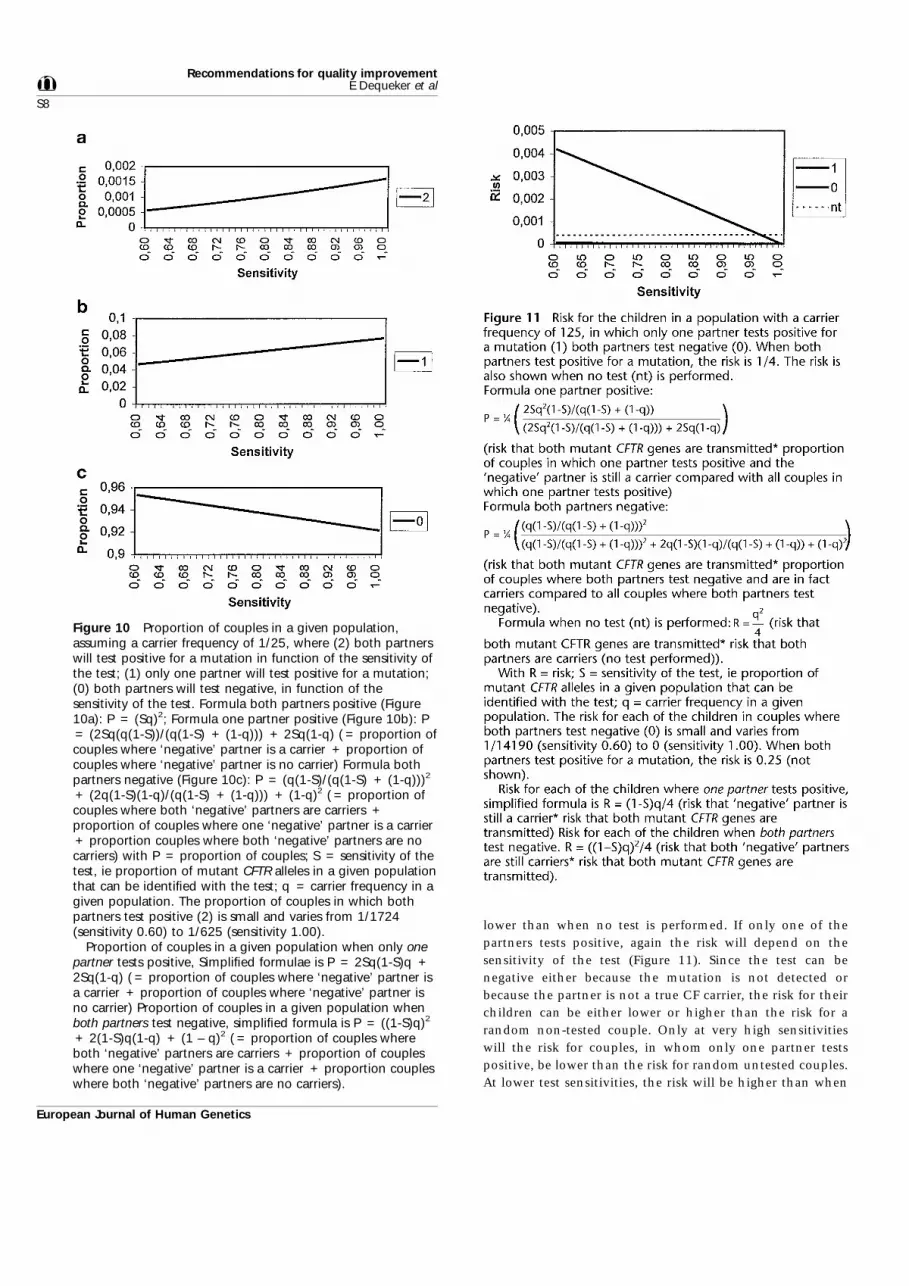
lower than when no test is performed. If only one of thepartners tests positive, again the risk will depend on thesensitivity of the test (Figure 11). Since the test can benegative either because the mutation is not detected orbecause the partner is not a true CF carrier, the risk for theirchildren can be either lower or higher than the risk for arandom non-tested couple. Only at very high sensitivitieswill the risk for couples, in whom only one partner testspositive, be lower than the risk for random untested couples.At lower test sensitivities, the risk will be higher than when
Figure 10 Proportion of couples in a given population,assuming a carrier frequency of 1/25, where (2) both partnerswill test positive for a mutation in function of the sensitivity ofthe test; (1) only one partner will test positive for a mutation;(0) both partners will test negative, in function of thesensitivity of the test. Formula both partners positive (Figure10a): P = (Sq)2; Formula one partner positive (Figure 10b): P= (2Sq(q(1-S))/(q(1-S) + (1-q))) + 2Sq(1-q) ( = proportion ofcouples where ‘negative’ partner is a carrier + proportion ofcouples where ‘negative’ partner is no carrier) Formula bothpartners negative (Figure 10c): P = (q(1-S)/(q(1-S) + (1-q)))2
+ (2q(1-S)(1-q)/(q(1-S) + (1-q))) + (1-q)2 ( = proportion ofcouples where both ‘negative’ partners are carriers +proportion of couples where one ‘negative’ partner is a carrier+ proportion couples where both ‘negative’ partners are nocarriers) with P = proportion of couples; S = sensitivity of thetest, ie proportion of mutant CFTR alleles in a given populationthat can be identified with the test; q = carrier frequency in agiven population. The proportion of couples in which bothpartners test positive (2) is small and varies from 1/1724(sensitivity 0.60) to 1/625 (sensitivity 1.00).
Proportion of couples in a given population when only onepartner tests positive, Simplified formulae is P = 2Sq(1-S)q +2Sq(1-q) ( = proportion of couples where ‘negative’ partner isa carrier + proportion of couples where ‘negative’ partner isno carrier) Proportion of couples in a given population whenboth partners test negative, simplified formula is P = ((1-S)q)2
+ 2(1-S)q(1-q) + (1 – q)2 ( = proportion of couples whereboth ‘negative’ partners are carriers + proportion of coupleswhere one ‘negative’ partner is a carrier + proportion coupleswhere both ‘negative’ partners are no carriers).
Recommendations for quality improvementy E Dequeker et al
S8
European Journal of Human Genetics

no test is performed. For this reason routine screening ofcouples in the population might be problematic. Indeed, theproportion of couples with only one partner who testspositive is about 1/20–1/25. They will have an increased riskafter testing, although only a minority of such couples has areal risk of having CF children. The proportion of thesecouples who have a real risk of having CF children is given inFigure 12. This proportion is again determined by thesensitivity of the test.
In the majority of couples, no mutation will be found(Figure 10). If the test has a 100% sensitivity, they will haveno risk of having CF children. If the test has a lowersensitivity, there is still a very small risk of having CF children(Figure 13).
ConclusionThe recommendations described here are an attempt toprovide written suggestions for the quality improvement ofCF testing in Europe. While it may be difficult for a numberof laboratories to fulfil all the criteria for a level 1 or 2laboratory in a short time, the approval of these recom-mendations by the many laboratories involved in CF testingsuggests that at least the awareness exists that qualityassessment is possible and necessary. Guidelines, as the wordindicates, attempt to be a guide to better quality, not laws
which must be respected and adhered to at the risk ofpunishment. The Steering Committee of the ConcertedAction also hopes that by publishing these recommendationsmany laboratories will be able to use the document to obtainimproved facilities and equipment from their authorities,and that regional, national or supra-national exchanges anddiscussions will lead to the evolvement of a free collaborativenetwork for CF testing to the benefit of patients, families andthe general population.
AcknowledgementsThis work was performed within the framework of the EuropeanConcerted Action on Cystic Fibrosis, (Biomed-2, contract numberBMH4-CT96-0462 and Inco-project, contract numberERBIC20-CT96-0058). The authors would like to thank Innogeneticsand Applied Biosystems for the financial support for publishing thissupplement). The authors thank the following for their comments andapproval of this document.
AustriaK Huber (Zentrallabor Donauspital, Vienna), PM Kroisel (Institut furMedizinische Biologie und Humangenetik, Universitat Graz, Graz), GUtermann, HG Kraft, J Loffler (Institut fur Medizinische Biologie undHumangenetik, Innsbruck), A Weinhausel (CCRI Labor ProfessorHaas, Vienna)
Figure 12 Proportion of couples, with only one partner whotests positive for a mutation, that have a real risk of having CFchildren, ie the couples where the partner that is negative forthe test is still a CF carrier. A carrier frequency of 1/25 isassumed.
(proportion of couples with one partner who tests positive butwhere the ‘negative’ partner is still a carrier compared to allcouples with one partner who tests positive) withP = proportion of couples, with only one partner who testspositive, that have a real risk of having CF children;S = sensitivity of the test, ie proportion of mutant CFTR allelesin a given population that can be identified with the test;q = Carrier frequency in a given population.
Proportion of couples, with only one partner testingpositive, that have a real risk of having CF children, simplifiedformulae P = 2Sq2(1-S)/(2Sq2(1-S) + 2Sq(1q)) (proportion ofcouples with one ‘positive’ partner in which the ‘negative’partner is still a carrier compared to all couples in which onepartner tests positive).
P = 2Sq2(1-S)/(q(1-S) + (1-q))
P = (2Sq2(1-S)/(q(1-S) + (1-q))) + 2Sq (1-q)P =
Figure 13 Proportion of couples, with both partners testingnegative, that have no risk of having CF children, eitherbecause only one or both partners are truly no CF carriers.A carrier frequency of 1/25 is assumed.
(proportion of couples where both partners test negative withone or both partners truly none carriers compared to allcouples where both partners test negative) with P = proportionof couples, where both partners test negative, that truly haveno risk of having CF children; S = sensitivity of the test,ie proportion of mutant CFTR alleles in a given population thatcan be identified with the test; q = carrier frequency in a givenpopulation.
Proportion of couples, where both partners test negative,and who truly have no real risk of having CF children,simplified formulae:
(proportion of couples in which both partners test negative inwhich one or both partners are truly no carriers compared toall couples in which both partners test negative).
P = (2q (1-S) (1-q)/(q (1-S) + (1-q))) + (1-q)2
P = (q(1-S)/(q(1-S) + (1-q)))2 + 2q(1-S)(1-q)/(q(1-S) + (1-q)) + (1-q)2
P = (1-q)2 + 2(1-S)q(1-q)
P = (1-q)2 + 2(1-S)q(1-q) + ((1-S)q)2P =
P =
Recommendations for quality improvementE Dequeker et al y
S9
European Journal of Human Genetics

BelgiumP Cochaux (Genetique Moleculaire, Hopital Erasme, Brussels), PHendrix (Innogenetics, Belgium), P Hilbert (IPG Biologie Moleculaire,Gerpinnes), G Matthijs (Molecular Diagnostic Laboratory, Center forHuman Genetics, Leuven), L Messiaen (Centrum Medische Genetica,UZ-Gent, Gent), K Storm (Center for Human Genetics, University ofAntwerp, Antwerp), C Verellen-Dumoulin (Center for HumanGenetics, UCL, Brussels)
BulgariaI Kremensky (Laboratory of Molecular Pathology, Sofia MedicalUniversity, Sofia)
CroatiaG Janackovic (Division of Molecular Medicine, Laboratory ofMolecular Oncology, Ludjel Boskovic Institute, Zagreb), J Sertic(Laboratory for Molecular Diagnosis, Clinical Hospital Centre Zagreb,Zagreb)
CyprusC Deltas (Molecular Genetics, The Cyprus Institute of Neurology andGenetics, Nicosia)
Czech RepublicM Macek Jr (CF Center, Molecular Genetic Laboratory, Institute ofBiology and Medical Genetics, Prague)
DenmarkM Schwartz (Department of Clinical Genetics, University ofCopenhagen, Copenhagen)
FranceE Bieth (Laboratoire de Genetique Medicale Hopital Purpan, Toulouse),JP Bonnefont, L Thuillier (Biochimie Genetique, HopitalNecker-Enfants Malades, Paris), J-C Chomel (Laboratoire deGenetique Cellulaire et Moleculaire, CHU de Poitiers, Poitiers), CClaval (Pol Bovin, Unite de Biologie Cellulaire, Hopital MaisonBlanche, Reims), V David (Laboratoire de Genetique Moleculaire,CHU Pontchaillou, Rennes), C Ferec (Molecular Genetics, Brest), MGoossens, E Girodon (Moleculair Genetics, Hospital Henri Mondor,Creteil), A Iron (Laboratoire de Biochimie, Centre HospitalierPellagrin, Bordeaux), J Lunardi (Biochimie de l’ AND, CHU Grenoble,Grenoble), P Malzac (Laboratoire de Biologie Moleculaire Applique,Service du Professeur Mattei, Marseille), H Mittre (Laboratoire deBiochemie B, CHU av Georges Clemenceau, Caen), F Thepot (CentreHospitalier Universitaire d’Amiens, Amiens)
GermanyC Aulehla-Scholz (Institut fur Klinische Genetik, Frauenklinik Berg,Stuttgart), I Bauer (University of Rostock, Children’s Hospital,Department of Medical Genetics, Rostock), I Bohm(Gemeinschaftspraxis, Dr Ovens-Raeder / Dr C Waldenmaier), RFahsold (Gemeinschaftspraxis Prager und Junge, Dresden), W Friedl,M Jungck (Institut fur Humangenetik, Universitat Bonn, Bonn), DGlaser (Laborpraxis Dres. Mehnert, Medizinische Genetik Neu-Ulm),U Hammer (Molecular Diagnostics, Klinikum derFriedrich-Schiller-Universitat Jena, Jena), S Jakubiczka(Otto-von-Guericke-Universitat, Institut fur Human Genetik,Magdeburg), B Janssen (Institut fur Humangenetik, Heidelberg), H-GKlein (Facharzt fur Laboratoriumsmedizin, Medizinische Genetik,Planegg/Martinsried), P Kozlowski (Institut fur Praenatal Medizin und
Genetik, Dusseldorf), C Kraus (Institut fur Humangenetik, Erlangen),K Kutsche (Institut fur Humangenetitk, Universitat-KrankenhausEppendorf, Hamburg), F Laccone (Institute of Human Genetics,Gottingen), L Lubbe (Carl-Thiem-Klinikum Institut fur Pathologie,Cottbus), E Meese (Department of Human Genetics, University ofSaarland, Homburg/Saar), U Muller, K Atland (Institute of HumanGenetics, Justus-Liebig-University, Giessen), U Pantke(Humangenetische Praxis, Giessen), H Skladny (Praxis Dr Greiner,Mannheim), E Schroder (Institut fur Humangenetik Dusseldorf), PSteinbach (Abteillung Medizinische Genetik, Universitat Ulm, Ulm), MStuhrmann (Institut fur Humangenetik, Medizinische HochschuleHannover, Hannover), G Wildhardt (Universitat Mainz, Mainz), CZuhlke (Humangenetik, Medizinische Universitat zu Lubeck, Lubeck),Gemeinschaftspraxis fur Laboratoriumsmedizin, Prov Dr med MichaelGiesing und Partner (Recklinghausen)
HungaryK Nemeth, A Varadi, G Fekete (Molecular Genetics 2nd Department ofPediatrics, SOTE, Budapest)
ItalyE d’Alcamo (Servizio Talassemia, Unita di Ricenca ‘Piera Cutino’,Palermo), A Amoroso (Medical Genetics, Children Hospital, Trieste), CArduino (Centro Genetica Medica, Turin), M Bartolozzi (SezioneGenetica Medica, Grosseto), M Benettazo, PF Pignatti (Institute ofBiology and Genetics, University of Verona, Verona), A Cantu-Rajnoldi(Anatomia ed Istologia Patologica, Sez. Genetica Molecolare, Milan),Dagna Bricarelli, P Scartezzini (Laboratoire Genetica Umana,Genova), M Furbetta, A Angius, M Stasi (Genetica Moleculare, Istitutodi Pediatria Clinica Sociale e Preventiva, Perugia), G Novelli (HumanGenetics, Tor Vergata University, Rome), L Picci, M Scarpa (MolecularGenetics Laboratory, Padua), MC Rosatelli, A Cappai, A Cao(Genetica Moleculare, Istituto di Clinica e Biologia Dell’eta’ Evolutiva,Cagliari), T Santostasi (Laboratoria Fibrosi Cystica, CentroRiferimeno, Bari), P Simi (U.O. Citogenetica e Genetica Moleculare,Instituto Clinica Pediatrica Universita via Roma, Pisa), F Torricelli(Unita Operativa Citogenetica e Genetica, Florence)
LatviaV Kroshkina (Department of Medical Biology and Genetics, MedicalAcademy of Latvia, Riga)
Macedonia RepublicG Efremov (Macedonian Academy of Science and Arts, Research Centerfor Genetic Engineering and Biotechnology, Skopje)
NorwayK Eiklid (Department of Medical Genetics, Ulleval University Hospital,Oslo)
PolandJ Bal (Department of Genetics, National Research Institute of Motherand Child, Warsaw), M Witt (Insitute of Human Genetics, PolishAcademy of Sciences, Poznan)
PortugalP Pacheco (Centro de Genetica Humana, Instituto Nacional de SaudeDr Ricardo Jorge, Lisbon)
Recommendations for quality improvementy E Dequeker et al
S10
European Journal of Human Genetics

SpainA Arnaiz-Villena (Sesvicio de Inmunologia Hospital Universitario 12de Octubre, Madrid), J Benitez (Mercedes Robledo, GeneticsDepartment, Madrid), M Fernandez-Burriel (Unidad Genetica,Hospital Materno-Infantil, Canary Islands), G Glover (Centro deBioquimica y Genetica Clinica, Espanardo, Murcia), J Molano (UnidadGenetica Molecular Bioquimica, Hospital La Paz, Madrid), JL SanMillan (Unidad de Genetica Molecular, Hospital Ramon y Cajal,Madrid), J Telleria-Orriols, J Alonso (Department Pediatria, Facultadde Medicina, Valladolid)
SwedenU Kristofferson (Department of Clinical Genetics, Lund)
SwitzerlandM Hergersberg (Institut fur Medizinische Genetik, Universitat Zurich,Zurich)
The NetherlandsH Scheffer (Department of Medical Genetics, University of Groningen,Groningen)
UkraineL Livshits (Group of Molecular Diagnosis of Hereditary Diseases,Department of Human Genetics, Kiev)
United KingdomJ Dodge (Department of Child Health, University of Swansea,Swansea), R Elles, M Coleman (Regional Molecular GeneticLaboratory, St Mary’s Hospital, Manchester), C Graham (MolecularGenetics, Belfast City Hospital Trust, Belfast), F MacDonald, S Bullock(DNA Laborary, Birmingham Women’s Hospital, Birmingham)
YugoslaviaD Radojkovic (IMGGE, Laboratory for Molecular Biology, Belgrade)
References1 Dork T, Dworniczak B, Aulehla-Scholz C et al: Distinct spectrum of
CFTR gene mutations in congenital absence of vas deferens. HumGenet 1997; 100: 365–377.
2 Harris R: Genetic services in Europe. Eur J Hum Genet 1997; 5(supplement 2): 1– 220.
3 Cotton RGH: Mutation detection and mutation databases. ClinChem Lab Med 1998; 36(8): 519–522.
4 Grody W, Desnick R, Carpenter N, Noll W: Diversity of cysticfibrosis mutation screening practices. Am J Hum Genet 1998; 62:1252–1254.
5 Estivill X, Bancells C, Ramos C, the Biomed CF Mutation AnalysisConsortium: Geographic distribution and regional origin of 272cystic fibrosis mutations in European populations. Hum Mut 1997;10: 135–154.
6 Rending S, Calafelli F, Cappello N et al: Genetic history of cysticfibrosis mutations in Italy. I. Regional distribution. Ann Hum Genet1997; 61: 411–424.
7 Stuhrmann-Sprangenberg M, Aulehla-Scholz C, Dworniczak B,Reiss J, Berufsverband Medizinische Genetik: Qualitatssicherung,Leitlinien zur Molekulargenetischen Diagnostik der CystischenFibrose. Med Genetik 1997; 4: 553–559.
8 The CF mutation database. 1999 http://www.genet.sickkids.on.ca/cfr
9 Bridge P: The calculation of genetic risk – worked examples in DNAdiagnostic, Johns Hopkins University Press: Baltimore and London,1997.
10 National Committee for Clinical Laboratory Standards: MolecularDiagnostic Methods for Genetic Diseases; Proposed Guidelines. NCCLSdocument MM1-P ISBN 1-56238-340-X. 1997.
11 Prence E: A practical guide for the validation of genetic tests. GenTest 1999; 3: 201–205.
12 OECD: The OECD Principles of Good Laboratory Practices, (revised1997): OECD Environmental Health and Safety Publications.Series on Principles of Good Laboratory Practice and ComplianceMonitoring 1998; No. 1: ENV/MC/CHEM (98)17.
13 Visviskis S, Schlenck A, Maurice M: DNA extraction and stabilityfor epidemiological studies. Clin Chem Lab Med 1998; 36(8):551–555.
14 Madison L, Hoar DI, Holroyd CD, Crisp M, Hodes ME: DNAbanking: the effects of storage of blood and isolated DNA on theintegrity of DNA. Am J Med Genet 1997; 27: 379–390.
15 Cotton R: Mutation Detection. Oxford University Press, Oxford,New York, Tokyo, 1997.
16 Rommes J, Kerem B, Greer W, Chang P, Tsui LC, Ray P: Rapidnonradioactive detection of the major cystic fibrosis mutation.Am J Hum Genet 1990; 46: 395–396.
17 Taylor GR, Noble JS, Hall JL, Quirke P, Stewart AD, Mueller RF:Rapid screening for dF508 deletion in cystic fibrosis. Lancet 1989;1345.
18 Cuppens H, Buyse I, Baens M, Marynen P, Cassiman JJ: Simultane-ous screening for 11 mutations in the cystic fibrosis transmem-brane conductance regulator gene by multiplex amplification andreverse dot-blot. Mol Cell Probes 1992; 6: 33–39.
19 Ferrie RM, Schwarz MJ, Robertson NH et al: Development,multiplexing and application of ARMS tests for common muta-tions in the CFTR gene. Am J Hum Genet 1992; 51(2): 251–262.
20 Ravnik-Glavac M, Glavac D, Chernick M, di-Sant’Agnese P, DeanM: Screening for CF mutations in adult cystic fibrosis patientswith a directed and optimized SSCP strategy. Hum Mut 1994; 3(3):231–238.
21 Ravnik-Glavac M, Glavac D, Dean M: Sensitivity of single-strandconformation polymorphism and heteroduplex method formutation detection in the cystic fibrosis gene. Hum Mol Genet1994; 3: 801–807.
22 Fanen P, Ghanem N, Vidaud M et al: Molecular characterization ofcystic fibrosis: 16 novel mutations identified by analysis of thewhole cystic fibrosis conductance transmembrane regulator(CFTR) coding regions and splice site junctions. Genomics 1992;13: 770–776.
23 Costes B, Girodon E, Ghanem N et al: Psoralem-modifiedoligonucleotide primers improve detection of mutations bydenaturing gradient gel electrophoresis and provide an alternativeto GC-clamping. Hum Mol Genet 1993; 2: 393–397.
24 Taylor G: Laboratory Methods for the Detection of Mutations andPolymorphisms in DNA. CRC Press, Boca-Raton, New York, Lon-don, Tokyo, 1996.
25 Dequeker E, Cassiman J-J: Evaluation of CFTR gene mutationtesting methods in 136 diagnostic laboratories: report of a largeEuropean external quality assessment. Eur J Hum Gen 1998; 6:165–175.
26 Elles R: Methods in Molecular Medicine. Molecular Diagnosis ofGenetic Diseases. Totowa, New Jersey 1996.
27 Huisman W: Quality system in the medical laboratory; the role ofa quality manual. Ann Biol Clin 1994; 52: 457–461.
28 Dybkaer R: A quality manual for clinical laboratory including theelements of a quality system. Scand J Clin Lab Invest 1993; 53(suppl 21).
29 Eucromic Quality Assessment Group: Quality guidelines andstandards for genetic laboratories/clinic in prenatal diagnosis onfetal samples obtained by invasive procedures: Eur J Hum Genet.1997; 5: 342–350.
30 Clinical Pathology Accreditation UK: EQA Scheme Accreditation.Company Registration no. 2675095 1996.
Recommendations for quality improvementE Dequeker et al y
S11
European Journal of Human Genetics

31 Neumaier M, Braun A, Wagener N: Fundamentals of qualityassessment of molecular amplification methods in clinical diag-nosis. Clin Chem 1998; 44(1): 12–26.
32 M Schwarz and Clinical Molecular Genetics Society: CysticFibrosis: Guidelines. 1996. http://www.molgenqa.demon.co.uk/cf.htm.
33 The American College of Medical Genetics: Standard and Guide-lines: Clinical Genetics Laboratories. 1994.
34 College of American Pathologists: Commission on LaboratoryAccreditation. 1993; 60093-2750.
35 Kwok S, Higuchi R: Avoiding false positives with PCR. Nature1989; 339: 237–238.
36 Kitchin P, Szotyori Z, Fromholc C, Almond N: Avoidance of falsepositives. Nature 1990; 344: 201.
37 Persing DH: Polymerase chain reactions: trenches to benches. JClin Microbiol 1991; 29: 1281–1285.
38 Koch D, Peters T: Selection and evaluation of methods with anintroduction to statistical techniques. In: Burtis C, Ashwood E(eds). Fundamentals of Clinical Chemistry. Saunders: Philadel-phia, 1996, pp 170–181.
39 Shultz E: Clinical interpretation of laboratory procedures. In:Burtis C, Ashwood E (eds). Fundamentals of Clinical Chemistry.Saunders: Philadelphia, 1996, pp 192–199.
40 Ad Hoc Committee on Mutation Nomenclature: Update onnomenclature for human gene mutation. Hum Mut 1996; 8:197–202.
41 Antonarakis S and The Nomenclature Working Group: Reco-mendations for a nomenclature system for human gene muta-tions. Hum Mut 1998; 11: 1–3.
42 Beaudet A, Tsui LC: A suggested nomemclature for designatingmutations. Hum Mut 1993; 2: 245–248.
43 Beutler E, McKusick V, Motulsky A, Scriver C, Hutchison F:Mutation nomenclature: nicknames, systematic names andunique identifiers. Hum Mut 1996; 8: 203–206.
44 Beutler E: The designation of mutation. Am J Hum Genet 1993; 53:783–785.
45 Dork T, Neumann T, Wulbrand U et al: Intra- and extragenicmarker haplotypes of CFTR mutations in cystic fibrosis families.Hum Genet 1992; 88: 417–425.
46 Cuppens H, Heng H, Raeymaekers P, De Boeck C, Cassiman JJ:CFTR haplotype backgrounds on normal and mutant CFTR genes.Hum Mol Genet 1994; 3: 607–614.
47 Morral N, Estivill X: Multiplex PCR amplification of threemicrosatellites within the CFTR gene. Genomics 1992; 13:1362–1364.
Recommendations for quality improvementy E Dequeker et al
S12
European Journal of Human Genetics

Appendix 1More detailed guidelines about laboratory methods, organi-sation and reporting are given here. These guidelines arebased as much as possible on existing internationally availa-ble documents. Aspects which particularly pertain to CF havebeen highlighted or have been added.
MethodsThere are three essential steps in mutation screening: theDNA extraction, DNA banking, and the mutation testingmethod.
DNA extraction A number of methods exist for thepreparation of nucleic acid samples for molecular geneticsanalysis. Since this stage can have a significant impact on thequality of the final results, care should be exercised to ensurethat a validated protocol is followed, independent of whetherthe extraction method was developed in-house, obtainedfrom the literature, or purchased as a kit from a manufacturer.Protocols that have been either developed in-house or as amodification of a manufacturer’s kit should be validated (seefurther). Protocols that follow exactly a validated publishedmethod should undergo thorough performance verifica-tion.10,11 Written procedures of the methods used for DNApurification, including the sources of all components used,should be kept. Complete references should be included instandard operating procedure manuals. Changes in any ofthe procedures or source of components should be docu-mented and approved by the laboratory, with date andinitials recorded.12
The ideal method of DNA isolation in a moleculardiagnostic laboratory must be simple, fast, safe and economi-cal, but also precise and reliable. It must yield a high qualityand quantity of high molecular weight DNA.
DNA banking Isolated material must be stored at 4°C orfrozen. Excess DNA sample material should be stored at atemperature not higher than 0–5°C to ensure long-termstability. Concerning DNA stability and storage we refer tothe literature.13,14 An optimal storage procedure wouldinvolve aliquoting of the DNA solution in one primary stocksolution frozen at –80°C, and multiple portions for sub-sequent analyses stored at 4°C and/or –20°C. This procedureavoids repeated freeze/thaw cycles and minimises the possi-bility of DNA contamination.
Cystic fibrosis mutation testing methods A wide range ofmutation testing methods15–24 is currently used in diagnosticlaboratories.25 The most frequently used mutation detectionmethods for cystic fibrosis are heteroduplex analysis, restric-tion enzyme analysis, reverse dot-blot, the commercial kitsINNO-LiPA CF2 (Innogenetics nv, Gent, Belgium), ElucigeneCF4 and CF12 (AstraZeneca Diagnostics, Abingdon, Oxford-shire, UK), and OLA Cystic Fibrosis Assay (PE AppliedBiosystems, New Jersey, USA). Single strand conformation
polymorphism (SSCP), denaturing gradient gel electrophor-esis (DGGE), two-dimensional DNA electrophoresis, andsequencing are mostly used as CF mutation screeningmethods.26 The ideal method for mutation testing in amolecular diagnostic laboratory must be rapid and cheap,allow automisation, 100% efficiency and avoid the use ofradioactivity and toxic reagents.
Whatever the methods used in a diagnostic laboratory, it isimportant that they are thoroughly validated (see testvalidation and characterisation). Written standard operatingprocedures, including the sources of all components, shouldbe kept. Changes in any of the procedures or source ofcomponents should be documented and approved, with dateand initials recorded.12
Laboratory organisation
Quality system A quality system for a molecular geneticslaboratory should be directed at all fundamental aspects of itsfunction. This means the setting-up of a quality systemaccording to a molecular genetic translation of the criteria ofgood laboratory practices as developed by the OECD, theEN 45001 standard (the general criteria for the operation oftesting laboratories) or ISO 17025, which are accepted in theEuropean Union as the present standard. A quality systemalso includes the obligation to join external quality assess-ment schemes, and to make use of their results. Informationabout the requirements for a quality system is described inthe literature.10,27–29
Because the methodology of molecular biological diag-nostics is constantly changing, no general standard can atpresent be defined. We suggest the guidelines formulated bythe National Committee for Clinical Laboratory Standards(NCCLS) and the European Concerted Action (BMH-CT93-1673) (Eucromic)30 be followed. A guide to funda-mentals of quality assessment of molecular amplificationmethods in clinical diagnostics has recently been publishedby M Neumaier.31 These guidelines describe in detail reagentquality control, equipment calibration and maintenance,proficiency testing, training of technical staff, internal andexternal quality control.
Specimen types, specimen identification and access Collec-tion and identification of the specimens are (often) carriedout by non-laboratory personnel. Written procedures forproper collection, packing, shipment and handling of speci-mens are recommended by all the guidelines,7,10,32,33 docu-ments, and quality assessment authorities consulted.12,34
Based on a review of these guidelines, we propose imple-mentation of the following items.
(1) Specimen typesSince genetic analysis in cystic fibrosis is PCR-based, aminimal amount of DNA is required in the vast majorityof cases. Unless extensive analysis is expected or planned,a mouthwash sample (or a buccal scrape sample in the
Recommendations for quality improvementE Dequeker et al y
S13
European Journal of Human Genetics

case of a baby) is sufficient. Otherwise a small volume ofblood (2–5 ml) in EDTA will provide enough DNA forextensive analysis.
Prenatal diagnosis of cystic fibrosis is usually carriedout on a chorionic villus sample taken during the firsttrimester of pregnancy. The samples should be checkedby microscopic inspection and dissected immediately.Ideally they are independently prepared samples, whichshould all be tested. This minimises the potential prob-lem of maternal cell contamination of the sample.
Amniocytes can also be used for molecular geneticanalysis – either directly spun down from amniocentesissample or after 10–14 days culture. Direct analysis shouldbe carried out with caution, as the foetal cells areinvariably contaminated with maternal cells. Tissueculture of the cells can either remove/reduce maternalcomplication or result in overgrowth of maternal fibro-blasts. The analysis of a few polymorphic markers (CArepeats or STR) can identify the presence of contamina-tion when compared with maternal DNA.
(2) Specimen identificationThe specimen container should be clearly marked with aunique patient identifier, such as a hospital patientidentification number. In most situations, the patient’sname is not sufficient, although the combination ofpatient name and birth date is generally enough toprevent identification errors. The container should alsobe labelled with the date and time that the specimen wasacquired.
(3) Request formsAll specimens should be accompanied by a request formwhich contains as much of the following information aspossible
Patient’s name
Date of birth
Date of collection
Gender
Ethnicity (if applicable)
Place of birth of patient, parents and grandparents
Unique identifier found on the specimencontainer
Specimen type (blood, amniotic fluid, etc)
Reason for requesting the test, based on clinicalinformation
Relevant clinical or laboratory information, includ-ing sweat test results
Pedigree (recommended for all cases)
Referring physician or health professional
Weeks of gestation (for prenatal diagnosis)
Biling information (if applicable)
(4) Criteria for rejecting specimens10
It is recommended that each laboratory has writtencriteria for acceptance or rejection of specimens.
Rejection of specimens is strongly recommended ifeither the specimen or the request form lacks sufficientinformation for the laboratory or clinician to uniquelyidentify the specimen, or lacks other information neces-sary to determine if the specimen or test requested isappropriate for answering the clinical question. It is alsostrongly recommended that improperly handled or trans-ported specimens should be rejected.
Other conditions for accepting or rejecting specimensare left to the discretion of the laboratory. Given thepower of gene amplification techniques, it is difficult toset an arbitrary minimum cellular content or volume forspecimen acceptance, but it is recommended that eachlaboratory develops its own standards. It is recommendedthat prenatal specimens, especially chorionic villus sam-ples, be assessed for maternal cell contamination; this isdone morphologically and by PCR-based DNA analysis.
(5) Accessing specimensEach specimen should be assigned a unique laboratoryidentifier when accepted for testing. This identifiershould be linked with the unique patient/family identi-fier and with other identifiers, such as those for individ-ual electrophoretic gels or blots that may be used in thelaboratory. The unique laboratory identifier should dif-ferentiate between specimens from different patients,between different specimens submitted from the samepatient, and between specimens, from different patientsof the same family.
(6) Specimen transport and storageEach laboratory should establish criteria based on its ownexperience in successful extraction of analysable DNAfrom the various sample types. In general, frozen tissueshould be transported on dry ice, fresh tissue on wet ice,and fixed or dried tissue at room temperature. Amnioticfluids should be transported at room temperature whichallow the establishment of cell cultures. DNA can beextracted from whole blood stored at room temperaturefor a week or more.14 Frozen tissue can be stored at –70°Cindefinitely; fixed or dried samples can be stored at roomtemperature for variable periods depending on thelaboratory’s experience.
(7) Specimen retentionA primary issue regarding specimen retention involvesethical and legal considerations such as specimen owner-ship, confidentiality, and informed consent.
Until universal recommendations are adopted or reg-ulations are implemented, each laboratory should estab-lish its own policy regarding specimen retention and theuse of archived specimens or stored DNA (anonymisation
Recommendations for quality improvementy E Dequeker et al
S14
European Journal of Human Genetics

when used in research etc). Such policies will need to bein compliance with institutional regulations that mayexist with federal and/or state regulations as they becomeimplemented.
Controlling false-positive nucleic acid and target amplifica-tion reactions A significant challenge facing the diagnosticamplification of nucleic acids is the occurrence of false-positive results due to contaminating nucleic acids. Theability of amplification techniques to produce large numbersof copies of a sequence from minute quantities of nucleicacid necessitates that extreme care should be taken to avoidfalse-positive results due to transfer of DNA betweensamples.
(1) Laboratory designIdeally, three physically separate areas of the laboratoryshould be available for reagent preparation, specimenpreparation, and amplification and product detection.The reagent preparation area, for those laboratories usingonly commercially available kits, is considered to be atthe site of manufacture. In laboratories where enzymaticor chemical means of inactivating amplified products areused, the demands for physical separation of pre- andpostamplification procedures may be somewhat reduced,but good laboratory practice should still be diligentlyexercised.
(a) WorkflowSpecimens should be processed in an area of thelaboratory that is isolated from amplification anddetection areas. Ideally, the specimen preparationarea should be under positive pressure to otherareas of the laboratory. If the specimen preparationarea cannot be maintained at a positive pressure toother areas of the laboratory, specimen preparationshould be performed in a class II biological safetycabinet to prevent contamination. The pre- andpostamplification laboratories should be served byseparate ventilation systems. Also, the postamplifi-cation area should be under negative pressure.Traffic of personnel should be from the specimenpreparation area, with a change of laboratorycoats, to the pre- and postamplification areas.Laboratory coats should be dedicated to specificareas and changed when going in and out of eacharea. With the introduction of commerciallylicensed tests and new methods, some of theserequirements may be reduced. However, if con-tamination becomes a problem, then separateareas of the laboratory should be devised toaccommodate these different processing stages.
(b) Containment devicesIn the event of separate laboratory space not beingavailable to segregate pre- and postamplification
activities, a class II biological safety cabinet shouldbe used as a containment device for specimenpreparation. Class I safety cabinets do not provideprotection for material contained within them.Dead-air boxes with ultraviolet light attachmentscan provide a clean bench area for specimenpreparation in a dedicated specimen preparationlaboratory; UV lamps lose energy efficiency overtime.
(2) Laboratory practiceSpecific laboratory practice should be implemented tominimise the occurrence of false-positive results. Theseinclude the preparation of reagents and solutions, thecorrect use of pipettes, the use of laboratory coats andgloves and procedures for manipulation of reactiontubes. In addition, special care should be taken in the useof appropriate controls.10,35–37
(3) Selection and preparation of controlsA positive control that amplifies weakly but consistentlyshould be selected. The use of dilute positive controlsprevents the unnecessary generation of large amounts ofamplified product that can result in contamination.
Applicable reagent controls should be included witheach amplification batch run. These controls contain allnecessary components of the reaction without the addi-tion of template nucleic acid or human DNA.
Negative controls should be dispensed last so that theyreflect the state of the reagents added.
Assays based on presence or absence of PCR products mustinclude known control primers yielding a positive resultto check for proper amplification and sizing of the PCRproducts and to ensure that a negative result is accurate.This should include a positive result with control primersdetecting a spiked additive or a constitutivecomponent.
When specimens are analysed for sequence variation,controls containing all alleles to be detected must beincluded.
Assays in which the result is based on fragment sizemust include size markers (sequencing ladders, etc)covering the range of expected results during gelelectrophoresis.
Assays based on changing of electrophoretic mobility mustinclude appropriate controls to ensure correct inter-pretation. New samples should be confirmed by alter-native methods. Any unexpected result requires a repeatof the assay. Procedures for the analysis of possible newmutations should be available.
Test validation and characterisation For most testsperformed in general clinical laboratories the process of testvalidation is fairly straightforward and can be done accordingto well established guidelines.38 However, the unique aspects
Recommendations for quality improvementE Dequeker et al y
S15
European Journal of Human Genetics

of genetic testing make the validation of genetic tests achallenge in many instances. Validation of a clinicallaboratory test should be on both analytical and clinicallevels. Analytical validation involves determination of thevarious parameters themselves, such as accuracy, precision,analytical range, sensitivity, specificity, detection limit,interferences, and recovery.38 Clinical validation refers todetermination of the predictive value of the test, or theprobability that a person with a positive test result will haveor will develop the disease.39
Test validation should be conducted before a new test isintroduced for clinical use. The test should be subjected toliterature review and to analytical and laboratory/clinicalcorrelation studies. This means characterisation of thedetected mutations, establishing the performance propertiesof the test to ensure the test’s ability to provide consistentand reliable results, establishing the clinical utility of the test,defining aspects of the procedure which should be carefullyregulated to maintain test performance, and defining thelimitations of the test.
Such validation is necessary to assure the safe and effectiveapplication of a genetic test for its intended use. Eachlaboratory should develop its own validation protocol. Werecommend the recently published practical guide for thevalidation of genetic tests by E Prence11 be followed andused.
Safety Training and use of safe laboratory practices areessential for the protection of all personnel. Good LaboratoryPractices12 and the Commission on Laboratory Accreditationof the College of American Pathologist34 recommend ageneral safety file in each laboratory. This document shoulddescribe safety measures concerning infective samples,dangerous chemicals, radiation and electronic danger, andinstructions for proper cleaning of the laboratory.
Reports
Reports of the results Genetic test results should becommunicated to the referring physician or geneticprofessional and to any physician designated by the patient.Reports of test results should be issued in a standardisedform, clearly intelligible to the non-specialist. In general, thelaboratory should not directly report on the results to thepatient: the laboratory should ensure that the clinicianreporting to the patient has a full understanding of theresults and the underlying clinical meaning of the result. Thelaboratory report should include:
collecting date
nature of the sample
name of the individual
date of birth/place of birth
laboratory identification number of patient and sample
date of report
reason for testing
the genotype and/or haplotype established for theindividual
interpretation of the data (should relate to the reason fortesting prenatal diagnosis, carrier testing, the sensitivity ofthe test, etc)
the signature of the laboratory director or otherauthorised individual and his/her name
It is recommended that along with this information, thefollowing be included: the family number (if it has beenassigned), and a pedigree with the genotype informationindicated if applicable (eg linkage study).10,33
Nomenclature for designation of mutations A systematiccommon nomenclature is essential for the deposition ofmutations into computerised databases and their subsequentaccessibility to the research and clinical community.Databases of mutations in genes are required for efficientaccess by clinicians and researchers. Clinical geneticists canidentify and study patients with the same mutations andperhaps provide prognostic information. Researchers canreadily determine whether a specific mutation has beendescribed.15
It is obvious that the most unambiguous nomenclaturesystem is based on genomic DNA. However, lengthpolymorphisms may create a problem in the numbering ofnucleotides. Therefore a reference sequence standard needsto be established. We recommend the nomenclature systemsuggested by the Ad Hoc Committee on MutationNomenclature40 and Antonarakis et al.41
Thanks to the electronically available database set up byL-C Tsui,8 the nomenclature of cystic fibrosis mutations isuniversally accepted. Although the nomenclature for CF issimple, results of the European quality control trials(Dequeker and Cassiman25) demonstrated that a significantnumber of laboratories violated the nomenclature rules.Based on earlier published documents,7,10,32,41–44 we describein the following the major issues of the CF mutationnomenclature system.
(1) General recommendationsA single letter code is used for designating amino acids.Nucleotides are designated as DNA bases, not as RNAbases. Nucleotides are designated in the sense strand (egATG for a methionine codon). With this nomenclaturesystem, mutation designations starting with a letter referto an amino acid and the number refers to a codonposition. In contrast, mutation designations starting witha number refer to a nucleotide position in the codingsense strand, and subsequent letters refer to DNA bases.Nucleotide changes are indicated by arrows. Superscriptsand subscripts should not be used, and there should be
Recommendations for quality improvementy E Dequeker et al
S16
European Journal of Human Genetics

no spaces between the numbers and letters in a mutationdesignation.
(2) Missense and nonsense mutationsMissense and nonsense mutations are described in termsof the change in the gene product. A missense mutationis designated by the number of the amino acid positionand the single-letter abbreviations of the amino acidsinvolved. The abbreviation for the normal amino acidprecedes the number, and the mutant amino acid followsthe number, with no spaces between. For example,G551D indicates that the glycine residue at position 551in the protein has been replaced by an aspartic acidresidue. If different nucleotide substitutions lead to thesame amino acid substitution, such as that occurring atS549 in the CFTR gene, the mutation designation shouldinclude the nucleotide change, within parenthesis,immediately following the designation for the aminoacid substitution. Using the cystic fibrosis gene example,this would be S549R(1777 A/C), or S549R(1778 T/G).
Nonsense mutations are designated similarly, exceptthat X represents a termination codon. For example,G542X indicates that the glycine residue at amino acidposition 542 has been replaced by a termination codon.
(3) Insertions and deletionsInsertion or deletion mutations are designated by anucleotide number of the sense strand, followed by ins(for insertion) or del (for deletion). The nucleotideposition is the one preceding an insertion, or the firstthat is deleted. The exact nucleotides are specified if onlyone or two are involved. For example, 441delA indicatesthe deletion of deoxyadenylic acid at nucleotideposition 441. The ‘name’ 241delAT indicates the deletionof deoxyadenylic acid at nucleotide position 241, and thedeletion of deoxythymidylic acid from nucleotideposition 242. Mutations involving both substitution anda small insertion or deletion can be designated by the firstaltered base, followed by the nucleotide change. Forexample, 2183AA–G indicates replacement of AA atnucleotide positions 2183 and 2184 in the normalsequence by G in the mutant allele.
(4) In-frame deletionsDeletions of single amino acids result from deletions ofthree bases and are represented by a ∆ followed by thesingle-letter code of the amino acid and its position, eg∆F508 is the deletion of phenylalanine (F) at pos-ition 508. An acceptable alternative is ‘Delta’, ‘delta’ or‘del’. It has been proposed to write amino acid deletionsin the future as amino acid codon del e.g. F508 del.41
(5) Complex deletions/insertionsThere are several mutations which involve the deletionor insertion of four or more bases and these are usuallynamed as the number of bases which are deleted orinserted, eg 1461ins4 which is the insertion of four bases(in this case AGAT) after base 1461, and 1949del84 whichis the deletion of 84 bases from base 1949. There are somemuch larger deletions which remove one or morecomplete exons, eg CFTRdel2 which is the deletion ofexon 2.
(6) Splicing mutationsThese are (usually) the substitution of a base in the spliceacceptor site (an AG dinucleotide at the 3' end of theintron) or the splice donor site (a GT dinucleotide at the5' end of the intron), both of which are highly conservedin human genomic DNA. The position of the mutatedbase is numbered from the first or last base in the exon, asintronic bases are not themselves numbered. Thus themutation 621 + 1G > T is the substitution of a guanineby a thymidine at the first base in intron 4 (the last baseof exon 4 being numbered 621 and the first base ofintron 4 being 621 + 1). Similarly, 621 + 2T > C is asubstitution at the second base in intron 4. Numbering ofthe acceptor site bases is from the 5' end of the exon, eg1717–1G > A is the substitution of the last base ofintron 10 (a guanine) by an adenine. Some splicingmutations are quite distant form intron/exonboundaries, eg 3849 + 10 kbC > T.
(7) Other nomenclature rulesNomenclature rules suggested for larger deletions andinsertions, splicing mutations, mutations in thenoncoding sequence and the more complex mutationsare addressed by the Ad Hoc Committee on MutationNomenclature.40
Recommendations for quality improvementE Dequeker et al y
S17
European Journal of Human Genetics

Appendix 2Members of the ECCACF connected with a geneticdiagnostic laboratoryAustriaK Huber (Zentrallabor Donauspital, Vienna), PM Kroisel(Institut fur Medizinische Biologie und Humangenetik, Uni-versitat Graz, Graz), G Utermann, HG Kraft, J Loffler (Institutfur Medizinische Biologie und Humangenetik, Innsbruck), AWeinhausel (CCRI Labor Professor Haas, Vienna).
BelgiumP Cochaux (Genetique Moleculaire, Hopital Erasme, Brus-sels), P Hilbert (IPG Biologie Moleculaire, Gerpinnes), WLissens (Center for Medical Genetics, AZ-VUB, Brussels), GMatthijs (Molecular Diagnostic Laboratory, Center forHuman Genetics, Leuven), L Messiaen (Centrum MedischeGenetica, UZ-Gent, Gent), K Storm (Center for HumanGenetics, University of Antwerp, Antwerp), C Verellen-Dumoulin (Center for Human Genetics, UCL, Brussels).
BulgariaI Kremensky (Laboratory of Molecular Pathology, SofiaMedical University, Sofia).
CroatiaG Janackovic (Division of Molecular Medicine, Laboratory ofMolecular Oncology, Ludjel Boskovic Institute, Zagreb), JSertic (Laboratory for Molecular Diagnosis, Clinical HospitalCentre Zagreb, Zagreb).
CyprusC Deltas (Molecular Genetics, The Cyprus Institute ofNeurology and Genetics, Nicosia).
Czech RepublicM Macek Jr (CF Center, Molecular Genetic Laboratory,Institute of Biology and Medical Genetics, Prague).
DenmarkM Schwartz (Department of Clinical Genetics, University ofCopenhagen, Copenhagen).
FranceT Bienvenu, M Delpech (Laboratoire de Biochimie et Geneti-que Moleculaire, Hopital Cochin, Paris), E Bieth (Laboratoirede Genetique Medicale, Hopital Purpan, Toulouse), P Blouin,A Cheyrou (Laboratoire Ruffie et Associes, Bordeaux), JPBonnefont, L Thuillier (Biochimie Genetique, HopitalNecker-Enfants Malades, Paris), D Bozon (Biochimie Pedia-trique, Lyon), J-C Chomel (Laboratoire de Genetique Cellu-laire et Moleculaire, CHU de Poitiers, Poitiers), C Claval (PolBovin, Unite de Biologie Cellulaire, Hopital Maison Blanche,Reims), M Claustres (Laboratoire de Genetique Moleculaire,Institut de Biologie, Montpellier), I Creveaux (BiochimieMedicale et Biologie Moleculaire, Faculte de Medicine, Cler-
mont-Ferrand), J-M Costa (Molecular Biology Laboratory,American Hospital of Paris, Neuilly-sur-Seine), V David(Laboratoire de Genetique Moleculaire, CHU Pontchaillou,Rennes), E Denamur (Laboratoire de Biochim Genetique,Hopital R Debre, Paris), V Dumur, G Lalau (Laboratoire deBiochimie et de Biologie Moleculaire, Hopital Calmette,Lille), D Feldmann (Laboratoire de Biochimie, Hopital ATrousseau, Paris), C Ferec (Molecular Genetics, Brest), MGoossens, E Girodon (Molecular Genetics, Hospital HenriMondor, Creteil), A Iron (Laboratoire de Biochimie, CentreHospitalier Pellagrin, Bordeaux), P Jonveaux (Laboratoire deGenetique, CHU Brabois, Vandoeuvre-les-Nancy), P Lewin(CERBA Laboratoire, Cergy Pontiere), J Lunardi (Biochemiede l’AND, CHU Grenoble, Grenoble), MC Malinge (Service deGenetique, CHU Angers, Angers), P Malzac (Laboratoire deBiologie Moleculaire Applique, Service du Professeur Mattei,Marseille), H Mittre (Laboratoire de Biochemie B, CHU av.Georges Clemenceau, Caen), A Mantel (Hormonologie etBiologie Moleculaire, Hopital de Bicetre, Le Kremlin Bicetre),J-P Moisan (Laboratoire de Genetique Moleculaire, Institut deBiologie, Centre Hospitalier Universitaire, Nantes), Y Morel, FCabet/Bey-Omar (Biochimie Endocrinienne et Moleculaire,Hopital Debrousse, Lyon), E Mornet (SESEP, Centre d’Etudesde Biologie, Prenatale, Universite de Versailles-St-Ouentin,Versailles), JP Muh, C Gendrot (Biochemie et BiologieMoleculaire, CHU Brettoneau, Tours).
GermanyJ Arnemann (Laboratory of Molecular Genetics, Frankfurt), CAulehla-Scholz (Institut fur Klinische Genetik, FrauenklinikBerg, Stuttgart), I Bauer (University of Rostock, Children’sHospital, Department of Medical Genetics, Rostock), I Bohm(Gemeinschaftspraxis, Dr Ovens-Raeder/Dr C Waldenmaier,Munich), T Eggermann (Division of Clinical Genetics, Uni-versity of Tubingen, Tubingen), R Fahsold (Gemeinschaft-spraxis Prager und Junge, Dresden), PC Fink (Zentralkranken-haus St Jurgen Straße, Institut fur Laboratoriumsmedizin,Bremen), W Friedl, M Jungck (Institut fur Humangenetik,Universitat Bonn, Bonn), U Froster (Institut fur Human-genetik, Universitat Leipzig, Leipzig), D Glaser (LaborpraxisDres. Mehnert, Medizinische Genetik Neu-Ulm), A Golla, TMeitinger (Abteilung Medizinische Genetik, Munich), MHagemann (Institut fur Humangenetik und MedizinischeBiologie, Martin-Luther-Universitat Halle-Wittenberg, Halle/Saale), U Hammer (Molecular Diagnostics, Klinikum derFriedrich-Schiller-Universitat Jena, Jena), K Held (Institut furImmunologie, Pathologie und Molekularbiologie, Hamburg),W Hoppner, P Matthiesen (Gemeinschaftspraxis Leidenber-ger, Weise under Partner, Department Molecular Diagnostic,Hamburg), S Jakubiczka (Otto-von-Guericke-Universitat,Institut fur Human Genetik, Madgeburg), B Janssen (Institutfur Humangenetik, Heidelberg), H-G Klein (Facharzt furLaboratoriumsmedizin, Medizinische Genetik, Planegg/Mar-tinsried), P Kozlowski (Institut fur Praenatal Medizine undGenetik, Dusseldorf), C Kraus (Institut fur Humangenetik,
Recommendations for quality improvementy E Dequeker et al
S18
European Journal of Human Genetics

Erlangen), K Kutsche (Institut fur Humangenetik, Uni-versitat-Krankenhaus Eppendorf, Hamburg), F Laccone (Insti-tute of Human Genetics, Gottingen), L Lubbe (Carl-Thiem-Klinikum Institut fur Pathologie, Cottbus), E Meese(Department of Human Genetics, University of Saarland,Homburg/Saar), U Muller, K Atland (Institute of HumanGenetics, Justus-Liebig-University, Giessen), E Natt (DNA-Diagnostik, Institut fur Humangenetik, Freiburg), C Nevinny-Stichel, (Gemeinschaftspraxis Dr W Bieger, Munich), B Pabst,S Arps (Labor Dres. med. Fenner, Hamburg), U Pantke(Humangenetische Praxis, Giessen), K Potschick, E Spitzer, RGrosse (Praxis fur Humangenetik, Berlin), J Reiss (Justus-Leibig-Universitat Giessen, Institut fur Humangenetik, Gies-sen), H Roth (Institut fur Medizinische Genetik, Osnabruck),W Schmidt, U Peters (Institut fur Immunologie, Pathologieund Molekularbiologie, Hamburg), E Schroder (Institute furHumangenetik, Dusseldorf), G Schulter (Humangenetik,Karlsruhe), H Skaldny (Praxis Dr Greiner, Mannheim), PSteinbach (Abteillung Medizinische Genetik, UniversitatUlm, Ulm), M Stuhrmann (Institut fur Humangenetik,Medizinische Hochschule Hannover, Hannover), G Wild-hardt (Universitat Mainz, Mainz), K Wilke (WestfalischeWilhelms-Universitat, Institut fur Humangenetik, Munster),R Yamamoto (Gemeinschaftspraxis fur Labormedizin, DrEberhard und Partner, Dortmund), C Zuhlke (Humangenetik,Medizinische Universitat zu Lubeck), Laboratorium Dr Jung(GmbH, Cologne), Labor fur Pranatale Diagnostik (Linden),Gemeinschaftspraxis fur Laboratoriumsmedizin, Prof Dr medMichael Giesing und Partner (Recklinghausen).
HungaryK Nemeth, A Varadi, G Fekete (Molecular Genetics 2ndDepartment of Pediatrics, SOTE, Budapest).
IrelandD Barton (National Centre for Medical Genetics, Our Lady’sHospital for Sick Children, Dublin).
IsraelD Abeliovich (Department of Human Genetics, Jerusalem).
ItalyE d’Alcamo (Servizio Talassemia, Unita di Ricenca PieraCutino, Palermo), A Amoroso (Medical Genetics, ChildrenHospital, Trieste), C Arduino (Centro Genetica Medica,Torino), M Bartolozzi (Sezione Genetica Medica, Grosseto), MBenettazo, PF Pignatti (Institute of Biology and Genetics,University of Verona, Verona), G Cabrini (Molecular Pathol-ogy, Cystic Fibrosis Centre, Verona), A Cantu-Rajnoldi (Anat-omia ed Istologia Patologica, Sez. Genetica Molecolare,Milan), G Cossu (Centro Genetica Medica, Ozieri-Sassari),Dagna Bricarelli, P Scartezzini (Laboratoire Genetica Umana,Genoa), M Ferrari (Unit of Genetics and Molecular Diag-nostics, HS Raffael, Milano), M Furbetta, A Angius, M Stasi(Genetica Moleculare, Istituto di Pediatria Clinica Sociale e
Preventiva, Perugia), P Gasparino (Medical Genetics Service,IRCCS Casa Sollievo, S Glovanni Rotondo), MN Lizzio(Genetica Molecular, Clinical Pediatrics, Catania), M Lucar-elli (Cystic Fibrosis Centre Laboratory, University of Rome LiSapienza, Rome), G Novelli (Human Genetics, Tor VergataUniversity, Rome), L Picci, M Scarpa (Molecular GeneticsLaboratory, Padua), MC Rosatelli, A Cappai, A Cao (GeneticaMoleculare, Istituto di Clinica e Biologia Dell’eta’ Evolutiva,Cagliari), F Salvatore (Department of Biochemistry andMedical Biotechnology, Naples), T Santostasi (LaboratoriaFibrosi Cystica, Centro Riferimeno, Bari), P Simi (UO Cit-ogenetica e Genetica Moleculare, Instituto Clinica PediatricaUniversita via Roma, Pisa), C Toffili (Diagnosi PrenataleMolecolare Geneticamedica, Ferrara), F Torricelli (UnitaOperativa Citogenetica e Genetica, Florence), G Uzielli(Centre for Human Genetics and Molecular Medicine,Florence).
LatviaV Kroshkina (Department of Medical Biology and Genetics,Medical Academy of Latvia, Riga).
Macedonia RepublicG Efremov (Macedonian Academy of Science and Arts,Research Center for Genetic Engineering and Biotechnology,Skopje).
NorwayI Eftedal (Beate Skinningsrud, Department of Medical Genet-ics, University Hospital of Trondheim, Trondheim), K Eiklid(Department of Medical Genetics, Ulleval University Hospi-tal, Oslo).
PolandJ Bal (Department of Genetics, National Research Institute ofMother and Child, Warsaw), M Witt (Institute of HumanGenetics, Polish Academy of Sciences, Poznan).
PortugalP Pacheco (Centro de Genetica Humana, Instituto Nacionalde Saude Dr Ricardo Jorge, Lisbon).
RussiaN Petrova, EG Ginter (Laboratorium of Genetic Epidemiol-ogy, Research Centre for Medical Genetics, Moscow).
Slovak RepublicL Kadasi (Genexpress, Bratislava).
SloveniaR Komel, K Vouk (Medical Centre for Molecular Biology,Institute of Biochemistry, Ljubljana), M Ravnik-Glavac (Labo-ratory of Molecular Pathology, Medical Faculty, Ljubljana).
Recommendations for quality improvementE Dequeker et al y
S19
European Journal of Human Genetics
SpainG Antinolo (Unidad de Genetica Medica y DiagnosticoPrenatal, Hospital Universitanio Virgen del Rocio, Sevilla), AArnaiz-Villena (Sesvicio de Immunologia Hospital Universi-tario 12 de Octubre, Madrid), M Beneyto (Unidad deGenetica y Diagnostico Prenatal, Hospital Universitario LaFe, Valencia), J Benitez (Mercedes Robledo, Genetics Depart-ment, Madrid), M Carrera (Centro de Patologia Celular yDiagnostico Prenatal, Instituto Universitario Dexeus, Barce-lona), X Estivill, T Casals (Centre de Genetica Medica IMolecular, Hospital Duran I Reynals, Barcelona), M Fernan-dez-Burriel (Unidad Genetica, Hospital Materno-Infantil,Canary Islands), G Glover (Centro de Bioquimica y GeneticaClinica, Espanardo, Murcia), J Molano (Unidad GeneticaMolecular Bioquimica, Hospital La Paz, Madrid), A-M PalacioDe Parada (Centro de Analisis Geneticos C.A.G.T., Zaragoza),JL San Millan (Unidad de Genetica Molecular, HospitalRamon y Cajal, Madrid), J Telleria-Orriols, J Alonso (Depart-ment Pediatria, Facultdad de Medicina, Valladolid), D Valve-rde (Laboratorio Analisis Clinicos Lema Y Bandin, Vigo,Pontevedra).
SwedenU Kristofferson (Department of Clinical Genetics, Lund).
SwitzerlandM Hergersberg (Institut Fur Medizinische Genetik, Uni-versitat Zurich, Zurich), S Liechti-Gallati (Leiterin MoleculareHumangenetik, Universitats-Kinderklinik, Berne), M Morris(Molecular Diagnostic Laboratory, Division of MedicalGenetics, Geneva), F Thonney, F Schordenet (GenetiqueMoleculaire, Division de Genetique Medicale, Lausanne).
The NetherlandsJJP Gille, LP ten Kate (Clinical Genetics, Vrije Universiteit,Amsterdam), DJJ Halley, AMW van den Ouweland (Depart-ment of Clinical Genetics, Erasmus University, Rotterdam), HScheffer (Department of Medical Genetics, University ofGroningen, Groningen).
United KingdomA Buckingham (Kennedy-Galton Centre, DNA Laboratory,Harrow), A Curtis (Northern Genetics Service, Human Genet-ics Department, Newcastle-upon-Tyne), J Dodge (Depart-ment of Child Health, University of Swansea, Swansea), RElles, M Coleman (Regional Molecular Genetics Laboratory,St Mary’s Hospital, Manchester), L Ellis (DNA Laboratory, StJames’s University Hospital, Leeds), A Gardner (RegionalCytogenetics Centre, Southmead Hospital, Bristol), A Gilfil-land (Human Genetics Unit, Molecular Medicin Centre,Western General Hospital, Edinburgh), C Graham (RegionalMolecular Genetics Laboratory, Belfast City Hospital Trust,Belfast) K Kelly (DNA Laboratory, Department of MedicalGenetics, Medical School Foresterhill, Aberdeen), G Lanyon(Catherine Crew, Duncan Guthrie Institute of Medical Genet-ics, Royal Hospital for Sick Children, Glasgow), F MacDonald,S Bullock (DNA Laboratory, Birmingham Women’s Hospital,Birmingham), R Mountford (Regional Molecular GeneticsLaboratory, Liverpool, Women’s Hospital, Liverpool), MSchwarz (North-West Regional Molecular Genetics Labo-ratory, Paediatric Genetics Unit, Royal Manchester Children’sHospital, Manchester), A Seller (Oxford Medical GeneticsLaboratories, Churchill Hospital, Oxford), J Warner (HumanGenetics Unit, Edinburgh), J Whittaker, R Clark (MolecularGenetics Laboratory, Cambridge).
UkraineL Livshits (Group of Molecular Diagnosis of HereditaryDiseases, Department of Human Genetics, Kiev).
YugoslaviaD Radojkovic (IMGGE, Laboratory for Molecular Biology,Belgrade).
Recommendations for quality improvementy E Dequeker et al
S20
European Journal of Human Genetics

Table 1 Mutations found at a frequency higher than 1% in Europe5–7
Country
Albania dF508 (70.0)Austria dF508 (63.7) G542X (2.1) R1162X (1.9) G551D (1.1)Belarus dF508 (63.0) N1303K (2.7) G542X (2.1) W1282X (1.7)Belgium dF508 (75.5) N1303K (2.9) G542X (2.7) W1282X (1.5) S1251N (1.3) 1717-1G>A (1.1)Bulgaria dF508 (61.0) N1303K (409) G542X (3.6) R347P (2.0) 1677delTA (2.0) R1070Q (1.5) W1282X (1.3) G1244V+S912L (1.0)Croatia dF508 (64.5) N1303K (3.6) G542X (3.3) G551D (1.1)Cyprus dF508 (46.7) L346P (16.7) 1677delTA (6.7)Czechia dF508 (71.6) G551D (4.0) N1303K (3.0) G542X (2.2) 1898+1G>A (2.0) 2143delT (1.2) CFTRdele2,3(21kb) (4.6)Denmark dF508 (87.2) 394delTT (1.9) N1303K (1.0)Estonia dF508 (54.0) 394delTT (15.0)Finland dF508 (46.2) 394delTT (28.8)France dF508 (66.8) G542X (3.1) 1717-1G>A (1.6) N1303K (1.4)Germany dF508 (73.2) R553X (2.7) R347P (1.3) G551D (1.3) N1303K (1.2) G542X (1.2) 3849+10kbC>T (1.2) CFTRdele2,3(21kb) (1.5)Greece dF508 (52.2) 621+1G>T (4.5) G542X (3.9) N1303K (3.3) 2183AA>G (1.8) 2789+5G>A (1.8) E822K (1.6) R117H (1.2) R344W (1.2) 3272-26A>G (1.0) R1158X (1.0) G85E (1.0)Hungary dF508 (54.7) G542X (2.2) N1303K (1.0)Ireland dF508 (72.7) G551D (6.9) R117H (2.0) G542X (1.0)Israel dF508 (32.2) W1282X (36.2) G542X (5.4) 3849+10kbC>T (4.6) 405+1G>A (3.8) N1303K (3.0) Q359K+T360K (1.9) S549R (1.1)Italy dF508 (51.1) G542X (4.8) N1303K (4.8) 2183AA>G (2.7) R1162X (2.4) 1717-1G>A (2.1) W1282X (1.2) R553X (1.2)Latvia dF508 (54.2) 3849+10kbC>T (12.5) N1303K (8.3) W1282X (4.2)Lithuania dF508 (30.9) R553X (4.8) N1303K (2.4)Macedonia dF508 (56.0) G542X (3.3) N1303K (2.6) 621+1G>T (1.9) 3849+10kbC>T (1.9) 457TAT>G (1.3) V1397E (1.3)North Africa dF508 (32.0) N1303K (10.2) W1282X (8.2) 711+1G>T (7.5) G542X (4.8) R1162X (2.7) L227R (1.4) S549R (1.4) S549N (1.4) S549I (1.4) G551D (1.4) S945L (1.4)Northern Ireland dF508 (68.0) G551D (5.1) R117H (4.1) R560T (2.9) G542X (2.2) 621+1 G>T (2.2) dl507 (1.7)Norway dF508 (66.7) 394delTT (4.2) R117H (3.0) G551D (1.2)Poland dF508 (52.9) 3849+10kbC>T (2.6) G542X (2.5) N1303K (1.7) 1717-1G>A (1.7) R553X (1.0) CFTdele2,3(21kb) (2.0)Portugal dF508 (52.3) R1066C (3.5) G542X (2.3) R334W (2.0) A561E (2.0) 3272-26A>G (1.5) N1303K (1.5)Romania dF508 (27.0)Russia P dF508 (45.0) W1282X (2.1) N1303K (1.6) 1677delTA (1.6) 2143delT (1.0) CFTRdele2,3(21kb) (7.5)Russia M dF508 (54.5) N1303K (2.6) 2143delT (2.0) 2184insA (2.0) W1282X (2.0) G542X (1.8) 3849+10kbC>T (1.8) CFTRdele2,3(21kb) (8.4)Slovakia dF508 (59.4) G542X (5.6) R553X (3.4) N1303K (3.0) R347P (1.7) CFTRdele2,3(21kb) (4.6)Spain dF508 (54.4) G542X (7.7) N1303K (2.5) 1811+1.6kbA>G (1.5) R1162X (1.3) 712-1G>T (1.1) 1609delCA (1.0)Sweden dF508 (73.3) 394delTT (9.7) 3659delC (3.0) 175insT (2.4) T338I (1.2)Switzerland dF508 (43.2) R553X (24.2) 3905insT (14.7) G542X (3.2) 1717-1G>A (2.1) K1200E (2.1)The Netherlands dF508 (74.4) A455E (3.3) 1717-1G>A (1.5) G542X (1.3) R553X (1.2)Turkey dF508 (34.8) N1303K (6.4) 1677delTA (2.8) E92X (2.8) R347H (2.8) G542X (2.8) K68N (1.4) 2043delG (1.4) 2183AA>G (1.4) 2789+5G>A (1.4)Ukraine dF508 (50.0)United Kingdom dF508 (75.3) G551D (3.1 G542X (1.7)Yugoslavia dF508 (66.3) G542X (5.3)
Sources: Deltas C., Doerk T., Graham C., Macek M. Jnr., Pacheco. P. (Personal communication, January 1999).
Reco
mm
end
ation
s for q
uality imp
rovem
ent
E Deq
ueker et alyS21
Europ
ean Jo
urnal o
f Hum
an G
enetics

Table 2 Overview of total sensitivity for mutations found ata frequency higher than 1% (January 1999)
Country Total No. % mutationssensitivity % mutations not found
Albania 70 1 28.1Austria 68.8 4 35Belarus 69.5 4 29.5Belgium 85 6 8.4Bulgaria 77.3 8 14.5Croatia 72.5 4 25Cyprus 90 8 10Czechia 88.6 7 4.9Denmark 90.1 3 4Estonia 69.4 2 13.5Finland 75 2 21.2France 72.9 4 17.5Germany 83.6 8 16.4Greece 74.5 12 13.7Hungary 57.9 3 41.1Ireland 82.6 4 13.9Israel 88.2 8 8.4Italy 70.3 8 22.7Latvia 79.2 4 20.8Lithuania 38.1 3 66Macedonia 68.3 7 50North Africa 73.8 12 24.5Northern Ireland 86.2 7 13.8Norway 75.1 4 23.8Poland 64.4 7 31.1Portugal 64.8 7 27.7Romania 27 1 57.3Russia (sint Petersburg) 58.8 6 52.8Russia (Moscow) 75.1 8Slovakia 77.7 6 21Spain 69.5 7 17.9Sweden 89.6 5 7.3Switzerland 89.5 6 9.5The Netherlands 81.7 5 13.5Turkey 58 10 41.1Ukraine 50 1 49.1United Kingdom 80.1 3 13.9Yugoslavia 71.6 2 26.4
Table 3 Intragenic polymorphisms in the CFTR gene
Polymorphic locus Reference
IVS6a-GATT Dörk et al.45
1001+11C→T Cuppens et al.46
IVS8-CA Morral et al.47
Tn (5T-7T-9T) Cuppens et al.46
TUB9 (intron 9 1525-61 A or G) Dörk et al.45
M470V Dörk et al.45, Cuppens et al.46
T854T Dörk et al.45, Cuppens et al.46
IVS17B-TA Morral et al.47
IVS17B-CA Morral et al.47
TUB18 (intron 18 3601-65 A or C) Dörk et al.45
TUB20 (intron 20 4006-200 A or G) Dörk et al.45
Q1463Q Cuppens et al.46
Table 4 Cost comparative study for the most frequently used CFTR mutation detection methods (January 1999)
Mutation detection method Time scheme Price of material and No. mutationssupplies for analysing
Working time Waiting time Total time 5 samples
INNO-LiPA CF2 1h40min–2h 6h–8h 6h40min–10h 157.3–200 Euro 8 mutationsElucigene CF12 1h30min–2h 5h30min–6h 6h–8h 157.3–200 Euro 12 mutationsOLA cystic fibrosis assay 1h30min–3h 4h30min–10h30min 7h–12h30min 237.5–275 Euro 31 mutationsRestriction enzyme analysis 1h15min–2h 5h–10h 7h–13h 16.2–21.3 Euro 1 mutationHeteroduplex analysis 1h30min–1h 6h–8h 7h–12h15min 10–12.5 Euro 1 or 2 mutationsDGGE 1h30min–2h 6h–20h 8h–22h 20–22.5 Euro 1 exon or multiplexSSCP 2h30min–3h 10h–22h 12h30min–25h 16.2–18.7 Euro 1 exon
These results were obtained from the comparative study of the actual cost and hands-on time for different CF mutation detection techniques.The data obtained from the workshop on DNA extraction and CF mutation detection technique held in Leuven, September 1996 were comparedwith the information obtained from a detailed questionnaire which was sent to laboratories which participate in the European Quality controltrial 1998 (except for the OLA kit).
For each technique, the total working time and the costs were evaluated. The total time for the different techniques was divided into the work-ing time i.e. the time needed for one person to do the practical work, and the waiting time, i.e. the time when the samples are in a machine orduring a chemical reaction without any handling by a person. The costs for the CFTR mutation detection techniques were costs for material andsupplies. Material and supplies were valued on the basis of replacement prices (Western European prices).
Recommendations for quality improvementy E Dequeker et al
S22
European Journal of Human Genetics

Table 5 Features of the most common CF mutation detection techniques (January 1999)
Mutation specific methods
Heteroduplex Useful for small deletions/insertionsAnalysis Mostly used for dF508/dI507
Other insertions/deletions eg 394delTT, 1461insAGATVisualisation with either ethidium bromide or silver stainingCheap, easy to use
Restriction Ideally used for confirmation, verification or mutations detected by other methodsEnzyme Analysis Can be used for local specific mutation
Testing of relatives for ‘family’ mutationsUsed in restriction generating PCRSpecificity not absoluteRelatively expensive
ARMS Allele-specific PCR, useful for isolated (local specific) mutationsResult may rely on a absence of PCR productCan be useful in determining maternal contaminationMay be applied to pooled samples, in a research setting only!Difficulty in designing primersCheap once primers have been designed
RG PCR Useful for isolated (local specific) mutationsMay be useful for certain applications
Reverse/Normal Dot Blots Developed according to local population frequenciesUseful for large numbers of samplesVisualisation/labelling methodsDifficult to establish initiallyMay require radioactivity
INNO-LiPA See Table 6 for mutations testedINNO-LiPA CFTR12 and INNO-LiPA CFTR17+Tn (latter includes polyT)INNO-LiPA will distinguish heterozygotes (Wild-type probes included for every mutation detected)No special equipment required, except for a shaking waterbathMore robust with respect to methodologyHigh specificitySensitivity generally high (European-wide) but not locally variableRelatively expensive (although not per mutation)Labour costs reducedAutomatable with the auto-LiPA
Elucigene See Table 6 for mutations testedElucigene does not generate normal alleles for mutations except dF508More robust with respect to methodologyElucigene may be sensitive to DNA sample typeHigh specificitySensitivity generally high (European-wide) but not locally variableRelatively expensive (although not per mutation)Labour costs reduced
OLA See Table 6 for mutation testedOLA will distinguish heterozygotesRequirement fo expensive equipment ABI 310 or 377More robust with respect to methodologyHigh specificitySensitivity generally high (European-wide) but not locally variableRelatively expensive (although not per mutation)Labour costs reduced
Generic methods
SSCP Cheap, once establishedDGGE Difficult to set up (primer design)
High pick-up rate, especially when multiplexedSequencing For absolute confirmation of new findings
Exclusion of polymorphismsPolymorphisms
For Prenatal Diagnosis where one or no mutations have been foundFor assessment of maternal cell contaminationPolyT interpretation – TGm
For confirmation of zygosityExclusion of uniparental disomy
Recommendations for quality improvementE Dequeker et al y
S23
European Journal of Human Genetics

Table 6 Mutations detected by commercial kits(November 1999)
INNO-LiPACF2: dF508, dI507, G542X, 1717-1G>A, G551D,
R553X, W128X, N1303KCFTR12: dF508, dI507, G542X, 1717-1G>A, G551D,
R553X, W1282X, N1303K, S1251N, R560T,3905insT, Q552X
CFTR17+Tn: 394delTT, G85E, 621+1G>T, R117H, 1078deIT,R347P, R334W, E60X, 2183AA>G, 2184delA,711+5G>A, 2789+5G to A, R1162X,3659delC, 3849+10kbC>T, 2143delT, A455E,(5T/7T/9T)
ElucigeneCF4: dF508, 542X, G551D, 621+1G>TC12 dF508, G542X, G551D, N1303K, W1282X,
1717-1G>A, R553X, 621+1G>T, R117H,R1162X, 3849+10kbC>T, R334W
CF20: 1717-1G>A, G542X, W1282X, N1303K,dF508, 3849+10kbC>T, 621+1G>T, R553X,G551D, R117H, R1160X, R334W, A455E,2183AA>G, 3659delC, 1078delT, dI507,R345P, S1251N, E60X
CF Poly-T 5T/7T/9T
OLACF OLA assay: dF508, F508C, dI507, Q493X, V520F,
1717-1G>A, G542X, G551D, R553X, R560T,S549R, S549N, 3849+10kbC>T, 3849+4A>G,R1162X, 3659delC, W1282X, 3905insT,N1303K, G85E, 621+1G>T, R117H, Y122X,711+1G>T, 1078delT, R347P, R347H, R334W,A455E, 1898+1G>A, 2183AA>G, 2789+5G>A
Recommendations for quality improvementy E Dequeker et al
S24
European Journal of Human Genetics


