Embed Size (px)
Citation preview

Reproducibility of the Condylar Position Indicator David Lavine, Richard Kulbersh, Perry Bonner, and Frank Eric Pink
Gnathologically oriented practitioners believe that the tolerance of the hu- man body for centric relation to maximum intercuspation discrepancies is very small, 0.5 mm transversely and 1.0 mm anteroposteriorly and vertically. The condylar position indicator (CPI; Panadent Corp, Grand Terrace, CA) was designed to measure these discrepancies in 3 planes of space. To achieve meaningful results with this instrument, there must be very little technique and material-induced error. The purpose of this study was to assess the nature and magnitude of such error by studying intra- and interoperator variability using the CPI at the levels of the articulator mountings and CPI recordings. CPI recordings were taken by 3 operators using both standard- ized mounted acrylic and stone models. Results were compared in a descrip- tive manner for intra- and interoperator variability using standard deviation. CPI readings were very accurate both among and between operators. Trans- verse recordings showed the least amount of variability. A trend of in- creased variability was noted as the complexity, number of steps, and materials involved increased. (Semin Orthod 2003;9:96-101.) Copyright2003, Elsevier Science (USA). All rights reserved.
V arious individuals have discussed occlusal considerations that may act as sustaining
and contr ibut ing factors to t emporomand ibu l a r dysfunction (TMD). v'9 In an a t tempt to limit TMD exacerbat ion factors, dental practitio- ners 9,3,7-m have r e c o m m e n d e d that an impor tan t or thodont ic goal should be to diagnose f rom and treat to the most physiologic seated position of the mandibula r condyle (ie, to the centric relation [CR] position). In an effort to record CR and jaw functional borde r movements , ar- ticulators were manufac tured that a t t empted to duplicate the anatomic positions of the maxilla and mandible extraorally. In addition, instru- ments such as Panadent ' s condylar position in- dicator (CPI; Panden t Corp, Grand Terrace,
From the Private Practice of Dr D. Lavine, Laurel, MD; the Orthodontic Department, University of Detroit Mer O" Dental School, Detroit, MI; the Private Practice of Dr P. Bonnet; Goithersburg, MD.
Address correspondence to Richard Kulbersh, DMD, MS', Uni- versity of Detroit Mercy Dental School, 8200 W Outer Drive, PO Box 19900, Box 189, Detroit, M1 48219.
Copyright 2003, Elsevier ,Science (USA). All rights reserved. 1073-8746/03/0902-0001 $35.00/0 doi: l O. 1053/sodo. 2003.34030
CA) were manufac tured to record condylar nlovenlents.
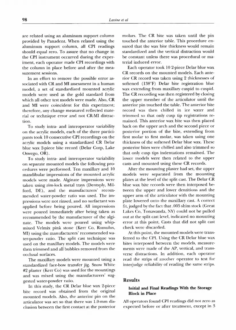
The CPI assesses condylar position changes at the level of the occlusion by using models moun ted in CR on the Panadent Articulator (Fig 1A). These models are then transferred to the CPI (Fig 1B) and related to each other using a m a x i m u m intercuspation (MI) record. Measure- ments can be made of the vertical, transverse, and anterior-posterior distraction of the condyle f rom CR to MI by using the CPI (Fig 1C and D). This system can then be used to assess condylar position in diagnosis and t rea tment planning for patients. According to Roth, 7 the clinically ac- ceptable difference between CR and MI in terms of condylar position as measured by the CPI is approximately 1.0 m m anteroposteriorly, 1.0mm vertically, and 0.5 m m transversely. Discrepan- cies greater than these may possibly contr ibute to the deve lopment of clinical TMD signs and symptoms, z0 Because of these small tolerances, CPI variability must be less than 0.5 m m for this ins t rument ' s measurements to be valid.
Some ~ have quest ioned whether there is at least 0.5 m m of procedural error in the measure-
96 Seminars in Orthodontics, Vol 9, No 2 (rune), 2003: pp 96-101

The Condylar Position indicator 97
Figure 1. Panadent Articulator (A). Panadent Condylar Position Indicator (B). Condylar Position Indicator Recording-Right (C). Condylar Position Indicator Recording Graph (D).
inent technique a n d / o r materials. Possible sources of e r ror include the following:
1. Impress ion materials: expansion and con- traction
2. CR and MI records: imprope r t r imming of the records, dimensional changes of the waxes over time, and distortion of the waxes
3. Dental stone and dental plaster: expansion, contraction, water-powder ratio, and bub- bles on models
4. Face-bow transfer: variation in technique, distortion in transferring ti-om patients to models, and imprope r t r imming of the im- pression c o m p o u n d
5. Placement of the adhesive recording strips 6. Width of the recorded dots 7. Variability of recording the exact CR posi-
tion within and between individual opera- tors
8. Imprope r posit ioning of the adhesive re- cording strips on the CPI
9. Width of the dots on the CPI adhesive re- cording strips
10. Film thickness of separating agent 11. Disinfecting agents causing distortion of iin-
pression material
The reliabilib7 of the CPI is dependent on the proper transfer of anatomical information ti'om the patient to the articulator and finally to the CPI. The purpose of this study was to test the consis- tency and accuracy of the CPI hy assessing both intra- and inte.roperator variability at the levels of the articulator mounting and CPI recording.
Methods and Materials
The CPI is a delicate instrument. When not in use, the upper and lower members of the CPI
98 Lav ine et al
are related using an a luminum suppor t co lumn provided by Panadent . When related using the a luminum suppor t column, all CPI readings should equal zero. To assure that no change in the CPI ins t rument occurred dur ing the exper- iment, each opera to r made CPI recordings with the co lumn in place before and after the mea- su rement sessions.
In an effort to remove the possible error as- sociated with CR and MI assessment in a h u m a n model , a set of standardized m o u n t e d acrylic models were used as the gold s tandard f rom which all o ther test models were made. Also, CR and MI were coincident for this exper iment ; therefore, any change measured reflected mate- rial or technique er ror and not CR-MI distrac- tion.
To study intra- and in teropera tor variability on the acrylic models, each of the three partici- pants took 10 consecutive CPI recordings on the acrylic models using a standardized CR Delar blue wax 2-piece bite record (Delar Corp, Lake Oswego, OR).
To study intra- and in teropera tor variability on separate m o u n t e d models the following pro- cedures were per formed. Ten maxillary and 10 mandibula r impressions of the m oun t ed acrylic models were made. Alginate impressions were taken using rim-lock metal trays (Dentsply, Mil- ford, DE), and the manufacturers ' recom- m e n d e d water-powder ratio was used. The im- pressions were not rinsed, and no surfactant was applied before being poured. All impressions were poured immediately after being taken as r e c o m m e n d e d by the manufac ture r of the algi- nate. The models were poured using whip- mixed Velmix pink stone (Kerr Co, Romulus, MI) using the manufac turers ' r e c o m m e n d e d wa- ter-powder ratio. The split cast technique was used on the maxillary models. The models were then t r immed and all bubbles removed f rom the occlusal surfaces.
The maxillary models were m oun t ed using a standardized face-bow transfer jig. Snow White #2 plaster (Kerr Co) was used for the mount ings and was mixed using the manufacturers ' sug- gested water-powder ratio.
In this study, the CR Delar blue wax 2-piece bite record was obta ined f rom the original moun ted models. Also, the anter ior pin on the articulator was set so that there was 1.0-mm dis- clusion between the first contact at the poster ior
molars. The CR bite was taken until the pin touched the anter ior table. This procedure en- sured that the wax bite thickness would remain standardized and the vertical distraction would be constant unless there was procedural or ma- terial induced error.
Each opera tor took 10 2-piece Delar blue wax CR records on the moun ted models. Each ante- rior CR record was taken using 2 thicknesses of softened (138°F) Delar bite registration blue wax extending f rom maxillary cuspid to cuspid. The CR recording was then registered by closing the uppe r m e m b e r of the articulator until the anter ior pin touched the table. The anter ior bite record was then chilled in ice water and t r immed so that only cusp tip registrations re- mained. This anter ior wax bite was then placed back on the uppe r arch and the second piece or poster ior por t ion of the bite, extending f rom first molar to first molar, was taken using one thickness of the softened Delar blue wax. These poster ior bites were chilled and also t r immed so that only cusp tips indentat ions remained. The lower models were then related to the uppe r casts and m o u n t e d using these CR records.
After the mount ing plaster had set, the upper models were separated f rom the mount ing plates at the level of the split cast. The Delar CR blue wax bite records were then interposed be- tween the uppe r and lower dentit ions and the uppe r a rm of the articulator with the mount ing plate lowered onto the maxillary cast. A correct fit, j udged by the fact that .005 shim stock (Great Lakes Co, Tonawanda, NY) could not be pulled out at the split cast level, indicated no mount ing error at this point. Casts that did not split cast check were discarded.
At this point, the moun ted models were trans- ferred to the CPI. Using the CR Delar blue wax bites interposed between the models, measure- ments were made of the AP, vertical, and trans- verse distractions. In addition, each opera tor read the strips of ano ther opera tor to test for interjudge reliability of reading the same strips.
R e s u l t s
Initial and Final Readings With the Storage Block in Place
All operators found CPI readings did not zero as expected before or after treatment, except in 3

The Condylar Position indicator 99
ins tances (Tab le 1). T h e r ange in the r igh t x axis was - 0 . 1 5 m m to 0.10 ram. T h e r a n g e in the r igh t y axis was - 0 . 2 5 m m to 0.15 ram. Fo r the left x axis, the r a n g e was 0.15 m m to 0.25 ram. For the left y axis, the r a n g e was 0.0 m m to 0.20 ram. Fo r the t ransverse p lane , the r a n g e was - 0 . 0 5 mrn to 0.1 ram.
Interoperator Variability of Reading Same Recording Strips
As shown in Tab le 2, s t a n d a r d devia t ions were e x t r e m e l y low a m o n g o p e r a t o r s r e a d i n g the same r e c o r d i n g strips.
Intraoperator Variability Using Acrylic and Individually Mounted Stone Models
A l t h o u g h m e a n s d i d n o t a p p r o a c h zero in mos t cases, an e x t r e m e l y low s t a n d a r d dev ia t ion was f o u n d wi thin all o p e r a t o r s ' i nd iv idua l r ead ings for bo th the acrylic m o u n t e d m o d e l s a n d the ind iv idua l ly m o u n t e d s tone m o d e l s (Table 3).
T h e t ransverse r ead ings h a d cons is ten t ly lower s t a n d a r d dev ia t ions than the ver t ical x o r y read-
ings. &s e x p e c t e d , s t a n d a r d dev ia t ions were g r e a t e r for the trials with the ind iv idua l ly m o u n t e d s tone m o d e l s than the acD,lic mode ls .
Interoperator Variability on the Acrylic and Individually Mounted Stone Models
&s shown in Tab le 4, var iabi l i ty a m o n g o p e r a t o r s r e a d i n g the i r own r e c o r d i n g s o f the same mod- els was also qu i te low.
D i s c u s s i o n
Because CR is c o i n c i d e n t with MI for this expe r - imen t , all r e c o r d i n g s s h o u l d have e q u a l e d zero. T h e r e are several factors tha t m i g h t exp l a in why this was n o t so. At the mos t basic level, the CPI
Table 2. Interoperator Variability of Reading the Same CPI Recording Strips (Standard Deviation in ram)
Rig~z/ 1.eft
x * 3' ? x ~: 7 "ra n ~ $
A1 0.02 0.02 0.00 0.02 0.00 A2 0.05 0.17 0.04 0.16 0.02 A3 0.02 0.08 0.00 0.06 0.02 A4 0.04 0.07 0.02 0.{}4 0.02 A5 0.09 0.05 0.02 0.08 0.04 A6 (}.09 0.05 0.07 (}. 10 0.06 A7 (}.05 0.13 0.{}0 0.07 0.02 A8 0. I 3 (}.02 0.02 l).02 ().(}{i A9 0.08 0.18 0.14 0.11 0.02 AI{} 0.02 0.09 (}.{}4 (}.(){i 0.02 Mean 0.06 (}.(}9 0.04 0.(}7 (}.03 SI (}.04 0.{}2 (}.05 {}.14 0.06 $2 0.07 0.00 0.08 0 . i 2 0.12 SB (}.0{.) 0.05 0.05 0.05 (}. 12 $4 {}.05 0.(}7 0.24 0.02 0.1 I $5 0.04 0.02 0.02 0.02 0.08 $6 0.07 0.02 0.14 0.05 0.09 $7 (}.09 O. 11 0.04 O. 13 0.0(} $8 0.12 0.09 0,06 0.02 0.02 $9 0.06 0.02 0.(}9 0.04 (}.(}6 S10 0.04 0.02 0. I 1 0.(}4 0.02 Mean 0.07 0.04 0.09 (}.06 0.07
Abbrevia t ions : A, acD'lic models ; S, s tone *Rel{ers to hor i zon ta l CPI axis tReR~rs to vert ical CPI axis +Rctbrs to hansve r sc CPt axis
m o d e l s .
shou ld zero with the s torage b lock in place, h d id not. A t t e m p t s were m a d e by P a n a d e n t to co r r ec t the p l a c e m e n t o f the crosshai rs be fo re the e x p e r i m e n t . T h e p l a c e m e n t , however , was never exact ly co r r ec t as shown in Tab le 1. I f the CPI is no t z e r o e d ti-om the start, the resuhs may be m i s i n t e r p r e t e d nnless the ini t ial a m o u n t o f m a c h i n e CPI d i s t rac t ion e r r o r is known a n d sub-
t r ac l ed f rom the readings . Because d iagnos t i c issues were no t a t ac to r in this e x p e r i m e n t , exact
CPI n u m b e r s were no t as i m p o r t a n t as was re- peatabi l i ty ; t he re fo re , the d i f fe rences were not subtracle( t . In add i t ion , t he re was some "play" in
Table 1. I n i t i a l a n d F i n a l C P I R e a d i n g s W i t h t h e S t o r a g e B l o c k in P l a c e ( r a m )
Operator 1 Operator 2 Operator 3
Right l .dt flight l.d~ Rt¢t+/ Ldt
Axis: x* y'[- x y Tran,s¢ x y x y Tmn,~ x 3 x y Trans
hil t . 0.10 - 0 . 2 5 0.20 0.00 - 0 . 0 5 0.15 0.20 0.15 0.15 0.10 0.05 0.15 0.20 0.20 0.00 Final - 0 . 1 0 - 0 . 2 0 0.25 0.20 - 0 . 0 5 0.00 0.20 0.20 0.15 0.05 0.05 0.15 0.20 0.20 0.00
*Refbrs t o hor i zon t a l CP1 axis. /Re fe r s to vert ical CPI axis. +Rcfi 'rs to t ransverse CPI axis.

100 Lavine et al
Table 3. Intraoperator Variability for Reading CPI Recording Strips Using Acrylic Versus Stone Models (Standard Deviation in nun)
Operator 1 Operator 2 Operator 3
Right Left t¢~¢ht Left Right Left
Axis x y x y Trans x y x y Trans x y x y Tram
Mounted acD, lic models
Mean -0 .20 - 0 . 0 7 -0 .09 0.32 (1.14 0.19 -0 .05 0.10 0.30 -0 .16 0.18 -0 .22 0,18 0.17 -0 .28 SD 0.08 0.12 0.10 0.09 0.04 0.07 0.07 0.11 0.08 0.06 0.06 0.05 0.02 0.02 0.03
Mounted stone models
Mean -0 .10 0.11 0.06 0.43 0.00 0.05 0.11 0.06 0.29 0.10 0.03 0.04 0.14 0.15 0.05 SD 0.15 0.13 0.13 0.13 0.05 0.14 0.12 0.12 0.13 0.06 0.16 0.14 0.10 0.11 0.13
Abbreviations: SD, s tandard deviation. x - Refers to horizontal CPI axis. y - R e t e r s to vertical CP1 axis. Trans - transverse CPI axis.
the recording styli as they were moved to mark the strips. This "play" er ror was observed to range f rom 0.01 to 0.05 mm.
The next level of possible technique error was in te ropera tor variability in reading the same strips. Although the means did not equal zero, the largest s tandard deviation mean was remark- ably low, (+_ 0.09 mm) indicating good repeat-
Table 4. lnteroperator Variability for Reading CPI Recording Strips Using Acrylic and hldividually Mounted Stone Models (Standard Deviation in ram)
R ~ t L~
Axis x y x y Trans
A1 0.08 0.11 0.12 0.00 0.12 A2 0.04 0.06 0.07 0.06 0.06 A3 0.02 0.07 0.08 0.02 0.04 A4 0.07 0.09 0.07 0.04 0.06 A5 0.06 0.06 0.11 0.06 0.02 A6 0.05 0.08 0.14 0.11 0.05 A7 0.05 0.12 0.09 0.09 0.07 A8 0.02 0.12 0.18 0.08 0.08 A9 0.04 0.15 0.24 0.13 0.08 A10 0.04 0.09 0.19 0.06 0.08 Mean 0.05 0.10 0.13 0.07 0.07 S1 0.20 0.07 0.12 0.05 0.21 $2 0.05 (/.07 0.05 0.18 0.09 $3 0.07 0.05 0.05 0.16 0.13 $4 0.00 0.05 0.11 0.25 0.08 $5 0.00 0.05 0.02 0.08 0.08 $6 0.07 0.07 0.12 0.02 0.08 $7 0.05 0.04 0.11 0.02 0.07 $8 0.15 0.18 0.06 0.09 0.02 $9 0.12 0.05 0.12 0.02 0.11 S10 0.08 0.05 0.02 0.11 0.08 Mean 0.08 0.07 0.08 0.10 0.10
Abbreviations: A, acrylic models; S, stone x = Refers to horizontal CPI axis. y - Refers to vertical CPI axis. Trans - transverse CPI axis.
models.
ability. The ability to locate the center of the recorded dot with the magnifying loops was also apparen t in these readings. The er ror associated with posit ioning the graph pape r recording strips on the CPI was small. This was shown by the low s tandard deviat ions in i n t e rope ra to r variability of r ead ing ope ra to r -marked strips in the same category (Table 2).
Finally, in t raopera tor variability was smaller using the aclylic-mounted models than the s tone-mounted models (Table 3). Trials with the acrylic-mounted models showed lower standard deviations than those with the stone models. This indicates that some opera tor a n d / o r mate- rial e r ror was certainly incorpora ted as the list of tested variables increased. Standard deviations approach ing 0.16 were recorded. In the authors ' ()pinion, this may be considered the outer limit of acceptability. Transverse standard deviations were generally smaller than x or y standard de- viations. These results seem credible since the transverse reading is a midline structure and, therefore, less sensitive to error than the more laterally located x and y parameters .
Most recordings showed very low standard deviations. There were a few, however, that were considerably different f rom the others. When the strips for these aberrant readings were re- read, some were found to be grossly in error. The wrong recordings were not replaced with the correct ones and are presented here as op- erator error. This emphasizes the fact that me- ticulous at tention to detail is required using these techniques because er roneous readings

The Condylar Position h~dicalor 101
may be misinterpreted as clinical temporoman- dibular joint dysflmction.
These findings indicate that inter- and intra- operator reliability of the CPI is acceptable at least under the controlled laborato W conditions in th is s tudy. T h e e x a c t s o u r c e s of error, m a t e r i a l
or h u m a n , w e r e n o t a s ses sed ; h o w e v e r , a t r e n d
of i n c r e a s e d var iab i l i ty was n o t e d as t h e com- plexity a n d n u m b e r o f t h e s t eps a n d m a t e r i a l s
i n c r e a s e d . T h e h i g h e s t s t a n d a r d d e v i a t i o n s a t
the most complex point in this experiment (Ta- ble 3, intraoperator variability using stone- mounted models) are, however, still within an acceptable range for both the transverse (0.13 ram) and x-y (0.16 ram) recordings.
Conclusions
CPI r e c o r d i n g s w e r e r e p r o d u c i b l e b o t h a m o n g
a n d b e t w e e n o p e r a t o r s . A t r e n d o f i n c r e a s e d
var iab i l i ty was n o t e d as t h e c o m p l e x i t y , n u m b e r
of s teps , a n d m a t e r i a l s i n v o l v e d i n c r e a s e d .
R e f e r e n c e s
1. Creekmore TD, Cetlin NM, Ricketts RM, et al..]CO Roundtable: Diagnosis and treatment planning..I Clin Orthod 1992;26:585-606.
2. Slavicek R. Inten, iew with Dr. Slavicek Parts 1-10..J Clin Orthod 1988;22:358-370.
3. Slavicek R. Functions and dysflmctions of the mastica- tor 7 organ. Diagnosis and therapy. Presented to Vienna Group IV, July 28 to August 5, 1990, Vienna, Austria.
4. Farrar WB. Diagnosis and treatment of anterior disloca- tion of the disc. NYJ Dent 1971;41:348-351.
5. Lindbloom G. Disorders of the temporomandibular joint: Causal factors and therapy. Acta Odont Scand 1953; 11:61-94.
6. Ricketts TM. Clinical implications of the tcmporoman- dibular joint. Am J Orthod 1966;52:416-439.
7. Roth RH. Temporonaandibular pain-dysflmction and oc- clusal relationships. Angle Orthod 1973;43:136-152.
8. Roth RH. Functional occlusion ti)r the orthodontist. Part 1.J Clin Orthod 1981;15:32-51.
9. Roth Rtt, RolEs DA. Functional occlusion lot the orth- odontist. Part 2. J Clin Orthod 1981 ; 15:100-123.
10. Roth RH. Occlusion and condylar position. Am,] Orthod Dentofac Orthop 1995;107:315-318.
1 I. Per D' HT. Temporomendibularjoint and Occlusion. An- gle Orthod 1976;46:284-293.
12. Thompson ,JR. Abnormal function of the /emporomen- dibularjoints and related musculature: Orthodontic im- plications part 2. Angle Orthod 1986;56:181-195.
13. Jamsa T, Kirveskari P, Alanen P. Malocclusion and its association with clinical signs of craniomandibular dis- orders in 5, 10 and 15-year old children in Finland. Pro( Fins Dent Soc 1988;85:235-240.
14. Egermark-Eriksson 1, Carlsson (;1:2, Magnusson T. A long-term epidemiologic study of the relationship be- tween o~clusal tactnrs and mandibular dysfunction in (hildren and adolescents. J Dent Res 1987;66:63-71.
15. Riolo M, Brendt I), Ten Have' T. Associations between occlusaI characteristics and signs and symptoms of TMJ dysftmction in children and ~oung aduhs. Am,] Orthod Dentofac Orthoi) 1987;92:461-477.
16. Magnusson T, Carlsson GE. Occlusal ae!justmenl in pa- lienls with residual or recurrent signs of mandilmlar dyslhnction. ,J Prosthet 1)enl 1983;49:706-710.
17. Wennbcrg N. Nystrom "I', Carlsson GE. Occlusal equili- bration and other stomatognathic treatment in palients with mand dvstunction and headache..] Prosthet Dclll 1988;59:478-484.
18. Wyatt WE, Prexenling adverse efI~'cts on the temporo- mandibular joint through orthodontic treatment. AM ,] Orlhod I)ento Fac Or/hop 1987;91:493-499.
19. Zarb (;A, Speck JE. The u'eatment of mandibular dvs- fimctio. In: Zarb (;A, Carlsson (;E (eds). Temporoman- dibular Joint Function and l)ysthncfion. Copenhagen, Munksgaard, 1997;373-396.
20. Crawtbrd SD. Condylar axis position, as determined by the occlusion and measured by lhe CPI instrument, and signs and symplnms of lempromandibular dysfunction. Angle Orlhod 1999;69:103-116.
21. Phillips RW. Report of committee on Scientitic Investi- gation of the American Academy of Reslorative Den- tistr)'.,}. Prosthel Dent 1986;55:736-772.


