Embed Size (px)
Citation preview

/. mar. bid. Ass. U.K. (1994), 74, 301-312 301Printed in Great Britain
RESPIRATION RATES AND BIO VOLUMES OF COMMONBENTHIC FORAMINIFERA (PROTOZOA)
FIONA HANNAH*, ANDREW ROGERSON* ANDJOHANNA LAYBOURN-PARRY+
"University Marine Biological Station, Millport, Isle of Cumbrae, Scotland, KA28 OEGdepartment of Zoology, La Trobe University, Bundoora, Melbourne, Victoria 3083, Australia
The respiration rates of five genera of benthic Foraminifera were determined byCartesian diver microrespirometry. Across all genera studied, the rate averaged ll-3xlO~3
JLLI O2 individual1 h"1 at 10°C. Estimates of foraminiferal mean biovolumes ranged from0-66 to 6-54xlO6 urn3, giving an overall mean volume-specific respiration rate of 7-36xlO'9 jn.1O2 h'1 urn3. For non-symbiont-bearing Foraminifera, in general, volume-specific rates (logul O2 h
4 urn3) are best described by the equation, -0-98 x (log cell biovolume, urn3) - 2-01.The respiration results show that these benthic Foraminifera respire some ten times morerapidly than naked amoebae of equivalent size. The combination of high respiratory ratesand the often large standing stocks of Foraminifera encountered, suggests that theseorganisms may contribute significantly to total microbial benthic respiration.
INTRODUCTION
When modelling field data to describe the transfer of materials between trophiclevels, it is important to consider metabolic losses incurred by individual populations,or, more usually, by size classes within the community. Published relationships relatinglog body size to log respiration rate provide an exponent, b, in the range 0-66 to 0-75 forunicellular organisms (Zeuthen, 1953; Hemmingsen, 1960; Phillipson, 1981). This valueis somewhat lower than b values for multicellular ectotherms (0-73-0-88) reported by thesame authors. A comprehensive review of protozoan respiration by Fenchel & Finlay(1983), provides an exponent, b, of 0-74 which is very similar to that suggested byHemmingsen (1960) for multicellular ectotherms. Fenchel & Finlay's (1983) data ismainly for flagellate and ciliate Protozoa since amoebae are less well represented in thepublished record. Respiration studies on amoebae are also biased towards 'experimen-tal' genera, such as Amoeba and Chaos, forms not commonly encountered in fieldsamples, or to the genera Acanthamoeba and Naegleria, which are cultivated axenically inchemically defined medium. Furthermore, the scatter in this relationship is consider-able, with, for example, the range for a small protozoan spanning a factor of 50 (Fenchel& Finlay, 1983).
These shortcomings limit the usefulness of the relationship for predicting fieldrespiration, particularly in the case of amoebae, and underline the need for additionalstudies on the respiration of Protozoa not represented in the data. Benthic Foraminifera

302 F. HANNAH, A. ROGERSON AND J. LAYBOURN-PARRY
are one such group. Since they are common in all marine habitats from intertidalmudflats (over 1000 live specimens per 10 cm2; Ellison, 1984) to deep-sea sediment (over1000-2000 live specimens per 10 cm2; Gooday, 1986) it is possible that they may accountfor a significant proportion of the total respiration of the benthos. Despite their numeri-cal importance, there have been only three previous studies on the respiration ofbenthic Foraminifera and none of these investigations has featured Protozoa fromtemperate zones (Bradshaw, 1961; Lee & Muller, 1973; Schwab & Hofer, 1979).
Studies on the respiration of Protozoa have traditionally employedmacrorespirometers, such as the Warburg apparatus, or microrespirometers, such asthe Cartesian diver. Criticism has been levelled at both methods; in the case of theformer, many cells are required for an experimental run, and this crowding is believedto induce stress effects (Baldock et ah, 1982), while in the case of the Cartesian apparatus,the need for long incubations without food is thought to underestimate metabolic ratebecause of starvation (Fenchel & Finlay, 1983). In the case of benthic Foraminifera,however, such starvation effects may not apply since we have observed many toaccumulate sediment particles and organic detritus intracellularly, and presumablytake time to digest this food material. Moreover, some deep-sea Foraminifera arecapable of metabolizing their own protoplasm in times of starvation (Linke, 1992).
To use empirically derived respiration measurements for predicting field rates, it isnecessary to determine the cell biovolume of cells. This is not straightforward inForaminifera because of their irregular tests and the fact that the cell cytoplasm does notfill the entire lumen of the test. Most investigators have calculated biomass from thevolumes of appropriate geometric shapes, or from weight determinations of cells andconversion factors (Murray, 1973, Gerlach et al., 1985, Widbom, 1984, Altenbach, 1987).
The present study provides new information on the respiration rates and cell vol-umes of benthic Foraminifera from fine-grained sediments and reviews these in thecontext of published values for other amoeboid Protozoa.
MATERIALS AND METHODS
Isolation of Foraminifera
Benthic samples were collected from 45 m water depth at a site off the Isle of Cumbrae(55°46-39'N 4°53-07'W) in the Clyde Estuary, Scotland, between July and October 1993.Undisturbed cores of fine mud were obtained using a Craib corer lowered from theUMBSM research vessel 'Aplysia'. Foraminifera for study were micropipetted from thefraction retained on a 73-um mesh. Specimens were placed in sterile MYIOOS medium at10°C (the approximate summer temperature of this deep site), prepared by adding 0-1 gmalt extract and 0-1 g yeast extract (Sigma Chemical Co.) to 11 of filtered natural sea-water (33%o). The MYIOOS medium was particularly effective in stimulating many ofthe Foraminifera to extend pseudopodia, either as a reticulate net or as thin cytoplasmicstrands, confirming the viability of the cells.
FORAMINIFERAL RESPIRATION 303
Cartesian diver microrespirometry
Foraminifera from five genera were isolated and used in the respirometry experi-ments. These were Ammonia, Buliminella, Reophax sp. 1, Reophax sp. 2, Elphidium andQuinquinoculina. Specimens containing conspicuous cytoplasmic content andpseudopodial activity were micropipetted through eight changes of sterile MY100Smedium to remove attendant surface bacteria. The efficiency of this washing procedurewas verified by scanning electron microscopy. Individual Foraminifera were firstmeasured (maximum test length) and then loaded into a stoppered diver with a gasphase between 2-3 and 3-0 ul (Klekowski, 1981). After a 1-h acclimatization period at10°C, readings were recorded over the next 1-2 h at 10 min intervals. A diver containingthe washings of the final wash was used as a control against bacterial contamination.
Scanning electron microscopy
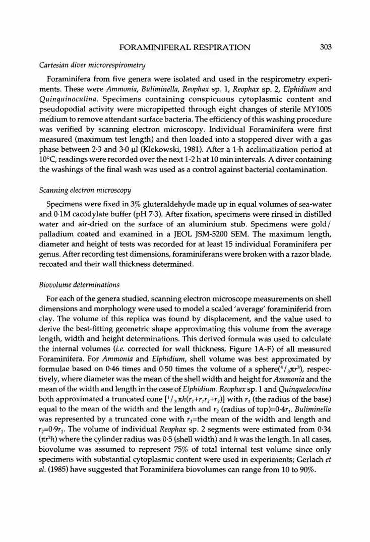
Specimens were fixed in 3% gluteraldehyde made up in equal volumes of sea-waterand 01M cacodylate buffer (pH 7-3). After fixation, specimens were rinsed in distilledwater and air-dried on the surface of an aluminium stub. Specimens were gold/palladium coated and examined in a JEOL JSM-5200 SEM. The maximum length,diameter and height of tests was recorded for at least 15 individual Foraminifera pergenus. After recording test dimensions, foraminiferans were broken with a razor blade,recoated and their wall thickness determined.
Biovolume determinations
For each of the genera studied, scanning electron microscope measurements on shelldimensions and morphology were used to model a scaled 'average' foraminiferid fromclay. The volume of this replica was found by displacement, and the value used toderive the best-fitting geometric shape approximating this volume from the averagelength, width and height determinations. This derived formula was used to calculatethe internal volumes (i.e. corrected for wall thickness, Figure 1A-F) of all measuredForaminifera. For Ammonia and Elphidium, shell volume was best approximated byformulae based on 0-46 times and 0-50 times the volume of a sphere(4/37ir3), respec-tively, where diameter was the mean of the shell width and height for Ammonia and themean of the width and length in the case of Elphidium. Reophax sp. 1 and Quinqueloculinaboth approximated a truncated cone [V3 nh(r1+r1r2+r2)] with r1 (the radius of the base)equal to the mean of the width and the length and r2 (radius of top)=0-4r2. Buliminellawas represented by a truncated cone with r2=the mean of the width and length andr2=0-9r1. The volume of individual Reophax sp. 2 segments were estimated from 0-34(nr2h) where the cylinder radius was 0-5 (shell width) and h was the length. In all cases,biovolume was assumed to represent 75% of total internal test volume since onlyspecimens with substantial cytoplasmic content were used in experiments; Gerlach etal. (1985) have suggested that Foraminifera biovolumes can range from 10 to 90%.

304 F. HANNAH, A. ROGERSON AND J. LAYBOURN-PARRY
Figure 1. Scanning electron micrographs of wall cross-sections. (A) Reophax sp. 1. (B) Buliminella. (C)Elphidium. (D) Ammonia. (E) Quinqueloculina. (F) Reophax sp. 2. (G) Scanning electron micrograph ofReophax sp. 2, side profile. Scale bars: A,C,D,F, 10 nm; B, 1 \x.m; E,G, 100 (xm.
Dry weight determinations
Living specimens of Ammonia, Reophax spp. 1 & 2, and Quinqueloculina were pickedout and fixed in 4% formaldehyde (buffered with hexamine, pH 8-9) in sea-water(N=58). After three washes in distilled water, the specimens were weighed on aSartorius Ultra Micro Balance (4504 MP8), with an accuracy of 01 (ig, in small weighed

FORAMINIFERAL RESPIRATION 305
aluminium boats, which had been precombusted at 550°C for 4 h. Dry weights of theindividuals were then obtained by heating samples for 12 h at 70°C, cooling to roomtemperature in a desiccator, and reweighing. The samples were then combusted at550°C for 4 h and reweighed to obtain the ash-free dry weight; by subtraction theamount of organic carbon was determined. Three boats with distilled water from thefinal wash were similarly treated and weighed as controls. The carbon content of anindividual 'average' Foraminifera was converted to equivalent biovolume (um3) usingthe value, 110 fg C urn3 (Turley et al., 1986).
RESULTS
All the benthic genera used in the present study were markedly flattened, as seen forReophax sp. 2 (Figure 1G), with cell width averaging 2-2 times shell height (mean of allmeasured Foraminifera from five genera, N=99). Cross-sections of the shell walls of thefive genera studied are given in Figure 1A-F. The relative importance of shell thicknesswhen calculating internal, rather than external, shell volume is evident in the case ofQuinqueloculina which had an average wall thickness of 22-3 um (+6-3SD, N=20).Reophax sp. 2, on the other hand, had an average wall thickness of only 3-8 um (+1-1SD,N=25).
The calculated volumes of foraminiferans used in this study are shown in Figures 2 &3 (equations given in legends). These equations, relating maximum test length (urn) tototal internal volume (um3) of the Foraminifera test (corrected for wall thickness), havegeneral applicability for the estimation of internal test space in Foraminifera of thesefive genera, and of morphologically similar genera, from the measurement of a singlelinear dimension (i.e. maximum test length in urn).
108
| 106 =
105i
104
100Length (um)
1000
Figure 2. Relationship between maximum test length (|xm) and internal test volume (|im3, correctedfor wall thickness) of Elphidium (A), Ammonia (O), Quinqueloculina (O) and Reophax sp. 1 ( • ) . The totalinternal space (log (im3) of Elphidium = 3-22 (log length) -1 -26 (R2=0-95), of Ammonia = 3-61 (log length)- 2-81 (R2=0-97), of Quinqueloculina = 3-79 (log length) - 3-26 (R2=0-96), of Reophax sp. 1 = 2-77 (loglength) - 0-61 (R2=0-87).

306 F. HANNAH, A. ROGERSON AND J. LAYBOURN-PARRY
107
106
\
I
100010 100
Length (urn)1000
Figure 3. Relationship between maximum test length (|im) and internal test volume (|im3 correctedfor wall thickness) of Buliminella (•) and individual Reophax sp. 2 (•) segments. The total internalspace (log |im3) of Buliminella = 3-72 (log length) - 2-60 (R2=0-91), of Reophax sp. 2 segments = 3-13 (loglength) - 0-91 (R2=0-96).
Examination of sectioned Foraminifera confirmed that bacteria were not contaminat-ing the internal surfaces of the shell, nor the outer surface of washed specimens, evenwhen the surface was irregular as in the agglutinated foraminiferan, Reophax sp. 1.
The mean respiration rates of Foraminifera (xlO3 ul O2individual1 h"1) with theirmean biovolumes (um3) are presented in Table 1. The variation in rates was consider-able, probably due to differences in cell size, physiological state and degrees of activitywithin divers. Variation between and within genera was such that an overall mean rateof ll-3xlO~3ul O2 individual"1 h'1 was appropriate across all the genera studied.
These mean values for all species of Foraminifera are compared in Figure 4 (arrowed)with all the available respiration data for other amoebae (mainly naked forms). Thisclearly shows that the rates for these Foraminifera are higher than those of most otheramoebae, and that the metabolic rate to body weight exponent b for amoebae is 0-71.
Table 1. Respiration rates (xW~3 fd O2 individual1 k1) and mean cell biovolumes (xlO6 /urn3,cellular material only, assuming that biomass occupies 75% of the shell space) of five genera ofbenthic Foraminifera atlO°C. Values are means ±SD with number of observations in parenthesis.
GenusAmmoniaReophax sp. 1Reophax sp. 2QuinqueloculinaElphidiumBuliminellaAllogromia*
Respiration rate7.50
10.6012.8214.309.46
11.7613.02
±2.78±3.81±7.28±5.55±4.72±7.20
(N=3)(N=8)(N=ll)(N=5)(N=4)(N=3)(N=2)
Biovolume6.54 ±5.633.32 ±1.901.80 ±0.802.06 ±0.661.19±1.100.66 ±0.180no data
*two additional readings, no volume data; cells used were 150 (xm and 200 um long.

FORAMINIFERAL RESPIRATION
100 T
307
0.0011000 104 10= 106 107
Volume (|im3)
10a 109
Figure 4. Published data on amoeboid protozoan respiration rates per individual (nl O2 h1). Data
from this study (arrowed points are mean values of each Foraminifera species) plus references citedin Fenchel & Finlay (1983) and Crawford el at, (1994). log y = 0-71 (log x) - 4-41 (R2=0-79). All valuesnormalized to 20°C.
Across the size range of Foraminifera measured, there was a ten-fold increase inbiovolume. Volume-specific respiration rates changed across this range of genera,showing, as expected, that as cell size (|im3) increased the respiration rate (ul O2) perunit of protozoan biomass decreased (Figure 5). The negative slope of -0-9 implies thatthe rate of change is almost directly related to changes in protozoan cell size. This isunusual for Protozoa where the specific respiration rate generally does not decrease asrapidly with decreasing cell size (e.g. slope for Protozoa in general =-0-34, Caron et ah,
~ 10
&
.2
atoCD
.y
- 7
v 8
10-9
* 10- 1 0
10-11
105 106 107
Cell volume108 109
Figure 5. Volume specific respiration rates (ul O2 lun^h1) of benthic Foraminifera: A, this study; • ,published data (Lee & Muller, 1973; Schwab & Hofer, 1979). All values for 10°C using Q10=20; cellvolumes for this study assumed to represent 75% of shell space, log y = -0-98 (log x) - 209 (R2=0-87).

308 F. HANNAH, A. ROGERSON AND J. LAYBOURN-PARRY
1990). Despite this incongruity the log specific respiration rate of benthic Foraminiferaat 10°C (xlO9 ul O2 Jim3 h"1) can best be estimated from the available data by theequation, -0-98 x (log cell biovolume) - 209 (R2=0-8732). For reasons outlined later, thismay represent maximum rates of well-fed, active Foraminifera cells.
DISCUSSION
Respiration rates for Foraminifera in this study averaged ll-3xlO~3ul O2 h"1, regard-less of genus, corresponding to a rate of 22-7xlO~3 ulO2 h"1 at 20°C, assuming a Q10of 20.This value is considerably higher than rates derived by Cartesian microrespirometryfor naked and testate amoebae of comparable size. For example, Amoeba proteus andChaos carolinense, with volumes of l-4xlO6 and 5-5xl06um3, respectively, had metabolicrates at 20°C of l lx lO 3 and l-4xlO"3 ul C^h"1 (Scholander et ah, 1952; Rogerson, 1981) andDifflugia, with a volume of 0-97xl06um3, had a rate of l-2xlO"3 ul O2 h
1 (Zeuthen, 1943).Rates for much larger Chaos were still lower, ranging from l-2xlO3 to 8-6xlO3 ul O2 h"1
for cells in the size range 35-50xl06 urn3 (Claff & Tahmisian, 1949; Holter & Zeuthen,1948). Assuming our Foraminifera biovolume determinations are accurate (see laterdiscussion) then Foraminifera respire some ten times more rapidly than other amoebaeof similar volumes. This conclusion is, however, not at odds with most of the previouslypublished respiration rates for Foraminifera. Respiration rates of Allogromia laticollarisranged from 8-3x10 3 to 15-2xlO"3 ul O2 h
1, Rosalina sp. from 20 to 58-4xlO"3 ul O2 h"1 andrates for Spiroloculina hyalina, Ammonia sp. and Spirallina were 19-3xlO'3, 0-8xl0~3, and0-2xl03 ul O2 h
1, respectively (Bradshaw, 1961; Lee & Muller, 1973; Schwab & Hofer,1979; all values converted to 20°C). Variation within the Foraminifera data is consider-able, but the predominance of high rates strongly suggests that these Protozoa haveunusually high rates of respiration relative to other amoebae. At this time we can onlyspeculate as to why this may be the case. It has been suggested that Cartesian divermicrorespirometry measures the respiration of starved cells since lengthy incubations(in some cases 4 h) without prey are required to complete an experimental run. Theeffect of incubation time on respiration rate was demonstrated for the amoebaTrichosphaerium using a single-cell 14C method (Crawford et al., 1994). Rates fell 20-foldover a 24-h incubation period, clearly showing that the Cartesian method can underes-timate respiration rates. However, benthic foraminiferans harbour large quantities ofintracellular detritus and other foodstuffs, and may be unique among Protozoa inCartesian diver experiments in that they do not face the same starvation conditions andconsequently maintain high metabolic rates. It is also possible that Foraminifera typi-cally have high metabolic costs of locomotion. They produce extensive anastomizingreticulopodial networks involving a considerable degree of cytoplasmic streaming.While Fenchel & Finlay (1983) maintain that the cost of locomotion in ciliates andflagellates is less than 1% of the total metabolic rate, based on theoretical considerations,Crawford (1992) has calculated that fast-swimming protists can incur locomotory costs>10% total metabolic rate, and the amoeba Trichosphaerium may use up to 56% of its totalmetabolic energy on active locomotion (Crawford et al., 1994). The evidence suggests

FORAMINIFERAL RESPIRATION 309
that Foraminifera also have high costs of locomotion, particularly in the Cartesianexperiments where cells were suspended in MY100S medium, a medium which stimu-lated cytoplasmic motion and accounted for the rapid locomotion of cells observed inthe diver chambers. In one case an individual Reophax was stationary throughout thefirst half of the experimental run and then became active. This change in activity causedan approximate two-fold increase in respiration rate.
Boltovskoy & Wright (1976) reviewed oxygen requirements of foraminiferans andsuggested that these are variable, and that some benthic genera, such as Elphidium andBuliminella, may require relatively large amounts of oxygen and possess endosymbioticalgae to supply this need. If this is true, then high metabolic costs in Foraminifera couldexplain why many are inactive in situ for long periods of time and why generation timesfor these protists are long, 8 d in the case of asexually dividing Allogromia (Lee & Muller,1973). Moreover, the extensive granulose pseudopodial network in foraminiferans isrich in mitochondria (Lee, 1993) and gives the cell an extremely high surface-to-volumeratio. Both these features are consistent with high respiration rates. It is even possiblethat the pseudopodial network in foraminiferans is an adaptation to satisfying the cells'high metabolic requirements, as well as a mechanism for gathering food and forlocomotion in the small interstitial spaces in sediments.
As with the metazoan ectotherms, one may expect the Protozoa to show a widediversity in their energy partitioning to different physiological functions and their netand gross production efficiencies. The fact that these foraminiferans are detrital feederswith low growth rates (Murray, 1991) and high respiration rates suggests that theypossess low net production efficiencies and partition a large portion of their energy intomovement, a large degree of which is associated with the acquisition of food resources.This contrasts with published data on other Protozoa which generally have high netproduction efficiencies (Laybourn-Parry, 1984).
Of central importance when predicting metabolic rates of cells is an accurate assess-ment of cell biomass. Most estimates for Foraminifera have relied on calculating theirvolume from the closest-fitting geometric shape. This, however, makes no allowancefor the irregular shape of the outer surface, the fact that the walls of some Foraminiferaare thick and the fact that the entire lumen of the shell does not reflect biovolume ofcytoplasm; cytoplasm reportedly occupies from 10% to 90% of the shell space (Gerlachet al., 1985; Altenbach, 1987). It follows, therefore, that some published values ofbiovolume may be over-estimates. Our method, using scale models to derive a best-fitting formula, with consideration of shell thickness and cytoplasmic content, arebelieved to yield better estimates of true biovolume. Mean foraminiferan biovolumes inthis study ranged from 0-66xl06 to 6-54xlO6 um3 for cells in the approximate lengthrange 100 to 1000 um (Figures 3 & 4), values which make sense in terms of publishedvolumes of equivalently sized naked amoebae like A. proteus which has an averagelength of 425 um and a biovolume of around 1-OxlO6 um3 (Rogerson, 1981; Page, 1988;Rogerson et al., 1994). While the published value of 2-lxlO6 um3, for an 'average'Foraminifera (Gerlach et al., 1985), agrees well with our results, published values forSpiroloculina hyaline, Elphidium and Reophax are higher, in the size range 3-4xlO6 to

310 F. HANNAH, A. ROGERSON AND J. LAYBOURN-PARRY
17-5xlO6 urn3 (Wefer & Lutze, 1976; Fenchel & Finlay, 1983). It is easy to over-estimatevolume by applying the wrong formula, illustrated here in the case of Quinqueloculina.If the formula for a half prolate or oblate spheroid is used to compute volume, as hasbeen used by others (e.g. Murray, 1973), instead of the derived formula, then volume isincreased by a factor of six times and ten times, respectively. Moreover, if wall thicknessand reduced biovolume (e.g. 75% used in this study) are not considered, then a further2-1-fold over-estimation can result.
An alternative method commonly used for estimating biovolume is to measure thedry weight and ash-free dry weight of cells and to estimate biomass from the amount oforganic carbon. In this study we used this approach for comparison. By this method theaverage biovolume of an individual foraminiferan cell was 53-0xl06 |J.m3, a full order ofmagnitude higher than that of the best-fitting equation method. This over-estimate isprobably due to the inclusion of organic carbon from ingested detritus and organic wallcomponents or to the loss of calcium carbonate on combustion at 550°C (Telek &Marshall, 1974). Estimates of cell size derived in this way frequently appear to overesti-mate biovolume, as was the case for reported volumes of 54-71xlO6 |J.m3for Allogromialaticollaris and 125xlO6 for Rosalina leei (reported in Fenchel & Finlay, 1983).
In Figure 4 we compare our mean Foraminifera result with available publishedrespiration values for amoebae and obtain a slope of 0-71, which lies within the rangesuggested for unicellular organisms (Hemmingsen, 1960; Phillipson, 1981). It is clearthat this mean foraminiferan respiration value is higher than that of most amoebae,although this may be, in part, due to over-estimates of cell biomass of some nakedamoebae through the use of inappropriate formulae; their shapes rarely approximateconvenient geometric shapes. In a recent study of naked amoebae, it was clear thatmany amoebae, despite appearing large, have small biovolumes because of theirflattened shapes (Rogerson et al., 1994). In the respiration data presented by Fenchel &Finlay (1983) most amoebae lie below the slope and away from the data points forciliates and flagellates, prompting Fenchel (1987) to comment, perhaps incorrectly, thatamoebae have lower rates of respiration than ciliates and flagellates.
Pooling the data for all 36 experimental Foraminifera data points in this study gives amean volume specific respiration rate of 7-36xlO"9 |il O2 h
1 \xm~3 (±710 SD) at 10°C(equivalent to 14-72xlO9 ul O2 at 20°C). While this value is high relative to otherProtozoa of comparable size, it is not without precedent within the Protozoa in general.The amoeba Acanthamoeba, the ciliate Tetrahymena and the flagellate Pleuromonas can allhave specific rates as high as 13xlO"9, 20xl0~9 and 32xlO~9 |il O2h1, respectively (Lovlie,1963; Baldock et al., 1980; Fenchel & Finlay, 1983). The specific respiration value forForaminifera in this study is higher than expected. When considered in the light of thehigh numbers of Foraminifera encountered in the sediments, this would suggest thatthey may contribute significantly to total microbial benthic respiration. However, thereare inconsistencies with this notion that require further investigation. Firstly, manystudies have found apparently live Foraminifera at depth in the sediment (Buzas, 1977;Corliss, 1985; Gooday, 1986; Murray, 1992) while other studies of Foraminifera fromsoft sediments have shown that at least some species have the ability to cope with low

FORAMINIFERAL RESPIRATION 311
oxygen content and anoxia (Bernhard, 1993; Moodley & Hess, 1992). Clearly, therefore,there is a need for further respiration studies of benthic Foraminifera before their fullecological role can be elucidated.
This study was supported by a grant from the Natural Environmental Research Council.
REFERENCES
Altenbach, A.V., 1987. The measurement of organic carbon in Foraminifera. Journal ofForaminiferalResearch, 17,106-110.
Baldock, B., Baker, J.H. & Sleigh, M.A., 1980. Laboratory growth rates of six species of freshwaterGymnamoebia. Oecologia, 47,156-159.
Baldock, B.M., Rogerson, A. & Berger, ]., 1982. Further studies on respiratory rates of freshwatergymnamoebia. Microbial Ecology, 8, 55-60.
Bernhard, J.M., 1993. Experimental and field evidence of Antarctic foraminiferal tolerance toanoxia and hydrogen sulphide. Marine Micropaleontology, 20, 203-213.
Boltovskoy, E. & Wright, R., 1976. Recent Foraminifera. The Hague: Junk Publishers.Bradshaw, J.S., 1961. Laboratory experiments on the ecology of Foraminifera. Contributions from
the Cushman Foundation for Foraminiferal Research, 12, 87-106.Buzas, M.A., 1977. Vertical distribution of Foraminifera in the Indian River, Florida. Journal of
Foraminiferal Research, 7, 234-237.Caron, D.A., Goldman, J.C. & Fenchel, T., 1990. Protozoa respiration and metabolism. In Ecology
of marine Protozoa (ed. G.M. Capriulo), pp 307-322. New York: Oxford University Press.Claff, C.L. & Tahmisian, T.N., 1949. Cartesian diver technique. Journal of Biological Chemistry, 179,
577-583.Corliss, B.H., 1985. Microhabitats of benthic Foraminifera within deep-sea sediments. Nature,
London, 314, 435-438.Crawford, D.W., 1992. Metabolic cost of motility in planktonic protists: theoretical considerations
on size scaling and swimming speed. Microbial Ecology, 24,1-10.Crawford, D.W., Rogerson, A. & Laybourn-Parry, ]., 1994. Respiration of the marine amoeba
Trichosphaerium sieboldi determined by 14C labelling and Cartesian diver methods. MarineEcology Progress Series, in press.
Ellison, R.L., 1984. Foraminifera and meiofauna on an intertidal mudflat, Cornwall, England:populations; respiration and secondary production; and energy budget. Hydrobiologia, 109,131-148.
Fenchel, T., 1987. Ecology of Protozoa: the biology of free-living phagotrophic protists. Madison,Wisconsin: Science Technical Publishers.
Fenchel, T. & Finlay, B.J., 1983. Respiration rates in heterotrophic, free-living Protozoa. MicrobialEcology, 9, 99-122.
Gerlach, S.A., Hahn, A.E. & Schrage, M., 1985. Size spectra of benthic biomass and metabolism.Marine Ecology Progress Series, 26,161-173.
Gooday, A.J., 1986. Meiofaunal foraminiferans from the bathyal Porcupine Seabight (north-eastAtlantic): size structure, standing stock, taxonomic composition, species diversity and verti-cal distribution in the sediment. Deep-Sea Research, 33,1345-1373.
Hemmingsen, A.M., 1960. Energy metabolism as related to body size and respiratory surfaces,and its evolution. Report of the Steno Memorial Hospital and the Nordisk Insulinlabatorium,Copenhagen, 9(2), 1-110.
Holter, H. & Zeuthen, E., 1948. Metabolism and reduced weight in starving Chaos chaos. ComptesRendus des Travaux du Laboratoire de Carlsberg, Serie Chimique, 26, 277-296.
Klekowski, R.Z., 1981. Ecology of aquatic organisms. 3. Animals. Size dependence of metabolismon protozoans. Verhandlungen der International Vereinigung fur Theoretische und AngewandteLimnologie, 21,1498-1502.

312 F. HANNAH, A. ROGERSON AND J. LAYBOURN-PARRY
Laybourn-Parry, J., 1984. A functional biology of free-living Protozoa. London: Croom Helm.Lee, J.J., 1993. 'On a piece of chalk' - updated. Journal of Eukaryotic Microbiology, 40, 395-410.Lee, J.J. & Muller, W.A., 1973. Trophic dynamics and niches of salt marsh Foraminifera. American
Zoologist, 13, 215-223.Linke, P., 1992. Metabolic adaptations of deep-sea benthic Foraminifera to seasonally varying
food input. Marine Ecology Progress Series, 81, 51-63.Lovlie, A., 1963. Growth in mass and respiration rate during the cell cycle of Tetrahymena
pyriformis. Comptes Rendus des Travaux du Laboratoire de Carlsberg, Copenhague, 33, 377-413.Moodley, L. & Hess, C, 1992. Tolerance of infaunal benthic Foraminifera for low and high oxygen
concentrations. Biological Bulletin. Marine Biological Laboratory, Woods Hole, 183, 94-98.Murray, J.W., 1973. Distribution and ecology of living benthic foraminiferids. London: Heinemann
Educational Books.Murray, J.W., 1991. Ecology and palaecology of benthic Foraminifera. New York: Longman Scientific
and Technical.Murray, J.W., 1992. Distribution and population dynamics of benthic Foraminifera from the
southern North Sea. Journal of Foraminiferal Research, 22,114-128.Page, F.C., 1988. A new key to freshwater and soil gymnamoebae. Ambleside: Freshwater Biological
Association.Phillipson, J., 1981. Bioenergetic options and phylogeny. In Physiological biology: an evolutionary
approach to resource use (ed. C.R. Townsend and P. Calow), pp. 20-45. Oxford: BlackwellScientific Publications.
Rogerson, A., 1981. The ecological energetics of Amoeba proteus (Protozoa). Hydrobiologia, 85,117-128.
Rogerson, A., Butler, H.G. & Thomason, J.C., 1994. Estimation of amoeba cell volume fromnuclear diameter and its application to studies in protozoan ecology. Hydrobiologia, in press.
Scholander, P.F., Claff, C.L. & Sveinsson, S.L., 1952. Respiratory studies of single cells. I. Methods.Biological Bulletin. Marine Biological Laboratory, Woods Hole, 102,157-177.
Schwab, D. & Hofer, H.W., 1979. Metabolism in the protozoan Allogromia laticollaris Arnold.Zeitschrifr fur Mikroskopisch Anatomische Forschung, 93, 715-727.
Telek, G. & Marshall, N., 1974. Using a CHN analyser to reduce carbonate interference inparticulate organic carbon analyses. Marine Biology, 24, 219-221.
Turley, CM., Newell, R.C. & Robins, D.B., 1986. Survival strategies of two small marine ciliatesand their role in regulating bacterial community structure under experimental conditions.Marine Ecology Progress Series, 33, 59-70.
Wefer, G.V. & Lutze, G.F., 1976. Benthic Foraminifera biomass production in the western Baltic.Kieler Meeresforschungen, 3, (supplement), 76-81.
Widbom, B., 1984. Determination of average individual dry weights and ash-free dry weights indifferent sieve fractions of marine meiofauna. Marine Biology, 4,101-108.
Zeuthen, E., 1943. A Cartesian diver micro-respirometer with a gas volume of 0-1 (J.1. Respirationmeasurements with an experimental error of 2-10"5 (xl. Comptes Rendus des Travaux du Laboratoire,Carlsberg, Serie Chimique, 24, 479-518.
Zeuthen, E., 1953. Oxygen uptake as related to body size in organisms. Quarterly Review of Biology,28,1-12.
Submitted 18 November 1993. Accepted 3 February 1994.