Embed Size (px)
Citation preview
HAL Id: hal-01874527https://hal-univ-rennes1.archives-ouvertes.fr/hal-01874527
Submitted on 20 Sep 2018
HAL is a multi-disciplinary open accessarchive for the deposit and dissemination of sci-entific research documents, whether they are pub-lished or not. The documents may come fromteaching and research institutions in France orabroad, or from public or private research centers.
L’archive ouverte pluridisciplinaire HAL, estdestinée au dépôt et à la diffusion de documentsscientifiques de niveau recherche, publiés ou non,émanant des établissements d’enseignement et derecherche français ou étrangers, des laboratoirespublics ou privés.
Results of a phase I study in patients suffering fromsecondary-progressive multiple sclerosis demonstratingthe safety of the amino acid copolymer PI-2301 and apossible induction of an anti-inflammatory cytokine
responseJ. Kovalchin, J. Krieger, M. Genova, K. Collins, M. Augustyniak, A. Masci, T.
Hittinger, B. Kuca, Gilles Edan, C. Braudeau, et al.
To cite this version:J. Kovalchin, J. Krieger, M. Genova, K. Collins, M. Augustyniak, et al.. Results of a phase I studyin patients suffering from secondary-progressive multiple sclerosis demonstrating the safety of theamino acid copolymer PI-2301 and a possible induction of an anti-inflammatory cytokine response.Journal of Neuroimmunology, Elsevier, 2010, 225 (1-2), pp.153-163. �10.1016/j.jneuroim.2010.04.008�.�hal-01874527�
Results of a phase I study in patients suffering from secondary-progressive multiplesclerosis demonstrating the safety of the amino acid copolymer PI-2301 and a
possible induction of an anti-inflammatory cytokine response☆
critptJ. Kovalchin a, J. Krieger a, M. Genova a, K. Collins a, M. Augustyniak a, A. Masci a, T. Hittinger a, B. Kuca a,
G. Edan b, C. Braudeau c, M. Rimbert c, U. Patel a, E. Mascioli a, E. Zanelli a,⁎a Peptimmune Inc., 64 Sidney Street, Cambridge, MA-02139, USAb Service de Neurologie, CIC-P 02-03, INSERM, Centre Hospitalier Universitaire rue Henri Le Guillou, 35033 Rennes, Francec CIMNA, CHU Hôtel-Dieu, quai Moncousu, 44093 Nantes, France
uld be
progress. The mover timend for a
☆ The authors from Peptimmune Inc., a for-profit organGilles Edan is a consultant for Peptimmune Inc. Thcontracted for services.⁎ Corresponding author. Tel.: +1 617 715 8042; fax:
E-mail address: [email protected] (E. Za
s
PI-2301 is an immunomodulator that coperformed in patients with secondary-label treatment period with active drugserum levels of IL-3, IL-13, and CCL22 oMRI data indicated a non-significant tre anuan alternative therapy for MS. A placebo-controlled, multiple-ascending dose, double-blind study was
ive MS. Treatment was given subcutaneously once weekly for 8 weeks, followed by a 4-week open-st common adverse event was transient injection site reactions. Non-significant trend for increases in
were suggestive of a beneficial TH2 immune response in subjects dosed with PI-2301 at 3 and 10 mg.
reduction of lesion numbers in subjects treated with 1 and 3 mg PI-2301.
ization, are employees and Dr.e employees of CIMNA were
+1 617 661 8855.nelli).
m
Accep
ted1. Introduction
Glatiramer acetate (Copaxone®) is an amino acid copolymer, amixture of peptides of various length synthesized using four aminoacids, Y, E, A, and K, in a defined ratio but in random order (Varkony etal., 2009; Teitelbaumet al., 1971). Treatment of relapsing remittingMS(RR-MS) with Copaxone requires once daily subcutaneous (SC)administration of a 20 mg dose. Recent efforts using a highermolecular weight fraction of Copaxone, known as Protiramer, havehypothesized that the larger molecular weight peptide species aremore immunoreactive than the lower ones, thus allowing for lessfrequent dosing, i.e., onceweekly. However, results of twopilot studiesin RR-MS patients have been inconclusive so far (De Stefano et al.,2009).
Copaxone's most common side effects are injection site reactionsand transient, self-resolving, systemic post-injection reactions such aschest pain, flushing, dyspnea, palpitations, and anxiety (Ziemssen etal., 2001). However, Copaxone has not been associated with clinically
significant immune suppression or other serious side effects (Boggild,2009). Daily SC administration of Copaxone to patients with RR-MShas been shown to significantly decrease relapse rates, slow disabilityprogression, and reduce the number of new gadolinium-enhancingCNS lesions (Dhib-Jalbut, 2003).
Although themechanismsunderlying amino acid copolymer efficacyin MS are not fully elucidated, down modulation of anti-self immuneresponses through binding to major histocompatibility complex (MHC)molecules is believed to be a central component (Fridkis-Hareli et al.,1994) as well as TH1 to TH2 immune deviation (Miller et al., 1998). Suchmechanisms are fundamentally distinct from mechanisms of other RR-MS therapeutic modalities, such as beta-interferon (e.g., Avonex®,Betaseron®, Rebif®) or alpha-4 integrin inhibitor (Tysabri®) treat-ments which interfere with cellular migration (Dressel et al., 2007; Pratet al., 2005; Papeix and Lubetzki, 2009). Patients suffering from RR-MSare at need for a better immunomodulator that is safe and does notrequire daily injections. With secondary-progressive MS (SP-MS), thetherapeutic options are limited asmost existing therapies forMS are notefficacious in this subset of MS patients. The only compound approvedfor SP-MS is mitoxantrone, but it is not commonly used due to itstoxicity (Jarius et al., 2003; Comi, 2009; Martinelli et al., 2009).
PI-2301 is a new immunomodulator in the class of amino acidcopolymers. PI-2301 contains four naturally occurring amino acids Y, F, A,and K. It is made of 52-mer peptides manufactured using solid-phase
m
pted
synthesis and acetylated at the NH2 terminus, in contrast to Copaxonemade of non-acetylatedpeptides ranging from20 to 200 amino acids andmanufacturedby solution-phase synthesis. The lackof short peptides, i.e.,less than 52-mers, and the N-terminal acetylation significantly increasethe protection of PI-2301 from peptidase degradation resulting inenhanced serum exposure and more efficient capture by antigen-presenting cells.
PI-2301 has been shown to bind antigenic peptide-presentingMHC Class II molecules with greater avidity than Copaxone (Fridkis-Hareli et al., 2002). PI-2301 binds various MHC Class II moleculesincluding HLA-DR2, encoded by HLA-DRA*0101 and HLA-DRB1*1501which is the main predisposing allele for MS (Fridkis-Hareli et al.,2002; Gauthier et al., 1998). PI-2301's promiscuous binding to variousHLA polymorphic variants makes it potentially clinically efficacious ina wide range of MS patients (Fridkis-Hareli et al., 2002).
PI-2301 has shown significantly better immunomodulatory effectsin pre-clinical studies when compared to Copaxone (Illés et al., 2004;Stern et al., 2004). PI-2301 has also been shown to be safe in a singleascending dose study in human subjects; transient self-resolvinginjection site reaction was the most common adverse event (acceptedmanuscript in Journal of Clinical Pharmacology). In comparison toCopaxone, PI-2301 is designed to be administered weekly, rather thandaily. Thus, weekly administration of PI-2301 will result in fewerinjections for the patient andwill probably enhance patient compliance.
Here we report the results of a Phase Ib multicenter study insubjects with SP-MS that was designed to determine primarily thesafety of once weekly SC administrations of PI-2301 at 1, 3, 10, 30, and60 mg for 12 consecutive weeks. We also examined the effect of theseweekly injections of PI-2301 on the immune function by looking atstandard lymphocyte subsets through cytofluorometry, serum levelsof mechanistically relevant soluble factors, and anti-compoundantibody titers. Our intention was to identify potential biomarkersof efficacy and safety that could be used in future clinical trialsinvolving a larger number of patients. Magnetic resonance imaging(MRI) and expanded disability status score (EDSS) were alsomonitored.
2. Materials and methods
2.1. Study design
This study was a placebo-controlled, multiple-ascending dose(MAD), double-blind study performed in subjects with SP-MS. Thestudy was conducted in accordance with The Declaration of Helsinkiadopted by theWorldMedical Assembly in June 1964, and all applicablenational laws. The study was conducted on an out-patient basis for
AcceTable 1
Summary of demographic characteristics at baseline (N=50).
Parameter(unit)
Statistics/category
PI-2301
1 mg(N=6)
3 mg(N=7)
10 m(N=
Age (years) Mean 48.2 48.4 45SD 8.5 7.1 7
Sex Male 2 3 5Female 4 4 4
Country France 6 3 3Serbia 0 1 5Hungary 0 3 1
Race Caucasian 4 6 9Black 1 0 0Other 1 1 0
Weight (kg) Mean 70.9⁎ 63.7 76SD 8.1 13.7 15
Height (cm) Mean 168.8 167.9 172SD 8.0 13.2 10
⁎ N=3, data not available for three subjects.
anus
critpt
approximately 13 weeks, with all medication administered and assess-ments made at weekly clinical visits during a 12-week period. Aftersigning an informed consent document and completing screeningassessments (Visit 1 (initial screening visit)), eligible subjects receiveddouble-blind medication weekly (starting on Visit 2, (Dose 1)) for8 weeks, followed by a 4-week open-label treatment period for allsubjects (starting Visit 10, (Dose 9)). During the double-blind period,subjectswere randomized according to a 6:2 treatment ratiowith eithera fixed dose of PI-2301 (1, 3, 10, 30, 60 mg PI-2301 with a maximumconcentration of 20 mg/mL in 42 mg/mL mannitol) or placebo (42 mg/mLmannitol). All medications were administered subcutaneously (SC).Injection volume did not exceed 1 mL, so for subjects receiving 30 or60 mg of PI-2301 two or three injection sites were used respectively.
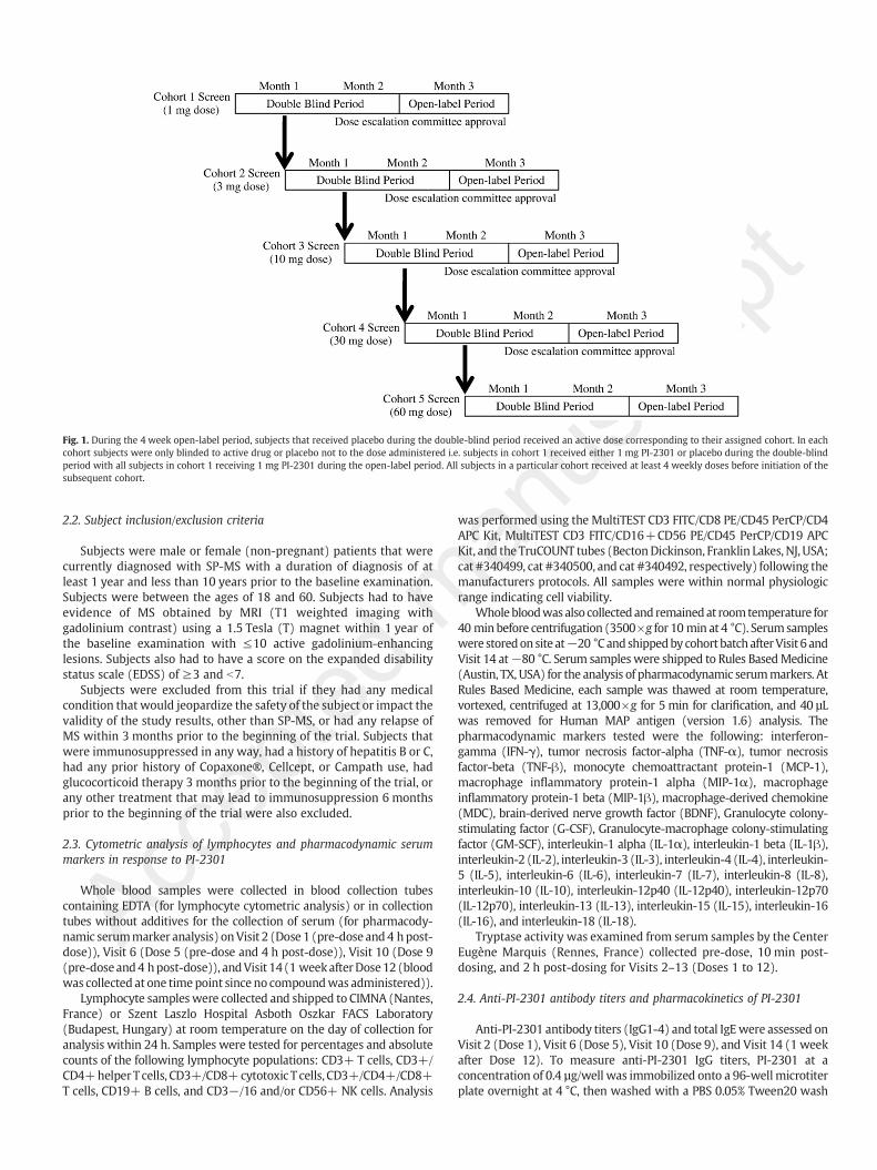
Table 1 shows the demographics of each cohort (France n=22,Serbia n=17, and Hungary n=11). The first group of subjectsentering the study were treated with the lowest dose of PI-2301tested. Additional cohorts were treated with higher doses of PI-2301,with the increase in dosing following a step-wise fashion. The start oftreatment for the next cohort, at the next higher dose, began after allsubjects in the previous cohort had received at least 4 weekly doses ofstudy drug, completed safety assessments, and with unanimousagreement by a dose escalation committee constituted of fourphysicians was obtained. Subjects randomized to placebo for thefirst 8 weeks of the study received treatment with PI-2301 at thecorresponding dose of their respective cohorts during the last 4 weeksof the study (open-label period). A final assessment occurred at13 weeks after the first dose (Visit 14 (1 week after Dose 12)) (Fig. 1).
Subjects were monitored for treatment emergent adverse events(TEAEs) that occurred within the 48 h after dosing (during thehospitalization period) or that were present prior to dosing andexacerbated within the 48 h after dosing. Basic urinalysis, hematology,and blood chemistry analysis were performed on each subject duringthe course of this study to assess the general health of all subjects. Eachinjection site was scored (scale of 0–4) for each of the followingcharacteristics: erythema, induration, pruritus, nodules and/or cysts,ecchymosis, duration, pain, or other local signs or symptoms. All ISRswere recorded as AEs using standardized criteria and pre-specifieddescriptive terms as noted in the protocol. A cumulative score wasgenerated for each subject for each visit with the highest cumulativescore reported for that visit. The mean value±SEM was calculated foreach dosing cohort. For subjects that had multiple injection sites or hadinjection site reactions (ISRs) from the previous dosing, these valueswere also added in the range of cumulative scores 0 (lowest) and 32(highest for single injection site in 1, 3, and 10 mg cohorts), 64 (highestfor two injection sites in the 30 mg cohort), or 96 (highest for threeinjection sites in the 60 mg cohort.)
Placebo(N=14)
Overall(N=50)
g9)
30 mg(N=6)
60 mg(N=8)
.6 44.0 48.3 44.4 46.2
.2 6.5 8.2 8.8 7.82 2 6 204 6 8 300 3 7 226 0 5 170 5 2 116 8 13 460 0 0 10 0 1 3
.9 60.4 70.0 72.9 70.1
.0 8.6 13.0 12.3 13.2
.4 173.5 170.3 172.4 171.1
.2 7.3 7.7 9.1 9.2
mus
critpt
Fig. 1. During the 4 week open-label period, subjects that received placebo during the double-blind period received an active dose corresponding to their assigned cohort. In eachcohort subjects were only blinded to active drug or placebo not to the dose administered i.e. subjects in cohort 1 received either 1 mg PI-2301 or placebo during the double-blindperiod with all subjects in cohort 1 receiving 1 mg PI-2301 during the open-label period. All subjects in a particular cohort received at least 4 weekly doses before initiation of thesubsequent cohort.
Accep
ted
2.2. Subject inclusion/exclusion criteria
Subjects were male or female (non-pregnant) patients that werecurrently diagnosed with SP-MS with a duration of diagnosis of atleast 1 year and less than 10 years prior to the baseline examination.Subjects were between the ages of 18 and 60. Subjects had to haveevidence of MS obtained by MRI (T1 weighted imaging withgadolinium contrast) using a 1.5 Tesla (T) magnet within 1 year ofthe baseline examination with ≤10 active gadolinium-enhancinglesions. Subjects also had to have a score on the expanded disabilitystatus scale (EDSS) of ≥3 and b7.
Subjects were excluded from this trial if they had any medicalcondition that would jeopardize the safety of the subject or impact thevalidity of the study results, other than SP-MS, or had any relapse ofMS within 3 months prior to the beginning of the trial. Subjects thatwere immunosuppressed in any way, had a history of hepatitis B or C,had any prior history of Copaxone®, Cellcept, or Campath use, hadglucocorticoid therapy 3 months prior to the beginning of the trial, orany other treatment that may lead to immunosuppression 6 monthsprior to the beginning of the trial were also excluded.
2.3. Cytometric analysis of lymphocytes and pharmacodynamic serummarkers in response to PI-2301
Whole blood samples were collected in blood collection tubescontaining EDTA (for lymphocyte cytometric analysis) or in collectiontubes without additives for the collection of serum (for pharmacody-namic serummarker analysis) onVisit 2 (Dose 1 (pre-dose and4 hpost-dose)), Visit 6 (Dose 5 (pre-dose and 4 h post-dose)), Visit 10 (Dose 9(pre-doseand4 hpost-dose)), andVisit 14(1 weekafterDose12 (bloodwas collected at one timepoint since no compoundwas administered)).
Lymphocyte sampleswere collected and shipped to CIMNA (Nantes,France) or Szent Laszlo Hospital Asboth Oszkar FACS Laboratory(Budapest, Hungary) at room temperature on the day of collection foranalysis within 24 h. Samples were tested for percentages and absolutecounts of the following lymphocyte populations: CD3+ T cells, CD3+/CD4+helper T cells, CD3+/CD8+cytotoxic T cells, CD3+/CD4+/CD8+T cells, CD19+ B cells, and CD3−/16 and/or CD56+ NK cells. Analysis
anwas performed using the MultiTEST CD3 FITC/CD8 PE/CD45 PerCP/CD4APC Kit, MultiTEST CD3 FITC/CD16+CD56 PE/CD45 PerCP/CD19 APCKit, and the TruCOUNT tubes (BectonDickinson, Franklin Lakes, NJ, USA;cat #340499, cat #340500, and cat #340492, respectively) following themanufacturers protocols. All samples were within normal physiologicrange indicating cell viability.
Wholebloodwasalso collectedand remainedat roomtemperature for40 min before centrifugation (3500×g for 10 min at 4 °C). Serumsampleswere storedonsite at−20 °Cand shippedby cohort batchafterVisit 6 andVisit 14 at−80 °C. Serum sampleswere shipped to Rules BasedMedicine(Austin, TX, USA) for the analysis of pharmacodynamic serummarkers. AtRules Based Medicine, each sample was thawed at room temperature,vortexed, centrifuged at 13,000×g for 5 min for clarification, and 40 µLwas removed for Human MAP antigen (version 1.6) analysis. Thepharmacodynamic markers tested were the following: interferon-gamma (IFN-γ), tumor necrosis factor-alpha (TNF-α), tumor necrosisfactor-beta (TNF-β), monocyte chemoattractant protein-1 (MCP-1),macrophage inflammatory protein-1 alpha (MIP-1α), macrophageinflammatory protein-1 beta (MIP-1β), macrophage-derived chemokine(MDC), brain-derived nerve growth factor (BDNF), Granulocyte colony-stimulating factor (G-CSF), Granulocyte-macrophage colony-stimulatingfactor (GM-SCF), interleukin-1 alpha (IL-1α), interleukin-1 beta (IL-1β),interleukin-2 (IL-2), interleukin-3 (IL-3), interleukin-4 (IL-4), interleukin-5 (IL-5), interleukin-6 (IL-6), interleukin-7 (IL-7), interleukin-8 (IL-8),interleukin-10 (IL-10), interleukin-12p40 (IL-12p40), interleukin-12p70(IL-12p70), interleukin-13 (IL-13), interleukin-15 (IL-15), interleukin-16(IL-16), and interleukin-18 (IL-18).
Tryptase activity was examined from serum samples by the CenterEugène Marquis (Rennes, France) collected pre-dose, 10 min post-dosing, and 2 h post-dosing for Visits 2–13 (Doses 1 to 12).
2.4. Anti-PI-2301 antibody titers and pharmacokinetics of PI-2301
Anti-PI-2301 antibody titers (IgG1-4) and total IgEwere assessed onVisit 2 (Dose 1), Visit 6 (Dose 5), Visit 10 (Dose 9), and Visit 14 (1 weekafter Dose 12). To measure anti-PI-2301 IgG titers, PI-2301 at aconcentration of 0.4 µg/well was immobilized onto a 96-well microtiterplate overnight at 4 °C, then washed with a PBS 0.05% Tween20 wash
solution and blocked (10% FBS/PBS) for 2 h. Following a wash step,serially diluted serum samples were added to the wells and incubatedfor 2 h, allowing anti-PI-2301 antibodies to bind to the immobilizedPI-2301. Unbound material was then washed away and a mouse anti-human IgG1, 2, 3, or 4 antibody conjugated to HRP (Southern Biotech,Birmingham, AL, USA) was added to the wells for 1 h. Unbounddetection antibody was removed by washing and a substrate (TMB/H2O2 from VWR; West Chester, PA, USA) was added to the wells andincubated for 15 min after which stop solution (2 N H2SO4 from VWR;West Chester, PA, USA)was added. Theoptical densitywasmeasured ona Tecan Shell microplate reader at 450 nm and the intensity of the colormeasured was proportional to the amount of anti-PI-2301 antibodybound by the immobilized PI-2301. Total IgE was measured in serumsamples sent to Rules Based Medicine (RBM; Austin, TX, USA).
The pharmacokinetic assay for PI-2301 is a qualified directcompetition ELISA. This method was transferred to and qualified byAvogadro (Fontenilles, France) and is acceptable to give relativequantitative levels of PI-2301 concentrations in human serum in alinear range of 69–527 ng/mL (with a limit of detection of 27 ng/mL).Intra-assay precision using the % coefficient of variation (CV) was 2.9to 13.2 and the inter-assay precision was 5.9 to 13.2. This method candetect ≥30-mer peptide fragments of PI-2301 which are biologicallyactive (data not shown) and non-specific binding (as calculated by %bound/maximal binding) was 6.6%. The assay passed all stabilitycontrols for samples kept at −80 °C for up to 4 months.
Briefly, PI-2301 is immobilized on a 96-well microtainer plateovernight at 4 °C, then blocked for 2 h, and washed. Human serum (apool of 10 male donors from Bioreclamation Westbury, NY, USA)containing known or unknown concentrations of PI-2301 are added tothe washed plates along with Protein A purified biotinylated anti-PI-2301 antibodies (made in-house from rabbit polyclonal antiserumproduced by Maine Biotechnology Services Portland, ME, USA) andincubated for 2 h on a plate shaker. Unbound material is washed away
Accep
ted m
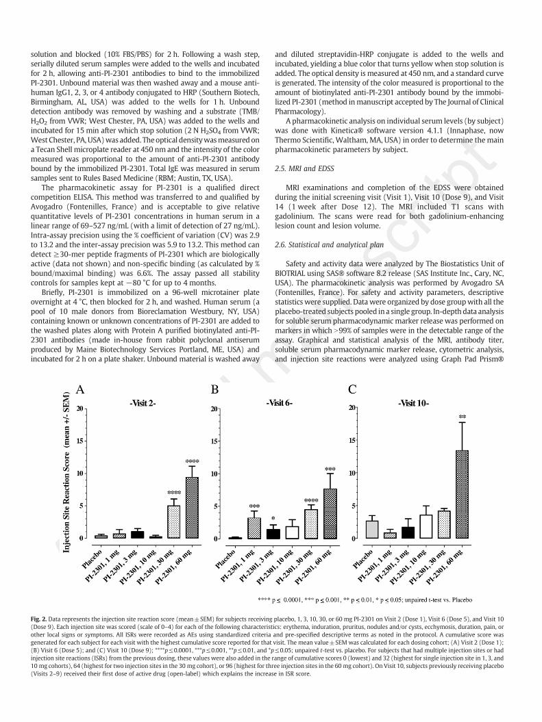
Fig. 2. Data represents the injection site reaction score (mean±SEM) for subjects receiving(Dose 9). Each injection site was scored (scale of 0–4) for each of the following characteristother local signs or symptoms. All ISRs were recorded as AEs using standardized criteriagenerated for each subject for each visit with the highest cumulative score reported for that(B) Visit 6 (Dose 5); and (C) Visit 10 (Dose 9); ****p≤0.0001, ***p≤0.001, **p≤0.01, and *pinjection site reactions (ISRs) from the previous dosing, these values were also added in the r10 mg cohorts), 64 (highest for two injection sites in the 30 mg cohort), or 96 (highest for thr(Visits 2–9) received their first dose of active drug (open-label) which explains the increas
anus
critpt
and diluted streptavidin-HRP conjugate is added to the wells andincubated, yielding a blue color that turns yellow when stop solution isadded. The optical density is measured at 450 nm, and a standard curveis generated. The intensity of the color measured is proportional to theamount of biotinylated anti-PI-2301 antibody bound by the immobi-lized PI-2301 (method inmanuscript accepted by The Journal of ClinicalPharmacology).
A pharmacokinetic analysis on individual serum levels (by subject)was done with Kinetica® software version 4.1.1 (Innaphase, nowThermo Scientific, Waltham, MA, USA) in order to determine themainpharmacokinetic parameters by subject.
2.5. MRI and EDSS
MRI examinations and completion of the EDSS were obtainedduring the initial screening visit (Visit 1), Visit 10 (Dose 9), and Visit14 (1 week after Dose 12). The MRI included T1 scans withgadolinium. The scans were read for both gadolinium-enhancinglesion count and lesion volume.
2.6. Statistical and analytical plan
Safety and activity data were analyzed by The Biostatistics Unit ofBIOTRIAL using SAS® software 8.2 release (SAS Institute Inc., Cary, NC,USA). The pharmacokinetic analysis was performed by Avogadro SA(Fontenilles, France). For safety and activity parameters, descriptivestatistics were supplied. Data were organized by dose groupwith all theplacebo-treated subjects pooled in a single group. In-depth data analysisfor soluble serum pharmacodynamic marker release was performed onmarkers in which N99% of samples were in the detectable range of theassay. Graphical and statistical analysis of the MRI, antibody titer,soluble serum pharmacodynamic marker release, cytometric analysis,and injection site reactions were analyzed using Graph Pad Prism®
placebo, 1, 3, 10, 30, or 60 mg PI-2301 on Visit 2 (Dose 1), Visit 6 (Dose 5), and Visit 10ics: erythema, induration, pruritus, nodules and/or cysts, ecchymosis, duration, pain, orand pre-specified descriptive terms as noted in the protocol. A cumulative score wasvisit. The mean value±SEMwas calculated for each dosing cohort; (A) Visit 2 (Dose 1);≤0.05; unpaired t-test vs. placebo. For subjects that had multiple injection sites or hadange of cumulative scores 0 (lowest) and 32 (highest for single injection site in 1, 3, andee injection sites in the 60 mg cohort). On Visit 10, subjects previously receiving placeboe in ISR score.
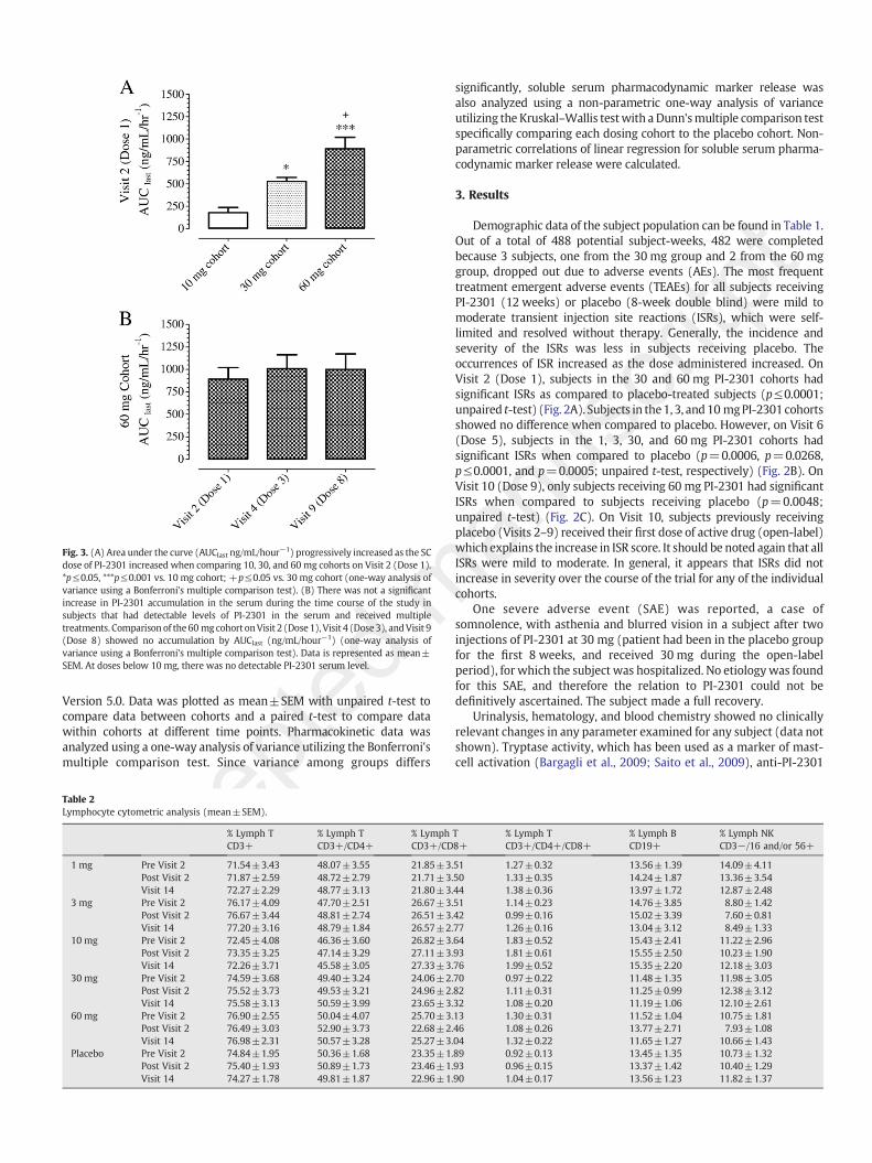
mFig. 3. (A) Area under the curve (AUClast ng/mL/hour−1) progressively increased as the SCdose of PI-2301 increased when comparing 10, 30, and 60 mg cohorts on Visit 2 (Dose 1).*p≤0.05, ***p≤0.001 vs. 10 mg cohort; +p≤0.05 vs. 30 mg cohort (one-way analysis ofvariance using a Bonferroni's multiple comparison test). (B) There was not a significantincrease in PI-2301 accumulation in the serum during the time course of the study insubjects that had detectable levels of PI-2301 in the serum and received multipletreatments. Comparison of the60 mgcohort onVisit 2 (Dose 1),Visit 4 (Dose3), andVisit 9(Dose 8) showed no accumulation by AUClast (ng/mL/hour−1) (one-way analysis ofvariance using a Bonferroni's multiple comparison test). Data is represented as mean±SEM. At doses below 10 mg, there was no detectable PI-2301 serum level.
tedVersion 5.0. Data was plotted as mean±SEM with unpaired t-test tocompare data between cohorts and a paired t-test to compare datawithin cohorts at different time points. Pharmacokinetic data wasanalyzed using a one-way analysis of variance utilizing the Bonferroni'smultiple comparison test. Since variance among groups differs
AccepTable 2
Lymphocyte cytometric analysis (mean±SEM).
% Lymph TCD3+
% Lymph TCD3+/CD4+
% LymphCD3+/CD
1 mg Pre Visit 2 71.54±3.43 48.07±3.55 21.85±3.Post Visit 2 71.87±2.59 48.72±2.79 21.71±3.Visit 14 72.27±2.29 48.77±3.13 21.80±3.
3 mg Pre Visit 2 76.17±4.09 47.70±2.51 26.67±3.Post Visit 2 76.67±3.44 48.81±2.74 26.51±3.Visit 14 77.20±3.16 48.79±1.84 26.57±2.
10 mg Pre Visit 2 72.45±4.08 46.36±3.60 26.82±3.Post Visit 2 73.35±3.25 47.14±3.29 27.11±3.Visit 14 72.26±3.71 45.58±3.05 27.33±3.
30 mg Pre Visit 2 74.59±3.68 49.40±3.24 24.06±2.Post Visit 2 75.52±3.73 49.53±3.21 24.96±2.Visit 14 75.58±3.13 50.59±3.99 23.65±3.
60 mg Pre Visit 2 76.90±2.55 50.04±4.07 25.70±3.Post Visit 2 76.49±3.03 52.90±3.73 22.68±2.Visit 14 76.98±2.31 50.57±3.28 25.27±3.
Placebo Pre Visit 2 74.84±1.95 50.36±1.68 23.35±1.Post Visit 2 75.40±1.93 50.89±1.73 23.46±1.Visit 14 74.27±1.78 49.81±1.87 22.96±1.
anus
critpt
significantly, soluble serum pharmacodynamic marker release wasalso analyzed using a non-parametric one-way analysis of varianceutilizing the Kruskal–Wallis testwith a Dunn'smultiple comparison testspecifically comparing each dosing cohort to the placebo cohort. Non-parametric correlations of linear regression for soluble serum pharma-codynamic marker release were calculated.
3. Results
Demographic data of the subject population can be found in Table 1.Out of a total of 488 potential subject-weeks, 482 were completedbecause 3 subjects, one from the 30 mg group and 2 from the 60 mggroup, dropped out due to adverse events (AEs). The most frequenttreatment emergent adverse events (TEAEs) for all subjects receivingPI-2301 (12 weeks) or placebo (8-week double blind) were mild tomoderate transient injection site reactions (ISRs), which were self-limited and resolved without therapy. Generally, the incidence andseverity of the ISRs was less in subjects receiving placebo. Theoccurrences of ISR increased as the dose administered increased. OnVisit 2 (Dose 1), subjects in the 30 and 60 mg PI-2301 cohorts hadsignificant ISRs as compared to placebo-treated subjects (p≤0.0001;unpaired t-test) (Fig. 2A). Subjects in the1, 3, and10 mgPI-2301cohortsshowed no difference when compared to placebo. However, on Visit 6(Dose 5), subjects in the 1, 3, 30, and 60 mg PI-2301 cohorts hadsignificant ISRs when compared to placebo (p=0.0006, p=0.0268,p≤0.0001, and p=0.0005; unpaired t-test, respectively) (Fig. 2B). OnVisit 10 (Dose 9), only subjects receiving 60 mg PI-2301 had significantISRs when compared to subjects receiving placebo (p=0.0048;unpaired t-test) (Fig. 2C). On Visit 10, subjects previously receivingplacebo (Visits 2–9) received their first dose of active drug (open-label)which explains the increase in ISR score. It should be noted again that allISRs were mild to moderate. In general, it appears that ISRs did notincrease in severity over the course of the trial for any of the individualcohorts.
One severe adverse event (SAE) was reported, a case ofsomnolence, with asthenia and blurred vision in a subject after twoinjections of PI-2301 at 30 mg (patient had been in the placebo groupfor the first 8 weeks, and received 30 mg during the open-labelperiod), for which the subject was hospitalized. No etiologywas foundfor this SAE, and therefore the relation to PI-2301 could not bedefinitively ascertained. The subject made a full recovery.
Urinalysis, hematology, and blood chemistry showed no clinicallyrelevant changes in any parameter examined for any subject (data notshown). Tryptase activity, which has been used as a marker of mast-cell activation (Bargagli et al., 2009; Saito et al., 2009), anti-PI-2301
T8+
% Lymph TCD3+/CD4+/CD8+
% Lymph BCD19+
% Lymph NKCD3−/16 and/or 56+
51 1.27±0.32 13.56±1.39 14.09±4.1150 1.33±0.35 14.24±1.87 13.36±3.5444 1.38±0.36 13.97±1.72 12.87±2.4851 1.14±0.23 14.76±3.85 8.80±1.4242 0.99±0.16 15.02±3.39 7.60±0.8177 1.26±0.16 13.04±3.12 8.49±1.3364 1.83±0.52 15.43±2.41 11.22±2.9693 1.81±0.61 15.55±2.50 10.23±1.9076 1.99±0.52 15.35±2.20 12.18±3.0370 0.97±0.22 11.48±1.35 11.98±3.0582 1.11±0.31 11.25±0.99 12.38±3.1232 1.08±0.20 11.19±1.06 12.10±2.6113 1.30±0.31 11.52±1.04 10.75±1.8146 1.08±0.26 13.77±2.71 7.93±1.0804 1.32±0.22 11.65±1.27 10.66±1.4389 0.92±0.13 13.45±1.35 10.73±1.3293 0.96±0.15 13.37±1.42 10.40±1.2990 1.04±0.17 13.56±1.23 11.82±1.37
antibody titers for IgG1, IgG2, IgG4, and total IgE showed nodifferences between treatments and did not increase over the timecourse of the study (data not shown). Three subjects, one subject inthe 3 mg cohort and two in the 10 mg cohort, appeared to generate alimited anti-PI-2301 IgG3 antibody response. At the 1/400 dilution,serum from the subject in the 3 mg cohort showed absorbances of0.012, 0.015, 0.602, and 0.437 on Visits 2, 6, 10, and 14 respectively byELISA on a scale from 0 to 4. Values for the two subjects in the 10 mgcohort at the same time points were 0.013, 0.009, 0.076, 0.124 and 0,0, 0, 0.132, respectively.
Pharmacokinetic analysis showed the presence of PI-2301 in theserum in similar kinetics observed in our single ascending dose study(manuscript accepted in Journal ofClinical Pharmacology).Atdosesbelow10mg, there was no detectable PI-2301 serum level. Cmax (mean±SEM)progressively increased as the SC dose of PI-2301 increased whencomparing 10, 30, and 60mg cohorts on Visit 2 (Dose 1) (73.62±10.93 ng/mL, 184.97±14.73 ng/mL, and 288.96±40.20 ng/mL, respec-tively). The same was true when comparing the area under the curve(AUClast) (mean±SEM) (177.82±57.94 ng/mL h, 525.96±45.40 ng/mL h, 891.37±128.46 ng/mL h, respectively) (Fig. 3A). There was not asignificant increase in PI-2301 accumulation in the serumduring the timecourse of the study in subjects that had detectable levels of PI-2301 in theserumand receivedmultiple treatments. Comparison of the 60 mg cohorton Visit 2 (Dose 1), Visit 4 (Dose 3), and Visit 9 (Dose 8) showed noaccumulation by Cmax (mean±SEM) (288.96±40.20 ng/mL, 313.79±
Accep
ted m
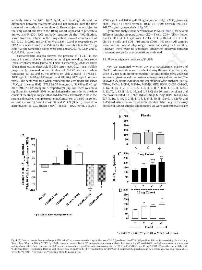
Fig. 4. (A) Data represents the mean change±SEM in IL-13 serum concentration (pg/mL) be3 mg, 10 mg, 30 mg, or 60 mg PI-2301. *p≤0.05 vs. placebo, unpaired t-test. When applying anot significant. (B–D) Data represents the IL-13 serum concentration (pg/mL) for subjects recVisit 2 (pre-Dose 1), Visit 6 (pre-Dose 5), Visit 10 (pre-Dose 9), and Visit 14 (1 week after Dos*p≤0.05, **p≤0.01, ***p≤0.001 vs. Visit 2 (pre-Dose 1), paired t-test.
scritp
t
47.68 ng/mL, and320.52±49.09 ng/mL, respectively)orAUClast (mean±SEM) (891.37±128.46 ng/mL h, 1008.17±154.95 ng/mL h, 999.38±161.07 ng/mL h, respectively) (Fig. 3B).
Cytometric analysis was performed on PBMCs (Table 2) for severaldifferent lymphocyte populations (CD3+ T cells, CD3+/CD4+ helperT cells. CD3+/CD8+ cytotoxic T cells, CD3+/CD4+/CD8+ T cells,CD19+ B cells, and CD3−/16 and/or CD56+ NK cells). All sampleswere within normal physiologic range indicating cell viability.However, there were no significant differences observed betweentreatment groups for any populations evaluated.
3.1. Pharmacodynamic markers of PI-2301
Next we examined whether any pharmacodynamic markers ofPI-2301 administration were evident during the course of the study.Since PI-2301 is an immunomodulator, serum samples were analyzedfor serum cytokines and chemokines at time points and visits noted. Thefollowing 26 serum cytokines and chemokines were analyzed: IFN-γ,TNF-α, TNF-β, MCP-1, MIP-1α, MIP-1β, MDC, BDNF, G-CSF, GM-SCF,IL-1α, IL-1β, IL-2, IL-3, IL-4, IL-5, IL-6, IL-7, IL-8, IL-10, IL-12p40,IL-12p70, IL-13, IL-15, IL-16, and IL-18. Of the 26 serum cytokines andchemokines tested, 17 (IFN-γ, TNF-α, TNF-β, MIP-1β, BDNF, G-CSF, GM-SCF, IL-1α, IL-1β, IL-2, IL-4, IL-5, IL-6, IL-10, IL-12p40, IL-12p70, andIL-15) had values thatwere not within the detectable range of the assayfor several subject samples and thereforewewere unable to statistically
anu
tween Visit 2 (pre-Dose 1) and Visit 10 (pre-Dose 9) in subjects receiving placebo, 1 mg,one-way analysis of variance using a Kruskal–Wallis multiple comparison test, data waseiving placebo (B), 3 mg PI-2301 (C), and 10 mg PI-2301 (D) over the course of the trial;e 12). On Visit 14, subjects in the placebo groupwere receiving active drug (open-label).
analyze these markers. A descriptive analysis of cytokines andchemokines which are implicated in inflammation such as IFN-γ, TNF-α, IL-12p40, and IL-12p70 showed no clear trends in relation to PI-2301administration. Even though IL-10 was not fully analyzed due toinsufficient samples for each cohort, an apparent trend was observedshowing an increase over time in the 3 and 10 mg cohorts (data notshown). Since some sampleswere notwithin the rangeof detection, it isdifficult to draw conclusions from this data. No other clear trends wereobserved in these cytokines or chemokines. Nine cytokines andchemokines (MCP-1, MIP-1α, MDC, IL-3, IL-7, IL-8, IL-13, IL-16, and IL-18) were further analyzed as pharmacodynamic markers.
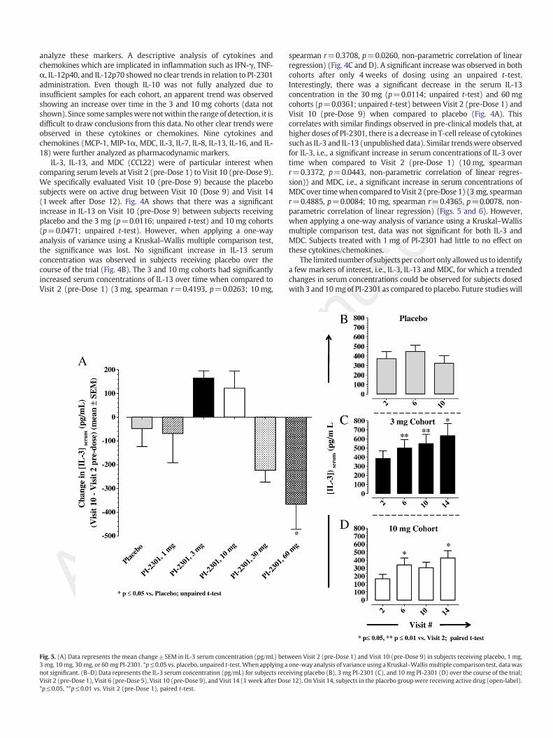
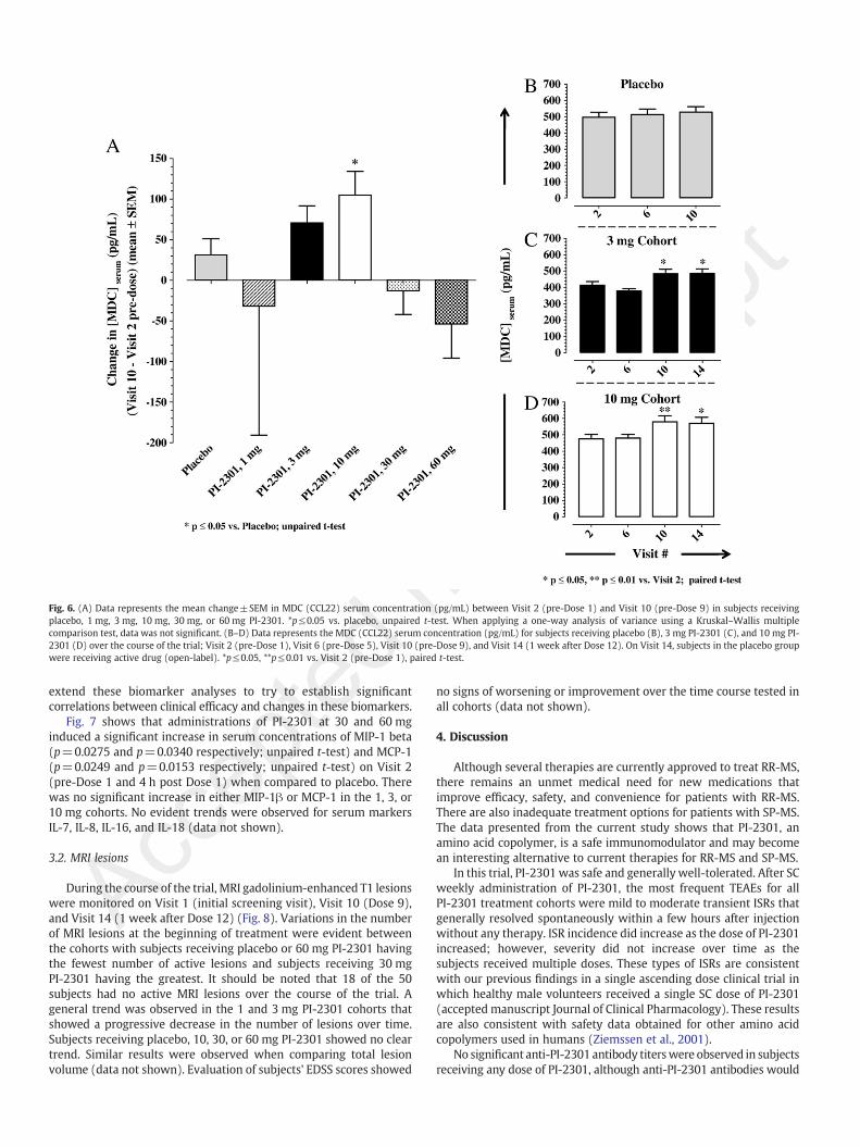
IL-3, IL-13, and MDC (CCL22) were of particular interest whencomparing serum levels at Visit 2 (pre-Dose 1) to Visit 10 (pre-Dose 9).We specifically evaluated Visit 10 (pre-Dose 9) because the placebosubjects were on active drug between Visit 10 (Dose 9) and Visit 14(1 week after Dose 12). Fig. 4A shows that there was a significantincrease in IL-13 on Visit 10 (pre-Dose 9) between subjects receivingplacebo and the 3 mg (p=0.0116; unpaired t-test) and 10 mg cohorts(p=0.0471; unpaired t-test). However, when applying a one-wayanalysis of variance using a Kruskal–Wallis multiple comparison test,the significance was lost. No significant increase in IL-13 serumconcentration was observed in subjects receiving placebo over thecourse of the trial (Fig. 4B). The 3 and 10 mg cohorts had significantlyincreased serum concentrations of IL-13 over time when compared toVisit 2 (pre-Dose 1) (3 mg, spearman r=0.4193, p=0.0263; 10 mg,
Accep
ted m
Fig. 5. (A) Data represents the mean change±SEM in IL-3 serum concentration (pg/mL) bet3 mg, 10 mg, 30 mg, or 60 mg PI-2301. *p≤0.05 vs. placebo, unpaired t-test. When applying anot significant. (B–D) Data represents the IL-3 serum concentration (pg/mL) for subjects receVisit 2 (pre-Dose 1), Visit 6 (pre-Dose 5), Visit 10 (pre-Dose 9), and Visit 14 (1 week after Dos*p≤0.05, **p≤0.01 vs. Visit 2 (pre-Dose 1), paired t-test.
scritp
t
spearman r=0.3708, p=0.0260, non-parametric correlation of linearregression) (Fig. 4C and D). A significant increase was observed in bothcohorts after only 4 weeks of dosing using an unpaired t-test.Interestingly, there was a significant decrease in the serum IL-13concentration in the 30 mg (p=0.0114; unpaired t-test) and 60 mgcohorts (p=0.0361; unpaired t-test) between Visit 2 (pre-Dose 1) andVisit 10 (pre-Dose 9) when compared to placebo (Fig. 4A). Thiscorrelates with similar findings observed in pre-clinical models that, athigher doses of PI-2301, there is a decrease in T-cell release of cytokinessuch as IL-3 and IL-13 (unpublished data). Similar trendswere observedfor IL-3, i.e., a significant increase in serum concentrations of IL-3 overtime when compared to Visit 2 (pre-Dose 1) (10 mg, spearmanr=0.3372, p=0.0443, non-parametric correlation of linear regres-sion)) and MDC, i.e., a significant increase in serum concentrations ofMDCover timewhen compared toVisit 2 (pre-Dose 1) (3 mg, spearmanr=0.4885, p=0.0084; 10 mg, spearman r=0.4365, p=0.0078, non-parametric correlation of linear regression) (Figs. 5 and 6). However,when applying a one-way analysis of variance using a Kruskal–Wallismultiple comparison test, data was not significant for both IL-3 andMDC. Subjects treated with 1 mg of PI-2301 had little to no effect onthese cytokines/chemokines.
The limitednumberof subjects per cohort only allowedus to identifya few markers of interest, i.e., IL-3, IL-13 and MDC, for which a trendedchanges in serum concentrations could be observed for subjects dosedwith 3 and 10 mgof PI-2301 as compared to placebo. Future studieswill
anu
ween Visit 2 (pre-Dose 1) and Visit 10 (pre-Dose 9) in subjects receiving placebo, 1 mg,one-way analysis of variance using a Kruskal–Wallis multiple comparison test, data wasiving placebo (B), 3 mg PI-2301 (C), and 10 mg PI-2301 (D) over the course of the trial;e 12). On Visit 14, subjects in the placebo groupwere receiving active drug (open-label).
man
uscri
tpt
Fig. 6. (A) Data represents the mean change±SEM in MDC (CCL22) serum concentration (pg/mL) between Visit 2 (pre-Dose 1) and Visit 10 (pre-Dose 9) in subjects receivingplacebo, 1 mg, 3 mg, 10 mg, 30 mg, or 60 mg PI-2301. *p≤0.05 vs. placebo, unpaired t-test. When applying a one-way analysis of variance using a Kruskal–Wallis multiplecomparison test, data was not significant. (B–D) Data represents the MDC (CCL22) serum concentration (pg/mL) for subjects receiving placebo (B), 3 mg PI-2301 (C), and 10 mg PI-2301 (D) over the course of the trial; Visit 2 (pre-Dose 1), Visit 6 (pre-Dose 5), Visit 10 (pre-Dose 9), and Visit 14 (1 week after Dose 12). On Visit 14, subjects in the placebo groupwere receiving active drug (open-label). *p≤0.05, **p≤0.01 vs. Visit 2 (pre-Dose 1), paired t-test.
Accep
tedextend these biomarker analyses to try to establish significantcorrelations between clinical efficacy and changes in these biomarkers.
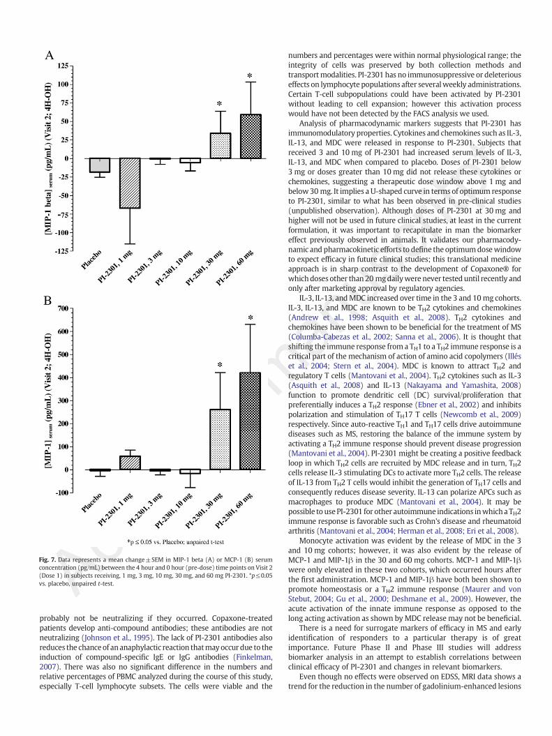
Fig. 7 shows that administrations of PI-2301 at 30 and 60 mginduced a significant increase in serum concentrations of MIP-1 beta(p=0.0275 and p=0.0340 respectively; unpaired t-test) and MCP-1(p=0.0249 and p=0.0153 respectively; unpaired t-test) on Visit 2(pre-Dose 1 and 4 h post Dose 1) when compared to placebo. Therewas no significant increase in either MIP-1β or MCP-1 in the 1, 3, or10 mg cohorts. No evident trends were observed for serum markersIL-7, IL-8, IL-16, and IL-18 (data not shown).
3.2. MRI lesions
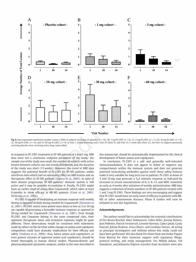
During the course of the trial, MRI gadolinium-enhanced T1 lesionswere monitored on Visit 1 (initial screening visit), Visit 10 (Dose 9),and Visit 14 (1 week after Dose 12) (Fig. 8). Variations in the numberof MRI lesions at the beginning of treatment were evident betweenthe cohorts with subjects receiving placebo or 60 mg PI-2301 havingthe fewest number of active lesions and subjects receiving 30 mgPI-2301 having the greatest. It should be noted that 18 of the 50subjects had no active MRI lesions over the course of the trial. Ageneral trend was observed in the 1 and 3 mg PI-2301 cohorts thatshowed a progressive decrease in the number of lesions over time.Subjects receiving placebo, 10, 30, or 60 mg PI-2301 showed no cleartrend. Similar results were observed when comparing total lesionvolume (data not shown). Evaluation of subjects' EDSS scores showed
no signs of worsening or improvement over the time course tested inall cohorts (data not shown).
4. Discussion
Although several therapies are currently approved to treat RR-MS,there remains an unmet medical need for new medications thatimprove efficacy, safety, and convenience for patients with RR-MS.There are also inadequate treatment options for patients with SP-MS.The data presented from the current study shows that PI-2301, anamino acid copolymer, is a safe immunomodulator and may becomean interesting alternative to current therapies for RR-MS and SP-MS.
In this trial, PI-2301was safe and generally well-tolerated. After SCweekly administration of PI-2301, the most frequent TEAEs for allPI-2301 treatment cohorts were mild to moderate transient ISRs thatgenerally resolved spontaneously within a few hours after injectionwithout any therapy. ISR incidence did increase as the dose of PI-2301increased; however, severity did not increase over time as thesubjects received multiple doses. These types of ISRs are consistentwith our previous findings in a single ascending dose clinical trial inwhich healthy male volunteers received a single SC dose of PI-2301(accepted manuscript Journal of Clinical Pharmacology). These resultsare also consistent with safety data obtained for other amino acidcopolymers used in humans (Ziemssen et al., 2001).
No significant anti-PI-2301 antibody titerswere observed in subjectsreceiving any dose of PI-2301, although anti-PI-2301 antibodies would
Accep
ted m
Fig. 7. Data represents a mean change±SEM in MIP-1 beta (A) or MCP-1 (B) serumconcentration (pg/mL) between the 4 hour and 0 hour (pre-dose) time points on Visit 2(Dose 1) in subjects receiving, 1 mg, 3 mg, 10 mg, 30 mg, and 60 mg PI-2301. *p≤0.05vs. placebo, unpaired t-test.
probably not be neutralizing if they occurred. Copaxone-treatedpatients develop anti-compound antibodies; these antibodies are notneutralizing (Johnson et al., 1995). The lack of PI-2301 antibodies alsoreduces the chanceof ananaphylactic reaction thatmayoccur due to theinduction of compound-specific IgE or IgG antibodies (Finkelman,2007). There was also no significant difference in the numbers andrelative percentages of PBMC analyzed during the course of this study,especially T-cell lymphocyte subsets. The cells were viable and the
anus
critpt
numbers and percentages were within normal physiological range; theintegrity of cells was preserved by both collection methods andtransportmodalities. PI-2301 has no immunosuppressive or deleteriouseffects on lymphocyte populations after severalweekly administrations.Certain T-cell subpopulations could have been activated by PI-2301without leading to cell expansion; however this activation processwould have not been detected by the FACS analysis we used.
Analysis of pharmacodynamic markers suggests that PI-2301 hasimmunomodulatory properties. Cytokines and chemokines such as IL-3,IL-13, and MDC were released in response to PI-2301. Subjects thatreceived 3 and 10 mg of PI-2301 had increased serum levels of IL-3,IL-13, and MDC when compared to placebo. Doses of PI-2301 below3 mg or doses greater than 10 mg did not release these cytokines orchemokines, suggesting a therapeutic dose window above 1 mg andbelow30 mg. It implies a U-shaped curve in terms of optimum responseto PI-2301, similar to what has been observed in pre-clinical studies(unpublished observation). Although doses of PI-2301 at 30 mg andhigher will not be used in future clinical studies, at least in the currentformulation, it was important to recapitulate in man the biomarkereffect previously observed in animals. It validates our pharmacody-namic and pharmacokinetic efforts to define the optimumdosewindowto expect efficacy in future clinical studies; this translational medicineapproach is in sharp contrast to the development of Copaxone® forwhichdoses other than20 mgdailywere never testeduntil recently andonly after marketing approval by regulatory agencies.
IL-3, IL-13, andMDC increased over time in the 3 and 10mg cohorts.IL-3, IL-13, and MDC are known to be TH2 cytokines and chemokines(Andrew et al., 1998; Asquith et al., 2008). TH2 cytokines andchemokines have been shown to be beneficial for the treatment of MS(Columba-Cabezas et al., 2002; Sanna et al., 2006). It is thought thatshifting the immune response from a TH1 to a TH2 immune response is acritical part of the mechanism of action of amino acid copolymers (Illéset al., 2004; Stern et al., 2004). MDC is known to attract TH2 andregulatory T cells (Mantovani et al., 2004). TH2 cytokines such as IL-3(Asquith et al., 2008) and IL-13 (Nakayama and Yamashita, 2008)function to promote dendritic cell (DC) survival/proliferation thatpreferentially induces a TH2 response (Ebner et al., 2002) and inhibitspolarization and stimulation of TH17 T cells (Newcomb et al., 2009)respectively. Since auto-reactive TH1 and TH17 cells drive autoimmunediseases such as MS, restoring the balance of the immune system byactivating a TH2 immune response should prevent disease progression(Mantovani et al., 2004). PI-2301 might be creating a positive feedbackloop in which TH2 cells are recruited by MDC release and in turn, TH2cells release IL-3 stimulating DCs to activate more TH2 cells. The releaseof IL-13 from TH2 T cells would inhibit the generation of TH17 cells andconsequently reduces disease severity. IL-13 can polarize APCs such asmacrophages to produce MDC (Mantovani et al., 2004). It may bepossible to usePI-2301 for other autoimmune indications inwhicha TH2immune response is favorable such as Crohn's disease and rheumatoidarthritis (Mantovani et al., 2004; Herman et al., 2008; Eri et al., 2008).
Monocyte activation was evident by the release of MDC in the 3and 10 mg cohorts; however, it was also evident by the release ofMCP-1 and MIP-1β in the 30 and 60 mg cohorts. MCP-1 and MIP-1βwere only elevated in these two cohorts, which occurred hours afterthe first administration. MCP-1 and MIP-1β have both been shown topromote homeostasis or a TH2 immune response (Maurer and vonStebut, 2004; Gu et al., 2000; Deshmane et al., 2009). However, theacute activation of the innate immune response as opposed to thelong acting activation as shown byMDC release may not be beneficial.
There is a need for surrogate markers of efficacy in MS and earlyidentification of responders to a particular therapy is of greatimportance. Future Phase II and Phase III studies will addressbiomarker analysis in an attempt to establish correlations betweenclinical efficacy of PI-2301 and changes in relevant biomarkers.
Even though no effects were observed on EDSS, MRI data shows atrend for the reduction in the number of gadolinium-enhanced lesions
man
uscri
tpt
Fig. 8. Data represents total lesion number (mean±SEM) in subjects receiving (A) placebo (n=14), (B) 1 mg PI-2301 (n=6), (C) 3 mg PI-2301 (n=7), (D) 10 mg PI-2301 (n=9),(E) 30 mg PI-2301 (n=6), and (F) 60 mg PI-2301 (n=8) at Visit 1 (initial screening visit), Visit 10 (Dose 9), and Visit 14 (1 week after Dose 12). On Visit 14, subjects previouslyreceiving placebo were receiving active drug (open-label).
Accep
tedin response to PI-2301 treatment in SP-MS patients at 1 and 3 mg. MRIdata were not a conclusive endpoint parameter of the study; thesample size of the study was small, the number of subjects with activelesions between cohorts was not evenly distributed, and the durationof the study was short (12 weeks). However, the trend in MRI datasuggests the potential benefit of PI-2301 for SP-MS patients, unlikeinterferon-beta which had no worsening effect on MRI lesions and notherapeutic effect in SP-MS patients (Inglese et al., 2003). In spite oftheir disease progression, SP-MS patients' immune system is stillactive and it may be possible to modulate it. Finally, PI-2301 mighthave an earlier onset of action than Copaxone®, which takes at least6 months to show efficacy in RR-MS patients (Comi et al., 2001;Wolinsky et al., 2002).
PI-2301 is capable of modulating an immune response with weeklydosing as opposed to daily dosing needed for Copaxone® (Ziemssen etal., 2001). PI-2301 seems more potent since doses as low as 3 or 10 mgare capable of generating a favorable immune response as opposed to20 mg needed for Copaxone® (Ziemssen et al., 2001). Even thoughPI-2301 and Copaxone belong to the same compound class, theiroptimum therapeutic doses and treatment regimens might be quitedifferent. These observations would be consistent with statementsmade by others on the fact that subtle changes in amino acid copolymercomposition could have dramatic implications for their efficacy andsafety (Varkony et al., 2009); thus, future amino acid copolymers, oreven generic versions of approved amino acid copolymers, should betested thoroughly in human clinical studies. Pharmacokinetic andpharmacodynamic parameter analyses, similar to the ones described in
this manuscript, should be systematically implemented for the clinicaldevelopment of future amino acid copolymers.
In conclusion, PI-2301 is a safe and generally well-toleratedimmunomodulator. It does not appear to delete or suppress anycompartments within the immune system and does not generatepotential neutralizing antibodies against itself; these safety featuresmake it very suitable for long term use in patients. PI-2301 at doses of3 and 10 mg may generate a TH2 immune response as indicated byincreases in serum concentrations of IL-3, IL-13, and MDC sometimeas early as 4 weeks after initiation of weekly administration. MRI datasuggests a reduction of lesion numbers in SP-MS patients treated with1 and 3 mg PI-2301. These findings are very encouraging and suggestthat PI-2301would have an early onset of efficacy in patients with RR-MS or other autoimmune diseases. Phase II studies will soon beinitiated to test this hypothesis.
Acknowledgements
The authors would like to acknowledge the essential contributionsof Drs Bruno Brochet, Marc Debouverie, Gilles Defer, Jerome DeSeze,Jean Pelletier, Patrick Vermersch, Levente Kerenyi, Gabor Jakab, GyulaPanczel, Jelena Drulovic, Evica Dincic, and Gordana Toncev, all actingas principal investigators and without whom this study could nothave been performed.We also thank Drs Alain Patat and Andre Paquinfrom Biotrial for their immense support with regulatory filings,protocol writing, and study management. Drs Mihaly Juhasz, VidStanulovic, and Jelisaveta Popovic-Gavrilov from Accelsiors were also
m
cepte
d
essential partners in the submission and execution of this study inHungary and Serbia. We are also grateful to Dr. Eric Lacoste, Dr.Stephane Bronner, and Claire Toutin from Avogadro for their technicalexpertise with the pharmacokinetic analysis.
References
Andrew, D.P., Chang, M., McNinch, J., Wathen, S.T., Rihanek, M., Tseng, J., Spellberg, J.P.,Elias III, C.G., 1998. STCP-1 (MDC) CC chemokine acts specifically on chronicallyactivated Th2 lymphocytes and is produced by monocytes on stimulation with Th2cytokines IL-4 and IL-13. J. Immunol. 161, 5027–5038.
Asquith, K.L., Ramshaw, H.S., Hansbro, P.M., Beagley, K.W., Lopez, A.F., Foster, P.S., 2008.The IL-3/IL-5/GM-CSF common receptor plays a pivotal role in the regulation of theTh2 immunity and allergic airway inflammation. J. Immunol. 180, 1199–1206.
Bargagli, E.,Mazzi, A.,Mezzasalma, F., Perrone, A., Olivieri, C., Prasse, A., Bianchi, N., Pieroni,M.G., Rottoli, P., 2009. The analysis of tryptase in serum of sarcoidosis patients.Inflammation 32, 310–314.
Boggild, M., 2009. Immunosuppression followed by immunomodulation. J. Neurol. Sci.277, S50–S54.
Columba-Cabezas, S., Serafini, B., Ambrosini, E., Sanchez, M., Penna, G., Adorini, L., Aloisi,F., 2002. Induction of macrophage-derived chemokine/CCL22 expression inexperimental autoimmune encephalomyelitis and cultured microglia: implicationsfor disease regulation. J. Neuroimmunol. 130, 10–21.
Comi, G., 2009. Treatment of multiple sclerosis: role of natalizumab. Neurol. Sci. 30,S155–S158.
Comi, G., Filippi, M., Wolinsky, J.S., 2001. European/Canadian multicenter, double-blind,randomized, placebo-controlled study of the effects of glatiramer acetate onmagnetic resonance imaging—measured disease activity and burden in patientswith relapsing multiple sclerosis. European/Canadian Glatiramer Acetate StudyGroup. Ann. Neurol. 49, 290–297.
De Stefano, N., Filippi, M., Confavreux, C., Vermersch, P., Simu, M., Sindic, C., Hupperts,R., Bajenaru, O., Edan, G., Grimaldi, L., Marginean, I., Medaer, R., Orefice, G., Pascu, I.,Pelletier, J., Sanders, E., Scarpini, E., Mancardi, G.L., 2009. The results of twomulticenter, open-label studies assessing efficacy, tolerability and safety ofprotiramer, a high molecular weight synthetic copolymeric mixture, in patientswith relapsing–remitting multiple sclerosis. Mult. Scler. 15, 238–243.
Deshmane, S.L., Kremlev, S., Amini, S., Sawaya, B.E., 2009. Monocyte chemoattractantprotein 1 (MCP-1): an overview. J. Interferon Cytokine Res. 29, 313–326.
Dhib-Jalbut, S., 2003. Glatiramer acetate (Copaxone®) therapy for multiple sclerosis.Pharmacol. Ther. 98, 245–255.
Dressel, A., Mirowska-Guzel, D., Gerlach, C., Wber, F., 2007. Migration of T-cell subsetsin multiple sclerosis and the effect of interferon-beta1a. Acta Neurol. Scand. 116,164–168.
Ebner, S., Hofer, S., Nguyen, V.A., Furhapter, C., Herold, M., Fritsch, P., Heufler, C.,Romani, N., 2002. A novel role for IL-3: human monocytes cultured in the presenceof IL-3 and IL-4 differentiate into dendritic cells that produce less IL-12 and shift Thcell responses toward a Th2 cytokine pattern. J. Immunol. 168, 6199–6207.
Eri, R., Kodumudi, K.N., Summerlin, D.J., Srinivasan, M., 2008. Suppression of coloninflammation by CD80 blockade: evaluation in twomurinemodels of inflammatorybowel disease. Inflamm. Bowel Dis. 14, 458–470.
Finkelman, F.D., 2007. Anaphylaxis: lessons from mouse models. J. Allergy Clin.Immunol. 120, 506–515.
Fridkis-Hareli,M., Teitelbaum, D., Gurevich, E., Pecht, I., Brautbar, C., Kwon, O.J., Brenner, T.,Arnon, R., Sela, M., 1994. Direct binding of myelin basic protein and syntheticcopolymer 1 to class IImajor histocompatibility complexmolecules on living antigen-presenting cells—specificity and promiscuity. Proc. Natl. Acad. Sci. U. S. A. 91,4872–4876.
Fridkis-Hareli, M., Santambrogio, L., Stern, J.N.H., Fugger, L., Brosnan, C., Strominger, J.L.,2002. Novel synthetic amino acid copolymers that inhibit autoantigen-specfic T cellresponses and suppress experimental autoimmuneencephalomyelitis. J. Clin. Investig.109, 1635–1643.
Gauthier, L., Smith, K.J., Pyrdol, J., Kalandadze, A., Strominger, J.L., Wiley, D.C.,Wucherpfennig, K.W., 1998. Expression and crystallization of the complex HLA-DR2 (DRA, DRB1*1501) and an immunodominant peptide of human myelin basicprotein. Proc. Natl. Acad. Sci. U. S. A. 95, 11,828–11,833.
Ac
anus
critpt
Gu, L., Tseng, S., Horner, R.M., Tam, C., Loda, M., Rollins, B.J., 2000. Control of TH2polarization by the chemokine monocyte chemoattractant protein-1. Nature 404,407–411.
Herman, S., Zurgil, N., Langevitz, P., Ehrenfeld, M., Deutsch, M., 2008. Methotrexateselectively modulates TH1/TH2 balance in active rheumatoid arthritis patients.Clin. Exp. Rheumatol. 26, 317–323.
Illés, Z., Stern, J.N.H., Reddy, J., Waldner, H., Mycko, M.P., Brosnan, C.F., Ellmerich, S.,Altmann, D.M., Santambrogio, L., Strominger, J.L., Kuchroo, V.K., 2004.Modified aminoacid copolymers suppress myelin basic protein 85–99-induced encephalomyelitis inhumanized mice through different effects on T cells. Proc. Natl. Acad. Sci. U. S. A. 101,11,749–11,754.
Inglese,M., vanWaesberghe, J.H., Rovaris, M., Beckmann, K., Barkhof, F., Hahn, D., Kappos, L.,Miller, D.H., Polman, C., Pozzilli, C., Thompson, A.J., Yousry, T.A., Wagner, K., Comi, G.,Filippi, M., 2003. The effect of interferon beta-1b on quantities derived fromMTMRI insecondary progressive MS. Neurology 60, 853–860.
Jarius, S., Hohlfeld, R., Voltz, R., 2003. Diagnosis and treatment of multiple sclerosis.Update, 2003. M.M.W. Fortschr. Med. 145, 88–91, 93, 95.
Johnson, K.P., Brooks, B.R., Cohen, J.A., Ford, C.C., Goldstein, J., Lisak, R.P., Myers, L.W.,Panitch, H.S., Rose, J.W., Schiffer, R.B., 1995. Copolymer 1 reduces relapse rate andimproves disability in relapsing–remitting multiple sclerosis: results of a phase IIImulticenter, double-blind placebo-controlled trial. The Copolymer 1 MultipleSclerosis Study Group. Neurology 45, 1268–1276.
Mantovani, A., Sica, A., Sozzani, S., Allavena, P., Vecchi, A., Locati, M., 2004. Thechemokine system in diverse forms of macrophage activation and polarization.Trends Immunol. 25, 677–686.
Martinelli, V., Radaelli, M., Straffi, L., Rodegher, M., Comi, G., 2009. Mitoxantrone:benefits and risks in multiple sclerosis patients. Neurol. Sci. 30, S167–S170.
Maurer, M., von Stebut, E., 2004. Macrophage inflammatory protein-1. Int. J. Biochem.Cell Biol. 36, 1882–1886.
Miller, A., Shapiro, S., Gershtein, R., Kinarty, A., Rawashdeh, H., Honigman, S., Lahat, N.,1998. Treatment of multiple sclerosis with copolymer-1 (Copaxone): implicatingmechanisms of Th1 to Th2/Th3 immune-deviation. J. Neuroimmunol. 92, 113–121.
Nakayama, T., Yamashita, M., 2008. Initiation andmaintenance of Th2 cell identity. Curr.Opin. Immunol. 20, 265–271.
Newcomb, D.C., Zhou,W., Moore,M.L., Goleniewska, K., Hershey, G.K.K., Kolls, J.K., PeeblesJr., R.S., 2009. A functional IL-13 receptor is expressed on polarizedmurine CD4+ Th17cells and IL-13 signaling attenuates Th17 cytokine production. J. Immunol. 182,5317–5321.
Papeix, C., Lubetzki, C., 2009. Monoclonal antibodies in multiple sclerosis. Med. Sci.(Paris) 25, 1113–1115.
Prat, A., Biernacki, K., Antel, J.P., 2005. Th1 and Th2 lymphocytemigration across the humanBBB is specifically regulated by interferon beta and copolymer-1. J. Autoimmun. 24,119–124.
Saito, H., Kato, A., Matsumoto, K., 2009. Role of mast cells in allergy. Nippon Rinsho 67,2083–2087.
Sanna, A., Fois, M.L., Arru, G., Huang, Y.M., Link, H., Pugliatti, M., Rosati, G., Sotgiu, S., 2006.Glatiramer acetate reduces lymphocyte proliferation and enhances IL-5 ad IL-13production through modulation of monocyte-derived dendritic cells in multiplesclerosis. Clin. Exp. Immunol. 143, 357–362.
Stern, J.N.H., Illés, Z., Reddy, J., Keskin, D.B., Sheu, E., Fridkis-Hareli, M., Nishimura, H.,Brosnan, C.F., Santambrogio, L., Kuchroo, V.K., Strominger, J.L., 2004. Ameliorationof proteolipid protein 139-151-induces encephalomyelitis in SJL mice by modifiedamino acid copolymers and their mechanisms. Proc. Natl. Acad. Sci. U. S. A. 101,11,743–11,748.
Teitelbaum, D., Meshorer, A., Hirshfeld, T., Arnon, R., Sela, M., 1971. Suppression ofexperimental allergic encephalomyelitis bya syntheticpolypeptide. Eur. J. Immunol. 1,242–248.
Varkony, H., Weinstein, V., Klinger, E., Sterling, J., Cooperman, H., Komlosh, T., Ladkani, D.,Schwartz, R., 2009. The glatiramoid class of immunomodulator drugs. Expert Opin.Pharmacother. 10, 657–668.
Wolinsky, J.S., Comi, G., Filippi, M., Ladkani, D., Kadosh, S., Shifroni, G., 2002. Copaxone'seffect on MRI-monitored disease in relapsing MS is reproducible and sustained.Neurology 59, 1284–1286.
Ziemssen, T., Neuhaus, O., Hohlfeld, R., 2001. Risk–benefit assessment of glatirameracetate in multiple sclerosis. Drug Saf. 24, 979–990.


