Embed Size (px)
Citation preview

n engl j med 365;9 nejm.org september 1, 2011 787
The new england journal of medicineestablished in 1812 september 1, 2011 vol. 365 no. 9
Early versus Later Rhythm Analysis in Patients with Out-of-Hospital Cardiac Arrest
Ian G. Stiell, M.D., Graham Nichol, M.D., M.P.H., Brian G. Leroux, Ph.D., Thomas D. Rea, M.D., M.P.H., Joseph P. Ornato, M.D., Judy Powell, B.S.N., James Christenson, M.D., Clifton W. Callaway, M.D., Ph.D., Peter J. Kudenchuk, M.D., Tom P. Aufderheide, M.D., Ahamed H. Idris, M.D., Mohamud R. Daya, M.D.,
Henry E. Wang, M.D., Laurie J. Morrison, M.D., Daniel Davis, M.D., Douglas Andrusiek, M.Sc., Shannon Stephens, E.M.T.-P., Sheldon Cheskes, M.D., Robert H. Schmicker, M.S., Ray Fowler, M.D.,
Christian Vaillancourt, M.D., David Hostler, Ph.D., E.M.T.-P., Dana Zive, M.P.H., Ronald G. Pirrallo, M.D., M.H.S.A., Gary M. Vilke, M.D., George Sopko, M.D., and Myron Weisfeldt, M.D., for the ROC Investigators*
A BS TR AC T
The authors’ affiliations are listed in the Appendix. Address reprint requests to Dr. Stiell at [email protected].
*The investigators in the Resuscitation Outcomes Consortium (ROC) are listed in the Supplementary Appendix, available at NEJM.org.
N Engl J Med 2011;365:787-97.Copyright © 2011 Massachusetts Medical Society.
Background
In a departure from the previous strategy of immediate defibrillation, the 2005 resus-citation guidelines from the American Heart Association–International Liaison Com-mittee on Resuscitation suggested that emergency medical service (EMS) personnel could provide 2 minutes of cardiopulmonary resuscitation (CPR) before the first analy-sis of cardiac rhythm. We compared the strategy of a brief period of CPR with early analysis of rhythm with the strategy of a longer period of CPR with delayed analysis of rhythm.
Methods
We conducted a cluster-randomized trial involving adults with out-of-hospital cardiac arrest at 10 Resuscitation Outcomes Consortium sites in the United States and Cana-da. Patients in the early-analysis group were assigned to receive 30 to 60 seconds of EMS-administered CPR and those in the later-analysis group were assigned to receive 180 seconds of CPR, before the initial electrocardiographic analysis. The primary out-come was survival to hospital discharge with satisfactory functional status (a modified Rankin scale score of ≤3, on a scale of 0 to 6, with higher scores indicating greater disability).
Results
We included 9933 patients, of whom 5290 were assigned to early analysis of cardiac rhythm and 4643 to later analysis. A total of 273 patients (5.9%) in the later-analysis group and 310 patients (5.9%) in the early-analysis group met the criteria for the primary outcome, with a cluster-adjusted difference of −0.2 percentage points (95% confidence interval, −1.1 to 0.7; P = 0.59). Analyses of the data with adjustment for confounding factors, as well as subgroup analyses, also showed no survival benefit for either study group.
Conclusions
Among patients who had an out-of-hospital cardiac arrest, we found no difference in the outcomes with a brief period, as compared with a longer period, of EMS-adminis-tered CPR before the first analysis of cardiac rhythm. (Funded by the National Heart, Lung, and Blood Institute and others; ROC PRIMED ClinicalTrials.gov number, NCT00394706.)
The New England Journal of Medicine Downloaded from nejm.org at NIH on June 11, 2012. For personal use only. No other uses without permission.
Copyright © 2011 Massachusetts Medical Society. All rights reserved.

T h e n e w e ngl a nd j o u r na l o f m e dic i n e
n engl j med 365;9 nejm.org september 1, 2011788
Out-of-hospital cardiac arrest is a common and lethal problem, leading to an estimated 330,000 deaths each year in the
United States and Canada.1 Overall, the rate of sur-vival to hospital discharge among patients with an out-of-hospital cardiac arrest who are treated by emergency medical services (EMS) personnel is low but varies greatly, with rates ranging from 3.0% to 16.3%.1 This variation in the rate of survival can be attributed partly to local variations in the five key links in the chain of survival: rapid EMS access, early cardiopulmonary resuscitation (CPR), early defibrillation, early advanced cardiac life support, and effective care after resuscitation.2-6 Concerted efforts by EMS personnel to strengthen these links have led to only a slight increase in survival rates in recent years.
The traditional approach to out-of-hospital car-diac arrest has been to emphasize early analysis of cardiac rhythm, with delivery of defibrillatory shocks, if indicated, as quickly as possible. It has been suggested, however, that many patients may benefit from a period of CPR before the first analysis of rhythm.7 The 2005 resuscitation guide-lines from the American Heart Association–In-ternational Liaison Committee on Resuscitation (AHA–ILCOR) departed from its previous “shock first” strategy by suggesting that responders could provide 2 minutes of CPR before analysis of car-diac rhythm.3 These changes in the guidelines are supported by the findings of three clinical stud-ies8-10 but are not supported by two others,11,12 and in the 2010 guidelines, the recommendation was modified to say that “there is inconsistent evi-dence to support or refute” such a delay in the analysis of cardiac rhythm.13 Therefore, the pre-ferred initial approach remains uncertain.14 Our objective was to compare two approaches to the timing of CPR by EMS personnel — a brief period of manual chest compressions and ventilations with prompt initiation of rhythm analysis and de-fibrillation (early analysis) versus a longer period of compressions and ventilations before the first analysis of cardiac rhythm (later analysis).
Me thods
Study Design and Oversight
A detailed description of the methods has been published previously.15 The Resuscitation Out-comes Consortium (ROC) is a clinical trial consor-
tium comprising 10 U.S. and Canadian universities and their regional EMS systems.16 The ROC inves-tigators designed the Prehospital Resuscitation Im-pedance Valve and Early Versus Delayed Analysis (ROC PRIMED) trial to study two randomized com-parisons.15,17 The first comparison, in which early analysis of cardiac rhythm was compared with later rhythm analysis, is the subject of this article. The second, concurrent comparison, in which the use of an impedance threshold device (ITD) was com-pared with the use of a sham ITD, is reported else-where in this issue of the Journal.18 Most patients were enrolled simultaneously in both the early-analysis-versus-later-analysis component and the active-ITD-versus-sham-ITD component of the ROC PRIMED trial, although the two components had slightly different eligibility criteria. Additional details are provided in the Supplementary Appen-dix, available with the full text of this article at NEJM.org.
The protocol was approved by the institutional review or research ethics boards at each partici-pating site. The trial protocol, including the sta-tistical analysis plan, is available at NEJM.org. All the authors vouch for the completeness and accu-racy of the data and the analyses and for the fidel-ity of the study to the trial protocol.
Study Setting and Population
The trial was conducted at 150 of the 260 EMS agencies participating in the ROC. The trial agen-cies were selected because they had the capability to provide advanced cardiac life-support interven-tions and to record CPR process measures and be-cause they met prespecified quality criteria during an initial run-in phase.
We included all persons 18 years of age or older who had an out-of-hospital cardiac arrest that was not the result of trauma and who were treated with defibrillation, delivery of chest compressions, or both by EMS providers. Persons were excluded if the arrest was witnessed by EMS personnel; if they had a blunt, penetrating, or burn-related injury; if the arrest was due to exsanguination; if they were pregnant; if they were prisoners; if they had an “opt-out” bracelet, indicating that they wished to opt out of the study; if they had “do not attempt resuscitation” orders; if the rhythm analysis was performed by police or a lay responder; or if they received initial treatment by an EMS agency that was not in the ROC. Patients were not required to
The New England Journal of Medicine Downloaded from nejm.org at NIH on June 11, 2012. For personal use only. No other uses without permission.
Copyright © 2011 Massachusetts Medical Society. All rights reserved.
Early vs. Later Rhythm Analysis in Cardiac Arrest
n engl j med 365;9 nejm.org september 1, 2011 789
provide informed consent; according to the regu-lations of the Food and Drug Administration and the Canadian Tri-Council agreement, this study qualified for exception from the requirements for informed consent because it involved research con-ducted during an emergency situation.
Randomization
Each of the 10 participating ROC centers (or sites) was divided into approximately 20 subunits, des-ignated as “clusters,” according to EMS agency or geographic boundaries or according to defibrilla-tor device, ambulance, station, or battalion. Ran-domization of clusters was stratified according to site. All episodes of cardiac arrest in a cluster were randomly assigned to one CPR strategy; after a set period of time, ranging from 3 to 12 months, all episodes in that cluster were then assigned to the other strategy. All the clusters were assigned to cross over to the other strategy one or more times during the study at fixed intervals; we estimated that approximately 100 patients would be included during each interval.
Study Intervention
Patients in the early-analysis group were assigned to receive 30 to 60 seconds of chest compressions and ventilations (sufficient time to place defibrilla-tor electrodes) before electrocardiographic (ECG) analysis, and those in the late-analysis group were assigned to receive 3 minutes of chest compres-sions and ventilations before ECG analysis. The as-signed intervention was implemented by the first qualified EMS provider to arrive at the scene (defi-brillation-capable firefighter, emergency medical technician, or paramedic). The start and stop times for CPR were recorded by the responders, and the information was supplemented by the recording of defibrillator time.
The training of participating EMS providers emphasized uninterrupted chest compressions ex-cept for required ventilations, with compressions and ventilations applied in a 30:2 ratio, and speci-fied that advanced airway devices were to be placed with minimal interruptions to compressions. Every 6 months, the EMS providers underwent some re-training that included written reminders, slide presentations, and Web-based modules. All ROC sites implemented high-quality electronic monitor-ing of the CPR process with the use of defibrillator hardware and software. Adherence to the protocol-
specified performance targets and to the require-ments for data submission was monitored through-out the study by a study monitoring committee, which provided regular feedback to sites.
Outcome Measures
The primary outcome was survival to hospital dis-charge with satisfactory functional status, defined as a score of 3 or less on the modified Rankin scale.19-21 This is a validated scale, ranging from 0 to 6, that is commonly used for measuring the performance of daily activities by people who have had a stroke. Lower scores represent better perfor-mance; scores of 4 or higher represent severe dis-ability or death. Secondary outcomes were survival to discharge, survival to hospital admission, and return of spontaneous circulation at the time of arrival at the emergency department.
Statistical Analysis
We estimated that with enrollment of 13,239 pa-tients who could be evaluated, the study would have 99.6% power to detect an improvement in the pri-mary outcome from 5.4% with early analysis of heart rhythm to 7.4% with later analysis, assum-ing a group-sequential stopping rule at a two-sided alpha level of 0.05 with up to three interim analyses (O’Brien–Fleming boundaries).22 This calculation took into consideration the concurrent ITD portion of the trial, which required the enrollment of 14,154 patients who could be evaluated, in order to have 90% power to detect a 25% difference in the out-come between the two groups in that trial.
Analyses of the primary and secondary effec-tiveness outcomes were performed on the basis of a modified intention-to-treat principle with data from eligible patients in whom the cardiac arrest was not due to drowning, strangulation, or electro-cution and for whom the primary outcome was known. An independent data and safety monitor-ing board reviewed the data at prespecified inter-vals and used a group-sequential stopping rule. The primary analysis compared the outcomes be-tween the groups with the use of the Wald statistic for the treatment group in a generalized linear mixed model.23 The model included random ef-fects for each of the clusters, accommodated the binary distribution of the outcome variable, and used a linear-link function to estimate an abso-lute difference in risk.
The between-group difference in the primary
The New England Journal of Medicine Downloaded from nejm.org at NIH on June 11, 2012. For personal use only. No other uses without permission.
Copyright © 2011 Massachusetts Medical Society. All rights reserved.
T h e n e w e ngl a nd j o u r na l o f m e dic i n e
n engl j med 365;9 nejm.org september 1, 2011790
outcome, adjusted for baseline characteristics, was calculated with the use of a multiple linear regres-sion model, with robust standard errors to accom-modate clustering and the binary distribution of the outcome. Analyses of binary secondary out-comes and subgroup analyses were performed with the use of generalized-estimating-equation models to estimate differences in risk.24 Mean scores on the modified Rankin scale were com-pared between the two treatment groups with the use of a linear model.
We conducted further exploratory analyses of the data using kernel density estimators to esti-mate the distribution of time from the start of CPR to the actual analysis of cardiac rhythm, sepa-rately within treatment groups.25 The association between the primary outcome and the time of cardiac-rhythm analysis was explored with the use of smoothing splines, and confidence intervals were computed with the use of the bootstrap method.26,27
R esult s
Enrollment and Randomization
The first site commenced the run-in phase in June 2007. All the sites stopped enrollment in November 2009, when the data and safety monitoring board recommended that the trial be stopped early be-cause continuing recruitment was unlikely to change the outcome of the study. Of 13,460 pa-tients screened, 10,365 were enrolled, and 10,153 underwent randomization. Of these, 195 were ex-cluded from the data analysis when their cardiac arrest was confirmed to be due to drowning, stran-gulation, or electrocution, and 25 were excluded because the outcome with respect to the primary end point was unknown. Thus, 9933 patients were included in the primary data analysis (Fig. 1 in the Supplementary Appendix).
Characteristics of the Two Study Groups
The early-analysis group comprised more pa-tients than the later-analysis group (5290 vs. 4643) owing to early termination of the trial. The two study groups were evenly balanced with re-spect to baseline characteristics except that there were small group imbalances in the distribution of patients across sites (Table 1); however, these would not have any appreciable effect on the results because of the cluster-crossover design,
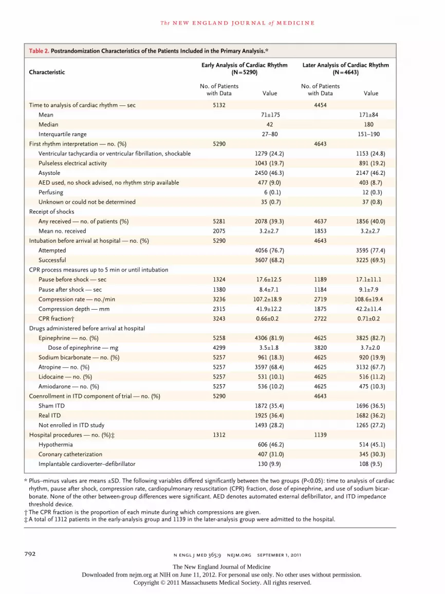
which yields treatment comparisons within clus-ters. Not all the scheduled cluster crossovers had occurred at the time of termination, although each cluster had crossed over at least once. The postrandomization characteristics of the patients in each group are provided in Table 2. The me-dian time to the analysis of cardiac rhythm was 42 seconds (interquartile range, 27 to 80) in the early-analysis group and 180 seconds (interquar-tile range, 151 to 190) in the later-analysis group. A majority of patients in each group received rhythm analysis within the targeted range for that group: 68% of patients in the early-analysis group re-ceived analysis of cardiac rhythm within the tar-geted range of 0 to 60 seconds and 60% of pa-tients in the later-analysis group received analysis of cardiac rhythm within the targeted range of 150 to 210 seconds (Fig. 2 in the Supplementary Appendix).
Primary and Secondary Outcomes
A total of 310 patients in the early-analysis group (5.9%) and 273 patients in the later-analysis group (5.9%) survived to hospital discharge with a mod-ified Rankin score of 3 or less, with a cluster-adjusted difference between later cardiac analysis and early cardiac analysis of −0.2 percentage points (95% confidence interval [CI], −1.1 to 0.7; P = 0.59) (Table 3). There was also no significant difference between the study groups with respect to any of the secondary outcomes. An analysis adjusted for potential confounders evaluated the effect of study group on survival and showed a difference of −0.3 percentage points (95% CI, −1.3 to 0.7) be-tween later cardiac analysis and early cardiac analysis (P = 0.61).
Additional Analyses
We conducted a number of prespecified and post hoc subgroup analyses (Fig. 1) and found that the absence of significant differences in the rate of sur-vival between the two study groups was consistent across subgroups. The relationship between the site-specific treatment effect and the site-specific probability of survival overall is shown in Figure 3 in the Supplementary Appendix.
When the outcomes were analyzed on an as-treated basis, the rates of survival with satisfactory functional status were 6.0% among the 3982 pa-tients in whom the analysis of cardiac rhythm was performed between 0 and 60 seconds and 5.9%
The New England Journal of Medicine Downloaded from nejm.org at NIH on June 11, 2012. For personal use only. No other uses without permission.
Copyright © 2011 Massachusetts Medical Society. All rights reserved.

Early vs. Later Rhythm Analysis in Cardiac Arrest
n engl j med 365;9 nejm.org september 1, 2011 791
among the 3115 patients in whom the analysis of cardiac rhythm was performed between 150 and 210 seconds (P = 0.97). In an additional exploratory analysis, we evaluated the rate of survival as a function of the actual time to the first rhythm analysis, regardless of the study group (Fig. 2). The chance of survival with satisfactory functional
status did not improve with increasing time to the first analysis of cardiac rhythm, and among pa-tients with an initial rhythm of ventricular tachy-cardia or ventricular fibrillation who received CPR from a bystander, the rate of survival tended to decline with increasing time to the first rhythm analysis.
Table 1. Baseline Characteristics of the Patients Included in the Primary Analysis.*
CharacteristicEarly Analysis of Cardiac Rhythm
(N = 5290)Later Analysis of Cardiac Rhythm
(N = 4643)
Age — yr† 66.7±16.6 66.7±16.6
Male sex — no./total no. (%) 3408/5289 (64.4) 2965/4643 (63.9)
Cause of cardiac arrest obvious — no./total no. (%)‡
110/5288 (2.1) 105/4643 (2.3)
Cardiac arrest occurring in public location — no. (%)
737 (13.9) 655 (14.1)
Cardiac arrest witnessed by bystander — no. (%)
2316 (43.8) 2029 (43.7)
CPR performed by bystander — no. (%) 2098 (39.7) 1904 (41.0)
Time from dispatch to first arrival of EMS — min§
6.0±3.7 6.0±5.9
Time from dispatch to first EMS arrival ≤4 min — no./total no. (%)
1060/5243 (20.2) 868/4601 (18.9)
Time from dispatch to first arrival of ALS providers — min¶
9.1±5.8 9.1±7.4
Treated with ALS — no. (%) 5105 (96.5) 4492 (96.7)
Site — no. (%)
Alabama 40 (0.8) 60 (1.3)
Dallas 113 (2.1) 78 (1.7)
Milwaukee 408 (7.7) 354 (7.6)
Ottawa–OPALS‖ 915 (17.3) 694 (14.9)
Pittsburgh 129 (2.4) 118 (2.5)
Portland, OR 334 (6.3) 314 (6.8)
San Diego, CA 206 (3.9) 218 (4.7)
King County, WA 672 (12.7) 642 (13.8)
Toronto 1873 (35.4) 1536 (33.1)
Vancouver, BC 600 (11.3) 629 (13.5)
* Plus–minus values are means ±SD. The distribution of sites differed significantly between the two groups (P<0.05). None of the other between-group differences were significant. ALS denotes advanced life support, CPR cardiopulmo-nary resuscitation, and EMS emergency medical services.
† The comparison with respect to age was based on 5279 patients in the early-analysis group and 4625 in the later-analysis group.
‡ Obvious causes included, but were not limited to, drug or chemical poisoning and mechanical suffocation (foreign body or hanging).
§ The comparison with respect to the time from dispatch to first arrival of EMS was based on 5243 patients in the early-analysis group and 4601 in the later-analysis group.
¶ The comparison with respect to the time from dispatch to first arrival of advanced life support was based only on the cases for which advanced life support was on the scene (5104 in the early-analysis group and 4490 in the later-analysis group).
‖ The Ottawa–Ontario Prehospital Advanced Life Support (OPALS) group is a group of 7 EMS services and 13 cities in Ontario.
The New England Journal of Medicine Downloaded from nejm.org at NIH on June 11, 2012. For personal use only. No other uses without permission.
Copyright © 2011 Massachusetts Medical Society. All rights reserved.
T h e n e w e ngl a nd j o u r na l o f m e dic i n e
n engl j med 365;9 nejm.org september 1, 2011792
Table 2. Postrandomization Characteristics of the Patients Included in the Primary Analysis.*
CharacteristicEarly Analysis of Cardiac Rhythm
(N = 5290)Later Analysis of Cardiac Rhythm
(N = 4643)
No. of Patients with Data Value
No. of Patients with Data Value
Time to analysis of cardiac rhythm — sec 5132 4454
Mean 71±175 171±84
Median 42 180
Interquartile range 27–80 151–190
First rhythm interpretation — no. (%) 5290 4643
Ventricular tachycardia or ventricular fibrillation, shockable 1279 (24.2) 1153 (24.8)
Pulseless electrical activity 1043 (19.7) 891 (19.2)
Asystole 2450 (46.3) 2147 (46.2)
AED used, no shock advised, no rhythm strip available 477 (9.0) 403 (8.7)
Perfusing 6 (0.1) 12 (0.3)
Unknown or could not be determined 35 (0.7) 37 (0.8)
Receipt of shocks
Any received — no. of patients (%) 5281 2078 (39.3) 4637 1856 (40.0)
Mean no. received 2075 3.2±2.7 1853 3.2±2.7
Intubation before arrival at hospital — no. (%) 5290 4643
Attempted 4056 (76.7) 3595 (77.4)
Successful 3607 (68.2) 3225 (69.5)
CPR process measures up to 5 min or until intubation
Pause before shock — sec 1324 17.6±12.5 1189 17.1±11.1
Pause after shock — sec 1380 8.4±7.1 1184 9.1±7.9
Compression rate — no./min 3236 107.2±18.9 2719 108.6±19.4
Compression depth — mm 2315 41.9±12.2 1875 42.2±11.4
CPR fraction† 3243 0.66±0.2 2722 0.71±0.2
Drugs administered before arrival at hospital
Epinephrine — no. (%) 5258 4306 (81.9) 4625 3825 (82.7)
Dose of epinephrine — mg 4299 3.5±1.8 3820 3.7±2.0
Sodium bicarbonate — no. (%) 5257 961 (18.3) 4625 920 (19.9)
Atropine — no. (%) 5257 3597 (68.4) 4625 3132 (67.7)
Lidocaine — no. (%) 5257 531 (10.1) 4625 516 (11.2)
Amiodarone — no. (%) 5257 536 (10.2) 4625 475 (10.3)
Coenrollment in ITD component of trial — no. (%) 5290 4643
Sham ITD 1872 (35.4) 1696 (36.5)
Real ITD 1925 (36.4) 1682 (36.2)
Not enrolled in ITD study 1493 (28.2) 1265 (27.2)
Hospital procedures — no. (%)‡ 1312 1139
Hypothermia 606 (46.2) 514 (45.1)
Coronary catheterization 407 (31.0) 345 (30.3)
Implantable cardioverter–defibrillator 130 (9.9) 108 (9.5)
* Plus–minus values are means ±SD. The following variables differed significantly between the two groups (P<0.05): time to analysis of cardiac rhythm, pause after shock, compression rate, cardiopulmonary resuscitation (CPR) fraction, dose of epinephrine, and use of sodium bicar-bonate. None of the other between-group differences were significant. AED denotes automated external defibrillator, and ITD impedance threshold device.
† The CPR fraction is the proportion of each minute during which compressions are given.‡ A total of 1312 patients in the early-analysis group and 1139 in the later-analysis group were admitted to the hospital.
The New England Journal of Medicine Downloaded from nejm.org at NIH on June 11, 2012. For personal use only. No other uses without permission.
Copyright © 2011 Massachusetts Medical Society. All rights reserved.

Early vs. Later Rhythm Analysis in Cardiac Arrest
n engl j med 365;9 nejm.org september 1, 2011 793
Discussion
In this randomized trial, we tested the hypothesis that patients with an out-of-hospital cardiac arrest might benefit from the administration of CPR by EMS personnel for approximately 3 minutes before the first analysis of cardiac rhythm (with delivery of a defibrillator shock as appropriate). We found that there was no significant difference in the rate of survival with satisfactory functional status be-tween the two EMS strategies of a brief period of CPR with early analysis of cardiac rhythm and a longer period of CPR with delayed analysis of rhythm. Subgroup and adjusted analyses also did not show any significant differences in the out-comes between the two study groups. We further explored the relationship between the rate of sur-vival and the actual time to rhythm analysis and found that outcomes did not improve with in-creasing time to analysis. This finding suggests that there is no advantage of delaying the analysis of cardiac rhythm during EMS-administered CPR. Indeed, the data suggest that there may be a disad-
vantage of delaying the rhythm analysis in the sub-group of patients with a first rhythm of either ven-tricular tachycardia or ventricular fibrillation who have received CPR from a bystander. Overall, our data suggest that the administration of 2 minutes of CPR by EMS personnel before the first analysis of rhythm, which was suggested in the 2005 guide-lines of the AHA–ILCOR, is unlikely to provide a greater benefit than CPR of shorter duration.
The hypothesis that a brief period of initial CPR before analysis of cardiac rhythm could be bene-ficial is based primarily on the concept that a few minutes of chest compressions may increase myo-cardial perfusion, thus improving the metabolic state of the cardiac myocytes and enhancing the likelihood of successful defibrillation.7 Several studies in animals with experimentally induced ventricular fibrillation showed that the outcomes with delayed countershock after a period of chest compressions were better than the outcomes with earlier countershock,21,28,29 whereas other studies failed to show a benefit of CPR before shock.30,31 Five previous clinical studies also attempted to
Table 3. Outcomes for the Patients Included in the Primary Analysis.*
Outcome
Early Analysis of Cardiac Rhythm
(N = 5290)
Later Analysis of Cardiac Rhythm
(N = 4643)
Difference: Later Analysis − Early Analysis
(95% CI) P Value
Transported to hospital — no. (%) 2812 (53.2) 2468 (53.2) 0.0 (−2.7 to 2.7) 1.00
Pulse present on arrival at emergency department — no. (%)
1352 (25.6) 1218 (26.2) 0.7 (−1.0 to 2.4) 0.44
Survival to hospital admission — no. (%) 1303 (24.6) 1132 (24.4) −0.3 (−1.6 to 1.1) 0.71
Survival to hospital discharge — no. (%) 427 (8.1) 372 (8.0) −0.1 (−1.2 to 1.1) 0.92
Modified Rankin score — no. (%)†
≤3‡ 310 (5.9) 273 (5.9) −0.2 (−1.1 to 0.7) 0.59
0 82 (1.6) 71 (1.5)
1 117 (2.2) 100 (2.2)
2 20 (0.4) 29 (0.6)
3 91 (1.7) 73 (1.6)
4 69 (1.3) 55 (1.2)
5 48 (0.9) 44 (0.9)
6 4863 (91.9) 4271 (92.0)
Mean modified Rankin score 5.7±1.1 5.7±1.1 0.00 (−0.05 to 0.05) 0.98
* Plus–minus values are means ±SD.† The modified Rankin scale is commonly used for measuring the performance of daily activities by people who have had
a stroke. Scores range from 0 to 6, with lower scores representing better performance; a score of 6 indicates death.‡ A modified Rankin score of 3 or less was a primary outcome. The between-group difference was adjusted for cluster
randomization.
The New England Journal of Medicine Downloaded from nejm.org at NIH on June 11, 2012. For personal use only. No other uses without permission.
Copyright © 2011 Massachusetts Medical Society. All rights reserved.

T h e n e w e ngl a nd j o u r na l o f m e dic i n e
n engl j med 365;9 nejm.org september 1, 2011794
evaluate this issue, but all five had limitations in-volving the design or sample size, and none had findings that were definitive.8-12 Cobb et al.,8 in a before-and-after study, showed that the rate of survival increased after the implementation of a policy that required 90 seconds of CPR before analysis of cardiac rhythm when an automated external defibrillator was used. Wik et al.9 con-ducted a randomized trial and found no significant difference between the outcomes after immediate defibrillation and those after 3 minutes of basic CPR before defibrillation, but the outcomes in a subgroup with response times exceeding 5 min-utes were better after initial CPR than after im-mediate defibrillation. Randomized trials reported
by Jacobs et al.11 and Baker et al.12 showed no sig-nificant difference in outcomes with early as com-pared with late defibrillation. Bradley et al.10 per-formed an observational analysis and found that CPR by EMS personnel for 46 to 195 seconds be-fore defibrillation was weakly associated with an improved rate of survival.
Given the complex clinical circumstances of out-of-hospital cardiac arrest, precise control of the time to the first analysis of cardiac rhythm is dif-ficult to achieve. In our trial, the duration of CPR before the first analysis of rhythm did not fall within the assigned target for 36% of the patients. Although this observation raises the question of quality control in training and trial supervision,
Later AnalysisBetter
Early AnalysisBetter
mITT population
Prespecified subgroups
First rhythm interpretation
VT or VF
Pulseless electrical activity
Asystole
Other
Time to arrival of EMS
<4 min
≥4 min
Bystander CPR
Given
Not given or unknown
Exploratory subgroups
Protocol-adherent, 1st vehicle
Yes
No
Protocol-adherent, 1st CPR
Yes
No
Bystander-witnessed
Yes
No
ITD study group
Sham
Active
Not in ITD study
Early Analysis Difference: Later Analysis – Early Analysis(95% CI)
Later AnalysisSubgroup
1.1 (−0.4 to 2.5)
−1.5 (−3.4 to 0.4)
0.1 (−1.7 to 1.9)0.0 (−0.8 to 0.8)
0.2 (−1.3 to 1.7)
0.0 (−1.6 to 1.6)
0.2 (−1.0 to 1.4)
0.3 (−0.9 to 1.5)
−0.2 (−1.7 to 1.4)
0.0 (−1.0 to 1.0)
−0.1 (−1.9 to 1.6)
0.7 (−1.9 to 3.4)
−0.1 (−1.1 to 0.9)
−0.9 (−4.0 to 2.2)
−1.0 (−3.0 to 1.3)−0.2 (−0.7 to 2.0)
1.5 (0.0 to 3.1)
0 3−4 −3 −2 −1 1 2
0.0 (−0.9 to 0.9)
P Value forInteraction
310/5290 (5.9)
248/1279 (19.4)
25/1043 (2.4)
17/2450 (0.7)
17/512 (3.3)
66/875 (7.5)
241/4368 (5.5)
181/2098 (8.6)
129/3192 (4.0)
211/3488 (6.0)
92/1644 (5.6)
214/3528 (6.1)
89/1576 (5.6)
242/2316 (10.4)
68/2974 (2.3)
104/1925 (5.4)
88/1872 (4.7)
118/1493 (7.9)
273/4643 (5.9)
213/1153 (18.5)
35/891 (3.9)
10/2147 (0.5)
10/440 (2.3)
59/712 (8.3)
211/3889 (5.4)
162/1904 (8.5)
111/2739 (4.1)
170/2669 (6.4)
97/1785 (5.4)
170/2697 (6.3)
97/1719 (5.6)
214/2029 (10.5)
59/2614 (2.3)
94/1682 (5.6)
98/1696 (5.8)
81/1265 (6.4)
0.16
0.56
0.90
0.63
0.81
0.90
0.11
no. of patients/total no.(%)
Figure 1. Subgroup Analyses of the Primary Outcome.
Shown are the results of analyses of the primary outcome (survival to hospital discharge with a score on the modified Rankin scale of ≤3, on a scale of 0 to 6, with higher scores indicating greater disability), according to prespecified subgroups and post hoc exploratory subgroups. The impedance threshold device (ITD) study group refers to a concurrent study (involving most of the patients who were en-rolled in this study), in which the use of an active ITD during cardiopulmonary resuscitation (CPR) was compared with the use of a sham ITD. The abbreviation mITT denotes modified intention to treat, VF ventricular fibrillation, and VT ventricular tachycardia.
The New England Journal of Medicine Downloaded from nejm.org at NIH on June 11, 2012. For personal use only. No other uses without permission.
Copyright © 2011 Massachusetts Medical Society. All rights reserved.

Early vs. Later Rhythm Analysis in Cardiac Arrest
n engl j med 365;9 nejm.org september 1, 2011 795
the participating EMS agencies were high-func-tioning services with advanced-level paramedics; in addition, they had collected high-quality patient data before the start of the trial, and they made continuous efforts to reinforce performance tar-gets. Thus, although implementation of the proto-col was imperfect, it nonetheless represents the degree of precision with which such therapies are likely to be practiced in the clinical setting of out-of-hospital cardiac arrest. Furthermore, despite this limitation, there was very good separation between the two study groups in the duration of CPR, and a variety of data analyses confirmed the primary finding of no significant difference in the outcome between patients who had early rhythm analysis and those who had later rhythm analysis.
Our results indicate that in most cases, the outcome is similar with as few as 30 seconds and as many as 180 seconds of EMS-adminis-tered CPR before the analysis of cardiac rhythm. The exception is the case of cardiac arrest wit-nessed by EMS responders, which was not evalu-ated in this study and for which rapid defibrilla-tion remains the standard of care.13 Our results also do not address the strategy of immediate analysis of cardiac rhythm without any preceding CPR, since we deliberately insisted on some CPR for the early-analysis group, in the belief that good patient care required cardiopulmonary support while the defibrillator was being prepared.
Exploratory examination of our data suggests that a strategy of brief CPR and early analysis may be more appropriate than longer CPR and later analysis for patients who have received CPR from a bystander before the arrival of professional re-sponders. Conversely, for patients who have not received CPR from a bystander, there is no ap-proach that is clearly advantageous with respect to the time to analysis of rhythm. The 2010 guide-lines of the AHA–ILCOR give little direction as to the preferred period of CPR before analysis of cardiac rhythm.13 Each EMS system should con-sider its operational situation when deciding on its strategy for initial EMS-administered CPR. We believe that it is important to administer CPR for some period while the defibrillator pads are being applied and that compressions should be of high quality with minimal interruptions.
In conclusion, in a large clinical trial, we evalu-ated the timing of the analysis of cardiac rhythm during CPR in patients who had an out-of-hospital cardiac arrest that was not witnessed by EMS per-sonnel. We found no difference in the outcome
between the EMS strategy of a brief period of CPR before early rhythm analysis and that of a longer period of CPR before delayed rhythm analysis.
Supported by grants from the National Heart, Lung, and Blood Institute, the National Institute of Neurological Disorders and Stroke, the Canadian Institutes of Health Research–Insti-tute of Circulatory and Respiratory Health, and the Heart and Stroke Foundation of Canada. The Resuscitation Outcome Con-sortium is supported by a series of cooperative agreements with 10 regional clinical centers and one data coordinating center (5U01 HL077863, HL077881, HL077871 HL077872, HL077866, HL077908, HL077867, HL077887, HL077873, HL077865) from the National Heart, Lung, and Blood Institute in partnership with the National Institute of Neurological Disorders and Stroke, U.S. Army Medical Research and Material Command, the Insti-tute of Circulatory and Respiratory Health of the Canadian In-
Prob
abili
ty o
f Pri
mar
y O
utco
me
Prob
abili
ty o
f Pri
mar
y O
utco
me
0.30
0.20
0.10
0.000 60 120 180 240 300 360
Time to Analysis (sec)
B Cases without Bystander CPR
A Cases with Bystander CPR
VT or VF (N=1221)
No VT or VF (N=2598)
0.30
0.20
0.10
0.000 60 120 180 240 300 360
Time to Analysis (sec)
VT or VF (N=1134)
No VT or VF (N=4500)
Figure 2. Rate of the Primary Outcome, According to Actual Time to Analysis of Cardiac Rhythm.
The primary outcome was survival to hospital discharge with a score on the modified Rankin scale of 3 or less. The rate of the primary outcome is shown according to the actual time to the analysis of cardiac rhythm, re-gardless of the study group, among patients who received cardiopulmonary resuscitation (CPR) from a bystander (Panel A) and among patients who did not receive CPR from a bystander (Panel B). In each panel, the rates are shown for patients in whom the first rhythm was ventricular fibrillation (VF) or ventricular tachycardia (VT) (thick solid lines, with 95% confidence intervals indicated by thin solid lines) and for patients in whom the first rhythm was neither VF nor VT (thick broken lines, with 95% confidence in-tervals indicated by thin broken lines).
The New England Journal of Medicine Downloaded from nejm.org at NIH on June 11, 2012. For personal use only. No other uses without permission.
Copyright © 2011 Massachusetts Medical Society. All rights reserved.

T h e n e w e ngl a nd j o u r na l o f m e dic i n e
n engl j med 365;9 nejm.org september 1, 2011796
stitutes of Health Research, Defence Research and Development Canada, the Heart and Stroke Foundation of Canada, and the American Heart Association.
Dr. Nichol reports receiving grant support from the Laerdal Foundation for Acute Medicine and Medtronic Foundation and travel fees from Sotera Wireless, being a board member of Medic One Foundation, and participating in research collaborations with Gambro Renal, Lifebridge Medizintechnik, and Sotera Wireless; Dr. Ornato, serving on an advisory board for ZOLL Circulation; Dr. Callaway, that he and his institution receive royalties for patents from Medtronic related to the timing of defibrillation; Dr. Aufderheide, consulting fees from Jolife and Medtronic; Dr. Daya, consulting and lecture fees from Philips
Healthcare and owning stock in Amgen, Johnson & Johnson, and Roche; Dr. Morrison, grant support from the Laerdal Foundation for Acute Medicine; Dr. Pirrallo, consulting fees from ZOLL Medi-cal; and Dr. Weisfeldt, that he and his institution receive royalties for patents from Imricor Medical Systems related to an MRI In-sensitive Pacemaker. No other potential conflict of interest rele-vant to this article was reported.
Disclosure forms provided by the authors are available with the full text of this article at NEJM.org.
We thank the EMS providers and first responders for their efforts in making this logistically challenging trial possible; and Alfred P. Hallstrom, Ph.D., Scott S. Emerson, M.D., Ph.D., and Gerald van Belle, Ph.D., for their leadership.
AppendixThe authors’ affiliations are as follows: the Department of Emergency Medicine and Ottawa Hospital Research Institute, University of Ot-tawa, Ottawa (I.G.S., C.V.); the Clinical Trials Center, Department of Biostatistics (G.N., B.G.L., J.P., R.H.S.), and the Department of Medi-cine (G.N., T.D.R., P.J.K.), University of Washington; and the University of Washington–Harborview Center for Prehospital Emergency Care (G.N.) — both in Seattle; the Department of Emergency Medicine, Virginia Commonwealth University, Richmond (J.P.O.); the Department of Emergency Medicine (J.C.) and the School of Population and Public Health (D.A.), University of British Columbia, and British Columbia Emergency and Health Services Commission (J.C., D.A.) — both in Vancouver, Canada; the Department of Emergency Medicine, University of Pittsburgh, Pittsburgh (C.W.C., D.H.); the Department of Emergency Medicine, Medical College of Wisconsin, Milwaukee (T.P.A., R.G.P.); the Department of Surgery (Emergency Medicine), University of Texas Southwestern Medical Center at Dallas, Dallas (A.H.I., R.F.); Center for Policy and Research in Emergency Medicine, Department of Emergency Medicine, Oregon Health and Science University, Portland (M.R.D., D.Z.); the Department of Emergency Medicine, University of Alabama at Birmingham, Birmingham (H.E.W., S.S.); the Division of Emergency Medicine, Department of Medicine (L.J.M.) and Department of Family and Community Medicine (S.C.), University of Toronto; and Rescu Keenan Research Centre, Li Ka Shing Knowledge Institute, St. Michael’s Hospital (L.J.M., S.C.) — both in Toronto; the Depart-ment of Emergency Medicine, University of California, San Diego, San Diego (D.D., G.M.V.); the National Heart, Lung, and Blood Institute, National Institutes of Health, Bethesda, MD (G.S.); and the Department of Medicine, Johns Hopkins Medical Institutions, Baltimore (M.W.).
References
1. Nichol G, Thomas E, Callaway CW, et al. Regional variation in out-of-hospital cardiac arrest incidence and outcome. JAMA 2008;300:1423-31. [Erratum, JAMA 2008;300:1763.]2. Cummins RO, Ornato JP, Thies WH, Pepe PE. Improving survival from sudden cardiac arrest: the “chain of survival” concept. Circulation 1991;83:1832-47.3. 2005 American Heart Association guidelines for cardiopulmonary resuscita-tion and emergency cardiovascular care. Circulation 2005;112:Suppl:IV-1–IV-203.4. Stiell IG, Wells GA, Field BJ, et al. Ad-vanced cardiac life support in out-of-hos-pital cardiac arrest. N Engl J Med 2004; 351:647-56.5. Rea TD, Cook AJ, Stiell IG, et al. Pre-dicting survival after out-of-hospital car-diac arrest: role of the Utstein data ele-ments. Ann Emerg Med 2010;55:249-57.6. Peberdy MA, Ornato JP. Post-resusci-tation care: is it the missing link in the chain of survival? Resuscitation 2005;64: 135-7.7. Weisfeldt ML, Becker LB. Resuscita-tion after cardiac arrest: a 3-phase time-sensitive model. JAMA 2002;288:3035-8.8. Cobb LA, Fahrenbruch CE, Walsh TR, et al. Influence of cardiopulmonary re-suscitation prior to defibrillation in pa-tients with out-of-hospital ventricular fi-brillation. JAMA 1999;281:1182-8.9. Wik L, Hansen TB, Fylling F, et al. De-laying defibrillation to give basic cardio-pulmonary resuscitation to patients with out-of-hospital ventricular fibrillation: a
randomized trial. JAMA 2003;289:1389-95.10. Bradley SM, Gabriel EE, Aufderheide TP, et al. Survival increases with CPR by emergency medical services before defi-brillation of out-of-hospital ventricular fibrillation or ventricular tachycardia: ob-servations from the Resuscitation Out-comes Consortium. Resuscitation 2010; 81:155-62.11. Jacobs IG, Finn JC, Oxer HF, Jelinek GA. CPR before defibrillation in out-of-hospital cardiac arrest: a randomized trial. Emerg Med Australas 2005;17:39-45. [Er-ratum, Emerg Med Australas 2009;21:430.]12. Baker PW, Conway J, Cotton C, et al. Defibrillation or cardiopulmonary resus-citation first for patients with out-of-hos-pital cardiac arrests found by paramedics to be in ventricular fibrillation? A ran-domised control trial. Resuscitation 2008; 79:424-31.13. Link MS, Atkins DL, Passman RS, et al. Part 6: electrical therapies: automated external defibrillators, defibrillation, car-dioversion, and pacing: 2010 American Heart Association guidelines for cardio-pulmonary resuscitation and emergency cardiovascular care. Circulation 2010;122: Suppl 3:S706-S719. [Erratum, Circulation 2011;123(6):e235.]14. Meier P, Baker P, Jost D, et al. Chest compressions before defibrillation for out-of-hospital cardiac arrest: a meta-analysis of randomized controlled clinical trials. BMC Med 2010;8:52.15. Stiell IG, Callaway CW, Davis D, et al. Resuscitation Outcomes Consortium (ROC)
PRIMED cardiac arrest trial methods part 2: rationale and methodology for “analyze later vs. analyze early” protocol. Resusci-tation 2008;78:186-95.16. Davis DP, Garberson LA, Andrusiek DL, et al. A descriptive analysis of emer-gency medical service systems participat-ing in the Resuscitation Outcomes Con-sortium (ROC) network. Prehosp Emerg Care 2007;11:369-82.17. Aufderheide TP, Kudenchuk PJ, Hedg-es JR, et al. Resuscitation Outcomes Con-sortium (ROC) PRIMED cardiac arrest trial methods part 1: rationale and meth-odology for the impedance threshold de-vice (ITD) protocol. Resuscitation 2008; 78:179-85.18. Aufderheide TP, Nichol G, Rea TD, et al. A trial of an impedance threshold device in out-of-hospital cardiac arrest. N Engl J Med 2011;365:798-806.19. Newcommon NJ, Green TL, Haley E, Cooke T, Hill MD. Improving the assess-ment of outcomes in stroke: use of a structured interview to assign grades on the modified Rankin Scale. Stroke 2003; 34:377-8.20. van Alem AP, de Vos R, Schmand B, Koster RW. Cognitive impairment in sur-vivors of out-of-hospital cardiac arrest. Am Heart J 2004;148:416-21.21. Niemann JT, Cruz B, Garner D, Lewis RJ. Immediate countershock versus car-diopulmonary resuscitation before coun-tershock in a 5-minute swine model of ventricular fibrillation arrest. Ann Emerg Med 2000;36:543-6.
The New England Journal of Medicine Downloaded from nejm.org at NIH on June 11, 2012. For personal use only. No other uses without permission.
Copyright © 2011 Massachusetts Medical Society. All rights reserved.

Early vs. Later Rhythm Analysis in Cardiac Arrest
n engl j med 365;9 nejm.org september 1, 2011 797
22. O’Brien PC, Fleming TR. A multiple testing procedure for clinical trials. Bio-metrics 1979;35:549-56.23. Breslow NE, Clayton DG. Approximate inference in generalized linear mixed models. J Am Stat Assoc 1993;88:9-25.24. Liang K, Zeger S. Longitudinal data analysis using generalized linear models. Biometrika 1986;73:13-22.25. Silverman BW. Density estimation. London: Chapman & Hall, 1986.26. Hastie TJ. Generalized additive mod-els. In: Chambers JM, Hastie TJ, eds. Sta-
tistical models in S. Pacific Grove, CA: Wadsworth & Brooks/Cole, 1992:249-307.27. Efron B, Tibshirani R. An introduction to the bootstrap. New York: Chapman & Hall, 1993.28. Yakaitis RW, Ewy GA, Otto CW, Taren DL, Moon TE. Influence of time and ther-apy on ventricular defibrillation in dogs. Crit Care Med 1980;8:157-63.29. Menegazzi JJ. Pragmatic problems in prehospital research. Prehosp Disaster Med 1993;8:Suppl:S15-S19.30. Rittenberger JC, Suffoletto B, Salcido
D, Logue E, Menegazzi JJ. Increasing CPR duration prior to first defibrillation does not improve return of spontaneous circu-lation or survival in a swine model of pro-longed ventricular fibrillation. Resuscita-tion 2008;79:155-60.31. Wang YL, Zhong JQ, Tao W, Hou XM, Meng XL, Zhang Y. Initial defibrillation versus initial chest compression in a 4-minute ventricular fibrillation canine model of cardiac arrest. Crit Care Med 2009;37:2250-2.Copyright © 2011 Massachusetts Medical Society.
The New England Journal of Medicine Downloaded from nejm.org at NIH on June 11, 2012. For personal use only. No other uses without permission.
Copyright © 2011 Massachusetts Medical Society. All rights reserved.


