Embed Size (px)
Citation preview

Clin. exp. Immunol. (1979) 38, 615-620.
Reticular dysgenesis: report of two brothers
T. ESPA-1OL, J. COMPTE,* C. ALVAREZ, N. TALLADA,t R. LAVERDE & G. PEGUEROtImmunology Laboratory, t Pathology Department and t Neonatology Department, Pediatric Hospital-Residencia Sanitaria
Fco Franco, Barcelona
(Acceptedfor publication 4 June 1979)
SUMMARY
We describe two brothers with marked leucopenia, lymphopenia, no immunological response toinfections (no Ig production, negative PHA response and very low number ofT and B lympho-cytes in peripheral blood) and hypocellular marrow. They died at 12 and 8 days of life withinfection (E. coli and Klebsiella, respectively).
INTRODUCTION
Reticular dysgenesis is a very rare and severe form of immunodeficiency (Cooper et al., 1973; Waldmann& Broder, 1978) characterised by congenital leucopenia, lymphopenia, absent Ig production, lymphoidhypoplasia and thymic agenesia, which is manifested with severe progressive neonatal infections. Wedescribe two affected brothers.
CASE REPORTS
The patients were the first and second children of young, healthy and unrelated parents. There was norelevant family history. The pregnancies were normal, with no history of illness, intoxication or irradia-tion. Relevant haematological and immunological data from the parents are shown in Table 1.
TABLE 1. Haematological and immunological data of the parents
Histocompatibility ErythrocyticBlood group testing adenosine deaminase*
Mother Al Rh (-) HLA-Aw 33-24 (9) 0-701 p/g HbB 7-14 (w6)
Father 0 Rh (-) HLA-A 3-11 0-814 ,p/g HbB 8-44 (12 w6, w4)Cw5
* Method of Beutler (1975). Normal values (from control adult population):0-87-1-29 p/g Hb.
Case 1
A 10 day-old male, body weight 2-3 kg, developed omphalitis at nine days of age. The umbilicalstump was infected with phlebitis of the abdominal wall, necrotic lesions in the perineal area and a smallabscess behind the ear. Haematological and immunological data are shown in Table 2. Umbilical exudate,throat, gastric contents and urine cultures grew E. coli colonies. CSF culture was sterile. Chest X-ray:no thymic shadow.
Correspondence: T. Espahol, M.D. Immunology Laboratory, Clinica Infantil-Residencia Fco Franco, Barcelona, Spain.0099-9104/79/1200-0615$02.00 © 1979 Blackwell Scientific Publications
615
616 T. Espahol et al.He was treated with intravenous fluid and antibiotics (ampicillin and gentamicin). Necrotic lesions
developed very rapidly, spreading on the abdomen, and perianal area; he deteriorated and died 44 hrafter admission.
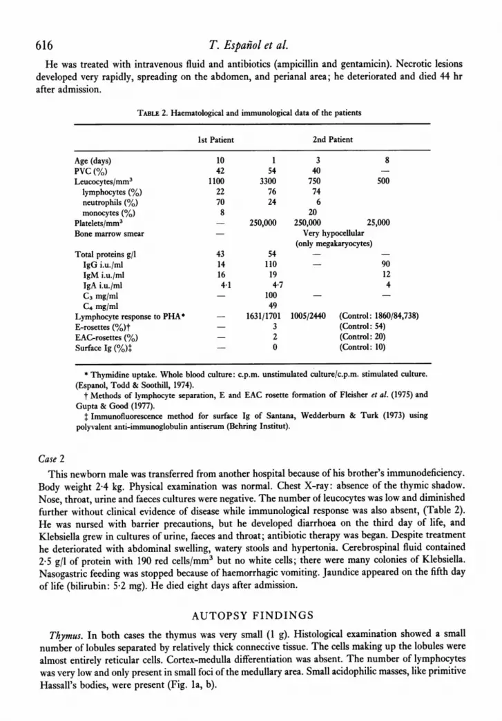
TABLE 2. Haematological and immunological data of the patients
1st Patient 2nd Patient
Age (days) 10 1 3 8PVC (%) 42 54 40Leucocytes/mm3 1100 3300 750 500
lymphocytes (%) 22 76 74neutrophils (%) 70 24 6monocytes (%) 8 20
Platelets/mm3 - 250,000 250,000 25,000Bone marrow smear - Very hypocellular
(only megakaryocytes)Total proteins g/l 43 54IgG i.u./ml 14 110 - 90IgMi.u./ml 16 19 12IgA i.u./ml 4-1 4-7 4C3 mg/ml - 100 -
C4 mg/ml 49Lymphocyte response to PHA* 1631/1701 1005/2440 (Control: 1860/84,738)E-rosettes (%)t 3 (Control: 54)EAC-rosettes (%) - 2 (Control: 20)Surface Ig (%)+ 0 (Control: 10)
* Thymidine uptake. Whole blood culture: c.p.m. unstimulated culture/c.p.m. stimulated culture.(Espanol, Todd & Soothill, 1974).
t Methods of lymphocyte separation, E and EAC rosette formation of Fleisher et a!. (1975) andGupta & Good (1977).
t Immunofluorescence method for surface Ig of Santana, Wedderburn & Turk (1973) usingpolyvalent anti-immunoglobulin antiserum (Behring Institut).
Case 2This newborn male was transferred from another hospital because of his brother's immunodeficiency.
Body weight 2-4 kg. Physical examination was normal. Chest X-ray: absence of the thymic shadow.Nose, throat, urine and faeces cultures were negative. The number of leucocytes was low and diminishedfurther without clinical evidence of disease while immunological response was also absent, (Table 2).He was nursed with barrier precautions, but he developed diarrhoea on the third day of life, andKlebsiella grew in cultures of urine, faeces and throat; antibiotic therapy was began. Despite treatmenthe deteriorated with abdominal swelling, watery stools and hypertonia. Cerebrospinal fluid contained2-5 g/l of protein with 190 red cells/mm3 but no white cells; there were many colonies of Klebsiella.Nasogastric feeding was stopped because of haemorrhagic vomiting. Jaundice appeared on the fifth dayof life (bilirubin: 5-2 mg). He died eight days after admission.
AUTOPSY FINDINGS
Thymus. In both cases the thymus was very small (1 g). Histological examination showed a smallnumber of lobules separated by relatively thick connective tissue. The cells making up the lobules werealmost entirely reticular cells. Cortex-medulla differentiation was absent. The number of lymphocyteswas very low and only present in small foci of the medullary area. Small acidophilic masses, like primitiveHassall's bodies, were present (Fig. la, b).

Reticular dysgenesis 617
FIG.~ ~~ ~ ~ ~~~~~~~~Mla,-W.41sfoh is n eod ae H&E 1 n 3.
spriAi l,~~~~~N
M I L _ S~~S#
J",,#0
FIG. la, b. Thymus from the first and second case. (H & E, x 210 and 330.)
El,Wj<E.S
4
FIG. 2. Lymph node. (H & E, x 300.)
Lymph nodes. Macroscopically the nodes were very small. Although the sinusoidal architecture wasrecognizable, they were almost devoid of lymphocytes and no lymphoid follicles were seen. A fewhistiocytes were present in the sinuses (Fig. 2).
Gut. In the gastro-intestinal tract reticular-like cells were seen, with marked depletion of lymphocytes.No plasma cells were seen (Fig. 3).
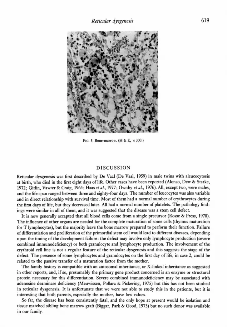
Spleen. Normal size. The section was devoid of lymphoid tissue, though there was a little extra-medullary haemopoiesis indicated by the presence of megakaryocytes; the usual periarterial cuffingwas absent. There was some increase in interstitial connective tissue and phagocytic cells were seen toline some sinusoids. In the second case, haemosiderin and sinusoid dilatation were seen (Fig. 4a, b).Bone marrow. Very similar in both cases: markedly hypocellular with much of the marrow space
occupied by erythrocytes. Megakaryocytes were present in normal or slightly increased numbers.

T. Espahol et a!.
FIG. 3. Gut. (H & E, x 300.)
FIG. 4a,*b. Spleen:fromit fr An secnd (Hg & ,
FIG. 4a, b. Spleen from the first and second case. (H & E, x 240.)
Erythroid cells were normally grouped and present in normal numbers. Myeloid cells were greatly re-duced in number. The marrow and surrounding soft tissues appeared normal (Fig. 5).
Umbilicus. In the first case, there was a marked inflammatory reaction with oedema, fibrin depositsand many bacteria, but no polymorphonuclear cells.
Lungs. Most of the alveoli contained proteinaceous material, numerous bacteria, but no polymor-phonuclear cells.
Oesophagus. In the second case, there was an extensive ulceration of the inferior third, with manybacteria but no cellular reaction.
Central nervous system. The second case also revealed congested meninges and swollen and softenedcerebral tissue.
618
Reticular dysgenesis 619
*,IA .9~~~~~~~;.
FIG. 5. Bone-marrow. (H & E, x 300.)
DISCUSSION
Reticular dysgenesis was first described by De Vaal (De Vaal, 1959) in male twins with aleucocytosisat birth, who died in the first eight days of life. Other cases have been reported (Alonso, Dew & Starke,1972; Gitlin, Vawter & Craig, 1964; Haas et aL., 1977; Ownby et aL., 1976). All, except two, were males,and the life span ranged between three and eighty-four days. The number of leucocytes was also variableand in direct relationship with survival time. Most of them had a normal number of erythrocytes duringthe first days of life, but they decreased later. All had a normal number of platelets. The pathology find-ings were similar in all of them, and it was suggested that the disease was a stem cell defect.
It is now generally accapted that all blood cells come from a single precursor (Rosse & Press, 1978).The influence of other organs are needed for the complete maturation of some cells (thymus maturationfor T lymphocytes), but the majority leave the bone marrow prepared to perform their function. Failureof differentiation and proliferation of the primordial stem cell would lead to different diseases, dependingupon the timing of the development failure: the defect may involve only lymphocyte production (severecombined immunodeficiency) or both granulocyte and lymphocyte production. The involvement of theerythroid cell line is not a regular feature of the reticular dysgenesis and this suggests the stage of thedefect. The presence of some lymphocytes and granulocytes on the first day of life, in case 2, could berelated to the passive transfer of a maturation factor from the mother.The family history is compatible with an autosomal inheritance, or X-linked inheritance as suggested
in other reports, and, if so, presumably the primary gene product concerned is an enzyme or structuralprotein necessary for this differentiation. Severe combined immunodeficiency may be associated withadenosine deaminase deficiency (Meuwissen, Pollara & Pickering, 1975) but this has not been studiedin reticular dysgenesis. It is unfortunate that we were not able to study this in the patients, but it isinteresting that both parents, especially the mother, have low values.So far, the disease has been consistently fatal, and the only hope at present would be isolation and
tissue matched sibling bone marrow graft (Biggar, Park & Good, 1973) but no such donor was availablein our family.

620 T. Espahol et a?.
REFERENCES
ALONSO, K., DEW, J.M. & STARKE, W.R. (1972) Thymicalymphoplasia and congenital aleucocytosis (reticulardysgenesis). Arch. Path. 94, 179.
BEUTLER, E. (1975) Red cell metabolism. A Manual ofBiochemical Methods. 2nd edn. Grune and Stratton, NewYork.
BIGGAR, W.D., PARK, B.H. & GOOD, R.A. (1973) Immuno-logical reconstitution. Annu. Rev. Med. 24, 135.
COOPER, M.D., FAULK, W.P., FUDENBERG, H.H., GOOD,R.A., HITZIG, W., KUNKEL, H., ROSEN, F.S., SELIGMANN,M., SOOTHILL, J.F. & WEDGWOOD, R.J. (1973) Classi-fication of primary immunodeficiencies. N. Engl. J. Med.288, 966.
DE VAAL, O.M. (1959) Reticular dysgenesis. Lancet, ii,1123.
ESPANOL, T., TODD, G.B. & SOOTHILL, J.F. (1974) The effectof anaesthesia on the lymphocyte response to phyto-haemagglutinin. Clin. exp. Immunol. 18, 73.
FLEISHER, T.A., LUCKASEN, J.R., SABAD, A., GEHRTZ, R.C.& KERSEY, J.H. (1975) T and B lymphocyte subpopula-tions in children. Pediatrics, 55, 162.
GITLIN, D., VAWTER, G. & CRAIG, J.M. (1964) Thymicalymphoplasia and congenital aleucocytosis. Pediatrics,33, 184.
GUPTA, S. & GOOD, R.A. (1977) Subpopulations of humanlymphocytes. I. Studies in immunodeficient patients.Clin. exp. Immunol. 30, 222.
HAAs, R.J., NIETHAMMER, D., GOLDMANN, S.F., HEIT, W.,BIENZIE, U. & KLEIHAuER, E. (1977) Congenital immuno-deficiency and agranulocytosis (reticular dysgenesis).Acta Paediatr. Scand. 66, 279.
MFuwIssEN, H.J., POLLARA, B. & PICKERING, R.J. (1975)Combined immunodeficiency diseases with adenosinedeaminase deficiency. ]. Pediatr. 86, 169.
OWNBY, D.R., Pizzo, S., BLACKMON, L., GALL, S.A. &BUCKLEY, R.H. (1976) Severe combined immuno-deficiency with leukopenia (Reticular dysgenesis) insiblings: Immunologic and histopathologic findings.J. Pediatr. 86, 169.
RossE, C. & PRESS, O.W. (1978) The differentiation of Band T lymphocytes from precursor cells resident in thebone marrow. Blood cells, 4, 65.
SANTANA, V., WEDDERBURN, N. & TURK, J.L. (1974)Demonstration of immunoglobulin on the surface ofthymus lymphocytes. Immunology, 27, 65.
WALDMANN, T.A. & BRODER, S. (1978) T cell disorders inprimary immunodeficiency diseases. Springer SeminarImmunopathology 1, 238.


![[Gonadal dysgenesis and tumors: genetic and clinical features]](https://img.pdfslide.net/doc/110x75/635d71251b1c1ace26090920/gonadal-dysgenesis-and-tumors-genetic-and-clinical-features.jpg)


