Embed Size (px)
Citation preview
ORIGINAL ARTICLE
Retinol Binding Protein 4 and Retinol inSteatotic and Nonsteatotic Rat Livers in theSetting of Partial Hepatectomy UnderIschemia/ReperfusionMaria Elias-Mir�o,1* Marta Massip-Salcedo,1,2* Jens Raila,3 Florian Schweigert,3 Mariana Mendes-Braz,4
Fernando Ramalho,4 M�onica B. Jim�enez-Castro,1 Aranı Casillas-Ramırez,1 Raquel Bermudo,5
Antoni Rimola,2,6 Juan Rodes,1,2 and Carmen Peralta1,2
1August Pi i Sunyer Institute for Biomedical Research, Barcelona, Spain; 2Network Center for BiomedicalResearch in Hepatic and Digestive Diseases, Barcelona, Spain; 3Institute of Nutritional Science, University ofPotsdam, Nuthetal, Germany; 4Department of Pathology and Forensic Medicine, Faculty of Medicine,University of Sao Paulo, Sao Paulo, Brazil; and 5Tumour Bank; and 6Liver Unit, Hospital Clinic, Barcelona, Spain
Steatotic livers show increased hepatic damage and impaired regeneration after partial hepatectomy (PH) under ischemia/reperfusion (I/R), which is commonly applied in clinical practice to reduce bleeding. The known function of retinol-bindingprotein 4 (RBP4) is to transport retinol in the circulation. We examined whether modulating RBP4 and/or retinol could pro-tect steatotic and nonsteatotic livers in the setting of PH under I/R. Steatotic and nonsteatotic livers from Zucker rats weresubjected to PH (70%) with 60 minutes of ischemia. RBP4 and retinol levels were measured and altered pharmacologically,and their effects on hepatic damage and regeneration were studied after reperfusion. Decreased RBP4 levels wereobserved in both liver types, whereas retinol levels were reduced only in steatotic livers. RBP4 administration exacerbatedthe negative consequences of liver surgery with respect to damage and liver regeneration in both liver types. RBP4 affectedthe mobilization of retinol from steatotic livers, and this revealed actions of RBP4 independent of simple retinol transport.The injurious effects of RBP4 were not due to changes in retinol levels. Treatment with retinol was effective only for steatoticlivers. Indeed, retinol increased hepatic injury and impaired liver regeneration in nonsteatotic livers. In steatotic livers, retinolreduced damage and improved regeneration after surgery. These benefits of retinol were associated with a reduced accu-mulation of hepatocellular fat. Thus, strategies based on modulating RBP4 could be ineffective and possibly even harmful inboth liver types in the setting of PH under I/R. In terms of clinical applications, a retinol pretreatment might open new ave-nues for liver surgery that specifically benefit the steatotic liver. Liver Transpl 18:1198-1208, 2012. VC 2012 AASLD.
Received February 27, 2012; accepted June 3, 2012.
In clinical situations, partial hepatectomy (PH) underischemia/reperfusion (I/R) is usually performed tocontrol bleeding during parenchymal dissection.1
Hepatic steatosis, a major risk factor for liver surgery,is associated with an increased complication indexand increased postoperative mortality after major liver
Abbreviations: ALT, alanine aminotransferase; AST, aspartate aminotransferase; BrdU, bromodeoxyuridine; HGF, hepatocyte growthfactor; I/R, ischemia/reperfusion; Ln, lean; mRNA, messenger RNA; Ob, obese; PH, partial hepatectomy; RBP4, retinol-bindingprotein 4; TGF-b, transforming growth factor b; TTR, transthyretin; I/R(RT), ischemia/reperfusion at different reperfusion times.
This research was supported by the Spanish Ministry of Science and Innovation (project grant BFU2009-07410) and the Agencyfor the Innovation and Internationalization of Catalan Enterprise (project grant VALTEC08-2-0033). Mariana Mendes-Brazreceived a fellowship from the Agency for the Support and Evaluation of Graduate Education (Brazilian Ministry of Education).M�onica B. Jim�enez-Castro received a fellowship from the Spanish Society for Liver Transplantation Foundation.
*These authors contributed equally to this work.
Address reprint requests to Carmen Peralta, M.D., Ph.D., August Pi i Sunyer Institute for Biomedical Research, Rosell�o 149-153, 3rd Floor,Office 3.8, Barcelona, Spain 08036. Telephone: þ34932275400, extension 4177; FAX: þ34933129406; E-mail: [email protected]
DOI 10.1002/lt.23489View this article online at wileyonlinelibrary.com.LIVER TRANSPLANTATION.DOI 10.1002/lt. Published on behalf of the American Association for the Study of Liver Diseases
LIVER TRANSPLANTATION 18:1198-1208, 2012
VC 2012 American Association for the Study of Liver Diseases.

resection.1 In comparison with nonsteatotic livers,steatotic livers show impaired regenerative responsesand reduced tolerance to hepatic damage.2
Retinol-binding protein 4 (RBP4) is an adipokinesynthesized by the liver; its known function is totransport retinol in the circulation.3 The mobilizationof liver vitamin A, which is stored predominantly asretinyl esters, requires the hydrolysis of retinyl estersto free retinol.4,5 The retinol-RBP4 complex is secretedinto the circulation, in which it binds transthyretin(TTR). The association of RBP4 with TTR stabilizes thecomplex in the circulation. Upon the delivery of retinolto target cells, RBP4 loses its affinity for TTR and isthen eliminated through the kidneys.4,5 It should beconsidered that RBP4 is not merely a transport pro-tein for retinol. Indeed, RBP4 directly exerts injuriouseffects in several pathologies, including diabetes andcardiovascular diseases.3,6,7 However, the administra-tion of RBP4 has been reported to be beneficialagainst I/R damage in steatotic liver transplantation.8
In the present study, we examined whether themodulation of RBP4 could protect steatotic and/ornonsteatotic livers against damage and regenerativeliver failure after PH under I/R. Because of the centralimportance of RBP4 in the homeostatic regulation ofretinol,9 we evaluated whether changes in RBP4 levelsinduced by PH under I/R could affect the circulatingand tissue levels of retinol. Indeed, the accumulationof retinol in the liver during inflammation and the lowplasma retinol levels observed in different pathologieshave been attributed to a decrease in hepatic RBP4synthesis.10,11 We also investigated the role of retinolin nonsteatotic and steatotic livers in the setting of PHunder I/R, which is commonly applied in clinicalpractice to reduce blood loss. To the best of ourknowledge, only 1 experimental study of hepatic I/R(notably focused on nonsteatotic livers without hepa-tectomy) has reported that a derivative of retinol (all-trans retinoic acid) protects against I/R damage.12
Some studies of PH without vascular occlusion thathave focused on nonsteatotic livers have reportedapparently controversial effects of retinol or its deriva-tives on hepatic regeneration.13,14 A greater under-standing of the role of retinol in the setting of PHunder I/R could contribute to the development of newpharmacological strategies for hepatic resections.
MATERIALS AND METHODS
Experimental Animals
Male, homozygous, obese (Ob) Zucker rats (400-450g) and male, heterozygous, lean (Ln) Zucker rats (350-400 g; Iffa Credo, France) that were 14 to 16 weeksold were used in these experiments. The Ob Zuckerrats showed severe macrovesicular and microvesicularfatty infiltration in hepatocytes (60%-70% steatosis),whereas the Ln Zucker rats showed no evidence ofsteatosis. This study complied with European Unionregulations on animal experiments (directive 86/609/EEC).
Surgical Procedure
The experiments in this study employed a rat modelof PH (70%) with 60 minutes of ischemia, as previ-ously described.15 Briefly, after anesthesia with iso-flurane and resection of the left hepatic lobe, a micro-vascular clamp was placed for 60 minutes across theportal triad supplying the median lobe. Congestion ofthe bowel was prevented during the clamping periodthrough the preservation of the portal flow throughthe right and caudate lobes. At the end of ischemia,the right lobe and caudate lobes were resected, andreperfusion of the median lobe was achieved by therelease of the clamp.
Experimental Design
Protocol 1
The effects of RBP4 and retinol on the parameters ofliver regeneration and damage 24 hours after reperfu-sion were examined:
1. Sham group (Ln and Ob rats). The hepatic hilarvessels of the animals were dissected.
2. PHþI/R group (Ln and Ob rats). The animalsunderwent PH (70%) with 60 minutes ofischemia.15
3. PHþI/RþRBP4 group (Ln and Ob rats). The ani-mals were treated as the animals in group2 were, but they were also treated with RBP4(5 lg/kg intravenously) before the surgicalprocedure.8
4. PHþI/Rþretinol group (Ln and Ob rats). The ani-mals were treated as the animals in group 2were, but they were also treated with retinol (10mg/kg intraperitoneally) before the surgicalprocedure.16
5. PHþI/RþRBP4þretinol group (Ob rats). The ani-mals were treated as the animals in group 2were, but they were also treated with RBP4 (5 l/kg intravenously) and retinol (10 mg/kg intra-peritoneally) before the surgical procedure.8,16
RBP4 and retinol levels, hepatic damage (transami-nases and damage scores), liver regeneration parame-ters [percentages of Ki-67–positive hepatocytes andlevels of hepatocyte growth factor (HGF) and trans-forming growth factor b (TGF-b)], retinyl ester andTTR levels, and RBP4/retinol ratios were determinedfor the groups corresponding to protocol 1 24 hoursafter reperfusion.
Protocol 2
This protocol involved the reperfusion time–dependenteffects of retinol on hepatic damage, proliferative ac-tivity, and the degree of steatosis. To establish a rela-tionship between the effects of retinol and proliferativeactivity in steatotic and nonsteatotic livers, we sub-jected animals to interventions similar to those usedfor groups 2 and 4 (protocol 1), but the samples wereobtained 12, 24, and 48 hours after reperfusion.
LIVER TRANSPLANTATION, Vol. 18, No. 10, 2012 ELIAS-MIRO ET AL. 1199

Bromodeoxyuridine (BrdU) was administered intra-peritoneally at 50 mg/kg 1 hour before the animalswere sacrificed at the indicated times, and they wereprocessed to determine the incorporation of BrdU andmitotic indices.17 Under these conditions, hepaticdamage (transaminases and damage scores) and thedegree of steatosis were also evaluated.
The interventions and measurements used in proto-cols 1 and 2 are shown in Fig. 1. The doses and pre-treatment times used for RBP4 and retinol wereselected on the basis of previous studies8,16 and pre-liminary studies by our group. Control experimentswere performed with the corresponding vehicle foreach drug (saline and dimethyl sulfoxide for RBP4and retinol, respectively).
Biochemical Determinations
Aspartate aminotransferase (AST), alanine amino-transferase (ALT), HGF (a potent mitogen), total andactive TGF-b (considered the main inhibitor of hepaticproliferation), RBP4, and triglycerides were measuredas described elsewhere.8,15,18
Analytical Determination of Retinol
and Retinyl Esters
Retinol and retinyl esters were measured with areversed-phase high-performance chromatographysystem (Waters, Eschborn, Germany).19 Vitamin Awas extracted from the plasma and liver and was sep-arated on a C18 column (Repro-Sil 70, Alltech Grom,Rottenburg-Hailfingen, Germany). Retinol and retinylesters were quantified by the measurement of theabsorption at 325 nm with a photodiode array detec-
tor (model 996, Waters). The detection limits for reti-nol and retinyl palmitate were 2.0 and 2.4 ng, respec-tively; the coefficient of variation between runs was4%, and the recovery rate was greater than 95%.
Real-Time Reverse-Transcription
Polymerase Chain Reaction
Quantitative real-time reverse-transcription polymer-ase chain reaction analyses were performed withAssays-on-Demand TaqMan probes (Rn01451317_g1for RBP4 and Rn00667869_m1 for b-actin, AppliedBiosystems, Foster City, CA) according to the manu-facturer’s protocol.8
Western Blotting
Western blotting for TTR was performed as describedelsewhere.20 Anti-TTR antibodies were acquired fromDakoCytomation (Hamburg, Germany). Immunoreac-tive protein bands were visualized with chemilumines-cence reagents and were quantified densitometricallywith Quantity One software.
Histology, Red Oil Staining, and
Immunohistochemistry
To appraise the severity of hepatic injury, we gradedhematoxylin and eosin–stained sections with a point-counting method on an ordinal scale: (0) minimal orno evidence of injury; (1) mild injury consisting ofcytoplasmic vacuolation and focal nuclear pyknosis;(2) moderate to severe injury with extensive nuclearpyknosis, cytoplasmic hypereosinophilia, and a lossof intercellular borders; (3) severe necrosis with
Figure 1. Flow chart of the interventions and measurements corresponding to protocols 1 and 2: (1) effects of RBP4 and retinol onthe parameters of liver regeneration and damage 24 hours after reperfusion in the PHþI/R, PHþI/RþRBP4, PHþI/Rþretinol, andPHþI/RþRBP4þretinol groups and (2) reperfusion time–dependent effects of retinol on hepatic damage, proliferative activity, and thedegree of steatosis throughout reperfusion (12-48 hours) in the PHþI/R(RT) and PHþI/R(RT)þretinol groups.
1200 ELIAS-MIRO ET AL. LIVER TRANSPLANTATION, October 2012

disintegration of hepatic cords, hemorrhaging, andneutrophil infiltration; and (4) very severe necrosiswith disintegration of hepatic cords, hemorrhaging,and neutrophil infiltration.8,21 Liver steatosis wasevaluated via red oil staining on frozen specimens,and the percentage of steatosis was calculated byimage analysis according to the standard procedure.18
For liver regeneration, liver samples were immuno-stained with a rabbit monoclonal antibody against Ki-67 (clone SP6, Abcam, Cambridge, MA), developedwith diaminobenzidine, and counterstained with he-matoxylin.22 The percentages of proliferating hepato-cytes were also estimated through the quantificationof hepatocytes that incorporated BrdU. BrdU-positivecells were detected with a mouse anti-BrdU antibody(GE Healthcare, United States).17 The mitotic indexwas determined in hematoxylin and eosin–stainedliver sections.15 At least 30 high-power fields werecounted.
Statistics
Data are expressed as means and standard devia-tions, and they were compared statistically via a 1-way analysis of variance and then a post hoc Student-Newman-Keuls test; a P value < 0.05 was consideredsignificant. The Spearman correlation coefficient wasused to investigate correlations between the effects ofretinol therapy and the degree of steatosis; P values <0.05 were considered statistically significant.
RESULTS
Effect of RBP4 24 Hours After Reperfusion
RBP4 Levels in Steatotic and Nonsteatotic Livers
The RBP4 messenger RNA (mRNA) and protein levelsin nonsteatotic and steatotic livers from the PHþI/Rgroup were lower than the levels in livers from thesham group (Fig. 2). The plasma RBP4 levels showeda pattern similar to that described for hepatic RPB4levels (data not shown).
Hepatic Damage and Regeneration
The administration of RBP4 exacerbated hepatic dam-age in steatotic and nonsteatotic livers from thePHþI/RþRBP4 group and increased damage scoresand transaminase levels in comparison with thePHþI/R group (Fig. 3A). The number of Ki-67–positivehepatocytes in both liver types was lower for thePHþI/RþRBP4 group versus the PHþI/R group. Thisdecrease in proliferative cells was associated with lowHGF levels and high levels of active TGF-b (Fig. 3A);the total hepatic TGF-b levels were similar for allgroups (data not shown). We confirmed that theadministration of RBP4 at the used dose raised RBP4levels in both liver types with respect to the levels inthe sham group. The RBP4 (pmol/g of tissue) proteinlevels in steatotic livers from the sham, PHþI/R, andPHþI/RþRBP4 groups were 242.17 6 19.55, 82.41 6
3.91, and 239.18 6 20.62, respectively (P < 0.05 for
the PHþI/RþRBP4 group versus the PHþI/R group; P¼ not significant for the PHþI/RþRBP4 group versusthe sham group).
Figure 2. RBP4 mRNA and protein levels and retinol levels inboth liver types 24 hours after reperfusion. Six Ln animals and6 Ob animals from each group were included for eachmeasurement. The retinol levels in steatotic livers from the shamgroup were higher than the levels in nonsteatotic livers. Thepolymerase chain reaction fluorescent signals for RBP4 werenormalized to the signals obtained from an endogenousreference (b-actin). The b-actin–normalized RBP4 mRNA levelswere calculated with respect to the levels of the sham controlgroup with the 2�DDCt method. *P < 0.05 versus sham animals;#P < 0.05.
LIVER TRANSPLANTATION, Vol. 18, No. 10, 2012 ELIAS-MIRO ET AL. 1201

Effect of Retinol 24 Hours After Reperfusion
Retinol Levels in Steatotic and
Nonsteatotic Livers
In nonsteatotic livers, the retinol levels for the PHþI/Rgroup were similar to those for the sham group. The
retinol levels in steatotic livers from the sham groupwere significantly higher than the levels in nonstea-totic livers (Fig. 2), and this indicated that the pres-ence of fatty infiltration in and of itself (without anysurgical intervention) induced changes in retinol me-tabolism, as previously suggested.23 The retinol
Figure 3. (A) Effects of RBP4 and retinol (separately or in combination) on hepatic injury (plasma AST and ALT levels and liverdamage scores) and liver regeneration parameters (percentages of Ki-67–positive hepatocytes and plasma HGF and TGF-b levels) 24hours after reperfusion. Six Ln animals and 6 Ob animals from each group were included for each measurement. *P < 0.05 versussham animals; þP < 0.05 versus PHþI/R animals. (B) Representative photographs of hematoxylin and eosin staining 24 hours afterreperfusion show extensive areas of coagulative necrosis in the PHþI/R, PHþI/RþRBP4, and PHþI/RþRBP4þretinol groups and smallareas of coagulative necrosis in the retinol group (scale bar ¼ 1000 lm). Representative photographs of immunohistochemical stainingof Ki-67–positive hepatocytes in steatotic livers 24 hours after reperfusion show that the number of Ki-67–positive hepatocytes waslower in the PHþI/R, RBP4, and RBP4þretinol groups versus the retinol group (scale bar ¼ 1000 lm).
1202 ELIAS-MIRO ET AL. LIVER TRANSPLANTATION, October 2012

levels in steatotic livers from the PHþI/R groupwere reduced in comparison with the sham group.
Hepatic Damage and Regeneration
The effects of retinol were dependent on the type ofliver. The administration of retinol negatively affectedhepatic damage and liver regeneration in nonsteatoticlivers from the PHþI/Rþretinol group because the pa-rameters of hepatic injury were higher than those forthe PHþI/R group. This was associated with reducedpercentages of Ki-67–positive hepatocytes, reducedHGF levels, and high TGF-b levels in comparison withthe PHþI/R group (Fig. 3A). The administration of ret-inol to Ob animals in the PHþI/Rþretinol groupreduced hepatic damage; this was indicated by thereduction in transaminases and damage scores incomparison with the PHþI/R group. Retinol adminis-tration also increased the percentage of Ki-67–positivehepatocytes in steatotic livers in comparison with thePHþI/R group. This improvement was associated withhigh HGF levels and low TGF-b levels (Fig. 3A).
Effects of RBP4 and Retinol in Combination 24
Hours After Reperfusion
Taken together, the results presented up to this pointshow that neither the administration of RBP4 nor theadministration of retinol protects nonsteatotic liversin the setting of PH under I/R. Because of our demon-stration of the differential effects of RBP4 and retinolon steatotic livers and data indicating that changes inRBP4 induce changes in retinol levels,10,11 we eval-uated whether the injurious effects of RBP4 withrespect to damage and regeneration in steatotic liverscould be explained by changes in retinol levels.
Hepatic Damage and Regeneration
The combined administration of RBP4 and retinolresulted in injury and regeneration parameters forsteatotic livers from the PHþI/RþRBP4þretinol groupthat were similar to those for steatotic livers from thePHþI/RþRBP4 group (Figs. 3A). The histological anal-ysis revealed severe, extensive, and confluent areas ofcoagulative necrosis with neutrophil infiltration insteatotic livers from the PHþI/R, PHþI/RþRBP4,and PHþI/RþRBP4þretinol groups (Fig. 3B). Theliver damage scores for the PHþI/RþRBP4 andPHþI/RþRBP4þretinol groups were significantlyhigher than those recorded for the PHþI/R group.Both the number and extent of necrotic areas in stea-totic livers were reduced by retinol. In comparisonwith the PHþI/R group, the PHþI/Rþretinol groupalso exhibited an increased number of Ki-67–positivehepatocytes in steatotic livers. The number ofKi-67–positive hepatocytes in the PHþI/RþRBP4 andPHþI/RþRBP4þretinol groups was lower than thenumber in the PHþI/R group (Fig. 3A,B). Thus, theadministration of RBP4, separately or in combinationwith retinol, negatively affected hepatic damage andregeneration.
Retinyl Esters and Retinol in Liver and Plasma
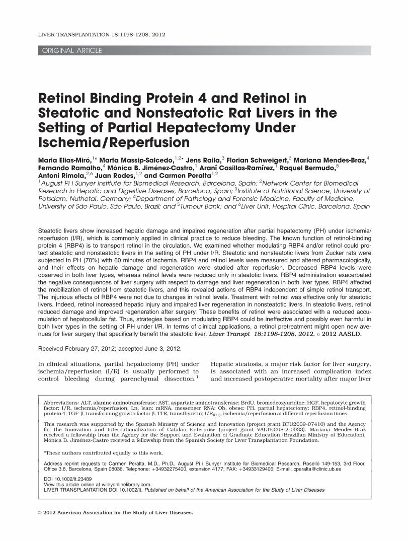
Vitamin A is stored in the liver as retinyl esters, andthey must be hydrolyzed into retinol before vitamin Acan be mobilized into the circulation.4 In line withthis, retinyl ester and retinol levels in steatotic liverswere reduced for the PHþI/R group versus the shamgroup (Fig. 4A). This decrease was associated withhigh plasma retinol levels (Fig. 4B). As previously sug-gested on the basis of studies in cultured cells,24
RBP4 could affect the storage and mobilization of reti-nol in steatotic livers. In fact, we found that retinylester levels (but not retinol levels) were increased insteatotic livers (Fig. 4A) and circulating retinol levelswere reduced (Fig. 4B) for the PHþI/RþRBP4 groupversus the PHþI/R group or the sham group. Asexpected, retinol administration increased both retinylester and retinol levels in steatotic livers from thePHþI/Rþretinol and PHþI/RþRBP4þretinol groupsversus the PHþI/R group and increased retinyl esterlevels (but not retinol levels) in plasma (Fig. 4A,B).Our results confirmed that RBP4 administration ledto higher RBP4 levels in steatotic livers from thePHþI/RþRBP4 and PHþI/RþRBP4þretinol groupsversus the PHþI/R group. For instance, the hepaticRBP4 levels for the PHþI/RþRBP4þretinol and PHþI/Rgroups were 239.6 6 21.65 and 82.41 6 3.91respectively (P < 0.05). The hepatic RBP4 (pmol/g oftissue) levels for the PHþI/Rþretinol group (83.45 6
4.56) were similar to the levels for the PHþI/R group(82.41 6 3.91, P ¼ not significant).
Transport of Retinol in Plasma
Reduced plasma RBP4 levels were observed for thePHþI/R group versus the sham group (Fig. 4C).The plasma RBP4 levels for the PHþI/RþRBP4 andPHþI/RþRBP4þretinol groups were lower than thosefor the PHþI/R group. The plasma RBP4 levels for thePHþI/Rþretinol group were similar to those for thePHþI/R group. The plasma TTR levels were reduced forthe PHþI/R group versus the sham group, whereas theTTR levels for the PHþI/RþRBP4, PHþI/Rþretinol, andPHþI/RþRBP4þretinol groups were similar to those forthe sham group. It has been reported that retinol is nor-mally found in plasma in a 1:1 molar ratio with RBP4.25
In the sham group, the RBP4/retinol ratio was approxi-mately 2; in the PHþI/R and PHþI/RþRBP4 groups,the RBP4/retinol ratio was greater than 1; and in thePHþI/Rþretinol and PHþI/RþRBP4þretinol groups,the RBP4/retinol ratio was less than 1 (Fig. 4C).
Reperfusion Time–Dependent Effect of Retinol
on Hepatic Damage, Proliferative Activity, and
Degree of Steatosis
Hepatic damage and proliferative activity in steatoticand nonsteatotic livers were measured throughoutreperfusion (12, 24, and 48 hours; Fig. 5). In Ln ani-mals, the parameters of hepatic injury were higher forthe partial hepatectomy under ischemia-reperfusion
LIVER TRANSPLANTATION, Vol. 18, No. 10, 2012 ELIAS-MIRO ET AL. 1203
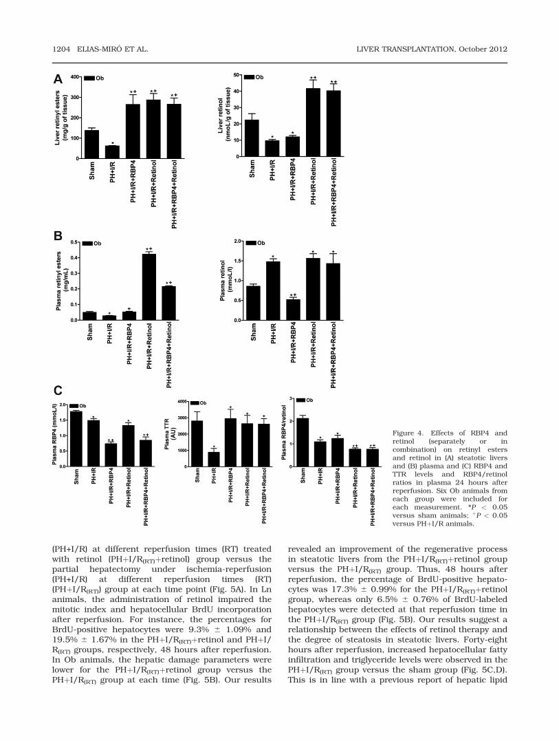
(PH+I/R) at different reperfusion times (RT) treatedwith retinol (PHþI/R(RT)þretinol) group versus thepartial hepatectomy under ischemia-reperfusion(PH+I/R) at different reperfusion times (RT)(PHþI/R(RT)) group at each time point (Fig. 5A). In Lnanimals, the administration of retinol impaired themitotic index and hepatocellular BrdU incorporationafter reperfusion. For instance, the percentages forBrdU-positive hepatocytes were 9.3% 6 1.09% and19.5% 6 1.67% in the PHþI/R(RT)þretinol and PHþI/R(RT) groups, respectively, 48 hours after reperfusion.In Ob animals, the hepatic damage parameters werelower for the PHþI/R(RT)þretinol group versus thePHþI/R(RT) group at each time (Fig. 5B). Our results
revealed an improvement of the regenerative processin steatotic livers from the PHþI/R(RT)þretinol groupversus the PHþI/R(RT) group. Thus, 48 hours afterreperfusion, the percentage of BrdU-positive hepato-cytes was 17.3% 6 0.99% for the PHþI/R(RT)þretinolgroup, whereas only 6.5% 6 0.76% of BrdU-labeledhepatocytes were detected at that reperfusion time inthe PHþI/R(RT) group (Fig. 5B). Our results suggest arelationship between the effects of retinol therapy andthe degree of steatosis in steatotic livers. Forty-eighthours after reperfusion, increased hepatocellular fattyinfiltration and triglyceride levels were observed in thePHþI/R(RT) group versus the sham group (Fig. 5C,D).This is in line with a previous report of hepatic lipid
Figure 4. Effects of RBP4 andretinol (separately or incombination) on retinyl estersand retinol in (A) steatotic liversand (B) plasma and (C) RBP4 andTTR levels and RBP4/retinolratios in plasma 24 hours afterreperfusion. Six Ob animals fromeach group were included foreach measurement. *P < 0.05versus sham animals; þP < 0.05versus PHþI/R animals.
1204 ELIAS-MIRO ET AL. LIVER TRANSPLANTATION, October 2012

Figure 5. Effects of retinol on hepatic injury (plasma AST and ALT levels) and liver regeneration (percentages of BrdU-positivehepatocytes and mitotic indices) throughout reperfusion (12, 24, and 48 hours) in (A) Ln animals and (B) Ob animals. Eighteen Lnanimals and 18 Ob animals (6 Ln and 6 Ob animals for each reperfusion time) were included for each measurement. (C) Triglyceridelevels and steatosis percentages and (D) representative photographs of red oil staining 48 hours after reperfusion are shown. Steatoticlivers from the retinol group showed reduced fat globules (red) in hepatocytes in comparison with steatotic livers from the sham andPHþI/R groups (scale bar ¼ 1000 lm). Six Ob animals from each group were included for each measurement. *P < 0.05 versus shamanimals; þP < 0.05 versus PHþI/R animals.
LIVER TRANSPLANTATION, Vol. 18, No. 10, 2012 ELIAS-MIRO ET AL. 1205

accumulation during regeneration after PH.26 Thetreatment with retinol decreased the accumulation offatty droplets and triglycerides in steatotic livers fromthe PHþI/R(RT)þretinol group versus the PHþI/R(RT)
group. The steatosis percentages were 35.0% 6 1.78%and 83.13% 6 3.40% for the PHþI/R(RT)þretinol andPHþI/R(RT) groups, respectively, 48 hours after reper-fusion. Figure 6 shows the relationship between theeffects of retinol therapy and the degree of steatosis 12,24, and 48 hours after reperfusion. A significant corre-lation between the degree of fat infiltration and theplasma AST level (r ¼ 0.9677, P < 0.05) and an inversecorrelation between the degree of fat infiltration andthe mitotic index (r ¼ �0.9724, P < 0.05) wereobserved. Treatment with retinol, therefore, attenuatedhepatic damage and improved hepatocellular prolifera-tion during liver regeneration. This was associated witha reduced accumulation of hepatocellular fat.
DISCUSSION
The decreased RBP4 levels observed in the circulationafter PH under I/R might reflect decreased hepatic
synthesis of RBP4 due to a loss of functional hepatictissue. Because the liver is the main site of RBP4 syn-thesis,8,27 hepatocyte damage decreases circulatingRBP4 levels. Moreover, the synthesis of negativeacute-phase proteins such as RBP4 might be reducedin the liver during inflammation or hepatic resectionas part of a compensatory response to the suddenincrease in the synthesis of positive acute-phase pro-teins.11 The effects of RBP4 could depend on the sur-gical conditions. Indeed, in contrast to steatotic livertransplantation,8 RBP4 administration worsens liverdamage and regenerative failure in the setting of PHunder I/R.
Retinol is decreased in steatotic livers after PHunder I/R and is increased in plasma. This is consist-ent with the increased secretion of retinol from stea-totic livers into the circulation. However, otherhypotheses proposed for different liver diseases,28
including increased catabolism of retinol in the liverand decreased liver uptake of retinyl esters formed inperipheral tissues, should not be discounted. Retinolis normally found in plasma in a 1:1 molar ratio withRBP4 (the RBP4/retinol ratio is approximately 1.0).29
However, in our study and in agreement with thereports of others,4 the RBP4/retinol ratio for thesham group of Ob (steatotic) animals was approxi-mately 2, and this indicated that the proportion ofRBP4 in plasma was higher than the proportion of ret-inol. In this context, it should be considered thatRBP4 is present as a holoform (retinol-bound) and anapoform (retinol-unbound) because RBP4 may trans-port and deliver other lipophilic molecules in additionto retinol.6,30 A question that arises is how the highplasma retinol levels in the PHþI/R group are main-tained in light of the low plasma RBP4 and TTR levels.Like RBP4, TTR is a negative acute-phase protein,and the hepatic syntheses of both are also decreasedduring inflammation.11 However, because the plasmaRBP4/retinol ratio in the PHþI/R group was greaterthan 1, there was enough RBP4 to bind retinol despitethe reduced RBP4 levels. It could be assumed that thereduced plasma TTR levels in the PHþI/R group werealso high enough to maintain the retinol-RBP4 com-plex in plasma, insofar as there is usually a 3- to 5-fold molar excess of circulating TTR with respect tocirculating RBP4.11,27 The lack of a correlationbetween RBP4 and TTR levels in plasma under oursurgical conditions could be explained, at least inpart, by the differential clearance of RBP4 and TTR bythe kidneys: TTR has a higher molecular weight andis, therefore, retained to a greater extent than RBP4.27
Moreover, TTR has additional functions (eg, the trans-porter for T4).4
RBP4 exerted injurious effects on steatotic liverswith respect to damage and regeneration independ-ently of retinol. Moreover, the changes in retinol me-tabolism induced by RBP4 could have occurred sim-ply as a result of disease progression and thus do notexplain the injurious effects of RBP4. The mechanismbehind this is unclear. Retinyl esters seem to beaffected by RBP4 administration, and this also
Figure 6. Relationship between the degree of steatosis and (A)the plasma AST level (r ¼ 0.9677, P < 0.05) and (B) the mitoticindex (r ¼ �0.9724, P < 0.05): (n) Ob PHþI/R(RT)þretinolanimals 12 hours after reperfusion, (l) Ob PHþI/R(RT)þretinolanimals 24 hours after reperfusion, and (~) Ob PHþI/R(RT)þretinol animals 48 hours after reperfusion.
1206 ELIAS-MIRO ET AL. LIVER TRANSPLANTATION, October 2012

suggests that RBP4 has actions independent of simpleretinol transport. However, the RBP4-inducedincrease in retinyl esters does not explain the injuri-ous effects of RBP4. Indeed, the loss of retinoid(rather than an increase in retinoid levels) contributesto the development of hepatic diseases.31
Serum RBP4 levels are elevated in insulin-resistantmice and humans with obesity and type 2 diabetes.The transgenic overexpression of human RBP4 or theinjection of recombinant RBP4 into normal micecauses insulin resistance. Conversely, the genetic de-letion of RBP4 enhances insulin sensitivity.6 Elucidat-ing whether RBP4 also has metabolic effects was notan aim of the present study. Nevertheless, because ofour results, this seems to not be the case for our ex-perimental model. The Ob Zucker rats used in thepresent study have a mutated leptin receptor and, asa result, are hyperphagic, Ob, and hyperinsulinemic.They are insulin-resistant, but they have normalblood glucose levels. Ob Zucker rats do not developdiabetes. Ln Zucker rats maintain an Ln phenotypethroughout life, and they show normal blood insulinand glucose levels. RBP4 administration did not alterthe plasma insulin or glucose levels in the Ob Zuckerrats undergoing PH and I/R (data not shown). Underour conditions, there was no relationship betweenRBP4 levels and insulin resistance. Indeed, Ln andOb Zucker rats in the sham group showed similarRBP4 levels in the liver and plasma, and only ObZucker rats are insulin-resistant. In contrast, Yanget al.6 have shown that genetically Ob mice and high-fat diet–induced Ob mice with insulin resistance ex-hibit increased plasma RBP4 in comparison with Lncontrols. The differences in plasma RBP4 levelsobserved in the 2 studies can be explained at leastpartially by the differences in RBP4 regulationbetween rats and mice.32 All these data indicate thatunder our conditions, RBP4 does not affect insulinresistance.
Further studies, which are not part of the objectivesof the present study, are required to answer why thepharmacological modulation of RBP4 exerted damag-ing effects in the setting of PH under I/R. It is possiblethat the compensatory changes in the protein synthe-sis of positive and negative acute-phase proteins,which were necessary to restore protein homeostasisafter hepatic resection, were disturbed in the remain-ing liver when RBP4 was administered. From a clini-cal perspective, strategies based on modulating RBP4might not be appropriate for hepatic resection orunder surgical conditions (including small-for-sizeliver transplantation). When we administered RBP4 tocompensate for the reduced RBP4 levels induced byliver surgery, we observed more injurious effects withrespect to damage and liver regeneration.
In contrast, pretreatment with retinol alone maycreate new possibilities for therapeutic interventionsin the resection of steatotic livers. As expected, retinoladministration alone increased vitamin A storage inthe liver. The increase in retinyl esters (but not retinolor RBP4) in plasma after retinol administration sug-
gests that retinol is incorporated as retinyl esters inhepatocytes and is secreted together with lipoproteins.Thus, retinol administration could supply vitamin Ato target tissues. Data indicate that the RBP4 mole-cule contains 1 binding site for a single molecule ofretinol,25 so the fact that the RBP4/retinol ratio afterretinol administration was less than 1 might suggestthat a fraction of retinol is bound to carriers otherthan RBP4, just as with other pathologies.33
The results presented here indicate that retinolreduces damage and improves liver regeneration in anexperimental model combining PH and I/R. Unpub-lished results from our group indicate that retinoltherapy for steatotic livers affects both I/R injury andPH. Thus, retinol reduces hepatic damage in Ob ratssubjected to partial hepatic ischemia (60 minutes)without hepatectomy. Retinol administration alsoreduces hepatic damage and improves liver regenera-tion in Ob rats subjected to 70% resection only (datanot shown).
Further studies will be required to explain how reti-nol reduces lipid accumulation in the regeneratingliver after surgery. The effects of retinol on the mobili-zation of fatty acids from peripheral stores and he-patic lipogenesis, fatty acid oxidation, and triglyceridesecretory mechanisms in the liver should be exploredas possible answers to this question. Our resultsshow that the benefits of retinol with respect to dam-age and liver regeneration in steatotic livers are asso-ciated with reduced hepatic lipid accumulation. It hasbeen reported that it is crucial to reduce steatosis toprevent the vulnerability of steatotic livers to I/Rinjury and regenerative failure.34
In conclusion, PH under I/R affects hepatic vitaminA metabolism by reducing hepatic RBP4 expressionand increasing the mobilization of retinol into the cir-culation. The results presented here suggest thatunder surgical conditions requiring liver regeneration,modulating RBP4 levels worsens the outcome and is,therefore, not advised. This study also points to newpossibilities for therapeutic interventions based onretinol pretreatment to protect steatotic livers againstdamage and regenerative failure after liver surgery.
ACKNOWLEDGMENTSThe authors thank Bioscience Writers for revising theEnglish text. They also thank Mrs. A. Hurtienne andMrs. U. Ullrich for their expert technical assistance.
REFERENCES
1. McCormack L, Petrowsky H, Jochum W, Furrer K, Clav-ien PA. Hepatic steatosis is a risk factor for postoperativecomplications after major hepatectomy: a matched case-control study. Ann Surg 2007;245:923-930.
2. Vetel€ainen R, van Vliet A, Gouma DJ, van Gulik TM. Ste-atosis as a risk factor in liver surgery. Ann Surg 2007;245:20-30.
3. Graham TE, Yang Q, Bluher M, Hammarstedt A, CiaraldiTP, Henry RR, et al. Retinol-binding protein 4 and insu-lin resistance in lean, obese, and diabetic subjects.N Engl J Med 2006;354:2552-2563.
LIVER TRANSPLANTATION, Vol. 18, No. 10, 2012 ELIAS-MIRO ET AL. 1207

4. Tuitoek PJ, Ritter SJ, Smith JE, Basu TK. Streptozotocin-induced diabetes lowers retinol-binding protein and trans-thyretin concentrations in rats. Br JNutr 1996;76:891-897.
5. Bellovino D, Apreda M, Gragnoli S, Massimi M, GaetaniS. Vitamin A transport: in vitro models for the study ofRBP secretion. Mol Aspects Med 2003;24:411-420.
6. Yang Q, Graham TE, Mody N, Preitner F, Peroni OD,Zabolotny JM, et al. Serum retinol binding protein 4 con-tributes to insulin resistance in obesity and type 2 diabe-tes. Nature 2005;436:356-362.
7. Pala L, Monami M, Ciani S, Dicembrini I, Pasqua A, Pez-zatini A, et al. Adipokines as possible new predictors ofcardiovascular diseases: a case control study. J NutrMetab 2012;2012:253428.
8. Casillas-Ramırez A, Alfany-Fern�andez I, Massip-SalcedoM, Juan ME, Planas JM, Serafın A, et al. Retinol-bindingprotein 4 and peroxisome proliferator-activated receptor-c in steatotic liver transplantation. J Pharmacol ExpTher 2011;338:143-153.
9. Raila J, Henze A, Spranger J, M€ohlig M, Pfeiffer AF,Schweigert FJ. Microalbuminuria is a major determinantof elevated plasma retinol-binding protein 4 in type 2 di-abetic patients. Kidney Int 2007;72:505-511.
10. Gieng SH, Raila J, Rosales FJ. Accumulation of retinolin the liver after prolonged hyporetinolemia in the vita-min A-sufficient rat. J Lipid Res 2005;46:641-649.
11. Rosales FJ, Ritter SJ, Zolfaghari R, Smith JE, Ross AC.Effects of acute inflammation on plasma retinol, retinol-binding protein, and its mRNA in the liver and kidneys ofvitaminA-sufficient rats. J Lipid Res 1996;37:962-971.
12. Rao J, Zhang C, Wang P, Lu L, Zhang F. All-trans reti-noic acid alleviates hepatic ischemia/reperfusion injuryby enhancing manganese superoxide dismutase in rats.Biol Pharm Bull 2010;33:869-875.
13. Kimura M, Watanabe M, Ishibashi N, Yanagida S, Ogi-hara M. Acyclic retinoid NIK-333 accelerates liver regen-eration and lowers serum transaminase activities in 70%partially hepatectomized rats, in vivo. Eur J Pharmacol2010;643:267-273.
14. Ohtake Y, Maruko A, Ohishi N, Kawaguchi M, Satoh T,Ohkubo Y. Effect of retinoic acid on transglutaminaseand ornithine decarboxylase activities during liver regen-eration. Cell Biochem Funct 2008;26:359-365.
15. Ramalho FS, Alfany-Fernandez I, Casillas-Ramirez A,Massip-Salcedo M, Serafın A, Rimola A, et al. Are angio-tensin II receptor antagonists useful strategies in stea-totic and nonsteatotic livers in conditions of partialhepatectomy under ischemia-reperfusion? J PharmacolExp Ther 2009;329:130-140.
16. Sato Y, Meller R, Yang T, Taki W, Simon RP. Stereo-selec-tive neuroprotection against stroke with vitamin A deriv-atives. Brain Res 2008;1241:188-192.
17. Duval H, Mbatchi SF, Grandadam S, Legendre C, Loyer P,Ribault C, et al. Reperfusion stress induced during inter-mittent selective clamping accelerates rat liver regenera-tion through JNK pathway. J Hepatol 2010;52:560-569.
18. Massip-Salcedo M, Zaouali MA, Padrissa-Alt�es S, Casil-las-Ramirez A, Rod�es J, Rosell�o-Catafau J, Peralta C.Activation of peroxisome proliferator-activated receptor-alpha inhibits the injurious effects of adiponectin in ratsteatotic liver undergoing ischemia-reperfusion. Hepato-logy 2008;47:461-472.
19. Schweigert FJ, Buchholz I, Bonitz K. Effect of age on thelevels of retinol and retinyl esters in blood plasma, liver
and kidney of dogs. Int J Vitam Nutr Res 1998;68:237-241.
20. Henze A, Rohn S, Gericke B, Raila J, Schweigert FJ.Structural modifications of serum transthyretin in ratsduring protein-energy malnutrition. Rapid CommunMass Spectrom 2008;22:3270-3274.
21. Serafın A, Rosell�o-Catafau J, Prats N, Xaus C, Gelpı E,Peralta C. Ischemic preconditioning increases the toler-ance of fatty liver to hepatic ischemia-reperfusion injuryin the rat. Am J Pathol 2002;161:587-601.
22. Schiffer E, Frossard JL, Rubbia-Brandt L, Mentha G,Pastor CM. Hepatic regeneration is decreased in a ratmodel of sinusoidal obstruction syndrome. J Surg Oncol2009;99:439-446.
23. Takitani K, Miyazaki H, Fukunishi S, Takaya R, YodenA, Higuchi K, Tamai H. Altered expression of both b-car-otene 15,150 monooxygenase and lecithin:retinol acyl-transferase in obese Zucker rats. J Nutr Sci Vitaminol(Tokyo) 2011;57:108-113.
24. Sauvant P, Sapin V, Alexandre-Gouabau MC, DodemanI, Delpal S, Quadro L, et al. Retinol mobilization fromcultured rat hepatic stellate cells does not require retinolbinding protein synthesis and secretion. Int J BiochemCell Biol 2001;33:1000-1012.
25. Quadro L, Hamberger L, Colantuoni V, Gottesman ME,Blaner WS. Understanding the physiological role of reti-nol-binding protein in vitamin A metabolism using trans-genic and knockout mouse models. Mol Aspects Med2003;24:421-430.
26. Gazit V, Weymann A, Hartman E, Finck BN, Hruz PW,Tzekov A, Rudnick DA. Liver regeneration is impaired inlipodystrophic fatty liver dystrophy mice. Hepatology2010;52:2109-2117.
27. Mody N, Graham TE, Tsuji Y, Yang Q, Kahn BB.Decreased clearance of serum retinol-binding proteinand elevated levels of transthyretin in insulin-resistantob/ob mice. Am J Physiol Endocrinol Metab 2008;294:E785-E793.
28. Sato M, Lieber CS. Changes in vitamin A status afteracute ethanol administration in the rat. J Nutr 1982;112:1188-1196.
29. Soprano DR, Smith JE, Goodman DS. Effect of retinolstatus on retinol-binding protein biosynthesis rate andtranslatable messenger RNA level in rat liver. J BiolChem 1982;257:7693-7697.
30. Berni R, Clerici M, Malpeli G, Cleris L, Formelli F. Reti-noids: in vitro interaction with retinol-binding proteinand influence on plasma retinol. FASEB J 1993;7:1179-1184.
31. Blaner WS, O’Byrne SM, Wongsiriroj N, Kluwe J,D’Ambrosio DM, Jiang H, et al. Hepatic stellate cell lipiddroplets: a specialized lipid droplet for retinoid storage.Biochim Biophys Acta 2009;1791:467-473.
32. Lanne B, Dahll€of B, Lindahl C, Ebefors K, Kanmert I,von Bahr H, et al. PPARa and PPARc regulation of liverand adipose proteins in obese and dyslipidemic rodents.J Proteome Res 2006;5:1850-1859.
33. Mallia AK, Smith JE, Goodman DW. Metabolism of reti-nol-binding protein and vitamin A during hypervitamino-sis A in the rat. J Lipid Res 1975;16:180-188.
34. Elias-Miro M, Massip-Salcedo M, Jimenez-Castro M, Per-alta C. Does adiponectin benefit steatotic liver transplan-tation? Liver Transpl 2011;17:993-1004.
1208 ELIAS-MIRO ET AL. LIVER TRANSPLANTATION, October 2012


