Embed Size (px)
Citation preview

Reversible and adaptive resistance to BRAF(V600E) inhibition in melanoma.
Chong Sun1*, Liqin Wang1*, Sidong Huang1, 2*, Guus J.J.E. Heynen1, Anirudh Prahallad1,
Caroline Robert3, John Haanen4, Christian Blank4, Jelle Wesseling5, Stefan M. Willems1,6,
Davide Zecchin7, 8, Sebastijan Hobor8, Prashanth K. Bajpe1, Cor Lieftink1, Christina Mateus3,
Stephan Vagner3, Wipawadee Grernrum1, Ingrid Hofland5, Andreas Schlicker1, Lodewyk
Wessels1, Roderick L. Beijersbergen1, Alberto Bardelli7,8,9, Federica Di Nicolantonio7,8,
Alexander M.M. Eggermont3 and Rene Bernards1#
1Divisions of Molecular Carcinogenesis, Medical Oncology4 and Pathology5, Cancer Systems
Biology Centre and Cancer Genomics Centre Netherlands, The Netherlands Cancer Institute,
Plesmanlaan 121, 1066 CX Amsterdam, The Netherlands.
2Department of Biochemistry, The Rosalind and Morris Goodman Cancer Centre, McGill
University, Montreal, Quebec H3G 1Y6, Canada
3Institut Gustave Roussy, 114 Rue Edouard Vaillant, 94800 Villejuif, France.
6Department of Pathology, University Medical Centre Utrecht, Heidelberglaan 100, 3584 CX,
Utrecht, The Netherlands.
7Institute for Cancer Research and Treatment at Candiolo (IRCC), SP142 Km 3.95, 10060
Candiolo (Torino), Italy
8Department of Oncology , University of Torino, 10060 Candiolo (Torino), Italy
9FIRC Institute of Molecular Oncology (IFOM), 20139 Milano, Italy
*These authors contributed equally to this work
#To whom correspondence should be addressed. E-mail: [email protected]

2
Treatment of BRAF(V600E) mutant melanoma by small molecule drugs that target the
BRAF or MEK kinases can be effective, but resistance develops invariably1,2. In
contrast, colon cancers that harbour the same BRAF(V600E) mutation are intrinsically
resistant to BRAF inhibitors, due to feedback activation of the Epidermal Growth
Factor Receptor (EGFR)3,4. We show here that 6 out of 16 melanoma tumours analysed
acquired EGFR expression after the development of resistance to BRAF or MEK
inhibitors. Using a chromatin regulator-focused shRNA library, we find that
suppression of sex determining region Y-box 10 (SOX10) in melanoma causes activation
of TGFβ signalling, thus leading to upregulation of EGFR and Platelet Derived Growth
Factor Receptor β (PDGFRB), which confer resistance to BRAF and MEK inhibitors.
Expression of EGFR in melanoma or treatment with TGFβ results in a slow-growth
phenotype with cells displaying hallmarks of oncogene-induced senescence. However
EGFR expression or exposure to TGFβ becomes beneficial for proliferation in the
presence of BRAF or MEK inhibitors. In a heterogeneous population of melanoma cells
having varying levels of SOX10 suppression, cells with low SOX10 and consequently
high EGFR expression are rapidly enriched in the presence of drug, but this is reversed
when the drug treatment is discontinued. We find evidence for SOX10 loss and/or
activation of TGFβ signalling in 4 of the 6 EGFR-positive drug-resistant melanoma
patient samples. Our findings provide a rationale for why some BRAF or MEK
inhibitor resistant melanoma patients may regain sensitivity to these drugs after a drug
holiday and identify patients with EGFR-positive melanoma as a group that may benefit
from re-treatment after a drug holiday.
3
Activating mutations in the BRAF oncogene are found in over half of the patients with
advanced melanoma5,6. Inhibition of the oncogenic BRAF protein with the small molecule
inhibitor PLX4032 (vemurafenib) or its downstream effector MEK with GSK1120212
(trametinib) have shown impressive initial responses in patients with BRAF mutant
melanoma1,2. However, single agent therapies for advanced cancers are rarely curative, due to
the rapid development of resistance. To date, several drug resistance mechanisms have been
identified in melanomas treated with vemurafenib, including increased expression of the gene
encoding the COT kinase, mutation of downstream MEK1 kinase, NRAS mutations and
amplification or alternative splicing of the BRAF gene7-11. Moreover, increased expression of
receptor tyrosine kinases (RTKs) has been observed as a mechanism of BRAF inhibitor
resistance11-13.
It has been shown recently that intrinsic resistance of BRAF mutant colon cancers to
vemurafenib is the result of feedback activation of EGFR when BRAF is inhibited3,4. To
investigate whether BRAF(V600E) mutant melanoma patients frequently develop resistance to
BRAF or MEK inhibitors through acquired expression of EGFR in their tumours, we obtained
biopsies from BRAF(V600E) mutant melanomas from sixteen patients treated with either the
MEK inhibitor trametinib (n=1) or the BRAF inhibitors dabrafenib (n=3) or vemurafenib
(n=12). Tumour biopsies collected both before treatment initiation and after the development
of drug resistance were stained for EGFR expression. We found that 6 out of 16 post
treatment biopsies gained significant EGFR expression as judged by immunohistochemistry
(Figure 1a, b Table S1).
Melanomas are derived from the neural crest and in general do not express EGFR14.
Hence, acquired EGFR expression during drug selection may represent a stress response that
is not favoured in the absence of drug treatment. Indeed, the proliferation rate of A375
melanoma cell lines engineered to express EGFR decreased as the concentration of EGFR
ligand increased (Figure 1c, ref3). Moreover, A375 cells that express EGFR also proliferate
slower compared to parental control cells in nude mouse xenografts, but are resistant to

4
trametinib (Fig 1d). To investigate the cause of this slow-growth phenotype, we performed
western blotting for a number of cell cycle-associated proteins on parental A375 cells and
EGFR-expressing derivatives. EGFR expression resulted in hypophosphorylated pRB protein,
induction of the CDK inhibitors CDKN1A (p21cip1) and CDKN1B (p27kip1) and acidic β-
galactosidase (Figure 1e, f), markers that have been associated with oncogene-induced
senescence15,16. These markers were also induced upon expression of oncogenic versions of
BRAF or MEK, but much less when activated mutants of AKT1 or PIK3CA were expressed in
A375 cells (Extended Data Fig. 1). We conclude that EGFR expression is disadvantageous for
BRAF(V600E) melanoma cells in the absence of BRAF or MEK inhibitor drugs, but it confers
a selective advantage in the presence of these drugs.
Acquired EGFR expression may be the result of an adaptive response of the cancer
cell population during drug selection. To ask in an unbiased way which factors might
modulate EGFR expression in melanoma cells, we compiled a “chromatin regulator” library
of shRNAs targeting 661 genes, including the KATs (lysine acetyltransferases), KMTs (lysine
methyltransferases), KDACs (lysine deacetylases), KDMs (lysine demethylases), chromatin
remodelling complexes and proteins that harbour chromatin binding/associated domains
(Table S2). A375 melanoma cells, which express very low levels of EGFR, were infected
with the chromatin regulator library and selected with vemurafenib for 3 weeks. After this,
the vemurafenib-resistant cells were harvested and strongly EGFR-positive cells (EGFRhigh)
were isolated from the drug-resistant population by Fluorescence-Activated Cell Sorting
(Figure 2a). Treatment of cells with either the chromatin regulator library or vemurafenib
alone did not increase the fraction of EGFRhigh cells. In contrast, a significant fraction of
EGFRhigh cells could be retrieved when cells were infected with the chromatin regulator
library and were selected for vemurafenib resistance (Figure 2b). We conclude that EGFRhigh
melanoma cells do not merely appear as a consequence of silencing of certain chromatin
regulators, but that these cells only emerge when the population is placed under drug-

5
selection pressure. This suggests that silencing of the gene(s) that induce EGFR expression is
not favoured in the absence of vemurafenib.
To identify which gene(s) in the chromatin regulator library can induce EGFR
expression, we isolated genomic DNA from the EGFRhigh cells and non-drug treated control
cells and determined the abundance of the shRNA vectors in each cell population by deep
sequencing, as described previously3. shRNAs that confer resistance to vemurafenib through
upregulation of EGFR should be enriched in the EGFRhigh fraction. shRNA screens are
notorious for yielding false positive results. Therefore, in principle only those genes that are
represented by multiple shRNAs should be followed up in a genetic screen17. However, in this
screen we did not identify any genes for which multiple shRNAs were enriched (Table S3).
We therefore focused on the top 10 most strongly enriched genes for follow up experiments.
We tested multiple additional shRNA vectors for each of these 10 genes for their ability to
increase EGFR expression, as this was a selection criterion in the genetic screen (Extended
Data Fig. 2a, b). Only suppression of the SRY (sex determining region Y)-box 10 (SOX10)
gene induced prominent EGFR expression when multiple SOX10 shRNAs (shSOX10) were
used in four melanoma cell line models (Figures 2c, 2d, Extended Data Fig. 2c, 4c, 5c).
SOX10 knockdown (SOX10KD) induced a slow-growth phenotype and also displayed the
hallmarks of oncogene-induced senescence in multiple melanoma models (Figure 2e,
Extended Data Fig. 2e, f, g, 4b, e, f, 5b, e, f).
Next we confirmed that SOX10KD indeed induced vemurafenib resistance in
melanoma. We infected A375 cells with shSOX10 and cultured cells in the presence of
vemurafenib. SOX10KD slowed down proliferation of A375 cells in the absence of drug, but in
the presence of vemurafenib SOX10KD conferred drug resistance, both in short-term and long-
term assays (Figure 2e, Extended Data Fig. 2d, e). Moreover, under vemurafenib selective
pressure, cells having a higher degree of SOX10KD were selected, which consequently also
expressed higher levels of EGFR, consistent with the notion that increased EGFR levels drive
drug resistance (Extended Data Fig. 2h). Vemurafenib resistance through SOX10 suppression
was also seen in additional melanoma cell lines (Extended Data Fig. 4a, 5a). Note that a low

6
concentration of vemurafenib actually increased proliferation rate of SOX10KD cells,
consistent with the model that hyperactive BRAF-MEK signalling induces senescence
markers, which is inhibited by vemurafenib (Extended Data Fig. 4a, g).
To study how SOX10 suppression induces EGFR expression, we performed
transcriptome sequencing (RNAseq) of both parental A375 and A375-SOX10KD cells (Table
S4). Gene set enrichment analysis of the SOX10-upregulated genes revealed an enrichment of
genes with SMAD2/3 (downstream mediators of TGFβ signalling) and JUN binding sites in
their promoters (Table S5). Consistent with this, SOX10 suppression induced TGFβ receptor
2 (TGFBR2) expression as well as a number of bona fide TGFβ target genes, including JUN,
in multiple melanoma cell models (Figure 3a, b, Extended Data Fig. 4d, 5d). Levels of active
JUN (pJUN) were also increased by SOX10KD (Figure 3a). That treatment of melanoma cells
with recombinant TGFβ causes resistance to vemurafenib further supports a role for TGFβ
signalling in vemurafenib resistance (Figure 3c and ref18). TGF-β1 ��������� not only
caused induction of EGFR expression, but also of Platelet Derived Growth Factor Receptor β
(PDGFRB, Figure 3d, e) and also resulted in induction of senescence-associated β-
galactosidase (Figure 3f). Consistently, SOX10 suppression also induced PDGFRB expression
(Extended Data Fig. 3c, 4c, 5c). Moreover, suppression of TGFBR2 inhibited EGFR and
PDGFRB induction in SOX10KD cells (Figures 3g, h), whereas ectopic expression of TGFBR2
induced pJUN, EGFR and PDGFRB expression (Figure 3i). JUN is a regulator of EGFR
expression and TGFβ regulates PDGFRB 19-21. Moreover, SMADs and JUN cooperate in
activation of EGFR expression22,23. SOX10 is known to regulate the melanocyte transcription
factor MITF24. Indeed, A375 cells with shSOX10 also had reduced MITF expression, but
MITF suppression alone did not change EGFR or PDGFRB expression and did not cause
vemurafenib resistance (Extended Data Fig. 7c, d, e). In summary, our data provide support
for a model in which activation of TGFβ signalling by SOX10 loss leads to increased EGFR
and PDGFRB expression and vemurafenib resistance.

7
Treatment of A375-SOX10KD cells with a combination of both vemurafenib and the
EGFR inhibitor gefitinib did not lead to proliferation arrest, indicating that EGFR was not the
sole driver of drug resistance in SOX10KD cells (Extended Data Fig. 3a). Indeed, an unbiased
survey of RTKs revealed that SOX10KD activated not only EGFR, but also PDGFRB and
ERBB3 (Extended Data Fig. 3b, 3c). A similar pattern of RTK activation was observed
following TGF-β1 treatment, highlighting the similarity between SOX10 suppression and
acquired TGFβ signalling (Extended Data Fig. 3b, d). Many RTKs share two major
downstream signalling pathways (RAS-MEK-ERK and PI3K-AKT). Consistent with this, we
found that combined inhibition of these two downstream pathways using BRAF and PI3K
inhibitors could restore growth inhibition in SOX10KD cells (Extended Data Fig. 3a).
Our data are consistent with a model in which cells with low SOX10 and high EGFR
and PDGFRB expression are positively selected in the presence of drug, but that such cells
are counter-selected in the absence of drug. To test this model directly, we infected A375
cells with shSOX10 and subjected this heterogeneous population of SOX10KD cells to
vemurafenib selection for one week. At this point, we harvested part of this population and
determined EGFR expression by FACS analysis. Under vemurafenib selection, an increased
level of EGFR and a markedly decreased level of SOX10 were observed. When these cells
were subsequently cultured for one more week in the absence of vemurafenib, the
EGFRhigh/SOX10low population was depleted (Figure 4a, Extended Data Fig. 6a). These data
indicate that acquired EGFR expression is only advantageous to melanoma cells in the
presence of drug selection, but is counter-selected in the absence of drug.
Consistent with a role for SOX10 in regulation of EGFR expression in melanoma, we
found an inverse correlation between SOX10 and EGFR expression in a panel of 34
melanoma cell lines25 (Figure 4b) and a similar inverse relation between SOX10 and
PDGFRB (Extended Data Fig. 6b). The most extreme cell line in this panel, LOXIMVI,
completely lacked SOX10 expression and had the highest EGFR expression. When we
expressed SOX10 in this cell line, EGFR and PDGFRB were reduced and TGFBR2 and

8
TGFBR3 as well as JUN and pJUN levels were also downregulated, consistent with the
notion that SOX10 regulates these RTKs through an effect on TGFβ signalling (Extended
Data Fig. 6c, d). Consistently, expression of SOX10 in LOXIMVI cells increased their
sensitivity to vemurafenib (Extended Data Fig. 6e).
To ask directly whether SOX10 is involved in EGFR-associated drug resistance in
BRAF(V600E) melanoma patients, we isolated RNA from the six patients studied above that
had gained EGFR expression after acquisition of trametinib, dabrafenib or vemurafenib
resistance (Table S1). We performed RNAseq analysis to determine changes in transcriptome
upon drug resistance. In two patients the levels of SOX10 mRNA were reduced (Figures 4c,
Extended Data Fig. 6f). EGFR and PDGFRB mRNA were greatly increased in patient 5,
whereas no evidence was found in this patient of alternative BRAF splicing7 or BRAF over-
expression (Extended Data Fig. 7a, b). Patient 3 has strong induction of EGFR protein post
resistance (Figure 1a), but at first glance, EGFR mRNA levels appear only minimally
induced. However, scrutiny of the RNAseq data reveals that the apparent lack of induction of
EGFR in this tumour sample pair is caused by the abnormally high EGFR transcript
abundance in the pre-treatment sample and not the lack of EGFR expression in the post-
treatment sample (Extended Data Fig. 6g). This is most likely due to the contamination of this
sample with the strongly EGFR positive skin material (see Figure 1a). These tumours also
manifested increased TGFβ signalling (Figure 4c, Extended Data Fig. 6h). Two further pairs
of tumour samples showed induction of EGFR and PDGFRB without significant loss of
SOX10 after drug resistance emerged. These tumours displayed induction of TGFβ receptor
expression and induction of a number of bona fide TGFβ targets, suggesting that these
tumours somehow had acquired TGFβ signalling (and subsequent induction of EGFR and
PDGFRB expression) in a SOX10-independent fashion (Figure 4c).
Clinical evidence indicates that melanoma patients that have developed vemurafenib
resistance can regain sensitivity to the drug after a drug holiday, suggesting a reversible and

9
adaptive transcriptional response to the drug26. That drug resistance is reversed in the absence
of drug indicates that this adaptive response is not favoured in the absence of drug. Our data
provide a molecular underpinning for the concept that drug resistance may arise at a fitness
cost in the absence of drug (Figure 4d). Melanoma patients whose tumours acquire EGFR
expression as a result of drug resistance development may be candidates to be re-treated with
drug after a drug holiday.

10
Acknowledgements.
We thank the NKI Core Facilities for Genomics and Molecular Pathology & Biobanking for
tumour tissue and support in DNA sequencing. We thank Severine Roy for collecting clinical
data and Nyam Kamsu Kom for tissue preparation. This work was supported by grants from
the European Research Council (ERC), the Dutch Cancer Society (KWF), the EU
COLTHERES project and grants by the Netherlands Organization for Scientific Research
(NWO) to Cancer Genomics Netherlands (CGC.NL). Additional support was provided by
Fondazione Piemontese per la Ricerca sul Cancro – ONLUS grant ‘Farmacogenomica – 5 per
mille 2009 MIUR’ (F.D.N.); AIRC MFAG 11349 (F.D.N.); AIRC IG grant n. 12812 (A.B.)
and Canadian Institutes of Health Research (CIHR) grant MOP-130540 (S.H.).

11
FIGURE LEGENDS
Figure 1 | Acquired EGFR expression in BRAF(V600E) mutant melanoma after
vemurafenib resistance.
a, b, Immunohistochemical (IHC) analysis (a, brown staining; b, pink staining) showing
increased EGFR expression in formalin-fixed paraffin embedded (FFPE) (Patient #1, #2, #3,
#4 and #5) and frozen (Patient #6) melanoma tissue sections from BRAF(V600E) mutant
melanoma patients who developed resistance to vemurafenib, dabrafenib or trametinib as
indicated. For each patient, the first biopsy is from the pre-treatment tumour; the second
biopsy was performed after the tumour had progressed under treatment. For patient #4, the
first biopsy was performed when the patient was in partial response, but rapidly developed
secondary resistance. 4.5 months later, the second biopsy was taken. c, EGFR expression
confers growth-disadvantage to BRAF(V600E) mutant melanoma cells and EGFR ligand
potentiates the growth deficiency in vitro. A375 BRAF(V600E) melanoma cells transduced
with control lentiviral vectors (Ctrl. , PLX304-GFP) or vectors expressing EGFR (EGFR,
PLX304-EGFR) were seeded at the same density and cultured in the presence of EGF at
indicated concentration for 2 weeks. The cells were fixed, stained and photographed. d,
EGFR expression confers growth-disadvantage to BRAF(V600E) mutant melanoma, but
induces trametinib resistance in vivo. CD1 nude mice were inoculated with BRAF(V600E)
mutant melanoma A375 cells transduced with control retroviral vectors or vectors expressing
EGFR. Once tumours were established, animals were treated with vehicle, trametinib.
Relative tumour volume is shown. Error bars represent SEM (n=5). * p <0.05, single-sided
Wilcoxon–Mann–Whitney test. e, Western blot analysis of RB protein, CDK inhibitors
CDKN1A (p21cip1) and CDKN1B (p27kip1) in EGFR expressing A375 cells. HSP90 served as
a loading control. f, EGFR expression induces senescence. Senescence was detected by
staining of β-galactosidase activity. All experiments shown except the ones that involve
clinical samples and animals were performed independently at least 3 times.

12
Figure 2 | FACS-assisted shRNA genetic screen identifies SOX10 as a determinant of
vemurafenib resistance and EGFR expression.
a, Schematic outline of the of the FACS-assisted shRNA screen. Human “Chromatin
Regulator” shRNA library polyclonal virus was generated to infect A375 cells, which were
then left untreated (control) or treated with 0.5μM vemurafenib. After 12 days, the untreated
cells were harvested. The cells that survived from 21 days of vemurafenib treatment were
FACS sorted for EGFR expression. Subsequently, shRNA inserts from both samples were
recovered by polymerase chin reaction (PCR) and identified by massive parallel sequencing.
b, EGFRhigh cells result from the combination of infection with chromatin regulator library
and vemurafenib selection. A375 cells infected with “chromatin regulator” library (Chr Lib)
were cultured in the presence of 0.5 μM vemurafenib for 21 days (right lower panel). Cells
were harvested with 2 mM EDTA, stained with anti-EGFR antibody and analysed for
EGFRhigh cells by flow cytometry. A375 cells cultured with or without vemurafenib, and
A375 cells infected by Chr Lib without vemurafenib treatment served as controls. c, d,
Suppression of SOX10 induces EGFR expression. (c) Western blot analysis of EGFR and
SOX10 levels in cells targeted by two independent shSOX10 vectors. HSP90 served as a
loading control. (d) The level of EGFR induction was determined by qRT-PCR analysis of the
relative mRNA level of EGFR. pLKO.1 empty vector served as a control vector (Ctrl.). Error
bars represent S.D. of measurement replicates (n=3). e, Two independent shRNAs targeting
SOX10 confer a proliferation-disadvantage in the absence of drug, but induce vemurafenib
resistance. A375 cells expressing shRNAs (as shown in figure 2c) targeting SOX10 were
seeded at the same density in 6-well plates and cultured in the absence (for 2 weeks) or
presence of vemurafenib (for 4 weeks) at the indicated concentrations. The cells were fixed,
stained and photographed. All experiments shown except shRNA screen were performed
independently at least 3 times.
Figure 3 | Activation of TGFβ signalling leads to increased EGFR and PDGFRB

13
expression
a, Suppression of SOX10 activates TGFBR/JUN signalling. Two independent shRNAs
targeting SOX10 were individually introduced into A375 cells by lentiviral transduction. The
levels of TGFBR2, p-JUN and JUN were determined by western blot analysis. HSP90 served
as a loading control. b, SOX10 loss leads upregulation of TGFβ receptors and its bona fide
target genes. Relative mRNA level of ANGPTL4, TAGLN, CYR61, CTGF, TGFBR3, TGFBR2
and JUN were determined by transcriptome sequencing. pLKO.1 empty vector served as a
control vector (Ctrl.). c, TGFβ activation confers a growth disadvantage but vemurafenib
resistance. A375 cells were seeded at the same density in 6-well plates and cultured in the
absence or presence of recombinant TGFβ or vemurafenib at the indicated concentrations.
The cells were fixed, stained and photographed. d, e, Recombinant TGF-β1 treatment
activates JUN and upregulates EGFR and PDGFRβ expression. A375 cells were cultured in
the absence or presence of 200pM recombinant TGF-β1 for 7 days before harvested for
western blot or qRT-PCR analysis. Error bars represent S.D. of measurement replicates (n=3).
f, Recombinant TGF-β1 treatment induces senescence. A375 cells were cultured in the
presence of 200pM recombinant TGFβ for 14 days. Senescence was detected by staining of
β-galactosidase activity. g, h, SOX10 loss induced EGFR and PDGFRβ upregulation is
TGFBR2-dependent. A375 cells were infected with lentiviral shRNA vectors as indicated.
Relative mRNA levels of EGFR and PDGFRB were determined by qRT-PCR analysis;
EGFR, PDGFRβ, TGFBR2 and SOX10 levels were determined by Western blot analysis.
Error bars represent S.D. of replicate measurements (n=3). i, TGFBR2 overexpression is
sufficient to upregulate EGFR and PDGFRβ. TGFBR2 was introduced to A375 cells by
lentiviral transduction (TGFBR2, PLX304-TGFBR2). PLX304-GFP serves as a control
vector (Ctrl.). The levels of EGFR, PDGFRβ, TGFBR2, p-JUN and JUN were determined by
Western blot analysis. All experiments shown except RNA-seq were performed
independently at least 3 times.

14
Figure 4 | Inverse relationship between SOX10 and RTKs expression in melanoma.
a. Intermittent drug dosing alters relative proportions of EGFRhigh and EGFRlow cell
populations. A375 cells were infected with shSOX10-1 to generate a polyclonal cell
population of SOX10KD cells. The infected cells were seeded in 6-well plates, harvested and
stained with antibody against EGFR for flow cytometry analysis at day 0, day 7 and day 14
(0.5μM vemurafenib treatment started on day 0 and stopped on day 7). PLKO.1 (Ctrl.) vector
served as a control. b, Inverse correlation between SOX10 and EGFR in a panel of human
BRAF mutant melanoma cell lines. Relative gene expression levels of SOX10 and EGFR were
acquired from Cancer Cell Line Encyclopedia (CCLE). R stands for Pearson product-moment
correlation coefficient. c, Differential gene expression of SOX10, EGFR, PDGFRB, TGFβ
receptors and TGFβ target genes in pre- and post-treatment patient tumour biopsies. Total
RNA was isolated from FFPE specimens derived from tumour biopsies of patient #5, #2 and
#6 both before and after development of drug resistance. After reverse transcription, gene
expression levels were determined by transcriptome sequencing (patient #5 and patient #2) or
qRT-PCR analysis (patient #6). Error bars represent S.D. of measurement replicates (n=3). d,
Model for senescence induction after development of vemurafenib resistance. Upregulation of
RTKs leads to enhanced signalling through the RAS-BRAF-MEK pathway. Consequently,
vemurafenib is no longer able to fully silence the signalling to MEK and drug resistance is
seen. When the drug is removed, supra-physiological levels of BRAF-MEK signalling
induced a state of oncogene-induced senescence, which subsequently leads to negative
selection of the RTKs and restores drug responsiveness. All experiments shown except the
ones that involve clinical samples were performed independently at least 3 times.

15
METHODS SUMMARY A detailed description of the methods is available in the Methods section.

16
METHODS
Cell Lines
A375 melanoma cell line was obtained from ATCC. SK-MEL-28 and COLO679 were kind
gifts from Dr D. Peeper (Amsterdam, The Netherlands). WM266-4 cell line was kindly
provided by Dr. Richard Marias. A375 and WM266-4 cells were cultured in DMEM medium
supplemented with 8% FBS, 1% penicillin/streptomycin and 2mM L-glutamine. COLO679
cell was cultured in RPMI medium supplemented with 8% FBS, 1% penicillin/streptomycin
and 2mM L-glutamine.
Compounds and antibodies
Trametinib (# S2673), vemurafenib (# S1267), gefitinib (# S1025) and GDC0941 (# S1065)
were purchased from Selleck Chemicals (Houston, Texas, US). TGF-β1 was purchased from
R&D (#240-B-010).
Antibody against HSP90 (H-114), p21 (C-19), TGFBR2 (C-16), p-c-Jun (KM-1) and c-Jun
(N) were from Santa Cruz Biotechnology anti-EGFR for FACS application (GR01L) was
from Millipore; anti-EGFR for western blot analysis (610017), Rb (554136) and p27
(610242) antibodies were from BD Biosciences; Antibody against TGFBR3(#2519), p-Rb
(#9307), p-MEK(#9154), MEK (4694) and PDGFRB(#4564, #3166) antibodies were from
Cell Signaling; Antibody against SOX10 (ab155279) was from Abcam.
Plasmids
Individual shRNA vectors used were collected from the TRC library (Table S6).
The following plasmids were purchased from Addgene to generate PLX304-EGFP,
PLX301-SOX10, PLX304-EGFR, PLX301-EGFR and PLX304-TGFBR2 constructs by
Gataway cloning8,27,28.

17
Plasmid 24749: pDONR221-hSOX10
Plasmid 25890: pLX304
Plasmid 25895: pLX301
Plasmid 25899: pDONR221_EGFP
Plasmid 23935: pDONR223-EGFR
Plasmid 23623: pDONR223-TGFBR2
FACS-assisted shRNA screen with a customized library
Lentiviral vectors (PLKO.1) encoding shRNAs that target chromatin regulator genes are
listed in Table S2. The chromatin regulator library contains six plasmids pools. Lentiviral
supernatants of the plasmids were produced as described at
http://www.broadinstitute.org/rnai/public/resources/protocols. A375 cells were infected
independently by the six virus pools (multiplicity of infection <1) and selected with
puromycin (2μg/ml) for cells containing integrated shRNA. Cells were then pooled and
seeded at 350.000 cells per 15cm dish in the absence or presence of 0.5μM vemurafenib (8
dishes for each condition) for 21 days. The medium was refreshed every 3 days. The cells
without vemurafenib treatment were harvested at day 12. At day 21, the cells treated with
vemurafenib were collected using 2mM EDTA (# E4884, Sigma-Aldrich). Then, the cells
were stained with mouse anti-human EGFR antibody primarily (#GR01L, Clone 528,
Millipore) followed by secondary staining with Alexa Fluor 647 conjugated goat anti-mouse
IgG antibody (#A-21236, Invitrogen), after which the cells were washed and suspended in D-
MEM medium containing 2% FBS. BD FACSAria™ III (BD Bioscience) was used to sort
out EGFRHigh cells. The FACS data was analysed by FlowJo programme version 7.6.3 (Tree
Star). The genomic DNA was isolated from non-drug treated control cells and drug treated
EGFRhigh cells using DNeasy® Blood and Tissue Kit (#69506 Qiagen). shRNA inserts were
recovered from 500ng genomic DNA following by the experimental steps of PCR

18
amplification (PCR1 and PCR2) as described3. PCR product purification was performed using
High Pure PCR Product Purification Kit according to manufactures’ instruction
(#11732676001, Roche). Purified PCR products were subjected to deep sequencing to
identify the shRNA inserts.
Staining of β-galactosidase activity
For Figure 1f, Extended Data Fig. 2f and Extended Data Fig. 4e, the staining method is as
follows:
Cells were washed with PBS and fixed with 0.5% glutaraldehyde solution (in PBS pH7.4) for
15 min at room temperature (RT). Then the cells were washed with PBS for 5 min and with
PBS/MgCl2 pH 6.0 twice for 5 min at RT. X-Gal staining solution (freshly prepared) was
added to the cells and the incubate was performed at 37°C for 8 hours to overnight. Cells
were washed again with PBS for 5 min at RT for 3 times before the pictures are taken.
For Figure 3f and Extended Data Fig. 5e, Senescence Cells Histochemical Staining Kit
(CS0030-1KT) from Sigma was applied according to the manufacturer’s instructions.
Long-term Cell Proliferation Assays
Cells were seeded into 6-well plates (3 ×10⁴ cells/well) and cultured both in the absence and
presence of drugs as indicated. For details, see 29.
Protein lysate preparation and Immunoblots
Cells were seeded in medium containing 8% fetal bovine serum (FBS) for 24�h, and then
washed with PBS and lysed with RIPA buffer supplemented with protease inhibitor
(cOmplete, Roche) and Phosphatase Inhibitor Cocktails II and III (Sigma). All lysates were
freshly prepared and processed with Novex® NuPAGE® Gel Electrophoresis Systems
(Invitrogen).
Mouse xenografts
19
Retroviral vector–transduced A375 cells (5 × 10⁶ cells/mouse) were injected subcutaneously
into the right posterior flanks of 7-week-old immunodeficient CD1 nude female mice (6
mice/group; Charles River Laboratories, Calco, Italy). Tumour formation was monitored
twice a week, and tumour volume based on calliper measurements was calculated by the
modified ellipsoidal formula (tumour volume = 1/2(length × width2)). When tumours reached
a volume of approximately 0.3 cm3, mice were randomized into treatment arms and treated
for a 21-day period. Trametinib was formulated in 0.5% hydroxypropylmethylcellulose
(Sigma) and 0.2% Tween-80 in distilled water pH 8.0, and it was dosed at 0.15 mg/Kg daily
by oral gavage. All animal procedures were approved by the Ethical Commission of the
University of Turin and by the Italian Ministry of Health and they were performed in
accordance with institutional guidelines.
Melanoma patient tumour samples
Permission was granted by the NKI or IGR ethical committee to take biopsies from
BRAF(V600E) mutant patients before and after vemurafenib, dabrafenib or trametinib
treatment. All patients consented to participate in the study. BRAF(V600E) mutation were
determined by Department of Pathology at NKI or IGR.
Immunohistochemistry
EGFR staining, FFPE samples
Immunohistochemistry was performed on a BenchMark Ultra autostainer (Ventana Medical
Systems, Inc.) Briefly, paraffin sections were cut at 4 μm, heated at 75 degrees for 28 minutes
and deparaffinized in the instrument with EZ prep solution (Ventana Medical Systems) Heat-
induced antigen retrieval was carried out using Cell Conditioning 1 (CC1, Ventana Medical
Systems). EGFR was detected by incubating sections with antibody clone 5B7 (5278457001;

20
Roche (Ventana)) for 16 minutes. Specific reactions were detected using UltraView Universal
Alkaline Phosphatase Red Detection or DAB Kit (Ventana Medical Systems), and slides were
counterstained with Hematoxylin.
EGFR staining, fresh frozen samples
Fresh frozen sections (4-um-thick) were mounted on 3-aminopropylethoxysilane (Sigma, St.
Louis, MO, USA) and glutaraldehyde coated slides. After 10 minutes fixation with ethanol,
slides were incubated with anti-EGFR using clone 31G7 (1:50; Life technologies, Zymed)
using standard procedures, followed by incubation with the PowerVision Poly-HRPanti-
Mouse IgG (ImmunoLogic, Duiven, The Netherlands). Sections were counterstained with
haematoxylin.
RNA isolation, qRT-PCR and RNA sequencing
FFPE samples
Method of total RNA isolation from FFPE samples is as described18. cDNA was obtained by
reverse transcription using High-Capacity cDNA Reverse Transcription kit (Applied
Biosystems, AB) according to manufacturer's manual. EGFR expression assay
(Hs01076078_m1), SOX10 expression assay (Hs00366918_m1), PDGFRB expression assay
(Hs01019589_m1), TGFBR3 expression assay (Hs01114253_m1), TGFBR2 expression assay
(Hs00234253_m1), CTGF expression assay (Hs01026927_g1), TAGLN expression assay
(Hs01038777_g1), CYR61 expression assay (Hs00998500_g1), JUN expression assay
(Hs01103582_s1) and ACTB expression assay (Hs01060665_g1) were used to detect the
gene expression on the AB 7500 Fast Real-time PCR system following the manufacturer's
instructions.
Cell line samples

21
RNA isolation from cell lines harvested with TRIzol® reagent (Invitrogen) according to the
manufacture’s instruction. cDNA synthesis was performed with Maxima Universal First
Strand cDNA Synthesis Kit (# K1661, Thermo scientific) according to manufacturer’s
instruction. The primers were used for QRT-PCR were described in Table S7.
For RNA sequencing, the library was prepared using TruSeq RNA sample prep kit according
to the manufacturer’s protocol (Illumina). RNA sequencing data is available at:
http://www.ncbi.nlm.nih.gov/geo/query/acc.cgi?acc=GSE50535
References:
1 Johannessen, C. M. et al. COT drives resistance to RAF inhibition through MAP
kinase pathway reactivation. Nature 468, 968-972, (2010).
2 Yang, X. et al. A public genome-scale lentiviral expression library of human ORFs.
Nature methods 8, 659-661, (2011).
3 Cronin, J. C. et al. Frequent mutations in the MITF pathway in melanoma. Pigment
Cell Melanoma Res 22, 435-444, (2009).
4 Prahallad, A. et al. Unresponsiveness of colon cancer to BRAF(V600E) inhibition
through feedback activation of EGFR. Nature 483, 100-103, (2012).
5 Huang, S. et al. ZNF423 is critically required for retinoic acid-induced differentiation
and is a marker of neuroblastoma outcome. Cancer Cell 15, 328-340, (2009).
6 Huang, S. et al. MED12 Controls the Response to Multiple Cancer Drugs through
Regulation of TGF-beta Receptor Signaling. Cell 151, 937-950, (2012).
Author Contributions:
R.B., A.B., F.D.N., L.W., C.R., RL. B., and A.E. supervised all research. R.B., and C.S. wrote the manuscript. C.S., L.W., S.H., G.H., A.P., D.Z., S.H., P.B., C.L., C.M., S.V., J.W., W.G., I.H., A.S. designed and performed experiments and J.H., C.B., C.R., S.V., A.E. provided clinical samples and gave advice.
22
References.
1 Chapman, P. B. et al. Improved Survival with Vemurafenib in Melanoma with BRAF
V600E Mutation. N Engl J Med 364, 2507-2516 (2011).
2 Flaherty, K. T. et al. Improved Survival with MEK Inhibition in BRAF-Mutated
Melanoma. N Engl J Med 367, 107-114 (2012).
3 Prahallad, A. et al. Unresponsiveness of colon cancer to BRAF(V600E) inhibition
through feedback activation of EGFR. Nature 483, 100-103 (2012).
4 Corcoran, R. B. et al. EGFR-mediated re-activation of MAPK signaling contributes
to insensitivity of BRAF mutant colorectal cancers to RAF inhibition with
vemurafenib. Cancer Discov 2, 227-235 (2012).
5 Davies, H. et al. Mutations of the BRAF gene in human cancer. Nature 417, 949-954
(2002).
6 Flaherty, K. T., Hodi, F. S. & Fisher, D. E. From genes to drugs: targeted strategies
for melanoma. Nature reviews 12, 349-361 (2012).
7 Poulikakos, P. I. et al. RAF inhibitor resistance is mediated by dimerization of
aberrantly spliced BRAF(V600E). Nature 480, 387-390 (2011).
8 Johannessen, C. M. et al. COT drives resistance to RAF inhibition through MAP
kinase pathway reactivation. Nature 468, 968-972 (2010).
9 Wagle, N. et al. Dissecting therapeutic resistance to RAF inhibition in melanoma by
tumour genomic profiling. Journal of clinical oncology 29, 3085-3096 (2011).
10 Shi, H. et al. Melanoma whole-exome sequencing identifies (V600E)B-RAF
amplification-mediated acquired B-RAF inhibitor resistance. Nat Commun 3, 724
(2012).
11 Nazarian, R. et al. Melanomas acquire resistance to B-RAF(V600E) inhibition by
RTK or N-RAS upregulation. Nature 468, 973-977 (2010).
23
12 Girotti, M. R. et al. Inhibiting EGF receptor or SRC family kinase signaling
overcomes BRAF inhibitor resistance in melanoma. Cancer Discov 3, 158-167
(2013).
13 Villanueva, J. et al. Acquired resistance to BRAF inhibitors mediated by a RAF
kinase switch in melanoma can be overcome by cotargeting MEK and IGF-1R/PI3K.
Cancer Cell 18, 683-695 (2010).
14 Real, F. X. et al. Expression of epidermal growth factor receptor in human cultured
cells and tissues: relationship to cell lineage and stage of differentiation. Cancer Res
46, 4726-4731 (1986).
15 Serrano, M., Lin, A. W., McCurrach, M. E., Beach, D. & Lowe, S. W. Oncogenic ras
provokes premature cell senescence associated with accumulation of p53 and
p16INK4a. Cell 88, 593-602 (1997).
16 Michaloglou, C. et al. BRAFE600-associated senescence-like cell cycle arrest of
human naevi. Nature 436, 720-724 (2005).
17 Brummelkamp, T. R. & Bernards, R. New tools for functional mammalian cancer
genetics. Nature reviews 3, 781-789. (2003).
18 Huang, S. et al. MED12 Controls the Response to Multiple Cancer Drugs through
Regulation of TGF-beta Receptor Signaling. Cell 151, 937-950 (2012).
19 Johnson, A. C. et al. Activator protein-1 mediates induced but not basal epidermal
growth factor receptor gene expression. Mol Med 6, 17-27 (2000).
20 Zenz, R. et al. c-Jun regulates eyelid closure and skin tumor development through
EGFR signaling. Developmental cell 4, 879-889 (2003).
21 Steller, E. J. et al. PDGFRB promotes liver metastasis formation of mesenchymal-
like colorectal tumor cells. Neoplasia 15, 204-217 (2013).
22 Zhang, Y., Feng, X.-H. & Derynck, R. Smad3 and Smad4 cooperate with c-Jun/c-Fos
to mediate TGF-[beta]-induced transcription. Nature 394, 909-913 (1998).

24
23 Mialon, A. et al. DNA topoisomerase I is a cofactor for c-Jun in the regulation of
epidermal growth factor receptor expression and cancer cell proliferation. Mol Cell
Biol 25, 5040-5051 (2005).
24 Bondurand, N. et al. Interaction among SOX10, PAX3 and MITF, three genes altered
in Waardenburg syndrome. Human Molecular Genetics 9, 1907-1917 (2000).
25 Garnett, M. J. et al. Systematic identification of genomic markers of drug sensitivity
in cancer cells. Nature 483, 570-575 (2012).
26 Seghers, A. C., Wilgenhof, S., Lebbe, C. & Neyns, B. Successful rechallenge in two
patients with BRAF-V600-mutant melanoma who experienced previous progression
during treatment with a selective BRAF inhibitor. Melanoma research 22, 466-472
(2012).
27 Yang, X. et al. A public genome-scale lentiviral expression library of human ORFs.
Nature methods 8, 659-661 (2011).
28 Cronin, J. C. et al. Frequent mutations in the MITF pathway in melanoma. Pigment
Cell Melanoma Res 22, 435-444 (2009).
29 Huang, S. et al. ZNF423 is critically required for retinoic acid-induced differentiation
and is a marker of neuroblastoma outcome. Cancer Cell 15, 328-340 (2009).
25
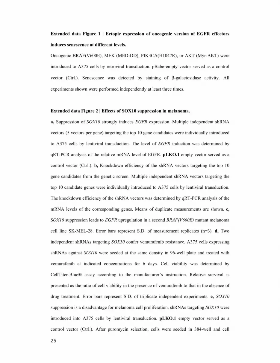
Extended data Figure 1 | Ectopic expression of oncogenic version of EGFR effectors
induces senescence at different levels.
Oncogenic BRAF(V600E), MEK (MED-DD), PIK3CA(H1047R), or AKT (Myr-AKT) were
introduced to A375 cells by retroviral transduction. pBabe-empty vector served as a control
vector (Ctrl.). Senescence was detected by staining of β-galactosidase activity. All
experiments shown were performed independently at least three times.
Extended data Figure 2 | Effects of SOX10 suppression in melanoma.
a, Suppression of SOX10 strongly induces EGFR expression. Multiple independent shRNA
vectors (5 vectors per gene) targeting the top 10 gene candidates were individually introduced
to A375 cells by lentiviral transduction. The level of EGFR induction was determined by
qRT-PCR analysis of the relative mRNA level of EGFR. pLKO.1 empty vector served as a
control vector (Ctrl.). b, Knockdown efficiency of the shRNA vectors targeting the top 10
gene candidates from the genetic screen. Multiple independent shRNA vectors targeting the
top 10 candidate genes were individually introduced to A375 cells by lentiviral transduction.
The knockdown efficiency of the shRNA vectors was determined by qRT-PCR analysis of the
mRNA levels of the corresponding genes. Means of duplicate measurements are shown. c,
SOX10 suppression leads to EGFR upregulation in a second BRAF(V600E) mutant melanoma
cell line SK-MEL-28. Error bars represent S.D. of measurement replicates (n=3). d, Two
independent shRNAs targeting SOX10 confer vemurafenib resistance. A375 cells expressing
shRNAs against SOX10 were seeded at the same density in 96-well plate and treated with
vemurafenib at indicated concentrations for 6 days. Cell viability was determined by
CellTiter-Blue® assay according to the manufacturer’s instruction. Relative survival is
presented as the ratio of cell viability in the presence of vemurafenib to that in the absence of
drug treatment. Error bars represent S.D. of triplicate independent experiments. e, SOX10
suppression is a disadvantage for melanoma cell proliferation. shRNAs targeting SOX10 were
introduced into A375 cells by lentiviral transduction. pLKO.1 empty vector served as a
control vector (Ctrl.). After puromycin selection, cells were seeded in 384-well and cell

26
confluence was measured by IncuCyte imaging system. Error bars represent S.D. of triplicate
independent experiments. f, SOX10 suppression induces senescence. Senescence was detected
by staining of β-galactosidase activity. g, Western blot analysis of RB protein, CDK
inhibitors CDKN1A (p21cip1) and CDKN1B (p27kip1) in SOX10 knockdown A375 cells.
HSP90 served as a loading control. h, Vemurafenib treatment selects for cells that have
higher level of EGFR and lower level of SOX10. A375 cells expressing shRNAs targeting
SOX10 as described above were cultured in the absence or in the presence of 1 μM
vemurafenib for 10 days before the harvest for qRT-PCR analysis. Error bars represent S.D.
of measurement replicates (n=3). All experiments shown except panel a and b were
performed independently at least three times.
Extended data Figure 3 | SOX10 loss and TGFβ activation induce multiple RTKs.
a, EGFR inhibition (gefinitib) is not sufficient to restore vemurafenib sensitivity of SOX10-
loss cells; Targeting PI3K, a common downstream effector of RTKs, with a selective inhibitor
(GDC0941) sensitizes SOX10-loss cells to vemurafenib. shRNAs targeting SOX10 were
introduced into A375 cells by lentiviral transduction. pLKO.1 empty vector served as a
control vector (Ctrl.). Cells were seeded in 6-well plates at the same density in the presence or
absence of drug(s) at indicated concentration. Cells were cultured for 2 weeks in the absence
of vemurafenib or 4 weeks in the presence of vemurafenib before fixing and staining. Figure
2e is shown again as a reference. b, Increased RTKs activation in SOX10-knockdown cells by
long-term vemurafenib treatment. A375 cells infected by shSOX10-1 vector or the PLKO.1
empty vector (Ctrl.) were cultured in the absence or presence of 1 μM vemurafenib for the
indicated number of days and processed with Human Phospho-Receptor Tyrosine Kinase
Array Kit (R&D) according to the manufacturer’s instructions. c, SOX10 knockdown
upregulates both EGFR and PDGFRβQuantification of protein and mRNA were
accomplished by Western blot and qRT-PCR analysis. Error bars represent S.D. of
measurement replicates (n=3). d, Increased RTKs activation in A375 cells by long-term

27
treatment with recombinant TGFβ (200 pM) and vemurafenib (1 μM). A375 cells were
cultured in the presence of vemurafenib (1μM), recombinant TGFβ (200pM) or their
combination for indicated number of days and processed with Human Phospho-Receptor
Tyrosine Kinase Array Kit (R&D) according to the manufacturer’s instructions. All
experiments shown except RTK array analysis were performed independently at least two
times.
Extended data Figure 4 | SOX10 loss activates TGFβ signalling and induces senescence
in WM266-4 cells.
a, SOX10 loss confers vemurafenib resistance in BRAF(V600D) melanoma cell line
WM266-4. Cells expressing empty vector PLKO.1 (Ctrl.) or shRNAs targeting SOX10
transduced by lentivirus were treated with increasing concentrations of vemurafenib for 6
days. Cell viability was determined by CellTiter-Blue® assay according to the instruction of
manufacturer. Relative survival is represented as the ratio of cell viability in the presence of
vemurafenib to that in the absence of drug treatment. Error bars represent S.D. of triplicate
independent experiments. b, SOX10 downregulation leads to growth deficit in WM266-4
cells. Cells expressing the control vector PLKO.1 (Ctrl.) or shRNAs against SOX10 were
seeded at the same density in 96-well plates and cultured for 6 days. Cell viability was
determined by CellTiter-Blue® assay. Error bars represent S.D. of triplicate independent
experiments. c, SOX10 suppression results in EGFR and PDGFRB upregulation in WM266-4
cells. Error bars represent S.D. of measurement replicates (n=3). d, SOX10 loss upregulates
TGFβ receptor and its bona fide target genes. Relative mRNA level of EGFR, PDGFRB,
SOX10, ANGPTL4, TAGLN, CYR61, CTGF, TGFBR2 and JUN were determined by qRT-
PCR analysis. pLKO.1 empty vector served as a control vector (Ctrl.). Error bars represent
S.D. of measurement replicates (n=3). e, SOX10 suppression induces senescence in WM266-4
cells. Senescence was detected by staining of β-galactosidase activity. f, Western blot analysis

28
of RB protein, p-RB (S780), and CDK inhibitor CDKN1B (p27kip1) in SOX10 knockdown
cells. HSP90 served as a loading control. g, Vemurafenib treatment compromises oncogene
induced senescence in SOX10 knockdown cells. WM266-4 cells expressing PLKO.1 (Ctrl.)
or shSOX10-1 were seeded at the same density in 6-well plates and cultured in the absence or
presence of vemurafenib at indicated concentration for 72 hours before the harvest for
western blot analysis. All experiments shown were performed independently at least three
times.
Extended data Figure 5 | SOX10 loss activates TGFβ signalling and induces senescence
in COLO679 cells.
a, SOX10 loss confers vemurafenib resistance in BRAF(V600E) melanoma cell line
COLO679. Cells expressing empty vector PLKO.1 (Ctrl.) or shRNAs targeting SOX10
transduced by lentivirus were treated with increasing concentrations of vemurafenib for 6
days. Cell viability was determined using CellTiter-Blue® according to the instruction of
manufacturer. Relative survival is represented as the ratio of cell viability in the presence of
vemurafenib to that in the absence of drug treatment. Error bars represent S.D. of triplicate
independent experiments. b, SOX10 downregulation leads to growth deficit in COLO679
cells. Cells expressing the control vector PLKO.1 (Ctrl.) or shRNAs targeting SOX10 were
seeded at the same density in 96-well plates and cultured for 6 days. Cell viability was
determined using CellTiter-Blue® assay. Error bars represent S.D. of triplicate independent
experiments. c, SOX10 suppression results in EGFR and PDGFRB upregulation in COLO679
cells. Error bars represent S.D. of measurement replicates (n=3). d, SOX10 loss upregulates
of TGFβ receptor and its bona fide target genes in COLO679 cells. Relative mRNA level of
EGFR, PDGFRB, SOX10, ANGPTL4, TAGLN, CYR61, CTGF, TGFBR2 and JUN were
determined by qRT-PCR analysis. pLKO.1 empty vector served as a control vector (Ctrl.).
Error bars represent S.D. of measurement replicates (n=3). e, SOX10 suppression induces

29
senescence in COLO679 cells. Senescence was detected by staining of β-galactosidase
activity. f, Western blot analysis of RB protein, p-RB (S780) and CDK inhibitor CDKN1B
(p27kip1) in SOX10 knockdown cells. HSP90 served as a loading control. All experiments
shown were performed independently at least three times.
Extended data Figure 6 | EGFR and SOX10 expression are inversely correlated in
melanoma
a, A375 cells infected by two independent non-overlapping shSOX10 vectors or the PLKO.1
empty vector (Ctrl.) were cultured in the absence or presence of 1 μM vemurafenib for the
indicated number of days. The last two samples (labelled in blue) were first treated with 1 μM
vemurafenib for 10 days and subsequently cultured in the absence of vemurafenib for the
indicated number of days. Means of duplicate measurements are shown. b, Inverse
correlation between SOX10 and PDGFRB in panel of human BRAF mutant melanoma cell
lines. Relative gene expression levels of SOX10 and PDGFRB were acquired from Cancer
Cell Line Encyclopedia (CCLE). R stands for Pearson product-moment correlation coefficient.
c, d, Ectopic expression of SOX10 suppresses TGFβ signalling and downregulates EGFR and
PDGFRB in LOXIMVI cell line. SOX10 was introduced to LOXIMVI cells by lentiviral
transduction (SOX10, PLX301-SOX10). PLX301-GFP served as a control vector (Ctrl.).
Protein levels were determined by Western blot analysis and mRNA levels were determined
by qRT-PCR analysis. Error bars represent S.D. of measurement replicates (n=3). e, Ectopic
expression of SOX10 sensitizes LOXIMVI cell to vemurafenib. Cells expressing GFP or
SOX10 transduced by lentivirus were treated with increasing concentrations of vemurafenib
for 6 days. Cell viability was determined using CellTiter-Blue® assay. Relative survival is
represented as the ratio of cell viability in the presence of vemurafenib to that in the absence
of drug treatment. Error bars represent S.D. of triplicate independent experiments. f, SOX10,
EGFR and PDGFRB expression levels in tumour biopsies from patient #3. g, EGFR
expression levels in patient tumour samples (patient #2, #3 and #5), represented as percentage
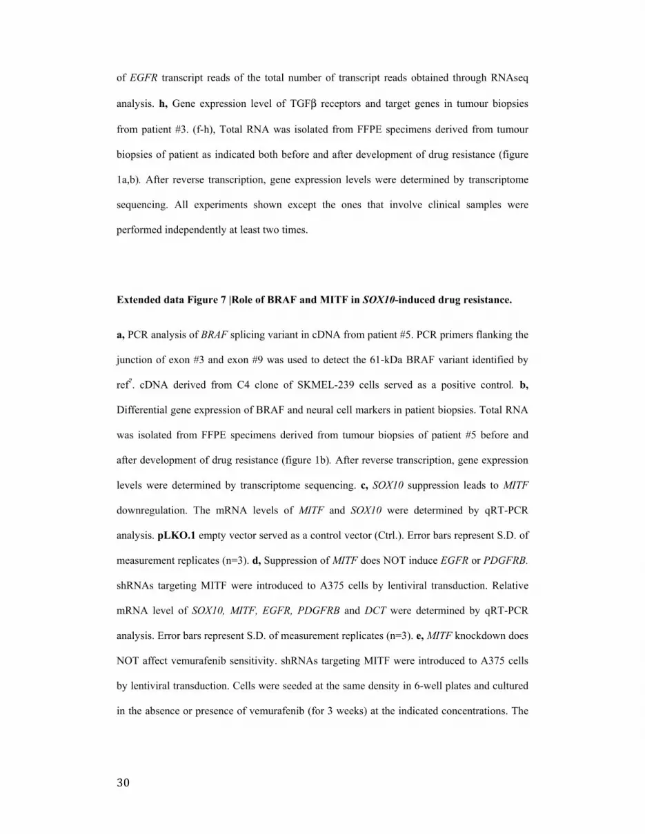
30
of EGFR transcript reads of the total number of transcript reads obtained through RNAseq
analysis. h, Gene expression level of TGFβ receptors and target genes in tumour biopsies
from patient #3. (f-h), Total RNA was isolated from FFPE specimens derived from tumour
biopsies of patient as indicated both before and after development of drug resistance (figure
1a,b). After reverse transcription, gene expression levels were determined by transcriptome
sequencing. All experiments shown except the ones that involve clinical samples were
performed independently at least two times.
Extended data Figure 7 |Role of BRAF and MITF in SOX10-induced drug resistance.
a, PCR analysis of BRAF splicing variant in cDNA from patient #5. PCR primers flanking the
junction of exon #3 and exon #9 was used to detect the 61-kDa BRAF variant identified by
ref7. cDNA derived from C4 clone of SKMEL-239 cells served as a positive control. b,
Differential gene expression of BRAF and neural cell markers in patient biopsies. Total RNA
was isolated from FFPE specimens derived from tumour biopsies of patient #5 before and
after development of drug resistance (figure 1b). After reverse transcription, gene expression
levels were determined by transcriptome sequencing. c, SOX10 suppression leads to MITF
downregulation. The mRNA levels of MITF and SOX10 were determined by qRT-PCR
analysis. pLKO.1 empty vector served as a control vector (Ctrl.). Error bars represent S.D. of
measurement replicates (n=3). d, Suppression of MITF does NOT induce EGFR or PDGFRB.
shRNAs targeting MITF were introduced to A375 cells by lentiviral transduction. Relative
mRNA level of SOX10, MITF, EGFR, PDGFRB and DCT were determined by qRT-PCR
analysis. Error bars represent S.D. of measurement replicates (n=3). e, MITF knockdown does
NOT affect vemurafenib sensitivity. shRNAs targeting MITF were introduced to A375 cells
by lentiviral transduction. Cells were seeded at the same density in 6-well plates and cultured
in the absence or presence of vemurafenib (for 3 weeks) at the indicated concentrations. The

31
cells were fixed, stained and photographed. All experiments shown except the ones that
involve clinical samples were performed independently at least two times.

0
100
200
300
400
500
0 5 10 15 20 % C
hang
e tu
mor
vol
ume
(Mea
n ±
SE
M)
Days since treatment start
A375-Ctrl. and A375-EGFR xenografts
Ctrl.-Vehicle Empty-Trametinib Ctrl.-Trametinib EGFR-Trametinib
a pre-treatment post-vemurafenib Patient #1 Patient #2
Patient #3
b
pre-treatment post-vemurafenib
pre-treatment post-dabrafenib Patient #6 pre-treatment post-vemurafenib
A375-Ctrl.
A375-EGFR
0 12 25 50 EGF (µg/ml) c
e
EGFR
RB
p27
p21
HSP90
A375
f A375-Ctrl.
A375-EGFR
d
pre-treatment post-dabrafenib Patient #4
Patient #5 pre-treatment post-trametinib
* *

EGFR
SOX10
HSP90
A375
a Chromatin regulator shRNA library
Vemurafenib selection
A375
EGFRhigh
EGFR
Untreated control
Sort EGFRhgh cells
Enriched shRNA
Deep-sequencing
b
DA
PI
- Chr Lib + Vemurafenib
+ Chr Lib + Vemurafenib
EGFRlow 73.5%
EGFRhigh 0.1%
EGFRlow 43.5%
EGFRhigh 18.1%
- Chr Lib - Vemurafenib
EGFRlow 85.4%
EGFRhigh 0.1%
+ Chr Lib - Vemurafenib
EGFRlow 82.9%
EGFRhigh 0.1%
DA
PI
Ctrl.
shSOX10-1
0 0.5 1 2
shSOX10-2
Vemurafenib (µM)
A375
e
0 0.2 0.4 0.6 0.8
1 1.2
Rel
ativ
e m
RN
A le
vel SOX10
Ctrl. shSOX10-1 shSOX10-2
0 1 2 3 4 5 6
Rel
ativ
e m
RN
A le
vel EGFR
A375
c
e
EGFR
0
1
2
3
4
5
6
Rel
ativ
e m
RN
A le
vel
SOX10 EGFR
d
A375
0.02
0.06
0.25
1.00
0.01 0.1 1 10
Rel
ativ
e su
rviv
al
Vemurafeinib (µM)
Ctrl. shSOX10-1 shSOX10-2
A375
d
Ctrl.
shSOX10-1
0 0.5 1 2
shSOX10-2
Vemurafenib (µM)
A375

0
1
2
3
4
5
6
Rel
ativ
e m
RN
A le
vel EGFR
PDGFRB
0
1
2
3
4
Untreated TGF-β1
Rel
ativ
e m
RN
A le
vel
EGFR PDGFRB
0
1
2
3
4
5
Rel
ativ
e m
RN
A le
vel
Ctrl. shSOX10-1 shSOX10-2
e
a
TGFBR2
p-c-JUN
c-JUN
HSP90
SOX10
A375
c
A375
b
A375
A375
Untreated
TGF-β1 (200pM)
f
A375
+ vemurafenib (2µM)
50 25 0 100 TGF-β1 (pM)
TGFBR2
HSP90
SOX10
shTGFBR2-1 shTGFBR2-2
EGFR
shSOX10 - + - + - + - - + + - - - - - - + +
A375
h
PDGFRβ
i
EGFR
p-c-JUN
TGFBR2
HSP90
c-JUN
A375
PDGFRβ
g
shTGFBR2-1 shTGFBR2-2
shSOX10 - + - + - + - - + + - - - - - - + +
A375
EGFR
p-c-JUN
HSP90
c-JUN
A375
d
PDGFRβ

0
1
2
3
4
5
6
7
EG
FR
PD
GFR
B
SO
X10
TG
FBR
3 TG
FBR
2 C
TGF
TAG
LN
CY
R61
JU
N
Rel
ativ
e m
RN
A le
vel
Paired tumor biopsies Patient #5
Pre-treatment
post-trametinib
a
2
4
6
8
10
12
4 6 8 Rel
ativ
e ge
ne e
xpre
ssio
n le
vel o
f S
OX
10
Relative gene expression level of EGFR
b
R= -0.72
EGFR
Polyclonal shSOX10-1
cell population
Ctrl. shSOX10
+vemurafenib
- vemurafenib
Time (day)
0
14
7
A375
0
1
2
3
4
5
6
7
EG
FR
PD
GFR
B
SO
X10
TG
FBR
3 TG
FBR
2 C
TGF
TAG
LN
CY
R61
JU
N
Rel
ativ
e m
RN
A le
vel
Paired tumor biopsies Patient #2
Pre-treatment post-vemurafenib
c
0
1
2
3
4
5
6
7
EG
FR
PD
GFR
B
SO
X10
TG
FBR
3 TG
FBR
2 C
TGF
TAG
LN
CY
R61
JU
N
Rel
ativ
e m
RN
A le
vel
Paired tumor biopsies Patient #6
Pre-treatment Post-vemurafenib
d
Drug
RAS
Drug response
RTK-1 RTK-1 RTK-2
Drug resistant
RTK-1 RTK-2
Senescence
RTK-1
Drug response
RAS RAS RAS
BRAF* BRAF* BRAF*
MEK
Drug
MEK MEK MEK
RAS
BRAF*
RTK-1
MEK
Proliferation
Drug resistance development Drug holiday Re-treat after drug holiday
Drug BRAF*


