Embed Size (px)
Citation preview

RhoA Regulates Peroxisome Association to Microtubulesand the Actin CytoskeletonLukas Schollenberger1., Thomas Gronemeyer2,3., Christoph M. Huber1{, Dorothee Lay1¤a, Sebastian
Wiese2¤b, Helmut E. Meyer2, Bettina Warscheid2¤b, Rainer Saffrich4, Johan Peranen5, Karin Gorgas6,
Wilhelm W. Just1*
1 Heidelberg Center of Biochemistry, University of Heidelberg, Heidelberg, Germany, 2 Medical Proteom-Center, University of Bochum, Bochum, Germany, 3 Department
for Molecular Genetics and Cell Biology, University of Ulm, Ulm, Germany, 4 Department of Internal Medicine V, University of Heidelberg, Heidelberg, Germany, 5 Institute
of Biotechnology, University of Helsinki, Finland, 6 Department of Anatomy and Medical Cell Biology, University of Heidelberg, Heidelberg, Germany
Abstract
The current view of peroxisome inheritance provides for the formation of new peroxisomes by both budding from theendoplasmic reticulum and autonomous division. Here we investigate peroxisome-cytoskeleton interactions and show byproteomics, biochemical and immunofluorescence analyses that actin, non-muscle myosin IIA (NMM IIA), RhoA, Rho kinase II(ROCKII) and Rab8 associate with peroxisomes. Our data provide evidence that (i) RhoA in its inactive state, maintained forexample by C. botulinum toxin exoenzyme C3, dissociates from peroxisomes enabling microtubule-based peroxisomalmovements and (ii) dominant-active RhoA targets to peroxisomes, uncouples the organelles from microtubules and favorsRho kinase recruitment to peroxisomes. We suggest that ROCKII activates NMM IIA mediating local peroxisomalconstrictions. Although our understanding of peroxisome-cytoskeleton interactions is still incomplete, a picture is emergingdemonstrating alternate RhoA-dependent association of peroxisomes to the microtubular and actin cytoskeleton. Whereasassociation of peroxisomes to microtubules clearly serves bidirectional, long-range saltatory movements, peroxisome-acto-myosin interactions may support biogenetic functions balancing peroxisome size, shape, number, and clustering.
Citation: Schollenberger L, Gronemeyer T, Huber CM, Lay D, Wiese S, et al. (2010) RhoA Regulates Peroxisome Association to Microtubules and the ActinCytoskeleton. PLoS ONE 5(11): e13886. doi:10.1371/journal.pone.0013886
Editor: Neil A. Hotchin, University of Birmingham, United Kingdom
Received June 16, 2010; Accepted October 18, 2010; Published November 8, 2010
Copyright: � 2010 Schollenberger et al. This is an open-access article distributed under the terms of the Creative Commons Attribution License, which permitsunrestricted use, distribution, and reproduction in any medium, provided the original author and source are credited.
Funding: This work was supported by the FP6 European Union Project ‘‘Peroxisomes in health and disease’’ (LSGH-CT-2004-512018) as well as by funds from theDeutsche Forschungsgemeinschaft (SFB 642). The funders had no role in study design, data collection and analysis, decision to publish, or preparation of themanuscript.
Competing Interests: The authors have declared that no competing interests exist.
* E-mail: [email protected]
. These authors contributed equally to this work.
¤a Current address: Lonza Ltd., Visp, Switzerland¤b Current address: Institute of Biology II/Center for Biological Signalling Systems, Albert-Ludwigs-University Freiburg, Freiburg, Germany
{ Deceased
Introduction
Mammalian peroxisomes associate with the microtubular
cytoskeleton for intracellular transport [1,2]. Three distinct states
of motility were recognized, long-range saltations, oscillations and
arrest. Furthermore, we demonstrated that peroxisome motility is
subject to regulation by extracellular ATP-lysophosphatidic acid
(LPA) receptor co-stimulation. Signaling involves trimeric Gi/Go
protein, PLC, Ca2+ influx, cPKC, MAP kinase and PLA2 and
mediates peroxisomal arrest [3–5]. Via G12/13 the LPA receptor
activates the Rho pathway preventing long-range peroxisomal
motility that seems to be regulated by a complex signaling network
[4,6].
These studies did not show the involvement of the actin
cytoskeleton in the motility of mammalian peroxisomes. However,
plant and yeast peroxisomes associate with the actin cytoskeleton
for saltatory movement and inheritance, respectively. In plant cells
interference with the actin cytoskeleton results in loss of saltatory
movement, aggregation, and complete cessation of peroxisome
motility [7]. In S.cerevisiae peroxisomes are targeted and segregated
to the developing bud by a highly ordered process involving actin
and Myo2p, a class V myosin motor protein [8,9]. Actin-based
movement of mammalian organelles other than peroxisomes has
however been demonstrated [10,11]. Two types of motility are
described. One is based on actin polymerization itself propelling
organelles by an ‘‘actin comet tail’’ toward the cell center. This
type has been described for phagosomes and macropinosomes and
may also be involved in transport between endosomes and
lysosomes [12,13]. The other type of movement depends on
actin-based myosin motors and utilizes myosins of the classes I, II,
V or VI. This myosin-dependent movement has been implicated
in dynamics of the ER, lysosomes, Golgi-derived vesicles, secretory
granules, recycling endosomes and melanosomes [14–16].
Some indirect evidence suggests that mammalian peroxisomes
also associate with the actin cytoskeleton. Drp-1 known to be
involved in mitochondrial fission in an actin cytoskeleton-
dependent manner [17] was shown also to localize to peroxisomes.
Over-expression of a dominant-negative mutant of Drp-1 prevents
peroxisome division in a human hepatoma cell line [18–20].
Generally, members of the dynamin protein family are known
PLoS ONE | www.plosone.org 1 November 2010 | Volume 5 | Issue 11 | e13886

regulators of vesicle trafficking implicated in constricting and
severing membrane tubules [21].
Ultrastructural studies on peroxisome proliferation in rat liver
suggest a sequential mechanism of membrane tubulation and
constriction prior to fission [22,23]. Thus, a force-generating
system such as the acto-myosin complex is likely to be involved.
Frequently, these processes are regulated by small GTPases known
to support membrane traffic as well as the local organization of
both the microtubular and the actin cytoskeleton [10,24].
Members of the Rho, Rab and Sar1/Arf families are implicated
[24,25] and Rho1p and Arf1 recently shown to associate with
peroxisomes [4,6,22,23,26,27]. Mammalian peroxisomes in pres-
ence of GTP-primed cytosol recruit Arf1 and the COPI coat
subunits and among the proteins specifically enriched with S.
cerevisiae peroxisomes Rho1p was identified by a mass spectromet-
ric screen.
So far our knowledge on the mechanism of peroxisome
proliferation and the role small GTPases and the cytoskeleton
play is rather incomplete. Peroxins, such as Pex11p, Pex25p and
Pex27p are likely to be involved [28–30]. However, factors are still
missing in order to fully understand the process. In the present
study we initiated proteomics analyses to identify components that
recruit onto peroxisomes from the cytosol. We found a significant
number of candidate proteins some of which were further
investigated. Here we report alternate binding of peroxisomes to
microtubules and actin microfilaments dependent on the activa-
tion state of RhoA. Our studies indicate a sequence of events in
which activated RhoA by binding to peroxisomes detaches the
organelles from microtubules. Concomitantly, activated RhoA
may favor peroxisome association with the actin cytoskeleton
possibly by recruiting Rho kinase (ROCKII). Phosphorylation of
myosin light chain induces actin-activated non-muscle myosin IIA
(NMM IIA) ATPase activity enabling force generation.
Results
Peroxisome-microtubule associationIntracellular movement of peroxisomes along microtubular
tracks is regulated by ATP/LPA receptor co-stimulation and
activation of heterotrimeric G proteins. Signaling by both
receptor co-stimulation and activation of G proteins caused
peroxisomal arrest [2–4]. The first clues for a possible
involvement of RhoA in this regulation came from LPA signaling
that is known to activate RhoA by the trimeric G protein
subfamily member G12/13 [31,32]. Therefore, we first tested C.
botulinum exoenzyme C3 that is known to inactivate Rho proteins
of the RhoA subfamily by ADP ribosylation [33]. A significant
increase in overall peroxisomal movements compared to
untreated cells was observed independent of whether the toxin
was added to the culture medium or expressed by transfection
(Fig. 1A). As shown by the time series, most peroxisomes moved
directionally with frequent changes in direction. Those organelles
that did not saltate were at rest for short periods. Following the
addition of Nocodazole to exoenzyme C3-treated cells, peroxi-
somes oscillated, similarly to what could be observed after
Nocodazole treatment only (not shown). Both resting and
saltating states presupposed association of the organelles to the
microtubular cytoskeleton. The motility state elicited by exoen-
zyme C3 was in sharp contrast to the control state, as the
frequency of peroxisomes moving longer distances was signifi-
cantly increased. On the other hand, transfecting cells with myc-
G14V-RhoA, the constitutively active form of RhoA exhibiting
remarkably reduced intrinsic GTPase activity and unresponsive-
ness to RhoA GTPase-activating protein [34], resulted in random
oscillations of peroxisomes that were indistinguishable from those
observed after Nocodazole treatment (Fig. 1B). This regulatory
effect was specific for RhoA. Dominant–active RhoD did not
influence peroxisomal motility (Fig. 1B).
By electron microscopy peroxisomal constrictions were fre-
quently seen preserving membrane continuities between segregat-
ed compartments (Fig. 2A–C). Both microtubules (Fig. 2B and C)
and microfilament bundles (Fig. 2D–F) were regularly found in
close proximity to the organelles (Fig. 2B and C) and smooth ER
cisternae possibly delivering Ca2+ positioned close to the sites of
constrictions. Taken together, these results suggest that peroxi-
somes contact both microtubules and cytoskeletal filaments.
Activated RhoA may uncouple peroxisomes from microtubules
as typical microtubule-dependent long-range saltations are no
longer observed.
Peroxisome association to myosin and the actincytoskeleton
Following incubation of unlabeled rat liver peroxisomes with35S-labeled rat hepatocyte cytosol, proteins with Mr of about 220,
60 and 40 kDa strikingly co-isolated with peroxisomes (not shown).
These proteins were identified by nano-HPLC/ESI-MS/MS as
non-muscle myosin heavy chain (MyH9) belonging to the class IIA
of conventional myosins, a- and b-tubulin and b-actin, respec-
tively. Additional proteins specifically recruiting from cytosol onto
peroxisome membranes were detected by extended proteomics
analysis. Table 1 lists the proteins identified and related to
organelle dynamics and cytoskeletal functions.
To confirm specific peroxisomal recruitment of these proteins,
an in vitro peroxisome-binding assay was developed. In this assay
peroxisomes were incubated with GMP-PNP-primed cytosol in
presence or absence of an ATP-regenerating system and recovered
after flotation in a discontinuous Nycodenz gradient. Pex11ap, a
peroxisomal membrane marker, floated up in the gradient to
fraction F2 just between F1 (250 mM sucrose) and F3 (45% w/v
Nycodenz) the latter retaining all the non-floating material. Using
antibodies against b-actin and NMM IIA heavy chain, both
myosin and actin were detected in the re-isolated peroxisomes.
Binding of NMM IIA to peroxisomes required ATP and was
observed in the presence of ATP plus GMP-PNP. However, actin
recruitment required both nucleotides; the sole presence of GMP-
PNP did not mediate recruitment of NMM or actin (Fig. 3A). In
control experiments ran in the absence of peroxisomes neither
NMM nor actin did float up (Fig. 3B, D, and F). Cytosol
preparations contain endogenous guanine nucleotides that in
presence of an ATP-regenerating system might become phos-
phorylated to GTP by nucleotide kinases. Therefore, the
nucleotide dependence of NMM IIA and actin recruitment was
investigated in presence of GDPcS, a metabolically inert GDP
analog. These studies clearly demonstrated that binding of NMM
IIA and actin onto peroxisomes requires both ATP and GTP
(Fig. 3C). Replacing the ATP-regenerating system for AMP-PNP,
a non-hydrolyzable ATP analog, prevented NMM IIA association
to peroxisomes suggesting that the association requires hydrolyz-
able ATP (Fig. 3E).
Peroxisomal NMM IIA association was also proved by
immunofluorescence. NMM IIA predominantly appeared as
filamentous structures bearing peroxisomes closely attached like
pearls on a string (Fig. 4A–C and inset in Fig. 4C) rather than
having them distributed at random. Analyzing NMM IIA and
actin filament stainings revealed nearly complete overlap of both
of these structures (Fig. 4D–F). Thus, these immunofluorescence
studies strongly suggest peroxisome acto-myosin associations.
Peroxisome Interactions
PLoS ONE | www.plosone.org 2 November 2010 | Volume 5 | Issue 11 | e13886

RhoA and its effector ROCKII localize to peroxisomesThe GTP dependence of NMM IIA and actin recruitment onto
peroxisomes indicate small GTP-binding protein(s) to be involved.
Using antibodies specific to RhoA, the GTP-dependent recruit-
ment of RhoA to peroxisomes was observed in vitro (Fig. 5). In line
with these biochemical assays, we also demonstrated co-localiza-
tion of peroxisomes and RhoA by transfecting myc-G14VRhoA, a
dominant-active mutant of RhoA, into mouse AT3 hepatoma cells
(Fig. 6). Whereas both wild type and dominant-active RhoA
associated to peroxisomes (Fig. 6A–F), Rho A was not detected on
the organelles upon treatment of cell cultures with exoenzyme C3
(Fig. 6G, H, I). By closer examination of the RhoA topology, it
appeared that activated RhoA concentrated at distinct sites on the
peroxisomal membrane rather than exhibiting a continuous
staining pattern (Fig. 6F and inset). In contrast to the RhoA
staining, catalase, a peroxisomal matrix protein, constantly labeled
the entire organelle. This localization of RhoA to discrete domains
of the peroxisomal membrane is reminiscent to the distribution of
Pex11ap and PMP69 in CHO cells overexpressing N-terminally
myc-tagged Pex11bp [22]. As Pex11bp expression was shown to
cause peroxisome proliferation [19], it is tempting to speculate that
segregation and concentration of distinct proteins within the
peroxisomal membrane and peroxisome proliferation are func-
tionally linked to each other.
Figure 1. Particle-tracking analysis of GFP-SKL-marked peroxisomes in CHO cells. (A) Treatment of cells with C. botulinum toxinexoenzyme C3 (5 mg/ml, 24 h prior to analysis) known to inactivate RhoA significantly increases the number of peroxisomes moving long distances.(B) In cells expressing dominant-active V14-RhoA long-range movement is abolished and peroxisomal motility is indistinguishable to that in cellstreated with nocodazole to depolymerize microtubules. Expression of dominant-active V26-RhoD does not show this effect. For clarity reasons thedisplacement graph of control peroxisomes expressing empty vector is omitted, as it is virtually identical to the control graph in (A).doi:10.1371/journal.pone.0013886.g001
Peroxisome Interactions
PLoS ONE | www.plosone.org 3 November 2010 | Volume 5 | Issue 11 | e13886

A known effector of RhoA signaling is Rho-associated coiled-
coil containing kinase (ROCK). By phosphorylation of myosin
light chain (MLC) Rho kinase induces formation of stress fibers
and focal adhesions [35]. Two Rho kinase isoforms have been
identified, ROCKI and ROCKII. Whereas ROCKI was recently
shown to localize to plasma membrane and centrosomes [36,37],
ROCKII appears to be mainly distributed in the cytoplasm and
translocates to membranes upon activation by GTP-bound RhoA
[38]. Therefore, we focused our attention on ROCKII. Again we
used the proxisomal binding assay and observed a significant 3-4-
fold increase in the amount of ROCKII recruited from cytosol
onto peroxisomes in the presence of ATP plus GMP-PNP (Fig. 7).
Interestingly, isolated peroxisomes without being further incubated
already had bound ROCKII in low concentration (Fig. 7A and C,
lane 6).
We also analyzed peroxisomal attachment of ROCKII by
immunofluorescence. Peroxisomes were labeled by GFP carrying a
C-terminal peroxisomal targeting signal 1 (GFP-SKL) and
endogenous ROCKII was stained using a rabbit polyclonal
antiserum (Fig. 8). According to the known broad cellular
distribution of ROCKII, cellular background staining was high.
Nevertheless, co-localization of ROCKII with the peroxisomal
signal was convincingly evident. Quantitative evaluation of 256
GFP-labeled peroxisomes revealed 66 peroxisomes to co-localize
with the ROCKII signal, i.e. more than 25% co-localizing
structures were recognized.
Rab8 recruits onto peroxisomesAs membrane docking of myosin motors is known to be
controlled by Rab proteins [10,39,40], we next investigated by an
Figure 2. Ultrastructural analysis of peroxisome proliferation in rat liver treated with clofibrate and thyroxin. Peroxisomalconstrictions are frequently seen in peroxisomes (P) of different sizes (black arrows in A and F) as well as in peroxisomes associated to microtubulartracks (black arrowheads in B) that sometimes localize close to the constrictions (black arrowhead in C). Filament bundles are running alongperoxisomes and close to peroxisomal constrictions (white arrows in D–F) suggesting peroxisome attachment to cytoskeletal filaments. Frequently,cisternae of smooth endoplasmic reticulum (white arrowheads in A–C) are positioned next to the constrictions. M, mitochondrium. The barsrepresent 200 nm in A, B, D–F and 100 nm in C.doi:10.1371/journal.pone.0013886.g002
Table 1. Cytoskeleton and cytoskeleton-related proteins associated with peroxisomes, identified by nano-HPLC/ESI-MS/MS.
Protein gi accessionMW[kDa] Sequence coverage [%]
Myosin,heavy polypeptide 9, non-muscle [Rattus norvegicus] 149066032 226,2166 6,70
Tubulin, alpha 1A [Rattus norvegicus] 11560133 50,1036 18,60
Tubulin, beta 2a [Rattus norvegicus] 157819845 49,9209 26,10
Tubulin, alpha 4A [Rattus norvegicus] 55741524 49,8924 13,40
Tubulin, alpha 6 [Rattus norvegicus] 58865558 49,8775 36,30
Tubulin, beta 2b [Rattus norvegicus] 110347600 49,8750 22,70
ATRTC actin beta [Rattus norvegicus] 71620 41,7273 40,50
Actin, beta [Rattus norvegicus] 13592133 41,7000 41,00
Cofilin 1 [Rattus norvegicus] 8393101 18,4280 19,60
14-3-3 zeta/delta [Rattus norvegicus] 62990183 27,7366 10,60
doi:10.1371/journal.pone.0013886.t001
Peroxisome Interactions
PLoS ONE | www.plosone.org 4 November 2010 | Volume 5 | Issue 11 | e13886

additional proteomics screen the recruitment of small GTPases to
the peroxisomal membrane focusing on membrane-associated
proteins with Mr between 15 and 30 kDa. We detected members
of the Rab family and the Rho family as well as ARF, ARF-related
and SAR1 proteins. To increase the stringency of our analysis, the
Rab proteins were semi-quantitatively profiled based on spectral
counts [41] over the entire Nycodenz density gradient separating
the post-nuclear fraction of a rat liver homogenate. In this
profiling, Rab8a and Rab8b exhibited a gradient distribution
strikingly similar to the peroxisomal matrix and membrane
markers, bifunctional protein and Pex11ap, respectively (Fig. 9).
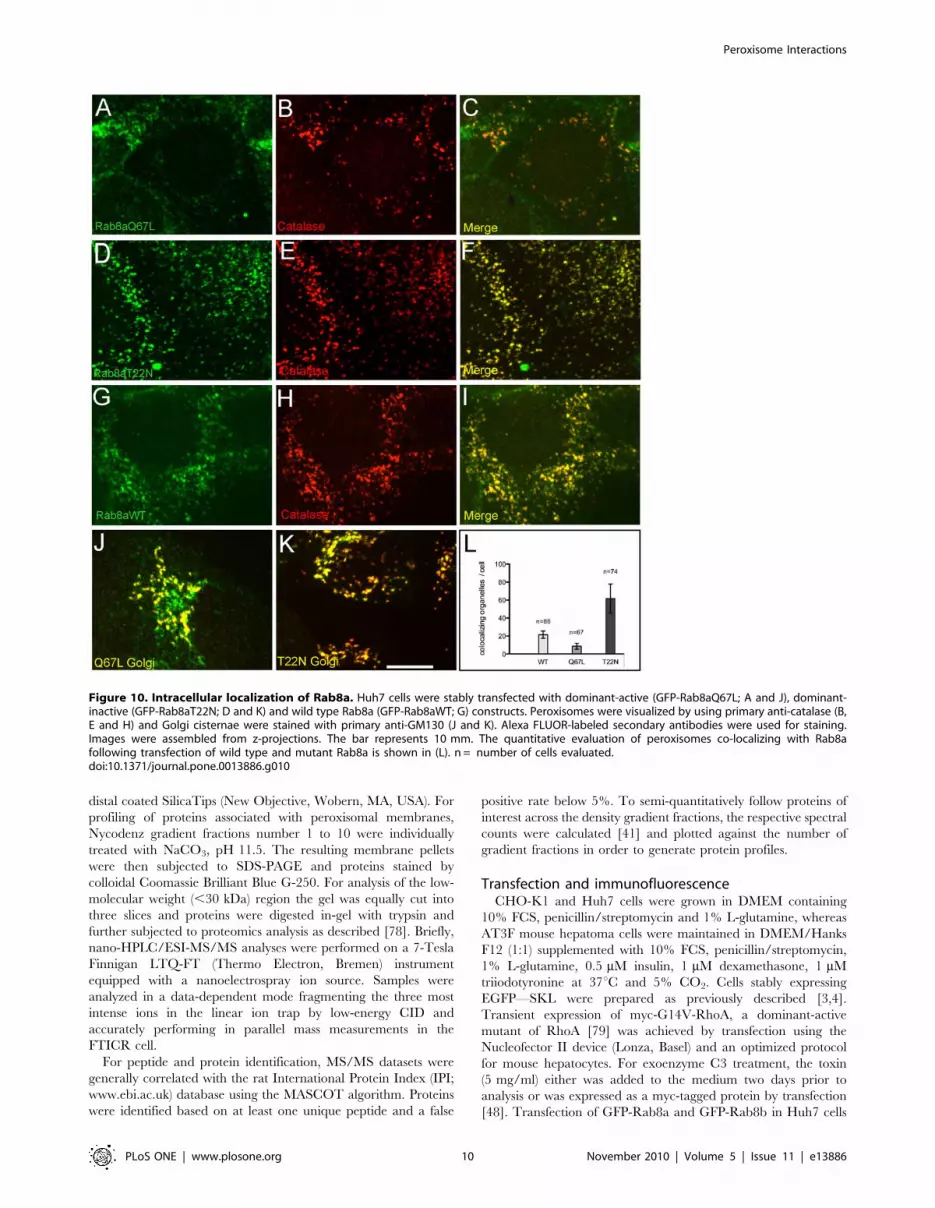
To further investigate the peroxisomal localization of Rab8,
Rab8a was cloned as 59-GFP fusion construct and dominant-
active (Q67L) and dominant-inactive (T22N) forms were stably
expressed in Huh7 human hepatocellular carcinoma cells. For co-
localization studies, peroxisomes and Golgi were stained with anti-
catalase and anti-GM130 antibodies, respectively. The results of
these experiments are shown in Fig. 10. (i) Rab8a clearly co-
localizes with peroxisomes (Fig. 10A–I) and Golgi cisternae
(Fig. 10J and K). (ii) The total average number of peroxisomes
per cell changed by expressing wild type GFP-Rab8aWT (5467;
n = 115; Fig. 10G–I), dominant-active Rab8aQ67L (3566;
n = 119; Fig. 10A–C) and dominant-inactive Rab8aT22N
(68615; n = 79; Fig. 10D–F). (iii) Applying organelle-based co-
localization (OBCOL) analysis [42], we noted an about tenfold
increase in the number of peroxisomes co-localizing with Rab8a in
cells transfected with dominant-inactive Rab8aT22N compared
with dominant-active Rab8aQ67L (Fig. 10L).
Discussion
RhoA signaling regulates peroxisome motilityOur current view of the multiple signaling pathways regulating
peroxisome microtubule interaction encompasses both activation
of PLA2 via Gi/Go liberating arachidonic acid by ATP and RhoA
via G12/13 by extracellular LPA [3,4,31]. Both signals synergize in
this process and RhoA appears to be a specific and important
intermediate. Most of the activated cytosolic RhoA translocates to
cellular membranes leading to saturation of RhoA binding sites
and terminating microtubule-peroxisome interactions. Peroxisome
oscillations are generated indistinguishable to those following
depolymerization of microtubules [43,44]. On the other hand,
exoenzyme C3 rendering RhoA in its inactive GDP-bound form
strikingly increased the number of long-range microtubule-based
peroxisome saltations. These observations are consistent with the
idea that cycling of RhoA between its GDP- and GTP-bound
states promotes attachment/detachment cycles of peroxisomes
from microtubules. Similar attachment/detachment cycles were
reported to affect the motility of pigment granules, Golgi vesicles,
lysosomes and mitochondria [45,46].
Peroxisome association with the actin cytoskeletonIn previous work evidence has been provided that the actin
cytoskeleton obviously is not involved in supporting peroxisome
motility. However, a regulated short-range motor protein-based
system may be advantageous, as it provides a mechanism tethering
organelles at actin-rich intracellular sites and/or exerting me-
chanical forces [11]. Although the actual mechanism facilitating
peroxisome acto-myosin interaction is not known, binding of
RhoA-GTP to the peroxisomal membrane may trigger this process
possibly by activating NMM IIA. By uncoupling peroxisomes from
microtubules, RhoA-GTP may concomitantly activate ROCKII, a
serine/threonine kinase acting immediately downstream of RhoA.
ROCKII is known to be involved in reorganizing actin structures
preventing actin depolymerization. By phosphorylation of the
myosin-binding subunit of myosin phosphatase and/or the myosin
light chain of myosin II, ROCKII enhances myosin ATPase
Figure 3. ATP- and GTP-dependent binding of NMM IIA heavychain and b-actin to isolated rat liver peroxisomes. Organelleswere incubated as indicated and floated up in a Nycodenz gradient.Three fractions were recovered from top to bottom (F1–F3). Binding isvisualized by immunoblotting (A and C). ATP must be hydrolyzable forbinding (E). Peroxisomes are localized in the gradient by theperoxisomal marker Pex11ap. Control incubations lacking peroxisomesdo not show floatation of NMM IIA and b-actin (B, D, F) suggestingperoxisomal membranes to be required for binding.doi:10.1371/journal.pone.0013886.g003
Peroxisome Interactions
PLoS ONE | www.plosone.org 5 November 2010 | Volume 5 | Issue 11 | e13886

activity [47,48]. Moreover, ROCKII contains a well-defined
pleckstrin homology domain binding to PtdIns(4,5)P2 the synthesis
of which in the peroxisomal membrane was recently reported [49].
Myosin might also act in cargo protein assembly at donor
membranes, as recently suggested for the biogenesis of melanosomes
[50,51]. Scaffolding of melanosome proteins requires coat and adaptor
proteins in addition to myosin. Similar processes may also occur in the
peroxisomal system where different to catalase N-myc-tagged Pex11bp
[22] and dominant-active RhoA (Fig. 6) both strikingly non-
homogenously localize to peroxisomes. Interaction of Pex11bp and
RhoA in the peroxisomal membrane and its possible downstream
effects on peroxisome biogenesis remain to be established.
Peroxisomal localization of Rab8Two Rab8 isotypes have been described, Rab8a and Rab8b. They
show distinct tissue distributions. Whereas endogenous Rab8a is
predominantly expressed in lung, kidney, skeletal muscle and liver,
Rab8b is mainly found in testis, spleen, brain, and heart [52]. The
high sequence identity of 83% of both genes might explain why
exogenous expression of both isotypes in Huh7 cells show the same
subcellular localization and develop rather similar phenotypes. In liver
Rab8a might be the predominantly expressed endogenous isotype.
So far Rab8 has been implicated in a variety of functions
including constitutive biosynthetic trafficking from the trans-Golgi
network and the recycling endosome [53,54], sorting of apical
membrane proteins [55], constitutive and regulated melanosome
traffic [56] and regulated ACTH secretion by interacting with
TRIP8b [57]. TRIP8b belongs to the tetratricopeptide domain
proteins with homology to the peroxisomal targeting signal 1 (PTS1)
receptor protein Pex5p known to be involved in peroxisomal matrix
protein import [58–60]. In vesicle and organelle motility Rab
GTPases were implicated in the association of actin motors with the
corresponding organellar membranes. Examples are Rab27a and its
associated protein MyRIP that mediate melanosome transport by
cellular activation of MyoVa and MyoVIIa [61] and Rab11a
involved in endosome recycling by linking the endocytic vesicles via
Rab11FIP2 to myosin Vb [62] as well as Rab8-activated optineurin
recruiting MyoVI [63]. There are still other examples of direct and
indirect motor protein effectors of Rab GTPases [54] elucidating
multiple ways by which these proteins assure correct transport and
vesicle/motor protein interaction.
ConclusionOur finding of NMM IIA recruiting onto peroxisomes might be
interpreted as actin-dependent intracellular peroxisome transport.
However, previous studies addressing this topic did not reveal
actin-dependent peroxisome motility [1,4]. Rather we believe that
Figure 4. Localization of peroxisomes to acto-myosin complexes in wild type CHO cells. Most peroxisomes visualized by the membranemarker PMP69 (red) are found attached to NMM IIA-associated acto-myosin filaments (A–C). In many cases a number of peroxisomes is aligned alongsingle filaments (see inset) suggesting that peroxisome distribution within the cell is not at random but may be determined by the organelle9sassociation to acto-myosin filaments (D–F). Images are reproduced from single z-layers. The bar represents 10 mm.doi:10.1371/journal.pone.0013886.g004
Figure 5. GTP-dependent recruitment of RhoA onto isolatedrat liver peroxisomes. No recruitment is seen in the presence of ATPalone (A) and no RhoA is floating up, if peroxisomes are omitted fromthe incubations (B). Note that without membranes more RhoAprecipitates during incubation escaping analysis.doi:10.1371/journal.pone.0013886.g005
Peroxisome Interactions
PLoS ONE | www.plosone.org 6 November 2010 | Volume 5 | Issue 11 | e13886

peroxisomal NMM IIA might locally act in force generation and/
or contractile function similar as for example observed in
cytokinesis and contractile ring formation [64,65]. The GTP
dependence of this recruitment suggested the involvement of a
GTPase. So far several small GTPases were related to peroxisome
biogenesis and function. Among these are Rho1p [27], Arf1 and
Arf3 in S. cerevisiae [26] and mammalian RhoA, Arf1 and Rab8b
[4,26,57,58]. In addition, various Rho-GTPases have been
implicated in intracellular vesicle trafficking. These include
Cdc42 that inhibits recruitment of the microtubular motor dynein
to COP I-coated Golgi vesicles [66] and RhoA and Rac affecting
both clathrin-dependent [67] and clathrin-independent [68]
endocytosis. The latter process was recently reported to involve
Arf family GTPases that might be linked to the Rho family via
GIT proteins, Arfaptins or ARAPs [69]. Interestingly, mammalian
peroxisomes were shown to bind Arf1 and the COP I coat, and in
S.cerevisiae oleate-induced peroxisome proliferation depends on
Arf1 and is strikingly stimulated by deleting Arf3 the ortholog of
mammalian Arf6 [26].
In this context the role of Rab8 recruitment to peroxisomes is
less clear. Overlap of the Rab8 function with that of Arf6 was
reported to occur in the formation of cell protrusions, a process
powered by actin polymerization [70,71]. As both dominant-active
and -inactive Rab8 co-localize with peroxisomes and the
dominant-inactive form goes along with an increase in peroxisome
number, Rab8 seems to occupy a key position on the peroxisomal
membrane regulating proliferation.
Materials and Methods
Reagents and antibodiesATP and creatine kinase were from Roche Applied Science
(Mannheim), creatine phosphate and exoenzyme C3 from
Calbiochem (Frankfurt). Polyclonal rabbit anti-non-muscle myosin
IIA and anti-ROCKII were obtained from Sigma (Munich).
Monoclonal mouse anti-RhoA (26C4) was from Santa-Cruz
Biotechnologies (San Diego) and the monoclonal mouse antibody
raised against the c-myc epitope (clone 9E10) from Invitrogen
(Karlsruhe). Polyclonal antibodies recognizing peroxisomal pro-
teins were raised in rabbits and were previously described [72].
The expression vector for dominant-active RhoA (pEXV-myc-
G14VRhoA) was a gift from Dr. Alan Hall (MRC Laboratory for
Molecular Cell Biology, University College, London). Exoenzyme
C3 (Sigma, Munich) was added to the cell culture medium at a
concentration of 5 mg/ml 24 h prior to analysis. The GFP-Rab8a
and GFP-Rab8b constructs were previously described [70]. The
vector coding for EGFP-PTS1 was obtained from Clontech
(Heidelberg). Other chemicals were from Sigma (Munich).
Particle-tracking analysisTime-lapse fluorescence microscopy using a cooled ccd camera
was carried out exactly as described [3,4]. Briefly, time series
consisted of 30 pictures taken every 16.560.5 s including the 1–2 s
time of exposure automatically adjusted according to the
fluorescence intensity. Peroxisomal motility was analyzed by
animation of the entire time series using the KHOROS software
package [73,74]. Major events of organelle motility were
statistically evaluated by subtraction of two consecutive images
using the PMIS software (Photometrics, Tucson, AZ, USA) and
counting the number of peroxisomes either at rest or in the
saltatory or oscillating state. Only the distance covered by the
organelles during the time of two consecutive images (16.560.5 s)
was taken as a measure to determine the number of saltating
peroxisomes. Saltations were recognized as being separated by a
distance greater than the maximum amplitude of oscillation that
was determined in a separate set of experiments and at most
corresponded to peroxisomes appearing attached in the overlay of
Figure 6. Co-localization of RhoA and peroxisomes in AT3 mouse hepatoma cells. Endogenous (A, C) and transfected dominant-activemyc-G14V-RhoA (D, F, red) co-localize to peroxisomes (C, F) visualized by the peroxisomal matrix protein catalase (B, E, green). At highermagnification (inset in F) a patchy distribution of RhoA on peroxisomes becomes apparent. Inactivation of RhoA by C. botulinum exoenzyme C3abolishes RhoA localization to peroxisomes (G–I). Images were reproduced from single z-layers. The bars represent 10 mm.doi:10.1371/journal.pone.0013886.g006
Peroxisome Interactions
PLoS ONE | www.plosone.org 7 November 2010 | Volume 5 | Issue 11 | e13886

two subsequent time frames [3]. Statistics were based on counting
100 peroxisomes from 4 to 10 cells of 3–7 independent
experiments. The displacement histogram was obtained by
analyzing complete time series using particle-tracking velocimetry
as described [75,76]. On the x-axis length of translations
determined at 0.1 pixel distances was plotted versus frequency
(y-axis) in percentage of total organelle counts.
Peroxisome protein-binding assayHighly purified peroxisomes were isolated by a combination of
rate zonal and Nycodenz equilibrium density gradient centrifuga-
tion as previously described [77]. They were obtained in a
concentration of 2.5–3.5 mg/ml and stored in small aliquots at
280uC prior to use. Rat liver cytosol was prepared by
centrifugation of a rat liver homogenate in buffer H (50 mM
Hepes/KOH, pH 7.55, 165 mM KAc, 2 mM MgAc2, 1 mM
dithiothreitol) supplemented with protease inhibitors (1 mM
leupeptin, 10 mM antipain, 100 mM phenylmethylsulfonyl fluo-
ride) [26]. After a final centrifugation at 180,0006 g for 90 min
(TFT 55.38 rotor, Kontron Instruments, Hanau), the resulting
supernatant was concentrated about 10-fold in a MINITAN ultra
concentration unit (molecular weight cut-off 10,000; Millipore,
Eschborn). The cytosol obtained usually had a final concentration
of 45–50 mg/ml. It was stored in aliquots at 280uCuntil use.
Thawed cytosol (5 mg/ml) was cleared by centrifugation
(20,0006 g for 20 min at 4uC) and primed by incubation for
30 min at 37uC with either guanosin 59-(b,-imido)triphosphate
(GMP-PNP, 100 mM), or guanosin 59-(b-thio)diphosphate (GDPcS,
100 mM) or buffer (50 mM HEPES/KOH, pH 7.5). Following
centrifugation (200,0006 g for 10 min at room temperature) an
ATP-regenerating system consisting of 2 mM ATP, 20 mM
creatine phosphate, 250 mg/ml creatine kinase in 50 mM
HEPES/KOH, pH 7.5 was added. Peroxisomes were freed from
Nycodenz by centrifugation and the pelleted peroxisomes (100 mg
of protein) resuspended in the cleared cytosol and incubated at 37uCfor 30 min. Some incubations contained ATP (2 mM) or AMP-
PNP (100 mM) or buffer instead of the ATP-regenerating system.
After incubation, organelles were left on ice for 30 min to
depolymerize microtubules prior to flotation in a Nycodenz density
gradient. To this end, incubated organelles were brought to 45%
(w/v) Nycodenz by the addition of 70% (w/v) Nycodenz and
overlayed by a layer of 40% (w/v) Nycodenz in sucrose buffer
(250 mM sucrose in 10 mM glycylglycine, pH 7.4) followed by a
layer of sucrose buffer. Total gradient volume was 600 ml.
Centrifugation was carried out at 150,0006g in the SW55II, rotor
(Beckman, Krefeld) for 3 h at 4uC. Three fractions indicated as F1,
F2 and F3 were collected from top to bottom.
Proteins from reisolated peroxisomes were separated by SDS-
PAGE using pre-cast Tris-Tricine 10–20% gradient gels (Anamed,
Bad Ems) and subsequently transferred onto PVDF-membranes
(Pall, Bad Kreuznach) for 2 hours at 400 mA. Proteins were
visualized by enhanced chemiluminescence.35S-labeled cytosol was isolated from metabolically labelled
primary rat hepatocytes grown in DMEM/Hanks’F12 (1:1)
containing 10% FCS, penicillin/streptomycin, 1% L-glutamine,
Figure 7. Recruitment of ROCKII onto isolated rat liverperoxisomes. Isolated organelles already have bound small amountsof ROCKII (lane 6 in A). In the presence of ATP and particularly ATP plusGMP-PNP this amount is increased about 2-fold and 5-fold, respectively(lanes 1 and 3 in C). Localization of peroxisomes in the gradientfollowing floatation is indicated by the peroxisomal membrane markerPMP69 (A). Omitting peroxisomes from the incubations abolishesROCKII floatation (B).doi:10.1371/journal.pone.0013886.g007
Figure 8. Peroxisomal localization of ROCKII in Huh7 cells. Peroxisomes and ROCKII are visualized by transfecting the cells with GFP-SKL (A,green) and using a ROCKII-specific antibody (B, red), respectively. Merged images (C) show co-localizing structures of which some are indicated bywhite arrows. Images were assembled from z-projections. The bar represents 10 mm.doi:10.1371/journal.pone.0013886.g008
Peroxisome Interactions
PLoS ONE | www.plosone.org 8 November 2010 | Volume 5 | Issue 11 | e13886

0.5 mM insulin, 1 mM dexamethasone and 1 mM triiodothyronine
over night at 37uC and 5% CO2. For radioactive labelling, cells
were incubated for 3 h in methionine/cysteine-free DMEM
supplemented with 10 mM HEPES/NaOH, pH 7.4, 10%
dialysed FCS, penicillin/streptomycin, 1% L-glutamine, 0.5 mM
insulin, 1 mM dexamethasone and 1 mM triiodotyronine in the
presence of 37.5 mCi/ml of 35S-methionine/cysteine (.1000 Ci/
mmol, GE-Healthcare, Braunschweig). Cells were homogenized
on ice in buffer H and the post-nuclear supernatant was
centrifuged at 100,0006g in the TLA45 rotor (Beckman, Krefeld)
for 1.5 hours at 4uC. Specific activity of the obtained 35S-labeled
cytosol was 45 mCi/mg of protein.
AutoradiographyPeroxisomes were incubated as described above using metabol-
ically labelled rat liver cytosol in the presence of 2 mM ATP,
100 mM GTPcS and phosphatase inhibitor cocktail 2 (Sigma-
Aldrich). Peroxisomes were separated from the bulk of cytosol by
Nycodenz-density gradient flotation through a layer of 43% (w/v)
Nycodenz and the peroxisomal proteins subjected to SDS-PAGE.
To visualize 35S-labelled proteins, gels were dried on Whatman
3 MM paper and exposed to X-ray films (BioMax MR Scientific
Imaging, Berlin).
Proteomics analyses of peroxisomes and peroxisomalmembranes
Proteins of the reisolated peroxisomes visualized by autoradi-
ography or by SDS-PAGE were identified by nano-HPLC/ESI-
MS/MS [78]. Tryptic peptide samples were analyzed on a Dionex
LC Packings HPLC system (Dionex LC Packings, Idstein) directly
coupled to a Bruker Daltronics HCT plus ion trap instrument
equipped with nanoelectrospray ion source (Bruker, Bremen) and
Figure 9. Peroxisomal localization of Rab8 by protein correlation profiling. The post-nuclear fraction of a rat liver homogenate wasseparated by Nycodenz density gradient centrifugation and the relative abundance of Rab8 (A) and peroxisomal matrix (bifunctional enzyme) andmembrane (Pex11ap) marker protein (B)-specific peptides (spectral counts) determined by nano-HPLC/ESI-MS-MS. Peptides specific for Rab8a andRab8b were exclusively found in fractions 3 and 4 identified as peroxisomal fractions by the marker proteins.doi:10.1371/journal.pone.0013886.g009
Peroxisome Interactions
PLoS ONE | www.plosone.org 9 November 2010 | Volume 5 | Issue 11 | e13886
distal coated SilicaTips (New Objective, Wobern, MA, USA). For
profiling of proteins associated with peroxisomal membranes,
Nycodenz gradient fractions number 1 to 10 were individually
treated with NaCO3, pH 11.5. The resulting membrane pellets
were then subjected to SDS-PAGE and proteins stained by
colloidal Coomassie Brilliant Blue G-250. For analysis of the low-
molecular weight (,30 kDa) region the gel was equally cut into
three slices and proteins were digested in-gel with trypsin and
further subjected to proteomics analysis as described [78]. Briefly,
nano-HPLC/ESI-MS/MS analyses were performed on a 7-Tesla
Finnigan LTQ-FT (Thermo Electron, Bremen) instrument
equipped with a nanoelectrospray ion source. Samples were
analyzed in a data-dependent mode fragmenting the three most
intense ions in the linear ion trap by low-energy CID and
accurately performing in parallel mass measurements in the
FTICR cell.
For peptide and protein identification, MS/MS datasets were
generally correlated with the rat International Protein Index (IPI;
www.ebi.ac.uk) database using the MASCOT algorithm. Proteins
were identified based on at least one unique peptide and a false
positive rate below 5%. To semi-quantitatively follow proteins of
interest across the density gradient fractions, the respective spectral
counts were calculated [41] and plotted against the number of
gradient fractions in order to generate protein profiles.
Transfection and immunofluorescenceCHO-K1 and Huh7 cells were grown in DMEM containing
10% FCS, penicillin/streptomycin and 1% L-glutamine, whereas
AT3F mouse hepatoma cells were maintained in DMEM/Hanks
F12 (1:1) supplemented with 10% FCS, penicillin/streptomycin,
1% L-glutamine, 0.5 mM insulin, 1 mM dexamethasone, 1 mM
triiodotyronine at 37uC and 5% CO2. Cells stably expressing
EGFP—SKL were prepared as previously described [3,4].
Transient expression of myc-G14V-RhoA, a dominant-active
mutant of RhoA [79] was achieved by transfection using the
Nucleofector II device (Lonza, Basel) and an optimized protocol
for mouse hepatocytes. For exoenzyme C3 treatment, the toxin
(5 mg/ml) either was added to the medium two days prior to
analysis or was expressed as a myc-tagged protein by transfection
[48]. Transfection of GFP-Rab8a and GFP-Rab8b in Huh7 cells
Figure 10. Intracellular localization of Rab8a. Huh7 cells were stably transfected with dominant-active (GFP-Rab8aQ67L; A and J), dominant-inactive (GFP-Rab8aT22N; D and K) and wild type Rab8a (GFP-Rab8aWT; G) constructs. Peroxisomes were visualized by using primary anti-catalase (B,E and H) and Golgi cisternae were stained with primary anti-GM130 (J and K). Alexa FLUOR-labeled secondary antibodies were used for staining.Images were assembled from z-projections. The bar represents 10 mm. The quantitative evaluation of peroxisomes co-localizing with Rab8afollowing transfection of wild type and mutant Rab8a is shown in (L). n = number of cells evaluated.doi:10.1371/journal.pone.0013886.g010
Peroxisome Interactions
PLoS ONE | www.plosone.org 10 November 2010 | Volume 5 | Issue 11 | e13886

was done using calcium phosphate. Cells were selected with
400 mg/l Geneticin for stable transfection and subsequently
maintained in medium containing 250 mg/l Geneticin. For
immunofluorescence, cells were grown on glass cover slips, fixed
in 3% PFA and permeabilized with 1% TX-100 for 5 min at room
temperature. After blocking with 10% BSA, cells were incubated
with primary antibody for 1 h at 37uC followed by the appropriate
FITC-, TRITC- or Alexa FLUOR-labeled secondary antibody
(Invitrogen, Karlsruhe). Cover slips were mounted using Mowiol/
p-phenylendiamine 10:1 in 0.1 M Tris/HCl, pH 8.7 (Sigma,
Munich) or VectaShield hard set mounting medium (Vector
Laboratories). Microscopy was carried out using the confocal
laser-scanning microscope LSM Meta 510 (Zeiss, Jena) or an Axio
Oberserver SD confocal microscope (Zeiss, Gottingen).
Colocalization analysisPrior to quantification of co-localizing structures, image stacks
were filtered using the Gauss filter implemented in the image
aquisition software (Axiovision 4.8, Zeiss, Gottingen) to remove
noise. Stacks were subsequently exported as 8-bit greyscale images
in tif-format and the analysis was performed using ImageJ (http://
rsb.info.nih.gov/ij/) and the OBCOL plugin [42]. The OBCOL
plugin represents an automated pipeline for structure segmenta-
tion/aggregation into 3D organelles and subsequent statistical
evaluation. OBCOL parameters were set to default of the
OBCOL pipeline, a Pearson coefficient of $0,5 indicated co-
localization. For counting the total number of organelles, the
’’nucleus counter‘‘ plugin of ImageJ was used.
Electron microscopyTo generate peroxisome proliferating conditions, rats weighing
about 150 g were treated with clofibrate (0.5% in the diet) and
daily s.c. injections of 100 mg T4 for five consecutive days.
Following anesthesia rats were fixed by transcardial perfusion with
2.5% glutaraldehyde in 0.1 M Na-cacodylate buffer, pH 7.6
containing 2% polyvinylpyrrolidone and 0.05% CaCl2. Micro-
slicer liver sections (60–80 mm thick) were incubated in alkaline
diaminobenzidine (pH 10.0) for 30 min. Samples were postfixed
by cacodylate-buffered 1% osmium tetroxide for 1 h and stained
en bloc in 1% uranyl acetate for 30 min. After dehydration in
graded ethanol samples were embedded in Epon. Ultrathin
sections were stained with lead citrate and analyzed using a Zeiss
EM 906E [80].
Acknowledgments
We thank Susanne Reusing and Ingrid Kuhn-Krause, Magdalena Pawlas
and Christian Bunse for skillful technical assistance and Dr. Allan Hall,
MRC Laboratory for Molecular Cell Biology, University College London
for the construct encoding myc-tagged exoenzyme C3. We are grateful to
Patricia McCabe, FEBS Letters editorial office, for carefully reading the
manuscript.
Author Contributions
Conceived and designed the experiments: LS TG BW WJ. Performed the
experiments: LS TG CH DL SW KG. Analyzed the data: LS TG SW KG
WJ. Contributed reagents/materials/analysis tools: HEM RS JP. Wrote
the paper: WJ.
References
1. Rapp S, Saffrich R, Anton M, Jakle U, Ansorge W, et al. (1996) Microtubule-
based peroxisome movement. J Cell Sci 109: 837–849.
2. Wiemer EA, Wenzel T, Deerinck TJ, Ellisman MH, Subramani S (1997)
Visualization of the peroxisomal compartment in living mammalian cells:
dynamic behavior and association with microtubules. J Cell Biol 136: 71–80.
3. Huber C, Saffrich R, Anton M, Passreiter M, Ansorge W, et al. (1997) A
heterotrimeric G protein-phospholipase A2 signaling cascade is involved in the
regulation of peroxisomal motility in CHO cells. J Cell Sci 110: 2955–2968.
4. Huber CM, Saffrich R, Ansorge W, Just WW (1999) Receptor-mediated
regulation of peroxisomal motility in CHO and endothelial cells. EMBO J 18:
5476–5485.
5. Huber CM, Saffrich R, Gorgas K, Just WW (1999) Organelle motility regulated
by the cell’s environment: dissection of signaling pathways regulating movements
of peroxisomes. Protoplasma 213: 18–27.
6. Huber C (1999) Regulation der Bewegung von Peroxisomen. Doctoral thesis,
University of Heidelberg.
7. Mathur J, Mathur N, Hulskamp M (2002) Simultaneous visualization of
peroxisomes and cytoskeletal elements reveals actin and not microtubule-based
peroxisome motility in plants. Plant Physiol 128: 1031–1045.
8. Fagarasanu A, Fagarasanu M, Rachubinski RA (2007) Maintaining peroxisome
populations: a story of division and inheritance. Annu Rev Cell Dev Biol 23:
321–344.
9. Hoepfner D, van den Berg M, Philippsen P, Tabak HF, Hettema EH (2001) A
role for Vps1p, actin, and the Myo2p motor in peroxisome abundance and
inheritance in Saccharomyces cerevisiae. J Cell Biol 155: 979–990.
10. Seabra MC, Coudrier E (2004) Rab GTPases and myosin motors in organelle
motility. Traffic 5: 393–399.
11. Soldati T, Schliwa M (2006) Powering membrane traffic in endocytosis and
recycling. Nature Rev 7: 897–908.
12. Orth JD, Krueger EW, Cao H, McNiven MA (2002) The large GTPase
dynamin regulates actin comet formation and movement in living cells. Proc
Natl Acad Sci U S A 99: 167–172.
13. Southwick FS, Li W, Zhang F, Zeile WL, Purich DL (2003) Actin-based
endosome and phagosome rocketing in macrophages: activation by the
secretagogue antagonists lanthanum and zinc. Cell Mot Cytoskeleton 54: 41–55.
14. Buss F, Luzio JP, Kendrick-Jones J (2002) Myosin VI, an actin motor for
membrane traffic and cell migration. Traffic 3: 851–858.
15. Ikonen E, de Almeid JB, Fath KR, Burgess DR, Ashman K, et al. (1997) Myosin
II is associated with Golgi membranes: identification of p200 as nonmuscle
myosin II on Golgi-derived vesicles. J Cell Sci 110: 2155–2164.
16. Wu X, Bowers B, Rao K, Wei Q, Hammer JA, 3rd (1998) Visualization of
melanosome dynamics within wild-type and dilute melanocytes suggests a
paradigm for myosin V function In vivo. J Cell Biol 143: 1899–1918.
17. De Vos KJ, Allan VJ, Grierson AJ, Sheetz MP (2005) Mitochondrial function
and actin regulate dynamin-related protein 1-dependent mitochondrial fission.Curr Biol 15: 678–683.
18. Koch A, Thiemann M, Grabenbauer M, Yoon Y, McNiven MA, et al. (2003)
Dynamin-like protein 1 is involved in peroxisomal fission. J Biol Chem 278:8597–8605.
19. Li X, Gould SJ (2003) The dynamin-like GTPase DLP1 is essential for
peroxisome division and is recruited to peroxisomes in part by PEX11. J Biol
Chem 278: 17012–17020.
20. Yan M, Rayapuram N, Subramani S (2005) The control of peroxisome numberand size during division and proliferation. Curr Opin Cell Biol 17: 376–383.
21. McNiven MA, Cao H, Pitts KR, Yoon Y (2000) The dynamin family of
mechanoenzymes: pinching in new places. TIBS 25: 115–120.
22. Lay D, Gorgas K, Just WW (2006) Peroxisome biogenesis: where Arf and
coatomer might be involved. Biochim Biophys Acta 1763: 1678–1687.
23. Passreiter M, Anton M, Lay D, Frank R, Harter C, et al. (1998) Peroxisomebiogenesis: involvement of ARF and coatomer. J Cell Biol 141: 373–383.
24. Takai Y, Sasaki T, Matozaki T (2001) Small GTP-binding proteins. Physiol Rev81: 153–208.
25. Grosshans BL, Ortiz D, Novick P (2006) Rabs and their effectors: achieving
specificity in membrane traffic. Proc Natl Acad Sci U S A 103: 11821–11827.
26. Lay D, Grossans L, Heid H, Gorgas K, Just WW (2005) Binding and functions
of ADPribosylation factor on mammalian and yeast peroxisomes. J Biol Chem280: 34489–34499.
27. Marelli M, Smith JJ, Jung S, Yi E, Nesvizhskii AI, et al. (2004) Quantitative mass
spectrometry reveals a role for the GTPase Rho1p in actin organization on theperoxisome membrane. J Cell Biol 167: 1099–1112.
28. Rottensteiner H, Stein K, Sonnenhol E, Erdmann R (2003) Conserved functionof pex11p and the novel pex25p and pex27p in peroxisome biogenesis. Mol Biol
Cell 14: 4316–4328.
29. Tam YY, Torres-Guzman JC, Vizeacoumar FJ, Smith JJ, Marelli M, et al.(2003) Pex11-related proteins in peroxisome dynamics: a role for the novel
peroxin Pex27p in controlling peroxisome size and number in Saccharomyces
cerevisiae. Mol Biol Cell 14: 4089–4102.
30. Thoms S, Erdmann R (2005) Dynamin-related proteins and Pex11 proteins inperoxisome division and proliferation. FEBS J 272: 5169–5181.
31. Moolenaar WH, van Meeteren LA, Giepmans BN (2004) The ins and outs of
lysophosphatidic acid signaling. Bioessays 26: 870–881.
32. Rumenapp U, Blomquist A, Schworer G, Schablowski H, Psoma A, et al. (1999)
Rho-specific binding and guanine nucleotide exchange catalysis by KIAA0380,a dbl family member. FEBS Lett 459: 313–318.
33. Fritz G, Aktories K (1994) ADP-ribosylation of Rho proteins by Clostridium
botulinum exoenzyme C3 is influenced by phosphorylation of Rho-associated
factors. Biochem J 300: 133–139.
Peroxisome Interactions
PLoS ONE | www.plosone.org 11 November 2010 | Volume 5 | Issue 11 | e13886

34. Garrett MD, Self AJ, van Oers C, Hall A (1989) Identification of distinct
cytoplasmic targets for ras/R-ras and rho regulatory proteins. J Biol Chem 264:10–13.
35. Kawano Y, Fukata Y, Oshiro N, Amano M, Nakamura T, et al. (1999)
Phosphorylation of myosin-binding subunit (MBS) of myosin phosphatase byRhokinase in vivo. J Cell Biol 147: 1023–1038.
36. Chevrier V, Piel M, Collomb N, Saoudi Y, Frank R, et al. (2002) The Rho-
associated protein kinase p160ROCK is required for centrosome positioning.J Cell Biol 157: 807–817.
37. Pinner S, Sahai E (2008) PDK1 regulates cancer cell motility by antagonisinginhibition of ROCK1 by RhoE. Nature Cell Biol 10: 127–137.
38. Noma K, Oyama N, Liao JK (2006) Physiological role of ROCKs in the
cardiovascular system. Am J Physiol 290: C661–668.
39. Hume AN, Collinson LM, Rapak A, Gomes AQ, Hopkins CR, et al. (2001)
Rab27a regulates the peripheral distribution of melanosomes in melanocytes.
J Cell Biol 152: 795–808.
40. Wu X, Rao K, Bowers MB, Copeland NG, Jenkins NA, et al. (2001) Rab27a
enables myosin Va-dependent melanosome capture by recruiting the myosin tothe organelle. J Cell Sci 114: 1091–1100.
41. Liu H, Sadygov RG, Yates JR, 3rd (2004) A model for random sampling and
estimation of relative protein abundance in shotgun proteomics. Anal Chem 76:4193–4201.
42. Woodcroft BJ, Hammond L, Stow JL, Hamilton NA (2009) Automated
organelle-based colocalization in whole-cell imaging. Cytometry A 75: 941–950.
43. Boukharov AA, Cohen CM (1998) Guanine nucleotide-dependent translocation
of RhoA from cytosol to high affinity membrane binding sites in humanerythrocytes. Biochem J 330: 1391–1398.
44. Kranenburg O, Poland M, van Horck FP, Drechsel D, Hall A, et al. (1999)
Activation of RhoA by lysophosphatidic acid and Galpha12/13 subunits inneuronal cells: induction of neurite retraction. Mol Biol Cell 10: 1851–1857.
45. Harada A, Takei Y, Kanai Y, Tanaka Y, Nonaka S, et al. (1998) Golgi
vesiculation and lysosome dispersion in cells lacking cytoplasmic dynein. J CellBiol 141: 51–59.
46. Tanaka Y, Kanai Y, Okada Y, Nonaka S, Takeda S, et al. (1998) Targeteddisruption of mouse conventional kinesin heavy chain, kif5B, results in abnormal
perinuclear clustering of mitochondria. Cell 93: 1147–1158.
47. Fukata Y, Amano M, Kaibuchi K (2001) Rho-Rho-kinase pathway in smoothmuscle contraction and cytoskeletal reorganization of non-muscle cells. Trends
Pharmacol Sci 22: 32–39.
48. Hall A (1998) Rho GTPases and the actin cytoskeleton. Science 279: 509–514.
49. Jeynov B, Lay D, Schmidt F, Tahirovic S, Just WW (2006) Phosphoinositide
synthesis and degradation in isolated rat liver peroxisomes. FEBS Lett 580:5917–5924.
50. Coudrier E (2007) Myosins in melanocytes: to move or not to move? Pigment
Cell Res 20: 153–160.
51. Sorkin A (2004) Cargo recognition during clathrin-mediated endocytosis: a team
effort. Cur Opin Cell Biol 16: 392–399.
52. Armstrong J, Thompson N, Squire JH, Smith J, Hayes B, et al. (1996)Identification of a novel member of the Rab8 family from the rat basophilic
leukaemia cell line, RBL.2H3. J Cell Sci 109: 1265–1274.
53. Knodler A, Feng S, Zhang J, Zhang X, Das A, et al. (2010) Coordination ofRab8 and Rab11 in primary ciliogenesis. Proc Natl Acad Sci U S A 107:
6346–6351.
54. Stenmark H (2009) Rab GTPases as coordinators of vesicle traffic. Nature Rev
10: 513–525.
55. Sato T, Mushiake S, Kato Y, Sato K, Sato M, et al. (2007) The Rab8 GTPaseregulates apical protein localization in intestinal cells. Nature 448: 366–369.
56. Chakraborty AK, Funasaka Y, Araki K, Horikawa T, Ichihashi M (2003)
Evidence that the small GTPase Rab8 is involved in melanosome traffic anddendrite extension in B16 melanoma cells. Cell Tissue Res 314: 381–388.
57. Chen S, Liang MC, Chia JN, Ngsee JK, Ting AE (2001) Rab8b and itsinteracting partner TRIP8b are involved in regulated secretion in AtT20 cells.
J Biol Chem 276: 13209–13216.
58. Fransen M, Amery L, Hartig A, Brees C, Rabijns A, et al. (2008) Comparison of
the PTS1- and Rab8b-binding properties of Pex5p and Pex5Rp/TRIP8b.Biochim Biophys Acta 1783: 864–873.
59. Ma C, Subramani S (2009) Peroxisome matrix and membrane protein
biogenesis. IUBMB Life 61: 713–722.60. Meinecke M, Cizmowski C, Schliebs W, Kruger V, Beck S, et al. (2010) The
peroxisomal importomer constitutes a large and highly dynamic pore. NatureCell Biol 12: 273–277.
61. Ramalho JS, Lopes VS, Tarafder AK, Seabra MC, Hume AN (2009) Myrip uses
distinct domains in the cellular activation of myosin VA and myosin VIIA inmelanosome transport. Pigment Cell Melanoma Res 22: 461–473.
62. Hales CM, Vaerman JP, Goldenring JR (2002) Rab11 family interacting protein2 associates with Myosin Vb and regulates plasma membrane recycling. J Biol
Chem 277: 50415–50421.63. Sahlender DA, Roberts RC, Arden SD, Spudich G, Taylor MJ, et al. (2005)
Optineurin links myosin VI to the Golgi complex and is involved in Golgi
organization and exocytosis. J Cell Biol 169: 285–295.64. Even-Ram S, Doyle AD, Conti MA, Matsumoto K, Adelstein RS, et al. (2007)
Myosin IIA regulates cell motility and actomyosin-microtubule crosstalk. NatureCell Biol 9: 299–309.
65. Yumura S (2001) Myosin II dynamics and cortical flow during contractile ring
formation in Dictyostelium cells. J Cell Biol 154: 137–146.66. Chen JL, Fucini RV, Lacomis L, Erdjument-Bromage H, Tempst P, et al. (2005)
Coatomer-bound Cdc42 regulates dynein recruitment to COPI vesicles. J CellBiol 169: 383–389.
67. Qualmann B, Mellor H (2003) Regulation of endocytic traffic by Rho GTPases.Biochem J 371: 233–241.
68. Nie Z, Randazzo PA (2006) Arf GAPs and membrane traffic. J Cell Sci 119:
1203–1211.69. Ridley AJ (2006) Rho GTPases and actin dynamics in membrane protrusions
and vesicle trafficking. TIBS 16: 522–529.70. Hattula K, Furuhjelm J, Tikkanen J, Tanhuanpaa K, Laakkonen P, et al. (2006)
Characterization of the Rab8-specific membrane traffic route linked to
protrusion formation. J Cell Sci 119: 4866–4877.71. Mejillano MR, Kojima S, Applewhite DA, Gertler FB, Svitkina TM, et al.
(2004) Lamellipodial versus filopodial mode of the actin nanomachinery: pivotalrole of the filament barbed end. Cell 118: 363–373.
72. Koster A, Heisig M, Heinrich PC, Just WW (1986) In vitro synthesis ofperoxisomal membrane polypeptides. Biochem Biophys Res Commun 137:
626–632.
73. Herr S, Bastian T, Pepperkok R, Boulin C, Ansorge W (1993) A fully automatedimage acquisition and analysis system for low light level fluorescence microscopy.
Meth Mol Cell Biol 4: 164–170.74. Rasure J, Williams C, Argiro D, Sauer T (1990) A visual language and software
development environment for image processing. Int J Imaging Systems Technol
2: 183–199.75. Hering F, Leue C, Wierzimok D, Jahne B (1997) Particle tracking velocimetry
beneath water waves. Part I: visualization and tracking algorithms. Exp Fluids23: 472–482.
76. Tvarusko W, Bentele M, Misteli T, Rudolf R, Kaether C, et al. (1999) Time-resolved analysis and visualization of dynamic processes in living cells. Proc Natl
Acad Sci U S A 96: 7950–7955.
77. Hartl FU, Just WW, Koster A, Schimassek H (1985) Improved isolation andpurification of rat liver peroxisomes by combined rate zonal and equilibrium
density centrifugation. Arch Biochem Biophys 237: 124–134.78. Wiese S, Gronemeyer T, Ofman R, Kunze M, Grou CP, et al. (2007)
Proteomics characterization of mouse kidney peroxisomes by tandem mass
spectrometry and protein correlation profiling. Mol Cell Proteomics 6:2045–2057.
79. Adamson P, Paterson HF, Hall A (1992) Intracellular localization of the P21rhoproteins. J Cell Biol 119: 617–627.
80. Gorgas K, Krisans SK (1989) Zonal heterogeneity of peroxisome proliferation
and morphology in rat liver after gemfibrozil treatment. J Lipid Res 30:1859–1875.
Peroxisome Interactions
PLoS ONE | www.plosone.org 12 November 2010 | Volume 5 | Issue 11 | e13886


