Embed Size (px)
Citation preview

Rho/ROCK and MEK/ERK activation by transforminggrowth factor-a induces articular cartilage degradationC Thomas G Appleton1,2, Shirine E Usmani1,2, John S Mort3 and Frank Beier1,2
Identification and characterization of therapeutic targets for joint conditions, such as osteoarthritis (OA), is exceedinglyimportant for addressing the increasing burden of disease. Transforming growth factor-a (TGFa) is upregulated byarticular chondrocytes in experimentally induced and human OA. To test the potential involvement of TGFa, which is anactivator of epidermal growth factor receptor (EGFR) signaling, in joint degeneration and to identify signalingmechanisms mediating articular chondrocyte responses to TGFa, rat chondrocytes and osteochondral explants weretreated with TGFa and various inhibitors of intracellular signaling pathways. Stimulation of EGFR signaling in articularchondrocytes by TGFa resulted in the activation of RhoA/ROCK (Rho kinase), MEK (MAPK/ERK kinase)/ERK (extracellular-signal-regulated kinase), PI3K (phosphoinositide 3-kinase) and p38 MAPK (mitogen-activated protein kinase) pathways.Modification of the chondrocyte actin cytoskeleton was stimulated by TGFa, but inhibition of only Rho or ROCK activationprevented morphological changes. TGFa suppressed expression of anabolic genes including Sox9, type II collagen andaggrecan, which were rescued only by inhibiting MEK/ERK activation. Furthermore, catabolic factor upregulation by TGFawas prevented by ROCK and p38 MAPK inhibition, including matrix metalloproteinase-13 and tumor necrosis factor-a,which are well known to contribute to cartilage digestion in OA. To assess the ability of TGFa to stimulate degradation ofmature articular cartilage, type II collagen and aggrecan cleavage fragments were analyzed in rat osteochondral explantsexposed to exogenous TGFa. Normal articular cartilage contained low levels of both cleavage fragments, but high levelswere observed in the cartilage treated with TGFa. Selective inhibition of MEK/ERK and Rho/ROCK activation greatlyreduced or completely prevented excess type II collagen and aggrecan degradation in response to TGFa. These datasuggest that TGFa is a strong stimulator of cartilage degradation and that Rho/ROCK and MEK/ERK signaling have criticalroles in mediating these effects.Laboratory Investigation (2010) 90, 20–30; doi:10.1038/labinvest.2009.111; published online 12 October 2009
KEYWORDS: articular cartilage; chondrocyte; epidermal growth factor receptor; osteoarthritis; transforming growth factor alpha
Osteoarthritis (OA) is a chronic, degenerative, synovial jointcondition, which affects the knees, hips and other smaller jointsof B10% of the North American population. The societalburden of OA is increasing and OA already accounts for 25% ofvisits to primary care physicians and for 50% of nonsteroidalanti-inflammatory drug prescriptions.1,2 Approximately 80% ofindividuals have radiographic evidence of OA by age 65 years,of which 60% are symptomatic.1 Furthermore, progressivejoint degeneration combined with an inherently low capacityfor articular cartilage regeneration makes treatment very diffi-cult. As current therapies are strictly palliative, identificationand characterization of therapeutic targets is exceedinglyimportant to meet the increasing burden of disease.
Although important differences exist between primary andsecondary types of OA, articular cartilage degeneration iscommon to all forms. Despite increasing research in thisfield, molecular interactions leading to the development ofOA are not completely understood. Although cartilage de-gradation is caused by enhanced extracellular matrix (ECM)digestion by proteases (eg, matrix metalloproteinase-13;MMP-13) and insufficient ECM synthesis,3 etiology isinfluenced by many factors, including age, obesity, history oftrauma and family history, among others.4,5 Therefore, agreater understanding of the pathogenic molecular interac-tions is required to design therapies for early interventionand for preventing progression.
Received 3 July 2009; revised 24 August 2009; accepted 25 August 2009
1CIHR Group in Skeletal Development and Remodeling, London, ON, Canada; 2Department of Physiology and Pharmacology, Schulich School of Medicine andDentistry, The University of Western Ontario, London, ON, Canada and 3Shriners Hospital for Children and Department of Surgery, McGill University, Montreal, QC,CanadaCorrespondence: Dr F Beier, Department Physiology and Pharmacology, The University of Western Ontario, London, ON, Canada N6A 5C1.E-mail: [email protected]
Laboratory Investigation (2010) 90, 20–30
& 2010 USCAP, Inc All rights reserved 0023-6837/10 $32.00
20 Laboratory Investigation | Volume 90 January 2010 | www.laboratoryinvestigation.org

We discovered recently that transforming growth factor-a(TGFa) is upregulated by articular chondrocytes in a ratmodel of joint destabilization-induced OA;6,7 it was alsoincreased in a subset of human OA samples.8 TGFa is amember of the epidermal growth factor (EGF) family of li-gands and is well characterized for its roles in inflammatoryresponses to infection, epithelial maintenance andmalignancies.9–11 Our further investigations determined thatTGFa inhibits anabolic and promotes catabolic processesin articular cartilage.8 Thus, interference with TGFa signalingin chondrocytes represents a potential therapeutic strategyfor OA.
TGFa signaling is mediated by binding to the epidermalgrowth factor receptor (EGFR), which is a receptor tyrosinekinase.9 It has been known for several years that TGFa levelsare increased in the synovium and synovial fluid of patientswith OA or rheumatoid arthritis (RA).12,13 Therefore, ourdiscovery that TGFa is produced by articular chondrocytesrepresents an additional source of TGFa production in OA.Furthermore, recent evidence in mice has shown that aconstitutive activation of EGFR signaling leads to OApathogenesis.14 Thus, strong molecular evidence points to arole had by TGFa in OA and, potentially, additionalarthropathies.
As a therapeutic strategy, specific inhibition of TGFabinding to the EGFR would require a biological approach tosequester the ligand (eg, similar to TNFa inhibitionwith adalimumab, etc. in RA), but none exists at present.Another approach, blockade of EGFR activation, is widelyknown to have strong side effects associated with cell turn-over interference (eg, epithelial maintenance), presentingadditional challenges. Alternatively, interruption of in-tracellular signaling downstream of EGFR activation may bea viable strategy. However, the intracellular mechanismsmediating the differential sequelae of TGFa signaling inchondrocytes have not been elucidated.
It is known that Rho/ROCK (Rho kinase) signaling reg-ulates actin cytoskeletal dynamics in chondrocytes,15–17
whereas MEK (MAPK/ERK kinase)/ERK (extracellular-signal-regulated kinase) signaling alters chondrocytematuration and phenotype during chondrogenesis.18
Furthermore, MEK/ERK and p38 MAPK (mitogen-activatedprotein kinase) signaling are implicated in the degenerativeprocesses in articular cartilage.19,20 Therefore, we hypothe-sized that these pathways also mediate TGFa signalingin articular chondrocytes and the induction of cartilagedegradation. The results of our study show that TGFa acti-vates several intracellular pathways including RhoA/ROCKand MEK/ERK and that each pathway mediates specificaspects of the cartilage response to TGFa.
MATERIALS and METHODSReagentsCell and tissue culture media reagents were purchased fromInvitrogen (Burlington, ON, Canada) or Sigma (Oakville,
ON, Canada). Pharmacological inhibitors were purchasedfrom Calbiochem (VWR CANLAB, Mississauga, ON,Canada).
Primary Chondrocyte and Osteochondral ExplantCulturePrimary epiphyseal chondrocytes were isolated from thedistal femoral condyles of 1-day-old Sprague–Dawley rats.21
Cells were plated at a density of 4.5� 104 cells per cm2 ontissue culture-coated multi-well plates (BD Falcon, Mis-sissauga, ON, Canada) in 2:3 DMEM:F12 culture mediumwith 10% fetal bovine serum, supplemented with 50 mg/mlascorbic acid, 0.25% L-glutamine and 0.25% penicillin/streptomycin for 48 h. Media was replaced with serum-freemedia 24 h before the treatment. Treatment was carried outfor 48 h in the serum-free media plus 0.1% DMSO vehicle(control) or 10 ng/ml recombinant human TGFa(Biosource—Medicorp, Montreal, QC, Canada) in the pre-sence or absence of 10 mM pharmacological inhibitor ofROCK (Y27632), MEK1/2 (U0126), PI3K (LY294002), or p38MAPK (SB202190) or 1 mg/ml cell-permeable toxin C3 Rhoinhibitor (Cytoskeleton, Denver, CO, USA).
Full joint-width osteochondral explants of distal femoralcondyle and proximal tibial plateau articular cartilages wereexcised from 4–5-month-old male Sprague–Dawley rat kneesusing a high-speed rotary tool under sterile saline irrigation.Explants were equilibrated for 24 h in a-MEM organ culturemedium supplemented with 50 mg/ml ascorbic acid, 0.25%L-glutamine and 0.25% penicillin/streptomycin and thentransferred into fresh plates with media plus vehicle or 10 ng/ml TGFa in the presence or absence of inhibitors as describedabove. Treatment was carried out for 5 days with mediachanges at 2 and 4 days (þ /� treatments as describedabove). Cell and explant cultures were maintained at 37 1C ina humidified environment with 5% CO2. The use of animalsfor these purposes was approved by the Animal Care and UseCommittee of the University of Western Ontario.
RhoA G-LISA AssayA RhoA G-LISA assay (Cytoskeleton) was used to assess levelsof guanosine tri-phosphate (GTP)- bound RhoA in primarychondrocytes grown to 60% confluence in monolayer, ser-um-starved for 24 h and treated with 0.1% DMSO vehicle(control) or 10 ng/ml TGFa. Protein was isolated in lysisbuffer (provided by the manufacturer) without treatmentand after 2, 4, 8, 15 and 30 min of treatment, cells wereprocessed immediately on ice. Lysates were clarified by cen-trifugation at 10 000 r.p.m. at 4 1C for 2 min. Proteinconcentration was equalized in all samples to 2 mg/ml beforeluminescent detection of GTP-bound RhoA levels.
Histological and Immunohistochemical AnalysesOsteochondral explants were fixed for 3 h in 4% paraf-ormaldehyde (PFA) at 4 1C, decalcified and processed forhistology, embedded in paraffin and sectioned in the coronal
Rho/ROCK and MEK/ERK induce cartilage degradation
CTG Appleton et al
www.laboratoryinvestigation.org | Laboratory Investigation | Volume 90 January 2010 21

plane. All processing and sectioning was carried out at theRobarts Research Institute Molecular Pathology Laboratoryat the University of Western Ontario. Immunohistochemistry(IHC) was performed on tissue sections with rabbit anti-phospho-LIM-kinase1/2 (LIMK1/2), rabbit anti-phospho-myosin light chain 2 (MLC2) (Cell Signaling Technology,Pickering, ON, Canada), goat anti-SOX9 (R&D Systems,Minneapolis, MN, USA), rabbit anti-VDIPEN (MMP-gen-erated aggrecan cleavage fragment) or rabbit anti-COL2-3/4C(MMP-13-generated type II collagen cleavage fragment) an-tibodies.22,23 Secondary antibodies included goat anti-rabbitIgG and rabbit anti-goat IgG antibodies conjugated tohorseradish peroxidase (Santa Cruz Biotechnology, SantaCruz, CA, USA). Colorimetric detection with diamino-benzidine substrate (Dako, Carpinteria, CA, USA) was car-ried out for equal time periods, followed by Harris’hematoxylin counterstaining. Additional sections werestained with 0.1% safranin-O, 0.02% fast green and Harris’hematoxylin.
Cytoskeletal Staining and ImagingPrimary chondrocyte cultures (N¼ 3 independent isolations)were plated on glass coverslips and cultured as describedabove. Cells were fixed with 4% PFA for 10 min at roomtemperature, washed with PBS, permeabilized with 0.05%Triton X-100/PBS and stained with phalloidin–rhodamine(Cytoskeleton). Nuclei were counterstained with Toto-3iodide (Molecular Probes, Burlington, ON, Canada) andcoverslips were mounted with Vectashield (Vector Labora-tories, Burlingame, CA, USA). Images were acquired using aZeiss LSM510 META confocal microscope and Axiovision LEimage processing software (Carl Zeiss, Toronto, ON, Canada).
Protein Isolation and Western BlottingProtein was extracted from primary chondrocyte cultures(N¼ 3 independent isolations) in 300 ml RIPA buffer(150 mM NaCl, 50 mM Tris-HCl, pH 7.5, 1% deoxycholate,0.1% SDS, 1% Triton X-100) supplemented with proteaseand phosphatase inhibitor tablets (Roche Diagnostics, In-dianapolis, IN, USA). Cell lysates were sonicated on ice;protein concentration was determined by bicinchoninic acidassay (Sigma). 10% SDS-polyacrylamide gel electrophoresiswas performed under reducing conditions, blotted to ni-trocellulose membrane and blocked with Odyssey BlockingReagent (LI-COR Biosciences, Lincoln, NB, USA). Overnightco-incubation at 4 1C with mouse and rabbit primary anti-bodies to phosphorylated and total LIMK1/2, MLC2, ERK1/2,AKT or p38 MAPK (Cell Signaling Technology and SantaCruz Biotechnology) and a-actin (Sigma) was carried out.Co-incubation with goat anti-mouse and goat anti-rabbitsecondary antibodies conjugated to 680CW and 800CW IR-Dye infrared detectors (LI-COR Biosciences) was then per-formed at room temperature for 1 h. Infrared fluorescencedetection and quantification was performed using theOdyssey Infrared Imaging System (LI-COR Biosciences).
RNA Isolation and Real-Time PCR AnalysesTotal RNA was isolated from primary cultures using a QiagenRNeasy Mini Kit (Qiagen, Mississauga, ON, Canada)according to the manufacturer’s protocols. Real-time PCRanalysis was performed using the Applied Biosystems7900HT Real-Time PCR System, Eurogentec One-Step RTqPCR MasterMix reagents (Eurogentec, San Diego, CA, USA)and TaqMan Gene Expression Assays (Applied Biosystems,Foster City, CA, USA). Each gene was assessed relative toGapdh expression using the DDCT method.
Luciferase SOX9 Reporter Gene AssayPrimary cultures were transiently transfected with a plasmidcontaining 4� 48 bp SOX9-binding region repeats from theCol2a1 gene as described previously using FuGENE 6 trans-fection reagent (Roche).24,25 Each transfection reaction con-tained 0.25 mg SOX9 reporter plasmid containing the fireflyluciferase gene and 0.5 mg control plasmid containing theRenilla luciferase gene (CMV promoter). Cultures were har-vested in lysis buffer and luminescence from firefly luciferasewas quantified relative to Renilla luciferase using the DualLuciferase Reporter Assay System (Promega, Madison, WI,USA).
Measurement of Chondrocyte Cell NumbersPrimary chondrocyte culture cell number was analyzed usinga Cell Proliferation Kit I (MTT) (Roche Diagnostics, Laval,QC, Canada) according to the manufacturer’s protocol asdescribed previously.8 A microplate reader (Tecan US,Durham, NC, USA) was used to quantify the colorimetricreaction by 600 nm absorbance readings.
Statistical AnalysesAll data were analyzed by one- or two-way analysis of var-iance with Tukey’s or Bonferroni’s post hoc test, respectively,using GraphPad Prism 4.0 software (GraphPad Software, SanDiego, CA, USA). Po0.05 was considered statistically sig-nificant.
RESULTSActivation of RhoA/ROCK Signaling by TGFa
We have previously shown that TGFa stimulates stress fiberformation in chondrocytes,8 a phenotype consistent withactivation of the RhoA pathway.15 Therefore, we assessedRhoA activity in primary chondrocyte cultures to determinethe ability of TGFa signaling (through EGFR) to activate theRhoA/ROCK pathway. In primary chondrocytes, levels ofGTP-bound (activated) RhoA were significantly increasedafter 4 min of stimulation with TGFa compared with controls(Figure 1a). Activated RhoA levels remained significantlyelevated after 30 min of treatment. Additional Rho familymembers (eg, RhoB, RhoC) were not assessed.
As RhoA activation leads to phosphorylation of effectormolecules such as MLC2 and LIMK1/2 by ROCK/Rho-kinasefamily members,26,27 we next assessed these downstream
Rho/ROCK and MEK/ERK induce cartilage degradation
CTG Appleton et al
22 Laboratory Investigation | Volume 90 January 2010 | www.laboratoryinvestigation.org

targets by western blot and IHC. Phospho-specific antibodieswere used to detect levels of phMLC2 and phLIMK1/2 in cellculture protein isolations and in explant culture sections. InTGFa-treated chondrocytes, higher levels of phMLC2 andphLIMK1/2 were seen after 15 and 30 min, respectively,compared with controls (vehicle-only) (Figure 1b). In situ,both phospho targets were increased in mid-zone articular
chondrocytes after 5 days of treatment with TGFa relative tocontrols (Figure 1c). Respectively, these data show immediateand sustained RhoA/ROCK effector activation after stimu-lation with TGFa in monolayer and in the three-dimensionalcontext of intact articular cartilage.
As Rho/ROCK signaling mediates cytoskeletal rearrange-ments in chondrocytes,15 we tested cytoskeletal responses in
Figure 1 Rho/ROCK pathway activation and morphology. (a) Chondrocytes were cultured with vehicle (control) or 10 ng/ml TGFa for up to 30 min. RhoA G-
LISA assay was used to assess levels of GTP-bound RhoA. Mean (N¼ 4 independent isolations) relative light units (RLU) (plus s.d.) are shown. Significantly
different means (Po0.05) compared with controls are indicated by asterisks. (b) Protein was isolated from chondrocyte cultures cultured for 15 or 30 min
with vehicle only (control) or with 10 ng/ml TGFa. Levels of phosphorylated LIM kinase (phLIMK1/2) and myosin light chain (phMLC2) were assessed relative
to total LIMK1/2, MLC2 and b-actin levels by Western blot. (c) Immunostaining for phLIMK1/2 and phMLC2 (brown) and hematoxylin counterstaining of
tissue sections from explant cultures incubated for 5 days with or without (control) 10 ng/ml TGFa. Scale bar represents 50 mm. (d) Chondrocytes were
cultured with vehicle (DMSO) or 10 ng/ml TGFa for 48 h in the presence or absence of 10 mM ROCK (Y27632), 1 mg/ml RhoA (C3) or 10 mM MEK1/2 (U0126)
inhibitor. Cells were fixed and stained with phalloidin–rhodamine for actin (red signal) and Toto-3 iodide for nuclei (green signal) and imaged by confocal
microscopy. Scale bar represents 20 mm.
Rho/ROCK and MEK/ERK induce cartilage degradation
CTG Appleton et al
www.laboratoryinvestigation.org | Laboratory Investigation | Volume 90 January 2010 23

primary chondrocyte cultures in the presence and absence ofa Rho (C3 toxin) or ROCK (Y27632) inhibitor. In positivecontrol (vehicle-only) cultures, TGFa induced stress fiberformation as described previously.8 However, cytoskeletalrearrangements and stress fiber formation in response toTGFa were prevented in chondrocytes treated with C3 orY27632 (Figure 1d), indicating that Rho and ROCK activa-tion is required for these morphological changes. In contrast,inhibition of MEK/ERK activation with the MEK-specificinhibitor U0126 did not prevent cytoskeletal changes inducedby TGFa (Figure 1d), demonstrating specific control ofchondrocyte shape through Rho/ROCK activation.
Differential Intracellular Signaling Pathway Activationby TGFa
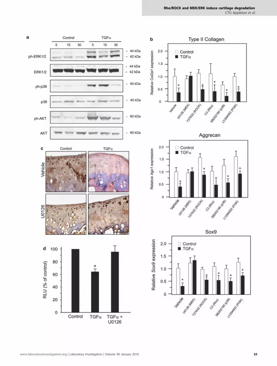
To investigate the extent to which additional pathways areactivated by TGFa in chondrocytes, we carried out westernblot analysis of chondrocyte protein lysates using antibodiesto detect the total and phosphorylated forms of ERK1/2(MEK/ERK pathway), p38 MAPK and AKT (PI3K pathway)kinases. Higher levels of phosphorylated ERK1/2, p38 MAPKand AKT were seen after stimulation with TGFa comparedwith untreated controls (Figure 2a). Infrared fluorescentsignal quantification determined that levels of phospho-ERK1/2 and phospho-AKT (normalized to total levels) weresignificantly elevated after 5, 15 and 30 min of stimulation,whereas phospho-p38 MAPK was significantly increased onlyup to 15 min (not shown).
ECM Gene Regulation Through MEK/ERK SignalingTGFa signaling in chondrocytes inhibits ECM gene expres-sion through the suppression of SOX9,8 a critical transcrip-tional regulator of type II collagen and aggrecan inchondrocytes.28,29 Thus, we assessed the contribution of theRho/ROCK, MEK/ERK, PI3K and p38 MAPK pathways tothe control of Sox9 and ECM gene expression by TGFa sig-naling. Inhibition of MEK/ERK activation was achieved withthe MEK inhibitor, U0126, and that of PI3K signaling in-hibition with the AKT inhibitor, LY294002. Real-time PCRshowed that type II collagen (Col2a1) and aggrecan (Agc1)mRNA downregulation in response to TGFa could only berescued by inhibition of MEK/ERK (Figure 2b). Correlatingwith these MEK/ERK-dependent reductions in ECM geneexpression, the suppression of Sox9 mRNA expression could
also be rescued by MEK/ERK inhibition, but not with othercompounds (Figure 2b). Finally, MEK/ERK inhibition re-sulted in the rescue of SOX9 protein expression in TGFa-treated explant cultures (Figure 2c) and SOX9 activity inluciferase reporter gene assay (Figure 2d), validating theseresults at protein and functional levels.
Complex Control of Proliferation and Catabolic GenesAs TGFa induces mitogenic activity and MMP-13 produc-tion, as well as drives inflammatory cytokine gene expression(eg, Tnfa, encoding TNFa) in chondrocytes,8 we exploredhow these responses are controlled by intracellular signalingpathways. As expected, TGFa induced a significant increasein chondrocyte number after 48 h (Figure 3a). Inhibition ofeach pathway tested (MEK/ERK, Rho/ROCK, p38 MAPK orPI3K) prevented the increase in cell number and significantlyreduced chondrocyte numbers relative to vehicle-only con-trols (Figure 3a); statistics for the latter are not shown forsimplicity.
Real-time PCR analyses of RNA from primary culturesdetermined that upregulation of Mmp13 could not be res-cued by inhibiting any of the pathways investigated (Figure3b). Conversely, blockade of Rho/ROCK or PI3K signalingdramatically enhanced Mmp13 upregulation in the presenceof TGFa (Figure 3b). Although MEK/ERK and PI3K inhibi-tion resulted in the abrogation of basal Tnfa expression belowdetectable levels (without TGFa treatment), neither couldprevent Tnfa induction by TGFa (Figure 3c). In contrast,Rho/ROCK and p38 MAPK inhibitors both prevented Tnfaupregulation (Figure 3c). In parallel experiments, TGFa didnot induce the expression of IL-1b mRNA in chondrocytes(not shown).
Degradation of Articular Cartilage ECM Stimulated byTGFa
We next analyzed cartilage explant tissues for histomorpho-logical changes. Chondrocyte clusters (suggestive of chon-drocyte division) were induced by TGFa treatment alone, asreported previously.8 Each inhibitor tested seemed to sup-press this increase in clusters (arrowhead, Figure 4), sup-porting our assessments of cell number. Interestingly, ROCKinhibition promotes hypertrophy of growth plate chon-drocytes in part by suppressing cell division,17 suggesting thatsimilar treatment may induce hypertrophy of articular
Figure 2 Activation of multiple signaling pathways and effects on ECM genes and Sox9. (a) Protein was isolated from chondrocyte cultures cultured
for up to 30 min with vehicle (control) or 10 ng/ml TGFa. Levels of phosphorylated (ph-) ERK1/2, p38 MAPK and AKT were assessed relative to total ERK1/2,
p38 MAPK or AKT levels, respectively, by western blot. (b) Real-time PCR was performed using RNA samples isolated from chondrocytes incubated
with vehicle (control) or 10 ng/ml TGFa for 48 h in the presence or absence of 10 mM ROCK (Y27632), MEK (U0126), p38 MAPK (SB202190) or PI3K (LY294002)
inhibitor. Mean gene expression values (plus s.d.) for type II collagen (Col2a1), aggrecan (Agc1) and Sox9 from N¼ 4 independent cell isolations are
shown normalized to controls. (c) Immunostaining for SOX9 (brown) and hematoxylin counterstaining of tissue sections from explant cultures incubated for
5 days with or without (control) 10 ng/ml TGFa in the presence or absence of 10 mM MEK inhibitor (U0126). Scale bar represents 100 mm. (d) Chondrocytes
were transfected with a reporter vector for SOX9 activity and cultured for 48 h with vehicle (control) or TGFa in the presence or absence of an MEK inhibitor.
Mean relative light units (RLU) (plus s.d.) from N¼ 4 independent cell isolations, normalized to untreated controls are shown. Significantly different
(Po0.05) means (compared with controls) are indicated by asterisks.
Rho/ROCK and MEK/ERK induce cartilage degradation
CTG Appleton et al
24 Laboratory Investigation | Volume 90 January 2010 | www.laboratoryinvestigation.org
Rho/ROCK and MEK/ERK induce cartilage degradation
CTG Appleton et al
www.laboratoryinvestigation.org | Laboratory Investigation | Volume 90 January 2010 25

chondrocytes. However, mRNA expression of several hyper-trophic markers including alkaline phosphatase (Alp1), bonemorphogenetic protein 6 (Bmp6), runt-related transcriptionfactor 2 (Runx2) and bone sialoprotein (Ibsp—undetectable,not shown) was suppressed by TGFa in primary chon-drocytes (Figure 5). Production of type X collagen assessedby immunostaining in articular cartilage explants was alsounchanged in the presence of TGFa, with or without theROCK inhibitor (data not shown).
To assess induction of cartilage degradation, im-munostaining for cartilage degradation markers was per-formed using antibodies against MMP-generated aggrecan(CGGVDIPEN) and type II collagen (COL2-3/4C) cleavagefragments.23,30 Striking increases in aggrecan and type IIcollagen cleavage fragment staining were observed in super-ficial and mid-zone chondrons after 5 days of treatment with
TGFa compared with untreated controls (Figure 4). The useof ROCK and MEK inhibitors alone reduced basal levels ofaggrecan and type II collagen fragments and prevented in-creases in aggrecan and type II collagen fragments in thepresence of TGFa (Figure 4). PI3K and p38 MAPK inhibitiondid not reduce basal aggrecan fragment but did reduce basaltype II collagen fragment levels (Figure 4). In the presence ofTGFa, PI3K and p38 MAPK inhibition was unable to reduceaggrecan cleavage and only slightly reduced type II collagencleavage (Figure 4).
DISCUSSIONWe have previously reported that TGFa upregulation by kneearticular chondrocytes is an early event in a rat model of jointdestabilization-induced knee OA and that TGFa is elevated in
Figure 3 Effects of different pathways on cell number, Mmp13 and Tnfa expression. (a) MTT analysis was performed by spectrophotometry of chondrocytes
cultured with vehicle (control) or 10 ng/ml TGFa in the presence or absence (vehicle) of 10 mM ROCK (Y27632), MEK (U0126), p38 MAPK (SB202190) or PI3K
(LY294002) inhibitor. The mean absorbance readings (plus s.d.) from N¼ 5 independent cell isolations are shown. Significant reductions compared with
vehicle only occurred in all inhibitor-treated samples þ /�TGFa (statistics not shown for simplicity). (b) Real-time PCR was performed using RNA samples
isolated from chondrocytes incubated with vehicle (control) or 10 ng/ml TGFa for 48 h in the presence or absence of 10 mM inhibitors as described above.
Mean gene expression values (plus s.d.) for matrix metalloproteinase-13 (Mmp13) and (c) tumor necrosis factor-a (Tnfa) from N¼ 4 independent cell
isolations are shown normalized to controls. Significantly different (Po0.05) TGFa-treated vs control means are indicated by asterisks.
Rho/ROCK and MEK/ERK induce cartilage degradation
CTG Appleton et al
26 Laboratory Investigation | Volume 90 January 2010 | www.laboratoryinvestigation.org

late-stage human knee OA chondrocytes.8 Although it wasfurther determined that TGFa induces chondrocyte pheno-type modification, the intracellular mechanisms mediatingthe suppression of anabolic and activation of catabolic pro-cesses remained unclear. The results of this study show thatRhoA/ROCK, MEK/ERK, p38 MAPK and PI3K/AKT signal-ing pathways are activated by the stimulation of articularchondrocytes with TGFa. Moreover, Rho/ROCK and MEK/ERK signaling mediate deleterious outcomes after TGFastimulation (Figure 6). We also showed that chondrocyteexposure to TGFa results in strong increases in MMP-mediated collagen and proteoglycan digestion within maturearticular cartilage. Thus, TGFa seems to be a powerfulstimulator of articular cartilage degradation and a modifierof chondrocyte phenotype. Given its temporal increase dur-ing cartilage degradation, TGFa is likely to be an importantdriver in the pathogenesis of joint conditions, such as OA.
It is well established that the phenotypic stability of ar-ticular chondrocytes is critical and that modifications tochondrocyte shape (eg, flattening, elongation, hypertrophy)impede the normal functions of the chondrocyte.31,32 Inter-estingly, changes in chondrocyte shape are commonly ob-served in OA.33 Our study shows, for the first time, that Rho/ROCK signaling is activated by TGFa in chondrocytes; this inturn induces elongation of chondrocyte shape and stress fiberformation, most likely through activation of the LIMkinase.27 As MEK/ERK inhibition was unable to preventstress fiber formation, this suggests that the involvement ofRho/ROCK signaling in this process is specific and direct.The possibility that Rho family members act downstream ofEGFR signaling has only recently been suggested.34 AlthoughRhoA activation after EGFR stimulation with heparin bind-ing EGF-like growth factor has been reported,35 this isthe first report showing Rho/ROCK activation by TGFa.
Figure 4 Histology and immunostaining for aggrecan and type II collagen cleavage fragments. Osteochondral explants were cultured for 5 days with
vehicle (control) or 10 ng/ml TGFa in the presence or absence of a ROCK (Y27632), MEK (U0126), p38 MAPK (SB202190) or PI3K (LY294002) inhibitor (10 mM).
Tissue sections were stained with safranin-O/fast green or immunostained for MMP-generated aggrecan or type II collagen cleavage fragments (brown
stain). All sections were counterstained with hematoxylin. Arrowhead indicates chondrocyte cluster. Scale bars represent 50 mm.
Rho/ROCK and MEK/ERK induce cartilage degradation
CTG Appleton et al
www.laboratoryinvestigation.org | Laboratory Investigation | Volume 90 January 2010 27

Interestingly, a crosstalk between Ras-mediated and Rho/ROCK pathways results in Rho/ROCK/LIMK inhibition inother cell types in a MEK-dependent manner.36,37 In contrast,our studies indicate that TGFa induces Rho/ROCK/LIMKactivation in chondrocytes concomitantly with the activationof multiple Ras-mediated pathways (eg, Ras-MEK/ERK andRas-PI3K). In addition, we observed simultaneous MLC2activation by TGFa, suggesting that contractility may beinduced in articular chondrocytes by EGFR activation.38
Although stress fiber formation and contractility is normal insome cell types such as fibroblasts,39 these events representunfavorable modification of the articular chondrocyte pheno-type through activation of Rho/ROCK signaling. Furthermore,these data suggest that inhibition of Rho/ROCK signaling wouldfavor articular cartilage maintenance and repair.
TGFa elicits a mitogenic response in chondrocytes andother cell types.8,40 Although this observation indicates an
opportunity to increase the number of chondrocytes avail-able for cartilage regeneration (eg, for tissue engineeringstrategies), such an approach would require that maintenanceof the chondrocyte phenotype could be achieved. However,in addition to severe phenotypic modulation, chondrocytesexhibit reduced expression of anabolic genes and elevatedMmp13 and Tnfa expression when stimulated withTGFa. Therefore, additional chondrocytes generated byTGFa-stimulated mitogenesis would be unlikely to regeneratedamaged articular cartilage. Moreover, as concerted activa-tion of all pathways studied herein is necessary to elicit amitogenic response to TGFa, attempts to block deleteriouseffects mediated by the Rho/ROCK and MEK/ERK pathwayswould also prevent induction of chondrocyte divisionand negate the possibility of generating additionalchondrocytes.
Hypertrophy of articular chondrocytes in OA is believed tocontribute to OA pathogenesis,41 despite being a normaldevelopmental process in growth plate chondrocytes. Inter-estingly, blocking Rho/ROCK signaling suppresses prolifera-tion and induces MMP-13 expression in growth platechondrocytes,17 similar to articular chondrocytes studiedherein. However, although Rho/ROCK inhibition promoteschondrocyte hypertrophy in the growth plate, none of thehypertrophic markers assessed were elevated by chondrocytesexposed to TGFa and a ROCK inhibitor. As articularchondrocytes are arrested in a non-hypertrophic state,42 anadditional stimulus may be required to ‘switch on’ articularchondrocytes and resume the developmental program tohypertrophy. We therefore conclude that exposure to TGFadoes not promote hypertrophy of articular chondrocytes,even in the presence of a ROCK inhibitor.
Nevertheless, it is clear that attempts to strategicallymodulate Rho/ROCK or MEK/ERK signaling could becomplicated by mixed outcomes. For example, MEK/ERK
Figure 5 Hypertrophic marker gene expression in articular chondrocytes. Real-time PCR was performed using RNA samples isolated from chondrocytes
incubated with vehicle (control) or 10 ng/ml TGFa for 48 h in the presence or absence of the ROCK inhibitor (Y27632). Mean gene expression values (plus s.d.) for
alkaline phosphatase (Alp1), bone morphogenetic protein 6 (Bmp6) and runt-related transcription factor 2 (Runx2) relative to Gapdh from N¼ 4 independent cell
isolations are shown normalized to control (untreated) levels. Asterisks indicate significantly different (Po0.05) means compared with controls.
Figure 6 Working model of TGFa signaling mechanisms and outcomes.
TGFa upregulation is stimulated in osteoarthritic cartilage leading to the
activation of multiple signaling pathways including Rho/ROCK and MEK/
ERK and increased expression of TNFa. Each pathway mediates multiple
outcomes as illustrated. The color reproduction of this figure is available on
the html full text version of the manuscript.
Rho/ROCK and MEK/ERK induce cartilage degradation
CTG Appleton et al
28 Laboratory Investigation | Volume 90 January 2010 | www.laboratoryinvestigation.org

inhibition restores ECM gene expression, but fewer chon-drocytes are generated, and Tnfa expression remains elevated.Similarly, inhibition of Rho/ROCK signaling blocks Tnfaexpression and prevents phenotype modification, but inducesMmp13.
Evidence for activation of multiple intracellular pathwaysby EGFR signaling has accumulated for many cell types otherthan chondrocytes.43,44 Not surprisingly then, we observedthat EGFR stimulation with TGFa results in the activation ofMEK/ERK, PI3K/AKT and p38 MAPK pathwayscontemporaneously with Rho/ROCK. Activation of thesepathways likely influences articular chondrocyte physiologyas in cartilage development. For example, Rho/ROCKsignaling modified chondrocyte shape in this study andinduced chondrocyte phenotype changes during chon-drogenesis.15–17,45 Similarly, MEK/ERK signaling led to thesuppression of chondrocyte differentiation46 and causedsuppression of ECM gene expression and SOX9 levels in ourexperiments with articular chondrocytes. These findings arefurther supported by Yagi et al,47 who showed that sup-pression of MEK/ERK signaling by Bcl-2 increases SOX9expression in chondrocytes. TGFa is upregulated by OAchondrocytes, our findings indicate that MEK/ERK activa-tion by TGFa reduces anabolic activity and thereby thecapacity to replace damaged and eroded articular cartilage.Interestingly, IGF-1 maintains proteoglycan production inchondrocytes through a PI3K-dependent mechanism,whereas MEK/ERK activation reduces chondrocyte respon-siveness to IGF-1 by inhibiting the activation of PI3K.48
Thus, TGFa may suppress aggrecan production by bluntingPI3K responses in chondrocytes through the MEK/ERKactivation. Taken together, data from our group and fromothers therefore strongly suggest that inhibition of MEK/ERKactivation factors is a key step in the prevention of cartilagedegradation.
We also show in this study that TGFa stimulates thecleavage of aggrecan and type II collagen by MMPs, in-dicating that MMP activity is increased by the activation ofthe EGFR. Interestingly, type II collagen cleavage fragmentstaining pattern was similar to that of MMP-13 expressioninduced by TGFa,8 whereas MMP-13 is known to stimulatetype II collagen digestion in the OA cartilage.23,49 Strikingly,degradation of type II collagen and aggrecan was reducedwhen MEK/ERK and Rho/ROCK inhibitors were used in thepresence of TGFa, although Mmp13 mRNA induction wasnot suppressed. Therefore, despite the fact that Mmp13transcript levels remain increased when Rho/ROCK or MEK/ERK signaling is blocked, MMP-13 activity is reduced. Rho/ROCK and MEK/ERK signaling inhibition may thereforeprevent MMP-13 activation at the post-transcriptional level.Furthermore, as TNFa induces MMP-13 activity50 and asblocking Rho/ROCK and MEK/ERK prevented Tnfa induc-tion/reduced basal Tnfa expression respectively, enhancedTNFa production may be a driver of ECM digestion inducedby TGFa.
Taken together, our observations suggest that blockadeof Rho/ROCK and MEK/ERK signaling effectively reducesECM damage in the cartilage exposed to TGFa. Whereasinhibition of Rho/ROCK activation by TGFa preventschanges in chondrocyte shape, MEK/ERK signaling inhibi-tion rescues the suppression of ECM gene expressionand SOX9. Our results align with previous studies thathave described roles for MEK/ERK signaling in articularcartilage degeneration19,20,48 and point to a similar role forRho/ROCK. This report also adds to the growing body ofevidence indicating that inappropriate activation of theEGFR leads to cartilage degradation.8,14,51 It is important tonote that additional pathways are stimulated by EGFRactivation with TGFa and that these pathways may conferprotective benefits. For example, as PI3K signaling is requiredfor proteoglycan production in response to IGF-1 but issuppressed by MEK/ERK signaling,48 PI3K activation byTGFa may be enhanced to stimulate anabolic responses whenMEK/ERK activation is blocked. Therefore, as an alternativestrategy to block EGFR activation itself or sequestration ofTGFa, we have shown the efficacy of interfering with TGFasignaling downstream of the EGFR to reduce cartilagedegeneration, as is the case for MEK/ERK and Rho/ROCKinhibition. Although a therapeutic approach involvingsystemic inhibition of these essential intracellular signalingpathways would not be viable, local inhibitor delivery maybe effective for bypassing systemic side effects. However, asrepeated intra-articular injection is not preferred as a methodof long-term local delivery, development of alternativeapproaches such as viral-based gene knockdown strategiesmay be required.
In summary, this study advances our understanding of themechanisms controlling the metabolic outcomes of TGFasignaling in articular cartilage. We conclude that modulationof the signaling pathways downstream of the EGFR, at leastwhen activated by TGFa, is a compelling strategy for thetreatment of joint conditions, such as OA.
ACKNOWLEDGEMENTS
We acknowledge the assistance of Dr L Coolen, K Pitchers, V Pitelka and J
Rockel at The University of Western Ontario. FB is the Canada Research Chair
in Musculoskeletal Health. JSM is supported by the Shriners of North
America. This study was supported by funding from the Canadian Institutes
of Health Research (Grant no. MOP86574) and the University of Western
Ontario Schulich Research Opportunities Program.
DISCLOSURE/CONFLICT OF INTEREST
The authors declare no conflict of interest.
1. Green GA. Understanding NSAIDs: from aspirin to COX-2. ClinCornerstone 2001;3:50–60.
2. Kopec JA, Rahman MM, Berthelot JM, et al. Descriptive epidemiologyof osteoarthritis in British Columbia, Canada. J Rheumatol2007;34:386–393.
3. Abramson SB, Attur MG, Yasici Y. Prospects for disease modification inosteoarthritis. Nat Clin Pract Rheumatol 2006;2:304–312.
4. Aigner T. Osteoarthritis. Curr Opin Rheumatol 2007;19:427–428.
Rho/ROCK and MEK/ERK induce cartilage degradation
CTG Appleton et al
www.laboratoryinvestigation.org | Laboratory Investigation | Volume 90 January 2010 29

5. Goldring MB, Goldring SR. Osteoarthritis. J Cell Physiol 2007;213:626–634.
6. Appleton CTG, McErlain D, Pitelka V, et al. Forced mobilizationaccelerates pathogenesis: characterization of a pre-clinical surgicalmodel of osteoarthritis. Arthritis Res Ther 2007;9:R13.
7. Appleton CTG, Pitelka V, Henry JL, et al. Global analyses of geneexpression in early experimental osteoarthritis. Arthritis Rheum2007;56:1854–1868.
8. Appleton CT, Usmani SE, Bernier SM, et al. Transforming growth factoralpha suppression of articular chondrocyte phenotype and Sox9expression in a rat model of osteoarthritis. Arthritis Rheum2007;56:3693–3705.
9. Brachmann R, Lindquist PB, Nagashima M, et al. TransmembraneTGF-alpha precursors activate EGF/TGF-alpha receptors. Cell 1989;56:691–700.
10. Groenen LC, Nice EC, Burgess AW. Structure-function relationshipsfor the EGF/TGF-alpha family of mitogens. Growth Factors 1994;11:235–257.
11. Macleod K, Mullen P, Sewell J, et al. Altered ErbB receptor signalingand gene expression in cisplatin-resistant ovarian cancer. Cancer Res2005;65:6789–6800.
12. Hallbeck AL, Walz TM, Briheim K, et al. TGF-alpha and ErbB2 productionin synovial joint tissue: increased expression in arthritic joints. Scand JImmunol 2005;34:204–211.
13. Ritchlin C, Dwyer E, Bucala R, et al. Sustained and distinctive patternsof gene activation in synovial fibroblasts and whole synovial tissueobtained from inflammatory synovitis. Scand J Immunol 1994;40:292–298.
14. Zhang Y-W, Su Y, Lanning N, et al. Targeted disruption of Mig-6 in themouse genome leads to early onset degenerative joint disease. ProcNatl Acad Sci USA 2005;102:11740–11745.
15. Woods A, Wang G, Beier F. RhoA/ROCK signaling regulates Sox9expression and actin organization during chondrogenesis. J Biol Chem2005;280:11626–11634.
16. Woods A, Beier F. RhoA/ROCK signaling regulates chondrogenesis in acontext-dependent manner. J Biol Chem 2006;281:13134–13140.
17. Wang G, Woods A, Sabari S, et al. RhoA/ROCK signaling suppresseshypertrophic chondrocyte differentiation. J Biol Chem2004;279:13205–13214.
18. Bobick BE, Kulyk WM. The MEK-ERK signaling pathway is a negativeregulator of cartilage-specific gene expression in embryonic limbmesenchyme. J Biol Chem 2004;279:4588–4595.
19. Fan Z, Bau B, Yang H, et al. IL-1 beta induction of IL-6 and LIF in normalarticular human chondrocytes involves the ERK, p38 and NF-kappa Bsignaling pathways. Cytokine 2004;28:17–24.
20. Fan Z, Soder S, Oehler S, et al. Activation of interleukin-1 signalingcascades in normal and osteoarthritic articular cartilage. Am J Pathol2007;171:938–946.
21. Seguin CA, Bernier SM. TNF-alpha suppresses link protein and type IIcollagen expression in chondrocytes: role of MEK1/2 and NF-kappa Bsignaling pathways. J Cell Physiol 2003;197:356–369.
22. Sztrolovics R, Alini M, Roughley PJ, et al. Aggrecan degradation inhuman intervertebral disc and articular cartilage. Biochem J1997;326:235–241.
23. Billinghurst RC, Dahlberg L, Ionescu M, et al. Enhanced cleavage oftype II collagen by collagenases in osteoarthritic articular cartilage.J Clin Invest 1997;99:1534–1545.
24. Weston AD, Chandraratna RAS, Torchia J, et al. Requirement for RAR-mediated gene repression in skeletal progenitor differentiation. J CellBiol 2002;158:39–51.
25. James C, Woods A, Underhill TM, et al. The transcription factorATF3 is upregulated during chondrocyte differentiation andrepresses cyclin D1 and A gene transcription. BMC Mol Biol 2006;7:30.
26. Yamashiro S, Totsukawa G, Yamakita Y, et al. Citron kinase, a Rho-dependent kinase, induces Di-phosphorylation of regulatory lightchain of myosin II. Mol Biol Cell 2003;14:1745–1756.
27. Maekawa M, Ishizaki T, Boku S, et al. Signaling from Rho to the actincytoskeleton through protein kinases ROCK and LIM-kinase. Science1999;285:895–898.
28. Lefebvre V, Behringer RR, de Crombrugghe B. L-Sox5, Sox6 and Sox9control essential steps of the chondrocyte differentiation pathway.Osteoarthr Cartil 2001;9:S69–S75.
29. Lefebvre V, Huang W, Harley VR, et al. SOX9 is a potent activator of thechondrocyte-specific enhancer of the pro alpha1(II) collagen gene. MolCell Biol 1997;17:2336–2346.
30. Hughes C, Caterson B, Fosang A, et al. Monoclonal antibodies thatspecifically recognize neoepitope sequences generated by‘aggrecanase’ and matrix metalloproteinase cleavage of aggrecan:application to catabolism in situ and in vitro. Biochem J 1995;305:799–804.
31. Benya PD, Shaffer JD. Dedifferentiated chondrocytes reexpress thedifferentiated collagen phenotype when cultured in agarose gels. Cell1982;30:215–224.
32. Zanetti NC, Solursh M. Induction of chondrogenesis in limbmesenchymal cultures by disruption of the actin cytoskeleton. J CellBiol 1984;99:115–123.
33. Pritzker KPH, Gay S, Jimenez SA, et al. Osteoarthritis cartilagehistopathology: grading and staging. Osteoarthr Cartil 2006;14:13–29.
34. Mateus AR, Seruca R, Machado JC, et al. EGFR regulates RhoA-GTPdependent cell motility in E-cadherin mutant cells. Hum Mol Genet2007;16:1639–1647.
35. Yin J, Lu J, Yu F-SX. Role of small GTPase Rho in regulating cornealepithelial wound healing. Invest Ophthalmol Vis Sci 2008;49:900–909.
36. Nebl G, Fischer S, Penzel R, et al. Dephosphorylation of cofilin isregulated through Ras and requires the combined activities of the Ras-effectors MEK and PI3K. Cell Signal 2004;16:235–243.
37. Jung J, Kim M, Choi S, et al. Molecular mechanism of cofilindephosphorylation by ouabain. Cell Signal 2006;18:2033–2040.
38. Ikebe M, Hartshorne DJ. Phosphorylation of smooth muscle myosin attwo distinct sites by myosin light chain kinase. J Biol Chem1985;260:10027–10031.
39. Iwabu A, Smith K, Allen FD, et al. Epidermal growth factor inducesfibroblast contractility and motility via a protein kinase C delta-dependent pathway. J Biol Chem 2004;279:14551–14560.
40. Richter A, Drummond DR, MacGarvie J, et al. Contribution of thetransforming growth factor-alpha beta-loop beta-sheet to binding andactivation of the epidermal growth factor receptor. J Biol Chem1995;270:1612–1616.
41. Kamekura S, Kawasaki Y, Hoshi K, et al. Contribution of runt-relatedtranscription factor 2 to the pathogenesis of osteoarthritis in mice afterinduction of knee joint instability. Arthritis Rheum 2006;54:2462–2470.
42. Yang X, Chen L, Xu X, et al. TGF-beta/Smad3 signals represschondrocyte hypertrophic differentiation and are required formaintaining articular cartilage. J Cell Biol 2001;153:35–46.
43. Alroy I, Yarden Y. The ErbB signaling network in embryogenesis andoncogenesis: signal diversification through combinatorial ligand-receptor interactions. FEBS Lett 1997;410:83–86.
44. Hackel PO, Zwick E, Prenzel N, et al. Epidermal growth factor receptors:critical mediators of multiple receptor pathways. Curr Opin Cell Biol1999;11:184–189.
45. Stanton LA, Sabari S, Sampaio AV, et al. p38 MAP kinase signalling isrequired for hypertrophic chondrocyte differentiation. Biochem J2004;378:53–62.
46. Yoon Y-M, Oh C-D, Kim D-Y, et al. Epidermal growth factor negativelyregulates chondrogenesis of mesenchymal cells by modulating theprotein kinase C-alpha, Erk-1, and p38 MAPK signaling pathways. J BiolChem 2000;275:12353–12359.
47. Yagi R, McBurney D, Horton Jr WE. Bcl-2 positively regulates Sox9-dependent chondrocyte gene expression by suppressing the MEK-ERK1/2 signaling pathway. J Biol Chem 2005;280:30517–30525.
48. Starkman BG, Cravero JD, Delcarlo M, et al. IGF-I stimulation ofproteoglycan synthesis by chondrocytes requires activation of the PI 3-kinase pathway but not ERK MAPK. Biochem J 2005;389:723–729.
49. Smith MD, Triantafillou S, Parker A, et al. Synovial membraneinflammation and cytokine production in patients with earlyosteoarthritis. J Rheumatol 1997;24:365–371.
50. Liacini A, Sylvester J, Qing Li W, et al. Induction of matrixmetalloproteinase-13 gene expression by TNF alpha is mediatedby MAP kinases, AP-1, and NF-kappa B transcription factorsin articular chondrocytes. Exp Cell Res 2003;288:208–217.
51. Vincourt J-B, Vignaud J-M, Lionneton F, et al. Increased expressionof matrilin-3 not only in osteoarthritic articular cartilage but also incartilage-forming tumors, and down-regulation of SOX9 via epidermalgrowth factor domain 1-dependent signaling. Arthritis Rheum2008;58:2798–2808.
Rho/ROCK and MEK/ERK induce cartilage degradation
CTG Appleton et al
30 Laboratory Investigation | Volume 90 January 2010 | www.laboratoryinvestigation.org


