Embed Size (px)
Citation preview

Role for Golgi reassembly and stacking protein (GRASP) inpolysaccharide secretion and fungal virulencemmi_7686 206..218
Lívia Kmetzsch,1‡ Luna S. Joffe,3,4‡
Charley C. Staats,1,2 Débora L. de Oliveira,3
Fernanda L. Fonseca,3 Radames J.B. Cordero,5
Arturo Casadevall,5,6 Leonardo Nimrichter,3
Augusto Schrank,1,2 Marilene H. Vainstein1,2† andMarcio L. Rodrigues3*†
1Centro de Biotecnologia and 2Departamento deBiologia Molecular e Biotecnologia, UniversidadeFederal do Rio Grande do Sul, Av. Bento Gonçalves9500, 43421, Caixa Postal 15005, Porto Alegre, RS91501-970, Brazil.3Laboratório de Estudos Integrados em BioquímicaMicrobiana, Instituto de Microbiologia Professor Paulode Góes, Universidade Federal do Rio de Janeiro, Riode Janeiro, 21941-902, Brazil.4Instituto de Bioquímica Médica, Programa deGlicobiologia, Universidade Federal do Rio de Janeiro,Rio de Janeiro, 21941-902, Brazil.5Department of Microbiology and Immunology and6Division of Infectious Diseases of the Department ofMedicine, Albert Einstein College of Medicine, 1300Morris Park Ave, Bronx, NY 10461, USA.
Summary
Secretion of virulence factors is a critical mechanismfor the establishment of cryptococcosis, a diseasecaused by the yeast pathogen Cryptococcus neofor-mans. One key virulence strategy of C. neoformansis the release of glucuronoxylomannan (GXM), acapsule-associated immune-modulatory polysaccha-ride that reaches the extracellular space throughsecretory vesicles. Golgi reassembly and stackingprotein (GRASP) is required for unconventionalprotein secretion mechanisms in different eukaryoticcells, but its role in polysaccharide secretion isunknown. This study demonstrates that a C. neofor-mans functional mutant of a GRASP orthologuehad attenuated virulence in an animal model of cryp-tococcosis, in comparison with wild-type (WT) and
reconstituted cells. Mutant cells manifested alteredGolgi morphology, failed to produce typical polysac-charide capsules and showed a reduced ability tosecrete GXM both in vitro and during animal infection.Isolation of GXM from cultures of WT, reconstituted ormutant strains revealed that the GRASP orthologuemutant produced polysaccharides with reduceddimensions. The mutant was also more efficientlyassociated to and killed by macrophages than WT andreconstituted cells. These results demonstrate thatGRASP, a protein involved in unconventional proteinsecretion, is also required for polysaccharide secre-tion and virulence in C. neoformans.
Introduction
Cryptococcus neoformans is a yeast-like pathogen asso-ciated with high mortality rates in immunosuppressedindividuals (Park et al., 2009; Prado et al., 2009). C. neo-formans virulence is dependent on the expression of anumber of virulence factors, including enzymes, pig-ments, polysaccharides and lipids (Li and Mody, 2010).Like many bacterial pathogens, C. neoformans is sur-rounded by a polysaccharide capsule that is an importantvirulence factor (Zaragoza et al., 2009). Capsule forma-tion in this fungal pathogen requires intracellular polysac-charide synthesis (Feldmesser et al., 2001; Yoneda andDoering, 2006), followed by secretion of capsular compo-nents to the extracellular space and their incorporationinto the cell surface (Rodrigues et al., 2008a; Zaragozaet al., 2009). Capsule expression is purportedly themost important constraint for cryptococcal virulence(McClelland et al., 2005).
Glucuronoxylomannan (GXM), the major capsular com-ponent of C. neoformans, is presumably synthesized inthe Golgi and targeted to the cell surface (Yoneda andDoering, 2006). The polysaccharide then traverses thecell wall in vesicles that reach the extracellular space(Rodrigues et al., 2007), where GXM is used for enlarge-ment of the cryptococcal capsule (Zaragoza et al., 2006).Exposure of C. neoformans to brefeldin A, which affectsthe formation of Golgi-related transport vesicles, results ina strong inhibition of capsule assembly (Hu et al., 2007).Consequently, the Golgi apparatus is suggested to berequired for GXM synthesis and secretion, based on
Accepted 25 April, 2011. *For correspondence. E-mail [email protected]; Tel. (+55) 21 2598 3035; Fax (+55) 21 2560 8344. †M.H.V. andM.L.R. share senior authorship on this article. ‡L.K. and L.S.J. con-tributed equally to this work.
Molecular Microbiology (2011) 81(1), 206–218 � doi:10.1111/j.1365-2958.2011.07686.xFirst published online 17 May 2011
© 2011 Blackwell Publishing Ltd
results of two independent studies. Yoneda and Doeringdemonstrated that a C. neoformans mutant lackingexpression of Sav1p, a putative secretory vesicle-associated Rab GTPase essential for exocytosis, accu-mulates intracellular, post-Golgi vesicles containing GXM(Yoneda and Doering, 2006). This is in agreement withresults described by Panepinto and colleagues, whoshowed that C. neoformans cells with deficient expressionof Sec6p, which mediates polarized targeting of secretoryvesicles to active sites of exocytosis, had a decreasedrate of GXM secretion (Panepinto et al., 2009). Althoughboth studies clearly indicated an association of Golgi-derived pathways with GXM secretion, the fact that cap-sular expression was apparently normal in both sav1 andsec6 mutants suggested that other components of Golgi-associated secretory pathways could have a role in GXMtraffic in C. neoformans.
Golgi reassembly and stacking proteins (GRASPs)have been implicated in the stacking of Golgi cisternae,vesicle tethering and mitotic progression (Nickel andSeedorf, 2008; Nickel and Rabouille, 2009). GRASP isprimarily attached peripherally to the cytoplasmic surfaceof Golgi membranes, but its distribution into other cellularcompartments is also expected (Nickel and Rabouille,2009). In Dictyostelium discoideum, the single GRASPorthologue (GrpA) is required for unconventional secre-tion of acyl-coenzyme A-binding protein (AcbA) duringspore differentiation (Kinseth et al., 2007), in a processthat requires secretory vesicles (Cabral et al., 2010).
GRASP is also required for the delivery of integrin asubunits to the plasma membrane of Drosophila melano-gaster in a Golgi-independent manner (Schotman et al.,2008). More recently, it has been demonstrated GRASP isalso required for starvation-induced secretion of AcbA inSaccharomyces cerevisiae and Pichia pastoris (Duranet al., 2010; Manjithaya et al., 2010).
The fact that a number of the cryptococcal virulencefactors are exocellular components (Li and Mody, 2010)implies that secretory activity is directly linked to virulencein C. neoformans. Most of the C. neoformans virulencefactors are released to the extracellular space apparentlythrough unconventional mechanisms of secretion (Rod-rigues et al., 2007; 2008a,b; Nosanchuk et al., 2008;Casadevall et al., 2009). The possible link between cryp-tococcal virulence and unconventional secretion led us toinvestigate the role of GRASP in an animal model of C.neoformans infection. Our results suggest that virulenceis attenuated in a GRASP orthologue mutant of C. neo-formans, which was associated with a defect in the abilityof yeast cells to secrete GXM. To our knowledge, this isthe first report showing a role for a GRASP orthologue inmicrobial virulence and in polysaccharide secretion ineukaryotic cells.
Results
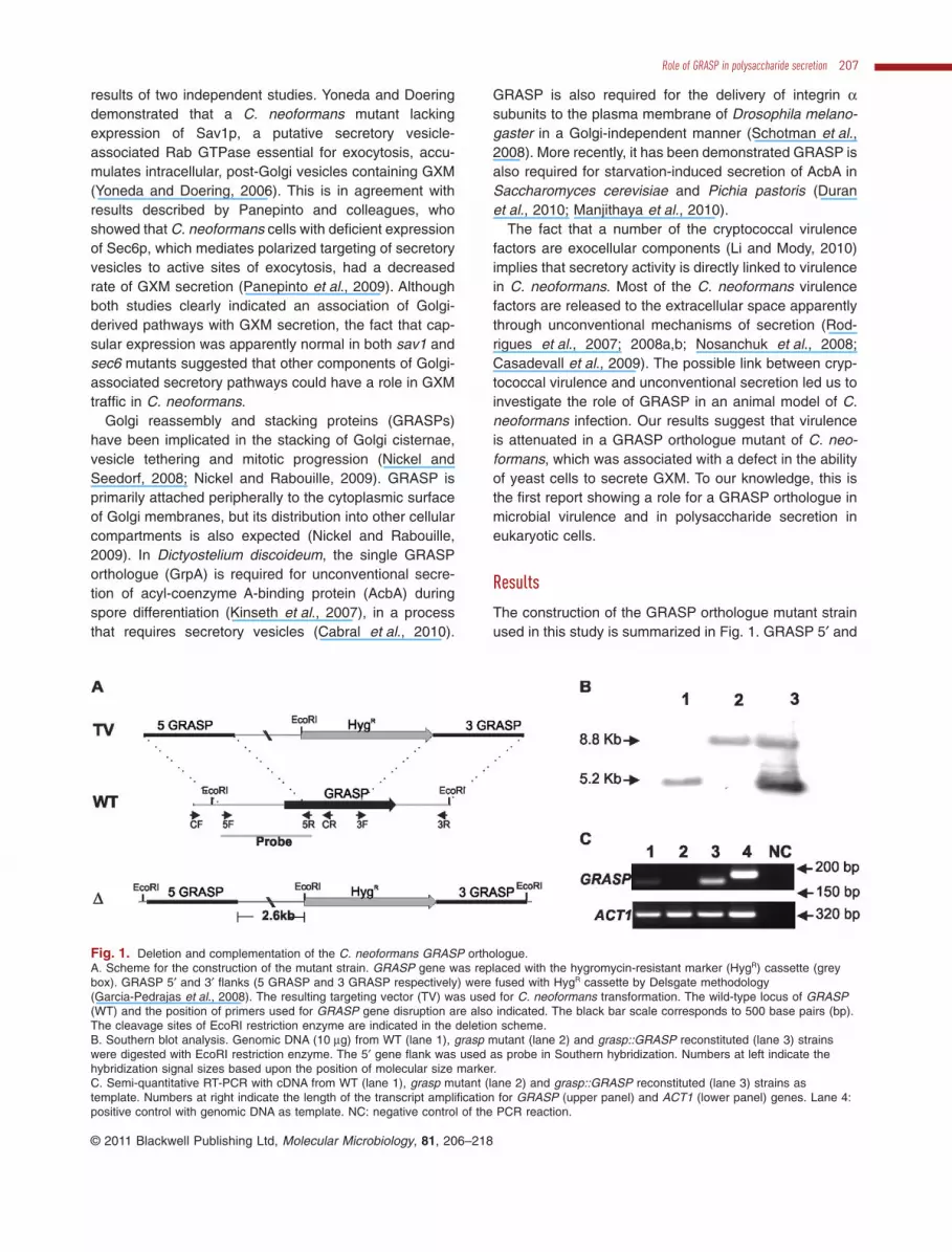
The construction of the GRASP orthologue mutant strainused in this study is summarized in Fig. 1. GRASP 5′ and
Fig. 1. Deletion and complementation of the C. neoformans GRASP orthologue.A. Scheme for the construction of the mutant strain. GRASP gene was replaced with the hygromycin-resistant marker (HygR) cassette (greybox). GRASP 5′ and 3′ flanks (5 GRASP and 3 GRASP respectively) were fused with HygR cassette by Delsgate methodology(Garcia-Pedrajas et al., 2008). The resulting targeting vector (TV) was used for C. neoformans transformation. The wild-type locus of GRASP(WT) and the position of primers used for GRASP gene disruption are also indicated. The black bar scale corresponds to 500 base pairs (bp).The cleavage sites of EcoRI restriction enzyme are indicated in the deletion scheme.B. Southern blot analysis. Genomic DNA (10 mg) from WT (lane 1), grasp mutant (lane 2) and grasp::GRASP reconstituted (lane 3) strainswere digested with EcoRI restriction enzyme. The 5′ gene flank was used as probe in Southern hybridization. Numbers at left indicate thehybridization signal sizes based upon the position of molecular size marker.C. Semi-quantitative RT-PCR with cDNA from WT (lane 1), grasp mutant (lane 2) and grasp::GRASP reconstituted (lane 3) strains astemplate. Numbers at right indicate the length of the transcript amplification for GRASP (upper panel) and ACT1 (lower panel) genes. Lane 4:positive control with genomic DNA as template. NC: negative control of the PCR reaction.
Role of GRASP in polysaccharide secretion 207
© 2011 Blackwell Publishing Ltd, Molecular Microbiology, 81, 206–218

3′ flanks were fused with the hygromycin-resistant markercassette by Delsgate methodology (Garcia-Pedrajaset al., 2008; Kmetzsch et al., 2010; 2011). The resultingtargeting vector was used for C. neoformans transforma-tion, which was monitored by Southern hybridizationand semi-quantitative RT-PCR. We identified a putativeGRASP orthologue in the C. neoformans var. grubii (sero-type A) genomic database (Broad Institute, Accession No.CNAG_03291.2). The 1051 bp putative GRASP has fourintrons and predicts a 256-amino-acid protein. GRASPsare characterized by the presence of two PDZ-likedomains in the N-terminal region (Kinseth et al., 2007). ABLAST search using the PDZ domains sequences fromRattus norvegicus GRASP revealed that these regionsare also present in the C. neoformans GRASP orthologue.
Sequence comparisons revealed that the identity rangefor the PDZ-1 and PDZ-2 domains were 40% and 48%respectively (Fig. 2A).
A broad phylogenetic analysis including GRASPsequences from distinct eukaryotic organisms rendered aphylogeny tree split into two major clades (Fig. 2B). CladeC-II encompasses the GRASPs from the majority of thefungi analysed, all belonging to the ascomycetes. CladeC-I harboured the GRASPs from the basidiomycetousfungi C. neoformans and Ustilago maydis, as well asthose from mammalian (R. norvegicus), fly (D. melano-gaster), worm (Caenorhabditis elegans), protist (Plasmo-dium falciparum) and amoeba (D. discoideum).
GRASP is primarily localized to the cytoplasmic surfaceof Golgi membranes, where it is apparently involved in the
Fig. 2. Phylogenetic analysis of the C. neoformans GRASP orthologue.A. Alignment of the GRASP PDZ domains (PDZ-1 and PDZ-2) from R. norvegicus (AAB81355.2), C. elegans (NP_501354), D. melanogaster(AAF49092), S. cerevisiae (NP_010805), D. discoideum (EAL60823), S. pombe (NP_593015.1), P. falciparum (AAN35366), A. nidulans (BroadInstitute Accession No. ANID_11248), U. maydis (Broad Institute Accession No. UM01076), C. albicans (Broad Institute Accession No.CAWG_01021) and C. neoformans (Broad Institute Accession No. CNAG_03291) using CLUSTALX2.B. Phylogenetic analysis applying the Neighbour-Joining method including GRASP sequences from distinct eukaryotic organisms listed above.The phylogeny tree splits into two major clades. C-I and C-II represents clades I and II respectively. The bar marker indicates the geneticdistance, which is proportional to the number of amino acid substitutions.
208 L. Kmetzsch et al. �
© 2011 Blackwell Publishing Ltd, Molecular Microbiology, 81, 206–218

stacking of Golgi cisternae in eukaryotic cells (Vinke et al.,2011). Based on this observation, we evaluated whetherlack of GRASP would affect Golgi morphology in C.neoformans. The Golgi marker N-[7-(4-nitrobenzo-2-oxa-1,3-diazole)]-6-aminocaproyl-D-erythro-sphingosine (C6-NBD-ceramide) stained wild-type (WT) cells producinga pattern that was very similar to that described for S.cerevisiae (Levine et al., 2000) (Fig. 3). Mutant cells,however, showed altered Golgi morphology. In thesecells, the Golgi apparatus appeared to be compacted andlimited to a smaller area of the cytoplasm.
Extracellular molecules are crucial to the cryptococcalpathogenesis (Li and Mody, 2010). Post-Golgi secretion isalso involved in the release of C. neoformans virulencefactors (Yoneda and Doering, 2006; Panepinto et al.,2009; Chayakulkeeree et al., 2011). These observationsand the recently described roles of GRASP in unconven-tional secretion (Kinseth et al., 2007; Levi and Glick,2007; Schotman et al., 2008; Duran et al., 2010; Manjith-aya et al., 2010) led us to hypothesize that Golgi integritywould be required for the pathogenic mechanisms of C.neoformans. Therefore, we evaluated some virulencedeterminants in the GRASP orthologue mutant in com-parison with both WT and complemented cells.
The GRASP orthologue mutant showed normal growthrates at 37°C (Fig. 4A). In an intranasal model of animalinfection, this mutant caused death of all animals in 18
Fig. 3. Modified morphology of the Golgi apparatus in C.neoformans GRASP orthologue mutant cells. WT cells (A) and theGRASP orthologue mutant (B) were sequentially incubated withC6-NBD-ceramide for Golgi visualization (green) and Uvitex 2B forstaining of the cell wall (blue). Scale bar, 3 mm.
Fig. 4. Virulence phenotype of the GRASPorthologue mutant.A. Growth rates of WT, mutant andcomplemented cells.B. The GRASP orthologue mutant exhibitedattenuated virulence in an animal model ofcryptococcosis. Mice were lethally infectedwith C. neoformans for daily monitoring ofsurvival. Animals infected with the mutantstrain survived significantly longer (P < 0.01).C. Association of FITC-labelled C.neoformans cells with murine phagocytes.The similarity in the fluorescence levels ofmacrophages after infection withnon-opsonized WT and reconstituted cells isindicative of similar indices of associationbetween fungal and host cells. Higherfluorescence levels were observed for themutant, suggesting increased phagocytosis.D. Survival of cryptococci after interaction withthe phagocytes. The GRASP orthologuemutant was significantly more susceptible tokilling by macrophages than WT (*) andcomplemented (**) cells (P < 0.01).
Role of GRASP in polysaccharide secretion 209
© 2011 Blackwell Publishing Ltd, Molecular Microbiology, 81, 206–218

days (Fig. 4B). In contrast, when animals were infectedwith either WT or complemented cells, 100% killing wasobserved within 11 and 12 days post infection respectively(P < 0.01, in comparison with mutant cells). These resultsrevealed that the GRASP orthologue mutant had attenu-ated virulence in an animal model of cryptococcosis.
Since the interaction of C. neoformans with macroph-ages is considered to be determinant in a number fungalinfections (Seider et al., 2010), we have also evaluatedwhether the attenuated virulence of the GRASP ortho-logue mutant was related to decreased levels of asso-ciation of C. neoformans with macrophages. For thispurpose, we incubated phagocytes with FITC-labelledyeast cells, and measured phagocytosis in an assaywhere the index of fluorescence of each macrophage inflow cytometry analysis was proportional to the efficacy ofthe fungi–host cell interaction. Our results indicate that WTand complemented strains showed similar levels ofassociation with murine phagocytes (Fig. 4C). The mutant,however, was more efficiently associated to macroph-ages, suggesting increased phagocytic indices. Treatmentof the macrophage–fungi complexes with trypan blue,which quenches the fluorescence of extracellularly asso-ciated yeast cells, resulted in a decreased of fluorescencelevels corresponding to 8%, 12% and 7% for WT, mutantand reconstituted cells respectively (data not shown). Therelatively high resistance of infected macrophages to losefluorescence after exposure to trypan blue indicates that,in all systems, C. neoformans cells were internalizedby the macrophages in high rates. WT and complementedcells were significantly more resistant than the GRASPorthologue mutant against the microbicidal activity ofmacrophages (P < 0.01). Similar experiments wereperformed after opsonization of yeast cells with mono-clonal antibody (mAb) 18B7, which recognizes GXM(Casadevall et al., 1998) (data not shown). Antibody treat-ment did not influence phagocytosis of both WT and
mutant cells (not shown), which can be linked to the highefficacy of fungal ingestion by the phagocytes after the18 h incubation.
Pigmentation, urease activity and synthesis andrelease of capsular components are associated with thesurvival of C. neoformans during infection of host cells(reviewed in Zaragoza et al., 2009). We therefore analy-sed the ability of the GRASP orthologue mutant topigment, to produce extracellular urease activity, tosecrete GXM and to form a polysaccharide capsule.Levels of pigmentation and urease activity in cultures ofWT, mutant and reconstituted cells were similar (Fig. 5).After cultivation under regular (non-inducing) conditions ofgrowth and capsule synthesis, the GRASP orthologuemutant manifested a hypocapsular phenotype, in com-parison with WT and reconstituted cells (Fig. 6A–C). Fluo-rescence microscopy revealed that GXM was detected atthe cell surface of both WT and reconstituted cells, as wellas in the mutant. In the latter, some of the cells appearedto lack a detectable capsule, while other cells showedthe hypocapsular phenotype as observed by India inkcounterstaining.
The hypocapsular phenotype of the GRASP mutantwas apparently related to a defective ability to secreteGXM, since the concentration of this major capsular com-ponent in culture supernatants of the GRASP orthologuemutant was significantly lower than in supernatants of WTand reconstituted cultures (Fig. 6D). Analysis of infectedorgans revealed that the concentration of lung GXMwas not significantly affected by lack of GRASP (datanot shown). In the brain, however, deletion of GRASPresulted in a marked decrease in the ability of cryptococcito release GXM (Fig. 6E).
Although secretion of capsular components is requiredfor capsule assembly, it was recently demonstrated thatcapsule enlargement also requires polysaccharide mol-ecules with higher effective diameters (Frases et al.,
Fig. 5. Absence of GRASP does not affectpigmentation (A and B) or urease activity (C)in cryptococcal cultures.A. Pigmentation of C. neoformans cells aftergrowth in the presence of L-DOPA. Pigmentedpellets of WT (a), mutant (b) and reconstitutedcells (c) are shown.B. Release of pigment-like molecules into C.neoformans cultures.C. Urease activity was detected (pink colour)in cultures of WT, mutant and reconstitutedcells, but not in cultures of a urease deletionmutant of C. neoformans (ure1D, yellow).
210 L. Kmetzsch et al. �
© 2011 Blackwell Publishing Ltd, Molecular Microbiology, 81, 206–218

2009). We then analysed this parameter in GXM fractionsfrom WT and mutant cells (Fig. 7). Profiles of diameterdistribution of GXM isolated from culture supernatants ofWT and complemented cells were very similar. However,values for the effective diameter for polysaccharide frac-tions obtained from cultures of the mutant were below thedetection limit under the conditions used in this study.Since interaction with divalent cations is known topromote GXM aggregation and to increase the dimen-sions of polysaccharide molecules (Nimrichter et al.,2007; Frases et al., 2008) we incubated the extracellularGXM samples with 1 mM CaCl2 for 1 h to favour detectionof smaller molecules. Under these conditions, extracellu-lar GXM fractions from the GRASP orthologue mutant
reached dimensions that were within the limit of detectionof the method.
In the presence of CaCl2, the complemented strain andthe GRASP orthologue mutant exhibited a similar patternof GXM diameter, in contrast to the apparently orderedstructures formed by WT cells. The explanation for thesefindings is still unclear. GXM aggregation is dependenton the availability of glucuronic acid residues, which isfavoured when larger polysaccharide fractions are tested(Nimrichter et al., 2007; Frases et al., 2008; 2009). There-fore, the pattern in size observed for each sample afteraddition of Ca2+ is likely influenced by the availability ofglucuronic acid residues. However, this remains to bedetermined. In fact, properties of capsule/GXM were
Fig. 6. GRASP is required for normal GXM secretion and capsule assembly.A. India ink counterstaining of C. neoformans cells. Yeast strains are indicated on the top of each panel.B. Reactivity of C. neoformans cells with calcofluor white (blue fluorescence) and a monoclonal antibody raised against GXM (greenfluorescence).Scale bars in (A) and (B) represent 20 and 10 mm respectively.C. Determination of capsule size of the C. neoformans cells illustrated in (A) and (B).D and E. GXM determination in culture supernatants (D) and infected brains (E) are shown, indicating that C. neoformans GRASP orthologuemutant shows a reduced content of extracellular GXM.Statistical analysis of the results shown in (C), (D) and (E) indicates that values obtained for the GRASP orthologue mutant are significantlysmaller than those found for WT and complemented systems (P < 0.05 in all cases).
Role of GRASP in polysaccharide secretion 211
© 2011 Blackwell Publishing Ltd, Molecular Microbiology, 81, 206–218

modified in some of the experiments in which the comple-mented strain was used. These modified characteristicsincluded exacerbated GXM secretion (Fig. 6) and aggre-gation in the presence of Ca2+ (Fig. 7). The complementedgraspD::GRASP strain used in this study was generatedby ectopic integration of the WT gene in the GRASPorthologue mutant, which is likely responsible for overex-pression of the GRASP-coding gene.
Discussion
Fungal cells evolved a number of unconventional secre-tion mechanisms to release proteins to the extracellularmilieu (Albuquerque et al., 2008; Rodrigues et al., 2008b;Panepinto et al., 2009; Duran et al., 2010; Manjithayaet al., 2010; Oliveira et al., 2010a,b). In addition to thewell-characterized unconventional secretory pathways,fungi have been proposed to exploit a vesicular pathwayfor the trans-cell wall passage of molecules to the extra-cellular milieu (Albuquerque et al., 2008; Rodrigues et al.,2007; 2008a,b; Nosanchuk et al., 2008; Casadevall et al.,2009; Eisenman et al., 2009; Oliveira et al., 2009; 2010b;Vallejo et al., 2010). The genes and cognate proteinsinvolved in such mechanisms, however, are only partiallycharacterized. Polysaccharides, lipids and pigments canalso be extracellularly secreted by fungi (Rodrigues et al.,2007; Eisenman et al., 2009), but the pathways requiredfor these processes remain largely unknown. GXM bio-synthesis in C. neoformans, in fact, illustrates this
scenario. Many of the enzymes required for synthesis ofthis polysaccharide have been described (reviewed inDoering, 2009), but several aspects related to its cellulardistribution and traffic remain unknown. Intracellular GXMis associated to the membranes of still uncharacterizedorganelles (Oliveira et al., 2009). Extracellular GXM isfound either free as a soluble polysaccharide (reviewed inZaragoza et al., 2009), or associated to exosome-likestructures (Rodrigues et al., 2007). Defects in GXM pro-duction and capsule assembly led to avirulent phenotypesin C. neoformans (reviewed in Zaragoza et al., 2009).Since GXM production and extracellular release arecrucial for cryptococcal pathogenesis, this is clearly anarea of active study.
A few examples of cryptococcal genes implicated inGXM traffic and secretion are available in the literature,including CAP59, SAV1 and SEC6. CAP59 gene pro-duces a 458-amino-acid protein of unknown functionthat has identity to CMT1, whose product has a-1,3-mannosyltransferase activity (Chang et al., 1995;Sommer et al., 2003). Deletion of CAP59 resulted inacapsular cells with increased cell body diameters(Garcia-Rivera et al., 2004), which was attributed to intra-cellular polysaccharide accumulation. In fact, a missensemutation of CAP59 partially hampered the trafficking ofGXM, but not of proteins (Garcia-Rivera et al., 2004). Theexistence of cellular pathways required for GXM exportwas further confirmed in studies focused on the role ofpost-Golgi secretion events in C. neoformans. The
Fig. 7. Effective diameter determination ofGXM obtained from cultures of WT, mutantand reconstituted cells. The upper panelshows diameter determination under regularconditions of GXM analysis. The lower panelillustrates diameter determination afterincubation of polysaccharide fractions with1 mM CaCl2.
212 L. Kmetzsch et al. �
© 2011 Blackwell Publishing Ltd, Molecular Microbiology, 81, 206–218

products of SAV1 and SEC6 genes, which correspondto a putative vesicle-associated Rab GTPase and to amember of the post-Golgi exocytic complex, respectively,were demonstrated to be involved in vesicle-mediatedexport of GXM to the surface of C. neoformans (Yonedaand Doering, 2006; Panepinto et al., 2009). GXM is thenreleased to the extracellular space in vesicles thattraverse the cell wall (Rodrigues et al., 2007). Therefore,extracellular GXM release is linked to elements of theconventional, post-Golgi secretory pathway (Hu et al.,2007; Panepinto et al., 2009; Yoneda and Doering, 2006)and to exosome-like structures (Rodrigues et al., 2007).Nevertheless, the involvement of other cellular pathwaysin the traffic of the polysaccharide is largely unknown.
In S. cerevisiae and P. pastoris, GRASP is involved inunconventional secretory mechanisms that require theparticipation of genes related to autophagy, early endo-somal compartments and MVBs (Duran et al., 2010;Manjithaya et al., 2010). The possibility raised by manyauthors that GXM secretion could involve such organelles(Takeo et al., 1973a,b; Yoneda and Doering, 2006; Albu-querque et al., 2008; Nosanchuk et al., 2008; Rodrigueset al., 2008a; Casadevall et al., 2009; Oliveira et al.,2009) combined to the emerging roles of GRASP inunconventional secretory pathways (Kinseth et al., 2007;Levi and Glick, 2007; Schotman et al., 2008; Nickel andRabouille, 2009; Cabral et al., 2010; Duran et al., 2010)prompted us to ask whether GRASP would also regulateGXM secretion and virulence in C. neoformans.
In comparison with WT and reconstituted cells, theGRASP orthologue mutant of C. neoformans was lessefficient in killing lethally infected mice and more effec-tively phagocytized by macrophages. A possible explana-tion for this finding would be its reduced ability to secreteGXM and/or to form regular capsules. Since the polysac-charide is believed to cause many deleterious effects tothe host (reviewed in Zaragoza et al., 2009), the reductionof the extracellular concentration of GXM would favourhost defence resulting in infection control. Although theabsence of GRASP was correlated with attenuated viru-lence, the C. neoformans mutant lacking this protein wasstill able to kill all the mice. Hence, it would be reasonableto infer that GRASP deletion generated a partial pheno-type in that capsule assembly, GXM secretion and viru-lence were affected. This scenario, therefore, wouldreflect quantitative and not qualitative alterations, sug-gesting that other molecules in addition to GRASP mayplay redundant roles in polysaccharide secretion andcapsule assembly. Remarkably, GXM obtained from theGRASP orthologue mutant had altered physical chemicalproperties (reduced dimensions), although still recog-nized by an antibody raised to the polysaccharide. Thefact that the mutant produced smaller GXM molecules isconsistent with the observation of a reduced capsule.
Capsule enlargement was demonstrated to be linked tothe availability of polysaccharide molecules of increaseddimensions (Frases et al., 2009).
Based on studies with mAbs, it is generally accepted thatthe first steps of GXM synthesis occur intracellularly (Feld-messer et al., 2001; Garcia-Rivera et al., 2004; Yonedaand Doering, 2006; Oliveira et al., 2009). However, it is notknown whether polysaccharide molecules distributed tointracellular organelles show the same structural featuresobserved in extracellular fractions. For instance, antibody-binding GXM small precursors could be synthesized inintracellular compartments to be then transferred to theextracellular space, where polymerization would occur byaggregative mechanisms (Nimrichter et al., 2007; Fraseset al., 2009). In this case, the reduced secretion of GXM bythe GRASP orthologue mutant would be simply related toimpaired cellular traffic, as described for other systems(Kinseth et al., 2007; Duran et al., 2010; Manjithaya et al.,2010). GXM polymerization would be favoured in condi-tions where the polysaccharide is more abundant (Nimrich-ter et al., 2007), and this might explain why the effectivediameter of GXM is reduced in supernatants of the mutant.
GRASP is required for unconventional secretion inD. discoideum, S. cerevisiae, P. pastoris and D. melano-gaster (Kinseth et al., 2007; Duran et al., 2010; Manjith-aya et al., 2010). It is well accepted that GRASP is aGolgi-associated protein, which is in agreement with ourcurrent results. However, it has also been proposed thatthis protein could mediate vesicle fusion events at theplasma membrane (Nickel and Rabouille, 2009). This pro-posal is in agreement with the fact that GRASP is local-ized at the plasma membrane during epithelial cellremodelling in D. melanogaster (Schotman et al., 2008).Remarkably, D. discoideum cells lacking GRASP showdefects in the final stage of fusion of vesicles required forunconventional secretion with the plasma membrane(Cabral et al., 2010). In contrast to what has beendemonstrated for a S. cerevisiae grasp mutant (Oliveiraet al., 2010b), C. neoformans cells lacking GRASPshowed normal extracellular release of vesicles (L.Sobrino, unpubl. data). This could suggest that, in C.neoformans, GRASP is required for GXM loading intosecretory vesicles rather than in the release of thesestructures to the extracellular space, although we still donot have any evidence that GXM and GRASP interact.Alternatively, the role played by GRASP in polysaccharidetraffic could be related to its presence in Golgi cisternae,which also requires experimental confirmation.
Protein secretion has been extensively explored inbacterial and eukaryotic cells. Although polysaccharidesecretion in fungi was described many decades ago, theregulatory mechanisms are largely unexplored. To ourknowledge, this is the first study to demonstrate a rolefor GRASP in polysaccharide secretion, as well as in micro-
Role of GRASP in polysaccharide secretion 213
© 2011 Blackwell Publishing Ltd, Molecular Microbiology, 81, 206–218
bial virulence. The observation that GRASP regulates aprocess required for the pathogenesis of C. neoformansadds a new function to the list of the important roles playedby this protein in the biology of eukaryotic organisms.
Experimental procedures
Fungal strains, plasmids and media
Cryptococcus neoformans H99 strain was employed as arecipient for creating target gene deletion. Plasmid pJAF15,which contains the hygromycin marker cassette, was a gen-erous gift of Joseph Heitman (Duke University, Durham, NCUSA). Plasmid pAI4, which contains the nourseothricinmarker cassette, was kindly provided by Alexander Idnurm(University of Missouri-Kansas City Kansas City, MO USA).The strains were maintained on YPD medium (1% yeastextract, 2% peptone, 2% dextrose and 1.5% agar). YPDplates containing hygromycin (200 mg ml-1) were used toselect C. neoformans grasp deletion transformants (graspDstrain). YPD plates containing nourseothricin (100 mg ml-1)were used to select C. neoformans grasp reconstituted trans-formants (graspD::GRASP strain).
In silico analysis of the C. neoformans GRASPorthologue
The putative C. neoformans GRASP gene sequence wasidentified by a BLAST search of the C. neoformans var. grubiistrain H99 genomic database at the Broad Institute usingGRASP sequences of S. cerevisiae (GenBank AccessionNo. NP_593015) and D. discoideum (GenBank AccessionNo. EAL60823). The amino acid sequences of GRASP ortho-logues from R. norvegicus, C. elegans, D. melanogaster, S.cerevisiae, D. discoideum, Schizosaccharomyces pombe, P.falciparum, Aspergillus nidulans, U. maydis, Candida albi-cans and C. neoformans were aligned using CLUSTALX2(Larkin et al., 2007). Mega4 were used for phylogeneticanalysis applying the Neighbour-Joining method and the treearchitecture was inferred from 10 000 bootstraps (Tamuraet al., 2007). The identification of the PDZ domains in thesequences was performed by BLAST search of the previoussequences with the R. norvegicus PDZ domains as previ-ously described (Kinseth et al., 2007).
Disruption and complementation of GRASP
Disruption of GRASP was achieved employing the Delsgatemethodology (Garcia-Pedrajas et al., 2008; Kmetzsch et al.,2010; 2011). A Gateway cloning system donor vector (Invitro-gen, Carlsbad, CA) containing the hygromycin selectablemarker for C. neoformans transformation was constructed. A2.2 kb PCR product spanning the hygromycin marker cassettefragment was amplified from pJAF15 and cloned into theEcoRV site of pDONR201 (Invitrogen, Carlsbad, CA). Theresulting vector was named pDONRHYG. The 5′ and 3′GRASP flanks (702 bp and 700 bp respectively) were PCR-amplified and purified from agarose gels (Illustra GFX PCRDNA and Gel Band Purification kit, GE Healthcare, Bucking-hamshire, UK). Approximately 300 ng of pDONRHYG and30 ng of each PCR product were submitted to BP clonasereaction, according to manufacturer’s instructions (Invitrogen,Carlsbad, CA). The product of this reaction was transformedinto Escherichia coli OmniMAX 2-T1. After confirmation of thecorrect deletion construct, the plasmid was linearized withI-SceI prior to C. neoformans biolistic transformation (Toffalettiet al., 1993). The mutants were screened by colony PCR, andthe deletion was confirmed by Southern blot and semi-quantitative RT-PCR analyses. For complementation, a 3 kbgenomic PCR fragment containing the WT GRASP gene wascloned into the SmaI site of pAI4. The resulting plasmid wasused for transformation of the GRASP orthologue mutantstrain. Genomic insertion of the complemented gene wasconfirmed by Southern blot and semi-quantitative RT-PCRanalyses. The primers used in these plasmid constructions arelisted in Table 1. The strategy used for generation of C. neo-formans grasp mutant strain is summarized in Fig. 1.
Staining of the Golgi apparatus
Staining of the Golgi apparatus was based on the protocolsdescribed by Pagano and colleagues (Pagano, 1989;Pagano et al., 1989). The Golgi staining reagent wasC6-NBD-ceramide, which accumulates at the Golgi appara-tus of either living or fixed cells (Pagano, 1989). C6-NBD-ceramide has been successfully used to stain S. cerevisiaeGolgi-related structures (Levine et al., 2000). Yeast cellswere fixed with paraformaldehyde 4% in PBS, followed bywashing with PBS and incubation with C6-NBD-ceramide(5 mM) for 16 h at 4°C. The cells were then incubated with
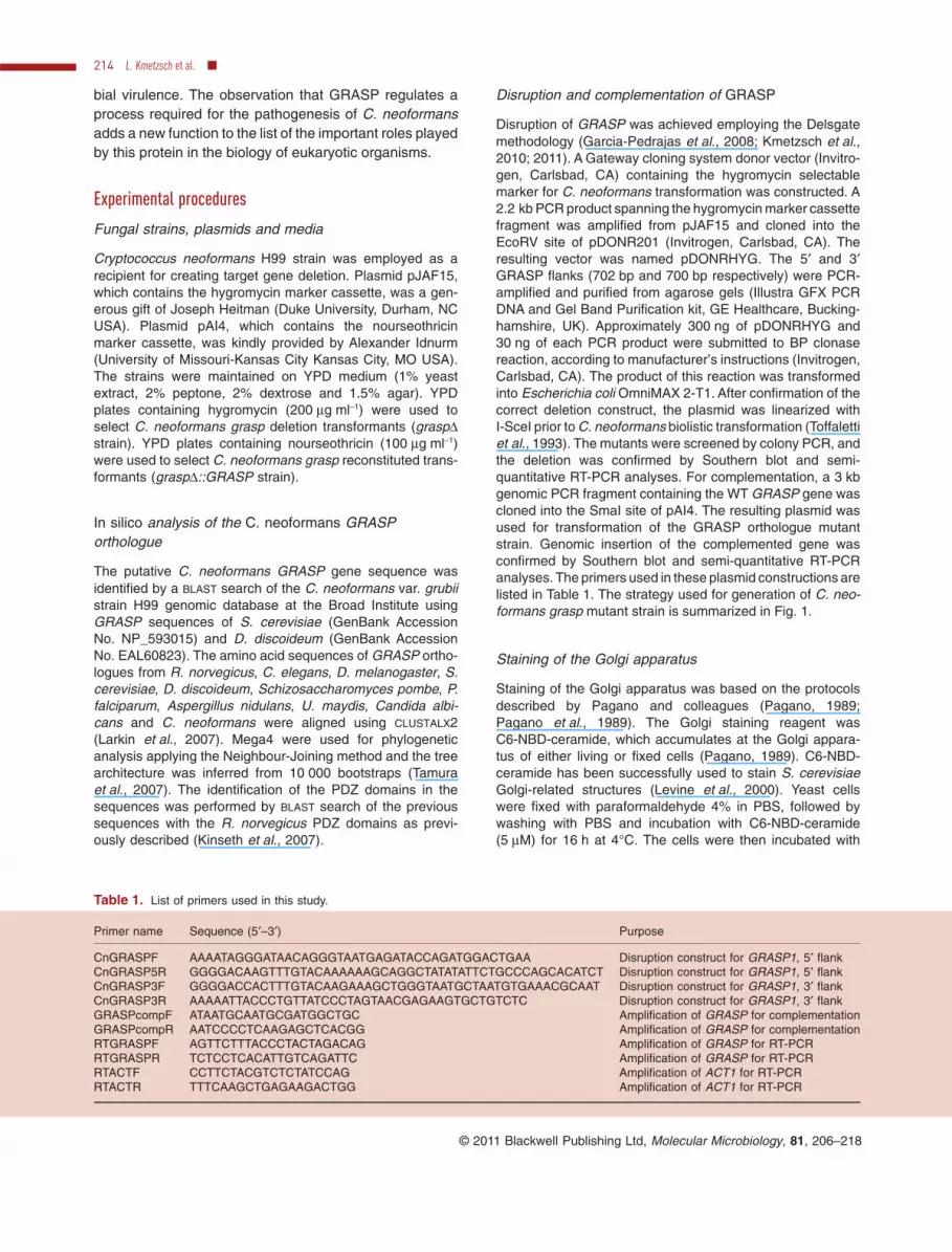
Table 1. List of primers used in this study.
Primer name Sequence (5′–3′) Purpose
CnGRASPF AAAATAGGGATAACAGGGTAATGAGATACCAGATGGACTGAA Disruption construct for GRASP1, 5′ flankCnGRASP5R GGGGACAAGTTTGTACAAAAAAGCAGGCTATATATTCTGCCCAGCACATCT Disruption construct for GRASP1, 5′ flankCnGRASP3F GGGGACCACTTTGTACAAGAAAGCTGGGTAATGCTAATGTGAAACGCAAT Disruption construct for GRASP1, 3′ flankCnGRASP3R AAAAATTACCCTGTTATCCCTAGTAACGAGAAGTGCTGTCTC Disruption construct for GRASP1, 3′ flankGRASPcompF ATAATGCAATGCGATGGCTGC Amplification of GRASP for complementationGRASPcompR AATCCCCTCAAGAGCTCACGG Amplification of GRASP for complementationRTGRASPF AGTTCTTTACCCTACTAGACAG Amplification of GRASP for RT-PCRRTGRASPR TCTCCTCACATTGTCAGATTC Amplification of GRASP for RT-PCRRTACTF CCTTCTACGTCTCTATCCAG Amplification of ACT1 for RT-PCRRTACTR TTTCAAGCTGAGAAGACTGG Amplification of ACT1 for RT-PCR
214 L. Kmetzsch et al. �
© 2011 Blackwell Publishing Ltd, Molecular Microbiology, 81, 206–218

fetal calf serum (10%) at 4°C for 1 h to remove the excess ofC6-NBD-ceramide (Pagano, 1989). For staining of the cellwall, the cells were then incubated for 15 min with Uvitex 2B(0.1 mg ml-1; Polysciences, Warrington, PA), followed bywashing with PBS and analysis by fluorescence microscopy.Images were generated in an ApoTome 2 epifluorescencemicroscope (Carl Zeiss, Germany) and processed with theAxioVision software (Carl Zeiss, Germany).
Virulence assay
Virulence studies were conducted according to a previouslydescribed intranasal inhalation infection model (Cox et al.,2000) using eight female BALB/c mice (approximately 6weeks old) for each strain. Mice were infected with 107 yeastcells suspended in 50 ml of saline and monitored daily. Animalstudies were approved by the Federal University of RioGrande do Sul Ethics Committee. Kaplan–Meier analysis ofsurvival was performed using GraphPad Software.
Phagocytosis of C. neoformans cells by murinemacrophages
Murine macrophage-like cells (RAW 264.7 lineage) wereobtained from the American Type Culture Collection (ATCC).Cultures were maintained and grown to confluence in 25 cm2
culture flasks containing Dulbecco’s modified Eagle’smedium (DMEM) supplemented with 10% (v/v) fetal bovineserum (FBS) at 37°C and 5% CO2. The culture medium wasreplaced with fresh media for further incubation with fluores-cein isothiocyanate (FITC, Sigma Aldrich Corp, St Louis,MO)-labelled C. neoformans yeast cells (Barbosa et al.,2006). Fluorescent yeast cells were prepared by staining with0.5 mg ml-1 FITC in PBS (25°C) for 10 min. FITC-labelled C.neoformans were then suspended in DMEM to generate aratio of 10 fungal cells per macrophage for further incubationat 37°C and 5% CO2 for 18 h. In some systems, yeast cellswere opsonized by treatment with mAb 18B7 (10 mg ml-1) for1 h at 37°C. For negative control we used an isotype-matched irrelevant IgG at the same concentrations used formAb 18B7. Non-adherent yeast cells were removed bywashing with PBS. Fungi–host cell complexes were thentreated for 10 min at 25°C with trypan blue (200 mg ml-1) todiscriminate between surface-associated and intracellularyeast cells. After removal from the plastic surface with a cellscrapper, the cells were analysed by flow cytometry asdescribed previously (Barbosa et al., 2006). Control prepara-tions were developed as described above using uninfectedcells and non-stained yeast. Alternatively, infected macroph-ages were lysed with cold water and the remaining suspen-sion was plated onto Sabouraud solid agar for counting ofcolony-forming units (cfu). These values were used for cal-culation of the survival indices of C. neoformans after inter-action with the phagocytes. The survival index was defined asthe number of cfu divided by the fluorescence index of mac-rophages in each experimental system.
GXM and capsule determination
Cryptococcus neoformans cells (WT, mutant and reconsti-tuted strains) were placed onto glass slides and mixed with
similar volumes of India ink. The suspensions were coveredwith glass coverslips and analysed with an Axioplan 2(Zeiss, Germany) microscope. Images were acquired usinga Color View SX digital camera and processed with the soft-ware system analySIS (Soft Image System). Capsule sizes,defined as the distances between the cell wall and the outerborder of the capsule in India ink stained yeast cells, weredetermined by using the ImageJ Software (version 1.33),elaborated and provided by National Institutes of Health(NIH, http://rsb.info.nih.gov/ij/). Cell diameters were deter-mined using the same software. Final measurements werepresented as ratio of capsule size/cell diameter. Cellularsuspensions were analysed by fluorescence microscopy.The staining reagents used in fluorescence microscopyincluded calcofluor white, which has been extensively usedto stain chitin in fungal cell walls, and the mAb 18B7, amouse IgG1 with high affinity for GXM of different crypto-coccal serotypes (Casadevall et al., 1998). Yeast cells (106)were suspended in 4% paraformaldehyde cacodylate buffer(0.1 M, pH 7.2) and incubated for 30 min at room tempera-ture. Fixed yeast cells were washed twice in PBS and incu-bated in 1% bovine serum albumin in PBS (PBS-BSA) for1 h. The cells were then suspended in 100 ml of a 25 mMcalcofluor white solution (Invitrogen, Carlsbad, CA) for30 min at 37°C. After washing in PBS, the cells were incu-bated for 1 h in the presence of mAb 18B7 (1 mg ml-1). Thecells were finally incubated with a fluorescein isothiocyanate(FITC)-labelled goat anti-mouse IgG (Fc-specific) antibody(Sigma Aldrich Corp, St Louis, MO). For negative control weused an isotype-matched irrelevant IgG at the same con-centrations used for mAb 18B7. Cell suspensions weremounted over glass slides as described above and analy-sed under an Axioplan 2 (Zeiss, Germany) fluorescencemicroscope. Images were acquired and processed asdescribed above.
Determination of GXM concentration in fungalsupernatants and infected tissues
Culture supernatants were obtained as recently described(Fonseca et al., 2010). GXM concentration in fungal super-natants was determined by ELISA with mAb 18B7, usingmodifications of a previously described protocol for GXMdetection (Casadevall et al., 1992; Fonseca et al., 2009).Briefly, 96-well polystyrene plates were coated with standardGXM, supernatant samples for further blocking with bovineserum albumin. The plates were sequentially incubated withmAb 18B7 and an alkaline phosphatase-conjugated goatanti-mouse IgG1 for 1 h. Reactions were developed after theaddition of p-nitrophenyl phosphate disodium hexahydrate,followed by measuring absorbance at 405 nm with a micro-plate reader (TP-reader, Thermo Plate). Antibody concen-tration in this assay corresponded to 1 mg ml-1. For in vivodeterminations, tissue samples were obtained after intrana-sal infection female BALB/c (n = 10) with 106 yeast cells ofWT, mutant or reconstituted C. neoformans cells (Cox et al.,2000). At day 7 post infection, animals were euthanized andtheir lungs and brains were aseptically excised. Tissues werethen homogenized by maceration in PBS. These suspen-sions were plated on Sabouraud agar for cfu counting or wereprocessed for GXM determination. For this purpose, suspen-
Role of GRASP in polysaccharide secretion 215
© 2011 Blackwell Publishing Ltd, Molecular Microbiology, 81, 206–218

sions were supplemented with proteinase K (0.2 mg ml-1,final concentration) and incubated overnight at 37°C.Samples were then heated for 20 min at 100°C, placed on iceand centrifuged at 10 000 g. Supernatants were then used infor GXM determination. For the in vivo systems, high back-grounds were observed when samples were analysed byELISA (data not shown). We therefore analysed in vivosamples by dot blotting. Samples were loaded onto nitro-cellulose membranes, which were allowed to dry for 1 h at37°C and then were blocked with PBS containing 5% bovineserum albumin. Blocked membranes were incubated withmAb 18B7 at a starting dilution of 1 mg ml-1. After beingwashed extensively, membranes were sequentially incubatedwith peroxidase-conjugated goat anti-mouse IgG1 and ortho-phenylene diamine solutions. Reactions were quantified bydensitometry with the Scion Image software (version 4.03)and normalized to cfu values and weight of infected tissues.
Analysis of virulence factors
Urease activity and melanin formation, two well-defined C.neoformans virulence factors that are related to vesicularsecretion (Rodrigues et al., 2008b; Eisenman et al., 2009),were used as prototypes in assays aiming at the evaluation ofvirulence determinants that are not associated to capsuleexpression. Urease activity was assayed after growth ofWT, mutant and reconstituted strains in Christensen’s agarmedium at 30°C for 48 h (Cox et al., 2000). This mediumcontains 300 mM urea and phenol red as a pH indicator. Theurease activity is expected to convert urea into ammonia,resulting in an increase in the pH of the medium. This featureis reflected by a colour change from yellow to bright pink. Aure1 mutant was used as a negative control. Analysis ofmelanin production followed the methodology described byBaker et al. (2007). WT, mutant and complemented cellswere inoculated in a chemically defined medium containingL-asparagine (1 g l-1), MgSO4·7H2O (0.5 g l-1), KH2PO4
(3 g l-1) and thiamine (1 mg l-1), supplemented with 10 mML-3,4-dihydroxyphenylalanine (L-DOPA). C. neoformans cellswere then cultivated at 30°C. After 24 h intervals, the cultureswere centrifuged 1000 g for 10 min; pellets were photo-graphed for visual analysis of pigmentation and the superna-tants were spectrophotometrically analysed (absorbance492 nm). Shorter times were also analysed, but pigmentationwas only observed after 24 h in the culture medium (data notshown).
GXM effective diameter
For diameter determination, extracellular GXM was isolatedfrom culture supernatants as previously described by ourgroup (Nimrichter et al., 2007). Yeast cells were cultivated ina minimal medium composed of glucose (15 mM), MgSO4
(10 mM), KH2PO4 (29.4 mM), glycine (13 mM) and thiamine-HCl (3 mM), pH 5.5, for 2 days at room temperature withshaking and separated from culture supernatants by centrifu-gation at 4000 g (15 min, 4°C). The supernatant fluids werecollected and again centrifuged at 15 000 g (15 min, 4°C), toremove smaller debris. The pellets were discarded and theresulting supernatant was concentrated approximately 20-fold
using an Amicon (Millipore, Danvers, MA) ultrafiltration cell(Nimrichter et al., 2007). After supernatant concentration, theviscous layer formed was collected with a cell scraper andtransferred to graduated plastic tubes for GXM determina-tions. The effective diameter of GXM in these samples wasdetermined by Quasi elastic light scattering in a 90Plus/BI-MAS Multi Angle Particle Sizing analyser (Brookhaven Instru-ments Corp., Holtsville, NY), using minor modifications of themethod described by Frases et al. (2009). Polysaccharidediameter was modulated by incubation of dialysed fractionswith 1 mM CaCl2 for 1 h at room temperature.
Acknowledgements
M.L.R., M.H.V., L.N. and A.S. are supported by grants fromCoordenação de Aperfeiçoamento de Pessoal de NívelSuperior (CAPES, Brazil), Conselho Nacional de Desenvolvi-mento Científico e Tecnológico (CNPq, Brazil), Fundação deAmparo a Pesquisa do Estado de São Paulo (FAPESP,Brazil), Fundação de Amparo a Pesquisa do Estado do Riode Janeiro (FAPERJ, Brazil) and Financiadora de Estudos eProjetos (FINEP, Brazil). A.C. is supported by NIH GrantsAI033142, AI033774, AI052733 and HL059842. We thankPriscila C. Albuquerque and Ana Claudia Zimbres for helpwith handling cultures of fungal mutants. We are thankfulto the reviewers for their insightful comments that helpedstrengthen this article. We are also thankful to Prof. VivekMalhotra for initial suggestions on the role of GRASP inpolysaccharide secretion.
References
Albuquerque, P.C., Nakayasu, E.S., Rodrigues, M.L., Frases,S., Casadevall, A., Zancope-Oliveira, R.M., et al. (2008)Vesicular transport in Histoplasma capsulatum: an effec-tive mechanism for trans-cell wall transfer of proteins andlipids in ascomycetes. Cell Microbiol 10: 1695–1710.
Baker, L.G., Specht, C.A., Donlin, M.J., and Lodge, J.K.(2007) Chitosan, the deacetylated form of chitin, is neces-sary for cell wall integrity in Cryptococcus neoformans.Eukaryot Cell 6: 855–867.
Barbosa, F.M., Fonseca, F.L., Holandino, C., Alviano,C.S., Nimrichter, L., and Rodrigues, M.L. (2006)Glucuronoxylomannan-mediated interaction of Cryptococ-cus neoformans with human alveolar cells results in fungalinternalization and host cell damage. Microbes Infect 8:493–502.
Cabral, M., Anjard, C., Malhotra, V., Loomis, W.F., andKuspa, A. (2010) Unconventional secretion of AcbA in Dic-tyostelium discoideum through a vesicular intermediate.Eukaryot Cell 9: 1009–1017.
Casadevall, A., Mukherjee, J., and Scharff, M.D. (1992)Monoclonal antibody based ELISAs for cryptococcalpolysaccharide. J Immunol Methods 154: 27–35.
Casadevall, A., Cleare, W., Feldmesser, M., Glatman-Freedman, A., Goldman, D.L., Kozel, T.R., et al. (1998)Characterization of a murine monoclonal antibody toCryptococcus neoformans polysaccharide that is a can-didate for human therapeutic studies. Antimicrob AgentsChemother 42: 1437–1446.
216 L. Kmetzsch et al. �
© 2011 Blackwell Publishing Ltd, Molecular Microbiology, 81, 206–218

Casadevall, A., Nosanchuk, J.D., Williamson, P., and Rod-rigues, M.L. (2009) Vesicular transport across the fungalcell wall. Trends Microbiol 17: 158–162.
Chang, Y.C., Wickes, B.L., and Kwon-Chung, K.J. (1995)Further analysis of the CAP59 locus of Cryptococcus neo-formans: structure defined by forced expression anddescription of a new ribosomal protein-encoding gene.Gene 167: 179–183.
Chayakulkeeree, M., Johnston, S.A., Oei, J.B., Lev, S.,Williamson, P.R., Wilson, C.F., et al. (2011) SEC14 is aspecific requirement for secretion of phospholipase B1 andpathogenicity of Cryptococcus neoformans. Mol Microbiol80: 1088–1101.
Cox, G.M., Mukherjee, J., Cole, G.T., Casadevall, A., andPerfect, J.R. (2000) Urease as a virulence factor in experi-mental cryptococcosis. Infect Immun 68: 443–448.
Doering, T.L. (2009) How sweet it is! Cell wall biogenesis andpolysaccharide capsule formation in Cryptococcusneoformans. Annu Rev Microbiol 63: 223–247.
Duran, J.M., Anjard, C., Stefan, C., Loomis, W.F., andMalhotra, V. (2010) Unconventional secretion of Acb1 ismediated by autophagosomes. J Cell Biol 188: 527–536.
Eisenman, H.C., Frases, S., Nicola, A.M., Rodrigues, M.L.,and Casadevall, A. (2009) Vesicle-associated melanizationin Cryptococcus neoformans. Microbiology 155: 3860–3867.
Feldmesser, M., Kress, Y., and Casadevall, A. (2001)Dynamic changes in the morphology of Cryptococcus neo-formans during murine pulmonary infection. Microbiology147: 2355–2365.
Fonseca, F.L., Frases, S., Casadevall, A., Fischman-Gompertz, O., Nimrichter, L., and Rodrigues, M.L. (2009)Structural and functional properties of the Trichosporonasahii glucuronoxylomannan. Fungal Genet Biol 46: 496–505.
Fonseca, F.L., Nohara, L.L., Cordero, R.J., Frases, S., Casa-devall, A., Almeida, I.C., et al. (2010) Immunomodulatoryeffects of serotype B glucuronoxylomannan from Crypto-coccus gattii correlate with polysaccharide diameter. InfectImmun 78: 3861–3870.
Frases, S., Nimrichter, L., Viana, N.B., Nakouzi, A., andCasadevall, A. (2008) Cryptococcus neoformans capsularpolysaccharide and exopolysaccharide fractions manifestphysical, chemical, and antigenic differences. EukaryotCell 7: 319–327.
Frases, S., Pontes, B., Nimrichter, L., Viana, N.B., Rodrigues,M.L., and Casadevall, A. (2009) Capsule of Cryptococcusneoformans grows by enlargement of polysaccharidemolecules. Proc Natl Acad Sci USA 106: 1228–1233.
Garcia-Pedrajas, M.D., Nadal, M., Kapa, L.B., Perlin, M.H.,Andrews, D.L., and Gold, S.E. (2008) DelsGate, a robustand rapid gene deletion construction method. FungalGenet Biol 45: 379–388.
Garcia-Rivera, J., Chang, Y.C., Kwon-Chung, K.J., and Casa-devall, A. (2004) Cryptococcus neoformans CAP59(or Cap59p) is involved in the extracellular traffickingof capsular glucuronoxylomannan. Eukaryot Cell 3: 385–392.
Hu, G., Steen, B.R., Lian, T., Sham, A.P., Tam, N., Tangen,K.L., and Kronstad, J.W. (2007) Transcriptional regulation
by protein kinase A in Cryptococcus neoformans. PLoSPathog 3: e42.
Kinseth, M.A., Anjard, C., Fuller, D., Guizzunti, G., Loomis,W.F., and Malhotra, V. (2007) The Golgi-associated proteinGRASP is required for unconventional protein secretionduring development. Cell 130: 524–534.
Kmetzsch, L., Staats, C.C., Simon, E., Fonseca, F.L., deOliveira, D.L., Sobrino, L., et al. (2010) The vacuolarCa(2)(+) exchanger Vcx1 is involved in calcineurin-dependent Ca(2)(+) tolerance and virulence in Cryptococ-cus neoformans. Eukaryot Cell 9: 1798–1805.
Kmetzsch, L., Staats, C.C., Simon, E., Fonseca, F.L.,Oliveira, D.L., Joffe, L.S., et al. (2011) The GATA-typetranscriptional activator Gat1 regulates nitrogen uptakeand metabolism in the human pathogen Cryptococcusneoformans. Fungal Genet Biol 48: 192–199.
Larkin, M.A., Blackshields, G., Brown, N.P., Chenna, R.,McGettigan, P.A., McWilliam, H., et al. (2007) Clustal Wand Clustal X version 2.0. Bioinformatics 23: 2947–2948.
Levi, S.K., and Glick, B.S. (2007) GRASPing unconventionalsecretion. Cell 130: 407–409.
Levine, T.P., Wiggins, C.A., and Munro, S. (2000) Inositolphosphorylceramide synthase is located in the Golgi ap-paratus of Saccharomyces cerevisiae. Mol Biol Cell 11:2267–2281.
Li, S.S., and Mody, C.H. (2010) Cryptococcus. Proc AmThorac Soc 7: 186–196.
McClelland, E.E., Bernhardt, P., and Casadevall, A. (2005)Coping with multiple virulence factors: which is most impor-tant? PLoS Pathog 1: e40.
Manjithaya, R., Anjard, C., Loomis, W.F., and Subramani, S.(2010) Unconventional secretion of Pichia pastoris Acb1 isdependent on GRASP protein, peroxisomal functions, andautophagosome formation. J Cell Biol 188: 537–546.
Nickel, W., and Rabouille, C. (2009) Mechanisms of regu-lated unconventional protein secretion. Nat Rev Mol CellBiol 10: 148–155.
Nickel, W., and Seedorf, M. (2008) Unconventional mecha-nisms of protein transport to the cell surface of eukaryoticcells. Annu Rev Cell Dev Biol 24: 287–308.
Nimrichter, L., Frases, S., Cinelli, L.P., Viana, N.B., Nakouzi,A., Travassos, L.R., et al. (2007) Self-aggregation of Cryp-tococcus neoformans capsular glucuronoxylomannan isdependent on divalent cations. Eukaryot Cell 6: 1400–1410.
Nosanchuk, J.D., Nimrichter, L., Casadevall, A., andRodrigues, M.L. (2008) A role for vesicular transport ofmacromolecules across cell walls in fungal pathogenesis.Commun Integr Biol 1: 37–39.
Oliveira, D.L., Nimrichter, L., Miranda, K., Frases, S., Faull,K.F., Casadevall, A., and Rodrigues, M.L. (2009) Cryp-tococcus neoformans cryoultramicrotomy and vesiclefractionation reveals an intimate association betweenmembrane lipids and glucuronoxylomannan. Fungal GenetBiol 46: 956–963.
Oliveira, D.L., Nakayasu, E.S., Joffe, L.S., Guimaraes, A.J.,Sobreira, T.J., Nosanchuk, J.D., et al. (2010a) Biogenesisof extracellular vesicles in yeast: many questions with fewanswers. Commun Integr Biol 3: 533–535.
Oliveira, D.L., Nakayasu, E.S., Joffe, L.S., Guimaraes, A.J.,Sobreira, T.J., Nosanchuk, J.D., et al. (2010b) Character-
Role of GRASP in polysaccharide secretion 217
© 2011 Blackwell Publishing Ltd, Molecular Microbiology, 81, 206–218

ization of yeast extracellular vesicles: evidence for the par-ticipation of different pathways of cellular traffic in vesiclebiogenesis. PLoS ONE 5: e11113.
Pagano, R.E. (1989) A fluorescent derivative of ceramide:physical properties and use in studying the Golgi apparatusof animal cells. Methods Cell Biol 29: 75–85.
Pagano, R.E., Sepanski, M.A., and Martin, O.C. (1989)Molecular trapping of a fluorescent ceramide analogue atthe Golgi apparatus of fixed cells: interaction with endog-enous lipids provides a trans-Golgi marker for bothlight and electron microscopy. J Cell Biol 109: 2067–2079.
Panepinto, J., Komperda, K., Frases, S., Park, Y.D., Djord-jevic, J.T., Casadevall, A., and Williamson, P.R. (2009)Sec6-dependent sorting of fungal extracellular exosomesand laccase of Cryptococcus neoformans. Mol Microbiol71: 1165–1176.
Park, B.J., Wannemuehler, K.A., Marston, B.J., Govender,N., Pappas, P.G., and Chiller, T.M. (2009) Estimation of thecurrent global burden of cryptococcal meningitis amongpersons living with HIV/AIDS. AIDS 23: 525–530.
Prado, M., Silva, M.B., Laurenti, R., Travassos, L.R., andTaborda, C.P. (2009) Mortality due to systemic mycoses asa primary cause of death or in association with AIDS inBrazil: a review from 1996 to 2006. Mem Inst OswaldoCruz 104: 513–521.
Rodrigues, M.L., Nimrichter, L., Oliveira, D.L., Frases, S.,Miranda, K., Zaragoza, O., et al. (2007) Vesicular polysac-charide export in Cryptococcus neoformans is a eukaryoticsolution to the problem of fungal trans-cell wall transport.Eukaryot Cell 6: 48–59.
Rodrigues, M.L., Nimrichter, L., Oliveira, D.L., Nosanchuk,J.D., and Casadevall, A. (2008a) Vesicular trans-cell walltransport in fungi: a mechanism for the delivery ofvirulence-associated macromolecules? Lipid Insights 2:27–40.
Rodrigues, M.L., Nakayasu, E.S., Oliveira, D.L., Nimrichter,L., Nosanchuk, J.D., Almeida, I.C., and Casadevall, A.(2008b) Extracellular vesicles produced by Cryptococcusneoformans contain protein components associated withvirulence. Eukaryot Cell 7: 58–67.
Schotman, H., Karhinen, L., and Rabouille, C. (2008)dGRASP-mediated noncanonical integrin secretion is
required for Drosophila epithelial remodeling. Dev Cell 14:171–182.
Seider, K., Heyken, A., Luttich, A., Miramon, P., and Hube, B.(2010) Interaction of pathogenic yeasts with phagocytes:survival, persistence and escape. Curr Opin Microbiol 13:392–400.
Sommer, U., Liu, H., and Doering, T.L. (2003) An alpha-1,3-mannosyltransferase of Cryptococcus neoformans. J BiolChem 278: 47724–47730.
Takeo, K., Uesaka, I., Uehira, K., and Nishiura, M. (1973a)Fine structure of Cryptococcus neoformans grown in vitroas observed by freeze-etching. J Bacteriol 113: 1442–1448.
Takeo, K., Uesaka, I., Uehira, K., and Nishiura, M. (1973b)Fine structure of Cryptococcus neoformans grown in vivo asobserved by freeze-etching. J Bacteriol 113: 1449–1454.
Tamura, K., Dudley, J., Nei, M., and Kumar, S. (2007) MEGA4:Molecular Evolutionary Genetics Analysis (MEGA) softwareversion 4.0. Mol Biol Evol 24: 1596–1599.
Toffaletti, D.L., Rude, T.H., Johnston, S.A., Durack, D.T., andPerfect, J.R. (1993) Gene transfer in Cryptococcus neofor-mans by use of biolistic delivery of DNA. J Bacteriol 175:1405–1411.
Vallejo, M.C., Matsuo, A.L., Ganiko, L., Medeiros, L.C.,Miranda, K., Silva, L.S., et al. (2010) The pathogenicfungus Paracoccidioides brasiliensis exports extracellularvesicles containing highly immunogenic {alpha}-galactosylepitopes. Eukaryot Cell 10: 343–351.
Vinke, F.P., Grieve, A.G., and Rabouille, C. (2011) The mul-tiple facets of the Golgi reassembly stacking proteins.Biochem J 433: 423–433.
Yoneda, A., and Doering, T.L. (2006) A eukaryotic capsularpolysaccharide is synthesized intracellularly and secretedvia exocytosis. Mol Biol Cell 17: 5131–5140.
Zaragoza, O., Telzak, A., Bryan, R.A., Dadachova, E., andCasadevall, A. (2006) The polysaccharide capsule of thepathogenic fungus Cryptococcus neoformans enlarges bydistal growth and is rearranged during budding. Mol Micro-biol 59: 67–83.
Zaragoza, O., Rodrigues, M.L., De Jesus, M., Frases, S.,Dadachova, E., and Casadevall, A. (2009) The capsule ofthe fungal pathogen Cryptococcus neoformans. Adv ApplMicrobiol 68: 133–216.
218 L. Kmetzsch et al. �
© 2011 Blackwell Publishing Ltd, Molecular Microbiology, 81, 206–218


