Embed Size (px)
Citation preview

REVIEW
Leonard J. Cleary, 1 John H. Byrne, and William N. Frost
Department of Neur0bi010gy and Anatomy
University of Texas Houston Medical School
Houston, Texas 77225
Role of Interneurons in Defensive Withdrawal Reflexes in Aplysia
Introduction Much attention has been focused on the contr ibution of sensory neurons and their synaptic connections to defensive reflexes in Aplysia. The two best understood withdrawal reflexes in terms of their underlying circuitry and their roles in learning and memory, are the siphon-elicited siphon-gill withdrawal reflex and the tail-elicited tail-siphon withdrawal reflex (Fig. 1; for review, see Kandel and Schwartz 1982; Carew and Sahley 1986; Hawkins et al. 1986; Byrne 1987; Byrne et al. 1991; Frost and Kandel 1995; Byrne and Kandel 1996). Both reflex responses have been used extensively to analyze the neural and molecular mechanisms of sensitization, a simple form of nonassociative learning in which an unexpected (often noxious) stimulus enhances the response to a subsequent test stimulus (for review, see Byrne et al. 1993; Hawkins et al. 1993). Another form of nonassociative learning, habituation, has also been the subject of study, although to a lesser extent. The duration of both forms of learning can be brief (minutes) or long (days), depending on the training protocol.
Much of the work on the mechanisms underlying sensitization in these two reflexes has focused on training-related modifications of the central sensory neurons. In particular, sensitizing stimuli act to enhance both sensory neuron excitability and transmitter release (for review, see Byrne et al. 1993; Hawkins et al. 1993). These changes occur via the activation of second messenger systems in a time- and state-dependent manner (Baxter and Byrne 1990; Braha et al. 1990; Ghirardi et al. 1992; Pieroni and Byrne 1992; Sugita et al. 1992, 1994; Byrne and Kandel 1996). Several of these storage processes operate in parallel to maintain both the short- and long-term components of the memory for sensitization (Frost et al. 1985; Greenberg et al. 1987; Schwartz and Greenberg 1987; Scholz and Byrne 1988; Bailey et al. 1992; Kennedy et al. 1992; Byrne et al. 1993; Alberini et al. 1994; O'Leary et al. 1995).
More recent studies have begun to explore the role of circuit interneurons in both the mediation and modulation of these withdrawal responses. This review will address several questions about the roles played by the interneurons in these circuits. What do the interneurons contr ibute to the mediation of these reflexes? Does their contr ibution differ qualitatively from that made by the monosynaptic sensory neuron to motor neuron connections? Do the interneurons themselves serve as sites of memory storage and, if so, do they use the same storage mechanisms as those utilized by the sensory neurons? What role do interneurons play in learning-related modulation of the circuits?
The findings reviewed here indicate that interneurons make important and distinctive contributions to the mediation of withdrawal reflexes in Aplysia. Furthermore, some of the interneurons serve as sites of information storage in habituation and sensitization, storing different
1Corresponding author, information, and utilizing different types of plasticity than the sensory
LEARNING & MEMORY 2:133-151 �9 1995 by Cold Spring Harbor Laboratory Press ISSN1072-0502/95 $5.00
& 133
L E A R N I N G M E M 0 R Y

Cleary et al.
A. Siphon-elicited Siphon-Gill Reflex B. Tail-elicited Tail-Siphon Reflex
1. Relaxed 2. Withdrawn 1. Relaxed 2. Withdrawn
Gill
~ Siphon
Stimulus ~ '"" -....' "
Tail Stimulus
Figure 1: Defensive withdrawal reflexes of Aplysia. (A) Siphon-elicited siphon and gill withdrawal. A dorsal view of the animal is illustrated in the relaxed position (1). A stimulus (e.g., a water jet, brief touch, or weak electric shock) applied to the siphon causes the siphon and the gill to withdraw into the mantle cavity (2). (B) Tail-elicited tail and siphon withdrawal reflex. From the relaxed position (1), a similar tactile stimulus applied to the tail elicits a reflex withdrawal of the tail and siphon (2). The gill will frequently contract as well.
The Siphon-elicited and Tail-elicited Reflex Withdrawal Circuits
THE SIPHON-ELICITED SIPHON WITHDRAWAL CIRCUIT
neurons. Finally, some interneurons serve as modulatory elements that activate the cellular mechanisms underlying sensitization.
The two defensive withdrawal reflexes that are the focus of this review share many similarities. Each has a well-characterized population of central mechanosensory neurons that connects to motor neurons via both monosynaptic connections and polysynaptic pathways involving interneurons (Byrne et al. 1974; Wakers et al. 1983). Although the motor neurons for both networks are well characterized, the interneurons are less well understood. In this review we focus on the siphon withdrawal component of the siphon-gill withdrawal circuit. The gill withdrawal component shares some of the same interneurons but is understood less completely (Hawkins et al. 1981a; Koester 1989).
Approximately 100 neurons in the abdominal ganglion are known to participate in siphon movements elicited by tactile siphon stimulation. These include 44 siphon sensory neurons, 41 interneurons, and 15 motor neurons (for review, see Frost and Kandel 1995). Because each sensory neuron's receptive field covers only a portion of the siphon (Byrne et al. 1974; Dubuc and Castellucci 1991 ), a punctate tactile stimulus will activate only a fraction of the sensory neuron population. An unidentified group of low-threshold sensory neurons also appears to contribute to siphon-elicited siphon withdrawals (Cohen et al. 1991; Fischer and Carew 1993; Kaplan et al. 1993; see also Byrne et al. 1978a). These cells appear to play an important role in mediating responses to weak tactile
& 134
L E A R N I N G M E M O R Y

ROLE OF INTERNEURONS IN LEARNING
THE TAIL-ELICITED TAIL AND SIPHON WITHDRAWAL CIRCUIT
siphon stimuli and display synaptic plasticity during habituation and dishabituation similar to that of the well-characterized LE cluster of sensory neurons (Kaplan et al. 1994).
The interneurons of the network for siphon-elicited siphon withdrawal are organized into two distinct subcircuits in the abdominal ganglion (Fig. 2). The first consists of four groups of neurons (L16, L29, L30, L34) that mediate reflex withdrawals only. The second subcircuit consists of interneurons (L25, R25, L26, L33) that are recrui ted during reflex withdrawals (Kanz et al. 1979; Byrne 1983; Eberly and Pinsker 1984; Frost and Kandel 1995) but that also mediate the spontaneous withdrawal associated with respiratory pumping (Byrne and Koester 1978; Byrne 1983; Koester 1989).
There are four clusters of siphon motor neurons within the abdominal ganglion (LBS, LDS, RDS, and LFS) and a peripheral group of motor neurons located along the siphon nerve (Perlman 1979; Bailey et al. 1979; Frost et al. 1988). Each motor neuron group contr ibutes a distinct component to the overall siphon-elicited siphon withdrawal response (Perlman 1979; Hickie 1995; Frost and Kandel 1995). Both the central and the peripheral motor neurons are innervated directly by the LE sensory neurons (Byrne et al. 1974; Bailey et al. 1979; Clark and Kandel 1984).
The afferent limb of the tail-elicited tail and siphon withdrawal reflex consists of mechanosensory neurons innervating the tail whose cell bodies are located in a cluster on the ventrolateral surface of the left and
Figure 2: Simplified diagram of circuits mediating the siphon-elicited siphon and tail-elicited tail and siphon withdrawal reflexes. Stimuli applied to the siphon or tail activate the afferent terminals of siphon sensory neurons (SSN) or of tail sensory neurons (TSN) whose somata are located in central ganglia. The sensory neurons make monosynaptic excitatory connections (A) with interneurons and motor neu- rons. The excitatory interneurons provide a parallel pathway for excitation of the motor neurons. Action potentials elicited in the motor neurons, triggered by the combined input from the sensory neurons and interneurons, propagate out periph- eral nerves to activate muscle cells and produce the subsequent reflex withdrawal. Not shown here are elements of the gill withdrawal circuit. Shaded areas represent ganglia in which cell bodies of circuit neurons are located.
& 1 3 5
L E A R N I N G M E M 0 R Y

Cleary et al.
Interneurons Make a Unique Contribution to the Tail- and Siphon-el icited Withdrawal Reflexes
THE ROLE OF
INTERNEURONS IN BASIC REFLEX FUNCTION
right pleural ganglia (Waiters et al. 1983). Of approximately 200 neurons in each cluster, at least 20 project through the posterior pedal nerve (P9), which innervates the tail (Waiters et al. 1983; Zhang et al. 1993). These tail sensory neurons make monosynaptic connect ions to three identified motor neurons located in the pedal ganglion (Figr 2). To date, two interneurons have been identified in this circuit. LPI17 excites tail motor neurons and appears to contribute to both the tail and siphon components of the response (Cleary and Byrne 1993). RPI4 inhibits tail motor neurons (Buonomano et al. 1992; Xu et al. 1994). Both LPI17 and RPI4 produce feedback inhibition onto tail sensory neurons. Another identified interneuron, RPI5, also inhibits tail sensory neurons but does not appear to receive direct input from them. Tail sensory neurons also provide input to the central pattern generator for locomotion (Hening et al. 1979; Waiters et al. 1981 ), but it is not known whether those connections are mono- or polysynaptic. Neither the tail interneurons nor motor neurons appear to receive input from the locomotor pat tern generator. Whereas the tail-elicited reflex circuitry is not unders tood in the same detail as the siphon-elicited reflex circuitry, features of these circuits are common to both and may represent general principles of reflex circuit organization.
In both the tail- and siphon-elicited withdrawal circuits, sensory neurons excite motor neurons via both mono- and polysynaptic pathways. These pathways are sufficiently strong that intracellular activation of single sensory neurons can elicit withdrawal responses in semi-intact preparations (Byrne et al. 1978a; Waiters et al. 1983). The role of the monosynaptic connection is straightforward; its activity in response to a skin stimulus depolarizes the motor neuron, typically eliciting a brief burst of action potentials. The role of the recrui ted interneurons is more varied and complex.
There is some variation in both the siphon-elicited and taR-elicited withdrawal reflexes, but the most common reflex response is a relatively quick initial contraction to maximal amplitude, followed by a slower relaxation phase (Perlman 1979; Carew et al. 1981; Waiters et al. 1983; Stopfer and Carew 1987; Frost and Kandel 1995). The temporal features of the contraction are primarily attributable to the activity pat tern of the motor neurons, which in most cases consists of a brief high frequency burst of action potentials, followed by a period of lower frequency firing lasting several seconds (Perlman 1979; Waiters et al. 1983; Hiclde 1995; Frost and Kandel 1995).
The sensory neurons fire only during the tactile stimulus (Byrne et al. 1978b) and thus contribute to the early activation of the motor neurons. Although the rapid monosynaptic sensory neuron to motor n e u r o n
excitatory postsynaptic potential (EPSP) makes a major contr ibution to the initial phasic component of motor neuron input, the recrui tment of interneurons is also important. For example, simultaneous recordings from sensory neurons, interneurons, and motor neurons in the
siphon-elicited withdrawal circuR have shown that single sensory neurons recruit interneurons that add substantially to the monosynaptic EPSP (Frost and Kandel 1995). In addition, hyperpolarization of individual L29 interneurons significantly reduced the amplitude of complex EPSPs evoked by s iphon-tap in siphon motor neurons (Fischer
& 136
L E A R N I N G M E M 0 R Y

ROLE OF INTERNEURONS IN LEARNING
THE ROLE OF INTERNEURONS IN COORDINATING THE BEHAVIORAL RESPONSE
SHAPING OF UNIQUE MOTOR
NEURON RESPONSES
MAINTENANCE OF SINGLENESS OF
ACTION
and Carew 1993). Furthermore, selective suppression of interneuronal recrui tment with high divalent cation solutions reduced input from siphon stimulation (Byrne et al. 1978a; Trudeau and Castellucci 1992).
The slow long-lasting component of the motor neuron response to tactile stimulation appears to be attributable entirely to recrui ted interneuronal input. Whereas the sensory neuron to motor neuron EPSPs are short-lived in duration, both circuits contain interneurons that connect with the motor neurons with slow synaptic potentials lasting up to several seconds in duration. In the siphon-elicited siphon withdrawal reflex, interneuron L29 produces a combined fast/slow EPSP onto the LFS siphon motor neurons which can produce long-lasting LFS firing (Frost and Kandel 1995). Similarly, in the tail-elicited withdrawal reflex, activity in LPI17 may elicit a biphasic response in tail motor neurons (Cleary and Byrne 1993). The fast component is frequently sufficient to elicit a burst of spikes. LPI 17 also produces a very strong and long-lasting slow EPSP in motor neurons innervating both the tail and siphon. Because short interneuron bursts can produce motor neuron activity that lasts much longer, these interneuronal connections may play a role in transforming brief sensory neuron inputs into long-lasting motor neuron firing responses (Byrne 1983). This suggestion is supported by simulations of realistic mathematical models (Frost et al. 1991; White et al. 1993).
Tail stimulation generally elicits an excitatory response in tail motor neurons. Siphon motor neurons, however, have a more diverse range of responses to tactile stimulation. As ment ioned above, the LFxS neurons respond to tactile stimulation of the siphon or tail with a phasic and long-lasting firing response. On the other hand, LBS neurons typically respond to tactile stimulation of the siphon with a prolonged period of inhibition that may be preceded by a small burst of action potentials (Perlman 1979; Hickie 1994). These differences in response are attributable to differences in interneuronal recrui tment by the sensory neurons. A more complex example is recrui tment of a pattern-generating ne twork Respiratory pumping is a s tereotyped behavior during which the siphon, gill, mantle, and parapodia contract in a coordinated manner (Kupfermann et al. 1974; Perlman 1979; Pinsker 1982; Eberly and Pinsker 1984). This behavior occurs spontaneously every few minutes but may also be triggered by tactile stimulation. During a pumping event, each class of motor neurons is activated, in part, by input from an identified population of interneurons (Byrne and Koester 1978; Byrne 1983; Alevizos et al. 1989; Frost and Kandel 1995).
Another way in which interneurons shape the motor neuron response is by regulating input from outside of the withdrawal reflex circuit. For example, in the siphon-elicited siphon withdrawal reflex, L21 and L35 are activated by neurons outside of the circuit (Frost and Kandel 1995). These neurons inhibit L29 and, therefore, could potentially interfere with reflex response. This is avoided, however, because siphon stimuli activate the L30 inhibitory interneurons, which, in addition to their connect ions
& 137
L E A R N I N G M E M O R Y

Cleary et al.
DISTRIBUTION OF SENSORY
INFORMATION TO OTHER
CIRCUITS
THE ROLE OF INTERNEURONS IN TRANSFORMING SENSORY NEURON PLASTICITY
in the reflex circuit, also suppress the activity of L21 and L35 (Frost and Kandel 1995). This suppression of outside inhibitory inputs occurs during the withdrawal response and may reduce the influence of interfering inputs during the siphon-elicited response.
In addition to mediating these two reflexes, interneurons also coordinate the total behavioral response by distributing information about sensory neuron stimulation to other parts of the nervous system (Byrne 1981; Cleary and Byrne 1993). In the siphon-elicited siphon withdrawal reflex, > 100 neurons in the abdominal ganglion are activated in response to a tactile siphon stimulus (Zecevic et al. 1989). Much of this may be attributable to the divergent output of the L25/R25 pat tern generating neurons that trigger respiratory pumping. In addition to their effects on siphon motor neurons, the L25/R25 neurons elicit both excitatory and inhibitory responses in a wide variety of neurons throughout the ganglion (Koester and Kandel 1977). These targets include gill motor neurons, the multifunction neuron LIO, and the left upper quadrant cells, which regulate closure of the renal pore. Information about siphon stimulation may also be conveyed by neurons that project to other ganglia. For example, L29 makes reciprocal excitatory connections with LPI17 in the pleural ganglion (Cleary and Byrne 1993).
In the tail-elicited withdrawal reflex, interneurons appear to have a major role in distributing information about tail stimulation to other parts of the nervous system (Cleary and Byrne 1993). Unlike the siphon sensory neurons, which are located in the same ganglia as their target motor neurons, tail sensory neurons project to the adjacent pedal ganglion. However, they do not project to the abdominal ganglion. Consequently, the siphon withdrawal component of the response to tail stimulation requires the activity of interneurons. LPI 17 is one such interneuron that excites siphon motor neurons, both directly and indirectly (Fig. 2). Other circuits receiving input from LPI17 include those mediating head withdrawal and mucus release.
In both habituation and sensitization of Aplysia defensive withdrawal reflexes, the synapses made by central sensory neurons onto motor neurons serve as important sites of memory storage (Kandel and Schwartz 1982). The parallel modulation of synaptic strength be tween sensory neurons and interneurons is also an effective mechanism for modifying behavior. In habituation, repetitive stimulation of either the skin or the sensory neurons themselves leads to progressive decrement of monosynaptic connections from sensory neurons onto circuit interneurons and motor neurons (Castellucci et al. 1970; Byrne 1982). As a result, interneurons that initially fire in response to siphon stimulation begin to drop out following repeated stimulation (Frost and Kandel 1995). This finding is supported by optical array recordings using voltage-sensitive dyes, which have shown that progressively fewer neurons fire in response to repeated siphon stimulation (Falk et al. 1993).
In sensitization, noxious stimulation of the skin, or direct stimulation of the peripheral nerves innervating the skin, leads to heterosynaptic facilitation of sensory neuron synapses in both reflex circuits. Because of their excitatory connections to interneurons (Hawkins et al. 1981a; Byrne 1983), this enhancement of sensory neuron synapses should lead
& 138
L E A R N I N G M E M 0 R Y
ROLE OF INTERNEURON5 IN LEARNING
INTERNEURONS SERVE AS SITES OF MEMORY STORAGE
INTERNEURONAL PLASTICITY RELATED TO HABITUATION
DEPRESSION OF EXCITATORY
INTERNEURONAL SYNAPSES
to increased interneuronal recrui tment in sensitization and, hence, increased motor neuron activation. Optical recording studies have confLrmed that increased numbers of neurons are activated by siphon stimulation during sensitization, although the identity of the activated neurons has not been established (Zecevic et al. 1989). These results are supported by realistic network modeling studies, in which modulat ion of synaptic connections between sensory neurons and excitatory interneurons had large effects on both the magnitude and duration of the motor neuron firing responses to sensory input (Lieb and Frost 1992; White et al. 1993).
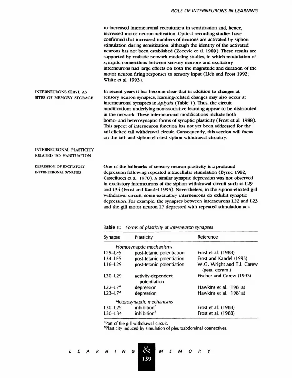
In recent years it has become clear that in addition to changes at sensory neuron synapses, learning-related changes may also occur at interneuronal synapses in Aplysia (Table 1). Thus, the circuit modifications underlying nonassociative learning appear to be distributed in the network. These interneuronal modifications include both homo- and heterosynaptic forms of synaptic plasticity (Frost et al. 1988). This aspect of interneuron function has not yet been addressed for the tail-elicited tail withdrawal circuit. Consequently, this section will focus on the tail- and siphon-elicited siphon withdrawal circuitry.
One of the hallmarks of sensory neuron plasticity is a profound depression following repeated intracellular stimulation (Byrne 1982; Castellucci et al. 1970). A similar synaptic depression was not observed in excitatory interneurons of the siphon withdrawal circuit such as L29 and L34 (Frost and Kandel 1995). Nevertheless, in the siphon-elicited gill withdrawal circuit, some excitatory interneurons do exhibit synaptic depression. For example, the synapses between interneurons L22 and L23 and the gill motor neuron L7 depressed with repeated stimulation at a
Table 1: Forms of plasticity at interneuron synapses
Synapse Plasticity Reference
Homosynaptic mechanisms L29-LFS post-tetanic potentiation L34-LFS post-tetanic potentiation L16-L29 post-tetanic potentiation
L30-L29 activity-dependent potentiation
L22-L7 a depression L23-L7 a depression
Heterosynaptic mechanisms L30-L29 inhibition b L30-L34 inhibition b
Frost et al. (1988) Frost and Kandel (1995) W.G. Wright and T.J. Carew
(pers. comm.) Fischer and Carew (1993)
Hawkins et al. (1981a) Hawkins et al. (1981a)
Frost et al. (1988) Frost et al. (1988)
apart of the gill withdrawal circuit. bplasticity induced by simulation of pleuroabdominal connectives.
& 139
L E A R N I N G M E M O R Y
Cleary et al.
FACILITATION OF INHIBITORY
INTERNEURONAL SYNAPSES
INTERNEURONAL PLASTICITY RELATED TO SENSITIZATION
POST-TETANIC POTENTIATION OF
EXCITATORY INTERNEURONAL
SYNAPSES
INHIBITION OF INHIBITORY
NEURONS
ALTERATIONS IN RESPIRATORY
PUMPING FREQUENCY
rate roughly parallel to that of sensory neuron depression (Hawkins et al. 198 l a). This depression may contribute to the reduced effectiveness of that circuit as a result of habituation.
The siphon-elicited siphon withdrawal circuit contains an important inhibitory interneuron, L30, which mediates both recurrent and lateral inhibition among the excitatory interneurons (Hawkins et al. 1981a; Fischer and Carew 1993, 1995; Frost and Kandel 1995). The circuit is thus designed with a built-in inhibitory system, which is activated during each instance of the reflex and which is thus in a position to influence reflex strength. The inhibitory synapses made by L30 undergo activity-dependent potentiation, lasting up to tens of seconds (Fischer and Carew 1993). This form of homosynaptic facilitation may contribute to habituation of the reflex when repeated siphon stimuli are delivered at appropriately short intervals.
Application of electrical shock to the tail or the pleuroabdominal connectives causes the L29 interneurons to fire strongly enough to produce post-tetanic potentiation at the L29-LFS synapse lasting several minutes (Frost et al. 1988; Hawkins and Schacher 1989). This form of homosynaptic plasticity may thus contribute to the memory for short-term sensitization of the siphon withdrawal reflex.
The inhibitory synapses made by interneuron L30 in the siphon-elicited siphon withdrawal circuit undergo heterosynaptic inhibition lasting several minutes in response to sensitizing stimuli (Frost et al. 1988; Fischer and Carew 1993). A similar suppression of inhibition within the circuit is also produced by bath application of either serotonin or cAMP analogs, suggesting a possible common modulatory mechanism for this and some of the sensory neuron changes observed in sensitization. Moreover, application of cholinergic antagonists, which blocks the synapse between L30 and excitatory interneurons, blocks the ability of tail stimulation to enhance complex PSPs produced in siphon motor neurons by siphon stimulation (Trudeau and Castellucci 1993). This result suggests that disinhibition contributes to sensitization.
Similar mechanisms may operate in the tail-withdrawal circuitry. Application of serotonin (5-HT) hyperpolarizes the inhibitory RPI4 interneurons, suppressing spontaneous spike activity and decreasing excitability (Xu et al. 1995). 5-HT also reduces or eliminates the inhibitory postsynaptic potentials produced in the motor neurons and sensory neurons by RPI4. Thus a modulatory agent (5-HT) that enhances connections of sensory neurons, inhibits the actions of an inhibitory interneuron in this ciruit.
The contractions of the mantle organs associated with respiratory pumping originate from the discharge of a burst generator network of interneurons, consisting of the L25 and R25 cells (Byrne and Koester 1978; Byrne 1983; Koester 1989; Alevizos et al. 1989). Sensitizing stimuli, such as tail shock, enhance the rate of spontaneous discharge of neurons in the L25/R25 pattern-generating circuit (Frost et al. 1988) and
& 1 4 0
L E A R N I N G M E M 0 R Y

ROLE OF INTERNEURONS IN LEARNING
Modulatory Interneurons Produce Learning-related Changes in Other Circuit Elements
IDENTIFIED MODULATORY INTERNEURONS
L29 NEURONS MODULATE SIPHON
SENSORY NEURONS
lower their threshold for recruitment by siphon stimuli (Eberly and Pinsker 1984). These changes may be attributable to plasticity located within the pattern generator network itself. R20 can enhance the rate of L25/R25 discharge but is not itself activated by sensitizing stimuli (Alevizos et al. 1989).
In addition to those interneurons participating in the reflex itself, a second class of interneurons has been studied. These are neurons that, when activated by appropriate stimuli, induce the synaptic plasticity that underlies learning (Fig. 3). By definition, these neurons participate in heterosynaptic forms of plasticity. So far, most of the neurons in this class appear to be extrinsic to the circuit, meaning that they do not participate in mediation of the reflex. However, L29 and L16 are intrinsic to the circuit, meaning that they are activated during each instance of the reflex.
The L29 neurons of the abdominal ganglion, which are intrinsic components of the siphon-elicited siphon withdrawal circuit (Fig. 2), are also thought to be involved in sensitization of the reflex mediated by that circuit. Tail shock activates L29, which in turn produces presynaptic facilitation of the LE sensory neurons (Hawkins et al. 1981a,b; Hawkins and Schacher 1989). The effects of L29 on sensory-motor transmission
Figure 3: Several modulatory neurons (shaded circles) have been identified that affect the siphon withdrawal circuit (A) or the tail withdrawal circuit (B). These neurons contain transmitters such as serotonin (5-HT), small cardioactive peptide (SCP), buccalin, and FMRFamide. The transmitters released by other modulatory interneurons are not known (depicted by small ?). The modulatory pathways (thick lines) regulate the properties of the circuit elements mediating the reflex (thin lines; some mediating connections have been omitted for clarity) and, presumably, the strength of the behavioral responses. L16 and L29 are elements of both modulating and mediating pathways. The pathway by which L16 modulates input to the motor neurons is not known (depicted by large ?). Modulatory neurons may have either excitatory (A) or inhibitory (&) actions. Several neurons have receptors for excitatory (open arrows) or inhibitory (solid arrows) modulatory transmitters, but neurons re- leasing these transmitters have not yet been identified.
& 141
L E A R N I N G M E M O R Y

Cleary et al.
L16 REGULATES ACTIVITY IN THE
SIPHON-ELICITED SIPHON
WITHDRAWAL CIRCUIT
CBI NEURONS MODULATE SIPHON
SENSORY NEURONS
Jm NEURONS MODULATE TAIL
SENSORY NEURONS
have been demonstrated in both intact ganglia and dissociated cell cultures containing just a single L29, a single sensory neuron, and a single motor neuron (Hawkins and Schacher 1989). Although the modulatory transmitter serotonin mimics many of the effects of L29 (see below), the L29s are not serotonergic (Hawkins and Schacher 1989; Kistler et al. 1985). This suggests that multiple facilitatory systems contribute to tail shock-induced modulation of the siphon sensory neurons.
Noxious stimulation of the tail generally produces a biphasic modulat ion of the siphon-elicited siphon withdrawal reflex: a transient inhibition of the reflex followed by an enhancement (Krontiris-Litowitz et al. 1987; Mackey et al. 1987; Marcus et al. 1988; Rankin and Carew 1989). This inhibition appears to require activation of the inhibitory interneuron L16 (Wright and Carew 1995). Unlike L30, which is activated by a wide range of intensities, L 16 is activated only by strong tail shock (Wright and Carew 1995). Hyperpolarization of L16 is sufficient to block the transient depression of water jet-evoked input to siphon motor neurons. On the other hand, activation of L16 alone is not sufficient to elicit that depression. Therefore, L16 appears to act as a gate for transient inhibition. During the period of inhibition, the amplitude of the complex EPSP evoked by tactile stimulation of the skin is decreased, but the amplitude of the monosynaptic sensorimotor EPSP is enhanced. This interesting dissociation illustrates how recrui tment of polysynaptic pathways can determine the behavioral response (Wright et al. 1991). Similar results were obtained when 5-HT was used to mimic the effects of strong tail shock (Fitzgerald and Carew 1991 ).
The CB 1 neuron in the B cluster of the cerebral ganglion has been identified as one component of the modulatory system underlying sensitization of the siphon-elicited siphon withdrawal reflex (Mackey et al. 1989). Tail shock activates CB 1, which in turn produces presynaptic facilitation of the excitatory synapses from the LE siphon sensory neurons to the LFS siphon motor neurons. Following tail shock, the CB 1 firing rate remains elevated for several minutes, although it is not clear that the level of activity is sufficient to maintain synaptic facilitation for an extended period of time. CB 1 also produces spike broadening in LE neurons bathed in 100 m i tetraethylammonium, consistent with a presynaptic site of action. These effects are consistent with CBI's serotonergic phenotype (see below). CB1 may affect the tail sensory neurons in a similar fashion (Wright et al. 1995).
Neurons in the J cluster of the cerebral ganglion produce effects in tail sensory neurons that mimic changes produced by sensitizing stimuli (Raymond and Byrne 1994). These neurons, referred to as Jm neurons, depolarize slowly, hyperpolarize slowly, or produce a dual-component response in the sensory neurons. The connections be tween Jm cells and the pleural sensory neurons were generally strong; a single spike in a Jm cell was often sufficient to produce a measurable slow EPSP in a sensory neuron. Sensory neurons that were depolarized by Jm stimulation also showed an increase in their excitability. Neurons in the J cluster also produced bursts of fast PSPs in tail motor neurons.
The Jm cells generally received excitatory input from cerebral nerves
& 147
L E A R N I N G M E M 0 R Y

ROLE OF INTERNEURONS IN LEARNING
LPL16 INHIBITS SIPHON
SENSORY NEURONS
IMMUNOHISTOCHEMICAL AND PHARMACOLOGICAL EVIDENCE FOR MODULATORY INTERNEURONS
C1-C3, which innervate the head and neck, and weaker, usually inhibitory input from pedal nerves P7-P9, which innervate the tail and posterior part of the animal. Thus, Jm neurons appear to be elements of a modulatory pathway involved in sensitization of the tail-elicited withdrawal reflexes from anterior parts of the animal.
In addition to the intrinsic inhibitory neurons (Fig. 2), extrinsic inhibitory neurons also appear to influence the siphon-elicited siphon withdrawal circuit. LPI16, located in the pleural ganglion, hyperpolarizes siphon sensory neurons and produces heterosynaptic depression of the synapse between siphon sensory and motor neurons (Small et al. 1992). This neuron is activated by tail shock and may contr ibute to the transient inhibition induced by tail stimulation. LPI16 is also immunoreact ive for FMRFamide. Because FMRFamide has both short- and long-term inhibitory effects on sensory neuron excitability, LPI16 may also contribute to long-term habituation of the tail-elicited siphon withdrawal reflex.
Although several modulatory neurons have now been identified, it is generally recognized that other unidentified modulators also exist. Therefore, additional experimental approaches have been utilized that are more efficient in terms of the fraction of the nervous system that they survey but are less direct than intracellular stimulation of single neurons. A common approach has been to identify neurotransmitters that have modulatory effects on tail or siphon sensory neurons and use immunocytochemistry to look for proximity of axons containing those transmitters to the sensory neurons.
This approach has been used most successfully for 5-HT, culminating in the identification of CB 1 as a serotonergic facilitatory neuron. A substantial amount of data has demonstrated the ability of 5-HT to mimic the effects of sensitization training on both tail and siphon sensory neurons (for review, see Kandel and Schwartz 1982; Byrne et al. 1993). Depletion of 5-HT from the central nervous system by intracoelomic injection of the neurotoxin, 5,7-dihydroxytryptamine, blocked behavioral sensitization (Glanzman et al. 1989). It also blocked presynaptic facilitation of the sensorimotor synapse following connective stimulation. Similar experiments have not yet been performed for the tail-elicited tail withdrawal circuit. The finding of 5-HT-immunoreactive fibers in contact with siphon sensory neurons (Kistler et al. 1985) supports the hypothesis that serotonergic neurons contr ibute to behavioral sensitization. Numerous serotonergic varicosities have also been observed surrounding somata and axon hillocks of tail sensory neurons (Zhang et al. 1991). Electron microscopy confirmed that these contacts were directly on the membrane of sensory neurons and were presumably synaptic in nature. These results suggest that 5-HT is released from varicosities directly onto somata of sensory neurons, eliciting the cellular changes correlated with sensitization. The proximity of serotonergic input to the soma may be crucial for triggering the cellular mechanisms underlying the long-term form of sensitization (Kistler et al. 1985; Nazif et al. 1991; Zhang et al. 1991; Bacskai et al. 1993; Clark and Kandel 1993; Emptage and Carew 1993; O'Leary et al. 1995).
Two other transmitters have been found to have facilitatory effects on sensory neurons, small cardioactive peptide (SCP) and buccalin (Abrams
& 143
L E A R N I N G M E M 0 R Y
Cleary et al.
CONCLUDING COMMENTS
et al. 1984; Ocorr and Byrne 1985; Raymond et al. 1989; Pieroni and Byrne 1992). There are, however, no identified neurons containing these transmitters that have modulatory effects on tail or siphon sensory neurons. The R20 neurons, which contain SCP and modulate the activity of the pat tern generator underlying respiratory pumping, do not appear to contribute to sensitization (Alevizos et al. 1989). Immunofluorescence techniques have been used to examine the distribution of SCP and buccalin within the cluster of pleural sensory neurons (Lo et al. 1987). The distribution of SCP within the pleural ganglion differs from that of 5-HT in that few SCP-immunoreactive fibers are in contact with somata of tail sensory neurons. This does not rule out the possibility that they make axoaxonal contacts with the sensory neurons. Buccalin-like immunoreactivity (IR) was found in each pleural ganglion and prominent buccalin-IR fiber tracts were found to course through the pleural ganglia, but no fibers were observed in contact with somata of tail sensory neurons (see also Miller et al. 1992). Although there are somata in the pleural ganglia immunoreactive for SCP or buccalin, these cells have not been analyzed electrophysiologically.
FMRFamide is a transmitter with inhibitory effects on tail and siphon sensory neurons. LP116 is a FMRFamide-IR neuron that produces heterosynaptic depression of siphon sensory neurons. FMRFamide is distributed within the cluster of sensory neurons in a pat tern similar to that of 5-HT, although there are fewer processes (Lo et al. 1987). Presumably, some of these fibers are intrinsic to the ganglion, as many somata within the pleural ganglion are immunopositive. For example, the inhibitory interneuron RPI5 contains FMRFamide-like IR and hyperpolarizes sensory neurons (Xu et al. 1994).
Like vertebrate spinal reflex circuits, the Aplysia siphon and tail withdrawal circuits are composed of monosynaptic sensory-motor neuron pathways in parallel with polysynaptic pathways through interneuronal populations. In contrast to the vertebrate nervous system, however, the small number, large diameter, and easy accessibility of interneurons in Aplysia withdrawal circuits has made it possible to explore several issues regarding the role of circuit interneurons in the mediation and modulation of reflex responses.
Interneurons serve an important role as recruitable circuit elements, which amplify the contribution made by the monosynaptic pathway. Interneurons further transform the sensory neuron output by convert ing stimulus intensity into response duration and by generating the stimulus-triggered respiratory-pumping component of the siphon- and gill-withdrawal reflex. Interneurons also participate as information storage sites, at least for the simple short-term nonassociative forms of learning examined so far. In sensitization, changes occur at interneuronal loci, in addition to the well-described facilitation of the monosynaptic connections between sensory and motor neurons. These interneuronal changes involve different types of plasticity, with excitatory synapses getting stronger and inhibitory synapses weaker. Thus, sensitization involves multiple types as well as multiple loci of plasticity (Frost et al. 1988). An important queston for future analysis will be the extent to which the interneurons serve as loci for long-term nonassociative and associative learning. The finding that sensitization of withdrawal reflexes in Aplysia involves multiple sites of plasticity seems to be a general
& 1 4 4
L E A R N I N G M E M O R Y

ROLE OF INTERNEURONS IN LEARNING
finding emerging from analyses of other systems as well (e.g., Mishkin et al. 1984; Cohen 1985; Frysztak and Crow 1994; Gallagher and Holland 1994; Perrett and Mauk 1995).
The last role of interneurons is as important modulatory elements in the circuitry. Interneurons, both intrinsic and extrinsic to the withdrawal reflex circuits, produce modulatory actions on other circuit elements. This modulation plays an important role in sensitization, but it is not known whether such heterosynaptic modulatory processes have a role in habituation. Interestingly, FMRFamide leads to long-term depression of sensorimotor connections in cell culture (Montarolo et al. 1988; Schacher et al. 1993), raising the possibility that FMRFamide containing interneurons may mediate long-term habituation in vivo.
The work reviewed in this paper represents a new level for analysis of nonassociative learning in Aplysia. It is fair to say that earlier studies in this animal were focused on the cellular properties of learning and memory, that is, the changes in elementary synaptic connections. These studies provided insights into the mechanisms by which synaptic transmission is modified by learning and demonstrated that there is a representation, at the cellular level, of behavioral modifications induced in the whole animal. There is currently tremendous interest, for both invertebrates and vertebrates, in the system properties of learning and memory. For this, it is important to investigate the parallel pathways involved in memory storage, just as it has been important to analyze parallel processing in vision, audition, and in motor systems. This analysis, for which invertebrates are uniquely advantageous, allows one to identify the contribution of different parallel mechanisms to acquisition and storage of acquired information. Thus, a solid foundation is now in place for future analyses of the system properties of learning and memory in Ap lysia.
Acknowledgments
References
We thank Lise Eliot for her thoughtful comments on earlier versions of the manuscript. This work was supported by National Science Foundation grant IBN-9320549 to L.J.C., National Institute of Mental Health (NIMH) award K05 MH00649 and National Institutes of Health grant R01 NS19895 to J.H.B.; and NIMH grant R29 MH48536 to W.N.F.
The publication costs of this article were defrayed in part by payment of page charges. This article must therefore be hereby marked "advertisement" in accordance with 18 USC section 1734 solely to indicate this fact.
Abrams, T.W., V.F. Castellucci, J.S. Camardo, E.R. Kandel, and P.E. Lloyd. 1984. Two endogenous neuropeptides modulate the gill and siphon withdrawal reflex in Aplysia by presynaptic facilitation involving cAMP-dependent closure of a serotonin-sensitive potassium channel. Proc. Natl. Acad. Sci. 81: 7956-7960.
Alberini, C.M., M. Ghirardi, R. Metz, and E.R. Kandel. 1994. C/EBP is an immediate-early gene required for the consolidation of long-term facilitation in Aplysia. Cell 76: 1-20.
Alevizos, A., K.R. Weiss, and J. Koester. 1989. SCP-containing R20 neurons modulate respiratory pumping in Aplysia. I. Neurosci. 9: 3058-3071.
Bacskai, B.J., B. Hochner, M. Mahaut-Smith, S.R. Adams, B.K. Kaang, E.R. Kandel, and R.Y. Tsien. 1993. Spatially resolved dynamics of cAMP and protein kinase A subunits in Aplysia sensory neurons. Science 260: 222-226.
Bailey, C.H., E.B. Thompson, V.F. Castellucci, and E.R. Kandel. 1979. Ultrastructure of the synapses of sensory neurons that mediate the gill-withdrawal reflex. I. Neurocytol. 8:415-444.
& 145
L E A R N I N G M E M O R Y
Cleary et al.
Bailey, C.H., M. Chen, F. Keller, and E.R. Kandel. 1992. Serotonin-mediated endocytosis of apCAM: An early step of learning-related synaptic growth in Aplysia. Science 256: 645-649.
Baxter, D.A. and J.H. Byrne. 1990. Differential effects of cAMP and serotonin on membrane current, action-potential duration, and excitability in somata of pleural sensory neurons of Aplysia. J. Neurophysiol. 641: 978-990.
Braha, O., N. Dale, B. Hochner, M. Klein, T.W. Abrams, and E.R. Kandel. 1990. Second messengers involved in the two processes of presynaptic facilitation that contribute to sensitization and dishabituation in Aplysia sensory neurons. Proc. Natl. Acad. Sci. 87: 2040-2044.
Buonomano, D.V., L.J. Cleary, and J.H. Byrne. 1992. Inhibitory neuron produces heterosynaptic inhibition of the sensory-to-motor neuron synapse in Aplysia. Brain Res. 577:147-150.
Byrne, J.H. 1981. Comparative aspects of neural circuits for inking behavior and gill withdrawal in Aplysia californica. J. Neurophysiol. 45: 98-106.
~ . 1982. Analysis of synaptic depression contributing to habituation of gill-withdrawal reflex in Aplysia californica. I. Neurophysiol. 48: 431-438.
. 1983. Identification and initial characterization of a cluster of command and pattern-generating neurons underlying respiratory pumping in Aplysia califomica. J. Neurophysiol. 49: 491-508.
�9 1987. Cellular analysis of associative learning. Physiol. Rev. 67: 329-439.
Byrne, J.H. and E�9149 Kandel. 1996. Presynaptic facilitation revisited: State and time-dependence. I. Neurosci. (in press).
Byrne, J�9149 and J. Koester. 1978. Respiratory pumping: neuronal control of a centrally commanded behavior in Aplysia. Brain Res. 143: 87-105.
Byrne, J.H., V. Castellucci, and E.R. Kandel. 1974. Receptive fields and response properties of mechanoreceptor neurons innervating siphon skin and mantle shelf of Aplysia. J. Neurophysiol. 3,7: 1041-1064.
Byrne, J.H., V. Castellucci, and E.R. Kandel. 1978a. Contribution of individual mechanoreceptor sensory neurons to defensive gill-withdrawal reflex in Aplysia. J. Neurophysiol. 41 : 418-431.
Byrne, J.H., V.F. Castellucci, T.J. Carew, and E.R. Kandel. 1978b. Stimulus-response relations and stability of mechanoreceptor and motor neurons mediating defensive gill-withdrawal reflex in Aplysia. J. Neurophysiol. 41:402-417.
Byrne, J.H., D.A. Baxter, D.V. Buonomano, L.J. Cleary, A. Eskin, J.R. Goldsmith, E. McClendon, F.A. Nazif, F. Noel, and K.P. Scholz. 1991. Neural and molecular bases of nonassociative and associative learning in Aplysia. Ann. N.Y. Acad. Sci. 627: 124-149.
Byrne, J.H., R. Zwartjes, R. Homayouni, S.D. Critz, and A. Eskin. 1993. Roles of second messenger pathways in neuronal plasticity and in learning and memory. Insights gained from Aplysia. Adv. Second Messenger Phosphoprotein Res. 27: 47-108.
Carew, T.J. and C.L. Sahley. 1986. Invertebrate learning and memory: from behavior to molecules�9 Annu. Rev. Neurosci. 9: 435-487.
Carew, T.J., E.T. Waiters, and E.R. Kandel. 1981. Classical conditioning in a simple withdrawal reflex in Aplysia californica. I. Neurosci. 1: 1426-1437.
& 146
L E A R N I N G M E M O R Y
ROLE OF INTERNEURONS IN LEARNING
Castellucci, V., H. Pinsker, I. Kupfermann, and E. Kandel. 1970. Neuronal mechanisms of habituation and dishabituation of the gill-withdrawal reflex in Aplysia. Science 167:1745-1748.
Clark, G.A. and E.R. Kandel. 1984. Branch-specific heterosynaptic facilitation in Aplysia siphon sensory neurons�9 Proc. Natl. Acad. Sci. 81: 2577-2581.
�9 1993. Induction of long-term facilitation in Aplysia-siphon sensory neurons by local application of serotonin to remote synapses. Proc. Natl. Acad. Sci. 90:1141 I - I 1415.
Cleary, L.J. and J.H. Byrne. 1993. Identification and characterization of a multifunction interneuron contributing to defensive arousal in Aplysia. J. Neurophysiol. 70: 1767-I 776.
Cohen, D.J. 1985. Some organizational principles of a vertebrate conditioning pathway: Is memory a distributed property? In: Brain structure, learning and memory (ed. N.M. Weinberger, J.L. McGaugh, and G. Lynch), pp. 27-48. Guilford Press, New York.
Cohen, T.E., V. Henzi, E.R. Kandel, and R.D. Hawkins. 1991. Further behavioral and cellular studies of dishabituation and sensitization in Aplysia. Soc. Neurosci. Abstr. 17:1302.
Dubuc, B. and V.F. Castellucci. 1991. Receptive fields and properties of a new cluster of mechanoreceptor neurons innervating the mantle region and the branchial cavity of the marine mollusk Aplysia californica. J. Exp. Biol. 156:315-334.
Eberly, L.B. and H.M. Pinsker. 1984. Neuroethological studies of reflex plasticity in intact Aplysia. Behav. Neurosci. 98: 609-630.
Emptage, N.J. and T.J. Carew. 1993. Long-term synaptic facilitation in the absence of short-term facilitation in Aplysia neurons. Science 262: 253-256.
Falk, C.X., J.Y. Wu, L.B. Cohen, and A.C. Tang. 1993. Nonuniform expression of habituation in the activity of distinct classes of neurons in the Aplysia abdominal ganglion. J. Neurosci. 13: 4072-4081.
Fischer, T.M. and T.J. Carew. 1993. Activity-dependent potentiation of recurrent inhibition: a mechanism for dynamic gain control in the siphon withdrawal reflex of Aplysia. I. Neurosci. 13:1302-1314.
�9 1995. Cutaneous activation of the inhibitory L30 interneurons provides a mechanism for regulating adaptive gain control in the siphon withdrawal reflex of Aplysia. J. Neurosci. 15: 762-773.
Fitzgerald, K. and T.J. Carew. 1991. Serotonin mimics tail shock in producing transient inhibition in the siphon withdrawal reflex of Aplysia. J. Neurosci. 11: 2510-2518.
Frost, W.N., V.F. Castellucci, R.D. Hawkins, and E.R. Kandel. 1985. Monosynaptic connections made by the sensory neurons of the gill- and siphon-withdrawal reflex in Aplysia participate in the storage of long-term memory for sensitization. Proc. Natl. Acad. Sci. 82: 8266--8269.
Frost, W.N., G.A. Clark, and E.R. Kandel. 1988. Parallel processing of short-term memory for sensitization in Aplysia. I. Neurobiol. 19: 297-334.
Frost, W.N. and E.R. Kandel. 1995. Structure of the network mediating siphon-elicited siphon withdrawal in Aplysia. J. Neurophysiol. 73- 2413-2427.
Frost, W.N., L.G. Wu, and J. Lieb. 1991. Simulation of the Aplysia siphon withdrawal reflex circuit: slow components of intemeuronal synapses contribute to the mediation of reflex duration. Soc. Neurosci. Abstr. 17: 1390.
Frysztak, R.J. and T. Crow. 1994. Enhancement of type B and A photoreceptor inhibitory synaptic connections in conditioned Hermissenda. J. Neurosci. 14:1245-1250.
& 147
L E A R N I N G M E M 0 R Y

Clean/et al.
Gallagher, M. and P.C. Holland. 1994. The amygdala complex: multiple roles in associative learning and attention. Proc. Natl. Acad. Sci. 91: 11771-11776.
Ghirardi, M., O. Braha, B. Hochner, P.G. Montarolo, E.R. Kandel, and N. Dale. 1992. Roles of PKA and PKC in facilitation of evoked and spontaneous transmitter release at depressed and nondepressed synapses in Aplysia sensory neurons. Neuron 9: 479--489.
Glanzman, D.L., S.L. Mackey, R.D. Hawkins, A.M. Dyke, P.E. Lloyd, and E.R. Kandel. 1989. Depletion of serotonin in the nervous system of Aplysia reduces the behavioral enhancement of gill withdrawal as well as the heterosynaptic facilitation produced by tail shock. I. Neurosci. 9:4200-4213.
Greenberg, S.M., V.F. Castellucci, H. Bayley, and J.H. Schwartz. 1987. A molecular mechanism for long-term sensitization in Aplysia. Nature 329: 62-65.
Hawkins, R.D. and S. Schacher. 1989. Identified facilitator neurons L29 and L28 are excited by cutaneous stimuli used in dishabituation, sensitization, and classical conditioning of Aplysia. I. Neurosci. 9: 4236-4245.
Hawkins, R.D., V.F. Castellucci, and E.R. Kandel. 1981a. Interneurons involved in mediation and modulation of gill withdrawal reflex in Aplysia. I. Identification and characterization. J. Neurophysiol. 45:304-314.
1981b. Interneurons involved in mediation and modulation of gill withdrawal reflex in Aplysia. II. Identified neurons produce heterosynaptic facilitation contributing to behavioral sensitization. J. Neurophysiol. 45: 315-326.
Hawkins, R.D., G.A. Clark, and E.R. Kandel. 1986. Cell biological studies of learning in simple vertebrate and invertebrate nervous systems. The nervous system: Higher functions of the brain (ed. F. Plum), pp. 25-83. American Physiological Society, Bethesda, MD.
Hawkins, R.D., E.R. Kandel, and S.A. Siegelbaum. 1993. Learning to modulate transmitter release: Themes and variations in synaptic plasticity. Annu. Rev. Neurosci. 16: 625-665.
Hening, W.A., E.T. Waiters, T.J. Carew, and E.R. Kandel. 1979. Motorneuronal control of locomotion in Aplysia. Brain Res. 179: 231-253.
Hickie, C. 1994. "Functional and motor neuronal analysis of defensive siphon responses in Aptysia californica,'" Ph.D. thesis. University of Texas Houston Health Science Center, TX.
Hickie, C. and E.T. Kandel. 1995. Motor neuronal control of tail-directed and head-directed siphon responses in Aplysia califomica. J. Neurophysiol. 74: 307-321.
Kandel, E.R. and J.H. Schwartz. 1982. Molecular biology of learning: Modulation of transmitter release. Science 218: 433--443.
Kanz, J.E., L.B. Eberly, J.S. Cobbs, and H.M. Pinsker. 1979. Neuronal correlates of siphon withdrawal in freely behaving Aplysia. J. Neurophysiol. 42:1538-1556.
Kaplan, S.W., E.R. Kandel, and R.D. Hawkins. 1993. Plasticity in the monosynaptic component of the Aplysia gill withdrawal reflex during habituation, dishabituation and sensitization. Soc. Neurosci. Abstr. 19: 16.
~ . 1994. Plasticity of LE and non-LE siphon sensory neurons during habituation and dishabituation of the Aplysia gill-withdrawal reflex. Soc. Neurosci. Abstr. 20: 1073.
Kennedy, T.E., D. Kuhl, A. Barzilai, J.D. Sweatt, and E.R. Kandel. 1992. Long-term sensitization training in Aplysia leads to an increase in calreticulin, a major presynaptic calcium-binding protein. Neuron 9:1013-1024.
Kistler, H.B. Jr., R.D. Hawkins, J. Koester, H.W. Steinbusch, E.R. Kandel, and J.H. Schwartz. 1985. Distribution of serotonin-immunoreactive cell bodies and processes in the abdominal ganglion of mature Aplysia. J. Neurosci. S: 72-80.
L E A R N I N G M E M 0 R Y

ROLE OF INTERNEURONS IN LEARNING
Koester, J. 1989. Chemically and electrically coupled interneurons mediate respiratory pumping in Aplysia. J. Neurophysiol. 62:1113-1126.
Koester, J. and E.R. Kandel. 1977. Further identification of neurons in the abdominal ganglion of Aplysia using behavioral criteria. Brain Res. 121: 1-20.
Krontiris-Litowitz, J.K., M.T. Erikson, and E.T. Waiters. 1987. Central suppression of defensive reflexes in Aplysia by noxious stimulation and by factors released from the body wall. Soc. Neurosci. Abstr. 13: 815.
Kupfermann, I., T.J. Carew, and E.R. Kandel. 1974. Local, reflex, and central commands controlling gill and siphon movements in Aplysia. I. Neurophysiol. 37: 996-1019.
Lieb, J.R. Jr. and W.N. Frost. 1992. Interneuronal plasticity contributes significantly to enhanced motor neuron firing during sensitization of the Ap/ysia siphon- withdrawal reflex. Soc. Neurosci. Abstr. 18: 531.
Lo, L.T., J.H. Byrne, and L.J. Cleary. 1987. Distribution of three modulatory transmitters within the pleural ganglion of Aplysia. Soc. Neurosci. Abstr. 13: 1073.
Mackey, S.L., D.L. Glanzman, S.A. Small, A.M. Dyke, E.R. Kandel, and R.D. Hawkins. 1987. Tail shock produces inhibition as well as sensitization of the siphon-withdrawal reflex of Aplysia: possible behavioral role for presynaptic inhibition mediated by the peptide Phe-Met-Arg-Phe-NH2. Proc. Natl. Acad. Sci. 84: 8730-8734.
Mackey, S.L., E.R. Kandel, and R.D. Hawkins. 1989. Identified serotonergic neurons LCB1 and RCB1 in the cerebral ganglia of Aplysia produce presynaptic facilitation of siphon sensory neurons. J. Neurosci. 9: 4227-4235.
Marcus, E.A., T.G. Nolen, C.H. Rankin, and T.J. Carew. 1988. Behavioral dissociation of dishabituation, sensitization, and inhibition in Aplysia. Science 241: 210-213.
Miller, M.W., A. Alevizos, E.C. Cropper, I. Kupfermann, and K.R. Weiss. 1992. Distribution of buccalin-like immunoreactivity in the central nervous system and peripheral tissues of Aplysia californica. J. Comp. Neurol. 320: 182-195.
Mishkin, M., B. Malamut, and J. Bachevalier. 1984. Memories and habits: Two neural systems. In Neurobiology of learning and memory (ed. G. Lynch, J.L. McGaugh, and N.M. Weinberger), pp. 65-77. Guilford Press, New York.
Montarolo, P.G., E.R. Kandel, and S. Schacher. 1988. Long-term heterosynaptic inhibition in Aplysia. Nature 333:171-174.
Nazif, F.A., J.H. Byrne, and L.J. Cleary. 1991. cAMP induces long-term morphological changes in sensory neurons of Aplysia. Brain Res. 539" 324-327.
Ocorr, K.A. and J.H. Byrne. 1985. Membrane responses and changes in cAMP levels in Aplysia sensory neurons produced by serotonin, tryptamine, FMRFamide and small cardioactive peptide B (SCPB). Neurosci. Lett. 55:113-118.
O'Leary, F.A., J.H. Byrne, and L.J. Cleary. 1995. Long-term structural remodeling in Aplysia sensory neurons requires de novo protein synthesis during a critical time period. J. Neurosci. 15: 3519-3525.
Perlman, A.J. 1979. Central and peripheral control of siphon-withdrawal reflex in Aplysia califomica. J. Neurophysiol. 42:510-529.
Perrett, S.P. and M.D. Mauk. 1995. Extinction of conditioned eyelid responses requires the anterior lobe of cerebellar cortex. I. Neurosci. 15: 2074-2080.
Pieroni, J.P. and J.H. Byrne. 1992. Differential effects of serotonin, FMRFamide, and small cardioactive peptide on multiple, distributed processes modulating sensorimotor synaptic transmission in Aplysia. 1. Neurosci. 12: 2633-2647.
& 149
L E A R N I N G M E M O R Y
Cleary et al.
Pinsker, H.M. 1982. Integration of reflex activity and central pattern generation in intact Aplysia. I. Physiol. (Paris) 78: 775-785.
Rankin, C.H. and T.J. Carew. 1989. Developmental analysis in Aplysia reveals inhibitory as well as facilitatory effects of tail shock. Behav. Neurosci. 103: 334-344.
Raymond, J.L. and J.H. Byrne. 1994. Distributed input to the tail-siphon withdrawal circuit in Aplysia from neurons in the J cluster of the cerebral ganglion. I. Neurosci. 14: 2444-2454.
Raymond, J.L., E.E. Shulman, D.A. Baxter, L.J. Cleary, and J.H. Byrne. 1989. Differential effects of the peptide buccalin and serotonin on membrane currents, action potential duration, and excitability in pleural sensory neurons of Aplysia. Soc. Neurosci. Abstr. 15: 1284.
Schacher, S., E.R. Kandel, and P.G. Montarolo. 1993. cAMP and arachidonic acid simulate long-term structural and functional changes produced by neurotransmitters in Aplysia sensory neurons. Neuron 10: 1079-1088.
Scholz, K.P. and J.H. Byrne. 1988. Intracellular injection of cAMP induces a long-term reduction of neuronal K § currents. Science 240:1664-1666.
Schwartz, J.H. and S.M. Greenberg. 1987�9 Molecular mechanisms for memory: Second-messenger induced modifications of protein kinases in nerve cells. Annu. Rev. Neurosci. 10: 459-476.
Small, S.A., T.E. Cohen, E.R. Kandel, and R.D. Hawkins. 1992. Identified FMRFamide-immunoreactive neuron LPL16 in the left pleural ganglion of Aplysia produces presynaptic inhibition of siphon sensory neurons. I. Neurosci. 12:1616-1627.
Stopfer, M. and T.J. Carew. 1987. Quantitative analysis of the relation between gill amplitude and siphon duration in the defensive withdrawal reflex of Aplysia. Behav. Neurosci. 101 : 292-295.
Sugita, S., J.R. Goldsmith, D.A. Baxter, and J.H. Byrne. 1992. Involvement of protein kinase C in serotonin-induced spike broadening and synaptic facilitation in sensorimotor connections of Aplysia. I. Neurophysiol. 68:643-651.
Sugita, S., D.A. Baxter, and J.H. Byrne. 1994. Activators of protein kinase C mimic serotonin-induced modulation of a voltage-dependent potassium current in pleural sensory neurons of Aplysia. J. Neurophysiol. 72:1240-1249.
Trudeau, L.-E. and V.F. Castellucci. 1992. Contribution of polysynaptic pathways in the mediation and plasticity of Aplysia gill and siphon withdrawal reflex: evidence for differential modulation. I. Neurosci. 12: 3838-3848.
�9 1993. Functional uncoupling of inhibitory interneurons plays an important role in short-term sensitization of Aplysia gill and siphon withdrawal reflex. I. Neurosci. 13: 2126-2135.
Waiters, E.T., T.J. Carew, and E.R. Kandel�9 1981. Associative learning in Aplysia. Evidence for conditioned fear in an invertebrate. Science 211: 504-506.
Waiters, E.T., J.H. Byrne, T.J. Carew, and E.R. Kandel. 1983. Mechanoafferent neurons innervating tail of Aplysia. I. Response properties and synaptic connections. I. Neurophysiol. 50:1522-1542.
White, J.A�9 I�9 Ziv, L�9149 Clean/, D.A�9 Baxter, and J.H. Byrne�9 1993. The role of interneurons in controlling the tail-withdrawal reflex in Aplysia: a network model. I. Neurophysiol. 70:1777-1786.
Wright, W.G�9 and T�9 Carew�9 1995. A single identified interneuron gates tail-shock induced inhibition in the siphon withdrawal reflex of Aplysia. J. Neurosci. 15: 790-797.
& 150
L E A R N I N G M E M 0 R Y

ROLE OF INTERNEURONS IN LEARNING
Wright, W.G., E.A. Marcus, and T.J. Carew. 1991. A cellular analysis of inhibition in the siphon withdrawal reflex of Aplysia. J. Neurosci. 11: 2498-2509.
Wright, W.G., K. Jones, P. Sharp, and B. Maynard. 1995. Widespread anatomical projections of the serotonergic modulatory neuron, CB1, in Aplysia. Invert. Neurobiol. (in press)
Xu, Y., L.J. Clean/, and J.H. Byrne. 1994. Identification and characterization of pleural neurons that inhibit tail sensory neurons and motor neurons in Aplysia: correlation with FMRFamide immunoreactivity. J. Neurosci. 14: 3565-3577.
Xu, Y. J.P. Pieroni, L.J. Clean/, and J.H. Byrne. 1995. Modulation of an inhibitory interneuron in the neural circuitry for the tail withdrawal reflex of Aplysia. J. Neurophysiol. 73: 1313-1318.
Zecevic, D., J.Y. Wu, L.B. Cohen, J.A. London, H.P. H6pp, and C.X. Falk. 1989. Hundreds of neurons in the Aplysia abdominal ganglion are active during the gill-withdrawal reflex. !. Neurosci. 9: 3681-3689.
Zhang, H., J.H. Byrne, and L.J. Clean/. 1993. Topographical organization of sensory neurons in the ventrocaudal cluster of the pleural ganglion of Aplysia. Soc. Neurosci. Abstr. 19:813.
Zhang, Z.S., B. Fang, D.W. Marshak, J.H. Byrne, and L.J. Clean/. 1991. Serotoninergic neurons make synaptic contacts with pleural sensory neurons in Aplysia. J. Comp. Neurol. 311: 259-270.
L E A R N I N G & 151
M E M 0 R Y
![Modulación de reflejos defensivos por claves contextuales: efecto de la luz-oscuridad ambiental [Modulation of defensive reflexes by contextual cues: Effect of environmental light-darkness]](https://img.pdfslide.net/doc/110x75/632424845f71497ea904aac0/modulacion-de-reflejos-defensivos-por-claves-contextuales-efecto-de-la-luz-oscuridad.jpg)


