Embed Size (px)
Citation preview

Role of Ortho-Retronasal Olfaction in MammalianCortical Evolution
Timothy B. Rowe1* and Gordon M. Shepherd2
1Jackson School of Geosciences, University of Texas at Austin, Austin, Texas, 78712 USA2Department of Neurobiology, Yale University School of Medicine, New Haven, Connecticut, 06510 USA
Fossils of mammals and their extinct relatives among
cynodonts give evidence of correlated transformations
affecting olfaction as well as mastication, head move-
ment, and ventilation, and suggest evolutionary coupling
of these seemingly separate anatomical regions into a
larger integrated system of ortho-retronasal olfaction.
Evidence from paleontology and physiology suggests
that ortho-retronasal olfaction played a critical role at
three stages of mammalian cortical evolution: early
mammalian brain development was driven in part by
ortho-retronasal olfaction; the bauplan for neocortex
had higher-level association functions derived from
olfactory cortex; and human cortical evolution was
enhanced by ortho-retronasal smell. J. Comp. Neurol.
000:000–000, 2015.
VC 2015 Wiley Periodicals, Inc.
INDEXING TERMS: mammalian; human; olfaction; cortex; evolution
In analyzing mechanisms in human brain evolution,
vision is usually considered paramount and olfaction of
minor importance. Moreover, across all primates an
evolutionary trade-off in neural processing volumes and
performance has been hypothesized between specializa-
tions in the visual and olfactory systems. Primates with
high visual performance are thought to have small
olfactory processing centers and correspondingly dimin-
ished olfactory performance, and vice versa (Barton
et al., 1995). However, olfaction is a dominant sense in
the behavior of most mammals (Stoddart, 1980) and,
moreover, the convergence of orthonasal and retronasal
signals (Rozin, 1982) lies in neocortical areas that are
tied to human cravings responsible for disorders such
as obesity and food addiction (Shepherd, 2012). Yet
the evolutionary history of this unique duality of ortho-
nasal and retronasal olfaction has not been studied in
detail.
Here we reconsider the role of ortho-retronasal olfac-
tion in mammalian cortical evolution in the light of
recent developments in paleontology and cortical physi-
ology. In integrating observations from these tradition-
ally separate fields we will provide evidence for the role
of ortho-retronasal olfaction at three critical stages in
cortical evolution: the earliest pulse of premammalian
encephalization; the transition from three-layer to multi-
layer cortex; and the enlargement of neocortex in Homo
sapiens.
DEFINING THE ENLARGED SENSE OFORTHO-RETRONASAL OLFACTION
Mammals generally retain the primitive tetrapod olfac-
tion mode of sniffing known as “orthonasal” smell, in
which airborne environmental odor molecules are drawn
through the nares (nostrils) into the nose to activate the
olfactory epithelium. Orthonasal smell in mammals has
its own special characteristics which derive from a huge
olfactory receptor (OR) genome. Approximately 1,200 OR
genes are thought to have been present in mammals
ancestrally, compared to �100 that were present in the
first tetrapods and amniotes (Niimura, 2009, 2012).
Mammals also employ a system of diaphragmatic ventila-
tion that, together with other distinctive features such as
their modes of head movement, confers unique attrib-
utes to mammalian orthonasal smell, such as their abil-
ities in scent-tracking.
The counterpart to orthonasal smell is “retronasal”
smell, in which air exhaled from the lungs carries with
Grant sponsor: National Science Foundation (NSF); Grant numbers:EAR-1160721, EAR-0948842 (to T.B.R.); Grant sponsor: National Insti-tutes of Health (NIH) NIDCD; Grant numbers: DC 00997701-05, DC011286-03 (to G.M.S.).
*CORRESPONDENCE TO: Timothy B. Rowe, Jackson School ofGeosciences, University of Texas at Austin, Austin, TX 78712. E-mail:[email protected]
Received January 14, 2015; Revised March 16, 2015;Accepted April 29, 2015.DOI 10.1002/cne.23802Published online Month 00, 2015 in Wiley Online Library(wileyonlinelibrary.com)VC 2015 Wiley Periodicals, Inc.
The Journal of Comparative Neurology | Research in Systems Neuroscience 00:00–00 (2015) 1
RESEARCH ARTICLE
J_ID: CNE Customer A_ID: CNE23802 Cadmus Art: CNE23802 Ed. Ref. No.: 15-0012.R1 Date: 11-June-15 Stage: Page: 1
ID: jwweb3b2server Time: 08:57 I Path: //chenas03/Cenpro/ApplicationFiles/Journals/Wiley/CNE#/Vol00000/150059/Comp/APPFile/JW-CNE#150059

it an entirely new information domain of odor molecules
liberated in the mouth through the breakdown of food
by chewing, saliva, and actions of the tongue (Fig.F1 1).
These molecules pass forward from the caudal part of
the mouth via the choana (internal naris) and across
the main olfactory epithelium before being expelled
through the nares. In retronasal smell, olfaction com-
bines with taste and other senses (e.g., somatosensa-
tion, vision, hearing) to generate our sensation of flavor
(Shepherd, 2004, 2006, 2012). Orthonasal smell, retro-
nasal smell, taste, and somatosensory signals from the
lips, tongue, and teeth all converge in the neocortical
area known as the orbitofrontal cortex (de Araujo et al.,
2003; Small et al., 2007; Rolls and Grabenhorst, 2008).
That flavor is a multisensory map in which distinct
classes of information are integrated is evident in clini-
cal data from patients who lost olfactory sensation fol-
lowing nasal infection or cranial trauma (Cullen and
Leopold, 1999; Franselli et al., 2004; Bonfils et al.,
2005) and from laboratory experiments (e.g., Heilmann
and Humel, 2004; Sun and Halpern, 2005; Gautam and
Verhagen, 2012).
Retronasal smell and the ortho-retronasal duality in
the construction of flavor are unique to mammals
among living species, as we explain below. Flavor is
cognitively experienced and referred to the mouth. To
emphasize the new appreciation of the duality of mam-
malian olfaction, involving both external smells and
internally generated volatiles, we will refer to olfaction
with the full term: “ortho-retronasal olfaction.” Many
facets of ortho-retronasal olfaction are dependent on
the spatial organization and mechanical performance of
the skull, dentition, and postcranial skeleton. In this
light, paleontology offers special insights into the role
of olfaction in cortical evolution.
ORTHO-RETRONASAL OLFACTION AND THEORIGINS OF MAMMALIAN CORTEX
As detailed below, the skeletal basis for ortho-
retronasal olfaction can be traced in the fossil record
back to the earliest members of Cynodontia (Fig. F22).
Older literature casts Cynodontia as an extinct group,
but used here in its monophyletic sense the name also
encompasses “crown” Mammalia, that is, the taxon
stemming from the last common ancestor of living
monotreme and therian species and all its descendants
(Rowe, 1988). Cynodontia includes all taxa descended
from the last common ancestor of mammals and the
Late Permian fossil Procynosuchus delaharpae (Rowe,
1993). Cynodontia is a member of the more inclusive
clade Synapsida, which diverged from other amniotes
by the Late Carboniferous, �310 million years ago
(Ma). The first cynodonts appeared �260 Ma (Gauthier
et al., 1988), and crown Mammalia originated by the
Early or Middle Jurassic, �180 Ma (Rowe, 1988).
In recent decades, a succession of phylogenetic anal-
yses (see Materials and Methods) identified well corro-
borated elements of cynodont phylogeny while also
mapping character variation among the skeletons of
early cynodonts and mammals. Correlated historical
patterns of variation in the braincase, dentition, skele-
ton, and in endocasts of the brain, combined with
developmental and experimental data, offer new
Figure 1. Comparison of orthonasal and retronasal smell in dogs and humans, showing the mammalian adaptations for retronasal smell
(from Shepherd, 2012).
T.B. Rowe and G.M. Shepherd
2 The Journal of Comparative Neurology |Research in Systems Neuroscience
J_ID: CNE Customer A_ID: CNE23802 Cadmus Art: CNE23802 Ed. Ref. No.: 15-0012.R1 Date: 11-June-15 Stage: Page: 2
ID: jwweb3b2server Time: 08:57 I Path: //chenas03/Cenpro/ApplicationFiles/Journals/Wiley/CNE#/Vol00000/150059/Comp/APPFile/JW-CNE#150059

evidence suggesting a driving role for ortho-retronasal
olfaction in shaping cynodont and cortical evolution in
the origins of mammals and humans.
MATERIALS AND METHODS
Phylogenetic analyses.Our review of morphological evolution is based on a
quarter-century of phylogenetic analyses aimed at
understanding the relationships of mammals and their
extinct relatives (Gauthier et al., 1988; Rowe, 1988,
1993; Luo and Wible, 2005; Kielan-Jaworowska et al.,
2004; Ji et al., 2006; Meng et al., 2006; Rougier et al.,
2007; O’Leary et al., 2013). These analyses were
largely free of functional or mechanistic agendas, and
were conducted in ever-increasingly detailed compara-
tive analyses of the distribution of variable skeletal and
dental characters, with the assistance of rapidly evolv-
ing computational algorithms for parsimony, maximum
likelihood, and Bayesian analysis (Swofford, 1998,
2003; Ronquist and Huelsenbeck, 2003). Although dif-
fering in details, a stable pattern of relationships and
character distributions among major clades of mammals
has emerged and forms the basis of our review. Meth-
ods for measuring encephalization quotient (EQ) esti-
mates cited below are described in Rowe et al. (2011).
Developmental analyses.In addition to literature cited, our observations on
ontogeny are based on a growth series of specimens of
the marsupial Monodelphis domestica, of precisely
documented ages provided by the Southwestern Foun-
dation for Biomedical Research, and fixed under proto-
cols approved at the time (VandeBerg, 1990). The
collection includes serial-sectioned histological prepara-
tions (azocarmine) of five specimens (postnatal days 0,
10, 15, 26, and 36), 120 cleared and double-stained
whole preparations (aged from day 0 through retired
breeders), and 30 dried skeletons representing different
COLOR
Figure 2. Outline of phylogeny of Synapsida (modified from Dingus and Rowe, 1998).
Olfaction and cortical evolution
The Journal of Comparative Neurology | Research in Systems Neuroscience 3
J_ID: CNE Customer A_ID: CNE23802 Cadmus Art: CNE23802 Ed. Ref. No.: 15-0012.R1 Date: 11-June-15 Stage: Page: 3
ID: jwweb3b2server Time: 08:57 I Path: //chenas03/Cenpro/ApplicationFiles/Journals/Wiley/CNE#/Vol00000/150059/Comp/APPFile/JW-CNE#150059

ages. The histological serial sections were studied to
trace development of the olfactory system and to pro-
vide landmarks for interpreting the distributions of dif-
ferent epithelial types in imaging analyses.
Imaging analysesUsing high-resolution X-ray computed tomography (CT),
31 specimens of Monodelphis and nearly 1,000 recent
and fossil mammals have been CT-scanned and
archived at the University of Texas High-Resolution X-
ray Computed Tomography Facility since it began oper-
ation in 1997 (Rowe et al., 1997; Carlson et al., 2003;
Rowe and Frank, 2011). This collection forms much of
the comparative anatomical basis for this study, in addi-
tion to literature cited. The scanned Monodelphis speci-
mens ranged from 1-day-old specimens through retired
breeders, and included dried skeletal preparations,
ETOH-preserved, and frozen whole individuals.
RESULTS
Development of the ortho-retronasalolfactory system and its skeleton
Olfaction is the first of the mammalian sensory sys-
tems to differentiate. Both the main olfactory system
(MOS) and vomeronasal system (VNS) develop from a
single pair of ectodermal olfactory placodes that form
at the rostral extremity of the neural plate (Schlosser,
2010). Soon after gastrulation, they invaginate to con-
tact the rostral end of the neural tube, initiating organo-
genesis and the formation of their direct synaptic
connection to the presumptive telencephalon. The ros-
tral position of the olfactory placodes may explain why
the olfactory system is the only sensory system that
projects directly to the telencephalon; the other cranial
sensory placodes are positioned laterally or caudal to
the presumptive diencephalon and form mature path-
ways to the telencephalon via the thalamus (Schlosser,
2010).
Contact between the placode and neural tube indu-
ces the onset of olfactory receptor (OR) and vomero-
nasal receptor (VR) gene expression. Primary contact
by axons from the first ORs and VRs induces differen-
tiation of the olfactory bulb (OB) and the accessory
olfactory bulb (AOB), respectively, in the rostral telen-
cephalon. From this moment onwards, the MOS and
VNS diverge onto independent ontogenetic trajectories.
Growth of OR cells induces the development of an
expansive olfactory epithelium (OE) of the nasal cap-
sule. The VRs mostly consolidate in the vomeronasal
organ (VNO), but some are spread diffusely over
the nasal septum (Rowe et al., 2005) and possibly
intermingled in the main OE, as they are in fish-like ver-
tebrates (Bruce and Braford, 2009).
The vomeronasal organ (VNO) is present in most
mammals, but absent in humans and probably reduced
to a diffuse epithelium in cetaceans (Colbert et al.,
2005). It generally forms a blind cylinder that opens
through a duct into the roof of the mouth or onto the
floor of the nasopharyngeal passage inside the nares.
VRs occupying the epithelium of its lumen are encoded
by V1R genes that are sensitive to small volatile mole-
cules, and V2R genes that are sensitive to soluble mol-
ecules (Dulac and Torell, 2003). The VNO functions in a
range of intraspecific behaviors and plays a role in
feeding in some marsupials (Ashwell, 2010). Axons
from the VNO make their first synapse in the accessory
olfactory bulb (AOB), which in turn projects to the
medial amygdala, and to the posterior medial cortical
amygdala (Bruce, 2007, 2009). Variation in numbers of
V1R and V2R genes among mammals suggests varia-
tion in VNO performance, but few behavioral correlates
are yet known. The VNO is most highly elaborated the
platypus, in which the V1R genome is 50% larger than
any other vertebrate yet reported, with 270 functional
and 579 pseudogenes, and 83 more intact genes than
any other mammal (Grus et al., 2007; Shi and Zhang,
2007). Its V2R genome has 15 intact and 112 pseudo-
genes, with 10 of the 15 functional genes segregating
into a platypus-specific clade (Grus et al., 2007).
Except for one or more foramina in mammals mark-
ing the passage of the terminal nerve (CN 0) to the
AOB, the VNO itself leaves no obvious anatomical signal
in the fossil record summarized below. In cases where
the AOB is visible on cranial endocasts, it plays little or
no role in the evolution of encephalization (Rowe et al.,
2011). The VNS seems ripe for further research to map
its anatomical and genetic variation and to understand
more fully its behavioral role in different species.
Despite its developmental origin in the olfactory pla-
code, there is currently no clear evidence that the VNS
contributes to the multisensory map that we refer to
broadly as ortho-retronasal olfaction. In the limited
space available we therefore focus on the relationship
of ortho-retronasal olfaction to the MOS and to skeletal
features that are prone to fossilization.
ORs of the MOS are G-protein-coupled neurons, and
each receptor type is encoded by a separate gene and
sensitive to its own narrow class of odor molecules
(Buck and Axel, 1991; Hildebrand and Shepherd, 1997;
Mombaerts, 2001, 2004; Bargmann, 2006). In all mam-
mals studied to date, each OR neuron expresses only
one of a possible �1,200 ORs encoded in the OR
genome (Komiyama and Luo, 2006). Mammalian ORs
tend to have a zonal distribution pattern in the OE
T.B. Rowe and G.M. Shepherd
4 The Journal of Comparative Neurology |Research in Systems Neuroscience
J_ID: CNE Customer A_ID: CNE23802 Cadmus Art: CNE23802 Ed. Ref. No.: 15-0012.R1 Date: 11-June-15 Stage: Page: 4
ID: jwweb3b2server Time: 08:57 I Path: //chenas03/Cenpro/ApplicationFiles/Journals/Wiley/CNE#/Vol00000/150059/Comp/APPFile/JW-CNE#150059

(Ressler et al., 1994; Vassar et al., 1994). ORs also
have short life spans of only about 60 days, and they
are continuously replaced over the lifespan of an indi-
vidual from populations of olfactory stem cells main-
tained in the OE. As new OR cells differentiate to
replace senescent cells they presumably express the
same OR gene as their predecessors, although the
mechanism for gene specification is unknown.
The OE begins its development on the inner walls of
the cartilaginous embryonic nasal capsule. In crown
Mammalia (but not its extinct relatives), the nasal cap-
sule becomes extensively ossified and within it grows
an elaborate labyrinth of thin bony struts known as
“ethmoid turbinals” (or turbinates). As OE growth
quickly exceeds the surface area of the nasal capsule
walls, it folds into the lumen of the capsule (Fig.F3 3).
Each OE fold is supported by a transient cartilage that
grows apically into the fold from the nasal capsule wall,
but at no time is there an extensive, stand-alone
cartilaginous framework. The growing cartilage is
quickly replaced by rigid perichondral bone that forms
the mature ethmoid turbinals. Growth of the OE and its
turbinals begins adjacent to the OB and proceeds ros-
trally. As they grow, the turbinals widen rostrally,
branching and interleaving in intricate patterns that
eventually occupy a large volume of the nasal space.
The mature OE is confined to the dorsal and caudal
regions of the nasal chamber, where the turbinals
sequester numerous pockets and recesses into which
odorant molecules volatilize. The turbinals subdivide the
nasal chamber, maintain spatial integrity of its epithelia,
and the spatial zonation of ORs. The number of func-
tional OR genes correlates most strongly with mature
OE surface area (Garrett and Steiper, 2014). The ossifi-
cation of ethmoid turbinals in the ancestral mammal
expanded the surface area of its olfactory epithelium by
an order of magnitude over nasal chambers lacking
such structures (Rowe et al., 2005).
COLOR
Figure 3. Histological sections through Monodelphis nose at day 10 (A,B) and day 36 (C,D) showing development of respiratory and olfac-
tory turbinals. Sections A and C are anterior to the orbit, through the nasal capsule. Sections B and D are through the orbit, and show the
transverse lamina separating the blind sphenethmoid recess from the nasopharyngeal passage just anterior to the choana. EtT, ethmoid
turbinal, Me, mesethmoid; NPP, nasopharyngeal passageway; OB, olfactory bulb; OE, olfactory epithelum; SeR, sphenethmoid recess; SP,
secondary palate; TvsL, transverse lamina; VNO, vomeronasal organ; Vo, vomer. Image brightness and background uniformity were
adjusted in Adobe Photoshop.
Olfaction and cortical evolution
The Journal of Comparative Neurology | Research in Systems Neuroscience 5
J_ID: CNE Customer A_ID: CNE23802 Cadmus Art: CNE23802 Ed. Ref. No.: 15-0012.R1 Date: 11-June-15 Stage: Page: 5
ID: jwweb3b2server Time: 08:57 I Path: //chenas03/Cenpro/ApplicationFiles/Journals/Wiley/CNE#/Vol00000/150059/Comp/APPFile/JW-CNE#150059
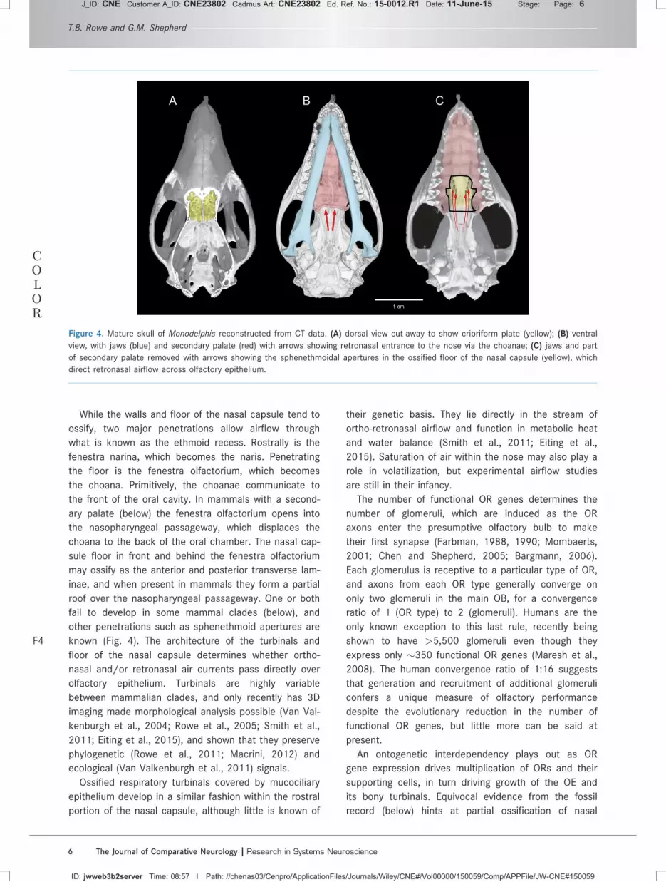
While the walls and floor of the nasal capsule tend to
ossify, two major penetrations allow airflow through
what is known as the ethmoid recess. Rostrally is the
fenestra narina, which becomes the naris. Penetrating
the floor is the fenestra olfactorium, which becomes
the choana. Primitively, the choanae communicate to
the front of the oral cavity. In mammals with a second-
ary palate (below) the fenestra olfactorium opens into
the nasopharyngeal passageway, which displaces the
choana to the back of the oral chamber. The nasal cap-
sule floor in front and behind the fenestra olfactorium
may ossify as the anterior and posterior transverse lam-
inae, and when present in mammals they form a partial
roof over the nasopharyngeal passageway. One or both
fail to develop in some mammal clades (below), and
other penetrations such as sphenethmoid apertures are
known (Fig.F4 4). The architecture of the turbinals and
floor of the nasal capsule determines whether ortho-
nasal and/or retronasal air currents pass directly over
olfactory epithelium. Turbinals are highly variable
between mammalian clades, and only recently has 3D
imaging made morphological analysis possible (Van Val-
kenburgh et al., 2004; Rowe et al., 2005; Smith et al.,
2011; Eiting et al., 2015), and shown that they preserve
phylogenetic (Rowe et al., 2011; Macrini, 2012) and
ecological (Van Valkenburgh et al., 2011) signals.
Ossified respiratory turbinals covered by mucociliary
epithelium develop in a similar fashion within the rostral
portion of the nasal capsule, although little is known of
their genetic basis. They lie directly in the stream of
ortho-retronasal airflow and function in metabolic heat
and water balance (Smith et al., 2011; Eiting et al.,
2015). Saturation of air within the nose may also play a
role in volatilization, but experimental airflow studies
are still in their infancy.
The number of functional OR genes determines the
number of glomeruli, which are induced as the OR
axons enter the presumptive olfactory bulb to make
their first synapse (Farbman, 1988, 1990; Mombaerts,
2001; Chen and Shepherd, 2005; Bargmann, 2006).
Each glomerulus is receptive to a particular type of OR,
and axons from each OR type generally converge on
only two glomeruli in the main OB, for a convergence
ratio of 1 (OR type) to 2 (glomeruli). Humans are the
only known exception to this last rule, recently being
shown to have >5,500 glomeruli even though they
express only �350 functional OR genes (Maresh et al.,
2008). The human convergence ratio of 1:16 suggests
that generation and recruitment of additional glomeruli
confers a unique measure of olfactory performance
despite the evolutionary reduction in the number of
functional OR genes, but little more can be said at
present.
An ontogenetic interdependency plays out as OR
gene expression drives multiplication of ORs and their
supporting cells, in turn driving growth of the OE and
its bony turbinals. Equivocal evidence from the fossil
record (below) hints at partial ossification of nasal
COLOR
Figure 4. Mature skull of Monodelphis reconstructed from CT data. (A) dorsal view cut-away to show cribriform plate (yellow); (B) ventral
view, with jaws (blue) and secondary palate (red) with arrows showing retronasal entrance to the nose via the choanae; (C) jaws and part
of secondary palate removed with arrows showing the sphenethmoidal apertures in the ossified floor of the nasal capsule (yellow), which
direct retronasal airflow across olfactory epithelium.
T.B. Rowe and G.M. Shepherd
6 The Journal of Comparative Neurology |Research in Systems Neuroscience
J_ID: CNE Customer A_ID: CNE23802 Cadmus Art: CNE23802 Ed. Ref. No.: 15-0012.R1 Date: 11-June-15 Stage: Page: 6
ID: jwweb3b2server Time: 08:57 I Path: //chenas03/Cenpro/ApplicationFiles/Journals/Wiley/CNE#/Vol00000/150059/Comp/APPFile/JW-CNE#150059

capsule elements prior to the origin of mammals (Kie-
lan-Jaworowska et al., 2004; Ruf et al., 2014). However,
full ossification of the ethmoid and its turbinals arose in
the last common ancestor of crown Mammalia (Rowe,
1988), potentially accommodating expression of the full
complement of �1,200 OR genes believed present
ancestrally (Rowe et al., 2011).
With a 10-fold increase in OR genes and ORs, the
problem of axonal guidance to their proper glomeruli
was amplified proportionately. 3D imaging suggests
that the shape of each turbinal is essentially that of a
funnel, which grows from the foramina of the cribriform
plate rostrally towards its mature, wide-mouthed termi-
nus (Rowe et al., 2005). Early developmental studies
(Harrison, 1910) discovered that axonal guidance was
determined by the geometry of their physical substrate,
and that they grow in a straight line along solid sub-
strates or follow visible topographic paths when such
paths were visible. Axons were never observed to shift
trajectories unless another solid surface or interface
was available (Harrison, 1914). The funnel-shape of
each turbinal provides passive, longitudinal guidance as
the growing OR axons are funneled very close to their
target glomerulus, just across the cribriform plate
(Rowe et al., 2005). A host of molecular guidance fac-
tors have also been proposed (Singer et al., 1995;
Wang et al., 1997; Bozza et al., 2002; Vassalli et al.,
2002; Mombaerts, 2004, 2006), and how these comple-
ment the turbinal architecture remains to be
determined.
Primary projections from the olfactory bulb (Fig.F5 5)
pass via the lateral olfactory tract to the pyriform (olfac-
tory) cortex, with fibers reaching also the anterior
olfactory nucleus, olfactory tubercle, entorhinal cortex,
and several nuclei in cortical amygdala (Nieuwenhuys
et al., 1998). Odors are believed to be encoded by dif-
ferential activation of the glomeruli to form “odor
images” that are transformed by the olfactory cortex
into “odor objects” as the neural basis for odor discrim-
ination (Shepherd, 1991, 2013; Wilson and Stevenson,
2006). These connections have been observed in mam-
mals (Ashwell, 2010, 2013), turtles, and squamates
(Bruce, 2007, 2009) and can be inferred as present in
amniotes ancestrally. However, the structural basis for
a dual ortho-retronasal olfactory system is unique to
Mammalia and its extinct relatives among Cynodontia
(below).
Humans follow this general developmental pattern,
although in a truncated form. Only �350–400 OR
genes are expressed, and human turbinals and OE are
correspondingly reduced from the condition inferred to
have been present in mammals, therians, and primates
ancestrally. Additionally, the floor of the human nasal
capsule fails to ossify and neither anterior nor posterior
transverse lamina forms (Smith and Rossi, 2006), leav-
ing the human olfactory epithelium broadly open to
orthonasal and retronasal air currents.
Early evolution of ortho-retronasal olfactionOrthonasal olfaction, that is, sniffing in, is an innova-
tion of Tetrapoda, and its history can be traced back
into the fossil record to the earliest Devonian fossils
belonging to the tetrapod stem (Jarvik, 1942). Known
as rhipidistian crossopterygians, these transitional
stem-tetrapods were the first vertebrates in which the
naris conveyed environmental molecules across the OE
and into the mouth via the choana. Orthonasal smell is
employed by nearly all tetrapod species, the exceptions
being secondarily adapted to a committed aquatic life-
style such as odontocete cetaceans (Colbert et al.,
2005; Racicot and Rowe, 2014) and sirenians, in which
the main olfactory system is largely or wholly aban-
doned, and possibly the terrestrial lungless plethodontid
salamanders.
With the origin of Amniota, orthonasal airflow became
tied to two distinct functions, each supported by a pri-
mary “choncha” or epithelial fold that covered a low
ridge of cartilage protruding into the lumen from the lat-
eral wall of the nasal capsule. The anterior choncha
consists of mucociliary respiratory epithelium and repre-
sents the primordium of the mammalian respiratory tur-
binal, while the posterior concha comprises olfactory
epithelium and represents the primordium of mamma-
lian olfactory turbinals (Gauthier et al., 1988). Building
on this foundation of olfactory organization, we next
discuss evidence preserved in fossil cynodonts relating
COLOR
Figure 5. Circuitry schematic of modern opossum (Didelphis)
brain showing (A) sensory inputs and (B) motor outputs (From
Rowe et al., 2011).
Olfaction and cortical evolution
The Journal of Comparative Neurology | Research in Systems Neuroscience 7
J_ID: CNE Customer A_ID: CNE23802 Cadmus Art: CNE23802 Ed. Ref. No.: 15-0012.R1 Date: 11-June-15 Stage: Page: 7
ID: jwweb3b2server Time: 08:57 I Path: //chenas03/Cenpro/ApplicationFiles/Journals/Wiley/CNE#/Vol00000/150059/Comp/APPFile/JW-CNE#150059

to the origin of ortho-retronasal smell and mammalian
cortical structure, using the cladogram in FigureF6 6.
Origin of Cynodontia and retronasalolfaction
During the first �50 million years of synapsid history,
there is little evidence of olfactory or cortical modifica-
tion. Basal synapsids were eye-minded terrestrial mac-
ropredators that dominated the terrestrial trophic
ecosystem throughout the Permian and into the mid-
Triassic. Major themes in early synapsid evolution
involved increased frontality of the orbits, increased
bite forces, modest dietary diversification, and improved
speed and agility. These changes surely involved corti-
cal modifications, but the early braincases were only
partly ossified and fail to record tangible evidence. Early
synapsid braincases were organized much like those of
basal amniotes.
The first measurable changes in the brain occurred
with the origin of Cynodontia1 in the Late Permian. The
early cynodonts remained terrestrial quadrupeds but
were smaller than their Early Permian forebears, being
roughly the size of a domestic cat. Their olfactory bulbs
remained small and the nasal capsule was entirely
unossified. The cerebellum was wider than the
Figure 6. Cynodont phylogeny described in text, with key characters indicated.
1Several of the cynodont characters described below reportedly evolved conver-
gently in Therocephalia, an extinct group customarily viewed as the sister taxon
to Cynodontia. However, it is currently unclear whether “therocephalians” com-
prise a paraphyletic assemblage, with some members positioned closer to Cyn-
odontia than others, and whether other members (e.g., Bauria) actually lie
within Cynodontia.
T.B. Rowe and G.M. Shepherd
8 The Journal of Comparative Neurology |Research in Systems Neuroscience
J_ID: CNE Customer A_ID: CNE23802 Cadmus Art: CNE23802 Ed. Ref. No.: 15-0012.R1 Date: 11-June-15 Stage: Page: 8
ID: jwweb3b2server Time: 08:57 I Path: //chenas03/Cenpro/ApplicationFiles/Journals/Wiley/CNE#/Vol00000/150059/Comp/APPFile/JW-CNE#150059

forebrain, the midbrain was exposed dorsally, and a
small pineal eye occupied a canal that perforated the
skull roof. Compared to living mammals, the first cyno-
donts possessed low-resolution olfaction, insensitive
hearing, with massive middle ear ossicles still attached
to the mandible, coarse tactile sensitivity, and relatively
unrefined motor coordination (Rowe et al., 2011). They
were scratch diggers, and evidence that they may have
hibernated comes from specimens found curled and
preserved in burrows (Groenewald et al., 2001). Never-
theless, when compared with more primitive synapsids,
early cynodonts record a dietary shift to more general
omnivory that is correlated with the first of three suc-
cessive pulses in encephalization that preceded the ori-
gin of Mammalia (Rowe et al., 2011).
First pulse in encephalization.The lateral wall of the cynodont braincase became ossi-
fied by ventral extensions of the frontal and parietal
bones, forward expansion of the prootic, and by the
newly formed alisphenoid. The alisphenoid is a
compound element built from an embryological remnant
of the epiperygoid footplate, and a new membranous
ossification of the spheno-obturator membrane that
consistently lies adjacent to the caudolateral pole of
olfactory cortex in mammals (Maier, 1987; Gauthier
et al., 1988). Appearance of the alisphenoid correlates
with expansion of the olfactory cortex, and this first
pulse of brain expansion raised early cynodont enceph-
alization quotient (EQ) to �0.20 (based on an average
for living mammals; Rowe et al., 2011). In nonmamma-
lian cynodonts, the alisphenoid is referred to inter-
changeably as the epipterygoid. However, the former
term is preferable because the critical transformation
combining two separate elements is characteristic of
cynodonts.
The secondary palate.One of the most diagnostic features of Cynodontia, and
a key structural element in ortho-retronasal olfaction, is
the secondary palate. In basal synapsids, the choanae
were located at the front of the mouth (Fig. F77). With
COLOR
Figure 7. Stages in the evolution of mammalian secondary palate and the ortho-retronasal olfaction duality. (A) Eusthenopteron, a stem-
tetrapod; (B) Seymouria, a stem amniote; (C) Dimetrodon, a basal synapsid; (D) Syodon, a more derived non-cynodontian synapsid; (E) Pro-
cynosuchus, the basal-most cynodont with an incipient secondary palate; (F) Thrinaxodon, an early cynodont with a complete secondary
palate; (G) Kayentatherium, a basal mammaliamorph with a complex dentition; (H) Morganucodon, a basal mammaliaform, with secondary
palate extending to back of tooth row; (I) Didelphis, with secondary extending behind tooth row. ch, choana; ect, ectopterygoid; max, max-
illa; pal, palatine; pmx, premaxilla; pt, pterygoid; vo, vomer.
Olfaction and cortical evolution
The Journal of Comparative Neurology | Research in Systems Neuroscience 9
J_ID: CNE Customer A_ID: CNE23802 Cadmus Art: CNE23802 Ed. Ref. No.: 15-0012.R1 Date: 11-June-15 Stage: Page: 9
ID: jwweb3b2server Time: 08:58 I Path: //chenas03/Cenpro/ApplicationFiles/Journals/Wiley/CNE#/Vol00000/150059/Comp/APPFile/JW-CNE#150059

simple conical teeth, these macropredators used their
mouths to subdue and dismember prey, and their mode
of inertial swallowing was aided by teeth on the roof of
the palate (Kemp, 2005). The appearance of the sec-
ondary palate marked a profound reorganization in
feeding behavior. Palatal teeth and the inertial mode of
swallowing were lost in cynodonts, and the tongue
became the major guide of food around the mouth and
toward the esophagus (Barghusen, 1986; Crompton,
1989). The uniquely complex chewing and swallowing
behaviors of mammals became the basis of retronasal
olfaction in early cynodonts.
The secondary palate separates the oral cavity from
the nasal cavity as shelves of the maxillae and palatines
grow toward the midline. The secondary palate also cre-
ates a new passageway through the nose, the nasopha-
ryngeal passage (Fig. 3), which lies above the
secondary palate (its floor) and beneath the nasal cap-
sule (its roof, where ossified). Orthonasal air passing
through the ethmoid recess now courses along the
nasopharyngeal passage before emptying into the back
of the mouth via posteriorly displaced choana; retro-
nasal air retraces this path in the opposite direction. In
nonmammalian cynodonts the nasal capsule was
entirely cartilaginous, but it became ossified in mam-
mals where it took on a remarkable diversity of form as
it adapted to contain the elaborate respiratory and
olfactory turbinals.
In the oral cavity, the secondary palate forms a rigid
roof against which the tongue can move food items
toward the cheek teeth for processing or toward the
esophagus for swallowing, and as a structural element
increases the occlusal forces that the face can with-
stand (Crompton, 1989; Kemp, 2005). The secondary
palate is fundamental to the new process of oral food
processing or mastication. The oral and nasal chambers
become confluent at the choana, which opens into the
pharynx behind the tooth row, and orthonasal air enters
the pharynx at the back of the mouth. Exhaled air can
now take two routes, either through the mouth, as in
panting, or back through the nasal chamber, via the ret-
ronasal pathway to re-cross the olfactory epithelium
before exiting through the nares.
In Procynosuchus, the basal-most cynodont, the max-
illae and palatines form a pair of shelves that extend
two-thirds the length of the dentition (Fig. 7E). The
shelves grow medially, but fail to meet on the midline
and a narrow channel separates them from the vomer.
This condition resembles the human congenital defor-
mity known as cleft palate, in which the maxillae and
palatines fail to grow sufficiently to meet on the mid-
line, and a longitudinal slot affords a narrow conduit for
some air to pass vertically between the nose and
mouth. This was probably the case in Procynosuchus
(Kemp, 1979). In all more derived cynodonts, the maxil-
lae and palatines meet on the midline beneath the
vomer to form a complete secondary palate (Fig. 7F–I).
By the origin of Mammaliaformes (Fig. 7H), the second-
ary palate extended to the back of the tooth row.
The cynodont dentition.The namesake feature of Cynodontia is their “dog-like”
dentition, in which a long canine separates simple inci-
sors in front from complex molariform teeth that line
the cheeks. The molariform teeth occluded in irregular
facets, actively masticating food before it is swallowed.
The basic plan of what would become the tritubercular
molar was set, in which the crowns have three longitu-
dinally aligned principal cusps, with the tallest in the
middle, and an encircling ring of tiny cusps at their
base known as cingula. Wear facets on the principal
cusps are marked by micro-striations, providing evi-
dence that upper and lower molariform teeth occluded
while processing different types of food. Mastication
shreds, dices, crushes, grinds, grates, chops, tears,
rips, minces, pulverizes, and generally triturates food
items before they are swallowed. This speeds the rate
and degree of caloric return while liberating a new
domain of information for analysis by the tongue and
nose. In concert with the secondary palate, mastication
made ortho-retronasal olfaction possible.
These developments initiated an episode of unprece-
dented dietary diversification reflected in a tremendous
acceleration in rates of dental evolution. To frame this
in a quantitative perspective, consider the basis of a
series of phylogenetic analyses. Gauthier et al. (1988)
scored 207 characters across 29 taxa that character-
ized skeletal variation in basal amniote clades (including
nonmammalian synapsids), and only 8% (17) of these
characters reflect dental variation. But for data matrices
designed to capture variation among extinct cynodonts
and basal mammals, Meng et al. (2006) scored 435
total osteological characters for 58 taxa, and 25% (108
characters) reflect dental variation. Building on that
matrix, Ji et al. (2006) scored 445 characters for 103
taxa, and found 39% (173) of the characters as describ-
ing dental variation. In a matrix of 3,660 osteological
characters for 86 fossil and extant mammaliaforms,
O’Leary et al. (2013) found 40% of total osteological
phenomic variation (1,450 characters) to reside in the
dentition. Amplifying this diversity is the finding that
homoplasy affects the cynodont dentition to a special
degree. In a study of cynodont and basal mammal rela-
tionships, Rowe (1993) found in a matrix of 151 charac-
ters for 24 taxa that there was 30% more homoplasy in
T.B. Rowe and G.M. Shepherd
10 The Journal of Comparative Neurology | Research in Systems Neuroscience
J_ID: CNE Customer A_ID: CNE23802 Cadmus Art: CNE23802 Ed. Ref. No.: 15-0012.R1 Date: 11-June-15 Stage: Page: 10
ID: jwweb3b2server Time: 08:58 I Path: //chenas03/Cenpro/ApplicationFiles/Journals/Wiley/CNE#/Vol00000/150059/Comp/APPFile/JW-CNE#150059

cynodont dental characters than in either the skull, the
postcranium, or the combined skeleton.
There is hardly a branch on the cynodont tree that
lacks its own unique molariform crown pattern. In most
clades the dentition reveals the finest levels of taxo-
nomic diversity and presents the diagnostic characters
by which living and extinct species are identified. Evolu-
tionary variability in the dentition parallels the high vari-
ability in the mammalian OR genome (Niimura, 2009,
2012) and is consistent with the view that olfactory
ecology was a primary influence on the shape of mam-
malian diversification (Hayden et al., 2010). In sum-
mary, retronasal smell gives added insight into the
significance of dentition in relation to feeding behavior
and evolution.
Associated changes in the mandible indicate a shift
in the force vectors of the adductor musculature. Primi-
tively, the greatest bite forces were exerted at the front
of the mouth for grasping prey. In cynodonts, reorgan-
ization of the mandible and zygomatic arch indicate
that the mammalian temporalis and masseter were dif-
ferentiated, and that the largest mandibular forces were
shifting toward the back of the tooth row for mastica-
tion (Gregory, 1953; Crompton, 1989; Kemp, 2005).
The cynodont cranio-vertebral joint.In basal synapsids, the skull articulates with the neck
via a single spherical occipital condyle that is posi-
tioned beneath the foramen magnum. In cynodonts, the
basioccipital largely recedes from the joint, and a
“double occipital condyle” is formed by the right and
left exoccipitals which are positioned at the ventrolat-
eral quadrants of the foramen magnum. The new articu-
lation expanded the degree of stable dorsoventral
excursion of the head on the neck without impairing
passage of the spinal cord through the foramen mag-
num and along the cervical neural canal (Jenkins,
1971). It also suggests that the head was habitually
held at a tilt, with the nose toward the ground. Many
mammals target their noses towards the ground and
move their heads rapidly from side to side in scent-
tracking and scent-guided navigation. More agile head
movement potentially enabled cynodont olfaction to
assert its importance in tracking, navigation, and geo-
graphic memory.
Diaphragmatic ventilation.In basal synapsids, the dorsal vertebrae are undifferen-
tiated and bony ribs extend from the neck to the pelvis.
In cynodonts, distinct thoracic and lumbar regions
become differentiated in which long ribs persist on the
anterior thoracic vertebrae, while the posterior three to
five ribs form attenuated processes that fuse to their
respective neural arches (Fig. F88). Differentiation of sep-
arate thoracic and lumbar regions marks the develop-
ment of a muscular diaphragm, which separated the
thoracic and abdominal cavities and initiated onset of
the stereotyped vacuum-chamber or bellows-like tidal
diaphragmatic ventilation of mammals
While stationary or at rest, ventilation in living mam-
mals is driven by the diaphragm (Bramble, 1989,
Alexander, 2003). Chewing food is mostly a stationary
action, thus ortho-retronasal olfaction is driven by dia-
phragmatic ventilation. The rapid sniffing so characteris-
tic of many mammals in exploring their olfactory
environments (Stoddart, 1980; Shepherd, 2012) is
mostly done between steps or when moving slowly, and
is also driven by the diaphragm. In basal cynodonts, the
proximal ends of the thoracic ribs are flattened and
imbricate in a condition unknown in living mammals.
Hence, their modes of breathing and locomotion were
not entirely modern, but the important new capacity of
diaphragmatic ventilation was introduced.
To summarize, in Late Permian cynodonts the struc-
tural basis of ortho-retronasal olfaction was established
with the origin of the secondary palate and occlusal
Figure 8. Skeletons drawn to scale of Lycaenops (a Permian gorgonopsian), Thrinaxodon, and Morganucodon. Note the differentiation of
thoracic and lumbar vertebrae in Thrinaxodon and Morganucodon skeletons, indicating presence of the diaphragm (modified from Dingus
and Rowe, 1998).
Olfaction and cortical evolution
The Journal of Comparative Neurology | Research in Systems Neuroscience 11
J_ID: CNE Customer A_ID: CNE23802 Cadmus Art: CNE23802 Ed. Ref. No.: 15-0012.R1 Date: 11-June-15 Stage: Page: 11
ID: jwweb3b2server Time: 08:58 I Path: //chenas03/Cenpro/ApplicationFiles/Journals/Wiley/CNE#/Vol00000/150059/Comp/APPFile/JW-CNE#150059

dentition, and with reorganization of the cranio-
vertebral joint and the introduction of diaphragmatic
ventilation. It was associated with a first modest pulse
in encephalization that primarily affected the pyriform
cortex. The principal question with the origin of cyno-
donts concerns the degree to which new cortical con-
nections and associations were established between
the multiple input modes that are integrated in mamma-
lian ortho-retronasal olfaction. That all the skeletal
equipment for ortho-retronasal olfaction was now in
place suggests that the trend toward multilayered neo-
cortex and the multisensory integration that we refer to
as flavor had begun its emergence in basal cynodonts.
With EQs measuring only �0.20, the first cynodonts fell
considerably short of the capabilities inferred to have
been present in the ancestral mammal (Rowe et al.,
2011). Nevertheless, neurophysiological evidence
reviewed below suggests that this was not a “simple”
cortex and was capable of higher orders of associations
underlying discrimination, learning, and memory.
Triassic cynodont diversificationOver the next �50 million years of Triassic cynodont
history there is little evidence of further changes in rel-
ative brain size or structure. We doubt that cortical evo-
lution was static, but it was insufficient to alter the
braincase structure within the resolution of modern
imaging techniques. Skeletal variation mostly involved
successive modifications of the masticatory and loco-
motor systems. In the former system, the postdentary
bones became increasingly decoupled from mastication,
reduced in size and dedicated to higher-frequency audi-
tion, although retaining their attachment to the
mandible.
Several other refinements of the masticatory appara-
tus pertain to ortho-retronasal olfaction, and these inno-
vations preceded a second pulse in encephalization in
the last common ancestor of Mammaliaformes (Node 8,
below), in which the neocortex almost certainly
emerged, and set the stage for the subsequent origin
of crown Mammalia.
At Node 2 (Fig. 6, unnamed), closure of the second-
ary palate was completed as the right and left maxillae
and palatines sutured together along the midline. More
powerful occlusal forces are indicated by successive
increases in height of the coronoid process of the den-
tary, the extent of the masseteric fossa, thickening of
the zygomatic arch, and expansion of the temporal
fenestra. (Gregory, 1953; Kemp, 2005). Postcranial
modifications suggest more agile and forceful locomo-
tion, and greater ventilation capacity.
At Eucynodontia (Node 3), a key innovation appeared
in the periodontal ligament. The teeth in basal
cynodonts had short open roots that occupied shallow
sockets and were held to the jaws by a ring of bone
that surrounded the crown (Crompton, 1963; Rowe
et al., 1995). In eucynodonts, the molariform teeth
have long roots that close around the dental nerve dur-
ing maturation and are implanted into deep sockets
and held in place by a periodontal ligament (Rowe,
1993). Eucynodont fossils are commonly found in which
the teeth slipped from their sockets before burial or, if
the jaws still hold their teeth, inevitably there is a thin
sheet of matrix around the roots, ancient sediment that
filled the space once occupied by the ligament.
The periodontal ligament enables precise occlusal
relationships to develop between upper and lower teeth
during ontogeny (Noble, 1969; Ten Cate, 1969). In liv-
ing mammals, tooth crowns erupt first, and opposing
teeth twist and rotate and adjust to one another in
forming consistent occlusal relationships. Implantation
of the roots into their sockets follows eruption of the
crown, and only after the occlusal relationship between
crowns has formed do the roots lock the teeth in place
with the periodontal ligament. It is this spatial plasticity
as crowns erupt that dentists exploit in using braces to
straighten and adjust the maturing teeth in adolescent
humans (Wise and King, 2008). The ligament also
serves as a shock absorber, enabling more powerful
occlusal forces. Its innervation supplements information
from the crown on the texture of masticated food items
via branches of the trigeminal nerve, which also convey
somatosensory information from the oral cavity and
tongue, and control most of the muscles involved in
chewing and some of the muscles of swallowing (Bar-
ghusen, 1986).
Accompanying this innovation was a reduction in the
rate and mode of tooth replacement. The primitive cyn-
odont pattern of continuous alternating replacement of
postcanine teeth throughout life occasioned frequent
disruption of occlusion (Crompton, 1963). Eucynodonts
adopted a pattern of consecutive replacement, and
slowed the rate of replacement such that fewer tooth
generations erupted from each socket and consistent
patterns of occlusal facets between upper and lower
molariform teeth could form (Rowe, 1993).
Node 4 (unnamed) is the taxon stemming from the
last common ancestor mammals share with the Early
Triassic Diademodon. Precladistic accounts portrayed
cynodont history as two grand radiations, one of herbiv-
orous “gomphodonts” with broad tooth crowns, and the
other of persistently predatory cynodonts from which
mammals ultimately descended (e.g., Hopson, 1969;
Crompton, 1972). The first phylogenetic analyses
quickly established that some “gomphodonts” such as
Exaeretodon and tritylodonts (below) are more closely
T.B. Rowe and G.M. Shepherd
12 The Journal of Comparative Neurology | Research in Systems Neuroscience
J_ID: CNE Customer A_ID: CNE23802 Cadmus Art: CNE23802 Ed. Ref. No.: 15-0012.R1 Date: 11-June-15 Stage: Page: 12
ID: jwweb3b2server Time: 08:58 I Path: //chenas03/Cenpro/ApplicationFiles/Journals/Wiley/CNE#/Vol00000/150059/Comp/APPFile/JW-CNE#150059

related to mammals than to Diademodon and that the
“gomphodonts” are a paraphyletic assemblage of pre-
sumed herbivores (e.g., Rowe, 1988, 1993; Gauthier
et al., 1988). In this light, it appears that dental occlu-
sion and mastication enabled a far greater measure of
dietary diversification than previously believed, and that
the adoption of herbivory occurred multiple times
among cynodonts.
At Node 4 the cheek teeth now have two roots, each
surrounding its own dental nerve and root canal. In
early forms like Diademodon, a thin web of bone still
connected the roots, but this was soon lost and the
roots became fully divided. The crowns in these cyno-
donts were generally expanded and closely packed,
forming in some taxa a broad, continuous occlusal sur-
face and one that provided greater levels of textural
information about food items (Rowe, 1993).
At Node 5 escalation of the role of the trigeminus in
feeding behaviors is evident in the presence of a partial
bony floor beneath the cavum epipterycum. This space
contains the trigeminal ganglion, and its partial enclo-
sure marks expansion of the ganglion (Bonaparte,
1966; Rowe, 1993). In crown mammals the cavum epi-
pterycum becomes completely enclosed by the bony
braincase in early development, although it remains
external to the meninges and outside of the cavum cra-
nii proper.
Mammaliamorpha (Node 6).This clade stems from the last common ancestor mam-
mals share with the Late Triassic tritylodontids (Rowe,
1988), and its origin coincides with a number of impor-
tant transformations that preceded a second pulse in
encephalization (Node 7, below). The first mammalia-
morphs became miniaturized (Fig. 8), and as adults
occupied the one or two smallest orders of vertebrate
size magnitude. A few descendant lineages regained
much larger size during the Mesozoic, but for the next
�135 million years, nearly the entire history of mamma-
liamorphs including crown mammals played out in tiny
animals (Kielan-Jaworowska et al., 2004). Only in the
Cenozoic did numerous mammalian clades independ-
ently evolve much larger body sizes, but even today the
greatest diversity is in small species. With miniaturiza-
tion, early mammaliamorphs encountered greater spa-
tial and environmental heterogeneity. Entry into new
microhabitats corresponds to diversification in diet,
activity patterns, and life history strategies (Harvey
et al., 1980; Mace et al., 1981). New food items such
as seeds, grains, fungi, small fruiting bodies, and small
invertebrates were available for the first time. Ortho-
retronasal olfaction now included high-resolution soma-
tosensory information provided by the oral field of the
trigeminus, enabling early mammaliamorphs to explore
and exploit more thoroughly their new microhabitats.
In the skull, the medial wall of the orbit was closed
as the orbitosphenoid became co-ossified with the
sheets of thin bone contributed by the palatine and
frontal (Sues, 1986), and the rear parts of the nasal
capsule were ossified for the first time (Kielan-Jaworow-
ska et al., 2004). Together with elaborate modifications
of the side wall of the braincase (Rowe, 1988, 1993),
this likely reflects its own modest pulse in encephaliza-
tion. However, efforts to image endocasts in tritylodon-
tids have been unsuccessful. Additionally, negative
allometry of the auditory ossicles indicates that they
were increasingly decoupled from their role in mastica-
tion in favor of high-frequency audition (Rowe,
1996a,b). The cochlea had begun to elongate (Kielan-
Jaworowska et al., 2004), but its full coiling occurred
much later, within various mammalian subclades (Luo
et al., 2011).
Miniaturization was accompanied by remodeling of
the postcranial skeleton, in which the joint surfaces
were more precisely sculpted, and the trochanteric
attachments for limb muscles resembled those found in
mammals. Locomotion at small size is metabolically far
less expensive than at larger size, and climbing vertically
costs little more than locomotion over flat surfaces
(McMahon and Bonner, 1983). There is also a regular
change in the mechanical advantage muscles have about
the joints of the skeleton as a consequence of the stabil-
ity of the joints under loads determined by inertia and
gravity, and flexion angles at joints increases as size
decreases. This implies that muscle spindles and joint
proprioceptors were recording more information than
before, as a new level of agility emerged. Herbivores
comprise the largest portion of biomass within modern
mammalian communities (Eisenberg, 1990), and even a
partial shift to the new trophic level of primary consumer
may have supported larger populations of early mamma-
liamorphs than in earlier cynodonts.
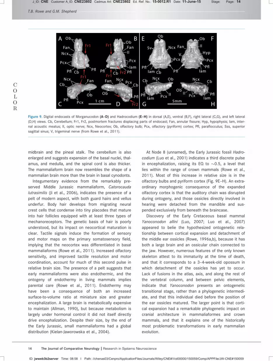
Mammaliaformes (Node 7).A second pulse of encephalization occurred in Mamma-
liaformes (Rowe et al., 2011), which is the clade stem-
ming from the last common ancestor shared by
Morganucodon and Mammalia (Rowe, 1988). The endo-
cranial cavity is fully enclosed, and endocasts indicate
the brain of Morganucodon (Fig. F99A–D) to be nearly
50% larger than more primitive cynodonts, with an EQ
of �0.32. The olfactory bulb and cortex are by far the
regions of greatest expansion. A deep annular fissure
separates the inflated olfactory bulb from the inflated
olfactory cortex, which by this time was much wider
than the hindbrain. The cortex and cerebellum cover the
Olfaction and cortical evolution
The Journal of Comparative Neurology | Research in Systems Neuroscience 13
J_ID: CNE Customer A_ID: CNE23802 Cadmus Art: CNE23802 Ed. Ref. No.: 15-0012.R1 Date: 11-June-15 Stage: Page: 13
ID: jwweb3b2server Time: 08:58 I Path: //chenas03/Cenpro/ApplicationFiles/Journals/Wiley/CNE#/Vol00000/150059/Comp/APPFile/JW-CNE#150059
midbrain and the pineal stalk. The cerebellum is also
enlarged and suggests expansion of the basal nuclei, thal-
amus, and medulla, and the spinal cord is also thicker.
The mammaliaform brain now resembles the shape of a
mammalian brain more than the brain in basal cynodonts.
Integumentary evidence from the remarkably pre-
served Middle Jurassic mammaliaform, Catorocauda
lutrasimilis (Ji et al., 2006), indicates the presence of a
pelt of modern aspect, with both guard hairs and vellus
underfur. Body hair develops from migrating neural
crest cells that condense into tiny placodes that mature
into hair follicles equipped with at least three types of
mechanoreceptors. The genetic basis of hair is poorly
understood, but its impact on neocortical maturation is
clear. Tactile signals induce the formation of sensory
and motor maps on the primary somatosensory field,
implying that the neocortex was differentiated in basal
mammaliaforms (Rowe et al., 2011). Increased olfactory
sensitivity, and improved tactile resolution and motor
coordination, account for much of this second pulse in
relative brain size. The presence of a pelt suggests that
early mammaliaforms were also endothermic, and the
ontogeny of endothermy in living mammals implies
parental care (Rowe et al., 2011). Endothermy may
have been a consequence of both an increased
surface-to-volume ratio at miniature size and greater
encephalization. A large brain is metabolically expensive
to maintain (Allman, 1990), but because metabolism is
largely under hormonal control it did not itself directly
drive encephalization. Despite their size, by the end of
the Early Jurassic, small mammaliaforms had a global
distribution (Kielan-Jaworowska et al., 2004).
At Node 8 (unnamed), the Early Jurassic fossil Hadro-
codium (Luo et al., 2001) indicates a third discrete pulse
in encephalization, raising its EQ to �0.5, a level that
lies within the range of crown mammals (Rowe et al.,
2011). Most of this increase in relative size is in the
olfactory bulbs and pyriform cortex (Fig. 9E–H). An extra-
ordinary morphogenic consequence of the expanded
olfactory cortex is that the auditory chain was disrupted
during ontogeny, and those ossicles directly involved in
hearing were detached from the mandible and sus-
pended exclusively from beneath the braincase.
Discovery of the Early Cretaceous basal mammal
Yanoconodon allini (Luo, 2007; Luo et al., 2007)
appeared to belie the hypothesized ontogenetic rela-
tionship between cortical expansion and detachment of
the middle ear ossicles (Rowe, 1996a,b), because it has
both a large brain and an ossicular chain connected to
the jaw. However, numerous features of the only known
skeleton attest to its immaturity at the time of death,
and that it corresponds to a 3–4-week-old opossum in
which detachment of the ossicles has yet to occur.
Lack of fusions in the atlas, axis, and along the rest of
the vertebral column, and between pelvic elements,
indicate that Yanoconodon presents an ontogenetic
transitional stage, rather than a phylogenetic intermedi-
ate, and that this individual died before the position of
the ear ossicles matured. The larger point is that corti-
cal expansion had a remarkable phylogenetic impact on
cranial architecture in mammaliaformes and crown
mammals, and that it explains one of the historically
most problematic transformations in early mammalian
evolution.
COLOR
Figure 9. Digital endocasts of Morganucodon (A–D) and Hadrocodium (E–H) in dorsal (A,E), ventral (B,F), right lateral (C,G), and left lateral
(D,H) views. Cb, Cerebellum; Fr1, Fr2, postmortem fractures displacing parts of endocast; Fan, annular fissure; Hyp, hypophysis; Iam, inter-
nal acoustic meatus; II, optic nerve; Ncx, Neocortex; Ob, olfactory bulb; Pcx, olfactory (pyriform) cortex; Pfl, paraflocculus; Sss, superior
sagittal sinus; V, trigeminal nerve (from Rowe et al., 2011).
T.B. Rowe and G.M. Shepherd
14 The Journal of Comparative Neurology | Research in Systems Neuroscience
J_ID: CNE Customer A_ID: CNE23802 Cadmus Art: CNE23802 Ed. Ref. No.: 15-0012.R1 Date: 11-June-15 Stage: Page: 14
ID: jwweb3b2server Time: 08:58 I Path: //chenas03/Cenpro/ApplicationFiles/Journals/Wiley/CNE#/Vol00000/150059/Comp/APPFile/JW-CNE#150059

In Hadrocodium, the cerebellum has also expanded
to such a degree that the occipital plate bulges back-
wards, enclosing a relatively large foramen magnum
and spinal cord. Only a few features separate Hadroco-
dium from crown mammals. Most important, it lacks
ossified turbinals; large pterygoid processes indicate
that its bilateral chewing and swallowing mechanics
were transitional to those in crown Mammalia. Once
the brain reached this level of relative size, it continued
to diversify as it supported diverse sensory partnerships
that evolved in the different mammal clades. That infor-
mation was an essential commodity to the diversifica-
tion of mammals is evident in the fact that further
increases in encephalization occurred independently in
nearly all of the major clades (Jerison, 1973; Rowe
et al., 2011).
It is noteworthy that evolutionary decreases in
encephalization are rare. One of the best-documented
cases is in the platypus lineage, where more highly
encephalized Cenozoic fossils indicate reduction of
both the olfactory bulb and overall EQ in the evolution
of the living platypus Ornithorhynchus (Macrini et al.,
2006). Decreases in encephalization are also associated
with domestication in various mammalian species
(Kruska, 2007).
The origin of Mammalia (Node 9).An ossified derivative of the embryonic nasal capsule,
known in total as the ethmoid bone and including the
elaborate skeleton of the ethmoid turbinals, arose with
the origin of Mammalia and followed the developmental
pathway described above (Rowe et al., 2005). Compari-
sons among mammalian subgenomes suggest �1,200
OR genes were present in mammals ancestrally (Nii-
mura, 2009, 2012) and their full expression was facili-
tated by the new surface area provided by the
turbinals.
It seems remarkable that an order of magnitude
expansion in OE surface area could occur without a cor-
responding increase in the size of the nose. This tempts
speculation that without compensation in the visual sys-
tem, expansion of the nose dorsally and laterally would
disrupt the forward binocular visual field, while caudal
expansion is limited by the optic chiasm, against which
the ethmoid abuts. The secondary palate and dental
occlusion may have also constrained organization of the
facial skeleton, reflecting the critical role that mastica-
tion had long since assumed in cynodont physiology.
Speculation aside, the developmentally adaptive nature
of trubinal growth enables it to support both patent air-
ways and a 10-fold expansion of OE within a confined
space. It also maintains spatial identity of the expres-
sion loci for specific OR genes as the ORs themselves
are renewed throughout ontogeny, and it helps funnel
new OR axons toward their target glomeruli. That all
this occurs without radically altering a plan of facial
architecture established in the first cynodonts under-
scores the integration of mastication as part of the
larger system of ortho-retronasal olfaction.
Three-layer association cortex and theorigins of neocortex
We have seen that the primitive skeletal structure of
early amniotes was profoundly transformed in the emer-
gence of early mammals, and that ortho-retronasal
olfaction played a central role in linking correlated
transformations in brain size, the masticatory system,
and in the postcranial skeleton. The neocortex arose in
this environment of vastly increased airborne and inter-
nal olfactory information, a major escalation in somato-
sensory information, and additional sources of new
information from the ear. Finally, and crucially, these
small creatures were endothermic, which supported a
constantly active cortically controlled motor system
that mediated prolonged foraging behavior to support
their high metabolism, their young, and survival among
larger predators.
The relative size of the neocortex in early mammals
was small. Figure F1010 shows the structure of the brain
inferred to have been present in the ancestral mammal,
in which the pyriform cortex was much larger than the
small neocortical areas subserving other sensory and
motor systems (Moln�ar et al., 2014). The somatosen-
sory area subserving the sensory function of hair was
introduced prior to the origin of mammals (in basal
Mammaliaformes), whereas successive increases in
Figure 10. Depiction of the mammalian "ancestral forebrain cor-
tex" (from Moln�ar et al., 2014). OF, orbitofrontal area; MF, medial
frontal area; S1, S2, RS, CS: primary, secondary, dorsal and cau-
dal somatosensory areas; g, gustatory area; V1, V2, t: primary,
secondary and temporal visual areas; Aud, auditory areas; CCv,
CCd: ventral and dorsal cingulate areas; RSg, RSa, retrosplenial
granular area and agranular areas; SC, superior colliculus; IC,
inferior colliculus.
Olfaction and cortical evolution
The Journal of Comparative Neurology | Research in Systems Neuroscience 15
J_ID: CNE Customer A_ID: CNE23802 Cadmus Art: CNE23802 Ed. Ref. No.: 15-0012.R1 Date: 11-June-15 Stage: Page: 15
ID: jwweb3b2server Time: 08:58 I Path: //chenas03/Cenpro/ApplicationFiles/Journals/Wiley/CNE#/Vol00000/150059/Comp/APPFile/JW-CNE#150059

pyriform cortex can be traced from the origin of cyno-
donts to the diversification of mammals. Given the evi-
dence for an evolutionary increase in the size of
pyriform cortex, we next consider to what extent princi-
ples of neocortical organization may have been devel-
oped from those involved in olfactory cortical function.
Olfactory or pyriform cortex appears to have had a
similar neural organization in turtles and lizards (Ulinski,
1983; Bruce, 2007, 2009; Bruce and Braford, 2009) as
in monotremes, marsupials, and placentals (Ashwell,
2010, 2013; Shepherd, 2011), supporting the inference
that this organization was present in amniotes ances-
trally. In brief, studies of opossum and rodent have
shown that in olfactory cortex, the output fibers from
the olfactory bulb course over the surface of the cortex,
emitting collaterals that terminate on the most distal
dendrites of the pyramidal cells (Fig.F11 11). Their action is
excitatory onto spines of these cells, as well as onto
smooth dendrites of interneurons which feed inhibition
forward onto the pyramidal neurons. The activated
pyramidal cells through their axon collaterals feed back
recurrent excitation onto themselves and neighboring
pyramidal neurons. They also excite interneurons which
feed back inhibition onto themselves and neighboring
pyramidal neurons. Thus, in the two critical operations
of the circuit—processing the input and the output—exci-
tation and inhibition are balanced. This circuit is shown
in Figure 11.
It is tempting to assume that olfactory cortex is
equivalent to primary sensory cortex in other senses.
However, Haberly (1985) showed many years ago that
the intrinsic organization of olfactory cortex, with its
long association fibers, was much more similar to
higher association cortical areas; for example, the face
area of inferotemporal cortex. This is now widely
accepted. As noted above, olfactory cortex takes input
from the olfactory bulb in the form of an odor image
and transforms it into a central representation as an
odor object (Shepherd, 1991; Wilson and Stevenson,
2006). The evidence from mammals, lizards, and turtles
indicates that this higher-order function was present
at the origin of Amniota, and was elaborated in basal
cynodonts in which occurred the first pulse in encephal-
ization associated with evolution of the uniquely mam-
malian neocortex.
A second major cortical area is the hippocampus, dif-
ferentiating from the medial wall in amniote forebrains.
Anatomical and physiological studies have shown that
across amniotes the neurons are similar to those in the
olfactory cortex, with similar interconnections for excita-
tion and inhibition (Connors and Kriegstein, 1986; Hab-
erly, 2001). Of special note is the tendency to burst
firing, believed to be analogous to the dorsal hippocam-
pus in rodents with susceptibility to seizure activity. As
in rodent hippocampus, tetanic stimulation of the sep-
tum in the turtle Pseudemys scripta gives rise to hetero-
synaptic long-term potentiation (LTP) (Mu~noz et al.,
1998).
Numerous studies show a close similarity between
the intrinsic organization of the hippocampus and the
basic organization of the olfactory cortex, in terms of
layering of inputs on the apical dendrites and long asso-
ciation fibers (Fig. F1212; Neville and Haberly, 2004). Since
the inputs to the hippocampus consist exclusively of
central sites in the limbic regions, it is clear that the
three-layer hippocampus from the start of amniote and
mammalian evolution was devoted to higher-order proc-
essing such as learning and memory. This adds to the
evidence that the three-layer cortex is not a primitive
“simple” cortex, but rather operates at the level of
higher-order associations underlying discrimination,
learning and memory. Note that the connections of
olfactory cortical and hippocampal pyramidal cells in
turtles, lizards, and mammals are almost exclusively
Figure 11. Top. Olfactory cortical areas on the ventrolateral sur-
face of the cerebrum of the rat. AOC, anterior olfactory cortex;
ctx, cortex; olfac tub, olfactory tubercle. From Neville and Haberly
(2004). Bottom: Microcircuit organization of the mammalian piri-
form (olfactory) cortex. Abbreviations: LOT, lateral olfactory tract;
SP, superficial pyramidal cell; DP, deep pyramidal cell; S, stellate
cell; C, centrifugal fiber. Arrows indicate the direction of flow of
activity. Open profiles: excitatory synaptic action; filled profiles:
inhibitory synaptic action. After Haberly and Shepherd (1973).
T.B. Rowe and G.M. Shepherd
16 The Journal of Comparative Neurology | Research in Systems Neuroscience
J_ID: CNE Customer A_ID: CNE23802 Cadmus Art: CNE23802 Ed. Ref. No.: 15-0012.R1 Date: 11-June-15 Stage: Page: 16
ID: jwweb3b2server Time: 08:58 I Path: //chenas03/Cenpro/ApplicationFiles/Journals/Wiley/CNE#/Vol00000/150059/Comp/APPFile/JW-CNE#150059

intratelencephalic; that is, they are restricted to the tel-
encephalon (cortex and basal ganglia) and do not pro-
ject to lower levels of the brain stem and spinal cord, a
point that will be important in comparing them with
neocortex (see below).
This leaves dorsal cortex as the anlagen, the precur-
sor, of neocortex in mammals. A pioneering anatomical
study by Smith et al. (1980) of the turtle Pseudemys
scripta reported connections through an interneuron
that provide for feedforward and lateral inhibition. This
was followed by an electrophysiological study which
incorporated feedback and lateral excitation and inhibi-
tion into the local circuit (Kriegstein and Connors,
1986; Connors and Kriegstein, 1986) (Fig.F13 13). The
close similarity across amniotes of this local circuit to
the olfactory and hippocampal circuits pointed to a
“basic circuit,” some would call it a “canonical circuit,”
common to all three forebrain regions (Kriegstein and
Connors, 1986). This does not mean that each region
does not have its own fine-tuning for its particular types
of input, but rather that there is a basic framework
common to all three. Since dorsal cortex in turtles
(Ulinksi, 1983) and lizards (Bruce, 2007, 2009) receives
input from the visual pathway, there is a tendency to
regard it as a primary visual cortical area, equivalent to
mammalian V1. However, the comparison with three-
layer hippocampal and olfactory cortices suggests that,
like those regions, dorsal cortex performs a higher-level
association on the visual input. In this view, the three-
layer dorsal cortex which gave rise to neocortex was
not a “simple” cortex for low-level processing, but
rather had an organization that subserved high-level
association functions analogous to those in pyriform
cortex and hippocampus.
This conclusion from comparative physiology is sup-
ported by the comparative anatomy of living amniotes,
which suggests that basal amniotes possessed a fore-
brain with three clearly demarcated regions, viz. a lat-
eral olfactory cortex; a medial hippocampus; and the
dorsal intermediate part that undergoes voluminous
expansion over the course of mammalian history. This
comparison supports the physiological evidence that
the dorsal intermediate part is the region from which
neocortex was successively elaborated in the earliest
cynodonts and mammaliaforms from a three-layer cor-
tex of basal amniotes and early synapsids.
These considerations suggest that visual cortex in
the ancestral amniote may have functioned as a higher-
order visual association area rather than a primary vis-
ual area. This adds to the interest of the pyriform cor-
tex as the best-studied example of the type of higher-
order processing carried out by so-called “simple”
three-layer cortex. As suggested by Fournier et al.
(2014, p. 122), "It could be that DCx [dorsal cortical]
neurons are selective to high-order correlations, and
process spatiotemporal sequences of distributed visual
cues in a manner similar to how PCx [pyriform cortex]
processes spatiotemporal activation of specific
glomeruli."
Pyriform cortex thus appears to have evolved to pro-
cess higher-order associations within the high-
dimensional space representing odor molecules, pushed
ever higher by the expansion of the OR subgenome in
mammals and the new volatiles released by retronasal
smell. A parallel higher-order organization in dorsal cor-
tex appears to have been amplified in neocortex. The
comparative and paleontological evidence thus suggest
that higher-order association functions were present at
the inception of mammalian neocortex. The olfactory
cortex expanded for the higher-level combinatorial asso-
ciations associated with the expansion of the OR subge-
nome and retronasal volatiles. The hippocampal cortex
expanded in processing limbic inputs involving more
Figure 12. Similarities between the laminar organization and hori-
zontal connections of olfactory cortex and hippocampus. 1. OB,
olfactory bulb; AOC, anterior olfactory cortex; APCV, ventral ante-
rior piriform cortex; APCD, dorsal anterior piriform cortex; PPC,
posterior piriform cortex; sup, superficial. 2. EC, entorhinal cortex;
DG, dentate gyrus; prox, proximal; dist, distal; SL-M, stratum
lacunosum-moleculare; SR, stratum radiatum; sup, superior; SP,
stratum pyramidale; SO, stratum oriens. From Neville and Haberly
(2004).
Olfaction and cortical evolution
The Journal of Comparative Neurology | Research in Systems Neuroscience 17
J_ID: CNE Customer A_ID: CNE23802 Cadmus Art: CNE23802 Ed. Ref. No.: 15-0012.R1 Date: 11-June-15 Stage: Page: 17
ID: jwweb3b2server Time: 08:58 I Path: //chenas03/Cenpro/ApplicationFiles/Journals/Wiley/CNE#/Vol00000/150059/Comp/APPFile/JW-CNE#150059

complex learning and memory. And the neocortex dif-
ferentiated new cell types and layers not only building
more sophisticated circuits for processing ortho-
retronasal olfaction, vision, somatosensation, and audi-
tion, but also for novel neocortical control of lower-level
motor connectivity needed for the survival of small
endothermic, continuously active, omnivorous mam-
mals. This is consistent with the transfer of much of vis-
ual processing from the collicular level in reptiles (and
presumably basal amniotes) to the cortical level in
mammals. Finally, the explosion of higher-order cortical
areas, numbering 50 or more in some mammals, was
associated with a corresponding explosion in connectiv-
ity between cell types interacting within the telencepha-
lon as well as connecting to downstream sites of input
and output (Harris and GMG Shepherd, 2015).
How did these properties provide the basis for the
emergence of neocortical neurons and circuits? It will
be useful to return to the early pulses of encephaliza-
tion in cynodonts and compare the likely changes in
neuronal organization that occurred in developing from
a three-layer dorsal cortex to neocortex in these crea-
tures. FigureF14 14 provides simplified diagrams, showing
on the left a single type of pyramidal cell (PC) of a
three-layer dorsal cortex, and on the right the three
pyramidal cell types that current studies identify as
characteristic of neocortex: intratelencephalic (IT) cells
(whose projections remain within the cortex and basal
ganglia); pyramidal tract (PT) cells (including all their
connections throughout the brain stem and spinal
cord); and corticothalamic (CT) cells (see Reiner et al.,
2010; Fame et al., 2011; Harris and GMG Shepherd,
2015). The PCs of dorsal cortex also give rise to associ-
ation fibers; that is, they function as IT cells (IT in the
diagram). They give rise to motor output through their
connections to the basal ganglia, whose cells project
further to the brain stem tectum (Reiner et al., 1998)
with further connections to the spinal cord. Reptiles
thus lack the direct control over the spinal cord motor
neurons that is made possible in mammals through the
PT cells.
On the basis of the analysis of the PC in three-layer
cortex, we hypothesize that its association functions
appear to provide a template for the association func-
tions of the IT PCs distributed throughout layers 2–6 of
the neocortex (indicated by the red coloring of both
types in the diagram). From this perspective, the critical
changes within neocortex were that the IT PCs and
their association connections became greatly amplified
within and between the layers, more specialized in their
outputs (corticocortical connections for superficial IT
cells and neostriatal outputs from deep IT cells), and
Figure 13. Left, above: Dorsal view of the brain of the turtle Pseudemys scripta. DC, dorsal cortex; H, hippocampus; P, pyriform (olfactory)
cortex. Below, cross-section of the forebrain at the level of the arrow in top diagram, showing the relative positions of pyriform (P), hippo-
campal (H), and dorsal cortex (DC). From Connors and Kriegstein (1986). Right: Summary of the microcircuit organization of turtle dorsal
cortex. Thalamocortical afferent volleys (1) excite pyramidal cell dendrites (a) and also inhibitory stellate cells (b). Stellate cell-pyramidal
cell contact (2) mediates feedforward inhibition. Pyramidal cell output mediates reciprocal excitation between pyramidal cells (3) as well
as feedback inhibition (4). Stellate–stellate cell contacts mediate inhibition (5). The pyramidal cell axons provide output (6). After Krieg-
stein and Connors (1986). Open profiles: excitatory synaptic action; filled profiles: inhibitory synaptic action.
T.B. Rowe and G.M. Shepherd
18 The Journal of Comparative Neurology | Research in Systems Neuroscience
J_ID: CNE Customer A_ID: CNE23802 Cadmus Art: CNE23802 Ed. Ref. No.: 15-0012.R1 Date: 11-June-15 Stage: Page: 18
ID: jwweb3b2server Time: 08:58 I Path: //chenas03/Cenpro/ApplicationFiles/Journals/Wiley/CNE#/Vol00000/150059/Comp/APPFile/JW-CNE#150059

extended to the multiple cortical areas in both hemi-
spheres. On the output side, what is new are outputs
through the PT cells of layer 5B to many subcortical
areas, and stronger connections to thalamus through
CT cells of layer 6. On the input side, the pathway
involving thalamocortical fibers and stellate (ST) cells is
special for neocortex.
How does this hypothesis relate to current studies
of the neurogenesis of cortical neuron types (Moln�ar
et al., 2014; Shibata et al., 2015)? In vertebrates, the
ventricular zone (VZ) is the primary embryonic neuro-
genic source, and in mammals the major source of
cells in the deeper neocortical layers (5–6) (Noctor et
al., 2002). Mammals in addition have a subventricular
zone (SVZ), controlled by different genes (Matinez-
Cerdeno et al., 2006; Cheung et al., 2010; Franco
et al., 2012). The SVZ is a source of gliogenesis, and
of neurons in the upper neocortical layers (2–4)
(Tarabykin et al., 2001); recent studies indicate
they can be in all layers (Guo et al., 2013; Vasistha
et al., 2014). The contribution of the SVZ in neuronal
production appears to grow in evolution as the com-
plexity of the neocortex increases (Martinez-Cerdeno
et al., 2006; Cheung et al., 2010; Moln�ar et al.,
2014).
Correlation of these studies with the specific neuro-
nal types shown by physiology is needed. The studies
cited suggest that upper layer cells include IT cells,
whereas deeper layer cells in addition include the PT
and CT types. Further studies are needed to determine
how the SVZ in mammals generates the IT cells and
their association circuits that appear to be based on
the three-layer PC template. We postulate that the SVZ,
or its phylogenetic antecedent, was present in cyno-
donts, the critical step toward generating the multiple
cell types and laminae of the earliest neocortex (Fig.
14). It appears to have developed more fully in basal
mammaliaforms, especially in Hadrocodium, owing to
the relative increase in the size of its brain.
More evidence is needed on whether all neocortical
elements were fully present in the earliest cynodonts.
Early cynodonts display all the skeletal equipment for
ortho-retronasal olfaction, inviting speculation on which
if any of these neocortical elements developed or were
present in some incipient form, and which were added
secondarily in basal mammaliaforms with the next jump
in encephalization. The amount of neocortex in the
ancestral forebrain was likely quite limited (Fig. 10).
One needs to understand more deeply the ecological
niche these early mammaliaforms occupied and their
COLOR
Figure 14. Comparison of the main types of pyramidal cells in three-layer cortex and the neocortex. Left: Three-layer cortex: PC, pyramidal
cell; INT, interneuron; VIS, visual input; IT, intratelencephalic (connections to cortex (C) and basal ganglia [BG]). Right: Neocortex: Intrate-
lencephalic (IT) pyramidal cells (shown in red) are analogous to association pyramidal cells in three-layer cortex (connections to cortex (C)
and neostriatum in the basal ganglia [BG]). They may be found throughout layers 2–6 (dotted line). New types of pyramidal cells (shown in
blue) in neocortex are stellate (ST) cells (layer 4) receiving input from thalamocortical (TC) fibers, pyramidal tract (PT) cells in layer 5B,
and corticothalamic (CT) cells in layer 6. IT cells are by far the most numerous pyramidal cell type in neocortex. Not shown are intrinsic
circuits for feedback and lateral excitation and inhibition, also adapted from three-layer cortex, as described in relation to Figs. 8–11. Dia-
gram adapted in part from Moln�ar et al. (2006), Reiner et al. (1998, 2010), Fame et al. (2011), and Harris and Shepherd (2015).
Olfaction and cortical evolution
The Journal of Comparative Neurology | Research in Systems Neuroscience 19
J_ID: CNE Customer A_ID: CNE23802 Cadmus Art: CNE23802 Ed. Ref. No.: 15-0012.R1 Date: 11-June-15 Stage: Page: 19
ID: jwweb3b2server Time: 08:58 I Path: //chenas03/Cenpro/ApplicationFiles/Journals/Wiley/CNE#/Vol00000/150059/Comp/APPFile/JW-CNE#150059

sensory adaptations. The paleontological evidence indi-
cates that the latter likely included heightened olfaction
for widened olfactory signals on land and new volatiles
through retronasal smell. The acquisition of fur appears
associated with endothermy and represents a connec-
tional invasion of new peripheral information. These
changes facilitated continuous sensory and motor activ-
ity, graded at all times with the possible presence of
prey or predator and the consumption of food. A critical
change was from behavioral control through the brain-
stem, as in visual and auditory reflexes through the
superior and inferior colliculi, respectively, to control at
the forebrain level through thalamus and cortex. Associ-
ation connections emerged for many new functions
involved in social bonding, care of the young, and even-
tually higher cognitive functions. These new connec-
tions made the neocortex dominant in sensorimotor
coordination and in the readout of higher-level associa-
tion cortex. This hypothesis based on paleontology and
physiology thus needs to join the mainstream of ana-
tomical, developmental, and molecular studies to focus
on the earliest steps in the critical transition from a
three-layer cortex to neocortex.
Ortho-retronasal olfaction and humanneocortical evolution
We turn finally to neocortical evolution in prehumans
and humans. Although the visual system has tradition-
ally been regarded as playing a large role, and olfaction
a minor role, recent evidence suggests that ortho-
retronasal olfaction was critical for this phase too.
As we have seen, diversification of the masticatory
system was linked to the system of ortho-retronasal
olfaction, and much of the history of cynodont (includ-
ing mammalian) diversification reflects elaboration of
these two coupled systems. As we have argued, it is
more apt to think of them as one integrated system,
although the olfactory component is generally over-
looked when studying mammalian masticatory evolu-
tion, and vice versa.
In haplorrhine primates, the clade to which humans
belong, there was a loss of functional OR genes to
about �350–400 that affects the entire clade (Niimura,
2009, 2012). Entrenched in the literature is the pre-
sumption that this reduction corresponds to poor olfac-
tory performance. However, a succession of new
discoveries is altering this view. Keen olfactory percep-
tion was demonstrated in primates (Laska et al., 2000)
that challenges a simple relationship between numbers
of olfactory receptor genes and olfactory performance
(Gilad et al., 2005), and the precise role of increased
primate encephalization is as yet unexplored in this
regard. Next, discovery in the human olfactory bulb of a
convergence ratio of 1 OR type to 16 glomeruli (Maresh
et al., 2008) indicates an eight-fold increase in glomer-
uli per OR, which may underlie an unprecedented olfac-
tory enhancement. It was also recently shown that
humans are capable of scent-tracking, much like the
performance in other macrosmatic mammals, and that
with practice their scent-tracking performance improves
(Porter et al., 2007). These investigators suggest that
the poor reputation of human olfaction may reflect
behavioral demands rather than ultimate abilities. Most
recently, it has been demonstrated that the lower limit
on human odorant stimuli is one trillion, which is sev-
eral orders of magnitude larger than the numbers of
colors and tones that humans discriminate (Bushdid
et al., 2014). Whether this last estimate applies beyond
humans remains unknown.
Anatomical changes are also involved. In non-
haplorhine primates the lamina transversalis forms the
caudal floor beneath the sphenethmoidal recess of the
nose, which is a blind space containing olfactory epithe-
lium (Smith and Rossie, 2006). In haplorhines, this thin
plate of bone fails to develop and the sphenethmoidal
recess is open broadly to the nasopharyngeal passage-
way, affording greater exposure of olfactory epithelium
to both orthonasal and retronasal air.
Airflow experiments on human cadavers demon-
strated that orthonasal and retronasal air moves
through the nose in different ways (Proetz, 1953). Since
the nostril is of much narrower diameter than the
choana, orthonasal air tends to flow in a laminar path
across the nasal cavity during diaphragmatic breathing,
while retronasal air takes on turbulent flow that carries
more volatiles to all parts of the olfactory epithelium.
Turbulence is caused as air passes through the large
aperture of the choana and backs up behind the
smaller nostril. With the loss of the transverse lamina,
both orthonasal and retronasal air currents reach more
of the olfactory epithelium, with the latter carrying the
additional information liberated by mastication. Current
experiments are under way to reconstruct the human
oro- and nasopharynx in order to carry out quantitative
modeling of airflows underlying human retronasal smell
(Ni R, Michalski M, Zinter J, Ouellette NT, Brown E,
Shepherd GM, unpubl. obs.).
If ortho-retronasal smell was strong in ancestral
humans, how did this affect the evolution of the human
brain? A key factor was the invention of controlled fire.
In Catching Fire: How Cooking Made Us Human, Richard
Wrangham (2009) provides strong evidence that the
most important property bestowed by domestic control
of fire was the increase in energy obtained from cooked
food, perhaps doubling or tripling both speed of
T.B. Rowe and G.M. Shepherd
20 The Journal of Comparative Neurology | Research in Systems Neuroscience
J_ID: CNE Customer A_ID: CNE23802 Cadmus Art: CNE23802 Ed. Ref. No.: 15-0012.R1 Date: 11-June-15 Stage: Page: 20
ID: jwweb3b2server Time: 08:58 I Path: //chenas03/Cenpro/ApplicationFiles/Journals/Wiley/CNE#/Vol00000/150059/Comp/APPFile/JW-CNE#150059

ingestion and caloric return. This was critical for the
brain, because pound for pound it is the most energy-
demanding organ. Humans also used fire to modify their
environments and inhabit higher latitudes and elevations,
and it was central to the emergence of settlement and
land use systems and the evolution of social systems
and languages (Rolland, 2004). Hominids inherited
the more general omnivorous primate diet, and adding
meat-procurement and meat-eating moved them up
the trophic chain to compete with other carnivores,
adding yet another measure of complexity to their
lives. Archeological associations of humans and fire
date to more than one million years before the pres-
ent. However, evidence that hominids controlled and
domesticated fire and cooked food is not securely
established until about 400,000 years ago, not long
after the estimated divergence of Homo sapiens from
Neanderthal, at �500,000 years ago (Green et al.,
2006). Microfossils in tooth calculus demonstrate the
consumption of cooked plants in Neanderthal diets
(Henry et al., 2011). They had also adapted sophisti-
cated lithic technology for hunting at least small ani-
mals (Rendu, 2010). Homo sapiens developed these
skills in unprecedented degree.
From our perspective, ortho-retronasal olfaction was
key to this role of cooked food. Cooking produces
many new flavors that make the food more attractive.
In Neurogastronomy: How the Brain Creates Flavor, and
Why it Matters (Shepherd, 2012) it is argued that flavor
resides not in the food, but is created by brain circuits.
Humans have more brain circuits for this task, and they
inherited all the mechanical equipment necessary for
ortho-retronasal olfaction (Fig. F1515). The closeness of
the oropharynx to the retronasal pathway in fact
appears to make humans better adapted for retronasal
olfaction than other mammals (cf. Fig. 1). The human
retronasal pathway excelled in making available new
domains of flavor from the cooked food, along with a
multiplicity of brain circuits to create perceptions of
food that humans want and crave.
In summary, ortho-retronasal olfaction was central
to the early evolution of the mammalian brain, the
expansion from three-layer cortex to six-layer neocor-
tex, and finally to the human craving for flavorful high-
energy food that led to the evolution of a much larger
brain and a richly diversified, highly interconnected
human neocortex. Smell thus was not relegated to
being a minor sense in human evolution, but rather
COLOR
Figure 15. The dual human olfactory system. (a) Brain systems involved in smell perception during orthonasal olfaction (sniffing in). (a)
Brain systems involved in smell perception during retronasal olfaction (breathing out), with food in the oral cavity. Air flows are indicated
by dashed and dotted lines; dotted lines indicate air carrying odor molecules. ACC, accumbens; AM, amygdala; AVI, anterior ventral insular
cortex; DI, dorsal insular cortex; LH, lateral hypothalamus; LOFC, lateral orbitofrontal cortex; MOFC, medial orbitofrontal cortex; NST,
nucleus of the solitary tract; OB, olfactory bulb; OC, olfactory cortex; OE, olfactory epithelium; PPC, posterior parietal cortex; Pont, pontine
taste nucleus; SOM, somatosensory cortex; VII, IX, X, cranial nerves; VC: primary visual cortex; VPM, ventral posteromedial thalamic
nucleus. Not shown are numerous additional systems involved in motivation, memory, emotion, reward, and language (from Shepherd,
2006).
Olfaction and cortical evolution
The Journal of Comparative Neurology | Research in Systems Neuroscience 21
J_ID: CNE Customer A_ID: CNE23802 Cadmus Art: CNE23802 Ed. Ref. No.: 15-0012.R1 Date: 11-June-15 Stage: Page: 21
ID: jwweb3b2server Time: 08:58 I Path: //chenas03/Cenpro/ApplicationFiles/Journals/Wiley/CNE#/Vol00000/150059/Comp/APPFile/JW-CNE#150059

was at the core of how our brains evolved to make us
human.
ACKNOWLEDGMENTSWe thank Charles A. Greer, Bruce Huckell, Antone
Jacobson, Jamie Mazur, Zoltan Moln�ar, Nenad �Sestan, and
Gordon M.G. Shepherd for valuable discussions.
CONFLICT OF INTEREST
The authors declare no conflicts of interest.
AUTHOR CONTRIBUTIONS
Both authors contributed equally to the research
design, writing, and preparation of illustrations for the
article.
LITERATURE CITEDAlexander RM. 2003. Principles of animal locomotion. Prince-
ton, NJ: Princeton University Press.
Allman J. 1990. The origin of the neocortex. Semin Neurosci2:257–262.
Ashwell K (ed.). 2010. The neurobiology of Australian marsu-pials. Cambridge, UK: Cambridge University Press.
Ashwell K (ed.). 2013. Neurobiology of monotremes: brainevolution in our distant mammalian cousins. Collingwood,Victoria, Australia: CSIRO Publishing.
Barghusen HR. 1986. On the evolutionary origin of the theriantensor veli palatini and tensor tympani muscles. In: Hot-ton NH III, MacLean PD, Roth JJ, Roth EC, editors. Theecology and biology of mammal-like reptiles. Washington,DC: Smithsonian Institution Press. p 253–262.
Bargmann CI. 2006. Comparative chemosensation from recep-tors to ecology. Nature 444:295–301.
Barton RA, Purvis A, Harvey PH. 1995. Evolutionary radiationof visual and olfactory brain systems in primates, batsand insectivores. Philos Trans Biol Sci 348:381–392.
Bonaparte JF. 1966. Sobre las cavidas cerebral, nasal y otrasestructuras del craneo de Exaeretodon sp. (Cynodontia-Traversodontidae). Tucuman, Argentina: Acta GeologicaLilloana 8:5–31.
Bonfils P, Avan P, Faulcon P, Malinvaud D. 2005. Distortedodorant perception — analysis of a series of 56 patientswith parosmia. Arch Otolaryngol Head Neck Surg 131:107–112.
Bozza T, Feinstein P, Zheng C, Mombaerts P. 2002. Odorantreceptor expression defines functional units in themouse olfactory system. J Neurosci 22:3033–3043.
Bramble DM. 1989. Axial-appendicular dynamics and the inte-gration of breathing and gait in mammals. Am Zool 29:171–186.
Bruce LL. 2007. Evolution of the nervous system in reptiles,chap. 5. In: Bullock TH, Rubenstein LR, Kaas JH, editors.Evolution of nervous systems: a comprehensive refer-ence, vol. II. The evolution of nervous systems in non-mammalian vertebrates. Oxford, UK: Elsevier AcademicPress. p 125–156.
Bruce LL. 2009. Evolution of the nervous system in reptiles.In: Kaas JH, editor. Evolutionary neuroscience. New York:Academic Press. p 233–265.
Bruce LL, Braford MR Jr. 2009. Evolution of the limbic system.In: Squire LR, editor. New encyclopedia of neuroscience.Oxford, UK: Elsevier Academic Press. p 43–55.
Buck L, Axel R. 1991. A novel multigene family may encodeodorant receptors: a molecular basis for odor recogni-tion. Cell 65:175–187.
Bushdid C, Magnasco MO, Vosshall LB, Keller A. 2014.Humans can discriminate more than 1 trillion olfactorystimuli. Science 343:1370–1372.
Carlson WD, Rowe T, Ketcham RA, Colbert MW. 2003. Geolog-ical applications of high-resolution X-ray computedtomography in petrology, meteoritics and palaeontology.In: Mees F, Swennen R, Van Geet M, Jacobs P, editors.Applications of X-ray computed tomography in the geo-sciences. London: Geol Soc Spec Pub 215:7–22.
Chen WR, Shepherd GM. 2005. The olfactory glomerulus: acortical module with specific functions. J Neurocytol 34:353–360.
Cheung AFP, Kondo S, Abdel-Mannan O, Chodroff RA, SireyEM, Bluy LE, Webber N, DeProto J, Karlen SJ, Krubitzer L,Stolp HB, Saunders NR, Molnar Z. 2010. The subventric-ular zone is the developmental milestone of a 6-layeredneocortex: Comparisons in metatherian and eutherianmammals. Cereb Cortex 20:1071–1081.
Colbert MW, Racicot R, Rowe TB. 2005. Anatomy of the cra-nial endocast of the bottlenose dolphin Tursiops trunca-tus, based on HRXCT. J Mammal Evol 12:197–209.
Connors BW, Kriegstein AR. 1986. Cellular physiology of theturtle visual cortex: distinctive properties of pyramidaland stellate neurons. J Neurosci 6:164–177.
Crompton AW. 1963. Tooth replacement in the cynodont Thri-naxodon liorhinus. Ann S Afr Mus 46:479–521.
Crompton AW. 1972. Postcanine occlusion in cynodonts andtritylodontids. Bull Br Mus Nat Hist (Geol) 21:27–71.
Crompton AW. 1989. The evolution of mammalian mastica-tion. In: Wake DB, Roth G, editors. Complex organismalfunctions: integration and evolution in vertebrates. NewYork: John Wiley & Sons. p 23–40.
Cullen MM, Leopold DA. 1999. Disorders of smell and taste.Med Clin North Am 83:57–74.
de Araujo IE, Rolls ET, Kringelbach ML, McGlone F, Phillips N.2003. Taste-olfactory convergence, and the representa-tion of the pleasantness of flavour, in the human brain.Eur J Neurosci 18:2059–2068.
Dingus L, Rowe TB. 1998. The mistaken extinction. New York:W.H. Freeman.
Dulac C, Torell AT. 2003. Molecular detection of pheromonesignals in mammals: from genes to behavior. Nat RevNeurosci 4:551–562.
Eisenberg JF. 1990. The behavioral/ecological significance ofbody size in the Mammalia. In: Damuth J, MacFadden B,editors. Body size in mammalian paleobiology: estimationand biological implications. Cambridge UK: CambridgeUniversity Press. p 25–37.
Eiting TP, Perot JB, Dumont ER. 2015. How much does nasalcavity morphology matter? Patterns and rates of olfac-tory airflow in phyllostomid bats. Proc R Soc B 282:20142161.
Fame RM, Macdonald JL, Macklis JD. 2011. Development,specification, and diversity of callosal projection neurons.Trends Neurosci 34:41–50.
Farbman AI. 1988. Cellular interactions in the development ofthe vertebrate olfactory system. In: Margolis FL, GetchellTV, editors. Molecular neurobiology of the olfactory sys-tem. New York: Plenum Press. p 319–332.
Farbman, AI. 1990. Olfactory neurogenesis: genetic or envi-ronmental controls? Trends Neurosci 13:362–365.
Fournier J, Muller CM, Laurent G. 2014. Looking for the rootsof cortical sensory computation in three-layered cortices.Curr Opin Neurobiol 31:119–128.
T.B. Rowe and G.M. Shepherd
22 The Journal of Comparative Neurology | Research in Systems Neuroscience
J_ID: CNE Customer A_ID: CNE23802 Cadmus Art: CNE23802 Ed. Ref. No.: 15-0012.R1 Date: 11-June-15 Stage: Page: 22
ID: jwweb3b2server Time: 08:58 I Path: //chenas03/Cenpro/ApplicationFiles/Journals/Wiley/CNE#/Vol00000/150059/Comp/APPFile/JW-CNE#150059

Franco SJ, Gil-Sanz C, Martinez-Garay I, Espinosa A, Harkins-Perry SR, Ramos C, Muller U. 2012. Fate-restricted neu-ral progenitors in the mammalian cerebral cortex. Sci-ence 337:746–749.
Franselli J, Landis BN, Heilmann S, Hauswald B, HuttenbrinkKB, Lacroix JS, Leopold DA, Hummel T. 2004. Clinicalpresentation of qualitative olfactory dysfunction. EurArch Otorhinolaryngol 261:411–415.
Garrett EC, Steiper ME. 2014. Strong links between genomicand anatomical diversity in both mammalian olfactorychemosensory systems. Proc R Soc Lond B Biol Sci 281:20132828.
Gautam SH, Verhagen JV. 2012. Retronasal odor representa-tions in the dorsal olfactory bulb of rats. J Neurosci 32:7949–7959.
Gauthier JA, Kluge AG, Rowe TB. 1988. Amniote phylogenyand the importance of fossils. Cladistics 4:105–209.
Gilad Y, Man O, Glusman G. 2005. A comparison of thehuman and chimpanzee olfactory receptor gene reper-toires. Genome Res 15:224–230.
Green RE, Krause J, Ptak SE, Briggs AW, Ronan MT, SimonsJF, et al. 2006. Analysis of one million base pairs ofNeanderthal DNA. Nature 444:330–336.
Gregory WK. 1953. Evolution emerging. New York: Macmillan.
Groenewald GH, Welman J, MacEachern JA. 2001. Vertebrateburrow complexes from the Early Triassic CynognathusZone (Driekoppen Formation, Beaufort Group) of theKaroo Basin, South Africa. Palaios 16:148–160.
Grus WE, Shi P, Zhang J. 2007. Largest vertebrate vomero-nasal Type 1 receptor gene repertoire in the semiaquaticplatypus. Mol Biol Evol 24:2153–2157.
Guo C, McKenna WL, Mckinsey GL, Rubenstein JLR, Chen B.2013. Fezf2 expression identifies a multipotent progeni-tor for neocortical projection neurons, astrocytes, andoligodendrocytes. Neuron 80:1167–1174.
Haberly LB. 1985. Neuronal circuitry in olfactory cortex: anat-omy and functional implications. Chem Senses 10:219–238.
Haberly LB. 2001. Parallel-distributed processing in olfactorycortex: new insights from morphological and physiologicalanalysis of neuronal circuitry. Chem Senses 26:551–576.
Haberly LB, Shepherd GM. 1973. Current-density analysis ofsummed evoked potentials in opossum prepyriform cor-tex. J Neurophysiol 36:789–802.
Harris KD, Shepherd GMG. 2015. The neocortical circuit:themes and variations. Nat Neurosci 18:170–181.
Harrison RG. 1910. The outgrowth of the nerve fiber as amode of protoplasmic movement. J Exp Zool 9:787–846.
Harrison RG. 1914. The reaction of embryonic cells to solidstructures. J Exp Zool 17:521–544.
Harvey PH, Clutton-Brock TH, Mace GM. 1980. Brain size andecology in small mammals and primates. Proc Natl AcadSci U S A 77:4387–4389.
Hayden S, Bekaert M, Crider TA, Mariani S, Murphy WJ, TeelingEC. 2010. Ecological adaptation determines functionalmammalian olfactory subgenomes. Genome Res 20:1–9.
Heilmann S, Hummel T. 2004. A new method for comparingorthonasal and retronasal olfaction. Behav Neurosci 118:412–419.
Henry AG, Brooks AS, Piperno DR. 2011. Microfossils in calcu-lus demonstrate consumption of plants and cookedfoods in Neanderthal diets (Shanidar III, Iraq; Spy I andII, Belgium). Proc Natl Acad Sci U S A 108:486–491.
Hildebrand JG, Shepherd GM. 1997. Mechanisms of olfactorydiscrimination: converging evidence for common princi-ples across phyla. Annu Rev Neurosci 20:595–631.
Hopson JA. 1969. The origin and adaptive radiation ofmammal-like reptiles and nontherian mammals. Ann N YAcad Sci 167:199–216.
Jarvik E.1942. On the structure of the snout of crossoptery-gians and lower gnathostomes in general. Zool BidragUppsala 21:235–675.
Jenkins FA. 1971. The postcranial skeleton of African cyno-donts: problems in the early evolution of the mammalianpostcranial skeleton. Bull Peabody Mus Nat Hist 36:1–216.
Jerison H. 1973. Evolution of the brain and intelligence. NewYork: Academic Press.
Ji Q, Luo ZX, Yuan CX, Tabrum AR. 2006. A swimming mam-maliaform from the Middle Jurassic and ecomorphologi-cal diversification of early mammals. Science 311:1123–1127.
Kemp TS. 1979. The primitive cynodont Procynosuchus: func-tional anatomy of the skull and relationships. PhilosTrans R Soc Lond B Biol Sci 285:73–122.
Kemp TS. 2005. The origin and evolution of mammals. Oxford,UK: Oxford University Press.
Kielan-Jaworowska Z, Cifelli RL, Luo ZX. 2004. Mammals fromthe age of dinosaurs: origins, evolution, and structure.New York: Columbia University Press.
Komiyama T, Luo L. 2006. Development of wiring specificityin the olfactory system. Curr Opin Neurobiol 16:67–73.
Kriegstein, AR, Connors BW. 1986. Cellular physiology of theturtle visual cortex: synaptic proper-ties and intrinsic cir-cuitry. J Neurosci 6:178–191.
Kruska DCT. 2007. The effects of domestication on brain size.Evol Nerv Syst 3:143–153.
Laska M, Seibt A, Weber A. 2000. ’Microsmatic’ primatesrevisited: olfactory sensitivity in the squirrel monkey.Chem Senses 25:47–53.
Luo ZX. 2007. Transformation and diversification in earlymammal evolution. Nature 450:1011–1019.
Luo ZX, Wible JR. 2005. A Late Jurassic digging mammal andearly mammalian diversification. Science 308:103–107.
Luo ZX, Crompton AW, Sun AL. 2001. A new mammaliaformfrom the early Jurassic and evolution of mammalian char-acteristics. Science 292:1535–1540.
Luo ZX, Chen P, Li G, Chen M. 2007. A new eutriconodontmammal and evolutionary development in early mam-mals. Nature 446:288–293.
Luo ZX, Ruf I, Schultz JA, Martin T. 2011. Fossil evidence onevolution of inner ear cochlea in Jurassic mammals. ProcR Soc Lond B Biol Sci 278:28–34.
Mace GM, Harvey PH, Clutton-Brock TH. 1981. Brain size andecology in small mammals. J Zool 193:333–354.
Macrini TE. 2012. Comparative morphology of the internalnasal skeleton of adult marsupials based on x-ray com-puted tomography. Bull Am Mus Nat Hist 365:1–91.
Macrini TE, Rowe TB, Archer MA. 2006. Description of a cra-nial endocast from a fossil platypus, Obdurodon dicksoni(Monotremata, Ornithorhynchidae), and the relevance ofendocranial characters to monotreme monophyly.J Morphol 267:1000–1015.
Maier W. 1987. The ontogenetic development of the orbito-temporal region in the skull of Monodelphis domestica(Didelphidae, Marsupialia), and the problem of the mam-malian alisphenoid. Mammalia Depicta 13:71–90.
Maresh A, Rodriguez GD, Whitman MC, Greer CA. 2008. Prin-ciples of glomerular organization in the human olfactorybulb—implications for odor processing. PLoS One 3:e2640.
Mart�ınez-Cerde~no V, Noctor SC, Kriegstein AR. 2006. The roleof intermediate progenitor cells in the evolutionary
Olfaction and cortical evolution
The Journal of Comparative Neurology | Research in Systems Neuroscience 23
J_ID: CNE Customer A_ID: CNE23802 Cadmus Art: CNE23802 Ed. Ref. No.: 15-0012.R1 Date: 11-June-15 Stage: Page: 23
ID: jwweb3b2server Time: 08:58 I Path: //chenas03/Cenpro/ApplicationFiles/Journals/Wiley/CNE#/Vol00000/150059/Comp/APPFile/JW-CNE#150059

expansion of the cerebral cortex. Cereb Cortex Suppl 1:i152–61.
Mart�ınez-Cerde~no V, Cunningham CL, Camacho J, Antczak JL,Prakash AN, Cziep ME, Walker AI, Noctor SC. 2012.Comparative analysis of the subventricular zone in rat,ferret and macaque: evidence for an outer subventricularzone in rodents. PLoS One 7:e30178.
McMahon TA, Bonner JT. 1983. On size and life. New York:Scientific American Library.
Meng J, Hu Y, Wang Y, Wang X, Li C. 2006. A Mesozoic glid-ing mammal from northeastern China. Nature 444:889–893.
Moln�ar Z, M�etin C, Stoykova A, Tarabykin V, Price DJ, FrancisF, Meyer G, Dehay C, Kennedy H. 2006. Comparativeaspects of cerebral cortical development. Eur J Neurosci23:921–934.
Moln�ar Z, Kaas JH, de Carlos JA, Hevner RF, Lein E, N�ımec P.2014. Evolution and development of the mammalian cer-ebral cortex. Brain Behav Evol 83:126–39.
Mombaerts P. 2001. How smell develops. Nat Neurosci 4:1192–1198.
Mombaerts P. 2004. Genes and ligands for odorant, vomero-nasal and taste receptors. Nat Rev Neurosci 5:263–278.
Mombaerts P. 2006. Axonal wiring in the mouse olfactory sys-tem. Annu Rev Cell Dev Biol 22:713–737.
Mu~noz MD, Gaztelu JM, Garcia-Austt E. 1998. Homo- and het-erosynaptic long-term potentiation in the medial cortexof the turtle brain in vitro. Brain Res 807:155–159.
Neville KR, Haberly LB. 2004. Olfactory cortex. In: ShepherdGM, editor. The synaptic organization of the brain. NewYork: Oxford University Press. p 415–454.
Nieuwenhuys R, Donkelaar HJ, Nicholson C, Smeets WJAJ,Wicht H. 1998. The central nervous system of verte-brates. Berlin: Springer.
Niimura Y. 2009. On the origin and evolution of vertebrateolfactory receptor genes: comparative genome analysisamong 23 chordate species. Genome Biol Evol 1:34–44.
Niimura Y. 2012. Olfactory receptor multigene family in verte-brates: from the viewpoint of evolutionary genomics.Curr Genomics 13:103–114.
Noctor SC, Flint AC, Weissman TA, Wong WS, Clinton BK,Kriegstein AR. 2002. Dividing precursor cells of theembryonic cortical ventricular zone have morphologicaland molecular characteristics of radial glia. J Neurosci22:3161–3173.
Noble HW. 1969. The evolution of the mammalian periodon-tium. In: Melcher AH, Bowen WH, editors. The biology ofthe periodontium. New York: Academic Press. p 1–26.
O’Leary M, Bloch JI, Flynn JJ, Gaudin TJ, Giallombardo A,Giannini MP, Goldberg SL, et al. 2013. The placentalmammal ancestor and the post-K-Pg radiation of placen-tals. Science 339:662–667.
Porter J, Craven B, Khan RM, Chang SJ, Kang I, Judkewitz B,Sobel N. 2007. Mechanisms of scent-tracking in humans.Nat Neurosci 10:27–29.
Proetz AW. 1953. Essays on the applied physiology of thenose. St. Louis, MO: Annals Publishing Company.
Racicot R, Rowe, TB. 2014. Endocranial anatomy of a newfossil porpoise (Odontoceti: Phocoenidae) from the Plio-cene San Diego Formation of California. J Palaeo 88:652–663.
Reiner A, Medina L, Veenman CL. 1998. Structural and func-tional evolution of the basal ganglia in vertebrates. BrainRes Rev 28:235–285.
Reiner A, Hart NM, Lei W, Deng Y. 2010. Corticostriatal pro-jection neurons—dichotomous types and dichotomousfunctions. Front Neuroanat 4:142.
Rendu W. 2010. Hunting behavior and Neanderthal adaptabil-ity in the Late Pleistocene site of Pech-de-l’Az�e I.J Archaeol Sci 37:1798–1810.
Ressler KJ, Sullivan SL, Buck LB. 1994. Information coding inthe olfactory system: Evidence for a stereotyped andhighly organized epitope map in the olfactory bulb. Cell79:1245–1255.
Rolland N. 2004. Was the emergence of home bases anddomestic fire a punctuated event? A review of the MiddlePleistocene record in Eurasia. Asian Perspect 43:248–280.
Rolls ET, Grabenhorst F. 2008. The orbitofrontal cortex andbeyond: from affect to decision-making. Prog Neurobiol86:216–244.
Ronquist F, Huelsenbeck JP. 2003. MrBayes 3: Bayesian phy-logenetic inference under mixed models. Bioinformatics19:1572–1574.
Rougier GW, Martinelli AG, Forasiepi AM, Novacek MJ. 2007.New Jurassic mammals from Patagonia, Argentina: areappraisal of australosphenidan morphology and interre-lationships. Novit Am Mus Nat Hist 3566:1–54.
Rowe TB. 1988. Definition, diagnosis and origin of Mammalia.J Vertebr Paleo 8:241–264.
Rowe TB. 1993. Phylogenetic systematics and the early his-tory of mammals. In: Szalay FS, Novacek MJ, McKennaMC, editors. Mammalian phylogeny. New York: Springer.p 129–145.
Rowe TB. 1996a. Coevolution of the mammalian middle earand neocortex. Science 273:651–654.
Rowe TB. 1996b. Brain heterochrony and evolution of themammalian middle ear. In: Ghiselin M, Pinna G, editors.New perspectives on the history of life. Calif Acad SciMemoir 20:71–96.
Rowe TB, Frank LR. 2011. The vanishing third dimension. Sci-ence 331:712–714.
Rowe TB, Carlson WD, Bottorff W. 1995. Thrinaxodon: digitalatlas of the skull. CD-ROM (2nd ed.: Windows and Mac-intosh platforms). Austin, TX: University of Texas Press.
Rowe TB, Kappelman J, Carlson WD, Ketcham RA, Denison C.1997. High-resolution computed tomography: a break-through technology for Earth scientists. Geotimes 42:23–27.
Rowe TB, Eiting TP, Macrini TE, Ketcham RA. 2005. Organiza-tion of the olfactory and respiratory skeleton in the noseof the gray short-tailed opossum Monodelphis domestica.J Mammal Evol 12:303–336.
Rowe TB, Macrini TE, Luo ZX. 2011. Fossil evidence on originof the mammalian brain. Science 332:955–957.
Rozin P. 1982. Taste-smell confusions and the duality of theolfactory sense. Attent Percep Psychophys 31:397–401.
Ruf I, Maier W, Rodrigues PG, Schultz CL. 2014. Nasal anat-omy of the non-mammaliaform cynodont Brasilitheriumriograndensis (Eucynodontia, Therapsida) reveals newinsight into mammalian evolution. Anat Rec 297:2018–2030.
Schlosser G. 2010. Making senses: development of vertebratecranial placodes. Int Rev Cell Molec Biol 283:129–234.
Shepherd GM. 1991. Computational structure of the olfactorysystem. In: Eichenbaum HM Davis J, editors. Olfaction: amodel for computational neuroscience. Cambridge, MA:MIT Press. p 3–42.
Shepherd GM. 2004. The human sense of smell: are we bet-ter than we think? PLoS Biol 2:e146.
Shepherd GM. 2006. Smell images and the flavour system inthe human brain. Nature 444:316–321.
Shepherd GM. 2011. The microcircuit concept applied to cort-ical evolution: from three-layer to six-layer cortex. FrontNeuroanat 5.30:1–15.
T.B. Rowe and G.M. Shepherd
24 The Journal of Comparative Neurology | Research in Systems Neuroscience
J_ID: CNE Customer A_ID: CNE23802 Cadmus Art: CNE23802 Ed. Ref. No.: 15-0012.R1 Date: 11-June-15 Stage: Page: 24
ID: jwweb3b2server Time: 08:58 I Path: //chenas03/Cenpro/ApplicationFiles/Journals/Wiley/CNE#/Vol00000/150059/Comp/APPFile/JW-CNE#150059

Shepherd GM. 2012. Neurogastronomy: how the brain createsflavor and why it matters. New York: Columbia UniversityPress.
Shi P, Zhang J. 2007. Comparative genomic analysis identifiesan evolutionary shift of vomeronasal receptor gene reper-toires in the vertebrate transition from water to land.Genome Res 17:166–174.
Shibata M, Gulden FO, Sestan N. 2015. From trans to cis:transcriptional regulatory networks in neocortical devel-opment. Trends Genet 31:77–87.
Singer MS, Shepherd GM, Greer CA. 1995. Olfactory recep-tors guide axons. Nature 377:19–20.
Small DM, Bender G, Veldhuizen MG, Rudenga K, Nachtigal D,Felsted J. 2007. The role of the human orbitofrontal cor-tex in taste and flavor processing. Ann N Y Acad Sci1121:136–151.
Smith TD, Rossie J. 2006. Primate olfaction: anatomy and evo-lution. In: Brewer WJ, Castle D, Pantelis C, editors. Olfac-tion and the brain: window to the mind. Cambridge, UK:Cambridge University Press. p 135–166.
Smith LM, Ebner FF, Colonnier, M. 1980. The thalamocorticalprojection in Pseudemys turtles: a quantitative electronmicroscopic study. J Comp Neurol 190:445–461.
Smith TD, Eiting TP, Rossie JB. 2011. Distribution of olfactoryand nonolfactory surface area in the nasal fossa of Micro-cebus murinus: implications for microcomputed tomogra-phy and airflow studies. Anat Rec 294:1217–1222.
Stoddart DM. 1980. Olfaction in mammals. London: AcademicPress.
Sues HD. 1986. Skull and dentition of two tritylodontid synap-sids from the Lower Jurassic of western North America.Bull Mus Comp Zool 151:217–268.
Sun BC, Halpern BP. 2005. Identification of air phase retro-nasal and orthonasal odorant pairs. Chem Senses 30:693–706.
Swofford DL. 1998. PAUP 4.0: phylogenetic analysis using par-simony. Washington, DC: Smithsonian Institution Press.
Swofford DL. 2003. PAUP*: phylogenetic analysis using parsi-mony, version 4.0 b10. Sunderland, MA: Sinauer Associates.
Tarabykin V, Stoykova A, Usman N, Gruss P. 2001. Corticalupper layer neurons derive from the subventricular zone
as indicated by Svet1 gene expression. Development128:1983–1993.
Ten-Cate AR. 1969. The mechanism of tooth eruption. In:Melcher AH, Bowen WH, editors. Biology of the periodon-tium. New York: Academic Press. p 91–103.
Ulinski PS. 1983. Dorsal ventricular ridge; a treatise on fore-brain organization in reptiles and birds. New York: JohnWiley & Sons.
Van Valkenburgh B, Theodor J, Friscia A, Rowe T. 2004. Respi-ratory turbinates of canids and felids: a quantitative com-parison. J Zool 264:1–13.
Van Valkenburgh B, Curtis A, Samuels JX, Bird D, Fulkerson B,Meachen-Samuels J, Slater GJ. 2011. Aquatic adapta-tions in the nose of carnivorans: evidence from the turbi-nates. J Anat 218:298–310.
VandeBerg JL. 1990. The gray short-tailed opossum (Monodel-phis domestica) as a model didelphid species for geneticresearch. Austral J Zool 37:235–247.
Vasistha NA, Garc�ıa-Moreno F, Arora S, Cheung AF, Arnold SJ,Robertson EJ, Moln�ar Z. 2014. Cortical and clonal contri-bution of Tbr2 expressing progenitors in the developingmouse brain. Cereb Cortex [Epub ahead of print].
Vassar R, Chao SK, Sitcheran R, Nu~nez JM, Vosshall LB, AxelR. 1994. Topographic organization of sensory projectionsto the olfactory bulb. Cell 79:981–991.
Vassalli A, Rothman A, Feinstein P, Zapotocky M, MombaertsP. 2002. Minigenes impart odorant receptor-specificaxon guidance in the olfactory bulb. Neuron 35:681–696.
Wang SS, Tsai RY, Reed RR. 1997. The characterization of theOlf-1/EBF-like HLH transcription factor family: implica-tions in olfactory gene regulation and neuronal develop-ment. J Neurosci 17:4149–4158.
Wilson DA, Stevenson RJ. 2006. Learning to smell — olfactoryperception from neurobiology to behavior. Baltimore,MD: Johns Hopkins University Press.
Wise GE, King G J. 2008. Mechanisms of tooth eruption andorthodontic tooth movement. J Dent Res 87:414–434.
Wrangham R. 2009. Catching fire: how cooking made ushuman. New York: Basic Books.
Olfaction and cortical evolution
The Journal of Comparative Neurology | Research in Systems Neuroscience 25
J_ID: CNE Customer A_ID: CNE23802 Cadmus Art: CNE23802 Ed. Ref. No.: 15-0012.R1 Date: 11-June-15 Stage: Page: 25
ID: jwweb3b2server Time: 08:58 I Path: //chenas03/Cenpro/ApplicationFiles/Journals/Wiley/CNE#/Vol00000/150059/Comp/APPFile/JW-CNE#150059
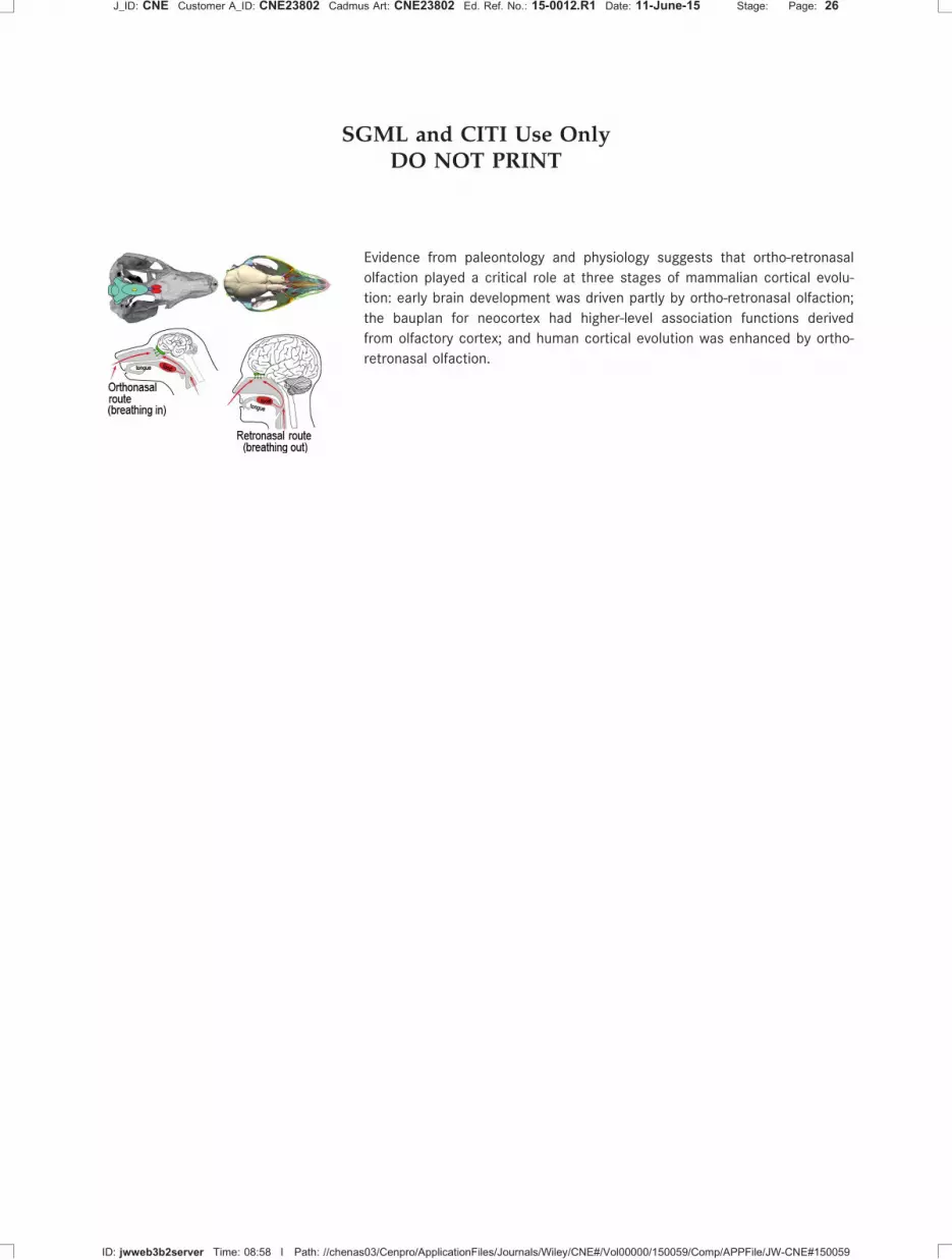
SGML and CITI Use OnlyDO NOT PRINT
Evidence from paleontology and physiology suggests that ortho-retronasal
olfaction played a critical role at three stages of mammalian cortical evolu-
tion: early brain development was driven partly by ortho-retronasal olfaction;
the bauplan for neocortex had higher-level association functions derived
from olfactory cortex; and human cortical evolution was enhanced by ortho-
retronasal olfaction.
J_ID: CNE Customer A_ID: CNE23802 Cadmus Art: CNE23802 Ed. Ref. No.: 15-0012.R1 Date: 11-June-15 Stage: Page: 26
ID: jwweb3b2server Time: 08:58 I Path: //chenas03/Cenpro/ApplicationFiles/Journals/Wiley/CNE#/Vol00000/150059/Comp/APPFile/JW-CNE#150059


