Embed Size (px)
Citation preview

Role of rRAB22b, an OligodendrocyteProtein, in Regulation of Transport ofVesicles From Trans Golgi to EndocyticCompartments
A.G. Rodriguez-Gabin,1 M. Cammer,2 G. Almazan,3 M. Charron,4 and J.N. Larocca1*1Department of Neurology/Neuroscience, Albert Einstein College of Medicine, Bronx, New York2Analytical Imaging Facility, Albert Einstein College of Medicine, Bronx, New York3Department of Pharmacology and Therapeutics, McGill University, Montreal, Quebec, Canada4Department of Biochemistry, Albert Einstein College of Medicine, Bronx, New York
Intracellular membrane trafficking plays an essential role inthe biogenesis and maintenance of myelin. Members of theRab protein family are important components of the sys-tems that regulate intracellular vesicle transport. We exam-ine the function of rRab22b, a novel rat Rab protein clonedfrom an oligodendrocyte cDNA library, by visualizing andidentifying in living Hela cells the organelles that containrRab22b. Our results show that rRab22b is present in thetrans Golgi/TGN and endocytic compartments. Traffickingof membranes from trans Golgi to endocytic compartmentstakes place via small tubulo vesicular organelles containingrRab22b. The formation of vesicles in the trans Golgi alsoappears to be regulated by rRab22b. Additionally, our re-sults suggest that rRab22b controls the transport of vesi-cles from the trans Golgi to endocytic compartments thatlocalize in oligodendrocyte processes. That rRab22b is in-volved in the transport of certain proteins from trans Golgito myelin is suggested by the evidence that certain proteinsbeing targeted to the plasma membrane are first trans-ported from trans Golgi to endocytic compartments. J.Neurosci. Res. 66:1149–1160, 2001. © 2001 Wiley-Liss, Inc.
Key words: Rab proteins; vesicle transport; oligoden-drocytes; signaling, myelin; endosomes; trans Golgi; my-elin proteins
Myelin is a multilamellar structure that surroundsaxon segments and is essential for saltatory conduction ofthe electrical impulse along the axon. In the central ner-vous tissue (CNS), myelin arises from cellular processesthat extend from the oligodendrocyte perikaryon andwrap in a spiral fashion around segments of the axon.Myelin is not formed as a unit; individual components aresynthesized in different cellular compartments, sorted outand transported to the site of myelin formation by severaldifferent mechanisms (Benjamins and Smith, 1984; Morelet al., 1994). Myelin basic protein (MBP) is synthesizedclose to the site of myelin assembly (Colman et al., 1982;Barbarese et al., 1995). In contrast, the proteolipid protein
(PLP) and both isoform, large and small, of myelin asso-ciated glycoprotein (MAGs) are synthesized in the endo-plasmic reticulum, then transported via vesicles to theGolgi and finally from the Golgi to myelin (Townsend andBenjamins, 1983; Trapp et al., 1989). Similarly, evidencesuggests that certain myelin lipids such as sulfatides aresynthesized in the Golgi and then transported to myelinvia membranous vesicles (Townsend et al., 1984; Sato etal., 1986). In eukaryotic cells, vesicle transport is regulatedby control of vesicle budding and fusion (Nuoffer andBalch, 1994; Rothman and Wieland, 1996). Additionally,movement along microtubules and microfilaments of thecytoskeleton (Cole and Lippincott-Schwartz, 1995) facil-itates the transport of vesicles. Despite much knowledge ofthe biochemistry and cell biology of oligodendrocytes, thesignaling systems that regulate intracellular membrane traf-ficking and its coordination with changes in cell shaperemain largely unknown. Rab proteins are key compo-nents in mechanisms that regulate intracellular membranetrafficking. Each member of this family of proteins isspecifically localized to a particular membrane traffickingpathway. Their functions depend on their ability to alter-nate between two conformational states, inactive (GDP-bound) and active (GTP-bound) (Bourne et al., 1990) andalso on their capacity to associate reversibly with specificmembrane compartments (Ossig et al., 1995).
In the present study, we identify membrane transportprocesses regulated by rRab22b, a novel Rab proteincloned from rat oligodendrocyte cultures. The role of
Contract grant sponsor: NMS; Contract grant number: RG 1941; Contractgrant sponsor: NSF; Contract grant number: ABN-9983255; Contractgrant sponsor: NIH; Contract grant number: DK47245; Contract grantsponsor: AECOM Cancer Center; Contract grant number: 5P30CA13330;Contract grant sponsor: MRC; Contract grant number: MT-12734.
*Correspondence to: J.N. Larocca, Department of Neurology and Neuro-science, AECOM, Yeshiva University, 1300 Morris Park Ave, New York,NY 10461. E-mail: [email protected]
Received 9 May 2001; Accepted 21 May 2001
Journal of Neuroscience Research 66:1149–1160 (2001)
© 2001 Wiley-Liss, Inc.

rRab22b in the regulation of transport vesicles containingthis protein was assessed. We examined living cells using achimeric protein of rRab22b with EYFP (a variant of thegreen fluorescent protein). rRab22b-EYFP was transientlyexpressed in Hela cells and rat oligodendrocytes and or-ganelles containing rRab22b were visualized by fluores-cent microscopy. Their morphologic changes and move-ments were observed in Hela cells imaging over short timeperiods to visualize the budding of vesicles from the donorcompartment and vesicle fusion in the acceptor compart-ment. Organelles were visualized containing bothrRab22b-EYFP and other intracellular markers taggedwith ECFP including Rab8 (trans Golgi network),pECFP-Golgi (trans-Golgi), Rab5b (early endosomes),and Rab7 (late endosomes).
Our observations show the presence of rRab22b intrans Golgi/TGN, in early endosomes and in late endo-somes. The traffic of membranes between trans Golgi/TGN and endocytic compartments occurs via small tubu-lar vesicles traveling along microtubules. Our results alsosuggest that rRab22b synchronizes the trafficking of mem-branes from the trans Golgi to endocytic compartmentsregulating vesicle formation in the trans Golgi.
EXPERIMENTAL PROCEDURES
Library ScreeningThe cDNA oligodendrocyte library was plated out
on a culture of XL1-Blue cells at a density of 5 3 104
plaque forming units per Petri dish (150 mm-diameter).Plaque lifts were performed in duplicate using nitrocellu-lose filters (Maniatis et al., 1989). The nitrocellulose filterswere probed, under conditions of high stringency (Wal-lace et al., 1981) with 32P-labeled PCR-generated probes.Briefly, the nitrocellulose filters containing immobilizedDNA were preincubated in 50% deionized formamide,50 mM Na2HPO4, pH 7.4, 750 mM NaCl, 1 mM EDTA,53 Denhardt’s (13 Denhardt’s is 0.2 g/l polyvinylpyrro-lidone, 0.2 g/l bovine serum albumin, 0.2 g/l Ficoll 400),0.1% SDS, 100 mg/ml poly(A), and 100 mg/ml heat-denatured salmon sperm DNA at 42°C for 1 hr, and thenfurther incubated in the above solution with 1–2 3 106
cpm/ml of PCR-constructed probe for 12–16 hr at 42°C.Filters were washed twice at 22°C in 23 SSC (SSC 515 mM NaCitrate, pH 7.00, 150 mM NaCl), 0.1% SDS at50°C. To reveal the positive clones the washed filters wereexposed to XAR film with an intensifying screen at270°C for 12–16 hr. Positive clones were purified byadditional rounds of plating and hybridization as describedabove. The sequence of the insert of interest was per-formed in the DNA Sequencing Facility at Einstein usingthe ABI Prism™ dye terminator kit with AmpliTaq DNApolymerase.
Northern Blot AnalysisA multiple-tissue mRNA blot, loaded with approx-
imately 2 mg of poly A1 RNA from different rat tissues(Clontech), was prehybridized for 4 hr at 50°C in ExpressHybridization Buffer (Clontech) with single-stranded
salmon testis DNA (100 mg/ml) and sequentially hybrid-ized with 32P-labeled rRab22b cDNA and a random-primed probe derived from a 2-kb human b-actin probefor 4 hr at 50°C. The blot was sequentially washed underincreasingly stringent conditions. The final wash was30 min in 0.13 SSC containing 0.1% SDS at 65°C. Thewashed membrane was expose to Biomax film from10–60 hr at 270°C. Similar procedure was used to detectrRab22b mRNA in blot containing 10 mg of total RNAfrom different primary cell cultures of neural cells. TotalRNA was isolated as described (Burcelin et al., 1997).
DNA ConstructThe pEYFP-C1 vector (Clontech) was used to con-
struct the rRab22b gene with the N terminal tagged withEYFP.
The rRab22b coding region was amplified by PCRfrom the lambda lgt11 phage containing the rRab22bcDNA using a 59 primer containing a Xhol restrictionendonuclease site and the first 20 nucleotides of therRab22b coding region, and a 39 primer containing aBamHI restriction endonuclease site and nucleotides com-plementary to the last 20 nucleotides of the rRab22bcoding sequence, including the stop codon. The PCR wascarried out as described (Burcelin et al., 1997). The PCRproducts were purified by agarose gel electrophoresis, andsubcloned into pGEMT vector. The rRab22b cDNA wassequenced to determine the fidelity of the PCR amplifi-cation. Then the pGEMT plasmid containing rRab22bcoding region was treated with Xhol and BamHI endo-nucleases. The rRab22b cDNA was purified and ligatedinto the Xhol and BamHI sites of the pEYFP-C1 mam-malian expression vector.
Similar procedures were used for the construction ofRab5, 7, and 8 chimeras with ECFP. In these cases thesequences of the coding region were synthesized by RT-PCR. Rat brain mRNA (Clontech) was converted intocomplementary DNA using MMLV-RT and poly dT asprimer. The PCR was carried out with primers directed tothe N and C terminal regions of the coding sequencescontaining Xhol and BamHI endonuclease restriction sites.
rRab22b site-directed mutagenesis was carried outaccording to the method of Ho et al. (1989).
Primary Cell CulturesPrimary astrocytes, microglia, oligodendrocyte pro-
genitors, and oligodendrocyte cultures for isolation of totalRNA, were established from newborn rat cerebral hemi-spheres as described by McCarthy and de Vellis (1980)with minor modifications (Burcelin et al., 1997).
Oligodendrocytes isolated from rat cerebral hemi-spheres from 12-day-old rats, as described previously(Berti Mattera et al., 1984), were used to expressrRab22b-EYFP. Isolated oligodendrocytes were plated onchambered coverglasses coated with Matrigel (Collabora-tive Biomedical Products, New Bedford, MA) at a densityof 5 3 104 cells (15 mm coverglasses) and maintained indefined serum-free medium consisting of a DMEM-F12mixture (1:1), 10 mM HEPES, 0.1% bovine serum albu-
1150 Rodriguez-Gabin et al.

min (BSA), 25 mg/ml human transferrin, 30 nM selenium,20 nM hydrocortisone, 20 nM progesterone, 10 nM bi-otin, 5 mg/ml insulin, 16 mg/ml putrescine, 30 nM tri-iodothyronine and Gibco’s mixture of trace elements.Oligodendrocytes were cultured for 3 days before genetransfer, and were maintained in cultures for 24–72 hrbefore analysis of chimera expression by fluorescence mi-croscopy.
Hela Cell CulturesHela cells (Epitheloid carcinoma, cervix, human,
American Type Culture Collection) were maintained inMEM supplemented with 10% FCS, 1% essential aminoacids, 2 mM glutamine, 100 U/ml penicillin and100 mg/ml streptomycin at 37°C in a 5% CO2 incubator.
For the realization of the transfection experimentsHela cells were plated on 15 mm coverglasses at a density0.4–1.6 3 105 cells for 24 hr before transfection.
TransfectionHela cells were transfected using SuperFect transfec-
tion reagent (Qiagen) according to the manufacturer’sindications. Briefly, cultures were treated with 1.2 ml ofgrowth media containing the DNA-SuperFect complex(3 mg DNA/37.5 mg SuperFect) at 37°C for 60 min.
Oligodendrocytes were transfected using Lipo-fectamine Plus (GIBCO BRL, Gaithersburg, MD), ac-cording to the instructions of the manufacturer. Oligo-dendrocytes were treated with 1.2 ml culture mediacontaining 2.5 mg of DNA, 12.5 mg of lipofectamine and10 ml of Plus reagent at 37°C for 3–6 hr.
Fluorescence Microscopy Analysis of Living CellsCells were imaged in culture medium without phe-
nol red at 37°C using an Olympus I370 microscope witha 603 Olympus Planapo oil immersion objective NA 1.4and equipped with a CCD camera (Photometrics KAF1400 PXL liquid cooled). Cells expressing ECFP chimeraswere excited using the 436 6 20 filter (Chroma Tech-nology) and imaged with a filter 480 6 40 (ChromaTechnology). The exciter filter 500 6 20 and emitter filter535 6 30 (Chroma Technology) were used to visualizeEYFP chimeras. Temperature was controlled with aNevtek air stream stage incubator (Burnsville, VA). Imagesfrom the CCD camera were digitized and collected di-rectly to RAM with an Apple Power Macintosh 8500.Image capturing, processing, and automatic and manualdata acquisition was performed using I.P. Lab Spectrumsoftware.
RESULTS
Cloning of rRab22b cDNAIn a previous study using RT-PCR we identified a
cDNA fragment, Rab0, as a novel Rab sequence ex-pressed in rat oligodendrocytes (Burcelin et al., 1997).Because the Rab0 sequence has high identity to humanRab22b we are redesignating Rab0 as rat Rab22b(rRab22b).
The complete sequence of rRab22b was obtained byscreening a rat oligodendrocyte lgt11 cDNA library ob-tained from Dr. A. Campagnoni (Baba et al., 1995). Theclones of interest were detected by probing the cDNAlibrary with a radioactive rRab22b cDNA fragment. ThecDNA fragment, used as probe, was labeled with[32P]dCTP by PCR using specific primers targeted to the59 and 39 terminals of a 180 bp Rab fragment, and thenitrocellulose filters were hybridized overnight at 42°C.Positive plaques were purified by a second round ofscreening. Two positive clones were found among the0.5 3 106 plaques screened, and these were plaque-purified. Both phages contained inserts of approximately3,600 bases (results not shown). The inserts were se-quenced and the sequences analyzed with University ofWisconsin GCG programs.
The cDNA sequence of rRab22b revealed an openreading frame of 582 bp and the deduced amino acidsequence indicated a protein containing 194 amino acids(GenBank accession number AF254800) (Fig. 1a,b) with amolecular weight of 21.6 kDa and an isoelectric pointof 7.05.
The deduced amino acid sequence of rRab22b con-tains both the four motifs that participate in the formationof the GTP-binding site in the superfamily of Ras-likeproteins and the domain required for effector proteininteraction (Fig. 1b). Analysis of the effector domain in-dicated that rRab22b is a member of the Rab family ofGTP-binding proteins. The carboxyl terminal region con-tains an isoprenylation motif, suggesting a protein that istargeted to membrane compartments (Fig. 1b). A com-parison of the rRab22b sequence with a closely relatedRab cDNA (human Rab 22b (Chen et al., 1996)) showed88% identity; the predicted amino acid sequence had 94%identity. These results suggest that rRab22b encodes therat homolog of human Rab22b. In view of the fact that asegment of the C terminal domain shows 9 differencesamong the 14 residues, however, one must consider thatrRab22b may very well encode a new member of the Rabfamily. This segment is part of the motif responsible for thetargeting of Rab proteins to specific membrane compart-ments (Chavrier et al., 1991).
Analysis of Rat Rab22b ExpressionExpression of rRab22b mRNAs was analyzed by
Northern blots. Nylon membranes containing mRNAfrom several rat tissues were probed with [32P] dCTPlabeled rRab22b cDNA. A band of approximately 3,600bp had the highest level of expression in brain, followed indecreasing order by heart, testis, kidney, lung, and spleen(Fig. 2a). None was detected in either liver or skeletalmuscle.
Rat Rab22b mRNA expression was assessed in pri-mary cultures of oligodendrocytes, oligodendrocyte pro-genitors, microglia and astrocytes by probing total RNAwith labeled rRab22b cDNA (Fig. 2c). Oligodendrocytesshowed a strong band of 3,600 bp and a weaker band inoligodendrocyte progenitors, microglia, and astrocytes.That these differences in rRab22b mRNAs were not due
rRab22b in Vesicle Transport 1151
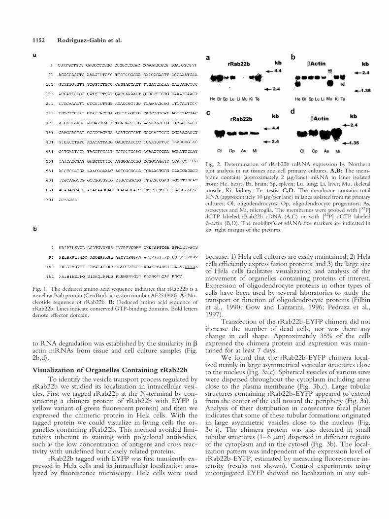
to RNA degradation was established by the similarity in bactin mRNAs from tissue and cell culture samples (Fig.2b,d).
Visualization of Organelles Containing rRab22bTo identify the vesicle transport process regulated by
rRab22b we studied its localization in intracellular vesi-cles. First we tagged rRab22b at the N-terminal by con-structing a chimera protein of rRab22b with EYFP (ayellow variant of green fluorescent protein) and then weexpressed the chimeric protein in Hela cells. With thetagged protein we could visualize in living cells the or-ganelles containing rRab22b. This method avoided limi-tations inherent in staining with polyclonal antibodies,such as the low concentration of antigens and cross reac-tivity with undefined but closely related proteins.
rRab22b tagged with EYFP was first transiently ex-pressed in Hela cells and its intracellular localization ana-lyzed by fluorescence microscopy. Hela cells were used
because: 1) Hela cell cultures are easily maintained; 2) Helacells efficiently express fusion proteins; and 3) the large sizeof Hela cells facilitates visualization and analysis of themovement of organelles containing proteins of interest.Expression of oligodendrocyte proteins in other types ofcells have been used by several laboratories to study thetransport or function of oligodendrocyte proteins (Filbinet al., 1990; Gow and Lazzarini, 1996; Pedraza et al.,1997).
Transfection of the rRab22b-EYFP chimera did notincrease the number of dead cells, nor was there anychange in cell shape. Approximately 35% of the cellsexpressed the chimera protein and expression was main-tained for at least 7 days.
We found that the rRab22b-EYFP chimera local-ized mainly in large asymmetrical vesicular structures closeto the nucleus (Fig. 3a,c). Spherical vesicles of various sizeswere dispersed throughout the cytoplasm including areasclose to the plasma membrane (Fig. 3b,c). Large tubularstructures containing rRab22b-EYFP appeared to extendfrom the center of the cell toward the periphery (Fig. 3a).Analysis of their distribution in consecutive focal planesindicates that some of these tubular formations originatedin large asymmetric vesicles close to the nucleus (Fig.3e–i). The chimera protein was also detected in smalltubular structures (1–6 mm) dispersed in different regionsof the cytoplasm and in the cytosol (Fig. 3b). The local-ization pattern was independent of the expression level ofrRab22b-EYFP, estimated by measuring fluorescence in-tensity (results not shown). Control experiments usingunconjugated EYFP showed no localization in any sub-
Fig. 1. The deduced amino acid sequence indicates that rRab22b is anovel rat Rab protein (GenBank accession number AF254800). A: Nu-cleotide sequence of rRab22b. B: Deduced amino acid sequence ofrRab22b. Lines indicate conserved GTP-binding domains. Bold lettersdenote effector domain.
Fig. 2. Determination of rRab22b mRNA expression by Northernblot analysis in rat tissues and cell primary cultures. A,B: The mem-brane contains (approximately 2 mg/lane) mRNA in lanes isolatedfrom: He, heart; Br, brain; Sp, spleen; Lu, lung; Li, liver; Mu, skeletalmuscle; Ki, kidney; Te, testis. C,D: The membrane contains totalRNA (approximately 10 mg/per lane) in lanes isolated from rat primarycultures; Ol, oligodendrocytes; Op, oligodendrocyte progenitors; As,astrocytes and Mi, microglia. The membranes were probed with [32P]dCTP labeled rRab22b cDNA (A,C) or with [32P] dCTP labeledb-actin (B,D). The mobility’s of ssRNA size markers are indicated inkb, right margin of the pictures.
1152 Rodriguez-Gabin et al.

cellular structures. There was an even distribution of flu-orescence throughout the cell including the nucleus. Thisobservation indicates that rRab22b-EYFP chimera proteinis targeted to specific structures.
Movement and Plastic Changes of OrganellesContaining rRab22b
The dynamics of organelles containing rRab22bwere studied to visualize the transport processes regulatedby rRab22b. Intracellular traffic of membranes takes placevia vesicles that bud from the donor compartment andthen target selectively to the acceptor compartment(Nuoffer and Balch, 1994; Rothman and Wieland, 1996).Travel between compartments occurs along elements ofthe cytoskeleton (Nuoffer and Balch, 1994).
Movement and changes in the morphology of or-ganelles containing rRab22b-EYFP were examined indigital time lapse images of chimera-expressing HELAcells. Analysis of time lapse images captured at 15-secintervals showed that the large asymmetrical fluorescentstructures close to the nucleus did not appear to moveconsistently in a single direction (Fig. 4). We observedlarge tubular structures extending, up to 25 mm, from thecell center to the periphery (Fig. 4a,b). Some seemed tocome from asymmetrical vesicles localized close to thenucleus (Fig. 4b). These tubular structures appeared to beflexible, to undulate while extending and to bend whilechanging direction of movement. Sometimes, these struc-tures appeared to interact with vesicular organelles presentin the cell periphery (Fig. 4a).
Our results indicate that the asymmetrical vesiclesand the larger tubular organelles localized close to thenucleus are the donor compartments. Indeed, we observedbudding of small tubular vesicles from these structures(Fig. 5a,b). The small tubular vesicles containing rRab22bhad kinetic and plastic properties similar to those associ-ated with post Golgi carriers (Hirschberg et al., 1998).These small organelles moved intermittently along eitherstraight or curvilinear tracks toward the cell periphery(approximately 0.2–0.6 mm/sec), and they frequentlychanged direction. Some contained swollen or sphericalareas along their length (Fig. 6a,b). The spherical areaswere not stationary, and their content of rRab22b washigher than in other parts of the formation. Small tubularorganelles underwent drastic shape changes, e.g., roundinginto spherical shapes that would then become elongated inthe direction of movement (Fig. 6a). They also displayedelastic properties, such as extension and retraction duringmovement. Occasionally, when they broke up, one frag-ment remained stationary whereas the other moved off(Fig. 6b). These tubular formations fused with small ves-icles. In Figure 6b, a tubular formation has a higherfluorescence in one end whereas a vesicle interacts brieflywith the other end. This interaction did not result invesicle fusion and the vesicle moved away from the for-mation. Immediately the fluorescence intensity at this endincreased and a second interaction between the vesicle andthe tubular formation occurred. This time the vesicle didfuse. The new structure then broke into spherical vesicles.
Fig. 3. Fluorescence microscopy of organelles in Hela cells expressingrRab22b-EYFP. Forty-eight hr after transfection cells were examinedat 37°C. A: Black arrowheads: large asymmetrical vesicles close to thenucleus, black arrows: two large tubular formations. B: White arrowssmall thin tubular formation, white arrow heads: small vesicles near theperiphery. C: white arrowheads: same as in (B); black arrowhead same
as in (A). D: Background staining with EYFP only. E–I: Serial 0.5 mmoptical sections. Images are stereo projections using the maximum pixelmethod. Black arrows: large tubular formations extending from asym-metrical vesicles closed to the nucleus. Scale bar 5 10 mm. Seeanimated movie sequence of e through l at Wiley InterScience (http://www.wiley.interscience.com).
rRab22b in Vesicle Transport 1153

In some cells, intensive traffic was seen of smalltubular vesicles going between the large asymmetricalstructures localized close to the nucleus, and a cluster oftubular organelles localized in the cell periphery close to
the plasma membrane (Fig. 7a). The small tubulo vesicularstructures moved along defined pathways in both antero-grade and retrograde directions. The clusters moved par-allel to the plasma membrane, and tubulo vesicular or-
Fig. 4. Formation and movement of large tubular organelles containing rRab22b-EFYP. Hela cellswere treated as in Figure 3. Selection of images captured at 15 sec intervals for 6 min period. A: Abrightly-labeled tubular formation (white arrow) extends from the center of the cell and makes contactwith a vesicle located in the cell periphery. B: Large tubular formation (white arrow) emerging fromintensively labeled asymmetrical vesicles (white arrowhead). Scale bar 5 10 mm. See movie sequenceat Wiley InterScience (http://www.wiley.interscience.com).
Fig. 5. Formation of small tubular vesicles involved in transport. Helacells treated as in Figures 3 and 4. Time lapse images obtained as inFigure 4. A: Small tubular vesicle budding from a rRab22b labeledvesicle close to the nucleus also contains rRab22b. Large panel indicatesrestricted area shown in small frames covering 10.5 min. Arrow shows
budding tubular vesicle and its movement toward the cell periphery.B: Small tubular vesicle (white arrowhead) forms from large tubularformation (white arrow). Large panel indicates restricted area shown insmall frames covering 4.5 min. Scale bar 5 10 mm. See movie sequenceat Wiley InterScience (http://www.wiley.interscience.com).
1154 Rodriguez-Gabin et al.

ganelles became detached and moved close to the plasmamembrane. These observations indicate that: 1) the accep-tor compartment is the cluster of organelles localized closeto the plasma membrane; and 2) organelles containingrRab22b move along elements of the cytoskeleton.
The treatment of the cells with nocodazole (10 mMfor 3 min), an inhibitor of microtubule polymerization(Rogalski and Singer, 1984), disrupted the movement oforganelles in the cell periphery (Fig. 7b). The cluster oftubular formations now was stationary and dispersed, withno movement of vesicles from the cell periphery towardthe center. However, some small vesicles moved from theperinuclear area and fused with tubular organelles locatedin the cell periphery, further supporting the conclusionthat these tubular organelles are the acceptor compart-ment. Additionally, from the data it appears that intactmicrotubules are required for directed transport of tubularstructures containing rRab22b. Because treatment of thecell with 10 mM cytochalasin B did not affect the kineticbehavior of organelles containing rRab22b actin micro-filaments are not involved (Cooper, 1987). (Quick-timemovies 7c,d).
Identification of Subcellular CompartmentsContaining rRab22b
To identify the subcellular organelles that containrRab22b cells were cotransfected with rRab22b-EYFPtogether with other markers tagged with ECFP (a cyanovariant of green fluorescent protein). These included Rab8(trans Golgi) (Huber et al., 1993), Rab5b (early endo-somes) (Gorvel et al., 1991), Rab7 (late endosomes) (Me-resse et al., 1995) and pECFP-Golgi (a chimera con-structed with ECFP and an 81 amino acid fragment of theN terminal region of human 1,4 beta galactosyl transferaselocalizing mainly in the trans-Golgi) (Llopis et al., 1998)(Fig. 8). Our results and those from other laboratories
demonstrated that the tagging of these intracellular mark-ers did not affect their localization (Llopis et al., 1998;Roberts et al., 1999; Bucci et al., 2000; Sonnichsen et al.,2000). Large asymmetric vesicles containing bothrRab22b and Rab8 localized closed to the nucleus (Fig.8a–c). Similarly, some large tubular formations extendingfrom the center of the cell to the periphery also containedboth proteins, indicating that these structures containingrRab22b are part of the trans Golgi network (TGN). Thelocalization of rRab22b in trans-Golgi is supported by thecolocalization of rRab22b-EYFP and pECFP-Golgi (Fig.8d–f). rRab22b partially colocalized in small vesicles withRab5 (Fig. 8g–i). Similarly, some vesicles contained bothRab22b and Rab7 (Fig. 8g–l), suggesting that rRab22b isalso present in organelles of the endocytic pathway.
A cluster of tubular organelles located close to areasof the plasma membrane where membrane extensions areformed was identified as early endosomes because theseorganelles also contained Texas Red-transferrin (after in-cubation of cells for 5 min with Texas-Red labeled trans-ferrin) (Fig. 8m–o).
Expression of a rRab22b Mutant (S19N) TaggedWith EYFP
Our previous results indicated that rRab22b regu-lates the transport of vesicles between the trans Golgi andthe endocytic compartment. The role of Rab proteins inregulating intracellular transport relies on two properties:1) the ability to alternate between two conformationalstates (GDP-bound, active and GTP-bound, inactive), and2) the capacity to bind to specific membrane compart-ments, donor compartment, inactive state; acceptor com-partment, active state. Dominant negative mutants withaltered guanine nucleotide-binding properties inhibit ves-icle transport both in vivo and in vitro (Olkkonen andStenmark, 1997). Substitution of S by N in the last residue
Fig. 6. Processing of organelles containing rRab22b. Hela cells treatedas in Figure 3. Time lapse images obtained as in Figure 4. A: Smalltubular formation (white arrow) moves toward cell periphery and fuses(Panel 9) with a small vesicle (white arrowhead) after rRab22b intensityincreases (Panel 8) in the region of interaction. The new formation withhigher centrally located rRab22b intensity (Panels 10 and 11) thendivides (Panel 12) into smaller organelles (white arrowhead and whitearrow, Panels 12 to 20). Large panel indicates restricted area shown in
small frames covering 9.5 min. B: Small tubular formation divides intospherical (white arrow) and tubular (white arrowhead) vesicles. Thelatter then rounds up and moves away becoming elongated in thedirection of movement. The vesicle shows varicosities of higherrRab22b intensity. Large panel indicates restricted area shown in smallframes covering 3.75 min. Scale bar 5 10 mm. See movie sequence atWiley InterScience (http://www.wiley.interscience.com).
rRab22b in Vesicle Transport 1155

of the first GTP binding motif of Rab1 results in a proteinthat probably remains in the GDP-bound state, therebyinhibiting transport between ER and Golgi (Nuoffer andBalch, 1994). When we substituted the 19 S residue by N
in the cDNA rRab22b sequence and expressed this mutant(tagged with EYFP) in Hela cells image analysis showedlocalization mainly in asymmetrical vesicular and tubularcompartments close to the nucleus (Fig. 9). Moreover, novesicle budding from these organelles was seen (Fig. 9).The fact that rRab22b mutant co-localized with pECFP-Golgi showed that both of these perinuclear compartmentsare part of the trans-Golgi (Fig. 10). These results areconsistent with the view that rRab22b regulates the trans-port of vesicles from the trans-Golgi to an endocyticcompartment.
Oligodendrocyte CulturesAfter 24 hr of transfection of the chimera protein of
rRab22b with EYFP in rat oligodendrocyte cultures, up to2% of oligodendrocytes expressed rRab22b-EYFP, an ex-pression that persisted for the 4 days of the experimentalperiod. Transfection did not produce an increase in thenumber of dead cells or a change in oligodendrocytemorphology.
Similar to the observations in Hela cells, rRab22bwas localized in large asymmetric vesicles close to thenucleus and was also present in organelles in oligodendro-cyte processes (Fig. 11). These results indicate that inoligodendrocytes and their processes as well as in Hela cellsrRab22b regulates the transport of vesicles between thetrans Golgi and endocytic compartments.
DISCUSSIONWe are studying the function of rRab22b in relation
to its role in myelin formation. rRab22b is a novel rat Rabprotein we detected in a rat cDNA library specific foroligodendrocytes, the cells that produce myelin.
To study rRab22b function we have constructed afusion protein with EYFP. Because the C terminal of Rabproteins is subject to post-translational isoprenylation, amodification essential for both membrane association andinteraction with effector proteins (Chavrier et al., 1991;Olkkonen and Stenmark, 1997), the N terminal was usedfor tagging. Although the N terminal sequence is impor-tant for Rab function (Tisdale and Balch, 1996), taggingthis terminal does not alter function (Bucci et al., 2000;Roberts et al., 1999; Sonnichsen et al., 2000).
By expressing this fusion protein in living cells wefollowed both the intracellular localization of rRab22b invesicles and the movement of these vesicles.
Two properties of rRab22b, GTP-binding and thepresence of terminal isoprenylation sites, indicate its prob-able targeting to membrane compartments. Participationin regulation of membrane traffic from trans Golgi to earlyendocytic compartments is shown by the following ob-servations obtained with Hela cells: 1) rRab22b wasmainly localized in asymmetrical vesicles and large tubularformations in the trans Golgi and TGN; and 2) rRab22bwas present in early endocytic compartments in the cellperiphery. Moreover, the mutant of rRab22b (N19S)behaved very differently. It localized only in the transGolgi compartment.
Fig. 7. Movement of rRab22b vesicles before and after disruption ofmicrotubules. Hela cells were examined 48 hr after transfection. Timelapse images obtained as in Figure 4. A: Before disruption of micro-tubules a highly mobile cluster of tubular organelles, localized in the cellperiphery moves rapidly along the plasma membrane (small frames,left). Large panel indicates restricted area shown in small frames cov-ering 7.5 min. B: After treatment of cells with nocodazole (10 mM,3 min) movement is disrupted, and the cluster of organelles disperses(frames panels, left). A small vesicular organelle (white arrow, bottomsmall frames 1 to 7) travels from the center of the cell to the cellperiphery and fuse with tubular organelle (head arrow, vesicle trajec-tory, frame 8). Large panel indicates restricted areas shown in smallframes covering 7.5 min. Scale bar 5 10 mm. See movie sequence atWiley InterScience (http://www.wiley.interscience.com).
1156 Rodriguez-Gabin et al.

This difference in behavior of the mutant N19S alsoclearly indicates that the distribution pattern of rRab22b inboth donor and acceptor compartments does not resultfrom over-expression of the chimera proteins, because thesubstitution of serine by asparagine in position 19 restrictslocalization to the donor compartment. Other consider-
Fig. 9. Mutant rRab22b (S19N) localization is restricted to large tu-bular formations and asymmetrical vesicles close to the nucleus. A–C:Images of three different cells examined 48 hr after transfection. Quickmovies show organelles with rRab22b mutant vibrate only in place.Scale bar 5 10 mm. For a and b see movie sequence at WileyInterScience (http://www.wiley.interscience.com).
Fig. 10. Mutant rRab22b (S19N) colocalizes with trans Golgi marker.Cells were examined 48 hr after cotransfection. A,D: rRab22b-EYFP(S19N) mutant; B,E: Golgi pECFP (trans-Golgi); C,F: overlapping(yellow organelles) of rRab22b-EYFP (S19N) mutant and GolgipECFP. Scale bar 5 10 mm.
Fig. 8. Presence of rRab22b in TGN, trans Golgi, early endosomes andlate endosomes by colocalization with compartment markers. Cellswere examined 48 hr after cotransfection. A: rab8-ECFP (TGN);B: rRab22b-EYFP; C: overlapping of (A) and (B), yellow organellescontain both Rab8-ECFP and rRab22b-EYFP. D: Golgi pECFP(trans-Golgi); E: rRab22b-EYFP; F: overlapping of (D) and (E). Coloras before. G: Rab5-ECFP (early endosomes); H: rRab22b-EYFP;I: overlapping of (G) and (H). Color as above. J: Rab7-ECFP (lateendosomes); K: rRab22b-EYFP; L: overlap of (J) and (K). Color asabove. M: Transferrin-Texas Red (early endosomes); N: rRab22b-EYFP; O: overlapping of (M) and (N). Color as above. Scale bar 5 10mm. For m–o see movie sequence at Wiley InterScience (http://www.wiley.interscience.com).
rRab22b in Vesicle Transport 1157

ations also argue against over-expression as a factor in theselocalizations. First, the distribution pattern of rRab22b-EYFP was independent of the level of expression. Second,over-expression did not appear to affect localization ofRab5-ECFP and Rab7-ECFP. Third, recent studiesshowed that over-expression of Rab tagged with GFP didnot affect the intracellular localization (Bucci et al., 2000;Roberts et al., 1999; Sonnichsen et al., 2000).
Our observations indicate that small tubovesicularorganelles containing rRab22b that bud from the transGolgi and fuse with endocytic compartments in the cellperiphery are the mechanism of transport of membraneconstituents from the trans Golgi to endocytic compart-ments. These organelles have morphological and kineticproperties similar to those presented by post Golgi carriers(Nakata et al., 1998; Toomre et al., 1999). These proper-ties are: 1) the presence of spherical areas along theirlength; 2) the drastic changes in shape that they undergo;3) their ability to break up or fuse with small vesicles; 4)their movement along tracts towards the cell periphery (atapproximately 0.2–0.6 mm/sec) with frequent changes indirection; and 5) the involvement of microtubules in theirtransport.
Because rRab22b was also present in late endosomes,one cannot exclude involvement of this protein in mem-brane traffic from early to late endosomes. Other Rabproteins are known to participate in more than one step inmembrane trafficking (Novick and Zerial, 1997).
The concentration of rRab22b was higher in someorganelle regions where fusion with small tubular forma-tions occurred. Although the functional relation is notclear, a similar observation with a different Rab protein,Rab5a, was reported in the area of contact of fusingendosomes (Roberts et al., 1999).
In addition to regulation of directional movement,rRab22b appears to be involved in the formation of smallvesicles, because expression of the rRab22b mutant(N19S) was not seen in vesicles budding from the transGolgi. According to previous evidence, Rab proteins par-ticipate both in the targeting of vesicles and in the buddingof vesicles (Jedd et al., 1997; McLauchlan et al., 1998;Jones et al., 1999). These results show that rRab22b is notsimply that of a passenger, but plays a functional role intransport.
Our results indicate that one mechanism of transportof membrane constituents from the trans Golgi to endo-somes involves distinct vesicles. Direct transfer of mem-brane components from the TGN to other organelles alsoneeds to be considered because large tubular formations ofthe TGN containing rRab22b extend toward the cellperiphery and interact with spherical vesicles there. Bothcis and trans cisternae of the Golgi are composed of exten-sive tubular networks (Sasaki et al., 1984; Jahn, 2000) thatcan be generated rapidly by Golgi membranes in vivo andin vitro under diverse conditions (Cluett et al., 1993;Banta et al., 1995). This mediation by tubules of mem-brane traffic between organelles is more clearly observedin cells treated with brefeldin A (Sciaky et al., 1997).
In oligodendrocyte cultures we find that: 1) a highlevel of rRab22b mRNA is present in differentiated oli-godendrocytes whereas such expression in their progeni-tors is very low; 2) rRab22b is present in trans Golgi/TGNvesicles; and 3) rRab22b is present in endosomic or-ganelles in oligodendrocyte processes.
These observations, especially the level of rRab22bmRNA suggest that trafficking of membrane componentsfrom the trans-Golgi to endocytic compartments regulatedby rRab22b is an important step in myelin formation andmaintenance. Because rRab22b mRNA is present in othercell types, the step in vesicular traffic that rRab22b regu-lates is probably not unique to oligodendrocytes.
That certain proteins, such as transferrin receptor andasialoglycoprotein receptor H1, are carried from the transGolgi to an endocytic compartment before reaching thecell surface (Futter et al., 1995; Leitinger et al., 1995) lendssupport to the inference that this pathway is involved inmyelin formation.
The cytoplasmic tail of these proteins contains themotif YXXf, where X is any amino acid and f is anamino acid containing a bulky hydrophobic residue. Ev-idence shows that the YXXf motif, originally known formediating rapid internalization from the cell surface(Bonifacino and Dell’Angelica, 1999) is also now recog-nized for mediating trans Golgi sorting mechanisms thattarget proteins to endocytic compartments (Bonifacinoand Dell’Angelica, 1999). The sequence of large MAGshows a segment in its carboxyl terminal region that con-forms to the consensus motif YXXf, YAEI. This se-
Fig. 11. rRab22b in oligodendrocyteperikaryon and its processes. A–C:Asymmetrical vesicles containingrRab22b localize close to the nucleus(white arrow) and organelles (whitearrowhead) are present in oligoden-drocyte processes. Images were taken48 hr after transfection. Scale bar 510 mm.
1158 Rodriguez-Gabin et al.

quence suggests that large MAG is transported from transGolgi to endocytic compartments, and our results in Helacells further suggest that this pathway is regulated byrRab22b. Indeed, endocytic compartments containingMAG are observed in oligodendrocyte processes includingareas close to the axon where active formation of myelinoccurs (Trapp et al., 1989; Bo et al., 1995).
These conclusions are in accord with the presence ofrRab22b in endocytic compartments of oligodendrocyteprocesses, endocytic compartments that in Hela cells lo-calize close to areas where plasma membrane extensionsoccur.
rRab22b may also be involved in the biogenesis ofendosomes because early endosomes receive newly syn-thesized proteins that have been sorted in the trans Golginetwork before being transported to late endosomes (Pe-ters and von Figure, 1994; Jackson et al., 1995).
The importance of the rRab22b dependent mem-brane trafficking to endosomes is particularly relevant be-cause: 1) this compartment is involved in the remodelingof myelin that occurs toward the end of the period ofactive myelination (Raine, 1984); 2) continuous cycles oflamella extension and retraction are observed in oligoden-drocyte cultures (Kachar et al., 1986), cycles that underliesubstrate exploration to find and wrap the axon in themyelin sheath (Kachar et al., 1986); and 3) proteins that arenot essential but are required for assembly need to beeliminated from myelin (Gould et al., 2000).
Endocytic processes of importance in oligodendro-cytes involve a number of Rab proteins including Rab5a,Rab5b, and Rab5c (Burcelin et al., 1997; Bouverat et al.,2000). It should be noted that the levels of their expressionincrease with oligodendrocyte differentiation (Bouverat etal., 2000).
In summary, our observations clearly indicate thatrRab22b is involved in the regulation of membrane traf-ficking from trans Golgi compartments to endocytic com-partments and further suggest that rRab22b participates inthe formation of vesicles budding from the trans Golgi.
ACKNOWLEDGMENTSWe thank Dr. Maurice Rapport for editing and
commenting on the manuscript. This work was supportedby NMS RG 1941 and NSF ABN-9983255 (JNL),NIH DK47245 and AECOM Cancer Center Grant5P30CA13330 (MJC), and MRC MT-12734 (GA).
REFERENCESBaba H, Fuss B, Urano J, Poullet P, Watson JB, Tamanoi F, Macklin WB.
1995. GapIII, a new brain-enriched member of the GTPase-activatingprotein family. J Neurosci Res 41:846–858.
Banta M, Polizotto RS, Wood SA, de Figueiredo P, Brown WJ. 1995.Characterization of a cytosolic activity that induces the formation of Golgimembrane tubules in a cell-free reconstitution system. Biochemistry34:13359–13366.
Barbarese E, Koppel DE, Deutscher MP, Smith CL, Ainger K, Morgan F,Carson JH. 1995. Protein translation components are colocalized ingranules in oligodendrocytes. J Cell Sci 108:2781–2790.
Benjamins JA, Smith ME. 1984. Metabolism of myelin. In: Morell P,
editor. Myelin. 225–258. 1984. New York, London: Plenum Press. p.225–258.
Berti Mattera LN, Larocca JN, Pellegrino de Iraldi A, Pasquini JM, Soto E.1984. Isolation of oligodendroglial cells from young and adult whole ratbrains using an in situ generated percoll density gradient. Neurochem Int6:41–51.
Bo L, Quarles RH, Fujita N, Bartoszewicz Z, Sato S, Trapp BD. 1995.Endocytic depletion of L-MAG from CNS myelin in quaking mice. J CellBiol 131:1811–1820.
Bonifacino JS, Dell’Angelica EC. 1999. Molecular bases for the recognitionof tyrosine-based sorting signals. J Cell Biol 145:923–926.
Bourne HR, Sanders DA, McCormick F. 1990. The GTPase superfamily:a conserved switch for diverse cell functions. Nature 348:125–132.
Bouverat BP, Krueger WH, Coetzee T, Bansal R, Pfeiffer SE. 2000.Expression of rab GTP-binding proteins during oligodendrocyte differ-entiation in culture. J Neurosci Res 59:446–453.
Bucci C, Thomsen P, Nicoziani P, McCarthy J, van Deurs B. 2000. Rab7:a key to lysosome biogenesis. Mol Biol Cell 11:467–480.
Burcelin R, Rodriguez-Gabin AG, Charron MJ, Almazan G, Larocca JN.1997. Molecular analysis of the monomeric GTP-binding proteins ofoligodendrocytes. Brain Res Mol Brain Res 50:9–15.
Chavrier P, Gorvel JP, Stelzer E, Simons K, Gruenberg J, Zerial M. 1991.Hypervariable C-terminal domain of rab proteins acts as a targeting signal.Nature 353:769–772.
Chen D, Guo J, Miki T, Tachibana M, Gahl WA. 1996. Molecular cloningof two novel rab genes from human melanocytes. Gene 174:129–134.
Cluett EB, Wood SA, Banta M, Brown WJ. 1993. Tubulation of Golgimembranes in vivo and in vitro in the absence of brefeldin A. J Cell Biol120:15–24.
Cole NB, Lippincott-Schwartz J. 1995. Organization of organelles andmembrane traffic by microtubules. Curr Opin Cell Biol 7:55–64.
Colman DR, Kreibich G, Frey AB, Sabatini DD. 1982. Synthesis andincorporation of myelin polypeptides into CNS myelin. J Cell Biol95:598–608.
Cooper JA. 1987. Effects of cytochalasin and phalloidin on actin. J Cell Biol105:1473–1478.
Filbin MT, Walsh FS, Trapp BD, Pizzey JA, Tennekoon GI. 1990. Role ofmyelin P0 protein as a homophilic adhesion molecule. Nature 344:871–872.
Futter CE, Connolly CN, Cutler DF, Hopkins CR. 1995. Newly synthe-sized transferrin receptors can be detected in the endosome before theyappear on the cell surface. J Biol Chem 270:10999–11003.
Gorvel JP, Chavrier P, Zerial M, Gruenberg J. 1991. Rab5 controls earlyendosome fusion in vitro. Cell 64:915–925.
Gould RM, Freund CM, Palmer F, Feinstein DL. 2000. Messenger RNAslocated in myelin sheath assembly sites. J Neurochem 75:1834–1844.
Gow A, Lazzarini RA. 1996. A cellular mechanism governing the severityof Pelizaeus-Merzbacher disease. Nat Genet 13:422–428.
Hirschberg K, Miller CM, Ellenberg J, Presley JF, Siggia ED, Phair RD,Lippincott-Schwartz J. 1998. Kinetic analysis of secretory protein trafficand characterization of golgi to plasma membrane transport intermediatesin living cells. J Cell Biol 143:1485–1503.
Ho SN, Hunt HD, Horton RM, Pullen JK, Pease LR. 1989. Site-directedmutagenesis by overlap extension using the polymerase chain reaction [seecomments]. Gene 77:51–59.
Huber LA, Pimplikar S, Parton RG, Virta H, Zerial M, Simons K. 1993.Rab8, a small GTPase involved in vesicular traffic between the TGN andthe basolateral plasma membrane. J Cell Biol 123:35–45.
Jackson MR, Fruh K, Karlsson L, Teyton L, Yang Y, Peterson PA. 1995.Assembly and intracellular transport of MHC class I and class II molecules.Cold Spring Harb Symp Quant Biol 60:249–261.
Jahn R. 2000. Sec1/Munc18 proteins: mediators of membrane fusionmoving to center stage. Neuron 27:201–204.
rRab22b in Vesicle Transport 1159

Jedd G, Mulholland J, Segev N. 1997. Two new Ypt GTPases are requiredfor exit from the yeast trans-Golgi compartment. J Cell Biol 137:563–580.
Jones S, Jedd G, Kahn RA, Franzusoff A, Bartolini F, Segev N. 1999.Genetic interactions in yeast between Ypt GTPases and Arf guaninenucleotide exchangers. Genetics 152:1543–1556.
Kachar B, Behar T, Dubois-Dalcq M. 1986. Cell shape and motility ofoligodendrocytes cultured without neurons. Cell Tissue Res 244:27–38.
Leitinger B, Hille-Rehfeld A, Spiess M. 1995. Biosynthetic transport of theasialoglycoprotein receptor H1 to the cell surface occurs via endosomes.Proc Natl Acad Sci USA 92:10109–10113.
Llopis J, McCaffery JM, Miyawaki A, Farquhar MG, Tsien RY.1998. Measurement of cytosolic, mitochondrial, and Golgi pH in singleliving cells with green fluorescent proteins. Proc Natl Acad Sci USA95:6803–6808.
Maniatis F, Fristsch EP, Sambrook J. 1989. Molecular cloning: a laboratorymanual. 1989. Cold Spring Harbor, NY: Cold Spring Harbor LaboratoryPress.
McCarthy KD, de Vellis J. 1980. Preparation of separate astroglial andoligodendroglial cell cultures from rat cerebral tissue. J Cell Biol 85:890–902.
McLauchlan H, Newell J, Morrice N, Osborne A, West M, Smythe E.1998. A novel role for Rab5-GDI in ligand sequestration into clathrin-coated pits. Curr Biol 8:34–45.
Meresse S, Gorvel JP, Chavrier P. 1995. The rab7 GTPase resides on avesicular compartment connected to lysosomes. J Cell Sci 108:3349–3358.
Morel P, Quarles R, Norton WT. 1994. Myelin formation, structure, andbiochemistry. In: Siegel GJ, editor. Basic neurochemistry: molecular,cellular, and medical aspects. New York: Raven Press. p. 147–195.
Nakata T, Terada S, Hirokawa N. 1998. Visualization of the dynamics ofsynaptic vesicle and plasma membrane proteins in living axons. J Cell Biol140:659–674.
Novick P, Zerial M. 1997. The diversity of Rab proteins in vesicletransport. Curr Opin Cell Biol 9:496–504.
Nuoffer C, Balch WE. 1994. GTPases: multifunctional molecular switchesregulating vesicular traffic. Annu Rev Biochem 63:949–990.
Olkkonen VM, Stenmark H. 1997. Role of Rab GTPases in membranetraffic. Int Rev Cytol 176:1–85.
Ossig R, Laufer W, Schmitt HD, Gallwitz D. 1995. Functionality andspecific membrane localization of transport GTPases carrying C-terminalmembrane anchors of synaptobrevin-like proteins. EMBO J 14:3645–3653.
Pedraza L, Fidler L, Staugaitis SM, Colman DR. 1997. The active transport
of myelin basic protein into the nucleus suggests a regulatory role inmyelination. Neuron 18:579–589.
Peters C, von Figura K. 1994. Biogenesis of lysosomal membranes. FEBSLett 346:108–114.
Raine CS. 1984. Morphology of myelin and myelination. In: Morell P,editor. Myelin. New York, London: Plenum Press. p. 1–41.
Roberts RL, Barbieri MA, Pryse KM, Chua M, Morisaki JH, Stahl PD.1999. Endosome fusion in living cells overexpressing GFP-rab5. J Cell Sci112:3667–3675.
Rogalski AA, Singer SJ. 1984. Associations of elements of the Golgiapparatus with microtubules. J Cell Biol 99:1092–1100.
Rothman JE, Wieland FT. 1996. Protein sorting by transport vesicles.Science 272:227–234.
Sasaki T, Motegi N, Higashi S. 1984. Morphological analysis of the Golgiapparatus in rat amelogenesis as revealed by the Ur-Pb-Cu block stainingmethod and freeze-fracture replication. J Electron Microsc (Tokyo) 33:19–33.
Sato C, Schriftman M, Larocca JN. 1986. Transport of sulfatides towardsmyelin. Effect of colchicine, monensin and calcium on their intracellulartranslocation. Neurochem Int 9:247–252.
Sciaky N, Presley J, Smith C, Zaal KJ, Cole N, Moreira JE, Terasaki M,Siggia E, Lippincott-Schwartz J. 1997. Golgi tubule traffic and the effectsof brefeldin A visualized in living cells. J Cell Biol 139:1137–1155.
Sonnichsen B, De Renzis S, Nielsen E, Rietdorf J, Zerial M. 2000. Distinctmembrane domains on endosomes in the recycling pathway visualized bymulticolor imaging of Rab4, Rab5, and Rab11. J Cell Biol 149:901–914.
Tisdale EJ, Balch WE. 1996. Rab2 is essential for the maturation ofpre-Golgi intermediates. J Biol Chem 271:29372–29379.
Toomre D, Keller P, White J, Olivo JC, Simons K. 1999. Dual-colorvisualization of trans-Golgi network to plasma membrane traffic alongmicrotubules in living cells. J Cell Sci 112:21–33.
Townsend LE, Benjamins JA. 1983. Effects of monensin on posttransla-tional processing of myelin proteins. J Neurochem 40:1333–1339.
Townsend LE, Benjamins JA, Skoff RP. 1984. Effects of monensin andcolchicine on myelin galactolipids. J Neurochem 43:139–145.
Trapp BD, Andrews SB, Cootauco C, Quarles R. 1989. The myelin-associated glycoprotein is enriched in multivesicular bodies and periaxonalmembranes of actively myelinating oligodendrocytes. J Cell Biol 109:2417–2426.
Wallace RB, Schold M, Johnson MJ, Dembek P, Itakura K. 1981. Oligo-nucleotide directed mutagenesis of the human beta-globin gene: a generalmethod for producing specific point mutations in cloned DNA. NucleicAcids Res 9:3647–3656.
1160 Rodriguez-Gabin et al.


