Embed Size (px)
Citation preview

Seediscussions,stats,andauthorprofilesforthispublicationat:https://www.researchgate.net/publication/261759310
TheNiemann-PickC1andcaveolin-1proteinsinteracttomodulateeffluxoflowdensitylipoprotein-derivedcholesterolfromlateendocyticcompartments
ARTICLE·FEBRUARY2014
CITATION
1
READS
46
4AUTHORS,INCLUDING:
DavidJelinek
UniversityofNewMexico
20PUBLICATIONS275CITATIONS
SEEPROFILE
RobertOrlando
UniversityofNewMexico
56PUBLICATIONS2,423CITATIONS
SEEPROFILE
WilliamGarver
UniversityofNewMexico
43PUBLICATIONS1,087CITATIONS
SEEPROFILE
Availablefrom:WilliamGarver
Retrievedon:03February2016
The Niemann-Pick C1 (NPC1) protein has a central
role in regulating the efflux of lipoprotein-derived cho-
lesterol from late endosomes/lysosomes and transport
to other cellular compartments. Since the NPC1 pro-
tein has been shown to regulate the transport of choles-
terol to cellular compartments enriched with the ubiq-
uitous cholesterol-binding and transport protein caveo-
lin-1, the present study was performed to determine
whether the NPC1 and caveolin-1 proteins interact and
function to modulate efflux of low density lipoprotein
(LDL)-derived cholesterol from endocytic compart-
ments. To perform these studies, normal human fibro-
blasts were grown in media with lipoprotein-deficient
serum (LPDS) or media with LPDS supplemented with
purified human LDL. The results indicated reciprocal
co-immunoprecipitation and partial co-localization of
the NPC1 and caveolin-1 proteins that was decreased
when fibroblasts were grown in media with LDL. Con-
sistent with interaction of the NPC1 and caveolin-1
proteins, a highly conserved caveolin-binding motif
was identified within a cytoplasmic loop located adja-
cent to the sterol-sensing domain (SSD) of the NPC1
protein. To examine the functional relevance of this
interaction, fibroblasts were transfected with caveolin-
1 siRNA and found to accumulate increased amounts
of LDL-derived cholesterol within late endosomes/
lysosomes. Together, this report presents novel results
demonstrating that the NPC1 and caveolin-1 proteins
interact to modulate efflux of LDL-derived cholesterol
from late endocytic compartments.
Research Article
David Jelinek1, Randy A. Heidenreich2, Robert A. Orlando1, and William S. Garver1
1Department of Biochemistry and Molecular Biology, 2Department of Pediatrics, School of Medicine, The University of New
Mexico Health Sciences Center, Albuquerque, New Mexico, US
Received on November 11, 2013; Accepted on January 30, 2014; Published on February 28, 2014
Correspondence should be addressed to William S. Garver; Phone: 001 505 272 4790; Fax: 001 505 272 6587; Email:
The Niemann-Pick C1 and caveolin-1 proteins interact to modulate efflux of low
density lipoprotein-derived cholesterol from late endocytic compartments
Introduction
A number of studies have indicated that most tissues
with the exception of liver obtain cholesterol primarily
through endogenous biosynthesis and to a lesser extent
through receptor-mediated endocytosis of remnant
lipoproteins (Dietschy et al. 1993, Osono et al. 1995,
Turley et al. 1981). The endocytosis of lipoproteins,
particularly cholesterol-enriched low-density lipopro-
tein (LDL) and the subsequent transport of LDL-
derived cholesterol through cellular compartments,
serves an important role in maintaining intracellular
cholesterol homeostasis. Early studies performed using
human fibroblasts demonstrated that LDL particles
bind to both the LDL receptor and LDL receptor-
related protein (LRP) present on the cell surface to fa-
cilitate the internalization of these particles into endo-
cytic compartments (Brown and Goldstein 1975,
Brown et al. 1976). It is within early endosomes, and
to a lesser extent late endosomes/lysosomes, that the
bulk of cholesteryl ester contained within the hydro-
phobic core of these particles is hydrolyzed by an
acidic lipase to generate LDL-derived cholesterol
(Goldstein et al. 1975, Sugii et al. 2003).
The transport of LDL-derived cholesterol from
endocytic compartments is primarily regulated by both
the Niemann-Pick C2 (NPC2) and Niemann-Pick C1
(NPC1) proteins, which have been shown to function
in a non-redundant and cooperative manner (Infante et
al. 2008b, Sleat et al. 2004). The soluble NPC2 protein
is contained within the lumen of endocytic compart-
ments, where it binds and facilitates the transport of
cholesterol to the limiting membrane (Cheruku et al.
2006, Friedland et al. 2003, Ko et al. 2003). In con-
trast, the membrane-bound NPC1 protein is associated
with a unique late-endosome, commonly referred to as
the NPC1 compartment, that transiently interacts with
cholesterol-enriched endocytic compartments (Garver
et al. 2000, Neufeld et al. 1999). Although the human
NPC1 cDNA opening reading frame predicts a protein
Abstract
Journal of Molecular Biochemistry (2014) 3, 14-26 © The Author(s) 2014. Published by Lorem Ipsum Press.

of 1,278 amino acids with an estimated molecular
weight of 142 kDa, the NPC1 protein migrates as a
doublet corresponding to 170 and 190 kDa due to vari-
able asparagine-linked glycosylation (Watari et al.
1999a, Watari et al. 1999b). Studies indicate that both
the 170 and 190 kDa NPC1 proteins are capable of
interacting with endocytic compartments but that the
170 kDa NPC1 protein is 50% less efficient in facili-
tating the efflux of cholesterol from these compart-
ments (Watari et al. 1999b). The NPC1 protein is also
capable of binding cholesterol through two distinct
domains, referred to as the amino or N-terminal do-
main (NTD) and the sterol-sensing domain (SSD)
(Infante et al. 2008a, Liu et al. 2009, Ohgami et al.
2004). A series of eloquent studies have demonstrated
that the NPC2 protein transfers cholesterol to the
NPC1-NTD positioned within the endocytic lumen,
followed by the NPC1 protein inserting the hydropho-
bic isooctyl side chain of cholesterol into the endocytic
membrane (Infante et al. 2008a, Infante et al. 2008b,
Kwon et al. 2009).
In general, studies performed using either NPC1 or
NPC2 human fibroblasts have demonstrated that these
proteins regulate the transport of LDL-derived choles-
terol to different cellular compartments, including the
Golgi appartus, plasma membrane, and endoplasmic
reticulum (Blanchette-Mackie et al. 1988, Coxey et al.
1993, Underwood et al. 1998, Wojtanik & Liscum
2003). More recent studies have indicated that a dis-
tinct region of the Golgi apparatus, the trans-Golgi
network (TGN), preferentially receives cholesterol
derived from the NPC1 compartment and distributes
this cholesterol to the endoplasmic reticulum and
plasma membrane (Garver et al. 2002, Urano et al.
2008). It should be noted that the TGN and TGN-
derived vesicles are enriched with caveolin-1, which is
known to be a ubiquitous cholesterol-binding and
transport protein (Dupree et al. 1993, Li & Papadopou-
los 1998, Murata et al. 1995). Consistent with this re-
sult, a number of studies indicate that caveolin-1 is
actively involved in facilitating the transport of choles-
terol between cellular compartments (Fielding &
Fielding 1996, Smart et al. 1996, Uittenbogaard et al.
2002, Uittenbogaard et al. 1998). Moreover, studies
have shown that caveolin-1 partially co-localizes with
the NPC1 compartment and that expression of caveolin
-1 is significantly increased in both human fibroblasts
and mouse tissues as a result of decreased NPC1 pro-
tein function, thereby suggesting that the NPC1 protein
and caveolin-1 may interact and function in an unde-
fined manner (Garver et al. 1997a, Garver et al. 2000,
Garver et al. 1997b, Higgins et al. 1999).
The present study was performed to determine
whether the NPC1 and caveolin-1 proteins interact and
function to modulate the efflux of LDL-derived cho-
lesterol from endocytic compartments. To perform
these studies, normal human fibroblasts were grown in
media containing lipoprotein-deficient serum (LPDS),
which represented the control or basal culture condi-
tion, in addition to media containing LPDS supple-
mented with purified human LDL. In brief, the results
indicated i) an inverse association between relative
amounts of the NPC1 and caveolin-1 proteins when
fibroblasts were grown in media with LDL, ii) recipro-
cal co-immunoprecipitation of the NPC1 and caveolin-
1 proteins was decreased when fibroblasts were grown
in media with LDL, iii) partial co-localization of the
NPC1 and caveolin-1 proteins was decreased when
fibroblasts were grown in media with LDL, iv) identi-
fication of a conserved caveolin-binding motif located
adjacent to the SSD of the NPC1 protein, and v) an
increased amount of LDL-derived cholesterol within
late endosomes/lysosomes resulting from caveolin-1
siRNA knockdown. Together, this report presents
novel results demonstrating that the NPC1 and caveo-
lin-1 proteins interact to modulate efflux of LDL-
derived cholesterol from endocytic compartments.
Materials and Methods
Materials
DMEM, PBS, trypsin-EDTA, and 100 U/ml penicillin/
streptomycin (P/S) were purchased from Invitrogen
Corporation (Carlsbad, CA). Fetal bovine serum (FBS)
and lipoprotein-deficient serum (LPDS) were pur-
chased from Cocalico Laboratories (Reamstown, PA).
Complete protease inhibitor cocktail tablets were pur-
chased from Roche Diagnostic (Indianapolis, IN). Hu-
man low-density lipoprotein was purchased from Ray-
Biotech (Norcross, GA). The RNeasy Mini Kit was
purchased from Qiagen (Valencia, CA). The TaqMan
Gene Expression Assay including PCR primers
(NPC1, Hs00264835_m1 and caveolin -1,
Hs00184697_ml) were purchased from Applied Bio-
systems (Foster City, CA). The Niemann-Pick C1
(NPC1) antibody was generated in rabbits against
a m i n o a c i d s 1 2 5 4 - 1 2 7 3
(NKAKSCATEERYGTERER) of the human NPC1
protein and purchased from Invitrogen Corporation
(Carlsbad, CA). The caveolin-1 antibodies (clones
C060 and 2297) were purchased from BD Biosciences
(San Jose, CA). The lysosome-associated membrane
protein-1 (LAMP-1) antibody (clone H4A3) was pur-
chased from Santa Cruz Biotechnology (Santa Cruz,
CA). Stealth caveolin-1 siRNA duplex oligomers,
Stealth non-specific siRNA Negative Control duplex
oligomers, Lipofectamine 2000, and Opti-MEM I Re-
duced Serum Medium were purchased from Invitrogen
15 Journal of Molecular Biochemistry, 2014

Corporation (Carlsbad, CA). The peroxidase, Cy2, and
Cy3-conjugated goat secondary antibodies were pur-
chased from Jackson ImmunoResearch Laboratories
(West Grove, PA). The West Pico SuperSignal Sub-
strate, Bicinchoninic Acid (BCA) protein assay, and
Protein A-Sepharose beads were purchased from
Pierce Chemical Company (Rockford, IL).
Cell culture and harvest
Normal human fibroblasts (CRL-2097) were pur-
chased from the American Type Culture Collection
(Manassas, VA) and cultured using basic media
(DMEM, 10% FBS, and 1% P/S) in a humidified incu-
bator at 37°C with 5% CO2. At ~ 30% confluence, the
cells were rinsed with PBS and media was changed to
DMEM, 5% LPDS, and 1% P/S to deplete cellular
sterol pools and increase expression of the LDL recep-
tor. When the cells reached ~ 60% confluence (24 h),
media was changed to fresh DMEM, 5% LPDS, and
1% P/S or the same media supplemented with human
LDL (50 µg/ml LDL). The cells were allowed to incu-
bate (24 h) and were harvested for experimentation.
RNA preparation
Total RNA was extracted from fibroblasts using the
RNeasy Mini Kit followed by treatment of the RNA
with RNase-free DNase to remove residual contami-
nating DNA. The concentration of RNA was deter-
mined by absorbance at 260 nm, with the purity of
RNA determined by the ratio of absorbance at 260 nm
and 280 nm.
Quantitative RT-PCR
The relative amounts of target mRNA were determined
using quantitative reverse transcription polymerase-
chain reaction (qRT-PCR) analysis. Reverse transcrip-
tion was performed using 2.5 µM random hexamers,
4.0 mM dNTP’s, 15 mM MgCl2, 50 U reverse tran-
scriptase, and 100 U RNAase inhibitor to produce
cDNA. Pre-developed commercially available primers
and probes were used for detection of NPC1 and cave-
olin-1 mRNA. The qRT-PCR was performed with a
TaqMan Gene Expression Assay containing PCR
primers and TaqMan MGB probe (FAM dye-labeled)
using an ABI-PRISM Sequence Detection System
(Applied Biosystems, Foster City, CA). The quantifi-
cation of PCR products was normalized to 18S rRNA
(internal control).
Reciprocal co-immunoprecipitation
Fibroblasts were solubilized using Buffer A (PBS con-
taining 1% v/v Triton X-100, 60 mM octylglucoside,
and protease inhibitor cocktail), homogenized using
needle aspiration, and centrifuged (100,000 x g at 4°C
for 30 min) to remove insoluble material. The protein
concentration of this solubilized homogenate was de-
termined and 250 µg aliquots were diluted to 1.0 ml
with Buffer A. The primary antibodies (rabbit anti-
human NPC1 or mouse anti-human caveolin-1) were
added to the solubilized and diluted homogenate
(1:250 dilution) and incubated during nutation (4°C for
4 h). The Protein A-Sepharose beads were rinsed and
blocked with Buffer A containing 10 mg/ml FAFA (4°
C for 4 h) and collected by centrifugation (1,000 x g at
4°C for 1 min). The solubilized and diluted homoge-
nate containing primary antibodies was added onto the
Protein A-Sepharose beads and incubated during nuta-
tion (4°C for 16 h). The beads were collected by cen-
trifugation (1,000 x g at 4°C for 5 min) and rinsed in a
sequential manner with Buffer A containing 10 mg/ml
FAFA and decreasing concentrations of NaCl (0.5 M,
0.4 M, 0.3 M, 0.2 M, and 0.1 M NaCl). Finally, the
rinsed beads were mixed with SDS-PAGE sample
buffer (50°C for 5 min to detect NPC1 and 100°C for 5
min to detect caveolin-1) in preparation for SDS-
PAGE.
Caveolin-1 siRNA knockdown
Fibroblasts were transfected with caveolin-1 siRNA
duplex oligomers to knockdown caveolin-1 gene ex-
pression. In brief, cells were plated using basic media
without antibiotics (DMEM and 10% FBS) and grown
to ~ 30% confluence. The cells were rinsed with PBS
and media was changed to DMEM and 5% LPDS, fol-
lowed by transfection (5 h) with either 2.0 nM Stealth
caveolin-1 siRNA duplex oligomers or 2.0 nM non-
specific siRNA Negative Control duplex oligomers
using Lipofectamine 2000. After transfection, the cells
were rinsed with PBS and incubated in DMEM and
5% LPDS (48 h). The media was changed to DMEM
and 5% LPDS or the same media supplemented with
human LDL (50 µg/ml LDL) and incubated (24 h).
Finally, the cells were rinsed with PBS and harvested
for experimentation.
Immunoblot analysis
The relative amounts of NPC1 and caveolin-1 protein
were determined using immunoblot analysis. Protein
samples were separated using either 7% or 13% SDS-
PAGE under reduced conditions and transferred to a
nitrocellulose membrane. In brief, blocking buffer (10
mM sodium phosphate pH 7.4, 150 mM NaCl, 0.05%
Tween 20, and 5% non-fat dry milk) was used to block
non-specific sites on the nitrocellulose membrane (2
h). The membranes were incubated in blocking buffer
containing rabbit anti-human NPC1 antibody (1:1,500
dilution) or mouse anti-human caveolin-1 antibody
(1:750 dilution) (4°C for 16 h). The membranes were
Journal of Molecular Biochemistry, 2014 16

rinsed (3 x 5 min) and incubated with blocking buffer
containing peroxidase-conjugated goat secondary anti-
body (1:5,000 dilution) (1.5 h). This particular combi-
nation of host specific primary and secondary antibod-
ies prevented cross-reaction while performing im-
munoblot analysis to detect reciprocal co-
immunoprecipiation of the NPC1 or caveolin-1 pro-
teins. Finally, the membranes were rinsed (3 x 5 min)
and bound secondary antibodies were identified using
enhanced chemiluminescence.
Fluorescence labeling
The fibroblasts were fixed onto coverslips using PBS
containing 4% paraformaldehyde (30 min). After fixa-
tion, the coverslips were rinsed with PBS (3 x 5 min)
and placed in quenching buffer (PBS and 50 mM
NH4Cl) for 15 min. The coverslips were then rinsed
with PBS (3 x 5 min) and placed in Buffer B (PBS,
10% goat serum, and 0.05% w/v saponin) or Buffer C
(PBS, 10% goat serum, and 0.05% w/v filipin) for 90
min. Following this, they were placed in Buffer B or C
containing rabbit anti-human NPC1 antibody (1:250
dilution), mouse anti-human caveolin-1 antibody
(1:250 dilution), or mouse anti-human LAMP-1 anti-
body (1:250 dilution) for 90 min. They were then
rinsed with PBS (3 x 5 min) and placed in Buffer B or
C containing Cy2 and Cy3-conjugated goat secondary
antibody (1:500 dilution) for 90 min. Finally, the cov-
erslips were rinsed with PBS (3 x 5 min) and mounted
onto slides using Aqua Poly/Mount.
Deconvolution fluorescence microscopy
Deconvolution fluorescence microscopy is a relatively
new technique that decreases the amount of unfocused
and distorted fluorescence through computational
processing, thereby promoting restoration of multiple
focal planes into a high-resolution three-dimensional
fluorescent image. The images were obtained using an
Olympus IX-70 inverted microscope equipped with a
60X and 100X (NA 1.4) oil immersion objective
(Olympus America, Melville, NY), Photometrics
cooled CCD camera (Roper Scientific Instruments,
Tucson, AZ), and DeltaVision RT restoration micros-
copy system software (Applied Precision, Issaquah,
WA). The emission wavelengths used for obtaining
fluorescent images were 350 nm for the DAPI filter,
528 nm for the Cy2 filter, and 617 nm for the Cy3 fil-
ter. For the different fluorescent probes, 10 sections
were obtained using a distance of 0.2 µm
(recommended step size for the NA of the objectives)
set between focal planes. The data, subjected to 5 de-
convolution iterations, was projected using Soft-
WoRX software and processed using identical parame-
ters with Adobe Photoshop software CS2 (Adobe Sys-
tems, Mountain View, CA). Finally, the percentage of
co-localization between the NPC1 and caveolin-1 pro-
teins, and the intensity of filipin-staining for choles-
terol within LAMP-1 vesicles, was determined using
the color range selection and area measurement/
intensity features with Adobe Photoshop software
CS2.
Identification and sequence comparison of caveolin-
binding motifs
The three potential caveolin-binding motifs, denoted
by the principal consensus sequence jXjXXXXj and
the less common consensus sequences jXXXXjXXj
and jXjXXXXjXXj, where j represents one of three
aromatic amino acids (Trp W, Phe F, or Tyr Y), were
used to screen the human NPC1 amino acid sequence
at the National Center for Biotechnology Information
(NCBI). Moreover, sequence comparison of caveolin-
binding motifs among orthologous NPC1 proteins was
performed according to the following species and ref-
erence sequences: Human (GenBank: AAH63302.1),
Chimpanzee (NCBI: XP_001155163.1), Mouse
(NCBI: NP_032746.2), Pig (NCBI: NP_00999487.1),
Dog (NCBI: NP_001003107.1), Hamster (GenBank:
AAF31692.1), Cattle (Genbank: AAI51277.1), Horse
(NCBI: XP_001490228.2), Bird (NCBI:
XP_419162.2), Rat (GenBank: EDL86695.1), and Cat
(NCBI: NP_001009829.2).
Statistical analysis
For all experiments, quantitative data is represented as
the mean ± standard deviation (SD) using five plates of
17 Journal of Molecular Biochemistry, 2014
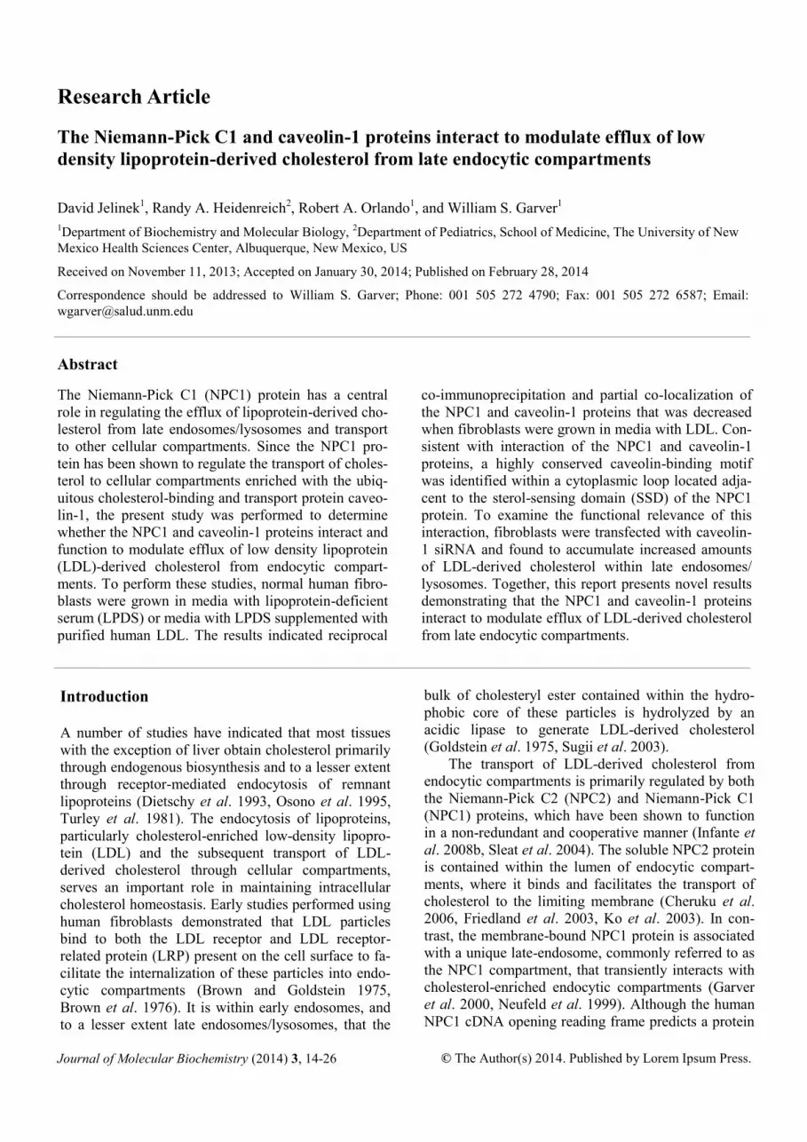
Figure 1. Relative amounts of the NPC1 and caveolin-1
proteins. The average amounts of NPC1 protein (A) and
caveolin-1 protein (B) for fibroblasts grown in media with
LPDS were normalized to β-actin (internal control) and as-
signed a value of 1.0, while the average amounts of NPC1
protein and caveolin-1 protein for fibroblasts grown in me-
dia with LDL were expressed as fold change. A representa-
tive immunoblot showing the relative amounts of NPC1 and
caveolin-1 protein for fibroblasts grown in LPDS (left lane)
and LDL (right lane) are included as insets within respective
graphs. Quantitative data is represented as the mean ± S.D.
using five plates of fibroblasts grown using both culture
conditions. *P ≤ 0.05 compared to fibroblasts grown in me-
dia with LPDS.

human fibroblasts (qRT-PCR and immunoblot analy-
sis) or ten fluorescent images (quantification of co-
localization) of human fibroblasts grown using both
culture conditions (LPDS or LDL). Significant differ-
ences (P 0.05) between two groups of data were de-
termined using the two-tailed Student’s t-test assuming
equal variance.
Results
Relative amounts of the NPC1 and caveolin-1 pro-
teins
Fibroblasts were grown using the two culture condi-
tions (LPDS and LDL) and processed to determine
relative amounts of the NPC1 and caveolin-1 proteins.
The results indicated a significant average decrease
(31%) in the relative amounts of NPC1 protein for fi-
broblasts grown in media with LDL compared to fibro-
blasts grown in media with LPDS (Figure 1A). In con-
trast, the results indicated a significant average in-
crease (52%) in the relative amounts of caveolin-1 pro-
tein for fibroblasts grown in media with LDL com-
pared to fibroblasts grown in media with LPDS
(Figure 1B). Similarly, consistent with transcriptional
regulation of the NPC1 and caveolin-1 genes, the re-
sults indicated a significant average decrease (33%) in
the relative amounts of NPC1 mRNA and a significant
average increase (33%) in the relative amounts of
caveolin-1 mRNA for fibroblasts grown in media with
LDL compared to fibroblasts grown in media with
LPDS (data not shown).
Reciprocal co-immunoprecipitation of the NPC1
and caveolin-1 proteins
Fibroblasts were grown using the two culture condi-
tions (LPDS and LDL) and processed to perform re-
ciprocal co-immunoprecipitation of the NPC1 and
caveolin-1 proteins. The results indicated that im-
munoprecipitation of caveolin-1 promoted co-
immunoprecipitation of the lower molecular weight
NPC1 protein (170 kDa) when fibroblasts were grown
in media with LPDS or LDL (Figure 2A, B, C). Simi-
larly, immunoprecipitation of the NPC1 protein pro-
moted co-immunoprecipitation of caveolin-1 when
fibroblasts were grown in media with LPDS or LDL
(Figure 2D, E, F). However, it must be noted that co-
immunoprecipitation of either the NPC1 or caveolin-1
protein was decreased for fibroblasts grown in media
with LDL compared to fibroblasts grown in media
with LPDS (Figure 2B, E).
Cellular distribution of the NPC1 and caveolin-1
proteins
Fibroblasts were grown using the two culture condi-
tions (LPDS and LDL) and processed to perform de-
convolution fluorescence microscopy. The NPC1 pro-
tein, which is known to be primarily associated with a
unique population of late endosomes, was represented
by vesicles approximately 1.5 mm in diameter and dis-
tributed throughout the perinuclear region of the cyto-
plasm when fibroblasts were grown in media with
LPDS or LDL (Figure 3A, D). In contrast, caveolin-1
was represented by a relatively increased number of
similarly sized vesicles that were also distributed
throughout the perinuclear region of the cytoplasm
when fibroblasts were grown in media with LPDS or
LDL (Figure 3B, E). The merged images indicated
partial co-localization of these vesicles when fibro-
blasts were grown in media with LPDS or LDL
(Figure 3C, F).
Percentage of co-localization between the NPC1
and caveolin-1 proteins
Fibroblasts were grown using the two culture condi-
tions (LPDS and LDL) and processed to perform de-
convolution fluorescence microscopy and determine
the percentage of co-localization between the NPC1
and caveolin-1 proteins. The results indicated that, on
average, caveolin-1 co-localized with 13% of the
NPC1 protein when fibroblasts were grown in media
with LPDS (Figure 4A). In contrast, the average
Journal of Molecular Biochemistry, 2014 18
Figure 2. Reciprocal co-immunoprecipitation of the NPC1
and caveolin-1 proteins. An equivalent amount of solubilized
and diluted homogenate protein from fibroblasts grown in
media with LPDS or LDL was incubated in the presence or
absence of rabbit anti-human NPC1 antibody or mouse anti-
human caveolin-1 antibody and resulting antibody-antigen
complexes were precipitated using Protein A-Sepharose
beads. The beads were incubated with sample buffer and
immunoblot analysis was performed using reciprocal anti-
bodies (same caveolin-1 or NPC1 antibody, respectively).
The solubilized and diluted homogenate protein (A, D)
served as a positive control for identification of the co-
immunoprecipitated proteins (B, E), while the solubilized
and diluted homogenate protein incubated in the absence of
NPC1 or caveolin-1 primary antibody (C, F) served as a
negative control.

amount of caveolin-1 that co-localized with the NPC1
protein was significantly decreased (62%) when fibro-
blasts were grown in media with LDL. Similarly, on
average, the NPC1 protein co-localized with 10% of
caveolin-1 when fibroblasts were grown in media with
LPDS, but the average amount was significantly de-
creased (40%) when fibroblasts were grown in media
with LDL (Figure 4B).
Identification and sequence comparison of caveolin-
binding motifs associated with orthologous NPC1
proteins
The principal caveolin-binding motif, denoted by the
consensus sequence jXjXXXXj, where j represents Trp
(W), Phe (F), or Tyr (Y), but not the less common
caveolin-binding motifs denoted by the consensus se-
quences jXXXXjXXj and jXjXXXXjXXj, was identi-
fied within the NPC1 protein amino acid sequence.
The amino acid consensus sequence (818-FRFFKNSY
-825) is encoded within cytoplasmic loop 4 adjacent to
19 Journal of Molecular Biochemistry, 2014
Figure 4. Percentage of co-localization between the NPC1
and caveolin-1 proteins. The total areas representing NPC1
or caveolin-1 staining were independently identified using
the color range selection feature within Adobe Photoshop
software, followed by determining the overlapping area and
percentage of co-localization for caveolin-1 staining (A) and
NPC1 staining (B) from fibroblasts grown in media with
LPDS or LDL. Fluorescent images were obtained using a
deconvolution fluorescence microscope equipped with an
Olympus 100x (NA 1.4) oil-immersion objective. Quantita-
tive data is represented as the mean ± S.D. using ten images
acquired in a random manner and processed using identical
conditions. *P 0.05 compared to fibroblasts grown in
media with LPDS.
Figure 3. Cellular distribution of the NPC1 and caveolin-1 proteins. A representative fibroblast grown in LPDS is provided in
the top set of images (A, B, C), while a representative fibroblast grown in LDL is provided in the bottom set of images (D, E,
F). Partial co-localization of the NPC1 and caveolin-1 proteins are indicated using white arrows. Fluorescent images were ob-
tained using a deconvolution fluorescence microscope equipped with an Olympus 100x (NA 1.4) oil-immersion objective. All
images were acquired and processed using identical conditions. Bar represents 15 mm. N, nucleus.

the sterol-sensing domain (amino acids 616-797) of the
human NPC1 protein. Since the NPC1 protein caveolin
-binding motif is positioned within the cytoplasm, it
would theoretically permit interaction with the caveo-
l i n - s c a f f o l d i n g d o m a i n ( 8 2 -
DGIWKASFTTFTVTKYWFYR-101) of caveolin-1
also positioned within the cytoplasm (Figure 5A). A
comparison of the amino acid sequences from ortholo-
gous NPC1 proteins indicated that the NPC1 protein
caveolin-binding motif is highly conserved among sev-
eral different species (Figure 5B). The one species
(cat) that does not encode the common caveolin-
binding motif consensus sequence, instead encodes
Journal of Molecular Biochemistry, 2014 20
what has been reported as a permissible caveolin-like
binding motif (I, L, or V instead of W, F or Y in place
of the second j) also capable of interaction with the
caveolin-scaffolding domain.
Relative amounts of the NPC1 and Caveolin-1 pro-
teins resulting from caveolin-1 siRNA knockdown
Fibroblasts were transfected with either non-specific
siRNA or caveolin-1 siRNA and grown using the two
culture conditions (LPDS and LDL) to determine the
relative average amounts of the NPC1 and caveolin-1
Figure 5. Identification and sequence comparison of caveo-
lin-binding motifs associated with orthologous NPC1 pro-
teins. A schematic representation of the topology for both
the NPC1 and caveolin-1 proteins is provided (A). The hu-
man NPC1 protein contains a previously unidentified caveo-
lin-binding motif (818-FRFFKNSY-825) located within
cytoplasmic loop 4 adjacent to the sterol-sensing domain
(amino acids 616-797). It is proposed that the NPC1 protein
caveolin-binding motif interacts with the caveolin-1 protein
through the caveolin-scaffolding domain (82-
DGIWKASFTTFTVTKYWFYR-101). The caveolin-1 pro-
tein also possesses a cholesterol recognition sequence (91-
TFTVTKYWFYRLL-103) located near the cytofacial mem-
brane and cytoplasm boundary that partially overlaps with
the caveolin-scaffolding domain. A comparison of the
amino acid sequences between orthologous NPC1 proteins
indicates that the NPC1 protein caveolin-binding motif
(jXjXXXXj), where j mostly represents the amino acids Trp
(W), Phe (F), or Tyr (Y), is conserved among several differ-
ent species (B).
Figure 6. Relative amounts of the NPC1 and caveolin-1
proteins resulting from caveolin-1 siRNA knockdown. The
relative amounts of the NPC1 and caveolin-1 proteins for
fibroblasts transfected with non-specific siRNA (control) or
caveolin-1 siRNA (siRNA) and grown in media with LPDS
or LDL was determined using immunoblot analysis, where 1
= LPDS Control, 2 = LPDS siRNA, 3 = LDL Control, and 4
= LDL siRNA (A). A field of representative fibroblasts
transfected with non-specific siRNA (control) or caveolin-1
siRNA (siRNA) and grown in media with LPDS (B and C)
or LDL (D and E) is provided. Fluorescent images were
obtained using a deconvolution fluorescence microscope
equipped with an Olympus 60x (NA 1.4) oil-immersion
objective. All images were acquired and processed using
identical conditions.

proteins. The results indicated no
significant difference in the rela-
tive amounts of the NPC1 protein
for fibroblasts transfected with
caveolin-1 siRNA when fibro-
blasts were grown in media with
LPDS or LDL (Figure 6A). In
contrast, the results indicated a
significant decrease in the relative
average amounts of caveolin-1
protein for fibroblasts transfected
with caveolin-1 siRNA when fi-
broblasts were grown in media
with LPDS or LDL (89% and
92%, respectively), compared to
fibroblasts transfected with the
non-specific (control) siRNA
(Figure 6A). Consistent with the
significantly decreased average
amounts of caveolin-1 protein de-
termined using immunoblot analy-
sis, the relative amounts of caveo-
lin-1 protein were noticeably de-
creased for fibroblasts transfected
with caveolin-1 siRNA when fi-
broblasts were grown in media
with LPDS or LDL compared to
fibroblasts transfected with the
non-specific (control) siRNA upon
examination using fluorescence
microscopy (Figure 6B, C, D, F).
Cellular distribution of choles-
terol in relation to late en-
dosomes/lysosome resulting
from caveolin-1 siRNA knock-
down
Fibroblasts were transfected with
non-specific siRNA or caveolin-1
siRNA and grown in media with
LDL to determine the relative cel-
lular distribution of cholesterol in
relation to late endosomes/
lysosomes using deconvolution
fluorescence microscopy. The im-
ages indicated one primary cellu-
lar distribution for cholesterol
which was localized within peri-
nuclear cytoplasmic vesicles
(Figure 7A, D). With respect to
fibroblasts transfected with the
non-specific siRNA (control), the
images indicated that only a cer-
tain population of the LAMP-1
21 Journal of Molecular Biochemistry, 2014
Figure 7. Cellular distribution of cholesterol in relation to late endosomes/lysosomes
resulting from caveolin-1 siRNA knockdown. Fibroblasts were processed to label
cholesterol using filipin and the LAMP-1 protein (a marker protein for late en-
dosomes/lysosomes). Fibroblasts transfected with a non-specific siRNA (control) and
grown in media with LDL are provided in the top set of images (A, B, C), while fibro-
blasts transfected with caveolin-1 siRNA and grown in media with LDL are provided
in the bottom set of images (D, E, F). A defined area (square white outline) within
fibroblasts are enlarged and placed directly below the corresponding image to visual-
ize the cellular distribution of cholesterol (A, D), the LAMP-1 protein (B, E), and
merged images (C, F). Fluorescent images were obtained using a deconvolution fluo-
rescence microscope equipped with an Olympus 60x (NA 1.4) oil-immersion objec-
tive. All images were acquired and processed using identical conditions. Bar repre-
sents 15 mm.

containing vesicles, represented by semi-rounded and
hollowed vesicles, co-localized with cholesterol
(Figure 7A, B, C). In contrast, fibroblasts transfected
with caveolin-1 siRNA had a noticeably increased
population of LAMP-1 containing vesicles that co-
localized with cholesterol (Figure 7D, E, F). There was
no noticeable difference between fibroblasts trans-
fected with the non-specific siRNA (control) and cave-
olin-1 siRNA when grown in media with LPDS (data
not shown), thereby suggesting an accumulation of
cholesterol within the LAMP-1 containing vesicles
was derived from LDL.
Relative amounts of cholesterol associated with late
endosomes/lysosomes resulting from caveolin-1
siRNA knockdown
Fibroblasts were transfected with non-specific siRNA
or caveolin-1 siRNA and grown in media with LDL to
determine the relative amount of LDL-derived choles-
terol associated with late endosomes/lysosomes using
deconvolution fluorescence microscopy. The results
indicated a significant average increase (44%) in the
relative amounts of cholesterol localized within LAMP
-1 containing vesicles, which are known to represent
late endosomes/lysosomes, for fibroblasts transfected
with caveolin-1 siRNA compared to fibroblasts trans-
fected with non-specific siRNA (control) (Figure 8).
These results again suggest an accumulation of choles-
terol within the LAMP-1 containing vesicles was de-
rived from LDL.
Discussion
The present study was performed to determine whether
the NPC1 and caveolin-1 proteins interact and function
to modulate the efflux of LDL-derived cholesterol
from endocytic compartments. To perform these stud-
ies, normal human fibroblasts were grown in media
containing LPDS, which represented the control or
basal culture condition, in addition to media containing
LPDS supplemented with purified human LDL. In
brief, the results indicated i) an inverse association be-
tween relative amounts of the NPC1 and caveolin-1
proteins when fibroblasts were grown in media with
LDL; ii) that reciprocal co-immunoprecipitation of the
NPC1 and caveolin-1 proteins was decreased when
fibroblasts were grown in media with LDL; iii) that
partial co-localization of the NPC1 and caveolin-1 pro-
teins was decreased when fibroblasts were grown in
media with LDL; iv) identification of a conserved
caveolin-binding motif located adjacent to the SSD of
the NPC1 protein, and v) an increased amount of LDL-
derived cholesterol within late endosomes/lysosomes
resulting from caveolin-1 siRNA knockdown. To-
Journal of Molecular Biochemistry, 2014 22
gether, this report presents novel results demonstrating
that the NPC1 and caveolin-1 proteins interact to
modulate efflux of LDL-derived cholesterol from late
endocytic compartments.
The results indicated an inverse association
between relative amounts of the NPC1 and caveolin-1
proteins when fibroblasts were grown in media with
LDL. Specifically, compared to fibroblasts grown in
media with LPDS, supplementation of media with
LDL significantly decreased the relative average
amounts of NPC1 protein, but significantly increased
the relative average amounts of caveolin-1 protein. A
logical explanation for the inverse expression of the
NPC1 and caveolin-1 genes is supported by studies
indicating that LDL-dependent feedback inhibition of
the SREBP pathway promotes downregulation of the
NPC1 gene and upregulation of the caveolin-1 gene
(Bist et al. 1997, Garver et al. 2008, Hailstones et al.
1998). However, it should be noted that feedback inhi-
bition of the SREBP pathway and subsequent tran-
scriptional regulation of the NPC1 and caveolin-1
genes may only serve as a partial explanation. It is well
Figure 8. Relative amounts of cholesterol associated with
late endosomes/lysosomes resulting from caveolin-1 siRNA
knockdown. The total area representing LAMP-1 containing
vesicles was identified using the color range selection fea-
ture, followed by determining the intensity of cholesterol
staining within this area to calculate the relative amounts of
cholesterol associated with late endosomes/lysosomes. Fluo-
rescent images were obtained using a deconvolution fluores-
cence microscope equipped with an Olympus 60x (NA 1.4)
oil-immersion objective. Quantitative data is represented as
the mean ± S.D. using ten fields acquired in random manner
and processed using identical conditions for fibroblasts
grown in media with LDL. *P 0.05 compared to fibro-
blasts transfected with non-specific siRNA (control) and
grown in media with LDL.

accepted that decreased NPC1 protein function alters
the efflux of cholesterol from endocytic compartments
and impairs feedback inhibition of the SREBP path-
way (Garver et al. 2007, Kruth et al. 1986, Liscum et
al. 1989, Pentchev et al. 1986). As a result, decreased
NPC1 protein function should decrease expression of
the caveolin-1 gene. This is contrary to previous re-
sults indicating that NPC1 heterozygous (NPC1+/-)
and NPC1 homozygous affected (NPC1-/-) human fi-
broblasts and mouse livers have significantly increased
amounts of caveolin-1 protein, suggesting that the
caveolin-1 protein may compensate for decreased
NPC1 protein function (Garver et al. 1997a, Garver et
al. 1997b Garver et al. 2002). Consistent with this hy-
pothesis, the relative average amounts of the NPC2
protein, which is well known to interact and function
in a coordinate manner with the NPC1 protein, is like-
wise increased in NPC1-/- mouse livers (Klein et al.
2006).
To determine whether the NPC1 and caveolin-
1 proteins interact, studies were performed using recip-
rocal co-immunoprecipitation. The results indicated
that immunoprecipitation of either the NPC1 or caveo-
lin-1 proteins resulted in co-immunoprecipitation of
the caveolin-1 and NPC1 proteins, respectively. Inter-
estingly, of the two NPC1 proteins (170 and 190 kDa)
which differ due to asparagine-linked glycosylation
within the NTD, only the lower molecular weight
NPC1 protein co-immunoprecipitated with caveolin-1.
Previous studies have indicated that this lower molecu-
lar weight NPC1 protein is ~50% less efficient in fa-
cilitating efflux of LDL-derived cholesterol from endo-
cytic compartments (Watari et al. 1999a, Watari et al.
1999b). Moreover, the results indicated that reciprocal
co-immunoprecipitation of the NPC1 or caveolin-1
proteins was decreased when fibroblasts were grown in
media with LDL, suggesting that the increased efflux
of cholesterol from endocytic compartments partially
inhibits interaction of the NPC1 and caveolin-1 pro-
teins.
Consistent with interaction of the NPC1 and
caveolin-1 proteins, the results indicated partial co-
localization of the NPC1 and caveolin-1 proteins when
examined using deconvolution fluorescence micros-
copy. This particular result confirms earlier reports
describing partial co-localization of the NPC1 and
caveolin-1 proteins using both human and mouse fi-
broblasts (Garver et al. 2000, Higgins et al. 1999).
Moreover, consistent with the decreased interaction of
the NPC1 and caveolin-1 proteins when fibroblasts are
grown in media with LDL, quantification of high-
resolution fluorescent images revealed significantly
less co-localization when fibroblasts were grown using
this condition. Interestingly, a recent study has indi-
cated that caveolin-1 enriched vesicles that transiently
interact with late endosomes/lysosomes are capable of
forming caveolin-1 enriched subdomains that partici-
pate in the selective release of endosomal cargo
(Pelkmans et al. 2004).
Studies have indicated that caveolin-1 is capa-
ble of specific and direct interaction with a number of
proteins through a scaffolding domain (Human caveo-
lin-1: 82-DGIWKASFTTFTVTKYWFYR-101) and
that this domain partially overlaps with a cholesterol
recognition sequence (Human caveolin-1: 91-
TFTVTKYWFYRLL-103) that has stable and high-
affinity for cholesterol (Li & Papadopoulos 1998, Mu-
rata et al. 1995). Both the caveolin-scaffolding domain
and cholesterol recognition sequence are located at the
cytofacial membrane and cytoplasm boundary near the
amino-terminus of caveolin-1. This caveolin-
scaffolding domain has been shown to interact with
proteins that possess a caveolin-binding motif consen-
sus sequence (jXjXXXXj), where j represents the
amino acids Trp (W), Phe (F), or Tyr (Y) (Couet et al.
1997). Importantly, the human NPC1 protein was
found to possess a previously unidentified caveolin-
binding motif (818-FRFFKNSY-825) located within
cytoplasmic loop 4 adjacent to the sterol-sensing do-
main (amino acids 616-797). A computer database
search also revealed that the human NPC1 protein
caveolin-binding motif is highly conserved among dif-
ferent species. This being the case, it is interesting to
note that Patched, a membrane-bound protein with ex-
tensive sequence homology to the NPC1 protein, has
also been shown to possess a caveolin-binding motif
located within a cytoplasmic loop adjacent to the sterol
-sensing domain and capable of interacting with caveo-
lin-1 (Karpen et al. 2001).
To determine whether the caveolin-1 protein
might have a role in modulating the efflux of LDL-
derived cholesterol from endocytic compartments, fi-
broblasts were transfected with caveolin-1 siRNA to
decrease expression of the caveolin-1 gene, and grown
in media with LDL. The results clearly demonstrated a
significant increase in the relative average amounts of
LDL-derived cholesterol within LAMP-1 containing
vesicles when compared to fibroblasts transfected with
control siRNA and grown using similar conditions.
However, compared to fibroblasts transfected with
control siRNA, the total concentration of cellular cho-
lesterol or cholesteryl ester was not significantly dif-
ferent for fibroblasts transfected with caveolin-1
siRNA, thereby suggesting that decreased amounts of
caveolin-1 protein alters the distribution of cellular
cholesterol (data not shown). This particular result is
consistent with an earlier report indicating that a cave-
olin-1 dominant-negative mutant promotes altered in-
23 Journal of Molecular Biochemistry, 2014

tracellular cholesterol homeostasis characterized by an
accumulation of cholesterol within late endosomes/
lysosomes, although the concentration of cellular cho-
lesterol remains unchanged (Pol et al. 2001, Roy et al.
1999). Moreover, a recent report using cells derived
from the caveolin-1 deficient mouse indicated that
caveolin-1 has a key role in maintaining intracellular
cholesterol homeostasis, characterized by only a mod-
est change in the concentration of cellular cholesterol
and cholesteryl ester (Frank et al. 2006). This being
the case, it is interesting to note that whole body cho-
lesterol homeostasis, including plasma lipid levels,
remain relatively normal for both the caveolin-1 defi-
cient mouse and a patient diagnosed with a rare form
of congenital lipodystrophy (Kim et al. 2008, Razani
et al. 2002, Razani et al. 2001).
Conclusion
In summary, the present study determined that the
NPC1 and caveolin-1 proteins interact and function to
modulate efflux of LDL-derived cholesterol from en-
docytic compartments. A plausible model may be en-
visioned whereby the conserved NPC1 protein caveo-
lin-binding motif allows interaction with the caveolin-
1 scaffolding domain in the absence of LDL-derived
cholesterol, but which is interrupted by the binding of
cholesterol to the high-affinity caveolin-1 cholesterol
recognition sequence which prompts dissociation of
the NPC1 and caveolin-1 proteins and subsequent
transport of cholesterol to other caveolin-1 cellular
compartments, most likely the TGN. Clearly, addi-
tional studies will be necessary to further define how
the NPC1 and caveolin-1 proteins modulate the efflux
of LDL-derived cholesterol from endocytic compart-
ments and maintain intracellular cholesterol homeosta-
sis.
Acknowledgments
This work was supported in part by grants received
from the National Institutes of Health (R21
DK071544), the Ara Parseghian Medical Research
Foundation, and private donations for the investigation
of childhood genetic and metabolic diseases. The au-
thors would like to express their appreciation to Sarah
Mount Patrick for expertise in performing deconvolu-
tion fluorescence microscopy and quantitation of fluo-
rescence co-localization.
Conficts of interest
The authors declare no conflicts of interest.
Author Contributions
DJ and WSG conceived and performed these studies,
while DJ, RAH, RAO and WSG interpreted results and
prepared the manuscript.
References
Bist A, Fielding PE & Fielding CJ 1997 Two sterol
regulatory element-like sequences mediate up-
regulation of caveolin gene transcription in response to
low density lipoprotein free cholesterol. Proc Natl
Acad Sci USA 94 10693-10698
Blanchette-Mackie EJ, Dwyer NK, Amende LM,
Kruth HS, Butler JD, Sokol J, Comly ME, Vanier MT,
August JT, Brady RO & Pentchev PG 1988 Type C
Niemann-Pick disease: Low density lipoprotein uptake
is associated with premature cholesterol accumulation
in the Golgi complex and excessive cholesterol storage
in lysosomes. Proc Natl Acad Sci USA 85 8022-8026
Brown MS & Goldstein JL 1975 Regulation of the
activity of the low density lipoprotein receptor in hu-
man fibroblasts. Cell 6 307-316
Brown MS, Ho YK & Goldstein JL 1976 The low-
density lipoprotein pathway in human fibroblasts: Re-
lation between cell surface receptor binding and endo-
cytosis of low-density lipoprotein. Ann NY Acad Sci
275 244-257
Cheruku SR, Xu Z, Dutia R, Lobel P & Storch J 2006
Mechanism of cholesterol transfer from the Niemann-
Pick type C2 protein to model membranes supports a
role in lysosomal cholesterol transport. J Biol Chem
281 31594-31604
Couet J, Li S, Okamoto T, Ikezu T & Lisanti MP 1997
Identification of peptide and protein ligands for the
caveolin-scaffolding domain. J Biol Chem 272 6525-
6533
Coxey RA, Pentchev PG, Campbell G & Blanchette-
Mackie EJ 1993 Differential accumulation of choles-
terol in Golgi compartments of normal and Niemann-
Pick type C fibroblasts incubated with LDL: A cyto-
chemical freeze-fracture study. J Lipid Res 34 1165-
1176
Dietschy JM, Turley SD, & Spady DK 1993 Role of
liver in the maintenance of cholesterol and low density
lipoprotein homeostasis in different animal species,
including humans. J Lipid Res 34 1637-1659
Dupree P, Parton RG, Raposo G, Kurzchalia TV &
Simons K 1993 Caveolae and sorting in the trans-
Golgi network of epithelial cells. EMBO J 12 1597-
1605
Fielding PE & Fielding CJ 1996 Intracellular transport
of low density lipoprotein derived free cholesterol be-
Journal of Molecular Biochemistry, 2014 24

gins at clathrin-coated pits and terminates at cell sur-
face caveolae. Biochemistry 35 14932-14938
Friedland N, Liou H-L, Lobel P & Stock AM 2003
Structure of a cholesterol-binding protein deficient in
Niemann-Pick type C2 disease. Proc Nat Acad Sci
USA 100 2512-2517
Garver WS, Erickson RP, Wilson JM, Colton TL, Hos-
sain GS, Kozloski MA & Heidenreich RA 1997a Al-
tered expression of caveolin-1 and increased choles-
terol in detergent insoluble membrane fractions from
liver in mice with Niemann-Pick disease type C. Bio-
chim Biophys Acta 1361 272-280
Garver WS, Heidenreich RA, Erickson RP, Thomas
MA & Wilson JM 2000 Localization of the murine
Niemann-Pick C1 protein to two distinct intracellular
compartments. J Lipid Res 41 673-687
Garver WS, Hsu SC, Erickson RP, Greer WL, Byers
DM & Heidenreich RA 1997b Increased expression of
caveolin-1 in heterozygous Niemann-Pick type II hu-
man fibroblasts. Biochem Biophys Res Comm 236 189-
193
Garver WS, Jelinek D, Oyarzo JN, Flynn J, Zucker-
man M, Krishnan K, Chung BH & Heidenreich RA
2007 Characterization of liver disease and lipid me-
tabolism in the Niemann-Pick C1 mouse. J Cell Bio-
chem 101 498-516
Garver WS, Krishnan K, Gallagos JR, Michikawa M,
Francis GA & Heidenreich RA 2002 Niemann-Pick C1
protein regulates cholesterol transport to the trans-
Golgi network and plasma membrane caveolae. J Lipid
Res 43 579-589
Garver WS, Jelinek D, Francis GA & Murphy BD
2008 The Niemann-Pick C1 gene is downregulated by
feedback inhibition of the SREBP pathway in humans
fibroblasts. J Lipid Res 49 1090-1102
Goldstein JL, Dana SE, Faust JR, Beaudet AL &
Brown MS 1975 Role of lysosomal acid lipase in the
metabolism of plasma low density lipoprotein. Obser-
vations in cultured fibroblasts from a patient with cho-
lesteryl ester storage disease. J Biol Chem 250 8487-
8495
Hailstone D, Sleer LS, Parton RG & Stanley KK 1998
Regulation of caveolin and caveolae by cholesterol in
MDCK cells. J Lipid Res 39 369-379
Higgins ME, Davies JP, Chen FW & Ioannou YA
1999 Niemann-Pick C1 is a late endosome-resident
protein that transiently associates with lysosomes and
the trans-Golgi network. Mol Gene Metab 68 1-13
Infante RE, Radhakrishnan A, Abi-Mosleh L, Kinch
LN, Wang ML, Grishin NV, Goldstein JL & Brown
MS 2008a Purified NPC1 Protein II. Localization of
sterol binding to a 240-amino acid soluble luminal
loop. J Biol Chem 283 1064-1075
Infante RE, Wang ML, Radhakrishnan A, Kwon HJ,
Brown MS & Goldstein JL 2008b NPC2 facilitates
bidirectional transfer of cholesterol between NPC1 and
lipid bilayers, a step in cholesterol egress from ly-
sosomes. Proc Natl Acad Sci USA 105 15287-15292
Karpen HE, Bukowski JT, Hughes T, Gratton J, Sessa
WC & Gailani MR 2001 The sonic hedgehog receptor
patched associates with caveolin-1 in cholesterol-rich
microdomains of the plasma membrane. J Biol Chem
276 19503-19511
Kim CA, Delepin M, Boutet M, El Mourabit H, Le
Lay S, Meier M, Nemani M, Bridel E, Leite CC, Ber-
tola DR, Semple RK, O'Rahilly S, Dugail I, Capeau J,
Lathrop M & Magre J 2008 Association of a homozy-
gous nonsense caveolin mutation with Berardinelli-
Seip congenital lipodystrophy. J Clin Endo Metab 93
1129-1134
Klein A, Amigo L, Retamal MJ, Morales MG, Miquel
JF, Rigotti A & Zanlungo S 2006 NPC2 is expressed
in human and murine liver and secreted into bile: Po-
tential implications for body cholesterol homeostasis.
Hepatology 43 126-133
Ko DC, Binkley J, Sidow A & Scott MP 2003 The
integrity of a cholesterol-binding pocket in Niemann-
Pick C2 protein is necessary to control lysosome cho-
lesterol levels. Proc Natl Acad Sci USA 100 2518-2525
Kruth HS, Comly ME, Butler JD, Vanier MT, Fink JK,
Wenger DA, Patel S & Pentchev PG 1986 Type C Nie-
mann-Pick disease. Abnormal metabolism of low den-
sity lipoprotein in homozygous and heterozygous fi-
broblasts. J Biol Chem 261 16769-16774
Kwon HJ, Abi-Mosleh, Wang ML, Deisenhofer J,
Goldstein JL, Brown MS & Infante RE 2009 Structure
of N-terminal domain of NPC1 reveals distinct subdo-
mains for binding and transfer of cholesterol Cell 137
1213-1224
Li H & Papadopoulos V 1998 Peripheral-type benzodi-
azepine receptor function in cholesterol transport.
Identification of a putative cholesterol recognition/
interaction amino acid sequence and consensus pattern.
Endocrinology 139 4991-4997
Liscum L, Ruggiero RM & Faust JR 1989 The intra-
cellular transport of low density lipoprotein-derived
cholesterol is defective in Niemann-Pick type C fibro-
blasts. J Cell Biol 108 1625-1636
Liu R, Lu P, Chu JWK & Sharom FJ 2009 Characteri-
zation of fluorescent sterol binding to purified human
NPC1. J Biol Chem 284 1840-1852
Murata M, Peranen J, Schreiner R, Wieland F, Kurz-
chalia TV & Simons K 1995 VIP21/caveolin is a cho-
lesterol-binding protein. Proc Natl Acad Sci USA 92
10339-10343
Neufeld EB, Wastney M, Patel S, Suresh S, Conney
AM, Dwyer NK, Roff CF, Ohno K, Morris JA, Carstea
25 Journal of Molecular Biochemistry, 2014

ED, Incardona JP, Strauss JF, Vanier MT, Patterson
MC, Brady RO, Pentchev PG & Blanchette-Mackie
EJ 1999 The Niemann-Pick C1 protein resides in a
vesicular compartment linked to retrograde transport of
multiple lysosomal cargo. J Biol Chem 274 9627-9635
Ohgami N, Ko DC, Thomas M, Scott MP, Chang CCY
& Chang TY 2004 Binding between the Niemann-Pick
C1 protein and a photoactivatable cholesterol analog
requires a functional sterol-sensing domain. Proc Natl
Acad Sci USA 101 12473-12478
Osono Y, Woollett LA, Herz J & Dietschy JM 1995
Role of the low density lipoprotein receptor in the flux
of cholesterol through the plasma and across the tis-
sues of the mouse. J Clin Invest 95 1124-1132
Pelkmans L, Burli T, Zerial M & Helenius A 2004
Caveolin-stabilized membrane domains as multifunc-
tional transport and sorting devices in endocytic mem-
brane traffic. Cell 118 767-780
Pentchev PG, Kruth HS, Comly ME, Butler JD, Vanier
MT, Wenger DA & Patel S 1986 Type C Niemann-
Pick disease. A parallel loss of regulatory responses in
both the uptake and esterification of low density lipo-
protein-derived cholesterol in cultured fibroblasts. J
Biol Chem 261 16775-16780
Pol A, Luetterforst R, Lindsay M, Heino S, Ikonen E
& Parton RG 2001 A caveolin dominant negative mu-
tant associates with lipid bodies and induces intracellu-
lar cholesterol imbalance. J Cell Biol 152 1057-1070
Razani B, Engelman JA, Wang XB, Schubert W,
Zhang XL, Marks CB, Macaluso F, Russell RG, Li M,
Pestell RG, Di Vizio D, Hou H, Kneitz B, Lagaud G,
Christ GJ, Edelmann W & Lisanti MP 2001 Caveolin-
1 null mice are viable but show evidence of hyperpro-
liferative and vascular abnormalities. J Biol Chem 276
38121-38138
Razani B, Combs TP, Wang XB, Frank PG, Park DS,
Russell RG, Li M, Tang B, Jelicks LA, Scherer PE &
Lisanti MP 2002 Caveolin-1-deficient mice are lean,
resistant to diet-induced obesity, and show hyper-
triglyceridemia with adipocyte abnormalities. J Biol
Chem 277 8635-8647
Roy S, Luetterforst R, Harding A, Apolloni A,
Etheridge M, Stang E, Rolls B, Hancock JF & Parton
RG 1999 Dominant-negative caveolin inhibits H-Ras
function by disrupting cholesterol-rich plasma mem-
brane domains. Nat Cell Biol 1 98-105
Sleat DE, Wiseman JA, El-Banna M, Price SM, Verot
L, Shen MM, Tint GS, Vanier MT, Walkley SU & Lo-
bel P 2004 Genetic evidence for nonredundant func-
tional cooperativity between NPC1 and NPC2 in lipid
transport. Proc Natl Acad Sci USA 101 5886-5891
Smart EJ, Ying Y, Donzell WC & Anderson RGW
1996 A role for caveolin in transport of cholesterol
from endoplasmic reticulum to plasma membrane. J
Biol Chem 271 29427-29435
Sugii S, Reid PC, Ohgami N, Du H & Chang TY 2003
Distinct endosomal compartments in early trafficking
of low density lipoprotein-derived cholesterol. J Biol
Chem 278 27180-27189
Turley SD, Spady DK & Dietschy JM 1995 Role of
liver in the synthesis of cholesterol and the clearance
of low density lipoproteins in the cynomolgus monkey.
J Lipid Res 36 67-79
Uittenbogaard A, Everson WV, Matveev SV & Smart
EJ 2002 Cholesteryl ester is transported from caveolae
to internal membranes as part of a caveolin-annexin II
lipid-protein complex. J Biol Chem 277 4925-4931
Uittenbogaard A, Ying Y & Smart EJ 1998 Characteri-
zation of a cytosolic heat-shock protein-caveolin chap-
erone complex. Involvement in cholesterol trafficking.
J Biol Chem 273 6525-6532
Underwood KW, Jacobs NL, Howley A & Liscum L
1998 Evidence for a cholesterol transport pathway
from lysosomes to endoplasmic reticulum that is inde-
pendent of the plasma membrane. J Biol Chem 273
4266-4274
Urano Y, Watanabe H, Murphy SR, Shibuya Y, Geng
Y, Peden AA, Chang CCY & Chang TY 2008 Trans-
port of LDL-derived cholesterol from the NPC1 com-
partment to the ER involves the trans-Golgi network
and the SNARE protein complex. Proc Natl Acad Sci
USA 105 16513-16518
Watari H, Blanchette-Mackie EJ, Dwyer NK, Glick
JM, Patel S, Neufeld EB, Brady RO, Pentchev PG &
Strauss III JF 1999a Niemann-Pick C1 protein: Obliga-
tory roles for N-terminal domains and lysosomal tar-
geting in cholesterol mobilization. Proc Natl Acad Sci
USA 96 805-810
Watari H, Blanchette-Mackie EJ, Dwyer NK, Watari
M, Neufeld EB, Patel S, Pentchev PG & Strauss III JF
1999b Mutations in the leucine zipper motif and sterol-
sensing domain inactivate the Niemann-Pick C1 glyco-
protein. J Biol Chem 274 21861-21866
Wojtanik KM & Liscum L 2003 The transport of low
density lipoprotein-derived cholesterol to the plasma
membrane is defective in NPC1 cells. J Biol Chem 278
14850-14856
Journal of Molecular Biochemistry, 2014 26


