Embed Size (px)
Citation preview

South Dakota State University South Dakota State University
Open PRAIRIE: Open Public Research Access Institutional Open PRAIRIE: Open Public Research Access Institutional
Repository and Information Exchange Repository and Information Exchange
Electronic Theses and Dissertations
2016
Role of Silencing RNA fgsiR34 in Fusarium Graminearum's Role of Silencing RNA fgsiR34 in Fusarium Graminearum's
Pathogenicity to Wheat Pathogenicity to Wheat
Subha Dahal South Dakota State University
Follow this and additional works at: https://openprairie.sdstate.edu/etd
Part of the Biology Commons, Microbiology Commons, and the Plant Biology Commons
Recommended Citation Recommended Citation Dahal, Subha, "Role of Silencing RNA fgsiR34 in Fusarium Graminearum's Pathogenicity to Wheat" (2016). Electronic Theses and Dissertations. 1103. https://openprairie.sdstate.edu/etd/1103
This Thesis - Open Access is brought to you for free and open access by Open PRAIRIE: Open Public Research Access Institutional Repository and Information Exchange. It has been accepted for inclusion in Electronic Theses and Dissertations by an authorized administrator of Open PRAIRIE: Open Public Research Access Institutional Repository and Information Exchange. For more information, please contact [email protected].

ROLE OF SILENCING RNA fgsiR34 IN FUSARIUM GRAMINEARUM’S
PATHOGENICITY TO WHEAT
BY
SUBHA DAHAL
A thesis submitted in partial fulfillment of the requirements for the
Master of Science
Major in Biological Sciences
Specialization in Microbiology
South Dakota State University
2016


iii
ACKNOWLEDGEMENTS
I would like to express my deepest gratitude to my advisor Dr. Yang Yen for
providing me the opportunity and support towards achieving this degree. My sincere
appreciation goes for his valuable guidance, encouragement, and constructive suggestions
for the accomplishment of this research.
I would like to thank Dr. Shaukat Ali, Dr. Yajun Wu, and Dr. Adam Hoppe for
graciously accepting to serve on my committee and providing me with instructive
comments and direction to achieve this goal. I am also immensely grateful to the faculty
and staff, and fellow graduate students in the Department of Biology and Microbiology
and the Department of Plant Science for their valuable suggestions.
My sincere thanks go to Dr. Sajag Adhikari and Dr. Simon Newkirk for their
invaluable help with some experiments. I would like to thank my former lab mate Dr.
Aravind Galla for his suggestions and guidance from the long distance. I am thankful to
my lab mates, Anjun Ma and Bimal Paudel for their support.
I would like to thank the Department of Biology and Microbiology for providing
me with the assistantship. I would like to acknowledge South Dakota Agriculture
Experimental Station for funding this research. I am also very grateful to Mr. Jerry Tiede
for providing me with the Jerry Tiede Graduate Assistantship for the summer of 2015.
I am indebted to my parents and my sister Bibha for their consistent motivation
and faith in me. I owe a special thank you to my husband Ramesh Adhikari for his
unconditional love and support despite being far away from me during my study period.
Lastly, I would like to express my heartfelt appreciation to all the people who
helped me in one way or another to complete my research work.

iv
TABLE OF CONTENTS
ABBREVIATIONS ......................................................................................................... viii
LIST OF FIGURES ......................................................................................................... xiii
LIST OF TABLES ........................................................................................................... xvi
ABSTRACT .................................................................................................................... xvii
Chapter 1 Literature Review ............................................................................................... 1
1.1 Introduction .......................................................................................................... 1
1.2 History of FHB study ........................................................................................... 2
1.3 Economic losses caused by FHB ......................................................................... 3
1.4 Causal organisms of FHB .................................................................................... 4
1.5 Fusarium graminearum disease cycle.................................................................. 5
1.6 FHB’s signs and symptoms .................................................................................. 6
1.7 F. graminearum produces mycotoxins ................................................................ 8
1.8 DON Biosynthesis Pathway ................................................................................. 9
1.9 Cell wall degrading enzymes in F. graminearum .............................................. 12
1.10 FHB resistance in wheat ..................................................................................... 13
1.11 QTL for FHB resistance in wheat ...................................................................... 14
1.12 Breeding for resistance ....................................................................................... 15
1.13 Transgenic approaches for resistance ................................................................. 16
1.14 Small RNAs and RNA interference in fungi ...................................................... 17
1.15 DNA methylation in fungi.................................................................................. 19

v
1.16 Strategies to manage FHB .................................................................................. 22
Chapter 2 Role of silencing RNA fgsiR34 in Fusarium graminearum's pathogenicity to
wheat ................................................................................................................ 24
2.1 Introduction ........................................................................................................ 24
2.2 Materials/ Methods ............................................................................................. 30
2.2.1 Fungal growth, RNA extraction and cDNA synthesis ................................ 30
2.2.2 PCR amplification, cloning and sequencing ............................................... 30
2.2.3 Vector construction for over-expressing fgsiR34 ....................................... 31
2.2.4 Fungal protoplasts generation ..................................................................... 33
2.2.5 Polyethylene glycol-mediated fungal transformation ................................. 33
2.2.6 DNA extraction and PCR amplification of GFP spacer and hygromycin
phosphotransferase gene ............................................................................. 34
2.2.7 Fungal inoculum preparation ...................................................................... 35
2.2.8 Spore production assay ............................................................................... 35
2.2.9 Radial growth assay .................................................................................... 36
2.2.10 Growth of wheat plants and spore inoculation ........................................... 36
2.2.11 RNA extraction from culture, cDNA synthesis, and Quantitative RT-PCR37
2.2.12 Poly(A) tailing coupled with reverse transcription, and RT-qPCR ............ 38
2.2.13 RNA extraction from inoculated spikelets, cDNA synthesis, and RT-qPCR
.................................................................................................................... 39
2.2.14 DNA extraction from inoculated spikelets and quantitative PCR assay to
determine F. graminearum biomass ........................................................... 40
2.3 Results ................................................................................................................ 44

vi
2.3.1 Production of fgsiR34-overexpressing mutant (ΔfgsiR34+) ....................... 44
2.3.2 F. graminearum strains display common morphology/physiology ............ 46
2.3.3 Over-expressing fgsiR34 downregulates Tri genes biosynthesis ..................... 48
2.3.4 Over-expressing fgsiR34 upregulates most of the cell wall degrading enzymes
................................................................................................................................... 49
2.3.5 Tri5 and Tri6 genes are downregulated in ΔfgsiR34+ inoculated spikelets ..... 50
................................................................................................................................... 52
2.3.6 Quantifying fungal biomass in inoculated spikelets ......................................... 52
2.3.7 Disease aggressiveness evaluation ................................................................... 53
2.4 Discussion .......................................................................................................... 56
Chapter 3 Exploring the mechanism by which fgsiR34 functions in FHB pathogenicity 62
3.1 Introduction ........................................................................................................ 62
3.2 Materials and Methods ....................................................................................... 66
3.2.1 Culture of F. graminearum ......................................................................... 66
3.2.2 Genomic DNA extraction ........................................................................... 66
3.2.3 Bisulfite modification of DNA ................................................................... 67
3.2.4 Purification of modified DNA .................................................................... 68
3.2.5 Methylation-specific PCR ........................................................................... 68
3.2.6 Cloning and sequencing of target DNA ...................................................... 69
3.2.7 RNA extraction, cDNA synthesis, and RT-qPCR of the non-coding RNA
transcript from the fgsiR34 seed region of F. graminearum genome ........ 69
3.3 Results ................................................................................................................ 72
3.3.1 Amplification of unmodified genomic DNA .............................................. 72

vii
3.3.2 No differential methylation observed between F. graminearum strains .... 72
3.3.3 Upregulation of non-coding transcript in the ΔfgsiR34+ relative to the
wildtype strain .............................................................................................. 74
Chapter 4 General Conclusions and future perspectives .................................................. 79
References ......................................................................................................................... 82
Appendix 1. fgsiR34’s coding sequence is two 391-bp repeated sequences in F.
graminearum mitochondrial genome ......................................................... 122
Appendix 2. Structure of pAN52-3 ................................................................................. 123
Appendix 3. Schematic structure of pTMH44.2 ............................................................. 124
Appendix 4. Disease aggressiveness evaluation of F. graminearum wildtype and
ΔfgsiR34+ strains ....................................................................................... 125
Appendix 5. Copy number of Tri6 genes of interest and total mass of DNA required for
each PCR reaction to set up a standard curve ............................................ 129
Appendix 6. NCBI blast hit showing the position of fgsiR34 upstream of Tri5 gene in the
core cluster in chromosome 2 of F. graminearum ..................................... 130
Appendix 7. Sequence of fgsiR34 potential target site on chromosome 2 of F.
graminearum .............................................................................................. 131

viii
ABBREVIATIONS
15-ADON 15-acetyl deoxynivalenol
3-ADON 3-acetyl deoxynivalenol
5hmc 5-hydroxy methyl cytosine
5mc 5-methyl cytosine
ABI Applied Biosystems
AFLP amplified fragment length polymorphism
AFP antifungal proteins
AGO Argonaute
Amp ampicillin
ATP adenosine tri-phosphate
BLAST Basic Local Alignment Search Tool
bp base pair
cDNA complementary DNA
CMC carboxymethyl cellulose
Ct cycle threshold
CWDE s cell wall degrading enzymes
DCL-2 Dicer like-2

ix
DCR Dicer
disiRNAs dicer-independent short interfering RNAs
DMSO dimethyl sulfoxide
DNA deoxyribo-nucleic acid
DNMTs DNA methyl transferases
dNTPs deoxyribonucleotide triphosphate
DON deoxynivalenol
dpi days post inoculation
dsRNAs double stranded RNAs
E-PAP Escherichia coli poly(A) polymerase
FDK Fusarium diseased kernels
Fg4 Fusarium graminearum isolate 4
fgsiR34 Fusarium graminearum small interfering RNA 34
FHB Fusarium head blight
GFP green fluorescent protein
gpdA glyceraldehyde 3-phosphate dehydrogenase promoter of Aspergillus
nidulans
hph hygromycin phosphotransferase

x
hpi hours post inoculation
IPTG isopropyl β-D-1-thiogalactopyranoside
IRT Inverse Repeat Transgene
LB Luria-Bertani
MAS marker-assisted selection
MeCP2 methyl-CpG associated domain-containing protein 2
milRNAs micro RNA-like RNAs
miRNAs micro RNAs
MSP methylation-specific PCR
NCBI National Center for Biotechnology
ncRNAs non-coding RNAs
NIL near isogenic line
NIV nivalenol
nt nucleotide
PCR polymerase chain reaction
PDA potato dextrose agar
PEG polyethylene glycol

xi
ppm parts per million
PvPGIP2 polygalactouronase-inhibiting protein
QDE-2 Quelling Deficient-2
qPCR quantitative PCR
QTL quantitative trait loci
RAPD random amplified polymorphic DNA
RdRP RNA-dependent RNA polymerase
RFLP restriction fragment length polymorphism
RHA RNA helicase A
RISC RNA-induced silencing complex
RITS RNA-induced transcriptional silencing
RM regeneration medium
RNA ribonucleic acid
RNAi RNA interference
ROS reactive oxygen species
RsAFP2 Raphanus sativus antifungal protein 2
RT reverse transcriptase

xii
RT-PCR reverse transcriptase polymerase chain reaction
RT-qPCR quantitative RT-PCR
SAR systemic acquired resistance
siRNA small interfering RNA/silencing RNA
sRNAs small RNAs
SSR simple sequence repeats
STC Sorbitol+ Tris-HCl+ CaCl2
tlp-1 thaumatin like protein-1
TRBP transactivation-responsive RNA binding protein
Tri trichothecene
trp C tryptophan C
UDG uracil-DNA glycosylase
US FDA United States Food and Drug Administration
USWBSI United States Wheat and Barley Scab Initiative
V8 Vegetable 8
YEPD yeast extract peptone dextrose
ZEA zearalenone

xiii
LIST OF FIGURES
Figure 1.1 Life Cycle of Fusarium graminearum (from Trail, 2009). ............................... 6
Figure 1.2 Symptoms of FHB on wheat spikelets and harvested kernels ........................... 7
Figure 1.3 Structure of the core Tri cluster on chromosome 2 of F. graminearum............ 9
Figure 1.4 DON biosynthesis pathway showing genes encoding enzymatic steps (from
Seong et al. 2009) ...................................................................................................... 11
Figure 1.5 Overview of bisulfite conversion and MSP..................................................... 20
Figure 1.6 Schematic approach of conversion of cytosine to thymine by bisulfite
treatment. Cytosine is converted into uracil by sodium bisulfite through a series of
sulfonation, hydrolytic deamination, and desulfonation reactions, while 5-methyl
cytosine is protected from the bisulfite reaction due to the presence of methyl group
that hinders the sulfonation by bisulfite. ................................................................... 21
Figure 2.1 Illustration of construct for over-expressing silencing RNA fgsiR34. ............ 31
Figure 2.2 Map of vector with hygromycin resistance gene for selecting transformants . 32
Figure 2.3 Photos showing growth of ΔfgsiR34+ (A) and empty vector (B) transformants
of F. graminearum in regeneration medium containing150 µg/mL hygromycin B. . 44
Figure 2.4 Photos showing growth of ΔfgsiR34+ (A) while no growth of wildtype (B) in
V8 medium containing 150 µg/mL hygromycin B ................................................... 45

xiv
Figure 2.5 Gel image showing amplification of the 280-bp gfp spacer and a 794-bp hph
fragment in the transformants .................................................................................... 46
Figure 2.6 Results of spore production assay. F. graminearum strains were analyzed for
spore production after a 6-day incubation in CMC ................................................... 47
Figure 2.7 Photo showing radial growth assay of the wildtype and the ΔfgsiR34+ strains
after 5 d incubation in PDA ....................................................................................... 48
Figure 2.8 Expression pattern of fgsiR34 and Tri genes in the ΔfgsiR34+ strain compared
to the wildtype strain assayed with RT-qPCR. The error bars represent the standard
error. The significance level was measured at p≤0.05 (*). ........................................ 49
Figure 2.9 Impacts of over-expressing fgsiR34 in the expression of the genes encoding
cell wall degrading enzyme (CWDEs) in culture assayed with RT-qPCR. The error
bars show the standard error. The significance level was measured at p≤0.05 (*). .. 50
Figure 2.10 Tri5 expression changes in NILs 260-2 and 260-4 between the ΔfgsiR34+
and the wildtype strains assayed with RT-qPCR. The error bars show the standard
error. The significance level was measured at p≤0.05 (*). ........................................ 51
Figure 2.11 Expression changes of Tri6 in NILs 260-2 and 260-4 between the ΔfgsiR34+
and the wildtype strains assayed with RT-qPCR. The error bars show the standard
error. The significance level was measured at p≤0.05 (*). ........................................ 52
Figure 2.12 Estimation of F. graminearum cell numbers in the spikelets of wheat NILs
260-2 and 260-4 infected by the wildtype (WT) or the ΔfgsiR34+ (MT) fungal
spores collected 9 dpi. The error bars represent the standard error. .......................... 53

xv
Figure 2.13 Phenotypes of NIL 260-4 spikes after inoculation with either the wildtype or
the ΔfgsiR34+ fungal spores. A and B: ΔfgsiR34+-inoculated spikes 9 dpi, C and D:
ΔfgsiR34+-inoculated spikes 21 dpi, E and F: wildtype-inoculated spikes 9 dpi, G
and H: wildtype- inoculated spikes 21 dpi ................................................................ 53
Figure 2.14 Results of disease aggressiveness evaluation of the wildtype- (WT) and the
ΔfgsiR34+ (MT) -inoculated spikes of FHB-resistant wheat NIL 260-2. The error
bars show the standard error. ..................................................................................... 54
Figure 2.15 Results of disease aggressiveness evaluation of the wildtype (WT)- and
ΔfgsiR34+ (MT)-inoculated spikes of FHB-resistant wheat NIL 260-4. The error
bars represent the standard error. ............................................................................... 55
Figure 2.16 Average grain yield (in grams) per wheat spike inoculated .......................... 55
Figure 3.1 Illustration (not in scale) showing the regions of transcript that were amplified
by RT-qPCR assay..................................................................................................... 69
Figure 3.2 A gel image showing amplification of bisulfite unmodified DNA of F.
gramineaum with W primer set. ................................................................................ 72
Figure 3.3 Amplification of seed region of fgsiR34 upstream of Tri5 gene after bisulfite
treatment of genomic DNA of wildtype and mutant strains using U (unmethylated)
primer set. .................................................................................................................. 73
Figure 3.4 Graph showing fold change in accumulation of the non-coding RNA
transcripts between the wildtype and the ΔfgsiR34+ mutant strains. The significance
level was measured at p≤0.05 (*). ............................................................................. 74

xvi
LIST OF TABLES
Table 2.1 List of oligonucleotides used in this section of study ...................................... 42
Table 3.1 Thermal cycler conditions for bisulfite conversion ......................................... 67
Table 3.2 List of primers used in this section of study .................................................... 71

xvii
ABSTRACT
ROLE OF SILENCING RNA fgsiR34 IN FUSARIUM GRAMINEARUM’S
PATHOGENICITY TO WHEAT
SUBHA DAHAL
2016
Fusarium graminearum is an ascomycetous fungal pathogen that causes
Fusarium head blight (FHB) disease in wheat and other cereal grains. Mycotoxin
produced by the fungus, predominantly deoxynivalenol (DON), is considered as an
important virulence factor for the spread of disease. Our previous study of a Dicer-like 2
knockdown mutant has led to our hypothesis that a silencing RNA, fgsiR34, might play a
key role in regulating DON biosynthesis and some other virulent factors. To test this
hypothesis, we generated an fgsiR34 over-expressing mutant (ΔfgsiR34+) using Inverse
Repeat Transgene method and studied the pathogenicity of the mutant in wheat. Though
no phenotypic alterations, such as spore production and growth rate on solid media, were
found in the mutant in comparison with the wildtype strain, altered expressions of Tri
genes and other pathogenic genes were observed. Tri4, Tri5, Tri6, Tri10, and Tri14 were
all significantly downregulated, while the cell wall degrading enzymes (CWDEs) were
upregulated in ΔfgsiR34+ strain. Wheat spikelets inoculated with ΔfgsiR34+ showed a
significant downregulation of both Tri5 and Tri6. The disease progression and F.
graminearum biomass were significantly reduced in ΔfgsiR34+-inoculated FHB-
susceptible NIL compared to the wildtype-inoculated ones. To understand the mechanism
of the pathogenic role by fgsiR34, we analyzed the methylation pattern of the seed region
of fgsiR34 at nearly 1000 bp upstream of Tri5 to elucidate if fgsiR34 induces methylation

xviii
to suppress expression of Tri genes. After bisulfite treatment and methylation-specific
PCR we found that the seed region of fgsiR34 in both the wildtype and the ΔfgsiR34+
strains was not methylated. In summary, our results suggested that fgsiR34 negatively
regulates Tri genes biosynthesis pathway, while positively regulates CWDEs. All these
results imply a significant, complex role of fgsiR34 in regulating Tri genes biosynthesis.
It seems that methylation is not involved in repressing the expression of Tri genes.
Interestingly, our RT-qPCR assay of non-coding transcript within the seed region
revealed that the fgsiR34 seed region was transcribed with increased transcript abundance
in the ΔfgsiR34+ mutant over the wildtype, suggesting a role of the non-coding transcript
in regulating expression of Tri genes. More research is, therefore, needed to elucidate the
mechanisms of FHB pathogenicity by fgsiR34.

1
Chapter 1 Literature Review
1.1 Introduction
Fusarium head blight (FHB), or head scab, or scab, primarily caused by Fusarium
graminearum (sexual state Gibberella zeae), is a destructive disease of cereal grains such
as wheat and barley. The disease development is mostly favored in moist and humid
climate. The disease is so severe that a high-yielding crop can be completely destroyed
within a few weeks of harvest (McMullen et al. 1997). The losses are mainly associated
with reduced yields, shriveled grains, mycotoxin contamination, and reduction in seed
quality (Parry et al. 1995; McMullen et al. 1997). On the one hand, the fungus causes
direct loss of grain yield with the blight, and on the other hand, the indirect loss occurs
through mycotoxin accumulation, predominantly deoxynivalenol (DON) produced by the
fungus. The mycotoxins are a potent health hazard to both humans and livestock (Bai et
al. 2001; Dexter et al. 2003). The sources of inoculum for the development of FHB are
mainly crop debris (Sutton 1982), alternative hosts like grass and weeds (Gordon 1959),
and Fusarium foot rot in cereal crops (Polley and Thomas 1991).
Because FHB is a widespread problem and its development in the host is a
complex process, both the disease and the principal causative agent have been
extensively studied. The genome of F. graminearum has been sequenced, annotated
(Cuomo et al. 2007) and compared with other organisms (Ma et al. 2010). The genomic
sequence and annotation of F. graminearum have been comprehensively completed and
is available at Ensembl Fungi (King et al. 2015), providing excellent information for
studying gene functions and performing comparative analysis with other species.

2
1.2 History of FHB study
FHB was first reported in England in 1884 by W.G. Smith. Kirchner reported the
disease in wheat, oat, barley, rye, and maize in Germany in 1890 (MacInnes and
Fogleman 1923). The disease was first reported in the United States in 1890 in Delaware
by Chester and was called into attention in Indiana and Ohio the following year (Arthur
1891; Detmers 1892). The disease had been detected in 31 states, covering most of the
central and eastern states by 1919 (Atanasoff 1920). Twenty-five percent of wheat fields
surveyed in Manitoba had scab severities of 10% or greater, showing an increased
incidence of FHB (Wong et al. 1992). The FHB epidemic in 1993 was the most
devastating and greatly affected the Tristate areas of Minnesota, North Dakota, South
Dakota, and the Canadian prairie province of Manitoba (McMullen et al. 1997). In 1995
significant levels of scab were reported from eastern regions of Illinois, Kansas, and
Nebraska. There was a devastating effect of FHB on the soft red and soft white winter
wheat, with epidemics in Iowa, Arkansas, Louisiana, Ohio, Indiana, Illinois, Wisconsin,
Michigan, and New York, and Ontario in Canada (Munkvold 1996; McMullen et al.
1997). A regional epidemic that occurred in the United States in 2003 ravaged a lot of
soft red winter wheat (USWBSI 2004). The effect of this epidemic prevailed in 62
counties with a huge economic loss (Cowger and Sutton 2005). In the years 2007 and
2008, FHB was less severe in the United States. However, the outbreaks were intense in
parts of Nebraska and Kansas (McMullen et al. 2012). In 2009, FHB outbreak was severe
in several parts of mid-south and southeastern states (Van Sanford 2009). FHB
occurrence was reported to be at reduced levels in 2010, yet some parts of Ohio had
severe FHB incidence (Lilleboe 2010). Despite the efforts made to mitigate FHB

3
incidences over the last decade, FHB infection and DON accumulation in grains have
caused severe economic losses (McMullen et al. 2012).
1.3 Economic losses caused by FHB
In the United States various types of winter and spring wheat are grown every
year in an area that covers about 29.1 million hectares. In 1919, scab caused an estimated
loss of 2.18 million metric tons of winter and spring wheat throughout the United States
(Dickson and Mains 1929). In 1982, scab caused an estimated 4% reduction in total
United States wheat production (Boosalis et al. 1983). In 1991, the soft red winter wheat
areas of Midwestern, Southeastern, and Mid-Atlantic States endured loss of 2.72 million
metric tons with the climatic condition that favored scab development (Kephart 1991).
Since 1991, many scab outbreaks have severely affected yield and quality of wheat
produced (McMullen et al. 1997). The outbreaks are usually common in warmer and
humid weather conditions (Schroeder and Christensen 1963; Wilcoxson et al. 1992). The
scab that struck in 1993 in the tri-state areas was so serious that producers suffered an
estimated $1 billion loss, one of the greatest losses in North America in a single year due
to any plant disease. The epidemic affected 4 million hectares in the United States (Busch
1995; McMullen et al. 1997). There were severe yield and quality losses in grains, and
mycotoxin (vomitoxin) level exceeded the U.S. Food and Drug Administration (US
FDA) guideline (Moore et al. 1993). From 1998 to 2002 economic losses due to FHB
reached $2.7 billion in Northern and Central USA (Nganje et al. 2002). In 2003, a
regional epidemic that outbroke in southeastern states of Georgia, Maryland, North
Carolina, South Carolina, and Virginia costed an estimated loss of $13.6 million (Cowger
and Sutton 2005). Though the occurrence of FHB was at low levels in 2007 and 2008 in

4
other parts of the states, Kansas alone suffered an estimated loss of $57 million
(McMullen et al. 2012). In 2009, FHB was epidemic in parts of Arkansas, Kentucky,
Maryland, Missouri, North Carolina, Georgia, Illinois, Indiana, Virginia, and Tennesse,
and grains conatined highly unacceptable levels of DON with poor yield (Lilliboe 2009).
Kansas suffered FHB probem for four consecutive years and the FHB index (incidence x
disease severity/100) ranged from 2 to 10% in the affected parts of the state. In 2010, the
overall impact of FHB in Kansas only accounted to be $13 million (Lilleboe 2010). In
2011, regional impacts of FHB outbreaks was observed in some states with serious losses
(Lilleboe 2011).
1.4 Causal organisms of FHB
Although F. graminearum is the most frequently encountered causative agent of
FHB in North America (Sutton 1982; Gilbert et al. 1995), several other Fusarium species
have also been isolated from the infected small grains worldwide (Parry et al. 1995). The
predominant Fusarium species that causes the disease in any region depends on the
climate of that region (Van Eeuwijk et al. 1995). Whilst F. graminearum is predominant
in hotter and humid regions of the world including parts of the North America, Canada,
China, Australia, and Central Europe, F. culmorum is more common in the cooler coastal
regions of Northwest Europe (Parry et al. 1995). Several other Fusarium species that
have been isolated from infected cereal grains include F. poae, F. equiseti, F. avenaceum,
F. acuminatum, F. crookwellense, F. sporotrichioides, F. semitectum, and F. tricinctum
(Stack and McMullen 1985; Wilcoxson et al. 1988; de-Galich 1997; Kosiak et al. 2003).
Though several species of Fusarium have been reported to cause the disease, the species

5
that are prominent throughout the world are F. graminearum, F. culmorum, and F.
avenaceum (Parry et al. 1995).
1.5 Fusarium graminearum disease cycle
Fusarium head blight infection is mostly favored in warm and humid conditions.
Wheat heads are more susceptible to infection during anthesis up through kernel
development (Sutton 1982). The asexual conidia or the sexually derived ascospores of the
fungus are largely dispersed by wind or rain, gain access on the exposed anthers of the
flower, and thus, initiate the infection cycle (Gilbert and Fernando 2004). F.
graminearum survives not only in the living plant tissues, but also on the dead tissues of
many cereals (Xu and Chen 1993; Shaner et al. 2003). Residues remaining on the
infected crops are the principal reservoir of FHB disease. Although ascospores,
macroconidia, chalmydospores, and hyphal fragments all can serve as inoculum, the
major inoculum that initiate epidemics are the ascospores released from the crop debris
(Xu and Chen 1993; Bai and Shaner 1994; Shaner et al. 2003). Spores released from crop
residues are carried initially in air currents or splashing water and are deposited in wheat
florets from where they germinate and initiate infection as shown in Figure 1.1 (Trail
2009). The fungus spreads infection in the extruded anthers and then throughout the
caryopsis, floral bracts, and rachis (Bai and Shaner 1994; Bushnell et al. 2003). It may
also initiate infection by penetrating directly into glumes, palea, or rachilla of the wheat
floret. The glumes of the infected florets develop dark-brown, water-soaked spots and
therefore become blighted. The infection then spreads to other spikelets through the
vascular bundles of the rachilla and the rachis. The florets eventually either fail to
produce grain, or they produce poorly filled grain (Bushnell et al. 2003). Severity of

6
Fusarium head blight typically depends on the abundance of inoculum, moist and warm
weather conditions, and anthesis of cereal crops (Bai and Shaner 2004).
Figure 1.1 Life Cycle of Fusarium graminearum (from Trail, 2009).
1.6 FHB’s signs and symptoms
The first visible lesions generally develop within 2 to 4 days of infection on the
first florets, usually near the middle of the head (Atanasoff 1920; Andersen 1948). When
temperature and moisture are favorable, lesions are water-soaked, purplish to brown
colored with a bleach at the center (Tu 1930; Bennett 1931). Later they become more
water-soaked and darker olive green in color and spread to the rachis (Atanasoff 1920).
During prolonged infection, the fungus produces macroconidia giving pinkish tint on the
surfaces of florets and glumes (Atanasoff 1920; Pugh et al. 1933). Eventually, the lesions

7
grow and coalesce and the entire florets become blighted (Figure 1.2). With the progress
in disease, the fungus can spread up and down and horizontally in the spike (Bushnell et
al. 2003; Bai and Shaner 2004). When the developing caryopsis (which matures into
kernel) is infected, it produces dark brown spots that disseminate, resulting in the
discoloration of the entire mature kernel. In case of severe infection, the mature kernels
are completely covered with pinkish fungal mycelia producing distinct “tombstone”
kernels (Bushnell et al. 2003) as shown in Figure 1.2. Generally, the effects are more
severe if the infection is early (Atanasoff 1920; Andersen 1948). Even the size and the
number of kernels in wheat decrease if the infection occurs during early anthesis than
after late anthesis (Andersen 1948).
Diseased kernels Healthy kernels
FHB on wheat spikelets
Figure 1.2 Symptoms of FHB on wheat spikelets and harvested kernels

8
1.7 F. graminearum produces mycotoxins
In addition to losses in yield of grains, the fungus produces mycotoxins in
infected grains. These mycotoxins are secondary metabolites and are recognized as a
health hazard for both humans and animals (Mankevičienė et al. 2007). The presence of
the mycotoxins in the host depends on several factors, such as fungal strain, climatic and
geographical conditions, susceptibility level of host plants, cultivation techniques, and
crop protection during storage (Pancaldi et al. 2010). DON is the most commonly studied
and the most important mycotoxin produced by F. graminearum. It has been observed to
show emetic effects on humans after consumption (Perkowski et al. 1990). While low
concentrations of DON in food can induce loss of appetite in animals, higher
concentrations induce vomiting (Bennett and Klich 2003). The US FDA has set the
maximum acceptable DON levels for human consumption in wheat grain from 0.5 to 2
ppm in the United States. Canada and some European countries follow the same limits
(Dexter et al. 2003; Shaner et al. 2003). Since the mycotoxin is able to withstand high
temperatures, contaminated wheat cannot be rendered safe (Hughes et al. 1999). DON is
known to inhibit protein synthesis by binding to the 60S subunit of eukaryotic ribosomes,
and interferes with peptidyl transferase (Ehrlich and Daigle 1987). In addition to DON, F.
graminearum produces other mycotoxins, namely, 15-acetyldeoxynivalenol (15-ADON),
3-acetyldeoxynivalenol (3-ADON), and nivalenol, as well as the phytoestrogen
zearalenone (ZEA) (Yoshizawa and Morooka 1973; Greenhalgh et al. 1983; Miller et al.
1983; Pestka et al. 1985; Abbas et al. 1986).
Chemically, DON is a member of the trichothecenes family of mycotoxins (Nagy
et al. 2005). Trichothecenes are a large group of terpene-derived secondary metabolites

9
produced by several genera of fungi, including Fusarium, Myrothecium, Stachybotrys,
Cephalosporium, Trichoderma, and Trichothecium (Sharma and Salunkhe 1991; Bennett
and Klich 2003). Besides the threat of trichothecenes to humans and animals and their
phytotoxicity to cereal crops, they also act as virulence factors in head blight of maize
and wheat caused by F. granimearum (Desjardins et al. 1996; Proctor et al. 1997; Harris
et al. 1999; Bai et al. 2002; Jansen et al. 2005). Because of their widespread occurrence in
cereal grains, trichothecenes are economically important mycotoxins (Cast 2003).
Therefore, it is an urge to have a better understanding of the pathways and genes that
regulate the biosynthesis of these mycotoxins.
1.8 DON Biosynthesis Pathway
DON is an end product of trichothecenes biosynthesis pathway (Desjardins et al.
1993). DON biosynthesis pathway and the Tri genes involved in its regulation have been
well identified in F. graminearum (Brown et al. 2001; 2002; Lee et al. 2002). The Tri
genes are positioned as more than one gene clusters in F. graminearum genome (Kimura
et al. 1998b; Brown et al. 2001; Jurgenson et al. 2002; Kimura et al. 2003a; Meek et al.
2003; Alexander et al. 2004). A core cluster consists of twelve genes within a 25 kb Tri5
cluster in chromosome 2 (Figure 1.3) (Kimura et al. 2003b).
Tri8 Tri7 Tri3 Tri4 Tri6 Tri5 Tri10 Tri9 Tri11 Tri12 Tri13 Tri14
Figure 1.3 Structure of the core Tri cluster on chromosome 2 of F. graminearum

10
In the core cluster, Tri5 gene encodes trichodiene synthase (sesquiterpene
synthase) which catalyzes the first reaction of DON biosynthesis pathway, i.e. cyclization
of farnesyl pyrophosphate to trichodiene (Hohn and Beremand 1989; Desjardins et al.
1993). There are reports that disruption of Tri5 gene in F. graminearum impairs the first
committed step of trichothecene biosynthesis and hence DON production (Proctor et al.
1995a; Desjardins and Hohn 1997). Tri6 and Tri10 are regulatory genes that control the
expression of all other Tri genes (Proctor et al. 1995b; Tag et al. 2001; Peplow et al.
2003b). Tri6 and Tri10 deletion mutants showed reduced pathogenicity and toxin
production in F. graminearum strain PH1 (Seong et al. 2009). Tri3 and Tri7 genes
encode acetyl transferase (McCormick et al. 1996; Lee et al. 2002). Tri4, Tri11, and
Tri13 encode P450 monooxygenase (Alexander et al. 1999; Hohn et al. 1999;
McCormick et al. 2006a; Tokai et al. 2007). Tri8 encodes esterase (McCormick and
Alexander 2002). Although Tri9 is located in the core cluster no known function of this
gene in trichothecene biosynthesis has been reported (Proctor et al. 2009). Tri12 is the
transporter gene that encodes a trichothecene efflux pump. Disruption of Tri12 gene in F.
sporotrichoides showed both reduced growth in culture medium as well as reduced level
of trichothecnene production (Alexander et al. 1999). Tri14 encodes trichodiene
oxygenase and it has been reported that the gene is required for increased virulence and
DON production on wheat but not for in vitro DON production (Dyer et al. 2005).
Besides the Tri5 core cluster, there are two mini Tri gene clusters that encode
trichothecene biosynthetic enzymes. One of these mini clusters consists of a single gene
Tri101 located on chromosome 4 that encodes acyl transferase. This gene is responsible
for esterification of acetate to hydroxyl function at carbon atom 3 of trichothecenes as

11
shown in Figure 1.4 (Kimura et al. 1998a; McCormick et al. 1999; Gale et al. 2005;
Alexander et al. 2009). The second mini cluster consists of two genes, Tri1 and Tri16, on
chromosome 1. Tri1 encodes a P450 monooxygenase, while Tri16 encodes an acyl
transferase (Brown et al. 2003; Meek et al. 2003; Peplow et al. 2003a). However, in F.
graminearum Tri16 is non-functional due to the presence of frameshifts and stop codons
in its coding region (Brown et al. 2003; McCormick et al. 2004; McCormick et al.
2006b). The pathway for DON biosynthesis is shown in Figure 1.4.
Figure 1.4 DON biosynthesis pathway showing genes encoding
enzymatic steps (from Seong et al. 2009)

12
1.9 Cell wall degrading enzymes in F. graminearum
Many fungal phytopathogens are known to secrete various extracellular enzymes
that can degrade the plant cell wall components and aid in host tissue infection. These
enzymes are known as cell wall degrading enzymes (CWDEs). They are considered as
important virulence factors and may assist in pathogenesis by degrading wax, cuticle and
cell walls of host (de Vries and Visser 2001; Wanjiru et al. 2002). Secretion of these
enzymes contribute to the penetration and colonization of host tissue (Jenczmionka and
Schäfer 2005). Various enzymes responsible for degrading cell walls have been identified
in F. graminearum (Phalip et al. 2005). Studies have revealed that F. graminearum
produces cellulase, xylanase, and pectinase that aid in infection by penetrating and
colonizing the wheat spike tissues (Kang and Buchenauer 2000a; Kang and Buchenauer
2000b; Wanjiru et al. 2002; Kang et al. 2005). Cytological studies conducted by
Wanyoike M. Wanjiru and colleagues on wheat spikes infected by F. graminearum
revealed degradation of host cell wall components such as cellulose, xylan, and pectin
suggesting CWDEs play a role during penetration and disease establishment (Wanjiru et
al. 2002). Pectinases are the first enzymes that are secreted when the fungi infect host
tissues (De Lorenzo et al. 1997; Idnurm and Howlett 2001). The action of pectinases
modify cell wall structure and make the components of cell wall more prone to
degradation by other CWDEs (Panda et al. 2004). When gene encoding lipase was
disrupted in F. graminearum, a reduced lipase activity in culture and decreased virulence
in both maize and wheat was observed suggesting role of lipase in virulence (Voigt et al.
2005). There are also reports that suggest involvement of cutinase in the penetration of
host surfaces thereby facilitating the infection (Kang and Buchenauer 2000a; Feng et al.

13
2005; Kang et al. 2005). Fusarium spp. causing FHB infection in barley contribute to β-
glucanase, xylanase, and proteinase activities of grain and subsequently affect the quality
of malt (Schwarz et al. 2001; Schwarz et al. 2002).
1.10 FHB resistance in wheat
Two types of FHB resistance in wheat have been generally recognized (Schroeder
and Christensen 1963): Type I resistance or resistance to initial infection and Type II
resistance or resistance to spread of infection within a spike. Type II resistance is
common in wheat cultivar (Bushnell 2002), while Type I resistance is more common in
barley (Steffenson et al. 2003). Different inoculation methods have been used in wheat to
distinguish the two types of resistance. Type I resistance can be detected by spraying
wheat heads with spore suspension and counting the blighted spikelets post inoculation.
Type II resistance can be detected by inoculating conidia into a single floret of a spike
and counting the diseased spikelets (Bai and Shaner 2004). Three other types of
physiological resistance have also been proposed, including resistance to kernel infection,
resistance to DON accumulation, and FHB tolerance (Miller et al. 1985; Mesterhazy
1995). The Chinese wheat cultivar Sumai 3 and its derivatives exhibit excellent type II
resistance and have been widely used as source of FHB resistance in wheat breeding
programs worldwide (Kolb et al. 2001; Bai and Shaner 2004). Other wheat cultivars that
are often used as parents for breeding include Frontana and Encruzilhada from Brazil as
they have also been reported to possess FHB resistance (Gilbert et al. 1997; Bai and
Shaner 2004). Some United States breeding programs use cultivars Ernie and Freedom
considering their low disease incidence and severity in the field (Rudd et al. 2001).
However, FHB resistance in wheat is a complex, quantitative trait that may often be

14
associated with undesirable agronomic traits such as small heads, tall stature, and late
maturity (Bai and Shaner 2004).
1.11 QTL for FHB resistance in wheat
A great number of quantitative trait loci (QTL) mapping studies have been
conducted to identify genetic regions of wheat that confer resistance to FHB. More than a
hundred QTLs for FHB resistance have been reported on all chromosomes of wheat, with
the exception of 7D (Buerstmayr et al. 2009). A major QTL that confers type II resistance
to FHB is located on the short arm of chromosome 3B (3BS) and has been named Qfhb1
(also designated as Qfhs.ndsu-3BS or Fhb1). QFhb1 possibly restricts the spread of
disease within a spike and DON accumulation in harvested grains (Buerstmayr et al.
2002; Bourdoncle and Ohm 2003; Buerstmayr et al. 2003). Another QTL on chromosome
5A, Qfhs.ifa-5A, mainly contributes to type I resistance (Buerstmayr et al. 2003). Sumai 3
contains a QTL located on chromosome 6BS, Fhb2, that confers field resistance
(Anderson et al. 2001; Cuthbert et al. 2007). QTL located on chromosomes 7A, 3B, 2B,
and 6B from Sumai 3 significantly reduce symptoms of FHB (Zhou et al. 2002). Notably,
a QTL located on chromosome 2DS in Sumai 3 has been reported to increase
susceptibility to FHB. This undesirable allele in 2DS might be responsible for the lack of
complete FHB resistance in many Sumai 3 derivatives (Basnet et al. 2012).
Marker-assisted selection (MAS) that uses different molecular markers to validate
the genes for FHB resistance has been an important tool for breeders. The DNA segments
that are genetically linked to FHB resistance genes are selected using the molecular
markers (Anderson et al. 2001). Random amplified polymorphic DNA (RAPD),

15
restriction fragment length polymorphism (RFLP), simple sequence repeats (SSR), and
amplified fragment length polymorphism (AFLP) are the commonly used methods to
identify the QTL for FHB resistance (Bai and Shaner 2004). They are useful in
genotyping and have varying degrees of efficiency and limitations associated in terms of
cost, time and reliance on DNA sequence information.
1.12 Breeding for resistance
The most effective method of controlling FHB in wheat is the development of
resistant varieties (Christensen et al. 1929; McMullen et al. 1997). Spring wheat
genotypes, such as Sumai 3, Nobeoka Bozu, Shanghai 7-31B, Nyubai, Fan 1, Ning 8343,
Ning 7840, Pekin 8, Frontana, Encruzilhada, are widely used as sources of FHB
resistance. The conventional breeding approach to develop FHB resistant cultivar
involves crossing of a resistant cultivar with a susceptible but agronomically superior
cultivar (Bai et al. 1989). Nevertheless, resistance to FHB is complicated due to
polygenic control of disease resistance, environmental effects on resistance phenotype,
and also many possible undesired agronomic traits in the FHB resistant sources. Breeding
wheat cultivars with the desired agronomic traits and a high level of FHB resistance
poses a great challenge for wheat breeders (Bai and Shaner 2004). Since only a few
cultivars have a high degree of resistance and they still bear some undesirable agronomic
traits, crossing between moderately resistant or moderately susceptible wheat cultivars
may yield transgressive segregates with better resistance and desired agronomic traits
(Wang et al. 1989; Liu et al. 1991). The cultivar Sumai 3 itself was selected from a cross
between two moderately susceptible cultivars, Taiwanxiaomai (a Chinese cultivar) and
Funo (an Italian cultivar) (Liu and Wang 1990). A high degree of resistance to FHB with

16
all the desired agronomic characters including resistance to other plant diseases and
insects still poses a great challenge (Bai and Shaner 2004).
1.13 Transgenic approaches for resistance
Besides the standard breeding technique, transgenic approaches have also been
used to enhance FHB resistance in wheat. Since FHB resistance in the germplasm of
wheat is unable to provide complete resistance from the disease and incorporating the
resistance through breeding is often arduous. Methods for introducing alien resistance
genes for transforming wheat have improved rapidly in recent years (Muehlbauer et al.
2003). The transgenic strategies enable the use of diverse genes that can add to FHB
resistance in wheat (Bai and Shaner 2004). A few genes that are promising against fungal
disease and have been used as transgenes include genes for antifungal proteins (AFP),
genes regulating systemic acquired resistance (SAR), the gene for trichothecene
acetyltransferase from Fusarium spp., and genes limiting apoptosis. Delayed FHB
symptoms was reported in transgenic wheat plants that carried a rice thaumatin-like
protein gene (Chen et al. 1999), maize ribosome inactivating protein b-32 (Balconi et al.
2007), bean polygalactouronase-inhibiting protein (PvPGIP2) (Ferrari et al. 2012).
Similarly, increased FHB resistance was observed in transgenic wheat that expressed
radish defensin (RsAFP2) (Li et al. 2011). Type II FHB resistance was conferred in FHB
susceptible wheat cultivar Bobwhite after expression of the Arabidopsis thaliana NPR1
gene (Makandar et al. 2006) and barley class II chitinase gene (Shin et al. 2008).
Furthermore, transgenic wheat overexpressing defense response genes α-1-purothionin,
thaumatin-like protein 1 (tlp-1), and β-1,3-glucanase showed increased resistance to FHB
(Mackintosh et al. 2007). However, there are several limitations of the transgenic

17
approach in terms of time, cost, and transformation efficiency which is about 0.3 to 4.3%
in wheat (Cheng et al. 1997). Moreover, identification of candidate transgenes is difficult
because our understanding of FHB resistance is incomplete (Muehlbauer et al. 2003).
1.14 Small RNAs and RNA interference in fungi
Small RNAs (sRNAs) are RNA species of 20-30 nucleotides (nt) in length,
frequent in eukaryotes, with critical role in RNA silencing or translation repression.
Many classes of small RNAs have been identified and described in eukaryotes, including
fungi, based on their precursor RNA molecule and biogenesis. sRNAs are mainly
categorized into two major classes- micro RNAs (miRNAs) and short interfering RNAs
(siRNAs). In terms of biogenesis, both miRNAs and siRNAs are derived from cleavage
of long double stranded RNA (dsRNA) precursors by Dicer to produce 21-24 nt short
duplexes. miRNAs are derived from miRNA-encoding genes that generate single-
stranded RNA precursor transcripts containing fold-back or hairpin structures. miRNAs
target mRNAs that do not have perfect match leading to target cleavage or translation
repression (Ambros et al. 2003; Bartel 2004; Chen 2009).
In contrast, siRNAs are derived from perfectly complementary long double
stranded RNA (dsRNA) molecules (Bartel 2009; Katiyar-Agarwal and Jin 2010). There
are various ways by which the trigger molecule, dsRNA, can be derived including
simultaneous sense and antisense transcription of specific genomic loci, repetitive
sequences that have foldback-structured transcripts, and intermediates from viral
replication (Saito et al. 2010). RNA interference comes into play when an RNase III
enzyme (Dicer) cleaves dsRNA molecules into short 21-25 nt duplexes (Bernstein et al.
2001). These short siRNA duplexes, later unwind. The sense strand referred to as the

18
passenger strand is degraded, while the anti-sense strand which is complementary to the
target called the guide strand is incorporated into RNA-induced silencing complex
(RISC). RISC consists of a protein called Argonaute 2 (AGO 2) that targets the
homologous RNA and cleaves it at specific site resulting in the mRNA degradation and
suppression of the target gene expression (Elbashir et al. 2001; Hammond et al. 2001).
Besides, additional factors that interact in the RNAi pathway include RNA helicase A
(RHA) that is associated with RISC, transactivation-responsive RNA binding protein
(TRBP) and Dicer (Robb and Rana 2007).
Although myriad of studies on sRNAs have been made in higher eukaryotes such
as plants and animals, the studies in fungi have diversified sRNA pathways in eukaryotes.
After the discovery of quelling, a transgene-induced silencing phenomenon, in the
filamentous fungus, Neurospora crassa, (Napoli et al. 1990; Romano and Macino 1992)
many fungal species have been investigated for gene silencing. Quelling detects and
targets transgenes and mediates suppression of gene expression and transposon expansion
(Chicas et al. 2005; Nolan et al. 2005). A set of RNA silencing proteins QDE-1
(Quelling deficient-1 as RdRP) (Cogoni and Macino 1999a), QDE-2 (AGO) (Cogoni and
Macino 1999b), and DCL-2 (Dicer-like 2) (Catalanotto et al. 2004) mediate this process.
Metzenberg and colleagues discovered another gene silencing phenomenon in N. crassa,
similar to quelling, known as meiotic silencing by unpaired DNA (MSUD) (Aramayo and
Metzenberg 1996; Shiu and Metzenberg 2002; Shin et al. 2008). MSUD appears to
function only during meiosis and dsRNA synthesis is required for this mechanism (Shiu
and Metzenberg 2002). Necrotrophic fungus, Botrytis cineria, employs its siRNAs as
virulence factor to mediate silencing of plant defense genes (Weiberg et al. 2013;

19
Weiberg et al. 2014). RNA silencing has been used as a functional genomics tool in
several other fungi including Saccharomyces pombe (Drinnenberg et al. 2009),
Aspergillus oryzae (Yamada et al. 2007), A. nidulans (Hammond and Keller 2005), A.
parasiticus, A. flavus, and F. graminearum (McDonald et al. 2005), Magnaporthe oryzae
(Kadotani et al. 2003), and a few filamentous fungi. A better understanding of RNA
silencing mechanism and sRNA pathways in plants and fungi could help explore plant-
fungal interaction as well as lead to the development of new targets for fungal control.
1.15 DNA methylation in fungi
Studies have shown the role of non-coding RNAs (ncRNAs) in regulating
epigenetic phenomena (Bernstein and Allis 2005). The ncRNAs are RNA molecules
transcribed from the genome segments that do not encode proteins. Many of them
regulate gene expression at transcriptional or post-transcriptional levels. The two major
groups of ncRNAs that appear to be involved in epigenetic regulation are short ncRNAs
(less than 30 nt) and long ncRNAs (greater than 200 nt). The short ncRNAs include
micro RNAs (miRNAs), short interfering RNAs (siRNAs), and piwi interacting RNAs
(Iyengar et al. 2015). Both short and long ncRNAs appear to play important role in
heterochromatin formation, histone modification, DNA methylation and gene silencing.
siRNAs have also been shown to induce heterochromatin formation via an RNA-induced
transcriptional silencing (RITS) complex which when bound to siRNA stimulates H3K9
methylation and chromatin condensation (Carthew and Sontheimer 2009).
DNA methylation plays an important role on gene function by maintaining a
repressive chromatin structure, employing methyl binding proteins, and interfering with
transcription binding factor (Curradi et al. 2002; Havliš and Trbušek 2002). Therefore,

20
detecting the methylation status of the cytosine residues in the genome has become
crucial to understand variety of cellular processes essential for normal development.
The methylation status of promoter regions of individual genes can be detected by
methylation-specific PCR (MSP) (Herman et al. 1996). This method is based on the
bisulfite modification of genomic DNA, after which, the unmethylated cytosines are
converted to uracils and to thymines after PCR, while the methylated cytosines do not
undergo modification. Furthermore, two sets of primer pairs are designed to the gene of
interest that anneal to sequences containing CpG dinucleotides. One primer pair specific
for the sequence recognizes methylated CpGs and is designated as M primer, whereas the
other primer pair specific for the sequence recognizes unmethylated CpGs and is
designated as U primer. Generally, the primer pairs specific for methylated and
unmethylated sequences are designed for the same gene, and the PCR reactions with the
two primer sets are performed on separate tubes to amplify the target the gene. An
overview of MSP is shown in Figure 1.5.
Figure 1.5 Overview of bisulfite conversion and MSP
ATUCGCGGTTUCGAATCG ATUUGUGGTTUUGAATUG
Methylated DNA Unmethylated DNA
ATCCGCGGTTCCGAATCG ATCCGCGGTTCCGAATCG
Bisulfite treatment
PCR
ATTCGCGGTTTCGAATCG ATTTGTGGTTTTGAATTG

21
The amplified products are then resolved on an agarose gel side by side for
comparison. If the band specific for M primer set is seen, the gene of interest is
considered methylated. On the other hand, if the band specific for U primer set is
observed, the sequence of interest is considered unmethylated (Ohashi 2002; Huang et al.
2013). The schematic representation of the biochemical reaction pathways for conversion
of unmethylated cytosine to uracil is shown in Figure 1.6.
Figure 1.6 Schematic approach of conversion of cytosine to thymine by bisulfite
treatment. Cytosine is converted into uracil by sodium bisulfite through a series of
sulfonation, hydrolytic deamination, and desulfonation reactions, while 5-methyl
cytosine is protected from the bisulfite reaction due to the presence of methyl group
that hinders the sulfonation by bisulfite.
In some fungi, DNA methylation has been associated with different growth
stages. Dormancy and lower transcriptional activity are associated with higher levels of
methylation, while higher transcriptional activity and active growth stages have been

22
observed with lower levels of methylation (Russel et al. 1985; Jupe et al. 1986; Russell et
al. 1987).
1.16 Strategies to manage FHB
FHB results from interaction between the host and the pathogen in a suitable
environment, and disrupting this interaction should help prevent the disease.
Understanding this interaction should help mitigate the effects of FHB on cereal grains.
Managing FHB requires several disease management strategies. There are integrated
factors that contribute to the development of FHB in crops which include moisture level
and climate, high proportions of minimum tillage, cultivated acres planted to susceptible
host crops, and short crop-rotation intervals between susceptible crops. Various practices
have been proposed to eliminate the sources of primary inoculum (Bai and Shaner 1994).
Some of these are the adoption of minimum tillage for soil conservation, seed treatment
and foliar application of fungicide at anthesis (Mesterházy et al. 2003), and application of
certain fungicides to cereals late in the season (Jones 2000; Shaner and Buechley 2003;
Steffenson et al. 2003).
The risk of severe FHB can be reduced by crop-rotations. It has been observed
that the possibility of FHB outbreak is higher if the preceding crop is susceptible to FHB
(McMullen et al. 1997). This is notably true in case of corn since it produces high amount
of crop residue (Leplat et al. 2013). Application of fungicides may be useful when
climate is warm and humid, which favors FHB development. Best results for FHB
control by fungicides have been achieved when they are sprayed at full flowering stage
directly from two sided of the head (Da Luz et al. 2003; Yoshida et al. 2012). Though the
use of fungicide minimizes direct yield loss, the level of mycotoxin in cereals is often not

23
acceptable for human consumption (Martin and Johnston 1982; Steffenson et al. 2003).
Therefore, solely relying on fungicides for the disease control is risky.
Biological control measures have also been practiced to protect wheat heads
against FHB infection. Most commonly studied bacterial strains belong to the genera of
Bacillus, Lysobacter, and Pseudomonas (Schisler et al. 2002; Da Luz et al. 2003; Khan et
al. 2004; Jochum et al. 2006). Yeast strains of Cryptococcus spp. have been promising
biological control for managing FHB severity in wheat (Khan et al. 2004).
Despite the efforts to reduce direct yield loss due to FHB severity in cereal grains,
potent mycotoxins harbor a portion of grains that make the grains unfit for consumption.
Therefore, measures to restrict mycotoxin production by pathogen are of great need. One
measure is using endophytic microorganisms (endophytic fungi and bacteria) as they can
penetrate plant tissues and confer ecological benefits (Redman et al. 2002; Zinniel et al.
2002; Schardl et al. 2004). It has been observed that a corn endophyte, Acremonium zeae,
reduced kernel rot and mycotoxin production by Aspergillus flavus and Fusarium
verticilloides (Donald et al. 2005). Employing enzymes that inhibit and detoxify DON is
another strategic measure. It has been reported that a UDP-glucosyltransferase enzyme
from Arabidopsis thaliana has ability to detoxify DON produced by F. graminearum
(Poppenberger et al. 2003). For optimum management of FHB and DON contamination
in cereal grains, exploiting integrated measures works effectively than single strategy
(McMullen et al. 2008; Blandino et al. 2012).

24
Chapter 2 Role of silencing RNA fgsiR34 in Fusarium graminearum's
pathogenicity to wheat
2.1 Introduction
Fusarium graminearum Schwabe (teleomorph Gibberella zeae [Schweinitz]
Petch) is one of the most devastating pathogenic fungi of cereal grains. It is the causative
agent of Fusarium Head Blight (FHB) of wheat and barley and is also known to cause
stalk and ear rot of maize (Bai and Shaner 1994; McMullen et al. 1997; Goswami and
Kistler 2004). FHB is mostly favored in warm and humid weather conditions during
anthesis up through early stages of kernel development (Parry et al. 1995; McMullen et
al. 1997; Gilbert and Tekauz 2000). F. graminearum is a saprophyte and survives not
only in the living tissues but also on the dead tissues and crop residues producing mycelia
on the soil surface which act as the major reservoir of the disease (Xu and Chen 1993;
Shaner et al. 2003).
Moist weather favors maturation of perithecia from which ascospores are forcibly
released concomitantly with the flowering of crops (Trail et al. 2002; Markell and Francl
2003) and serve as the source of inoculum for the spread of disease to crops via air, rain,
or wind (Sutton 1982; Xu and Chen 1993; Bai and Shaner 1994; Parry et al. 1995; Shaner
et al. 2003). Primarily, the fungus deposited on the wheat floret spreads from one spikelet
to the other through the vascular bundles in the rachis and rachilla (Ribichich et al. 2000).
Under temperate and humid conditions, the fungus may also directly invade glumes,
palea, or lemma (Bushnell et al. 2003). Infected wheat heads appear blighted and kernels
turn out shriveled, discolored and often poorly filled (Bushnell et al. 2003; McMullen et

25
al. 2012). FHB infection largely reduces grain yield and quality severely affecting the
market price.
F. graminearum, most importantly, contaminate the infected grains with
trichothecene mycotoxins, predominantly deoxynivalenol (DON), that render the
harvested grains unsuitable for food or feed (McMullen et al. 1997; Bushnell et al. 2003;
Desjardins 2006). Trichothecenes are sesquiterpene epoxides that have a common
tricyclic nucleus (hence the name trichothecene) and an epoxide group at C-12 and C-13
position, which is responsible for their toxicity (Gledhill et al. 1991). They generate free
radicals that subsequently produce harmful levels of oxidative stress (Suneja et al. 1989;
Riley and Norred 1996). Trichothecenes have been broadly classified into four types (A,
B, C, and D) based on their variation in chemical structure. The two major types- Type A
and Type B are predominantly produced by Fusarium spp. and are widely distributed in
cereals as toxic metabolites (Krska et al. 2001). The two types differ at their C-8 position.
Type A has either hydroxyl, or ester, or no oxygen substitution at C-8 group, while Type
B has carbonyl function at C-8 position (McCormick et al. 2011). F. graminearum and F.
culmorum are the most important Type B trichothecene producers (Birzele et al. 2000;
Homdork and Beck 2000). Nivalenol, fusarenon-X, DON, and DON’s acetylated
derivatives- 3-acetyldeoxynivalenol and 15-acteyldeoxynivalenol, collectively belong to
the Type B trichothecene group (Ueno and Hsieh 1985). There are two chemotypes of
Type B trichothecene producers- Type I produce DON and/or its acetylated derivatives,
while Type II produce NIV (Sydenham et al. 1991; Perkowski et al. 1997; Lee et al.
2001; Chandler et al. 2003).

26
DON is the most important and predominant mycotoxin associated with FHB
infection (Gale et al. 2003). It is a potent inhibitor of protein synthesis and acts by
binding to 60S ribosomal subunit of eukaryotic cells (Desjardins et al. 1993; Parry et al.
1995; McMullen et al. 1997). DON has emetic effects when ingested and therefore is also
known as vomitoxin. It is associated with feed refusal and reduced growth in animals,
and immune-suppression and teratogenic effects in humans (Snijders 1990; Rocha et al.
2005; Desjardins 2006).
DON biosynthesis pathway has been well characterized in F. graminearum
(Desjardins 2006; Proctor et al. 2009). DON biosynthesis is regulated by Tri gene
clusters located on three different chromosomes in F. graminearum (Cuomo et al. 2007;
Lee et al. 2008). The core cluster consists of twelve genes on chromosome 2 that are
responsible for the synthesis of core trichothecene molecule (Lee et al. 2008). Tri5
encodes an enzyme trichodiene synthase that catalyzes the very first step of trichothecene
biosynthesis (Proctor et al. 1995a; Bai et al. 2002). Studies have shown that mutant
strains of Tri5 exhibited reduced virulence on some cultivars of wheat (Proctor et al.
1995a). In another study of a DON non-producing strain of F. graminearum (generated
by disrupting Tri5 gene), the fungal growth, when inoculated in wheat spikelet was
limited only to the inoculated spikelet suggesting that DON is not required for initial
infection but plays an important role for the spread of disease (Bai et al. 2002). More
specifically, DON is required for the passage of fungi from infected florets to rachis and
to wheat head for further colonization (Jansen et al. 2005). Tri6 and Tri10 are
transcriptional regulators that control trichothecene biosynthesis (Proctor et al. 1995b;
Tag et al. 2001; Peplow et al. 2003b). Mutants of Tri6 and Tri10 showed greatly reduced

27
pathogenicity and toxin production as well as altered Tri gene transcript levels for all the
known Tri genes revealing global gene regulation by these genes in F. graminearum
(Seong et al. 2009). In addition to this, there are two separate smaller loci that encode
trichothecene biosynthetic enzymes- Tri1 and Tri16 on chromosome 1 (Brown et al.
2003; Meek et al. 2003) and Tri101 on chromosome 4 (Kimura et al. 1998b).
Moreover, DON has been shown to elicit the production of hydrogen peroxide in
wheat during infection that may lead to programmed cell death of the host and
consequently necrotrophic growth of the fungus (Desmond et al. 2008). The necrotrophic
stage of FHB pathogenesis favors vigorous colonization of the host tissue by the
pathogen (Inch and Gilbert 2003). Wheat responds to necrotrophic fungi by inducing
reactive oxygen species (ROS) such as superoxide dismutase that can neutralize free
radicals (Lamb and Dixon 1997). The level of ROS was found to be comparatively higher
in FHB resistant wheat cultivars compared to the FHB susceptible ones (Wang et al.
1992; Lifeng et al. 1997). Furthermore, DON is also known to inhibit thickening of cell
wall and callose deposition which are the defense responses of host against the
progression of the disease (Walter et al. 2010).
The discovery of sequence-specific gene silencing as a response to double-
stranded RNAs (dsRNAs), by Fire and Mello in 1998, has made a remarkable impact for
studying gene functions by inhibiting the expression of targeted genes. This phenomenon
is known as RNA interference. Diverse small RNAs and RNAi pathways have been
investigated across eukaryotic organisms (Nakayashiki 2005; Schumann et al. 2010).
Small RNAs (sRNAs) are non-coding RNA molecules frequent in eukaryotes. They are
20-30 nucleotides (nt) in length and function in gene regulation via RNA interference
28
(RNAi) related pathways. RNAi is a conserved eukaryotic mechanism in which double-
stranded RNA (dsRNA) mediates sequence-specific post-transcriptional gene silencing
(Hannon 2002; Ambros 2004; Bühler and Moazed 2007; Ghildiyal and Zamore 2009).
There are two main classes of sRNAs- short interfering RNAs (siRNAs) and micro RNAs
(miRNAs) (Carthew and Sontheimer 2009; Ghildiyal and Zamore 2009). Both are
generated from dsRNAs that are processed by ribonuclease III enzyme Dicers. However,
siRNAs and miRNAs differ in their biogenesis and mode of action. siRNAs generate
from exogenous dsRNAs, or endogenous transcripts from repetitive sequences, or from
transcripts with long hairpins (Hannon 2002; Carthew and Sontheimer 2009; Ghildiyal
and Zamore 2009). siRNAs target mRNAs that are fully complementary and mediate
transcriptional silencing via Argonaute (AGO) proteins. Their major function is genomic
defense. In contrast, miRNAs derive from long single stranded RNAs that fold and form
imperfect hairpin dsRNAs. miRNAs do not need to match fully to target mRNAs to cause
degradation and translation repression (Ambros et al. 2003; Bartel 2004).
Mechanism of RNAi has been studied in several species of fungi, including
Neurospora crassa, Magnaporthe oryzae, Cryptococcus neoformans, Aspergillus
fumigatus, A. nidulans, Histoplasma capsulatum, and Venturia inaequalis (Cogoni and
Macino 1999a; Liu et al. 2002; Kadotani et al. 2003; Catalanotto et al. 2004; Fitzgerald et
al. 2004; Mouyna et al. 2004; Rappleye et al. 2004; Hammond and Keller 2005). The
filamentous fungus, N. crassa, has been extensively used as the model organism for the
study of RNA silencing pathways since the discovery of quelling in 1992 (Romano and
Macino 1992). Quelling is the post-transcriptional gene silencing mechanism in fungi
triggered by introduction of transgenes and is related to RNAi (Fulci and Macino 2007).

29
Several fungal species that have been studied contain one or more genes for each of the
three components of RNAi machinery which include Dicer-like (DCL) proteins, AGO,
and RNA-dependent RNA polymerase (RdRP) (Nakayashiki 2005). RNAi functions
when aberrant RNAs that are generated from repetitive transgenes are recognized by
RdRps which convert them to dsRNAs. These dsRNAs are processed into 22-24 nt
siRNAs by DCL proteins which are subsequently loaded onto AGO proteins that confer
silencing of the target transcript (Nakayashiki et al. 2006). Though RNAi and its
components appear to be conserved in most of the fungal species, the pathway seems to
be lost in the budding yeast, Saccharomyces cerevisiae which lacks all the components of
the RNAi machinery (Nakayashiki et al. 2006; Laurie et al. 2008). Despite this, the lost
RNAi in S. cerevisiae has been reconstituted by introducing AGO1 and DCR1 proteins
from its close lineage S. castellii (Drinnenberg et al. 2009).
This study reports our efforts to understand the role of siRNA, fgsiR34, in FHB
pathogenesis of wheat. fgsiR34 was identified as the only significantly downregulated
siRNA in our previous study in F. graminearum isolate 4, Fg4 (a local isolate used in our
lab for FHB study) when DCL-2 gene was knocked down. In the dcl-2 mutant, Δdcl2-,
we also observed a significant knock down of the key Tri genes (Tri4, Tri5, Tri6, Tri10,
and Tri14). In association with the reduced expression of the Tri genes, we observed a
significantly reduced DON production in the Δdcl2- mutant inoculated grains. These
results led us to hypothesize that silencing RNA fgsiR34 might play an important role in
DON biosynthesis pathway and so in FHB pathogenicity of F. graminearum (Galla
2014).

30
2.2 Materials/ Methods
2.2.1 Fungal growth, RNA extraction and cDNA synthesis
The wildtype Fg4 was grown on potato dextrose agar (PDA) medium using the
hyphal tip inoculation method on the surface of the agar plate, and incubated at 28oC for
a week in the dark. Total RNA was isolated from the fungus using TRIzol® Reagent
(Life Technologies) followed by DNase I treatment (Thermo Scientific). Integrity of the
isolated RNA was determined by resolving in 1% agarose to ensure that it was not
degraded. The concentration of the RNA sample was determined using Nano Drop ND-
1000 Spectrophotometer. First-strand cDNA synthesis was done from the total RNA (1
µg/reaction) in a 20 µL reaction using Superscript III enzyme (Promega) and a 3’
adaptor. Reverse transcription was carried out at 25oC for 5 min, 42oC for 60 min and
70oC for 15 min. A two-fold dilution of the synthesized cDNA was made.
2.2.2 PCR amplification, cloning and sequencing
PCR amplification was done in a 20 µL reaction using 1 µL of the diluted cDNA,
2X GoTaq Green Master Mix, and primers spanning the sequence of fgsiR34. The PCR
program was set at an initial denaturation temperature of 95oC for 2 min, 30 cycles of
95oC for 45 s, 55oC for 45 s, 72oC for 60 s, and a final extension of 72oC for 5 min, and
then an infinite hold at 4oC. The PCR product obtained was resolved in 1% agarose and
purified using the QIAquick PCR Purification kit (Qiagen).
The purified PCR product was ligated into the pGEM-T vector (Promega) using
2X Rapid Ligation Buffer and T4 DNA ligase, transformed into competent E. coli cells
(JM 109), plated into LB/Amp/IPTG/X-Gal plates, and incubated overnight at 37oC. The

31
transformants were analyzed by colony PCR to confirm the presence of inserts. A single
white colony was picked and grown in 2 mL of LB broth containing 100 µg/mL of
ampicillin overnight at 37oC with shaking at 220 rpm. Plasmid DNA was isolated and
purified using the PureLink™ HQ Mini Plasmid Purification Kit and sent for sequencing
to GenScript. The sequence of the cloned fragment was validated and cloning of 204 bp
fragment containing sequence of fgsiR34 into the vector was successfully done.
2.2.3 Vector construction for over-expressing fgsiR34
For the vector construction, inverse repeat transgene (IRT) method (McDonald et
al. 2005) was used with slight modification. Around 204 bp of the sense and the anti-
sense strands containing fgsiR34 sequence were ligated between the gfp spacer of the
plasmid, pTMH44.2. This plasmid is a modified derivative of pAN52-3 by addition of
280-bp gfp spacer between gpdA promoter and trpC terminator.
The sense strand was generated by amplifying with a forward primer having Nco1
site and a reverse primer having Asc1 site. The amplified product was digested with the
enzymes Nco1 and Asc1 and ligated in sense orientation to Nco1/Asc1 site of pTMH44.2.
Similarly, the anti-sense strand was generated by amplifying with a forward primer
having Not1 site and a reverse primer having HindIII site, digested with Not1 and HindIII
enzymes, and ligated in anti-sense orientation to Not1/HindIII site of the same plasmid
Figure 2.1 Illustration of construct for over-expressing silencing RNA fgsiR34.

32
(Figure 2.1). The size of the plasmid after ligation of sense and anti-sense strands was
around 6.35 kb. The construct pTMH44.2-IRT was then cut at XbaI.
Another plasmid, pUCATPH, with hygromycin resistance cassette, was used for
the selection of transformants in hygromycin containing medium (Figure 2.2). The
plasmid was also cut with XbaI and an around 2100 bp fragment containing hygromycin
resistance cassette was released. This fragment was ligated to the XbaI site of
pTMH44.2-IRT construct. The resulting construct, pTMH44.2-IRT-pUhyg, which was
around 8.45 kb, was then transformed into fungal protoplasts to generate fgsiR34 over-
expression mutant (ΔfgsiR34+).
Construct for empty vector was also generated to study the effect of vector on
gene silencing. An around 2100 bp fragment of hygromycin resistance gene along with
trpC promoter and trpC terminator was released from pUCATPH after digestion with
XbaI and ligated to XbaI site of pTMH44.2 generating approximately 8 kb plasmid. The
Figure 2.2 Map of vector with hygromycin
resistance gene for selecting transformants

33
plasmid (pTMH44.2-pUhyg) was then transformed to Fg4 protoplasts to generate empty
vector transformant control.
2.2.4 Fungal protoplasts generation
The protoplasts for the transformation of Fg4 were prepared according to Hallen-
Adams et al (Hallen-Adams et al. 2011). For this purpose, discs of Fg4 mycelia grown on
PDA medium were added onto carboxymethyl cellulose (CMC) medium for sporulation.
After incubation for 6 d at 25oC and 250 rpm, the spores were transferred to YEPD (yeast
extract+ peptone+ dextrose) medium for 10-14 h at 25oC and 175 rpm for germination.
The germinated spores were treated with protoplasting buffer containing 1.2M KCl and
the cell wall digesting enzymes, driselase (from Basidiomycetes), chitinase (from
Streptomyces griseus), and lysing enzyme (from Trichoderma harzianum) for 1.5 h at
30oC and 80 rpm. This treatment temporarily removes the fungal cell wall and generates
protoplasts. After subsequent centrifugation steps the protoplasts were stored in STC
(sorbitol+ Tris-HCl+ CaCl2) buffer and preserved in 7% (v/v) DMSO at -80oC in 50 µL
aliquots.
2.2.5 Polyethylene glycol-mediated fungal transformation
The fungal protoplasts were transformed by the polyethylene glycol (PEG)
method as described by Hallen-Adams et al (2011). For this purpose, a mixture of 100 µL
protoplast suspension, 100 µL STC buffer, 50 µL 30% PEG, and 10 µL 40 µg vector
(pTMH-IRT-pUhyg) was made in a 15 mL Falcon tube. To this mixture, 2 mL 30% PEG
was added and further incubated for 20 min at room temperature. Ice cold 4 mL STC
buffer was added to this mixture and the whole reaction was poured into cooled 150 mL

34
regeneration medium (RM- made by mixing 135.5 g sucrose in 500 mL water, 0.5 g yeast
extract, 0.5 g N-Z amine, and 3.72 g agar and autoclaving the mixture). The reaction was
mixed, poured onto plates, and left at room temperature for 16 h to allow the protoplasts
to regenerate. The medium was then overlaid with RM containing 150 µg/mL of
hygromycin B and incubated in the dark at 28oC for 7 d for selection of transformants.
Emerging transformants were sub-cultured on V8 medium (made by mixing 163 mL V8
juice, 1 g CaCO3, and 15 g agar in 1 L water and autoclaving the mixture) containing 150
µg/mL of hygromycin B for two cycles to screen the putative ones. The putative
transformants were sub-cultured on PDA medium amended with 150 µg/mL of
hygromycin B to establish the population.
2.2.6 DNA extraction and PCR amplification of GFP spacer and hygromycin
phosphotransferase gene
Genomic DNA was extracted from one-week old F. graminearum strains of all
three types- wildtype, ΔfgsiR34+, and empty vector transformant grown on PDA plates.
For DNA extraction, Qiagen’s DNeasy Plant Mini Kit was used following manufacturer’s
instructions. The quality of the extracted DNA was tested by resolving it in 1% agarose
gel to assure its integrity and quantified using Nano-Drop ND-1000 Spectrophotometer.
PCR amplification was done from DNA extracted from all three strains using two
different primer sets: gfp spacer forward and reverse primer set and hygromycin
phosphotransferase (hph) gene forward and reverse primer set. PCR reaction was
prepared by mixing 10 µL 2X GoTaq® Green Master Mix, 1 µL each of 10 µM forward
and reverse primers, 1 µL (10 ng) genomic DNA and water to 20 µL. The PCR program
was set at an initial denaturation temperature of 95oC for 2 min, thirty-five cycles at 95oC

35
for 45 s, 55oC for 45 s, and 72oC for 60 s, a final extension of 72oC for 5 min, and an
infinite hold at 4oC. PCR products were run by resolving in 1% agarose gel and the gel
was observed in ABS Imaging System.
2.2.7 Fungal inoculum preparation
All the three fungal strains, the wildtype, ΔfgsiR34+, and the empty vector
transformant, were grown on PDA medium by hyphal tip method. The plates were
covered with aluminum foil to maintain dark condition and incubated at 28oC for 7 d. For
spore preparation, a pea sized mycelium was inoculated onto CMC medium and
incubated in a rotary shaker at 25oC and 250 rpm for 6 days. Prior to inoculation in wheat
spikelets, the spores were counted using hemocytometer and diluted to 80,000-100,000
spores/mL.
2.2.8 Spore production assay
The three strains of F. graminearum were analyzed for the number of
macroconidia production in culture. To perform this assay, a pea sized mycelium was
inoculated onto CMC medium, for 6 d at 25oC. The culture was filtered through sterile
Miracloth into a conical flask and the supernatant was collected. After subsequent
centrifugation steps of the supernatant, spores were pelleted and counted using
hemocytometer. Four biological replicates were cultured for each strain. The number of
macroconidia produced by each biological replicate was counted and averaged. The
average number of macroconidia produced by all three strains was compared using t-test
at p≤0.05 significance level.

36
2.2.9 Radial growth assay
The wildtype and ΔfgsiR34+ strains were compared for their growth rate on solid
media. For this four PDA plates were divided into two halves to maintain same condition
of growth for fungal strains. Spores were counted in hemocytometer prior to inoculation
and adjusted to 100,000 spores/mL. Ten µL spores of each type of fungal strain was
inoculated on each half of the plate. All the plates were incubated in the dark at 28oC for
5 d. The mean diameter of the four biological replicates was calculated for both wildtype
and mutant strains and t-test was conducted at p≤0.05 significance level.
2.2.10 Growth of wheat plants and spore inoculation
Wheat near isogenic lines (NILs) 260-2 (FHB- resistant, fhb1+) and 260-4 (FHB-
susceptible, fhb1-) were used in this study for infection by the fungus. NIL 260-2 differs
from 260-4 in bearing the FHB resistant allele (fhb1+) at Qfhb1 locus (Pumphrey et al.
2007). The plants were grown in greenhouse under a 16/8 h light/dark cycle and 20/16oC
day/night temperature supplied with cool, white fluorescent lamps. The plants were
watered every other day and fertilized every two weeks.
Three inoculation treatments, the wildtype, the mutant, and the empty vector,
were done for disease aggressiveness evaluation. The inoculation was performed during
anthesis (nearly 7 weeks after planting). Two adjacent spikelets per spike were marked
with a sharpie pen on the outer surface of the glumes. The glumes were slightly pulled
back and 5 µL of 100,000 spores/mL was injected between the glumes with a pipette in
the two adjacent florets. After inoculation, the F. graminearum challenged spikes were
immediately covered with plastic zip bags supplied with moist cotton balls into them for

37
72 h to ensure proper humidity and temperature facilitating disease establishment (Li and
Yen 2008). The number of diseased kernels were counted and recorded in 7, 14, 21, and
28 days post inoculation (dpi) and the mean percentage of Fusarium damaged kernels
(FDK) was calculated for each time point.
For gene expression analysis, 10 µL of 500,000 spores/mL of either wildtype or
ΔfgsiR34+ spores were inoculated in two adjacent florets per spike and covered with
plastic zip bags supplied with moist cotton balls for 72 h. The two inoculated spikelets
were collected 9 dpi in a 2 mL centrifuge tube. For each treatment, three biological
repeats were harvested and the harvested samples were immediately snap frozen in liquid
nitrogen and stored at -80oC until RNA extraction was done.
2.2.11 RNA extraction from culture, cDNA synthesis, and Quantitative RT-PCR
Total RNA was extracted from one-week old fungal cultures for both the wildtype
and ΔfgsiR34+ strains using TRIzol reagent and PureLink RNA Mini Kit (Ambion)
following manufacturer’s instructions. On-column DNase treatment was performed using
PureLink DNase (Ambion) to remove DNA from the samples prior to RNA elution. The
quality of extracted RNA was determined by resolving it in 1% agarose gel
electrophoresis to ensure its integrity and quantified using Nano-Drop ND-1000
Spectrophotometer.
For each sample, one µg of total RNA was used in a 20 µL reaction for cDNA
synthesis with GoScript Reverse Transcription System (Promega) and 3’ adaptor. An
initial incubation of RNA-primer mix was performed at 70oC for 5 min to denature RNA
secondary structure and immediately chilled on ice until reverse transcriptase mix was
added. Reverse transcription was carried out in a PCR machine with annealing

38
temperature of 25oC for 5 min, extension at 42oC for 60 min, and inactivation of reverse
transcriptase at 70oC for 15 min. A 5X dilution of the synthesized cDNA was made with
nuclease free water (Promega).
Quantitative RT-PCR (RT-qPCR) was conducted in Applied Biosystems (ABI)
7900HT Real-Time PCR system. For this purpose, a 20 µL reaction was prepared using
2X PowerUp SYBR Green Master Mix (Thermo Scientific), 0.5 µL of 10 µM each of
forward and reverse primers, and 2 µL of the diluted cDNA. The PCR program was set as
UDG activation step at 50oC for 2 min, an initial denaturation of 95oC for 2 min, 40
cycles of 95oC for 15 s and 60oC for 1 min, and the instrument’s default dissociation
stage of 95oC for 15 s, 60oC for 1 min, and 95oC for 15 s. Fungal actin gene was used as
an internal control to normalize the Cycle threshold (Ct) value. For each sample, three
technical repeats were conducted and the fold changes were calculated using 2-ΔΔCt
method (Livak and Schmittgen 2001). To test the significance of change in expression of
targets between wildtype and mutant strains, student’s t-test was conducted at p≤0.05
level.
2.2.12 Poly(A) tailing coupled with reverse transcription, and RT-qPCR
To determine the expression of siRNA, fgsiR34, in culture, a poly(A) tailing
based RT-qPCR was conducted following the protocol described by Shi et al. (Shi et al.
2012). A 10 µL reaction mix was made with total RNA, 3’ adaptor, and components from
TaqMan Reverse Transcription System (Applied Biosystems) and poly(A) Tailing Kit
(Ambion). One µL 10 X RT buffer, 2.5 µL 25mM MgCl2, 2 µL 10mM (2.5mM each)
dNTPs, 0.2 µL RNase inhibitor and 0.625 µL MultiScribe reverse transcriptase (from
TaqMan Reverse Transcription System), 0.25 µL 10 µM ATP and 0.5 µL E. coli poly(A)
39
polymerase (E-PAP) (from Ambion) were mixed together in a PCR tube with 200 ng of
total RNA and 25 pmol of 3’ adaptor. The assembled reaction mix was incubated at 37oC
for 1 h and was diluted 10 times with nuclease free water.
For RT-qPCR, the reaction was prepared by adding 10 µL 2X SYBR Green
Master Mix, 5 pmol each of forward and reverse primers, 1.5 µL of the diluted cDNA,
and nuclease free water to a volume of 20 µL. Each PCR reaction was set up with three
technical repeats and placed in ABI 7900HT System with the conditions set up as UDG
activation step at 50oC for 2 min, preheating at 95oC for 10 min, forty cycles at 95oC for
15 s and 60oC for 1 min, and the instrument’s default melt curve analysis setting of 95oC
for 15 s, 60oC for 1 min, and 95oC for 15 s. For concentration normalization F.
gramiearum actin gene was used as reference and the fold changes were calculated using
2-ΔΔCt method (Livak and Schmittgen 2001). The significance of change in expression of
fgsiR34 between wildtype and mutant strains was conducted by t-statistic at p≤0.05 level.
2.2.13 RNA extraction from inoculated spikelets, cDNA synthesis, and RT-qPCR
Total RNA was extracted from the inoculated spikelets using TRIzol reagent and
PureLink RNA Mini Kit (Ambion). DNase treatment was performed on-column using
PureLink DNase (Ambion). The integrity of RNA was determined by resolving it in 1%
agarose gel electrophoresis and quantified using Nano-Drop ND-1000
Spectrophotometer.
GoScript Reverse Transcription System with oligo(dT)15 primer (Promega) was
used for cDNA synthesis following manufacturer’s instructions. Three µg of total RNA
from each biological replicate was used for cDNA synthesis and a 20 µL reaction was
carried out in a thermocycler with an annealing at 25oC for 5 min, extension at 42oC for

40
60 min, and inactivation of reverse transcriptase at 70oC for 15 min. A two-fold dilution
of the cDNA was made.
Reaction for RT-qPCR was prepared by mixing 10 µL of 2X PowerUp SYBR
Green Master Mix (Thermo Scientific), 0.5 µL of 10 µM each of forward and reverse
primers, 2 µL of diluted cDNA, and nuclease free water to a volume of 20 µL. Three
independent biological replicates were used for gene expression analysis with three
technical repeats per biological replicate. Both Tri5 and Tri6 genes expression analysis
were performed and fungal actin gene was used as the reference for concentration
normalization of the Ct values. The relative gene expression was analyzed using 2-ΔΔCt
method (Livak and Schmittgen 2001). To study the significance of change in expression
of targets between the wildtype and the mutant inoculated NILs 260-2 and 260-4,
student’s t-statistic was conducted at p≤0.05 level.
2.2.14 DNA extraction from inoculated spikelets and quantitative PCR assay to
determine F. graminearum biomass
Genomic DNA was extracted from inoculated wheat spikelets as well as from F.
graminearum mycelia using QIA DNeasy Plant Minikit (Qiagen) following
manufacturer’s instructions. The concentration of DNA was measured using Nano-Drop
ND-1000 Spectrophotometer.
For quantitative real-time PCR (qPCR) assay of DNA samples extracted from the
inoculated spikelets, 2X PowerUp SYBR Green Master Mix, 50 ng of genomic DNA per
reaction, and primers specific to Tri6 gene were all assembled together and a 20 µL
reaction volume was set up. For each treatment, two biological replicates with three
technical repeats per replicate were used. The Ct averages obtained were then used to

41
estimate the number of F. graminearum cells in the inoculated spikelets by plugging
them against the standard curve. The significance of the test was conducted using
student’s t-statistic at p≤0.05.
The foundation of this estimation is that each F. graminearum cell has only one
copy of Tri6 gene. Therefore, the copy number of Tri6 in a DNA sample should represent
the number of F. graminearum cells in that sample. The illustration below shows the
method to calculate F. graminearum biomass in each reaction (Yun et al. 2006):
M = n x 1.09 x 10²¹g/bp
Where, M = mass per cell; n = genomic size.
Since n = 3.6 x 10⁷ bp for F. graminearum (Cuomo et al. 2007),
M = 3.6 x 107 bp x 1.09 x 1021g/bp = 0.039 picograms
Thus, each F. graminearum cell has 0.039 pg DNA, and 10 ng of F. graminearum DNA
has 256,410 cells.
Using this formula, a standard curve of F. graminearum cell number against Ct
value of Tri6 in qPCR assay was generated by making a 10-fold serial dilution of F.
graminearum DNA using 10 ng of the fungal DNA as the starting material. Primers
specific to Tri6 gene of F. graminearum was used to establish the standard curve.

42
Table 2.1 List of oligonucleotides used in this section of study
Name Sequence
fgsiR34- NcoI F TTCCATGGCGCCCGGGTGACATCATAGACAGTGGACTAT
fgsiR34- AscI R TCGGCGCGCCATCAAATCTTGTTAACCGTGA
fgsiR34- HindIII F CGAAGCTTGGAGCTCCTGACATCATAGACAGTGGACTAT
fgsiR34- NotI R GAGCGGCCGCTTCAAATCTTGTTAACCGTGA
3’ adaptor ATTCTAGAGGCCGAGGCGGCCGACATG(T)30(ACG)N
3’ adaptor primer ATTCTAGAGGCCGAGGCGGC
pTMH F TGCTACATCCATACTCCATCCTTCCC
pTMH R AGCTGACATCGACACCAACGATCT
GFP spacer F TCAAAGATGACGGGAACTAC
GFP spacer R ATCCATCCTCAATGTTGTGT
pUCATPH_hph F GATGTAGGAGGGCGTGGATA
pUCATPH_hph R TCAAGACCAATGCGGAGCA
fgsiR34 expression F TCAGTAGACAGTGGACTATCTT
Tri4 F CGGATTCTTGGGTCTTACCA
Tri4 R GCGGTCCTGAATCTCATCTC
Tri5 F TCTATGGCCCAAGGACCTGTTTGA
Tri5 R TGACCCAAACCATCCAGTTCTCCA
Tri6 F TTGCTTCGACTTTGACTTCG
Tri6 R CGCTTGAAGTGTTGTCGGTA
Tri10 F AAGGCTACCGACAGACGAGA
Tri10 R GCCTCTCCAACAATTGAAGC
Tri14 F GTTGGAACTCCCGTTGTGAT

43
Tri14 R GGGCAAGAATCCCAAGAACT
Fg actin F ATGGTGTCACTCACGTTGTCC
Fg actin R CAGTGGTGGAGAAGGTGTAACC
Cutinase F GTGCAATGTGGACAACAAGG
Cutinase R TTCCAGAGCCATGTGTGGTA
Exopolygalacturonase F CACGAGCACCGGAAGATAAT
Exopolygalacturonase R AGCCTTTCCAATGACGAATG
Pectin esterase F ATGCCGACAGCGGATACTAC
Pectin esterase R TCTTCTCGAACCAGGCAGAT
Pectolyase F GCCACTCTTTCCAACGTCAT
Pectolyase R CACCGTTGATGAAGGATGTG
Rhamnose F CCAACAACCAGTGGGAGACT
Rhamnose F TTTGGTTACCGAGCTTCTGG
Xylanase F TCGACGTTGCCTACACTGAG
Xylanase R CCGAGATTCCCCAGACAGTA

44
2.3 Results
2.3.1 Production of fgsiR34-overexpressing mutant (ΔfgsiR34+)
Transformation of the fungal protoplasts with pTMH44.2-IRT-pUhyg yielded one
transformant, ΔfgsiR34+. Transformation of the empty vector construct also yielded only
one transformant (Figure 2.3). These results indicate that transformation efficiency in F.
graminearum is quite low. The ΔfgsiR34+ transformant was able to grow in V8 medium
amended with 150 µg/mL hygromycin B while the wildtype fungus was not (Figure 2.4).
A B
Figure 2.3 Photos showing growth of ΔfgsiR34+ (A) and empty vector (B)
transformants of F. graminearum in regeneration medium containing150 µg/mL
hygromycin B.

45
The ΔfgsiR34+ and the empty vector transformants were confirmed by the PCR
amplification of a 280-bp gfp spacer and a 794-bp hygromycin phosphotransferase (hph)
gene from both the transformants (Figure 2.5). In the PCR assay, vectors pTMH44.2 and
pUCATPH were used as positive controls for gfp spacer and hph gene, respectively. The
wildtype Fg4 strain was used as a negative control and undoubtedly did not show any
amplification for both the genes.
A B
Figure 2.4 Photos showing growth of ΔfgsiR34+ (A) while no growth of
wildtype (B) in V8 medium containing 150 µg/mL hygromycin B

46
2.3.2 F. graminearum strains display common morphology/physiology
Macroscopically, all the three strains of F. graminearum, wildtype, ΔfgsiR34+,
and empty vector transformants, exhibit similar morphology. Four biological replicates of
each strain were examined for their rates of spore production in liquid medium after 6 d
incubation in CMC medium. This assay revealed no significant difference in spore
production rates among the strains. Figure 2.6 shows the assay in bar diagram with
standard error.
100 b
p
lad
der
100 b
p
lad
der
Δfg
siR
34+
Em
pty
vec
tor
pT
MH
44.2
(+
ve)
Wil
dty
pe
(-ve)
pU
CA
TP
H (
+ve)
Em
pty
vec
tor
Δfg
siR
34+
Wil
dty
pe
(-ve)
gfp amplification hph gene amplification
Figure 2.5 Gel image showing amplification of the 280-bp gfp spacer and a 794-bp hph
fragment in the transformants

47
Figure 2.6 Results of spore production assay. F. graminearum strains were
analyzed for spore production after a 6-day incubation in CMC
Similarly, assay for radial growth was performed to determine whether fgsiR34
plays an important role in the growth of the fungus. The wildtype and the ΔfgsiR34+
strains were cultured on four PDA plates side by side. After 5 d of incubation at 280C,
average diameter of growth for the wildtype and the mutant were found to be 46.25 mm
and 45.5 mm respectively. No significant difference in growth between the two strains
was observed (Figure 2.7).
0
20
40
60
80
100
Wt ΔfgsiR34+ Empty vector
Spore
s X
10˄
6
F. graminearum Strains
F. graminearum spore production assay

48
2.3.3 Over-expressing fgsiR34 downregulates Tri genes biosynthesis
Poly(A) tailed RT-qPCR assay of the wildtype and the ΔfgsiR34+ strains revealed
a significant up-regulation of fgsiR34 in the ΔfgsiR34+ strain compared to the wildtype.
Our data indicated that fgsiR34 was upregulated by 3.6 folds in the mutant compared to
the wildtype (Figure 2.8), indicating that the over-expression was successful.
We then investigated relative expression levels of the Tri4, Tri5, Tri6, Tri10, and
Tri14 genes between the wildtype and the ΔfgsiR34+ strains in culture by RT-qPCR
assay. Our results showed that all the five Tri genes were significantly downregulated in
the mutant (Figure 2.8). Comparatively, Tri5, the gene that encodes trichodiene synthase
that catalyzes the very first step of trichothecene biosynthesis, was significantly
downregulated by nearly six folds. Similarly, the expression of the transcription regulator
genes Tri6 and Tri10 were down-regulated by 1.5 and 5.1 folds respectively. Tri4 which
Figure 2.7 Photo showing radial growth
assay of the wildtype and the ΔfgsiR34+
strains after 5 d incubation in PDA

49
encodes P450 monooxygenase and catalyzes the first oxygenation step in trichothecene
biosynthesis was downregulated by 1.7 folds. Likewise, the expression of Tri14 gene,
which encodes trichodiene oxygenase, was significantly downregulated by 3.4 folds
(Figure 2.8).
Figure 2.8 Expression pattern of fgsiR34 and Tri genes in the ΔfgsiR34+ strain
compared to the wildtype strain assayed with RT-qPCR. The error bars represent
the standard error. The significance level was measured at p≤0.05 (*).
2.3.4 Over-expressing fgsiR34 upregulates most of the cell wall degrading enzymes
A variety of cell wall degrading enzymes (CWDEs) are produced by the fungus
that may assist in FHB pathogenesis by facilitating penetration of the host tissue during
the infection. The expression of some important CWDEs were compared between the
wildtype and the ΔfgsiR34+ strains in culture by qPCR. Our results showed that all the
CWDEs, but exopolygalacturonase, were significantly upregulated in the mutant (Figure
2.9). Cutinase was the most highly upregulated CWDE (~5 fold up-regulation). Pectin
*
*
*
*
*
*
-8
-6
-4
-2
0
2
4
6
fg-siR34 Tri4 Tri5 Tri6 Tri10 Tri14
Fold
chan
ge
Δfg
siR
34+
vs
Wt
Over-expressing fgsiR34 downregulates Tri genes

50
esterase, pectolyase, rhamnose, and xylanase were all upregulated by almost 2 folds,
while exopolygalacturonase was down-regulated by 1.5 folds in the ΔfgsiR34+ compared
to the wildtype.
Figure 2.9 Impacts of over-expressing fgsiR34 in the expression of the genes encoding
cell wall degrading enzyme (CWDEs) in culture assayed with RT-qPCR. The error bars
show the standard error. The significance level was measured at p≤0.05 (*).
2.3.5 Tri5 and Tri6 genes are downregulated in ΔfgsiR34+ inoculated spikelets
Tri5 and Tri6 genes were assayed with qPCR for their in planta expression in
NILs 260-2 and 260-4. The NIL pair were inoculated in the adjacent spikelets with either
the wildtype or the ΔfgsiR34+ spores and the spikelets were harvested 9 dpi. The
expression levels of both Tri5 and Tri6 genes were significantly downregulated in the
ΔfgsiR34+ inoculated NIL pair compared to the wildtype inoculated NIL pair. The
*
** * *
-2
-1
0
1
2
3
4
5
6
Fold
Chan
ge
Δfg
siR
34+
vs
Wt
Expression of CWDEs in fgsiR34 over-expressing mutant

51
expression of Tri5 gene was downregulated by around 14 folds and 8.3 folds in the
mutant inoculated NILs 260-2 and 260-4, respectively (Figure 2.10). Similarly, Tri6 gene
expression was down by nearly 9 folds in the mutant inoculated NIL 260-2 and almost 6
folds in NIL 260-4 (Figure 2.11).
*
*
-21
-18
-15
-12
-9
-6
-3
0
260-2 260-4
Fold
Chan
ge
Δfg
siR
34
+ v
s W
t
Tri5 expression in planta
Figure 2.10 Tri5 expression changes in NILs 260-2 and 260-4 between the
ΔfgsiR34+ and the wildtype strains assayed with RT-qPCR. The error bars
show the standard error. The significance level was measured at p≤0.05 (*).

52
2.3.6 Quantifying fungal biomass in inoculated spikelets
The number of F. graminearum cells in the inoculated spikelets was estimated by
copy number of Tri6 gene in 50 ng of genomic DNA isolated from the spikelets of the
NILs inoculated with the wildtype or the ΔfgsiR34+ pathogen strain. As indicated in
Figure 2.12, the wildtype F. graminearum strain has significantly higher biomass in the
inoculated NIL 260-4 spikelets than in the inoculated NIL 260-2 spikelets nine days after
the inoculation, while similar biomass of the mutant strain was estimated in both of the
NILs.
*
*
-14
-12
-10
-8
-6
-4
-2
0
260-2 260-4
Fold
Chan
ge
Δfg
siR
34+
vs
Wt
Expression of Tri6 gene in planta
Figure 2.11 Expression changes of Tri6 in NILs 260-2 and 260-4 between the
ΔfgsiR34+ and the wildtype strains assayed with RT-qPCR. The error bars show
the standard error. The significance level was measured at p≤0.05 (*).

53
Figure 2.12 Estimation of F. graminearum cell numbers in the spikelets of wheat
NILs 260-2 and 260-4 infected by the wildtype (WT) or the ΔfgsiR34+ (MT)
fungal spores collected 9 dpi. The error bars represent the standard error.
2.3.7 Disease aggressiveness evaluation
The disease progression was studied in the NIL pair over a period of 7, 14, 21,
and 28 days post inoculation with either the wildtype or the ΔfgsiR34+ fungal spores.
Figure 2.13 shows the typical disease phenotypes at each time point in the NILs.
0
500
1000
1500
2000
2500
3000
260-2 WT 260-2 MT 260-4 WT 260-4 MT
F.
gra
min
earu
m b
iom
ass
per
50
ng t
ota
l
DN
A f
rom
ino
cula
ted
sp
ikel
ets
F. graminearum biomass in inoculated spikelets
A B C D E F G H
Figure 2.13 Phenotypes of NIL 260-4 spikes after inoculation with either the wildtype or the
ΔfgsiR34+ fungal spores. A and B: ΔfgsiR34+-inoculated spikes 9 dpi, C and D: ΔfgsiR34+-
inoculated spikes 21 dpi, E and F: wildtype-inoculated spikes 9 dpi, G and H: wildtype-
inoculated spikes 21 dpi

54
Our data suggested that disease progression in the wildtype-inoculated NIL pair was
more aggressive than in the mutant-inoculated NILs at every time point we made the
observation (Figures 2.14 and 2.15). While the mutant fungal strain showed similar
aggressiveness in both NILs, the wildtype strain was almost one fold more aggressive in
the FHB-susceptible NIL than in the FHB-resistant NIL at all the time points during the
pathogenesis (Figures 2.14 and 2.15).
Figure 2.14 Results of disease aggressiveness evaluation of the wildtype-
(WT) and the ΔfgsiR34+ (MT) -inoculated spikes of FHB-resistant wheat NIL
260-2. The error bars show the standard error.
0
5
10
15
20
25
30
35
7 dpi 14 dpi 21 dpi 28 dpi
FD
K %
Disease aggressiveness evaluation in spore-inoculated NIL
260-2
WT
MT
55
Figure 2.15 Results of disease aggressiveness evaluation of the wildtype (WT)- and
ΔfgsiR34+ (MT)-inoculated spikes of FHB-resistant wheat NIL 260-4. The error
bars represent the standard error.
Furthermore, the grain yield (in grams) per spike of wheat was also examined.
The average grain yield per wheat spike was significantly higher in ΔfgsiR34+ spore-
inoculated NILs compared to the wildtype inoculated NILs (Figure 2.16).
Figure 2.16 Average grain yield (in grams) per wheat spike inoculated
with either the wildtype or the ΔfgsiR34+ spores of F. graminearum.
0
10
20
30
40
50
60
70
7 dpi 14 dpi 21 dpi 28 dpi
FD
K %
Disease aggressiveness evaluation in spore-inoculated NIL
260-4
WT
MT
0
0.2
0.4
0.6
0.8
1
1.2
1.4
260-2 260-4Gra
in y
ield
(in
gra
ms)
per
spik
e
Average grain yield per incoulated wheat spike
WT
MT

56
2.4 Discussion
The kingdom fungi consist of a varied group of eukaryotic organisms with
diversified roles. Apart from their critical role in maintaining the balance of ecosystem by
serving as the primary decomposers, many fungi are pathogenic and pose threats to
humans, animals, and plants causing them a wide variety of diseases. The filamentous
fungus, F. graminearum, is one of the most important pathogen of small cereal grains
causing severe losses in crop yield and marketability worldwide. Therefore, it is crucial
to understand the mechanisms underlying the pathogenesis of this fungus in cereal grains.
With the discovery of a conserved eukaryotic gene silencing mechanism in 1998
(Fire et al. 1998), the genetic dissection of a number of eukaryotes, including fungi, has
revealed diverse sRNAs and their biogenesis pathways. Studies conducted in
Saccharomyces pombe, Neurospora crassa, and several other fungi have suggested that
RNAi pathways in fungi function in gene regulation, genomic defense, and
heterochromatin formation (Dang et al. 2011). In the filamentous fungus, N. crassa,
several classes of sRNAs have been identified, including qiRNAs (sRNAs that interact
with AGO protein QDE-2, Quelling Deficient-2) (Lee et al. 2009), miRNA-like RNAs
(milRNAs), and dicer-independent siRNAs (disiRNAs) (Lee et al. 2010). These findings
have broadened our understanding of sRNA biogenesis and RNAi pathways in
filamentous fungi, to which F. graminearum belongs to. It has been observed that RNAi
in filamentous fungi mostly engenders in posttranscriptional gene silencing rather than
transcriptional gene silencing (Freitag et al. 2004; Chicas et al. 2005).
In the present study, we investigated the possible role of silencing RNA fgsiR34
in FHB pathogenesis of wheat by generating an fgsiR34 over-expressing mutant.
57
Previously, to study the possible role of sRNAs in FHB pathogenesis, DCL-2 gene, which
plays an important role in sRNA biogenesis, was knocked down from the wildtype F.
graminearum isolate 4 and Δdcl2- mutant was generated. Of the 52 sRNAs identified,
only one siRNA, fgsiR34, was significantly downregulated in the Δdcl2- mutant. This
result implied that fgsiR34 was the only siRNA that was controlled by DCL-2 gene in F.
graminearum. The Δdcl2- mutant was observed to be less pathogenic in planta compared
to the wildtype. The DON biosynthetic genes were also significantly downregulated and
DON production was significantly reduced in the Δdcl2- mutant inoculated wheat
spikelets compared to the spikelets inoculated with the wildtype fungal spores. All these
results suggested an important role of silencing RNA fgsiR34 in regulating DON
biosynthesis and hence FHB pathogenesis (Galla 2014).
To further elucidate the role of the silencing RNA fgsiR34 we generated fgsiR34
over-expressing mutant (ΔfgsiR34+) in the present study using IRT method with slight
modification (McDonald et al. 2005). The transformation of the wildtype fungal
protoplasts with the construct containing both sense and anti-sense strands of fgsiR34
(pTMH44.2-fgsiR34 IRT-pUhyg) yielded only one transformant. The presence of inverse
repeat in the transformant was confirmed by the amplification of a 280-bp GFP spacer in
the mutant, which forms the loop of the IRT (Figure 2.5).
The spore production rate in liquid medium as well as growth rate in solid
medium was studied and compared between the wildtype and the mutant strains. No
significant difference was observed among wildtype, ΔfgsiR34+, and empty vector strains
for spore production rate in liquid medium. The radial growth assay on solid medium
further confirmed that wildtype and ΔfgsiR34+ behave morphologically similar in culture

58
conditions and fgsiR34 has no significant role in maintaining the growth and physiology
of the fungus.
We studied and compared the expression levels of Tri genes and several CWDEs,
between the wildtype and the ΔfgsiR34+ strains in culture. Over-expressing fgsiR34
significantly downregulated the expression of Tri4, Tri5, Tri6, Tri10, and Tri14 (Figure
2.8). Since Tri genes encode the enzymes that catalyze the production of DON (Figure
1.4), our data suggested that fgsiR34 negatively regulates DON biosynthesis pathway.
DON is the final product of trichothecene biosynthesis pathway and is considered as one
of the most important and economically significant trichothecene mycotoxin produced by
the fungus (Proctor et al. 2009). The first step in trichothecene biosynthesis is catalyzed
by trichodiene synthase which is encoded by Tri5 gene (Hohn and Beremand 1989)
(Figure 1.4). Therefore, majority of studies have focused on Tri5 gene to understand
Fusarium-wheat interaction. In our study, among the Tri genes that we investigated, Tri5
was the one that showed the most altered expression level in the ΔfgsiR34+ strain relative
to the wildtype strain in culture (Figure 2.8). It was significantly downregulated by
almost 6 folds in the mutant. NCBI hit blast shows that at 997 bp upstream of Tri5 gene,
the sequence of fgsiR34 has a 60% match (Appendix 7). The seed region of fgsiR34 (2-11
bases) matches from the 918th to the 927th bases in chromosome 2 of F. graminearum
genome. Therefore, it could be possible that the incorporated or over-expressed fg-
siRNAs in the ΔfgsiR34+ strain might be negatively regulating Tri5 expression by
targeting and degrading mRNA with complementary base pairing using RNAi
mechanism. However, the transcription factor regulators Tri6 and Tri10 genes were also
significantly downregulated in the mutant relative to the wildtype. Since these two Tri

59
genes are the global gene regulators (Seong et al. 2009), their downregulation might have
influenced the downregulation of other Tri genes including Tri5 as well.
All the CWDEs that were studied were significantly upregulated in the ΔfgsiR34+
strain, except exopolygalacturonase which was downregulated but not significant (Figure
2.9). The gene that encodes the enzyme cutinase was upregulated by more than five folds.
The genes that encode for the enzymes pectin esterase, pectolyase, rhamnose, and
xylanase were upregulated by around two folds. This could be to compensate for the
decreased pathogenicity and reduced Tri genes biosynthesis in the mutant. Our study
suggested that fgsiR34 positively regulates the genes that encode for CWDEs. However,
the upregulation of CWDEs in the ΔfgsiR34+ strain was not sufficient to help disease
progression in the wheat inoculated with these mutant spores (Figures 2.13 to 2.15).
Previous studies on CWDEs have not been able to provide conclusive results on
their significance in fungal pathogenicity (Annis and Goodwin 1997). There have been
reports suggesting that mutants lacking in expression of a specific CWDE do not
necessarily have decreased virulence (Bowen et al. 1995; Wu et al. 1997) because
disruption of a certain enzyme may be compensated by secretion of other isoforms
manifesting similar properties (Hamer and Holden 1997). A study conducted by Sella et
al reported that when xyr1, the gene that encodes for the enzyme xylanase that is
responsible for degrading xylan of the host cell wall, was disrupted in F. graminearum,
the virulence of Δxyr1 mutant seemed to be unaffected in the inoculated wheat spikelets
and soybean seedlings compared to the wildtype strain (Sella et al. 2015). Though
CWDEs function to degrade plant cell walls and penetrate the tissue (An et al. 2005),
studies have suggested that trichothecenes are the major factors responsible for the

60
progress or spread of infection on both wheat and maize rather than CWDEs (Proctor et
al. 1995a; Harris et al. 1999).
We also examined the expression levels of two important Tri genes, Tri5 and
Tri6, in F. graminearum-inoculated FHB-susceptible or FHB-resistant wheat cultivars.
Our study showed that both Tri5 and Tri6 genes were significantly downregulated in the
spikelets of both the susceptible and the resistant wheat cultivars inoculated by ΔfgsiR34+
compared to the wildtype-inoculated spikelets of the cultivars (Figures 2.10 and 2.11).
This result was in accordance with the reduced Tri gene biosynthesis in culture (Figure
2.8). Previous studies have shown that mutants with disrupted or deficient Tri5 gene
were greatly reduced in virulence and toxin production in inoculated wheat spikelets
compared to those inoculated by the wildtype strains (Proctor et al. 1995a; Desjardins et
al. 1997). A similar study demonstrated that disease spread did not occur in wheat spikes
inoculated with Tri5-disrupted mutants though initial infection was observed to be
established (Bai et al. 2002).
From our disease aggressiveness evaluation, it was observed that the disease
progression was more severe in the wildtype-inoculated NIL 260-4 compared to the
mutant-inoculated one (Figure 2.15). In fact, when the biomass of F. gramineaum in the
inoculated NIL pair was analyzed by the qPCR assay of the DNA isolated from the
inoculated spikelets, there was no significant difference in the biomass between the
susceptible and resistance NILs inoculated with ΔfgsiR34+ fungal spores (Figure 2.12).
Contrastingly, the wildtype inoculated NIL 260-4 was observed to have significantly
higher biomass of F. graminearum than the rest of the treatments (Figure 2.12).

61
We observed that ΔfgsiR34+-inoculated FHB-susceptible cultivar had similar
disease aggressiveness as the ΔfgsiR34+-inoculated FHB-resistant cultivar (Figure 2.15).
This result was consistent with the average grain yield per spike in the ΔfgsiR34+
inoculated NIL pair, which showed no significant difference between the average grain
yield in the ΔfgsiR34+ inoculated NIL 260-2 and 260-4 (Figure 2.16). However, this
siRNA does not seem to have a significant phenotypic effect in the FHB-resistant NIL
260-2 (Figure 2.14). Clearly the pathogen interacts with the susceptible and the resistant
NILs differently.
In conclusion, our study shows that fgsiR34 negatively regulates DON
biosynthesis pathway and positively regulates the genes encoding CWDEs. This is in
contrast to our previous finding in the Δdcl2- mutant where knocking down DCL-2
downregulated the expression of fgsiR34 and also suppressed the Tri genes expression
and DON production in the Δdcl2- inoculated spikelets (Galla 2014). Though both of our
studies signify the important role of fgsiR34 in DON biosynthesis pathway, more
elucidation is required to fully understand the mechanism. Performing Southern blotting
analysis would be helpful to reveal the IRT integration pattern in the transformants.
RNA-seq analysis to study differentially expressed genes in the pathogen strains as well
as the inoculated wheat spikelets might be helpful to functionally characterize potential
pathogenicity factors. The interaction of the pathogen with the host is probably a
counterbalance expression of different genes, CWDEs, hormones, metabolites, or other
factors yet to be unraveled. A better understanding of all these mechanisms will be
valuable for the proper elucidation of the molecular interactions between F. graminearum
and its host and therefore manipulating effective control strategy.

62
Chapter 3 Exploring the mechanisms by which fgsiR34 functions in
FHB pathogenicity
3.1 Introduction
Fungi are the most important group of plant pathogens causing severe diseases in
crops with serious yield losses worldwide. Fusarium graminearum is an ascomycetous
fungal pathogen that causes Fusarium head blight (FHB) disease in wheat and other small
cereal grains. The disease is favored mostly in warm and humid conditions during
anthesis and early stages of kernel development (Gilbert and Tekauz 2000). FHB has a
devastating effect on cereal grains with severe yield losses. In the United States alone,
from 1991 to 1997, FHB outbreaks in wheat and barley have caused an estimated direct
loss of about $1.3 billion (Johnson et al. 2003). The disease not only results in direct
yield losses through low-weight kernels and reduced grain yield but also causes indirect
loss in grain quality by accumulation of potent trichothecene mycotoxins, mainly
deoxynivalenol (DON) (Bai et al. 2001; Dexter et al. 2003).
DON is considered as the most important mycotoxin produced by F.
graminearum, and acts as a virulence factor for the establishment and spread of the
disease in wheat (Proctor et al. 1995a; Desjardins and Hohn 1997; Bai et al. 2002; Jansen
et al. 2005; Ilgen et al. 2008). DON resists heat and is stable during storage and
processing of harvested grains (Wolf-Hall et al. 1999). Grains contaminated with DON
are unsafe for human and animal consumption causing vomiting in humans and reduced
feed intake and weight loss in animals (Desjardins and Hohn 1997). Since the role of
DON as a virulence factor in wheat has been reported in a number of studies (Proctor et
al. 1995a; Desjardins et al. 1996; Bai et al. 2002; Jansen et al. 2005; Maier et al. 2006;

63
Cuzick et al. 2008), understanding mechanisms that regulate toxin production is essential
for the effective FHB management and control strategies.
The biosynthesis of DON is catalyzed by several enzymes that are encoded by Tri
genes located on three different chromosomes in F. graminearum (Kimura et al. 1998b;
Brown et al. 2001; Jurgenson et al. 2002; Kimura et al. 2003a; Meek et al. 2003;
Alexander et al. 2004). The core cluster is located in chromosome 2 and consists of 12
genes (Lee et al. 2008), including Tri5, which encodes the enzyme trichodiene synthase
that catalyzes the first step of DON biosynthesis (Hohn and Vanmiddlesworth 1986;
Hohn and Desjardins 1992; Desjardins et al. 1993). The regulatory genes, Tri6 and Tri10
are also located in the same cluster (Proctor et al. 1995b; Tag et al. 2001; Peplow et al.
2003b). The two other gene loci consists of a two-gene mini cluster Tri1 and Tri16 on
chromosome 1 (Brown et al. 2003; Meek et al. 2003) and a single gene Tri101 on
chromosome 4 (Gale et al. 2005; Alexander et al. 2009).
DNA methylation is the most thoroughly studied epigenetic modification of
chromatin that can cause inheritable modification of genetic information. It is often
associated with suppression of gene expression (Law and Jacobsen 2010; Castel and
Martienssen 2013; Matzke et al. 2015). In most eukaryotic organisms, there is an addition
of a methyl moiety to cytosine residues that are located 5’ to guanine in the CpG
dinucleotide (Holliday and Grigg 1993). This epigenetic modification most notably
influences gene expression in CpG islands, which are CpG-rich areas located in the
promoter regions of many genes (Bird 1985; Bird 1992). CpG islands are stretches of
DNA spanning more than 200 bases, with at least 50% G+C content and a ratio of an
observed to statistically significant CpG frequency of at least 0.6 (Portela and Esteller

64
2010). Methylation of the cytosine residue in the CpG islands is known to be a gene
silencing mark that transcriptionally represses genes. DNA methylation has important
regulatory effects on gene expression, silencing of repetitive and centromeric sequences
from fungi to mammals, plant epigenetic variation, imprinting, X-chromosome
inactivation, cancers, and other diseases (Jullien and Berger 2010; Koh and Rao 2013;
Schmitz et al. 2013; Smith and Meissner 2013). There have been reports of aberrant
methylation of normally unmethylated CpG islands in transformed and aberrant cells
(Antequera et al. 1990). It is therefore crucial to determine the exact position of the
modified bases.
The enzymatic pathway that leads to the methylation of DNA involves multiple
DNA methyltransferases (DNMTs) enzymes. In eukaryotic systems, at least three
functional DNMTs have been identified. DNMT1 is the primary enzyme responsible for
maintaining methylation pattern, preferentially by adding methyl groups to hemi-
methylated DNA during cellular replication (Robertson 2001). Other functional methyl
transferases DNMT3a and DNMT3b contribute to de novo DNA methylation activity
(Okano et al. 1999; Rottach et al. 2009), while another methyl transferase DNMT2 does
not appear to have significant activity of DNA methylation (Robertson 2001). Methylated
cytosine (5-methyl cytosine or 5mc), at promoter regions, is often associated with the
binding of methyl-CpG associated domain-containing proteins, such as MeCP2. The
translocator proteins at this region can result in the oxidation of 5-methyl cytosine (5mc)
to 5-hydroxymethyl cytosine (5hmc), preventing access to DNMTs and thereby
maintaining an unmethylated state of the promoter region, and therefore leading to

65
transcriptional activation. Similar to cytosine methylation, cytosine hydroxymethylation
is also involved in epigenetic regulation (Branco et al. 2012).
In our previous studies, to investigate the possible role of small RNAs (sRNAs) in
DON biosynthesis pathway, an RNAi-induced knockdown mutant, Δdcl2-, of Dicer-like
2 (DCL-2) gene was generated from the wildtype F. graminearum isolate 4, Fg4 (Galla
2014). DCL-2 is a key player in sRNA biogenesis. It cleaves a long double stranded RNA
into short 21-25 nt sRNA duplexes. These sRNAs are exploited by RNAi machinery to
target and degrade cognate mRNA molecules leading to post transcriptional gene
silencing (Elbashir et al. 2001; Hammond et al. 2001). Our study in Δdcl2- mutant
suggested that of 52 sRNAs that were identified and functionally validated in Fg4, only
single silencing RNA, fgsiR34, was significantly downregulated compared to the
wildtype. The expression of Tri genes, Tri4, Tri5, Tri6, Tri10, and Tri14, were all
significantly down-regulated in Δdcl2-. The disease development in FHB-susceptible
wheat was reduced by ten folds in Δdcl2- compared to the wildtype. NCBI blast hit
showed that this silencing RNA has a potential target site upstream of Tri5 gene
(Appendix 6). This finding led us to propose that fgsiR34 might be affecting regulation of
Tri5 gene by methylation and hence the regulation of trichothecene biosynthesis pathway.
In this study we are detecting methylation status of the seed region of silencing
RNA fgsiR34 by bisulfite treatment of genomic DNA and methylation-specific PCR
(MSP). At 997 bp upstream of Tri5 gene there is a 60 percent match of fgsiR34 sequence
and 100 percent match in the seed region of fgsiR34 on F. graminearum chromosome 2
(Appendix 7).
66
3.2 Materials and Methods
We used two F. graminearum strains in this study- Fg4 as the wildtype and an fgsiR34
over-expression mutant (ΔfgsiR34+) of Fg4. Fg4 is a local isolate used in our lab for F.
graminearum pathogenicity study. The ΔfgsiR34+ was generated from the wildtype by
over-expressing silencing RNA, fgsiR34, using Inverse Repeat Transgene (IRT) method
(McDonald et al. 2005) with slight modification. In our previous study, we investigated
the expression of Tri genes in the wildtype and the mutant and observed a significant
downregulation of all the Tri genes in the mutant compared to the wildtype. Tri5 gene,
the key player of trichothecene biosynthesis, was down-regulated by almost six folds. All
these results made us to hypothesize that seed region of fgsiR34 upstream of Tri5 gene
might be methylated and therefore, affecting the regulation of Tri5 by DNA methylation.
3.2.1 Culture of F. graminearum
The wildtype and the ΔfgsiR34+ strains used in this study were cultured in potato
dextrose agar (PDA) for fungal growth. Ten µL of fungal spores of the wildtype and the
mutant type were inoculated separately on each PDA plate. After inoculation the plates
were kept in dark and incubated at 28oC for 7 d.
3.2.2 Genomic DNA extraction
Genomic DNA was extracted from both the fungal cultures after a week using
Qiagen’s DNeasy Plant Mini Kit. The quality of the extracted DNA was tested by
resolving in 1% agarose gel to assure its integrity and quantified using Nano-Drop ND-
1000 Spectrophotometer.

67
3.2.3 Bisulfite modification of DNA
The EpiTect Bisulfite Kit from Qiagen was used for sodium bisulfite conversion
of DNA. One µg of genomic DNA was used as the starting material to which 85 µL of
dissolved sodium bisulfite was added in a PCR reaction tube. Sodium bisulfite provides
optimal pH for complete conversion of unmethylated cytosines to uracils. Thirty-five µL
of DNA Protect Buffer was added to the mix which helps in preventing the fragmentation
of DNA usually associated with bisulfite treatment at high temperature and low pH
values. The volume of the mixture was adjusted to 140 µL by adding RNase free water
prior to the addition of dissolved bisulfite mix and DNA protect buffer. The PCR tubes
containing the bisulfite reaction mix were briefly spun to bring the contents to the bottom
of the tube. The bisulfite DNA conversion was performed using a thermal cycler. The
thermal cycler condition for bisulfite conversion is shown in Table 3.1:
Table 3.1 Thermal cycler conditions for bisulfite conversion
Step Time Temperature
Denaturation 5 min 95oC
Incubation 25 min 60oC
Denaturation 5 min 95oC
Incubation 85 min 60oC
Denaturation 5 min 95oC
Incubation 175 min 60oC
Hold Indefinite 20oC

68
3.2.4 Purification of modified DNA
After the bisulfite conversion reaction was complete, the bisulfite treated DNA
was cleaned up and purified using the EpiTect spin columns (Qiagen) following the
manufacturer’s protocol. The concentration of treated purified DNA was measured using
the Nano-Drop ND-1000 Spectrophotometer and the DNA was stored at -20oC in aliquots
for MSP.
3.2.5 Methylation-specific PCR
Two sets of primer pairs were designed to the gene of interest that annealed to
sequences containing CpG dinucleotides. One primer pair specific for the sequence
recognized methylated CpGs (M primer), whereas the other primer pair specific for the
sequence recognized unmethylated CpGs (U primer). The primer pairs specific for
methylated and unmethylated sequences were designed for the region nearly 100 bases
upstream of Tri5 gene on chromosome 2. The PCR reactions for two primer sets were
performed on separate tubes to amplify the target the gene. A third primer pair specific to
the sequence upstream of Tri5 region was also designed for bisulfite unmodified DNA to
check the efficiency of bisulfite modification. This primer pair was designated as W
(unmodified or wildtype sequence) primer set and it amplified genomic DNA unmodified
by bisulfite treatment.
All the reactions for PCR were set up with 10 µL 2X GoTaq Green Master Mix
(Promega), 1 µL of template DNA, 1 µL of each 10 µM forward and reverse primers and
nuclease free water to a final volume of 20 µL. The thermal cycler program was set at an
initial denaturation temperature of 95oC for 2 min, forty cycles at 95oC for 45 s, 55oC for
45 s, 72oC for 60 s, a final extension of 72oC for 5 min, and an infinite hold at 4oC. The

69
PCR products were then resolved on 1% agarose gel side by side for comparison. The gel
was visualized using ABS Imaging System.
3.2.6 Cloning and sequencing of target DNA
The amplified product obtained for the specific primer pair was purified using the
QIAGEN PCR Purification kit. The purified PCR product was ligated into the pGEM-T
vector (Promega) using 2X Rapid ligation buffer and T4 DNA ligase, transformed into
competent E. coli cells (JM 109), and plated onto LB/Amp/IPTG/X-Gal plates. The
plates were incubated overnight at 37oC. The transformants were analyzed by colony
PCR to confirm the presence of inserts. An isolated white colony was picked and grown
in 2 mL of LB broth containing 100 µg/mL of ampicillin overnight at 37oC with shaking
at 220 rpm. Plasmid DNA was isolated and purified using the PureLink™ HQ Mini
Plasmid Purification Kit and sent for sequencing by GenScript.
3.2.7 RNA extraction, cDNA synthesis, and RT-qPCR of the non-coding RNA
transcript from the fgsiR34 seed region of F. graminearum genome
To further investigate the potential role of fgsiR34 on its seed region between Tri6
and Tri5 genes, expression of non-coding RNA transcript between the wildtype and
Tri6 Tri5 Fragment 1
Fragment 2
Figure 3.1 Illustration (not in scale) showing the regions of transcript that were
amplified by RT-qPCR assay.

70
ΔfgsiR34+ strains was studied with RT-qPCR. Two sets of primers were designed: one
set flanking upstream of the fgsiR34 seed region and the second set that amplified the
seed region as shown in Figure 3.1.
First of all, total RNA was extracted from one-week-old fungal cultures for both
the strains using TRIzol reagent and PureLink RNA Mini Kit (Ambion) following
manufacturer’s instructions. To remove DNA from the samples prior to RNA elution, on-
column DNase treatment was performed using PureLink DNase (Ambion). The quality of
extracted RNA was determined by resolving it in 1% agarose gel electrophoresis to
ensure its integrity and quantified using Nano-Drop ND-1000 Spectrophotometer.
For each sample, one µg of total RNA was used in a 20 µL reaction for cDNA
synthesis with GoScript Reverse Transcription System (Promega) and 3’ adaptor.
Reverse transcription was carried out in a PCR machine with annealing temperature of
25oC for 5 min, extension at 42oC for 60 min, and inactivation of reverse transcriptase at
70oC for 15 min. A 5X dilution of the synthesized cDNA was made with nuclease free
water (Promega).
Quantitative RT-PCR (RT-qPCR) was conducted in Applied Biosystems (ABI)
7900HT Real-Time PCR system. For this purpose, a 20 µL reaction was prepared using
2X PowerUp SYBR Green Master Mix (Thermo Scientific), 0.5 µL of 10 µM each of
forward and reverse primers, and 2µL of the diluted cDNA. The PCR program was set as
UDG activation step at 50oC for 2 min, an initial denaturation of 95oC for 2 min, 40
cycles of 95oC for 15 s and 60oC for 1 min, and the instrument’s default dissociation
stage of 95oC for 15 s, 60oC for 1 min, and 95oC for 15 s. Fungal actin gene was used as
an internal control to normalize the Cycle threshold (Ct) value. For each sample, three

71
technical repeats were conducted and the fold changes were calculated using 2-ΔΔCt
method (Livak and Schmittgen 2001). To test the significance of change in expression of
targets between wildtype and mutant strains, student’s t-test was conducted at p≤0.05
level.
Table 3.2 List of primers used in this section of study
Name Sequences
Unmodified Sequence primer (W) F GAGGAGCGCAGCATCGCCAGTA
Unmodified Sequence primer (W) R CATATATCCCGCGGAGTTGGCT
Methylated_seed_Tri5 F CGATGATAGGTATGTTAGATAATCGT
Methylated_seed_Tri5 R TCTATACAAAATACAAAATCCCGTT
Unmethylated_seed_Tri5 F TGTTAGTGATGATAGGTATGTTAGATAATT
Unmethylated_seed_Tri5 R TCTATACAAAATACAAAATCCCATT
Fragment 1_seed site F CGGACTTGTCACAGTTGCTG
Fragment 1_seed site R TCAGGGTGTTCATGGTAGGG
Fragment 2_seed site F TGACAGGCATGCCAGATAAC
Fragment 2_seed site R CCGTTTTCAATTAACCCATGTC

72
3.3 Results
3.3.1 Amplification of unmodified genomic DNA
W primer set amplified genomic DNA unmodified by bisulfite treatment as
shown in Figure 3.2. No amplification was seen for bisulfite modified DNA with W
primer set indicating that the bisulfite treatment of the DNA sequence was complete and
that there was no technical flaw during the treatment procedure.
3.3.2 No differential methylation observed between F. graminearum strains
The methylation pattern of the seed region of fgsiR34 (nearly 1000 bp upstream of
Tri5 gene) was studied between the wildtype and the mutant strains to find out the
possibility whether fgsiR34 regulates Tri5 gene by methylation (Appendix 7). As shown
100 b
p l
adder
100 b
p l
adder
Untr
eate
d D
NA
Untr
eate
d D
NA
Tre
ated
DN
A
Tre
ated
DN
A
Wildtype ΔfgsiR34+
Amplification with W primer set
Figure 3.2 A gel image showing amplification of bisulfite unmodified DNA of
F. gramineaum with W primer set.
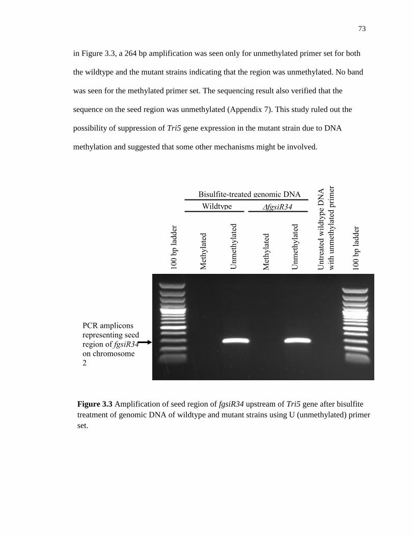
73
in Figure 3.3, a 264 bp amplification was seen only for unmethylated primer set for both
the wildtype and the mutant strains indicating that the region was unmethylated. No band
was seen for the methylated primer set. The sequencing result also verified that the
sequence on the seed region was unmethylated (Appendix 7). This study ruled out the
possibility of suppression of Tri5 gene expression in the mutant strain due to DNA
methylation and suggested that some other mechanisms might be involved.
100 b
p l
adder
Wildtype fgsiR34
+
Untr
eate
d w
ildty
pe
DN
A
wit
h u
nm
eth
yla
ted p
rim
er
100 b
p l
adder
Bisulfite-treated genomic DNA M
eth
yla
ted
pri
mer
Unm
eth
yla
ted
pri
mer
Met
hyla
ted
pri
mer
Unm
eth
yla
ted
pri
mer
PCR amplicons
representing seed
region of fgsiR34
on chromosome
2
Figure 3.3 Amplification of seed region of fgsiR34 upstream of Tri5 gene after bisulfite
treatment of genomic DNA of wildtype and mutant strains using U (unmethylated) primer
set.

74
3.3.3 Upregulation of non-coding transcript in the ΔfgsiR34+ relative to the
wildtype strain
Using RT-qPCR assay, we studied the presence and the accumulation of the non-
coding RNA transcribed from the F. graminearum chromosomal region between Tri6 and
Tri5 that hosts the fgsiR34 target. As shown in Figure 3.4, our data first demonstrated that
non-coding RNA was transcribed from this region. It further showed a significant larger
accumulation of this non-coding RNA transcript in the ΔfgsiR34+ strain compared to the
wildtype strain. The first fragment was upregulated by around 2.1 folds while the second
fragment was upregulated by around 7.4 folds.
Figure 3.4 Graph showing fold change in accumulation of the non-coding RNA
transcripts between the wildtype and the ΔfgsiR34+ mutant strains. The
significance level was measured at p≤0.05 (*).
*
*
0
1
2
3
4
5
6
7
8
9
Fragment 1 Fragment 2
Fold
chan
ge
in a
ccum
ula
tion o
f
non-c
odin
g R
NA
tra
nsc
ript
(Δfg
siR
34+
vs
wt)

75
3.4 Discussion
In this study, we analyzed the methylation pattern of the seed region of silencing
RNA fgsiR34 at nearly 1000 bp upstream of Tri5 gene to elucidate if methylation has any
role in down-regulating the expression of trichothecene genes, the genes that are involved
in mycotoxin biosynthesis in F. graminearum. Since DNA methylation suppresses
transcription, it plays an important role in gene silencing and inactivation. DNA
methylation usually occurs on the fifth carbon of the cytosine residue (5-methyl
cytosine), particularly at CG-rich regions of the genome. The presence of methylated
cytosine base directly interferes with the binding of transcription factor to DNA, and
therefore represses the expression of the corresponding gene (Prendergast and Ziff 1991;
Clark et al. 1997). Alternately, methylcytosine-binding proteins may hinder the passage
of regulatory elements to the corresponding gene sequence (Yu et al. 2000).
Sodium bisulfite modification of DNA is one of the most important methods to
determine the methylation status of individual cytosine residues. When genomic DNA is
subjected to sodium bisulfite, unmethylated cytosine residues are deaminated and
converted to uracil, while methylated cytosines are unaffected by the bisulfite treatment
since the methyl group at their 5-carbon atom prevents deamination. This treatment
brings about a change in the DNA sequence depending on the methylation status of
individual cytosine base (Frommer et al. 1992). Analyzing bisulfite treated DNA with
PCR that uses two specific primer pairs, methylated primer pair that recognizes
methylated CG region and unmethylated primer pair that recognizes unmethylated CG
region, helps to differentiate pattern of individual cytosine methylation in the gene of
interest (Herman et al. 1996).

76
To study the methylation pattern at the seed sequence upstream of Tri5 gene, we
used two strains of F. graminearum- wildtype and ΔfgsiR34+. After bisulfite treatment
and MSP we could not find a differential methylation for the gene of interest between the
two strains. Also we identified the seed sequence of fgsiR34 to be unmethylated. This led
us to depict that there is no role of methylation in repressing the expression of Tri genes
and some other mechanisms might be involved in this downregulation.
A few studies have been made on DNA methylation in fungi (Antequera et al.
1984; Russel et al. 1985; Abbas et al. 1986; Russell et al. 1987; Buckner et al. 1988;
Reyna-Lopez et al. 1997; Martienssen and Colot 2001). There has been a report of
differential fungal methylation during morphogenesis in dimorphic fungi (Reyna-Lopez
et al. 1997). A study conducted by Antequera et al. (1984) on the incidence and the
pattern of cytosine methylation in fungi showed disparate distribution of cytosine
methylation ranging from undetectable levels (≤0.1%) to low but detectable levels (~0.2
to 0.5%) (Antequera et al. 1984). Proffitt and his colleagues failed to detect methylated
cytosines in the budding yeast Saccharomyces cerevisiae (Proffitt et al. 1984). However,
a slime mold, Physarum polycephalum was observed to be hyper-methylated (Evans and
Evans 1970; Evans et al. 1973; Whittaker and Hardman 1980; Peoples et al. 1985).
Moreover, Bull and Wotton reported a heavy methylation of amplified DNA in
transformants of the model fungus, Neurospora crassa, while the wildtype had very low
levels of cytosine methylation in its genome. Their results implicated a mechanism for a
significant de novo DNA methylation (Bull and Wootton 1984). Because lower
eukaryotes, such as fungi and Drosophila, lack significant methylation in their respective
genomes (Hattman et al. 1978; Urieli-Shoval et al. 1982; Feher et al. 1983; Achwal et al.

77
1984; Antequera et al. 1984; Proffitt et al. 1984), DNA methylation might not have an
extensive role to control gene expression (Doerfler 1983; Bird 1984). Nevertheless, they
might be involved in DNA recombination or repair process (Bull and Wootton 1984).
Recent studies have shown that non-coding RNAs play a significant role not only
in gene silencing mechanism by degrading the cognate mRNA sequences via RNAi (Fire
et al. 1998; Elbashir et al. 2001), but also serve as activators of gene expression by
targeting gene regulatory sequences. This novel phenomenon was first discovered by Li
et al in 2006 and is referred to as RNA activation (RNAa) (Li et al. 2006; Place et al.
2008; Lopez et al. 2016; Meng et al. 2016). Several small RNAs, including short double
stranded RNAs (dsRNAs), small hairpin RNAs (shRNAs), and micro RNAs (miRNAs),
have been shown to positively regulate target sequences and are called small activating
RNAs (saRNAs) (Li et al. 2006; Place et al. 2008; Turunen et al. 2009; Huang et al.
2012). The target sequences for RNAa in most of the cases are sense and antisense non-
coding sequences (Schwartz et al. 2008; Matsui et al. 2010; Matsui et al. 2013). The non-
coding RNAs thus activate gene transcription by targeting non-coding regulatory regions
in the promoters of the gene (Li et al. 2006).
Our result demonstrated that a non-coding RNA is transcribed out of the region
between Tri6 and Tri5 on chromosome 2 in F. graminearum where fgsiR34 potentially
targets. At this moment, we do not know the exact function of this non-coding transcript.
Nor do we know why the two fragments of this non-coding transcript we assayed had a
differential accumulation in the ΔfgsiR34+ mutant. However, the significant larger
increase in accumulation of this transcript in the ΔfgsiR34+ strain than in the wildtype
(Figure 3.4) hinted at its possible functional link with fgsiR34. Moreover, this might

78
suggest the existence of multiple non-coding transcripts made from the same region.
Further research is needed to get a better picture.
Therefore, we hypothesized that siRNA, fgsiR34, might be acting as an saRNA
and activating promoter activity of the gene nearby its seed site by binding to or targeting
at the antisense transcript, which results in a functional promoter for gene transcription.
The significantly higher accumulation of this transcript in the ΔfgsiR34+ mutant relative
to the wildtype and the differential accumulation of different fragments of this transcript
in the mutant might suggest a more complex nature of fgsiR34 in regulating gene
expression. Identifying natural targets induced by this siRNA and studying their gene
expression profiles might give a clear idea on the regulation mechanism of this siRNA.

79
Chapter 4 General Conclusions and Future Perspectives
4.1 General conclusions
Fusarium head blight (FHB) is a devastating disease of wheat, and other small
cereal grains occurring in all major-cereal growing areas of the world. The disease is so
severe that it results in losses of grain yields and quality, and contaminates grains with
harmful mycotoxins (McMullen et al. 1997). Globally, Fusarium graminearum is the
most predominant and problematic species of Fusarium. The effective strategy to control
FHB is a high priority to assure the safety of harvested grains for food and feed
consumption as well as for fermentation purposes (Urban and Hammond-Kosack 2013).
The long-term goal of our research is to advance our understanding of the
molecular mechanisms of host-pathogen interaction during FHB of wheat. In particular,
we studied the role of silencing RNA, fgsiR34 in trichothecene biosynthesis pathway. In
our previous study, when DCL-2 gene was knocked down in F. graminearum isolate 4
(Fg4), fgsiR34 was the only siRNA that was found to be significantly downregulated in
the Δdcl2- mutant. At the same time, all Tri genes on chromosome 2 that we examined
were also significantly downregulated, leading to suppression of DON production in the
Δdcl2- mutant inoculated FHB susceptible wheat cultivar Y1193-6 (Galla 2014).
Therefore, we hypothesized that fgsiR34 positively regulates the Tri gene core on
chromosome 2.
To test our hypothesis, we successfully generated an Fg4 mutant that
overexpressed fgsiR34 (ΔfgsiR34+) and studied it in culture and in planta with
comparison to the wildtype Fg4. Our data showed that the ΔfgsiR34+ mutant generally

80
behaves similarly as the Δdcl2- mutant both in culture and in planta. Specifically, the Tri
genes tested were downregulated in both mutants, the genes encoding CWDEs also
behaved similarly in both the mutants, and both mutants had significantly reduced FHB
pathogenicity and grew poorly in either FHB-resistant or FHB-susceptible wheat
cultivars. These observations suggest that fgsiR34 was responsible for all of the gene
expression changes that we observed in the Δdcl2- mutant, playing a critical role in
regulating Tri genes and thus DON biosynthesis in F. graminearum. This is not
surprising since the only target site by fgsiR34 in F. graminearum genome lies between
Tri6 and Tri5 genes on chromosome 2. What surprised us is that our data from the
ΔfgsiR34+ mutant does not support our hypothesis developed from our analysis of the
Δdcl2- mutant but indicates a more complex mechanism of this regulation. Our data
suggest that methylation does not play a role in the fgsiR34’s regulation of Tri genes and
that the fgsiR34 target region seems to transcribe non-coding RNAs. More surprisingly,
these non-coding RNAs seemed to differentially respond to fgsiR34 overexpression.
Therefore, more research is needed to find out if these non-coding RNAs also have
different roles in the regulatory mechanism of fgsiR34.
NCBI BLAST hit shows that the coding sequence of fgsiR34 is two 391-bp
repeated sequences in the F. graminearum mitochondrial genome (Appendix 1).
fgsiR34’s coding sequence is also present in the mitochondrial genomes of other
Fusarium spp.: - one in F. oxysporum, two in F. culmorum, and three in F. gerlachii.
There are multiple questions that need to be answered to fully comprehend the role of
fgsiR34 in FHB pathogenesis in wheat. Does F. graminearum have a copy of its

81
mitochondrial genome in its nuclear genome? Otherwise, how does the biogenesis of
mitochondrial small RNAs occur? How does the small RNA get into the cytosol?
Further research on the role of fgsiR34 should expand the knowledge that we
currently have. Southern blotting analysis should reveal the patterns of integration of the
inverted repeat transgenes (IRTs) in the transformants because the integration patterns
also impact the gene expression levels in the mutant strains as demonstrated by Scherm et
al. (Scherm et al. 2011). Whole transcriptome analysis of the mutant and the wildtype
strains should enable the characterization of all RNA transcripts and reveal the
differentially regulated genes in both the strains. Electrophoretic mobility shift assay
(EMSA) should reveal the action of fgsiR34 in the functioning of Tri6 gene. We could
generate an fgsiR34 knockout mutant using CRISPR (clustered regularly inter-spaced
palindromic repeats)/Cas9 genome editing technology. Since we already have knocked
down and overexpressed mutants of fgsiR34, knockout mutant of fgsiR34 might help us
to conduct neat assays, although knocking out a gene might sometimes be lethal.

82
References
Abbas, H., Mirocha, C. and Tuite, J. (1986) Natural occurrence of deoxynivalenol, 15-
acetyl-deoxynivalenol, and zearalenone in refusal factor corn stored since 1972.
Applied and environmental microbiology 51, 841-843.
Achwal, C., Ganguly, P. and Chandra, H.S. (1984) Estimation of the amount of 5-
methylcytosine in Drosophila melanogaster DNA by amplified ELISA and
photoacoustic spectroscopy. The EMBO journal 3, 263.
Alexander, N., McCormick, S. and Hohn, T. (1999) TRI12, a trichothecene efflux pump
from Fusarium sporotrichioides: gene isolation and expression in yeast.
Molecular and General Genetics MGG 261, 977-984.
Alexander, N.J., McCormick, S.P., Larson, T.M. and Jurgenson, J.E. (2004) Expression
of Tri15 in Fusarium sporotrichioides. Current genetics 45, 157-162.
Alexander, N.J., Proctor, R.H. and McCormick, S.P. (2009) Genes, gene clusters, and
biosynthesis of trichothecenes and fumonisins in Fusarium. Toxin reviews 28,
198-215.
Ambros, V. (2004) The functions of animal microRNAs. Nature 431, 350-355.
Ambros, V., Bartel, B., Bartel, D.P., Burge, C.B., Carrington, J.C., Chen, X., Dreyfuss,
G., Eddy, S.R., Griffiths-Jones, S. and Marshall, M. (2003) A uniform system for
microRNA annotation. Rna 9, 277-279.
An, H.J., Lurie, S., Greve, L.C., Rosenquist, D., Kirmiz, C., Labavitch, J.M. and Lebrilla,
C.B. (2005) Determination of pathogen-related enzyme action by mass
spectrometry analysis of pectin breakdown products of plant cell walls.
Analytical biochemistry 338, 71-82.

83
Andersen, A. (1948) The development of Gibberella zeae headblight of wheat.
Phytopathology 38, 595-611.
Anderson, J.A., Stack, R., Liu, S., Waldron, B., Fjeld, A., Coyne, C., Moreno-Sevilla, B.,
Fetch, J.M., Song, Q. and Cregan, P. (2001) DNA markers for Fusarium head
blight resistance QTLs in two wheat populations. Theoretical and Applied
Genetics 102, 1164-1168.
Annis, S.L. and Goodwin, P.H. (1997) Recent advances in the molecular genetics of plant
cell wall-degrading enzymes produced by plant pathogenic fungi. European
Journal of Plant Pathology 103, 1-14.
Antequera, F., Boyes, J. and Bird, A. (1990) High levels of de novo methylation and
altered chromatin structure at CpG islands in cell lines. Cell 62, 503-514.
Antequera, F., Tamame, M., Villanueva, J. and Santos, T. (1984) DNA methylation in the
fungi. Journal of Biological Chemistry 259, 8033-8036.
Aramayo, R. and Metzenberg, R.L. (1996) Meiotic transvection in fungi. Cell 86, 103-
113.
Arthur, J.C. (1891) Wheat Scab. in: Purdue Univeristy Agricultural Experimet Station
Bulletin Purdue University.
Atanasoff, D. (1920) Fusarium-blight (scab) of heat and other cereals. Journal of
Agricultural Research 20 (1), 1-40.
Bai, G.-H., Desjardins, A. and Plattner, R. (2002) Deoxynivalenol-nonproducing
Fusarium graminearum causes initial infection, but does not cause DiseaseSpread
in wheat spikes. Mycopathologia 153, 91-98.

84
Bai, G. and Shaner, G. (1994) Wheat Scab: perspective and control. Plant Dis 78, 760-
766.
Bai, G. and Shaner, G. (2004) Management and resistance in wheat and barley to
Fusarium head blight 1. Annual review of phytopathology 42, 135-161.
Bai, G., Zhou, C., Qian, C. and Ge, Y. (1989) A study on scab resistance in new wheat
cultivars and advanced breeding lines. Jiangsu Agric Sci 7, 20-22.
Bai, G.H., Plattner, R., Desjardins, A. and Kolb, F. (2001) Resistance to Fusarium head
blight and deoxynivalenol accumulation in wheat. Plant Breeding 120, 1-6.
Balconi, C., Lanzanova, C., Conti, E., Triulzi, T., Forlani, F., Cattaneo, M. and Lupotto,
E. (2007) Fusarium head blight evaluation in wheat transgenic plants expressing
the maize b-32 antifungal gene. European Journal of Plant Pathology 117, 129-
140.
Bartel, D.P. (2004) MicroRNAs: genomics, biogenesis, mechanism, and function. cell
116, 281-297.
Bartel, D.P. (2009) MicroRNAs: target recognition and regulatory functions. cell 136,
215-233.
Basnet, B.R., Glover, K.D., Ibrahim, A.M., Yen, Y. and Chao, S. (2012) A QTL on
chromosome 2DS of ‘Sumai 3’increases susceptibility to Fusarium head blight in
wheat. Euphytica 186, 91-101.
Bennett, F. (1931) Gibberella Saubinetii (Mont.) Sacc. on British Cereals: II.
Physiological AKD Pathological Studies. Annals of Applied Biology 18, 158-
177.

85
Bennett, J.W. and Klich, M. (2003) Mycotoxins. Clinical Microbiology Reviews 16, 497-
516.
Bernstein, E. and Allis, C.D. (2005) RNA meets chromatin. Genes & development 19,
1635-1655.
Bernstein, E., Caudy, A.A., Hammond, S.M. and Hannon, G.J. (2001) Role for a
bidentate ribonuclease in the initiation step of RNA interference. Nature 409,
363-366.
Bird, A. (1992) The essentials of DNA methylation. Cell 70, 5-8.
Bird, A.P. (1984) DNA methylation--how important in gene control? Nature 307, 503.
Bird, A.P. (1985) CpG-rich islands and the function of DNA methylation. Nature 321,
209-213.
Birzele, B., Prange, A. and KrÄmer, J. (2000) Deoxynivalenol and ochratoxin A in
German wheat and changes of level in relation to storage parameters. Food
Additives & Contaminants 17, 1027-1035.
Blandino, M., Haidukowski, M., Pascale, M., Plizzari, L., Scudellari, D. and Reyneri, A.
(2012) Integrated strategies for the control of Fusarium head blight and
deoxynivalenol contamination in winter wheat. Field Crops Research 133, 139-
149.
Boosalis, M.G., Doupnik Jr, B.L., Wysong, D.S. and Watkins, J.E. (1983) The wheat
scab epidemic of 1982 [Fusarium graminearum, Nebraska]. Farm Ranch and
Home Quarterly Nebraska Agricultural Experiment Station.

86
Bourdoncle, W. and Ohm, H. (2003) Quantitative trait loci for resistance to Fusarium
head blight in recombinant inbred wheat lines from the cross Huapei 57-
2/Patterson. Euphytica 131, 131-136.
Bowen, J.K., Templeton, M.D., Sharrock, K.R., Crowhurst, R.N. and Rikkerink, E.H.
(1995) Gene inactivation in the plant pathogen Glomerella cingulata: three
strategies for the disruption of the pectin lyase gene pnIA. Molecular and
General Genetics MGG 246, 196-205.
Branco, M.R., Ficz, G. and Reik, W. (2012) Uncovering the role of 5-
hydroxymethylcytosine in the epigenome. Nature Reviews Genetics 13, 7-13.
Brown, D.W., McCormick, S.P., Alexander, N.J., Proctor, R.H. and Desjardins, A.E.
(2001) A genetic and biochemical approach to study trichothecene diversity in
Fusarium sporotrichioides and Fusarium graminearum. Fungal Genetics and
Biology 32, 121-133.
Brown, D.W., McCormick, S.P., Alexander, N.J., Proctor, R.H. and Desjardins, A.E.
(2002) Inactivation of a cytochrome P-450 is a determinant of trichothecene
diversity in Fusarium species. Fungal Genetics and Biology 36, 224-233.
Brown, D.W., Proctor, R.H., Dyer, R.B. and Plattner, R.D. (2003) Characterization of a
Fusarium 2-gene cluster involved in trichothecene C-8 modification. Journal of
agricultural and food chemistry 51, 7936-7944.
Buckner, B., Novotny, C.P. and Ullrich, R.C. (1988) Developmental regulation of the
methylation of the ribosomal DNA in the basidiomycete fungusSchizophyllum
commune. Current genetics 14, 105-111.

87
Buerstmayr, H., Ban, T. and Anderson, J.A. (2009) QTL mapping and marker‐assisted
selection for Fusarium head blight resistance in wheat: a review. Plant breeding
128, 1-26.
Buerstmayr, H., Lemmens, M., Hartl, L., Doldi, L., Steiner, B., Stierschneider, M. and
Ruckenbauer, P. (2002) Molecular mapping of QTLs for Fusarium head blight
resistance in spring wheat. I. Resistance to fungal spread (Type II resistance).
Theoretical and Applied Genetics 104, 84-91.
Buerstmayr, H., Steiner, B., Hartl, L., Griesser, M., Angerer, N., Lengauer, D., Miedaner,
T., Schneider, B. and Lemmens, M. (2003) Molecular mapping of QTLs for
Fusarium head blight resistance in spring wheat. II. Resistance to fungal
penetration and spread. Theoretical and Applied Genetics 107, 503-508.
Bühler, M. and Moazed, D. (2007) Transcription and RNAi in heterochromatic gene
silencing. Nature structural & molecular biology 14, 1041-1048.
Bull, J.H. and Wootton, J.C. (1984) Heavily methylated amplified DNA in transformants
of Neurospora crassa.
Busch, R. (1995) Breeding for scab resistance. Regional Scab Forum Minnesota Wheat
Research and Promotion Council, Red Lake Falls.
Bushnell, W., Hazen, B., Pritsch, C. and Leonard, K. (2003) Histology and physiology of
Fusarium head blight. Fusarium head blight of wheat and barley, 44-83.
Bushnell, W.R. (2002) Designating types of scab resistance: A discussion. Page 200 in:
Proceedings of 2002 National Fusarium Head Blight Forum, S. M. Canty, J.
Lewis, L. Siler and R. W. Ward, eds. Michigan State University, Erlanger, KY.

88
Carthew, R.W. and Sontheimer, E.J. (2009) Origins and mechanisms of miRNAs and
siRNAs. Cell 136, 642-655.
Cast, I. (2003) Mycotoxins: Risks in Plant, Animal and Human Systems. CAST Report
139, 4-29.
Castel, S.E. and Martienssen, R.A. (2013) RNA interference in the nucleus: roles for
small RNAs in transcription, epigenetics and beyond. Nature Reviews Genetics
14, 100-112.
Catalanotto, C., Pallotta, M., ReFalo, P., Sachs, M.S., Vayssie, L., Macino, G. and
Cogoni, C. (2004) Redundancy of the two dicer genes in transgene-induced
posttranscriptional gene silencing in Neurospora crassa. Molecular and cellular
biology 24, 2536-2545.
Chandler, E.A., Simpson, D.R., Thomsett, M.A. and Nicholson, P. (2003) Development
of PCR assays to Tri7 and Tri13 trichothecene biosynthetic genes, and
characterisation of chemotypes of Fusarium graminearum, Fusarium culmorum
and Fusarium cerealis. Physiological and Molecular Plant Pathology 62, 355-
367.
Chen, W., Chen, P., Liu, D., Kynast, R., Friebe, B., Velazhahan, R., Muthukrishnan, S.
and Gill, B. (1999) Development of wheat scab symptoms is delayed in
transgenic wheat plants that constitutively express a rice thaumatin-like protein
gene. Theoretical and Applied Genetics 99, 755-760.
Chen, X. (2009) Small RNAs and their roles in plant development. Annual Review of
Cell and Developmental 25, 21-44.
89
Cheng, M., Fry, J.E., Pang, S., Zhou, H., Hironaka, C.M., Duncan, D.R., Conner, T.W.
and Wan, Y. (1997) Genetic transformation of wheat mediated by
Agrobacterium tumefaciens. Plant Physiology 115, 971-980.
Chicas, A., Forrest, E.C., Sepich, S., Cogoni, C. and Macino, G. (2005) Small interfering
RNAs that trigger posttranscriptional gene silencing are not required for the
histone H3 Lys9 methylation necessary for transgenic tandem repeat stabilization
in Neurospora crassa. Molecular and cellular biology 25, 3793-3801.
Christensen, J.J., Stakman, E.C. and Immer, F.R. (1929) Susceptibility of wheat varieties
and hybrids to fusarial head blight in Minnesota.
Clark, S.J., Harrison, J. and Molloy, P.L. (1997) Sp1 binding is inhibited by m Cp m CpG
methylation. Gene 195, 67-71.
Cogoni, C. and Macino, G. (1999a) Gene silencing in Neurospora crassa requires a
protein homologous to RNA-dependent RNA polymerase. Nature 399, 166-169.
Cogoni, C. and Macino, G. (1999b) Posttranscriptional gene silencing in Neurospora by a
RecQ DNA helicase. Science 286, 2342-2344.
Cowger, C. and Sutton, A.L. (2005) The southeastern US Fusarium head blight epidemic
of 2003. Plant Health Progress 10.
Cuomo, C.A., Güldener, U., Xu, J.-R., Trail, F., Turgeon, B.G., Di Pietro, A., Walton,
J.D., Ma, L.-J., Baker, S.E. and Rep, M. (2007) The Fusarium graminearum
genome reveals a link between localized polymorphism and pathogen
specialization. Science 317, 1400-1402.
90
Curradi, M., Izzo, A., Badaracco, G. and Landsberger, N. (2002) Molecular mechanisms
of gene silencing mediated by DNA methylation. Molecular and cellular biology
22, 3157-3173.
Cuthbert, P.A., Somers, D.J. and Brulé-Babel, A. (2007) Mapping of Fhb2 on
chromosome 6BS: a gene controlling Fusarium head blight field resistance in
bread wheat (Triticum aestivum L.). Theoretical and Applied Genetics 114, 429-
437.
Cuzick, A., Urban, M. and Hammond‐Kosack, K. (2008) Fusarium graminearum gene
deletion mutants map1 and tri5 reveal similarities and differences in the
pathogenicity requirements to cause disease on Arabidopsis and wheat floral
tissue. New Phytologist 177, 990-1000.
Da Luz, W., Stockwell, C. and Bergstrom, C. (2003) Biological control of Fusarium
graminearum. In: Leonard, K.J., Bushnell,W.R. (Eds.), Fusarium Head Blight of
Wheat and Barley. Press, St. Paul, MN.
. American Phytopathological Society, 381-394.
Dang, Y., Yang, Q., Xue, Z. and Liu, Y. (2011) RNA interference in fungi: pathways,
functions, and applications. Eukaryotic cell 10, 1148-1155.
de-Galich, M. (1997) Fusarium head blight in Argentina. Fusarium head scab: Global
status and future prospects 1997, 19-28.
De Lorenzo, G., Castoria, R., Bellincampi, D. and Cervone, F. (1997) Fungal invasion
enzymes and their inhibition. In Plant Relationships. pp.61-83: Springer.

91
de Vries, R.P. and Visser, J. (2001) Aspergillus enzymes involved in degradation of plant
cell wall polysaccharides. Microbiology and molecular biology reviews 65, 497-
522.
Desjardins, A., Proctor, R., McCormick, S. and Hohn, T. (1997) Reduced virulence of
trichothecene antibiotic-nonproducing mutants of Gibberella zeae in wheat field
tests. Fusarium Head Scab: Global Status and Future Prospects; Proceedings of a
Workshop; El Batan, Mexico; 13-17 Oct 1996. In ^ TFusarium Head Scab:
Global Status and Future Prospects; Proceedings of a Workshop; El Batan,
Mexico; 13-17 Oct 1996^ ADubin, HJ Gilchrist, L Reeves, J McNab, A^
AMexico, DF (Mexico)^ BCIMMYT^ C1997: Centro Internacional de
Mejoramiento de Maiz y Trigo (CIMMYT), Mexico DF (Mexico).
Desjardins, A.E. (2006) Fusarium mycotoxins: chemistry, genetics, and biology:
American Phytopathological Society (APS Press).
Desjardins, A.E. and Hohn, T.M. (1997) Mycotoxins in plant pathogenesis. Molecular
Plant-Microbe Interactions 10, 147-152.
Desjardins, A.E., Hohn, T.M. and McCORMICK, S.P. (1993) Trichothecene biosynthesis
in Fusarium species: chemistry, genetics, and significance. Microbiological
reviews 57, 595-604.
Desjardins, A.E., Proctor, R.H., Bai, G., McCormick, S.P., Shaner, G., Buechley, G. and
Hohn, T.M. (1996) Reduced virulence of trichothecene-nonproducing mutants of
Gibberella zeae in wheat field tests. MPMI-Molecular Plant Microbe Interactions
9, 775-781.

92
Desmond, O.J., Manners, J.M., Stephens, A.E., Maclean, D.J., Schenk, P.M., Gardiner,
D.M., Munn, A.L. and Kazan, K. (2008) The Fusarium mycotoxin
deoxynivalenol elicits hydrogen peroxide production, programmed cell death and
defence responses in wheat. Molecular Plant Pathology 9, 435-445.
Detmers, F. (1892) Scab of Wheat. Ohio Agricultural Experiment Station Bulletin, 147-
149.
Dexter, J., Nowicki, T., Leonard, K. and Bushnell, W. (2003) Safety assurance and
quality assurance issues associated with Fusarium head blight in wheat.
Fusarium head blight of wheat and barley, 420-460.
Dickson, J.G. and Mains, E.B. (1929) Scab of wheat and barley and its control. in: USDA
Farmer's Bulletin, 1-17.
Doerfler, W. (1983) DNA Methylation and Cene Activity. Annual review of
biochemistry 52, 93-124.
Donald, T., Shoshannah, R., Deyrup, S.T. and Gloer, J.B. (2005) A protective endophyte
of maize: Acremonium zeae antibiotics inhibitory to Aspergillus flavus and
Fusarium verticillioides. Mycological Research 109, 610-618.
Drinnenberg, I.A., Weinberg, D.E., Xie, K.T., Mower, J.P., Wolfe, K.H., Fink, G.R. and
Bartel, D.P. (2009) RNAi in budding yeast. Science 326, 544-550.
Dyer, R.B., Plattner, R.D., Kendra, D.F. and Brown, D.W. (2005) Fusarium graminearum
TRI14 is required for high virulence and DON production on wheat but not for
DON synthesis in vitro. Journal of agricultural and food chemistry 53, 9281-
9287.

93
Ehrlich, K.C. and Daigle, K.W. (1987) Protein synthesis inhibition by 8-oxo-12, 13-
epoxytrichothecenes. Biochimica et Biophysica Acta (BBA)-General Subjects
923, 206-213.
Elbashir, S.M., Lendeckel, W. and Tuschl, T. (2001) RNA interference is mediated by
21-and 22-nucleotide RNAs. Genes & development 15, 188-200.
Evans, H.H. and Evans, T.E. (1970) Methylation of the deoxyribonucleic acid of
Physarum polycephalum at various periods during the mitotic cycle. Journal of
Biological Chemistry 245, 6436-6441.
Evans, H.H., Evans, T.E. and Littman, S. (1973) Methylation of parental and progeny
DNA strands in Physarum polycephalum. Journal of molecular biology 74, 563-
572.
Feher, Z., Kiss, A. and Venetianer, P. (1983) Expression of a bacterial modification
methylase gene in yeast.
Feng, J., Liu, G., Selvaraj, G., Hughes, G.R. and Wei, Y. (2005) A secreted lipase
encoded by LIP1 is necessary for efficient use of saturated triglyceride lipids in
Fusarium graminearum. Microbiology 151, 3911-3921.
Ferrari, S., Sella, L., Janni, M., De Lorenzo, G., Favaron, F. and D’Ovidio, R. (2012)
Transgenic expression of polygalacturonase‐inhibiting proteins in Arabidopsis
and wheat increases resistance to the flower pathogen Fusarium graminearum.
Plant Biology 14, 31-38.
Fire, A., Xu, S., Montgomery, M.K., Kostas, S.A., Driver, S.E. and Mello, C.C. (1998)
Potent and specific genetic interference by double-stranded RNA in
Caenorhabditis elegans. Nature 391, 806-811.

94
Fitzgerald, A., van Kan, J.A. and Plummer, K.M. (2004) Simultaneous silencing of
multiple genes in the apple scab fungus, Venturia inaequalis, by expression of
RNA with chimeric inverted repeats. Fungal Genetics and Biology 41, 963-971.
Freitag, M., Lee, D.W., Kothe, G.O., Pratt, R.J., Aramayo, R. and Selker, E.U. (2004)
DNA methylation is independent of RNA interference in Neurospora. Science
304, 1939-1939.
Frommer, M., McDonald, L.E., Millar, D.S., Collis, C.M., Watt, F., Grigg, G.W.,
Molloy, P.L. and Paul, C.L. (1992) A genomic sequencing protocol that yields a
positive display of 5-methylcytosine residues in individual DNA strands.
Proceedings of the National Academy of Sciences 89, 1827-1831.
Fulci, V. and Macino, G. (2007) Quelling: post-transcriptional gene silencing guided by
small RNAs in Neurospora crassa. Current opinion in microbiology 10, 199-203.
Gale, L., Leonard, K. and Bushnell, W. (2003) Population biology of Fusarium species
causing head blight of grain crops. Fusarium head blight of wheat and barley,
120-143.
Gale, L.R., Bryant, J., Calvo, S., Giese, H., Katan, T., O'Donnell, K., Suga, H., Taga, M.,
Usgaard, T.R. and Ward, T.J. (2005) Chromosome complement of the fungal
plant pathogen Fusarium graminearum based on genetic and physical mapping
and cytological observations. Genetics 171, 985-1001.
Galla, A. (2014) Role of Fusarium graminearum sRNAs in Fusarium head blight
pathogenesis on wheat. Ph.D. South Dakota State University.
Ghildiyal, M. and Zamore, P.D. (2009) Small silencing RNAs: an expanding universe.
Nature Reviews Genetics 10, 94-108.

95
Gilbert, J., Fedak, G., Procunier, J., Aung, T. and Tekauz, A. (1997) Strategies for
breeding for resistance to Fusarium head blight in Canadian spring wheat.
Fusarium head scab: global status and future prospects CIMMYT, DF, Mexico,
47-51.
Gilbert, J. and Fernando, W. (2004) Epidemiology and biological control of Gibberella
zeae/Fusarium graminearum. Canadian Journal of Plant Pathology 26, 464-472.
Gilbert, J. and Tekauz, A. (2000) Review: recent developments in research on Fusarium
head blight of wheat in Canada. Canadian Journal of Plant Pathology 22, 1-8.
Gilbert, J., Tekauz, A., Kaethler, R., Mueller, E. and Kromer, U. (1995) Occurrence of
Fusarium head blight in Manitoba in 1994. Canadian plant disease survey
(Canada).
Gledhill, L., Hesketh, A.R., Bycroft, B.W., Dewick, P.M. and Gilbert, J. (1991)
Biosynthesis of trichothecene mycotoxins: cell-free epoxidation of a trichodiene
derivative. FEMS microbiology letters 81, 241-245.
Gordon, W. (1959) The occurrence of Fusarium species in Canada: VI. taxonomy and
geographic distribution of Fusarium species on plants, insects, and fungi.
Canadian Journal of Botany 37, 257-290.
Goswami, R.S. and Kistler, H.C. (2004) Heading for disaster: Fusarium graminearum on
cereal crops. Molecular plant pathology 5, 515-525.
Greenhalgh, R., Neish, G. and Miller, J. (1983) Deoxynivalenol, acetyl deoxynivalenol,
and zearalenone formation by Canadian isolates of Fusarium graminearum on
solid substrates. Applied and environmental microbiology 46, 625-629.

96
Hallen-Adams, H.E., Cavinder, B.L. and Trail, F. (2011) Fusarium graminearum from
expression analysis to functional assays. Fungal Genomics: Methods and
Protocols, 79-101.
Hamer, J.E. and Holden, D.W. (1997) Linking approaches in the study of fungal
pathogenesis: a commentary. Fungal Genetics and Biology 21, 11-16.
Hammond, S.M., Boettcher, S., Caudy, A.A., Kobayashi, R. and Hannon, G.J. (2001)
Argonaute2, a link between genetic and biochemical analyses of RNAi. Science
293, 1146-1150.
Hammond, T.M. and Keller, N.P. (2005) RNA silencing in Aspergillus nidulans is
independent of RNA-dependent RNA polymerases. Genetics 169, 607-617.
Hannon, G.J. (2002) RNA interference. Nature 418, 244-251.
Harris, L., Desjardins, A.E., Plattner, R., Nicholson, P., Butler, G., Young, J., Weston,
G., Proctor, R. and Hohn, T. (1999) Possible role of trichothecene mycotoxins in
virulence of Fusarium graminearum on maize. Plant Disease 83, 954-960.
Hattman, S., Kenny, C., Berger, L. and Pratt, K. (1978) Comparative study of DNA
methylation in three unicellular eucaryotes. Journal of bacteriology 135, 1156-
1157.
Havliš, J. and Trbušek, M. (2002) 5-Methylcytosine as a marker for the monitoring of
DNA methylation. Journal of Chromatography B 781, 373-392.
Herman, J.G., Graff, J.R., Myöhänen, S., Nelkin, B.D. and Baylin, S.B. (1996)
Methylation-specific PCR: a novel PCR assay for methylation status of CpG
islands. Proceedings of the National Academy of Sciences 93, 9821-9826.

97
Hohn, T.M. and Beremand, P.D. (1989) Isolation and nucleotide sequence of a
sesquiterpene cyclase gene from the trichothecene-producing fungus Fusarium
sporotrichioides. Gene 79, 131-138.
Hohn, T.M. and Desjardins, A.E. (1992) Isolation and gene disruption of the Tox5 gene
encoding trichodiene synthase in Gibberella pulicaris. Mol Plant-Microbe
Interact 5, 249-256.
Hohn, T.M., Krishna, R. and Proctor, R.H. (1999) Characterization of a transcriptional
activator controlling trichothecene toxin biosynthesis. Fungal Genetics and
Biology 26, 224-235.
Hohn, T.M. and Vanmiddlesworth, F. (1986) Purification and characterization of the
sesquiterpene cyclase trichodiene synthetase from Fusarium sporotrichioides.
Archives of biochemistry and biophysics 251, 756-761.
Holliday, R. and Grigg, G. (1993) DNA methylation and mutation. Mutation
Research/Fundamental and Molecular Mechanisms of Mutagenesis 285, 61-67.
Homdork, S. and Beck, R. (2000) Influence of Different Storage Conditions on the
Mycotoxin Production and Quality of Fusarium‐Infected Wheat Grain. Journal of
Phytopathology 148, 7-15.
Huang, V., Place, R.F., Portnoy, V., Wang, J., Qi, Z., Jia, Z., Yu, A., Shuman, M., Yu, J.
and Li, L.-C. (2012) Upregulation of Cyclin B1 by miRNA and its implications
in cancer. Nucleic acids research 40, 1695-1707.
Huang, Z., Bassil, C.F. and Murphy, S.K. (2013) Methylation-specific PCR. Ovarian
Cancer: Methods and Protocols, 75-82.

98
Hughes, D., Gahl, M., Graham, C. and Grieb, S. (1999) Overt signs of toxicity to dogs
and cats of dietary deoxynivalenol. Journal of animal science 77, 693-700.
Idnurm, A. and Howlett, B.J. (2001) Pathogenicity genes of phytopathogenic fungi.
Molecular Plant Pathology 2, 241-255.
Ilgen, P., Maier, F. and Schäfer, W. (2008) Trichothecenes and lipases are host-induced
and secreted virulence factors of Fusarium graminearum. Cereal Research
Communications 36, 421-428.
Inch, S. and Gilbert, J. (2003) The incidence of Fusarium species recovered from
inflorescences of wild grasses in southern Manitoba. Canadian Journal of Plant
Pathology 25, 379-383.
Iyengar, B.R., Choudhary, A., Sarangdhar, M.A., Venkatesh, K., Gadgil, C.J. and Pillai,
B. (2015) Non-coding RNA interact to regulate neuronal development and
function. Regulatory RNAs in the Nervous System, 102.
Jansen, C., Von Wettstein, D., Schäfer, W., Kogel, K.-H., Felk, A. and Maier, F.J. (2005)
Infection patterns in barley and wheat spikes inoculated with wild-type and
trichodiene synthase gene disrupted Fusarium graminearum. Proceedings of the
National Academy of Sciences of the United States of America 102, 16892-
16897.
Jenczmionka, N.J. and Schäfer, W. (2005) The Gpmk1 MAP kinase of Fusarium
graminearum regulates the induction of specific secreted enzymes. Current
genetics 47, 29-36.
Jochum, C., Osborne, L. and Yuen, G. (2006) Fusarium head blight biological control
with Lysobacter enzymogenes strain C3. Biological Control 39, 336-344.

99
Johnson, D., Flaskerud, G., Taylor, R., Satyanarayana, V., Leonard, K. and Bushnell, W.
(2003) Quantifying economic impacts of Fusarium head blight in wheat.
Fusarium head blight of wheat and barley, 461-483.
Jones, R. (2000) Assessments of Fusarium head blight of wheat and barley in response to
fungicide treatment. Plant Disease 84, 1021-1030.
Jullien, P.E. and Berger, F. (2010) DNA methylation reprogramming during plant sexual
reproduction? Trends in genetics 26, 394-399.
Jupe, E., Magill, J. and Magill, C. (1986) Stage-specific DNA methylation in a fungal
plant pathogen. Journal of bacteriology 165, 420-423.
Jurgenson, J., Bowden, R., Zeller, K., Leslie, J., Alexander, N. and Plattner, R. (2002) A
genetic map of Gibberella zeae (Fusarium graminearum). Genetics 160, 1451-
1460.
Kadotani, N., Nakayashiki, H., Tosa, Y. and Mayama, S. (2003) RNA silencing in the
phytopathogenic fungus Magnaporthe oryzae. Molecular plant-microbe
interactions 16, 769-776.
Kang, Z. and Buchenauer, H. (2000a) Cytology and ultrastructure of the infection of
wheat spikes by Fusarium culmorum. Mycological Research 104, 1083-1093.
Kang, Z. and Buchenauer, H. (2000b) Ultrastructural and cytochemical studies on
cellulose, xylan and pectin degradation in wheat spikes infected by Fusarium
culmorum. Journal of Phytopathology 148, 263-275.
Kang, Z., Zingen-Sell, I. and Buchenauer, H. (2005) Infection of wheat spikes by
Fusarium avenaceum and alterations of cell wall components in the infected
tissue. European Journal of Plant Pathology 111, 19-28.

100
Katiyar-Agarwal, S. and Jin, H. (2010) Role of small RNAs in host-microbe interactions.
Annual review of phytopathology 48, 225.
Kephart, K. (1991) Climatic conditions and regional differences. Virginia Cooperative
Extension Service (USA).
Khan, N., Schisler, D., Boehm, M., Lipps, P. and Slininger, P. (2004) Field testing of
antagonists of Fusarium head blight incited by Gibberella zeae. Biological
Control 29, 245-255.
Kimura, M., Kaneko, I., Komiyama, M., Takatsuki, A., Koshino, H., Yoneyama, K. and
Yamaguchi, I. (1998a) Trichothecene 3-O-acetyltransferase protects both the
producing organism and transformed yeast from related mycotoxins cloning and
characterization of Tri101. Journal of Biological Chemistry 273, 1654-1661.
Kimura, M., Shingu, Y., Yoneyama, K. and Yamaguchi, I. (1998b) Features of Tri101,
the trichothecene 3-O-acetyltransferase gene, related to the self-defense
mechanism in Fusarium graminearum. Bioscience, biotechnology, and
biochemistry 62, 1033-1036.
Kimura, M., Tokai, T., Matsumoto, G., Fujimura, M., Hamamoto, H., Yoneyama, K.,
Shibata, T. and Yamaguchi, I. (2003a) Trichothecene nonproducer Gibberella
species have both functional and nonfunctional 3-O-acetyltransferase genes.
Genetics 163, 677-684.
Kimura, M., Tokai, T., O’Donnell, K., Ward, T.J., Fujimura, M., Hamamoto, H., Shibata,
T. and Yamaguchi, I. (2003b) The trichothecene biosynthesis gene cluster of
Fusarium graminearum F15 contains a limited number of essential pathway
genes and expressed non‐essential genes. FEBS letters 539, 105-110.

101
King, R., Urban, M., Hammond-Kosack, M.C., Hassani-Pak, K. and Hammond-Kosack,
K.E. (2015) The completed genome sequence of the pathogenic ascomycete
fungus Fusarium graminearum. BMC genomics 16, 1.
Koh, K.P. and Rao, A. (2013) DNA methylation and methylcytosine oxidation in cell fate
decisions. Current opinion in cell biology 25, 152-161.
Kolb, F., Bai, G., Muehlbauer, G., Anderson, J., Smith, K. and Fedak, G. (2001) Host
plant resistance genes for Fusarium head blight: mapping and manipulation with
molecular markers. Crop Science 41, 611-619.
Kosiak, B., Torp, M., Skjerve, E. and Thrane, U. (2003) The prevalence and distribution
of Fusarium species in Norwegian cereals: a survey. Acta Agric Scand (B) 53,
168-176.
Krska, R., Baumgartner, S. and Josephs, R. (2001) The state-of-the-art in the analysis of
type-A and-B trichothecene mycotoxins in cereals. Fresenius' journal of
analytical chemistry 371, 285-299.
Lamb, C. and Dixon, R.A. (1997) The oxidative burst in plant disease resistance. Annual
review of plant biology 48, 251-275.
Laurie, J.D., Linning, R. and Bakkeren, G. (2008) Hallmarks of RNA silencing are found
in the smut fungus Ustilago hordei but not in its close relative Ustilago maydis.
Current genetics 53, 49-58.
Law, J.A. and Jacobsen, S.E. (2010) Establishing, maintaining and modifying DNA
methylation patterns in plants and animals. Nature Reviews Genetics 11, 204-
220.
102
Lee, H.-C., Chang, S.-S., Choudhary, S., Aalto, A.P., Maiti, M., Bamford, D.H. and Liu,
Y. (2009) qiRNA is a new type of small interfering RNA induced by DNA
damage. Nature 459, 274-277.
Lee, H.-C., Li, L., Gu, W., Xue, Z., Crosthwaite, S.K., Pertsemlidis, A., Lewis, Z.A.,
Freitag, M., Selker, E.U. and Mello, C.C. (2010) Diverse pathways generate
microRNA-like RNAs and Dicer-independent small interfering RNAs in fungi.
Molecular cell 38, 803-814.
Lee, J., Jurgenson, J.E., Leslie, J.F. and Bowden, R.L. (2008) Alignment of genetic and
physical maps of Gibberella zeae. Applied and environmental microbiology 74,
2349-2359.
Lee, T., Han, Y.-K., Kim, K.-H., Yun, S.-H. and Lee, Y.-W. (2002) Tri13 and Tri7
determine deoxynivalenol-and nivalenol-producing chemotypes of Gibberella
zeae. Applied and Environmental Microbiology 68, 2148-2154.
Lee, T., Oh, D.-W., Kim, H.-S., Lee, J., Kim, Y.-H., Yun, S.-H. and Lee, Y.-W. (2001)
Identification of deoxynivalenol-and nivalenol-producing chemotypes of
Gibberella zeae by using PCR. Applied and Environmental Microbiology 67,
2966-2972.
Leplat, J., Friberg, H., Abid, M. and Steinberg, C. (2013) Survival of Fusarium
graminearum, the causal agent of Fusarium head blight. A review. Agronomy for
sustainable development 33, 97-111.
Li, G. and Yen, Y. (2008) Jasmonate and ethylene signaling pathway may mediate
Fusarium head blight resistance in wheat. Crop Science 48, 1888-1896.
103
Li, L.-C., Okino, S.T., Zhao, H., Pookot, D., Place, R.F., Urakami, S., Enokida, H. and
Dahiya, R. (2006) Small dsRNAs induce transcriptional activation in human
cells. Proceedings of the National Academy of Sciences 103, 17337-17342.
Li, Z., Zhou, M., Zhang, Z., Ren, L., Du, L., Zhang, B., Xu, H. and Xin, Z. (2011)
Expression of a radish defensin in transgenic wheat confers increased resistance
to Fusarium graminearum and Rhizoctonia cerealis. Functional & integrative
genomics 11, 63-70.
Lifeng, C., Yuli, S., Yonggao, X., Li, N. and Langlai, X. (1997) Comparison for activities
of superoxide dismutase and catalase between Scab-resistant and susceptible
wheat varieties. . Acta Phytopathologica Sinica 209-213.
Lilleboe, D. (2010) FHB in 2010 – An overview. Fusarium Focus 10:2. D.
Lilleboe, ed. http://scabusa.org/pdfs/fus-focus_09-10_newsletter.pdf
Lilleboe, D. (2011) Fusarium head blight in 2011: An overview.
http://scabusa.org/pdfs/USWBSI-Article_2011-Update_9-29-11.pdf.
Lilliboe, D. (2009) Scab’s ’09 impact: Benign in some areas, troublesome on others.
2009.
Fusarium Focus 9:2. http://www.scabusa.org/pdfs/fusfocus_10-09_newsletter.pdf.
Liu, H., Cottrell, T.R., Pierini, L.M., Goldman, W.E. and Doering, T.L. (2002) RNA
interference in the pathogenic fungus Cryptococcus neoformans. Genetics 160,
463-470.
Liu, Z. and Wang, Z. (1990) Improved Scab resistance in China: Sources of resistance
and problems. in: Wheat for the Nontraditional and Warm Areas DA Saunders,
ed, Foz do Iguacu, Brazil, 178-187.

104
Liu, Z., Wang, Z., Huang, D., Zhao, W., Huang, X., Yao, Q., Sun, X. and YM, Y. (1991)
Generality of scab resistance transgression in wheat and utilization of scab-
resistance genetic resources. A Supply Acta Agriculturae Shanghai 7, 65-70.
Livak, K.J. and Schmittgen, T.D. (2001) Analysis of relative gene expression data using
real-time quantitative PCR and the 2− ΔΔCT method. methods 25, 402-408.
Lopez, P., Wagner, K.-D., Hofman, P. and Van Obberghen, E. (2016) RNA Activation of
the Vascular Endothelial Growth Factor Gene (VEGF) Promoter by Double-
Stranded RNA and Hypoxia: Role of Noncoding VEGF Promoter Transcripts.
Molecular and cellular biology 36, 1480-1493.
Ma, L.-J., Van Der Does, H.C., Borkovich, K.A., Coleman, J.J., Daboussi, M.-J., Di
Pietro, A., Dufresne, M., Freitag, M., Grabherr, M. and Henrissat, B. (2010)
Comparative genomics reveals mobile pathogenicity chromosomes in Fusarium.
Nature 464, 367-373.
MacInnes, J. and Fogleman, R. (1923) Wheat Scab in Minnesota. in: University of
Minnesota Technical Bulletin.
Mackintosh, C.A., Lewis, J., Radmer, L.E., Shin, S., Heinen, S.J., Smith, L.A., Wyckoff,
M.N., Dill-Macky, R., Evans, C.K. and Kravchenko, S. (2007) Overexpression
of defense response genes in transgenic wheat enhances resistance to Fusarium
head blight. Plant cell reports 26, 479-488.
Maier, F.J., Miedaner, T., Hadeler, B., Felk, A., Salomon, S., Lemmens, M., Kassner, H.
and Schaefer, W. (2006) Involvement of trichothecenes in fusarioses of wheat,
barley and maize evaluated by gene disruption of the trichodiene synthase (Tri5)

105
gene in three field isolates of different chemotype and virulence. Molecular plant
pathology 7, 449-461.
Makandar, R., Essig, J.S., Schapaugh, M.A., Trick, H.N. and Shah, J. (2006) Genetically
engineered resistance to Fusarium head blight in wheat by expression of
Arabidopsis NPR1. Molecular Plant-Microbe Interactions 19, 123-129.
Mankevičienė, A., Butkutė, B., Dabkevičius, Z. and Supronienė, S. (2007) Fusarium
mycotoxins in Lithuanian cereals from the 2004-2005 harvests. Ann Agric
Environ Med 14, 103-107.
Markell, S. and Francl, L. (2003) Fusarium head blight inoculum: species prevalence and
Gibberella zeae spore type. Plant Disease 87, 814-820.
Martienssen, R.A. and Colot, V. (2001) DNA methylation and epigenetic inheritance in
plants and filamentous fungi. Science 293, 1070-1074.
Martin, R. and Johnston, H. (1982) Effects and control of Fusarium diseases of cereal
grains in the Atlantic Provinces. Canadian Journal of Plant Pathology 4, 210-216.
Matsui, M., Chu, Y., Zhang, H., Gagnon, K.T., Shaikh, S., Kuchimanchi, S., Manoharan,
M., Corey, D.R. and Janowski, B.A. (2013) Promoter RNA links transcriptional
regulation of inflammatory pathway genes. Nucleic acids research 41, 10086-
10109.
Matsui, M., Sakurai, F., Elbashir, S., Foster, D.J., Manoharan, M. and Corey, D.R. (2010)
Activation of LDL receptor expression by small RNAs complementary to a
noncoding transcript that overlaps the LDLR promoter. Chemistry & biology 17,
1344-1355.

106
Matzke, M.A., Kanno, T. and Matzke, A.J. (2015) RNA-directed DNA methylation: the
evolution of a complex epigenetic pathway in flowering plants. Annual review of
plant biology 66, 243-267.
McCormick, S., Harris, L., Alexander, N., Ouellet, T., Saparno, A., Allard, S. and
Desjardins, A. (2004) Tri1 in Fusarium graminearum encodes a P450 oxygenase.
Applied and environmental microbiology 70, 2044-2051.
McCormick, S.P. and Alexander, N.J. (2002) Fusarium Tri8 encodes a trichothecene C-3
esterase. Applied and environmental microbiology 68, 2959-2964.
McCormick, S.P., Alexander, N.J. and Proctor, R.H. (2006a) Fusarium Tri4 encodes a
multifunctional oxygenase required for trichothecene biosynthesis. Canadian
journal of microbiology 52, 636-642.
McCormick, S.P., Alexander, N.J. and Proctor, R.H. (2006b) Heterologous expression of
two trichothecene P450 genes in Fusarium verticillioides. Canadian journal of
microbiology 52, 220-226.
McCormick, S.P., Alexander, N.J., Trapp, S.E. and Hohn, T.M. (1999) Disruption of
TRI101, the Gene Encoding Trichothecene 3-O-Acetyltransferase, fromFusarium
sporotrichioides. Applied and Environmental Microbiology 65, 5252-5256.
McCormick, S.P., Hohn, T.M. and Desjardins, A.E. (1996) Isolation and characterization
of Tri3, a gene encoding 15-O-acetyltransferase from Fusarium sporotrichioides.
Applied and environmental microbiology 62, 353-359.
McCormick, S.P., Stanley, A.M., Stover, N.A. and Alexander, N.J. (2011)
Trichothecenes: from simple to complex mycotoxins. Toxins 3, 802-814.

107
McDonald, T., Brown, D., Keller, N.P. and Hammond, T.M. (2005) RNA silencing of
mycotoxin production in Aspergillus and Fusarium species. Molecular plant-
microbe interactions : MPMI 18, 539-545.
McMullen, M., Bergstrom, G., De Wolf, E., Dill-Macky, R., Hershman, D., Shaner, G.
and Van Sanford, D. (2012) A unified effort to fight an enemy of wheat and
barley: Fusarium head blight. Plant Disease 96, 1712-1728.
McMullen, M., Halley, S., Schatz, B., Meyer, S., Jordahl, J. and Ransom, J. (2008)
Integrated strategies for Fusarium head blight management in the United States.
Cereal Research Communications 36, 563-568.
McMullen, M., Jones, R. and Gallenberg, D. (1997) Scab of wheat and barley: a re-
emerging disease of devastating impact. Plant disease 81, 1340-1348.
Meek, I.B., Peplow, A.W., Ake Jr, C., Phillips, T. and Beremand, M.N. (2003) Tri1
encodes the cytochrome P450 monooxygenase for C-8 hydroxylation during
trichothecene biosynthesis in Fusarium sporotrichioides and resides upstream of
another new Tri gene. Applied and environmental microbiology 69, 1607-1613.
Meng, X., Jiang, Q., Chang, N., Wang, X., Liu, C., Xiong, J., Cao, H. and Liang, Z.
(2016) Small activating RNA binds to the genomic target site in a seed-region-
dependent manner. Nucleic acids research, gkw076.
Mesterhazy, A. (1995) Types and components of resistance to Fusarium head blight of
wheat. Plant breeding 114, 377-386.
Mesterházy, Á., Leonard, K. and Bushnell, W. (2003) Control of Fusarium head blight of
wheat fungicides. Fusarium head blight of wheat and barley, 363-380.

108
Miller, J., Taylor, A. and Greenhalgh, R. (1983) Production of deoxynivalenol and
related compounds in liquid culture by Fusarium graminearum. Canadian journal
of microbiology 29, 1171-1178.
Miller, J., Young, J. and Sampson, D. (1985) Deoxynivalenol and Fusarium head blight
resistance in spring cereals. Journal of Phytopathology 113, 359-367.
Moore, W., Olson, T., Nelson, R. and Hansen, D. (1993) The Quality of the Regional
(Montana, North and South Dakota, Minnesota) Hard Red Spring Wheat (DNS)
Crop. North Dakota Agricultural Experiment Station, Department of Cereal
Chemistry and Technology, Fargo.
Mouyna, I., Henry, C., Doering, T.L. and Latgé, J.-P. (2004) Gene silencing with RNA
interference in the human pathogenic fungus Aspergillus fumigatus. FEMS
microbiology letters 237, 317-324.
Muehlbauer, G., Bushnell, W. and Leonard, K. (2003) Transgenic approaches to
Fusarium head blight resistance. Fusarium head blight of wheat and barley, 318-
362.
Munkvold, G. (1996) Outbreak of Wheat Scab. Integrated Crop Management IC476(18).
Nagy, C.M., Fejer, S.N., Berek, L., Molnar, J. and Viskolcz, B. (2005) Hydrogen
bondings in deoxynivalenol (DON) conformations—a density functional study.
Journal of Molecular Structure: THEOCHEM 726, 55-59.
Nakayashiki, H. (2005) RNA silencing in fungi: mechanisms and applications. FEBS
letters 579, 5950-5957.
Nakayashiki, H., Kadotani, N. and Mayama, S. (2006) Evolution and diversification of
RNA silencing proteins in fungi. Journal of molecular evolution 63, 127-135.

109
Napoli, C., Lemieux, C. and Jorgensen, R. (1990) Introduction of a chimeric chalcone
synthase gene into petunia results in reversible co-suppression of homologous
genes in trans. The plant cell 2, 279-289.
Nganje, W.E., Bangsund, D.A., Leistritz, F.L., Wilson, W.W. and Tiapo, N.M. (2002)
Estimating the economic impact of a crop disease: the case of Fusarium head
blight in US wheat and barley. In National Fusarium Head Blight Forum
Proceedings. pp.275-281.
Nolan, T., Braccini, L., Azzalin, G., De Toni, A., Macino, G. and Cogoni, C. (2005) The
post-transcriptional gene silencing machinery functions independently of DNA
methylation to repress a LINE1-like retrotransposon in Neurospora crassa.
Nucleic acids research 33, 1564-1573.
Ohashi, H. (2002) Methylation-specific PCR. PCR Cloning Protocols, 91-97.
Okano, M., Bell, D.W., Haber, D.A. and Li, E. (1999) DNA methyltransferases Dnmt3a
and Dnmt3b are essential for de novo methylation and mammalian development.
Cell 99, 247-257.
Pancaldi, D., Tonti, S., Prodi, A., Salomoni, D., Dal Pra, M., Nipoti, P., Alberti, I. and
Annamaria, P. (2010) Survey of the main causal agents of fusarium head blight
of durum wheat around Bologna, northern Italy. Phytopathologia mediterranea
49, 258-266.
Panda, T., Nair, S.R. and Kumar, M.P. (2004) Regulation of synthesis of the pectolytic
enzymes of Aspergillus niger. Enzyme and microbial technology 34, 466-473.
Parry, D.W., Jenkinson, P. and McLeod, L. (1995) FUSARIUM EAR BLIGHT (SCAB)
IN SMALL-GRAIN CEREALS - A REVIEW. Plant Pathol 44, 207-238.

110
Peoples, O.P., Whittaker, P.A., Pearston, D. and Hardman, N. (1985) Structural
organization of a hypermethylated nuclear DNA component in Physarum
polycephalum. Microbiology 131, 1157-1165.
Peplow, A.W., Meek, I.B., Wiles, M.C., Phillips, T.D. and Beremand, M.N. (2003a)
Tri16 is required for esterification of position C-8 during trichothecene
mycotoxin production by Fusarium sporotrichioides. Applied and environmental
microbiology 69, 5935-5940.
Peplow, A.W., Tag, A.G., Garifullina, G.F. and Beremand, M.N. (2003b) Identification
of new genes positively regulated by Tri10 and a regulatory network for
trichothecene mycotoxin production. Applied and Environmental Microbiology
69, 2731-2736.
Perkowski, J., Chełkowski, J. and Wakuliński, W. (1990) Mycotoxins in cereal grain.
Part 13. Deoxynivalenol and 3‐acetyl‐deoxynivalenol in wheat kernels and chaff
with head fusariosis symptoms. Food/Nahrung 34, 325-328.
Perkowski, J., Kiecana, I., Schumacher, U., Müller, H.-M., Chełkowski, J. and Goliński,
P. (1997) Head infection and accumulation of Fusarium toxins in kernels of 12
barley genotypes inoculated with Fusarium graminearum isolates of two
chemotypes. European Journal of Plant Pathology 103, 85-90.
Pestka, J.J., El-Bahrawy, A. and Hart, L.P. (1985) Deoxynivalenol and 15-monoacetyl
deoxynivalenol production by Fusarium graminearum R6576 in liquid media.
Mycopathologia 91, 23-28.
Phalip, V., Delalande, F., Carapito, C., Goubet, F., Hatsch, D., Leize-Wagner, E.,
Dupree, P., Van Dorsselaer, A. and Jeltsch, J.-M. (2005) Diversity of the

111
exoproteome of Fusarium graminearum grown on plant cell wall. Current
genetics 48, 366-379.
Place, R.F., Li, L.-C., Pookot, D., Noonan, E.J. and Dahiya, R. (2008) MicroRNA-373
induces expression of genes with complementary promoter sequences.
Proceedings of the National Academy of Sciences 105, 1608-1613.
Polley, R. and Thomas, M. (1991) Surveys of diseases of winter wheat in England and
Wales, 1976-1988. Annals of Applied Biology 119, 1-20.
Poppenberger, B., Berthiller, F., Lucyshyn, D., Sieberer, T., Schuhmacher, R., Krska, R.,
Kuchler, K., Glössl, J., Luschnig, C. and Adam, G. (2003) Detoxification of the
Fusarium mycotoxin deoxynivalenol by a UDP-glucosyltransferase from
Arabidopsis thaliana. Journal of Biological Chemistry 278, 47905-47914.
Portela, A. and Esteller, M. (2010) Epigenetic modifications and human disease. Nature
biotechnology 28, 1057-1068.
Prendergast, G.C. and Ziff, E.B. (1991) Methylation-sensitive sequence-specific DNA
binding by the c-Myc basic region. Science 251, 186-189.
Proctor, R.H., Hohn, T.M. and McCormick, S.P. (1995a) Reduced virulence of
Gibberella zeae caused by disruption of a trichthecine toxin biosynthetic gene.
MPMI-Molecular Plant Microbe Interactions 8, 593-601.
Proctor, R.H., Hohn, T.M. and McCormick, S.P. (1997) Restoration of wild-type
virulence to Tri5 disruption mutants of Gibberella zeae via gene reversion and
mutant complementation. Microbiology 143, 2583-2591.
Proctor, R.H., Hohn, T.M., McCormick, S.P. and Desjardins, A.E. (1995b) Tri6 encodes
an unusual zinc finger protein involved in regulation of trichothecene

112
biosynthesis in Fusarium sporotrichioides. Applied and Environmental
Microbiology 61, 1923-1930.
Proctor, R.H., McCormick, S.P., Alexander, N.J. and Desjardins, A.E. (2009) Evidence
that a secondary metabolic biosynthetic gene cluster has grown by gene
relocation during evolution of the filamentous fungus Fusarium. Molecular
microbiology 74, 1128-1142.
Proffitt, J., Davie, J., Swinton, D. and Hattman, S. (1984) 5-Methylcytosine is not
detectable in Saccharomyces cerevisiae DNA. Molecular and cellular biology 4,
985-988.
Pugh, G.W., Johann, H. and Dickson, J.G. (1933) Factors affecting infection of Wheat
heads by Gibberella saubiuetii. Journal of Agricultural Research 46, 771-797.
Pumphrey, M.O., Bernardo, R. and Anderson, J.A. (2007) Validating the QTL for
Fusarium head blight resistance in near-isogenic wheat lines developed from
breeding populations. Crop Science 47, 200-206.
Rappleye, C.A., Engle, J.T. and Goldman, W.E. (2004) RNA interference in Histoplasma
capsulatum demonstrates a role for α‐(1, 3)‐glucan in virulence. Molecular
microbiology 53, 153-165.
Redman, R.S., Sheehan, K.B., Stout, R.G., Rodriguez, R.J. and Henson, J.M. (2002)
Thermotolerance generated by plant/fungal symbiosis. Science 298, 1581-1581.
Reyna-Lopez, G., Simpson, J. and Ruiz-Herrera, J. (1997) Differences in DNA
methylation patterns are detectable during the dimorphic transition of fungi by
amplification of restriction polymorphisms. Molecular and General Genetics
MGG 253, 703-710.

113
Ribichich, K.F., Lopez, S.E. and Vegetti, A.C. (2000) Histopathological spikelet changes
produced by Fusarium graminearum in susceptible and resistant wheat cultivars.
Plant Disease 84, 794-802.
Riley, R. and Norred, W. (1996) Mechanisms of mycotoxicity. In Human and Animal
Relationships. pp.193-211: Springer.
Robb, G.B. and Rana, T.M. (2007) RNA helicase A interacts with RISC in human cells
and functions in RISC loading. Molecular cell 26, 523-537.
Robertson, K.D. (2001) DNA methylation, methyltransferases, and cancer. Oncogene 20.
Rocha, O., Ansari, K. and Doohan, F. (2005) Effects of trichothecene mycotoxins on
eukaryotic cells: a review. Food additives and contaminants 22, 369-378.
Romano, N. and Macino, G. (1992) Quelling: transient inactivation of gene expression in
Neurospora crassa by transformation with homologous sequences. Molecular
microbiology 6, 3343-3353.
Rottach, A., Leonhardt, H. and Spada, F. (2009) DNA methylation‐mediated epigenetic
control. Journal of cellular biochemistry 108, 43-51.
Rudd, J., Horsley, R., McKendry, A. and Elias, E. (2001) Host plant resistance genes for
Fusarium head blight: sources, mechanisms, and utility in conventional breeding
systems. Crop Science 41, 620-627.
Russel, P., Rodland, K.D., Cutler, J.E., Rachlin, E.M. and McCloskey, J.A. (1985) DNA
methylation in Neurospora: chromatographic and isoschizomer evidence for
changes during development. In UCLA symposia on molecular and cellular
biology (USA).

114
Russell, P., Rodland, K., Rachlin, E. and McCloskey, J. (1987) Differential DNA
methylation during the vegetative life cycle of Neurospora crassa. Journal of
bacteriology 169, 2902-2905.
Saito, K., Miyoshi, K., Siomi, M.C. and Siomi, H. (2010) The Key Features of RNA
Silencing. In RNA Technologies and Their Applications. pp.1-28: Springer.
Schardl, C.L., Leuchtmann, A. and Spiering, M.J. (2004) Symbioses of grasses with
seedborne fungal endophytes. Annu Rev Plant Biol 55, 315-340.
Scherm, B., Orru, M., Balmas, V., Spanu, F., Azara, E., Delogu, G., Hammond, T.M.,
Keller, N.P. and Migheli, Q. (2011) Altered trichothecene biosynthesis in TRI6‐
silenced transformants of Fusarium culmorum influences the severity of crown
and foot rot on durum wheat seedlings. Molecular plant pathology 12, 759-771.
Schisler, D., Khan, N., Boehm, M. and Slininger, P. (2002) Greenhouse and field
evaluation of biological control of Fusarium head blight on durum wheat. Plant
Disease 86, 1350-1356.
Schmitz, R.J., He, Y., Valdés-López, O., Khan, S.M., Joshi, T., Urich, M.A., Nery, J.R.,
Diers, B., Xu, D. and Stacey, G. (2013) Epigenome-wide inheritance of cytosine
methylation variants in a recombinant inbred population. Genome research 23,
1663-1674.
Schroeder, H. and Christensen, J. (1963) Factors affecting resistance of wheat to scab
caused by Gibberella zeae. Phytopathology 53, 831-838.
Schumann, U., Ayliffe, M., Kazan, K. and Wang, M.-B. (2010) RNA silencing in fungi.
Frontiers in Biology 5, 478-494.
115
Schwartz, J.C., Younger, S.T., Nguyen, N.-B., Hardy, D.B., Monia, B.P., Corey, D.R.
and Janowski, B.A. (2008) Antisense transcripts are targets for activating small
RNAs. Nature structural & molecular biology 15, 842-848.
Schwarz, P., Schwarz, J., Zhou, A., Prom, L. and Steffenson, B. (2001) Effect of
Fusarium graminearum and F. poae infection on barley and malt quality.
Monatsschrift für Brauwissenschaft 54, 55-63.
Schwarz, P.B., JONES, B.L. and STEFFENSON, B.J. (2002) Enzymes associated with
Fusarium infection of barley. Journal of the American Society of Brewing
Chemists 60, 130-134.
Sella, L., Gazzetti, K., Castiglioni, C., Schäfer, W., D'Ovidio, R. and Favaron, F. (2015)
The Fusarium graminearum Xyr1 transcription factor regulates xylanase
expression but is not essential for fungal virulence. Plant Pathol.
Seong, K.Y., Pasquali, M., Zhou, X., Song, J., Hilburn, K., McCormick, S., Dong, Y.,
Xu, J.R. and Kistler, H.C. (2009) Global gene regulation by Fusarium
transcription factors Tri6 and Tri10 reveals adaptations for toxin biosynthesis.
Molecular microbiology 72, 354-367.
Shaner, G., Leonard, K. and Bushnell, W. (2003) Epidemiology of Fusarium head blight
of small grain cereals in North America. Fusarium head blight of wheat and
barley, 84-119.
Shaner, G.E. and Buechley, G. (2003) Control of Fusarium head blight with fungicide in
Indiana. National Fusarium Head Blight Forum Proc Bloomington, MN, 61-64.
Sharma, R.P. and Salunkhe, D.K. (1991) Mycotoxins and phytoalexins: CRC Press, Inc.

116
Shi, R., Yang, C., Sederoff, R. and Chiang, V. (2012) Validation of artificial microRNA
expression by poly (A) tailing-based RT-PCR. Protoc Exchange.
Shin, S., Mackintosh, C.A., Lewis, J., Heinen, S.J., Radmer, L., Dill-Macky, R.,
Baldridge, G.D., Zeyen, R.J. and Muehlbauer, G.J. (2008) Transgenic wheat
expressing a barley class II chitinase gene has enhanced resistance against
Fusarium graminearum. Journal of experimental botany 59, 2371-2378.
Shiu, P.K. and Metzenberg, R.L. (2002) Meiotic silencing by unpaired DNA: properties,
regulation and suppression. Genetics 161, 1483-1495.
Smith, Z.D. and Meissner, A. (2013) DNA methylation: roles in mammalian
development. Nature Reviews Genetics 14, 204-220.
Snijders, C. (1990) Fusarium head blight and mycotoxin contamination of wheat, a
review. Netherlands Journal of Plant Pathology 96, 187-198.
Stack, R.W. and McMullen, M.P. (1985) Head blighting potential of Fusarium species
associated with spring wheat heads. Canadian Journal of Plant Pathology 7, 79-
82.
Steffenson, B., Leonard, K. and Bushnell, W. (2003) Fusarium head blight of barley:
impact, epidemics, management, and strategies for identifying and utilizing
genetic resistance. Fusarium head blight of wheat and barley, 241-295.
Suneja, S., Wagle, D. and Ram, G. (1989) Effect of oral administration of T-2 toxin on
glutathione shuttle enzymes, microsomal reductases and lipid peroxidation in rat
liver. Toxicon : official journal of the International Society on Toxinology 27,
995-1001.

117
Sutton, J. (1982) Epidemiology of wheat head blight and maize ear rot caused by
Fusarium graminearum. Canadian Journal of Plant Pathology 4, 195-209.
Sydenham, E., Marasas, W., Thiel, P., Shephard, G. and Nieuwenhuis, J. (1991)
Production of mycotoxins by selected Fusarium graminearum and F.
crookwellense isolates. Food Additives & Contaminants 8, 31-41.
Tag, A.G., Garifullina, G.F., Peplow, A.W., Ake, C., Phillips, T., Hohn, T.M. and
Beremand, M.N. (2001) A novel regulatory gene, Tri10, controls trichothecene
toxin production and gene expression. Applied and Environmental Microbiology
67, 5294-5302.
Tokai, T., Koshino, H., Takahashi-Ando, N., Sato, M., Fujimura, M. and Kimura, M.
(2007) Fusarium Tri4 encodes a key multifunctional cytochrome P450
monooxygenase for four consecutive oxygenation steps in trichothecene
biosynthesis. Biochemical and biophysical research communications 353, 412-
417.
Trail, F. (2009) For blighted waves of grain: Fusarium graminearum in the postgenomics
era. Plant physiology 149, 103-110.
Trail, F., Xu, H., Loranger, R. and Gadoury, D. (2002) Physiological and environmental
aspects of ascospore discharge in Gibberella zeae (anamorph Fusarium
graminearum). Mycologia 94, 181-189.
Tu, C. (1930) Physiologic specialization in Fusarium spp. causing headblight of small
grains. Technical Bulletin Minnesota Agricultural Experiment Station.
Turunen, M.P., Lehtola, T., Heinonen, S.E., Assefa, G.S., Korpisalo, P., Girnary, R.,
Glass, C.K., Väisänen, S. and Ylä-Herttuala, S. (2009) Efficient regulation of

118
VEGF expression by promoter-targeted lentiviral shRNAs based on epigenetic
mechanism a novel example of epigenetherapy. Circulation research 105, 604-
609.
Ueno, Y. and Hsieh, D.P. (1985) The toxicology of mycotoxins. CRC Critical Reviews in
Toxicology 14, 99-132.
Urban, M. and Hammond-Kosack, K.E. (2013) Molecular genetics and genomic
approaches to explore Fusarium infection of wheat floral tissue. Fusarium
Genomics and Molecular and Cellular Biology (Proctor, RH and Brown, D, eds),
43-79.
Urieli-Shoval, S., Gruenbaum, Y., Sedat, J. and Razin, A. (1982) The absence of
detectable methylated bases in Drosophila melanogaster DNA. FEBS letters 146,
148-152.
USWBSI (2004) Fusarium DON Cripple Wheat Industry in Southeast US. in: News
Release, S.
Canty, ed. US Wheat and Barley Scab Initiative.
Van Eeuwijk, F., Mesterhazy, A., Kling, C.I., Ruckenbauer, P., Saur, L., Buerstmayr, H.,
Lemmens, M., Keizer, L., Maurin, N. and Snijders, C. (1995) Assessing non-
specificity of resistance in wheat to head blight caused by inoculation with
European strains of Fusarium culmorum, F. graminearum and F. nivale using a
multiplicative model for interaction. Theoretical and Applied Genetics 90, 221-
228.
Van Sanford, D. (2009) The 2009 southern SWW scab epidemic: Has the
USWBSI made a difference? Online. U.S. Wheat and Barley Scab Initiative.
http://www.scabusa.org/pdfs/s-sww09_epidemic_uswbsi-impact.pdf

119
Voigt, C.A., Schäfer, W. and Salomon, S. (2005) A secreted lipase of Fusarium
graminearum is a virulence factor required for infection of cereals. The Plant
Journal 42, 364-375.
Walter, S., Nicholson, P. and Doohan, F.M. (2010) Action and reaction of host and
pathogen during Fusarium head blight disease. New Phytologist 185, 54-66.
Wang, Y., Liu, Y., Shi, L., Pan, N. and Chen, Z. (1992) SOD activity of wheat varieties
with different resistance to scab. Journal of plant physiology and molecular
biology 19, 353-358.
Wang, Z., Liu, Z., Zhao, W., Hunag, D. and XM, H. (1989) Advance of scab resistance
testing and improvement in wheat varieties. A Supply Jiangsu Agr Sci 1, 64-68.
Wanjiru, W.M., Zhensheng, K. and Buchenauer, H. (2002) Importance of cell wall
degrading enzymes produced by Fusarium graminearum during infection of
wheat heads. European Journal of Plant Pathology 108, 803-810.
Weiberg, A., Wang, M., Bellinger, M. and Jin, H. (2014) Small RNAs: a new paradigm
in plant-microbe interactions. Phytopathology 52.
Weiberg, A., Wang, M., Lin, F.-M., Zhao, H., Zhang, Z., Kaloshian, I., Huang, H.-D. and
Jin, H. (2013) Fungal small RNAs suppress plant immunity by hijacking host
RNA interference pathways. Science 342, 118-123.
Whittaker, P.A. and Hardman, N. (1980) Methylation of nuclear DNA in Physarum
polycephalum. Biochemical Journal 191, 859-862.
Wilcoxson, R., Busch, R. and Ozmon, E. (1992) Fusarium head blight resistance in
spring wheat cultivars. Plant Disease 76, 658-661.

120
Wilcoxson, R., Kommedahl, T., Ozmon, E. and Windels, C. (1988) Occurrence of
Fusarium species in scabby wheat from Minnesota and their pathogenicity to
wheat. Phytopathology 78, 586-589.
Wolf-Hall, C.E., Hanna, M.A. and Bullerman, L.B. (1999) Stability of deoxynivalenol in
heat-treated foods. Journal of Food Protection® 62, 962-964.
Wong, L., Tekauz, A., Leisle, D., Abramson, D.A. and McKenzie, R. (1992) Prevalence,
distribution, and importance of Fusarium head blight in wheat in Manitoba.
Canadian Journal of Plant Pathology 14, 233-238.
Wu, S.-C., Ham, K.-S., Darvill, A.G. and Albersheim, P. (1997) Deletion of two endo-β-
1, 4-xylanase genes reveals additional isozymes secreted by the rice blast fungus.
Molecular plant-microbe interactions 10, 700-708.
Xu, Y. and Chen, L. (1993) Wheat scab: Theory and practice on control. Jiangsu Science
and technology publishing House, Nanjing
Yamada, O., Ikeda, R., Ohkita, Y., Hayashi, R., Sakamoto, K. and Akita, O. (2007) Gene
silencing by RNA interference in the koji mold Aspergillus oryzae. Bioscience,
biotechnology, and biochemistry 71, 138-144.
Yoshida, M., Nakajima, T., Tomimura, K., Suzuki, F., Arai, M. and Miyasaka, A. (2012)
Effect of the timing of fungicide application on Fusarium head blight and
mycotoxin contamination in wheat. Plant Disease 96, 845-851.
Yoshizawa, T. and Morooka, N. (1973) Deoxynivalenol and its monoacetate: new
mycotoxins from Fusarium roseum and moldy barley. Agricultural and
biological chemistry 37, 2933-2934.

121
Yu, F., Thiesen, J. and Strätling, W.H. (2000) Histone deacetylase-independent
transcriptional repression by methyl-CpG-binding protein 2. Nucleic acids
research 28, 2201-2206.
Yun, J.J., Heisler, L.E., Hwang, I.I., Wilkins, O., Lau, S.K., Hyrcza, M.,
Jayabalasingham, B., Jin, J., McLaurin, J. and Tsao, M.-S. (2006) Genomic DNA
functions as a universal external standard in quantitative real-time PCR. Nucleic
acids research 34, e85-e85.
Zhou, W., Kolb, F.L., Bai, G., Shaner, G. and Domier, L.L. (2002) Genetic analysis of
scab resistance QTL in wheat with microsatellite and AFLP markers. Genome /
National Research Council Canada = Genome / Conseil national de recherches
Canada 45, 719-727.
Zinniel, D.K., Lambrecht, P., Harris, N.B., Feng, Z., Kuczmarski, D., Higley, P.,
Ishimaru, C.A., Arunakumari, A., Barletta, R.G. and Vidaver, A.K. (2002)
Isolation and characterization of endophytic colonizing bacteria from agronomic
crops and prairie plants. Applied and environmental microbiology 68, 2198-
2208.

122
Appendix 1. fgsiR34’s coding sequence is in two 391-bp repeated sequences in F.
graminearum mitochondrial genome
• fgsiR34 has 22 nucleotides (5’-TCAGTAGACAGTGGACTATCTT-3’)
• Its coding sequences are in mitochondrial genomes of several Fusarium species-
one in F. oxysporum, two in F. graminearum, two in F. gerlachii and three in F.
culmorum
13561 caaaataatc gtgaagtagc ttctgctgtg caagcgagtg agcaaccacc aacagtagaa tcccctatgg 13641 tgaagacagc tttgcggatg cgccttatcc atatgaggct gaaagtccaa aggataatga taacaatagt 13721 aaggtcgtac agcagtggct tcgggtgacc aagtggtaga gggggaagag attgctctaa tatccgcaga 13801 agtgaagggg atttggcttt ttcaccgcaa ttttttatac cagagtcaac tccagatagt atttctagcg 13881 tcctattaat acgcaagcta accaggggat gggtgagatc ttagagtcaa atcttgttaa ccgtgatgag 13961 tagatggaga tttgtatcgt gaagttatga ataatgcggt aggggaagcc gtaatggaaa gtataaatcc 14041 cttcctgatc taccaactag taatcctgaa ccggtggata ctgaagatag tccactgtct actgaagtta 14121 tatgatgtca gacagtgaat tcttcgagct agaacgtcaa atcgcgagtc taaagccttc ttacttagaa 14201 gtgaggtaga cggttctggc caagatgcgg ctgacccaga ggctattata atatcttcag atgatagttc 14281 ggtgattatg gtaataatta tagtgaggca agtccgatta cgagcttacc cgggaatagg gctggccaaa 14361 agaggctatt ataagatctt cagatgatag ttctagtgag ggggtaccaa cgccacctaa ggaaacagat 14441 gaccgcttaa tactcctgta gcggaagtta ttcacgatag aactggaact aggttattga ctgttgatat 14521 ttaacggtaa tcggcccacc aaatgcacca gaacctcgtt tatggagtgt gggaaatgca aattattatc 14601 tcctgatttt ttttacgagc attatcctag gggtacggag gtaatgcctc ctaataatta tccgtcccaa 14681 tttatatacc tccgcctatg gataaactac gatgcgaatc tgatgggcgt attggggaaa tttgcatatg 14761 ttcgctactg tagtaccatt agtaaattac cctaggtcga gttatataga aggagcgcta ttcatgaaat 14841 ctcagaacca ttaggagata aggatttgag tgatggtttt aatttgagtg atcgtttgaa tttttcttcg 14921 ctaatgctta ttttgcacca gagccaactc cagatacaga tagtatatct agcgataact ctagtcctat 15001 gctaaccagg aggaggctgg taagccggag gttactctaa tatccgcaga tgatatgacc 15081 ttcaccgcaa ttttttatac cagagtcaac tccagatagt atttctagcg acaactctag tcctattaat 15161 accaggggat gggtgagatc ttagagtcaa atcttgttaa ccgtgatgag ccaatgcaag tagatgaaga 15241 gaagtcatga ataatgtggt aggggaagcc acaatggaaa ctataaatcc tatcctactt cttcctgacc 15321 taatcccgaa ccggtggata ctgaagatag tccactgtct actgaagata gtccactgtc tatgatgtca 15401 tcttcgagct agaacgggaa atatctaagc taggaaagtc tataggaaga gacaagaaag 15481 gggcgcaagc tcttactggt agatagctat aggtattata taagctatct accgtagggg acataattta 15561 tgctgacctt gcatgttaga aggctcggtt cgaatccgag tgtctccata taagcttgtg aagctcaatt

123
Appendix 2. Structure of pAN52-3
Feature Name Start End Enzyme Name Cut
M13_forward_primer 3061 3045 EcoRI 1
M13_pUC_fwd_primer 3076 3054 PstI 138
lacZ_a 3054 3197 BgIII 138
pGEX_3_primer 3389 3411 StuI 465
AmpR_promoter 3570 3598 BcII 853
Ampicillin 3640 4500 NheI 904
pBR#@@_origin 4655 5274 SaII 1944
lac_promoter 5583 5612 NcoI 2300
M13_pUC_rev_primer 5626 5648 HindIII 2306
M13_reverse_primer 5647 5665 XbaI 3032
NarI 3201
ORF Start End NdeI 3253
ORF frame 2 659 87 AatII 3508
ORF frame 1 3640 4500

124
Appendix 3. Schematic structure of pTMH44.2
Construction of pTMH44.2
pTMH44.2 is a modification of pAN52-3 by addition of a 280 bp fragment of gfp
spacer with a forward primer having an NcoI site and nested AscI site and a reverse
primer having a HindIII, nested NotI site, and nested BamHI site. This fragment was then
ligated into the NcoI/HindIII sites of pAN52-3 between a high expression fungal
promoter and a terminator (McDonald et al. 2005).

125
Appendix 4. Disease aggressiveness evaluation of F. graminearum wildtype and
ΔfgsiR34+ strains
260-2
wildtype
Inoculation
date
dpfi Number
of
Spikelets
FDK (%)
7 14 21 28 7 14 21 28
Sample 1 6/7/2016 0 2 8 8 22 0 9.1 36.36 36.36
Sample 2 6/7/2016 0 4 6 9 25 0 16 24 36
Sample 3 6/7/2016 0 0 4 8 28 0 0 14.29 28.57
Sample 4 6/7/2016 0 0 2 4 26 0 0 7.69 15.39
Sample 5 6/7/2016 2 2 8 13 26 7.7 7.7 30.77 50
Sample 6 6/7/2016 1 2 2 4 27 3.7 7.41 7.41 14.81
Sample 7 6/7/2016 0 0 1 3 23 0 0 4.35 13.04
Sample 8 6/7/2016 0 1 3 5 25 0 4 12 20
Sample 9 6/7/2016 0 3 7 7 19 0 15.8 36.84 36.84
Sample 10 6/7/2016 3 4 9 10 21 14.29 19.04 42.86 47.62
Sample 11 6/14/2016 2 3 3 5 21 9.5 14.29 14.29 23.81
Sample 12 6/14/2016 2 4 6 8 19 10.5 21.05 31.58 42.11
Sample 13 6/14/2016 0 2 2 4 21 0 9.52 9.52 19.05
Sample 14 6/14/2016 0 0 2 2 24 0 0 8.33 8.33
Sample 15 6/14/2016 0 0 2 2 23 0 0 8.7 8.7
Sample 16 6/14/2016 0 1 3 5 22 0 4.55 13.64 22.73
Sample 17 6/14/2016 0 0 3 7 22 0 0 13.64 31.82
Sample 18 6/14/2016 1 1 6 10 24 4.17 4.17 25 41.67
Sample 19 6/14/2016 0 1 2 4 23 0 4.35 8.7 17.39
Sample 20 6/14/2016 0 2 2 4 23 0 8.7 8.7 17.39

126
260-2
ΔfgsiR34
+
Inoculation
date
dpfi Number
of
Spikelets
FDK (%)
7 14 21 28 7 14 21 28
Sample 1 6/7/2016 0 0 0 2 23 0 0 0 8.7
Sample 2 6/7/2016 0 0 0 0 24 0 0 0 0
Sample 3 6/7/2016 0 0 2 2 26 0 0 7.69 7.69
Sample 4 6/7/2016 0 0 0 4 25 0 0 0 16
Sample 5 6/7/2016 0 2 2 3 25 0 8 8 12
Sample 6 6/7/2016 0 0 0 2 28 0 0 0 7.14
Sample 7 6/7/2016 0 0 0 3 23 0 0 0 13.04
Sample 8 6/7/2016 0 0 2 4 24 0 0 8.33 16.67
Sample 9 6/7/2016 0 0 2 4 22 0 0 9.09 18.18
Sample 10 6/7/2016 0 0 2 4 25 0 0 8 16
Sample 11 6/14/2016 0 2 2 5 24 0 8.33 8.33 20.83
Sample 12 6/14/2016 2 2 4 6 25 8 8 16 24
Sample 13 6/14/2016 0 2 3 5 22 0 9.09 13.64 22.73
Sample 14 6/14/2016 0 4 8 16 25 0 16 32 64
Sample 15 6/14/2016 0 2 4 8 20 0 10 20 40
Sample 16 6/14/2016 0 2 7 7 23 0 8.7 30.44 30.44
Sample 17 6/14/2016 0 1 6 12 25 0 4 24 48
Sample 18 6/14/2016 2 5 6 6 23 8.7 21.74 26.09 26.09
Sample 19 6/14/2016 0 1 3 3 22 0 4.35 13.64 13.64
Sample 20 6/14/2016 0 0 4 12 22 0 0 33.33 54.54

127
260-4
wildtype
Inoculation
date
dpfi Number
of
Spikelets
FDK (%)
7 14 21 28 7 14 21 28
Sample 1 6/7/2016 2 6 10 15 23 8.7 26.1 43.48 65.22
Sample 2 6/7/2016 0 4 9 14 24 0 16.67 37.5 58.33
Sample 3 6/7/2016 1 3 15 17 27 3.7 11.11 55.56 62.96
Sample 4 6/7/2016 0 4 16 18 26 0 15.39 61.54 69.23
Sample 5 6/7/2016 0 3 12 16 26 0 11.54 46.15 61.54
Sample 6 6/7/2016 0 4 15 18 28 0 14.29 53.57 64.29
Sample 7 6/7/2016 0 0 5 7 27 0 0 18.51 25.92
Sample 8 6/7/2016 0 0 4 8 28 0 0 14.28 28.57
Sample 9 6/7/2016 2 4 6 10 24 8.33 16.66 25 41.66
Sample 10 6/7/2016 2 4 4 9 24 8.33 16.66 16.66 37.5
Sample 11 6/14/2016 3 6 12 16 25 12 24 48 64
Sample 12 6/14/2016 3 7 18 22 22 13.63 31.81 81.81 100
Sample 13 6/14/2016 4 8 16 20 22 18.18 36.36 72.72 90.90
Sample 14 6/14/2016 1 3 4 8 19 5.26 15.78 21.05 42.10
Sample 15 6/14/2016 0 1 3 7 21 0 4.761 14.28 33.33
Sample 16 6/14/2016 2 4 7 12 24 8.33 16.66 29.16 50
Sample 17 6/14/2016 8 11 15 18 20 40 55 75 90
Sample 18 6/14/2016 0 2 4 6 23 0 8.69 17.39 26.08
Sample 19 6/14/2016 0 1 4 8 23 0 4.34 17.39 34.78
Sample 20 6/14/2016 0 2 8 10 25 0 8 32 40

128
260-4
ΔfgsiR34+
Inoculation
date
dpfi Number
of
Spikelets
FDK (%)
7 14 21 28 7 14 21 28
Sample 1 6/7/2016 0 0 2 3 22 0 0 9.09 13.64
Sample 2 6/7/2016 0 3 4 5 22 0 13.64 18.18 22.73
Sample 3 6/7/2016 0 0 0 3 23 0 0 0 13.04
Sample 4 6/7/2016 2 4 5 8 20 10 20 25 40
Sample 5 6/7/2016 0 4 4 8 24 0 16.67 16.67 33.33
Sample 6 6/7/2016 0 2 4 6 26 0 7.69 15.38 23.08
Sample 7 6/7/2016 0 0 2 5 23 0 0 8.7 21.74
Sample 8 6/7/2016 0 1 1 2 24 0 4.17 4.17 8.33
Sample 9 6/7/2016 0 0 0 4 26 0 0 0 15.38
Sample 10 6/7/2016 1 5 5 12 22 4.54 22.73 22.73 54.55
Sample 11 6/14/2016 0 2 5 6 26 0 7.69 19.23 23.08
Sample 12 6/14/2016 0 2 6 12 25 0 8 24 48
Sample 13 6/14/2016 0 4 5 12 25 0 16 20 48
Sample 14 6/14/2016 2 4 4 7 22 9.09 18.18 18.18 31.82
Sample 15 6/14/2016 0 1 2 5 25 0 4 8 20
Sample 16 6/14/2016 0 0 3 7 24 0 0 12.5 29.17
Sample 17 6/14/2016 2 3 5 12 25 8 12 20 48
Sample 18 6/14/2016 4 4 7 8 22 18.18 18.18 31.82 36.36
Sample 19 6/14/2016 2 2 4 6 22 9.09 9.09 18.18 27.27
Sample 20 6/14/2016 4 6 6 8 24 16.67 25 25 33.33

129
Appendix 5. Copy number of Tri6 genes of interest and total mass of DNA required
for each PCR reaction to set up a standard curve
Copy number of tri
genes of interest
Mass of haploid
genome
Total DNA mass (pg) to be used in
PCR reaction to set up a standard
curve
10,000,000 0.039 390000 (390 ng)
1,000,000 0.039 39000 (39 ng)
100,000 0.039 3900 (3.9 ng)
10,000 0.039 390 (0.39 ng)
1000 0.039 39 (0.039 ng)

130
Appendix 6. NCBI blast hit showing the position of fgsiR34 upstream of Tri5 gene in
the core cluster in chromosome 2 of F. graminearum
fgsiR34 seed region region
Tri
5

131
Appendix 7. Sequence of fgsiR34 potential target site on chromosome 2 of F.
graminearum
>gi|22087476|gb|AF508152.1| Gibberella zeae isolate A18 trichodiene synthetase (Tri5)
gene, complete cds
AAGCTTTGCGCGGTCAGTCAAATTCGCACATGTCGATACTTTTTGCTGGCATTGGCTCAGTATTCATCTA
TGAGTGGCTGAACCGTAGTTGATTCTCAAGTCCAACCCTAATAGTGCCCGGCGGAATGAGACGTTTTCGG
GTGTCTGTAGCCGAGATGCGGATAGTAACGGTAACCCTAGTCAAATGAGACATGGGCAGGGTTCATGGTT
GTTGAACCTTGTTCATCAGAATGTTGATGCCGTTGACCTACGGAATACCATCTTTCACATGTATTTGTTC
CCAACCCACGTGGCTATACCAACATCCGCTGCATACTGATTGGTTGGCTTCATATATTGATACAGGTAAG
TTCAAGATCGCGAGCGGGAATTTCCTTATCAAATCAGTGAGATGCTCTACGCCGTGGGGGTAAAAGTCAA
ATCAGATAAGAGCACAGCAACCGGCAATGCTCACGGGCTACAGTGAATGTTCGTGATATGATTGTGGCTT
CCTCTATATCACTCACATATCCACAATTATTCCAAACAGTTCGAGTTCTGTAGATAGTAGATACGTCTTC
CAGGATGTATATAATCTAGCAGCCGGTAGTTGAAACACCTACCATGTAGAGGCGAGGAGCGCAGCATCGC
CAGTATGCACGAAGTACGGATATGATGATCTCAATTGTGGTTCCCCTGCTGCTAGTGCGACCGGACTTGT
CACAGTTGCTGTATTCCCAAATTATGTCAGTTTCTGTCAGCGATGACAGGCATGCCAGATAACCGTTCTC
GTCATCATCGCCGTTAGATGGACAAGGGCAGCGTAGCTTTAAACTGTGAAAACGGTTGCATGCATGCTGC
ACGTGTAGATTCCTATGGCGTGAACACCCCTACCATGAACACCCTGATATCTTCGCATAGATAAGGACAT
GTCAAGACAGTAGACAGCGGCTATACGGACCATGTCCTAGAACTAAGACATGGGTTAATTGAAAACGGGA
TTCTGTACTCTGTACAGAATACAGAGTATTTCTTCGACATGGGGGAATAACCACCGTTTCCCGCCCCTTG
TAAAGCTAAGCGTTTTTAATATGGAAAACGGGGTTCATCTACAGTAGAGTCGACAAGATCTGCAATCTAT
CAATGCTTAAATGCCGTTCCAGCTACGCGACTTTCAGGCACCGGCAATGGAGATCTTGGCTAAGATTCTA
GTCTAACTAACACACAGGAAATGGGTTTTAGATCCTGCTAACCCTAAAAGGGCTAAGTTGCCCGAACCTT
ATTCAACGCAGTTGCAGTGCATTCGGGAGCCAACTCCGCGGGATATATGTGATGGCCGGCAGTTTTGTAG
TGCTGATCATAAAAGTGGTCATATTTAAGGCCTGTCGCCCAGGTAGACTTTTGCCAGGGCACAATATAAC
ACCGCGACTATCTAGGTAAGTGAGGCTTTCTTCTGTATTGATAGTCGGTCGTTTCACTAGTCCAAACACA
GACCACGGACAATGACCGAACTCAATATCCCGGTCCAAGGATTGATCCCTAGATTTAGGCCTACTCCCGG
CCCTTTGATACTAGCATCTAGCACCAATCGCTTTTGTAGGTCTTCCAAGTGTGTCGAGCTAAAGACAAAA
TGAACCAAGAGTTTGTTCCAAGAGCCGGATATTTTCTGACATCTATAGCCTTGCAGGGAATGAGAGAGCA
CGTCCATACGTCAAAGTCTCTCTTTACGACCGTCCGGTTGGGGACGCTATTTGCATTGACTTTGGATCAG
TCTTTAGGCCTAACAATACAATATCGACTAATAGATGTGTACGGGTCGAGATGTTTGCCATCTATTGATA
TATAGTTAATAGCAACGGCACTTTGTAACAAGAGAATGACAACGACTTGACTTGTTGATTTATTGAATCA
TTGTTACCAGTACAACCTTGCCAT
ATGGAGAACTTTCCCACCGAGTATTTTCTCAACACTAGCGTGCGC
CTTCTCGAGTATATTCGATACCGAGACAGCAATTACACCCGAGAAGAGCGCATTGAGAATTTGCACTATG
Potential binding site of Tri6 transcription
factor (it binds to TNAGGCCT consensus
sequence in the promoter of Tri cluster)
Tri8 Tri7 Tri3 Tri4 Tri6 Tri5 Tri10 Tri9 Tri11 Tri12 Tri13 Tri14
Seed region (2-11 bases) of fgsiR34
maps 997 bases upstream of Tri5 gene

132
CTTACAACAAGGCCGCCCATCACTTTGCTCAGCCTCGCCAACAGCAGATGCTCAAAGTAGACCCTAAGCG
ACTACAGGCTTCCCTCCAAACAATCGTTGGTATGGTCGTATACAGCTGGGCAAAGGTCTCCAAAGAGTGC
ATGGCGGATCTATCTATTCACTACACCTATACTCTCGTTTTGGATGATAGCAGCGATGACCCGCATCCTG
CCATGTTGAACTATTTTGACGACCTTCAAGCCGGACGAGAGCAGGCCCATCCATGGTGGGCACTTGTCAA
CGAACACTTTCCCAACGTCCTTCGCCATTTTGGACCCTTCTGCTCATTGAACCTTATCCGTAGCACTATG
GACTGTGAGTAGTAACTCAATTATATTATTTTCCTATATGAAGAGGCTGACGACAGTGGAACAACAGTTT
TTGAGGGATGCTGGATTGAGCAGTACAACTTTGGAGGATTCCCGGGCTCTGATGACTACCCTCAATTCCT
TCGACGCATGAACGGCTYGSGTCATTGTGTTGGGGCTTCTCTATGGCCCAAGGAGCTGTTCGACGAGCGA
AAGAATTTCCTCGAAATCACGACAGCCGTTGCCCAGATGGAGAACTGGATGGTTTGGGTCAATGATCTCA
TGTCATTCTACAAGGAATTCGACGATGAGCGTGACCAGATCAGTCTGGTCAAGAACTTTGTCACCTGTCA
TGAGATCACTCTGGATGAAGCTTTGGAGAAGCTCACGCAGGAAACTCTGCACTCATCCAAGCAGATGGTT
GCTGTCTTCGCGGATAAGGACCCTCAGGTGATGGACACGATTGAGTGTTTCATGCACGGCTACGTTACGT
GGCATTTGTGCGACGCCAGATACCGCCTCCATGAGATTTATAAAAAGGTCAAGGATCAAGATACAGAGGA
CGCTAAGAAGTTCTGCAAGTTCTTTGAGCAGGCAGCCAATGTCGGCGCTGTTGCACCCTCGGAGTGGGCT
TATCCACAAGTTGCACAACTGGCAAACGTTCGGGCCAAGGGCGATGTGAAGGAGGCTCAGAAGCCCATCC
TAAGTTCAATTGAGCTAGTGGAGTAACCGAAGGCGAGTTTGGAAGTATGTTTTGCGGGTACGGGTAGTCG
TTTGGAGAATGGTGGTCTGTTATAATGATTACAAATAGTTTAGTTGTGTTTTCATTACATAGAATGAACA
GTTGAACAAGGATAATTACTTCGGAATAGGCAGTTGAAACTGAATGGATGTATGTAACCTGAGCCTGTAA
CCATTTCCCACTCGAGTGCAGGCTTTTGCGTAACCAAGTCTGTACACCCGTCGGTGCGACAGGGCTACCC
CCAACCCTGCAACTGCAGCTGCAGCTGGCAGACTGGCGCTACGAATTGTAGTGCGATGCGGGCATCTTGT
ACCCGCTCGGAGGTTGGAGGGAGCTGGCACATTCTTTAGACCCGCGAATTGATCTTCAAAGCGCTTGCGT
TTTGTCCAGATCAGTGACCATACCCTTGCCTTCCCGCACCACCCAAACGTCAAGTGAACGAGGCGTACAG
AAACCACACAAGATAAGGTTTAATGCCTGCTTGAGCACTATGAGGGACACGACACTGCTGTAAAACTCTA
TCCTTGCATTATATTGTAACATCGTTTAA
Primers for MSP were designed on the highlighted sequence that spanned the seed
region. A 264 bp amplicon obtained using unmethylated primer pair was cloned into
pGEM-T vector and sent for sequencing to GenScript. The sequence result showed
conversion of all the cytosine residues to thymines, including the cytosines on the CpG
region. This showed that the seed region of fgsiR34 at 997 bp upstream of Tri5 gene was
unmethylated in both the wildtype and the ΔfgsiR34+ strains of Fusarium graminearum.
TGTTAGTGATGATAGGTATGTTAGATAATTGTTTTTGTATATTGTTGTTAGATGGATAAGGGTAG
TGTAGTTTTAAATTGTGAAAATGGTTTTATGTATAATGTATGTGTAGATTTTAATGGTGTGAATA
TTTTTATTGTGAATATTTTGATATTTTTGTATAGATAAGGATATGTTAAGATGGTAGATAGTGGT
TATATGGATTATGTTTTAGAATTAACATATGGGTTAATTGAAAATGGCATTTT
Tri5 gene


