Embed Size (px)
Citation preview

Analysis of the A-U Rich Hairpin from the IntergenicRegion of Tospovirus S RNA as Target and Inducer ofRNA SilencingMarcio Hedil, Afshin Hassani-Mehraban, Dick Lohuis, Richard Kormelink*
Laboratory of Virology, Department of Plant Sciences, Wageningen University, Wageningen, the Netherlands
Abstract
Earlier work indicated that Tomato spotted wilt virus (TSWV) messenger transcripts, and not the (anti)genomic RNAs, aretargeted by the RNA silencing machinery. Here, the predicted AU-rich hairpin (HP) structure encoded by the intergenicregion (IGR) of the TSWV S RNA, and present at the 39 end of viral mRNAs, was analyzed as a target and inducer for RNAsilencing. Virus-derived siRNAs (vsiRNAs) purified from virus infected plants were found to derive from all three genomicRNA segments but predominantly the ambisense M and S RNAs. Further profiling on the S RNA sequence revealed thatvsiRNAs were found from almost the entire S RNA sequence, except the IGR from where hardly any vsiRNAs were found.Similar profiles were observed with the distantly related Tomato yellow ring tospovirus (TYRV). Dicer cleavage assays usingDrosophila melanogaster (Dm) embryo extracts showed that synthetic transcripts of the IGR-HP region were recognized assubstrate for Dicer. Transient agroinfiltration assays of a GFP-sensor construct containing the IGR-HP sequence at its 39 UTR(GFP-HP) did not show more rapid/strong silencing and profiling of the corresponding siRNAs, generated outside thecontext of a viral infection, still revealed relatively low levels of IGR-HP-derived siRNAs. These data support the idea that theIGR-HP is a weak inducer of RNA silencing and only plays a minor role in the amplification of a strong antiviral RNAiresponse.
Citation: Hedil M, Hassani-Mehraban A, Lohuis D, Kormelink R (2014) Analysis of the A-U Rich Hairpin from the Intergenic Region of Tospovirus S RNA as Targetand Inducer of RNA Silencing. PLoS ONE 9(9): e106027. doi:10.1371/journal.pone.0106027
Editor: Eric Jan, University of British Columbia, Canada
Received April 8, 2014; Accepted July 30, 2014; Published September 30, 2014
Copyright: � 2014 Hedil et al. This is an open-access article distributed under the terms of the Creative Commons Attribution License, which permitsunrestricted use, distribution, and reproduction in any medium, provided the original author and source are credited.
Data Availability: The authors confirm that all data underlying the findings are fully available without restriction. All relevant data are within the paper and itsSupporting Information files.
Funding: The present research was partially supported by the Brazilian National Council for Scientific and Technological Development (CNPq; MH) and the DutchMinistry of Agriculture, Nature and Food Quality (AHM). The funders had no role in study design, data collection and analysis, decision to publish, or preparationof the manuscript.
Competing Interests: The authors have declared that no competing interests exist.
* Email: [email protected]
Introduction
RNA silencing, also named post transcriptional gene silencing
(PTGS), is a conserved cellular mechanism in plants and animals
in which double-stranded (ds)RNA, imperfect hairpin RNAs or
highly structured single-stranded (ss)RNA trigger a chain of
processes leading to sequence-specific RNA degradation [1,2].
During this process, dsRNA is processed into small interfering
RNAs (siRNAs) or microRNAs (miRNAs) of 21–26 nucleotides in
length by RNase-III-type enzymes called Dicer or dicer-like (DCL)
[3–7]. One strand of the siRNA duplex, named guide strand, is
incorporated into the RNA-induced silencing complex (RISC)
based on thermodynamic stabilities at the two ends [8,9]. The
RISC complex, being activated with the guide strand and a
member of the Argonaute (Ago) protein family, continuously
mediates recognition and subsequent cleavage of (m)RNA target
sequences with complementarity to the siRNA guide strand,
leading to endogenous or transgene silencing [10–12].
Plant viruses also induce RNA silencing often referred to as
Virus-Induced Gene Silencing (VIGS), as can be observed by the
generation of viral specific siRNA molecules during the infection
process [13]. To escape from this antiviral defence mechanism,
viruses have developed ways to counteract or evade it. One way
that has been postulated for viruses to evade from RNA silencing is
by inducing membrane cavities to replicate in (e.g. Brome Mosaicvirus) and thereby avoiding exposure of viral dsRNA molecules to
dicer [14]. Many plant viruses, though, encode proteins that are
able to suppress RNA silencing by direct interference in the
cascade of reactions that eventually leads to viral RNA degrada-
tion. Some RNA silencing suppressors (RSS) have been shown to
inhibit silencing by sequestering siRNAs (NS3, NSs, P19) thus
preventing their incorporation into RISC, whereas others avoid
cleavage of dsRNA into siRNAs (HC-Pro), systemic transport of
siRNAs (2b) or combinations of these [15–22]. In some other
cases, the RSS protein interferes with protein components of the
RNAi pathway (e.g. at the level of AGO1, DCL and RDR), and
prevent maturation of the RISC complex or cleavage of RNA
target sequences [13,15,23,24]. In all of these cases, the final
outcome is similar, i.e. viral RNA target molecules are prevented
from becoming degraded by the RISC complex.
In contrast to the increasing insight into the working
mechanisms of plant viral suppressor proteins, information on
the origin of dsRNA molecules that induce VIGS still remains
limited for many viruses. For RNA viruses it is generally assumed
that ds replicative intermediates play a role in this, but nice
examples exist, e.g. from Cymbidium ring spot tombusvirus [2,25],
PLOS ONE | www.plosone.org 1 September 2014 | Volume 9 | Issue 9 | e106027

in which cloning and sequence analysis of siRNAs from virus
infected plants have revealed more siRNAs from the (+) strand
than the (2) strand, pointing towards regions within the genomic
RNA and intramolecular hairpin structures as a source of dsRNA
for the production of siRNAs.
In plants silencing requires an amplification step involving a
host RNA-dependent RNA polymerase (RDR) and this may occur
in two ways. In the first way, primary siRNAs recruit RDR to
homologous RNA molecules that serve as template for the
generation of complementary RNA, thereby generating dsRNA
from which secondary siRNAs are synthesised. In the second way,
aberrant RNA molecules that arise as incomplete viral transcripts
or resulting from RISC-mediated RNA target cleavage are
recognised by RDR independent from primary siRNAs, and used
as template to generate dsRNA. The amplification not only results
in the production of secondary siRNAs identical to the dsRNA
inducer sequence but also to the adjacent regions of target mRNA.
This phenomenon of silencing spreading along the entire mRNA
target sequence is referred to as transitive RNA silencing [26].
Tospoviruses, with Tomato spotted wilt virus (TSWV) as its
representative, are the plant-infecting members of the arthropod-
borne Bunyaviridae, a family that primarily consists of animal
infecting viruses [27,28]. Tospoviruses have a tripartite single-
stranded RNA genome of negative/ambisense polarity. The
segments are denoted, according to their sizes, as large (L),
medium (M) and small (S) (Fig. 1). The viral (v) L RNA segment is
of negative polarity and encodes the viral RNA-dependent RNA-
polymerase (vRdRp) in the viral complementary RNA strand [29].
Both M and S RNA segments are of ambisense polarity and their
genes are expressed via the synthesis of subgenomic messenger
RNAs (sg-mRNAs) [30]. The M RNA segment encodes the
precursor of the two glycoproteins Gn and Gc in the viral-
complementary (vc) RNA strand and, in the viral (v) RNA strand,
the putative cell-to-cell movement protein (NSm) [31,32]. The S
RNA segment encodes the nucleoprotein (N) in the vcRNA and
the tospoviral suppressor of RNA silencing (NSs) in the vRNA
[33–35].
Ambisense RNA segments are relatively unique and besides
tospoviruses, only found with members of the family Arenaviridae,
the floating genus Tenuivirus and the genus Phlebovirus within
the Bunyaviridae [36]. They are characterized by the presence of
two non-overlapping open reading frames (ORFs) on opposite
strands and separated by an intergenic region (IGR) of a few
hundred nucleotides. Genes from ambisense RNA segments are
generally expressed by the synthesis of sub-genomic length
messenger RNAs that terminate in the IGR. The TSWV
ambisense S and M RNA encoded IGRs are highly rich in A-
and U- stretches and predicted to fold into a stable hairpin
structure (HP) (Fig. 2) [31,33]. Upon their formation, these are
proposed to act as a transcription termination signal. This is
supported by transcription studies, that have mapped the site of
transcription termination of both TSWV S RNA encoded genes
(N and NSs) to the 39 end of the IGR [37], indicating that viral
transcripts of the S RNA contain the predicted HP at their 39 ends.
Considering the presence of long stretches (30–40 nts) of almost
full complementarity within the predicted IGR encoding HP, and
thus within viral mRNA transcripts, here the TSWV S RNA-
derived IGR-HP was investigated as a potential target and inducer
of RNA silencing in planta. To confirm that the findings where
likely generic to all tospoviruses, the S-RNA-derived IGR-HP
from tomato yellow ring virus (TYRV), another distinct (Asian)
tospovirus, was included in the analysis. Results demonstrate that
synthetic IGR-HP transcripts are recognized as dsRNA substrate
during dicer-cleavage assays but during tospovirus infection, as
well as during transient expression in the absence of NSs, hardly
any siRNAs are produced from the IGR-HP.
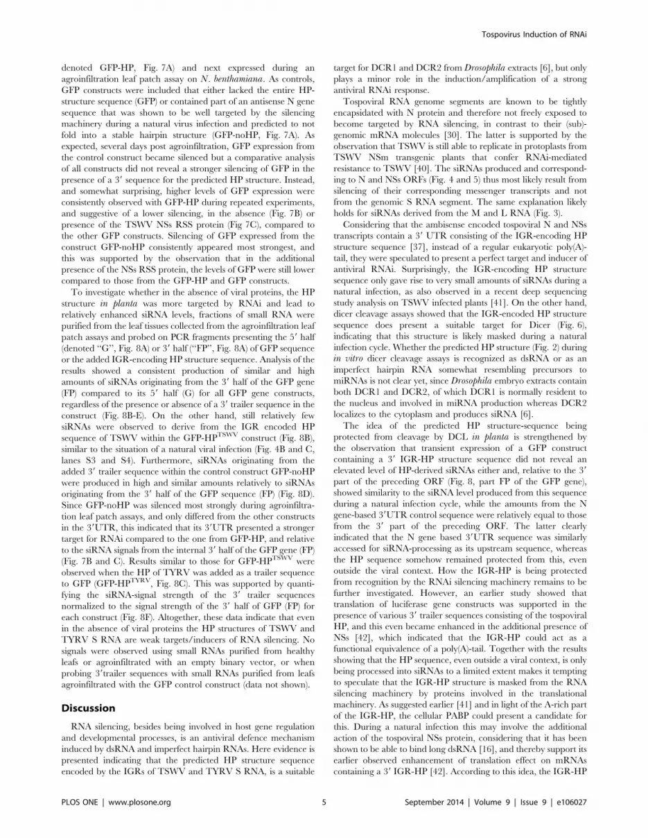
Figure 1. Schematic representation of the tospovirus tripartiteRNA genome.doi:10.1371/journal.pone.0106027.g001
Figure 2. Folding prediction of A-U rich hairpin structures fromtospovirus S RNA IGR: TSWV (left panel) and TYRV (rightpanel).doi:10.1371/journal.pone.0106027.g002
Tospovirus Induction of RNAi
PLOS ONE | www.plosone.org 2 September 2014 | Volume 9 | Issue 9 | e106027

Results
TSWV and TYRV infections predominantly lead toproduction of M- and S RNA-specific vsiRNAs
A common feature to all tospoviruses is the presence of an IGR
within the ambisense M and S RNA segments, that contains long
stretches of A-rich and U-rich sequences and is predicted to fold
into a stable HP (Fig. 2). Based on the presence of these structures,
it is tempting to hypothesize that the presence of these in viral
mRNA turns them into potent inducers (and targets) of antiviral
RNAi. If this is true, more vsiRNAs are expected to correspond to
the ambisense M and S RNA segments in comparison to the L
RNA segment that lacks such IGR sequence. To test for this, and
analyse whether M and S RNA indeed give rise to the production
of higher levels of vsiRNAs, small RNA molecules were purified
from TSWV-infected N. benthamiana leaf material and, after
radiolabeling, probed on total RNA and genomic RNA purified
from isolated viral RNPs (Fig. 3A).
While vsiRNAs were found hybridizing to the L, M and S RNA
segments, strong hybridization signals were observed with the
ambisense M and S RNA segments (Fig. 3A, lane 3). Hybridiza-
tion signals on total RNA purified from TSWV infected leafs were
weak, likely due to the relative lower amounts of viral RNA in
these fractions (Fig. 3A, lane 2). To test whether this pattern of
vsiRNAs was common to other tospoviruses, the same experiment
was performed with another distinct tospovirus, Tomato yellowring virus (TYRV) [38], from which the S RNA IGR was earlier
observed to contain extensive stretches of full complementarity
(Fig. 2B). The results again revealed the generation of relatively
high amounts of vsiRNAs derived from the M and S segments and
only low amounts from the L RNA (Fig. 3B, lanes 1 and 2).
Non-uniform production of vsiRNAs along the tospovirusS RNA sequence
To test whether the vsiRNAs originating from the ambisense M
and S RNA segments predominantly corresponded to the IGR
encoded HP, suggestive for the status of HP as strong inducer/
target of RNA silencing, the vsiRNAs were further fine mapped on
the S RNA segment. To this end, radiolabeled TSWV vsiRNAs
were hybridized to similarly sized PCR fragments spanning the
entire S RNA segment. Although vsiRNAs hybridized to
sequences covering the entire TSWV S RNA segment, and good
amounts were obtained from sequences of the NSs and N genes
(Fig. 4A and 4B), unexpectedly, hardly any siRNAs originated
from the IGR encoded HP sequence (Fig. 4B and 4C). No signals
were observed when small RNAs purified from healthy plants
were used as probe (data not shown).
To verify whether a similar vsiRNA distribution profile would
be obtained with TYRV, a similar fine mapping study was
performed for this virus. Like TSWV, TYRV infections gave rise
Figure 3. Production of vsiRNAs from tospoviral S, M and Lgenomic RNA segments. (A) Total RNA from healthy (lane 1) andTSWV infected N.benthamiana (lane 2); genomic RNA from TSWV RNPs(lane 3). As a size marker (m), ssRNA Ladder (NEB) was used. (B)Genomic RNA from TYRV RNPs, undiluted (lane 1) and diluted 1x (lane2). As a size marker (m), RiboRuler High Range RNA Ladder (ThermoScientific) was used. Left panel presents agarose gel. Right panelpresents Northern blot hybridized with radiolabeled siRNAs purifiedfrom TSWV or TYRV-infected N. benthamiana.doi:10.1371/journal.pone.0106027.g003
Figure 4. Distribution of vsiRNAs on TSWV ambisense S RNAsegment. (A) Schematic representation of TSWV S RNA segment.Intergenic region (IGR), with predicted hairpin structure (AU box), isindicated in red. PCR fragments spanning S RNA (S1 to S6) respectivebasepair sizes are indicated; dotted lines roughly demark positions ofprimers used. (B) Ethidium bromide staining of agarose gel containingfragments S1 to S6 (upper panel), and corresponding Southern blothybridized to radiolabeled siRNAs purified from TSWV-infected N.benthamiana (lower panel). (C) Relative signal strength of siRNAs oneach genomic cDNA fragment. Standard error of mean (SEM) from twoindependent experiments is indicated.doi:10.1371/journal.pone.0106027.g004
Tospovirus Induction of RNAi
PLOS ONE | www.plosone.org 3 September 2014 | Volume 9 | Issue 9 | e106027

to S RNA-derived vsiRNAs that mapped to all regions of the S
RNA segment (Fig. 5A and B), but those from the IGR encoded
HP structure were relatively scarce (Fig. 5B and C). Furthermore,
almost twice as much vsiRNAs were observed to originate from
the start region of the NSs ORF (fragment Y1; position 1-588 in
the vRNA), when compared to other regions of the S RNA
(Fig. 5B and 5C). A further fine mapping within this region
revealed that siRNAs specifically derived from the nucleotide
sequence 1-284 from TYRV S RNA (Fig. 5B, lower panel). No
signals were observed when siRNAs purified from healthy plants
were used as probe (data not shown).
HP transcript is cleaved by Dicer in vitroWhile only few vsiRNAs were found mapping to the IGR
encoded predicted hairpin-structure, this region was further
investigated as potential inducer and target of antiviral RNAi in
a dicer cleavage assay. To this end, synthetic radiolabeled
transcripts of the TSWV IGR-encoding HP sequence were made
and after being allowed to fold into a dsRNA hairpin structure,
subsequently offered to RNAi-induced Drosophila melanogaster(Dm) embryo extracts containing Dicer-1 and Dicer-2 [6,39].
Analysis of the products on non-denaturing acrylamide gels
showed that the HP transcript was cleaved into small RNAs, co-
migrating with siRNAs (21 nucleotides) cleaved from a 114 nt
dsRNA transcript and with the siRNA size marker (Fig. 6). Similar
results were obtained when using synthetic transcripts from the
TYRV S RNA IGR sequence (data not shown) and support the
idea that the IGR encoding hairpin structure is recognized as a
substrate for dicer.
The IGR-encoded HP-structure sequence is weaklytargeted by the RNAi machinery during transientexpression in planta
While synthetic transcripts from the IGR encoded HP structure
were recognized as substrate for dicer, the presence of only low
amounts of vsiRNAs derived from this sequence during a natural
infection could be due to the possibility that the hairpin structure is
being protected from Dicer cleavage by a viral protein, e.g. the
TSWV NSs RSS protein. If this is true, elevated levels of HP-
derived siRNAs would be expected when the HP structure is
expressed outside the context of a viral infection. To test this
hypothesis, and further investigate the IGR HP structure as a
potential target of RNAi, a functional GFP construct was made
containing the TSWV HP structure sequence at its 39 end (and
Figure 5. Distribution of vsiRNAs on TYRV ambisense S RNAsegment. (A) Schematic representation of TYRV S RNA segment.Intergenic region (IGR), with predicted hairpin structure (AU box), isindicated in red. PCR fragments spanning S RNA (Y1 to Y7) respectivebasepair sizes are indicated; dotted lines roughly demark positions ofprimers used. (B) Ethidium bromide staining of agarose gel containingPCR fragments Y1 to Y7 (upper panel), and corresponding Southernblot hybridized to radiolabeled siRNAs purified from TYRV-infected N.benthamiana (lower panel). Below, fine mapping of fragment Y1. (C)Relative signal strength of siRNAs on each genomic cDNA fragment.doi:10.1371/journal.pone.0106027.g005
Figure 6. Dicer-mediated cleavage of hairpin transcripts (HP)from TSWV S RNA IGR-encoded hairpin sequence. Radioactivelylabeled HP transcripts (lane 2) were incubated in the presence of dicercontaining Drosophila melanogaster (Dm) embryo extracts and cleavageproducts (lane 1) subsequently resolved on 8% denaturing acrylamidegel. As positive control, 114-nt dsRNA (lane 4) was included to verifydicer activity from Dm extracts (lane 3). As size marker, radiolabeled21nt siRNAs were included (lane 5).doi:10.1371/journal.pone.0106027.g006
Tospovirus Induction of RNAi
PLOS ONE | www.plosone.org 4 September 2014 | Volume 9 | Issue 9 | e106027
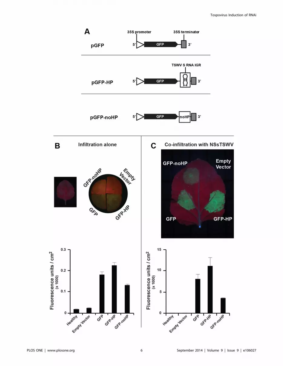
denoted GFP-HP, Fig. 7A) and next expressed during an
agroinfiltration leaf patch assay on N. benthamiana. As controls,
GFP constructs were included that either lacked the entire HP-
structure sequence (GFP) or contained part of an antisense N gene
sequence that was shown to be well targeted by the silencing
machinery during a natural virus infection and predicted to not
fold into a stable hairpin structure (GFP-noHP, Fig. 7A). As
expected, several days post agroinfiltration, GFP expression from
the control construct became silenced but a comparative analysis
of all constructs did not reveal a stronger silencing of GFP in the
presence of a 39 sequence for the predicted HP structure. Instead,
and somewhat surprising, higher levels of GFP expression were
consistently observed with GFP-HP during repeated experiments,
and suggestive of a lower silencing, in the absence (Fig. 7B) or
presence of the TSWV NSs RSS protein (Fig 7C), compared to
the other GFP constructs. Silencing of GFP expressed from the
construct GFP-noHP consistently appeared most strongest, and
this was supported by the observation that in the additional
presence of the NSs RSS protein, the levels of GFP were still lower
compared to those from the GFP-HP and GFP constructs.
To investigate whether in the absence of viral proteins, the HP
structure in planta was more targeted by RNAi and lead to
relatively enhanced siRNA levels, fractions of small RNA were
purified from the leaf tissues collected from the agroinfiltration leaf
patch assays and probed on PCR fragments presenting the 59 half
(denoted ‘‘G’’, Fig. 8A) or 39 half (‘‘FP’’, Fig. 8A) of GFP sequence
or the added IGR-encoding HP structure sequence. Analysis of the
results showed a consistent production of similar and high
amounts of siRNAs originating from the 39 half of the GFP gene
(FP) compared to its 59 half (G) for all GFP gene constructs,
regardless of the presence or absence of a 39 trailer sequence in the
construct (Fig. 8B-E). On the other hand, still relatively few
siRNAs were observed to derive from the IGR encoded HP
sequence of TSWV within the GFP-HPTSWV construct (Fig. 8B),
similar to the situation of a natural viral infection (Fig. 4B and C,
lanes S3 and S4). Furthermore, siRNAs originating from the
added 39 trailer sequence within the control construct GFP-noHP
were produced in high and similar amounts relatively to siRNAs
originating from the 39 half of the GFP sequence (FP) (Fig. 8D).
Since GFP-noHP was silenced most strongly during agroinfiltra-
tion leaf patch assays, and only differed from the other constructs
in the 39UTR, this indicated that its 39UTR presented a stronger
target for RNAi compared to the one from GFP-HP, and relative
to the siRNA signals from the internal 39 half of the GFP gene (FP)
(Fig. 7B and C). Results similar to those for GFP-HPTSWV were
observed when the HP of TYRV was added as a trailer sequence
to GFP (GFP-HPTYRV, Fig. 8C). This was supported by quanti-
fying the siRNA-signal strength of the 39 trailer sequences
normalized to the signal strength of the 39 half of GFP (FP) for
each construct (Fig. 8F). Altogether, these data indicate that even
in the absence of viral proteins the HP structures of TSWV and
TYRV S RNA are weak targets/inducers of RNA silencing. No
signals were observed using small RNAs purified from healthy
leafs or agroinfiltrated with an empty binary vector, or when
probing 39trailer sequences with small RNAs purified from leafs
agroinfiltrated with the GFP control construct (data not shown).
Discussion
RNA silencing, besides being involved in host gene regulation
and developmental processes, is an antiviral defence mechanism
induced by dsRNA and imperfect hairpin RNAs. Here evidence is
presented indicating that the predicted HP structure sequence
encoded by the IGRs of TSWV and TYRV S RNA, is a suitable
target for DCR1 and DCR2 from Drosophila extracts [6], but only
plays a minor role in the induction/amplification of a strong
antiviral RNAi response.
Tospoviral RNA genome segments are known to be tightly
encapsidated with N protein and therefore not freely exposed to
become targeted by RNA silencing, in contrast to their (sub)-
genomic mRNA molecules [30]. The latter is supported by the
observation that TSWV is still able to replicate in protoplasts from
TSWV NSm transgenic plants that confer RNAi-mediated
resistance to TSWV [40]. The siRNAs produced and correspond-
ing to N and NSs ORFs (Fig. 4 and 5) thus most likely result from
silencing of their corresponding messenger transcripts and not
from the genomic S RNA segment. The same explanation likely
holds for siRNAs derived from the M and L RNA (Fig. 3).
Considering that the ambisense encoded tospoviral N and NSs
transcripts contain a 39 UTR consisting of the IGR-encoding HP
structure sequence [37], instead of a regular eukaryotic poly(A)-
tail, they were speculated to present a perfect target and inducer of
antiviral RNAi. Surprisingly, the IGR-encoding HP structure
sequence only gave rise to very small amounts of siRNAs during a
natural infection, as also observed in a recent deep sequencing
study analysis on TSWV infected plants [41]. On the other hand,
dicer cleavage assays showed that the IGR-encoded HP structure
sequence does present a suitable target for Dicer (Fig. 6),
indicating that this structure is likely masked during a natural
infection cycle. Whether the predicted HP structure (Fig. 2) during
in vitro dicer cleavage assays is recognized as dsRNA or as an
imperfect hairpin RNA somewhat resembling precursors to
miRNAs is not clear yet, since Drosophila embryo extracts contain
both DCR1 and DCR2, of which DCR1 is normally resident to
the nucleus and involved in miRNA production whereas DCR2
localizes to the cytoplasm and produces siRNA [6].
The idea of the predicted HP structure-sequence being
protected from cleavage by DCL in planta is strengthened by
the observation that transient expression of a GFP construct
containing a 39 IGR-HP structure sequence did not reveal an
elevated level of HP-derived siRNAs either and, relative to the 39
part of the preceding ORF (Fig. 8, part FP of the GFP gene),
showed similarity to the siRNA level produced from this sequence
during a natural infection cycle, while the amounts from the N
gene-based 39UTR control sequence were relatively equal to those
from the 39 part of the preceding ORF. The latter clearly
indicated that the N gene based 39UTR sequence was similarly
accessed for siRNA-processing as its upstream sequence, whereas
the HP sequence somehow remained protected from this, even
outside the viral context. How the IGR-HP is being protected
from recognition by the RNAi silencing machinery remains to be
further investigated. However, an earlier study showed that
translation of luciferase gene constructs was supported in the
presence of various 39 trailer sequences consisting of the tospoviral
HP, and this even became enhanced in the additional presence of
NSs [42], which indicated that the IGR-HP could act as a
functional equivalence of a poly(A)-tail. Together with the results
showing that the HP sequence, even outside a viral context, is only
being processed into siRNAs to a limited extent makes it tempting
to speculate that the IGR-HP structure is masked from the RNA
silencing machinery by proteins involved in the translational
machinery. As suggested earlier [41] and in light of the A-rich part
of the IGR-HP, the cellular PABP could present a candidate for
this. During a natural infection this may involve the additional
action of the tospoviral NSs protein, considering that it has been
shown to be able to bind long dsRNA [16], and thereby support its
earlier observed enhancement of translation effect on mRNAs
containing a 39 IGR-HP [42]. According to this idea, the IGR-HP
Tospovirus Induction of RNAi
PLOS ONE | www.plosone.org 5 September 2014 | Volume 9 | Issue 9 | e106027
Tospovirus Induction of RNAi
PLOS ONE | www.plosone.org 6 September 2014 | Volume 9 | Issue 9 | e106027

structure sequence would then be engaged most of the times in
viral/host protein interactions and inaccessible for siRNA
generation by RNase-III type enzymes or to assist in the
generation of secondary siRNAs by RDR. In light of the structural
similarities, this would not only apply to the S RNA, but also to the
ambisense M RNA encoded transcripts where similar, stable
hairpin structures are predicted [31].
Our observations on siRNAs from the IGR-encoded HP
structure sequence are supported by recent deep sequencing data
[41,43], however in both studies the relative lower amounts of
vsiRNAs produced from the S and M RNA encoded IGR
sequences were not remarked by the authors.
The observations of high amounts of siRNAs mapping to the
NSs gene is interesting in light of this protein acting as a suppressor
of silencing [34,35] and when considering the RNA silencing effect
on viral replication and plant-virus dynamics [44]. Folding
predictions of the RNA sequence around the start of the TYRV
NSs ORF revealed a small hairpin structure (NSs-hairpin), and
similar ones at almost the same position were found in several
other tospoviruses. Hence, though speculative, the presence of an
RNAi target within the NSs gene might be involved in regulating
NSs expression and, consequently, tospovirus virulence.
In conclusion, the AU-rich hairpin structure in the tospoviral
IGR presents a suitable substrate for Dicer but appears to present
only a weak inducer and target of RNAi, likely due to being
masked by viral and/or host proteins. Elucidating the nature of
these will provide further insight into the role of the hairpin
structure in processes of viral transcription and translation.
Materials and Methods
Viruses and PlantsThe tospovirus strains TSWV BR-01 [45] and Tomato yellow
ring virus-tomato strain (TYRV-t, here referred simply as TYRV)
[38] were maintained by mechanical passage on hosts Nicotianabenthamiana and N. rustica cv. America.
Detection, isolation and labeling of siRNAs from plantleaves
Isolation of small RNAs was performed as previously described
[46,47]. In brief, leaf material (from healthy and systemically
infected N.benthamiana leaves) was ground in liquid nitrogen and
next mixed with extraction buffer (2% Sarcosyl – 5 M NaCl),
followed by phenol extraction. The aqueous phase was collected
and subjected to polyethylene glycol (PEG) precipitation [5], in
order to separate low-molecular-weight (LMW) RNA molecules
from DNA and larger RNA molecules. For the purification of
siRNAs, 15 to 30 mg of LMW RNAs were resolved on a 15%
denaturing polyacrylamide gel containing 8 M urea. After
ethidium bromide staining, the region containing siRNAs was
excised from the gel, ground to small pieces and incubated in 3 M
NaCl overnight at 4uC to extract the siRNAs from the gel by
diffusion. After centrifugation, the supernatant was collected and
the siRNAs were ethanol precipitated. Small interfering RNA
molecules were dephosphorylated with alkaline phosphatase and
subsequently end-labeled with [c-32P]-ATP (Perkin Elmer) by T4
polynucleotide kinase (Promega) according to the manufacturer’s
instructions.
Purification of tospovirus genomic RNA fromribonucleoproteins (RNPs) and northern blotting
Tospoviral RNPs were purified from N. rustica cv. America as
previously described [48]. Genomic RNA was purified using hot
phenol extraction followed by ethanol precipitation [30]. Purified
RNA was resolved in 1% agarose gel under RNase free conditions
and blotted to Hybond-N membrane (Amersham Biosciences) by
top-down blotting in neutral transfer conditions using Whatman
TurboBlotter system according to manufacturer’s instruction.
Filters were hybridized to [c-32P]-labelled siRNAs (see below)
purified from healthy and tospovirus-infected N.benthamianaleaves.
Southern blotting, siRNA purification and mapping onTSWV and TYRV S RNA
Total RNA was purified from systemically infected N.benthami-ana leafs using Trizol (Life Technologies). The S RNA segment
was RT-PCR-amplified, using Superscript RT (Invitrogen), in 6-7
fragments of similar size and spanning the entire S RNA segment
from TSWV and TYRV respectively. The products were further
cloned in pGem-T Easy (Promega) according to the manufactur-
er’s instructions and verified by sequence analysis. For TYRV S
RNA-specific fragments, equimolar amounts of PCR products
were resolved on 1% agarose gel. For TSWV S RNA, due to
difficulties in obtaining single PCR products, S RNA-specific
fragments were excised from pGem-T Easy plasmid DNA and
equimolar amounts resolved on 1% agarose gel. DNA was blotted
to Hybond-N membrane (Amersham Biosciences) by top-down
blotting. Filters were subsequently hybridized (at 48uC) overnight
in Church buffer [49] to [c-32P]-labelled siRNAs purified from
healthy or tospovirus-infected N.benthamiana leaves. After wash-
ing, filters were exposed for two days to phosphor screen (Kodak)
and visualized by phosphorimaging (Molecular Imager FX, Bio-
Rad). Signal quantification was performed with ImageJ software
[50].
Synthesis of [32P]-radiolabelled dsRNA substratesDNA templates of the A-U rich predicted hairpin encoding
sequence (from TSWV S RNA IGR) (Fig. 2) were RT-PCR
amplified using primers containing the T7 RNA polymerase
promoter sequence. PCR fragments were purified using High Pure
PCR purification kit (Roche) and radiolabelled RNA transcripts
were prepared by in vitro transcription using T7 RNA polymerase
(Promega) in the presence of [a-32P]-rNTP (PerkinElmer Inc.,
UK) according manufacture’s instruction. Products from the invitro transcription were resolved on an 8% denaturing acrylamide
gel and the radiolabelled A-U rich predicted hairpin transcript was
excised from the gel and extracted by diffusion into 20 ml 2x PK
buffer (200 mM Tris pH 7.5, 300 mM NaCl, 5 mM EDTA, 2%
SDS) followed by phenol chloroform and ethanol precipitation.
Prior to use, purified RNA transcripts were briefly heated for
10 min. at 85uC and gradually cooled down to room temperature
Figure 7. Agroinfiltration leaf patch assays of GFP gene constructs containing 39 hairpin trailer. (A) Schematic representation of GFPconstructs containing the different 39 trailer sequences analyzed. The noHP sequence consists of a partial N gene sequence in antisense polarity. (B)Transient GFP expression after agroinfiltration of GFP constructs in absence of RSS. As only very low levels of fluorescence were visual at first (left),leaf disks were further analysed on binocular stereomicroscope M3Z, Leica (right). (C) Similar as panel B, but in the additional presence of TSWV NSs.Fluorescence in panels B and C was quantified and depicted in the graphs underneath. Standard error of mean (SEM) from three leaf disks isindicated.doi:10.1371/journal.pone.0106027.g007
Tospovirus Induction of RNAi
PLOS ONE | www.plosone.org 7 September 2014 | Volume 9 | Issue 9 | e106027

to allow RNA folding. 114-nt dsRNA molecules were prepared as
previously described [16].
Dicer cleavage assay (DCA)Drosophila melanogaster (Dm) embryo extract was prepared as
previously described [39]. In brief, for the dicer cleavage reactions
a reaction mixture of 10 ml consisting of 5 ml Drosophila embryo
extract, 5 nM 32P-labeled transcript of the IR hairpin or dsRNA
were incubated for 2–3 h at 25uC [39], except potassium acetate
was omitted from the reaction mixture [16]. Next, samples were
deproteinized with proteinase K, RNA was phenol extracted and
analyzed on 8% denaturing acrylamide gel, which were then dried
for 30 minutes at 80uC, exposed to a phosphor screen (Kodak) for
12 hrs and scanned with PhosphorImager (Molecular Imager FX,
Bio-Rad).
Agrobacterium tumefaciens mediated transientexpression assay (ATTA) of GFP-hairpin constructs inplanta
To analyse the IGR hairpin as an inducer of silencing outside
the context of a tospoviral infection, leaf patch assays with the
Agrobacterium tumefaciens transient expression assay (ATTA)
system were performed as previously described [35,51]. To
monitor the effect of the hairpin sequence on the induction of
silencing of a functional green fluorescent protein (GFP) gene
construct, the hairpin-encoding sequence (nucleotide position
1044–1368 and 1032–1427 of, respectively, TSWV and TYRV
vc S RNA) was fused by PCR amplification to the 39 end of the
GFP gene, generating constructs GFP-HPTSWV and GFP-
HPTYRV. As a control, an inverted part of the TSWV N gene
sequence (nucleotide position 235–528 of vc S RNA, correspond-
ing to position 82–375 from ATG of N gene) was fused to the 39
end of the GFP gene, resulting in the GFP-noHP construct. All
GFP-HP, GFP-noHP and GFP constructs were cloned in binary
vector pK2GW7 [52] using the Gateway Cloning Technology
(Invitrogen). For suppression of silencing the TSWV NSs and
tombusvirus P19 genes were expressed from binary vectors
pK2GW7 and pBin19, respectively. To this end, binary vectors
were transformed to Agrobacterium tumefaciens strain cor308 [53]
and cultured in LB3 medium containing appropriate antibiotics
for selection (Tetracycline 2 mg/ml and Spectinomycin 250 mg/ml
– for pK2GW7 – or Kanamycin 100 mg/ml – for pBin19) at 28uCovernight. From the overnight culture, 600 ml was transferred to
3 ml induction medium (10.5 g/l K2HPO4, 4.5 g/l KH2PO4,
1.0 g/l (NH4)2SO4, 0.5 g/l Sodium Citrate Dihydrate, 0.25 g/l
MgSO4, 0.2% (w/v) glucose, 0.5% (v/v) glycerol, 50 mM
acetosyringone and 10 mM MES pH 5.6) and grown at 28uCovernight. The induced culture was pelleted and ressuspended in
Murashige-Skoog (MS) medium (30 g/l sucrose; 40 g/l MS;
pH 5.7) containing 150 mM acetosyringone and 10 mM MES
(pH 5.6) to an OD600 of 0.5. This suspension was used to infiltrate
fully expanded leafs of N. benthamiana plants. Silencing of GFP
and suppression by NSS and P19 proteins was assessed by UV light
and western blot analysis, respectively. To suppress silencing,
RNA silencing suppressor constructs were provided in a co-ATTA
with GFP constructs. To this end, induced Agrobacteriumsuspensions were mixed at a final OD600 of 0.5 prior to infiltration.
Figure 8. Production and distribution of siRNAs from GFPconstructs containing various 39 trailer sequences. Small RNAspurified from transient expression of GFP constructs were probed onSouthern blots containing PCR fragments spanning the respectiveconstruct sequence. (A) Schematic view of constructs and PCR productsspanning the sequence. The noHP sequence consists of a partial N genesequence in antisense polarity. Southern blot analysis of constructs: (B)GFP-HPTSWV; (C) GFP-HPTYRV; (D) GFP-noHP; (E) GFP. Ethidium bromide-staining of PCR products are shown below. (F) Graphical representation
of the siRNA signal strength corresponding to the 39 trailer sequencesand normalized to the signal strength of the 39 half of GFP (FP) of eachconstruct. Abbreviation: G: 59 half of GFP; FP: 39 half of GFP; HP: A-U richhairpin structure (from IGR of TSWV and TYRV S RNA); noHP: part ofTSWV N gene.doi:10.1371/journal.pone.0106027.g008
Tospovirus Induction of RNAi
PLOS ONE | www.plosone.org 8 September 2014 | Volume 9 | Issue 9 | e106027

Infiltrated plants were kept at 25uC and monitored for GFP
fluorescence during a 5-day period using a GFP fluorescence-
stereo-microscope. Pictures were taken at 5 dpi (days post
infiltration). Quantification of GFP fluorescence from 1 cm2 leaf
disk was performed using Fluorstar Optima (BMG Labtech) as
previously described [54]. As probes for southern blotting, small
RNAs were purified from 6 g of agroinfiltrated leafs and
radiolabeled as described above.
UV photography and quantification of GFP fluorescencePictures of whole leafs (as shown in Fig. 7B and 7C) were taken
with a digital camera (Canon PowerShot A3200 IS) by using a
hand-held UV light (Philips, 6W). In case of leafs agroinfiltrated
with GFP constructs without a suppressor of RNA silencing, close-
up UV pictures (as shown in Fig. 7A) were made using a digital
camera CoolSnap and a binocular stereomicroscope (M3Z, Leica).
For the quantification of GFP fluorescence, 5dpi leaf disks of 1 cm
in diameter were taken from infiltrated leaf area and analysed
using Fluorstar Optima (BMG Labtech), as previously described
[54].
Folding predictions for S RNA intergenic hairpinsequence
Folding predictions were performed at 37uC, using Mfold
[55,56].
Author Contributions
Conceived and designed the experiments: MH AHM RK. Performed the
experiments: MH AHM DL. Analyzed the data: MH AHM RK.
Contributed reagents/materials/analysis tools: MH AHM DL RK. Wrote
the paper: MH AHM RK.
References
1. Ahlquist P (2002) RNA-dependent RNA polymerases, viruses, and RNAsilencing. Science 296: 1270–1273.
2. Molnar A, Csorba T, Lakatos L, Varallyay E, Lacomme C, et al. (2005) Plant
virus-derived small interfering RNAs originate predominantly from highly
structured single-stranded viral RNAs. J Virol 79: 7812–7818.
3. Vermeulen A, Behlen L, Reynolds A, Wolfson A, Marshall WS, et al. (2005) Thecontributions of dsRNA structure to Dicer specificity and efficiency. RNA-a
Publication of the RNA Society 11: 674–682.
4. Bernstein E, Caudy AA, Hammond SM, Hannon GJ (2001) Role for a bidentate
ribonuclease in the initiation step of RNA interference. Nature 409: 363–366.
5. Hamilton AJ, Baulcombe DC (1999) A species of small antisense RNA in
posttranscriptional gene silencing in plants. Science 286: 950–952.
6. Lee YS, Nakahara K, Pham JW, Kim K, He ZY, et al. (2004) Distinct roles for
Drosophila Dicer-1 and Dicer-2 in the siRNA/miRNA silencing pathways. Cell117: 69–81.
7. Fire A, Xu SQ, Montgomery MK, Kostas SA, Driver SE, et al. (1998) Potentand specific genetic interference by double-stranded RNA in Caenorhabditis
elegans. Nature 391: 806–811.
8. Khvorova A, Reynolds A, Jayasena S (2003) Functional siRNAs and rniRNAs
exhibit strand bias. Cell 115: 209–216.
9. Schwarz DS, Hutvagner G, Du T, Xu ZS, Aronin N, Zamore PD (2003)Asymmetry in the assembly of the RNAi enzyme complex. Cell 115: 199–208.
10. Bohmert K, Camus I, Bellini C, Bouchez D, Caboche M, et al. (1998) AGO1defines a novel locus of Arabidopsis controlling leaf development. Embo Journal
17: 170–180.
11. Peters L, Meister G (2007) Argonaute proteins: Mediators of RNA silencing.
Molecular Cell 26: 611–623.
12. Rand TA, Petersen S, Du FH, Wang XD (2005) Argonaute2 cleaves the anti-
guide strand of siRNA during RISC activation. Cell 123: 621–629.
13. Ding SW, Voinnet O (2007) Antiviral immunity directed by small RNAs. Cell
130: 413–426.
14. Voinnet O (2005) Induction and suppression of RNA silencing: insights fromviral infections. Nat Rev Genet 6: 206–220.
15. Zhang X, Yuan YR, Pei Y, Lin SS, Tuschl T, et al. (2006) Cucumber mosaicvirus encoded 2b suppressor inhibits Arabidopsis Argonaute1 cleavage activity to
counter plant defense. Genes & Development 20: 3255–68.
16. Schnettler E, Hemmes H, Huismann R, Goldbach R, Prins M, et al. (2010)
Diverging Affinity of Tospovirus RNA Silencing Suppressor Proteins, NSs, forVarious RNA Duplex Molecules. J Virol 84: 11542–11554.
17. Mallory AC, Reinhart BJ, Bartel D, Vance VB, Bowman LH (2002) A viralsuppressor of RNA silencing differentially regulates the accumulation of short
interfering RNAs and micro-RNAs in tobacco. Proc Natl Acad Sci U S A 99:15228–15233.
18. Llave C, Kasschau KD, Carrington JC (2000) Virus-encoded suppressor ofposttranscriptional gene silencing targets a maintenance step in the silencing
pathway. Proc Natl Acad Sci U S A 97: 13401–13406.
19. Lakatos L, Csorba T, Pantaleo V, Chapman EJ, Carrington JC, et al. (2006)
Small RNA binding is a common strategy to suppress RNA silencing by severalviral suppressors. Embo Journal 25: 2768–2780.
20. Lakatos L, Szittya G, Silhavy D, Burgyan J (2004) Molecular mechanism ofRNA silencing suppression mediated by p19 protein of tombusviruses. Embo
Journal 23: 876–884.
21. Goto K, Kobori T, Kosaka Y, Natsuaki T, Masuta C (2007) Characterization of
silencing suppressor 2b of cucumber mosaic virus based on examination of itssmall RNA-Binding abilities. Plant Cell Physiol 48: 1050–1060.
22. Diaz-Pendon JA, Li F, Li WX, Ding SW (2007) Suppression of antiviral silencingby cucumber mosaic virus 2b protein in Arabidopsis is associated with drastically
reduced accumulation of three classes of viral small interfering RNAs. Plant Cell19: 2053–2063.
23. Giner A, Lakatos L, Garcıa-Chapa M, Lopez-Moya JJ, Burgyan J (2010) Viral
protein inhibits RISC activity by argonaute binding through conserved WG/
GW motifs. Plos Pathogens 6: e1000996.
24. Incarbone M, Dunoyer P (2013) RNA silencing and its suppression: novel
insights from in planta analyses. Trends Plant Sci 18: 382–392.
25. Szittya G, Molnar A, Silhavy D, Hornyik C, Burgyan J (2002) Short defective
interfering RNAs of tombusviruses are not targeted but trigger post-
transcriptional gene silencing against their helper virus. Plant Cell 14: 359–72.
26. Sijen T, Fleenor J, Simmer F, Thijssen KL, Parrish S, et al. (2001) On the role of
RNA amplification in dsRNA-triggered gene silencing. Cell 107: 465–476.
27. Elliott RM (1990) Molecular-Biology of the Bunyaviridae. J Gen Virol 71: 501–
522.
28. King AMQ, Adams MJ, Carstens EB, Lefkowitz EJ (2012) eds Ninth Report of
the International Committee on Taxonomy of Viruses. Elsevier/Academic
Press: London. 1338.
29. De Haan P, Kormelink R, Resende RD, Van PoelwijkF, , Peters D, et al. (1991)
Tomato Spotted Wilt Virus-L RNA Encodes a Putative RNA-Polymerase.
J Gen Virol 72: 2207–2216.
30. Kormelink R, De Haan P, Peters D, Goldbach R (1992) Viral-RNA Synthesis in
Tomato Spotted Wilt Virus-Infected Nicotiana-Rustica Plants. J Gen Virol 73:
687–693.
31. Kormelink R, De Haan P, Meurs C, Peters D, Goldbach R (1992) The
nucleotide sequence of the M RNA segment of Tomato spotted wilt virus, a
bunyavirus with two ambisense RNA segments. J Gen Virol 73: 2795–2804.
32. Kormelink R, Storms M, Van Lent J, Peters D, Goldbach R (1994) Expression
and Subcellular Location of the NSm Protein of Tomato Spotted Wilt Virus
(TSWV), a Putative Viral Movement Protein. Virology 200: 56–65.
33. De Haan P, Wagemakers L, Peters D, Goldbach R (1990) The S-RNA Segment
of Tomato Spotted Wilt Virus Has an Ambisense Character. J Gen Virol 71:
1001–1007.
34. Takeda A, Sugiyama K, Nagano H, Mori M, Kaido M, et al. (2002)
Identification of a novel RNA silencing suppressor, NSs protein of Tomato
spotted wilt virus. Febs Letters 532: 75–79.
35. Bucher E, Sijen T, de Haan P, Goldbach R, Prins M (2003) Negative-strand
tospoviruses and tenuiviruses carry a gene for a suppressor of gene silencing at
analogous genomic positions. J Virol 77: 1329–1336.
36. Nguyen M, Haenni AL (2003) Expression strategies of ambisense viruses. Virus
Res 93: 141–150.
37. van Knippenberg I, Goldbach R, Kormelink R (2005) Tomato spotted wilt virus
S-segment mRNAs have overlapping 3 ’-ends containing a predicted stem-loop
structure and conserved sequence motif. Virus Res 110: 125–131.
38. Hassani-Mehraban A, Saaijer J, Peters D, Goldbach R, Kormelink R (2005) A
new tomato-infecting tospovirus from Iran. Phytopathology 95: 852–858.
39. Haley B, Tang GL, Zamore PD (2003) In vitro analysis of RNA interference in
Drosophila melanogaster. Methods 30: 330–336.
40. Prins M, Kikkert M, Ismayadi C, de Graauw W, de Haan P, et al. (1997)
Characterization of RNA-mediated resistance to tomato spotted wilt virus in
transgenic tobacco plants expressing NSM gene sequences. Plant Mol Biol 33:
235–243.
41. Mitter N, Koundal V, Williams S, Pappu H (2013) Differential Expression of
Tomato Spotted Wilt Virus-Derived Viral Small RNAs in Infected Commercial
and Experimental Host Plants. PLoS ONE 8 (10).
42. Geerts-Dimitriadou C, Lu YY, Geertsema C, Goldbach R, Kormelink R (2012)
Analysis of the Tomato spotted wilt virus Ambisense S RNA-Encoded Hairpin
Structure in Translation. PLoS ONE 7 (2).
43. Hagen C, Frizzi A, Kao J, Jia LJ, Huang MY, et al. (2011) Using small RNA
sequences to diagnose, sequence, and investigate the infectivity characteristics of
vegetable-infecting viruses. Arch Virol 156: 1209–1216.
Tospovirus Induction of RNAi
PLOS ONE | www.plosone.org 9 September 2014 | Volume 9 | Issue 9 | e106027

44. Groenenboom MA, Hogeweg P (2012) Modelling the dynamics of viral
suppressors of RNA silencing. J R Soc Interface 9: 436–47.45. De Avila AC, De Haan P, Kitajima EW, Kormelink R, Resende RD, et al.
(1992) Characterization of a Distinct Isolate of Tomato Spotted Wilt Virus
(TSWV) from Impatiens Sp in the Netherlands. J Phytopathol 134: 133–151.46. Bucher E, Hemmes H, de Haan P, Goldbach R, Prins M (2004) The influenza A
virus NS1 protein binds small interfering RNAs and suppresses RNA silencing inplants. J Gen Virol 85: 983–991.
47. Ribeiro SG, Lohuis H, Goldbach R, Prins M (2007) Tomato chlorotic mottle
virus is a target of RNA silencing but the presence of specific short interferingRNAs does not guarantee resistance in transgenic plants. J Virol 81: 1563–1573.
48. De Avila AC, Huguenot C, Resende RD, Kitajima EW, Goldbach RW, et al.(1990) Serological Differentiation of 20 Isolates of Tomato Spotted Wilt Virus.
J Gen Virol 71: 2801–2807.49. Sambrook J, Fritsch EF, Maniatis T (1992) Molecular cloning - A laboratory
manual, 2nd edition. NY: Cold Spring Harbor Laboratory, Cold Spring Harbor.
50. Schneider CA, Rasband WS, Eliceiri KW (2012) NIH Image to ImageJ: 25years of image analysis. Nature Methods 9: 671–675.
51. Johansen LK, Carrington JC (2001) Silencing on the spot. Induction and
suppression of RNA silencing in the Agrobacterium-mediated transient
expression system. Plant Physiol 126: 930–938.
52. Karimi M, Inze D, Depicker A (2002) GATEWAY((TM)) vectors for
Agrobacterium-mediated plant transformation. Trends Plant Sci 7: 193–195.
53. Carbonell A, de Alba AEM, Flores R, Gago S (2008) Double-stranded RNA
interferes in a sequence-specific manner with the infection of representative
members of the two viroid families. Virology 371: 44–53.
54. De Ronde D, Butterbach P, Lohuis D, Hedil M, Van Lent JWM, et al. (2013)
Tsw gene-based resistance is triggered by a functional RNA silencing suppressor
protein of the Tomato spotted wilt virus. Mol Plant Pathol 14: 405–415.
55. Mathews DH, Sabina J, Zuker M, Turner DH (1999) Expanded sequence
dependence of thermodynamic parameters improves prediction of RNA
secondary structure. J Mol Biol 288: 911–940.
56. Zuker M (2003) Mfold web server for nucleic acid folding and hybridization
prediction. Nucleic Acids Res 31: 3406–3415.
Tospovirus Induction of RNAi
PLOS ONE | www.plosone.org 10 September 2014 | Volume 9 | Issue 9 | e106027


