Embed Size (px)
Citation preview

Jorimal ol'Neuroch~.mistr~, Raven Press, Ltd.. New York (c. 1993 International Society for Neurochemistrq
Role of the Cellular Stress Response in the Biogenesis of Cysteamine-Induced Astrocytic Inclusions in Primary Culture
*?$Marc B. Mydlarski, -f$Jin-Jun Liang, and *"fHyman M. Schipper
*Department of Neurology and Neurosurgery and ?Centre for Studies in Aging, McGill University; and $BloornJield Centre for Research in Aging, Lady Davis Institute for Medical Research,
Sir Mortimer B. Davis-Jewish General Hospital, Montreal, Quebec, Canada
Abstract: Cysteamine (CSH; 2-mercaptoethylamine) stim- ulates the accumulation of peroxidase-positive inclusions in cultured astroglia akin to those observed in the aging periventricular brain. Because CSH induces the synthesis of a stress protein (heme oxygenase) in rat liver, we hy- pothesized that aspects of the cellular stress response may play a role in the biogenesis of CSH-induced astro- cyte granules. In the present study, we performed indirect immunofluorescent staining and immunoblotting for various stress proteins in rat neuroglial cultures. Exposure of astrocyte cultures to CSH enhanced immunostaining for heme oxygenase-1 (HO-1) and heat-shock proteins 27, 72, and 90, but not glucose-regulated protein 94, relative to untreated cultures. CSH-pretreated astrocytes exhib- ited enhanced tolerance to H,O, toxicity relative to un- treated cells, providing physiological evidence of an ante- cedent stress response in the former. In addition, expo- sure for 12 days to H,O,, a known inducer of the stress response, elicited astrocyte granulation similar to that ob- served with CSH. Chronic induction of HO-1 and other stress proteins may participate in the biogenesis of metal- loporphyrin-rich inclusions in CSH-treated astroglial cul- tures and in astrocytes of the aging periventricular brain. Key Words: Astrocyte-Cysteamine-Heme oxygenase -Heat-shock proteins-Glucose-regulated protein- Peroxidase activity. J. Neurochem. 61,1755-1 765 (1 993).
A unique subpopulation of granule-laden neuroglia has been described in hippocampus, striatum, and periventricular brain regions of many terrestrial verte- brates, including humans (Schipper, 199 1). The cyto- plasmic granules are histochemically distinct from li- pofuscin, exhibit an affinity for Gomori stains (chrome alum hematoxylin, aldehyde fuchsin), emit an orange-red autofluorescence consistent with the presence of porphyrin, and stain intensely with the peroxidase marker 3,3'-diaminobenzidine (DAB) (Srebro and Cichocki, 197 1 ; Keefer and Christ, 1976; Goldgefter et al., 1980). The endogenous peroxidase activity is nonenzymatic in nature and is thought to be mediated by heme (ferrous) iron or by a related metalloporphyrin (Schipper et al., 1990~). In rodent
brain, the astrocytic inclusions accumulate with aging (Schipper et al., 198 1) and in response to x-irradiation (Srebro, 197 I ) and chronic estrogen exposure (Brawer et al., 1980, 1983; Schipper et al., 1990~).
In dissociated rat fetal and neonatal brain cell cul- tures, astroglial granules with identical tinctorial and histochemical properties progressively accumulate with time in vitro and can be massively induced by exposure to the sulfhydryl agent, cysteamine (CSH; 2-mercaptoethylamine) (Schipper et al., 1990b). The presence of iron-mediated DAB oxidation has been confirmed in CSH-induced astroglial inclusions at the ultrastructural level using energy-dispersive micro- probe analysis in conjunction with peroxidase cyto- chemistry (McLaren et al., 1992). We demonstrated that the pseudoperoxidase activity is capable of oxi- dizing dopamine and other catechols to their respec- tive orthosemiquinone radicals, suggesting that Go- mori-positive astrocytes may promote free radical-re- lated neurodegeneration in the aging nervous system (Schipper et al., 1991). In the present study, we at- tempted to delineate the mechanism(s) responsible for the accumulation of these iron-rich astrocytic or- ganelles. Because CSH induces a stress protein (heme oxygenase; EC 1.14.99.3) in rat liver (Peterson et al., 1989), we hypothesized that chronic or repeated acti- vation of the cellular stress response may participate in the biogenesis of CSH-induced astrocyte granules. To test this hypothesis, we determined whether (a) CSH induces the expression of various stress proteins
Received November 9, 1992; revised manuscript received March 1, 1993; accepted March 15, 1993.
Address correspondence and reprint requests to Dr. H. M. Schip- per at Lady Davis Institute, Jewish General Hospital, 3755 Cote Ste. Catherine Road, Montreal, Quebec H3T 1 E2, Canada.
Abbreviations used: CSH, cysteamine (2-mercaptoethylamine); DAB, 3,Y-diaminobenzidine; DIV, days in vitro; DMEM, Dul- becco's modified Eagle's medium; FITC, fluorescein isothiocya- nate; GFAP, glial fibrillary acidic protein; GRP, glucose-regulated protein; HO- 1, heme oxygenase- 1 ; HSP, heat-shock protein; MTT, 3 -( 4,5 -dimethylthiazol-2 -yl)-2,5 -diphenyltetrazolium bromide; PAGE, polyacrylamide gel electrophoresis; PBS, phosphate-buff- ered saline; SDS, sodium dodecyl sulfate.
1755

1756 M. B. MYDLARSKI ET AL.
in cultured astroglia, (b) CSH-treated astroglia exhibit relative resistance to H202 toxicity consistent with an antecedent stress response, and (c) chronic H202 ex- posure, a known inducer of the cellular stress re- sponse, stimulates the accumulation of peroxidase- positive astroglial inclusions.
MATERIALS AND METHODS
Materials Sprague-Dawley neonatal rats were obtained from
Charles River. Ham’s F 12, high-glucose Dulbecco’s modi- fied Eagle’s medium (DMEM), heat-inactivated horse serum, and fetal calf serum were purchased from GIBCO (Grand Island, NY, U.S.A.). Poly-D-lysine, penicillin-strep- tomycin, CSH, 3-(4,5-dimethylthiazol-2-yl)-2,5-diphenyl- tetrazolium bromide (MTT), and DAB were purchased from Sigma Chemical Co. (St. Louis, MO, U.S.A.). Mono- clonal antibodies for heat-shock proteins (HSPs) 27,72, and 90 and glucose-regulated protein (GRP) 94 were obtained from StressGen Biotechnologies Co. (Victoria, British Co- lumbia, Canada). Monoclonal and polyclonal anti-glial fi- brillary acidic protein (anti-GFAP) antisera were kindly provided by Dr. E. Wang of our institution. Fluorescein isothiocyanate (F1TC)-conjugated goat-derived monoclo- nal and polyclonal IgG antibodies were purchased from Jackson Immuno Research Laboratories (West Grove, PA, U.S.A.). Rhodamine-conjugated, goat-derived monoclonal and polyclonal IgG antibodies were purchased from Or- ganon Teknika Cappel (West Chester, PA, U.S.A.). Rabbit- derived polyclonal antibody raised against rat liver heme oxygenase-1 (HO- I ) was kindly provided by Dr. T. Yoshida (Yamagata University, Yamagata, Japan).
Brain cell cultures Primary neuroglial cultures were prepared by mechanoen-
zymatic dissociation of cerebral tissue derived from 2-day- old Sprague-Dawley rats as previously described (Schipper and Mateescu-Cantuniari, 199 I ) . Cells (3.5 X lo5 /ml) were plated on eight-chamber (LabTek) culture slides precoated with 0.0 1 % poly-D-lysine, on Primaria 24 multiwell plates, and on 100-mm-diameter Primaria dishes. Cells were grown in Ham’s F12 and DMEM (5050 vol/vol) supple- mented with 10 mM HEPES, 5 % heat-inactivated horse serum, 5% fetal calf serum, and penicillin-streptomycin (50 U/ml and 50 pg/ml, respectively). The cultures were incu- bated at 37°C in humidified 95% air and 5 % CO,, and cul- ture medium was changed twice weekly. Cultures were ei- ther left untreated (control) or received 880 pM CSH or 0.1 mM H,Oz with each change of culture medium between 6 and 18 days in vitro (DIV).
Immunofluorescence Indirect immunofluorescence for HSPs 27, 72, and 90,
GRP 94, and HO- 1 was performed on DIV 6 following ex- posure for 6 h to CSH. Cell monolayers were washed in 0.1 M phosphate-buffered saline (PBS; pH 7.2) before fixation. Cultures fixed in methanol at -20°C for 5 rnin were subse- quently incubated for 90 rnin at room temperature with anti-HSPs 27,72 (inducible), and 90 or anti-GRP 94 mono- clonal antibody diluted I :200 in PBS containing 0.15% bo- vine serum albumin, pH 7.2. The secondary antibody con- sisted of FITC-conjugated goat anti-mouse IgG ( 1 : 100 dilu- tion) applied for 30 rnin at room temperature. Astroglia
were identified by subsequent immunolabeling with rabbit- derived anti-GFAP antiserum (1: 100 dilution) for 30 min at room temperature followed by rhodamine-conjugated, goat anti-rabbit IgG ( 1 :200 dilution) for 30 rnin at room tempera- ture. Other astroglial cultures fixed in methanol/acetone (5050 vol/vol) at -20°C for 5 min were permeabilized in 2% Triton before incubation at room temperature for I h with anti-HO-1 polyclonal antibody diluted 150 in PBS containing 0.15% bovine serum albumin. The secondary antibody consisted of FITC-conjugated goat anti-rabbit IgG (1 :200 dilution) applied for 30 min at room temperature. Colocalization of HO- 1 immunoreactivity to astrocytes was performed using a mouse-derived anti-GFAP monoclonal antibody and laser scanning confocal microscopy (Bio- Rad). Nonspecific mouse ascites fluid and normal rabbit sera served as controls for monoclonal and polyclonal anti- bodies, respectively. The monolayers were rinsed in PBS, mounted in PBS/glycerol (5050 vol/vol), and examined and photographed under epifluorescence using a Leitz Dia- plan microscope equipped with appropriate filters.
Gel electrophoresis and immunoblotting Following exposure for 6 h to CSH or H202, astrocyte
monolayers were washed with ice-cold PBS and gently scraped off with a rubber policeman. The cell suspensions were spun at 1,000 rpm ( 1 12 g) for 5 min, and the cell pellets were solubilized in 5 volumes of 4% sodium dodecyl sulfate (SDS) sample buffer by passage through 21- and 25-gauge needles. The samples were heated to 100°C for 5 min, cooled at room temperature, and centrifuged for 10 rnin at 10,000 rpm (6,720 g). SDS-polyacrylamide gel electropho- resis (PAGE) was carried out according to the method of Laemmli ( 1970) using 7- 12.5% polyacrylamide separating gels and 4% stacking gets. Protein was quantified by the method of Lowry et al. (195 1). One hundred micrograms of total cell protein was loaded onto each lane. The proteins were stained with Coomassie Brilliant Blue (0.1%) or were electrophoretically transferred to nitrocellulose membranes according to the technique of Towbin et al. (1979). The nitrocellulose membranes were blocked with 0.5% Tween- 20 in Tris-buffered saline [0.02 MTris-HCI (pH 7.2) and 0.5 M NaCI] at room temperature for I h and incubated over- night at 4°C with anti-HSPs 27, 72, and 90, or GRP 94 ( 1 : 1,000 dilution) or anti-HO- 1 ( 1 : 100 dilution). For HO- 1 staining, the nitrocellulose paper was washed several times and incubated with horseradish peroxidase-conjugated goat anti-rabbit secondary antibody. For staining of the other stress proteins, washed nitrocellulose membranes were in- cubated in rabbit anti-mouse IgG solution and then incu- bated with horseradish peroxidase-conjugated goat anti- rabbit antibody (1-h incubations at room temperature). The visualization medium consisted of 50 mg/100 ml of 4- chloro-1-naphthol and 5.3 mM H,Oz in Tris-buffered sa- line.
Cytotoxicity Phase-contrast microscopy. On DIV 18, cell monolayers
were rinsed with PBS, incubated for 24 h in serum-free cul- ture medium to eliminate exogenous CSH and serum-de- rived antioxidants, and subsequently exposed for an addi- tional 24 h to serum-free medium containing 0,O. 1, 1 .O, or 10.0 mMH,O,. The cultures were examined for cytotoxic- ity using phase-contrast microscopy and photographed (Khera and Whalen, 1988).
MTT cytotoxicity assay. The tetrazolium salt (MTT) as-
J . Nrurochem., Vol. 61, N o 5. 1993

A STR OC YTE INCL USION BIOGENESIS 1757
say, based on the ability of living cells to convert MTT into an insoluble blue formazan (Mosmann, 1983), was used as a quantitative index of cell viability following exposure to H202 [modified after the method of Manthrope et al. (1986)]. The assay was performed on astrocytes pretreated with CSH for either 6 h (on DIV 6; short-term CSH) or 12 days (DIV 6-18; long-term CSH) and on untreated con- trols. H,O, in serum-free medium was serially diluted in 96-well microplates. Following a 24-h wash in serum-free medium, the cells were trypsinized, and 8 X lo5 cells/ml was added to 96-well microplates containing varying concentra- tions of H,O, (0-6.25 mM). Blanks consisted of equimolar concentrations of H202 without cells. Cells were incubated at 37°C for 22 h before addition of tetrazolium (final con- centration, 1 mg/ml) and maintained at 37°C for an addi- tional 2 h. Formazan solubilization was performed as de- scribed by Hansen et al. (1 989). Spectral absorbance at 570 nm was determined using an automated microwell multi- scanning spectrophotometer (THERMO-MAX; Molecular Devices Corp). Statistical analysis of the MTT results was performed using the Statistical Package for the Social Sciences (SPSS 4.1). Multifactorial two-way ANOVA, fol- lowed by Student-Newman-Keuls post hoc test, was used to determine statistical significance at the 0.050 level.
HPLC analysis Concentrations of intracellular CSH and its oxidized di-
sulfide form, cystamine, were assayed in the treated cultures immediately before and following the 24-h washout period using a Hewlett-Packard liquid chromatograph (HP1090) with a Bio Analytical System LC4B dual amperometric detector. The upstream (reductive) and downstream (oxida- tive) Hg/Au electrodes were operated at potentials of - 1.1 and 0.24 V, respectively, with a range setting of 100 nA. The column was a reverse-phase (200 X 2.1 mm) Adsorbosphere HS C- 18 (particle size, 5 pm; home-lab-packed). Mobile phase composition was 0.05 M KH,P04, 0.4 M 1-octane sulfonic acid, and 0.1 mM EDTA, pH 2.93 (with H,PO,). The flow rate was 0.2 ml/min with the column temperature maintained at 48°C. Standard peaks were determined for CSH- and cystamine-dH,O with 10 mM HCI. Cultured monolayers were scraped off the Petri dish surface in PBS and centrifuged for 3 min at 1,000 rpm (1 12 g). Cell pellets were homogenized in 2.7 mM Na,EDTA. After addition of 0.2 M HCIO, and centrifugation at 10,000 rpm (6,720 g) for 10 min, supernatants were collected, passed through BAS MF- I microfiltration system with a NYLON 66 filter (pore size, 0.2 pm), and injected (5-pl volumes) into the chromato- graph. Cell pellets were used for protein quantification (Lowry et al., 1951).
DAB histochemistry On DIV IS, cell cultures treated with 0.1 mM H,O,, and
‘control monolayers were rinsed in PBS and fixed in 4% paraformaldehyde for 30 min at room temperature. Stain- ing of the cultures for endogenous peroxidase activity was performed using modified Karnovsky medium consisting of 0.05% DAB and 0.09% H,Oz in 0.1 MPBS, as previously described (Schipper et al., 1990b).
RESULTS
Immunofluorescence Untreated (control) astroglial cultures exhibited
faint or no staining for HSPs 27,72, and 90 and mod-
erate cytoplasmic staining for GRP 94 and HO-1. In contrast, GFAP-positive astroglia exposed to CSH for 6 h showed strong immunostaining for HO-1 and HSPs 27, 72, and 90, whereas staining for GRP 94 remained unchanged from controls (Fig. 1). The foamy cytoplasmic appearance in HO- 1 -immuno- stained cultures is consistent with immunolocaliza- tion of the enzyme to the endoplasmic reticulum (Muller-Eberhard and Nikkila, 1989). Colocalization of HO- 1 immunoreactivity to GFAP-expressing as- trocytes was confirmed using laser scanning confocal microscopy (data not shown). Immunofluorescent la- beling of HO-1 and HSPs 27 and 72 was characterized by heterogeneous cytoplasmic staining with promi- nent nuclear rim intensification. Cells expressing HSP 90 exhibited both diffuse and punctate cytoplas- mic fluorescence with occasional perinuclear enhance- ment.
Gel electrophoresis and immunoblotting In the Coomassie Brilliant Blue-stained gels, expo-
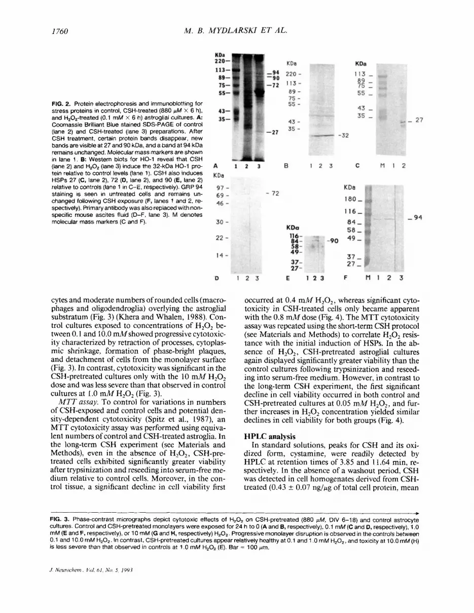
sure for 6 h to CSH consistently induced novel pro- tein bands at -27 and 90 kDa (Fig. 2). In contrast, a 94-kDa band was present in control preparations and did not appear to change with CSH treatment. Dis- tinct 32- (HO-1) and 72-kDa bands could not be clearly discerned in the treated and control prepara- tions. Many additional bands normally seen in con- trol gels were either diminished or completely elimi- nated by CSH treatment. A broad 55-kDa band was inconsistently observed in the CSH-treated gels, which may represent induction or accumulation of cytoskeletal proteins.
In western blots of untreated astrocyte cultures, HSPs 27 and 90 were consistently undetectable, and there was occasional faint staining of HSP 72 and moderate levels of HO-1 (Fig. 2). In contrast, the CSH-treated cells exhibited prominent staining for HSPs 27 and 90 and increased staining of HSP 72 (Fig. 2). Treatment of cultures with either CSH or H202 significantly increased the HO- 1 protein band intensity relative to controls. CSH induction of band triplets of HSP 27 probably represents various phos- phorylation states of the activated protein in this sys- tem (Crete and Landry, 1990; Zhou et al., 1993). In accord with the immunofluorescence data, moderate expression of GRP 94 was noted in immunoblots of control cultures and did not appear to be influenced by CSH exposure (Fig. 2). We observed a progressive increase in HSP 27, 72, and 90 expression in un- treated cultures from DIV 6 to 18, although HSP im- munostaining was generally more robust in CSH-ex- posed cells at any given time point (data not shown).
Cytotoxicity studies Phase microscopy. On DIV 20, in the absence of
H20z exposure, both control and CSH-treated cul- tures exhibited healthy confluent monolayers com- posed of phase-dark, flat, and process-bearing astro-
J. Neurochem.. Vol. 6 1. No. 5. 1993

I758 M. B. MYDLARSKI ET AL.
FIG. 1. (Above and facing page). lmrnunofluorescent labeling of stress proteins and GFAP in CSH-treated (880 KM x 6 h) and control glial cultures. Shown are control and CSH-treated cultures stained for HO-1 (A and 6, respectively), HSP 27 (C and D, respectively), HSP 72 (F and G, respectively), HSP 90 (H and I , respectively), and GRP 94 (J and K, respectively). With the exception of GRP 94 (which is constitutively present), CSH induces the expression of stress proteins in these cultures. Cells immunoreactive for HSP 27 (D) and GRP 94 (K) are shown to be GFAP-positive astrocytes in E and L, respectively. No imrnunostaining is observed when normal rabbit serum is substjtuted for anti-HO-1 (A, inset) or nonspecific mouse ascites fluid for anti-HSPs 27, 72, and 90 and anti-GRP 94 (D, inset). Bar = 25 w.
J . Neurochetn.. L’ol 61, No 5, 1993

ASTROCYTE INCLUSION RIOGENESIS 1759
J Neurochem.. Vol. 61, Mo. 5, 1993
I760 M. B. MYDLARSKI ET AL.
FIG. 2. Protein electrophoresis and immunoblotting for stress proteins in control, CSH-treated (880 pM X 6 h), and H,O,-treated (0.1 mM x 6 h) astroglial cultures. A Coomassie Brilliant Blue stained SDS-PAGE of control (lane 2) and CSH-treated (lane 3) preparations. After CSH treatment, certain protein bands disappear, new bands are visible at 27 and 90 kDa, and a band at 94 kDa remains unchanged. Molecular mass markers are shown in lane 1. B Western blots for HO-1 reveal that CSH (lane 2) and H,OZ (lane 3) induce the 32-kDa HO-1 pro- tein relative to control levels (lane 1). CSH also induces HSPs 27 (C, lane 2), 72 (D, lane 2), and 90 (E, lane 2) relative to controls (lane 1 in C-E, respectively). GRP 94 staining is seen in untreated cells and remains un- changed following CSH exposure (F, lanes 1 and 2, re- spectively). Primary antibody was also replaced with non- specific mouse ascites fluid (D-F, lane 3). M denotes molecular mass markers (C and F).
cytes and moderate numbers of rounded cells (macro- phages and oligodendroglia) overlying the astroglial substratum (Fig. 3) (Khera and Whalen, 1988). Con- trol cultures exposed to concentrations of H202 be- tween 0.1 and 10.0 M s h o w e d progressive cytotoxic- ity characterized by retraction of processes, cytoplas- mic shrinkage, formation of phase-bright plaques, and detachment of cells from the monolayer surface (Fig. 3 ) . In contrast, cytotoxicity was significant in the CSH-pretreated cultures only with the 10 mM H20z dose and was less severe than that observed in control cultures at 1 .O mM H,02 (Fig. 3 ) .
MTT assay. To control for variations in numbers of CSH-exposed and control cells and potential den- sity-dependent cytotoxicity (Spitz et al., 1987), an MTT cytotoxicity assay was performed using equiva- lent numbers of control and CSH-treated astroglia. In the long-term CSH experiment (see Materials and Methods), even in the absence of H,O,, CSH-pre- treated cells exhibited significantly greater viability after trypsinization and reseeding into serum-free me- dium relative to control cells. Moreover, in the con- trol tissue, a significant decline in cell viability first
occurred at 0.4 mM H202, whereas significant cyto- toxicity in CSH-treated cells only became apparent with the 0.8 M d o s e (Fig. 4). The MTT cytotoxicity assay was repeated using the short-term CSH protocol (see Materials and Methods) to correlate H202 resis- tance with the initial induction of HSPs. In the ab- sence of H202, CSH-pretreated astroglial cultures again displayed significantly greater viability than the control cultures following trypsinization and reseed- ing into serum-free medium. However, in contrast to the long-term CSH experiment, the first significant decline in cell viability occurred in both control and CSH-pretreated cultures at 0.05 mM H202, and fur- ther increases in H20, concentration yielded similar declines in cell viability for both groups (Fig. 4).
HPLC analysis In standard solutions, peaks for CSH and its oxi-
dized form, cystamine, were readily detected by HPLC at retention times of 3.85 and 11.64 min, re- spectively. In the absence of a washout period, CSH was detected in cell homogenates derived from CSH- treated (0.43 t 0.07 ng/wg of total cell protein, mean
v FIG. 3. Phase-contrast micrographs depict cytotoxic effects of H20z on CSH-pretreated (880 WM, DIV 6-18) and control astrocyte cultures. Control and CSH-pretreated monolayers were exposed for 24 h to 0 (A and B, respectively), 0.1 mM (C and D, respectively), 1 .O mM (E and F, respectively), or 10 mM (G and H, respectively) H,Oz. Progressive monolayer disruption is observed in the controls between 0.1 and 10.0 mM HzOp. In contrast, CSH-pretreated cultures appear relatively healthy at 0.1 and 1 .O mM H,O,, and toxicity at 10.0 mM (H) is less severe than that observed in controls at 1 .O mM HZ02 (E). Bar = 100 pm.

ASTROCYTE INCLUSION BIOGENESIS 1761
J. Neuroelzrm ~ Vol. 61, No. 5, 1993

1762 M. B. MYDLARSKI ET AL.
A B
160 r 180
20 ' ' ' . ' ' ' ' ' ' ' I ' ' ' ' ' I ' , J
0 0.1 0.2 0.3 0.4 0.5 0.6 0.7 0.8
FIG. 4. Automated MTT cell viability assay depicts cytotoxic effects of trypsinization and H202 exposure on fixed numbers (40,000 cells per well) of control and CSH-pretreated astrocytes. Optical density correlates directly with cell viability. A. Long-term CSH exposure (880 pM, DIV 6-1 8). CSH-pretreated cells (+) exhibit increased resistance to mechanical trauma and H 2 0 2 exposure relative to controls (0). 8: Short-term CSH exposure (880 gM x 6 h). As in A, CSH-pretreated cells (+) exhibit robust resistance to mechanical trauma relative to controls (0). However, normalizing for the effects of mechanical stress, both groups show similar declines in cell viability with increasing H202 concentrations. An asterisk denotes a significant difference from control values (p 5 0.050 by Student-Newman-Keuls post hoc test). An open star denotes first significant decline in cell viability relative to respective conditions at 0 mM H,O, ( p I 0.050 by Student- Newman-Keuls).
k SEM of three samplings; range, 0.29-0.5 ng/pg), but not control, cultures on DIV 18. However, follow- ing a 24-h washout period, intracellular CSH could no longer be detected in the CSH-pretreated prepara- tions. Cystamine, the disulfide form of CSH, was not detectable in any cell homogenates either before or after washout. Although the potential formation of CSH-derived mixed disulfides was not monitored, the HPLC results strongly argue against the possibility that CSH or cystamine serves as a direct cytoprotec- tant in the H,Oz toxicity assays.
DAB histochemistry Long-term exposure of astroglial cultures to H202
elicited a robust accumulation of DAB/peroxidase- positive cytoplasmic inclusions in comparison with untreated controls (Fig. 5). DAB staining in these cells is similar to that observed in cultured astroglia follow- ing prolonged CSH treatment (Schipper et al., 1990b).
DISCUSSION
In dissociated fetal or neonatal astroglial cultures, administration of CSH between 6 and 18 DIV in- duces the accumulation of cytoplasmic granules with histochemical and morphological features identical to those characterizing a subpopulation of glia in the ag- ing periventricular brain. Thus, this sulfhydryl com- pound appears to accelerate aging-related phenotypic changes in these cells (Schipper et al., 19906). As in situ, the accumulation of porphyrins and heme is thought to be responsible for the orange-red auto- fluorescence and nonenzymatic (ferrous iron-me- diated) peroxidase activity in the CSH-induced inclu-
sions, respectively (Schipper et al., 19906; McLaren et al., 1992). In the intact aging brain and in the CSH- treated cultures, the origin of the astrocytic granules and the mechanisms responsible for their biogenesis remain obscure. The results of the present study indi- cate that CSH induces HO-1 and various HSPs in cultured astroglia before granulation occurs. These observations suggest that the development of the as- trocytic organelles may be dependent on an anteced- ent cellular stress response.
CSH induces derangements in porphyrin-heme me- tabolism in liver (Peterson et al., 1989) and may influ- ence metalloporphyrin biosynthesis and sequestra- tion in astrocytes (Schipper et al., 19906). In rat liver, CSH induces HO-1 (Peterson et al., 1989), resulting in a rapid conversion of heme to biliverdin (Ten- hunen et al., 1969; Maines and Kappas, 1974). In re- sponse to acute cellular stress, induction of HO- 1 may protect cells by catabolizing prooxidant metallopor- phyrins such as heme (Misra and Fridovich, 1972; Gutteridge, 1987) to bile pigments with free radical- scavenging capabilities (Stocker, 1990; Applegate et al., 199 1). Conversely, the results of the present study suggest that chronic or repeated stress may result in complex dysregulation of the genes subserving por- phyrin-heme biosynthesis, leading to the progressive accumulation of redox-active metalloporphyrins (Maines and Kappas, 1975; Schipper et al., 1990b).
The HO- 1 gene has a heat-shock element in its pro- moter region (Pelham, 1985; Muller et al., 1987) and is up-regulated by metal ions, ionizing radiation, sulf- hydryl reactive agents, and H,02 as part of a con- certed cellular stress response (Keyse and Tyrell, 1987, 1989; Applegate et al., 1991; Dwyer et al.,
J Neirroclretn., L'oL 61. N o 5 , 1993

ASTROCYTE INCLUSION BIOG ENESIS 1763
FIG. 5. Effects of repeated H,On exposure on per- oxidase activity in cultured astroglia. H202 treat- ment (0.1 rnM, DIV 6-18) results in massive accu- mulation of peroxidase-positive cytoplasmic inclu- sions in comparison with untreated controls (inset). DAB stain. Bar = 25 um.
1992). In the present study, we observed that expo- sure of astrocyte cultures to CSH for 6 h induced HSPs 27,72, and 90 in addition to HO-1. The intensi- fication of perinuclear staining for HSPs 27, 72, and 90 noted in CSH-exposed astrocytes is consistent with reports of translocation of these stress proteins from cytoplasmic to nuclear compartments in response to heat shock (Welch and Feramisco, 1984; Welch and Suhan, 1986; Amgo and Welch, 1987; Kochevar et al., 1991).
CSH-induced HSPs in cultured astroglia may be responsible for the relative cytoprotection from me- chanical and H202 damage as determined by phase- contrast microscopy and a sensitive MTT cell viabil- ity assay. In many tissues, the increased expression of HSPs associated with a sublethal insult confers en- hanced resistance to a host of subsequent stressors (Li and Hahn, 1978; Li and Werb, 1982; Spitz et al., 1987; Morimoto et al., 1990; Oesterreich et al., 199 1). HSPs may protect cells undergoing stress by preven- tion of damage to the translational apparatus (Liu et al., 1992), maintenance of lipid membrane integrity (Burdon et al., 1987), accelerating degradation of de- natured and abnormal proteins (Ananthan et al., 1986), and prevention of deleterious protein aggrega- tion by prior binding to exposed hydrophobic surfaces (Finley et al., 1984).
The specific CSH-induced stress protein(s) directly responsible for subsequent H202 resistance in astro- cytes remain(s) to be determined. Huot et al. (1 99 1) demonstrated that transfected fibroblasts overex- pressing HSP 27 were more resistant to treatments with H202, sodium arsenite, heat, and certain anti- cancer agents relative to nonoverexpressing trans- fected controls. Astrocytes assessed for cytotoxicity at 24 h following the initiation of CSH exposure, long before granulation occurs, exhibited robust resistance
to mechanical stress but similar vulnerability to H202 relative to controls. In contrast, cells pretreated with CSH for 12 days, a regimen that induces the accumu- lation of peroxidase-positive granules (Schipper et al., 1990b), displayed significant resistance to both me- chanical trauma and H202. These observations raise the interesting possibility that, in addition to the clas- sic heat-shock response, the nonenzymatic peroxidase activity induced by CSH in these cells confers some cytoprotection against H202. The decreased vulnera- bility of control DIV 18 cells as compared with DIV 6 cells at the lower range of H20, concentrations (Fig. 4) may be due to the gradual increase in HSP expres- sion noted in untreated cultures with time in vitro (Nishimura et al., 199 1 ; present study).
The CSH-induced astrocytic inclusions may repre- sent “heat-shock granules” akin to those observed in other cells following sustained stress. In some cases, heat-shock granules consisting of polymerized small HSPs, such as HSP 27 and HSP-protein complexes, may protect various mRNAs and critical proteins from stress-related damage and degradation (Pelham, 1986; Nover et al., 1989). Using laser scanning confo- cal microscopy, we have recently observed that HSP 27 colocalizes to the orange-red autofluorescent astro- cyte inclusions in CSH-treated astroglial cultures and in subependymal regions of the adult rat brain (Myd- larsh and Schipper, 1993). Because CSH-induced as- trocyte inclusions are histochemically and morpholog- ically identical to those that accumulate in periventric- ular brain regions as a function of aging, our results indicate that chronic or repeated induction of the cel- lular stress response may be responsible for the bio- genesis of peroxidase-positive “stress granules” in as- trocytes ofthe aging periventricular brain. It is ofinter- est that in all species surveyed, the glial inclusions predominate in blood-brain bamer-deficient regions
J. Ncuroc-hem., Vol. 61, No. 5, 1993

I764 M. B. MYDLARSKI ET AL.
of the CNS (reviewed by Schipper, 199 1). Astrocytes inhabiting these areas may be particularly susceptible to HSP induction (and subsequent granulation) by unidentified blood-borne stressors.
The astroglial stress proteins induced by CSH, namely, HO-1 and HSP 27, 72, and 90, are typically up-regulated by heat shock and oxidative stressors (Applegate et al., 1991; Dwyer et al., 1992; for re- views, see Schlesinger et al., 1982; Donati et al., 1990). In contrast, CSH had little or no effect on the expres- sion of GRP 94, a stress protein responsive to glucose deprivation and calcium ionophores but not to heat shock or oxidative stress (Welch et al., 1983; Gomer et al., 1991). The oxidation of CSH in the presence of transition metals generates several prooxidant spe- cies, including the thiyl radical, superoxide anion, H202, and the hydroxyl radical (Munday, 1989). Fur- thermore, H202, a potent oxidant and inducer of HO- 1 in rat astrocytes (Dwyer et al., 1992; present study) and other mammalian cells (Keyse and Tyrell, 1987), stimulated the accumulation of peroxidase-positive astrocyte granules following prolonged treatment (Fig. 5), analogous to the effects of CSH (Schipper et al., 19906). Taken together, our results suggest that sustained intracellular oxidative stress may be the “final common pathway” responsible for the con- certed up-regulation of HO- 1 ’ and other heat-shock proteins that participate in the biogenesis of metallo- porphyrin-rich astrocytic inclusions in vitro and in the intact aging brain. In support of this hypothesis, ionizing radiation, a known generator of intracellular prooxidant intermediates, increases numbers of per- oxidase-positive glial granules in the rat hypothala- mus in a dose-dependent manner (Srebro, 197 1).
Peroxidase-positive astrocytes accumulate in aging human forebrain (Schipper, 199 l), and identical non- enzymatic (heme-mediated) peroxidase activity in- duced in rat astroglia by CSH exposure promotes the robust oxidation of dopamine to potentially neuro- toxic semiquinone intermediates (Schipper et al., 199 1). These observations raise the possibility that specific stress-related derangements in glial por- phyrin-heme metabolism may play an active role in the development of parkinsonism and other free radi- cal-related neurodegenerations.
Acknowledgment: The authors thank Dr. Mane Beaudet for assistance with statistical processing and Ms. Adina Ma- teescu-Cantuniari and Mr. William Lubenskyi for excellent technical assistance. We also gratefully acknowledge the sec- retarial help of Ms. Rhona Rosenzweig. This work was funded by the Medical Research Council of Canada and the Fonds de la Recherche en SantC d u QuCbec.
REFERENCES Ananthan J., Goldberg A. L., and Voellmy R. (1986) Abnormal
proteins serve as eukaryotic stress signals and trigger the activa- tion of heat shock genes. Science 232, 522-524.
Applegate L. A., Luscher P., and Tyrrell R. M. (199 1) Induction of
heme oxygenase: a general response to oxidant stress in cul- tured mammalian cells. Cancer Rex 51,974-978.
Amgo A. P. and Welch W. J. ( I 987) Purification and characteriza- tion of the small 28 kd mammalian stress protein. J. Biol. Chem. 262, 15359- 15369.
Brawer J., Schipper H., and Naftolin F. (1980) Ovary-dependent degeneration in the hypothalamic arcuate nucleus. Endocrinol-
Brawer J., Schipper H., and Robaire B. (1983) Effects of long-term androgen and estradiol exposure on the hypothalamus. Endo- crinology 112, 194-199.
Burdon R. H., Gill V. M., and Rice-Evans C. (1987) Oxidative stress and heat shock protein induction in human cells. Free Radic. Rev. Commun. 3, 129-139.
Crete P. and Landry J. (1990) Induction ofHSP27 phosphorylation and thermoresistance in Chinese hamster cells by arsenite, cy- cloheximide, A23187 and EGTA. Radiat. Res. 121,320-327.
Donati Y. R. A,, Slosman D. O., and Polla B. S. (1990) Oxidative injury and the heat shock response. Biochem. Pharmacol. 40,
Dwyer B. E., Nishimura R. N., de Vellis J., and Yoshida T. (1992) Heme oxygenase is a heat shock protein and PEST protein in rat astroglial cells. Glia 5, 300-305.
Finley D., Ciechanover A,, and Varshavsky A. (1984) Thermolabil- ity of ubiquitin-activating enzyme from the mammalian cell cycle mutant ts85. Cell 37, 43-55.
Goldgefter L., Schejter A. S., and Gill D. (1980) Structural and microspectrofluorometric studies on glial cells from the peri- ventricular and arcuate nuclei of the rat hypothalamus. Cell Tissue Res. 211, 503-510.
Gomer C. J., Ferrario A., Rucker N., Wong S., and Lee A. S. ( 1 99 I ) Glucose regulated protein induction and cellular resistance to oxidative stress mediated by porphyrin photosensitization. Cancer Res. 51, 6574-6579.
Gutteridge J. M. C. ( I 987) The antioxidant activity of haptoglobin towards hemoglobin-stimulated lipid peroxidation. Biochim. Biophys. Acta 917, 219-223.
Hansen M. B., Nielsen S. E., and Berg K. (1989) Re-examination and further development of a precise and rapid dye method for measuring cell growth/cell kill. J. Immunol. Methods 119,
Huot J., Roy G., Lambert H., Chretien P., and Landry J. (1991) Increased survival after treatments with anticancer agents of Chinese hamster cells expressing the human M, 27,000 heat shock protein. Cancer Rex 51, 5245-5252.
Keefer D. A. and Christ J. F. (1976) Distribution of endogenous diaminobenzidine-staining cells in the normal rat brain. Brain Res. 116, 312-316.
Keyse S. M. and Tyrrell R. M. (1987) Both near ultraviolet radia- tion and the oxidizing agent hydrogen peroxide induce a 32- kDa stress protein in normal human skin fibroblasts. J. Biol. Chem. 262, 14821-14825.
Keyse S. M. and Tyrrell R. M. (1989) Heme oxygenase is the major 32-kDa stress protein induced in human skin fibroblasts by UVA radiation, hydrogen peroxide, and sodium arsenite. Proc. Natl. Acad. Sci. USA 86, 99-103.
Khera K. S. and Whalen C. (1988) Detection of neuroteratogens with an in vitro cytotoxicity assay using primary monolayers cultured from dissociated foetal rat brains. Toxicd. I n Vilro 2,
Kochevar D. T., Aucoin M. M., and Cooper J. (1991) Mammalian heat shock proteins: an overview with a systems perspective. Toxicol. Lett. 56, 243-261.
Laemmli U. K. (1970) Cleavage of structural proteins during the assembly of the head of bacteriophage T4. Nature 227, 680- 685.
Li G. C. and Hahn G. M. (1978) Ethanol induced tolerance to heat and to adriamycin. Nature 274,699-701.
Li G. C. and Werb Z. ( 1 982) Correlation between synthesis of heat shock proteins and development of thermotolerance in Chi-
ogy 107,214-219.
2571-2577.
203-2 10.
251-273.
J Nruroclwm, I h l 61, N o 5 . 1993

ASTROCYTE INCLUSION BIOGENESIS 1765
nese hamster fibroblasts. Proc. Natl. Acad. Sci. USA 79,321 8- 3222.
Liu R. Y., Li X., Li L., and Li G. C. (1992) Expression of human hsp7O in rat fibroblasts enhances cell survival and facilitates recovery from translational and transcriptional inhibition fol- lowing heat shock. Cancer Res. 52,3667-3673.
Lowry 0. H., Rosebrough N. J., Farr A. L., and Randall R. J. (I95 1) Protein measurements with the Folin phenol reagent. J. Biol. Chem. 193,265-275.
Maines M. D. and Kappas A. (1974) Cobalt induction of hepatic heme oxygenase; with evidence that cytochrome P-450 is not essential for this enzyme activity. Proc. Natl. Acad. Sci. USA
Maines M. D. and Kappas A. (1975) Cobalt stimulation of heme degradation in the liver. J. Biol. Chem. 250,4 17 1-4 177.
Manthorpe M., Fagnani R., Skaper S. D., and Varon S. (1986) An automated colorimetric microassay for neuronotrophic fac- tors. Dev. Brain Res. 25, 191-198.
McLaren J., Brawer J. R., and Schipper H. M. (1992) Iron content correlates with peroxidase activity in cysteamine-induced as- troglial organelles. J. Histochem. Cytochem. 40, 1887- 1897.
Misra H. P. and Fridovich I. (1972) The generation of superoxide radical during the autoxidation of hemoglobin. J. Biol. Chem.
Morimoto R. I., Tissieres A,, and Georgopoulos C. (1990) The stress response, function of the proteins, and perspectives, in Stress Protein in Biology and Medicine (Morimoto R. I., Tis- sieres A,, and Georgopoulos C., eds), pp. 1-36. Cold Spring Harbor Laboratory, Cold Spring Harbor, New York.
Mosmann T. (1983) Rapid colorimetric assay for cellular growth and survival: application to proliferation and cytotoxic assays. J. Immunol. Methods 65, 55-63.
Miiller R. M., Taguchi H., and Shibahara S. (1987) Nucleotide sequence and organization of the rat heme oxygenase gene. J. Biol. Chem. 262,6795-6802.
Muller-Eberhard U. and Nikkila H. (1989) Transport of tetrapyr- roles by proteins. Semin. Hematol. 26, 86-104.
Munday R. (1989) Toxicity of thiols and disulphides: involvement of free-radical species. Free Radic. Biol. Med. 7,659-673.
Mydlarski M. B. and Schipper H. M. (1993) Stress protein co-locali- zation to autofluorescent astrocytic inclusions in situ and in cysteamine-treated glial cultures. Brain Res. (in press).
Nishimura R. N., Dwyer B. E., Clegg K., Cole R., and de Vellis J. ( 199 I) Comparison of the heat shock response in cultured cor- tical neurons and astrocytes. Mol. Brain Res. 9, 39-45.
Nover L., Scharf K.-D., and Neumann D. (1989) Cytoplasmic heat shock granules are formed from precursor particles and are associated with a specific set of mRNAs. Mol. Cell. Biol. 9,
Oesterreich S., Schunck H., Benndorf R., and Bielka H. (1991) Cisplatin induces the small heat shock protein HSP25 and thermotolerance in Ehrlich ascites tumor cells. Biochem. Biophys. Res. Commun. 180,243-248.
Pelham H. R. B. (1985) Activation of heat-shock genes in eukary- otes. Trends Genet. 1, 3 1-35.
Pelham H. R. B. (1986) Speculations on the functions of the major heat shock and glucose-regulated proteins. Cell 46,959-96 I .
Peterson T., Peterson M., and Williams C. (1989) The role of heme
71,4293-4297.
247,6960-6962.
1298-1308.
oxygenase and aryl hydrocarbon hydroxylase in the protection by cysteamine from acetaminophen hepatotoxicity. Toxicol. Appl. Pharmacol. 97,430-439.
Schipper H. M. (199 I ) Gomori-positive astrocytes: biological prop- erties and implications for neurologic and neuroendocrine dis- orders. Glia 4, 365-377.
Schipper H. M. and Mateescu-Cantuniari A. (I99 1) Identification of peroxidase-positive astrocytes by combined histochemical and immunolabeling techniques in situ and in cell culture. J. Histochem. Cytochem. 39, 1009-1016.
Schipper H., Brawer J. R., Nelson J. F., Felicio L. S., and Finch C. E. (1 98 1) Role of the gonads in the histologic aging of the hypothalamic arcuate nucleus. Bid. Reprod. 25, 4 13-4 19.
Schipper H. M., Lechan R. M., and Reichlin S. (1990a)Glial perox- idase activity in the hypothalamic arcuate nucleus: effects of estradiol valerate-induced persistent estrus. Brain Res. 507,
Schipper H. M., Scarborough D. E., Lechan R. M., and Reichlin S. (l990b) Gomori-positive astrocytes in primary culture: effects of in uitro age and cysteamine exposure. Dev. Brain Res. 54,
Schipper H. M., Kotake Y., and Janzen E. G. (1991) Catechol oxi- dation by peroxidase-positive astrocytes in primary culture: an electron spin resonance study. J. Neurosci. 11, 2 170-2 176.
Schlesinger M. J., Ashburner M., and Tissieres A., eds (1982) Heat Shock: From Bacteria to Man. Cold Spring Harbor Labora- tory, Cold Spring Harbor, New York.
Spitz D. R., Dewey W. C., and Li G. C. (1987) Hydrogen peroxide or heat shock induces resistance to hydrogen peroxide in Chi- nese hamster fibroblasts. J. Cell. Physiol. 131, 364-373.
Srebro Z. (197 1) Penventricular Comori-positive glia in brains of X-irradiated rats. Brain Res. 35,463-468.
Srebro Z. and Cichocki T. ( 1 97 I ) A system of penventricular glia in brain characterized by large peroxisome-like organelles. Acta Histochem. 41, 108-1 14.
Stocker R. ( 1 990) Induction of haem oxygenase as a defence against oxidative stress. Free Radic. Res. Commun. 9, 10 1 - 1 12.
Tenhunen R., Marver H. S., and Schmid R. (1969) Microsomal heme oxygenase: characterization of the enzyme. J. Biol. Chem. 244,6388-6394.
Towbin H., Staehelin T., and Gordon J. (1979) Electrophoretic transfer of proteins from polyacrylamide gels to nitrocellulose sheets: procedure and some applications. Proc. Natl. Acad. Sci.
Welch W. J. and Feramisco J. R. (1984) Nuclear and nucleolar location of the 72000-dalton heat shock protein in heat- shocked mammalian cells. J. Biol. Chem. 259,4501-45 13.
Welch W. J. and Suhan J. P. (1986) Cellular and biochemical events in mammalian cells during and after recovery from physiological stress. J. Cell Biol. 103, 2035-2052.
Welch W. J., Carrels J. G., Thomas G. P., Lin J. J., and Feramisco J. R. (1983) Biochemical characterization of the mammalian stress proteins and identification of two stress proteins as glu- cose and Ca+’ ionophore regulated proteins. J. Biol. Chem.
Zhou M., Lambert H., and Landry J. ( 1 993) Transient activation of a distinct serine protein kinase is responsible for 27-kDa heat shock protein phosphorylation in nitrogen-stimulated and heat-shocked cells. J. Biol. Chem. 268, 35-43.
200-207.
7 1-79.
USA 76,4350-4354.
258,7 102-7 1 1 I .
J. Neirrochem., Vol. 61, No. 5 , 1993


