Embed Size (px)
Citation preview

doi:10.1111/j.1440-1746.2006.04353.x
Journal of Gastroenterology and Hepatology
22
(2007) 749–756 © 2006 The Authors
749
Journal compilation © 2007 Journal of Gastroenterology and Hepatology Foundation and Blackwell Publishing Asia Pty Ltd
Blackwell Publishing AsiaMelbourne, AustraliaJGHJournal of Gastroenterology and Hepatology0815 93192006 Blackwell Publishing Asia Pty Ltd200622749756Original Article Candida aggravates duodenal perforationT Nakamura
et al.
GASTROENTEROLOGY
Candida albicans
aggravates duodenal ulcer perforation induced by administration of cysteamine in rats
Tetsuya Nakamura,* Masashi Yoshida,* Hideki Ishikawa,
†
Kaori Kameyama,
‡
Go Wakabayashi,* Yoshihide Otani,* Motohide Shimazu,* Minoru Tanabe,* Shigeyuki Kawachi,* Koichiro Kumai,
§
Tetsuro Kubota,* Yoshiro Saikawa,* Katsuko Sano* and Masaki Kitajima*
Departments of *Surgery and
†
Emergency and Critical Care Medicine,
‡
Division of Diagnostic Pathology, and
§
Center for Diagnostic and Therapeutic Endoscopy, Keio University School of Medicine, Tokyo, Japan
Abstract
Background:
Candida sp
are frequently isolated from the ascitic fluid of patients withperforated ulcers. The present study was performed to examine whether
Candida
infectionmay be involved in the process of ulcer perforation.
Methods:
Male Wistar rats were divided into a saline group (
n
=
15) and a
Candida
group(
n
=
17). Cysteamine-HCl (Sigma; 31 mg/100 g) was administered thrice on day 1 to bothgroups of animals.
Candida albicans
at a density of 10
8
in 0.5 mL of saline was adminis-tered 1 h before, and 12 h and 24 h after the first administration of cysteamine in the
Candida
group.
Results:
Perforated duodenal ulcers were observed in 94.1% of the rats in the
Candida
group, but only 26.7% of the rats in the saline group (
P
<
0.01). The area of the duodenalulcers in the
Candida
group was 40.89
±
33.07 mm
2
, whereas that in the saline group was16.53
±
20.4 mm
2
(
P
<
0.05). The mortality rate was significantly higher in the
Candida
group than in the saline group. In the
Candida
group, colonization by
C. albicans
wasrecognized at the ulcer base, surrounded by marked granulocytic infiltration. The numberof eosinophils infiltrating the ulcer base was also significantly greater in the
Candida
groupthan in the saline group. Immunohistochemical analysis revealed the expression of secre-tory aspartyl protease (SAP) in the region of the ulcer showing colonization by
C. albicans
in the
Candida
group.
Conclusion:
Candida albicans
aggravates duodenal ulcer perforation in the experimentalmodel of cysteamine-induced duodenal ulcer perforation. The present findings suggest thatSAP and host–parasite relationships, including granulocyte-dependent mechanisms, maybe involved in the aggravation of ulcer perforation by
C. albicans.
Introduction
Although the mechanisms of ulcer formation have been well-studied, the process of ulcer perforation has not yet been clearlyelucidated. While the incidence of surgical intervention in gas-troduodenal ulcer diseases has reduced dramatically with thedevelopment of effective antisecretory drugs and effective regi-mens for the eradication of
Helicobacter pylori
, the incidence ofulcer perforation remains unchanged.
1
A strong association hasbeen reported between gastroduodenal ulcers and
H. pylori
infec-tion, and the mean reported prevalence of
H. pylori
infection inuncomplicated ulcer disease is 90–100%.
2
In contrast, the meanprevalence of
H. pylori
infection in patients with perforated peptic
ulcers is only approximately 42.1–73.3%.
2–4
Therefore, it wassurmised that some other factors may be involved in the process ofulcer perforation.
In a previous study,
Candida
species were isolated from theperitoneal fluid in 23 out of 62 patients with perforated pepticulcer, and bacteria were isolated in 10 out of the remaining 39cases.
5
In another study, fungal colonization was reported in54.2% of cases of gastric ulcer, 10.3% of cases of chronic gastritis,and 4.3% of negative controls.
6
Pathological examination ofresected specimens from patients with perforated ulcers in onestudy revealed the abundant presence of
Candida albicans
in theeffusion layer, granulation layer, and the necrotic tissue layer at theulcer base.
7
Thus, we speculated that
C. albicans
may play a role
Key words
eosinophil, helicobacter pylori, infection, secretory aspartyl protease, stomach.
Accepted for publication 16 September 2005.
Correspondence
Dr Masashi Yoshida, 35 Shinanomachi, Shinjyuku-ku, Tokyo 160-8582, Japan. Email: [email protected]
750
Journal of Gastroenterology and Hepatology
22
(2007) 749–756 © 2006 The Authors
Journal compilation © 2007 Journal of Gastroenterology and Hepatology Foundation and Blackwell Publishing Asia Pty Ltd
Candida
aggravates duodenal perforation
T Nakamura
et al.
in the process of gastroduodenal ulcer perforation, and performedanimal experiments to verify this hypothesis.
Propionitrile,
8
1-methyl-4-phenyl-1,2,3,6-tetrahydropyridine,
9
mepirizole
10
and cysteamine-HCl (cysteamine)
11
have beenreported as duodenal ulcerogens, and among these, cysteamine hasbeen shown to be the most potent at inducing perforation ofduodenal ulcers.
12
Cysteamine administered either s.c. or intragastrically (i.g), inexperimental studies has been shown to become concentrated inthe duodenum.
13
Increase in duodenal endothelin-1 concentrationsfrom 15 min, and decreased duodenal mucosal blood flow at30 min after cysteamine administration have been demon-strated.
14,15
Endothelin-related increased vascular permeability hasbeen commonly observed in the early phase of duodenalulceration
16
and it has been suggested that organ- and molecular-specific overexpression of
p27
and
p21
in the duodenal mucosamay be associated with decreased cellular proliferation and repre-sent new pathogenetic elements in ulcer development.
17
In the present study,
C. albicans
and cysteamine were adminis-tered i.g. to male Wistar rats in order to examine whether or not
C. albicans
would aggravate duodenal ulcer perforation.
Methods
Animals and procedures
Male Wistar rats weighing 220–240 g were anesthetized withdiethyl ether, before administration of cysteamine (Sigma, StLouis, MO, USA) at the dose of 31 mg/kg three times at 4-hourlyintervals on day 1.
Candida albicans
(
C. albicans
19002 Fujisawa,kindly provided by Fujisawa Pharmaceutical, Osaka, Japan
18
) wasadministered at a density of 10
8
in 0.5 mL saline 1 h before, and12 h and 24 h after the cysteamine administration. The animalexperiments were conducted in accordance with the guidelines ofthe Keio University School of Medicine. There were two experi-mental groups in the study: the saline group (
n
=
15) and the
Candida
group (
n
=
17).In additional experiments, six rats (
Candida
without cysteaminegroup) were given saline and
C. albicans
1 h before, and 12 h and24 h after saline administration.
Gastric and duodenal ulcer perforation
Seventy-two hours after cysteamine administration, the rats werekilled, and the depth and size of the ulcers were evaluated. Theulcers were classified according to the depth, as follows: Ul-I(erosion); Ul-II (shallow), ulceration extending to the submucosa;Ul-III (intermediate), ulceration extending to the muscularis pro-pria; and Ul-IV (deep excavation), ulceration extending beyondthe muscularis propria.
19
The two greatest diameters of the ulcerwere measured and the area of the ulcer was calculated.
20
Speci-mens were stained with hematoxylin–eosin (HE), periodic acid–Schiff (PAS) reagent, Grocott’s silver, and Luna. The number ofeosinophils infiltrating the ulcer base was counted. We also con-ducted immunohistochemical staining of the duodenal ulcer usinga monoclonal antibody (TaKaRa code M166, TaKaRa Bio, Shiga,Japan) to recognize the expression of secretory aspartyl protease 2(Sap-2). Rats that died within 72 h of cysteamine administrationwere autopsied and the depth and size of the ulcers were evaluated.
Enzyme-linked immunosorbent assay for Sap-2
The Sap-2 concentrations in the ulcer tissue specimens (100 mm
2
)were measured using an enzyme-linked immunosorbent assay(ELISA) kit (KPL code 05-10-06, Kirkegaard & Perry Laborato-ries, Gaithersburg, MD, USA) with Sap 2 antigen (TaKaRa codeMG002) and antibody (TaKaRa code M166), in accordance withthe manufacturer’s instructions. There were two experimentalgroups in the study: the saline group and the
Candida
group.
Statistical analysis
Data are presented as mean
±
SE. The statistical significance ofdifferences between the two groups was determined by Fisher’sexact test, the Kaplan–Meier method and the Mann–Whitney test.
Results
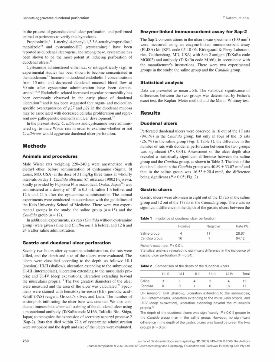
Duodenal ulcers
Perforated duodenal ulcers were observed in 16 out of the 17 rats(94.1%) in the
Candida
group, but only in four of the 15 rats(26.7%) in the saline group (Fig. 1, Table 1); the difference in thenumber of rats with duodenal perforation between the two groupswas significant (
P
<
0.01). Assessment of the ulcer depth alsorevealed a statistically significant difference between the salinegroup and the
Candida
group, as shown in Table 2. The area of theduodenal ulcers in the
Candida
group was 40.89
±
33.07 mm
2
andthat in the saline group was 16.53
±
20.4 mm
2
, the differencebeing significant (
P
<
0.05; Fig. 2).
Gastric ulcers
Gastric ulcers were also seen in eight out of the 15 rats in the salinegroup and 12 out of the 17 rats in the
Candida
group. There was nosignificant difference in the depth of the gastric ulcers between the
Table 1
Incidence of duodenal ulcer perforation
Positive Negative Rate (%)
Saline group 4 11 26.67
Candida
group 16 1 94.12
Fisher’s exact test
P
<
0.01.Statistical analysis revealed no significant difference in the incidence ofgastric ulcer perforation (
P
=
0.34).
Table 2
Comparison of the depth of the duodenal ulcers
Ul- 0 Ul-I Ul-II Ul-III Ul-IV Total
Saline 3 1 4 3 4 15
Candida
0 0 1 0 16 17
Ul-I (erosion); Ul-II (shallow), ulceration extending to the submucosa;Ul-III (intermediate), ulceration extending to the muscularis propria; andUl-IV (deep excavation), ulceration extending beyond the muscularispropria.
19
The depth of the duodenal ulcers was significantly (
P
<
0.01) greater inthe
Candida
group than in the saline group. However, no significantdifference in the depth of the gastric ulcers was found between the twogroups (
P
=
0.67).

T Nakamura
et al.
Candida
aggravates duodenal perforation
Journal of Gastroenterology and Hepatology
22
(2007) 749–756 © 2006 The Authors
751
Journal compilation © 2007 Journal of Gastroenterology and Hepatology Foundation and Blackwell Publishing Asia Pty Ltd
two groups. Perforation of the gastric ulcer was seen in three ratsin the saline group and in six rats in the
Candida
group, thedifference between the two groups not being significant.
The area of the gastric ulcers was 15.70
±
6.37 mm
2
in thesaline group and 25.66
±
7.45 mm
2
in the
Candida
group. Thus,the area of the gastric ulcers in the
Candida
group wasapproximately 63% larger than that in the saline group; the differ-ence between the two groups, however, did not attain statisticalsignificance.
Survival rate
Figure 3 presents the results of the Kaplan–Meier survival analysisconducted to compare the mortality rate between the saline group
and the Candida group. The survival rate was found to be signifi-cantly lower in the Candida group than in the saline group(P < 0.05).
Histological findings and results of enzyme-linked immunosorbent assay
Staining of the specimens with HE, PAS reagent and Grocott’ssilver revealed the presence of neither the hyphal forms nor theyeast forms of fungi in the saline group. However, in the Candidagroup, the ulcer base was found to be colonized by C. albicans,surrounded by marked granulocytic infiltration (Fig. 4a,b). Speci-mens stained with PAS reagent and Grocott’s silver also revealed
Figure 1 Macroscopic features of gastric and duodenal mucosa in the (a) saline group and (b) Candida group.
(a) (b)
Figure 2 Area of the duodenal and gastric ulcers. The area of theduodenal ulcers was larger in the (�) Candida group than that in the (�)saline group (**P < 0.01). Statistical analysis showed no significant dif-ference in the area of the gastric ulcers between the two groups.
01020304050607080
Ulc
er s
ize
(mm
2 )
**NS
Duodenal ulcer Gastric ulcer(n=17) (n=17)(n=15) (n=15)
Figure 3 Survival rate after administration of cysteamine. The survivalrate in the (––) Candida group (n = 17) was significantly lower than thatin the (····) saline group (n = 15; P < 0.05; Kaplan–Meier).
0
0.2
0.4
0.6
0.8
1.0
0 10 20 30 40 50 60 70 80Time (h)
Surv
ival
rat
e

752 Journal of Gastroenterology and Hepatology 22 (2007) 749–756 © 2006 The Authors
Journal compilation © 2007 Journal of Gastroenterology and Hepatology Foundation and Blackwell Publishing Asia Pty Ltd
Candida aggravates duodenal perforation T Nakamura et al.
Figure 4 Histological findings. Histological examination of sections stained with (a,b) HE, (c,d) periodic acid–Schiff (PAS) and (e,f) Grocott’s silverstains. Both yeasts and hyphae of Candida albicans were recognized by PAS and Grocott’s silver staining. Colonization by C. albicans was found onlyat the ulcer base in the Candida group, and the organisms were surrounded by marked granulocytic infiltration. The Candida-infected parts of the ulcerbase were found to especially deep. Bars: (a,c,e) 2.0 mm; (b,d,f) 200 µm.
A B
C D
E F

T Nakamura et al. Candida aggravates duodenal perforation
Journal of Gastroenterology and Hepatology 22 (2007) 749–756 © 2006 The Authors 753
Journal compilation © 2007 Journal of Gastroenterology and Hepatology Foundation and Blackwell Publishing Asia Pty Ltd
the presence of yeasts, and fungal hyphae were easily recognizedunder low magnification (Fig. 4c–f). Examination under low mag-nification revealed that the part of the ulcer base colonized byCandida was especially deep. In the Candida group, eosinophilinfiltration was also demonstrated at the ulcer surface by Lunastaining (Fig. 5b), and statistical analysis showed that the numberof infiltrating eosinophils in the Candida group was significantlylarger than that in the saline group (Fig. 6; P < 0.05). Furthermore,immunohistochemical staining with an anti-Sap-2 antibodyrevealed the distribution of Sap-2 in the ulcer in the Candidagroup. It was found in the tissue around the infiltrating part of
C. albicans, including the extracellular part of inflammatory cells(Fig. 7c); in contrast, negligible expression of this enzyme wasfound in the saline group (Fig. 7a,b). The Sap-2 concentrations inthe ulcer tissue specimens (100 mm2) were 79.08 ± 10.13 ng/mg(protein) in the Candida group and 31.50 ± 9.36 ng/mg (protein)in the saline group, the difference between the two groups beingsignificant (P < 0.01; Fig. 8).
Administration of Candida alone
Among the six rats given Candida alone without cysteamine (Can-dida without cysteamine group), only one rat had gastric erosion,and none of the rats had gastric or duodenal ulceration (Table 3).
DiscussionIt was shown in the present study that C. albicans aggravatedcysteamine-induced duodenal ulcer perforation in rats. As notedhere, Candida sp have been frequently reported to be isolated fromthe ascitic fluid of patients with perforated peptic ulcer. Althoughit is difficult to demonstrate the role of Candida sp in the processof ulcer perforation by clinical examination, in vivo animal exper-iments are a necessary approach to investigate the mechanisms ofulcer perforation.
In regard to ulcers in the duodenum, the rate of perforation,depth, and area of the ulcers were significantly worse in the Can-dida group than in the saline group in the present study. The
Figure 5 Histological examination of Luna-stained sections for eosinophils. Luna staining revealed eosinophilic infiltration at the ulcer base in the (b)Candida group, whereas only negligible infiltration was observed in the (a) saline group. Bars, 200 µm.
A B
Figure 6 Eosinophil counts at the ulcer base. The number of eosino-phils infiltrating the ulcer base was counted, and statistical analysisshowed that the number of infiltrating eosinophils observed per high-power field (HPF) was higher in the (�) Candida group than in the (�)saline group (*P < 0.05).
0
10
20
30
40
50
60
70
80
90
100*
Eos
inop
hilc
ount
/HPF
Candida groupSaline group(n=4) (n=8)
Table 3 Administration of Candida alone
Ul- 0 Ul-I Ul-II Ul-III Ul-IV Total
Duodenal lesion 6 0 0 0 0 6Gastric lesion 5 1 0 0 0 6
Six rats were given only saline followed by candida albicans 1 h beforeand 12 h and 24 h after saline administration (Candida without cysteam-ine group); only one rat in this group had gastric erosions, and none ofthe rats had gastric or duodenal ulceration.

754 Journal of Gastroenterology and Hepatology 22 (2007) 749–756 © 2006 The Authors
Journal compilation © 2007 Journal of Gastroenterology and Hepatology Foundation and Blackwell Publishing Asia Pty Ltd
Candida aggravates duodenal perforation T Nakamura et al.
mechanisms of ulcer aggravation can be discussed from twoperspectives: host–parasite relationships, including granulocyte-dependent mechanisms,21 and virulence factors of Candida sp. Ithas been reported that locally produced interleukin (IL)-5 inducedby Candida antigen contributes to the eosinophilic infiltration,22
and that expression of matrix metalloproteinase-1 (MMP-1)mRNA can be observed in granulocytes, particularly in eosino-phils, and fibroblasts infiltrating the ulcer base in surgicallyresected specimens.23 The extracellular matrix of the upper gas-trointestinal wall consists mainly of type I and III collagen, whichare selectively degraded by MMP-1. In the present study, a greaternumber of eosinophils was found at the ulcer base in the Candidagroup as compared to that in the saline group. It is possibletherefore that granulocytes, including eosinophils, also participatein the process of ulcer perforation. In regard to the virulencefactors of the Candida sp, some of the factors that can be consid-ered are adhesion, secretion of proteases and hyphal formation.Proteases, including Sap and phospholipases, secreted by Candidasp may directly induce damage of the gastroduodenal wall.24,25 Inthe present study, Sap expression at the ulcer base was found onimmunohistochemical examination only in the Candida group,and the Sap-2 concentrations were significantly higher in the Can-dida group than in the saline group. It has been reported that this
Figure 7 Immunohistochemical analysis for secretory aspartyle protease-2 (Sap-2). (c) Immunohistochemical staining of duodenal ulcer withantisecretory aspartyl protease antibody revealed expression of SAP in the portion of the ulcer colonized by C. albicans in the Candida group, whereas(a,b) only negligible expression of the enzyme was observed in the saline group. Bars: (a) 2.0 mm; (b,c) 200 µm.
A B
C
Figure 8 Secretory aspartyle protease-2 (Sap-2) concentrations inulcer tissue. The Sap- 2 concentrations in the ulcer tissue specimens(100 mm2) were measured using an enzyme-linked immunosorbentassay (ELISA) kit (KPL code 05-10-06) with Sap-2 antigen (TaKaRa codeMG002) and antibody (TaKaRa code M166), in accordance with themanufacturer’s instructions. Statistical analysis showed that the Sap-2concentrations in the ulcer tissue were higher in the (�) Candida groupthan in the (�) saline group (**P < 0.01).
0
20
40
60
80
100
Saline group Candida group
**
ng/m
g(pr
otei
n)
(n=6) (n=13)

T Nakamura et al. Candida aggravates duodenal perforation
Journal of Gastroenterology and Hepatology 22 (2007) 749–756 © 2006 The Authors 755
Journal compilation © 2007 Journal of Gastroenterology and Hepatology Foundation and Blackwell Publishing Asia Pty Ltd
protease enables the organism to invade the epithelium.26 Clinicaland experimental evidence suggests that secretory protease mayplay a role as a virulence factor in the pathogenesis of candidalvaginitis.27
It is possible therefore that Sap also has a role in the process ofulcer perforation. From the perspective of wound healing, we havereported that disturbance of the healing process not only delayswound healing, but also aggravates ulcerative lesions.28 Candidahas been reported as a possible cause of delayed wound healing inthe skin (by causing failure of epithelialization).29 Therefore, it ispossible that C. albicans also disturbs the healing process of gas-troduodenal ulcers. From our observations, we propose that bothvirulence factors of C. albicans, and host–parasite relationships,including granulocyte-dependent mechanisms, may be involved inthe process of ulcer perforation.
In the present study Candida alone did not induce gastric orduodenal ulceration. Therefore, Candida was considered to be anaggravating factor for duodenal perforation.
According to the original report of Selyle and Szabo, cysteam-ine did not induce gastric ulceration.11 Although the incidence ofgastric ulcers induced by cysteamine was lower than that of duode-nal ulcers, it is unclear why the same substance induced gastriculceration in the present study. The discrepancy may be explainedby the differences in the animals and the companies from whichcysteamine was procured between the previous studies and thepresent study. We used male Wistar rats, whereas female Spraque–Dawley (SD) rats were used in the previous studies. Anotherpossibility is the potential minor difference of cysteamine suppliedbetween Aldrich and Sigma.
ConclusionsIt was shown in the present study that colonization by C. albicansaggravated duodenal ulcer perforation in the present experimentalmodel of cysteamine-induced duodenal ulcer perforation in rats.This may explain the frequent isolation of Candida sp. in patientswith perforated ulcers. It is suggested that aspartyl proteasesecreted by the Candida sp, and host–parasite relationships,including granulocyte-dependent mechanisms, may be involved inthe development of ulcer perforation.
AcknowledgmentsThe encyclopedic duodenal ulcer knowledge of Professor SandorSzabo (Professor of Pathology and Pharmacology, University ofCalifornia, Irvine, Chief of Staff, VA Long Beach HealthcareSystem, CA, USA) is gratefully acknowledged. The creation of theexperimental model in the present study was also directed byProfessor Sandor Szabo. We are grateful to Fujisawa Pharmaceuti-cal, in particular, to Mr Satoshi Matsumoto and Dr Fumiaki Ikeda,for the generous donation of their C. albicans (19002 Fujisawa).
References1 Towfigh S, Chandler C, Hines OJ, McFadden DW. Outcomes from
peptic ulcer surgery have not benefited from advances in medical therapy. Am. Surg. 2002; 68: 385–9.
2 Gisbert JP, Legido J, Garcia-Sanz I, Pajares JM. Helicobacter pylori and perforated peptic ulcer prevalence of the infection and role of
non-steroidal anti-inflammatory drugs. Dig. Liver Dis. 2004; 36: 116–20.
3 Sakaguchi M, Oka H, Amemoto K et al. Clinical investigation of perforated duodenal ulcer: with special reference to the presence of Helicobacter pylori infection and rate of recurrence. Nippon Shokak-ibyo Gakkai Zasshi 2002; 99: 1197–204 (in Japanese).
4 Metzger J, Styger S, Sieber C, von Flue M, Vogelbach P, Harder F. Prevalence of Helicobacter pylori infection in peptic ulcer perfora-tions. Swiss Med. Wkly 2001; 131: 99–103.
5 Lee SC, Fung CP, Chen HY et al. Candida peritonitis due to peptic ulcer perforation: incidence rate, risk factors, prognosis and suscepti-bility to fluconazole and amphotericin B. Diagn. Microbiol. Infect. Dis. 2002; 44: 23–7.
6 Zwolinska-Wcislo M, Budak A, Bogdal J, Trojanowska D, Stachura J. Fungal colonization of gastric mucosa and its clinical relevance. Med. Sci. Monit. 2001; 7: 982–8.
7 Ogiwara E, Nakamura N. Examination of bacteriology of the case of bleeding of perforating peptic ulcer. J. Abdom. Emerg. Med. 2000; 20: 505–12 (in Japanese).
8 Szabo S, Selye H. Duodenal ulcers produced by propionitril in rats. Arch. Pathol. 1972; 93: 390–1.
9 Szabo S, Brown A, Pihan G, Dali H, Neumeyer JL. Duodenal ulcer induced by MPTP (1-methyl-4-phenyl-1, 2, 3, 6–tetrahydropyridine). Proc. Soc. Exp. Biol. Med. 1985; 180: 567–71.
10 Okabe S, Ishihara Y, Inoo H, Tanaka H. Mepirizole-induced duodenal ulcers in rats and their pathogenesis. Dig. Dis. Sci. 1982; 27: 242–9.
11 Selyle H, Szabo S. Experimental model for production of perforating duodenal ulcer by cysteamine in the rat. Nature 1973; 244: 458–9.
12 Szabo S, Reynolda ES, Unger SH. Structure–activity relations between alkyl nucleophilic chemicals causing duodenal ulcer and adrenocorti-cal vecrosis. J. Pharmacol. Exp. Ther. 1982; 223: 68–76.
13 Ikeda Y, Kitajima M, Sohma S. Experimental studies on pathogenesis of duodenal ulcer in cysteamine induced rats (2nd report). Nippon Shokakibyo Gakkai Zasshi 1982; 79: 2063–70 (in Japanese).
14 Szabo S, Vincze A, Sandor Z et al. Vascular approach to gastroduode-nal ulceration. New studies with endothelins and VEGF. Dig. Dis. Sci. 1998; 43: 40S–45S.
15 Ikeda Y, Kitajima M, Ueda M, Sohma S. Studies on pathogenesis of cysteamine-induced duodenal ulcer in rats (1st report). Nippon Shokakibyo Gakkai Zasshi 1981; 78: 2308–15 (in Japanese).
16 Yoshida M, Szabo S, Khomenko T et al. Endothelin-related increased vascular permeability is a common element in the early phase of duodenal ulceration. In: Terano A, Szabo S, eds. Further Advances in Gastrointestinal Ulcer Disease. Tokyo: Biomedis International, 1999; 13–16.
17 Ishikawa H, Szabo S, Khomenko T, Deng X, Florsheim W. Over-expression of cyclin-dependent kinase inhibitor p27 in duodenal ulceration induced by cysteamine in rats. In: Terano A, Kitajima M, Szabo S, eds. Gastrointestinal Cell Damage and Repair. Tokyo: Biomedis International, 2001; 41–4.
18 Nakai T, Hatano K, Ikeda F, Mutoh S. Therapeutic effect of micafun-gin on oropharyngeal candidiasis in congenitally immunodeficient mice. Jpn J. Chemother. 2002; 50 (Suppl. 1): 48–53 (in Japanese).
19 Hasebe T, Harasawa S, Miwa T. Factors affecting depth of gastric ulcers. Tokai J. Exp. Clin. Med. 1998; 23: 177–82.
20 Kusstatscher S, Sandor Z, Szabo S, Seddon A, Bohlen P. Different molecular forms of basic fibroblast growth factor (bFGF) accelerate duodenal ulcer healing in rats. J. Pharmacol. Exp. Ther. 1995; 275: 456–61.
21 Ishikawa H, Yoshida M, Wakabayashi G, Nakamura M, Shimazu M, Kitajima M. Sialyl Lewis X analog attenuates gastric microcirculatory disturbance and gastric mucosal erosion induced by thermal injury in rats. J. Gastroenterol. Hepatol. 2003; 18: 47–52.
22 Terada N, Konno A, Shirotori K et al. Mechanism of eosinophil infil-tration in the patient with subcutaneous angioblastic lymphoid hyper-

756 Journal of Gastroenterology and Hepatology 22 (2007) 749–756 © 2006 The Authors
Journal compilation © 2007 Journal of Gastroenterology and Hepatology Foundation and Blackwell Publishing Asia Pty Ltd
Candida aggravates duodenal perforation T Nakamura et al.
plasia with eosinophilia (Kimura’s disease). Mechanism of eosinophil chemotaxis mediated by candida antigen and IL-5. Int. Arch. Allergy Immunol. 1994; 104 (Suppl. 1): 18–20.
23 Otani Y, Sakurai Y, Kameyama K et al. Matrix metalloproteinase gene expression in chronic gastric ulcer: a potential role of eosinophils in perforation. J. Clin. Gastroenterol. 1997; 25 (Suppl. 1): 101–4.
24 Ribeiro MA, Mirandal AE, Gambale W, Paula CR. Prevalence and exoenzyme secretion by Candida albicans isolates from oral and vagi-nal mucosas of HIV-infected women. Mycopathologia 2004; 157: 255–61.
25 Naglik JR, Rodgers CA, Shirlaw PJ et al. Differential expression of Candida albicans secreted aspartyl proteinase and phospholipase B genes in human correlates with active oral and vaginal infections. J. Infect. Dis. 2003; 188: 469–79.
26 Jones JM. Humoral immune response to Candida albicans. In: Kurstak E, ed. Immunology of Fungal Diseases. New York: Marcel Dekker, 1989; 375–400.
27 De Bernardis F, Agatensi L, Ross IK et al. Evidence for a role for secreted aspartate proteinase of Candida albicans in vulvovaginal candidiasis. J. Infect. Dis. 1990; 161: 1276–83.
28 Yoshida M, Wakabayashi G, Ishikawa H et al. A possible defensive mechanism in the basal region of gastric mucosa and the healing of erosions. Clin. Hemorheol. Microcirc. 2003; 29: 301–12.
29 Giandoni MB, Grabski WJ. Cutaneous candidasis as a cause of delayed surgical wound healing. J. Am. Acad. Dermatol. 1994; 30: 981–4.


