Embed Size (px)
Citation preview

RESEARCH Open Access
Role of tissue transglutaminase 2 in theacquisition of a mesenchymal-like phenotype inhighly invasive A431 tumor cellsChun-Yu Lin1,3, Pei-Hsun Tsai1, Chithan C Kandaswami2, Geen-Dong Chang1, Chia-Hsiung Cheng3,Chang-Jen Huang1,3, Ping-Ping Lee3, Jiuan-Jiuan Hwang4* and Ming-Ting Lee1,3*
Abstract
Background: Cancer progression is closely linked to the epithelial-mesenchymal transition (EMT) process. Studieshave shown that there is increased expression of tissue tranglutaminase (TG2) in advanced invasive cancer cells.TG2 catalyzes the covalent cross-linking of proteins, exhibits G protein activity, and has been implicated in themodulation of cell adhesion, migration, invasion and cancer metastasis. This study explores the molecularmechanisms associated with TG2’s involvement in the acquisition of the mesenchymal phenotype using the highlyinvasive A431-III subline and its parental A431-P cells.
Results: The A431-III tumor subline displays increased expression of TG2. This is accompanied by enhancedexpression of the mesenchymal phenotype, and this expression is reversed by knockdown of endogenous TG2.Consistent with this, overexpression of TG2 in A431-P cells advanced the EMT process. Furthermore, TG2 inducedthe PI3K/Akt activation and GSK3b inactivation in A431 tumor cells and this increased Snail and MMP-9 expressionresulting in higher cell motility. TG2 also upregulated NF-�B activity, which also enhanced Snail and MMP-9expression resulting in greater cell motility; interestingly, this was associated with the formation of a TG2/NF-�Bcomplex. TG2 facilitated acquisition of a mesenchymal phenotype, which was reversed by inhibitors of PI3K, GSK3and NF-�B.
Conclusions: This study reveals that TG2 acts, at least in part, through activation of the PI3K/Akt and NF-�Bsignaling systems, which then induce the key mediators Snail and MMP-9 that facilitate the attainment of amesenchymal phenotype. These findings support the possibility that TG2 is a promising target for cancer therapy.
Keywords: epithelial-mesenchymal transition, tissue transglutaminase, matrix metalloproteinase, PI3K/Akt, NF-�?κ?B,Snail, migration
BackgroundThe epithelial-mesenchymal transition (EMT), firstrecognized as a hallmark of embryogenesis in the early1980, is a crucial morphogenic process during embryo-nic development [1,2]. During the EMT, the non-motilepolarized epithelial cells that originally display manycell-cell junctions lose contact with each other and gra-dually convert into individual, non-polarized, motile,and invasive mesenchymal cells [3]. There is growing
acceptance that the detachment of single carcinomatouscells and their migration into the stroma replicates thedevelopmental EMT process [4-6]. The EMT is avibrant, dynamic and transient process, and thereforethe process manifests as epithelial cell plasticity duringtumor progression. A striking characteristic of the EMTis the loss of E-cadherin expression, an important care-taker of the epithelial phenotype [1]. Several transcrip-tion factors have been implicated in the transcriptionalrepression of E-cadherin, including the zinc finger pro-teins of the Snail/Slug family, Twist, δEF1/ZEB1, SIP1,and the basic helix-loop-helix factor E12/E47 [4,7].These repressors also act as molecular triggers of the
* Correspondence: [email protected]; [email protected] of Biochemical Sciences, National Taiwan University, Taipei, Taiwan4Institute of Physiology, National Yang-Ming University, Taipei, TaiwanFull list of author information is available at the end of the article
Lin et al. Molecular Cancer 2011, 10:87http://www.molecular-cancer.com/content/10/1/87
© 2011 Lin et al; licensee BioMed Central Ltd. This is an Open Access article distributed under the terms of the Creative CommonsAttribution License (http://creativecommons.org/licenses/by/2.0), which permits unrestricted use, distribution, and reproduction inany medium, provided the original work is properly cited.

EMT program by repressing a subset of common genesthat encode cadherins, claudins, cytokines, integrins,mucins, plakophilin, occludin, and zonula occludensproteins, thereby promoting EMT. All of these tran-scription factors have been duly recognized as playing acritical role in cell survival, differentiation, andmetastasis.Tissue transglutaminase (TG2/tTG), a member of the
transglutaminase family, is a calcium-dependent enzymethat catalyzes the covalent cross-linking of proteins.This multifunctional protein is expressed ubiquitouslyand abundantly, and has been implicated in a variety ofcellular processes, such as cell differentiation, death,inflammation, migration, and wound healing [8-12].Patients suffering from cancers may become refractoryto anticancer agents (drug resistance) following che-motherapy or undergo cancer cell metastasis. Research-ers have noticed that cancer cells exhibiting resistanceto anticancer drugs together with those that are isolatedfrom metastatic sites have relatively higher TG2 expres-sion levels [13-16]. Additionally, down-regulation ofTG2 by gene-specific siRNA, antisense RNA or ribo-zyme approaches reverses drug-resistance in breast, pan-creatic, lung, and ovarian carcinoma cells [17-22].Recently, Shao and coworkers documented that TG2modulated the EMT and contributed to increased ovar-ian cancer cell invasiveness and tumor metastasis [23].They showed that TG2 induced Zeb1 by activating theNF-�B complex. The effects of TG2 on ovarian cancercell phenotype and invasiveness translated into increasedmetastasis and tumor formation in vivo, as assessed inan orthotopic ovarian xenograft model. Kumar and cow-orkers also have shown that aberrant expression of TG2is sufficient to induce the EMT in epithelial cells, andthey also established a strong link between TG2 expres-sion and progression of metastatic breast disease [24].The nature of TG2 involvement in the EMT has notbeen well elucidated. Nevertheless, the above studiesprovide evidence implying that TG2 promotes EMT andenhances tumor metastasis by activating oncogenicsignaling.We have isolated a highly invasive tumor cell subline
(A431-III) from parental A431 tumor cells (A431-P)using a Boyden chamber system with matrigel-coatedmembrane support. These A431-III cells secrete ahigher level of MMP-9 and exhibit greater adhesion,spreading, migration, and invasive capability comparedto A431-P cells [25]. Based on the above, A431-P cellsand A431-III subline should be able to serve as a modelsystem that will help to delineate the mechanismsinvolved in the EMT. We observed that MMP-9-induced acquisition of an invasive phenotype in A431-III cells was associated with marked and decisiveincreases in the levels of fibronectin and TG2 [26]. In
addition, our most recent study produced an interestingfinding whereby MMP-9 and Snail form a mutual regu-latory loop, and work cooperatively within the EMTinduction process [27]. Since highly invasive A431-IIIcells display enhanced expression of TG2 [26], and TG2expression modulates the EMT [23,24], we wereprompted to explore the role of TG2 in the induction ofthe EMT in A431-P and A431-III cells.In this study we have demonstrated that TG2 partici-
pates in the acquisition of the mesenchymal phenotypein A431-P and A431-III cells. We propose that TG2,acting via activation of NF-�B and PI3K/Akt-GSK3b sig-naling, enhances the expression of Snail, and that thisleads to the acquisition of mesenchymal phenotype inA431-III cells. This in turn promotes MMP-9 activity,which increases cancer cell motility and metastaticpotential. This and other studies support the contentionthat TG2 is a promising therapeutic target for studiesthat explore reversing drug resistance and inhibiting themetastatic potential of tumor cells.
MethodsMaterialsThe A431 tumor cell line was obtained from the Ameri-can Type Culture Collection (ATCC; Manassas, VA).The epidermoid carcinoma cell line A-431 was origin-ally derived from a cervical solid tumor of an 85-year-old female [28]. TG2 siRNA, and non-specific siRNAwere purchased from Invitrogen (Carlsbad, CA). Anti-TG2 was purchase from Thermo Scientific (Fremont,CA). Anti-Snail was obtained from Abcam (Cambridge,MA) and anti-N-cadherin was purchased from Abgent(San Diego, CA). Anti-fibronectin and anti-b-actin werepurchased from Sigma (St. Louis, MO). Anti-vimentin(V9) and anti-I�Ba were obtained from Santa Cruz(Santa Cruz, CA). Anti-p-Akt(Ser473), anti-p-GSK3b(Ser9), anti-Lamin A, and anti-cyclin D1 were obtainedfrom GeneTex (Irvine, CA). Anti-Akt was obtainedfrom Cell Signaling (Boston, MA). Anti-NF-�B andanti-GSK3bwere obtained from BD Transduction(Franklin Lakes, NJ). All PCR forward and reverse pri-mers were purchased from Purigo Biotech (Taipei,Taiwan).
Preparation of cell lysates and nuclear extractsThe cells were lysed in gold lysis buffer, containing 20mM Tris-HCl (pH 7.9), 1 mM EGTA, 0.8% NaCl, 0.1mM b-glycerylphosphate, 1 mM sodium pyrophosphate,10 mM NaF, 1 mM Na4P2O7, 1 mM Na3VO4, 10% gly-cerol, 1% Triton X-100, 1 mM PMSF, 10 μg/ml aproti-nin, and 10 μg/ml leupeptin. Insoluble material wasseparated by centrifugation at 14,000 × g for 20 min at4°C. Protein concentrations were determined using themethod of Bradford [29].
Lin et al. Molecular Cancer 2011, 10:87http://www.molecular-cancer.com/content/10/1/87
Page 2 of 13
The nuclear fraction extraction procedure was per-formed as described by Schreiber et al. [30]. Briefly, thecell pellets were resuspended in 400 μL of buffer A,containing 10 mM HEPES (pH 7.9), 10 mM KCl, 0.1mM EDTA, 0.1 mM EGTA, 1 mM DTT, PMSF 1 mM.The cells were incubated on ice 15 min and then 25 μLof 10% NP-40 was added. The cells were centrifuged at500 × g for 5 min. The supernatant, which contains thecytoplasmic fraction, was then collected. The nuclearpellet was resuspended in 50 μL of cold buffer B, con-taining 20 mM HEPES (pH 7.9), 0.4 M NaCl, 1 mMEDTA, 1 mM EGTA, 1 mM DTT, 1 mM PMSF. Thevials then rocked vigorously on a shaking platform for15 min, which was followed by centrifugation at 500 × gfor 5 min. The supernatant nuclear fraction was thencollected.
Western blottingProtein samples were separated on 10% SDS-polyacryla-mide gels. The membrane blots were blocked in PBScontaining 5% BSA for 1 h at room temperature, andincubated with primary antibody overnight at 4°C. Afterwashing with TBST containing 20 mM Tris-HCl (pH7.6), 0.8% (w/v) NaCl, and 0.25% Tween-20, the blotswere incubated with secondary antibody conjugated withhorseradish peroxidase. The immunoreactive bands weredetected with ECL reagents (Millipore, Billarica, MA)and exposed using Fujifilm (Tokyo, Japan). The relativequantification of the ECL signals on the X-ray film wascarried out by Image J software (NIH, Bethesda, MD).
Reverse transcriptase-polymerase chain reaction (RT-PCR)Total RNA was isolated using a PureLink RNA Mini Kit(Invitrogen, Carlsbad, CA), and reverse transcribedusing a MMLV High Performance Reverse Transcriptasekit (Epicentre, Madison, WI). PCR amplication was per-formed over 20-40 cycles that consisted of denaturationat 94°C for 30s, annealing at 55°C to 60°C for 30s, andextension at 72°C for 30s-60s. Forward and reverse pri-mers for the gene cDNA amplification are listed in theTable 1. The PCR products were separated on 1% agar-ose gels, stained with SYBR safe DNA stain (Invitrogen),and visualized under UV light.
Gene construction and transfectionThe full length cDNA encoding TG2 was isolated fromhuman cervical epithelial cancer cell A431-III cDNA byRT-PCR using the specific primers, hTG2-F, 5’-AGGAGCCACCGCCCCCGCCCGACCATGGCC-3’ andhTG2-R, 5’-CAGCAGGCTGGGAGCAGGGGTCCCT-TAGGC-3’. The full length of TG2 was then clonedinto the pGEMT-Easy vector (Promega, San LuisObispo, CA) and identified by DNA sequencing. Thecoding region of TG2 was removed from the pGEMT-
Easy vector using the restriction enzymes EcoRI andXhoI, and then subcloned into the EcoRI and XhoI sitesof the pcDNA3.1 vector. Ligation of the restrictionenzyme digested TG2 and pcDNA3.1 vector generatedpcDNA3-TG2.A431-P cells were seeded into 6-cm cultured dishes
and then transfected with 4 μg of pcDNA3-TG2 usingthe Xfect transfection reagent (Clontech, MountainView, CA) following the manufacturer’s instructions.Expression of TG2 was screening by Western blottingand RT-PCR.
Transfection of small interfering RNA (siRNA)TG2 siRNA and non-specific siRNA were dissolved inRNase-free water provided by the manufacturer to astock concentration of 20 μM. A431-P and A431-IIIcells were plated into 60 mm culture dishes and thentransfected with 40 nM of siRNA using lipofectamine2000 transfection reagent (Invitrogen, Carlsbad, CA) fol-lowing the manufacturer’s instructions. All assays wereperformed 48 h after transfection.
NF-�B reporter luciferase assayA431-P and A4331-III cells were seeded into 6-wellplates. The cells were transfected with 2.5 μg of pNF-�B-Luc (Panomics, Dumbarton Circle Fremont, CA) orempty control vector using Xfect transfection reagent(Clontech), following the manufacturer’s instructions.To detect the luciferase activity, the cells were lysed inluciferase cell-culture lysis reagent (Promega) and 50 μLof cell lysate was then mixed with 50 μL of luciferaseassay substrate. The relative light units produced byeach sample were detected by 1420 LuminescenceCounter (Perkin Elmer, Waltham, MA). The sampledata were normalized against the empty vector controland the protein concentrations.
Gelatin zymographySamples of conditioned media were subjected to electro-phoresis on 8% SDS-polyacrylamide gels copolymerizedwith 0.1% gelatin. The volume of each medium sample
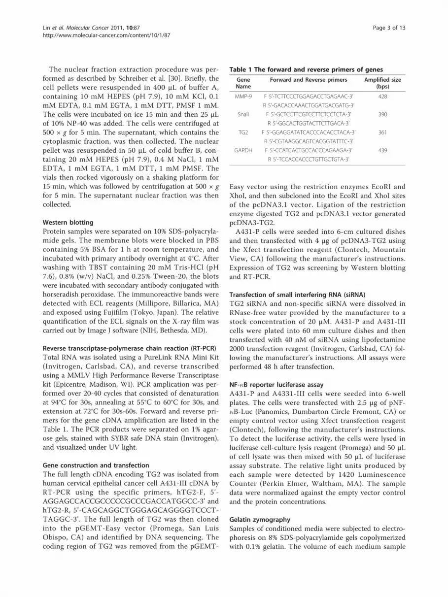
Table 1 The forward and reverse primers of genes
GeneName
Forward and Reverse primers Amplified size(bps)
MMP-9 F 5’-TCTTCCCTGGAGACCTGAGAAC-3’ 428
R 5’-GACACCAAACTGGATGACGATG-3’
Snail F 5’-GCTCCTTCGTCCTTCTCCTCTA-3’ 390
R 5’-GGCACTGGTACTTCTTGACA-3’
TG2 F 5’-GGAGGATATCACCCACACCTACA-3’ 361
R 5’-CGTAAGGCAGTCACGGTATTTC-3’
GAPDH F 5’-CCATCACTGCCACCCAGAAGA-3’ 439
R 5’-TCCACCACCCTGTTGCTGTA-3’
Lin et al. Molecular Cancer 2011, 10:87http://www.molecular-cancer.com/content/10/1/87
Page 3 of 13

analyzed was normalized according to the cell number.After electrophoresis, the gels were washed for 60 minin 2.5% Triton X-100, and incubated in reaction buffer(50 mM Tris-HCl, pH 8.0, containing 5 mM CaCl2, and0.02% NaN3) at 37°C for 24 h. The gels were thenstained with Coomassie Blue R-250 in 10% acetic acid/20% ethanol for 1 h, followed by destaining in the samesolution without dye. A clear zone on the gel indicatedthe presence of gelatinase activity, which was then quan-tified by densitometry.
Immunofluorescence stainingA431-P and III cells were plated into 6-well plates con-taining glass coverslips without a fibronectin coating.Following treatment with TG siRNA and non-specificsiRNA, or following transfection with the TG2 expres-sion vector, the cells were fixed with 4% paraformalde-hyde. Cells were permeabilized with 0.1% Triton X-100in PBS for 10 min. The permeabilized cells were thenincubated with 3% BSA in PBS to block non-specificbinding for 1 h at room temperature. After thoroughrinsing with PBS, the cells were incubated with mousemonoclonal anti-vimentin and rabbit polyclonal anti-fibronectin antibodies at 4°C overnight. Next the cellswere incubated with fluorescently labeled secondaryantibodies for 1 h at room temperature in the dark.After rinsing with PBS, the cells were then stained withDAPI in PBS for 5 min at room temperature. The cov-erslips were then mounted using mounting medium onmicroslides and visualized by confocal microscopy.
In vitro wound-healing migration assayBoth A431 and A431-III cells transfected with eitherTG2 siRNA or the full length TG2 expression vectorwere plated onto six-well culture plates in RPMI-1640containing 10% FBS. After 24 h, the cell monolayerswere wounded by manually scratching it with a pipettetip; this was followed by washing with PBS. The mono-layers were then incubated at 37°C for 24 h. The mono-layers were photographed at 0 h and 24 h afterwounding using phase contrast microscopy and anOlympus IX70 camera. The experiments were per-formed in triplicate for each treatment group.
Statistical analysisThe quantitative data derived from three to six indepen-dent experiments are expressed as means (± SEM).Unpaired Student’s t-tests were used to analyze betweengroup differences that is repeated and p < 0.05 was con-sidered statistically significant.
ResultsPreviously, we have demonstrated that TG2 and fibro-nectin are both upregulated in the highly invasive A431-
III subline compared with the parental A431-P cells,and that knockdown of TG2 decreased integrin’s asso-ciation with fibronectin as well as reducing the level ofMMP-9 and MMP-1; these events were accompanied bya reduction the A431-III cells’ capability of undergoingadhesion, migration and invasion [26]. This promptedus to further explore the potential role of TG2 in themodulation of the EMT as well as the associatedmechanisms using the A431-P and A431-III system thathad been established in our laboratory.
TG2 modulation of various EMT markers in A431-P andA431-III cellsTo understand whether TG2 plays a role in the induc-tion of the EMT process in A431 cells, we employedtwo experimental approaches. The first involved thetransfection of TG2 siRNA into A431-P and A431-IIIcells. We found that knockdown of endogenous TG2resulted in the reduced expression of various mesenchy-mal markers, namely fibronectin, vimentin, N-cadherin,and Snail (a key transcriptional repressor promotingEMT process). This knockdown had a greater effect onthe A431-III subline than on A431-P cells as was shownby immunoblotting and confocal microscopy analysis(Figures 1A &1B). In addition, and consistent with ourprevious study [26], knockdown of TG2 decreased theexpression and activity of MMP-9, and this reduced thecells’ migratory activity; these finding were obtained byRT-PCR, gelatin zymography and in vitro wound healingassays, respectively (Figures 1C to 1E).Next, we used the alternative approach of over-expres-
sing TG2 in A431-P cells that show a naturally low levelof TG2 (Figure 1A) by transfection with full-length TG2(pcDNA3.1-TG2). A431-P cells normally produce com-pact clusters of cells in culture, and these clustersbecame more scattered and fibroblastic in nature follow-ing TG2 over-expression (Figure 2A). These changeswere accompanied by increased expression of variousmesenchymal markers, fibronectin, vimentin, N-cadherinand Snail (Figures 2B &2C). Additionally, the A431-Pcells over-expressing TG2 showed an increased expres-sion of MMP-9 as well as displaying enhanced migratorypotential (Figures 2D &2E). Collectively, these resultssuggest that TG2 induces the acquisition of an EMT-like phenotype in A431-P and A431-III cells.
Involvement of PI3K/Akt-GSK3 signaling in the TG2-facilitated EMT processRecent studies have demonstrated that activation ofPI3K/Akt-GSK-3b signaling may induce the EMT pro-cess, a loss of cell-to-cell adhesion and cell polarity,morphological changes, an induction of cell motility,and decreased cell-matrix adhesion [31]. GSK-3b, a ubi-quitously expressed protein serine kinase, is active in
Lin et al. Molecular Cancer 2011, 10:87http://www.molecular-cancer.com/content/10/1/87
Page 4 of 13

resting epithelial cells [32], and inhibition of GSK-3bactivity or its expression may lead to the EMT [33]. Wetherefore were interested to explore the role of PI3K/Akt- GSK-3b signaling in the TG2-facilitated EMT pro-cess in A431 cells. We first examined Akt and GSK-3bactivity and their relationship with TG2. A431-III cellsshowed a relatively higher level of phosphorylated Akt-S473 (activation) and an increased level of phosphory-lated GSK-3b-S9 (inactivation) when compared withA431-P cells (Figure 3A). In addition, knockdown of
TG2 resulted in decreased Akt activity and increasedGSK-3b activity in A431-III cells (Figure 3A).Next, we examined the potential involvement of PI3K/
Akt-GSK3 signaling in the EMT using specific inhibitorsof PI3K (LY294002) and GSK-3b (SB415286). Treatmentof A431-III cells with LY294002 reduced the level ofSnail and secreted MMP-9, and this was accompaniedby reduced cell motility (Figures 3B to 3D). In parallel,treatment of A431-P cells with SB415286 increased theexpression of Snail and MMP-9, as well as promoting
Figure 1 Effect of TG2 knockdown on mesenchymal markers in A431-P and A431-III cells. (A) The cells were treated with 40 nM of TG2-specific siRNA or control siRNA. At 48 h post-transfection, cell lysates were prepared and subjected to immunoblotting analysis for TG2, Snailfibronectin, N-cadherin, vimentin and b-actin served as internal controls. (B) The cells were plated onto non-fibronectin-coated cover slips in six-well plate for 24 h. The cells were treated with 40 nM of TG2 siRNA or control siRNA, and then immuno-stained for fibronectin (green) andvimentin (red) with the nuclei stained with DAPI (blue). The fluorescence images were visualized using confocal microscopy. (C) Total RNA wasextracted at 48 h after siRNA transfection and analyzed for TG2, Snail and MMP-9 by RT-PCR with GAPDH served as the internal control. (D) Theculture conditioned media of TG2-silenced cells were collected and normalized by cell numbers prior to gelatin zymography analysis. (E) AfterTG2 knockdown, a wound healing assay was performed by scratching the cell layer with a pipette tip, and phase-contrast images were taken at0 h and 24 h later to assess cell migration into the open space. Quantitative data are presented as the mean (± SD) percentage of migrationdistance (n = 20). * and # indicate a significant difference compared with the respective control (p <0.05).
Lin et al. Molecular Cancer 2011, 10:87http://www.molecular-cancer.com/content/10/1/87
Page 5 of 13

Figure 2 Effect of TG2 over-expression on mesenchymal-like phenotype in A431-P cells. (A) Phase-contrast images of empty vector(pcDNA3.1) or full length TG2 (pcDNA3.1-TG2)-transfected cells cultured on six-wells plates in culture medium containing 10% FBS (×100magnification). At 48 h post-transfection, (B) cell lysates were prepared and subjected to immunoblotting analysis for TG2, Snail, fibronectin, N-cadherin, and vimentin. (C) Cells were immuno-stained for fibronectin (green) and vimentin (red) as well as having DAPI (blue) staining of thenuclei. The fluorescence images were visualized by confocal microscopy. (D) Total mRNA was extracted and analyzed for TG2, Snail and MMP-9by RT-PCR. (E) Cell migratory activity was determined using the wound healing assay as described in figure 1. Quantitative data are presented asthe mean (± SD) percentage of migration distance (n = 20). * indicates a significant difference compared with the control (p <0.05).
Lin et al. Molecular Cancer 2011, 10:87http://www.molecular-cancer.com/content/10/1/87
Page 6 of 13

cell motility (Figures 4A to 4C). Cyclin D was used as apositive control as it is subject to GSK-3b-dependentproteolysis [34]. To further ascertain the involvementPI3K/Akt-GSK-3b signaling in the TG2-induced acquisi-tion of the mesenchymal phenotype, we used the alter-native approach of transfecting pcDNA3.1-TG2 intoA431-P cells. TG2-overexpresson in A431-P cellsresulted in increased Akt activity and attenuated GSK-3b activity, and these effects were abrogated by treat-ment with the PI3K inhibitor LY294002 (Figures 4D). Ina similar manner to that observed for A431-III cells,treatment of TG2-overexpressing A431-P cells withLY294002 reduced the level of Snail and secreted MMP-9, as well as reducing cell motility (Figures 4D to 4F).These results together suggest that the TG2 induced-acquisition of an EMT-like phenotype by the highly
invasive A431-III subline involves an activation of PI3K/Akt signaling and an inactivation of GSK-3b.
Involvement of NF-�B signaling in TG2-facilitated EMTprocessWirth et al. identified NF-�B as a central mediator ofthe EMT in a mouse model of breast cancer progression[35]. In order to elucidate the role of NF-�B signaling inthe TG2-facilitated EMT process in A431 cells, we con-ducted experiments using three approaches. The firstwas to examine NF-�B activity and its relationship withEMT. When compared to A431-P cells, the A431-IIIsubline, which exhibits relatively high TG2 expression,showed a markedly reduced level of I�Ba (an endogen-ous inhibitor of NF-�B), and an increased nuclear levelof NF-�B relative to a similar total cellular level of NF-
Figure 3 Positive association of PI3K/Akt-GSK-3b signaling activation with the EMT phenotype in A431-P and A431-III cells. (A) Cellswere treated with 40 nM of control or specific TG2 siRNA. Cellular activity of Akt and GSK-3b were determined by analyzing theirphosphorylation status using immunoblotting. (B-D) Cells were treated with 20 μM Akt inhibitor LY294002 for 24 h. (B) The cellular protein andRNA levels of Snail were respectively determined by immunoblotting and RT-PCR. (C) The secreted activity of MMP-9 was measured usinggelatin zymography. (D) Cell migratory activity was determined by wound healing assay. Quantitative data are presented as the mean (± SD)percentage of migration distance (n = 20). * indicates a significant difference compared with the respective control (p <0.05). # indicates asignificant difference compared with the A431-P (p < 0.05).
Lin et al. Molecular Cancer 2011, 10:87http://www.molecular-cancer.com/content/10/1/87
Page 7 of 13

�B (Figure 5A). Using a NF-�B luciferase reporter assay,we found that NF-�B activity was significantly increasedin A431-III cells, and this was suppressed by treatmentwith an NF-�B inhibitor, JSH-23 (Figure 5B). Theincreased NF-�B activity in A431-III cells was positivelycorrelated with the increased nuclear level of TG2 (Fig-ure 5A), and an increased association of TG2 with
NF�B (Figure 5C). Additionally, treatment with JSH-23reduced the level of Snail, secreted MMP-9 activity andthe A431-III subline migratory activity (Figures 5D to5F), which suggests the potential involvement of NF-�Bin the EMT process.Next, we explored the effect of TG2 siRNA transfec-
tion on A431-P and A431-III cells. Knockdown of TG2
Figure 4 Upregulation of PI3K/Akt-GSK-3b signaling activation is associated with the EMT phenotype in TG2-overexpressing A431-Pcells. (A-C) The cells were treated with 25 μM of the specific GSK3 inhibitor SB415286 for 48 h. (A) The cellular protein and RNA levels of Snailand MMP-9 were respectively determined by immunoblotting and RT-PCR. Cyclin D served as the indicator of the inhibition of GSK-3b activity.(B) The secreted MMP-9 activity was detected using gelatin zymography. (C) Cell migratory activity was determined by the wound healing assay.Quantitative data are presented as the mean (± SD) percentage of migration distance (n = 20). (D-F) A431-P cells were transfected with emptypcDNA3.1 vector or pcDNA3.1-TG2, and then treated with 20 μM of PI3K inhibitor LY294002 for 24 h. (D) Cell lysates were analyzed forphosphorylated Akt, GSK-3b, Snail and TG2 using immunoblotting. (E) The secreted activity of MMP-9 was detected by gelatin zymography. (F)Cell migratory activity was determined using the wound healing assay. Quantitative data are presented as the mean (± SD) percentage ofmigration distance (n = 20). * indicates a significant difference compared with the respective control (p <0.05). # indicates a significant differencecompared with the A431-P (p < 0.05).
Lin et al. Molecular Cancer 2011, 10:87http://www.molecular-cancer.com/content/10/1/87
Page 8 of 13

Figure 5 Positive association of GSK-3b activity with TG2 and the EMT phenotype in A431-P and A431-III cells. (A) Total cell lysates andcytosolic and nuclear extracts were prepared and analyzed for TG2, NF-�B, and I�Ba by immunoblotting. (B) Cells were treated with 25 μM ofJSH-23 for 24 h, and the cellular NF-�B of activity was determined using a luciferase reporter assay. (C) The interaction of TG2, NF-�B, and I�Bain A431-P and the A431-III sub-line. (D-E) Cells were treated with 20 or 25 μM of JSH-23 for 24 h, and cell lysates were analyzed for Snail byimmunoblotting, and the conditioned media was analyzed for MMP-9 activity by gelatin zymography. (F) Cells were treated with 25 μM of JSH-23 for 24 h, and analyzed for migratory activity using wound healing assay. Quantitative data are presented as the mean (± SD) percentage ofmigration distance (n = 20). (G) Cells were transfected with control or specific TG2 siRNA, and cellular NF-�B activity was determined using aluciferase reporter assay. * indicates a significant difference compared with the respective control (p < 0.05). # indicates a significant differencecompared with the A431-P (p < 0.05). (H) Cellular protein levels of I�Ba and TG2 were detected by immunoblotting.
Lin et al. Molecular Cancer 2011, 10:87http://www.molecular-cancer.com/content/10/1/87
Page 9 of 13

led to a decrease of NF-�B activity and an increase inI�Ba by the A431-III subline cells (Figures 5G &5H),suggesting that upregulation of TG2 may induce NF-�Bactivity through a reduction of its inhibitor, I�Ba. Tofurther confirm this, we used an alternative approachwhereby we transfected pcDNA3.1-TG2 into A431-Pcells. TG2 over-expressing A431-P cells exhibited adepressed level of I�Ba, which was accompanied by ele-vated NF-�B activity (Figures 6A &6B). In a similarmanner to A431-III cells, treatment of TG2-overexpres-sing A431-P cells with the NF-�B inhibitor JSH-23reduced the level of Snail, secreted MMP-9 activity, andmigratory activity (Figures 6C to 6E). These resultstogether indicate that the TG2-induced acquisition of anEMT-like phenotype by the highly invasive A431-IIIcells involves the activation of NF-�B signaling.
DiscussionMetastasis is a very complex and highly intriguing process.While some of the associated molecular mechanisms have
begun to be unraveled, many remain to be understood. Inorder to further reveal some of the elusive factors thatcontribute to this complex process, our laboratory hasestablished a model system using an A431 parental tumorcells (A431-P) and a highly invasive derivative subline(A431-III) [25].To date, the role of TG2 in the EMT process is not
well understood. Evidence from a limited number ofstudies has revealed that TG2 localized to the cell sur-face serves as a co-receptor for fibronectin by simulta-neously associating with fibronectin and its receptorintegrin, mainly b1 and b3, and that this process is inde-pendent of the catalytic activity of TG2 [36,37]. Thisprocess may in turn promote cell adhesion, spread, andmigration [36,38]. We have further documented theincreased expression of TG2 and Snail in the A431-IIItumor subline that are both associated with the acquisi-tion of the mesenchymal-like phenotype [26]. Addition-ally, the upregulated expression of TG2 in A431-III cellsnot only enhances the association of fibronectin with b
Figure 6 Upregulation of NF-�B activity is associated with the EMT phenotype in TG2-overexpressed A431-P cells. (A) A431-P cells weretransfected with empty pcDNA3.1 vector or pcDNA3.1-TG2. Cellular protein levels of I�Ba and TG2 were determined at 48 h post-transfection byimmunoblotting. (B-E) Cells were treated with or without 25 μM of specific NF-�B inhibitor JSH-23 for 24 h. (B) Cellular NF-�B activity wasdetermined using a luciferase reporter assay. (C) Cellular protein levels of Snail and TG2 were detected by immunoblotting. (D) The secretedactivity of MMP-9 was analyzed by gelatin zymography. (E) Cell migratory activity was determined using a wound healing assay. Quantitativedata are presented as the mean (± SD) percentage of migration distance (n = 20). * indicates a significant difference compared with therespective control (p < 0.05). # indicates a significant difference compared with the A431-P (p < 0.05).
Lin et al. Molecular Cancer 2011, 10:87http://www.molecular-cancer.com/content/10/1/87
Page 10 of 13

integrin, but also induces the expression of fibronectin[26]. Moreover, we presented an interesting hypothesiswhereby Snail and MMP-9 form a mutual positive regu-latory loop in highly invasive A431-III cells that mayaccount for the facilitation of the EMT progression [27].The present study further shows that TG2 seems to beable to positively regulate the expression of Snail inA431 cells (Figures 1 &2), which suggests that TG2 mayact, at least in part, through an upregulation of Snail inorder to promote the EMT process. The molecularmechanism of TG2 involvement in the acquisition ofthe mesenchymal-like phenotype in A431-III cells stillawaits further investigation.Previous studies have documented that the aberrant
expression of TG2 in epithelial cells results in constitu-tive activation of focal adhesion kinase (FAK), Akt, andNF-�B [13,19,20,39]. These pathways are involved in theregulation of the EMT, in conferring drug resistance,and in promoting metastasis [35,40,41]. The aberrantactivation of Akt signaling is widely implicated in manyhuman cancers. Several studies have reported that acti-vation of the PI3K/Akt-GSK-3b signaling pathway isalso a central feature of the EMT, and that this involvesaccumulation of Snail in the nucleus [33]. As comparedto its parental cells, the highly invasive A431-III sublinedisplays increased Akt activity together with a concomi-tant reduction in GSK-3b activity, as well as havingincreased Snail and MMP-9 levels (Figure 3). Inhibitionof PI3K activity (LY294002) in A431 tumor cells attenu-ated the TG2-induced activation of Akt and GSK-3b,and suppressed the increases in Snail, MMP-9, and cellmotility (Figure 4). Additionally, inhibition of GSK-3activity (SB415286) in A431-P cells elevated the expres-sion of Snail and MMP9, and enhanced cell motility(Figure 4). These results support the concept that TG2acts in part through an activation of PI3K/Akt signalingin A431 tumor cells that inhibits GSK-3b, and thereforeupregulates Snail/MMP-9 and ultimately promotes EMTprogression. Verma et al. documented that overexpres-sion of TG2 in cancer cells is associated with a constitu-tive activation of focal adhesion kinase (FAK) and itsdownstream PI3K/Akt pathway [16]. Our earlier studyalso showed that the highly invasive A431-III sublineexhibits increased FAK activity compared to its parentalcells [25], and that knockdown of endogenous TG2reduced the phosphorylation activation of FAK-Tyr397(data not shown). Collectively, these results imply thatTG2 may sequentially activate FAK and PI3K/Akt- inA431 tumor cells, which in turn induces the acquisitionof a mesenchymal phenotype by A431 tumor cells.Constitutive activation of NF-�B is known to confer
resistance to cell death-inducing stimuli, including che-motherapeutic agents [42], and to promote metastasisby inducing EMT [35]. As compared to its parental
cells, the highly invasive A431-III subline has increasedNF-�B activity that is largely due to a reduction ofI�Ba, and this has been positively associated with TG2expression (Figures 5 &6). In addition to the increase inTG2 level in the A431-III subline, there was also anincreased association of TG2 with p65 NF-�B (Figure5). These results agree with the findings of otherresearchers who have shown that drug-resistant cancercells have increased levels of TG2, which enhances NF-�B activity, in turn, through a novel mechanism, namelythe formation of a TG2- NF-�B complex [13,15,20,43].In this study, we also demonstrated that there was aninhibition NF-�B activity (JSH-23) in A431 tumor cells,which led to a suppression of the TG2-inducedincreases in Snail, MMP-9, and cell motility (Figures 5&6). We proposed that TG2 acts at least in part throughan activation of NF-�B to upregulate Snail and MMP-9;this then boosts the acquisition of mesenchymal charac-teristics by the A431 tumor cells.It is believed that cancer cells are at intermediate
states in the EMT process. Indeed, once cells haveinvaded the primary tumor and penetrated the sur-rounding tissue, they must be able to colonize at a newtissue site. To achieve this, cancer cells may undergo amesenchymal-epithelial transition (MET) resulting in areformation of the epithelial phenotype in terms of cell-cell adhesion [44,45]. A cancer cell that attains plasticityand also shifts through the EMT and MET may accountfor many of the difficulties associated with cancer clini-cal therapy.
ConclusionsIn a previous study, we described a rapid method toprobe, explore and learn about the EMT. As enun-ciated elsewhere, we obtained a highly invasive A431-III sub-line from A431-P cells via Boyden chamberselection. Both A431-P and A431-III cells exertedEMT characteristics, while A431-III cells have acquireda mesenchymal-like phenotype, and also exhibit an ele-vated expression of Snail, one of the prime factorsinvolved in the acquisition of the mesenchymal-likephenotype [27]. In the present study, we have demon-strated that the expression of Snail is associated withenhanced NF-�B and PI3K/Akt-GSK-3b and that TG2participates in the acquisition of a mesenchymal transi-tion. We conjecture that the enhancement of Snailexpression by TG2 induces the acquisition in A431-IIIcells of a mesenchymal-like phenotype that then pro-motes the secretion of MMP-9, which enhances cancercell motility and increases metastatic potential. Theseobservations also support our contention that thatTG2 is a promising therapeutic target for reversingdrug resistance and inhibiting the early advent ofmetastasis of tumor cells.
Lin et al. Molecular Cancer 2011, 10:87http://www.molecular-cancer.com/content/10/1/87
Page 11 of 13

List of AbbreviationsTG2: Tissue Transglutaminase; MMP-9: Matrix Metalloproteinase-9; EMT:Epithelial-Mesenchymal Transition; GSK-3: Glycogen Synthase Kinase-3.
AcknowledgementsThis work was supported in part by grants from the National ScienceCouncil of Taiwan (NSC 94-2320-B-001-034 to M.T.L; NSC 99-2320-B-010-013-MY3 to J.J.H) and the Taiwan Academia Sinica Thematic Project (AS-96-TP-B06 to M.T.L.)
Author details1Institute of Biochemical Sciences, National Taiwan University, Taipei, Taiwan.2Castle Hills Health, 2267 Sir Amant Drive, Lewisville, TX 75056, USA.3Institute of Biological Chemistry, Academia Sinica, Taipei, Taiwan. 4Instituteof Physiology, National Yang-Ming University, Taipei, Taiwan.
Authors’ contributionsConceived and designed the experiments: CYL and MTL. Performed theexperiments: CYL and PHT Data analyzed: GDC, CHC, CJH, PPL and CYL.Paper writing and editing: CYL, JJH and CCK. All authors have read andapproved the final manuscript.
Competing interestsThe authors declare that they have no competing interests.
Received: 9 March 2011 Accepted: 21 July 2011 Published: 21 July 2011
References1. Thiery JP: Epithelial-mesenchymal transitions in tumour progression. Nat
Rev Cancer 2002, 2:442-454.2. Thiery JP: Epithelial-mesenchymal transitions in development and
pathologies. Curr Opin Cell Biol 2003, 15:740-746.3. Yilmaz M, Christofori G: EMT, the cytoskeleton, and cancer cell invasion.
Cancer Metastasis Rev 2009, 28:15-33.4. Iwatsuki M, Mimori K, Yokobori T, Ishi H, Beppu T, Nakamori S, Baba H,
Mori M: Epithelial-mesenchymal transition in cancer development and itsclinical significance. Cancer Sci 2010, 101:293-299.
5. Huber MA, Kraut N, Beug H: Molecular requirements for epithelial-mesenchymal transition during tumor progression. Curr Opin Cell Biol2005, 17:548-558.
6. Guarino M: Epithelial-mesenchymal transition and tumour invasion. Int JBiochem Cell Biol 2007, 39:2153-2160.
7. Peinado H, Portillo F, Cano A: Transcriptional regulation of cadherinsduring development and carcinogenesis. Int J Dev Biol 2004, 48:365-375.
8. Fesus L, Szondy Z: Transglutaminase 2 in the balance of cell death andsurvival. FEBS Lett 2005, 579:3297-3302.
9. Fesus L, Piacentini M: Transglutaminase 2: an enigmatic enzyme withdiverse functions. Trends Biochem Sci 2002, 27:534-539.
10. Lorand L, Graham RM: Transglutaminases: crosslinking enzymes withpleiotropic functions. Nat Rev Mol Cell Biol 2003, 4:140-156.
11. Collighan RJ, Griffin M: Transglutaminase 2 cross-linking of matrixproteins: biological significance and medical applications. Amino Acids2009, 36:659-670.
12. Ientile R, Caccamo D, Griffin M: Tissue transglutaminase and the stressresponse. Amino Acids 2007, 33:385-394.
13. Verma A, Mehta K: Transglutaminase-mediated activation of nucleartranscription factor-kappaB in cancer cells: a new therapeuticopportunity. Curr Cancer Drug Targets 2007, 7:559-565.
14. Mehta K: High levels of transglutaminase expression in doxorubicin-resistant human breast carcinoma cells. Int J Cancer 1994, 58:400-406.
15. Verma A, Mehta K: Tissue transglutaminase-mediated chemoresistance incancer cells. Drug Resist Updat 2007, 10:144-151.
16. Verma A, Wang H, Manavathi B, Fok JY, Mann AP, Kumar R, Mehta K:Increased expression of tissue transglutaminase in pancreatic ductaladenocarcinoma and its implications in drug resistance and metastasis.Cancer Res 2006, 66:10525-10533.
17. Cao L, Petrusca DN, Satpathy M, Nakshatri H, Petrache I, Matei D: Tissuetransglutaminase protects epithelial ovarian cancer cells from cisplatin-induced apoptosis by promoting cell survival signaling. Carcinogenesis2008, 29:1893-1900.
18. Hwang JY, Mangala LS, Fok JY, Lin YG, Merritt WM, Spannuth WA, Nick AM,Fiterman DJ, Vivas-Mejia PE, Deavers MT, et al: Clinical and biologicalsignificance of tissue transglutaminase in ovarian carcinoma. Cancer Res2008, 68:5849-5858.
19. Herman JF, Mangala LS, Mehta K: Implications of increased tissuetransglutaminase (TG2) expression in drug-resistant breast cancer (MCF-7) cells. Oncogene 2006, 25:3049-3058.
20. Kim DS, Park SS, Nam BH, Kim IH, Kim SY: Reversal of drug resistance inbreast cancer cells by transglutaminase 2 inhibition and nuclear factor-kappaB inactivation. Cancer Res 2006, 66:10936-10943.
21. Verma A, Guha S, Diagaradjane P, Kunnumakkara AB, Sanguino AM, Lopez-Berestein G, Sood AK, Aggarwal BB, Krishnan S, Gelovani JG, Mehta K:Therapeutic significance of elevated tissue transglutaminase expressionin pancreatic cancer. Clin Cancer Res 2008, 14:2476-2483.
22. Han JA, Park SC: Reduction of transglutaminase 2 expression isassociated with an induction of drug sensitivity in the PC-14 humanlung cancer cell line. J Cancer Res Clin Oncol 1999, 125:89-95.
23. Shao M, Cao L, Shen C, Satpathy M, Chelladurai B, Bigsby RM, Nakshatri H,Matei D: Epithelial-to-mesenchymal transition and ovarian tumorprogression induced by tissue transglutaminase. Cancer Res 2009,69:9192-9201.
24. Kumar A, Xu J, Brady S, Gao H, Yu D, Reuben J, Mehta K: Tissuetransglutaminase promotes drug resistance and invasion by inducingmesenchymal transition in mammary epithelial cells. PLoS One 2010, 5:e13390.
25. Kao WT, Lin CY, Lee LT, Lee PP, Hung CC, Lin YS, Chen SH, Ke FC,Hwang JJ, Lee MT: Investigation of MMP-2 and -9 in a highly invasiveA431 tumor cell sub-line selected from a Boyden chamber assay.Anticancer Res 2008, 28:2109-2120.
26. Chen SH, Lin CY, Lee LT, Chang GD, Lee PP, Hung CC, Kao WT, Tsai PH,Schally AV, Hwang JJ, Lee MT: Up-regulation of fibronectin and tissuetransglutaminase promotes cell invasion involving increased associationwith integrin and MMP expression in A431 cells. Anticancer Res 2010,30:4177-4186.
27. Lin CY, Tsai PH, Kandaswami CC, Lee PP, Huang CJ, Hwang JJ, Lee MT:Matrix metalloproteinase-9 cooperates with transcription factor Snail toinduce epithelial-mesenchymal transition. cancer science 2011,102:815-827.
28. Giard DJ, Aaronson SA, Todaro GJ, Arnstein P, Kersey JH, Dosik H, Parks WP:In vitro cultivation of human tumors: establishment of cell lines derivedfrom a series of solid tumors. Journal of the National Cancer Institute 1973,51:1417-1423.
29. Bradford MM: A rapid and sensitive method for the quantitation ofmicrogram quantities of protein utilizing the principle of protein-dyebinding. Anal Biochem 1976, 72:248-254.
30. Schreiber E, Matthias P, Muller MM, Schaffner W: Rapid detection ofoctamer binding proteins with ‘mini-extracts’, prepared from a smallnumber of cells. Nucleic Acids Res 1989, 17:6419.
31. Grille SJ, Bellacosa A, Upson J, Klein-Szanto AJ, van Roy F, Lee-Kwon W,Donowitz M, Tsichlis PN, Larue L: The protein kinase Akt inducesepithelial mesenchymal transition and promotes enhanced motility andinvasiveness of squamous cell carcinoma lines. Cancer Res 2003,63:2172-2178.
32. Papkoff J, Aikawa M: WNT-1 and HGF regulate GSK3 beta activity andbeta-catenin signaling in mammary epithelial cells. Biochem Biophys ResCommun 1998, 247:851-858.
33. Bachelder RE, Yoon SO, Franci C, de Herreros AG, Mercurio AM: Glycogensynthase kinase-3 is an endogenous inhibitor of Snail transcription:implications for the epithelial-mesenchymal transition. J Cell Biol 2005,168:29-33.
34. Diehl JA, Cheng M, Roussel MF, Sherr CJ: Glycogen synthase kinase-3betaregulates cyclin D1 proteolysis and subcellular localization. Genes Dev1998, 12:3499-3511.
35. Huber MA, Azoitei N, Baumann B, Grunert S, Sommer A, Pehamberger H,Kraut N, Beug H, Wirth T: NF-kappaB is essential for epithelial-mesenchymal transition and metastasis in a model of breast cancerprogression. J Clin Invest 2004, 114:569-581.
36. Akimov SS, Belkin AM: Cell surface tissue transglutaminase is involved inadhesion and migration of monocytic cells on fibronectin. Blood 2001,98:1567-1576.
Lin et al. Molecular Cancer 2011, 10:87http://www.molecular-cancer.com/content/10/1/87
Page 12 of 13

37. Chhabra A, Verma A, Mehta K: Tissue transglutaminase promotes orsuppresses tumors depending on cell context. Anticancer Res 2009,29:1909-1919.
38. Akimov SS, Krylov D, Fleischman LF, Belkin AM: Tissue transglutaminase isan integrin-binding adhesion coreceptor for fibronectin. J Cell Biol 2000,148:825-838.
39. Verma A, Guha S, Wang H, Fok JY, Koul D, Abbruzzese J, Mehta K: Tissuetransglutaminase regulates focal adhesion kinase/AKT activation bymodulating PTEN expression in pancreatic cancer cells. Clin Cancer Res2008, 14:1997-2005.
40. Thiery JP, Acloque H, Huang RY, Nieto MA: Epithelial-mesenchymaltransitions in development and disease. Cell 2009, 139:871-890.
41. Kalluri R, Weinberg RA: The basics of epithelial-mesenchymal transition. JClin Invest 2009, 119:1420-1428.
42. Orlowski RZ, Baldwin AS Jr: NF-kappaB as a therapeutic target in cancer.Trends Mol Med 2002, 8:385-389.
43. Mann AP, Verma A, Sethi G, Manavathi B, Wang H, Fok JY,Kunnumakkara AB, Kumar R, Aggarwal BB, Mehta K: Overexpression oftissue transglutaminase leads to constitutive activation of nuclear factor-kappaB in cancer cells: delineation of a novel pathway. Cancer Res 2006,66:8788-8795.
44. Peinado H, Olmeda D, Cano A: Snail, Zeb and bHLH factors in tumourprogression: an alliance against the epithelial phenotype? Nat Rev Cancer2007, 7:415-428.
45. Hugo H, Ackland ML, Blick T, Lawrence MG, Clements JA, Williams ED,Thompson EW: Epithelial–mesenchymal and mesenchymal–epithelialtransitions in carcinoma progression. J Cell Physiol 2007, 213:374-383.
doi:10.1186/1476-4598-10-87Cite this article as: Lin et al.: Role of tissue transglutaminase 2 in theacquisition of a mesenchymal-like phenotype in highly invasive A431tumor cells. Molecular Cancer 2011 10:87.
Submit your next manuscript to BioMed Centraland take full advantage of:
• Convenient online submission
• Thorough peer review
• No space constraints or color figure charges
• Immediate publication on acceptance
• Inclusion in PubMed, CAS, Scopus and Google Scholar
• Research which is freely available for redistribution
Submit your manuscript at www.biomedcentral.com/submit
Lin et al. Molecular Cancer 2011, 10:87http://www.molecular-cancer.com/content/10/1/87
Page 13 of 13


