Embed Size (px)
Citation preview

RPE and neuronal differentiation of allotransplantated porcineciliary epithelium-derived cells
Jasenka Guduric-Fuchs,1 Wing Chen,2 Henrietta Price,3 Desmond B. Archer,1 Tiziana Cogliati1
1Centre for Vision and Vascular Sciences, Queen’s University Belfast, Royal Victoria Hospital, Institute of Clinical Science,Northern Ireland, UK; 2Royal Victoria Hospital, Grosvenor Road, Belfast, Northern Ireland, UK; 3The Agri-Food and BiosciencesInstitute (AFBI), Chemical Surveillance Branch, Veterinary Sciences Division, Stoney Road, Stormont, Belfast, Northern IrelandUK
Purpose: Cell replacement has the potential to be applied as a therapeutic strategy in retinal degenerative diseases suchas retinitis pigmentosa and age-related macular degeneration (AMD) for which no adequate pharmacological and surgicaltreatments are currently available. Although controversial, the use of ciliary epithelium (CE)-derived cells is supportedby evidence showing their differentiation into retinal phenotypes. This study examines the differentiation potential ofporcine CE-derived cells in vitro and their survival, migration, morphological characteristics, and immunohistochemicalphenotype in vivo, upon transplantation into the subretinal space of normal pigs.Methods: Cells were isolated from the CE of postnatal pigs and were grown in a suspension sphere culture. Differentiationwas assessed in vitro after exposure to laminin and the addition of serum. For transplantation, CE-derived spheres weredissociated, labeled with CM-DiI vital dye, and the cells were injected subretinally into one eye of eight week-oldallorecipients. The eyes were examined at eight days and at two and four weeks after transplantation.Results: Cells positive for neuronal and retinal pigment epithelium (RPE) markers were detected byimmunohistochemistry in differentiation cultures. Reverse Transcriptase-Polymerase Chain Reaction (RT–PCR) revealedupregulation of neuronal markers after in vitro differentiation. CM-DiI dye-labeled CE-derived cells dissociated fromprimary spheres survived for up to four weeks after transplantation in vivo. Some of the surviving cells migrated distantlyfrom the injection site. Large clusters of transplanted cells integrated into the RPE layer and multilayered RPE-likestructures positive for RPE65 were often observed. Grafted cells were also identified in the neuroretina where 5%–10%were positive for recoverin, protein kinase C alpha (PKCα), and calbindin.Conclusions: The efficient conversion to an RPE-like phenotype suggests that CE-derived cells could be a potential sourceof RPE for cell replacement. Our data also suggest that the ability of these cells to acquire neuronal phenotypes is influencedby the environment. Thus, pre-differentiated or (re)programmed CE-derived cells may be more amenable for retinal repair.
Cell replacement is a promising approach to restoreneural function in the degenerating nervous system, includingthe retina. Since retinal dystrophies are ultimatelycharacterized by the loss of photoreceptors, efforts have beenmade in the last decade to identify suitable sources of stem/progenitor cells and drive their differentiation along thephotoreceptor lineage in vitro and in vivo. Several cellpopulations with retinal progenitor properties have beenidentified in the eye, including Müller glia, ciliary epithelium(CE)-derived, and iris-derived, and their ability to generateretinal cell types has been reported [1-4]. CE-derived cellshave been shown to display stem/progenitor cell features,including clonal expansion and differentiation toward retinal
Correspondence to: Jasenka Guduric-Fuchs, QUB-Centre for Visionand Vascular Science, RVH-Institute of Clinical Science, BelfastBT12 6BA, N. Ireland, UK; Phone: +44-28-9063-2729; FAX:+44-28-9063-2699; email: [email protected]
Dr. Tiziana Cogliati is now at Neurobiology-Neurodegeneration &Repair Laboratory, NIH - National Eye Institute, Bldg 6, Rm 302,Bethesda, MD 20892-0610.
phenotypes under appropriate conditions in vitro and in vivo[5-8]. The CE is located in a surgically accessible part of theeye, therefore, cells derived from this tissue offer an attractivepossibility for autologous transplantation.
It is well established that continuous growth of the eye inlower vertebrates such as fish and frogs depends on the retinalstem cells located in the ciliary marginal zone [9]. A similarbut less potent stem cell zone has also been identified inchickens [10]. Although an analogous structure does not existin mammals, it has been proposed that multipotent retinal stemcells can be isolated from the CE [5,6]. However, the natureand the developmental potential of cells derived from the CEhave been the object of controversy in recent literature. First,the existence of a small quiescent population of retinal stemcells (RSCs) in the CE that can be propagated in vitro has beenchallenged [11]. Second, the “stemness” of cells inneurospheres derived from the CE has been questioned on thebasis of the persistence of pigmentation and of theirexpression of makers and the characteristics of pigmented CE[11]. Finally, doubts have been expressed about thedevelopmental potential of CE-derived cultures and their
Molecular Vision 2011; 17:2580-2595 <http://www.molvis.org/molvis/v17/a279>Received 10 August 2010 | Accepted 2 October 2011 | Published 5 October 2011
© 2011 Molecular Vision
2580

capacity to differentiate along retinal lineages [11,12]. Whileall published literature is concordant in reporting the limitedself-renewal capacity of CE-derived cells [5,7,8,13,14], workfrom several laboratories has shown their differentiation, bothin vitro and in vivo, into neuronal and photoreceptor-likephenotypes [7,13,15,16]. Thus, although the definition asRSCs might not be the most appropriate, further investigationis needed to test the potential of CE-derived cells to generateretinal photoreceptors either by direct differentiation,transdifferentiation, or genetic manipulation.
Due to its close similarity to the human eye, a pig eyeprovides an appropriate system for the evaluation of potentialtherapeutic strategies for retinal degeneration [17,18].Furthermore, the size of a pig eye enables accurate dissectionof the ciliary epithelium without contamination from tissuessuch as the retina or RPE. In addition, pig eyes can be freshlyharvested from euthanized animals, offering an advantageover human specimens that are usually available for researchafter a prolonged post-mortem period. Although porcine CE-derived cells have been isolated and studied before by theauthors of this report and by others [8,19], this is the first studyto include the subretinal transplantation of these cells. To date,the transplantation studies using CE derived cells have onlybeen performed in murine animal models, althoughxenotransplantation of human cells into a developing mouseeye has also been performed [7,11,20]. Here, we evaluated theability of postnatal porcine CE-derived cells to generateretinal cell types in vitro and when injected subretinally intoallorecipient eyes. We adopted surgical procedures similar tothose used for subretinal transplantation of fetal retinalprogenitor cells in pigs [21,22].
METHODSAll animals procedures were approved by The Queen’sUniversity of Belfast Animal Ethics Committee and wereperformed in accordance with the UK Animals (ScientificProcedures) Act, 1986 and the ARVO statement on animaluse. Mixed sex, white Landrance pigs were obtained fromAgri-Food and Biosciences Institute (Hillsborough, NorthernIreland).
Cell isolation and culture: One to two week-old pigletswere anaesthetized with 15 mg/kg of intra-muscularazaperone (Stresnil, Janssen Animal Health, Saunderton, UK)and 20 mg/kg ketamine (Ketaset, Fort Dodge Animal Health,Southampton, UK) and were euthanized by intravenous orintra-cardiac injection of 100 mg/kg pentobarbitone(Pentoject, Animalcare, Masham Ripon, UK). The eyes wereenucleated and placed into oxygenated artificial cerebralspinal fluid (aCSF: 124 mM NaCl, 5 mM KCl, 1.3 mMMgCl2, 26 mM NaHCO3, and 10 mM D-glucose, pH 7.5). Theeyes were bisected at the ora serrata. The vitreous wasdecanted from the anterior half and the lens was removed. Theciliary body was dissected from the iris and pars plana. Thestrips of ciliary body were enzymatically digested in Hanks’
Balanced Salt Solution (HBSS) containing 2 mg/ml dispase(all from Sigma-Aldrich, Poole, UK) for 20 min at 37 °C,followed by digestion in Earle's Balanced Salt Solution(EBSS) containing 1.33 mg/ml trypsin, 0.67 mg/mlhyaluronidase, and 78 units/ml collagenase (Sigma-Aldrich)for 20 min at 37 °C. The supernatant was decanted andreplaced with a serum-free medium (SFM, DMEM/F12 [1:1]containing 0.6% [w/v] glucose, 2 mM glutamine, 5 mMHEPES buffer, 2% [v/v] B27, 100 units/ml penicillin, and 100units/ml streptomycin) with 1 mg/ml trypsin inhibitor(Invitrogen, Paisley, UK) and was incubated for 5 min at roomtemperature. The strips of ciliary body were subsequentlyplaced in a 60 mm cell culture dish containing the SFM.Epithelial cells were peeled off and the non-epithelial tissuewas discarded. The epithelial cellular debris was gentlytriturated 10–15 times using a pipette. Cells were pelleted at1,000× g for 10 min, resuspended in the SFM, and were passedthrough a 40 μm cell strainer (BD Biosciences, FranklinLakes, NJ). The cells were counted and plated at a density of3×104 cells/ml in the SFM supplemented with 20 ng/ml of anepidermal growth factor (EGF, Invitrogen) and 20 ng/ml of abasic fibroblast growth factor (bFGF, Invitrogen). After sevendays, newly formed sphere colonies were collected, pelletedat 1,000× g for 10 min, digested in an Accumax cell countingsolution (ICT, San Diego, CA) for 20 min at 20 °C, and weremechanically dissociated into single cells by pipetting andreplating at a density of 3×10⁴ cells/ml.
For differentiation, CE-derived spheres were collected atthe first passage, plated on poly-D-lysine and laminin-coatedglass coverslips (BD Biosciences), and were allowed todifferentiate for 20 days in the presence of either fetal calfserum (1%, 5%, and 10%), or of 1% serum with growth factors(10 ng/ml of EGF and bFGF). The medium was replaced everythree days. After 20 days of differentiation, the cells werefixed in 4% PFA for 20 min at room temperature and wereprocessed for immunocytochemistry.
For cell transplantation, spheres from the first passagewere collected and dissociated into single cells usingAccumax (ICT, San Diego, CA). The cells were labeled withCM-DiI (Invitrogen) following the manufacturer's protocoland were injected as described below.
Conventional RT–PCR: Total RNA was extracted usingan RNeasy Mini Kit (Qiagen, Crawley, UK). On columnDNaseI digestion was performed to digest any contaminatinggenomic DNA. One µg of RNA was reverse transcribed usingrandom primers and SuperScript II (Invitrogen) according tothe manufacturer’s instructions. No RT controls wereperformed by omission of reverse transcriptase in the reaction.PCR was performed in a 30 μl reaction volume containing1 μl of cDNA, 0.2 μM sense and anti-sense primers, 1× PCRbuffer (Qiagen), 10 mM dNTP mix (Roche, Burgess Hill,UK), and 1 μl Hot Start DNA polymerase (Qiagen). Primersequences are shown in Table 1. PCR was performed for 40
Molecular Vision 2011; 17:2580-2595 <http://www.molvis.org/molvis/v17/a279> © 2011 Molecular Vision
2581
cycles using a thermocycler (ABI 2720, Applied Biosystems,Foster City, CA). PCR products were resolved on 1.5%agarose gel.
Real time RT–PCR: For differentiation, CE-derivedspheres were collected at the first passage, plated on poly-D-lysine and laminin-coated six well plates (BD Biosciences),and were allowed to differentiate for 20 days in the presenceof 1% serum and 10 ng/ml of EGF and bFGF. The mediumwas replaced every three days. After 20 days, the cells wereharvested and RNA was isolated and reverse transcribed asoutlined above. Real time PCR was performed with 2×Maxima SYBR Green qPCR Mastermix (Fermentas,Cambridge, UK) in 10 µl reactions containing 2 µl of 1:15cDNA dilution and 0.5 µM of the gene specific primer. Primerefficiencies were determined from standard curvesconstructed using serial dilutions of pooled cDNA.Hypoxathineposphoribosyltransferase (HPRT) was used asthe housekeeping gene for normalization. Primer sequencesare shown in Table 1. Reactions were performed on aLightCycler PCR system (Roche) with the followingprogram: initial denaturation at 95 °C for 10 min, followed by40 cycles at 95 °C for 15 s, 58 °C for 10 s, and 72 °C for 15s. Relative gene expression (including statistical analysis) wasdetermined using REST software. The RNA from threeindependent experiments was analyzed and all reactions wereperformed in triplicate.
Surgical procedure: One eye from eight week-old(weight from 17.5 to 21 kg) female pigs (n=8) wastransplanted. Prior to transfer to the operating theater, theanimals were sedated with 2 mg/kg of azaperone (Stresnil;Janssen Animal Health, Saunderton, UK) by intramuscularinjection. In the theater, the animals were sedated byintramuscular injection of 1 mg/kg xylazine (Rompun 2%;Bayer, Newbury, UK) and 4 mg/kg of ketamine hydrochloride(Ketaset 100 mg/ml; Fort Dodge, Southampton, UK),
followed by administration of 0.2 mg/kg morphine (Morphinesulfate 10mg/ml; controlled drug [CD], UK). Anesthesia wasinduced for tracheal intubation with 1 mg/kg intravenousalfaxalone (Alfaxan 10mg/ml; Vetoquinol, Buckingham,UK), and was maintained using 1%–1.75% isoflurane inoxygen. Approximately 0.5 l of lactated Ringer’s (Hartman’s)isotonic solution was infused intravenously during anesthesia.
The pupil in each eye was dilated with topical medication(1 to 3 drops each of Gt cyclopentolate 1% and Gtphenylephrine 2.5%). A standard three-port pars planavitrectomy was performed. The sclerotomies were positioned2 mm posterior to the limbus. A retinotomy in the areacentralis was performed using a 42G needle (Bausch andLomb, Whelehan Group, Dublin). A small subretinal airbubble was created through the retinotomy, followed by theinjection of 1x10⁶ cells in a maximum volume of 0.1 ml ofphosphate-buffered saline (PBS) into the subretinal space.The sclerotomies were closed using 7/0 braided polyglactinsutures (Vicryl; Ethicon, Livingston, UK).
One pig was killed before recovery from anesthesia byintravenous pentobarbitone overdose at the end of the surgery.An intravenous injection of 2–4 mg/kg carprofen (RimadylLarge Animal 50 mg/ml; Pfizer, Sandwich, UK) wasadministered to the other pigs after transplantation and beforerecovery from the anesthetic (by discontinuation ofisoflurane), and xylazine sedation was reversed as necessaryusing approximately 0.2 mg/kg atipamezole (Antisedan 5 mg/ml; Pfizer). The animals were kept in a warm chamber for thefirst day after surgery. Topical eye drops containing 0.3%tobramycin and 0.1% dexamethasone (Tobradex; Alcon,Hemel Hempstead, UK) were instilled at the end of surgery,and then daily for 14 days post surgery.
At 8, 14, or 28 days after transplantation the animals werelethally anaesthetized with intravenous injection ofpentobarbitone (2–4 g), and their eyes were enucleated and
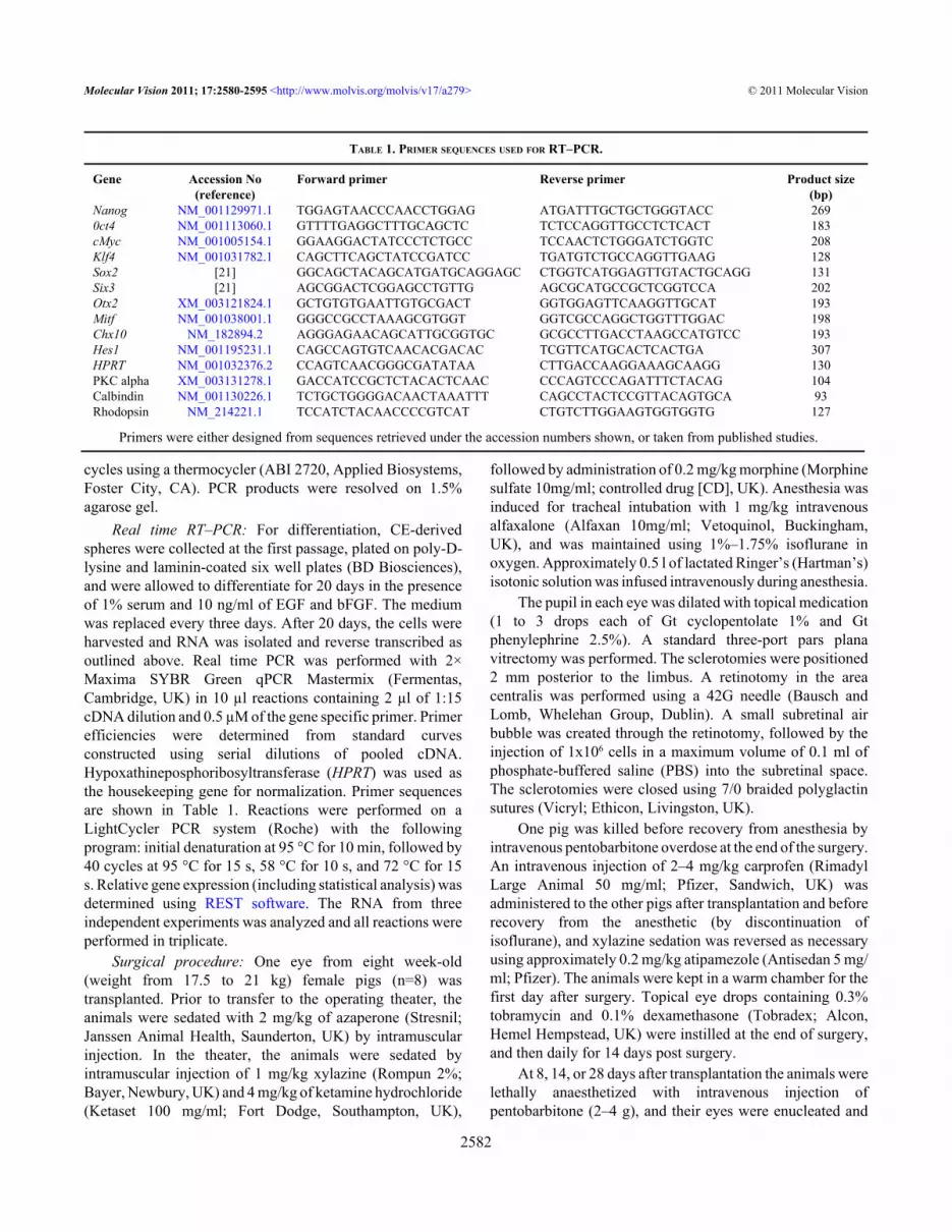
TABLE 1. PRIMER SEQUENCES USED FOR RT–PCR.
Gene Accession No(reference)
Forward primer Reverse primer Product size(bp)
Nanog NM_001129971.1 TGGAGTAACCCAACCTGGAG ATGATTTGCTGCTGGGTACC 2690ct4 NM_001113060.1 GTTTTGAGGCTTTGCAGCTC TCTCCAGGTTGCCTCTCACT 183cMyc NM_001005154.1 GGAAGGACTATCCCTCTGCC TCCAACTCTGGGATCTGGTC 208Klf4 NM_001031782.1 CAGCTTCAGCTATCCGATCC TGATGTCTGCCAGGTTGAAG 128Sox2 [21] GGCAGCTACAGCATGATGCAGGAGC CTGGTCATGGAGTTGTACTGCAGG 131Six3 [21] AGCGGACTCGGAGCCTGTTG AGCGCATGCCGCTCGGTCCA 202Otx2 XM_003121824.1 GCTGTGTGAATTGTGCGACT GGTGGAGTTCAAGGTTGCAT 193Mitf NM_001038001.1 GGGCCGCCTAAAGCGTGGT GGTCGCCAGGCTGGTTTGGAC 198Chx10 NM_182894.2 AGGGAGAACAGCATTGCGGTGC GCGCCTTGACCTAAGCCATGTCC 193Hes1 NM_001195231.1 CAGCCAGTGTCAACACGACAC TCGTTCATGCACTCACTGA 307HPRT NM_001032376.2 CCAGTCAACGGGCGATATAA CTTGACCAAGGAAAGCAAGG 130PKC alpha XM_003131278.1 GACCATCCGCTCTACACTCAAC CCCAGTCCCAGATTTCTACAG 104Calbindin NM_001130226.1 TCTGCTGGGGACAACTAAATTT CAGCCTACTCCGTTACAGTGCA 93Rhodopsin NM_214221.1 TCCATCTACAACCCCGTCAT CTGTCTTGGAAGTGGTGGTG 127
Primers were either designed from sequences retrieved under the accession numbers shown, or taken from published studies.
Molecular Vision 2011; 17:2580-2595 <http://www.molvis.org/molvis/v17/a279> © 2011 Molecular Vision
2582

washed in PBS. After removal of the cornea and lens, the eyeswere fixed in 4% PFA in PBS for 1 h at room temperature.The eyecups were cryoprotected in 10% sucrose for 6 hfollowed by 30% sucrose overnight, embedded in an optimalcutting temperature (OCT) compound (Sakura, Kobe, Japan),and were snap frozen in an isopentane bath on dry ice.Transverse cryosections (20 µm) were cut, mounted ontoSuperfrost Plus glass slides (Fisher Scientific, Loughborough,UK), and stored at −80 °C until used.
Immunohistochemistry: Immunohistochemistry on tissuesections was performed as described previously [18]. Briefly,slides were thawed at room temperature and were post-fixedin 4% formaldehyde (Sigma-Aldrich) in PBS for 20 min atroom temperature. After rinsing in PBS, sections wereblocked for 1 h in 10% normal goat serum (NGS), 0.3% TritonX-100, 0.01% NaN3 in PBS, at room temperature. Slides wereincubated for 24 h at 4 °C with a primary antibody diluted in10% NGS, 0.3% Triton X-100, and 0.01% NaN3 in PBS. Theprimary antibodies used are listed in Table 2. After removalof the primary antibody, slides were washed 6×5 min in PBSand were incubated for 1 h at room temperature in a secondaryantibody (Alexa Fluor488 goat anti-mouse or goat anti-rabbit),1:500 in PBS. After 3×5 min washing steps in PBS, cell nucleiwere counterstained with 5 μM DAPI (Invitrogen) for 10 min.The slides were mounted in a fluorescent mounting medium(Dako, Ely, UK). Negative immunohistochemistry controlswere performed in parallel by omission of the primaryantibody. Immunoreactive cells were visualized and imageswere recorded using an inverted confocal microscope (Nikon,Model Eclipse TE 2000-U, Tokyo, Japan) and Nikon EZ-C1software. Every tenth or twentieth section (200–400 µm step)was stained for the same antibody.
For isolectin B4 staining, sections were blocked in 5%BSA for 30 min, incubated with biothynilated Griffoniasimplicifolia Isolectin B4 (Vector) 1:100 for 1 h, washed for3×5 min with PBS, and were finally incubated withstreptavidin-FITC 1:200 for 1 h.
For the immunocytochemistry of the differentiated cells,post-fixation glass slides were washed 3× in PBS, incubated
in 10% NGS, 0.3% Triton X-100, and 0.01% NaN3 in PBS for1 h at room temperature, followed by overnight incubation at4 °C. The slides were incubated for 24 h at 4 °C with theprimary antibody diluted in 10% NGS, 0.3% Triton X-100,and 0.01% NaN3 in PBS. For double labeling, the secondprimary antibody was added after removal of the first primaryantibody and was incubated for another 24 h at 4 °C. Afterremoval of the second primary antibody, the slides werewashed for 6×5 min with PBS and were incubated in the firstsecondary antibody (Alexa Fluor488 goat antimouse) diluted1:500 in PBS for 1 h. Subsequently, incubation with anothersecondary antibody (Alexa Fluor568 goat antirabbit), wasperformed for 1 h. The slides were washed for 3×5 min withPBS and were counterstained and mounted as describedabove. The cells were visualized and the images were capturedwith an epifluorescence microscope (Nikon) using NisElements (Nikon) software. The number of positive cells wascounted in 20 random fields at 40× magnification.
RESULTSAnalysis of the gene expression of CE-derived spheres:Expression of the key pluripotency genes [23,24] and thegenes active during normal retinal development was analyzedby RT–PCR using RNA extracted from P1 CE-derivedspheres. Transcripts for three pluripotency genes, namelycMyc, Klf4, and Sox2 were present in CE-derived cultures,while mRNAs for Nanog and Oct4 were not detected (Figure1A). Transcription factors associated with the eyespecification and retinal histogenesis, including Six3, Mitf,Hes1, Otx2 and Chx10, were also expressed in CE-derivedspheres (Figure 1B).
In vitro differentiation of CE-derived spheres: The capacityof CE-derived cells from newborn pigs to differentiate intoretinal phenotypes was first evaluated in vitro, after platingCE spheres on adherent substrates (poly-D-lysine andlaminin) and culturing for 20 days with a differentiationmedium containing serum and growth factors. Growth factors(10 ng/ml bFGF and EGF) enhanced retinal differentiation inthe presence of 1% serum (Figure 2). Photoreceptor markers
TABLE 2. PRIMARY ANTIBODIES USED FOR IMMUNOHISTOCHEMICAL ANALYSIS.
Antibody Host Dilution SourceRecoverin rabbit 1:1000 Kind gift from Karl-Wilhelm KochRhodopsin (Rho4D2) mouse 1:100 Kind gift from Robert MoldayPKCα mouse 1:400 Sigma-AldrichCalbindin rabbit 1:1500 Chemicon, MilliporeRPE65 mouse 1:400 Chemicon, MilliporeKi67 mouse 1:300 BD BiosciencesNeurofilament (NF)-M mouse 1:350 Sigma-AldrichHuC/D mouse 1:200 Molecular probes, Invitrogen
Host animal, dilution and source for each antibody are shown.
Molecular Vision 2011; 17:2580-2595 <http://www.molvis.org/molvis/v17/a279> © 2011 Molecular Vision
2583

recoverin (Figure 2A-C) and rhodopsin (Figure 2D-F), thebipolar cell marker PKCα (Figure 2G-I), the ganglion,amacrine, and horizontal cell marker calbindin (Figure 2J-L)and the RPE marker RPE65 (Figure 2P-R) were detected byimmunocytochemistry in different proportions of cells.Labeling specificity was verified on mouse skin fibroblasts asnegative controls and on pig retinal progenitor cells as positivecontrols (data not shown). Recoverin labeling was detected inabout 20±3.2% of cells, rhodopsin labeling in 14.5±3.2%,PKCα labeling in 19.3±4.1%, and calbindin labeling in21.4±2.4% of cells. Cells immunopositive for neuronalmarkers in vitro extended thin, long processes, which aresuggestive of neuronal differentiation. Rhodopsin-labeledcells were positive for recoverin in double labelingexperiments (Figure 2D,E). Double labeling also revealed thatPKCα and recoverin antibodies stained a distinct populationof cells and there was no overlap between these two markers.However, PKCα-labeled cells were usually found adjacent torecoverin-positive cells (Figure 2G). RPE65immunoreactivity was detected in 12.2±3.8% cells. Cellswithin the spheres remained pigmented and although rare,pigment granules were sometimes observed within the cellsexpressing retinal markers (Figure 2M-O).
Real time PCR confirmed upregulation of PKCα(p<0.05), calbindin (p<0.05), and rhodopsin (p=0.053) indifferentiation cultures, relative to their expression levelbefore differentiation. Concomitantly, the retinal progenitormarker Hes1 was downregulated after differentiation (Figure3).
Transplantation of CE-derived cells: Prior to initiating in vivotransplantation experiments, the CM-DiI dye was tested forlong-term stability. CM-DiI showed long-term retention inCE-derived cells in proliferating (10 day follow-up) anddifferentiating (4 week follow-up) conditions in vitro (Figure4A-B). In vivo, subretinal localization of CM-DiI-labeledcells 10 min after grafting was confirmed in cryosections fromone animal. CM-DiI-labeled cells (red) were found betweenthe RPE and outer nuclear layer (ONL). Retinal detachmentat the injection area was also observed (Figure 4C,D).
Incorporation of transplanted cells into the RPE layer andformation of multilayered RPE-like structures: Eight daysfollowing transplantation, large CM-DiI positive cellaggregates were observed within the RPE layer (Figure 5).Clusters of CE-derived cells in the RPE were either RPE65-negative (Figure 5A), or showed strong RPE65immunoreactivity (Figure 5B,C). At two and four weeks aftertransplantation, many CM-DiI-labeled cells were localized inthe RPE layer (Figure 5D-I). Due to the phagocytic nature ofthe RPE, some of the CM-DiI labeling in this layer may beattributed to the uptake of the dye from dead transplanted cells.However, four weeks following transplantation, areas of theRPE were often multilayered, suggesting the de novoformation of additional RPE-like layers on top of the host RPEon the basal side (Figure 5G-I). The thickness of the RPEincreased due to the formation of multilayers; in some areasit was comparable to that of the ONL (Figure 5G-I). SomeCM-DiI-labeled cells were also observed at the level of thechoroid, but they were negative for RPE65.
Figure 1. Gene expression of ciliaryepithelium (CE)-derived cellsdetermined by RT–PCR. RNA wasisolated from passage 1 and wassubjected to conventional RT–PCR.PCR products were resolved on 1.5%agarose gel. A: Amplification of mRNAfor pluripotency markers. B:Amplification of mRNA for retinalprogenitor genes. Sizes in base pairs forthe corresponding marker bands (M) areshown on the left, adjacent to the gelimages. PCR reactions performed withcDNA template are shown in lanesmarked as '+' and negative controlreactions performed with templatesfrom the RT where reverse transcriptasewas omitted are shown in lanes markedas '–'. Amplicons for a housekeepinggene (HPRT) under the same conditionsare shown in the bottom panels.
Molecular Vision 2011; 17:2580-2595 <http://www.molvis.org/molvis/v17/a279> © 2011 Molecular Vision
2584

Figure 2. Microphotographs of the immunolabeling of newborn pig ciliary epithelium (CE)-derived cells after in vitro differentiation on poly-D-Lysine, laminin coated coverslips in the presence of 1% serum and 10 ng/ml basic fibroblast growth factor (bFGF) and epidermal growthfactor (EGF). The images were acquired using an epifluorescent microscope. A, B: Cells labeled for recoverin are clustered together (arrows).C: 4',6-diamidino-2-phenylindole (DAPI) nuclear staining corresponding to A and B. D, E: Cells double-labeled for recoverin (red, D) andrhodopsin (green, D and E) are depicted by arrows. The focus is set to show rhodopsin-positive cell processes. Strong recoverin staining inthe cytoplasm masks the nuclear DAPI staining, which is shown separately in F. Cells positive for protein kinase α (PKCα; green in G andH, arrows) did not co-label with recoverin (red in G and H and another example in the inset in G, arrowheads). The focus is set to show theprocesses of PKCα-labeled cells in G and H, and the recoverin-labeled processes in the inset in G. Corresponding DAPI nuclear stain is shownin I. J-K: A calbindin immunopositive cell is depicted by the arrow. The focus is set to show the processes of the labeled cell. CorrespondingDAPI nuclear stain is shown in L. M, N: Recoverin-positive cells (arrows in M and N) that had retained pigmented granules (arrow in thebright-field image in O). P, Q: RPE-65 immunopositive cells (arrows) and the corresponding nuclear DAPI staining in R.
Molecular Vision 2011; 17:2580-2595 <http://www.molvis.org/molvis/v17/a279> © 2011 Molecular Vision
2585

Migration of transplanted cells into the neuroretina andexpression of retinal markers: Some transplanted cellsmigrated into the neuroretina and both CM-DiI positivepigmented and non-pigmented cells were observedinterspersed with host retinal cells. The number of CM-DiI-labeled cells in the central and peripheral neurororetina wasquantified by counting the cells in transverse sectionscontaining the optic nerve head. At all time points, aproportion of surviving CM-DiI-labeled cells was found in theperipheral retina, indicating that transplanted cells hadmigrated tangentially from the site of injection in the centralretina to more peripheral sites (Figure 6A). Cell proliferationof the transplanted cells was assessed by immunolabeling forthe cell proliferation marker Ki67. A small number ofimunopositive cells were found within the CM-DiI-labeledcell aggregates in the subretinal space, but no Ki67 stainingwas observed in the neuroretina (Figure 6B,C). Therefore, theincrease in CM-DiI cell numbers in the neuroretina—from 8to 14 and 28 days—post transplantation is likely to be due tocell migration rather than proliferation of the transplantedcells within the retina. Isolectin B4 labeling was performed toidentify the distribution of immune cells (macrophage/microglia) in the injected retinas. Round, large cells positivefor isolectin were identified in the subretinal space. Thesecells contained red particles, suggesting phagocytosis oftransplanted cells by macrophages (Figure 6D,E). However,cells double-labeled with CM-DiI and isolectin were notfound within the neuroretina.
To assess whether transplanted CE-derived cells that hadmigrated into the neuroretina displayed features suggestive ofneuronal differentiation, sections of the transplanted eyeswere assayed with antibodies for retinal cell markers. CM-DiIcells positive for the photoreceptor marker recoverin weredetected in the ONL (Figure 7A-E). Double CM-DiI/PKCα-positive cells displayed oval shapes and were usually foundoutside the inner nuclear layer (INL), adjacent to bipolar cells(Figure 7F,G). PKCα labeling in pigs and cows is more intensein the ganglion cell layer (GCL), where it has been colocalizedwith astrocytes [18,25]. CM-DiI cells positive for the earlyneuronal marker HuC/D (Figure 7H,I) and calbindin-immunopositive cells were found in the GCL (Figure 7J,K),where they had a rounded or oval shape, with short thinprocesses (Figure 7J, inset). Since those cells were oftenobserved close to the vitreal side, it cannot be excluded thatthey were retracted to the vitreous after the injectionprocedure, or had migrated back to the vitreous through theneedle track. The percentage of CM-DiI-labeled cellslocalized in the neuroretina and positive for retinal neuronalmarkers was at an average of 8%–10% for recoverin, 5%–6%for PKCα, and 6%–9% for calbindin. The CM-DiI-labeledcells in the neuroretina always appeared slightly displacedfrom the pattern of the host retina and their morphologyremained distinguishable from the recipients’ cells.
DISCUSSIONRecent reports have highlighted the limited understanding wehave of the nature and developmental potential of CE-derived
Figure 3. Quantitative real time PCR data for ciliary epithelium (CE)-derived cell cultures following in vitro differentiation. RNA was isolatedfrom CE-derived cells after in vitro differentiation on poly-D-Lysine, laminin coated plates in the presence of 1% serum and 10 ng/ml basicfibroblast growth factor (bFGF) and epidermal growth factor (EGF) for 20 days. The data was analyzed using REST software for the relativequantification. The expression ratio represents the ratio of expression in differentiated compared to undifferentiated cultures. Afterdifferentiation, protein kinase α (PKCα; p<0.05), calbindin (p<0.05), and rhodopsin (p=0.054) were upregulated, while the retinal progenitormarker Hes1 was downregulated (p<0.01). The data are from a representative experiment performed in triplicate. Similar results were obtainedfrom three independent differentiation experiments.
Molecular Vision 2011; 17:2580-2595 <http://www.molvis.org/molvis/v17/a279> © 2011 Molecular Vision
2586

cells and the need for further investigations to re-evaluate theirbiology and potential for cell therapies in retinal degenerativediseases.
Several sources of cells have been considered for retinalcell replacement therapies and tested for their ability to
generate retinal cell types. Transplanted retinal progenitorshave been shown to express retinal cell markers in mice andpigs [21,26]. Functional cell replacement was demonstratedfor the first time in a study by MacLaren et al. [27], wheredissociated photoreceptor precursors from postnatal mice
Figure 4. CM-DiI labeling of ciliary epithelium (CE)-derived cells. Dissociated cells at passage 1 were labeled and cultured in a suspensionculture to form spheres (A), or were plated on poly-D-Lysine, laminin coated coverslips in differentiation conditions. The CM-DiI label wasretained for up to 10 days in proliferating spheres (depicted by arrows in A), and up to four weeks in differentiating conditions (arrows inB) in vitro. C, D: Microphotographs of grafted CM-DiI-labeled CE-derived cells (red) in the recipient retina 10 min after subretinal injection.C: CM-DiI labeling merged with nuclear DAPI staining. D: 4',6-diamidino-2-phenylindole (DAPI) staining only. Red CM-DiI-labeled cellswere found between the RPE (arrow) and the ONL. Retinal pigment epithelium (RPE); outer nuclear layer (ONL).
Molecular Vision 2011; 17:2580-2595 <http://www.molvis.org/molvis/v17/a279> © 2011 Molecular Vision
2587

were used for transplantation. However, this strategy wouldbe inadequate in humans, where the cells of the comparableontogenic stage would have to be obtained from fetal retina,and being postmitotic, could not be expanded.
Human embryonic (hES) stem cells and inducedpluripotent stem (iPS) cells are very attractive sources of cellsfor cell replacement. Efficient protocols for retinaldifferentiation of ES cells have been developed and are
constantly improving [28-31]. The feasibility of using ES-derived photoreceptors has been demonstrated after theirtransplantation into adult Crx−/− mice with subsequentimprovement in visual function [32]. Similarly,photoreceptors have been generated from iPS cells and theirintegration into both mice and pig retina has been reported[33,34]. Recently, transplantation of iPS-derivedphotoreceptor precursors from mice has been shown to restore
Figure 5. Microphotographs of red CM-DiI-labeled ciliary epithelium (CE)-derived cells in the retinal pigment epithelium (RPE) layer. A:Red-labeled pigmented CE-derived cells localized to the RPE layer, which were negative for RPE65 (arrows). B: At the same time point,transplanted red-labeled RPE65-positive cells were also found (red and green merged in B and green RPE65 labeling only in C, arrows). D-F: Two weeks following transplantation, CM-DiI-labeled cells in the RPE layer were strongly (arrow) and weakly (arrowhead) positive forRPE65. G-I: Four weeks after transplantation, the RPE appeared uneven and multilayered (arrows). Nuclei are labeled with 4',6-diamidino-2-phenylindole (DAPI; blue). Bright-field images are merged with the dark field in A, G, H, and I. Subretinal space (SS); and outer nuclearlayer (ONL).
Molecular Vision 2011; 17:2580-2595 <http://www.molvis.org/molvis/v17/a279> © 2011 Molecular Vision
2588

Figure 6. Analysis of migration,proliferation, and death of porcineciliary epithelium (CE)-derived cellsafter subretinal transplantation. A:Quantification of the transplanted cellsthat had migrated into the neuroretina.CM-DiI-labeled cells were counted in20 random sections from each eye. Themiddle third, containing the optic nerve,was considered to be the central retinaand the two peripheral thirds, includingthe ora serrata, were considered to be theperipheral retina. The results arepresented as the mean±SEM B, C: Cellproliferation assessed by Ki67 labelingin transplanted retinas. CM-DiI-positivecell aggregates in the subretinal space(red in B) contained rare Ki67-labeledcells (green, arrow in C), eight days aftertransplantation. D, E: Phagocytosis oftransplanted cells by macrophages. CM-DiI-labeled particles (red in D, arrow)contained within isolectin B4-positivemacrophages (green in E, arrow). Thenuclei are labeled with 4',6-diamidino-2-phenylindole (DAPI;blue). Outer nuclear layer (ONL); innernuclear layer (INL); and subretinalspace (SS).
Molecular Vision 2011; 17:2580-2595 <http://www.molvis.org/molvis/v17/a279> © 2011 Molecular Vision
2589

Figure 7. The immunoreactivity of transplanted ciliary epithelium (CE)-derived cells in the neuroretina. A: Microphotograph (13.3 µm confocalstack) showing red CM-DiI-labeled cells positive for recoverin (green, arrows) in the outer nuclear layer (ONL). B: Recoverin-only labelingof the same confocal stack as in A. The arrows point to the transplanted cells. C: A 0.7 µm confocal slice from the boxed area in A, showingdouble-labeled CM-DiI/recoverin positive cells (arrows) D: Green recoverin labeling in the same area (arrow). E: Red CM-DiI labeling and4',6-diamidino-2-phenylindole (DAPI) nuclear staining of the same area. F, G: CM-DiI-labeled, protein kinase α (PKCα)-positive cell (green)in the inner plexiform layer (arrows). H, I: HuC/D (green) and CM-DiI-positive cell in the ganglion cell layer (GCL; arrow in H and I). J,K: Calbindin (green) and CM-DiI-labeled cells in the GCL (arrows). The inset is at a higher magnification with visible processes (arrowhead).The nuclei are labeled with DAPI (blue). Inner nuclear layer (INL).
Molecular Vision 2011; 17:2580-2595 <http://www.molvis.org/molvis/v17/a279> © 2011 Molecular Vision
2590

visual function in rho−/− mice [35]. RPE cells have also beengenerated from hESC [36-40] and their transplantationrescued visual function in a rat model of retinal degeneration[41,42]. Cells with RPE features have also been differentiatedfrom human iPSCs and they were able to delay retinaldegeneration in animal transplantation studies [37,43].Although the use of ES and iPS for cell replacement therapiesshows great promise, issues such as oncogenic potential andimmunogenicity have to be fully addressed before ES or iPScells can be considered for treatment. Transplantationstrategies for retinal replacement also require optimizationdue to the low rate of cell survival and the integration oftransplanted cells. Efforts have been made to improve cellsurvival by transplanting cells on biodegradable scaffolds[44-47]. It has been identified that the outer limitingmembrane (OLM) represents a barrier to cell integration andseveral approaches to controlling OLM disruption have led toenhanced integration of transplanted cells [48-52]. Until asuccessful method and a reliable cell-type for cell-basedtherapies in the retina have been identified, it is preferable tocontinue the investigation of cells from different sources anddevelopmental origins with the potential to generate thedifferentiated progeny of interest. Although thedifferentiation potential of CE-derived cells is currentlydebatable, literature from the past decade suggests that thebehavior of these cells in differentiation cultures is dependenton the experimental conditions. Such conditions could eitherpromote a direct transition to the epithelial RPE-likephenotype or the development of retinal neuronal phenotypes,possibly through transdifferentiation or via de-differentiationand a stem-like transition state [53].
To gain insight into the differentiation potential ofpostnatal porcine CE-derived cells, we determined theexpression of the key pluripotency genes in P1 spheres [23,24]. We were able to detect mRNAs for Klf4,Sox2, andcMyc, while the transcripts for Nanog and Oct4 were absent.Cells positive for cMyc, but negative for nMyc were recentlyidentified as a retinal stem cell population in Xenopus and inzebrafish ciliary margins. Therefore, it has been suggestedthat the expression patterns of cMyc and nMyc could be usedto localize stem cells in the mammalian developing retina andCE [54]. The lack of expression of the whole set ofpluripotentcy genes highlights the important differencesbetween CE-derived cells and embryonic or iPS cells. OurPCR data are in agreement with recently published analysisof NRL-eGFP mice CE-derived cultures that could not bedifferentiated into photoreceptors when subjected to theretinal differentiation protocol for ES cells [12]. However,porcine CE-derived cells contained mRNAs for genesexpressed during retinal development, from the optic vesiclestage (Six3 and Mitf) to retinal histogenesis (Hes1,Chx10, andOtx2). Mitf transcription factor also plays a role in promotingand maintaining the RPE [55].
Our previous study has shown that the proliferationcapacity of porcine CE-derived cells decreases with the ageof the donor animal [8], suggesting that the cells from youngeranimals may be more stem-like, with a higher ability forretinal differentiation. However, it is important to note thatretinal histogenesis is complete after birth, with all retinallayers and cell types present in newborn pigs [18]. It remainsto be evaluated whether the capacity of porcine CE-derivedcells to generate retinal cell types actually decreases with theage of the cell donor.
Cells from the first passage in our current study generateda higher number of photoreceptor-like cells compared to ourprevious study, where cells were used for differentiation afterpassage three [8]. The fact that cells from earlier passagespossess higher differentiation potential is a limiting factor forthe expansion of CE-derived cells. This issue requires ourattention and it will have to be resolved if these cells are to beused for cell replacement. In our current study, colabeling forrhodopsin and recoverin and the lack of cells double-labeledfor recoverin and PKCα confirmed the photoreceptor-likephenotype of the differentiated cells. Interestingly, PKCα-labeled cells were always found to be closely associated withrecoverin-positive cells, suggesting that they might influenceeach other’s differentiation.
Expression of neuronal cell markers in our differentiationcultures coincided with significant, though not alwayscomplete, loss of pigmentation. Persistence of somepigmented granules in differentiated cells indicates that theyoriginate from pigmented cells, but may require additionaltime to clear their pigment content.
The differentiation potential of porcine CE-derived cellsin vitro was also previously studied by MacNeil et al. [19].Although the expression of generic neuronal markers such asβ-III-Tubulin and Neu-N was demonstrated, no expression ofmore specific retinal markers was detected in differentiationcultures in their study. The discrepancy relative to our resultscould be explained by the difference in the donor animal’s age,the post-mortem time before cell isolation, or by theconditions for in vitro differentiation. Retention of growthfactors in our differentiation cultures increased the number ofdifferentiated cells, suggesting a role for growth factors in thedifferentiation and/or survival of CE-derived cells. Notably,EGF does not affect in vitro photoreceptor survival in rats[56], but it can stimulate the survival of porcinephotoreceptors under the same conditions [57]. In addition tothe role of both bFGF and EGF in neuronal and retinaldifferentiation [58-61], bFGF has been shown to play a rolein transdifferentiation of RPE and iris pigment epithelium intoretinal tissue [62-64]. Finally, although EGF was reported tobe a negative regulator of photoreceptor differentiation duringretinal development [65,66], it has been shown, in vitro, to actas a neuronal differentiation factor for retinal stem cells [67].
In vivo, transplanted CE-derived cells showedremarkable migration potential as they were found in the
Molecular Vision 2011; 17:2580-2595 <http://www.molvis.org/molvis/v17/a279> © 2011 Molecular Vision
2591

peripheral regions of the RPE and retina. The injection ofdissociated cells rather than of intact spheres, as performed inother studies, might have facilitated migration [11].
We identified the recruitment of isolectin B-positivemacrophages to the subretinal space of injected eyes with thepresence of red particles contained within the phagocytes.Given the relatively short timeframe, an active immunerejection of subretinal allografts is unlikely to have beeninduced [21,27], therefore the macrophages are probablyresponsible for scavenging cell debris from the deadtransplanted cell.
Some transplanted CE-derived cells that had migratedinto the neuroretina displayed positive and specificimmunostaining for neuronal cell markers. The markersshown to be expressed in vitro in our current and previousstudy [8] were also detected in CE-derived cells aftertransplantation. All antibodies used in this study have beencarefully and extensively characterized on pig tissue [18],therefore, nonspecific antibody labeling of transplanted cellsis unlikely. Furthermore, the CM-DiI dye was previouslysuccessfully used for the long-term follow-up of neural stemcells after transplantation, and no diffusion of the dye wasreported [68].
The majority of transplanted cells formed multilayeredRPE-like structures positive for RPE65. Although, in vivo weobserved preferential differentiation of CE-derived cellsalong the RPE lineage, immunoreactivity for the RPE markerRPE65 in vitro was relatively low, suggesting that thedifferentiation protocol we adopted was not optimal for theefficient generation of RPE-like phenotypes. Indeed, ourprotocol was designed to generate retinal neurons. However,the culture conditions for efficient in vitro differentiation ofCE-derived cells into the RPE phenotype have recently beenreported [69]. Cells generated with this protocol hadepithelilal morphology, immunocytochemical, andultrastructural features of RPE and a capacity forphagocytosis. Another study has shown that high expressionof RPE65 can be induced in CE-derived cells in a mediumsupplemented with vasoactive intestinal peptide (VIP) or in aRPE cell-conditioned medium [70].
In vivo, differentiation along both the RPE and neuronallineages could be advantageous. Cell therapies can be mosteffective if the contribution of different cell types is harnessed,not only to replace lost cells, but also to maintain existingfunction and prevent further degeneration. CE-derived RPE-like cells could contribute a protective effect by promotingphotoreceptor survival [37], while newly differentiatedphotoreceptors could replace lost ones. Indeed, subretinaltransplantation of sheets of human retinal progenitor cellstogether with their RPE is the only method thus far shown tobe effective in humans [46]. Promising results in generatingfunctional photoreceptors from CE-derived cells in vitro andin vivo after gene transfer and modulation of transcriptionfactors have recently been reported [15,16].
In conclusion, our study shows that the cells frompostnatal pig CEs have the ability to generate cells with themorphological and immunohistochemical features of retinalneurons and RPE, both in vitro and after subretinaltransplantation in vivo. Revealing the paracrine effects andthe influence of the cellular environment in determining thefate of these cells may identify specific factors that enablecontrolled differentiation, or in vivo activation of these cells.Understanding the pathways behind this cell plasticity mayprovide important clues for the development of future cellreplacement therapies to combat retinal degeneration.
ACKNOWLEDGMENTSThe authors thank Alan Stitt for valuable comments, TazMcClintock, Paul Crawford, David Beattie, Lorraine Hannaand Mildred Wylie for technical support, Elaine Latimer forproject coordination, Robert Molday and Karl-Wilhelm Kochfor kindly donating antibodies. This work was supported inpart by funding generously provided by Fighting Blindness,ROI and The Fraser Homes Foundation for OphthalmicResearch, UK.
REFERENCES1. Lamba D, Karl M, Reh T. Neural regeneration and cell
replacement: a view from the eye. Cell Stem Cell 2008;2:538-49. [PMID: 18522847]
2. West EL, Pearson RA, MacLaren RE, Sowden JC, Ali RR. Celltransplantation strategies for retinal repair. Prog Brain Res2009; 175:3-21. [PMID: 19660645]
3. Bhatia B, Singhal S, Jayaram H, Khaw PT, Limb GA. Adultretinal stem cells revisited. Open Ophthalmol J 2010;4:30-8. [PMID: 20871757]
4. Wallace VA. Concise review: making a retina–from thebuilding blocks to clinical applications. Stem Cells 2011;29:412-7. [PMID: 21425405]
5. Tropepe V, Coles BL, Chiasson BJ, Horsford DJ, Elia AJ,McInnes RR, van der Kooy D. Retinal stem cells in the adultmammalian eye. Science 2000; 287:2032-6. [PMID:10720333]
6. Ahmad I, Tang L, Pham H. Identification of neural progenitorsin the adult mammalian eye. Biochem Biophys Res Commun2000; 270:517-21. [PMID: 10753656]
7. Coles BL, Angenieux B, Inoue T, Del Rio-Tsonis K, SpenceJR, McInnes RR, Arsenijevic Y, van der Kooy D. Facileisolation and the characterization of human retinal stem cells.Proc Natl Acad Sci USA 2004; 101:15772-7. [PMID:15505221]
8. Gu P, Harwood LJ, Zhang X, Wylie M, Curry WJ, Cogliati T.Isolation of retinal progenitor and stem cells from the porcineeye. Mol Vis 2007; 13:1045-57. [PMID: 17653049]
9. Agathocleous M, Harris WA. From progenitors to differentiatedcells in the vertebrate retina. Annu Rev Cell Dev Biol 2009;25:45-69. [PMID: 19575661]
10. Fischer AJ, Reh TA. Identification of a proliferating marginalzone of retinal progenitors in postnatal chickens. Dev Biol2000; 220:197-210. [PMID: 10753510]
11. Cicero SA, Johnson D, Reyntjens S, Frase S, Connell S, ChowLM, Baker SJ, Sorrentino BP, Dyer MA. Cells previously
Molecular Vision 2011; 17:2580-2595 <http://www.molvis.org/molvis/v17/a279> © 2011 Molecular Vision
2592

identified as retinal stem cells are pigmented ciliary epithelialcells. Proc Natl Acad Sci USA 2009; 106:6685-90. [PMID:19346468]
12. Gualdoni S, Baron M, Lakowski J, Decembrini S, Smith AJ,Pearson RA, Ali RR, Sowden JC. Adult ciliary epithelial cells,previously identified as retinal stem cells with potential forretinal repair, fail to differentiate into new rod photoreceptors.Stem Cells 2010; 28:1048-59. [PMID: 20506130]
13. Inoue T, Kagawa T, Fukushima M, Shimizu T, Yoshinaga Y,Takada S, Tanihara H, Taga T. Activation of canonical Wntpathway promotes proliferation of retinal stem cells derivedfrom adult mouse ciliary margin. Stem Cells 2006;24:95-104. [PMID: 16223856]
14. De Marzo A, Aruta C, Marigo V. PEDF Promotes RetinalNeurosphere Formation and Expansion In Vitro. Adv ExpMed Biol 2010; 664:621-30. [PMID: 20238066]
15. Inoue T, Coles BL, Dorval K, Bremner R, Bessho Y, KageyamaR, Hino S, Matsuoka M, Craft CM, McInnes RR, TremblayF, Prusky GT, van der Kooy D. Maximizing functionalphotoreceptor differentiation from adult human retinal stemcells. Stem Cells 2010; 28:489-500. [PMID: 20014120]
16. Jomary C, Jones SE, Lotery AJ. Generation of light-sensitivephotoreceptor phenotypes by genetic modification of humanadult ocular stem cells with Crx. Invest Ophthalmol Vis Sci2010; 51:1181-9. [PMID: 19850845]
17. Hendrickson A, Hicks D. Distribution and density of medium-and short-wavelength selective cones in the domestic pigretina. Exp Eye Res 2002; 74:435-44. [PMID: 12076087]
18. Guduric-Fuchs J, Ringland LJ, Gu P, Dellett M, Archer DB,Cogliati T. Immunohistochemical study of pig retinaldevelopment. Mol Vis 2009; 15:1915-28. [PMID: 19784390]
19. MacNeil A, Pearson RA, MacLaren RE, Smith AJ, Sowden JC,Ali RR. Comparative analysis of progenitor cells isolatedfrom the iris, pars plana, and ciliary body of the adult porcineeye. Stem Cells 2007; 25:2430-8. [PMID: 17600111]
20. Chacko DM, Das AV, Zhao X, James J, Bhattacharya S, AhmadI. Transplantation of ocular stem cells: the role of injury inincorporation and differentiation of grafted cells in the retina.Vision Res 2003; 43:937-46. [PMID: 12668063]
21. Klassen H, Kiilgaard JF, Zahir T, Ziaeian B, Kirov I, ScherfigE, Warfvinge K, Young MJ. Progenitor cells from the porcineneural retina express photoreceptor markers aftertransplantation to the subretinal space of allorecipients. StemCells 2007; 25:1222-30. [PMID: 17218397]
22. Klassen H, Warfvinge K, Schwartz PH, Kiilgaard JF, ShamieN, Jiang C, Samuel M, Scherfig E, Prather RS, Young MJ.Isolation of progenitor cells from GFP-transgenic pigs andtransplantation to the retina of allorecipients. Cloning StemCells 2008; 10:391-402. [PMID: 18729769]
23. Yamanaka S, Takahashi K. [Induction of pluripotent stem cellsfrom mouse fibroblast cultures]. Tanpakushitsu KakusanKoso 2006; 51:2346-51. [PMID: 17154061]
24. Takahashi K, Tanabe K, Ohnuki M, Narita M, Ichisaka T,Tomoda K, Yamanaka S. Induction of pluripotent stem cellsfrom adult human fibroblasts by defined factors. Cell 2007;131:861-72. [PMID: 18035408]
25. Shin T, Kim S, Ahn M, Kim H. An immunohistochemical studyof protein kinase C in the bovine retina. J Vet Med Sci 2006;68:71-4. [PMID: 16462121]
26. Klassen HJ, Ng TF, Kurimoto Y, Kirov I, Shatos M, Coffey P,Young MJ. Multipotent retinal progenitors expressdevelopmental markers, differentiate into retinal neurons, andpreserve light-mediated behavior. Invest Ophthalmol Vis Sci2004; 45:4167-73. [PMID: 15505071]
27. MacLaren RE, Pearson RA, MacNeil A, Douglas RH, Salt TE,Akimoto M, Swaroop A, Sowden JC. Ali RRl. Retinal repairby transplantation of photoreceptor precursors. Nature 2006;444:203-7. [PMID: 17093405]
28. Lamba DA, Karl MO, Ware CB, Reh TA. Efficient generationof retinal progenitor cells from human embryonic stem cells.Proc Natl Acad Sci USA 2006; 103:12769-74. [PMID:16908856]
29. Osakada F, Ikeda H, Sasai Y, Takahashi M. Stepwisedifferentiation of pluripotent stem cells into retinal cells. NatProtoc 2009; 4:811-24. [PMID: 19444239]
30. Lamba DA, Reh TA. Microarray characterization of humanembryonic stem cell-derived retinal cultures. InvestOphthalmol Vis Sci 2011; 52:4897-906. [PMID: 21345990]
31. Eiraku M, Takata N, Ishibashi H, Kawada M, Sakakura E,Okuda S, Sekiguchi K, Adachi T, Sasai Y. Self-organizingoptic-cup morphogenesis in three-dimensional culture.Nature 2011; 472:51-6. [PMID: 21475194]
32. Lamba DA, Gust J, Reh TA. Transplantation of humanembryonic stem cell-derived photoreceptors restores somevisual function in Crx-deficient mice. Cell Stem Cell 2009;4:73-9. [PMID: 19128794]
33. Lamba DA, McUsic A, Hirata RK, Wang PR, Russell D, RehTA. Generation, purification and transplantation ofphotoreceptors derived from human induced pluripotent stemcells. PLoS ONE 2010; 5:e8763. [PMID: 20098701]
34. Zhou L, Wang W, Liu Y, de Castro JF, Ezashi T, Telugu BP,Roberts RM, Kaplan HJ, Dean DC. Differentiation of inducedpluripotent stem cells of Swine into rod photoreceptors andtheir integration into the retina. Stem Cells 2011;29:972-80. [PMID: 21491544]
35. Tucker BA, Park IH, Qi SD, Klassen HJ, Jiang C, Yao J, RedentiS, Daley GQ, Young MJ. Transplantation of Adult Mouse iPSCell-Derived Photoreceptor Precursors Restores RetinalStructure and Function in Degenerative Mice. PLoS ONE2011; 6:e18992. [PMID: 21559507]
36. Klimanskaya I, Hipp J, Rezai KA, West M, Atala A, Lanza R.Derivation and comparative assessment of retinal pigmentepithelium from human embryonic stem cells usingtranscriptomics. Cloning Stem Cells 2004; 6:217-45. [PMID:15671670]
37. Carr AJ, Vugler AA, Hikita ST, Lawrence JM, Gias C, ChenLL, Buchholz DE, Ahmado A, Semo M, Smart MJ, Hasan S,da Cruz L, Johnson LV, Clegg DO, Coffey PJ. Protectiveeffects of human iPS-derived retinal pigment epithelium celltransplantation in the retinal dystrophic rat. PLoS ONE 2009;4:e8152. [PMID: 19997644]
38. Lu B, Malcuit C, Wang S, Girman S, Francis P, Lemieux L,Lanza R, Lund R. Long-term safety and function of RPE fromhuman embryonic stem cells in preclinical models of maculardegeneration. Stem Cells 2009; 27:2126-35. [PMID:19521979]
39. Carr AJ, Vugler A, Lawrence J, Chen LL, Ahmado A, Chen FK,Semo M, Gias C, da Cruz L, Moore HD, Walsh J, Coffey PJ.Molecular characterization and functional analysis of
Molecular Vision 2011; 17:2580-2595 <http://www.molvis.org/molvis/v17/a279> © 2011 Molecular Vision
2593

phagocytosis by human embryonic stem cell-derived RPEcells using a novel human retinal assay. Mol Vis 2009;15:283-95. [PMID: 19204785]
40. Liao JL, Yu J, Huang K, Hu J, Diemer T, Ma Z, Dvash T, YangXJ, Travis GH, Williams DS, Bok D, Fan G. Molecularsignature of primary retinal pigment epithelium and stem-cell-derived RPE cells. Hum Mol Genet 2010; 19:4229-38.[PMID: 20709808]
41. Lund RD, Wang S, Klimanskaya I, Holmes T, Ramos-KelseyR, Lu B, Girman S, Bischoff N, Sauve Y, Lanza R. Humanembryonic stem cell-derived cells rescue visual function indystrophic RCS rats. Cloning Stem Cells 2006; 8:189-99.[PMID: 17009895]
42. Vugler A, Carr AJ, Lawrence J, Chen LL, Burrell K, Wright A,Lundh P, Semo M, Ahmado A, Gias C, da Cruz L, Moore H,Andrews P, Walsh J, Coffey P. Elucidating the phenomenonof HESC-derived RPE: anatomy of cell genesis, expansionand retinal transplantation. Exp Neurol 2008; 214:347-61.[PMID: 18926821]
43. Buchholz DE, Hikita ST, Rowland TJ, Friedrich AM, HinmanCR, Johnson LV, Clegg DO. Derivation of functional retinalpigmented epithelium from induced pluripotent stem cells.Stem Cells 2009; 27:2427-34. [PMID: 19658190]
44. Tomita M, Lavik E, Klassen H, Zahir T, Langer R, Young MJ.Biodegradable polymer composite grafts promote thesurvival and differentiation of retinal progenitor cells. StemCells 2005; 23:1579-88. [PMID: 16293582]
45. Tao S, Young C, Redenti S, Zhang Y, Klassen H, Desai T,Young MJ. Survival, migration and differentiation of retinalprogenitor cells transplanted on micro-machined poly(methylmethacrylate) scaffolds to the subretinal space. Lab Chip2007; 7:695-701. [PMID: 17538710]
46. Radtke ND, Aramant RB, Petry HM, Green PT, Pidwell DJ,Seiler MJ. Vision improvement in retinal degenerationpatients by implantation of retina together with retinalpigment epithelium. Am J Ophthalmol 2008; 146:172-82.[PMID: 18547537]
47. Redenti S, Neeley WL, Rompani S, Saigal S, Yang J, KlassenH, Langer R, Young MJ. Engineering retinal progenitor celland scrollable poly(glycerol-sebacate) composites forexpansion and subretinal transplantation. Biomaterials 2009;30:3405-14. [PMID: 19361860]
48. West EL, Pearson RA, Tschernutter M, Sowden JC, MacLarenRE, Ali RR. Pharmacological disruption of the outer limitingmembrane leads to increased retinal integration oftransplanted photoreceptor precursors. Exp Eye Res 2008;86:601-11. [PMID: 18294631]
49. Tucker B, Klassen H, Yang L, Chen DF, Young MJ. ElevatedMMP Expression in the MRL Mouse Retina Creates aPermissive Environment for Retinal Regeneration. InvestOphthalmol Vis Sci 2008; 49:1686-95. [PMID: 18385092]
50. Pearson RA, Barber AC, West EL, MacLaren RE, Duran Y,Bainbridge JW, Sowden JC, Ali RR. Targeted disruption ofouter limiting membrane junctional proteins (Crb1 and ZO-1)increases integration of transplanted photoreceptor precursorsinto the adult wild-type and degenerating retina. CellTransplant 2010; 19:487-503. [PMID: 20089206]
51. Jiang C, Klassen H, Zhang X, Young M. Laser injury promotesmigration and integration of retinal progenitor cells into hostretina. Mol Vis 2010; 16:983-90. [PMID: 20577598]
52. Yao J, Tucker BA, Zhang X, Checa-Casalengua P, Herrero-Vanrell R, Young MJ. Robust cell integration from co-transplantation of biodegradable MMP2-PLGA microsphereswith retinal progenitor cells. Biomaterials 2011;32:1041-50. [PMID: 21030072]
53. Tsonis PA, Del Rio-Tsonis K. Lens and retina regeneration:transdifferentiation, stem cells and clinical applications. ExpEye Res 2004; 78:161-72. [PMID: 14729349]
54. Xue XY, Harris WA. Using myc genes to search for stem cellsin the ciliary margin of the Xenopus retina. Dev Neurobiol2011; ▪▪▪ [PMID: 21465669]
55. Wang SZ. Tales of retinogenesis told by human stem cells. ProcNatl Acad Sci USA 2009; 106:16543-4. [PMID: 19805334]
56. Fontaine V, Kinkl N, Sahel J, Dreyfus H, Hicks D. Survival ofpurified rat photoreceptors in vitro is stimulated directly byfibroblast growth factor-2. J Neurosci 1998; 18:9662-72.[PMID: 9822727]
57. Traverso V, Kinkl N, Grimm L, Sahel J, Hicks D. Basicfibroblast and epidermal growth factors stimulate survival inadult porcine photoreceptor cell cultures. Invest OphthalmolVis Sci 2003; 44:4550-8. [PMID: 14507904]
58. Hicks D, Courtois Y. Fibroblast growth factor stimulatesphotoreceptor differentiation in vitro. J Neurosci 1992;12:2022-33. [PMID: 1535104]
59. Hunter DD, Murphy MD, Olsson CV, Brunken WJ. S-lamininexpression in adult and developing retinae: a potential cue forphotoreceptor morphogenesis. Neuron 1992; 8:399-413.[PMID: 1550669]
60. Israsena N, Hu M, Fu W, Kan L, Kessler JA. The presence ofFGF2 signaling determines whether beta-catenin exertseffects on proliferation or neuronal differentiation of neuralstem cells. Dev Biol 2004; 268:220-31. [PMID: 15031118]
61. Garcez RC, Teixeira BL, Schmitt Sdos S, Alvarez-Silva M,Trentin AG. Epidermal growth factor (EGF) promotes the invitro differentiation of neural crest cells to neurons andmelanocytes. Cell Mol Neurobiol 2009; 29:1087-91. [PMID:19415484]
62. Opas M, Dziak E. bFGF-induced transdifferentiation of RPE toneuronal progenitors is regulated by the mechanicalproperties of the substratum. Dev Biol 1994; 161:440-54.[PMID: 8313994]
63. Thumann G. Development and cellular functions of the irispigment epithelium. Surv Ophthalmol 2001; 45:345-54.[PMID: 11166346]
64. Vergara MN, Del Rio-Tsonis K. Retinal regeneration in theXenopus laevis tadpole: a new model system. Mol Vis 2009;15:1000-13. [PMID: 19461929]
65. Lillien L. Changes in retinal cell fate induced by overexpressionof EGF receptor. Nature 1995; 377:158-62. [PMID: 7675083]
66. Ahmad I, Dooley CM, Afiat S. Involvement of Mash1 in EGF-mediated regulation of differentiation in the vertebrate retina.Dev Biol 1998; 194:86-98. [PMID: 9473334]
67. Angénieux B, Schorderet DF, Arsenijevic Y. Epidermal growthfactor is a neuronal differentiation factor for retinal stem cellsin vitro. Stem Cells 2006; 24:696-706. [PMID: 16179425]
68. Vossmerbaeumer U, Kuehl S, Kern S, Kluter H, Jonas JB,Bieback K. Induction of retinal pigment epithelium propertiesin ciliary margin progenitor cells. Clin ExperimentOphthalmol 2008; 36:358-66. [PMID: 18700924]
Molecular Vision 2011; 17:2580-2595 <http://www.molvis.org/molvis/v17/a279> © 2011 Molecular Vision
2594

69. Imitola J, Raddassi K, Park KI, Mueller FJ, Nieto M, Teng YD,Frenkel D, Li J, Sidman RL, Walsh CA, Snyder EY, KhourySJ. Directed migration of neural stem cells to sites of CNSinjury by the stromal cell-derived factor 1alpha/CXCchemokine receptor 4 pathway. Proc Natl Acad Sci USA2004; 101:18117-22. [PMID: 15608062]
70. Aruta C, Giordano F, De Marzo A, Comitato A, Raposo G,Nandrot EF, Marigo V. In vitro differentiation of retinalpigment epithelium from adult retinal stem cells. PigmentCell Melanoma Res 2011; 24:233-40. [PMID: 21232026]
Molecular Vision 2011; 17:2580-2595 <http://www.molvis.org/molvis/v17/a279> © 2011 Molecular Vision
Articles are provided courtesy of Emory University and the Zhongshan Ophthalmic Center, Sun Yat-sen University, P.R. China.The print version of this article was created on 10 October 2011. This reflects all typographical corrections and errata to thearticle through that date. Details of any changes may be found in the online version of the article.
2595


