Embed Size (px)
Citation preview

ADVERTIMENT. Lʼaccés als continguts dʼaquesta tesi queda condicionat a lʼacceptació de les condicions dʼúsestablertes per la següent llicència Creative Commons: http://cat.creativecommons.org/?page_id=184
ADVERTENCIA. El acceso a los contenidos de esta tesis queda condicionado a la aceptación de las condiciones de usoestablecidas por la siguiente licencia Creative Commons: http://es.creativecommons.org/blog/licencias/
WARNING. The access to the contents of this doctoral thesis it is limited to the acceptance of the use conditions setby the following Creative Commons license: https://creativecommons.org/licenses/?lang=en

UNIVERSITAT AUTÒNOMA DE BARCELONA
DEPARTAMENT DE BIOQUÍMICA I BIOLOGIA MOLECULAR, FACULTAT DE BIOCIÈNCIES
DOCTORAT EN BIOQUÍMICA, BIOLOGIA MOLECULAR I BIOMEDICINA
TARGETED DRUG DELIVERY FOR THE SELECTIVE
ELIMINATION OF CXCR4+ CANCER CELLS IN
METASTATIC COLORECTAL CANCER MODELS
Memòria presentada per la Rita Sala Faig per optar al títol de doctora en Bioquímica,
Biologia Molecular i Biomedicina per la Universitat Autònoma de Barcelona
Directors: Ramon Mangues Bafalluy MªVirtudes Céspedes Navarro
Doctoranda: Tutor: Rita Sala Faig Francisco Blanco Vaca
RITA SALA FAIG
2020

CONTENTS
2
CONTENTS
ABBREVIATIONS ................................................................................................................................................................... 8
LIST OF FIGURES .................................................................................................................................................................. 11
LIST OF TABLES .................................................................................................................................................................... 14
ABSTRACT ............................................................................................................................................................................ 15
I. INTRODUCTION ............................................................................................................................................................... 20
1) COLORECTAL CANCER...................................................................................................... 20
1.1. General aspects ........................................................................................................ 20
1.2. Epidemiology ............................................................................................................ 20
1.2.1. Incidence ............................................................................................................ 20
1.2.2. Survival .............................................................................................................. 21
1.2.3. Etiology and risk factors...................................................................................... 22
1.3. Molecular pathways involved in tumor progression .................................................. 23
1.4. CRC treatment .......................................................................................................... 25
1.4.1. Chemotherapy ................................................................................................... 26
1.4.2. Molecularly targeted therapies........................................................................... 27
1.5. Cell death mechanisms induced by anticancer drugs ................................................. 27
1.5.1. Apoptosis ........................................................................................................... 28
1.5.2. Pyroptosis .......................................................................................................... 29
1.5.3. Mitotic catastrophe ............................................................................................ 30
2) METASTASIS AND CXCR4 RECEPTOR ................................................................................ 31
2.1. Metastasis ................................................................................................................ 31
2.1.1. Metastatic routes ............................................................................................... 34
2.1.2. Chemokines and chemokine receptors ............................................................... 35
2.2. The CXCR4 chemokine receptor ................................................................................ 35
2.3. Role of CXCR4 in cancer ............................................................................................ 37
2.3. CXCR4 clinical significance ......................................................................................... 39

CONTENTS
3
2.3.1. Anti-cancer drugs targeting CXCR4 ..................................................................... 39
3) MOUSE MODELS IN CANCER RESEARCH .......................................................................... 41
3.1. CRC mouse models ................................................................................................... 41
3.2. Cell-derived and patient-derived xenografts ............................................................. 43
3.2.1. Heterotopic mouse models ................................................................................ 44
3.2.2. Orthotopic mouse models .................................................................................. 45
4) NANOPARTICLES FOR CANCER TREATMENT .................................................................... 46
4.1. Nanoparticles and their key properties ..................................................................... 46
4.2. Targeted drug delivery for cancer treatment ............................................................. 47
4.2.1. Passive targeting ................................................................................................ 48
4.2.2. Active targeting .................................................................................................. 48
4.3. Classification of drug nanocarriers ............................................................................ 49
4.3.1. Protein-based nanoparticles ............................................................................... 49
4.4. FDA approved nanomedicines for cancer therapy ..................................................... 50
4.5. T22-GFP-H6 protein-based nanoparticle targeting CXCR4+ cells ................................. 51
4.5.1. Structure of the T22-GFP-H6 nanoparticle .......................................................... 52
4.5.2. T22-GFP-H6 functionalization with the endosomal escape peptide HA2 ............. 53
4.6. Therapeutic derivatives of the T22-GFP-H6 nanoparticle ........................................... 54
4.6.1. Nanoconjugates ................................................................................................. 55
4.6.2. Toxin-based nanoparticles .................................................................................. 56
II. AIMS AND OBJECTIVES .................................................................................................................................................. 59
III. MATERIALS AND METHODS........................................................................................................................................ 61
1. IN VITRO EXPERIMENTS ................................................................................................... 61
1.1. Cell culture ............................................................................................................... 61
1.1.2. CRC cell lines ...................................................................................................... 61
1.1.2. Cell transfection with Luciferase reporter vector ................................................ 61
1.1.3. Flow cytometry for CXCR4 levels determination ................................................. 62
1.1.4. ELISA for supernatant SDF-1 alpha determination............................................... 62

CONTENTS
4
1.2. Fluorescent nanoparticles internalization assays ....................................................... 63
1.2.1. Flow cytometry .................................................................................................. 63
1.2.2. Confocal microscopy .......................................................................................... 63
1.3. Cell death determination assays................................................................................ 64
1.3.1. Cell proliferation assays ...................................................................................... 64
1.3.2. DAPI staining assays to detect DNA condensation............................................... 64
1.3.3. Immunocytochemistry analysis of cell blocks ...................................................... 65
1.4. Statistical analysis ..................................................................................................... 65
2. IN VIVO EXPERIMENTS ..................................................................................................... 66
2.1. Mouse strains ........................................................................................................... 66
2.2. Cell implantation techniques ..................................................................................... 66
2.2.1. Subcutaneous injection of CRC cells .................................................................... 66
2.2.2. Tumor disaggregation ......................................................................................... 67
2.2.3. Orthotopic injection of CRC cells to generate metastatic models ........................ 67
2.3. In vivo monitoring of tumor growth by bioluminescence ........................................... 68
2.4. Nanoparticle biodistribution and antineoplastic effect .............................................. 69
2.4.1. Biodistribution assays in subcutaneous CRC models ........................................... 69
2.4.2. Biodistribution in CXCR4 competition assays ...................................................... 70
2.4.3. Antitumor activity assessment in subcutaneous CRC models .............................. 70
2.4.4. Antimetastatic activity assessment in orthotopic CRC models ............................. 71
2.5. Necropsy and tissue processing ................................................................................ 72
2.5.1. Necropsy and bioluminescence ex vivo imaging .................................................. 72
2.5.2. Sample processing and histopathological analysis .............................................. 73
2.5.3. Immunohistochemistry analysis.......................................................................... 74
2.5.4. Immunofluorescence analysis ............................................................................. 76
2.5.5. Assessment of mitotic, apoptotic and necrotic rates ........................................... 77
2.6. Metastatic foci quantitation ...................................................................................... 77
2.7. Statistical analysis ..................................................................................................... 78

CONTENTS
5
IV. RESULTS .......................................................................................................................................................................... 80
1) DEVELOPING CXCR4-DEPENDENT SUBCUTANEOUS AND HIGHLY METASTATIC CRC MOUSE
MODELS. ............................................................................................................................. 80
1.1. Cell surface expression of CXCR4 in CRC cell lines ...................................................... 80
1.2. Effect of CXCR4 expression in the tumorigenic capacity of the SW1417 cell line ........ 81
1.3. Generation of a cell-derived CXCR4+ bioluminescent metastatic CRC mouse model .. 83
1.3.1. The metastatic capacity of the CXCR4+ SW1417 cell line increases in severely
immunosuppressed mice ............................................................................................. 85
1.4. Generation of a patient-derived metastatic CRC mouse model.................................. 86
1.5. CXCR4 expression in primary tumor and distant metastases ..................................... 87
2) IN VITRO SELECTIVE INTERNALIZATION AND ANTITUMOR ACTIVITY OF T22-GFP-H6 AND ITS
THERAPEUTIC DERIVATIVES IN CXCR4+ CRC CELL LINES. ....................................................... 89
2.1. CXCR4-dependent internalization of T22-GFP-H6 in a human CXCR4+ CRC cell line ... 89
2.2. The T22-GFP-H6-Aur nanoconjugate maintains CXCR4-dependent internalization in
CXCR4+ CRC cells .............................................................................................................. 91
2.3. Cytotoxic effect of T22-GFP-H6-Aur and induction of cell death in CRC cells by mitotic
catastrophe ..................................................................................................................... 92
2.4. Highly potent and CXCR4-dependent cytotoxic effect of the T22-PE24-H6 nanotoxin in
CXCR4+ CRC cells .............................................................................................................. 95
2.4.1. In vitro activation of the pyroptotic cell death pathway induced by the T22-PE24-
H6 nanotoxin ............................................................................................................... 96
3) IN VIVO BIODISTRIBUTION OF T22-GFP-H6 AND ITS THERAPEUTIC DERIVATIVES TO CXCR4+
TUMORS AND NON-TUMOR ORGANS. ................................................................................. 98
3.1. Maintenance of a highly selective tumor uptake for the T22-GFP-H6-Aur in mice bearing
subcutaneous CXCR4+ CRC tumors ................................................................................... 99
3.1.1. T22-GFP-H6 and CXCR4 receptor co-localization in the cell membrane ............. 100
3.1.2. Biodistribution of the T22-GFP-H6-Aur nanoconjugate in non-tumor organs and
absence of toxicity ..................................................................................................... 101
3.1.3. T22-GFP-H6-Aur achieves targeted drug delivery leading to selective depletion of
CXCR4+ cells ............................................................................................................... 103

CONTENTS
6
3.2. HA2 endosomal escape peptide site-dependent accommodation in the T22-GFP-H6
construct enhances tumor uptake ................................................................................. 104
3.3. Selective CXCR4-dependent T22-GFP-H6 and T22-GFP-HA2-H6 tumor uptake in
subcutaneous CRC tumors ............................................................................................. 107
4) IN VIVO EVALUATION OF THE ANTINEOPLASTIC ACTIVITY OF T22-GFP-H6 THERAPEUTIC
DERIVATIVES IN THE DEVELOPED HIGHLY METASTATIC CXCR4+ CRC MOUSE MODEL. ........ 108
4.1. T22-GFP-H6-Aur repeated dose administration activates a lethal immunogenic
response in Swiss nude mice.......................................................................................... 109
4.2. T22-GFP-H6-Aur prevents only transcelomic metastases in the SW1417 cell-derived
CRC model ..................................................................................................................... 110
4.3. T22-PE24-H6 repeated dose administration reduces tumor volume without toxicity in
non-target organs .......................................................................................................... 112
4.3.1. T22-PE24-H6 triggers pyroptotic cell death in SW1417 tumors ......................... 113
4.4. Definition of a dose regime for repeated T22-PE24-H6 administration in NSG mice 115
4.5. T22-PE24-H6 prevents the development of lymphatic and hematogenous metastasis in
the SW1417 cell-derived CRC model .............................................................................. 118
V. DISCUSSION .................................................................................................................................................................. 122
1) DEVELOPMENT OF CXCR4+ SUBCUTANEOUS AND HIGHLY METASTATIC CRC MOUSE
MODELS FOR THEIR USE IN PRECLINICAL STUDIES ............................................................. 124
1.1. Cell and patient-derived subcutaneous xenografts for nanoparticle preclinical
evaluation ..................................................................................................................... 125
1.2. Severe immunosuppression increases the metastatic capacity of CRC cells,
disseminating to clinically relevant sites in orthotopic mouse models ............................ 126
2) T22-GFP-H6 ACHIEVES HIGHLY SELECTIVE INTERNALIZATION AND TUMOR UPTAKE IN
CXCR4+ CRC MODELS ......................................................................................................... 129
3) CXCR4+ TARGETED NANOPARTICLES CONJUGATED TO THE AURISTATIN TOXIN ACHIEVE
POOR ANTIMETASTATIC EFFECT IN CRC MODELS .............................................................. 133
4) CXCR4+ TARGETED NANOPARTICLES WITH INTRINSIC CYTOTOXIC ACTIVITIES INDUCE
PYROPTOSIS AND DISPLAY HIGH THERAPEUTIC INDEX IN CRC MODELS ............................. 136
VI. CONCLUSIONS ............................................................................................................................................................. 142

CONTENTS
7
VII. REFERENCES ................................................................................................................................................................ 145
VIII. ANNEXES .................................................................................................................................................................... 159
1) ANNEX 1: ARTICLE 1 ...................................................................................................... 159
2) ANNEX 2: ARTICLE 2 ...................................................................................................... 181
3) ANNEX 3: ARTICLE 3 ...................................................................................................... 191

ABBREVIATIONS
8
ABBREVIATIONS
5-FU: 5-fluorouracil
ADA: anti-drug antibody
ADC: antibody-drug conjugate
AML: acute myeloid leukemia
APC: adenomatous polyposis coli
APC: antigen presenting cell
ATCC: American type culture collection
AUC: area under the curve
Aur: Auristatin E
CAF: cancer associated fibroblasts
CIMP: CpG island methylator phenotype
CRC: colorectal cancer
CSC: cancer stem cell
CTC: circulating tumor cell
CXCR4: C-X-C chemokine receptor 4
DISC: death-inducing signalling complex
DLBCL: diffuse large B-cell lymphoma
DMEM: Dulbeco’s modified eagle medium
DNA: deoxyribonucleic acid
DTC: disseminated tumor cell
ECM: extracellular matrix
EGFR: endothelial growth factor receptor

ABBREVIATIONS
9
ELISA: enzyme-linked immunoassay
EMT: epithelial-to-mesenchymal transition
EPR: enhanced permeability and retention
FAP: familial adenomatous polyposis
FDA: food and drug administration
FdU: floxuridine
G-CSF: granulocyte colony-stimulating factor
GEMM: genetically engineered mouse models
GFP: green fluorescent protein
GPCR: G protein-coupled receptor
GSDMD: gasdermin D
H&E: haematoxylin and eosin
HDI: human development index
HIF-1: hypoxia-inducible factor-1
HIV: human immunodeficiency virus
HSC: hematopoietic stem cell
HSPC: hematopoietic stem and progenitor cell
i.p.: intraperitoneal
i.v.: intravenous
ICC: immunocytochemistry
IHC: immunohistochemistry
LV: leucovorin
MIF: migration inhibitory factor

ABBREVIATIONS
10
MMAE: monomethyl Auristatin E
MMR: mismatch repair
MOMP: mitochondrial outer membrane permeabilization
MPS: mononuclear phagocytic system
MSI: microsatellite instability
NK: natural killer
OCMI: orthotopic cell microinjection
ORT: orthotopic
PBS: phosphate-buffered saline
PDX: patient-derived xenograft
PEG: polyethylene glycol
PIGF: placental growth factor
RES: reticuloendothelial system
ROS: reactive oxygen species
SC: subcutaneous
SDF-1 or CXCL12: stromal cell-derived factor-1
SEC: size exclusion chromatography
SSP: sessile serrated polyp
TBS: tris-buffered saline
TIL: tumor-infiltrating lymphocyte
UAB: Univeristat Autònoma de Barcelona
VAT: visceral adipose tissue
VEGF: vascular endothelial growth factor

LIST OF FIGURES
11
LIST OF FIGURES
Figure 1. Epidemiology of colorectal cancer…………………………………………………………………………….21
Figure 2. Schematic diagram of the polyp to CRC progression…………………………………………….……24
Figure 3. CRC treatment approach depending on the CRC stage……………………..……………………….25
Figure 4. Apoptosis vs pyroptosis cell death pathways…………………………………….…………….………..30
Figure 5. Schematic representation of spatial and temporal metastasis events………………..……..33
Figure 6. Three-dimensional structure of the CXCR4 receptor and its ligand CXCL12………….……36
Figure 7. CXCR4 expression as a prognostic factor in metastatic CRC patients………………….…….38
Figure 8. Mouse models for cancer research……………………………………………………………….….……….42
Figure 9. Targeted drug delivery for cancer treatment…………………………………………………..………..47
Figure 10. Characterization of the T22-GFP-H6 protein nanoparticles………………………………………52
Figure 11. Incorporation of the fusogenic peptide HA2 into the T22-GFP-H6
nanoparticle…………………………………………………………………………………………….…………………………….54
Figure 12. T22-GFP-H6-based nanoconjugates and their synthesis……………………………….…..…..56
Figure 13. Characterization of toxin-based nanoparticles……………………………………………….……..57
Figure 14. pPK-CMV-F3 plasmid map……………………………………………………………………………….…….62
Figure 15. OCMI procedure into the cecum of immunosuppressed mice……………………………….68
Figure 16. Administration regime for the CXCR4 blocking nanoparticle biodistribution
assays…………………………………………………………………………………………………………………………………….70
Figure 17. Overview of the main steps of IHC using Dako systems on paraffin-embedded tissue
slides………………………………………………………………………………………………………………………………..……75
Figure 18. Schematic representation of the antineoplastic effect assessment…………………….…78
Figure 19. Flow cytometry histograms of the CXCR4 cell surface expression of different CRC cell
lines………………………………………………………………………………………………………………………………………..81

LIST OF FIGURES
12
Figure 20. Tumorigenic capacity of the SW1417 cell line…………………………………………………..……..82
Figure 21. Primary tumor growth and metastatic dissemination of CXCR4+ SW1417 CRC cell-
derived orthotopic models ………………………………………………………………………….………………………….84
Figure 22. Metastatic colonization of SW1417 CRC cells in NOD/SCID and NSG mice……………..85
Figure 23. CXCR4 receptor expression in cancer tissues determined by immunohistochemistry
analysis…………………………………………………………………………………………………………………………………..88
Figure 24. T22-GFP-H6 selective internalization in CXCR4+ SW1417 cells determined by confocal
microscopy………………………………………………………………………………………………………………………..……90
Figure 25. In vitro T22-GFP-H6 and T22-GFP-H6-Aur selective internalization in the CXCR4+
SW1417 CRC cell line……………………………………………………………………………………………………………….92
Figure 26. Cytotoxic activity of the T22-GFP-H6-Aur nanoconjugate in CXCR4+ SW1417 cells,
measured with two different methods………………………………………………….………………………………..94
Figure 27. Antitumor activity of the proapoptotic nanoparticles and nanotoxins in CXCR4+
SW1417 cells……………………………………………………………………………………….………………………………….96
Figure 28. Activation of the pyroptotic pathway in CXCR4+ SW1417 cells after treatment with
T22-PE24-H6……………………………………………………………………………………………………….………………….97
Figure 29. Tumor accumulation of the T22-GFP-H6-Auristatin nanoconjugate and the T22-GFP-
H6 nanocarrier………………………………………………………………………………………………………………………100
Figure 30. Co-localization of T22-GFP-H6 and the CXCR4 receptor in the cell
membrane………………………………………………………………………………………….………………………………..101
Figure 31. Organ biodistribution of T22-GFP-H6 and T22-GFP-H6-Aur…………………..………..….…102
Figure 32. Tumor cell death at 2, 5 and 24 h after T22-GFP-H6-Aur administration………..……..103
Figure 33. Tumor biodistribution of nanoparticles in the highly CXCR4+ expressing SP5 patient-
derived CRC model ………………………………………………………………………………………………………….……105
Figure 34. Biodistribution and lack of toxicity of the nanoparticles in non-target
organs………………………………………………………………………………………………………………………..…………106
Figure 35. Graphic representation of total nanoparticle exposure………………………………………..107

LIST OF FIGURES
13
Figure 36. CXCR4-dependent biodistribution of T22-GFP-H6 and T22-GFP-HA2-H6
nanoparticles……………………………………………………………………………………………………………..…………108
Figure 37. Antitumor effect of T22-GFP-H6-Aur and absence of toxicity in normal organs of dead
mice……………………………………………………………………………………………………………………………………..110
Figure 38. T22-PE24-H6 antitumoral effect in the cell-derived subcutaneous SW1417 CRC
model………………………………………………………………………………………………………………….……………….113
Figure 39. In vivo assessment of T22-PE24-H6-induced activation of the apoptotic or the
pyroptotic cell death pathways……………………………………………………………………………………………..114
Figure 40. Definition of an effective and safe T22-PE24-H6 dosage to be administered in NSG
mice……………………………………………………………………………………………………………………..………………116
Figure 41. Antimetastatic activity induced by T22-PE24-H6 in regional and distant sites,
measured ex vivo……………………………………………………………………………………………………….…………118
Figure 42. T22-PE24-H6 antimetastatic effect in the cell-derived orthotopic SW1417 CRC
model……………………………………………………………………………………………………………………………………119
Figure 43. Differential expression of CXCR4 in tumor and non-tumor organs…………………….……131
Figure 44. Schematic representation of the different strategies used for nanoparticle
functionalization…………………………………………………………………………………………………………..………140
LIST OF TABLES
14
LIST OF TABLES
Table 1. Synthetic CXCR4 antagonists under clinical investigation……………………………………………40
Table 2. Nanomedicines approved by FDA for cancer therapy…………………………………………………51
Table 3. Experimental conditions for the in vivo CRC mouse models………………………………………68
Table 4. Doses and dose regime details of T22-GFP-H6-Aur nanoconjugate and the T22-PE24-H6
nanotoxin used in the antimetastatic effect experiments with the CXCR4+ SW1417 cells growing
orthotopically in NSG mice…………………………………………………………………………………..………………….71
Table 5. Primary antibodies used in the IHC studies…………………………………………………………………74
Table 6. Antibodies used in the IF studies………………………………………………………………………………..76
Table 7. Dissemination pattern of NOD/SCID and NSG mice orthotopically injected with CXCR4+
SW1417 CRC cells……………………………………………………………………………………………………………………86
Table 8. Metastatic dissemination in NSG mice orthotopically injected with a patient-derived cell
suspension of SP5 disaggregated from subcutaneous tumors in donor mice……………………………87
Table 9. Evaluation of T22-GFP-H6-Aur antimetastatic effect by preventing the development of
metastases in the SW1417 cell-derived CRC metastatic model………………………………………………111
Table 10. T22-PE24-H6 antimetastatic effect measured by prevention of metastases
development in the SW1417 cell-derived CRC metastatic model…………………………………………..120

ABSTRACT
15
ABSTRACT
Colorectal cancer (CRC) remains the third cause of cancer-related mortality in Western
countries, being metastases the main cause of death. Despite progress in prevention
strategies that decreased CRC incidence and mortality, still nearly a quarter of patients
are diagnosed at an advanced metastatic stage, with only a 15% five-year survival rate.
Thus, inhibition of metastasis development by targeting cancer stem cells, which are
associated with cancer dissemination, will significantly increase the benefits of current
cancer therapies. In collaboration with the Nanobiotechnology group from de UAB, we
developed self-assembling protein-based nanoparticles targeting the CXCR4 receptor
whose overexpression correlates with tumor dissemination, poor survival, and
recurrence in CRC patients. Preclinical evaluation of the efficacy and toxicity of the T22-
GFP-H6 nanocarrier and its therapeutic derivatives requires the use of adequate in vivo
disseminated CRC models. For that purpose, we generated subcutaneous and highly
metastatic models of CRC that overexpress CXCR4, derived from the SW1417 CRC cell
line or the SP5 patient sample. In order to increase the metastatic efficiency of previous
CXCR4+ CRC models, we orthotopically implanted luciferase expressing SW1417 cells in
the cecum of severe immunodeficient mice. NOD/SCID mice (deficient in T and B cells)
presented a low metastatic rate. In contrast, orthotopic microinjection in NSG mice
(deficient in T, B and NK cells) replicated the dissemination pattern observed in patients,
causing mice death and resulting in a higher number and size of hepatic and pulmonary
metastases as compared to NOD/SCID mice.
In the assessment of nanoparticles’ biodistribution, T22-GFP-H6 achieved a highly
selective tumor uptake in a CXCR4+ CRC subcutaneous model, as detected by fluorescent
emission (around 70% of the total), while displaying only transient accumulation in non-
tumor organs. We demonstrated that the nanocarrier tumor accumulation was CXCR4-
dependent because pre-treatment with AMD3100, a CXCR4 antagonist, reduced tumor
uptake. Furthermore, tumor accumulation was increased by the functionalization of the
nanocarrier with the fusogenic HA2 peptide, which promotes endosomal escape. On the
one hand, we observed that the therapeutic nanoconjugate T22-GFP-H6-Aur maintained
the nanocarrier’s biodistribution but its antitumoral effect was surprisingly poor. T22-
GFP-H6-Aur inhibited only tanscelomic metastasis in a highly metastatic CRC model,

ABSTRACT
16
while activating a lethal immunogenic response when repeatedly administered in low
immunosuppressed mice. As an alternative therapeutic option, we replaced the GFP
protein in the nanoparticle by the de-immunized PE24 toxin, to reduce its
immunogenicity while promoting a potent and intrinsic cytotoxic activity. The
administration of low doses of the T22-PE24-H6 nanotoxin prevented the development
of lymphatic and hematogenous metastasis in the highly metastatic CRC model without
toxicity. We demonstrated that the T22-PE24-H6 nanotoxin induced cancer cell death
through the non-apoptotic pathway, pyroptosis. In conclusion, the use of the T22-PE24-
H6 nanotoxin could be a promising strategy to selectively eliminate CXCR4+ CRC stem
cells in the absence of systemic toxicity, applicable to chemotherapy-resistant and
disseminated CRC associated with the upregulation of CXCR4 and antiapoptotic
mechanisms.

ABSTRACT
17
RESUM
El càncer colorectal (CCR) representa la tercera causa de mortalitat per càncer en països
occidentals, essent les metàstasis la principal causa de mort. Tot i el progrés en les
estratègies de prevenció que han disminuït la incidència i mortalitat del CCR, prop d’un
quart dels pacients encara són diagnosticats en estadis metastàtics avançats, amb una
taxa de supervivència a cinc anys de només el 15%. És per això que la inhibició del
desenvolupament de metàstasis actuant sobre cèl·lules mare canceroses incrementarà
significativament els beneficis de les teràpies actuals. En col·laboració amb el grup de
recerca en Nanobiotecnologia de la UAB, hem desenvolupat nanopartícules proteiques
autoensamblables dirigides al receptor CXCR4, la sobreexpressió del qual es
correlaciona amb la disseminació tumoral, baixa supervivència, i recurrència en pacients
de CCR. L’avaluació preclínica de l’eficàcia i toxicitat del nanoportador T22-GFP-H6 i els
seus derivats terapèutics requereix l’ús de models in vivo de CCR disseminats. Amb
aquest objectiu, hem desenvolupat models de CCR subcutanis i altament metastàtics
que sobreexpressen CXCR4, derivats de la línia cel·lular de CCR SW1417 o de la mostra
de pacient SP5. Per tal d’incrementar l’eficiència metastàtica dels models CXCR4+
anteriors, vam implantar ortotòpicament cèl·lules SW1417 amb expressió de luciferasa
en el cec de ratolins amb immunodeficiència severa. Els ratolins NOD/SCID (deficients
en cèl·lules T i B), presentaren taxes metastàtiques baixes. Per contra, la microinjecció
ortotòpica en ratolins NSG (deficients en cèl·lules T, B i NK), replicà el patró de
disseminació observat en pacients, provocant la mort dels ratolins i un nombre i mida
més gran de metàstasis hepàtiques i pulmonars en comparació als ratolins NOD/SCID.
En l’avaluació de la biodistribució de les nanopartícules, tal i com indica l’emissió de
fluorescència (al voltant del 70% del total), la T22-GFP-H6 va assolir una alta acumulació
selectiva en tumors del model subcutani CXCR4+, mentre que l’acumulació en òrgans no
tumorals fou només transitòria. Vam demostrar que l’acumulació al tumor del
nanoportador és CXCR4-dependent, perquè el pretractament amb AMD3100, un
antagonista de CXCR4, va reduir l’acumulació tumoral. A més, aquesta va incrementar
amb la funcionalització del nanoportador amb el pèptid fusogènic HA2, que afavoreix
l’escapament endosomal. Per altra banda, vam observar que el nanoconjugat terapèutic
T22-GFP-H6-Aur era capaç de mantenir la biodistribució del nanoportador, però el seu

ABSTRACT
18
efecte antitumoral fou sorprenentment baix. El T22-GFP-H6-Aur només va inhibir
metàstasis transcel·lòmiques en un model de CCR altament metastàtic i a més, va activar
una resposta immunogènica letal en la seva administració repetida en ratolins poc
immunodeprimits. Com a opció terapèutica alternativa, vam substituir la proteïna GFP
de la nanopartícula per la toxina PE24 desimmunitzada, per tal de reduir la seva
immunogenicitat afegint al mateix temps una potent activitat citotòxica intrínseca.
L’administració de dosis baixes de la nanotoxina T22-PE24-H6, va prevenir el
desenvolupament de metàstasis limfàtiques i hematògenes en el model de CCR
altament metastàtic sense toxicitat. Vam demostrar que la nanotoxina T22-PE24-H6
indueix la mort de cèl·lules tumorals per la via no apoptòtica de la piroptosi. En resum,
l’ús de la nanotoxina T22-PE24-H6 podria ser una estratègia prometedora per a
l’eliminació selectiva de cèl·lules mare de CCR CXCR4+ en absència de toxicitat sistèmica,
aplicable a CCR metastàtics i resistents a la quimioteràpia que s’associïn a la
sobreexpressió de CXCR4 i a mecanismes antiapoptòtics.

I. INTRODUCTION

INTRODUCTION
20
I. INTRODUCTION
1) COLORECTAL CANCER
1.1. General aspects
Colorectal cancer (CRC), also known as colorectal adenocarcinoma, is the abnormal
growth of epithelial cells from the colon or rectum (large intestine). The main function
of the colon is the reabsorption of water and remaining nutrients, and preparation of
waste products from the body for their elimination. The colon is held in place by
peritoneum, a thin layer of tissue that supports the abdominal organs. In order to help
the absorption, the gastrointestinal epithelium is formed by invaginations called colonic
crypts. Colon stem cells are located in the bottom of the crypts. These pluripotent cells
function in self-renewal. When the progenitor cells differentiate into specialised
epithelium cells, they migrate from the base to the surface in about 3-5 days. Normal
cells die at the surface and are replaced by the continuous stream of new cells from
below. All these processes are controlled by a protein signalling gradient, in which the
most common proteins are Wnt, TGF-B and BMP (1).
Most of CRC tumors (96%) typically arise from pre-existing benign polyps. The dividing
cells in these polyps may accumulate sufficient genetic and epigenetic changes by which
they acquire the ability to invade the bowel wall, a hallmark of CRC, and eventually
spread to local lymph nodes and finally to distant metastatic sites (2). Only 10% of all
the polyps progress to invasive cancer, although the risk of cancer increases as the polyp
grows larger (3).
1.2. Epidemiology
1.2.1. Incidence
Colorectal cancer (CRC) is the third most commonly diagnosed malignancy and the
fourth leading cause of cancer death in the world, accounting for about 1.8 million new
cases and almost 881,000 deaths in 2018 (4). CRC has a higher incidence in men than in
women, being 3–4 times more common in developed nations whereas the risk of the
disease increases with age, with most patients aged over 50 years at diagnosis.

INTRODUCTION
21
Figure 1. Epidemiology of colorectal cancer A) Correlation between a country’s human development
index (HDI) and colorectal cancer (CRC) incidence rates worldwide in 2018. HDI is a composite score of life
expectancy, education and income, reflecting the economic development of a country. Higher incidence
rates of CRC are observed in countries with higher values of HDI. B) Estimated incidence rates of CRC by
country worldwide in 2018, showing wide geographical variations. Adapted from (5).
Two-thirds of all CRC cases and about 60% of all deaths are occurring in countries with
a high human development index (HDI). But nowadays, CRC is considered one of the
clearest markers of the cancer transition. Countries undergoing rapid social and
economic evolution show fast increases in both incidence and mortality rates of cancers
more frequent in high-income countries (Figure 1). On the other hand, in high indexed
HDI countries (USA, Australia and Western Europe) CRC incidence and mortality rates
have been stabilised or decreasing, partially due to the increase in early detection and
prevention through colonoscopy and polyp removal.
1.2.2. Survival
Cancer overall survival is highly dependent on the cancer stage and the type of cancer
involved. Survival rates for early stage detected cancers are about five times higher than
that for late stage cancers. As CRC only becomes symptomatic at an advanced stage,
worldwide screening programmes are being implemented, which aim to increase early
detection and reduce morbidity and mortality. The 5-year survival rate of people with

INTRODUCTION
22
localized CRC is 90%. However, only 39% of patients are diagnosed at this early stage. If
the cancer has spread to regional tissues or regional lymph nodes, the 5-year
survival rate is 71%. For metastatic CRC patients, where the cancer has spread to distant
parts of the body, the 5-year survival rate decreases to only 14%. In CRC the most
common sites of metastasis are the liver (70%), lungs (32%), and peritoneum (21%) (6).
Distant lymph node metastases are less frequent (15%), occur independently of the
hematogenous spread and represents a major prognostic factor in CRC (7).
1.2.3. Etiology and risk factors
Both genetic and environmental factors play an important role in CRC development. The
majority of CRC tumors are sporadic and only 15-20% of CRC patients have a positive
family history. Sporadic CRC are caused by point mutations in specific genes, altering
important cell signalling pathways. In contrast, inherited CRC are caused by inherited
mutations that affect one of the alleles of the gene and a spontaneous point mutation
in the other allele, triggers the occurrence of a cancer cell and the adenocarcinoma.
There are different genetic syndromes associated to the development of hereditary CRC.
The most common is the Lynch syndrome, which is caused by a mutation in one of the
DNA mismatch-repair genes such as MLH1, MSH2, MSH6, PSM2 or EPCAM. Impaired
mismatch repair during replication produces an accumulation of DNA mutations, which
increase the probability of developing CRC. Another syndrome strongly associated with
CRC is familial adenomatous polyposis (FAP) which is caused by mutations in the
adenomatous polyposis coli (APC) gene, which controls activity of the Wnt signalling
pathway (8).
Many environmental lifestyle factors influence the risk of developing polyps and
CRC. The main risk factors of CRC are older age, male sex, smoking, alcohol intake and
obesity. In the case of alcohol consumption, the main metabolite of ethanol,
acetaldehyde, has been described as carcinogenic, being high alcohol consumption
associated to an increase in the risk of the 50% (9). Moreover, smoking can increase CRC
affectation by up to 10% because of the content of carcinogens such as nicotine that can
easily reach the intestine (10). Obesity is another important risk factor for CRC and can
be related to sedentary lifestyles or type 2 diabetes mellitus (11). Both, food intake and

INTRODUCTION
23
increased levels of visceral adipose tissue (VAT), can promote the development of CRC
through the secretion of proinflammatory cytokines in the intestine. So, diet is strongly
associated to CRC risk, increasing the chances up to 70% because of unhealthy
nutritional habits. Moreover, red meat releases heme groups in the intestine, which
enhance the formation of carcinogenic compounds (12).
1.3. Molecular pathways involved in tumor progression
The histological progression from polyp to cancer is the result of an accumulation of
several genetic and epigenetic changes in the colonic epithelial cells that deregulate
conserved signalling pathways involved in cellular proliferation, differentiation, survival,
and apoptosis. Mutations in the DNA can be sporadic or inherited, and the order of
occurrence seems to play an important role in CRC carcinogenesis. There are two
different types of polyps from which CRC develops, adenomas and sessile serrated
polyps (SSPs), and they normally originate by two main genetic pathways.
On the one hand, traditional adenomas, are associated with the chromosomal instability
pathway, which is observed in 85% of all sporadic cancers (13). It is characterized by a
cascade of accumulating mutations and imbalances in the number of chromosomes,
commonly being the mutation in the APC gene the first to occur. This alteration in the
APC gene affects chromosome segregation during cell division and causes the
translocation of β-catenin to the nucleus, promoting cell division and invasion. Next,
genetic events of progression frequently consist in mutations in the KRAS oncogene,
leading to a constitutive activation of MAP kinase, thus increasing cell proliferation.
Finally, over time, these mutations can cause a loss of function of the p53 gene, that
controls the main cell-cycle checkpoint, causing an uncontrolled entry in the cell cycle
and resulting in carcinogenesis (Figure 2).

INTRODUCTION
24
Figure 2. Schematic diagram of the polyp to CRC progression. Two different activated signalling pathways
in the normal colon to CRC development have been identified. Both sequential events involve the
progression of normal colon epithelial cells to aberrant crypt foci, followed by early and advanced polyps
with subsequent progression to early cancer and finally advanced cancer. The traditional route is the
pathway that involves the development of tubular adenomas that can progress to adenocarcinomas. An
alternate pathway is that involving serrated polyps and their progression to serrated CRC. The genes
mutated or epigenetically altered are indicated for each pathway. Some genes are shared between the
two pathways whereas others are pathway specific (i.e. BRAF mutations and CpG Island Methylator
Phenotype (CIMP) only in the serrated pathway). Adapted from (14).
On the other hand, SSPs tend to develop from mutations in the BRAF gene, which results
in altered cell growth and loss of apoptosis. Moreover, epigenetic instability, which is
responsible for the CpG island methylator phenotype (CIMP), is another common
feature in CRC originated from SSPs. The main characteristic of CIMP tumors is the
hypermethylation of oncogene promoters, which leads to gene silencing and a loss of
protein expression.
Another mechanism leading to genetic diversity in CRC, that can occur in both
adenomatous and serrated polyps, is microsatellite instability (MSI). MSI can result from
a hypermutable phenotype due to loss of expression of mismatch repair genes (MMR).
Therefore, mutations tend to accumulate leading to tumor progression. This loss of DNA
repair mechanisms can be caused by spontaneous events (promoter hypermethylation)
or germinal mutations such as those found in Lynch syndrome. In general, MSI tumors
have a better prognosis than sporadic tumors.

INTRODUCTION
25
1.4. CRC treatment
The choice of the first-line treatment for CRC patients is currently based in different
aspects of the disease and depending on the tumor stage, chemotherapy may be used
in addition to surgery. Tumor-related characteristics (localized or metastatic, number
and localization of metastases or the presence or absence of biochemical markers) and
patient-related factors (co-morbidity and prognosis) are used to classify patients in
stages and to select the right treatment strategy. The current classification of CRC
patients depends on the extent of local invasion, the degree of lymph node involvement
and the presence of distant metastasis (TNM staging system), creating four different risk
groups.
In stage I tumors, cells have grown through the mucosa and have invaded the muscular
layer of the colon or rectum, whereas in stage II tumors, cells have already invaded
nearby tissues such as the peritoneum but have not spread into lymph nodes or distant
organs. In these patients the recommended management consists on complete surgical
resection of the tumor with adequate margins and without chemotherapy since it has
no benefits to the overall survival of these stages. The standard procedure is a partial
colectomy where the affected part of the colon or rectum is resected together with its
mesocolon and blood supply to facilitate removal of draining lymph nodes.
Figure 3. CRC treatment approach depending on the CRC stage. Early-stage CRC patients are normally
treated with surgery, resecting only the polyps or the affected part of the colon (colectomy). Patients at
advanced stages are treated with a combination of surgery, chemotherapy and targeted therapies.
Chemotherapy consists of different regimes that combine drugs such as 5-fluorouracil, oxaliplatin,
leucovorin and irinotecan. Currently used targeted therapies include monoclonal antibodies targeting
VEGF (bevacizumab) or the EGFR (cetuximab and panitumumab).

INTRODUCTION
26
If cancer has spread to the lymph nodes or distant organs such as liver, lung or
peritoneum, which is the case in stage III and stage IV CRC respectively, chemotherapy
is an integral part of the treatment. Approximately two-thirds of patients with stage III
CRC (as well as some patients with stage II disease) receive adjuvant chemotherapy to
lower their risk of recurrence. For stage IV CRCs, when there are limited metastases, the
surgical treatment is usually combined with chemotherapy and/or radiation therapy
(Figure 3). Currently, several targeted drug therapies are also available to treat
metastatic disease, and in some cases, depending on the tumor’s molecular
characteristics immunotherapy may also be appropriate.
1.4.1. Chemotherapy
As mentioned before, most metastatic CRC patients are treated with classic cytotoxic
agents in combination with molecularly targeted therapies. Chemotherapy can be used
as neoadjuvant therapy to shrink the tumor before surgery, facilitating its resection, and
for the depletion of the remaining cancer cells after surgery. First-line chemotherapy
includes drugs as fluoropyrimidines (5-fluorouracil (5-FU) or capecitabine), leucovorin
(LV), oxaliplatin or irinotecan. The drugs capecitabine and fluorouracil are
interchangeable, being respectively oral or intravenous fluoropyrimidine agents.
Currently, some specific regimens which combine these chemotherapeutic agents are
used in metastatic CRC to avoid the emergence of resistances. In FOLFOX and CAPOX
regimens oxaliplatin is combined with LV and 5-FU or capecitabine respectively. In
contrast, in FOLFIRI regimen, irinotecan is combined with 5-FU and LV. Moreover, all
these cytotoxic drugs (5-FU, LV, oxaliplatin and irinotecan) are combined in the
FOLFOXIRI regime. The use of LV reduces the toxicity of the treatment, whereas the use
of the other cytotoxic agents has been shown to increase the progression-free survival
despite worsening the toxic effects of the treatment.
Although all these chemotherapeutic agents have shown efficacy, toxicity remains the
main limitation of cancer treatment. All these agents are biodistributed within the body
by passive diffusion, affecting both normal and cancer cells. Common side effects
produced by chemotherapy are anaemia, fatigue, hair loss, nauseas, diarrhea, muscle
disorders and neuropathy.

INTRODUCTION
27
1.4.2. Molecularly targeted therapies
In order to increase antitumor activity while reducing toxicity and side effects of cancer
treatments, new targeted therapies were designed to halt the growth and spread of
cancer cells by targeting or interfering with important and specific molecules of tumor
progression and growth. These targeted therapies can interfere with pathways causing
apoptosis of cancer cells or stopping the growth of abnormal blood vessels that feed
tumors (antiangiogenesis). Nowadays, in first-line treatment of metastatic CRC,
traditional chemotherapy is combined with antiangiogenic drugs, improving patient
overall survival (15). For that, monoclonal antibodies or proteins against different
effectors of the angiogenic pathway can be used. On the one hand, strategies targeting
the vascular endothelial growth factor (VEGF) have been developed, being the most
common the monoclonal antibody Bevacizumab and the recombinant fusion protein
Aflibercept. Bevacizumab targets circulating VEGF-A, therefore inhibiting signalling from
the VEGF receptor, while Aflibercept blocks multiple angiogenic growth factors such as
VEGF-A, VEGF-B and placental growth factor (PIGF). These compounds might act by
normalizing the dysregulated tumor vasculature, which would lead to improved tumor
oxygenation and delivery of chemotherapy (16).
On the other hand, anti-EGFR targeting therapies are used in metastatic CRC treatment.
Approximately 80% of all CRC express EGFR and overexpression correlates with reduced
survival and increased risk of metastases. There are two anti-EGFR targeted agents
approved for CRC: Cetuximab which is a recombinant chimeric monoclonal IgG1
antibody (17) and Panitumumab which is a human EGFR-specific antibody. Anti-EGFR
therapies are only used in the absence of RAS mutations because they have been proven
ineffective in KRAS or NRAS mutated gene tumors (18). RAS is mutated in about half of
all CRC. Thus, the RAS status of the tumor must be examined before making decisions to
treat with EGFR targeted therapies.
1.5. Cell death mechanisms induced by anticancer drugs
Cell death was believed to be the result of only two different processes in mammalian
tissues: apoptosis, also known as programmed cell death, or necrosis, the uncontrolled
cell death. However, in recent years, several other forms of cell death have been

INTRODUCTION
28
discovered, demonstrating that cells can die via distinct pathways. It has also been
noticed that classic chemotherapeutic agents kill cancer cells not only activating
apoptosis but other forms of non-apoptotic cell death such as necrosis, autophagy,
pyroptosis and mitotic catastrophe or inhibiting growth by entering senescence. The
new cell death classification is based on many biochemical and morphological
characteristics present in dying cells. Thus, the development of more efficient and safer
chemotherapeutics might succeed by understanding these novel cell death
mechanisms. The activation or inhibition of their mediators could lead to the design of
new anticancer agents. Moreover, the discovery of these cell death pathways can also
help to address the issue of drug resistance, by understanding the mechanisms used by
cancer cells to inhibit cell death, and directly influencing in their susceptibility to
chemotherapeutic agents.
1.5.1. Apoptosis
Apoptosis is the best known form of programmed cell death in multicellular organisms
and is responsible for maintaining tissue homeostasis by regulating the equilibrium
between cell proliferation and death. Furthermore, it has been considered the major
mechanism of chemotherapy-induced cell death. Apoptosis leads to morphological cell
changes and death induction. These morphological characteristics include cell
membrane blebbing, cell shrinkage, chromatin condensation and nucleosomal
fragmentation. So, apoptosis produces cell fragments called apoptotic bodies that
phagocytic cells engulf and remove from tissues.
The induction of apoptosis is highly regulated by activating mechanisms, existing two
main signalling pathways: the intrinsic, or mitochondria-mediated pathway, and the
extrinsic, or extracellular activated pathway. The intrinsic pathway is usually activated
in response to intracellular stress signals and depends on protein release from the
intermembrane space of mitochondria. These signals include DNA damage, high levels
of reactive oxygen species (ROS), viral infection and exposure to cytotoxic agents. The
BCL-2 protein family senses these signals resulting in the initiation of mitochondrial
apoptosis. The BCL-2 family is formed by the anti-apoptotic (BCL-2, MCL-1 and BCL-xL)
and the pro-apoptotic (PUMA, NOXA and BAD) proteins as well as the effector proteins

INTRODUCTION
29
(BAK and BAX). The balance among the different family members determines the
activation of BAK and/or BAX causing mitochondrial outer membrane permeabilization
(MOMP) and release of pro-apoptotic proteins such as cytochrome c, facilitating
apoptosome formation and the activation of caspases 9 and 3. The extrinsic pathway is
initiated by the binding of an extracellular ligand to cell-surface receptors, leading to the
formation of the death-inducing signalling complex (DISC), which activates caspases 8
and 10. Activated caspase 8 can then cleave the effector caspases 3 and 7 to amplify the
death signal (Figure 4). In both pathways the proteolytic enzymes caspases are triggered
to mediate a rapid disorganization of cellular organelles and architecture, as well as
enabling a crosstalk between the two apoptotic pathways, resulting in death signal
amplification.
1.5.2. Pyroptosis
Pyroptosis is a highly inflammatory form of non-apoptotic programmed cell death that
is most frequently activated upon microbial infection (19). This form of cell death
displays many morphological differences as compared to apoptosis. Classical apoptosis
is characterised by the compartmentalisation of intracellular components and removal
of cellular debris without any damage for the surrounding tissues. In contrast, during
pyroptosis the nucleus remains undamaged, but the plasma membrane is disrupted,
resulting in the leakage of intracellular components into the extracellular milieu.
Once the pyroptotic pathway is activated by a pathogen or some anticancer drugs,
procaspase-1 is cleaved to active caspase-1 through the formation of the inflammasome
(NLRP3/ASC/Procaspase-1). Then, active caspase-1 processes the proforms of the
inflammatory cytokines interleukin 1β (IL-1β) and IL-18 into their active forms, resulting
in cell death, that associates with the release of inflammatory cytokines into the
surrounding environment. Moreover, caspase-1 also cleaves gasdermin D (GSDMD)
generating the N-terminal and C-terminal fragments. The N-terminus of GSDMD
translocates to the membrane and undergoes pore formation by oligomerization, which
leads to extracellular content infiltration, cell swelling and then cell lysis (Figure 4).
Recent studies indicate that chemotherapeutic drugs and targeted therapy drugs could
activate pyroptosis in different cancer types. Molecular analysis of in vitro and in vivo

INTRODUCTION
30
studies with HT29 and HCT116 CRC cell lines, revealed that lobaplatin reduced their
viability exhibiting microscopic features of cell swelling and large bubbles emerging from
the plasma membrane, as well as multiple pores in the membrane. In this lobaplatin-
induced pyroptosis, GSDME, rather than GSDMD, was cleaved due to caspase-3
activation (20). Thus, the pyroptotic cell death could be a new target in cancer therapy.
Figure 4. Apoptosis vs pyroptosis cell death pathways. Cells respond to death-inducing stimuli such as
anticancer agents by initiating a variety of molecular pathways leading to cell death. Recently, non-
apoptotic cell death mechanisms, such as pyroptosis, have been described. Apoptosis leads to cell death
by activation of initiator caspases which in turn activate effector caspases to cleave cellular substrates.
Pyroptosis is a cell death pathway mediated by the activation of caspase-1, a protease that also activates
the inflammatory cytokines, IL-1β, and IL-18. This pathway is therefore inherently proinflammatory.
1.5.3. Mitotic catastrophe
Mitotic catastrophe is a process involving abnormal mitosis resulting from improper
segregation of chromosomes during sister chromatid separation. Generally, it is not
considered itself a form of cell death, but rather an irreversible trigger for death (21).
Mitotic catastrophe results in the formation of giant, multinucleated cells with

INTRODUCTION
31
condensed chromosomes, distinguishing them morphologically from other mechanisms
of cell death. There are some biochemical hallmarks shared with apoptosis, in particular
mitochondrial permeabilization and caspase activation (22). It is the most common
mechanism of cell death in cancer cells exposed to ionizing radiation and other cytotoxic
agents affecting DNA or microtubule assembly. Some of the anticancer agents affecting
cancer cells by mitotic catastrophe are etoposide, taxol, cisplatin or bleomycin (23).
Taxanes drive cancer cells to mitotic catastrophe through the hyperpolymerization of
the microtubules (24). Moreover, since cancer cells are frequently deficient in cell cycle
checkpoints, tumor cells may be particularly susceptible to the induction of mitotic
catastrophe by these drugs.
2) METASTASIS AND CXCR4 RECEPTOR
2.1. Metastasis
One of the major hallmarks of cancer is the spread of primary tumor cells to adjacent
organs or to distal sites. This process is referred to as metastasis and is associated with
poor patient prognosis, being the foremost cause of cancer-related death. Metastasis
causes 90% of all deaths from cancer and exhibits specific clinical characteristics. The
metastatic progression is a dynamic process in which cancer cells undergo a series of
sequential and complex steps (Figure 5).
i) Dissociation and local invasion: to leave the primary tumor and infiltrate to the
surrounding stroma, is required the activation of cellular mechanisms enabling cell
movement, weakening cell–cell adhesions or degradation of the extracellular matrix
(ECM). These processes are similar in normal cells during embryonic development
and are known as epithelial-to-mesenchymal transition (EMT). Cells can migrate
individually or collectively as multicellular groups, when cell–cell adhesions are
retained. Molecularly, tumor cell dissociation requires loss of cell–cell adhesion,
which is mediated by molecules such as cadherins, selectins and integrins, while
mesenchymal cell invasion depends on protease activities.
ii) Intravasation: In the first step, tumor cells invade the endothelial basal lamina and
migrate between the endothelial cells of the capillaries, and then enter the

INTRODUCTION
32
circulation. During entry into the vascular system, tumor cells exhibit changes in
shape which enable them to penetrate into endothelial cell–cell junctions. What
governs cancer cell intravasation is still not fully elucidated, but evidence points
toward intrinsic cancer cell signals, the activity of stromal cells such as macrophages
and neutrophils, and organization of the ECM. Cell–cell communication and
chemotaxis are also key elements in the intravasation process that can occur via
paracrine signals mediated by cytokines or chemokines or by direct contact between
different cell types such as macrophages and neutrophils, during tumor cell invasion.
iii) Survival in the circulation: only a small fraction of circulating tumor cells (CTCs) are
capable to survive and extravasate in distant sites. Studies have shown that CTCs
travel either as individual cells or, more often, as clusters (25). These clusters appear
to maintain a partial EMT program which facilitate resistance to anoikis and an
increased probability to seed and survive at secondary sites. This resistance to anoikis
(apoptosis induced by inadequate cell–cell or cell–ECM interactions) in CTCs is driven
through various mechanisms, including expression of the tyrosine kinase receptor
TrkB192 or activation of non-canonical Wnt signalling. Moreover, during circulation,
there is an important crosstalk among tumor cells and accompanying cancer-
associated fibroblasts (CAFs), platelets, leukocytes, and endothelial cells.
iv) Extravasation: cells migrate from the blood or lymphatic system into the target
metastatic organ. In this process, cancer cells first adhere to the vascular
endothelium and then migrate across the endothelial cell lining, entering the
surrounding tissue. Both the motility and vascular endothelium permeability of
cancer cells are important for extravasation. In the seed and soil hypothesis, primary
tumors in different organs show unique patterns of metastatic colonization to
specific organs through site-selective adhesion (27). Several molecules such as the
CXCR4 receptor, play a pivotal role in organ-specific metastasis. Another hypothesis
supports that tumor cells are trapped in small vessels due to their size limit, since
they tend to be larger than other circulating cells when they aggregate with platelets.
Cancer cells start to proliferate in the lumen of vessels and destroy their walls and
finally penetrate into the surrounding tissues.

INTRODUCTION
33
Figure 5. Schematic representation of spatial and temporal metastasis events. The process can be
broadly divided into the following stages: i) invasion/migration at/near the primary tumor, ii)
intravasation into the local blood and lymphatic vessels, iii) survival and transit of cancer cells in the
circulation, iv) arrest and extravasation at secondary sites, and v) overt colonization of secondary sites.
Adapted from (26).
v) Colonization of the secondary site: despite a high number of cancer cells enter the
bloodstream daily, only a very small proportion survive, escape, and progress toward
established metastases. It is known that the microenvironment plays an important
role in sustaining their survival, regulating their growth, and conferring resistance to
therapy.
These events are both influenced by the intrinsic cellular mutational burden of cancer
cells and the crosstalk between malignant and tumor microenvironment cells. To
colonize distant organs, metastatic cells must overcome many obstacles such as evading
immune defences, adapting to supportive niches and surviving as latent tumor initiating
cells. That is why, metastasis is a highly inefficient process, but once metastatic foci have
been established, current treatments are failing in controlling their growth.
Conventional drugs for cancer treatment are mainly cytostatic agents designed to target
the intrinsic cancer cell mechanisms such as cell cycle progression and further induction

INTRODUCTION
34
of apoptosis. In many cases they are successful in reducing primary tumor size, however,
they have poor effect on disseminated tumor cells since these cells have increased their
heterogeneity and mutational burden, evading cell death. Thus, current research is
focused in designing drugs which interfere with cell motility and targeting the different
phases of the metastatic spread.
2.1.1. Metastatic routes
Primary tumor cells can spread to distant organs by activating different pathways which
may vary among the different target organs. Cancer cells can travel along the body
through the lymphatic system or the blood circulation.
Lymphatic spread consists in the transport of tumor cells to surrounding lymph nodes of
the primary tumor and then, to distant lymph nodes. It is also the initial and the most
common route of metastasis in carcinomas, whereas it is uncommon in sarcoma’s
progression. Localized spread to regional lymph nodes is not normally described as
metastasis, but as secondary tumor, although is also correlated with poor outcome.
Since the lymphatic system drains from the thoracic duct and right lymphatic duct into
the systemic venous system, the metastatic cells can also eventually spread through the
haematogenous route.
Intravasated tumor cells into the blood circulation are travelling through the
haematogenous route. This is the most common route of sarcomas’ metastasis, but also
for certain types of carcinoma, like renal cell carcinoma. Because of their thinner walls,
veins are more frequently invaded than arteries. Moreover, metastasis tends to follow
the pattern of venous flow, with particular features depending on the location of the
primary tumor. CRC spreads primarily through this route invading the portal vein and
colonizing the liver.
In addition, there are other metastatic spreading routes such as the transcelomic in
which tumor cells invade the serosal wall of the coelomic cavity to spread through the
coelomic fluid. The peritoneal cavity is normally involved in CRC metastasis, but only
occasionally pleural and pericardial cavities are affected.
INTRODUCTION
35
2.1.2. Chemokines and chemokine receptors
Chemokines and their receptors are involved in the cancer metastasis process.
Chemokines are a family of small (8-10 kDa) cytokines or signalling proteins secreted by
cells. Their name is derived from their ability to induce chemotaxis, a directional
migration of cells towards a gradient of the chemokine that binds to its corresponding
G-protein-coupled receptor. These chemokine receptors are selectively found in the
surface of their target cells. Receptors can form dimers or oligomers, which significantly
increases the sensitivity and strength of the chemokine response.
Chemokines are divided into two main subfamilies depending on the arrangement of
two N-terminal cysteine residues: CC chemokines with two adjacent cysteines and CXC
chemokines with an amino acid between the two cysteines. There are almost 50
chemokines that bind 25 different types of receptors: while some chemokines bind a
single receptor, others can interact with more than one, and, likewise, some chemokine
receptors can be activated by several chemokines.
The major role of chemokines is to serve as chemoattractant to guide the migration of
cells, and functionally, they can be classified as inflammatory or homeostatic
chemokines. Inflammatory chemokines are released in response to bacterial or virus
infection and actively participate in the inflammatory response by attracting leukocytes,
monocytes and neutrophils from the blood to the infection sites. In contrast,
homeostatic chemokines, such as CXCL12, are constitutively secreted by stromal cells of
the bone marrow to coordinate cell trafficking and homing, essential processes during
development and for immune system activation and homeostasis (28). Moreover,
chemokines produced in distinct tissue microenvironments promote survival and cancer
cell migration.
2.2. The CXCR4 chemokine receptor
Among chemokine receptors, the chemokine (C-X-C motif) receptor 4 (CXCR4) is the
most commonly overexpressed in a variety of cancer types. This receptor belongs to the
superfamily of seven transmembrane domain heterotrimeric G protein-coupled
receptors (GPCRs) and is functionally expressed on the cancer cell surface. CXCR4 has an

INTRODUCTION
36
extracellular N-terminus (34 aa), seven transmembrane alpha helices connected by
three extracellular and three intracellular loops (ICL), and a C-terminus that is located in
the cytoplasm (Figure 6). The CXCR4 natural ligand is the stromal cell-derived factor-1
(SDF-1 or CXCL12) which is mainly secreted by bone marrow stromal cells. Stromal cells
secreting CXCL12 can be found in various tissues, such as the liver, lungs, lymphatic
tissues and the marrow (29). CXCR4 can form homodimers, heterodimers with other
GPCRs such as the CXCR7 receptor or high-ordered oligomers. Recent studies support a
1:1 over a 1:2 CXCL12:CXCR4 binding stoichiometry. Upon ligand binding, CXCR4 is
internalized by endocytosis and degraded in the lysosomes, through a degradation motif
in its C-terminus and ubiquitination of vicinal lysine residues (30). This binding also
triggers signalling cascades activating chemotaxis, enhanced intracellular calcium, cell
adhesion, survival, proliferation, and gene transcription, through multiple and divergent
pathways.
Although CXCL12 is the best known CXCR4 specific ligand, recent findings showed that
there are other natural ligands able to bind and activate CXCR4 such as the pro-
inflammatory chemokine macrophage migration inhibitory factor (MIF) (31) and
Ubiquitin (32).
Figure 6. Three-dimensional structure of the CXCR4 receptor and its ligand CXCL12. A) Crystal structure
of CXCR4 [Protein data bank identifier 3ODU]. CXCR4 can exist as a homodimer; here, only chain A is
depicted. B) Crystal structure of CXCL12 isoform α [Protein data bank identifier 3GV3]. Each monomer
includes a three-stranded β-sheet and one α-helix. Adapted from (33).

INTRODUCTION
37
The CXCL12/CXCR4 signalling pathway plays and important role in many physiological
processes. In physiological conditions low numbers of hematopoietic stem and
progenitor cells (HSPCs) circulate from the bone marrow to the blood and back. This axis
plays a crucial role in the homing and retention of HSPCs on the stem cell niches of the
bone marrow and in regulating their mobilization into peripheral tissues upon injury or
stress. CXCR4 is commonly expressed on most hematopoietic cell types including
macrophages, monocytes, T and B lymphocytes and stem cells in blood or bone marrow.
Thus, these CXCR4-expressing cells respond to and migrate along constitutive CXCL12
gradients secreted by endothelial cells in the bone marrow sinusoids as well as by bone
marrow stromal cells.
Moreover, the CXCL12/CXCR4 axis has been widely studied in many pathological
processes such as HIV infection, cardiovascular disease and cancer. For example, CXCR4
acts as an important coreceptor for human immunodeficiency virus (HIV) facilitating its
entry in host CD4-positive T cells. Moreover, different studies have revealed that the
CXCL12/CXCR4 axis is also expressed in cardiac myocytes and fibroblasts supporting a
protective role after myocardial ischemia through an increase of cardiomyocytes
survival and recruitment of protective circulating cells (31). The transcription factor
hypoxia-inducible factor-1 (HIF-1), gets upregulated in hypoxic states and induces the
local expression of CXCL12, which attracts circulating progenitor cells for tissue repair.
2.3. Role of CXCR4 in cancer
Although the initial studies were focused on the participation of CXCR4 in T-cells HIV
infection, both the discovery of its involvement in B-cell trafficking and tissue
localization in chronic leukaemia patients (34) and the regulation of organ-specific
metastasis in breast cancer models (29) linked CXCR4 to a new research topic in cancer.
The expression of CXCR4 is low or absent in many healthy tissues but is overexpressed
in different tumor types being the most widely overexpressed chemokine receptor in
cancer. Additionally, overexpression of CXCR4 in primary tumors has been associated
with metastases in 15 different cancer types and contributes to tumor growth,
angiogenesis, metastasis, and therapy resistance. CXCR4 overexpressing tumors, are
likely to metastasise in an organ-specific and CXCL12-depedent manner (35), being lung,
INTRODUCTION
38
liver, brain, kidney, skin and bone marrow the CXCL12 expressing organs. Supporting
this fact, inhibition of the CXCL12/CXCR4 axis resulted in a reduced metastatic load in
many cancer mouse models (36).
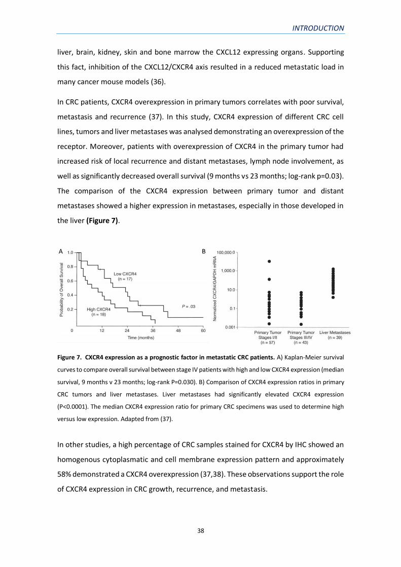
In CRC patients, CXCR4 overexpression in primary tumors correlates with poor survival,
metastasis and recurrence (37). In this study, CXCR4 expression of different CRC cell
lines, tumors and liver metastases was analysed demonstrating an overexpression of the
receptor. Moreover, patients with overexpression of CXCR4 in the primary tumor had
increased risk of local recurrence and distant metastases, lymph node involvement, as
well as significantly decreased overall survival (9 months vs 23 months; log-rank p=0.03).
The comparison of the CXCR4 expression between primary tumor and distant
metastases showed a higher expression in metastases, especially in those developed in
the liver (Figure 7).
Figure 7. CXCR4 expression as a prognostic factor in metastatic CRC patients. A) Kaplan-Meier survival
curves to compare overall survival between stage IV patients with high and low CXCR4 expression (median
survival, 9 months v 23 months; log-rank P=0.030). B) Comparison of CXCR4 expression ratios in primary
CRC tumors and liver metastases. Liver metastases had significantly elevated CXCR4 expression
(P<0.0001). The median CXCR4 expression ratio for primary CRC specimens was used to determine high
versus low expression. Adapted from (37).
In other studies, a high percentage of CRC samples stained for CXCR4 by IHC showed an
homogenous cytoplasmatic and cell membrane expression pattern and approximately
58% demonstrated a CXCR4 overexpression (37,38). These observations support the role
of CXCR4 expression in CRC growth, recurrence, and metastasis.

INTRODUCTION
39
Surprisingly, in vitro CXCR4 surface expression levels were found to be low or absent in
CRC cell lines while high expression levels were observed in vivo in animal models of
liver metastasis (39). These findings suggested that CXCR4 expression by CRC cells is
regulated by tumor microenvironment signals and the isolated metastatic cells exploit
CXCR4 signalling for proliferation.
2.3. CXCR4 clinical significance
The important roles of the CXCR4 receptor in several diseases, including human
immunodeficiency virus (HIV) infection, cancer, hypogammaglobulinemia,
myelokathexis (WHIM) syndrome and pulmonary injury, have encouraged the
development of viable CXCR4 antagonists for their treatment. Several CXCR4 inhibitors
are being developed, especially for targeting CXCR4+ cancer cells. Moreover, intense
efforts have also been directed towards the development of suitable CXCR4-targeted
molecular imaging agents (40).
Nowadays, the granulocyte colony-stimulating factor (G-CSF) is used in clinical
management to stimulate the production of granulocytes, to accelerate patient
recovery and reduce neutropenia after chemotherapy. G-CSF is also used in donors for
hematopoietic stem cell transplantation to increase the number of hematopoietic stem
cells in the blood of donors before collection by leukapheresis. Some studies revealed
that G-CSF promotes the degradation of HSC anchorage in bone marrow and the release
of these cells into peripheral blood, by down-regulating CXCR4 expression in bone
marrow myeloid cells, which attenuate their response to SDF-1.
2.3.1. Anti-cancer drugs targeting CXCR4
Given the clinical relevance of CXCR4 expression concerning tumor growth and
spreading in different types of cancer, a multitude of CXCR4-directed antagonists have
been developed during the last decade. Among them, the AMD3100 (Plerixafor) is the
unique compound approved by the FDA for the mobilization of hematopoietic stem cells
and for the treatment of multiple myeloma and non-Hodgkin’s lymphoma (41).
Plerixafor is commonly used in combination with G-CSF to inhibit hematopoietic stem
cell attachment to the bone marrow.

INTRODUCTION
40
Many other CXCR4 antagonists are being developed and some of them are under clinical
trials (Table 1). Four major classes of CXCR4 antagonists can be distinguished: a) small
peptide CXCR4 antagonists (T140), b) non-peptide CXCR4 antagonists (AM3100), c)
antibodies against CXCR4 and d) modified agonists and antagonists for SDF-1.
Table 1. Synthetic CXCR4 antagonists under clinical investigation.
Drug name Other names Mechanism of action Classification Indication Status
Plerixafor
AMD 3100;
GZ316455;
Mozobil
CXCR4 chemokine
antagonist; Neuroplastin
inhibitor
Non-peptide
Chronic
lymphocytic
leukemia;
Glioblastoma;
Neutropenia;
Non-Hodgkin’s
lymphoma;
Marketed
USL-311 Proximagen CXCR4 chemokine
antagonist
Small
molecule
Glioblastoma;
Inflammatory
disease; Solid
tumor
Phase 2
Clinical
LY-2510924 CXCR4 peptide
inhibitor
CXCR4 chemokine
antagonist; Stromal cell-
derived factor 1 ligand
inhibitor
Small peptide
Renal cell
carcinoma; Small-
cell lung cancer
Phase 2
Clinical
PRX17756 CXCR4 chemokine
antagonist
Small
molecule Glioblastoma
BL-8040
TG-0054;
bulishafu;
burixafor
CXCR4 chemokine
antagonist Small peptide
Acute
myelogenous
leukemia;
Metastatic
pancreatic
cancer; Multiple
myeloma;
Thrombocytopenia
Phase 2
Clinical
Ly2510924 is a small cyclic peptide and a potent selective CXCR4 antagonist. In a phase
I trial, it showed few adverse effects and the stabilization of the disease in 9 patients
(only 20%) (42). Other novel experimental CXCR4 antagonists are PRX17756 and BL-
8040. PRX17756 has been shown to actively penetrate the blood-brain barrier and to
accelerate GSC differentiation. The combination of PRX177561 with bevacizumab

INTRODUCTION
41
resulted in a synergistic reduction of tumor growth with an increase of disease-free
survival (DSF) and overall survival (OS) in a preclinical model of glioblastoma (43). BL-
8040 is a small synthetic peptide with high affinity for CXCR4. When compared to other
antagonists such as AMD3100, BL-8040 demonstrates higher affinity and longer
receptor occupancy, providing a greater effect on the retention–mobilization balance of
bone marrow stem cells in preclinical studies. In a recent clinical trial, safety and efficacy
of the treatment with BL-8040 in combination with pembrolizumab and chemotherapy
of metastatic pancreatic cancer patients was evaluated. The results suggest that
combined CXCR4 and PD-1 blockade may expand the benefit of chemotherapy in
pancreatic cancer (44). Therefore, all of these drugs need still to be tested in phase II
clinical trials to assess their antitumor efficacy alone or in combination with classic
radiotherapy or chemotherapy.
3) MOUSE MODELS IN CANCER RESEARCH
Prior to the development of genetic mouse models, cancer was mainly studied using in
vitro cell culture systems of cell lines derived from human tumors. Although in vitro
studies continue to provide valuable information and remain important to cancer
research, their main limitations include the inability to examine interactions among
tumor cells and their microenvironment, such as the extracellular matrix, and stromal
and immune cells. The use of in vivo animal models has been crucial to understand the
genetic basis of tumor development and cancer progression and continues to be
relevant to test the efficacy of novel anti-cancer agents.
The animal model mostly used in cancer research is the mouse (Mus musculus). There
are some advantages that justify the choice of the mouse as a model, such as its small
size, easy handling, short tumor generation and its price, allowing the use of large
numbers for statistical assessment. Moreover, it is the best genetically characterized of
all mammals used in cancer research.
3.1. CRC mouse models
In the recent decades, despite a growing insight into the biology of CRC and many
therapeutic improvements, the development of preclinical in vivo models remains

INTRODUCTION
42
essential. The ideal CRC model must recapitulate the CRC progression from a
precancerous adenoma to an invasive carcinoma with metastatic potential and should
reflect the molecular heterogeneity of the disease among individuals. Cancer mouse
models can be classified into different groups: syngeneic models, carcinogen-induced
models, genetically engineered models and xenografts models (Figure 8).
Syngeneic mouse models consist in the implantation of mouse cancer cell lines in an
immunocompetent mouse strain. This type of mouse model retains an intact immune
system, which is important to study the role of immune cells in the different phases of
cancer progression. Moreover, they can provide an effective approach for studying how
newly developed cancer therapies respond in the presence of a functional immune
system and represents an important tool for efficacy and toxicology preclinical studies.
However, the main limitations of these models, are the low number of mouse cancer
cell lines available, their usually low metastatic yield and the possible differences
between mouse and human tumor development and cancer progression.
Figure 8. Mouse models for cancer research. Human tumor cell lines or patient-derived tumor samples
can be transplanted in immunocompromised mice to develop a xenograft cancer model. In contrast,
immunocompetent mouse models are developed by transplanting established mouse cancer cell lines in

INTRODUCTION
43
recipient mice (syngeneic models) or by generating genetically engineered mouse (GEM) models or
carcinogen-induced models. Adapted from (45).
In carcinogen-induced models, mice are exposed to specific carcinogenic compounds,
until they develop the cancer of study. In CRC they provide a platform to evaluate the
influence of diet, dietary supplements, chemopreventive interventions and the gut
microbiome using diverse administration routes such as ad libitum feeding, oral,
intraperitoneal, subcutaneous or intramuscular injection, or enema. In this model, the
immune system remains intact, enabling the study of its contribution in this particular
type of carcinogenesis. However, chemical-induced carcinomas are very heterogenous
and rarely show invasive properties while distant metastases are usually absent.
Genetically engineered mouse models (GEMM) are obtained by modifying the mouse
genome through the use of genetic engineering techniques. Genetic modifications can
induce a gain of function of tumorigenic genes (oncogenes) or a loss of function of tumor
suppressor genes, and spontaneously originate the cancer of interest. GEMM are the
second most used model in cancer research and many CRC GEMM have been developed
during the last decades contributing enormously to the understanding of the molecular
processes of CRC initiation, progression and crosstalk of common cancer-associated
pathways. However, these models have several limitations. First, they are more
expensive and time-consuming than the other cancer mouse models because of the
difficulty in the breeding to achieve the desired alteration. The breading process also
generates many mice which are neither used for further breeding nor for research.
Finally, the use of GEMM in preclinical studies is limited due to a lack of genetic
heterogeneity and differences with the human tumor development.
3.2. Cell-derived and patient-derived xenografts
The majority of cancer studies use xenograft mouse models (45). Thus, our first objective
was improving human CRC mouse models with high metastatic load to evaluate the
antineoplastic effect of novel therapeutic nanoparticles. For the development of
xenograft mouse models, human tumor grafts are implanted in immunocompromised
mice for the successful engraftment and growth of human cells. As human tumor grafts,
tumor cells, organoids or tumor tissue pieces can be employed. The original material of

INTRODUCTION
44
these grafts can be either established human cell lines for the development of cell-
derived xenografts or patient tumor samples to generate patient-derived xenografts
(PDXs).
Cell-derived xenografts remain the most commonly used mouse models in basic and
translational cancer research (82%) (45) because of their low cost, the synchronous
tumor growth and their easy manipulation. However, PDXs are able to retain molecular,
genetic and histopathological features of their originating tumor, for some limited in
vivo passages, therefore being the best model to study inter-patient and intra-tumor
heterogeneity of human cancer (46).
The use of xenografts models has some advantages compared to other mouse models.
The employment of human cell lines or patient samples makes possible the study of
specific signalling pathways in tumor progression, which in a syngeneic mouse model
could show a different molecular basis. Moreover, the experimental conditions can be
more easily controlled by deciding the localization of cell injection or the number of cells
injected. However, the use of immunocompromised mice doesn’t enable the study of
the interactions between immune cells and tumors or the immune response in the
development of new therapeutic agents. This limitation can be solved by humanizing
the mice by the engraftment of various types of human leukocytes and purified human
CD34+ hematopoietic stem cells (HSCs) (47).
In addition, human cancer cells can be implanted in different anatomical locations. Thus,
a particularly relevant xenograft mouse model is the orthotopic, which is generated by
cell implantation in the organ that coincides with the original location of the primary
tumor (e.g. colonic mucosa) while heterotopic xenograft model is considered when cells
are implanted in a different location (e.g. subcutis).
3.2.1. Heterotopic mouse models
The subcutaneous models are among the most widely used heterotopic models, in
which cells are injected under the skin, normally in the flanks of the mouse. They are
frequently used in antitumoral drug development because of their easy establishment,
reproducibility and tumor growth monitoring. However, large drug screenings showed

INTRODUCTION
45
that subcutaneous models are of low value for the prediction of clinical response in
humans (48). Moreover, the lack of interactions with the right tumor microenvironment
makes that cancer cells rarely metastasise to regional o distant organs which render
them unsuitable for the study of mechanisms of metastatic spread or for the
development of antimetastatic therapies.
In previous studies, our group has shown that a subcutaneous preconditioning prior to
orthotopic microinjection increased the metastatic dissemination of both HCT116 and
SW48 orthotopic CRC models by increasing tumor cell survival and invasion at the tumor
invasion front (49).
3.2.2. Orthotopic mouse models
The growing need for preclinical models that replicate more accurately human diseases,
has led to the development of orthotopic cancer models. In the case of orthotopic CRC
mouse models, cancer cells are injected in the serosa layer of the intestine (50) or in the
cecal wall (51). These mouse models are more clinically relevant than subcutaneous
models, because of their organ-specific tumor microenvironment which differs in each
cancer type. The original microenvironment contains the relevant vasculature, a similar
hypoxic condition and the stromal cell infiltration of the primary tumor. These adequate
conditions can lead to a greater capability of cells to metastasize.
Whereas subcutaneous models are relatively easy to set up, the establishment of
orthotopic tumor models requires a surgical procedure, which demands the use of
anaesthesia. So, it represents a more invasive procedure that could affect the mouse
general condition, being sometimes necessary a recovery time. Moreover, orthotopic
tumor models need longer time courses for development and furthermore tumor
growth and cancer cells dissemination can compromise animal welfare. Another major
challenge is the follow-up of tumor progression in an orthotopic tumor model. Unlike
subcutaneous models, primary tumors in orthotopic models are not always visible and
bioluminescent or other optical imaging systems may be required (52).
INTRODUCTION
46
4) NANOPARTICLES FOR CANCER TREATMENT
Currently, nanocarriers are widely studied in cancer diagnosis and therapy, as well as for
the treatment of other disorders like cardiovascular and infection diseases. Following,
we are describing their properties and their use for targeted drug delivery in oncology.
4.1. Nanoparticles and their key properties
Nanoparticles have unique biological properties given their small size (diameter within
1–100 nm) and high surface-to-volume ratio, which allows them to bind and carry
anticancer agents, such as drugs, nucleic acids and proteins, along with imaging agents,
with higher efficiency than currently used drugs.
Size is one of the most important features for nanoparticle’s proper circulation and
biodistribution. Nanoparticles smaller than 7 nm, can be easily cleared by physiological
systems (filtration through the kidney), while particles larger than 200 nm may be
cleared by phagocytic cells in the reticuloendothelial system (RES). Therefore,
therapeutic nanoparticles with a size of <100 nm have longer circulation time in the
bloodstream. Consistently, many studies reported that therapeutic nanoparticles in 20–
200 nm size showed higher accumulation in tumors because they cannot be recognized
by the RES or excreted by the kidney (53).
For different purposes, nanoparticles can be designed and functionalized to increase
their efficiency, by controlling their size and surface properties. The surface charge of
nanoparticles dictates their interaction with cell surface molecules and hence their
cellular uptake (54). In general, nanoparticles functionalized with positively charged
ligands exhibit higher internalization into cells compared to the neutral or negatively
charged nanoparticles (55). Another issue that can be solved by functionalization is the
entrapment of nanoparticles in the endosomes, preventing their access to the cytosol.
Thus, the incorporation of signalling peptides in the nanoparticles’ surface allows them
to escape from endosomes and prevent lysosomal degradation (56). Moreover, in order
to use nanoparticles as imaging agents for cancer diagnosis or to study nanoparticles’
biodistribution, they can be functionalized by conjugation of fluorescent probes (57) or
radioactive isotopes (58).

INTRODUCTION
47
4.2. Targeted drug delivery for cancer treatment
Conventional therapeutic agents have limitations such as non-selectivity, undesirable
side effects, low efficiency, and poor biodistribution. Current cancer research is focused
on the use of nanoparticles to deliver drugs to specific cells or tissues. Targeted drug
delivery refers to the capacity to direct therapeutic agents, aiming to enhance their
accumulation in a desirable site. For efficient targeted delivery, the drug carrier should
be retained in the physiological system, evade the immune system, target the specific
cell or tissue, and release the payload therapeutic agent in the target site (59).
As a result, delivering drugs only to cancer cells using nanoparticles decreases
cytotoxicity in normal cells, increases the antitumor effect and increases its systemic
circulation time by avoiding renal and hepatic clearance (which can be retained in the
liver sinusoids without being uptaken inside its parenchyma). In addition, highly potent
drugs or toxins, or higher doses can be used without affecting normal cells. Tumor-
specific targeting can be actively or passively achieved (Figure 9).
Figure 9. Targeted drug delivery for cancer treatment. Graphical illustration of passive and active drug
targeting strategies. In passive targeting, the nanocarriers pass through the fenestrated capillaries
irrigating the tumor and accumulate at the tumor tissue by the enhanced permeability and retention (EPR)
effect. Active targeting can be achieved using specific ligands that bind to the receptors on the target
tumor cell surface to enhance the delivery of the payload drug inside its cytosol. Adapted from (60).

INTRODUCTION
48
4.2.1. Passive targeting
Passive targeting, in which nanoparticles reach the target tissue through enhanced
permeability and retention (EPR) effect resulting from the leaky vasculature of the
tumor cells, has been so far the most extensively explored strategy for targeting cancer
(61), despite some researchers question their relevance in human tumors. In theory, the
EPR effect allows preferential extravasation of the circulating macromolecules and poor
lymphatic drainage which can lead to retention and passive accumulation of the
nanoparticles in tumor tissues (Figure 9). However, only a small percentage of these
nanoparticles accumulate even in high-EPR xenografted tumors (less than 1%) (62). This
could be due to multiple physiological barriers (clearance systems, endothelial barrier
and plasma membrane) and a high degree of stochasticity involved in nanoparticles
extravasation through the tumor vasculature. A major proportion of nanoparticles are
also cleared by the mononuclear phagocytic system (MPS), some get trapped in the
sinusoids of the liver and others are taken up by hepatocytes and Kupffer cells (63).
4.2.2. Active targeting
To overcome the challenges presented by passive targeting, active cellular targeting
strategies have been developed. These approaches involve adding affinity ligands for
cell surface markers on the surface of the nanoparticles to trigger specific homing,
increased retention at the target site and uptake by target cancer cells (Figure 9) (64).
The ligands are usually selected to bind to overexpressed receptors on cell membrane
(e.g. HER2, CD44, etc).
Active targeting strategies are much more complexes than passive approaches. The
major challenge is the complex design and engineering of these systems, which can
complicate their pharmaceutical development, the scale-up production and significantly
increase the cost of therapy. Despite these difficulties, one major advantage of active
targeting nanoparticle-based therapies is their ability to target disseminated locations
throughout the body that could potentially improve the treatment of hematological
malignancies and metastatic lesions where EPR could not play a role.

INTRODUCTION
49
4.3. Classification of drug nanocarriers
Generally, the drug nanocarriers used in cancer treatment can be classified into two
major types: carriers that use organic molecules as major building blocks and those that
use inorganic elements as their main structure. Organic nanocarriers are comprised of
polymer-based nanoparticles (nanogels, dendrimers, protein nanoparticles) and lipid-
based nanoparticles (liposomes, lipids and micelles). Inorganic nanocarriers as magnetic
nanoparticles, quantum dots, gold nanoparticles and carbon nanotubes are commonly
used in cancer drug development. Essential properties for nanocarriers success
regarding clinical development are biodegradability, low toxicity, high specificity and
efficiency and low immunogenicity.
4.3.1. Protein-based nanoparticles
Protein-based nanoparticles have many attributes that make them highly attractive as
biological nanomaterials. For example, they are soluble, monodisperse, biodegradable,
metabolizable, and easily adaptable by surface modifications to allow attachment of a
drug and/or targeting ligands. Moreover, genetic, molecular and crystal structure
information of many protein-based nanoparticles are available, facilitating their design
and allowing for chemical and genetic modifications (65).
Protein-based nanoparticles can be produced as virus-like particles (VLP), bacterial
inclusion bodies, eukaryotic aggresomes or self-assembling proteins, either in their
natural hosts, as recombinant proteins in expressions systems (bacteria, yeast, plants
and mammalian cells) or by cell-free protein synthesis. Most protein nanoparticles used
in cancer therapy have been expressed and produced recombinantly in Escherichia coli.
Due to their large macromolecular structures, protein nanoparticle purification
protocols tend to involve size exclusion chromatography (SEC) and/or differential
centrifugation steps. To achieve higher purity, SEC is generally combined with affinity
chromatography, in which the nanoparticle displays a pre-selected purification tag
(histidine-tag), or ion-exchange, in which the outer surface charge is exploited.

INTRODUCTION
50
4.4. FDA approved nanomedicines for cancer therapy
In the last few decades, nanomedicine has contributed significantly to the treatment of
cancer and other diseases, however, only a few drugs are available in the market or
undergoing clinical trials. Doxil was the first FDA approved (1995) nanodrug for the
treatment of metastatic ovarian cancer and AIDS-related Kaposi’s sarcoma. It contains
doxorubicin, a member of the anthracycline group, encapsulated in an 80-90 nm size
unilamellar liposome coated with PEG, that allows a longer recirculation of the drug in
the bloodstream increasing the probability to reach cancer cells, while decreasing the
cardiotoxicity produced by administration of free doxorubicin (66).
Another FDA approved nanomedicine is the albumin-conjugated nanoparticle
(Abraxane®), a version of the anticancer drug paclitaxel. Paclitaxel (Taxol) belongs to an
important class of antitumor agents called taxanes. Taxanes are cell cycle-specific agents
that bind to microtubules resulting in the inhibition of mitosis and induction of cell
death. However, they are highly hydrophobic, showing very low solubility in water. In
order to solubilize paclitaxel cremophore and ethanol are used, which cause
hypersensitivity reactions. However, Abraxane does not contain cremophore, avoiding
the taxane dose-limiting toxicity. Abraxane formulation has increased the bioavailability
of paclitaxel and resulted in higher tumor uptake, facilitated by albumin-receptor (gp60)
mediated endothelial transcytosis (67). This therapeutic nanoparticle yields lower side
effects and improved therapeutic indices than paclitaxel. Abraxane was approved for
the treatment of metastatic breast cancer, advanced non-small cell lung cancer and
advanced pancreatic cancer (68).
Table 2 summarizes the nanomedicines approved by the FDA for cancer treatment,
which include liposomes, metallic and protein-based nanoparticles and the tumor types
in which they are currently used.

INTRODUCTION
51
Table 2. Nanomedicines approved by FDA for cancer therapy.
Nanostructure Product Nanoparticle formulation Drug Indication
Liposomes
Marqibo® Sphingomyelin and
cholesterol Vincristine
Acute lymphoid
leukaemia
Mepact®
1-Palmitoyl-2-oleoyl-snglycero-
3-phosphocholine
and 1,2-Dioleoyl-sn-glycero-
3-phospho-l-serine
liposomes
Mifamurtide
Non-
metastasizing
osteosarcoma
Onivyde® Nanoliposomes Irinotecan
Pancreatic
cancer,
Colorectal cancer
Vyxeos®
Distearoylphosphatidylcholine,
Distearoylphosphatidylglycerol,
Cholesterol
Daunorubicin
Cytarabine
Acute myeloid
leukaemia
Protein-drug
conjugates
Kadcyla® Maytansine derivative, DM1 Trastuzumab HER2+ breast
cancer
Abraxane® Albumin Paclitaxel
Non-small lung
cancer,
Pancreatic
cancer
Metallic
nanoparticles NanoTherm®
Nanoparticles of
superparamagnetic iron
oxide coated with amino
silane
-
Glioblastoma,
prostate,
pancreatic
cancer
4.5. T22-GFP-H6 protein-based nanoparticle targeting CXCR4+ cells
Most FDA-approved nanomedicines for cancer treatment try to exploit passive targeting
strategies. In collaboration with the Nanobiotechnology group led by Dr. Villaverde,
from the Universitat Autònoma de Barcelona (UAB), our group developed protein-based
nanoparticles capable of internalizing in CXCR4+ cancer cells. We exploited the strategy
of using the receptor-mediated endocytosis to improve nanocarrier uptake in tumor

INTRODUCTION
52
cells, that overexpress CXCR4, and consequently enhance its anticancer effect while
reducing its systemic toxicity. For that, we initially studied the ability of four different
peptide ligands of the CXCR4 receptor with internalization capacity in CXCR4+ cells (69).
Only the T22 peptide efficiently penetrated target cells via rapid receptor-specific
endosomal route. Thus, we propose T22 as a novel cell-targeting peptide suitable for
functionalization of protein nanoparticles and appropriate for intracellular delivery in
CXCR4 overexpressing tumors.
4.5.1. Structure of the T22-GFP-H6 nanoparticle
The T22 peptide is an engineered version of the polyphemusin II peptide from the
horseshoe crab, in which three substitutions at residues Tyr5, Lys7 and Tyr12 highly
increase its natural affinity for CXCR4 (70). This cationic peptide (8 K+R) was fused to the
construct formed by the GFP fluorescent protein and a C-terminal polyhistidine tail
(His6) and was then produced in recombinant Escherichia coli. Ten of these T22-GFP-H6
polypeptides spontaneously self-assemble as 12 nm in diameter nanoparticles with a
toroidal architecture which avoid being filtered by the kidneys (Figure 10).
Figure 10. Characterization of the T22-GFP-H6 protein nanoparticles A) Predicted and schematic
representation of the CXCR4 targeting nanoparticle formed by the self-assembling of 10 monomers of
T22-GFP-H6. The colour code is maintained here for both ligand- (red) and H6- (blue) overhanging ends.
B) Dynamic light scattering size analysis of T22-GFP-H6 nanoparticles in NaCO3H buffer. C) Transmission
electron microscopy and scanning emission microscopy of T22-GFP-H6 nanoparticles.

INTRODUCTION
53
The GFP protein allows the localization and quantification of the uptaken nanoparticles
through its fluorescence emission, but it also contributes to the stability of the
nanoparticles’ final structure and to follow-up their in vivo biodistribution. The
polyhistidine tail is necessary for their purification but it is also a key architectonic player
in the supramolecular architecture of the nanoparticles. The presence of both the
cationic T22 and the H6-tail is necessary for assembly, as GFP-H6 and T22-GFP solely
occur as unassembled proteins. Moreover, divalent cations coming from the Nickel II
(Ni2+) cation remaining in the producing bacteria lysates are involved in protein
assembly. Thus, the addition of the EDTA chelator promoted efficient disassembling of
the nanoparticles. Free Ni2+ as well as Cu2+, Ca2+ and Zn2+, but not Cs+ and K+ cations,
recovered the original nanoparticle size when added to the solution of disassembled
polypeptides (71). In other studies, we found that changes in the number of histidines
forming the tail, also affected the stability of the nanoparticles and their capacity of
accumulating in tumor tissue (72).
4.5.2. T22-GFP-H6 functionalization with the endosomal escape peptide HA2
By incorporating the T22 ligand to our protein-based nanoparticles, our group has
achieved an active CXCR4+ cancer cell targeting, which directs specific binding and
further endosomal-mediated cell uptake. Unfortunately, endosomal uptake drives the
internalized material to a lysosomal pathway, resulting in acidification and proteolysis.
A fraction of the nanoparticles reaches the cytoplasm due to background endosomal
leakage and endosomolytic activities naturally present in the recombinant protein.
In order to increase the fraction of internalized material that escapes from endosomes,
we searched for natural or modified peptides identified as endosomolytic. The N-
terminal peptide HA2 from the influenza virus hemagglutinin has been widely explored
in protein constructs designed for drug delivery (73). In acidic environments, such as the
endosome, the anionic amino acids of HA2 get protonated, an alpha helix is formed and
the peptide acts as an amphiphilic anionic stretch that destabilizes the cell membrane,
promoting endosomal release of fusion proteins (74). Thus, we incorporated the HA2
peptide into the T22-GFP-H6 nanoparticle in two alternative inner positions (in the
amino terminus of the core GFP, or its carboxy terminus) (Figure 11). So, by efficiently

INTRODUCTION
54
combining the selectivity of the T22 peptide with the HA2 endosomal escape domain we
have achieved the expected functional improvement, therefore, designing powerful
vehicles for targeted drug delivery (75).
Figure 11. Incorporation of the fusogenic peptide HA2 into the T22-GFP-H6 nanoparticle A) Schematic
representation of the polypeptidic building blocks forming the nanoparticles. Box sizes are only indicative.
HA2 is the fusogenic HA2 peptide from the influenza virus hemagglutinin. A summary of relevant
properties of the nanoparticles is found in the table at the bottom. B) Representative FESEM images of
the assembled nanoparticles. Bars represent 40 nm. C) Isosurface representation of cultured HeLa cells
within a 3D volumetric z axes stack upon incubation with 0.5 mM protein nanoparticles for 24 h. The cell
membrane was labelled with CellMask (rendering a red signal), the nuclear DNA was labelled with Hoechst
33342 (rendering a blue signal) and protein nanoparticles are seen in green because of their intrinsic
fluorescence. Bars indicate 5 µm.
4.6. Therapeutic derivatives of the T22-GFP-H6 nanoparticle
Once we developed the T22-GFP-H6 nanocarrier, we demonstrated its capacity to
internalize efficiently in CXCR4+ cells of the primary tumor and distant metastasis of an
orthotopic CRC mouse model (69) without signs of toxicity. We then proceeded to use
it as a promising drug delivery system for cancer treatment, using different strategies to
develop novel therapeutic nanoparticles. Firstly, the T22-GFP-H6 nanoparticle was
conjugated to different therapeutic drugs, that are chemotherapeutics commonly used
in the clinics or to very potent agents, not used in patients because of their toxicity.
Secondly, toxins of bacteria (76), animals (77) and plants (78) or human pro-apoptotic
factors were incorporated to the T22 polypeptides, to test the selectivity in delivering
these agents to CXCR4+ cancer cells and their antitumor activity.

INTRODUCTION
55
4.6.1. Nanoconjugates
On the basis of these previous results, we generated T22-GFP-H6-based
nanoconjugates, using a variety of therapeutic agents covalently bound to the T22-GFP-
H6 nanocarrier (Figure 12A). In the first place, a cytotoxic drug commonly used to treat
CRC liver metastases, Floxuridine (FdU), was covalently bound to the T22-GFP-H6
nanoparticle. T22-GFP-H6-FdU was synthesized by functionalizing the pentameric
nucleotide oligo-FdU with thiol, which was subsequently conjugated to the T22-GFP-H6
nanoparticle previously bound to a chemical linker (Figure 12B). Floxuridine is an
antineoplastic pyrimidine analogue produced from a rapid intracellular metabolism of
5-fluorouracil when administered by intravenous injection. FdU is the main active
metabolite of 5-fluorouracil and works by interfering with the DNA synthesis, preventing
the incorporation of thymidine nucleotides into the DNA strand. We have already
demonstrated an antimetastatic activity of T22-GFP-H6-FdU nanoconjugate as well as
the absence of toxicity have already been demonstrated in metastatic CRC mouse
models (79). In contrast to free oligo-FdU, intravenous repeated administration of T22-
GFP-H6-FdU selectively accumulates and internalizes in CXCR4+ cancer cells, triggering
DNA damage and apoptosis, which leads to their selective elimination and to reduced
tumor re-initiation capacity.
Since, the selective delivery of drugs to cancer cells using nanoparticles allows to
increase the administered dose or even the use of highly cytotoxic drugs, we generated
an additional nanoconjugate tested for anticancer effect in this thesis work. This novel
nanoconjugate was produced by binding the T22-GFP-H6 nanoparticle to the toxin
Auristatin E (Aur). Monomethyl Auristatin E (MMAE) is a potent tubulin polymerization
inhibitor that was recently introduced in clinical oncohematology using antibody-drug
conjugates (ADCs) (80). The T22-GFP-H6-Aur nanoconjugate was synthesized by
covalent binding of a maleimide functionalized MMAE (MC-MMAE) to the amine groups
of the external lysins of the T22-GFP-H6 nanocarrier (Figure 12C). This reaction yielded
an average of 18.5 MMAE molecules per protein monomer, that represents 204.5
MMAE molecules per nanoparticle.

INTRODUCTION
56
Figure 12. T22-GFP-H6-based nanoconjugates and their synthesis A) Schematic representation of a
nanoconjugate, containing the fusion protein composed of the peptide T22 as a CXCR4 ligand, the GFP
fluorescent protein and a histidine tail, bound to a generic payload drug. B) T22-GFP-H6-FdU chemical
synthesis. C) T22-GFP-H6-Aur chemical synthesis using a maleimide functionalized Monomethyl Auristatin
E (MMAE).
4.6.2. Toxin-based nanoparticles
The high potency exhibited by some toxins for toxin-mediated cell killing is an attractive
approach to be explored in monotherapy or in combination with conventional
chemotherapies. Over the last years, some promising treatments using immunotoxins
that incorporated highly cytotoxic bacterial proteins have been tested in clinical trials
(81). Only three of them, denileukin diftitox, tagraxofusp, and moxetumomab pasudotox
have been FDA-approved for treating hematological cancers (82,83). No immunotoxins
against solid tumors have been approved so far for clinical use.

INTRODUCTION
57
Figure 13. Characterization of toxin-based nanoparticles. A) Schematic representation of the
polypeptidic building blocks of toxin nanoparticles. Box sizes are only indicative. Relevant properties of
the nanoparticles are shown in the table at the bottom. B) Representative FESEM images of the T22-
DITOX-H6 and T22-PE24-H6 nanoparticles. Bars represent 50 nm.
In this context, we produced novel nanotoxins to also test their anticancer activity in
CRC models, during the development of this thesis. To that purpose, we engineered self-
assembling nanoparticles containing the catalytic domain of the diphtheria toxin
from Corynebacterium diphtheriae (DITOX) and the Pseudomonas aeruginosa exotoxin
(PE24), targeting CXCR4, for the treatment of CXCR4+ CRC tumors (Figure 13). Both
microbial toxins are ribosome inactivating proteins (RIPs) that inhibit the eukaryotic
elongation factor 2 (eEF-2), preventing protein synthesis and consequently leading to
irreversible cell death. When DITOX and PE24 toxins are fused to the CXCR4 ligand T22,
they self-assemble as nanoparticles of a size between 30 and 60 nm. Moreover, a KDEL
sequence was added in the C-terminus of T22-PE24-H6, which allows the efficiently
binding to KDEL receptors at the Golgi apparatus during intracellular trafficking (84).
Furin-cleavage sites were also inserted between the T22 ligand and each of the toxins
to release the amino terminal peptide once internalized into target cells.

II. AIMS AND OBJECTIVES
AIMS AND OBJECTIVES
59
II. AIMS AND OBJECTIVES
Although the survival for CRC patients has improved because of advancements in
diagnosis and treatment, the invasion and metastasis of CRC cells to proximal and
distant organs, associated with chemotherapy resistance and relapse, remain the major
challenge for its treatment. Therefore, the general aim of this Ph.D. project was to
develop highly metastatic CRC models with CXCR4 overexpression (CXCR4+) to study the
antineoplastic effect of therapeutic nanoparticles targeting CXCR4+ cancer stem cells
using different strategies.
The objectives to achieve were:
1) Developing subcutaneous tumor models displaying CXCR4-dependent growth and
highly metastatic CRC mouse models with CXCR4-dependent dissemination.
2) Studying in vitro the selective internalization and antitumor activity of T22-GFP-H6
and its therapeutic derivatives in CXCR4+ CRC cell lines.
3) Studying in vivo the biodistribution and toxicity of T22-GFP-H6 and its therapeutic
derivatives in CXCR4+ subcutaneous tumors and non-tumor organs.
4) Evaluating the antineoplastic activity of the T22-GFP-H6 therapeutic derivatives in
the developed CXCR4+ CRC mouse models with high metastatic rate.

III. MATERIALS AND METHODS

MATERIALS AND METHODS
61
III. MATERIALS AND METHODS
1. IN VITRO EXPERIMENTS
1.1. Cell culture
1.1.2. CRC cell lines
SW1417, SW48 and HT29 CRC cell lines were purchased from the American Type Culture
Collection (ATCC) and were cultured at 37 ºC and 5% CO2 in DMEM (Gibco)
supplemented with 10% fetal bovine serum (FBS, Sigma-Aldrich), 50 units/ml penicillin
and 50 mg/ml streptomycin (Life Technologies). The SW1417 cell line is a human cell line
stablished from a Dukes’ type C, grade III, colorectal adenocarcinoma. We have found
differences in CXCR4 expression between two vials obtained from different sources of
this cell line. We named them as CXCR4+ SW1417 the one with 70% of the cultured cells
showing CXCR4 expression and CXCR4- SW1417 the one with no CXCR4 expression in
any of the cultured cells.
1.1.2. Cell transfection with Luciferase reporter vector
The SW1417 CXCR4 expressing cell line was engineered to express firefly luciferase
through a plasmid (pPK-CMV-F3, Promokine) transfection process using lipofectamine
2000 protocol (ThermoFisher) followed by an antibiotic selection (Figure 14). For that,
SW1417 cells (2.5×10⁵ cells/well) were seeded in six-well plates overnight. Next day, we
incubated the cells for 2 hours with Opti-MEM medium (Gibco) and transfected them
with the luciferase plasmid (4 µg and 5 µg) according to the manufacturer’s instructions.
We tested two different ratios of DNA:lipofectamine (1:2 and 1:3) to find the more
efficient condition. The control group was treated under the same condition but without
DNA. After 24 hours, the medium was changed for DMEM medium containing 0.8 mg/ml
of geneticin (G418, Life Technologies) and after 3 days we isolated the clone expressing
higher levels of luciferase using the IVIS Spectrum Imaging System (PerkinElmer). This
stable luciferase expressing clone was always maintained with DMEM medium
containing 0.4 mg/ml of geneticin.

MATERIALS AND METHODS
62
Figure 14. pPK-CMV-F3 plasmid map. Diagram of the plasmid used to transfect SW1417 cells to express
luciferase. It shows the position of the luciferase sequence and the location of the plasmid functional
elements. Restriction enzyme sites, promoters (CMV and SV40p) and terminators (SV40pA and HSVpA),
resistance genes (Kan/Neo) and origin of replication (pUC ori).
1.1.3. Flow cytometry for CXCR4 levels determination
Before conducting all in vitro experiments and in vivo cell injections to generate CRC
models, the levels of CXCR4 expression in the cell membrane were assessed by flow
cytometry. For that purpose, we labelled 1 million cells in 100 µl of PBS with 5 µl of the
PE-Cy5 Mouse Anti-Human CD184 monoclonal antibody (BD Bioscience) and 1 million
cells in 100 µl of PBS with 5 µl of the PE-Cy5 Mouse IdG2a isotype (BD Bioscience) as a
negative control. Cells were incubated in the dark for 20 minutes at 4ºC with gentle
agitation, washed and analysed in the FACSCalibur cytometer (BD Bioscience). We
measured both the percentage of the population expressing CXCR4 and the intensity of
the expression expressed as mean fluorescence intensity (MFI).
1.1.4. ELISA for supernatant SDF-1 alpha determination
The enzyme-linked immunosorbent assay (ELISA) is a commonly used analytical
biochemistry assay which uses a solid-phase immunoassay to detect the presence of a
ligand, normally a protein, using antibodies directed against the protein of interest. In
order to analyse and quantify the amount of stromal cell derived factor 1 (SDF-1 alpha)

MATERIALS AND METHODS
63
secreted by different cell lines, we used the commercially available Human SDF-1 alpha
ELISA kit (RayBiotech). For that purpose, 5x10⁴ SW1417 cells and 1BR3.G control
fibroblasts were cultured in DMEM with 10% FBS, on 24 well plates for 48 and 72 hours.
Then, supernatants were collected and analysed. In this case, the kit is designed as a
colorimetric sandwich-based ELISA, in which the 96 well plate is coated with a capture
antibody. Supernatants were added to the wells so that the SDF-1 alpha present in the
samples was captured by the specific antibody. With a washing step, any unbound
protein was removed from the wells. Then, an enzyme-linked antibody was added which
also bound to the antigen. Finally, the enzyme’s substrate was added and converted by
the enzyme to a detectable colour. The absorbance of each well was measured using a
spectrophotometer (FLUOstar OPTIMA, BMG Labtech) and the values were correlated
to those from the standard curve to quantify the amount (pg/ml) of SDF-1 alpha per
sample.
1.2. Fluorescent nanoparticles internalization assays
1.2.1. Flow cytometry
To evaluate the capacity of the GFP fluorescent nanoparticles to internalize in the
CXCR4+ SW1417 cells, we used flow cytometry after 24 hours of cell exposure to 400 nM
nanoparticles concentration. Then, cells were washed with PBS and treated with trypsin
1 mg/ml (Life Technologies) in order to remove non-specific binding of the nanoparticles
to the cell membrane. Cells were analysed in the FACSCalibur cytometer (BD Bioscience).
The internalization competition assays were done by preincubating the cells with
AMD3100 (ratio 1-T22-GFP-H6:10-AMD3100). Results of fluorescence emission were
analysed with the Cell Quest Pro software and expressed as mean fluorescence intensity
(MFI).
1.2.2. Confocal microscopy
We also evaluated the capacity of the GFP fluorescent nanoparticles to internalize in the
CXCR4+ SW1417 cells using confocal microscopy. For that, we seeded 2x10⁴ cells in each
well of Lab Tek II Chamber Slides (Thermo Fisher) and incubated with 1 µM of T22-GFP-
H6 and GFP-H6 as a negative control for 1, 5 and 24 hours. Then, cell membranes were

MATERIALS AND METHODS
64
labelled with 2.5 μg/mL CellMaskTM Deep Red (Molecular Probes) for 10 minutes in the
dark. Cells were washed in PBS and fixed with 3,75% formaldehyde for 10 minutes and
mounted with ProLong Gold Antifade DAPI (Life technologies) labelling also the cell
nucleus. Cells were imaged with the TSC SP5 II confocal laser microscope (Leica
Microsystems) using a HC PL APO 63 × /1.4 oil objective.
1.3. Cell death determination assays
1.3.1. Cell proliferation assays
Cell viability was evaluated measuring the cell metabolic capacity using the XTT
colorimetric cell proliferation kit II (Roche Diagnostics). The assay is based on the
cleavage of the tetrazolium salt XTT in the presence of an electron-coupling reagent, to
produce a soluble formazan salt. This conversion only occurs in viable cells. SW1417 cells
were seeded in 96 well plates and incubated O/N at 37ºC. After 24 hours, the T22-GFP-
H6 nanoparticle, or its therapeutic derivatives were added at different concentration
range [0-2 µM] for 48 hours. Then, they were incubated with the XTT labelling mixture
for 4 hours and quantitated the formazan dye formed using a scanning multi-well
spectrophotometer at 490 nm (FLUOstar OPTIMA, BMG Labtech). The measured
absorbance directly correlates with the number of viable cells. Data were reported as
percentage of cell viability of nanoparticle-treated cells in relation to buffer-treated cell
viability.
1.3.2. DAPI staining assays to detect DNA condensation
To quantify and determine the mechanism of cell death mediated by T22-GFP-H6-
Auristatin in CRC cells, we detected DNA condensation by enhanced nuclear staining
with DAPI dye in CXCR4+ SW1417 cells exposed to 1 µM T22-GFP-H6-Aur or buffer for
48 hours, in Lab Tek II Chamber Slides (Thermo Fisher). After incubation, cells were
rinsed once with PBS and fixed (3.7% p-formaldehyde in PBS, pH= 7.4) for 10 minutes.
Finally, cells were mounted with ProLong Gold Antifade DAPI (Life technologies). Under
a fluorescence microscope (Olympus BX53, Olympus) the appearance of dead cells
bodies (apoptosis) and mitotic figures (mitotic catastrophe) were observed and
quantified in 10 high-power fields (200x) of each condition. The total number of cells

MATERIALS AND METHODS
65
adhered to the slide was also quantified in the same fields and results presented as
mean ± s.e.m..
1.3.3. Immunocytochemistry analysis of cell blocks
The study of cell death pathways induced by the different therapeutic nanoparticles was
performed by aggregating treated cells in cell blocks and analysing them by
immunocytochemistry (ICC). CXCR4+ SW1417 cells were treated with 6 nM of T22-PE24-
H6 for 2, 5, 24 and 48 hours and then trypsinized and centrifuged. The obtained pellet
was mixed with plasma and thrombin (Sigma Aldrich) and quickly agitated to form a clot.
The clot was placed in a tissue cassette, fixed with formaldehyde and then paraffin-
embedded for further immunocytochemistry analysis (see 2.5.3. section). Percentage of
stained surface and its intensity were calculated using the ImageJ software and the
Colour Deconvolution Plugin with the H DAB vector to split the brown staining adjusting
the threshold to 100 for NLRP3 images or 120 for caspase-1 images. Then, the Analyse
particles plugin was used to detect all stained areas and the mean grey value was
obtained combining all selected black areas. The intensity value was calculated by
subtracting to 255 the mean grey value obtained in the analysis. Up to 5 high-power
fields (400x) of each sample were analysed and the results were expressed as mean ±
s.e.m..
1.4. Statistical analysis
All in vitro experiments, both internalization and XTT cell viability assays were performed
in biological triplicates and the data were expressed as mean ± standard error of mean
(s.e.m.). Normal distribution of the values was tested using the Shapiro–Wilk test.
Results with normal distribution were analysed using the Student t-test whereas Mann-
Whitney U test was used for the non-normal distribution data. All statistical tests were
performed using GraphPad Prism 8 (GraphPad Software, Inc). Differences among groups
were considered significant at a p < 0.05. IC50 were calculated with SigmaPlot (Systat
Software, Inc) using non-linear regression test with Hill-3 parameter adjustment

MATERIALS AND METHODS
66
2. IN VIVO EXPERIMENTS
2.1. Mouse strains
To generate the CRC models to be used in the evaluation of the biodistribution and
antimetastatic effect of the T22-GFP-H6 nanocarrier and its therapeutic derivatives we
used different strains of immunodeficient mice. All animals were obtained from Charles
Rivers Laboratories.
We used four-week-old female Swiss Nude (Crl:NU(Ico)-Foxn1nu) mice to generate CRC
subcutaneous models for conducting biodistribution and antitumoral assays. They carry
a genetic mutation that causes an absent thymus, resulting in an inhibited immune
system without mature T cells. The phenotype of the mouse is a lack of body hair.
We used four-week-old female NOD SCID (NOS.CB17-Prkdcscid/NcrCrl) mice to generate
CRC metastatic models by orthotopic cell implantation, to assess the antimetastatic
effect of our tested nanoconjugates and nanotoxins. They are immunodeficient mice
due to a mutation in the SCID gene impairing normal T and B lymphocyte development.
We used four-week-old female NSG or NOD scid gamma (NOD.Cg-Prkdcscid
II2rgtm1WjI/SzJ) mice to also generate CRC metastatic models by orthotopic cell injection.
These mice lack mature T cells, B cells, functional NK cells, and are also deficient in
cytokine signalling.
Mice were housed in controlled ventilation racks, under specific-pathogen-free (SPF)
conditions and fed with sterile food and water ad libitum. All animal experiments were
approved by the institutional animal Ethics Committee of Hospital de Sant Pau i la Santa
Creu and they were part of an Animal Experimentation Procedure Protocol approved by
the Generalitat de Catalunya. The experimental conditions, including the mouse strain
used for all the in vivo experiments are summarized in Table 3.
2.2. Cell implantation techniques
2.2.1. Subcutaneous injection of CRC cells
Mice were intraperitoneal (i.p.) anesthetized with a mix of 100 mg/kg of ketamine
(Ketolar, Pfizer) and 10 mg/kg of xylazine (Rompun, Bayer). Once asleep, a cellular

MATERIALS AND METHODS
67
suspension mixed with matrigel (1:1, Corning) was injected in one or two subcutaneous
flanks using a 21G needle syringe (BD Bioscience). All the material and reagents
(matrigel, media, syringes and needles) were previously cooled to avoid matrigel
solidification. For all the conditions, we used harvested cell culture, washed with the
corresponding media, counted and re-suspended to inject 150-100 µl per flank. The
volume of the generated tumor was measured twice per week with a digital caliper.
We also generated a subcutaneous CRC patient derived xenograft (PDX) from the patient
sample SP5. For that purpose, mice were anesthetized as explained above, and 10 mg
of SP5 tumor tissue were implanted in the subcutis using a trocar. Several mice were
used as donor animals to maintain in vivo the line. The tumor volume was measured
twice per week with a digital caliper.
2.2.2. Tumor disaggregation
We have previously described that the subcutaneous preconditioning increased the
invasion and metastatic capacity of CRC cell lines (49). To generate these SC+ORT models
we used SP5 subcutaneous derived tumors from donor animals of the same strain.
When these subcutaneous tumors reached a volume of 600 mm3, mice were sacrificed
by cervical dislocation and tumors were excised, discarding the necrotic areas, and 300
mg of viable tissue was then cut into pieces and disaggregated in 0.05% trypsin
(Invitrogen) and 100 mg/ml DNase (Sigma-Aldrich). The mix was pipetted 30 times, using
a 10-ml pipette, and incubated for 10 min at 37°C with shaking. It was then re-pipetted
30 times, using 10-, 3-, and 1- pipettes, and re-incubated for 5 min at 37°C with shaking.
The obtained cell suspension was filtered through a 0,45 µm cell filter (Millipore) and
centrifuged at 1000 g for 10 min before cell counting.
2.2.3. Orthotopic injection of CRC cells to generate metastatic models
Mice were intraperitoneally (i.p) anesthetized with 100 mg/kg of ketamine and 10 mg/kg
of xylazine. Once the paw withdrawal reflex disappeared, the cecum was exteriorized by
a laparotomy. A total of 2 million cells re-suspended in 50 µl of culture media were then
inoculated directly into the mouse cecum wall. A micropipette obtained as described
(51) was used for the cell injection (Figure 15). Afterwards, we pulled the pipette out

MATERIALS AND METHODS
68
and cleaned the area around the injection with 3% iodine to avoid seeding of unlikely
refluxed tumor cells into the abdominal cavity. The small diameter and flexible tip of the
pipette and the angular and slow rate of administration diminished resistance to the
injection, limiting tissue damage and bleeding, ensuring the absence of cell reflux. After
injection, the gut was returned to the abdominal cavity and closed with surgical staples.
Mice were followed once a week and euthanized when they had lost 10% of their body
weight or showed signs of pain or illness.
Figure 15. OCMI procedure into the cecum of immunosuppressed mice A) Micropipette made from
Vitrex capillaries with a 250 µm-diameter tip B) Injection of two million human CRC cells per animal C)
Reddish area depicts the tissue where tumor cell suspension has been injected.
Table 3. Experimental conditions for the in vivo CRC mouse models.
Location Mouse strain Cell line Cell number CXCR4 expression
SC Swiss Nude CXCR4 +
SW1417 5x10⁶ +
SC Swiss Nude CXCR4 -
SW1417 5x10⁶ -
SC Swiss Nude SP5 10 mg +++
ORT NOD/SCID SW1417 2x10⁶ +
ORT NSG SW1417 2x10⁶ +
SC+ORT NSG SP5 2x10⁶ +++
2.3. In vivo monitoring of tumor growth by bioluminescence
The successful transfection of the luciferase reporter vector in SW1417 CRC cells allowed
us to monitor tumor development along time in in vivo experiments. Tumor growth in
MATERIALS AND METHODS
69
mice was measured every 2-3 days by injecting intraperitoneally 2.25 mg of D-Luciferin
(150 µl of the prepared 15 mg/mL stock, diluted in sterile physiological solution;
PerkinElmer). After 5 minutes and under isoflurane anaesthesia (XGI-8 system,
Xenogen), bioluminescent signals emitted by tumor cells, were detected and measured
using the IVIS Spectrum Imaging System (PerkinElmer). Registration of bioluminescence
emission was performed in the mouse ventral or dorsal position depending on the cell
injection site. Exposure time was adjusted automatically according to the
bioluminescence intensity of each mouse and cell line. Then, the obtained images were
analysed with the Living Image Software (PerkinElmer) and the photons/sec emitted by
cells were quantified for each mouse.
2.4. Nanoparticle biodistribution and antineoplastic effect
2.4.1. Biodistribution assays in subcutaneous CRC models
To study the biodistribution of different fluorescent nanoparticles such as the T22-GFP-
H6 nanocarrier, the modified nanocarrier T22-HA2-GFP-H6 and the nanoconjugate T22-
GFP-H6-Aur in tumor tissues and normal organs in mice, we used the subcutaneous CRC
model established from the SP5 patient sample. Nanoparticles were administered. when
tumors reached a volume of approximately 500 mm3. Each experimental group of mice
(N=2-3) received 200 µg or 326 µg single i.v. bolus of nanoparticle. Control animals (N=2-
3) were i.v. administered with 150 µl of the correspondent buffer.
At 2, 5 and 24 hours after the i.v. injection, mice were euthanized and subcutaneous
tumors and normal organs, including brain, lung, heart, kidney, liver, and bone marrow
were collected. Biodistribution of the containing GFP nanoparticles was determined
measuring ex vivo the fluorescence emitted by tumors and normal organs using the IVIS
Spectrum equipment (PerkinElmer). The fluorescent signal (FLI) was first digitalized,
displayed as a pseudocolor overlay, and expressed as radiant efficiency. FLI values were
calculated subtracting the FLI signal from the protein-treated mice by the FLI auto-
fluorescent signal of control mice.
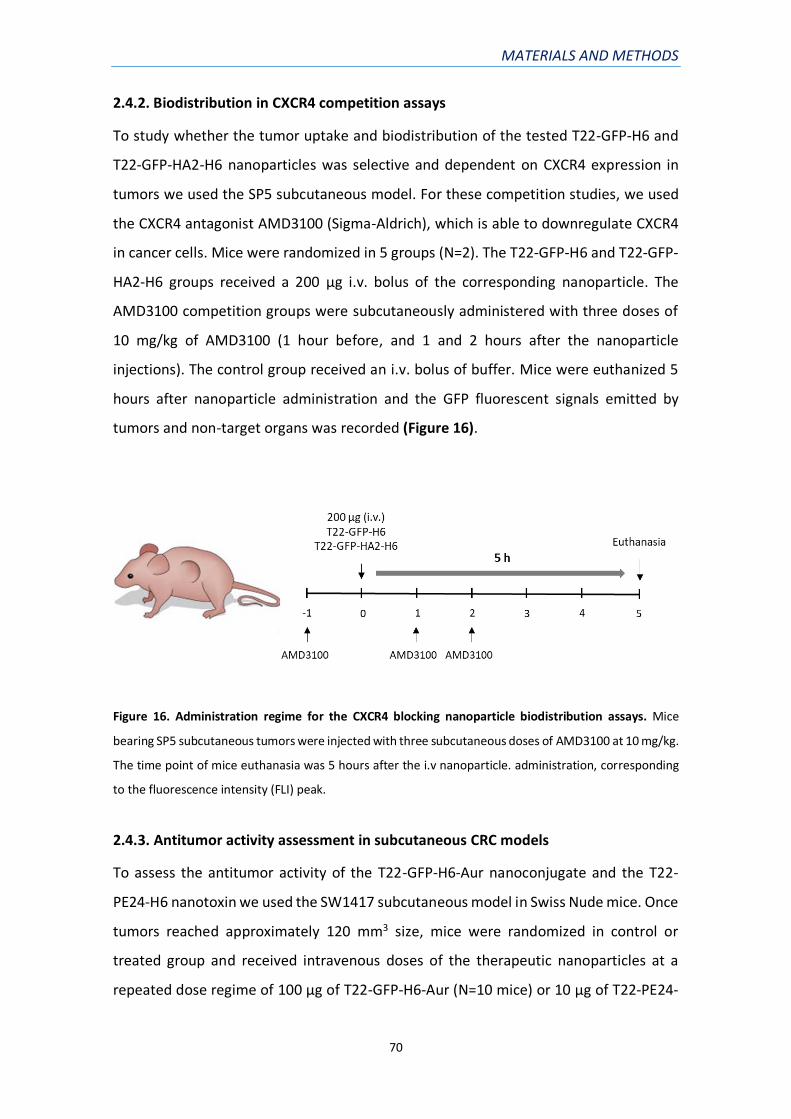
MATERIALS AND METHODS
70
2.4.2. Biodistribution in CXCR4 competition assays
To study whether the tumor uptake and biodistribution of the tested T22-GFP-H6 and
T22-GFP-HA2-H6 nanoparticles was selective and dependent on CXCR4 expression in
tumors we used the SP5 subcutaneous model. For these competition studies, we used
the CXCR4 antagonist AMD3100 (Sigma-Aldrich), which is able to downregulate CXCR4
in cancer cells. Mice were randomized in 5 groups (N=2). The T22-GFP-H6 and T22-GFP-
HA2-H6 groups received a 200 µg i.v. bolus of the corresponding nanoparticle. The
AMD3100 competition groups were subcutaneously administered with three doses of
10 mg/kg of AMD3100 (1 hour before, and 1 and 2 hours after the nanoparticle
injections). The control group received an i.v. bolus of buffer. Mice were euthanized 5
hours after nanoparticle administration and the GFP fluorescent signals emitted by
tumors and non-target organs was recorded (Figure 16).
Figure 16. Administration regime for the CXCR4 blocking nanoparticle biodistribution assays. Mice
bearing SP5 subcutaneous tumors were injected with three subcutaneous doses of AMD3100 at 10 mg/kg.
The time point of mice euthanasia was 5 hours after the i.v nanoparticle. administration, corresponding
to the fluorescence intensity (FLI) peak.
2.4.3. Antitumor activity assessment in subcutaneous CRC models
To assess the antitumor activity of the T22-GFP-H6-Aur nanoconjugate and the T22-
PE24-H6 nanotoxin we used the SW1417 subcutaneous model in Swiss Nude mice. Once
tumors reached approximately 120 mm3 size, mice were randomized in control or
treated group and received intravenous doses of the therapeutic nanoparticles at a
repeated dose regime of 100 μg of T22-GFP-H6-Aur (N=10 mice) or 10 μg of T22-PE24-

MATERIALS AND METHODS
71
H6 (N=6 mice), 3 times a week, per 8 doses. The control group received buffer using the
same administration schedule. Overall the experimental period, mouse body weight and
tumor volume were registered 3 times a week. Tumor bioluminescence was also
measured once a week using the IVIS Spectrum equipment. When tumors reached a
volume of 600 mm3, mice were euthanized and mitotic, apoptotic, necrotic rates and
CXCR4 expression were assessed in tumor tissue samples (Figure 18).
2.4.4. Antimetastatic activity assessment in orthotopic CRC models
To assess the antimetastatic activity of the nanoconjugate T22-GFP-H6-Aur and the
nanotoxin T22-PE24-H6 we used the SW1417 orthotopic model in NSG mice. SW1417
cells were injected in the cecum of NSG mice and 3 days after, primary tumor
bioluminescence was measured using the IVIS Spectrum equipment. Mice were then
randomized into the control (N=9-10) and the nanoparticle-treated (N=9-10) group.
Treated mice received intravenous doses of T22-GFP-H6-Aur or T22-PE24-H6 at
repeated dose regime of 100 μg or 5 μg respectively, 3 times a week, for a total of 12 or
18 doses, respectively (Table 4). The control group received buffer using the same
administration schedule. Overall the experimental period, mouse body weight and
health status were controlled 3 times per week. Primary tumor bioluminescence was
registered once per week using the IVIS Spectrum equipment and the experiment was
finished when the first mouse of the buffer-treated group needed to be euthanized.
Necropsy and ex vivo imaging of the organs of interest were performed 24 hours after
the last dose (Figure 18).
Table 4. Doses and dose regime details of T22-GFP-H6-Aur nanoconjugate and the T22-PE24-H6
nanotoxin used in the antimetastatic effect experiments with the CXCR4+ SW1417 cells growing
orthotopically in NSG mice.
Nanoparticle Mice
number Dose
Dose
regime
Number of
bolus
Cumulative
dose
NaCOH3
NaCl buffer
pH 8
N=10 150 μl 3 times per
week 12 -----

MATERIALS AND METHODS
72
T22-GFP-
H6-Aur N=10 100 μg
3 times per
week 12 1200 µg
NaCOH3
buffer pH 8 N=9 150 μl
3 times per
week 18 -----
T22-PE24-H6 N=9 5 μg 3 times per
week 18 90 µg
2.5. Necropsy and tissue processing
2.5.1. Necropsy and bioluminescence ex vivo imaging
In all experiments, mice were euthanized by cervical dislocation. Endpoint criteria was
different in each experiment according to the objective. In experiments with mice
carrying subcutaneous tumors, they were euthanized when the tumor reached a
maximum size of 800 mm3. Tumor final volume was calculated using the formula: V =
(Dxd^2)/2 where D is the longer diameter and d the shorter diameter. To assess mouse
survival time after orthotopic cell injection, mice were euthanized when the health
status of the mouse was poor (mucous secretions, lack of mobility, dishevelled hair),
there was loss of body weight (more than 20% of the initial weight) or due to behavioural
changes (self-mutilations, hunched posture, passiveness). In antimetastatic effect
experiments, all mice were euthanized when the first control mouse met the endpoint
criteria explained above.
Animal models derived from bioluminescent cells (SW1417 cell line) were imaged using
the IVIS Spectrum Imaging System as explained in 2.3. section (in vivo imaging) before
the euthanasia. Then, a complete necropsy procedure was performed and all relevant
organs (liver, kidneys, lungs, diaphragm, spleen, pancreas, colon, intestinal tract and
mesenteric lymph nodes) were collected and covered in a 0.75 mg/kg D-Luciferin
solution in 12 or 6 wells plates and imaged (ex vivo imaging). For non-bioluminescent
mouse models (SP5 patient-derived samples), a complete necropsy procedure was
performed, organs were collected in cassettes (Thermo Fisher), fixed and cut for
histopathology and immunohistochemistry analyses.

MATERIALS AND METHODS
73
2.5.2. Sample processing and histopathological analysis
After necropsy, the collected organs were fixed in 4% formaldehyde in phosphate-buffer
solution (PBS), dehydrated, paraffin embedded, cut and stained with hematoxylin-eosin
or used for immunohistochemistry or immunofluorescence assays.
1. Sample dehydration: after tissue fixation for 24 hours, samples were dehydrated.
Dehydration is the removal of water from aqueous-fixed tissue. Since most fixing
solutions are aqueous, this step is necessary to prepare the tissue for embedding
in non-aqueous media like paraffin. First, organs were washed 3 times with
distilled water to remove all the fixing solution. Then, samples were immersed 3
times for 10 minutes each in solutions with increasing concentrations of ethanol
(70, 80, 96 and 100%). During this procedure the water in tissues is replaced by
alcohol. Last, samples were incubated for 10 minutes in 3 solutions of xylene
100% to dissolve the alcohol in the samples.
2. Paraffin embedding: the dehydrated tissue is ready for infiltration with a suitable
histological wax. The most popular is the wax-based paraffin. Paraffin is liquid at
60°C and can be infiltrated into tissue at this temperature. At 20°C it solidifies to
a consistency that allows sections to be easily cut. Thus, samples were 3 times
immersed for 1 hour in a paraffin solution at 60ºC. After 3 hours, each sample
was located in a mould over a cassette, filled with molten paraffin and placed in
a cold plate to solidify.
3. Sectioning: once paraffin was solidified, samples were cut in 3-5 µm sections with
a microtome and mounted in glass microscope slides.
4. Rehydration: to remove the paraffin from tissues, slides were incubated for 1
hour at 60ºC and then immersed twice in 100% xylene. To hydrate the tissues,
slides were immersed twice in 100% ethanol, once in 96% ethanol, 80% ethanol
and 50% ethanol.
5. Hematoxylin-eosin staining: slides were incubated for 5 minutes with distilled
water, following 5-10 minutes with the Mayer hematoxylin solution and washed
with distilled water for 15 minutes. Once some of the tissue structures were
distinguished, 2 more washing steps were done, and then slides were incubated

MATERIALS AND METHODS
74
for 0.5-2 minutes with 0.2% eosin. Samples were again dehydrated and covered
with mounting media (Toluene-Free Mounting Medium, Dako) and a coverslip.
2.5.3. Immunohistochemistry analysis
Immunohistochemistry (IHC) is the most common application of immunostaining and
involves the process of selectively identifying antigens (proteins) in cells of a tissue
section. IHC staining is widely used in the diagnosis of abnormal cells such as those found
in tumors or detection of specific molecular markers which are characteristic of
particular cellular events such as proliferation or cell death.
We have used IHC in the tissue samples obtained from in vivo experiments to assess the
expression of CXCR4 in subcutaneous tumors, CRC primary tumors and metastases
located in distal organs. Markers of apoptosis such as active caspase-3 protein (early
apoptosis) or proteolyzed PARP (late apoptosis) or pyroptosis such as NLRP3, cleaved
caspase-1 and cleaved GSDMD, were also assessed in tumors to evaluate
mechanistically the antitumor effect of the administered nanoconjugate or nanotoxin
(Table 5).
Table 5. Primary antibodies used in the IHC studies.
Primary antibody Dilution Secondary antibody Suppliers
CXCR4 1/200 Rabbit Abcam
Active Caspase-3 1/300 Rabbit BD Pharmigen
Proteolyzed PARP 1/300 Rabbit Promega
NLRP3 1/300 Mouse Adipogen
Cleaved Caspase-1 1/400 Rabbit Life Technologies
Cleaved GSDMD 1/200 Rabbit Cell Signaling
All IHC studied samples were fixed, dehydrated, paraffin embedded and sectioned as
explained in 2.5.2. section. Tissue sections were mounted in coated microscope slides

MATERIALS AND METHODS
75
with positive charges (FLEX IHC Microscope Slides, Dako) to assure sample adhesion.
Then, samples were incubated for 1 hour at 60ºC and rehydrated as explained in 2.5.2.
section. Masked epitopes were recovered by heat-induced antigen retrieval using the
PT Link system (Dako) at 95ºC for 10 minutes and at 65ºC for 45ºC in a Tris/EDTA buffer
solution with pH 9 (high pH, Dako) or a citrate buffer solution with pH 6 (low pH, Dako).
The automatized Autostainer Link 48 (Dako) was used for the sample staining following
the next incubation procedure: 1 minute with rinse buffer, 5 minutes with Peroxidase-
Blocking Reagent, 1 minute washing with rinse buffer, 30 minutes with the primary
antibody diluted in rinse buffer (Table), 1 minute washing with rinse buffer, 20 minutes
with EnVision/HRP, 1 minute washing with rinse buffer, minutes with the chromogenic
solution (DAB+ Chromogen and Substrate Buffer), 1 minute washing with rinse buffer, 5
minutes with hematoxylin and 1 minute final washing with rinse buffer (Figure 17).
Figure 17. Overview of the main steps of IHC using Dako systems on paraffin-embedded tissue slides.
Results were presented as number of stained cells or as percentage of stained surface.
The percentage of brown area was calculated using the ImageJ software and the Colour
Deconvolution Plugin with the H DAB vector to split the brown staining. Up to 5 high-
power fields (400x) per sample were analysed and results were expressed as mean ±
s.e.m..

MATERIALS AND METHODS
76
2.5.4. Immunofluorescence analysis
Immunofluorescence assays were used to detect nanoparticles, containing the GFP
domain, in mouse tissues and to study their co-localization with the CXCR4 membrane
receptor in cancer cells. Paraffin-embedded SP5 tumors treated for 5 hours with T22-
GFP-H6 or T22-GFP-H6-Aur were sectioned and then stained with the primary antibodies
anti-CXCR4, and anti-GFP together with the DAPI fluorescent stain.
Sample fixation, rehydration and antigen retrieval were completed as explained in the
immunohistochemistry analyses section. Subsequently, we performed 3 washing steps
with TBS 1x (132 mM NaCl, 10 mM Tris pH 7.5, 0.1% Tween 20) and blocked the
unspecific binding of the secondary antibody by incubating the samples 1 hour with a
TBS + 0.5% triton + 3% donkey serum solution (Millipore). Primary antibodies were also
diluted (Table 6) in this solution and samples incubated overnight at 4ºC. They were
washed again 3 times with TBS 1x and then incubated with the secondary antibodies
(Table 6) diluted in TBS 1x for 2 hours at 37ºC. Tissue auto-fluorescence was reduced by
incubating the samples with sudan black 1% (Panreac) in 70% OH for 4 minutes. Samples
were washed again 3 times with TBS 1x, stained for 10 minutes with a 1/10000 DAPI
staining solution (ThermoFisher), covered with a special mounting medium and a
coverslip and sealed with nail polish.
Table 6. Antibodies used in the IF studies.
Antibody Type of
antibody Host species Dilution Fluorescent dye Suppliers
Anti-CXCR4 Primary Rabbit 1/300 - Abcam
Anti-GFP Primary Chicken 1/250 - AVES
Anti-Rabbit Secondary Donkey 1/200 Alexa Fluor 647 Jackson
Anti-
Chicken Secondary Donkey 1/50 Alexa Fluor 488 Jackson

MATERIALS AND METHODS
77
Representative pictures were taken using Confocal TCS SPE (Leica) at 200× or 600×
magnifications. Immunofluorescence measurements were performed using the ImageJ
software.
2.5.5. Assessment of mitotic, apoptotic and necrotic rates
Tumor sections stained with DAPI were also used to evaluate the cancer cell
proliferation capacity and tumor growth rate by counting the number of mitotic figures
per 10 high-power fields (magnification 400x). To assess the apoptotic effect of the
administered nanoparticles in tumors we counted the number of apoptotic bodies in
H&E and DAPI stained tissues in 10 high-power fields (magnification 400x). DAPI staining
was performed in Triton X-100 (0.5%) permeabilized sections mounted with DAPI
mounting media (ProLong Gold antifade reagent, Thermo Fisher). Samples were
evaluated under a fluorescence microscope at a wavelength of λex = 334 nm/λem = 465
nm. We also analysed the percentage of necrotic areas in H&E stained tumors by using
Cell^D Olympus software at 15x magnification.
2.6. Metastatic foci quantitation
In the orthotopic in vivo experiments, the spreading of cells from the primary tumor
(colon) to other distant organs was analysed in H&E-stained samples. We studied those
organs were metastatic dissemination is expected in CRC: liver, lung, mesenteric lymph
nodes and peritoneum. These organs were collected at necropsy, imaged ex vivo using
the IVIS Spectrum and processed for histological analysis. Depending of the experiment
and the bioluminescence signal, only one or several slices were analysed. When the
signal was high, only one slice of each organ was quantified. If the signal was low
because of the low number of metastatic foci, 3 slices of each organ were quantified. An
Olympus microscope with the Cell^D Olympus software was used to count the number
and measure the size (expressed as µm2) of all observed metastatic foci. Representative
images of them were also taken.
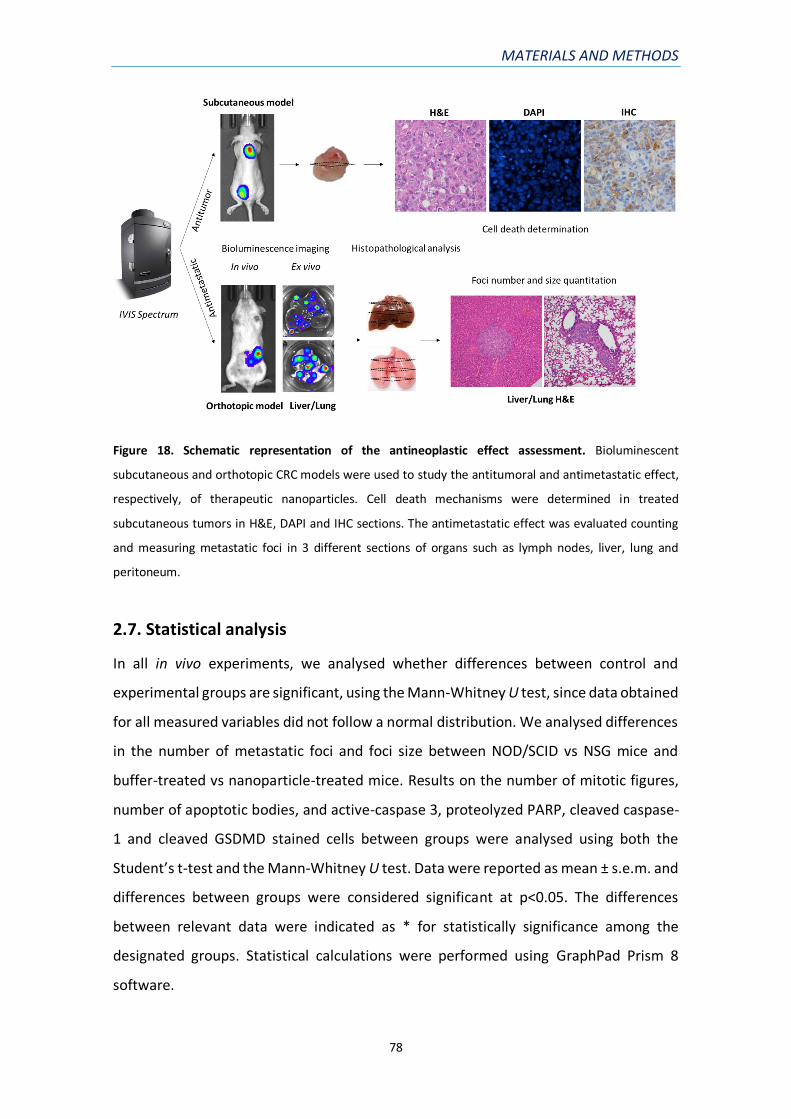
MATERIALS AND METHODS
78
Figure 18. Schematic representation of the antineoplastic effect assessment. Bioluminescent
subcutaneous and orthotopic CRC models were used to study the antitumoral and antimetastatic effect,
respectively, of therapeutic nanoparticles. Cell death mechanisms were determined in treated
subcutaneous tumors in H&E, DAPI and IHC sections. The antimetastatic effect was evaluated counting
and measuring metastatic foci in 3 different sections of organs such as lymph nodes, liver, lung and
peritoneum.
2.7. Statistical analysis
In all in vivo experiments, we analysed whether differences between control and
experimental groups are significant, using the Mann-Whitney U test, since data obtained
for all measured variables did not follow a normal distribution. We analysed differences
in the number of metastatic foci and foci size between NOD/SCID vs NSG mice and
buffer-treated vs nanoparticle-treated mice. Results on the number of mitotic figures,
number of apoptotic bodies, and active-caspase 3, proteolyzed PARP, cleaved caspase-
1 and cleaved GSDMD stained cells between groups were analysed using both the
Student’s t-test and the Mann-Whitney U test. Data were reported as mean ± s.e.m. and
differences between groups were considered significant at p<0.05. The differences
between relevant data were indicated as * for statistically significance among the
designated groups. Statistical calculations were performed using GraphPad Prism 8
software.

IV.RESULTS

RESULTS
80
IV. RESULTS
1) DEVELOPING CXCR4-DEPENDENT SUBCUTANEOUS AND
HIGHLY METASTATIC CRC MOUSE MODELS.
In order to generate animal models to test the biodistribution and antineoplastic effect
of the T22-GFP-H6 nanoparticle and its therapeutic derivatives, new CRC mouse models
were developed. For that purpose, we first studied the CXCR4 cell surface expression in
different CRC cell lines and their tumorigenic capacity. Following, we developed non-
metastatic subcutaneous CRC models and highly metastatic orthotopic CRC models that
replicate the pathogenesis, progression and clinical evolution observed in CRC patients.
As PDX models have become very important in translational cancer research, because
they retain tumor heterogeneity and better predict the sensitivity to treatment than
cell-line derived models, an orthotopic CRC mouse model derived from a patient sample
was also developed. CXCR4 receptor over-expression was observed in cancer cells in all
models, both in primary tumors and in distant metastases.
1.1. Cell surface expression of CXCR4 in CRC cell lines
The expression of the CXCR4 chemokine receptor in the cell surface of different CRC cell
lines (SW1417, SW48 and HT29) was determined by staining with a specific antibody and
subsequent analysis by flow cytometry. Contrary to previous reports, and as shown in
Figure 19, SW48 and HT29 cell lines were negative for CXCR4 expression (85,86). In the
SW1417 cell line analyses, we have noticed differences in CXCR4 expression between
this cell line purchased from the ATCC and the same cell line used in different
laboratories. These cell lines were maintained separately and named CXCR4+ SW1417 or
CXCR4- SW1417, depending on their presence or absence of CXCR4 receptor expression.
We have also noticed that the percentage of cells expressing CXCR4 in their membrane
can decrease with consecutive passages or treatments (87) because this receptor tends
to internalize into the cytoplasm while being cultured in vitro. We determined that the
percentage of CXCR4+ cells used for all the in vitro testing of our nanoparticles and

RESULTS
81
therapeutic nanoconjugates, or to generate relevant and consistent CXCR4+ CRC models,
had to be around 70-80% to ensure reproducibility and comparability among studies.
Figure 19. Flow cytometry histograms of the CXCR4 cell surface expression of different CRC cell lines.
The CXCR4 cell surface expression was analysed in the HT29, SW48 and SW1417 CRC cell lines. The M1
interval represents the CXCR4 positive cells from the whole population.
1.2. Effect of CXCR4 expression in the tumorigenic capacity of the SW1417
cell line
The differential capacity to develop subcutaneous tumors derived from the CXCR4+
SW1417 and CXCR4- SW1417 cell lines was assessed by injecting them in the subcutis of
Swiss nude mice. Figure 20A shows tumor growth curves and the differential CXCR4
expression in tumor tissue developed in the subcutis at the end of the experiment. All
injected mice formed tumors within 1 week after implantation. However, in CXCR4-
SW1417 cells injected mice (N=5) tumor volume decreased at 14 days post-

RESULTS
82
transplantation, being total tumor growth negligible at the end of the experiment. In
contrast, all mice injected with CXCR4+ SW1417 cells (N=5) developed subcutaneous
tumors that displayed a constant growth (Figure 20A). Moreover, a significant
percentage of cancer cells in the model derived from CXCR4+ cells overexpressed the
receptor in their cell membrane (Figure 20B). Thus, we have developed SC CRC models
that maintain CXCR4 expression in tumor tissue and could be useful to test CXCR4
targeted therapeutic nanoparticles.
Figure 20. Tumorigenic capacity of the SW1417 cell line A) Tumor growth of subcutaneous tumors
derived from CXCR4+ SW1417 or CXCR4- SW1417 cells implanted in Swiss nude mice B) Representative
images of CXCR4 immunohistochemistry staining of the developed subcutaneous tumors. * p<0.05; **
p<0.01. Scale bars: 50 µm.
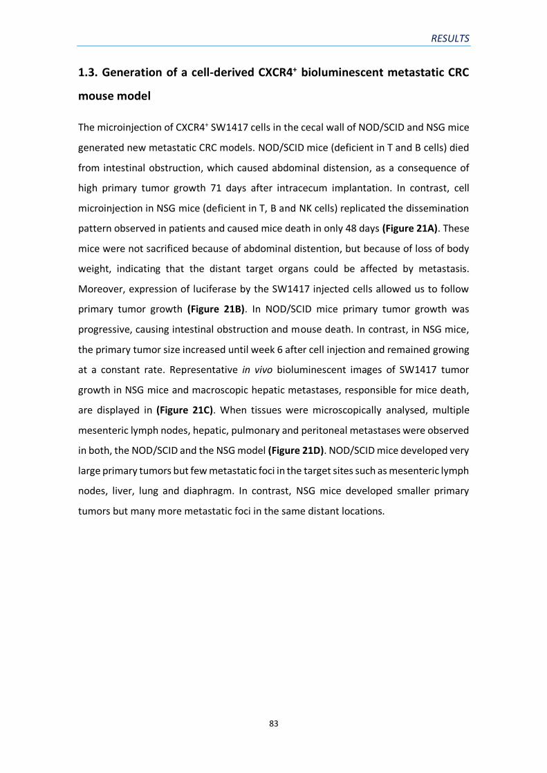
RESULTS
83
1.3. Generation of a cell-derived CXCR4+ bioluminescent metastatic CRC
mouse model
The microinjection of CXCR4+ SW1417 cells in the cecal wall of NOD/SCID and NSG mice
generated new metastatic CRC models. NOD/SCID mice (deficient in T and B cells) died
from intestinal obstruction, which caused abdominal distension, as a consequence of
high primary tumor growth 71 days after intracecum implantation. In contrast, cell
microinjection in NSG mice (deficient in T, B and NK cells) replicated the dissemination
pattern observed in patients and caused mice death in only 48 days (Figure 21A). These
mice were not sacrificed because of abdominal distention, but because of loss of body
weight, indicating that the distant target organs could be affected by metastasis.
Moreover, expression of luciferase by the SW1417 injected cells allowed us to follow
primary tumor growth (Figure 21B). In NOD/SCID mice primary tumor growth was
progressive, causing intestinal obstruction and mouse death. In contrast, in NSG mice,
the primary tumor size increased until week 6 after cell injection and remained growing
at a constant rate. Representative in vivo bioluminescent images of SW1417 tumor
growth in NSG mice and macroscopic hepatic metastases, responsible for mice death,
are displayed in (Figure 21C). When tissues were microscopically analysed, multiple
mesenteric lymph nodes, hepatic, pulmonary and peritoneal metastases were observed
in both, the NOD/SCID and the NSG model (Figure 21D). NOD/SCID mice developed very
large primary tumors but few metastatic foci in the target sites such as mesenteric lymph
nodes, liver, lung and diaphragm. In contrast, NSG mice developed smaller primary
tumors but many more metastatic foci in the same distant locations.

RESULTS
84
Figure 21. Primary tumor growth and metastatic dissemination of CXCR4+ SW1417 CRC cell-derived
orthotopic models A) Kaplan-Meier survival curves of NOD/SCID (N=9) and NSG (N=10) mice injected
orthotopically with SW1417 cells (p=0.016, Log-Rank test) B) NOD/SCID and NSG primary tumor growth
analysed by measuring the bioluminiscence emitted by SW1417 cells C) Representative in vivo
bioluminescent images of tumor growth over time in NSG mice, and macroscopic primary tumor and
hepatic metastases D) H&E staining of representatives CRC primary tumor and hepatic, pulmonar and
other metastases observed in both NOD/SCID and NSG models.

RESULTS
85
1.3.1. The metastatic capacity of the CXCR4+ SW1417 cell line increases in severely
immunosuppressed mice
NOD/SCID and NSG mice orthotopically injected with CXCR4+ SW1417 cells showed both
100 % of engraftment. However, the metastatic capacity of the tumors derived from this
cell line increased in NSG mice, a more immunosuppressed mouse strain. As compared
to the SW1417 NOD/SCID model, the metastatic rate in SW1417 NSG mice resulted in a
5-fold increase in the number of hepatic (p<0.01) and pulmonary (p<0.01) metastases
(Table 7). Moreover, the absence of a specific immune cell type in NSG as compared to
NOD/SCID mouse strain, that is the lack of NK cells, allowed for a greater growth of
metastatic foci, especially in the liver where some of them reached a visible size (p<0.01)
(Figure 22). These results are consistent with the fact that NK cells are involved in tumor
surveillance by a NKG2D-mediated recognition of cancer cells (88), nevertheless, this
hypothesis remains to be proven.
Figure 22. Metastatic colonization of SW1417 CRC cells in NOD/SCID and NSG mice. Total foci number
(panel A) and mean foci area (panel B) in organs affected by metastases in NOD/SCID and NSG
immunosuppressed mice orthotopically injected with CXCR4+ SW1417 CRC cells. * p<0.01.

RESULTS
86
Table 7. Dissemination pattern of NOD/SCID and NSG mice orthotopically injected with CXCR4+ SW1417
CRC cells.
1.4. Generation of a patient-derived metastatic CRC mouse model
To develop a patient-derived orthotopic CRC mouse model, we first established the SP5
subcutaneous tumor line, derived from a CRC patient of our hospital. We used a
fragment of this tumor line to generate subcutaneous (SC) tumors in donor animals.
When these subcutaneous tumors reached 600 mm3, were disaggregated to obtain a
cell suspension that was inocluated directly in the cecum of NSG mice. A short time after
cell injection (31 days) mice started showing signs of abdominal distension and
developed a large abdominal carcinomatosis tumor mass. At necropsies, visible hepatic,
lymph node and ovarian carcinomatosis were observed. When tissues were
microscopically analysed, multiple mesenteric lymph nodes, hepatic, pulmonary and
peritoneal metastases were observed and quantified (Table 8). As compared to the
100% take rate observed in the cell-derived orthotopic model, the primary tumor take
rate of the SP5 model was 85%, since at the end of the experiment there were 2 mice
free of both primary tumor and metastases. However, the metastatic rate in the SP5
orthotopic model resulted in a 3.6-fold increase in the number of lymph node
metastases as compared to the cell-line derived model in NSG mice (Table 7). Regarding
the size of the generated metastatic foci, in the patient-derived model we observed a
greater growth of lymph node, pulmonary and peritoneal metastases. In accordance
with the fact that mice died because of the rapid growth of the primary tumor, the size
Primary tumor Lymph node Mets Liver Mets Lung Mets Peritoneal Mets
mice % mice % # foci mice % # foci mice % # foci mice % # foci
88% 11% 89% 100%
1.2 ± 0.15 0.11 ± 0.11a 3.2 ± 0.66a 1.1 ± 0.11b
60% 100% 100% 90%
1.3 ± 0.52 50.1 ± 7.5a 155.5 ± 24.2a 2.4 ± 0.45b
Group Primary tumor Lymph nods Mets Liver Mets Lung Mets Peritoneal Mets
NOD/SCID 48.9 ± 6.9c6.9 14.7 ± 1.9 742.6 ± 115.5
NSG 809.1 ± 298c241.6 ± 76.4 15.2 ± 4 1523.9 ± 337.5
Mean + s.e.m. metastatic foci number or area (µm2) per mouse, counted in one entire histology section.
a p<0.0001; b p=0.02; c p=0.02
Metastatic foci size (µm² x 103)
SW1417 cell-derived ortothopic model
Group
NOD/SCID 9/9 100%
NSG 10/10 100%

RESULTS
87
of the peritoneal metastases was 28 times bigger than in the NSG cell-derived model, in
which mice died because of the distant metastases (Table 7).
Table 8. Metastatic dissemination in NSG mice orthotopically injected with a patient-derived cell
suspension of SP5 disaggregated from subcutaneous tumors in donor mice.
1.5. CXCR4 expression in primary tumor and distant metastases
Since one of the objectives of this thesis project is to study the T22-GFP-H6 nanoparticle
capacity of targeting CXCR4+ cancer cells, the expression of this receptor was studied in
the subcutaneous and orthotopic models derived from the CXCR4+ SW1417 cell line or
from the CXCR4+ SP5 patient-derived CRC tumor line. Immunohistochemistry was used
to analyse the intensity of the membrane expression of CXCR4 and its subcellular
localization pattern in subcutaneous tumors, primary tumors and metastases. Figure 23
depicts representative images of anti-CXCR4 immunohistochemistry in the cell-derived
and patient-derived subcutaneous tumors and orthotopic models. The SP5 patient-
derived model showed CXCR4 expression in a larger percentage of tumor cells, with a
higher intensity than the reached in SW1417 tumors and with a clear membrane
localization in both models. Moreover, in both models, the CXCR4 expression was
maintained in distant metastases, especially in metastatic foci affecting lymph nodes
and liver.
Primary tumor Lymph node Mets Liver Mets Lung Mets Peritoneal Mets
mice % mice % # foci mice % # foci mice % # foci mice % # foci
85% 85% 85% 85%
4.7 ± 0.7 18.2 ± 2.5 191.4 ± 27.2 3.2 ± 1
Group Primary tumor Lymph node Mets Liver Mets Lung Mets Peritoneal Mets
NSG 7288.8 ± 1506.7 172.7 ± 67.7 48.4 ± 9 43903.9 ± 9616.4
Mean + s.e.m. metastatic foci number or area (µm2) per mouse, counted in one entire histology section.
SP5 patient-derived orthotopic model
Group
NSG 11/13 85%
Metastatic foci size (µm² x 103)

RESULTS
88
Figure 23. CXCR4 receptor expression in cancer tissues determined by immunohistochemistry analysis.
Images of a subcutaneous tumor, CRC primary tumor, peritoneal, lymph node, lung and liver metastases
of the cell-derived (SW1417) and the patient-derived (SP5) orthotopic models in NSG mice. Scale bars:
100 µm.

RESULTS
89
2) IN VITRO SELECTIVE INTERNALIZATION AND ANTITUMOR
ACTIVITY OF T22-GFP-H6 AND ITS THERAPEUTIC DERIVATIVES IN
CXCR4+ CRC CELL LINES.
In collaboration with the Nanobiotechnology group from the Universitat Autònoma de
Barcelona (UAB), led by Dr. Villaverde, we developed protein-based nanoparticles
capable of internalizing in CXCR4+ cells. All these nanoparticles are built around the T22-
GFP-H6 nanocarrier basic structure. On the one hand, we developed nanoconjugates by
conjugating therapeutic agents to the nanocarrier. Floxuridine (FdU), the cytotoxic drug
currently used to treat liver metastases (89) was covalently bound to the T22-GFP-H6
nanocarrier, and assayed for selective internalization and FdU delivery in CXCR4+ cancer
cells, that demonstrated to induce their specific depletion in a CRC model (79). In an
attempt to further increase the antineoplastic effect of these nanoconjugates we chose
as a first option to bind a therapeutic agent with higher potency, the Auristatin E toxin
(Aur), to develop the new T22-GFP-H6-Aur nanoconjugate. So, we tested the in vitro
capacity of the T22-GFP-H6 nanocarrier and the T22-GFP-H6-Aur nanoconjugate for
selectively internalizing into CXCR4+ CRC cells, expecting to deliver the Auristatin
payload in their cytosol, and to kill them.
On the other hand, we developed derivatives of the T22-GFP-H6 nanoparticle with
intrinsic therapeutic activities by incorporating proapoptotic peptides (BAK, PUMA or
BAXPORO) or bacterial toxin domains (PE24 or DITOX) to their chimeric sequences. Thus,
we studied the sensitivity of the CXCR4+ CRC cells to these therapeutic nanoparticles and
selected those with CXCR4-dependent cytotoxic activity in vitro for in vivo testing. In all
the in vitro studies, we used the SW1417 CRC cell line due to its success in generating a
mouse model of metastatic CRC by orthotopic implantation.
2.1. CXCR4-dependent internalization of T22-GFP-H6 in a human CXCR4+
CRC cell line
To understand the kinetics of the T22-GFP-H6 nanoparticle’s internalization in the
SW1417 CXCR4+ CRC cell line, confocal microscopy imaging was performed exposing the

RESULTS
90
cells to 1 µM of T22-GFP-H6 for different time periods (1, 5 and 24 hours). To prove
CXCR4-dependent internalization of the nanoparticles we used the unliganded GFP-H6
protein version (lacking the T22 peptide) as a control. Moreover, we stained the cell
membrane with a red dye (CellMask) and the cell nucleus with a blue dye (DAPI) to
localize the green fluorescent signal emitted by the nanoparticles inside the cell.
According to previous data obtained with HeLa (69) no cell penetration of GFP-H6
(negative control) was observed. In contrast, 1 hour after T22-GFP-H6 exposure,
nanoparticles were already bound in low abundance to the plasma membrane and
progressively increased their cell uptake until 24 hours, displaying a perinuclear pattern
(Figure 24). The cytosolic location of T22-GFP-H6 near the nuclear region indicates that
T22-GFP-H6 nanoparticles were able to escape from degradation in the endosomes
because of the accompanying hexahistidine tag, that has powerful endosomolytic
properties.
Figure 24. T22-GFP-H6 selective internalization in CXCR4+ SW1417 cells determined by confocal
microscopy. SW1417 cells were exposed to 1 µM of T22-GFP-H6 or GFP-H6 (as a negative control) for
internalization selectivity, during 1, 5, and 24 h. Plasma cell membranes were stained with a red dye
(CellMask) whereas cell nucleus in blue (DAPI) and imaged with confocal microscopy. Green signal
corresponds to the GFP contained in the nanoparticles. Magnification: 200x.

RESULTS
91
2.2. The T22-GFP-H6-Aur nanoconjugate maintains CXCR4-dependent
internalization in CXCR4+ CRC cells
Following, we evaluated whether the conjugation of the Auristatin toxin to the T22-GFP-
H6 nanoparticle could alter the protein conformation or function by measuring its CXCR4
targeting capacity. To that aim, we first tested the levels of CXCR4 in the cell membrane
of SW1417 cells by immunohistochemistry. This CRC cell line shows high expression of
the CXCR4 receptor, having a very clear membrane staining, but not in all the cell
population (Figure 25A). Afterwards, we measured and confirmed the absence of
endogenous production of the natural CXCR4 ligand SDF-1α, by the SW1417 cells to
discard a possible competition of this ligand with the nanoparticles for cell
internalization (Figure 25B). The human 1BR3.G fibroblasts were used as a positive
control of SDF-1α production and release. Then, we evaluated by flow cytometry the
internalization capacity of the T22-GFP-H6 nanocarrier and the T22-GFP-H6-Aur
nanoconjugate in the SW1417 cell line. The T22-GFP-H6 nanocarrier internalized in
SW1417 cells after 24 hours exposure at a 1000 nM concentration. In contrast, the GFP-
H6 protein, used as negative control for CXCR4-dependent internalization, did not
internalize in these cells as expected (Figure 25C). The exposure of the SW1417 cells to
a lower concentration of the T22-GFP-H6-Aur (400nM) showed similar capacity to
internalize in this cell line but less efficiently. The nanoconjugate also maintained the
dependence on CXCR4 for internalization, since a 30 minutes pre-treatment with the
CXCR4 antagonist AMD3100 was able to decrease its internalization (Figure 25D).

RESULTS
92
Figure 25. In vitro T22-GFP-H6 and T22-GFP-H6-Aur selective internalization in the CXCR4+ SW1417 CRC
cell line A) Assessment of CXCR4 membrane expression by anti-CXCR4 immunocytochemistry in SW1417
cells in culture. Magnification: 400x B) Lack of human SDF-1α release from cultured SW1417 cells, as
measured by ELISA, whereas human control 1BR3.G fibroblasts express high SDF-1α levels, after 48 or 72
h of growth in culture C) T22-GFP-H6 selective internalization in SW1417 cells measured as intracellular
fluorescence by flow cytometry, after 24 h exposure to 1000 nM of T22-GFP-H6 or GFP-H6 D) T22-GFP-
H6-Aur internalization in SW1417 cells after 24 h exposure to 400 nM of the nanoconjugate or after a 30
min pre-treatment with the antagonist AMD3100 (4 µM). Data presented as mean ± s.e.m., N=3, * p<0.05;
** p<0.01.
2.3. Cytotoxic effect of T22-GFP-H6-Aur and induction of cell death in CRC
cells by mitotic catastrophe
Once we demonstrated the selective T22-GFP-H6-Aur nanoconjugate internalization in
CXCR4+ cells, its cytotoxic activity was also evaluated in vitro in the SW1417 cell line,
using XTT cell viability assays (which measures the metabolic activity of the cells) or DAPI
nuclear staining assays. We used the T22-GFP-H6 nanocarrier and the T22-GFP-H6-FdU

RESULTS
93
nanoconjugate, as negative or positive controls respectively, because of their known
cytotoxic effects. Surprisingly, we observed no effect of the T22-GFP-H6-Aur
nanoconjugate on the viability of the SW1417 cells upon 48 hours exposure to a range
of low (0, 10, 25 and 50 nM), intermediate (200 and 500 nM) and high (1 µM)
concentrations (Figure 26A).
Since we expected a high cytotoxic effect because Auristatin is usually a more potent
drug than Floxuridine in different cell types, cell death was analysed using a different
method, that is the nuclear staining with DAPI (to measure cell detachment, induction
of DNA condensation and blockade in mitosis) of cells exposed to 1 µM of T22-GFP-H6-
Aur for 48 hours. In these assays, we observed that the number of cells adhered to the
slide surface was significantly lower than cells treated with buffer, T22-GFP-H6
nanocarrier or T22-GFP-H6-FdU (Figure 26B). Moreover, the evaluation of the obtained
images allowed us to study if the mechanism of cell death induced by the T22-GFP-H6-
Aur nanoconjugate was the one described for the microtubule inhibitor Auristatin (90).
As expected, the nuclear morphology of dead cells indicated that cells treated with the
nanoconjugate died because of mitotic catastrophe in contrast with the apoptosis
caused by the T22-GFP-H6-FdU nanoconjugate (Figure 26D).
Additionally, the CXCR4 dependence of the T22-GFP-H6-Aur cytotoxic effect was also
evaluated with a competition assay using the CXCR4 antagonist AMD3100. The pre-
treatment of cultures with AMD3100 showed a significant decrease in the number of
cells affected by mitotic catastrophe, but cell death was not completely reverted maybe
due to drug leakage, and therefore, having free Auristatin in the nanoconjugate solution
(Figure 26C). In summary, the T22-GFP-H6-Aur nanoconjugate is active against SW1417
CRC cells at 1 µM concentration, an effect that is, at least, partially dependent on CXCR4.
In contrast, cell viability measured by the metabolic XTT assay could not be adequate to
study the induction of cell death by Auristatin or our conjugates that include a
microtubule inhibitor as a payload. Further studies will be necessary to explain the
differences observed between the results that determine cell death by XTT with those
measuring mitotic catastrophe induction.

RESULTS
94
Figure 26. Cytotoxic activity of the T22-GFP-H6-Aur nanoconjugate in CXCR4+ SW1417 cells, measured
with two different methods A) Lack of cytotoxicity after 48 h exposure to high concentrations of T22-GFP-
H6-Aur (1 µM) or to a range of concentration (0, 10 25, 50, 200 and 500 nM) measured as percentage of
cell viability (XTT assay) in CXCR4+ SW1417 cells (N=2 in triplicate experiments) B) Detection of cytotoxic
effect as measured by total cell number remaining attached to the culture plate, quantified in DAPI
staining assays after 48 h exposure to 1 µM of T22-GFP-H6-Aur C) Percentage of cells in mitotic
catastrophe (DAPI staining assay) after 48 h exposure to 1 µM of T22-GFP-H6-Aur D) Representative
images of the DAPI staining of SW1417 cells treated with different nanoparticles. White arrows show cells
in mitotic catastrophe. Magnification: 400x. In DAPI staining assays 10 high-power fields (200x) were

RESULTS
95
counted and data expressed as mean ± s.e.m. T22-GFP-H6 was used as a negative control and T22-GFP-
H6-FdU as a positive control.
2.4. Highly potent and CXCR4-dependent cytotoxic effect of the T22-PE24-
H6 nanotoxin in CXCR4+ CRC cells
Given the unexpected low potency of the tested T22-GFP-H6-Aur nanoconjugate in CRC
cells in vitro, we decided to perform an in vitro antitumor activity screening of
alternative agents for the treatment of this tumor type, consisting in fully polypeptidic
proapoptotic and bacterial toxin-based nanoparticles in SW1417 cells using XTT cell
viability assays. Similarly to the T22-GFP-Aur nanoconjugate, T22-PUMA-GFP-H6, T22-
BAXPORO-GFP-H6 and T22-BAK-GFP-H6 proapoptotic nanoparticles, which production
has been described by our group in (91), also showed lack of effect on SW1417 cells after
48 hours exposure to 1 µM or 2 µM of each nanoparticle (Figure 27A).
Again, when testing toxin-based nanoparticles, we found that SW1417 cells were
neither sensitive to T22-DITOX-GFP-H6 nanoparticle, which contains a cytotoxic domain
of Corynebacterium diphtheriae.
In contrast, SW1417 cell exposure to 1 µM or 2 µM of T22-PE24-H6 nanotoxin,
containing a cytotoxic domain of Pseudomonas aeruginosa, showed a dramatic
reduction in their viability (Figure 27A). Then, the cytotoxic effect of the T22-PE24-H6
nanotoxin was evaluated in both CXCR4+ and CXCR4- SW1417 cell lines. XTT assays in
which cells were exposed for 48 hours to a concentration range of 0-20 nM showed that,
as expected, T22-PE24-H6, had no cytotoxic effect in CXCR4- SW1417 cells. In contrast,
the nanotoxin displayed a dose-dependent antineoplastic effect in CXCR4+ SW1417 cells
with an IC50 of 6.56 nM (Figure 27B). Finally, the competition assays showed that pre-
treating the CXCR4+ SW1417 cells with AMD3100 significantly decreased T22-PE24-H6
anticancer activity, preventing the nanotoxin-induced cell death and maintaining the cell
viability close to 100% (Figure 27B). So, the T22-PE24-H6 nanotoxin displays a
completely CXCR4-dependent cytotoxic effect in CXCR4+ SW1417 cells.

RESULTS
96
Figure 27. Antitumor activity of the proapoptotic nanoparticles and nanotoxins in CXCR4+ SW1417 cells
A) Cell viability of the CXCR4+ SW1417 cells upon exposure to 1 µM or 2 µM of the proapoptotic
nanoparticles (T22-PUMA-GFP-H6, T22-BAXPORO-GFP-H6 and T22-BAK-GFP-H6) and the toxin-based
nanoparticles (T22-PE24-H6 and T22-DITOX-H6) for 48 h B) Dose-response curve of CXCR4+ and CXCR4-
SW1417 cells upon 48 h exposure to the T22-PE24-H6 nanotoxin. The selectivity of the cytotoxic effect
was also studied by performing a 30 minutes pre-treatment with the antagonist AMD3100 previous to
nanotoxin exposure in CXCR4+ SW1417 cells. Data presented as mean ± s.e.m. (N=2 in triplicate
experiments).
2.4.1. In vitro activation of the pyroptotic cell death pathway induced by the T22-PE24-
H6 nanotoxin
We wanted to molecularly characterize the different cell death pathways activated by
the T22-PE24-H6 nanotoxin. We know that diverse therapeutic nanoparticles developed
so far using different cytotoxic agents kill cells through different cell death mechanisms,
mainly by apoptosis or mitotic catastrophe. In previous cell proliferation assays we
determined that this toxin-based nanoparticle has a CXCR4-dependent highly potent
cytotoxic effect in SW1417 CRC cells. Moreover, it has recently been described a new
mechanisms of programmed cell death occurring upon infection with intracellular
bacterial pathogens, known as pyroptosis (92). Therefore, to analyse if the PE24
exotoxin induces pyroptosis in our CRC models, different molecular components of the
pyroptotic pathway were analysed in T22-PE24-H6 treated SW1417 cells by
immunohistochemistry (Figure 28A).

RESULTS
97
Figure 28. Activation of the pyroptotic pathway in CXCR4+ SW1417 cells after treatment with T22-PE24-
H6 A) Detection of pyroptosis markers such as NLRP3, cleaved caspase-1 and cleaved GSDMD by ICC in
cell blocks of CXCR4+ SW1417 cells treated during 2, 5, 24 or 48 h with 6 nM of T22-PE24-H6. Stained
sections were compared to untreated control CXCR4+ SW1417 cells. Scale bars: 50 µm B) Quantitation
values of each marker staining in SW1417 control cells or in cells treated with T22-PE24-H6. In ICC of
NLRP3 and caspase-1 up to 5 high-power fields (400x) were imaged and the percentage of stained surface
measured with ImageJ. In GSDMD staining, up to 5 high-power fields (400x) were analysed by counting
the number of GSDMD positive cells C) Quantitation of NLRP3, cleaved caspase-1 and cleaved GSDMD
staining intensity in the ICC sections presented as mean grey value obtained using the ImageJ Software
(see Methods 1.3.3. section). All the results are presented as the mean ± s.e.m. * p<0.05; ** p<0.001.

RESULTS
98
For that purpose, CXCR4+ SW1417 cells were cultured and treated with the calculated
IC50 of T22-PE24-H6 (6 nM) at different exposure times: 2, 5, 24 and 48 hours. Control
SW1417 cells were also cultured without being exposed to the nanotoxin. After
treatment, cells were collected, fixed as cell blocks and stained using
immunocytochemistry. At short times, such as 2 and 5 hours, we found an
overexpression of the NLRP3 marker, which participate at the beginning of the signalling
cascade of pyroptosis. This overexpression is maintained from 24 to 48 hours showing a
few stained cancer cells that expressed NLRP3 with high intensity (Figure 28C). The
activation of the NLRP3 inflammasome increases the production of active caspase-1 at
longer exposure times (5, 24 and 48 hours) which, in turn, cleaves pro-Gasdermin D (pro-
GSDMD) in an active fragment which translocates to the plasma membrane, where it
oligomerizes and forms pores (Figure 28B). This translocation is clearly and highly
observed at 48 hours exposure to T22-PE24-H6.
3) IN VIVO BIODISTRIBUTION OF T22-GFP-H6 AND ITS
THERAPEUTIC DERIVATIVES TO CXCR4+ TUMORS AND NON-
TUMOR ORGANS.
Once we proved that the T22-GFP-H6 nanoparticle and its therapeutic derivatives
internalized selectively in CXCR4+ CRC cell lines, we assessed their in vivo biodistribution
in tumor and non-tumor tissues. In all biodistribution assays, we used the patient-
derived SP5 subcutaneous CRC mouse model because of its high expression of CXCR4 in
the cancer cell membranes. All assessed nanoparticles were injected as a single i.v. bolus
dose and 2, 5 and 24 hours after we measured the ex vivo fluorescence emission in the
different organs.
On one hand, as we have previously described that changes in the structure of the
nanoparticles could change their tissue biodistribution (72), consequently decreasing
their accumulation in tumors, while increasing their liver uptake, we first studied the
effect on T22-GFP-H6 in vivo biodistribution caused by the conjugation of the

RESULTS
99
therapeutic agent Auristatin. Moreover, the possible antitumor effect after the
internalization of the nanoconjugate T22-GFP-H6-Aur in CXCR4+ cells was also assessed.
On the other hand, in an attempt to improve the payload delivery capacity of the T22-
GFP-H6 nanocarrier, we engineered an enhanced endosomal escape capacity by the
incorporation of the fusogenic HA2 peptide from the influenza virus hemagglutinin
(73,93) into the T22-GFP-H6 building blocks of the self-assembled nanoparticle.
Considering the proteolytic degradation of protein-based materials, this new peptide
could induce an early escape of endosomes, thus avoiding the lysosomal route and their
proteolytic processing. This approach could represent an important step ahead in their
engineering and adaptation to intracellular delivery. Therefore, we assessed the in vivo
biodistribution of the T22-GFP-H6 nanoparticles containing the HA2 peptide, as well as
their selectivity for CXCR4+ cancer cells.
3.1. Maintenance of a highly selective tumor uptake for the T22-GFP-H6-
Aur in mice bearing subcutaneous CXCR4+ CRC tumors
As we previously described for the cell-derived SW1417 SC CRC model (94), the T22-GFP-
H6 nanocarrier increased progressively its tumor uptake from the 2 to 24 hours period.
In this study, we used the patient-derived SC SP5 CRC model that shows higher levels of
CXCR4 overexpression than the SW1417 model. After administering 326 µg of T22-GFP-
H6, T22-GFP-H6-Aur or buffer, the GFP-emitted fluorescence of both, the nanocarrier
and the nanoconjugate was measured. The T22-GFP-H6 nanocarrier accumulated in
tumor at short times, increasing its uptake from 2 to 5 hours period, followed by a slow
decrease at 24 hours (Figure 29). Interestingly, the T22-GFP-H6-Aur nanoconjugate
showed similar kinetics at 2 and 5 hours but continued increasing its tumor
accumulation at 24 hours. The fluorescent intensity of both, the nanocarrier and the
nanoconjugate, was similar but the peak intensity for the tumor uptake of the
nanocarrier was registered at 5 hours, whereas the peak for the nanoconjugate occurred
at 24 hours. Therefore, the T22-GFP-H6-Aur nanoconjugate maintained the highly
selective tumor uptake observed for the T22-GFP-H6 nanocarrier, even improving tumor
accumulation for a longer time.
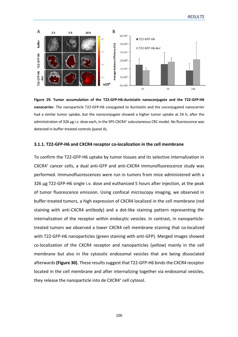
RESULTS
100
Figure 29. Tumor accumulation of the T22-GFP-H6-Auristatin nanoconjugate and the T22-GFP-H6
nanocarrier. The nanoparticle T22-GFP-H6 conjugated to Auristatin and the unconjugated nanocarrier
had a similar tumor uptake, but the nanoconjugate showed a higher tumor uptake at 24 h, after the
administration of 326 μg i.v. dose each, in the SP5 CXCR4+ subcutaneous CRC model. No fluorescence was
detected in buffer-treated controls (panel A).
3.1.1. T22-GFP-H6 and CXCR4 receptor co-localization in the cell membrane
To confirm the T22-GFP-H6 uptake by tumor tissues and its selective internalization in
CXCR4+ cancer cells, a dual anti-GFP and anti-CXCR4 immunofluorescence study was
performed. Immunofluorescences were run in tumors from mice administered with a
326 µg T22-GFP-H6 single i.v. dose and euthanized 5 hours after injection, at the peak
of tumor fluorescence emission. Using confocal microscopy imaging, we observed in
buffer-treated tumors, a high expression of CXCR4 localized in the cell membrane (red
staining with anti-CXCR4 antibody) and a dot-like staining pattern representing the
internalization of the receptor within endocytic vesicles. In contrast, in nanoparticle-
treated tumors we observed a lower CXCR4 cell membrane staining that co-localized
with T22-GFP-H6 nanoparticles (green staining with anti-GFP). Merged images showed
co-localization of the CXCR4 receptor and nanoparticles (yellow) mainly in the cell
membrane but also in the cytosolic endosomal vesicles that are being dissociated
afterwards (Figure 30). These results suggest that T22-GFP-H6 binds the CXCR4 receptor
located in the cell membrane and after internalizing together via endosomal vesicles,
they release the nanoparticle into de CXCR4+ cell cytosol.

RESULTS
101
Figure 30. Co-localization of T22-GFP-H6 and the CXCR4 receptor in the cell membrane. Representative
immunofluorescence images from SP5 SC tumors of mice 5 h after treatment with 326 µg of T22-GFP-H6
or buffer. T22-GFP-H6 and CXCR4 co-localization was seen in the cell membrane (yellow) followed by T22-
GFP-H6 internalization in endosomal vesicles towards the cytosol. DAPI staining for nuclei (blue), anti-GFP
protein (green), anti-CXCR4 receptor (red) and merged images from the three stains. Magnification: 630x.
3.1.2. Biodistribution of the T22-GFP-H6-Aur nanoconjugate in non-tumor organs and
absence of toxicity
We also assessed whether the T22-GFP-H6 nanocarrier and the T22-GFP-H6-Aur
nanoconjugate accumulated in non-tumor tissues, and their toxicity on normal cells.
Biodistribution in non-tumor tissues such as liver, kidney, lung and heart and brain was
studied 2, 5 and 24 hours after the i.v. injection of 326 µg of the nanoparticles. At 2, 5
and 24 hours we did not detect ex vivo fluorescence emission in brain, lung or heart,
since the fluorescence emission (FLI) signal was undistinguishable from background
auto-fluorescence measured in buffer-treated mice (Figure 31).
In contrast, a differential biodistribution of the nanocarrier and the nanoconjugate in
non-tumor drug clearance organs (liver and kidney) was observed. The T22-GFP-H6
nanocarrier was detected in low amounts in liver at 24 hours and at constant levels along
the studied period in kidneys. However, the fluorescence emission signal in these organs
was very low compared to the signal detected in tumors where it was 4 times higher.
Differently, the T22-GFP-H6-Aur nanoconjugate accumulated more rapidly in non-tumor
tissues, reaching a peak of accumulation in liver and kidneys at 2 hours post-injection,

RESULTS
102
that decreased progressively to become undetectable at 24 hours (Figure 31). The signal
of fluorescence emission measured at the 2-hour peak in the kidneys (2.5x10⁷
p/s/cm²/sr) was higher than the emission coming from the tumor (1.8x10⁷ p/s/cm²/sr,
Figure 29) and in the liver was moderately lower (1.5 x10⁷ p/s/cm²/sr).
We also assessed in haematoxylin and eosin (H&E) stained samples of normal tissues, if
the accumulation of these nanoparticles, specially the conjugate carrying a potent
antimitotic drug, caused any toxic effect. No histological alterations were found in non-
tumor organs (liver, kidney, lung and brain) 24 hours after nanoparticle’s administration,
since treated tissues had similar histology, and lack of apoptotic bodies, to the buffer-
treated mice (Figure 31).
Accordingly, the conjugation of the drug Auristatin to the T22-GFP-H6 nanocarrier
changed its biodistribution, probably by changing the structure of the nanoconjugate,
showing similar tumor accumulation, but accelerating and increasing its uptake in liver
and kidneys, without causing any toxicity.
Figure 31. Organ biodistribution of T22-GFP-H6 and T22-GFP-H6-Aur. Representative ex vivo
fluorescence images (FLI) of normal organs (liver, kidney, lung and brain tissues) after i.v. administration
of 326 μg dose of each nanoparticle at 2, 5 and 24 h. Lack of histological alterations are observed after 24
h of the administration by H&E staining of paraffin-embedded tissues. Scale bars: 100 μm.

RESULTS
103
3.1.3. T22-GFP-H6-Aur achieves targeted drug delivery leading to selective depletion
of CXCR4+ cells
Once confirmed the receptor-dependent tumor uptake of the T22-GFP-H6-Aur
nanoconjugate in CXCR4 highly expressing subcutaneous tumors, we assessed whether
the selective internalization and the Auristatin delivery into the cytosol of target CXCR4+
cells was followed by target cancer cell depletion. For this purpose, we analysed cell
death in the SP5 subcutaneous tumors of mice 2, 5 and 24 hours after treatment with
buffer, T22-GFP-H6 or T22-GFP-H6-Aur each at a single 326 μg i.v. dose. Since Auristatin
is a potent antimitotic agent which inhibits cell division by blocking the polymerization
of tubulin, cells in mitotic catastrophe were quantified in H&E and DAPI stained sections
of treated or control tumors. Cells in mitotic catastrophe are characterized by the
formation of giant, multinucleated cells carrying uncondensed chromosomes, as shown
in Figure 32A in H&E and DAPI stained sections. Two hours after T22-GFP-H6-Aur
treatment the number of cells in mitotic catastrophe in treated tumors (17,4 ± 1,1) was
significantly higher than in buffer-treated tumors (11,6 ± 1,8) but not in T22-GFP-H6
treated tumors (15,4 ± 1,6). At 5 hours, higher tumor uptake led to higher cell death,
finding a 1.6-fold increase in the number of cells in mitotic catastrophe in T22-GFP-H6-
Aur treated tumors than in buffer or T22-GFP-H6 treated tumors (Figure 32).
Consequently, at 24 hours cell death induction is maintained due to the increased tumor
uptake of the nanoconjugate. T22-GFP-H6-Aur induction of cell death by mitotic
catastrophe proved the capacity of the nanoconjugate to release Auristatin in the
cytosol of target CXCR4+ cells and block cell division.

RESULTS
104
Figure 32. Tumor cell death at 2, 5 and 24 h after T22-GFP-H6-Aur administration A) Representative H&E
and DAPI staining of tumor sections 24 h after buffer, T22-GFP-H6 and T22-GFP-H6-Aur administration.
Cells in mitotic catastrophe (white arrows) were counted in 5 high-power fields (x400) per sample B)
Quantitation of tumor cells in mitotic catastrophe at 2, 5 and 24 h after administration of 326 μg i.v. dose
in H&E staining sections. *p < 0.05 bars indicate a statistically significant between the designated groups.
3.2. HA2 endosomal escape peptide site-dependent accommodation in
the T22-GFP-H6 construct enhances tumor uptake
In an attempt to improve the T22-GFP-H6 nanocarrier regarding its tumor uptake, we
incorporated a known endosomal escape domain with cell penetration capacity to avoid
lysosomal degradation. Thus, to discriminate between exclusive or cooperative effects
of membrane activity and receptor-mediated cell penetrability, we studied the
biodistribution of T22-HA2-GFP-H6 and T22-GFP-HA2-H6 modular proteins, in which the
fusogenic peptide HA2 had been placed in alternative accommodation sites, using mice
bearing SP5 subcutaneous tumors with high expression of CXCR4. For that purpose, 200
µg of T22-HA2-GFP-H6 or T22-GFP-HA2-H6 were administered intravenously, as well as
the parental T22-GFP-H6 protein acting as control.
At different time points upon single dose injections, T22-GFP-H6 accumulated in
subcutaneous tumors at levels comparable to those previously described in similar
animal models (95), and a similar result was observed for T22-HA2-GFP-H6 nanoparticle
(Figure 33). However, a fast and efficient tumor retention of T22-GFP-HA2-H6 was
unexpectedly observed, peaking at 2 hours, a fact that was necessarily linked to the
incorporation of the viral HA2 peptide at the particular site between GFP and H6
modules.
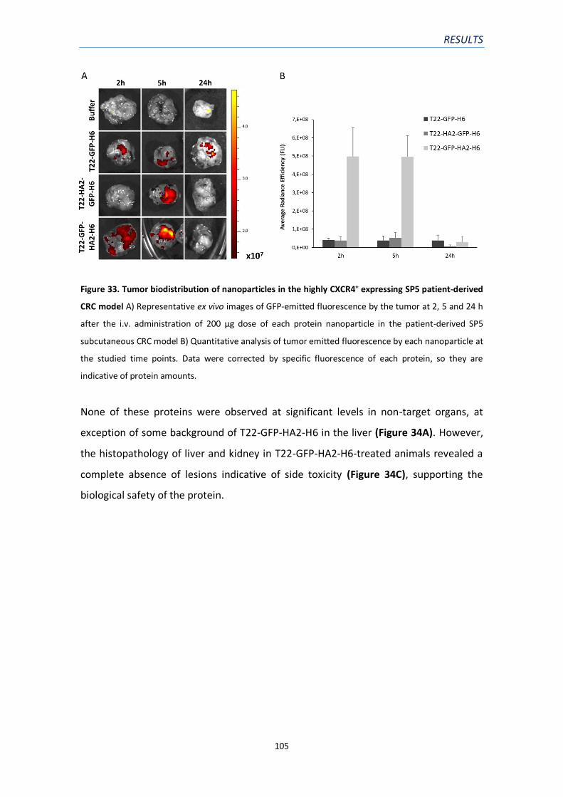
RESULTS
105
Figure 33. Tumor biodistribution of nanoparticles in the highly CXCR4+ expressing SP5 patient-derived
CRC model A) Representative ex vivo images of GFP-emitted fluorescence by the tumor at 2, 5 and 24 h
after the i.v. administration of 200 µg dose of each protein nanoparticle in the patient-derived SP5
subcutaneous CRC model B) Quantitative analysis of tumor emitted fluorescence by each nanoparticle at
the studied time points. Data were corrected by specific fluorescence of each protein, so they are
indicative of protein amounts.
None of these proteins were observed at significant levels in non-target organs, at
exception of some background of T22-GFP-HA2-H6 in the liver (Figure 34A). However,
the histopathology of liver and kidney in T22-GFP-HA2-H6-treated animals revealed a
complete absence of lesions indicative of side toxicity (Figure 34C), supporting the
biological safety of the protein.

RESULTS
106
Figure 34. Biodistribution and lack of toxicity of the nanoparticles in non-target organs A) Representative
ex vivo images of GFP-emitted fluorescence in the main non-tumor organs at 5 h in the SC SP5 colorectal
cancer model B) Quantitative analysis of liver and kidney emitted fluorescence by each nanoparticle at 2,
5 and 24 h of emitted fluorescence C) Absence of histopathological alterations in liver or kidney in H&E
stained tissue sections, at the studied time points after the administration of the T22-GFP-HA2-H6
nanoparticle. Magnification: 200x.
The numerical analyses of the accumulated materials (AUC, area below the curve, was
fully convincing regarding the superiority of T22-GFP-HA2-H6 as a CXCR4-targeted
material, that combined the cell specificity of T22 and the cell-penetrating abilities
empowered by HA2 (Figure 35).

RESULTS
107
Figure 35. Graphic representation of total nanoparticle exposure. Area under the curve: (AUC = FLI
emission x time (h)) registered in tumor and non-tumor organs along the studied period (2-24 h) for all
three tested nanoparticles (200 μg dose) using the SC SP5 colorectal cancer model, and its quantitation.
Fluorescence emission intensity (FLI) signal from experimental mice was calculated subtracting the FLI
auto-fluorescence of control buffer-treated mice. FLI, fluorescent intensity (expressed as average
radiance efficiency).
3.3. Selective CXCR4-dependent T22-GFP-H6 and T22-GFP-HA2-H6 tumor
uptake in subcutaneous CRC tumors
An obvious concern was whether the extraordinary tumor targeting manifested by T22-
GFP-HA2-H6 was the result of the expected CXCR4 selectivity or it was rather mediated
by a combination of the cell-penetrating properties of HA2 and of an enhanced
permeability and retention (EPR) effect (96), or alternative ways to enter tumor tissues,
such as endothelial transcytosis (97). To discriminate between these two possibilities,
an in vivo competition experiment was designed in which the CXCR4 antagonist,
AMD3100 (98), was used to block tumor accumulation of the administered protein
materials. As observed (Figure 36), the antagonist dramatically minimized the presence
of T22-GFP-H6 nanoparticles in tumoral tissues, supporting again the role of T22 in
active targeting for CXCR4+ cancers. Interestingly, AMD3100 equally reduced the tumor
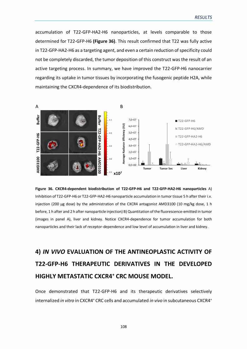
RESULTS
108
accumulation of T22-GFP-HA2-H6 nanoparticles, at levels comparable to those
determined for T22-GFP-H6 (Figure 36). This result confirmed that T22 was fully active
in T22-GFP-HA2-H6 as a targeting agent, and even a certain reduction of specificity could
not be completely discarded, the tumor deposition of this construct was the result of an
active targeting process. In summary, we have improved the T22-GFP-H6 nanocarrier
regarding its uptake in tumor tissues by incorporating the fusogenic peptide H2A, while
maintaining the CXCR4-dependence of its biodistribution.
Figure 36. CXCR4-dependent biodistribution of T22-GFP-H6 and T22-GFP-HA2-H6 nanoparticles A)
Inhibition of T22-GFP-H6 or T22-GFP-HA2-H6 nanoparticle accumulation in tumor tissue 5 h after their i.v.
injection (200 μg dose) by the administration of the CXCR4 antagonist AMD3100 (10 mg/kg dose, 1 h
before, 1 h after and 2 h after nanoparticle injection) B) Quantitation of the fluorescence-emitted in tumor
(images in panel A), liver and kidney. Notice CXCR4-dependence for tumor accumulation for both
nanoparticles and their lack of receptor-dependence and low level of accumulation in liver and kidney.
4) IN VIVO EVALUATION OF THE ANTINEOPLASTIC ACTIVITY OF
T22-GFP-H6 THERAPEUTIC DERIVATIVES IN THE DEVELOPED
HIGHLY METASTATIC CXCR4+ CRC MOUSE MODEL.
Once demonstrated that T22-GFP-H6 and its therapeutic derivatives selectively
internalized in vitro in CXCR4+ CRC cells and accumulated in vivo in subcutaneous CXCR4+

RESULTS
109
tumors without causing any systemic toxicity, we next evaluated the therapeutic
potential of the engineered T22-GFP-H6 therapeutic derivatives.
First, we assessed the antitumor activity of the T22-GFP-H6-Aur nanoconjugate and the
T22-PE24-H6 nanotoxin. For that purpose, we studied the capacity of each therapeutic
nanoparticle to block tumor growth in the SW1417 cell-derived subcutaneous model, by
administering them on a repeated dose regime. Next, we evaluated the antimetastatic
activity of the same nanoparticles. In this case, the previously developed highly
metastatic CRC model derived from the SW1417 cell line was used to study whether
these therapeutic nanoparticles were able to prevent cancer cell dissemination from the
primary tumors to other organs and inhibit metastases formation.
4.1. T22-GFP-H6-Aur repeated dose administration activates a lethal
immunogenic response in Swiss nude mice
To study the possible antitumor effect of the T22-GFP-H6-Aur nanoconjugate, we used
the SW1417 cell-derived subcutaneous model. When subcutaneous tumors reached an
approximate volume, around 120 mm3, mice were randomized in two groups; the
buffer-treated group (N=10), i.v. administered with 150 µl of buffer, and the
experimental group i.v. administered with 100 µg of T22-GFP-H6-Aur three times per
week (N=10). Surprisingly, 8 out of 10 mice administered with T22-GFP-H6-Aur died
minutes after the fourth dose. At that time, the tumor volume of the T22-GFP-H6-Aur
treated group was smaller than the buffer-treated group, but without being significant
(Figure 37A).
Subcutaneous tumors and other normal organs were collected to evaluate the mouse
cause of death. Surprisingly, none of the studied organs (liver, kidneys, brain, lungs and
heart) showed any histological alteration in H&E stained sections (Figure 37B); thus, the
cause of death remains unknown. The fact that all mice died after the fourth
nanoconjugate bolus and the certitude that Swiss nude mice maintain active B cell and
robust NK cell responses, made us think that the activation of an immunogenic response
by T22-GFP-H6-Aur could be the cause of the 80% death in the treated mice; however,
this remains to be proven in future studies. Taking into account the high lethality

RESULTS
110
observed in nanoconjugate-treated mice, the preclinical development of this
nanoconjugate is uncertain.
Figure 37. Antitumor effect of T22-GFP-H6-Aur and absence of toxicity in normal organs of dead mice
A) SW1417 subcutaneous tumor growth of mice treated with buffer or the T22-GFP-H6-Aur
nanoconjugate. Mice died after the fourth dose B) Representative H&E images of liver and kidneys from
both, buffer and T22-GFP-H6-Aur treated mice, showing normal histological features. Scale bars: 50 µm.
4.2. T22-GFP-H6-Aur prevents only transcelomic metastases in the
SW1417 cell-derived CRC model
We then evaluated the capacity of T22-GFP-H6-Aur to prevent metastasis using a more
immunosuppressed mouse strain than Swiss nude to avoid the immunogenic reaction
described above. Thus, we used the previously developed NSG mouse model to be
treated with this nanoconjugate at a repeated dose schedule and to analyse at the end
of the treatment period the percentage of mice free of metastasis and the reduction in
foci number and foci size in mice with detectable metastases. For this purpose, we used
the bioluminescent SW1417 cell-derived orthotopic model, which is highly metastatic,
developing cancer foci in lymph nodes, liver, lung and peritoneum, and expresses
moderate levels of CXCR4. We started the treatment with the T22-GFP-H6-Aur
nanoconjugate three days after cecum cell injection, following a schedule of 100 µg,
three times per week and a total of 12 doses.

RESULTS
111
At the end of the experiment, all target metastatic organs were collected and quantified
in three H&E stained sections the number and size of the developed metastatic foci, in
each affected organ (Table 9). In contrast with the buffer-treated group, T22-GFP-H6-
Aur treatment did not have the expected prevention capacity since only transcelomic
metastases development were reduced. The percentage of mice affected by
transcelomic metastases was slightly lower than in the buffer-treated group, but the
number of peritoneal metastasis was significantly lower (3.2-fold reduction). Lymphatic
(lymph node) and hematogenous (liver and lung) metastases development were not
inhibited by the T22-GFP-H6-Aur treatment, since we did not find significant differences
either in the percentage of mice affected by lymph node, hepatic or pulmonary
metastasis, nor in the number of metastatic foci in these organs. Moreover, the growth
of metastatic foci in lymph node, liver, lung and peritoneum was not either inhibited by
the T22-GFP-H6-Aur nanoconjugate, since we did not find differences between the size
of foci found in nanoconjugate-treated versus buffer-treated mice (Table 9). Thus, based
on the in vitro results and the lack of antimetastatic activity in lymph node, liver and
lung, the T22-GFP-H6-Aur nanoconjugate, which carries a potent inhibitor of tubulin
polymerization, is not a good candidate to treat CRC metastases, consequently excluding
its further development as treatment for this indication.
Table 9. Evaluation of T22-GFP-H6-Aur antimetastatic effect by preventing the development of
metastases in the SW1417 cell-derived CRC metastatic model.
T22-GFP-H6-Aur prevention of metastasis
Primary tumor Lymph node Mets Liver Mets Lung Mets Peritoneal Mets
mice % mice % # foci mice % # foci mice % # foci mice % # foci
90% 100% 90% 100%
3.2 ± 0.7 3.5 ± 0.4 17.6 ± 5.4 5.5 ± 1.6a
80% 80% 90% 90%
2.9 ± 0.7 3.6 ± 1.6 9.2 ± 4.1 1.7 ± 0.4a
Group Primary tumor Lymph nods Mets Liver Mets Lung Mets Peritoneal Mets
Buffer 9990.5 ± 5137.9 449.7 ± 127.2 13.79 ± 3.1 4.4 ± 0.9 4702.2 ± 2353
T22-GFP-H6-Aur 7390.7 ± 3168.8 719.6 ± 125.1 45.3 ± 25.9 8.9 ± 3.4 8781.3 ± 3633
Mean + s.e.m. metastatic foci number or area (µm2) per mouse, counted in three entire histology sections.
a p=0.03
Metastatic foci size (µm² x 103)
SW1417 cell-derived ortothopic model
Group
Buffer 10/10 100%
T22-GFP-H6-Aur 10/10 100%

RESULTS
112
4.3. T22-PE24-H6 repeated dose administration reduces tumor volume
without toxicity in non-target organs
We next evaluated the antitumor effect of CXCR4-targeted nanoparticles carrying
cytotoxic payloads capable of activating cell death mechanisms alternative to the
apoptosis induced by chemotherapy genotoxic drugs or to the antimitotic microtubule
inhibitors. Thus, active fragments of Pseudomonas aeruginosa exotoxin (PE24) were
produced in Escherichia coli as self-assembled nanoparticles composed by modular
fusion protein T22-PE24-H6 monomers, with the aim to induce targeted CXCR4+ cancer
cell death through the activity of the catalytic fragments of this protein nanodrug. As
explained in 2.4. section, purified T22-PE24-H6 nanoparticles were first tested in vitro in
SW1417 CXCR4+ cells, showing a specific reduction of cell viability in a dose-dependent
manner and with a low IC50 (6.56 nM) compared to other therapeutic nanoparticles. To
perform the T22-PE24-H6 nanotoxin in vivo evaluation we started by studying its
antitumor effect in the subcutaneous cell-derived SW1417 model. SW1417 CXCR4+ cells
were injected in Swiss nude mice, in which we have previously observed that a high dose
of 10 µg of T22-PE24-H6 did not produce any toxic effect in non-target organs. Thus,
when subcutaneous tumors reached a volume of approximately 120 mm3, mice were
randomized in two groups (N=7) and buffer or the nanotoxin were administered in a
dose regime of 10 µg, three times per week, per 8 doses.
After only four doses of T22-PE24-H6, significant differences (p=0.02) in tumor volume
were already detected between control mice treated with buffer and mice treated with
T22-PE24-H6 (Figure 38B). Moreover, at the end of the experiment after the eighth
dose, we observed a 1.6-fold reduction in tumor volume, as compared to buffer-treated
mice (p=0.005), associated with a reduction in tumor final weight (p=0.04) (Figure 38C).
This antitumor effect was most likely a consequence of the 3.0-fold increase in cell death
bodies in tumor tissue (p<0.05) (Figure 38D), with no significant differences in body
weight between nanotoxin-treated and control groups (Figure 38A). The lack of toxicity
in non-target organs, such as liver and kidneys, was assessed by H&E stained sections
and no histological alteration were found.

RESULTS
113
Figure 38. T22-PE24-H6 antitumoral effect in the cell-derived subcutaneous SW1417 CRC model A)
Follow-up of mouse body weight (g) during the repeated dose administration of 10 µg of T22-PE24-H6
(three times a week, 8 total doses) B) Antitumor effect of T22-PE24-H6 measured by the analysis of tumor
volume (mm3) C) Antitumor effect of T22-PE24-H6 measured by the tumor weight (g) at the end of the
experiment D) Increase in the number of cell death bodies in the SC SW1417 tumors collected at the end
of the experiment. All data are presented as mean ± s.e.m., N=7. * p<0.05; ** p<0.01.
4.3.1. T22-PE24-H6 triggers pyroptotic cell death in SW1417 tumors
CXCR4+ SW1417 subcutaneous tumors treated with repeated doses of 10 µg of T22-
PE24-H6 allowed us to study the cell death mechanism induced by T22-PE24-H6 in vivo.
Since we have found an in vitro activation of pyroptosis in SW1417 cells and an in vivo
3-fold increase in the number cell death bodies in nanotoxin-treated tumors, we
evaluated whether the observed cell death bodies were a result of apoptosis or, as
expected from the in vitro results, of the induction of pyroptosis. By performing
immunohistochemistry assays against apoptotic markers in tumor tissue sections, we
found no detectable or very low signal in active caspase-3 and proteolyzed-PARP
staining of nanotoxin-treated tumors (Figure 39A), suggesting that the mechanism of
cell death was not mediated by apoptosis induction.

RESULTS
114
Figure 39. In vivo assessment of T22-PE24-H6-induced activation of the apoptotic or the pyroptotic cell
death pathways A) Representative images of H&E and IHC staining of apoptotic (active-caspase-3 and
proteolyzed PARP) and pyroptotic markers (NLRP3, cleaved caspase-1 and cleaved GSDMD) of SW1417
subcutaneous tumors from Swiss nude mice treated with 10 µg of T22-PE24-H6, three times a week and
8 total doses. Scale bars: 50 µm B) Quantification of each marker staining in buffer-treated mice and T22-

RESULTS
115
PE24-H6 treated mice. In NLRP3 stained sections, 5 high-power fields (400x) were analysed with ImageJ
and quantification expressed as percentage of stained surface. In caspase-1 and GSDMD stained sections,
5 high-power fields (400x) were analysed by counting positive cells. Measurements in tissue sections were
performed 24 h after the last administered dose and were compared to buffer-treated control mice. All
the data are expressed as mean ± s.e.m.. * p<0.05; ** p<0.001.
As we have described before, besides apoptosis, there are other cell death mechanisms
playing general physiopathological roles that may also have therapeutic implications.
Pyroptosis, is an inflammatory caspase-dependent form of programmed cell death that
occurs usually in response to microbial infection and similarly to apoptosis promotes
nuclear condensation and chromatin DNA fragmentation. Expectedly, pyroptosis
markers activated at different points of the pyroptotic pathway, display a high
expression in tumors treated with T22-PE24-H6 compared to their expression in buffer-
treated tumors. Thus, NLRP3, cleaved caspase-1 and cleaved Gasdermin D (GSDMD)
showed a higher expression 24 hours after the last dose in the repeated T22-PE24-H6
nanotoxin administration (Figure 39B). As observed in the in vitro experiments, the first
marker to be activated is NLRP3 with a cytoplasmatic expression that represents a 35%
of the stained surface tissue. After that, caspase-1 is activated showing a very specific
staining of already pyroptotic bodies which increased 6 times compared to buffer-
treated tumors. Finally, GSDMD is also activated in those pyroptotic bodies causing pore
formation and cell death because of its pyroptotic cell effector role. These in vivo results,
which are in full agreement with our in vitro observations, indicate that the T22-PE24-
H6 nanotoxin induces pyroptosis in SW1417 subcutaneous tumors.
4.4. Definition of a dose regime for repeated T22-PE24-H6 administration
in NSG mice
Since the different immunosuppressed mouse strains have a different tolerance to the
diverse developed therapeutic nanoparticles, particularly regarding its toxicity on
normal tissues, a preliminary experiment was performed to find the necessary T22-
PE24-H6 repeated dose schedule that would achieve antimetastatic effect while
showing lack of toxicity on non-tumor organs of NSG mice. For that purpose, we used
the cell-derived CXCR4+ SW1417 bioluminescent orthotopic model in NSG mice and
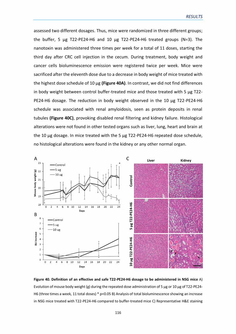
RESULTS
116
assessed two different dosages. Thus, mice were randomized in three different groups;
the buffer, 5 µg T22-PE24-H6 and 10 µg T22-PE24-H6 treated groups (N=3). The
nanotoxin was administered three times per week for a total of 11 doses, starting the
third day after CRC cell injection in the cecum. During treatment, body weight and
cancer cells bioluminescence emission were registered twice per week. Mice were
sacrificed after the eleventh dose due to a decrease in body weight of mice treated with
the highest dose schedule of 10 µg (Figure 40A). In contrast, we did not find differences
in body weight between control buffer-treated mice and those treated with 5 µg T22-
PE24-H6 dosage. The reduction in body weight observed in the 10 µg T22-PE24-H6
schedule was associated with renal amyloidosis, seen as protein deposits in renal
tubules (Figure 40C), provoking disabled renal filtering and kidney failure. Histological
alterations were not found in other tested organs such as liver, lung, heart and brain at
the 10 µg dosage. In mice treated with the 5 µg T22-PE24-H6 repeated dose schedule,
no histological alterations were found in the kidney or any other normal organ.
Figure 40. Definition of an effective and safe T22-PE24-H6 dosage to be administered in NSG mice A)
Evolution of mouse body weight (g) during the repeated dose administration of 5 µg or 10 µg of T22-PE24-
H6 (three times a week, 11 total doses) * p<0.05 B) Analysis of total bioluminescence showing an increase
in NSG mice treated with T22-PE24-H6 compared to buffer-treated mice C) Representative H&E staining

RESULTS
117
images showing normal liver and kidney histology in control and 5 µg T22-PE24-H6 mice and protein
deposits (arrows) in the renal tubules of 10 µg T22-PE24-H6 treated mice. Scale bars: 100 µm.
When analysing the progression of bioluminescence emission by SW1417-luciferase
expressing cancer cells in vivo, we observed a 4.0-fold increase in control mice, while
this increase was less pronounced (2.9-fold) in mice treated with the 5 µg repeated dose
schedule than in those treated with 10 µg of T22-PE24-H6. Here, it is relevant to state
that this small decrease in full body bioluminescence in nanotoxin-treated mice included
the signal emitted by the primary tumor as well as that coming from the metastatic foci.
Importantly, the ex vivo analysis of the different organs affected by metastasis, showed
differences in bioluminescence emission in mice treated with both dose schedules of
the nanotoxin, that were due to the growth inhibition of metastases rather than to their
effect on the primary tumor (Figure 41). The bioluminescence emitted by the tested
organs of the nanotoxin-treated mice, which included metastatic foci disseminated to
peritoneum, liver and lung, was lower than that emitted by the same organs of control
mice, showing a dose-dependent effect, which correlates with a reduced metastatic
load. Therefore, the 5 µg T22-PE24-H6 repeated dose was selected as the schedule to
be administered in a new experiment with higher number of NSG mice bearing
orthotopic tumors, to test whether the T22-PE24-H6 achieves an antimetastatic effect
that significantly reduces the mouse metastatic load in the absence of associated
systemic toxicity.

RESULTS
118
Figure 41. Antimetastatic activity induced by T22-PE24-H6 in regional and distant sites, measured ex
vivo A) Comparison of bioluminescence emission between the buffer-treated, 5 µg T22-PE24-H6 and 10
µg T22-PE24-H6 repeated dose schedule groups in primary tumor, mesenteric lymph nodes, liver, lung
and peritoneum. Results are presented as mean ± s.e.m. bioluminescence values in photons per second
(total flux [p/s]) B) Representative bioluminescence images comparing primary tumor and metastatic
dissemination in liver and lungs of mice treated with the nanotoxin (5 µg or 10 µg T22-PE24-H6) or buffer
(control).
4.5. T22-PE24-H6 prevents the development of lymphatic and
hematogenous metastasis in the SW1417 cell-derived CRC model
Once we selected the 5 g T22-PE24-H6 repeated dose schedule as the non-toxic dose
that achieves antimetastatic effect in NSG mice, we performed an assay to determine
the capacity of this nanotoxin to prevent cancer cell dissemination and metastatic foci
growth at the different target organs in the cell-derived orthotopic CXCR4+ SW1417 CRC
model. This model metastasizes to lymph nodes, liver, lung and peritoneum, expecting
that the treatment with T22-PE24-H6 could reduce the number and size of metastatic
foci at the end of the experiment. So, a total of 18 NSG mice were implanted in the
cecum with 2 million CXCR4+ SW1417 CRC cells. Three days after cell implantation, mice
were randomized into two groups and we started administering the nanotoxin to the
mice in the experimental group (N=9) following a dose schedule of 5 µg, three days per
week for a total of 18 doses, or buffer following the same schedule (N=9). The treatment

RESULTS
119
continued until the first mouse belonging, either to the control or treated group,
achieved the euthanasia criteria.
After 18 doses, control mice started to lose weight due to metastatic dissemination, a
time point at which all mice were euthanised (Figure 42A). At this point, we could find
differences close to significance in total body bioluminescence between mice treated
with T22-PE24-H6 and buffer-treated mice (Figure 42B). The ex vivo bioluminescence
analysis of each cancer-affected organ was performed, showing differences between
both groups, as we previously found in the preliminary assay of dose definition.
Figure 42. T22-PE24-H6 antimetastatic effect in the cell-derived orthotopic SW1417 CRC model A)
Evolution of mouse body weight (g) the repeated dose administration of 5 µg of T22-PE24-H6 (three times
a week, 18 total doses) B) Total body bioluminescence (BLI; Total Flux [p/s]) of buffer- and T22-PE24-H6
treated mice, measured once per week along all the experiment C) All data are expressed as mean ±
s.e.m., N=9. * p<0.05.
Next, a histological evaluation of metastatic foci number and size was performed for
each metastatic localization. Mice treated with T22-PE24-H6 presented a reduction in
metastatic load (total metastatic area) in all affected organs, especially evident in lymph
nodes (p<0.05) (Figure 42C). In contrast to the findings in buffer-treated group, T22-
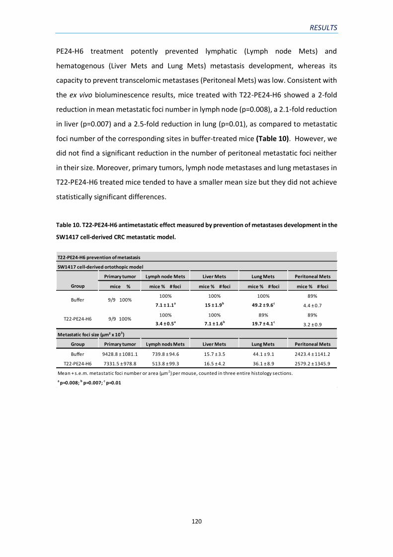
RESULTS
120
PE24-H6 treatment potently prevented lymphatic (Lymph node Mets) and
hematogenous (Liver Mets and Lung Mets) metastasis development, whereas its
capacity to prevent transcelomic metastases (Peritoneal Mets) was low. Consistent with
the ex vivo bioluminescence results, mice treated with T22-PE24-H6 showed a 2-fold
reduction in mean metastatic foci number in lymph node (p=0.008), a 2.1-fold reduction
in liver (p=0.007) and a 2.5-fold reduction in lung (p=0.01), as compared to metastatic
foci number of the corresponding sites in buffer-treated mice (Table 10). However, we
did not find a significant reduction in the number of peritoneal metastatic foci neither
in their size. Moreover, primary tumors, lymph node metastases and lung metastases in
T22-PE24-H6 treated mice tended to have a smaller mean size but they did not achieve
statistically significant differences.
Table 10. T22-PE24-H6 antimetastatic effect measured by prevention of metastases development in the
SW1417 cell-derived CRC metastatic model.
T22-PE24-H6 prevention of metastasis
Primary tumor Lymph node Mets Liver Mets Lung Mets Peritoneal Mets
mice % mice % # foci mice % # foci mice % # foci mice % # foci
100% 100% 100% 89%
7.1 ± 1.1a 15 ± 1.9b 49.2 ± 9.6c4.4 ± 0.7
100% 100% 89% 89%
3.4 ± 0.5a 7.1 ± 1.6b 19.7 ± 4.1c3.2 ± 0.9
Group Primary tumor Lymph nods Mets Liver Mets Lung Mets Peritoneal Mets
Buffer 9428.8 ± 1081.1 739.8 ± 94.6 15.7 ± 3.5 44.1 ± 9.1 2423.4 ± 1141.2
T22-PE24-H6 7331.5 ± 978.8 513.8 ± 99.3 16.5 ± 4.2 36.1 ± 8.9 2579.2 ± 1345.9
Mean + s.e.m. metastatic foci number or area (µm2) per mouse, counted in three entire histology sections.
a p=0.008; b p=0.007; c p=0.01
Metastatic foci size (µm² x 103)
SW1417 cell-derived ortothopic model
Group
Buffer 9/9 100%
T22-PE24-H6 9/9 100%

V. DISCUSSION

DISCUSSION
122
V. DISCUSSION
Colorectal cancer (CRC) is one of the most common neoplasias in developed countries,
accounting for almost 900,000 deaths per year. Its incidence is progressively rising
worldwide due to the adoption of sedentary and unhealthy lifestyles such as red meat,
alcohol and tobacco consumption. However, the promotion of early detection tests and
new treatment options have reduced its mortality. Despite this advance, nearly a
quarter of CRC patients are diagnosed at an advanced stage carrying already metastases,
which hinders their surgical removal and subsequent tumor-related deaths. For these
patients, treatment consists in shrinking the tumor and supressing cancer spread and
growth, using radiotherapy and chemotherapy or combination with targeted therapies.
Current chemotherapy for CRC is mainly based in fluoropyrimidine (5-FU), which results
in the improvement in survival of approximately 30%, with an addition of 20%
improvement when used in a combined regime with oxaliplatin in stage III CRC (99).
Some targeted therapies that inhibit VEGF and EGFR pathways have also demonstrated
to improve the outcome in metastatic CRC patients when combined with chemotherapy.
However, both chemotherapy and molecularly targeted agents, especially that have a
low molecular weight, display a passive diffusion affecting non-tumor and tumor tissues,
without any selectivity for cancer cells; thus, damaging normal cells and inducing
systemic toxicity. Chemotherapy interferes with cell division targeting all rapidly dividing
cells in the body, harming also healthy tissues, especially those tissues that have a high
replacement rate (intestinal lining and immune cells). Moreover, chemotherapy has
other limitations apart from systemic toxicity, such as low response rate, development
of resistance and absence of tumor selectivity. Molecularly targeted therapies, mainly
based in monoclonal antibody development, also present limitations like toxicities due
to the fact of targeting a general biological process in the human body such as
angiogenesis or a lack of response in the case of KRAS mutated tumors when treated
with anti-EGFR therapies. For all these observations, the tolerated doses of these agents
are low, which is translated in insufficient antitumor effect.
Due to the limitations of current therapies, further investigations are still required to
develop new strategies to improve current metastatic CRC treatments. One main goal

DISCUSSION
123
of these new approaches pursues the complete elimination of cancer cells without
damaging non-tumor cells or organs of the rest of the body. For this purpose,
nanoparticles are being exploited as nanocarriers attempting to deliver cytotoxic drugs
only to cancer cells. The acquired knowledge about the mechanisms leading to cancer
progression and metastasis is a valuable tool for the selection of specific cancer cell
antigens, which can be actively targeted by these nanocarriers.
In this thesis, we aimed at taregting cancer stem cells (CSCs) which current therapies are
failing to eliminate (100). Within the tumor, CSCs are a subset of cells that possess the
ability to self-renew and to differentiate into all cell populations. Moreover, recent
findings demonstrate that a subpopulation of CSCs with epithelial-mesenchymal
transition (EMT) properties or partial EMT, can initiate tumor growth in secondary sites
(metastatic CSCs) (101,102). Thus, we chose the CXCR4 receptor as a specific target
present in metastatic CSCs, whose overexpression has been associated with
chemotherapy resistance, relapse, metastasis (103) and poor clinical outcome (37) in
CRC and other cancer types (29). According to the homing theory, target organs
produce and release specific chemokines such as CXCL12 and attract distant CXCR4+
cancer cells. Moreover, it is known that CRC cells with metastatic initiation capacity
possess both stemness/EMT (CD133+) and metastatic (CXCR4+) properties. It has also
been shown that CD133+CXCR4+ CRC cells have enhanced migratory capacity in vitro
and higher metastatic potential in vivo than CD133+CXCR4- cells (104). CXCR4 is also
expressed on circulating tumor cells (CTCs) and the number of CD133+CXCR4+ cells in
the bloodstream correlates with poor prognosis (104). Taken together, these findings
indicate that metastatic CSCs expressing CXCR4 are clinically relevant targets; thus,
their selective elimination could represent an advance in metastases control.
As described in the introduction, in collaboration with the Nanobiotechnology group
of the UAB, we developed self-assembled protein-only nanoparticles for actively
targeting CXCR4+ cells which present high biocompatibility, biodegradability and
functional versatility. The narrow therapeutic window of classic cytotoxic drugs can be
solved by loading them in these nanoparticles, enhancing their pharmacokinetic
characteristics, enabling passive targeting in tumors via the EPR effect described in
mouse tumors (105), or alternatively increasing tumor uptake by endothelial

DISCUSSION
124
transcytosis. Since the existence of EPR effect is being questioned in human tumors
(106), nanoparticles allow as well functionalization with specific peptides for active
targeting of tumor cells, thus reducing systemic toxicity.
Therefore, the main objective of this work has been the preclinical validation of the
different T22-GFP-H6 therapeutic derivatives as potential treatments for metastatic
CRC. The selective biodistribution of the nanoparticles and the antitumor effect of their
therapeutic derivatives had to be studied in vitro and in newly developed CXCR4+ CRC
mouse models. Using both established CRC cell lines and patient samples, we developed
highly metastatic CRC models, which were then used to assess the antimetastatic effect
of T22-GFP-H6-derived nanoconjugates or of newly synthesized protein-only
nanoparticles with intrinsic cytotoxic activity.
1) DEVELOPMENT OF CXCR4+ SUBCUTANEOUS AND HIGHLY
METASTATIC CRC MOUSE MODELS FOR THEIR USE IN
PRECLINICAL STUDIES
The discovery and preclinical testing of novel therapeutic strategies in CRC, as in other
cancers, requires the use of in vitro and in vivo models of each cancer type. In vitro
cancer models, using cell culture, are important tools for cancer research and have been
widely used as low-cost screening platforms for anticancer agents. Nowadays, three
dimensional (3-D) organoid tissue cultures derived from self-renewing stem cells, are
able to recapitulate the in vivo architecture, functions and genetic and molecular
imprints of their original tissues, representing an advanced in vitro tool for anticancer
drug discovery (107,108). However, they present some limitations since they are not
able to replicate important processes or mechanisms activated during tumor
progression that occur only in vivo, such as their interactions with the tumor
microenvironment, invasion and metastatic dissemination, which is the cause of the
majority of cancer-related deaths. On this basis, in vivo CRC models that recapitulate the
pathogenesis observed in patients (angiogenesis, metastasis and response to therapy)
are essential for the development of effective therapies. Currently, the most used in vivo
models for therapeutic studies are murine xenografts, where CRC cells are injected

DISCUSSION
125
subcutaneously in immunosuppressed mice, but they still have limitations that could be
overcome by the development of patient-derived xenografts (PDXs) and orthotopic
models.
1.1. Cell and patient-derived subcutaneous xenografts for nanoparticle
preclinical evaluation
In this thesis, we developed CXCR4+ subcutaneous CRC models for their use in the
assessment of nanoparticle biodistribution and antitumor effect, in which we could also
study their selective internalization in cancer cells and the induction of specific
mechanisms of cell death. All these models were generated in Swiss nude mice, which
is the cheapest and the less immunosuppressed mouse strain. We used xenograft
models for these assays because of their low cost, which allows the use of a bigger
experimental number to reach sufficient statistical power, the short time needed to
develop it and their ease of use, especially for measuring tumor growth.
On the one hand, cell-derived xenograft models were developed by injecting the CXCR4-
overexpressing (CXCR4+) SW1417 cell line in the subcutis of Swiss nude mice. The tumors
derived from these cells maintained a constant growth rate that was dependent on
CXCR4 expression, since CXCR4- SW1417 cells were unable to generate subcutaneous
tumors at all. These results correlate with those reported for prostate cancer in vivo
experiments, in which NOD/SCID mice injected with cells that expressed higher levels of
CXCR4 (PC3LG-CXCR4 and 22Rv1G-CXCR4) developed larger tumors than mice injected
with the parental cells (109). Moreover, exposure to specific antibodies against CXCR4
inhibited the CXCR4-dependent growth in the PC3LG-CXCR4 cell line. Other results
suggested that the activation of the CXCL12/CXCR4 axis which promotes tumor growth
in prostate cancer, is driven by the loss of PTEN, frequently observed in cancer, and
subsequent activation of Akt (110). In CRC, approximately 40% of tumors show
decreased expression of PTEN, often in association with its mutation or deletion (111).
Moreover, in vitro studies demonstrated a correlation between PTEN loss and
CXCL12/CXCR4/Akt activation, promoting an increase in cell proliferation and invasion
(112). So, the CXCR4 overexpression might exert favourable conditions not only for

DISCUSSION
126
invasion and migration, but also in enhancing tumor growth, which cloud lead to a more
efficient seeding of CTCs to distant metastatic sites.
On the other hand, we also developed patient-derived xenografts (PDXs) by engrafting
portions of tumor patient tissue into immunosuppressed mice. CRC tumor samples were
collected in collaboration with the Department of General and Digestive Surgery of the
Hospital de la Santa Creu i Sant Pau after obtaining the informed and signed patient
consent, and they were implanted in the subcutis of Swiss nude mice. A total of 30% of
the collected and implanted tumor samples, developed viable subcutaneous tumors.
One of them, the SP5 sample was maintained in vivo as a tumor line through successive
passes in Swiss nude mice. Moreover, the SP5 subcutaneous tumor line maintained in
vivo showed a significantly higher membrane expression of the CXCR4 receptor than
that observed in SW1417 cell-derived subcutaneous tumors. In contrast to cell-derived
xenografts, in PDXs tumors, both stromal and cancer cells grow, allowing for tumor-
stroma crosstalk and conserving cellular and molecular heterogeneity. Furthermore,
PDXs tumors maintain important characteristics of tumor histology, vascularity and
architecture of primary CRCs; therefore, their use could be more appropriate for the
development of novel therapeutic approaches than cell-derived tumor models.
1.2. Severe immunosuppression increases the metastatic capacity of CRC
cells, disseminating to clinically relevant sites in orthotopic mouse models
Despite metastasis is the main cause of death in CRC patients, GEM and subcutaneous
xenografts mouse models fail in reproducing the pathogenesis observed in patients,
since they are barely metastatic. In contrast, the orthotopic implantation of cancer cells
into the mouse cecum is a promising approach for the development of metastatic CRC
models. However, it is scarcely used because of the technical challenges and difficulty in
tumor monitoring. In this thesis, we also developed highly metastatic CRC mouse models
in a short time (1-2 months to develop distant metastases) using the orthotopic
microinjection of CRC cell lines or disaggregated patient tumor samples in
immunosuppressed mice, which displayed overexpression of the CXCR4 receptor in

DISCUSSION
127
epithelial cancer cells. These models were used for the evaluation of the antimetastatic
effect of the newly developed therapeutic nanoparticles targeting the CXCR4 receptor.
The OCMI procedure which consists on the microinjection of CRC cells between the
mucosa and the muscularis externa layers of the cecal wall, was used for this purpose.
In previous studies where CRC cells lines (HCT116, SW620 and DLD1) were microinjected
in Swiss nude mice, we found that the dissemination pattern closely replicated all
relevant metastatic sites observed in humans and enhanced the metastatic rate
compared to previous methods (51). Despite this advance, when metastases developed,
mostly microfoci having an area lower than 750,000 µm2 were observed, limiting their
use for the preclinical evaluation of antimetastatic compounds.
Previous reports have described the role of the immune system in the control of cancer
progression and immune surveillance. Thus, considering the limitations of our previous
models, our next aim was to increase the metastatic rate of the newly developed
orthotopic model by depleting tumor surveillance, therefore using alternative mouse
strains with an increased immunosuppression condition. We chose to orthotopically
inject CXCR4+ SW1417 CRC cells, which are also bioluminescent allowing to monitor
tumor and metastases growth. These cells were implanted orthotopically in two
immunosuppressed mouse strains; NOD/SCID and NSG mice. Both mouse strains have a
total lack of adaptive immune cells such as B and T lymphocytes and impairment in the
innate immunity including loss of complement and impaired NK, macrophage, and
dendritic cell functions (113). Neutrophils and defective dendritic cells and macrophages
constitute most of the remaining mouse immune cells detectable in peripheral blood in
both mouse strains. However, NOD/SCID mice contain residual NK activities, which are
totally absent in NSG mice (114). Therefore, we observed that the lack of NK cells
reduced the survival of NSG mice carrying CRC tumors in comparison to NOD/SCID mice.
Our results showing a high increase in metastatic rate when using NSG mice, are
consistent with several studies in different types of human cancers, including CRC,
where an improved overall survival was observed in patients with higher levels of tumor-
infiltrating lymphocytes (TILs) and NK cells, that mediate cytotoxic functions
independent of MHC-mediated antigen presentation (115,116). Other studies using
models of spontaneous leukemia and prostate cancer, showed an accelerated

DISCUSSION
128
development of cancer in animals with a depletion of NK cells compared to animals with
a normal NK cells activity (117).
In our hand, both NOD/SCID and NSG mouse models displayed a high dissemination
pattern. We observed that SW1417 cells were able to infiltrate the lymph nodes of the
intestinal wall draining the tumor, developing first mesenteric lymph node metastases,
then migrating through the hematogenous route into the liver and lung, and through
the transcelomic cavity to generate metastases in the peritoneum, as observed in CRC
patients. The orthotopic placement of cancer cells into the submucosal compartment
improves the dissemination of the cells compared to other methods such as GEM or tail
vein injection by replicating the interactions with tumor microenvironment,
extracellular matrix (ECM) of the lymphatic system and vasculature, both, playing
significant roles in migration, intravasation and colonization at distant sites (118).
Moreover, in NSG mice the hematogenous dissemination rate of SW1417 cells was
higher compared to NOD/SCID mice, presenting a 5-fold increase in metastatic foci in
lung and liver. In contrast, we did not find differences in the number of metastatic foci
in lymph node; however, the developed metastases presented a larger area in NSG mice.
So, the immunosuppressed background of NSG, totally deficient in NK cells, allowed the
SW1417 cells to colonize distant organs through the hematogenous route with a higher
efficiency and also to enhance the growth of liver and lymph node metastases to reach
a macrofoci size or even visible metastases. Massagué et al. described that in early
stages of cancer progression, the primary tumor cells capable to enter the circulation
(CTCs) and infiltrate the distant organs, are particularly vulnerable to immune
surveillance; however, some of them can remain as latent disseminated tumor cells
(DTCs), single cells or micrometastases (119). The process by which latent DTCs are able
to proliferate and develop macrometastases is not completely understood. It is known
that the DTCs have an attenuation of Wnt signalling and that a depletion of NK cells by
different means can lead to an aggressive metastatic outgrowth of latent cancer cells
(120). Thus, consistent with our findings, when the immune surveillance is suppressed,
latent cancer cells can enter the cell cycle, and generate proliferative clusters that will
progress to macrometastases. Moreover, the specific presence of immune cells in an
organ can also influence its susceptibility to be colonized by cancer cells. For example,

DISCUSSION
129
the liver, in which we found greater differences in SW1417 cells colonization between
NOD/SCID and NSG mice, is particularly rich in NK cells. Consistently, in some studies the
neutralization of the pro-apoptotic NK-derived factor TRAIL or genetic depletion of NK
cells in mice, increased hepatic metastasis (121).
Since CRC cell lines commonly show low or moderate levels of CXCR4 and its expression
is not always maintained in vivo, we also developed a patient-derived CRC orthotopic
model (SP5) using NSG mice, which was generated in a time as short as 1 month. The
severe mouse immunosuppression allowed the dissemination of cancer cells derived
from a SP5 disaggregated subcutaneous tumor to relevant sites (lymph nodes, liver, lung
and peritoneum), while showing in parallel an accelerated primary tumor growth, which
caused mouse death. In this model, both primary tumor cells and metastatic foci
maintained the high levels of membrane CXCR4 expression observed in the resected
patient tumor sample. This might be explained because PDXs do not lose tumor-
microenvironment interactions which make them preserve intratumor heterogeneity
that has been demonstrated to be stable along the time in neuroblastoma orthotopic
PDXs (122,123).
2) T22-GFP-H6 ACHIEVES HIGHLY SELECTIVE INTERNALIZATION
AND TUMOR UPTAKE IN CXCR4+ CRC MODELS
Once, we had developed highly metastatic CRC mouse models that overexpress the
CXCR4 receptor, we studied the T22-GFP-H6 nanocarrier biodistribution, using these
models, to evaluate whether they are sufficiently selective in reaching cancer tissues
before testing their antimetastatic effect. Regarding this issue, recent studies report that
nanomedicines present a huge limitation in delivering drugs to cancer cells due to the
fact that only the 0.07-7% of the injected dose reaches the tumor (62,124). The
combination of nanoparticle size and its functionalization by PEGylation, the
incorporation of endosomal escape peptides or peptide-mediated active targeting might
solve this problem by improving tumor uptake. In this thesis we demonstrated that
active targeting of CXCR4+ cancer cells achieves a selective internalization and a high

DISCUSSION
130
tumor tissue uptake for the T22-GFP-H6 nanocarrier in the newly developed CXCR4+ CRC
models.
In cell culture, the CXCR4 peptide ligand T22 confers the T22-GFP-H6 nanocarrier the
capacity to selectively bind to the CXCR4 receptor and internalize in CXCR4+ SW1417
cells in vitro, reaching within 24 hours a perinuclear location as demonstrated before in
HeLa cells (69). This perinuclear location shows that the T22-GFP-H6 nanocarrier is able
to efficiently escape from endosomes due to proton-sponge activity of the
accompanying polyhistidines tail, which also has important structural and stability roles
(72), and to escape degradation in the lysosome, efficiently delivering the cargo into the
cytoplasm.
Furthermore, when the T22-GFP-H6 was tested in the patient-derived SP5 subcutaneous
CRC mouse model, we observed that tumor uptake achieved 72% of the total emitted
fluorescence (tumor + non-tumor organs fluorescence) and the rest was mainly found
in normal organs, especially in liver and kidney. Fluorescence emission from other
CXCR4- organs such as brain, lung or heart and from CXCR4+ non-tumor organs (bone
marrow and spleen) was undetectable or very low. Our approach achieves the goal of
targeting cancer cells by exploiting their CXCR4 membrane overexpression as compared
to cells of non-tumor tissues (Figure 43). We have recently reported other studies in
diffuse large B-cell lymphoma (DLBCL) models, where we also found a very high tumor
uptake of approximately 86% of the total emitted fluorescence (125). The higher
accumulation of the T22-GFP-H6 nanocarrier in the DLBCL models might be explained
by the high membrane levels of CXCR4 expressed by the DLBCL Toledo cell line and the
capacity of the T22-GFP-H6 to highly internalize in target cells after interacting with
CXCR4 because of the T22 ligand multivalency displayed by its nanostructure. Thus, our
results showed that most of the proteolytic metabolism of T22-GFP-H6 occurs in the
tumor, whereas clearance in liver or kidney is not significant. The T22-GFP-H6
nanocarrier has been detected in these two normal organs, probably by transiently
accessing the fenestrated vessels during a short time period to finally return to
bloodstream circulation, but without reaching their parenchyma or causing toxicity.

DISCUSSION
131
Virus have been evolving during millions of years to overcome degradation once they
have internalized in the host cell to increase their survival (126). Interestingly, when
combining active targeting and the accommodation of the fusogenic HA2 peptide from
the influenza virus, in a particular site between the GFP and H6 modules within the
designed fusion protein, dramatically enhanced the accumulation of nanoparticles in
target tumor tissues. A fast and efficient tumor retention of T22-GFP-HA2-H6 was
unexpectedly observed, peaking between 2 and 5 hours, and proving cooperativity
between endosomal escape and receptor-based tumor cell targeting, leading to an
extremely high accumulation of this nanoparticle in the target cell cytosol, a finding that
could be exploited in future therapeutic nanoparticle development.
Figure 43. Differential expression of CXCR4 in tumor and non-tumor organs. High overexpression of the
CXCR4 receptor in the membrane of cancer cells in SP5 subcutaneous tumors. In contrast, non-tumor
organs showed moderate and mostly cytosolic CXCR4 expression (spleen) or negligible CXCR4 expression
(kidney and liver). Scale bars: 100 μm.
The results on the T22-GFP-H6 biodistribution in the subcutaneous PDX model
confirmed the selective internalization of the nanocarrier in CXCR4 expressing cells. On
the one hand, we observed a specific co-localization of the nanocarrier and the CXCR4
receptor in the cell membrane. Moreover, the presence of the nanocarrier also in
endosomal vesicles confirmed their internalization via endocytosis, the capacity to
escape from endosomes and the delivery of the material into the cytoplasm, before its
final intracellular proteolysis (69). On the other hand, competition assays with the CXCR4
antagonist AMD3100 showed a reduced tumor uptake of both T22-GFP-H6 and T22-
GFP-HA2-H6 nanoparticles in the subcutaneous PDX model, demonstrating their
selectivity in targeting CXCR4+ cancer cells.

DISCUSSION
132
The enhancement of T22-GFP-H6 tumor uptake that reached 70% of the total emitted
fluorescence in CRC models may represent an advance for its use as a drug carrier in
cancer treatment compared to the uptake reached by passively targeted
nanomedicines. This success may be explained not only by the incorporation of a
targeting peptide but also by the nature of the nanocarrier material. Several studies
have demonstrated that active targeting enhances selective intracellular uptake at the
same time that plays a role in enhancing tumor accumulation (127). Mesoporus silica
nanoparticles (MSN) binding to CD105 on tumor neovasculature through the TRC105
antibody achieved a 10 % ID/g (Injected Dose/g) tumor uptake in murine breast cancer
models, that was 3 times higher than the achieved by the non-targeted nanoparticle, as
measured by positron emission tomography (PET) imaging (128). Nevertheless, despite
this progress, TRC105-MSN were mostly accumulated in the liver, an issue pervasively
observed in both inorganic (gold, silica, iron) and organic (dendrimers, liposomes
polymers, hydrogels) nanoparticles, but not found in protein-based nanoparticles (129).
When the non-protein-based nanoparticles reach the systemic circulation, proteins
circulating in blood bind to the nanoparticle surface creating a protein corona, which
changes nanoparticles surface properties, blocking their capacity to reach target cells in
cancer mouse models (despite having demonstrated targeting capacity in cell culture)
and making them easily recognized by the innate immune system. Consequently, they
undergo a quick clearance by phagocytic cells located in organs such as lungs, liver and
spleen (130,131). Therefore, the use of proteins as drug carriers has had a great impact
in the development of nanomedicines for cancer therapies because of their high
biodegradability, low toxicity and their non-immunogenic character, especially when
using protein sequences of the same species as the one being treated.
In conclusion, our results validate T22-GFP-H6 as a highly efficient nanocarrier for
selective drug delivery to CXCR4+ cancer cells, showing a high specific tumor uptake and
absence of toxicity in non-tumor organs. This novel approach could significantly improve
current cancer treatment increasing the antitumor effect by delivering higher
concentrations of anticancer agents to CXCR4+ cells responsible for cancer growth and
dissemination.
DISCUSSION
133
3) CXCR4+ TARGETED NANOPARTICLES CONJUGATED TO THE
AURISTATIN TOXIN ACHIEVE POOR ANTIMETASTATIC EFFECT IN
CRC MODELS
We conjugated different therapeutic agents to the T22-GFP-H6 nanocarrier to be
evaluated as anticancer agents. Our first targeted drug delivery strategy, aiming at
selectively eliminating CXCR4+ cancer cells, was tested in metastatic CRC models.
Specifically, we evaluated T22-GFP-H6-Aur obtained by the conjugation of the
nanocarrier to the potent antimitotic agent Auristatin E, which is a microtubule
destabilizing toxin recently introduced in haematological cancer treatment (132). We
demonstrated that the T22-GFP-H6-Aur nanoconjugate replicated the capacity for self-
assembling and selective internalization in CXCR4+ CRC cells previously observed for the
T22-GFP-H6 nanocarrier in vitro, as well as maintained its in vivo biodistribution by
showing a high tumor uptake and poor accumulation in non-tumor organs. Moreover,
this nanoconjugate was able to transport and release the Auristatin into the cytosol of
CXCR4+ cancers cells, leading to cell death through induction of mitotic catastrophe.
Surprisingly, repeated administration of the T22-GFP-H6-Aur nanoconjugate activated a
lethal immunogenic response and only inhibited the development of transcelomic
metastases, having no impact on lymphatic or hematogenous metastases, in the highly
metastatic cell-derived CRC model in NSG mice, which questions its further preclinical
development.
Biocompatibility and low immunogenicity are key properties for biomaterials to be used
in targeted drug delivery. We found that our highly biocompatible protein-based
nanoconjugates T22-GFP-H6-Aur and T22-GFP-H6-FdU (results not shown) administered
at repeated doses, activated a lethal immunogenic response only in Swiss nude mice,
the less immunosuppressed mouse strain. Swiss nude mice present a deletion in the
FOXN1 gene causing a deteriorated or absent thymus, which results in deficient adaptive
immunity due to a reduction of mature T lymphocytes. In contrast, they have intact B
cells and innate immunity with functional macrophages, granulocytes and NK cells,
therefore being able to induce immunogenicity as it would happen in
immunocompetent mice (e.g. syngeneic or humanized mouse models). When protein

DISCUSSION
134
therapeutics are administered, antigen presenting cells (APCs), such as B or dendritic
cells, present them as peptides to T cells being recognized as foreign (133). This immune
cell cascade can result in unwanted immunogenicity, by the generation of anti-drug
antibodies (ADAs) which might neutralize or compromise their clinical effect, being
sometimes associated with serious adverse effects related to cross-reactivity with
autologous proteins (134). Interestingly and in accordance with our results, a T-cell-
independent immune response has been demonstrated for polyvalent antigens of
bacterial and viral origin. This T-independent stimulation of B cells may occur when the
protein forms a multimeric structure that can effectively cross-link the B-cell receptor to
a point where co-stimulation from T cells is not required (135,136).
Contrarily, the repeated administration of low doses of similar nanoparticles with
intrinsic cytotoxic activities such as the T22-PE24-H6 nanotoxin, where the GFP protein
had been replaced by the deimmunized PE24 toxin, did not show any immunogenic
response in Swiss nude mice. Thus, the GFP protein might be the responsible for the
activation of the lethal immune response, due to its non-human nature. So, our lab is
working on decreasing this effect by replacing the GFP protein of the nanocarrier by a
human protein (e.g. albumin or entactin), which could also maintain the structure and
the self-assembling capacity of the nanoparticles.
The obtained results did not show the expected increase in antimetastatic effect of the
nanoconjugate that covalently binds the T22-GFP-H6 nanocarrier to the Auristatin E
toxin, which a priori had a higher therapeutic potency as compared to current
chemotherapeutics such as Floxuridine (FdU). In previous studies, shown in Annex 1, we
demonstrated that the repeated administration of the nanocarrier conjugated to FdU
achieved a potent and site-dependent metastasis prevention, observing a reduction of
metastatic foci number and size in lymph nodes, liver, lung and peritoneum compared
to buffer and free-oligo-FdU treated mice. Furthermore, our group also demonstrated
that the T22-GFP-H6-Aur nanoconjugate achieved anticancer activity in a CXCR4+ acute
myeloid leukemia (AML) model, by significantly reducing the leukemic cell burden in the
bone marrow and circulating blood, and by inducing a potent blockade of leukemic cell
spread to extramedullar organs (137). T22-GFP-H6-Aur had a potent cytotoxic effect in
AML cell lines in vitro, showing an IC50 of 100-150 nM, which we did not observed in

DISCUSSION
135
CRC cells by XTT assays, finding a complete lack of antitumor effect with a 1 µM cell
exposure. Thus, the poor antimetastatic effect displayed by the T22-GFP-H6-Aur in CRC
models, could be due to a lower sensitivity of CRC cells to the Auristatin toxin compared
to AML cells, but not due to the anticancer conjugation protocol, which showed
effectiveness when conjugated, using the same procedure, to FdU.
Current therapeutic agents using Auristatin E for cancer treatment, are mainly
developed as antibody-drug conjugates (ADC). Brentuximab vedotin was the first FDA
approved ADC based on Auristatin E, made by conjugation of the monomethyl auristatin
E (MMAE), to an anti-CD30 antibody, which is a marker of activated lymphocytes and it
is highly expressed in Hodgkin lymphoma and anaplastic large-cell lymphoma malignant
cells (138). Recently, another MMAE-derived ADC, polatuzumab vedotin-piiq, was
approved to treat relapsed or refractory diffuse large B-cell lymphoma (139). Thus, all
FDA approved Auristatin therapies have proved efficacy in haematological cancer and
few of them are under clinical trials for the treatment of solid tumors (140–142). This
fact shows that Auristatin ADCs or nanoconjugates could be less efficient in eliminating
solid cancers cells due to their reduced sensitivity to Auristatin compared to
haematological malignant cells. Moreover, in hematological neoplasias, targeted
antigens are generally well-characterized, uniformly expressed and thought to be more
accessible to molecules circulating in plasma, as compared to those present on solid
tumors (143).
Multiple drug resistance is a major limiting factor of chemotherapeutic drug treatment.
Observations in different cancer types models, including CRC, suggest that some tumors
may be refractory to MMAE conjugated ADCs, despite the maintenance of the target
antigen overexpression (144,145). The major mechanisms of drug resistance involve the
interaction of the anticancer agents with efflux transporters located in the lysosomal
membrane, which pump the cytotoxic drugs out of tumors cells to minimize intracellular
exposure (146). It has been demonstrated that Auristatin E and maytasine are
substrates for efflux transporters such as p-glycoprotein (147) and specifically P-gp-
MDR1, that can sequester its substrates in the lysosomes and prevents the cytotoxic
payloads from reaching their intracellular targets such as the nucleus or microtubules

DISCUSSION
136
(148). Furthermore, tumor cells that highly express P-gp-MDR1 on the plasma or
lysosomal membranes show resistance to the MMAE payload of brentuximab vedotin.
Other in vitro studies, indicated that dysregulation of spindle checkpoint regulators
compromise the sensitivity of cancer cells to microtubule inhibitors such as paclitaxel
and docetaxel (149,150). This can explain why these agents and MMAE conjugated ADCs
have failed to demonstrate significant clinical benefit in CRC. Approximately 80-85% of
CRC tumors display chromosomal instability that can lead to abnormalities or mutations
in spindle checkpoint regulators.
In conclusion, in spite of its selective tumor accumulation and lack of toxicity in normal
organs, the T22-GFP-H6-Aur nanoconjugate showed low antitumor and antimetastatic
effect in CRC models due to their intrinsic resistance or low sensitivity to Auristatin E.
Moreover, our results show the importance of using a metastatic CRC model for
preclinical evaluation of the antineoplastic effect, since the PDX subcutaneous model
could not predict the low therapeutic index of the T22-GFP-H6-Aur.
4) CXCR4+ TARGETED NANOPARTICLES WITH INTRINSIC
CYTOTOXIC ACTIVITIES INDUCE PYROPTOSIS AND DISPLAY HIGH
THERAPEUTIC INDEX IN CRC MODELS
The development of self-assembling protein-based nanoparticles with intrinsic cytotoxic
activity could be a promising approach to overcome the previous described limitations
of using nanoconjugates for the treatment of metastatic CRC. The previously designed
nanoconjugates presented difficulties in their chemical synthesis, especially in the
conjugation reactions, intrinsic resistance to the payloads and immunogenicity.
Moreover, the efficacy of the drug-conjugated nanocarriers could be inadequate due to
the possibility of drug leakage during circulation leading to side effects (151).
Therefore, in this thesis we selected different cytotoxic agents to replace the GFP, and
therefore to be accommodated in the same position as the GFP protein in the T22-GFP-
H6 nanoparticle. Among all cytotoxic domains found in organisms, bacterial toxins are
already being used in new strategies for cancer therapy (152) and exhibit highly potent

DISCUSSION
137
anticancer effects. In this thesis work, the de-immunized version of the catalytic domain
of the exotoxin A from Pseudomonas aeruginosa was incorporated to the T22 peptide
and His-tag, generating the self-assembling T22-PE24-H6 nanotoxin. This new approach
allowed us to avoid the conjugation steps which hindered the synthesis and production
of the therapeutic nanoparticles and intrinsic resistance to microtubule inhibitors by
incorporating mechanistically different cytotoxic agents.
Resistance to the classical chemotherapy regimens is an important problem in different
types of cancer treatment, including CRC. Previous reports demonstrated that
continuous exposure to 5-fluorouracil can cause tumor relapse by the emergence of a
population of cancer cells with stem-like properties, resistant to this drug. Cancer cells
acquire resistance to chemotherapy through the emergence of genetic mutations or
epigenetic changes, favouring the activation of signalling pathways related to
chemotaxis, cell survival or proliferation, including the CXCL12/CXCR4 axis (153). In
human cancers, the anti-apoptotic proteins (e.g. Bcl-2, Bcl-XL and Mcl1) are often
upregulated in cancer cells, enabling them to evade apoptotic cell death and losing the
capacity to undergo apoptosis in response to chemotherapeutic drugs (154,155). The
Bcl-2 gene has been shown to be overexpressed in many solid tumor cell lines (156–158)
and clinically, a high Bcl-2 expression in patient samples correlates with a poor response
to therapy (159). Furthermore, it has been demonstrated that the downregulation of
Bcl-2 and Bcl-XL using antisense techniques was able to sensitise cells to chemotherapy
(160), whereas a loss of Bax (pro-apoptotic protein) expression resulted in increased
resistance (161). Thus, treatment with the T22-PE24-H6 nanotoxin could represent a
promising tool for targeting CXCR4+ cancer stem cells and rendering sensitive the CRC
tumors with acquired resistance to chemotherapy-induced apoptosis after their
treatment, by inducing alternative cell death mechanisms.
The GFP protein is extensively used as a tracking agent, allowing us to follow our
nanocarrier within the mouse whole-body but playing also an important role as a
scaffold protein. In the context of nanotoxin development, GFP becomes a dispensable
and exchangeable part in the whole multifunctional domain. Moreover, as observed
after the repeated administration of T22-GFP-H6-derived nanoconjugates, the GFP
protein could be responsible for causing a lethal immunogenic response in Swiss nude

DISCUSSION
138
mice that have low level of immunosuppression, since they have functional all immune
cells except for the T cells. Furthermore, the presence of the GFP in the nanodrug could
be a concerning aspect regarding immunogenicity and cytotoxicity for the approval of a
drug-delivery system by the regulatory agencies (162). Thus, by replacing the GFP
protein by the deimmunized PE24 toxin we conferred cytotoxic activity to the construct,
maintaining the structure and the self-assembling capacity, and reducing the possible
immunogenicity.
Our results demonstrated that the T22-PE24-H6 nanotoxin displays a potent CXCR4+
dependent cytotoxic effect in CRC cells and also that its use at low doses in a repeated
treatment regimen is capable of inhibiting tumor growth and metastasis development
without associated toxicity. Thus, exposure of the CXCR4+ SW1417 cell line to the toxin
showed a reduction in cell viability, and a lack of antitumor activity in the CXCR4- cell
line or after competition with the AMD3100 antagonist. Furthermore, T22-PE24-H6-
treated mice bearing subcutaneous tumors underwent a 1.6-fold reduction in tumor
volume and a 3-fold increase in tumor cell death body induction at the end of the
experiment, compared to buffer-treated mice. Moreover, when repeatedly
administering low doses of T22-PE24-H6 in the highly metastatic cell-derived orthotopic
model, we observed a significant 2-fold reduction in the number of lymphatic and
hematogenous metastatic foci and a decreasing trend in the primary tumor and
metastatic foci size. Consistently with these results, our group also proved the T22-PE24-
H6 capacity to block dissemination in a diffuse large B-cell lymphoma (DLBCL) model
without associated toxicity, by the elimination of CXCR4+ cancer cells (163).
The success of this new approach relies again in achieving highly selective targeted drug
delivery by exploiting the CXCR4 membrane overexpression of CRC cells as compared to
normal tissues but also, and importantly, because of the capacity of the PE24 toxin to
trigger a mechanism of cell death alternative to apoptosis. Thus, we found that target
cell death induction by the T22-PE24-H6 nanotoxin in in vitro and in vivo CRC models
follows the following steps: the nanotoxin binds to CXCR4 receptors exposed in the
membrane of target cells through its T22 ligands, internalizes by endocytosis, releasing
afterwards the toxin active fragment into the cytosol. Furin-cleavage sites inserted
between the T22 peptide and the cytotoxic PE24 domain allow this cleavage and its

DISCUSSION
139
intracellular release. Finally, this protein domain will block protein synthesis by
inhibition of the elongation factor 2 (EF2) to induce the activation of pyroptosis, a
scarcely explored non-apoptotic cell death mechanism in cancer therapy (164,165).
Thus, in this thesis, we described the activation of the main mediators of pyroptosis,
namely, NLRP3, active caspase-1 and active Gasdermin-D (GSDMD), in CXCR4+ tumors
treated with repeated doses of T22-PE24-H6, after 24 hours of the last injection.
Furthermore, we found absence or low activation of apoptotic markers such as active
caspase-3 and proteolyzed PARP, in the same tumor samples. A sequential timing on the
signalling cascade for pyroptotic activation was clearly observed in CXCR4+ SW1417 cells
exposed to the T22-PE24-H6 nanotoxin. At short times, we found an overexpression of
the NLRP3 protein, which in turn, promoted the activation of caspase-1 and then,
GSDMD increased expression at 48 hours. We also observed the translocation of the
activate GSDMD to the cell membrane where it undergoes oligomerization, leading to
pore formation and the induction of pyroptotic cell death (166).
The use of immunotoxins for cancer therapy gained importance during recent years.
Many toxin-based therapies are under clinical trials, but Moxetumomab pasudotox is
one of the few approved by the FDA for the treatment of hairy-cell leukemia. This
immunotoxin is composed of an anti-CD22 antibody fused to a 38 kDa portion of the
Pseudomonas exotoxin A (167). However, immunotoxin therapy has been successfully
achieved against hematological malignancies and several issues still represent
significant barriers for their effective use for solid cancers treatment (168). These
hurdles include dose-limiting toxicities, immunogenicity and cytosolic delivery efficacy
(169). Most of the developed immunotoxins fused to the Exotoxin A use the PE38
fragment. Recent studies, demonstrated that de-immunization of recombinant toxins
by identifying and removing B-cell epitopes, decreased the immune response when
administered in mice. Investigations have focused, for instance, in the elimination of the
B-cell epitopes of the 25 kDa portion (HA22-LR-8M) of the PE toxin to produce a fully
cytotoxic protein effective against leukemia cell lines (170). Therefore, in this thesis we
incorporated the PE24 toxin to our T22-based nanoparticles which has been
demonstrated to be less immunogenic and better tolerated than previous versions of
this toxin.

DISCUSSION
140
In conclusion, our novel therapeutic approach has developed intrinsically cytotoxic
nanoparticles with reduced immunogenicity and a wider therapeutic index than the
previously developed nanoconjugates used to treat metastasis in CRC models.
Moreover, the T22-PE24-H6 nanotoxin could be a promising tool for effectively
overcome the apoptosis blockade associated with chemotherapeutic-resistant tumors,
by triggering the induction of pyroptosis, an alternative cell death pathway,
mechanistically different to apoptosis.
Figure 44. Schematic representation of the different strategies used for nanoparticle functionalization.
The novel properties achieved by each approach are explained for each nanoparticle; from endosomal
escape to drug conjugation and finally, incorporation of intrinsic cytotoxic domains.

VI. CONCLUSIONS

CONCLUSIONS
142
VI. CONCLUSIONS
1) Subcutaneous CRC mouse models are useful to evaluate tumor and normal tissue
biodistribution of nanoparticles targeting CXCR4+ CRC cells (e.g. T22-GFP-H6) and reveal
the mechanistic pathways leading to cancer cell death. However, they are limited for
the evaluation of the antimetastatic effect of newly developed therapeutic
nanoparticles.
2) Severe immunosuppression, and particularly the lack of NK cells, highly increases the
metastatic capacity of CRC models derived from cell lines (i.e. SW1417) or patient
samples (i.e. SP5) after their orthotopic implantation.
3) We successfully developed CXCR4-overexpressing SW1417 and SP5 CRC models in
NSG mice displaying increased metastatic colonization by large tumor foci in all clinically
relevant sites.
4) Patient-derived xenografts, such as the SP5 model, maintain tumor heterogeneity,
representing a more clinically relevant platform than cell line-derived models, for
preclinical drug evaluation.
5) T22-GFP-H6, a protein-only nanoparticle targeting CXCR4, achieves in vivo active
targeting in CXCR4+ CRC models. This is a highly selective and effective nanocarrier that
reaches a 70% tumor uptake of the total emitted fluorescence after nanoparticle
injection, with low or negligible accumulation in non-tumor organs.
6) The accommodation of the HA2 fusogenic peptide in a specific localization within the
T22-GFP-H6 nanocarrier sequence, highly increases tumor uptake by boosting
endosomal escape and avoiding lysosomal degradation, without losing CXCR4 selectivity
in CXCR4+ CRC models.

CONCLUSIONS
143
7) The green fluorescent protein present in the T22-empowered nanoconjugates could
be responsible for the activation of a lethal immunogenic response when administered
at repeated doses in Swiss nude mice. They display a lack of T cells but still maintain the
innate and a partial adaptive immune response, suggesting the need to substitute the
GFP by a human scaffold protein that reduces its immunogenicity, allowing clinical
translation.
8) The T22-GFP-H6-Aur nanoconjugate achieves poor antimetastatic effect, since it only
prevents the development of transcelomic metastasis in a CXCR4+ highly metastatic CRC
model. CRC cells are clearly much less sensitive to microtubules destabilizing agents than
hematological malignant cells.
9) The replacement of the GFP within the T22-GFP-H6 nanoparticle, for the de-
immunized PE24 Pseudomonas aeruginosa toxin, improves its therapeutic efficacy by
reducing immunogenicity and achieving high antimetastatic effect due to its intrinsically
potent cytotoxic activity. Thus, the T22-PE24-H6 nanotoxin prevents the development
of lymphatic and hematogenous metastasis in the SW1417 CXCR4+ highly metastatic
CRC model.
10) The T22-PE24-H6 nanotoxin displays a wide therapeutic index that relies on its
capacity to induce cell death to CXCR4+ cancer cells through pyroptosis, a non-apoptotic
cell death mechanism. Thus, this nanotoxin could represent a promising tool for the
treatment of CXCR4+ chemotherapy-resistant CRC tumors, which have developed a
blockade of the apoptotic mechanisms.

VII. REFERENCES

REFERENCES
145
VII. REFERENCES
1. Kosinski C, Li VSW, Chan ASY, Zhang J, Ho C, Tsui WY, et al. Gene expression patterns of human colon tops and basal crypts and BMP antagonists as intestinal stem cell niche factors. Proc Natl Acad Sci U S A. 2007 Sep 25;104(39):15418–23.
2. Frank SA. Dynamics of Cancer: Incidence, Inheritance, and Evolution [Internet]. Princeton (NJ): Princeton University Press; 2007 [cited 2020 Oct 16]. Available from: http://www.ncbi.nlm.nih.gov/books/NBK1568/
3. Stryker SJ, Wolff BG, Culp CE, Libbe SD, Ilstrup DM, MacCarty RL. Natural history of untreated colonic polyps. Gastroenterology. 1987 Nov;93(5):1009–13.
4. Bray F, Ferlay J, Soerjomataram I, Siegel RL, Torre LA, Jemal A. Global cancer statistics 2018: GLOBOCAN estimates of incidence and mortality worldwide for 36 cancers in 185 countries. CA Cancer J Clin. 2018;68(6):394–424.
5. Keum N, Giovannucci E. Global burden of colorectal cancer: emerging trends, risk factors and prevention strategies. Nat Rev Gastroenterol Hepatol. 2019 Dec;16(12):713–32.
6. Riihimäki M, Hemminki A, Sundquist J, Hemminki K. Patterns of metastasis in colon and rectal cancer. Sci Rep [Internet]. 2016 Jul 15 [cited 2020 Jul 16];6. Available from: https://www.ncbi.nlm.nih.gov/pmc/articles/PMC4945942/
7. Knijn N, van Erning FN, Overbeek LIH, Punt CJA, Lemmens VEPP, Hugen N, et al. Limited effect of lymph node status on the metastatic pattern in colorectal cancer. Oncotarget. 2016 Apr 27;7(22):31699–707.
8. Lynch HT, de la Chapelle A. Hereditary Colorectal Cancer. N Engl J Med. 2003 Mar 6;348(10):919–32.
9. Fedirko V, Tramacere I, Bagnardi V, Rota M, Scotti L, Islami F, et al. Alcohol drinking and colorectal cancer risk: an overall and dose-response meta-analysis of published studies. Ann Oncol Off J Eur Soc Med Oncol. 2011 Sep;22(9):1958–72.
10. Liang PS, Chen T-Y, Giovannucci E. Cigarette smoking and colorectal cancer incidence and mortality: systematic review and meta-analysis. Int J Cancer. 2009 May 15;124(10):2406–15.
11. Martinez-Useros J, Garcia-Foncillas J. Obesity and colorectal cancer: molecular features of adipose tissue. J Transl Med. 2016 Jan 22;14:21.
12. Song M, Garrett WS, Chan AT. Nutrients, foods, and colorectal cancer prevention. Gastroenterology. 2015 May;148(6):1244-1260.e16.
13. GRADY WM, CARETHERS JM. Genomic and Epigenetic Instability in Colorectal Cancer Pathogenesis. Gastroenterology. 2008 Oct;135(4):1079–99.
14. Walther A, Johnstone E, Swanton C, Midgley R, Tomlinson I, Kerr D. Genetic prognostic and predictive markers in colorectal cancer. Nat Rev Cancer. 2009 Jul;9(7):489–99.

REFERENCES
146
15. Hurwitz H, Fehrenbacher L, Novotny W, Cartwright T, Hainsworth J, Heim W, et al. Bevacizumab plus Irinotecan, Fluorouracil, and Leucovorin for Metastatic Colorectal Cancer. N Engl J Med. 2004 Jun 3;350(23):2335–42.
16. Willett CG, Boucher Y, di Tomaso E, Duda DG, Munn LL, Tong RT, et al. Direct evidence that the VEGF-specific antibody bevacizumab has antivascular effects in human rectal cancer. Nat Med. 2004 Feb;10(2):145–7.
17. Jonker DJ, O’Callaghan CJ, Karapetis CS, Zalcberg JR, Tu D, Au H-J, et al. Cetuximab for the Treatment of Colorectal Cancer. N Engl J Med. 2007 Nov 15;357(20):2040–8.
18. Misale S, Yaeger R, Hobor S, Scala E, Janakiraman M, Liska D, et al. Emergence of KRAS mutations and acquired resistance to anti EGFR therapy in colorectal cancer. Nature. 2012 Jun 28;486(7404):532–6.
19. Ma B, Bt C. Salmonella induces macrophage death by caspase-1-dependent necrosis. Mol Microbiol. 2000 Oct 1;38(1):31–40.
20. Yu J, Li S, Qi J, Chen Z, Wu Y, Guo J, et al. Cleavage of GSDME by caspase-3 determines lobaplatin-induced pyroptosis in colon cancer cells. Cell Death Dis. 2019 25;10(3):193.
21. Weaver BAA, Cleveland DW. Decoding the links between mitosis, cancer, and chemotherapy: The mitotic checkpoint, adaptation, and cell death. Cancer Cell. 2005 Jul;8(1):7–12.
22. Castedo M, Perfettini J-L, Roumier T, Valent A, Raslova H, Yakushijin K, et al. Mitotic catastrophe constitutes a special case of apoptosis whose suppression entails aneuploidy. Oncogene. 2004 May 27;23(25):4362–70.
23. Tounekti O, Pron G, Belehradek J, Mir LM. Bleomycin, an apoptosis-mimetic drug that induces two types of cell death depending on the number of molecules internalized. Cancer Res. 1993 Nov 15;53(22):5462–9.
24. Torres K, Horwitz SB. Mechanisms of Taxol-induced Cell Death Are Concentration Dependent. :8.
25. Pantel K, Speicher MR. The biology of circulating tumor cells. Oncogene. 2016 Mar;35(10):1216–24.
26. Chitty JL, Filipe EC, Lucas MC, Herrmann D, Cox TR, Timpson P. Recent advances in understanding the complexities of metastasis. F1000Research [Internet]. 2018 Sep 10 [cited 2020 Jul 17];7. Available from: https://www.ncbi.nlm.nih.gov/pmc/articles/PMC6073095/
27. Fidler IJ. The pathogenesis of cancer metastasis: the ‘seed and soil’ hypothesis revisited. Nat Rev Cancer. 2003 Jun;3(6):453–8.
28. Moser B, Loetscher P. Lymphocyte traffic control by chemokines. Nat Immunol. 2001 Feb;2(2):123–8.
29. Müller A, Homey B, Soto H, Ge N, Catron D, Buchanan ME, et al. Involvement of chemokine receptors in breast cancer metastasis. Nature. 2001 Mar;410(6824):50–6.

REFERENCES
147
30. Kufareva I, Stephens BS, Holden LG, Qin L, Zhao C, Kawamura T, et al. Stoichiometry and geometry of the CXC chemokine receptor 4 complex with CXC ligand 12: molecular modeling and experimental validation. Proc Natl Acad Sci U S A. 2014 Dec 16;111(50):E5363-5372.
31. Döring Y, Pawig L, Weber C, Noels H. The CXCL12/CXCR4 chemokine ligand/receptor axis in cardiovascular disease. Front Physiol [Internet]. 2014 [cited 2020 Jul 8];5. Available from: https://www.frontiersin.org/articles/10.3389/fphys.2014.00212/full
32. Saini V, Marchese A, Majetschak M. CXC Chemokine Receptor 4 Is a Cell Surface Receptor for Extracellular Ubiquitin. J Biol Chem. 2010 May 14;285(20):15566–76.
33. Pawig L, Klasen C, Weber C, Bernhagen J, Noels H. Diversity and Inter-Connections in the CXCR4 Chemokine Receptor/Ligand Family: Molecular Perspectives. Front Immunol [Internet]. 2015 Aug 21 [cited 2020 Jul 7];6. Available from: https://www.ncbi.nlm.nih.gov/pmc/articles/PMC4543903/
34. Burger JA, Burger M, Kipps TJ. Chronic lymphocytic leukemia B cells express functional CXCR4 chemokine receptors that mediate spontaneous migration beneath bone marrow stromal cells. Blood. 1999 Dec 1;94(11):3658–67.
35. Ben-Baruch A. Organ selectivity in metastasis: regulation by chemokines and their receptors. Clin Exp Metastasis. 2008 Jun 1;25(4):345–56.
36. Kim SY, Lee CH, Midura BV, Yeung C, Mendoza A, Hong SH, et al. Inhibition of the CXCR4/CXCL12 chemokine pathway reduces the development of murine pulmonary metastases. Clin Exp Metastasis. 2008;25(3):201–11.
37. Kim J, Takeuchi H, Lam ST, Turner RR, Wang H-J, Kuo C, et al. Chemokine receptor CXCR4 expression in colorectal cancer patients increases the risk for recurrence and for poor survival. J Clin Oncol Off J Am Soc Clin Oncol. 2005 Apr 20;23(12):2744–53.
38. Schimanski CC, Schwald S, Simiantonaki N, Jayasinghe C, Gönner U, Wilsberg V, et al. Effect of Chemokine Receptors CXCR4 and CCR7 on the Metastatic Behavior of Human Colorectal Cancer. Clin Cancer Res. 2005 Mar 1;11(5):1743–50.
39. Zeelenberg IS, Stalle LR-V, Roos E. The Chemokine Receptor CXCR4 Is Required for Outgrowth of Colon Carcinoma Micrometastases. Cancer Res. 2003 Jul 1;63(13):3833–9.
40. Weiss ID, Jacobson O. Molecular imaging of chemokine receptor CXCR4. Theranostics. 2013;3(1):76–84.
41. Rapid Mobilization of CD34+ Cells Following Administration of the CXCR4 Antagonist AMD3100 to Patients With Multiple Myeloma and Non-Hodgkin’s Lymphoma | Journal of Clinical Oncology [Internet]. [cited 2020 Jul 10]. Available from: https://ascopubs.org/doi/full/10.1200/JCO.2004.07.131?url_ver=Z39.88-2003&rfr_id=ori:rid:crossref.org&rfr_dat=cr_pub%20%200pubmed
42. Galsky MD, Vogelzang NJ, Conkling P, Raddad E, Polzer J, Roberson S, et al. A phase I trial of LY2510924, a CXCR4 peptide antagonist, in patients with advanced cancer. Clin Cancer Res Off J Am Assoc Cancer Res. 2014 Jul 1;20(13):3581–8.

REFERENCES
148
43. Gravina GL, Mancini A, Marampon F, Colapietro A, Delle Monache S, Sferra R, et al. The brain-penetrating CXCR4 antagonist, PRX177561, increases the antitumor effects of bevacizumab and sunitinib in preclinical models of human glioblastoma. J Hematol OncolJ Hematol Oncol [Internet]. 2017 Jan 5 [cited 2020 Jul 10];10. Available from: https://www.ncbi.nlm.nih.gov/pmc/articles/PMC5217647/
44. Bockorny B, Semenisty V, Macarulla T, Borazanci E, Wolpin BM, Stemmer SM, et al. BL-8040, a CXCR4 antagonist, in combination with pembrolizumab and chemotherapy for pancreatic cancer: the COMBAT trial. Nat Med. 2020 Jun;26(6):878–85.
45. Gengenbacher N, Singhal M, Augustin HG. Preclinical mouse solid tumour models: status quo, challenges and perspectives. Nat Rev Cancer. 2017 Dec;17(12):751–65.
46. Hidalgo M, Amant F, Biankin AV, Budinská E, Byrne AT, Caldas C, et al. Patient-derived xenograft models: an emerging platform for translational cancer research. Cancer Discov. 2014 Sep;4(9):998–1013.
47. Morton JJ, Bird G, Refaeli Y, Jimeno A. Humanized Mouse Xenograft Models: Narrowing the Tumor-Microenvironment Gap. Cancer Res. 2016 01;76(21):6153–8.
48. Voskoglou-Nomikos T, Pater JL, Seymour L. Clinical predictive value of the in vitro cell line, human xenograft, and mouse allograft preclinical cancer models. Clin Cancer Res Off J Am Assoc Cancer Res. 2003 Sep 15;9(11):4227–39.
49. Alamo P, Gallardo A, Pavón MA, Casanova I, Trias M, Mangues MA, et al. Subcutaneous preconditioning increases invasion and metastatic dissemination in mouse colorectal cancer models. Dis Model Mech. 2014 Mar;7(3):387–96.
50. Rajput A, Agarwal E, Leiphrakpam P, Brattain MG, Chowdhury S. Establishment and Validation of an Orthotopic Metastatic Mouse Model of Colorectal Cancer. ISRN Hepatol [Internet]. 2013 Apr 21 [cited 2020 Jun 15];2013. Available from: https://www.ncbi.nlm.nih.gov/pmc/articles/PMC4907346/
51. Céspedes MV, Espina C, García-Cabezas MA, Trias M, Boluda A, Gómez del Pulgar MT, et al. Orthotopic Microinjection of Human Colon Cancer Cells in Nude Mice Induces Tumor Foci in All Clinically Relevant Metastatic Sites. Am J Pathol. 2007 Mar;170(3):1077–85.
52. Jenkins DE, Oei Y, Hornig YS, Yu S-F, Dusich J, Purchio T, et al. Bioluminescent imaging (BLI) to improve and refine traditional murine models of tumor growth and metastasis. Clin Exp Metastasis. 2003;20(8):733–44.
53. Ernsting MJ, Murakami M, Roy A, Li S-D. Factors controlling the pharmacokinetics, biodistribution and intratumoral penetration of nanoparticles. J Control Release Off J Control Release Soc. 2013 Dec 28;172(3):782–94.
54. Nel AE, Mädler L, Velegol D, Xia T, Hoek EMV, Somasundaran P, et al. Understanding biophysicochemical interactions at the nano-bio interface. Nat Mater. 2009 Jul;8(7):543–57.
55. Cho EC, Xie J, Wurm PA, Xia Y. Understanding the role of surface charges in cellular adsorption versus internalization by selectively removing gold nanoparticles on the cell surface with a I2/KI etchant. Nano Lett. 2009 Mar;9(3):1080–4.

REFERENCES
149
56. Varkouhi AK, Scholte M, Storm G, Haisma HJ. Endosomal escape pathways for delivery of biologicals. J Control Release Off J Control Release Soc. 2011 May 10;151(3):220–8.
57. Caponetti V, Trzcinski JW, Cantelli A, Tavano R, Papini E, Mancin F, et al. Self-Assembled Biocompatible Fluorescent Nanoparticles for Bioimaging. Front Chem [Internet]. 2019 [cited 2020 Jun 1];7. Available from: https://www.frontiersin.org/articles/10.3389/fchem.2019.00168/full
58. CHAKRAVARTY R, GOEL S, DASH A, CAI W. Radiolabeled inorganic nanoparticles for positron emission tomography imaging of cancer: an overview. Q J Nucl Med Mol Imaging Off Publ Ital Assoc Nucl Med AIMN Int Assoc Radiopharmacol IAR Sect Soc Of. 2017 Jun;61(2):181–204.
59. Davis ME, Shin DM. Nanoparticle therapeutics: an emerging treatment modality for cancer. Drug Discov. :12.
60. Navya PN, Kaphle A, Srinivas SP, Bhargava SK, Rotello VM, Daima HK. Current trends and challenges in cancer management and therapy using designer nanomaterials. Nano Converg. 2019 Jul 15;6(1):23.
61. Peer D, Karp JM, Hong S, Farokhzad OC, Margalit R, Langer R. Nanocarriers as an emerging platform for cancer therapy. Nat Nanotechnol. 2007 Dec;2(12):751–60.
62. Wilhelm S, Tavares AJ, Dai Q, Ohta S, Audet J, Dvorak HF, et al. Analysis of nanoparticle delivery to tumours. Nat Rev Mater. 2016 Apr 26;1(5):1–12.
63. Sadauskas E, Wallin H, Stoltenberg M, Vogel U, Doering P, Larsen A, et al. Kupffer cells are central in the removal of nanoparticles from the organism. Part Fibre Toxicol. 2007 Oct 19;4:10.
64. Byrne JD, Betancourt T, Brannon-Peppas L. Active targeting schemes for nanoparticle systems in cancer therapeutics. Adv Drug Deliv Rev. 2008 Dec 14;60(15):1615–26.
65. Moon H, Lee J, Min J, Kang S. Developing genetically engineered encapsulin protein cage nanoparticles as a targeted delivery nanoplatform. Biomacromolecules. 2014 Oct 13;15(10):3794–801.
66. Park JW. Liposome-based drug delivery in breast cancer treatment. Breast Cancer Res. 2002 Jun 1;4(3):95.
67. Desai N, Trieu V, Yao Z, Louie L, Ci S, Yang A, et al. Increased antitumor activity, intratumor paclitaxel concentrations, and endothelial cell transport of cremophor-free, albumin-bound paclitaxel, ABI-007, compared with cremophor-based paclitaxel. Clin Cancer Res Off J Am Assoc Cancer Res. 2006 Feb 15;12(4):1317–24.
68. Von Hoff DD, Ramanathan RK, Borad MJ, Laheru DA, Smith LS, Wood TE, et al. Gemcitabine plus nab-paclitaxel is an active regimen in patients with advanced pancreatic cancer: a phase I/II trial. J Clin Oncol Off J Am Soc Clin Oncol. 2011 Dec 1;29(34):4548–54.
69. Unzueta U, Céspedes MV, Ferrer-Miralles N, Casanova I, Cedano J, Corchero JL, et al. Intracellular CXCR4+ cell targeting with T22-empowered protein-only nanoparticles. Int J Nanomedicine. 2012;7:4533–44.

REFERENCES
150
70. Murakami T, Zhang TY, Koyanagi Y, Tanaka Y, Kim J, Suzuki Y, et al. Inhibitory mechanism of the CXCR4 antagonist T22 against human immunodeficiency virus type 1 infection. J Virol. 1999 Sep;73(9):7489–96.
71. López-Laguna H, Unzueta U, Conchillo-Solé O, Sánchez-Chardi A, Pesarrodona M, Cano-Garrido O, et al. Assembly of histidine-rich protein materials controlled through divalent cations. Acta Biomater. 2019 Jan 1;83:257–64.
72. López‐Laguna H, Sala R, Sánchez JM, Álamo P, Unzueta U, Sánchez‐Chardi A, et al. Nanostructure Empowers Active Tumor Targeting in Ligand-Based Molecular Delivery. Part Part Syst Charact. 2019;36(11):1900304.
73. Wagner E, Plank C, Zatloukal K, Cotten M, Birnstiel ML. Influenza virus hemagglutinin HA-2 N-terminal fusogenic peptides augment gene transfer by transferrin-polylysine-DNA complexes: toward a synthetic virus-like gene-transfer vehicle. Proc Natl Acad Sci U S A. 1992 Sep 1;89(17):7934–8.
74. Ye S, Tian M, Wang T, Ren L, Wang D, Shen L, et al. Synergistic effects of cell-penetrating peptide Tat and fusogenic peptide HA2-enhanced cellular internalization and gene transduction of organosilica nanoparticles. Nanomedicine Nanotechnol Biol Med. 2012 Aug;8(6):833–41.
75. Sánchez-García L, Serna N, Mattanovich M, Cazzanelli P, Sánchez-Chardi A, Conchillo-Solé O, et al. The fusogenic peptide HA2 impairs selectivity of CXCR4-targeted protein nanoparticles. Chem Commun. 2017;53(33):4565–8.
76. Sánchez-García L, Serna N, Álamo P, Sala R, Céspedes MV, Roldan M, et al. Self-assembling toxin-based nanoparticles as self-delivered antitumoral drugs. J Control Release Off J Control Release Soc. 2018 28;274:81–92.
77. Díaz R, Sánchez-García L, Serna N, Sánchez-Chardi A, Cano-Garrido O, Sánchez JM, et al. Engineering a recombinant chlorotoxin as cell-targeted cytotoxic nanoparticles. Sci China Mater. 2019 Jun 1;62(6):892–8.
78. Díaz R, Pallarès V, Cano-Garrido O, Serna N, Sánchez-García L, Falgàs A, et al. Selective CXCR4+ Cancer Cell Targeting and Potent Antineoplastic Effect by a Nanostructured Version of Recombinant Ricin. Small Weinh Bergstr Ger. 2018;14(26):e1800665.
79. Céspedes MV, Unzueta U, Aviñó A, Gallardo A, Álamo P, Sala R, et al. Selective depletion of metastatic stem cells as therapy for human colorectal cancer. EMBO Mol Med [Internet]. 2018 Oct [cited 2020 Apr 21];10(10). Available from: https://www.ncbi.nlm.nih.gov/pmc/articles/PMC6180303/
80. Bates D, Eastman A. Microtubule destabilising agents: far more than just antimitotic anticancer drugs. Br J Clin Pharmacol. 2017;83(2):255–68.
81. Alewine C, Hassan R, Pastan I. Advances in anticancer immunotoxin therapy. The Oncologist. 2015 Feb;20(2):176–85.
82. Nobre CF, Newman MJ, DeLisa A, Newman P. Moxetumomab pasudotox-tdfk for relapsed/refractory hairy cell leukemia: a review of clinical considerations. Cancer Chemother Pharmacol. 2019;84(2):255–63.

REFERENCES
151
83. Jen EY, Gao X, Li L, Zhuang L, Simpson NE, Aryal B, et al. FDA Approval Summary: Tagraxofusp-erzs For Treatment of Blastic Plasmacytoid Dendritic Cell Neoplasm. Clin Cancer Res [Internet]. 2019 Sep 23 [cited 2020 Jun 11]; Available from: https://clincancerres.aacrjournals.org/content/early/2019/11/19/1078-0432.CCR-19-2329
84. Seetharam S, Chaudhary VK, FitzGerald D, Pastan I. Increased cytotoxic activity of Pseudomonas exotoxin and two chimeric toxins ending in KDEL. J Biol Chem. 1991 Sep 15;266(26):17376–81.
85. Dwinell MB, Eckmann L, Leopard JD, Varki NM, Kagnoff MF. Chemokine receptor expression by human intestinal epithelial cells. Gastroenterology. 1999 Aug 1;117(2):359–67.
86. Jordan NJ, Kolios G, Abbot SE, Sinai MA, Thompson DA, Petraki K, et al. Expression of functional CXCR4 chemokine receptors on human colonic epithelial cells. J Clin Invest. 1999 Oct 15;104(8):1061–9.
87. Nieto JC, Cantó E, Zamora C, Ortiz MA, Juárez C, Vidal S. Selective loss of chemokine receptor expression on leukocytes after cell isolation. PloS One. 2012;7(3):e31297.
88. Waldhauer I, Steinle A. NK cells and cancer immunosurveillance. Oncogene. 2008 Oct;27(45):5932–43.
89. Shi L, Zhao J, Lu Q, Chen X, Wang H, Jiang Y, et al. Initial hepatic artery infusion and systemic chemotherapy for asymptomatic colorectal cancer with un-resectable liver metastasis. Int J Clin Exp Med. 2015 Jan 15;8(1):1000–8.
90. Buckel L, Savariar EN, Crisp JL, Jones KA, Hicks AM, Scanderbeg DJ, et al. Tumor radiosensitization by monomethyl auristatin E: mechanism of action and targeted delivery. Cancer Res. 2015 Apr 1;75(7):1376–87.
91. Sánchez-García L, Sala R, Serna N, Álamo P, Parladé E, Alba-Castellón L, et al. A refined cocktailing of pro-apoptotic nanoparticles boosts anti-tumor activity. Acta Biomater. 2020 Jun 27;
92. Jorgensen I, Miao EA. Pyroptotic cell death defends against intracellular pathogens. Immunol Rev. 2015 May;265(1):130–42.
93. Neundorf I, Rennert R, Hoyer J, Schramm F, Löbner K, Kitanovic I, et al. Fusion of a Short HA2-Derived Peptide Sequence to Cell-Penetrating Peptides Improves Cytosolic Uptake, but Enhances Cytotoxic Activity. Pharmaceuticals. 2009 Sep;2(2):49.
94. Céspedes MV, Unzueta U, Álamo P, Gallardo A, Sala R, Casanova I, et al. Cancer-specific uptake of a liganded protein nanocarrier targeting aggressive CXCR4+ colorectal cancer models. Nanomedicine Nanotechnol Biol Med. 2016;12(7):1987–96.
95. Rueda F, Céspedes MV, Conchillo‐Solé O, Sánchez‐Chardi A, Seras‐Franzoso J, Cubarsi R, et al. Bottom-Up Instructive Quality Control in the Biofabrication of Smart Protein Materials. Adv Mater. 2015;27(47):7816–22.
REFERENCES
152
96. Nel A, Ruoslahti E, Meng H. New Insights into “Permeability” as in the Enhanced Permeability and Retention Effect of Cancer Nanotherapeutics. ACS Nano. 2017 Oct 24;11(10):9567–9.
97. Pandit S, Dutta D, Nie S. Active transcytosis and new opportunities for cancer nanomedicine. Nat Mater. 2020 May;19(5):478–80.
98. Song JS, Kang CM, Kang HH, Yoon HK, Kim YK, Kim KH, et al. Inhibitory effect of CXC chemokine receptor 4 antagonist AMD3100 on bleomycin induced murine pulmonary fibrosis. Exp Mol Med. 2010 Jun 30;42(6):465–72.
99. de Gramont A, Boni C, Navarro M, Tabernero J, Hickish T, Topham C, et al. Oxaliplatin/5FU/LV in adjuvant colon cancer: Updated efficacy results of the MOSAIC trial, including survival, with a median follow-up of six years. J Clin Oncol. 2007 Jun 20;25(18_suppl):4007–4007.
100. Sleeman J, Steeg PS. Cancer metastasis as a therapeutic target. Eur J Cancer Oxf Engl 1990. 2010 May;46(7):1177–80.
101. Brabletz T, Jung A, Spaderna S, Hlubek F, Kirchner T. Migrating cancer stem cells — an integrated concept of malignant tumour progression. Nat Rev Cancer. 2005 Sep;5(9):744–9.
102. Oskarsson T, Batlle E, Massagué J. Metastatic stem cells: sources, niches, and vital pathways. Cell Stem Cell. 2014 Mar 6;14(3):306–21.
103. Schimanski CC, Bahre R, Gockel I, Müller A, Frerichs K, Hörner V, et al. Dissemination of hepatocellular carcinoma is mediated via chemokine receptor CXCR4. Br J Cancer. 2006 Jul;95(2):210–7.
104. Zhang S, Han Z, Jing Y, Tao S, Li T, Wang H, et al. CD133+CXCR4+ colon cancer cells exhibit metastatic potential and predict poor prognosis of patients. BMC Med. 2012 Aug 7;10(1):85.
105. Fang J, Nakamura H, Maeda H. The EPR effect: Unique features of tumor blood vessels for drug delivery, factors involved, and limitations and augmentation of the effect. Adv Drug Deliv Rev. 2011 Mar 18;63(3):136–51.
106. Danhier F. To exploit the tumor microenvironment: Since the EPR effect fails in the clinic, what is the future of nanomedicine? J Control Release Off J Control Release Soc. 2016 28;244(Pt A):108–21.
107. Bartfeld S, Clevers H. Stem cell-derived organoids and their application for medical research and patient treatment. J Mol Med. 2017 Jul 1;95(7):729–38.
108. Takahashi T. Organoids for Drug Discovery and Personalized Medicine. Annu Rev Pharmacol Toxicol. 2019 Jan 6;59(1):447–62.
109. Darash-Yahana M, Pikarsky E, Abramovitch R, Zeira E, Pal B, Karplus R, et al. Role of high expression levels of CXCR4 in tumor growth, vascularization, and metastasis. FASEB J. 2004;18(11):1240–2.

REFERENCES
153
110. Conley-LaComb MK, Saliganan A, Kandagatla P, Chen YQ, Cher ML, Chinni SR. PTEN loss mediated Akt activation promotes prostate tumor growth and metastasis via CXCL12/CXCR4 signaling. Mol Cancer. 2013 Jul 31;12(1):85.
111. Goel A, Arnold CN, Niedzwiecki D, Carethers JM, Dowell JM, Wasserman L, et al. Frequent inactivation of PTEN by promoter hypermethylation in microsatellite instability-high sporadic colorectal cancers. Cancer Res. 2004 May 1;64(9):3014–21.
112. Ma J, Sun X, Wang Y, Chen B, Qian L, Wang Y. Fibroblast-derived CXCL12 regulates PTEN expression and is associated with the proliferation and invasion of colon cancer cells via PI3k/Akt signaling. Cell Commun Signal CCS [Internet]. 2019 Sep 10 [cited 2020 Sep 18];17. Available from: https://www.ncbi.nlm.nih.gov/pmc/articles/PMC6734448/
113. Kikutani H, Makino S. The murine autoimmune diabetes model: NOD and related strains. Adv Immunol. 1992;51:285–322.
114. Okada S, Vaeteewoottacharn K, Kariya R. Application of Highly Immunocompromised Mice for the Establishment of Patient-Derived Xenograft (PDX) Models. Cells [Internet]. 2019 Aug 13 [cited 2020 Oct 6];8(8). Available from: https://www.ncbi.nlm.nih.gov/pmc/articles/PMC6721637/
115. Idos GE, Kwok J, Bonthala N, Kysh L, Gruber SB, Qu C. The Prognostic Implications of Tumor Infiltrating Lymphocytes in Colorectal Cancer: A Systematic Review and Meta-Analysis. Sci Rep. 2020 Feb 25;10(1):3360.
116. Coca S, Perez-Piqueras J, Martinez D, Colmenarejo A, Saez MA, Vallejo C, et al. The prognostic significance of intratumoral natural killer cells in patients with colorectal carcinoma. Cancer. 1997 Jun 15;79(12):2320–8.
117. Guerra N, Tan YX, Joncker NT, Choy A, Gallardo F, Xiong N, et al. NKG2D-deficient mice are defective in tumor surveillance in models of spontaneous malignancy. Immunity. 2008 Apr;28(4):571–80.
118. Liotta LA, Kohn EC. The microenvironment of the tumour-host interface. Nature. 2001 May 17;411(6835):375–9.
119. Massagué J, Obenauf AC. Metastatic Colonization. Nature. 2016 Jan 21;529(7586):298–306.
120. Malladi S, Macalinao DG, Jin X, He L, Basnet H, Zou Y, et al. Metastatic Latency and Immune Evasion Through Autocrine Inhibition of WNT. Cell. 2016 Mar 24;165(1):45–60.
121. Milsom CC, Lee CR, Hackl C, Man S, Kerbel RS. Differential Post-Surgical Metastasis and Survival in SCID, NOD-SCID and NOD-SCID-IL-2Rγnull Mice with Parental and Subline Variants of Human Breast Cancer: Implications for Host Defense Mechanisms Regulating Metastasis. PLoS ONE [Internet]. 2013 Aug 14 [cited 2020 Sep 22];8(8). Available from: https://www.ncbi.nlm.nih.gov/pmc/articles/PMC3743873/
122. Braekeveldt N, Stedingk K von, Fransson S, Martinez-Monleon A, Lindgren D, Axelson H, et al. Patient-Derived Xenograft Models Reveal Intratumor Heterogeneity and Temporal Stability in Neuroblastoma. Cancer Res. 2018 Oct 15;78(20):5958–69.

REFERENCES
154
123. Sprouffske K, Kerr G, Li C, Prahallad A, Rebmann R, Waehle V, et al. Genetic heterogeneity and clonal evolution during metastasis in breast cancer patient-derived tumor xenograft models. Comput Struct Biotechnol J. 2020 Jan 1;18:323–31.
124. Lammers T, Kiessling F, Ashford M, Hennink W, Crommelin D, Storm G. Cancer nanomedicine: is targeting our target? Nat Rev Mater. 2016 Sep 7;1(9):1–2.
125. Falgàs A, Pallarès V, Unzueta U, Céspedes MV, Arroyo-Solera I, Moreno MJ, et al. A CXCR4-targeted nanocarrier achieves highly selective tumor uptake in diffuse large B-cell lymphoma mouse models. Haematologica. 2020 Mar;105(3):741–53.
126. Skehel JJ, Cross K, Steinhauer D, Wiley DC. Influenza fusion peptides. Biochem Soc Trans. 2001 Aug 1;29(4):623–6.
127. Pirollo KF, Chang EH. Does a targeting ligand influence nanoparticle tumor localization or uptake? Trends Biotechnol. 2008 Oct 1;26(10):552–8.
128. Chen F, Hong H, Shi S, Goel S, Valdovinos HF, Hernandez R, et al. Engineering of Hollow Mesoporous Silica Nanoparticles for Remarkably Enhanced Tumor Active Targeting Efficacy. Sci Rep. 2014 May 30;4(1):5080.
129. Morales-Cruz M, Delgado Y, Castillo B, Figueroa CM, Molina AM, Torres A, et al. Smart Targeting To Improve Cancer Therapeutics. Drug Des Devel Ther. 2019;13:3753.
130. Cai H, Ma Y, Wu Z, Ding Y, Zhang P, He X, et al. Protein corona influences liver accumulation and hepatotoxicity of gold nanorods. NanoImpact. 2016 Jul 1;3–4:40–6.
131. Rampado R, Crotti S, Caliceti P, Pucciarelli S, Agostini M. Recent Advances in Understanding the Protein Corona of Nanoparticles and in the Formulation of “Stealthy” Nanomaterials. Front Bioeng Biotechnol [Internet]. 2020 [cited 2020 Sep 24];8. Available from: http://www.ncbi.nlm.nih.gov/pmc/articles/PMC7145938/
132. Field JJ, Kanakkanthara A, Miller JH. Microtubule-targeting agents are clinically successful due to both mitotic and interphase impairment of microtubule function. Bioorg Med Chem. 2014 Sep 15;22(18):5050–9.
133. Chirino AJ, Ary ML, Marshall SA. Minimizing the immunogenicity of protein therapeutics. Drug Discov Today. 2004 Jan 15;9(2):82–90.
134. De Groot AS, Scott DW. Immunogenicity of protein therapeutics. Trends Immunol. 2007 Nov;28(11):482–90.
135. Martin F, Oliver AM, Kearney JF. Marginal zone and B1 B cells unite in the early response against T-independent blood-borne particulate antigens. Immunity. 2001 May;14(5):617–29.
136. Vos Q, Lees A, Wu ZQ, Snapper CM, Mond JJ. B-cell activation by T-cell-independent type 2 antigens as an integral part of the humoral immune response to pathogenic microorganisms. Immunol Rev. 2000 Aug;176:154–70.
137. Pallarès V, Unzueta U, Falgàs A, Sánchez-García L, Serna N, Gallardo A, et al. An Auristatin nanoconjugate targeting CXCR4+ leukemic cells blocks acute myeloid leukemia

REFERENCES
155
dissemination. J Hematol OncolJ Hematol Oncol [Internet]. 2020 Apr 15 [cited 2020 Sep 28];13. Available from: https://www.ncbi.nlm.nih.gov/pmc/articles/PMC7160905/
138. Garnock-Jones KP. Brentuximab vedotin: a review of its use in patients with hodgkin lymphoma and systemic anaplastic large cell lymphoma following previous treatment failure. Drugs. 2013 Mar;73(4):371–81.
139. Sehn LH, Flowers C, McMillan A, Morschhauser F, Salles G, Felizzi F, et al. Estimation of Long-Term Survival with Polatuzumab Vedotin Plus Bendamustine and Rituximab for Patients with Relapsed/Refractory Diffuse Large B-Cell Lymphoma (r/R Dlbcl). Hematol Oncol. 2019;37(S2):257–8.
140. Newman DJ. The “Utility” of Highly Toxic Marine-Sourced Compounds. Mar Drugs [Internet]. 2019 May 31 [cited 2020 Oct 16];17(6). Available from: https://www.ncbi.nlm.nih.gov/pmc/articles/PMC6627392/
141. Cleary JM, Calvo E, Moreno V, Juric D, Shapiro GI, Vanderwal CA, et al. A phase 1 study evaluating safety and pharmacokinetics of losatuxizumab vedotin (ABBV-221), an anti-EGFR antibody-drug conjugate carrying monomethyl auristatin E, in patients with solid tumors likely to overexpress EGFR. Invest New Drugs. 2020 Oct 1;38(5):1483–94.
142. Tolaney SM, Do KT, Eder JP, LoRusso PM, Weekes CD, Chandarlapaty S, et al. A Phase I Study of DLYE5953A, an Anti-LY6E Antibody Covalently Linked to Monomethyl Auristatin E, in Patients with Refractory Solid Tumors. Clin Cancer Res [Internet]. 2020 Jul 21 [cited 2020 Oct 16]; Available from: http://clincancerres.aacrjournals.org/content/early/2020/09/19/1078-0432.CCR-20-1067
143. Sharkey RM, Goldenberg DM. Use of antibodies and immunoconjugates for the therapy of more accessible cancers. Adv Drug Deliv Rev. 2008 Sep;60(12):1407–20.
144. Yu S-F, Zheng B, Go M, Lau J, Spencer S, Raab H, et al. A Novel Anti-CD22 Anthracycline-Based Antibody–Drug Conjugate (ADC) That Overcomes Resistance to Auristatin-Based ADCs. Clin Cancer Res. 2015 Jul 15;21(14):3298–306.
145. Gallery M, Zhang J, Bradley DP, Brauer P, Cvet D, Estevam J, et al. A monomethyl auristatin E-conjugated antibody to guanylyl cyclase C is cytotoxic to target-expressing cells in vitro and in vivo. PLOS ONE. 2018 Jan 25;13(1):e0191046.
146. Conseil G, Deeley RG, Cole SPC. Polymorphisms of MRP1 (ABCC1) and related ATP-dependent drug transporters. Pharmacogenet Genomics. 2005 Aug;15(8):523–33.
147. Kovtun YV, Audette CA, Mayo MF, Jones GE, Doherty H, Maloney EK, et al. Antibody-maytansinoid conjugates designed to bypass multidrug resistance. Cancer Res. 2010 Mar 15;70(6):2528–37.
148. Liu-Kreyche P, Shen H, Marino AM, Iyer RA, Humphreys WG, Lai Y. Lysosomal P-gp-MDR1 Confers Drug Resistance of Brentuximab Vedotin and Its Cytotoxic Payload Monomethyl Auristatin E in Tumor Cells. Front Pharmacol [Internet]. 2019 Jul 17 [cited 2020 Sep 29];10. Available from: https://www.ncbi.nlm.nih.gov/pmc/articles/PMC6650582/

REFERENCES
156
149. Masuda A, Maeno K, Nakagawa T, Saito H, Takahashi T. Association between Mitotic Spindle Checkpoint Impairment and Susceptibility to the Induction of Apoptosis by Anti-Microtubule Agents in Human Lung Cancers. Am J Pathol. 2003 Sep 1;163(3):1109–16.
150. Ganguly A, Yang H, Pedroza M, Bhattacharya R, Cabral F. Mitotic Centromere-associated Kinesin (MCAK) Mediates Paclitaxel Resistance. J Biol Chem. 2011 Oct 21;286(42):36378–84.
151. Sharma A, Madhunapantula SV, Robertson GP. Toxicological considerations when creating nanoparticle based drugs and drug delivery systems? Expert Opin Drug Metab Toxicol. 2012 Jan;8(1):47–69.
152. Weldon JE, Pastan I. A guide to taming a toxin: recombinant immunotoxins constructed from Pseudomonas exotoxin A for the treatment of cancer. FEBS J. 2011 Dec;278(23):4683–700.
153. Touil Y, Igoudjil W, Corvaisier M, Dessein A-F, Vandomme J, Monté D, et al. Colon Cancer Cells Escape 5FU Chemotherapy-Induced Cell Death by Entering Stemness and Quiescence Associated with the c-Yes/YAP Axis. Clin Cancer Res. 2014 Feb 15;20(4):837–46.
154. Wilson TR, Longley DB, Johnston PG. Chemoresistance in solid tumours. Ann Oncol Off J Eur Soc Med Oncol. 2006 Sep;17 Suppl 10:x315-324.
155. Pan S-T, Li Z-L, He Z-X, Qiu J-X, Zhou S-F. Molecular mechanisms for tumour resistance to chemotherapy. Clin Exp Pharmacol Physiol. 2016;43(8):723–37.
156. Yang X, Zheng F, Xing H, Gao Q, Wei W, Lu Y, et al. Resistance to chemotherapy-induced apoptosis via decreased caspase-3 activity and overexpression of antiapoptotic proteins in ovarian cancer. J Cancer Res Clin Oncol. 2004 Jul 1;130(7):423–8.
157. Goldsmith KC, Gross M, Peirce S, Luyindula D, Liu X, Vu A, et al. Mitochondrial Bcl-2 Family Dynamics Define Therapy Response and Resistance in Neuroblastoma. Cancer Res. 2012 May 15;72(10):2565–77.
158. Yip KW, Reed JC. Bcl-2 family proteins and cancer. Oncogene. 2008 Oct 27;27(50):6398–406.
159. Geng M, Wang L, Li P. Correlation between chemosensitivity to anticancer drugs and Bcl-2 expression in gastric cancer. Int J Clin Exp Pathol. 2013 Oct 15;6(11):2554–9.
160. Hayward RL, Macpherson JS, Cummings J, Monia BP, Smyth JF, Jodrell DI. Enhanced oxaliplatin-induced apoptosis following antisense Bcl-xl down-regulation is p53 and Bax dependent: Genetic evidence for specificity of the antisense effect. :10.
161. Zhang L, Yu J, Park BH, Kinzler KW, Vogelstein B. Role of BAX in the apoptotic response to anticancer agents. Science. 2000 Nov 3;290(5493):989–92.
162. Ansari AM, Ahmed AK, Matsangos AE, Lay F, Born LJ, Marti G, et al. Cellular GFP Toxicity and Immunogenicity: Potential Confounders in in Vivo Cell Tracking Experiments. Stem Cell Rev Rep. 2016 Oct 1;12(5):553–9.

REFERENCES
157
163. Falgàs A, Pallarès V, Serna N, Sánchez-García L, Sierra J, Gallardo A, et al. Selective delivery of T22-PE24-H6 to CXCR4+ diffuse large B-cell lymphoma cells leads to wide therapeutic index in a disseminated mouse model. Theranostics. 2020 Apr 6;10(12):5169–80.
164. Tait SWG, Ichim G, Green DR. Die another way – non-apoptotic mechanisms of cell death. J Cell Sci. 2014 May 15;127(10):2135–44.
165. Nagarajan K, Soundarapandian K, Thorne RF, Li D, Li D. Activation of Pyroptotic Cell Death Pathways in Cancer: An Alternative Therapeutic Approach. Transl Oncol. 2019 Jul 1;12(7):925–31.
166. Mulvihill E, Sborgi L, Mari SA, Pfreundschuh M, Hiller S, Müller DJ. Mechanism of membrane pore formation by human gasdermin‐D. EMBO J [Internet]. 2018 Jul 13 [cited 2020 Oct 6];37(14). Available from: https://www.ncbi.nlm.nih.gov/pmc/articles/PMC6043855/
167. Kreitman RJ, Tallman MS, Robak T, Coutre S, Wilson WH, Stetler-Stevenson M, et al. Phase I trial of anti-CD22 recombinant immunotoxin moxetumomab pasudotox (CAT-8015 or HA22) in patients with hairy cell leukemia. J Clin Oncol Off J Am Soc Clin Oncol. 2012 May 20;30(15):1822–8.
168. Shan L, Liu Y, Wang P. Recombinant Immunotoxin Therapy of Solid Tumors: Challenges and Strategies. J Basic Clin Med. 2013;2(2):1–6.
169. Kim J-S, Jun S-Y, Kim Y-S. Critical Issues in the Development of Immunotoxins for Anticancer Therapy. J Pharm Sci. 2020 Jan 1;109(1):104–15.
170. Onda M, Beers R, Xiang L, Lee B, Weldon JE, Kreitman RJ, et al. Recombinant immunotoxin against B-cell malignancies with no immunogenicity in mice by removal of B-cell epitopes. Proc Natl Acad Sci U S A. 2011 Apr 5;108(14):5742–7.

VIII. ANNEXES

ANNEXES
159
VIII. ANNEXES
1) ANNEX 1: ARTICLE 1

ANNEXES
160

ANNEXES
161

ANNEXES
162
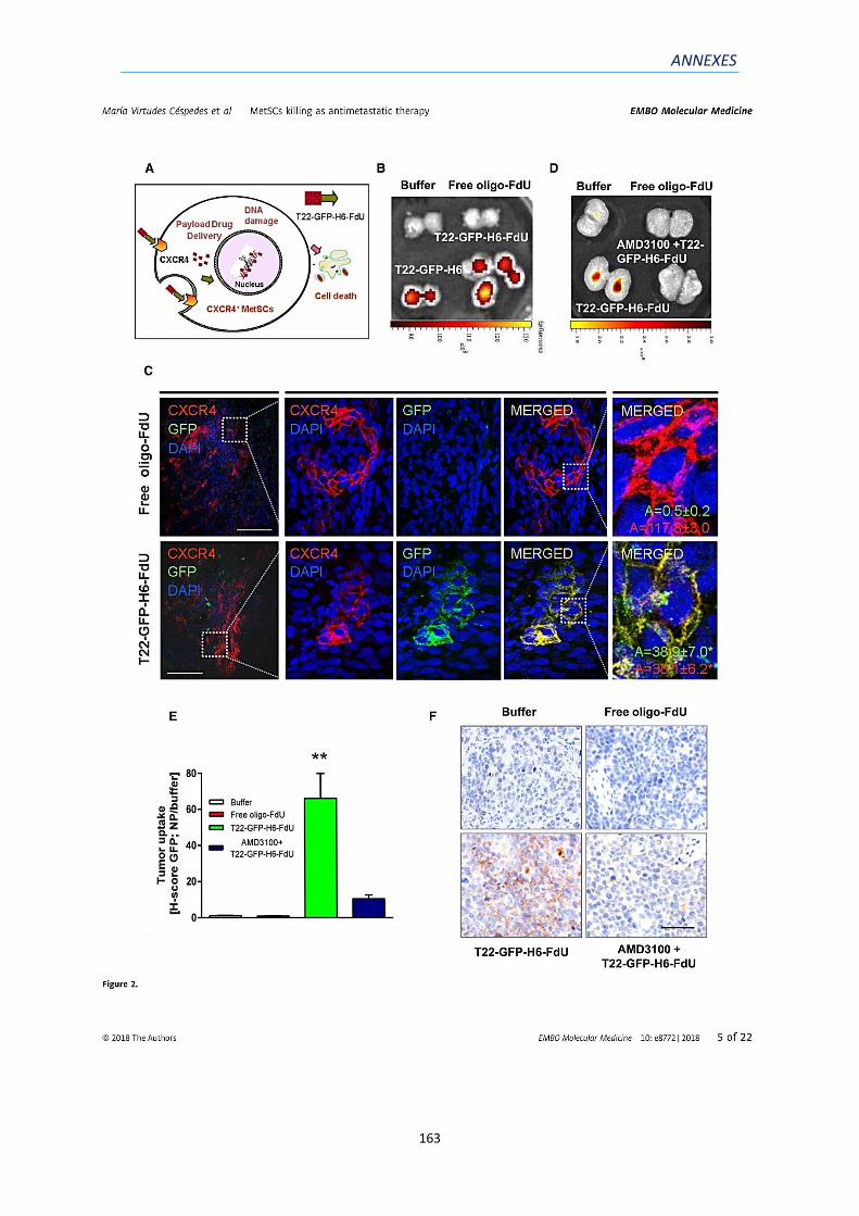
ANNEXES
163

ANNEXES
164

ANNEXES
165

ANNEXES
166

ANNEXES
167

ANNEXES
168

ANNEXES
169

ANNEXES
170

ANNEXES
171

ANNEXES
172

ANNEXES
173

ANNEXES
174

ANNEXES
175

ANNEXES
176

ANNEXES
177

ANNEXES
178

ANNEXES
179

ANNEXES
180

ANNEXES
181
2) ANNEX 2: ARTICLE 2

ANNEXES
182

ANNEXES
183

ANNEXES
184

ANNEXES
185
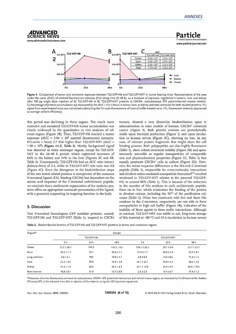
ANNEXES
186

ANNEXES
187

ANNEXES
188

ANNEXES
189

ANNEXES
190

ANNEXES
191
3) ANNEX 3: ARTICLE 3
Abstract
A functional 29 amino acid-segment of the helix α5 from the human BAX protein has
been engineered for production in recombinant bacteria as self-assembling, GFP-
containing fluorescent nanoparticles, which are targeted to the tumoral marker CXCR4.
These nanoparticles, of around 34 nm in diameter, show a moderate tumor
biodistribution and limited antitumoral effect when systemically administered to mouse
models of human CXCR4+ colorectal cancer (at 300 μg dose). However, if such BAX
nanoparticles are co-administered in cocktail with equivalent nanoparticulate versions
of BAK and PUMA proteins at the same total protein dose (300 μg), protein
biodistribution and stability in tumor is largely improved, as determined by fluorescence
profiles. This fact leads to a potent and faster destruction of tumor tissues when
compared to individual pro-apoptotic factors. The analysis and interpretation of the
boosted effect, from both the structural and functional sides, offers clues for the design
of more efficient nanomedicines and theragnostic agents in oncology based on precise
cocktails of human proteins. STATEMENT OF SIGNIFICANCE: Several human pro-
apoptotic peptides (namely BAK, BAX and PUMA) have been engineered as self-
assembling protein nanoparticles targeted to the tumoral marker CXCR4. The systemic
administration of the same final amounts of those materials as single drugs, or as
combinations of two or three of them, shows disparate intensities of antitumoral effects
in a mouse model of human colorectal cancer, which are boosted in the triple
combination on a non-additive basis. The superiority of the combined administration of
pro-apoptotic agents, acting at different levels of the apoptotic cascade, opens a

ANNEXES
192
plethora of possibilities for the development of effective and selective cancer therapies
based on the precise cocktailing of pro-apoptotic nanoparticulate agents.
Keywords: Cancer; Colorectal cancer; Drug cocktail; Drug delivery; Human proteins;
Nanomedicine; Nanoparticles; Pro-apoptotic factors; Pro-apoptotic peptide;
Recombinant protein; Targeted drug delivery.


