Embed Size (px)
Citation preview
DEPARTAMENTO DE FARMACOLOGÍA REGULACIÓN NITRÉRGICA DE LA FUNCIÓN MOTORA GASTROINTESTINAL EN LA ENDOTOXEMIA ELSA QUINTANA FERNÁNDEZ
UNIVERSITAT DE VALENCIA Servei de Publicacions
2004
Aquesta Tesi Doctoral va ser presentada a Valencia el día 31 de Maig de 2004 davant un tribunal format per:
- D. Esteban Morcillo Sánchez - D. Miguel Martí Cabrera - D. Julián Panes Díaz - D. Alberto Álvarez Barrientos - D. Miguel Bixquext
Va ser dirigida per: D. Juan V. Espulgues Mota Dª. Mª Dolores Barrachina Sancho ©Copyright: Servei de Publicacions Elsa Quintana Fernández Depòsit legal: I.S.B.N.:84-370-1444-1
Edita: Universitat de València Servei de Publicacions C/ Artes Gráficas, 13 bajo 46010 València Spain Telèfon: 963864115
1
“REGULACIÓN NITRÉRGICA DE LA
FUNCIÓN MOTORA GASTROINTESTINAL
EN LA ENDOTOXEMIA”
TESIS DOCTORAL
ELSA QUINTANA FERNÁNDEZ
Valencia, 2004
UNIVERSITAT DE VALÈNCIA FACULTAT DE FARMÀCIA
DEPARTAMENT DE FARMACOLOGIA
Resumen
2
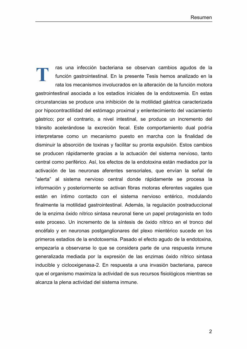
ras una infección bacteriana se observan cambios agudos de la
función gastrointestinal. En la presente Tesis hemos analizado en la
rata los mecanismos involucrados en la alteración de la función motora
gastrointestinal asociada a los estadios iniciales de la endotoxemia. En estas
circunstancias se produce una inhibición de la motilidad gástrica caracterizada
por hipocontractilidad del estómago proximal y enlentecimiento del vaciamiento
gástrico; por el contrario, a nivel intestinal, se produce un incremento del
tránsito acelerándose la excreción fecal. Este comportamiento dual podría
interpretarse como un mecanismo puesto en marcha con la finalidad de
disminuir la absorción de toxinas y facilitar su pronta expulsión. Estos cambios
se producen rápidamente gracias a la actuación del sistema nervioso, tanto
central como periférico. Así, los efectos de la endotoxina están mediados por la
activación de las neuronas aferentes sensoriales, que envían la señal de
“alerta” al sistema nervioso central donde rápidamente se procesa la
información y posteriormente se activan fibras motoras eferentes vagales que
están en íntimo contacto con el sistema nervioso entérico, modulando
finalmente la motilidad gastrointestinal. Además, la regulación postraduccional
de la enzima óxido nítrico sintasa neuronal tiene un papel protagonista en todo
este proceso. Un incremento de la síntesis de óxido nítrico en el tronco del
encéfalo y en neuronas postganglionares del plexo mientérico sucede en los
primeros estadios de la endotoxemia. Pasado el efecto agudo de la endotoxina,
empezaría a observarse lo que se considera parte de una respuesta inmune
generalizada mediada por la expresión de las enzimas óxido nítrico sintasa
inducible y ciclooxigenasa-2. En respuesta a una invasión bacteriana, parece
que el organismo maximiza la actividad de sus recursos fisiológicos mientras se
alcanza la plena actividad del sistema inmune.
T
3
D. JUAN VICENTE ESPLUGUES MOTA, Catedrático de Farmacología de la
Universitat de València y
Dña. MARÍA DOLORES BARRACHINA SANCHO, Profesora Titular del
Departamento de Farmacología de la Universitat de València
HACEN CONSTAR:
Que el trabajo titulado “Regulación nitrérgica de la función motora
gastrointestinal en la endotoxemia”, presentado por la Licenciada Elsa
Quintana Fernández para obtener el grado de Doctor, ha sido realizado en el
Departamento de Farmacología de esta Facultad bajo nuestra dirección.
Concluido el trabajo experimental y bibliográfico, autorizamos la
presentación de esta Tesis Doctoral para que sea juzgada por el Tribunal
correspondiente.
Valencia, Febrero de 2004
Fdo. Dr. Juan V. Esplugues Mota Fdo. Dra. M. D. Barrachina Sancho
UNIVERSITAT DE VALÈNCIA FACULTAT DE FARMÀCIA
DEPARTAMENT DE FARMACOLOGIA

4
A mi familia

5
AGRADECIMIENTOS
Quiero expresar mi más sincero agradecimiento:
En primer lugar al que ha sido el fundador de este Departamento, el
Profesor Juan Esplugues, gracias por sus entrañables consejos.
A mis directores, el Profesor Juan Vicente Esplugues, por su apoyo
incondicional y preocupación por mi formación científica, y la Profesora María
Dolores Barrachina, por su constante dedicación, entusiasmo, respaldo y
amistad; a ella debo mi espíritu científico. A ambos, gracias por haber confiado
en mí.
A Sara, por su enorme contribución al desarrollo de esta tesis, su
rectitud en el trabajo ha sido un modelo para mí. A Mª Ángeles, por su tutela en
el comienzo de mi carrera investigadora. A las dos, gracias por su inestimable
ayuda diaria, por su amistad.
A mis compañeros de laboratorio con los que he compartido tantos
buenos momentos. A Eugenia, Ángeles y Carlos, sin ellos todo habría sido
mucho más difícil, gracias por su sincera amistad. A los más recientes,
Alejandra, Nadezda, Reme, Sales, Cristina Núñez, Víctor, Marta, Susana,
Jesús, Sol, Ana, Kenneth, Juan, Cristina Amézcua y por supuesto, a la eficiente
Paqui. Al resto de compañeros del Departamento de Farmacología,
especialmente a Inés, Amparo, Regina, Miguel, Dora y Maruja. Muchas gracias
a todos por su ayuda, apoyo, ánimo y confianza que han hecho el día a día tan
agradable.
A Raquel, Elvira, Pilar y Alberto, por haber hecho tan grata y especial la
estancia en su laboratorio.
A Noelia, por su apoyo, paciencia y comprensión durante estos años de
convivencia.
A todos, gracias por haber contribuido a la satisfacción con la que he
vivido esta aventura.
6
ABREVIATURAS
2-DG ........ 2-deoxi-D-glucosa 7-NI ........ 7-nitroindazol Ach ........ acetilcolina ATP ........ adenosina trifosfato BHE ........ barrera hematoencefálica canal CaK+ ........ canal de potasio dependiente de calcio CDV ........ complejo dorsal vagal CGRP ........ péptido relacionado con el gen de la calcitonina COX ........ ciclooxigenasa DAF-FM ........ 4-amino-5-metilamino-2’,7’-difluorofluoresceína DETA-NO ........ (Z)-1-[2-(2-aminoetil)-N-(2-amonioetil)amino]diazon-1-io-1,2-diolato GCs ........ guanilato ciclasa soluble GMPc ........ guanosina monofosfato cíclica IL ........ interleucina L-NAME ........ Nω-nitro-L-arginina metil éster L-NIL ........ N6-(1-iminoetil)-L-lisina L-NOARG ........ Nω-nitro-L-arginina LPS ........ lipopolisacárido NANC ........ no-adrenérgico no-colinérgico NDV ........ núcleo dorsal del vago NO ........ óxido nítrico NOS ........ óxido nítrico sintasa NOSe ........ óxido nítrico sintasa endotelial NOSi ........ óxido nítrico sintasa inducible NOSn ........ óxido nítrico sintasa neuronal NTS ........ núcleo del tracto solitario ODQ ........ 1H-[1,2,4]oxadiazolo[4,3-a]quinoxalin-1-ona PGs ........ prostaglandinas PIG ........ presión intragástrica SNC ........ sistema nervioso central SNE ........ sistema nervioso entérico SP ........ sustancia P TNF-α ........ factor de necrosis tumoral-α TRIM ........ 1-[2-(trifluorometil)fenil]imidazol TTX ........ tetrodotoxina VIP ........ péptido intestinal vasoactivo

7
ÍNDICE
Índice
8
INTRODUCCIÓN 11
I. Fase aguda de la infección bacteriana 12
I.1. Señalización al sistema nervioso central 13
I.1.1. Vía humoral 14
I.1.2. Vía nerviosa 14
I.2. Alteración de la función gastrointestinal 15
II. Motilidad gastrointestinal 17
II.1. Regulación nerviosa 17
II.1.1. Inervación intrínseca 17
II.1.2. Inervación extrínseca 20
II.2. Fisiología del vaciamiento gástrico 22
II.3. Fisiología del tránsito intestinal y excreción fecal 25
III. Óxido nítrico 27
III.1. Síntesis 27
III.2. Mecanismo de acción 29
III.3. Óxido nítrico en el sistema nervioso 31
III.3.1. Sistema nervioso central 32
III.3.2. Sistema nervioso periférico 33
III.4. Regulación nitrérgica de la motilidad gastrointestinal 34
OBJETIVO 37

Índice
9
ARTÍCULOS DE INVESTIGACIÓN 38
I. Efectos de la endotoxina sobre la motilidad gástrica 39
Artículo 1: “A cerebral nitrergic pathway modulates endotoxin-induced changes in gastric motility”. British Journal of Pharmacology (2001) 134: 325-332. 42
Artículo 2: “Transcriptional up-regulation of nNOS in the dorsal vagal complex during low endotoxemia”. Neurogastroenterology and motility (2004, en revisión). 51
Artículo 3: “Synthesis of nitric oxide in post-ganglionic myenteric neurons during endotoxemia: implications for gastric motor function”. The FASEB Journal express (2004) 18:531-533. DOI:10.1096/fj.03-0596fje. 70
Artículo 4: “Endotoxin inhibits gastric emptying in rats via a capsaicin-sensitive afferent pathway”. Naunyn-Schmiede-berg’s Archives of Pharmacology (2001) 363: 276-280. 100
Artículo 5: “Downregulation of nNOS and synthesis of PGs associated with endotoxin-induced delay in gastric emptying”. American Journal of Physiology (2002) 283: G1360-G1367.
106
II. Efectos de la endotoxina sobre la motilidad colónica 115
Artículo 6: “Endotoxin stimulates fecal pellet output in rats through a neural mechanism”. Naunyn-Schmiedeberg’s Archives of Pharmacology (2003) 367(1): 51-5. 116
III. Análisis global 122
Artículo 7: “Nitrergic modulation of gastrointestinal function during early endotoxemia”. Current Pharmaceutical Design (2004, en revisión). 122
Índice
10
RESUMEN DE RESULTADOS Y DISCUSIÓN GENERAL 156
I. Efectos de la endotoxina sobre la motilidad gástrica 157
II. Efectos de la endotoxina sobre la motilidad colónica 169
CONCLUSIONES 172
BIBLIOGRAFÍA 174

Introducción
11
INTRODUCCIÓN
Introducción
12
I. FASE AGUDA DE LA INFECCIÓN BACTERIANA
Nuestro organismo está continuamente expuesto a agentes patógenos y
por ello ha desarrollado mecanismos innatos que le permiten eliminar el agente
invasor de una manera rápida y eficaz. Tras una infección, además de coordinar
una respuesta inmunitaria periférica, el organismo reacciona señalizando al
sistema nervioso central (SNC) y desencadenando una serie de eventos en
cascada, principalmente cambios neuroendocrinos, metabólicos y del
comportamiento, que se engloban bajo el nombre de “reacción de fase aguda”.
Los síntomas típicos de este estadio incluyen fiebre, confusión mental,
hipotensión transitoria, pérdida del apetito y alteración de la función
gastrointestinal caracterizada por vómito y diarrea. Esta fase constituye la
primera reacción de defensa del organismo frente a una infección. Se trata de
una respuesta inespecífica, rápida, intensa y eficaz que consigue, en la mayoría
de los casos de infecciones leves, restaurar la homeostasis sin que se llegue a
observar la sintomatología típica de la inflamación.
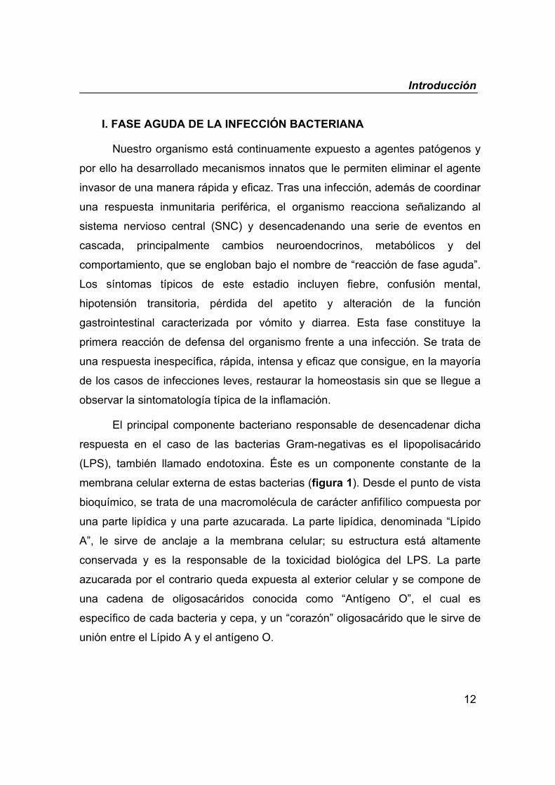
El principal componente bacteriano responsable de desencadenar dicha
respuesta en el caso de las bacterias Gram-negativas es el lipopolisacárido
(LPS), también llamado endotoxina. Éste es un componente constante de la
membrana celular externa de estas bacterias (figura 1). Desde el punto de vista
bioquímico, se trata de una macromolécula de carácter anfifílico compuesta por
una parte lipídica y una parte azucarada. La parte lipídica, denominada “Lípido
A”, le sirve de anclaje a la membrana celular; su estructura está altamente
conservada y es la responsable de la toxicidad biológica del LPS. La parte
azucarada por el contrario queda expuesta al exterior celular y se compone de
una cadena de oligosacáridos conocida como “Antígeno O”, el cual es
específico de cada bacteria y cepa, y un “corazón” oligosacárido que le sirve de
unión entre el Lípido A y el antígeno O.
Introducción
13
Figura 1. Lipopolisacárido de las bacterias Gram-negativas. Arquitectura del
lipopolisacárido de Escherichia coli (a), junto con una microfotografía electrónica de la misma
(b) (Beutler & Rietschel, 2003) y una representación esquemática de la localización del
lipopolisacárido en la pared celular de la bacteria (c). Gn, D-glucosamina; H, L-glicero-D-mano-
heptosa; C, ácido 2-ceto-3-deoxi-octulosónico; P, fosfato.
I.1. Señalización al sistema nervioso central
El mecanismo por el cual el LPS en la periferia señaliza al SNC para
desencadenar la “reacción de fase aguda” ha sido tema de controversia en la
última década (Blatteis et al., 2004; Romanovsky, 2004). Los mensajeros
propuestos encargados de informar al cerebro de la presencia periférica del LPS
son determinadas citocinas liberadas a partir de macrófagos activados,
principalmente las inlerleucinas 1 y 6 (IL-1, IL-6) y el factor de necrosis tumoral
α (TNF-α). Este hecho está basado en las siguientes evidencias: a) estas
LPS(endotoxina)
membranaexterna
peptidoglicanomembrana
interna
Lípido AcorazónAntígeno On
Gn
P
P
P
PH C
CH H
C
P
P
P
P
PH C
CH H
C
P
NH3+
polisacárido lípidoa
b c
Gn14
14
1412
14
14
Introducción
14
citocinas se detectan en sangre durante los 30-90 minutos tras la exposición al
LPS (Givalois et al., 1994; Jansky et al., 1995) y b) la administración exógena de
dichas citocinas mimetiza la mayoría de los efectos inducidos por el LPS en la
fase aguda (Rothwell & Luheshi, 1994; Schobitz et al., 1994).
I.1.1. Vía humoral
Clásicamente se había aceptado que las citocinas secretadas en
respuesta al LPS acceden al SNC vía sanguínea; sin embargo, se hace difícil el
paso a través de la barrera hematoencefálica (BHE) debido a su gran tamaño
molecular y carácter hidrofílico. Se propusieron distintas teorías de
incorporación al SNC: a) acceso directo por difusión simple a través de
estructuras centrales carentes de BHE, como los órganos circunventriculares; b)
acceso mediante sistemas de transporte específicos o c) unión a receptores
específicos presentes en las células endoteliales de los vasos cerebrales y
posterior liberación de otros mediadores en el parénquima cerebral. Existen
evidencias de que todas estas situaciones se dan; sin embargo, hasta la fecha
no se ha demostrado de forma directa que esta vía participe en los efectos
mediados por el SNC, inducidos por el LPS.
I.1.2. Vía nerviosa
En 1987, se especuló sobre la posible participación de nervios periféricos
en la fiebre inducida por el LPS (Morimoto et al., 1987). La idea de la actuación
de las neuronas aferentes en la señalización central de mediadores periféricos
no era nueva en neurobiología ya que se había demostrado que estas fibras
nerviosas mediaban los efectos de hormonas y péptidos periféricos sobre la
función cerebral, incluida la adrenalina (Williams & McGaugh, 1993) o la
colecistocinina (Anika et al., 1977). Los primeros trabajos que demostraron de
Introducción
15
una manera clara que el nervio vago estaba involucrado en los efectos centrales
del LPS surgieron en 1994 (Bluthe et al., 1994; Watkins et al., 1994) y desde
entonces, el número de publicaciones entorno a este tema incrementó
considerablemente. Durante este tiempo los estudios se han centrado
principalmente en la fiebre inducida por el LPS, utilizando como herramientas
experimentales el bloqueo químico (aplicación local o sistémica de la
capsaicina) o quirúrgico (vagotomía) de la transmisión vagal, y el estudio de la
activación nerviosa mediante la detección del gen de expresión temprana c-fos.
Actualmente, existe una elevada evidencia científica que apoya la teoría de la
vía nerviosa en la que las neuronas aferentes vagales median el comienzo de
los efectos centrales inducidos por el LPS (Blatteis et al., 2000; Maier et al.,
1998; Romanovsky, 2000).
Sin embargo, todavía existe controversia sobre si la activación de las
fibras aferentes vagales está mediada por las citocinas secretadas o si es una
acción directa de la endotoxina (Blatteis et al., 2000). Las citocinas no existen
almacenadas en los macrófagos sino que requieren expresión de novo, lo cual
necesita cierto tiempo. Existe una clara discrepancia temporal entre la primera
detección de citocinas en sangre (30-90 min) (Givalois et al., 1994; Jansky et al.,
1995) y la aparición de algunos de los efectos mediados por el SNC como por
ejemplo la fiebre (15 min) (Li et al., 1999). En este sentido, se ha propuesto que
citocinas como el TNF-α no juegan un papel en la génesis sino en el
mantenimiento de la fiebre (Roth et al., 1998).
I.2. Alteración de la función gastrointestinal
En la fase aguda de una infección bacteriana se observan cambios en la
función gastrointestinal. Por ejemplo, una endotoxemia moderada cursa en la
clínica con una hiposecreción ácida gástrica (Hurley, 1995). Este hecho ha sido
reproducido ampliamente en estudios experimentales. La administración de
Introducción
16
dosis bajas de LPS produce una inhibición rápida (30 minutos) de la secreción
ácida gástrica en la rata (Barrachina et al., 1995b; Calatayud et al., 1999;
Esplugues et al., 1996; Garcia-Zaragoza et al., 2000; Martinez-Cuesta et al.,
1994). Por otra parte se ha visto que la endotoxina tiene un efecto dual sobre la
integridad mucosa dependiendo de la dosis administrada. Durante el shock
séptico se desarrollan importantes lesiones gástricas. Sin embargo,
sorprendentemente, la administración de dosis bajas de endotoxina protege la
mucosa gástrica frente a la agresión inducida por distintos estímulos
ulcerogénicos como puede ser el estrés, el tratamiento con antiinflamatorios no
esteroideos o la administración de etanol (Barrachina et al., 1995a; Calatayud et
al., 2003; Tsuji et al., 1993). Recientemente se ha sugerido que la prevención
del daño se debe a un incremento del flujo sanguíneo de la microcirculación
gástrica ya que se ha demostrado que el LPS previene la caída del flujo
sanguíneo inducido por los antiinflamatorios no esteroideos (Calatayud et al.,
2003).
Nuestro grupo de investigación ha estudiado los mecanismos involucrados
en este proceso. Así, sabemos que los cambios agudos de la función
gastrointestinal inducidos por la endotoxina son consecuencia de un mecanismo
rápido e independiente de la síntesis proteica característica de un estadio más
tardío. Además, hemos visto que está regulado por los sistemas nerviosos
autónomo y central y parece que la síntesis constitutiva de óxido nítrico (NO)
juega un papel determinante.
De entre las distintas funciones gastrointestinales, la motilidad se altera de
forma significativa en la fase aguda de la infección bacteriana observándose una
inhibición del vaciamiento gástrico y un incremento del tránsito intestinal. Sin
embargo, muy poco se sabe acerca del mecanismo por el que sucede este
proceso.
Introducción
17
II. MOTILIDAD GASTROINTESTINAL
La función motora gastrointestinal requiere del funcionamiento coordinado e
integrado de las distintas capas musculares según la zona y el estado de cada
momento. Esta función se ve modulada por diversas condiciones como son
estado de ayuno, cualidades y componentes de la dieta, agentes nocivos,
factores sensoriales y emocionales, etc. Una compleja red nerviosa distribuida a
lo largo de todo el tracto digestivo (inervación intrínseca) y comunicada con el
SNC (inervación extrínseca), desempeña un papel esencial en la coordinación
motora.
II.1. Regulación nerviosa
II.1.1 Inervación intrínseca
En todas las culturas antiguas y modernas se ha tenido la conciencia, al
menos popular, de que nuestro estómago es capaz de experimentar emociones.
Tras décadas de trabajo, los científicos están en condiciones de afirmar que en
el tracto gastrointestinal se aloja un “segundo cerebro” muy similar al que
tenemos en la cabeza. El tubo digestivo está literalmente tapizado por más de
100 millones de células nerviosas (equivalente al contenido de la médula
espinal) que constituyen el sistema nervioso entérico (SNE). Aunque está
dotado de total autonomía, también mantiene estrechas conexiones con el SNC.
Los cuerpos celulares de las neuronas del SNE se encuentran aglutinados
en estructuras ganglionares que a su vez se interconectan entre sí formando
dos grandes plexos nerviosos: el plexo mientérico o plexo de Auerbach y el
plexo submucoso o plexo de Meissner (figura 2). El plexo mientérico, ubicado
entre las capas musculares longitudinal y circular, se extiende desde el tercio
medio del esófago hasta el canal anal proporcionando inervación motora a
ambas capas musculares e inervación secreto-motora a las células de la
Introducción
18
mucosa. El plexo submucoso, ubicado por debajo de la muscularis mucosae,
presenta su máximo desarrollo en el intestino delgado donde desempeña un
importante papel en el control de la secreción. Ambos plexos mantienen
estrechas conexiones entre sí.
Al igual que en el SNC, el SNE tiene tres clases de neuronas de acuerdo a
su funcionalidad:
a) Neuronas sensoriales, cuyas áreas receptivas se encuentran en la
musculatura de la pared y en la mucosa. Las terminaciones libres de estas
neuronas tienen receptores capaces de responder a cambios de energía
química, térmica o mecánica, transformando estos cambios en señales
eléctricas. Son esenciales en el control de la digestión.
b) Interneuronas, que forman múltiples sinapsis entre sí dando lugar a
circuitos integrados “lógicos”. Estos circuitos son capaces de descifrar y
procesar la información proveniente de las neuronas sensoriales y generar
programas de respuestas reflejas motoras coherentes con la funcionalidad
global del órgano. Constituyen la base de la propagación de las ondas
peristálticas.
c) Neuronas motoras, que son el brazo efector del sistema que transmite
las señales, estimulantes o inhibidoras, a los distintos efectores: vaso, músculo
y epitelio.
La lista de neurotransmisores presentes en el SNE es larguísima y en
general son prácticamente los mismos que se encuentran presentes en el SNC.
Su naturaleza es muy diversa: aminas, aminoácidos, purinas, gases o péptidos.
Los más importantes en la regulación de la función motora son acetilcolina (Ach)
y sustancia P (SP), entre los neurotransmisores excitadores, y VIP, ATP y NO
(Wood, 1994), entre los inhibidores.
Introducción
19
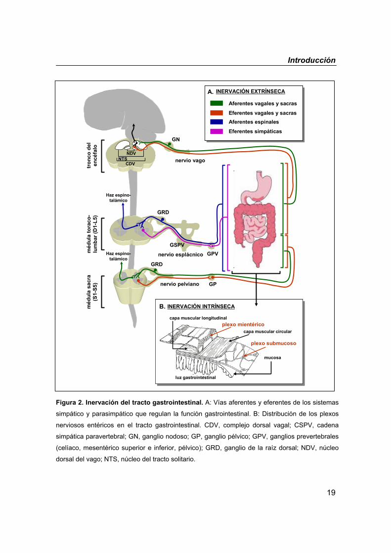
Figura 2. Inervación del tracto gastrointestinal. A: Vías aferentes y eferentes de los sistemas
simpático y parasimpático que regulan la función gastrointestinal. B: Distribución de los plexos
nerviosos entéricos en el tracto gastrointestinal. CDV, complejo dorsal vagal; CSPV, cadena
simpática paravertebral; GN, ganglio nodoso; GP, ganglio pélvico; GPV, ganglios prevertebrales
(celíaco, mesentérico superior e inferior, pélvico); GRD, ganglio de la raíz dorsal; NDV, núcleo
dorsal del vago; NTS, núcleo del tracto solitario.
plexo mientéricocapa muscular circular
plexo submucoso
mucosa
luz gastrointestinal
capa muscular longitudinal
INERVACIÓN INTRÍNSECAB.
plexo mientéricocapa muscular circular
plexo submucoso
mucosa
luz gastrointestinal
capa muscular longitudinal
INERVACIÓN INTRÍNSECAB.
tron
co d
el
encé
falo
méd
ula
tora
co-
lum
bar (
D1-
L5)
méd
ula
sacr
a (S
1-S5
)
Haz espino-talámico
nervio pelviano
GRD
GP
Haz espino-talámico
GRD
GPVGSPV
nervio esplácnico
nervio vago
GN
NDVNTS
CDV
Aferentes vagales y sacras
Eferentes vagales y sacrasAferentes espinales
Eferentes simpáticas
INERVACIÓN EXTRÍNSECAA.
Aferentes vagales y sacras
Eferentes vagales y sacrasAferentes espinales
Eferentes simpáticas
INERVACIÓN EXTRÍNSECAA.
Introducción
20
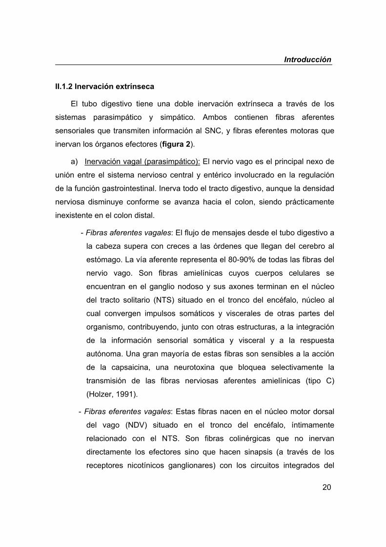
II.1.2 Inervación extrínseca
El tubo digestivo tiene una doble inervación extrínseca a través de los
sistemas parasimpático y simpático. Ambos contienen fibras aferentes
sensoriales que transmiten información al SNC, y fibras eferentes motoras que
inervan los órganos efectores (figura 2).
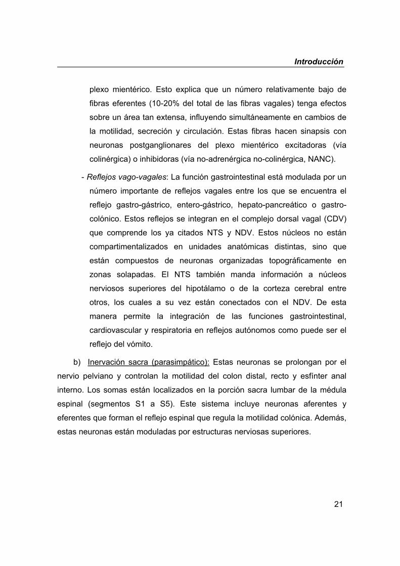
a) Inervación vagal (parasimpático): El nervio vago es el principal nexo de
unión entre el sistema nervioso central y entérico involucrado en la regulación
de la función gastrointestinal. Inerva todo el tracto digestivo, aunque la densidad
nerviosa disminuye conforme se avanza hacia el colon, siendo prácticamente
inexistente en el colon distal.
- Fibras aferentes vagales: El flujo de mensajes desde el tubo digestivo a
la cabeza supera con creces a las órdenes que llegan del cerebro al
estómago. La vía aferente representa el 80-90% de todas las fibras del
nervio vago. Son fibras amielínicas cuyos cuerpos celulares se
encuentran en el ganglio nodoso y sus axones terminan en el núcleo
del tracto solitario (NTS) situado en el tronco del encéfalo, núcleo al
cual convergen impulsos somáticos y viscerales de otras partes del
organismo, contribuyendo, junto con otras estructuras, a la integración
de la información sensorial somática y visceral y a la respuesta
autónoma. Una gran mayoría de estas fibras son sensibles a la acción
de la capsaicina, una neurotoxina que bloquea selectivamente la
transmisión de las fibras nerviosas aferentes amielínicas (tipo C)
(Holzer, 1991).
- Fibras eferentes vagales: Estas fibras nacen en el núcleo motor dorsal
del vago (NDV) situado en el tronco del encéfalo, íntimamente
relacionado con el NTS. Son fibras colinérgicas que no inervan
directamente los efectores sino que hacen sinapsis (a través de los
receptores nicotínicos ganglionares) con los circuitos integrados del
Introducción
21
plexo mientérico. Esto explica que un número relativamente bajo de
fibras eferentes (10-20% del total de las fibras vagales) tenga efectos
sobre un área tan extensa, influyendo simultáneamente en cambios de
la motilidad, secreción y circulación. Estas fibras hacen sinapsis con
neuronas postganglionares del plexo mientérico excitadoras (vía
colinérgica) o inhibidoras (vía no-adrenérgica no-colinérgica, NANC).
- Reflejos vago-vagales: La función gastrointestinal está modulada por un
número importante de reflejos vagales entre los que se encuentra el
reflejo gastro-gástrico, entero-gástrico, hepato-pancreático o gastro-
colónico. Estos reflejos se integran en el complejo dorsal vagal (CDV)
que comprende los ya citados NTS y NDV. Estos núcleos no están
compartimentalizados en unidades anatómicas distintas, sino que
están compuestos de neuronas organizadas topográficamente en
zonas solapadas. El NTS también manda información a núcleos
nerviosos superiores del hipotálamo o de la corteza cerebral entre
otros, los cuales a su vez están conectados con el NDV. De esta
manera permite la integración de las funciones gastrointestinal,
cardiovascular y respiratoria en reflejos autónomos como puede ser el
reflejo del vómito.
b) Inervación sacra (parasimpático): Estas neuronas se prolongan por el
nervio pelviano y controlan la motilidad del colon distal, recto y esfínter anal
interno. Los somas están localizados en la porción sacra lumbar de la médula
espinal (segmentos S1 a S5). Este sistema incluye neuronas aferentes y
eferentes que forman el reflejo espinal que regula la motilidad colónica. Además,
estas neuronas están moduladas por estructuras nerviosas superiores.
Introducción
22
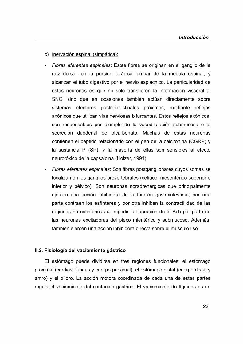
c) Inervación espinal (simpática):
- Fibras aferentes espinales: Estas fibras se originan en el ganglio de la
raíz dorsal, en la porción torácica lumbar de la médula espinal, y
alcanzan el tubo digestivo por el nervio esplácnico. La particularidad de
estas neuronas es que no sólo transfieren la información visceral al
SNC, sino que en ocasiones también actúan directamente sobre
sistemas efectores gastrointestinales próximos, mediante reflejos
axónicos que utilizan vías nerviosas bifurcantes. Estos reflejos axónicos,
son responsables por ejemplo de la vasodilatación submucosa o la
secreción duodenal de bicarbonato. Muchas de estas neuronas
contienen el péptido relacionado con el gen de la calcitonina (CGRP) y
la sustancia P (SP), y la mayoría de ellas son sensibles al efecto
neurotóxico de la capsaicina (Holzer, 1991).
- Fibras eferentes espinales: Son fibras postganglionares cuyos somas se
localizan en los ganglios prevertebrales (celíaco, mesentérico superior e
inferior y pélvico). Son neuronas noradrenérgicas que principalmente
ejercen una acción inhibidora de la función gastrointestinal; por una
parte contraen los esfínteres y por otra inhiben la contractilidad de las
regiones no esfintéricas al impedir la liberación de la Ach por parte de
las neuronas excitadoras del plexo mientérico y submucoso. Además,
también ejercen una acción inhibidora directa sobre el músculo liso.
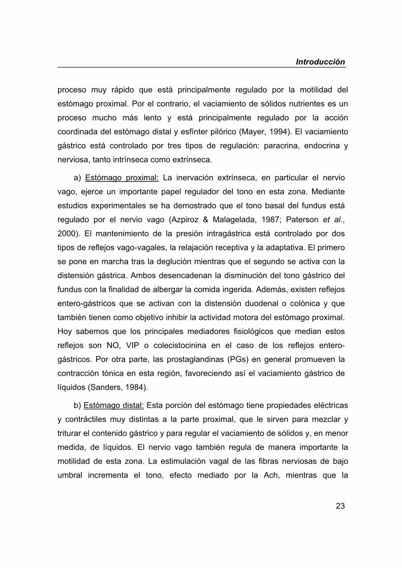
II.2. Fisiología del vaciamiento gástrico
El estómago puede dividirse en tres regiones funcionales: el estómago
proximal (cardias, fundus y cuerpo proximal), el estómago distal (cuerpo distal y
antro) y el píloro. La acción motora coordinada de cada una de estas partes
regula el vaciamiento del contenido gástrico. El vaciamiento de líquidos es un
Introducción
23
proceso muy rápido que está principalmente regulado por la motilidad del
estómago proximal. Por el contrario, el vaciamiento de sólidos nutrientes es un
proceso mucho más lento y está principalmente regulado por la acción
coordinada del estómago distal y esfínter pilórico (Mayer, 1994). El vaciamiento
gástrico está controlado por tres tipos de regulación: paracrina, endocrina y
nerviosa, tanto intrínseca como extrínseca.
a) Estómago proximal: La inervación extrínseca, en particular el nervio
vago, ejerce un importante papel regulador del tono en esta zona. Mediante
estudios experimentales se ha demostrado que el tono basal del fundus está
regulado por el nervio vago (Azpiroz & Malagelada, 1987; Paterson et al.,
2000). El mantenimiento de la presión intragástrica está controlado por dos
tipos de reflejos vago-vagales, la relajación receptiva y la adaptativa. El primero
se pone en marcha tras la deglución mientras que el segundo se activa con la
distensión gástrica. Ambos desencadenan la disminución del tono gástrico del
fundus con la finalidad de albergar la comida ingerida. Además, existen reflejos
entero-gástricos que se activan con la distensión duodenal o colónica y que
también tienen como objetivo inhibir la actividad motora del estómago proximal.
Hoy sabemos que los principales mediadores fisiológicos que median estos
reflejos son NO, VIP o colecistocinina en el caso de los reflejos entero-
gástricos. Por otra parte, las prostaglandinas (PGs) en general promueven la
contracción tónica en esta región, favoreciendo así el vaciamiento gástrico de
líquidos (Sanders, 1984).
b) Estómago distal: Esta porción del estómago tiene propiedades eléctricas
y contráctiles muy distintas a la parte proximal, que le sirven para mezclar y
triturar el contenido gástrico y para regular el vaciamiento de sólidos y, en menor
medida, de líquidos. El nervio vago también regula de manera importante la
motilidad de esta zona. La estimulación vagal de las fibras nerviosas de bajo
umbral incrementa el tono, efecto mediado por la Ach, mientras que la
Introducción
24
estimulación de fibras de elevado umbral inhibe el tono, efecto mediado
principalmente por el NO y el VIP (Dickens et al., 2000). Existe un reflejo vago-
vagal mediante el cual la distensión del fundus provoca la contracción del antro.
Este reflejo parece que es el que inicia el proceso de la digestión, al promover la
fase de mezcla y trituración. A diferencia del estómago proximal, a nivel distal el
efecto de las PGs es inhibidor ya que disminuye la amplitud de las
contracciones y la capacidad del músculo liso de responder a estímulos
excitadores. Como consecuencia, el vaciamiento gástrico de sólidos parece
estar retrasado por las PGs endógenas (Sanders, 1984).
c) Esfínter pilórico: El píloro es el que permite la evacuación del contenido
gástrico, regulando el volumen expulsado e impidiendo además la regurgitación
del contenido duodenal. Posee unas propiedades musculares y nerviosas
únicas que le distinguen de toda la zona de su alrededor. Así, se trata de un
músculo grueso con elevada proporción de tejido conectivo. La densidad
nerviosa es de 3 a 5 veces superior a la presente en el antro adyacente y la
cantidad de neuronas extrínsecas aferentes que proyectan al NTS también es
superior a la del duodeno (Daniel et al., 1989). Comparativamente con el antro y
el duodeno, existe un elevado número de neuronas que contienen VIP, NO, SP,
encefalinas, neuropéptido Y o galanina, sugiriendo un papel regulador en el tono
del esfínter para estas sustancias. La actividad pilórica motora se ha visto
modulada por fibras vagales, excitadoras e inhibidoras, e inervación esplácnica
que principalmente induce la contracción pilórica. Además, está regulada por
vías nerviosas intrínsecas ascendentes (la contracción duodenal produce
contracción pilórica) y descendentes (la despolarización del músculo antral
relaja el esfínter), (Yuan et al., 2001). La regulación nitrérgica es la principal
responsable de la relajación del esfínter (Ishiguchi et al., 2000). Además, ATP,
VIP, galanina, prostaglandina E1 y serotonina son sustancias que relajan el
píloro; mientras que colecistocinina, secretina o histamina contraen el esfínter.
Introducción
25
II.3. Fisiología del tránsito intestinal y excreción fecal
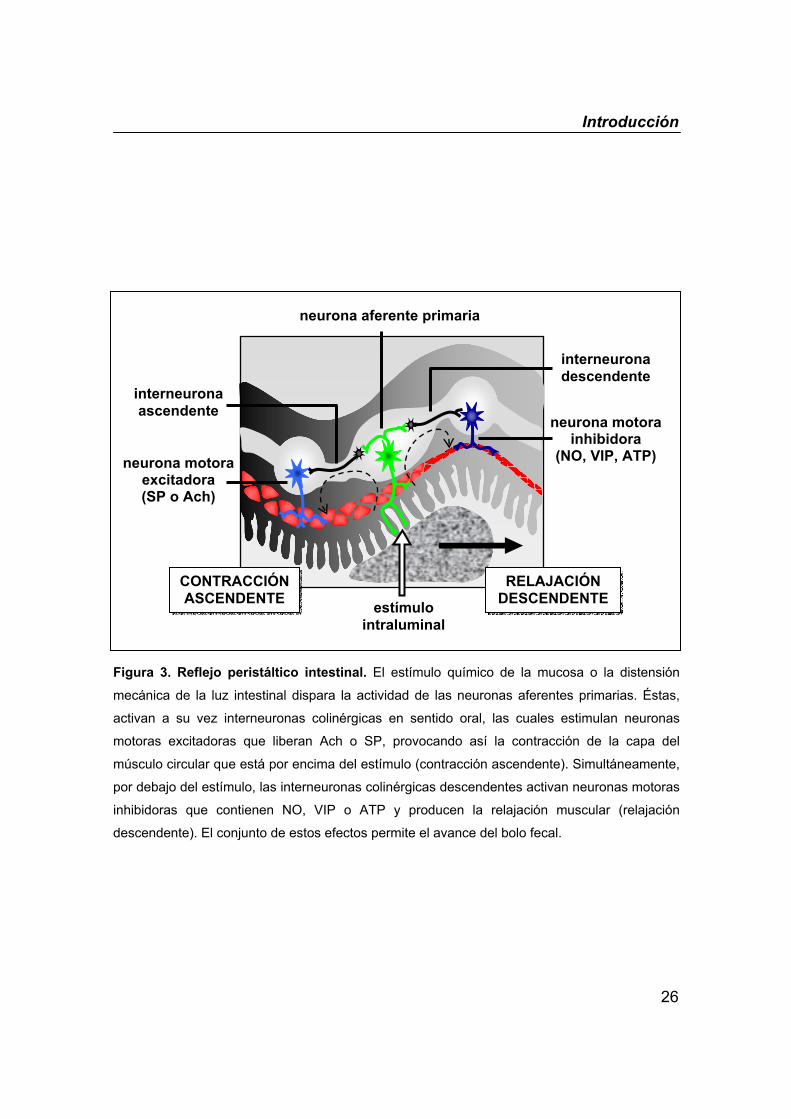
El peristaltismo es el principal mecanismo responsable del avance del
bolo fecal a lo largo del intestino. El reflejo peristáltico básico es el resultado de
una serie de reflejos locales cada uno de los cuales consiste en una primera
contracción del músculo intestinal por encima de un estímulo intraluminal, que
suele ser la distensión mecánica ejercida por el bolo, seguida de la relajación
del músculo por debajo del estímulo (figura 3). La suma de estos efectos
consigue la propulsión del contenido en dirección anterógrada y conforme el
bolo avanza desencadena sucesivos reflejos. Este proceso está regulado
principalmente por el SNE. La liberación controlada de los neurotransmisores
excitadores e inhibidores determinará la velocidad de la propulsión del contenido
intestinal. Estudios experimentales demuestran que el bloqueo de la síntesis de
los neurotransmisores excitadores, Ach o SP, así como de los inhibidores, VIP o
NO, retrasa el avance del bolo fecal (Foxx-Orenstein & Grider, 1996).
La excreción fecal es un proceso complejo regulado por mecanismos
voluntarios e involuntarios. El incremento de la motilidad en el colon
descendente y recto promueve el avance de las heces hacia el ano, donde la
relajación de los esfínteres anales externo e interno facilita su expulsión. Este
proceso está regulado por el reflejo entérico inhibidor recto-anal, mediante el
cual la distensión del recto produce una marcada disminución, volumen-
dependiente, en la presión del esfínter anal interno. Las fibras sensoriales
intrínsecas son las responsables de la activación del reflejo mientras que VIP y
NO son los principales mediadores de la relajación (Jones et al., 2003; Sangwan
& Solla, 1998).
Introducción
26
Figura 3. Reflejo peristáltico intestinal. El estímulo químico de la mucosa o la distensión
mecánica de la luz intestinal dispara la actividad de las neuronas aferentes primarias. Éstas,
activan a su vez interneuronas colinérgicas en sentido oral, las cuales estimulan neuronas
motoras excitadoras que liberan Ach o SP, provocando así la contracción de la capa del
músculo circular que está por encima del estímulo (contracción ascendente). Simultáneamente,
por debajo del estímulo, las interneuronas colinérgicas descendentes activan neuronas motoras
inhibidoras que contienen NO, VIP o ATP y producen la relajación muscular (relajación
descendente). El conjunto de estos efectos permite el avance del bolo fecal.
interneurona ascendente
interneurona descendente
neurona motora inhibidora
(NO, VIP, ATP)
neurona aferente primaria
neurona motora excitadora (SP o Ach)
CONTRACCIÓN ASCENDENTE
RELAJACIÓN DESCENDENTE estímulo
intraluminal
Introducción
27
III. ÓXIDO NÍTRICO
El NO fue identificado en 1987 como un mediador endógeno liberado por
las células endoteliales que inducía la relajación vascular (Palmer et al., 1987), e
inhibía la agregación plaquetaria (Radomski et al., 1987) y la adhesión de
neutrófilos al endotelio vascular (Kubes et al., 1991). Desde entonces una gran
cantidad de estudios se han centrado en caracterizar esta particular molécula y
actualmente sabemos que su papel fisiológico no sólo se circunscribe al sistema
cardiovascular sino que está ampliamente distribuido y que posee una gran
diversidad de funciones. En el SNC, actúa como un neurotransmisor implicado
en procesos como el aprendizaje y la memoria, el desarrollo neuronal o la
regulación neuroendocrina. En el sistema nervioso periférico, se le considera el
mediador de la transmisión NANC, responsable de la relajación del músculo liso
de los sistemas gastrointestinal, respiratorio y genitourinario. Además, el NO
juega un importante papel en la respuesta inmunitaria no específica ya que su
síntesis a partir de las células inflamatorias forma parte de los mecanismos de
defensa inespecífica contra microorganismos y células tumorales (Moncada et
al., 1991).
III.1. Síntesis
El NO es una molécula gaseosa que se sintetiza a partir del aminoácido
semiesencial L-arginina en una reacción catalizada por una flavoproteína
denominada NO-sintasa (NOS). Se trata de una reacción de oxidación
dependiente de nicotín adenín difosfato y que requiere de la presencia de
determinados cofactores y de la unión del complejo Ca+2/calmodulina para su
correcto funcionamiento (figura 4). La NOS es una enzima compleja desde el
punto de vista de su regulación, pudiendo estar controlada su expresión a nivel
transcripcional así como su localización y actividad a nivel postraduccional.
Introducción
28
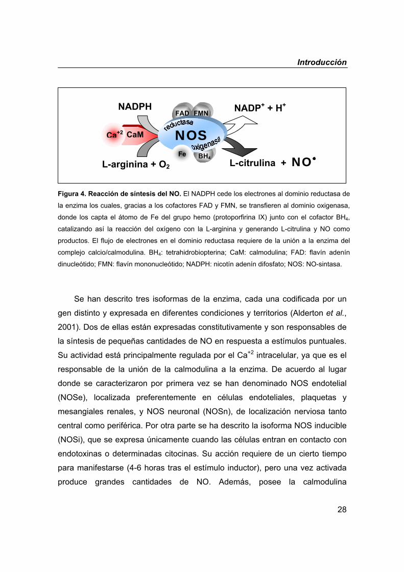
Figura 4. Reacción de síntesis del NO. El NADPH cede los electrones al dominio reductasa de
la enzima los cuales, gracias a los cofactores FAD y FMN, se transfieren al dominio oxigenasa,
donde los capta el átomo de Fe del grupo hemo (protoporfirina IX) junto con el cofactor BH4,
catalizando así la reacción del oxígeno con la L-arginina y generando L-citrulina y NO como
productos. El flujo de electrones en el dominio reductasa requiere de la unión a la enzima del
complejo calcio/calmodulina. BH4: tetrahidrobiopterina; CaM: calmodulina; FAD: flavín adenín
dinucleótido; FMN: flavín mononucleótido; NADPH: nicotín adenín difosfato; NOS: NO-sintasa.
Se han descrito tres isoformas de la enzima, cada una codificada por un
gen distinto y expresada en diferentes condiciones y territorios (Alderton et al.,
2001). Dos de ellas están expresadas constitutivamente y son responsables de
la síntesis de pequeñas cantidades de NO en respuesta a estímulos puntuales.
Su actividad está principalmente regulada por el Ca+2 intracelular, ya que es el
responsable de la unión de la calmodulina a la enzima. De acuerdo al lugar
donde se caracterizaron por primera vez se han denominado NOS endotelial
(NOSe), localizada preferentemente en células endoteliales, plaquetas y
mesangiales renales, y NOS neuronal (NOSn), de localización nerviosa tanto
central como periférica. Por otra parte se ha descrito la isoforma NOS inducible
(NOSi), que se expresa únicamente cuando las células entran en contacto con
endotoxinas o determinadas citocinas. Su acción requiere de un cierto tiempo
para manifestarse (4-6 horas tras el estímulo inductor), pero una vez activada
produce grandes cantidades de NO. Además, posee la calmodulina
NADPH NADP+ + H+
CaM NOS
FAD FMN
L-arginina + O2 L-citrulina + NO Fe BH4
Ca+2
Introducción
29
fuertemente unida, a diferencia de las otras dos isoformas, lo cual hace que
esta enzima sea insensible a las concentraciones de Ca+2 intracelular y la
mantiene en un estado tónicamente activado dando lugar a la síntesis continua
y prolongada de NO (horas/días). Inicialmente fue descrita en macrófagos y
hepatocitos, si bien posteriormente se ha observado en numerosas células
(músculo liso vascular, neutrófilos, células endoteliales,…). Sin embargo, hay
que decir que la terminología constitutiva frente a inducible es una
simplificación ya que se ha descrito que la expresión de las isoformas
“constitutivas”, tanto NOSe como NOSn, puede ser inducida en determinadas
condiciones fisiológicas (Forstermann et al., 1998) y, por el contrario, se ha
descrito que la NOSi se expresa de forma constitutiva en determinadas células
(Alderton et al., 2001; Guo et al., 1995).
III.2. Mecanismo de acción
La diversidad de funciones que tiene el NO se debe a su capacidad de
reaccionar con un rango amplio de moléculas distintas. Este mediador interactúa
con “dianas” moleculares específicas localizadas en la propia célula donde es
generado (regulación autocrina) o en células vecinas (regulación paracrina).
La principal diana del NO caracterizada hasta la fecha es la enzima
guanilato ciclasa soluble (GCs) (Southam & Garthwaite, 1993). El NO reacciona
con el hierro del grupo hemo de esta enzima activándola y desencadenando así
la liberación de guanosín monofosfato cíclico (GMPc), mediador final
responsable de la mayoría de las acciones fisiológicas del NO. El GMPc, a
través de la activación de una serie de cinasas, reduce la concentración de Ca+2
intracelular por distintos mecanismos; además, activa directamente los canales
iónicos de K+ hiperpolarizando a la célula (Carvajal et al., 2000), (figura 5).
Todas estas acciones conducen a la relajación de la célula del músculo liso. Por
Introducción
30
otra parte, el NO también interacciona con el hierro del grupo hemo presente en
la hemoglobina constituyendo este proceso la principal vía metabólica del NO.
Otra enzima con la que interacciona el NO es la citocromo c oxidasa
presente en el complejo terminal de la cadena de transporte electrónico
mitocondrial (Nisoli et al., 2004). Se ha visto que el NO compite con el oxígeno
inhibiendo de manera reversible la cadena respiratoria, por lo que se ha
sugerido que pudiera estar actuando como un regulador fisiológico de la
respiración celular (Brown, 2000). Apoyando esta teoría, existen evidencias que
apuntan hacia la presencia de actividad NOS Ca+2-dependiente en la membrana
interna mitocondrial (Ghafourifar & Richter, 1997; Lopez-Figueroa et al., 2000).
Además, debido a su condición de radical libre, el NO reacciona con el
anión superóxido (O2-) para generar peroxinitritos (ONOO-), sustancias que son
altamente reactivas y que poseen un potente efecto oxidante. De esta manera
se explica el efecto citotóxico del NO (Beckman & Koppenol, 1996). Esta
reacción química se ve favorecida en circunstancias en las que existe una
elevada concentración de NO como consecuencia de la inducción de la NOS.
El NO también reacciona con los grupos tiol (SH) de ciertas proteínas
dando lugar a los nitrosotioles. Se ha propuesto que ésta podría ser una forma
natural de almacén y transporte del NO (Muller et al., 2002). Además, se ha
demostrado en células vasculares aisladas que el NO puede activar
directamente los canales de K+ sensibles al Ca+2 gracias a este mecanismo
(Bolotina et al., 1994), desencadenando la relajación del músculo liso (figura 5).
Sin embargo, la contribución de esta vía a la relajación vascular inducida por el
NO in vivo está todavía por determinar.
Por último, también se ha visto que el NO puede combinarse con metales
de transición (hierro, cobre o zinc) presentes en determinadas enzimas como la
ciclooxigenasa, lipooxigenasa, citocromo P-450 y la aconitasa, modulando su
función (Ortega Mateo & Amaya Aleixandre de Artinano, 2000).
Introducción
31
III.3. Óxido nítrico en el sistema nervioso
El descubrimiento del NO como neurotransmisor cambió radicalmente el
concepto clásico de sinapsis nerviosa. El NO es de naturaleza gaseosa y posee
una vida media muy corta (segundos). A diferencia de los neurotransmisores
típicos, no se almacena en vesículas sinápticas sino que se sintetiza cuando se
necesita y no se libera por exocitosis ya que difunde libremente a través de las
membranas. En 1997 se adoptó el término de nervios nitrérgicos (Moncada et
al., 1997) aplicados a aquéllos cuya función transmisora depende de la
liberación de NO.
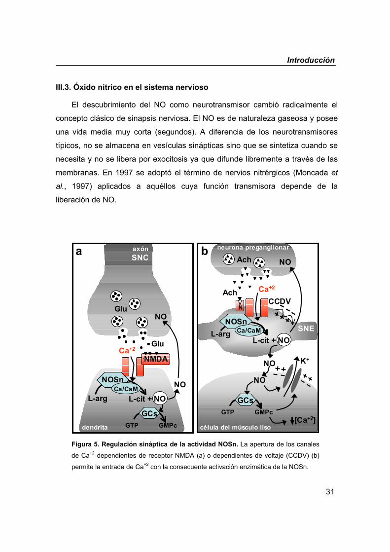
Figura 5. Regulación sináptica de la actividad NOSn. La apertura de los canales
de Ca+2 dependientes de receptor NMDA (a) o dependientes de voltaje (CCDV) (b)
permite la entrada de Ca+2 con la consecuente activación enzimática de la NOSn.
NMDA
GluCa+2
NOSnCa/CaM
L-arg
GCsGTP GMPc
L-cit + NO
NO
NO
axónSNC
dendrita
K+
Ach Ca+2
NOSnCa/CaML-arg
CCDV
L-cit + NO
Glu
Achneurona preganglionar
célula del músculo liso
SNE
NO
GCsGTP GMPc
[Ca+2]
NO
NO
NN
a b
Introducción
32
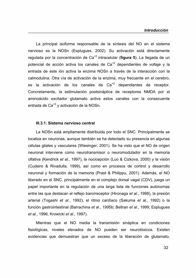
La principal isoforma responsable de la síntesis del NO en el sistema
nervioso es la NOSn (Esplugues, 2002). Su activación está directamente
regulada por la concentración de Ca+2 intracelular (figura 5). La llegada de un
potencial de acción activa los canales de Ca+2 dependientes de voltaje y la
entrada de este ión activa la enzima NOSn a través de la interacción con la
calmodulina. Otra vía de activación de la enzima, muy frecuente en el cerebro,
es la activación de los canales de Ca+2 dependientes de receptor.
Concretamente, la estimulación postsináptica de receptores NMDA por el
aminoácido excitador glutamato activa estos canales con la consecuente
entrada de Ca+2 y activación de la NOSn.
III.3.1. Sistema nervioso central
La NOSn está ampliamente distribuida por todo el SNC. Principalmente se
localiza en neuronas, aunque también se ha detectado su presencia en algunas
células gliales y vasculares (Wiesinger, 2001). Se ha visto que el NO de origen
neuronal interviene como neurotransmisor o neuromodulador en la memoria
olfativa (Kendrick et al., 1997), la nocicepción (Luo & Cizkova, 2000) y la visión
(Cudeiro & Rivadulla, 1999), así como en procesos de control y desarrollo
neuronal y formación de la memoria (Prast & Philippu, 2001). Además, el NO
liberado en el SNC, principalmente en el complejo dorsal vagal (CDV), juega un
papel importante en la regulación de una larga lista de funciones autónomas
entre las que destacan el reflejo barorreceptor (Hironaga et al., 1998), la presión
arterial (Togashi et al., 1992), el ritmo cardíaco (Sakuma et al., 1992) o la
función gastrointestinal (Barrachina et al., 1995b; Beltran et al., 1999; Esplugues
et al., 1996; Krowicki et al., 1997).
Mientras que el NO media la transmisión sináptica en condiciones
fisiológicas, niveles elevados de NO pueden ser neurotóxicos. Existen
evidencias que demuestran que un exceso de la liberación de glutamato,
Introducción
33
actuando sobre receptores NMDA, media la neurotoxicidad que contribuye a la
generación de patologías neurodegenerativas como el Alzheimer o la
enfermedad de Huntington (Boje, 2004).
III.3.2. Sistema nervioso periférico
En 1963 se demostró por primera vez que la activación de nervios
periféricos en presencia de bloqueantes colinérgicos y adrenérgicos tenía un
efecto relajante del músculo liso (Burnstock et al., 1963), introduciéndose así el
término de neurotransmisión no-adrenérgica no-colinérgica (NANC). Estos
experimentos se desarrollaron en el tracto gastrointestinal, sin embargo
rápidamente se observó que este fenómeno también se daba en los sistemas
urogenital, respiratorio y cardiovascular (Burnstock, 1986). La identificación del
neurotransmisor NANC ha sido objeto de discusión durante todos estos años.
Inicialmente se habló del ATP o del VIP. Actualmente se acepta que es el NO el
principal neurotransmisor NANC, aunque no se descarta la participación de los
otros dos mediadores (Sanders & Ward, 1992).
Los efectos del NO como neurotransmisor NANC regulan procesos
fisiológicos de especial relevancia. En el sistema respiratorio se cree que la
inervación nitrérgica representa la principal vía broncodilatadora en los
humanos, y su disfunción puede estar involucrada en el proceso asmático
(Belvisi et al., 1995). Además, la inhalación de NO constituye una importante
herramienta terapéutica en el tratamiento de algunas enfermedades
respiratorias. En el sistema urogenital, la neurotransmisión NANC contribuye a
la erección del pene y la impotencia se ha relacionado con la alteración de la
transmisión nitrérgica (Andersson, 2003). Además, la relajación NANC
contribuye a la disminución de la presión intrauretral que precede a la micción
(de Groat & Yoshimura, 2001). Por otra parte se ha visto que en los vasos
sanguíneos, además de la importancia del NO derivado de la NOSe en la
Introducción
34
regulación del tono vascular, la inervación nitrérgica también modula esta
función en determinadas regiones como el mesenterio (Toda & Okamura, 2003).
En el tracto gastrointestinal la regulación nitrérgica juega un papel fisiológico
fundamental como se comenta a continuación.
III.4. Regulación nitrérgica de la motilidad gastrointestinal
Estudios de inmunohistoquímica revelan la amplia distribución de la enzima
NOS en el tracto gastrointestinal. Se ha detectado actividad NOS en células
endoteliales, secretoras, musculares y nerviosas (Salzman, 1995). Dicha
actividad refleja en su conjunto la participación del NO como mediador
endógeno en la regulación de funciones tales como motilidad, secreción,
integridad tisular y microcirculación del tracto gastrointestinal.
La NOSn está ampliamente distribuida a lo largo del plexo mientérico (Aimi
et al., 1993). La mayoría de las fibras nerviosas nitrérgicas son neuronas
intrínsecas cuyas varicosidades están situadas muy próximas a las células del
músculo liso que contienen GCs (Ekblad et al., 1994). La regulación nitrérgica
NANC está implicada en muchos reflejos nerviosos fisiológicos tales como la
relajación adaptativa (Desai et al., 1991b) y receptiva (Desai et al., 1991a) del
estómago. También está implicada en la relajación de los esfínteres esofágico
(Boeckxstaens GE & Pelckmans PA, 1997) y pilórico (Allescher et al., 1992); a
su vez, la ausencia de dicha regulación constituye la base de patologías tales
como la acalasia (Mearin et al., 1993) o la estenosis hipertrófica del píloro
(Vanderwinden et al., 1992). A nivel intestinal, la inervación NANC participa en
el reflejo peristáltico (Foxx-Orenstein & Grider, 1996) y también permite el paso
del contenido fecal al intestino grueso al participar en la relajación de la unión
íleo-colónica (Boeckxstaens et al., 1990). Por último, la regulación nitrérgica
también es la responsable de la relajación del esfínter anal interno, siendo
crucial en el proceso de la defecación (Chakder & Rattan, 1993).
Introducción
35
El papel fisiológico de la inervación nitrérgica en la regulación de la
motilidad gastrointestinal ha sido ampliamente estudiado. Sin embargo, poco se
sabe acerca del papel de dicha inervación en las alteraciones motoras
asociadas a circunstancias fisiopatológicas tales como la endotoxemia.

Objetivo
36
OBJETIVO
Objetivo
37
El objetivo planteado en el presente trabajo es caracterizar los mecanismos
involucrados en la alteración de la función motora gastrointestinal asociada a los
estadios tempranos de la endotoxemia.

Artículos de investigación
38
ARTÍCULOS DE INVESTIGACIÓN
Artículos de investigación
39
I. EFECTOS DE LA ENDOTOXINA SOBRE LA MOTILIDAD GÁSTRICA
Vía nerviosa y mediadores implicados
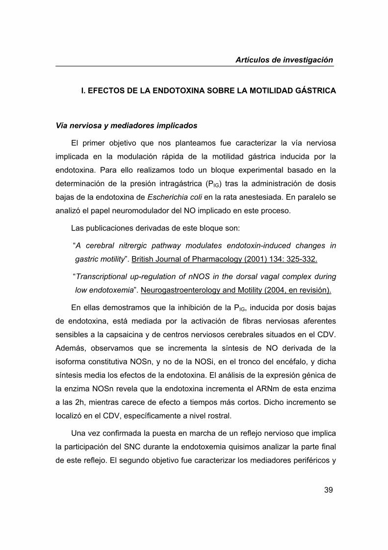
El primer objetivo que nos planteamos fue caracterizar la vía nerviosa
implicada en la modulación rápida de la motilidad gástrica inducida por la
endotoxina. Para ello realizamos todo un bloque experimental basado en la
determinación de la presión intragástrica (PIG) tras la administración de dosis
bajas de la endotoxina de Escherichia coli en la rata anestesiada. En paralelo se
analizó el papel neuromodulador del NO implicado en este proceso.
Las publicaciones derivadas de este bloque son:
“A cerebral nitrergic pathway modulates endotoxin-induced changes in
gastric motility”. British Journal of Pharmacology (2001) 134: 325-332.
“Transcriptional up-regulation of nNOS in the dorsal vagal complex during
low endotoxemia”. Neurogastroenterology and Motility (2004, en revisión).
En ellas demostramos que la inhibición de la PIG, inducida por dosis bajas
de endotoxina, está mediada por la activación de fibras nerviosas aferentes
sensibles a la capsaicina y de centros nerviosos cerebrales situados en el CDV.
Además, observamos que se incrementa la síntesis de NO derivada de la
isoforma constitutiva NOSn, y no de la NOSi, en el tronco del encéfalo, y dicha
síntesis media los efectos de la endotoxina. El análisis de la expresión génica de
la enzima NOSn revela que la endotoxina incrementa el ARNm de esta enzima
a las 2h, mientras carece de efecto a tiempos más cortos. Dicho incremento se
localizó en el CDV, específicamente a nivel rostral.
Una vez confirmada la puesta en marcha de un reflejo nervioso que implica
la participación del SNC durante la endotoxemia quisimos analizar la parte final
de este reflejo. El segundo objetivo fue caracterizar los mediadores periféricos y
Artículos de investigación
40
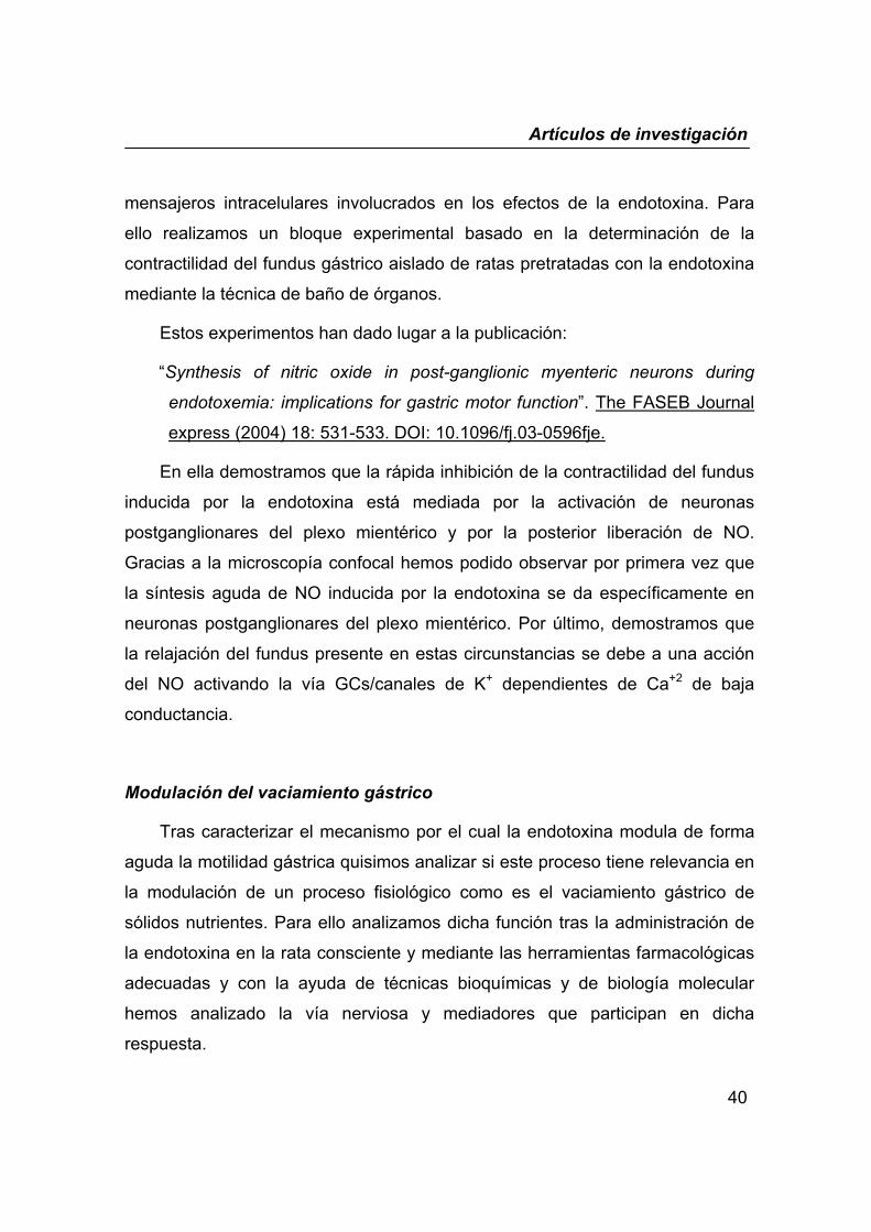
mensajeros intracelulares involucrados en los efectos de la endotoxina. Para
ello realizamos un bloque experimental basado en la determinación de la
contractilidad del fundus gástrico aislado de ratas pretratadas con la endotoxina
mediante la técnica de baño de órganos.
Estos experimentos han dado lugar a la publicación:
“Synthesis of nitric oxide in post-ganglionic myenteric neurons during
endotoxemia: implications for gastric motor function”. The FASEB Journal
express (2004) 18: 531-533. DOI: 10.1096/fj.03-0596fje.
En ella demostramos que la rápida inhibición de la contractilidad del fundus
inducida por la endotoxina está mediada por la activación de neuronas
postganglionares del plexo mientérico y por la posterior liberación de NO.
Gracias a la microscopía confocal hemos podido observar por primera vez que
la síntesis aguda de NO inducida por la endotoxina se da específicamente en
neuronas postganglionares del plexo mientérico. Por último, demostramos que
la relajación del fundus presente en estas circunstancias se debe a una acción
del NO activando la vía GCs/canales de K+ dependientes de Ca+2 de baja
conductancia.
Modulación del vaciamiento gástrico
Tras caracterizar el mecanismo por el cual la endotoxina modula de forma
aguda la motilidad gástrica quisimos analizar si este proceso tiene relevancia en
la modulación de un proceso fisiológico como es el vaciamiento gástrico de
sólidos nutrientes. Para ello analizamos dicha función tras la administración de
la endotoxina en la rata consciente y mediante las herramientas farmacológicas
adecuadas y con la ayuda de técnicas bioquímicas y de biología molecular
hemos analizado la vía nerviosa y mediadores que participan en dicha
respuesta.
Artículos de investigación
41
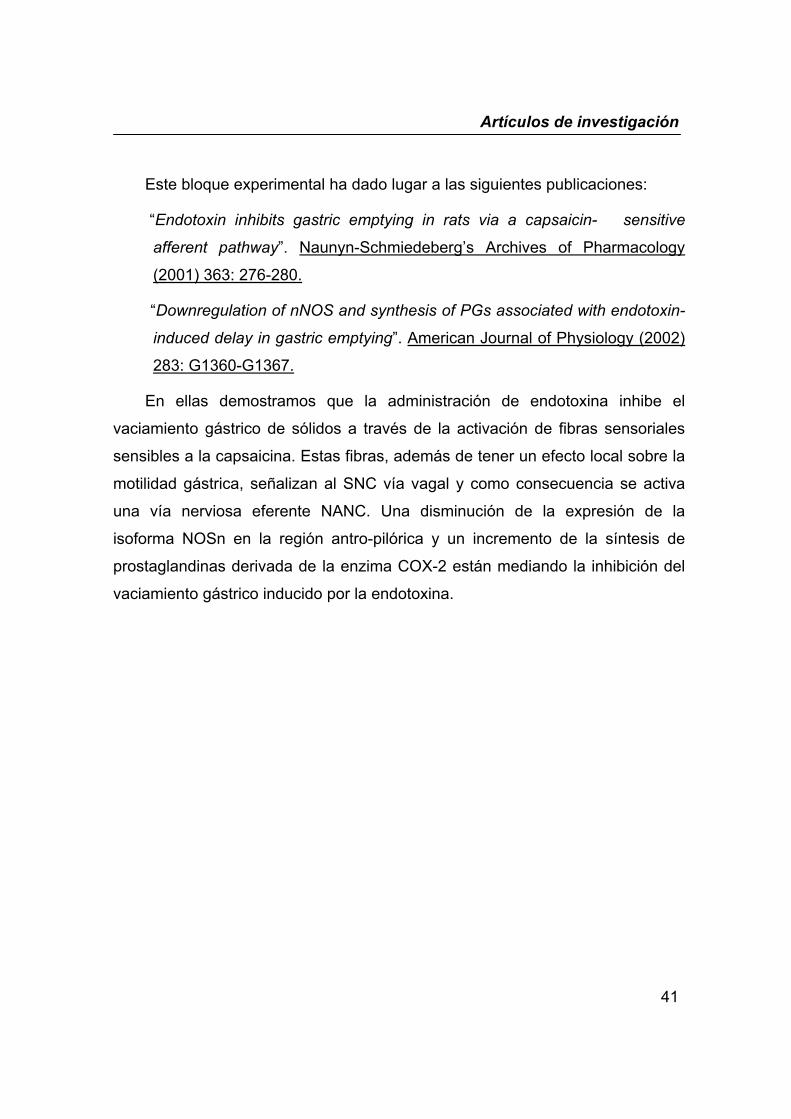
Este bloque experimental ha dado lugar a las siguientes publicaciones:
“Endotoxin inhibits gastric emptying in rats via a capsaicin- sensitive
afferent pathway”. Naunyn-Schmiedeberg’s Archives of Pharmacology
(2001) 363: 276-280.
“Downregulation of nNOS and synthesis of PGs associated with endotoxin-
induced delay in gastric emptying”. American Journal of Physiology (2002)
283: G1360-G1367.
En ellas demostramos que la administración de endotoxina inhibe el
vaciamiento gástrico de sólidos a través de la activación de fibras sensoriales
sensibles a la capsaicina. Estas fibras, además de tener un efecto local sobre la
motilidad gástrica, señalizan al SNC vía vagal y como consecuencia se activa
una vía nerviosa eferente NANC. Una disminución de la expresión de la
isoforma NOSn en la región antro-pilórica y un incremento de la síntesis de
prostaglandinas derivada de la enzima COX-2 están mediando la inhibición del
vaciamiento gástrico inducido por la endotoxina.
Artículos de investigación
42
Artículo 1
“A cerebral nitrergic pathway modulates
endotoxin-induced changes in gastric
motility”
Elsa Quintana, Eugenia García-Zaragozá, M. Ángeles Martínez-Cuesta,
Sara Calatayud, Juan V. Esplugues & María D. Barrachina
British Journal of Pharmacology (2001) 134: 325-332
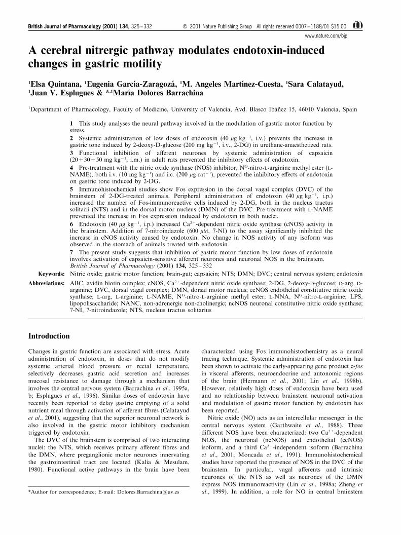
A cerebral nitrergic pathway modulates endotoxin-inducedchanges in gastric motility
1Elsa Quintana, 1Eugenia Garcõ a-Zaragoza , 1M. Angeles Martõ nez-Cuesta, 1Sara Calatayud,1Juan V. Esplugues & *,1Marõ a Dolores Barrachina
1Department of Pharmacology, Faculty of Medicine, University of Valencia, Avd. Blasco Iba nÄ ez 15, 46010 Valencia, Spain
1 This study analyses the neural pathway involved in the modulation of gastric motor function bystress.
2 Systemic administration of low doses of endotoxin (40 mg kg71, i.v.) prevents the increase ingastric tone induced by 2-deoxy-D-glucose (200 mg kg71, i.v., 2-DG) in urethane-anaesthetized rats.
3 Functional inhibition of a�erent neurones by systemic administration of capsaicin(20+30+50 mg kg71, i.m.) in adult rats prevented the inhibitory e�ects of endotoxin.
4 Pre-treatment with the nitric oxide synthase (NOS) inhibitor, NG-nitro-L-arginine methyl ester (L-NAME), both i.v. (10 mg kg71) and i.c. (200 mg rat71), prevented the inhibitory e�ects of endotoxinon gastric tone induced by 2-DG.
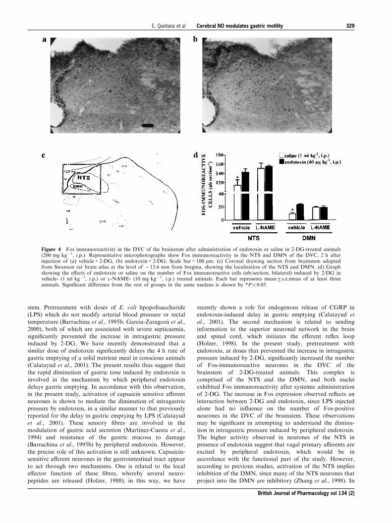
5 Immunohistochemical studies show Fos expression in the dorsal vagal complex (DVC) of thebrainstem of 2-DG-treated animals. Peripheral administration of endotoxin (40 mg kg71, i.p.)increased the number of Fos-immunoreactive cells induced by 2-DG, both in the nucleus tractussolitarii (NTS) and in the dorsal motor nucleus (DMN) of the DVC. Pre-treatment with L-NAMEprevented the increase in Fos expression induced by endotoxin in both nuclei.
6 Endotoxin (40 mg kg71, i.p.) increased Ca2+-dependent nitric oxide synthase (cNOS) activity inthe brainstem. Addition of 7-nitroindazole (600 mM, 7-NI) to the assay signi®cantly inhibited theincrease in cNOS activity caused by endotoxin. No change in NOS activity of any isoform wasobserved in the stomach of animals treated with endotoxin.
7 The present study suggests that inhibition of gastric motor function by low doses of endotoxininvolves activation of capsaicin-sensitive a�erent neurones and neuronal NOS in the brainstem.British Journal of Pharmacology (2001) 134, 325 ± 332
Keywords: Nitric oxide; gastric motor function; brain-gut; capsaicin; NTS; DMN; DVC; central nervous system; endotoxin
Abbreviations: ABC, avidin biotin complex; cNOS, Ca2+-dependent nitric oxide synthase; 2-DG, 2-deoxy-D-glucose; D-arg, D-arginine; DVC, dorsal vagal complex; DMN, dorsal motor nucleus; ecNOS endothelial constitutive nitric oxidesynthase; L-arg, L-arginine; L-NAME, NG-nitro-L-arginine methyl ester; L-NNA, NG-nitro-L-arginine; LPS,lipopolisaccharide; NANC, non-adrenergic non-cholinergic; ncNOS neuronal constitutive nitric oxide synthase;7-NI, 7-nitroindazole; NTS, nucleus tractus solitarius
Introduction
Changes in gastric function are associated with stress. Acute
administration of endotoxin, in doses that do not modifysystemic arterial blood pressure or rectal temperature,selectively decreases gastric acid secretion and increases
mucosal resistance to damage through a mechanism thatinvolves the central nervous system (Barrachina et al., 1995a,b; Esplugues et al., 1996). Similar doses of endotoxin have
recently been reported to delay gastric emptying of a solidnutrient meal through activation of a�erent ®bres (Calatayudet al., 2001), suggesting that the superior neuronal network is
also involved in the gastric motor inhibitory mechanismtriggered by endotoxin.The DVC of the brainstem is comprised of two interacting
nuclei: the NTS, which receives primary a�erent ®bres and
the DMN, where preganglionic motor neurones innervatingthe gastrointestinal tract are located (Kalia & Mesulam,1980). Functional active pathways in the brain have been
characterized using Fos immunohistochemistry as a neural
tracing technique. Systemic administration of endotoxin hasbeen shown to activate the early-appearing gene product c-fosin visceral a�erents, neuroendocrine and autonomic regions
of the brain (Hermann et al., 2001; Lin et al., 1998b).However, relatively high doses of endotoxin have been usedand no relationship between brainstem neuronal activation
and modulation of gastric motor function by endotoxin hasbeen reported.Nitric oxide (NO) acts as an intercellular messenger in the
central nervous system (Garthwaite et al., 1988). Threedi�erent NOS have been characterized: two Ca2+-dependentNOS, the neuronal (ncNOS) and endothelial (ecNOS)isoform, and a third Ca2+-independent isoform (Barrachina
et al., 2001; Moncada et al., 1991). Immunohistochemicalstudies have reported the presence of NOS in the DVC of thebrainstem. In particular, vagal a�erents and intrinsic
neurones of the NTS as well as neurones of the DMNexpress NOS immunoreactivity (Lin et al., 1998a; Zheng etal., 1999). In addition, a role for NO in central brainstem
British Journal of Pharmacology (2001) 134, 325 ± 332 ã 2001 Nature Publishing Group All rights reserved 0007 ± 1188/01 $15.00
www.nature.com/bjp
*Author for correspondence; E-mail: [email protected]

circuits which control gastric secretory and motor functionshas been reported (Esplugues et al., 1996; Krowicki et al.,1997). Thus, synthesis of NO is involved in the inhibition of
gastric acid secretion by stress (Beltra n et al., 1999; Esplugueset al., 1996) or modulates the inhibition of gastric motorfunction by substance P (Krowicki & Hornby, 1996).In the present study, in order to characterize the neural
pathway and endogenous mediators involved in the changesin gastric motor function associated with stress, weevaluated the e�ects of low doses of endotoxin on: (1)
intragastric pressure induced by the antimetabolic glucoseanalogue, 2-DG; (2) the pattern of brainstem neuronalactivation and (3) the NOS activity both in the brainstem
and the stomach.
Methods
Male Sprague-Dawley rats (220 ± 250 g) were fasted for 16 ±20 h before the experiments but were allowed access to
drinking water.
Determination of intragastric pressure
Rats were anaesthetized with urethane (1.5 g kg71, i.p.), thetrachea intubated and a jugular vein cannulated. After
performing a laparotomy, an intraluminal latex balloon wasinserted in the stomach through an incision in the fore-stomach and held in place with a ligature. The balloon and
catheter system was connected to a pressure transducer andintragastric pressure was registered on line by a multi-channelrecorder (Power Lab.). After reaching an intragastric pressureof between 4 ± 5 cm H2O, by ®lling the balloon with 2 ±
3 ml H2O at 378C, rats were allowed to stabilize for 1 h.Rectal temperature was monitored during the experiment andmaintained at 36 ± 378C. Unless mentioned otherwise, data is
expressed as D intragastric pressure, calculated as thedi�erence between stimulated (the average measurement 30to 60 min after 2-DG administration) and basal (the average
in the last 10 min before 2-DG administration) intragastricpressure.
Experimental protocol
In a ®rst group of experiments, endotoxin (40 mg kg71, i.v.)was administered 30 min prior to a bolus injection of 2-DG
(200 mg kg71, i.v.) and intragastric pressure was monitoredfor 60 min. In order to analyse the role of sensory ®bres, ratswere administered with capsaicin (a selective neurotoxin on
C-®bres; 20+30+50 mg kg71, i.m.) or its vehicle (10%ethanol+10% Tween-80+80% saline, 1 ml kg71, i.m.) for3 consecutive days, 15 days prior to the experiments. The role
of NO in the e�ects of endotoxin was analysed by pre-treatment (15 min) with the NO synthesis inhibitor, L-NAME(10 mg kg71, i.v. or 200 mg rat71, i.c.) or its vehicle(1 ml kg71 or 10 ml rat71, respectively).
In order to analyse further the direct role of NO synthesisin the brain on intragastric pressure, in a second group ofexperiments some animals received an i.c. injection of L-
arginine (200 mg rat71, L-arg), D-arginine (200 mg rat71, D-arg) or saline (10 ml rat71) and gastric contractility wasmonitored for 60 min.
Fos immunohistochemistry
Procedures were carried out as previously described (Barra-
china et al., 1997). Ten minutes prior to the administration of2-DG (200 mg kg71, i.p.), rats received two consecutive i.p.injections of either L-NAME (10 mg kg71)+endotoxin(40 mg kg71), L-NAME+saline (1 ml kg71), vehicle
(1 ml kg71)+endotoxin or vehicle+saline. Additional groupsof rats received a single i.p. injection of saline, endotoxin orL-NAME and were included as control groups. Two hours
later, animals were anaesthetized with pentobarbitone(280 mg kg71, i.p.) and transcardially perfused with 0.9%saline followed by paraformaldehyde solution (4%). Brains
were post®xed in the same ®xative and cryoprotectedovernight by immersion in 30% sucrose. Immunohistochem-istry for Fos expression was processed on frozen brain
coronal sections (50 mm thick) using the avidin ± biotincomplex (ABC) method (Hsu et al., 1981). Sections wereincubated for 24 h at 48C with the primary antibody c-fos(sheep policlonal, Genosys) diluted at 1 : 2000. Then, sections
were incubated with biotynylated anti-sheep IgG (VectorLabs.), diluted at 1 : 200, followed by ABC (Vectastain ABCKit, Vector Labs.), in both cases for 1 h at 258C. Sectionswere incubated for 8 min in a substrate for peroxidase kit(Vector VIP, Vector Labs.) and were mounted, air-dried,dehydrated, cleared and coverslipped. The sections were
observed and photographed using a bright®eld microscope(Zeiss). The counting of Fos-immunoreactive cells wasperformed bilaterally in six sections per animal, regardless
of the intensity of staining, for the NTS and DMN. The L.W.Swanson rat brain atlas was used to determine theanatomical locations of the nuclei.
Determination of NOS activity
Rats were administered with endotoxin (40 mg kg71, i.p.) or
saline (1 ml kg71, i.p.) and sacri®ed by cervical dislocation30 min later. Both, a section of the brainstem containing theDVC (+1 mm to 71.4 mm to the obex) and the stomach,
were quickly introduced in liquid nitrogen and stored at7808C. NO synthase activity was measured as the rate ofconversion of L-[U-14C]-arginine to L-[U-14C]-citrulline (Salteret al., 1990). Brie¯y, the samples were homogenized (Ultra-
Turrax) in an ice-cold bu�er (330 mg ml71; pH 7.2) contain-ing 320 mM sucrose, 20 mM HEPES, 1 mM EDTA, 1 mM
DL-dithiothreitol, 10 mg ml71 leupeptin, 10 mg ml71 soybean
trypsin inhibitor and 2 mg ml71 aprotinin followed bycentrifugation at 10,0006g for 20 min at 48C. Afterwardscentrifugation, 40 ml of supernatant was incubated at 378Cfor 20 min in assay bu�er (pH 7.4) containing (mM) KH2PO4
50, MgCl2 1, CaCl2 0.2, L-valine 50, L-citrulline 1, L-arginine0.02, DL-dithiothreitol 1, and 100 mM NADPH, 3 mM FAD,
3 mM FMN, 3 mM BH4 and 950 nM L-[U-14C]-arginine(348 mCi mmol71). The speci®city of L-arginine conversionby NOS to L-citrulline was further con®rmed using the NOsynthesis inhibitors, NG-nitro-L-arginine (L-NNA, 1 mM).
Additionally, 1 mM EGTA, a calcium chelating agent wasused to di�erentiate between Ca2+-dependent and Ca2+-independent isoform of NOS. The speci®c inhibitor of
neuronal constitutive NOS (ncNOS), 7-nitroindazole(600 mM, 7-NI; a dose chosen from a previous dose-responsecurve; Babbedge et al., 1993) was used to di�erentiate
British Journal of Pharmacology vol 134 (2)
Cerebral NO modulates gastric motilityE. Quintana et al326
between ncNOS and endothelial constitutive NOS (ecNOS)isoform activity. All activities are expressed as picomol ofproduct generated per minute per gram of tissue.
Drugs
Urethane, 2-deoxy-D-glucose, Escherichia coli endotoxin
(serotype 026:B6), L-NAME, L-arginine, D-arginine, Tween80 and all reagents used for determination of NOS activitywere purchased from Sigma Chemical Co. (St. Louis, MO,
U.S.A.). L-[14C]-arginine was obtained from Amersham LifeScience and Capsaicin was purchased from Fluka ChemicAG. Sodium pentobarbitone was used as a commercially
available preparation (Abbott, Madrid). Unless mentionedotherwise all drugs have been dissolved in saline.
Statistical analysis
Data are expressed as mean+s.e.mean. Comparisons betweengroups were performed by one-way analysis of variance
followed by a Newman-Keuls test. Data are consideredstatistically signi®cant when P50.05.
Results
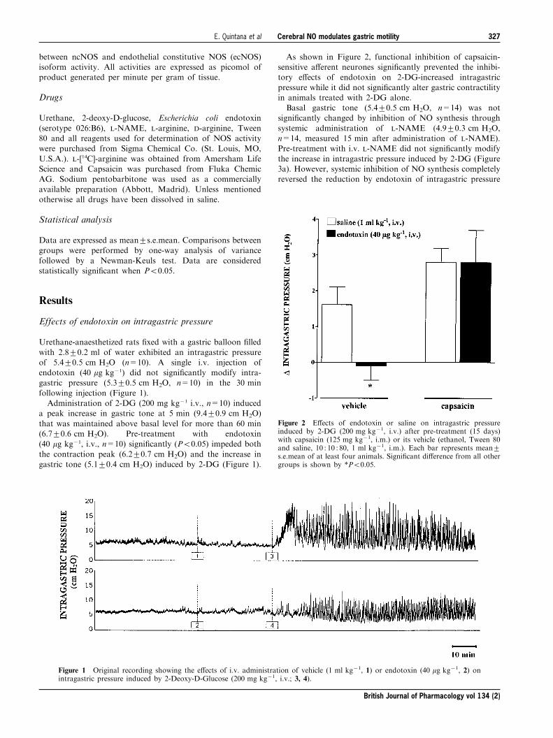
Effects of endotoxin on intragastric pressure
Urethane-anaesthetized rats ®xed with a gastric balloon ®lled
with 2.8+0.2 ml of water exhibited an intragastric pressureof 5.4+0.5 cm H2O (n=10). A single i.v. injection ofendotoxin (40 mg kg71) did not signi®cantly modify intra-gastric pressure (5.3+0.5 cm H2O, n=10) in the 30 min
following injection (Figure 1).Administration of 2-DG (200 mg kg71 i.v., n=10) induced
a peak increase in gastric tone at 5 min (9.4+0.9 cm H2O)
that was maintained above basal level for more than 60 min(6.7+0.6 cm H2O). Pre-treatment with endotoxin(40 mg kg71, i.v., n=10) signi®cantly (P50.05) impeded both
the contraction peak (6.2+0.7 cm H2O) and the increase ingastric tone (5.1+0.4 cm H2O) induced by 2-DG (Figure 1).
As shown in Figure 2, functional inhibition of capsaicin-sensitive a�erent neurones signi®cantly prevented the inhibi-tory e�ects of endotoxin on 2-DG-increased intragastric
pressure while it did not signi®cantly alter gastric contractilityin animals treated with 2-DG alone.Basal gastric tone (5.4+0.5 cm H2O, n=14) was not
signi®cantly changed by inhibition of NO synthesis through
systemic administration of L-NAME (4.9+0.3 cm H2O,n=14, measured 15 min after administration of L-NAME).Pre-treatment with i.v. L-NAME did not signi®cantly modify
the increase in intragastric pressure induced by 2-DG (Figure3a). However, systemic inhibition of NO synthesis completelyreversed the reduction by endotoxin of intragastric pressure
Figure 1 Original recording showing the e�ects of i.v. administration of vehicle (1 ml kg71, 1) or endotoxin (40 mg kg71, 2) onintragastric pressure induced by 2-Deoxy-D-Glucose (200 mg kg71, i.v.; 3, 4).
Figure 2 E�ects of endotoxin or saline on intragastric pressureinduced by 2-DG (200 mg kg71, i.v.) after pre-treatment (15 days)with capsaicin (125 mg kg71, i.m.) or its vehicle (ethanol, Tween 80and saline, 10 : 10 : 80, 1 ml kg71, i.m.). Each bar represents mean+s.e.mean of at least four animals. Signi®cant di�erence from all othergroups is shown by *P50.05.
British Journal of Pharmacology vol 134 (2)
Cerebral NO modulates gastric motilityE. Quintana et al 327
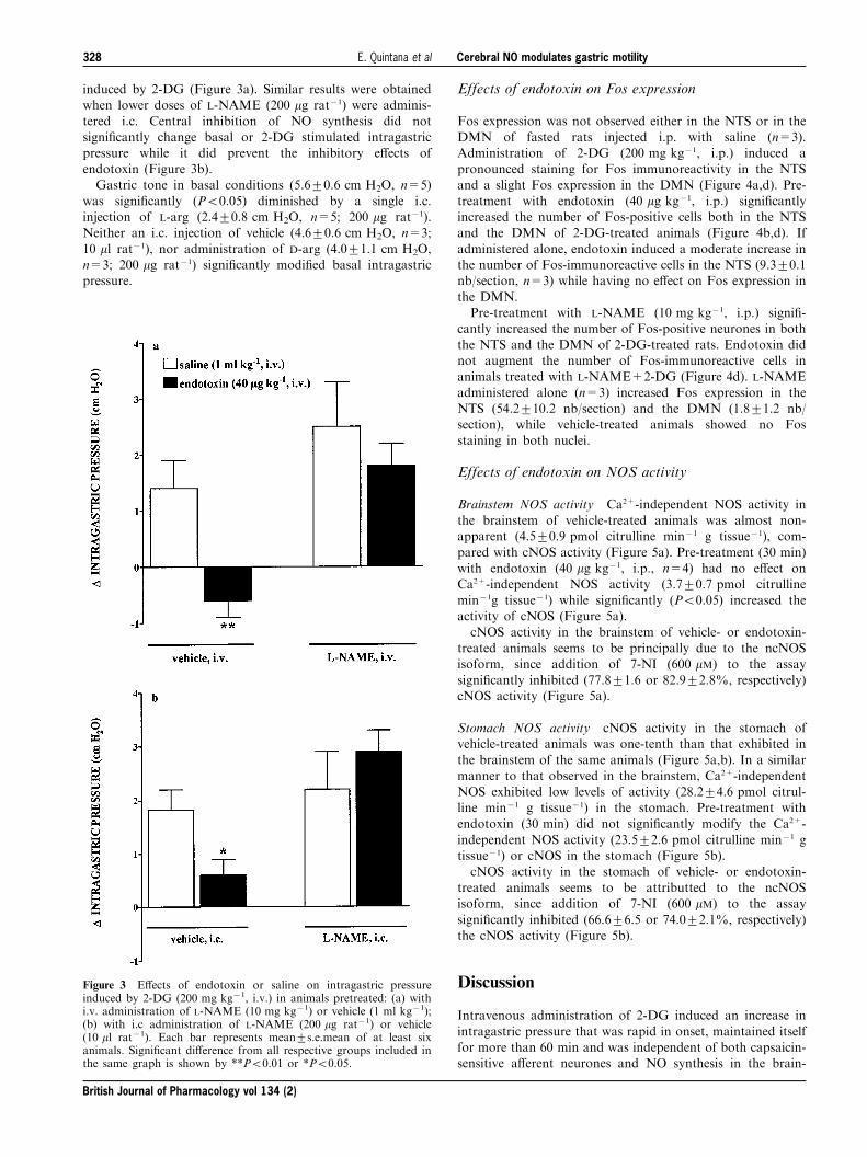
induced by 2-DG (Figure 3a). Similar results were obtainedwhen lower doses of L-NAME (200 mg rat71) were adminis-tered i.c. Central inhibition of NO synthesis did not
signi®cantly change basal or 2-DG stimulated intragastricpressure while it did prevent the inhibitory e�ects ofendotoxin (Figure 3b).Gastric tone in basal conditions (5.6+0.6 cm H2O, n=5)
was signi®cantly (P50.05) diminished by a single i.c.injection of L-arg (2.4+0.8 cm H2O, n=5; 200 mg rat71).Neither an i.c. injection of vehicle (4.6+0.6 cm H2O, n=3;
10 ml rat71), nor administration of D-arg (4.0+1.1 cm H2O,n=3; 200 mg rat71) signi®cantly modi®ed basal intragastricpressure.
Effects of endotoxin on Fos expression
Fos expression was not observed either in the NTS or in the
DMN of fasted rats injected i.p. with saline (n=3).Administration of 2-DG (200 mg kg71, i.p.) induced apronounced staining for Fos immunoreactivity in the NTSand a slight Fos expression in the DMN (Figure 4a,d). Pre-
treatment with endotoxin (40 mg kg71, i.p.) signi®cantlyincreased the number of Fos-positive cells both in the NTSand the DMN of 2-DG-treated animals (Figure 4b,d). If
administered alone, endotoxin induced a moderate increase inthe number of Fos-immunoreactive cells in the NTS (9.3+0.1nb/section, n=3) while having no e�ect on Fos expression in
the DMN.Pre-treatment with L-NAME (10 mg kg71, i.p.) signi®-
cantly increased the number of Fos-positive neurones in both
the NTS and the DMN of 2-DG-treated rats. Endotoxin didnot augment the number of Fos-immunoreactive cells inanimals treated with L-NAME+2-DG (Figure 4d). L-NAMEadministered alone (n=3) increased Fos expression in the
NTS (54.2+10.2 nb/section) and the DMN (1.8+1.2 nb/section), while vehicle-treated animals showed no Fosstaining in both nuclei.
Effects of endotoxin on NOS activity
Brainstem NOS activity Ca2+-independent NOS activity inthe brainstem of vehicle-treated animals was almost non-apparent (4.5+0.9 pmol citrulline min71 g tissue71), com-
pared with cNOS activity (Figure 5a). Pre-treatment (30 min)with endotoxin (40 mg kg71, i.p., n=4) had no e�ect onCa2+-independent NOS activity (3.7+0.7 pmol citrullinemin71g tissue71) while signi®cantly (P50.05) increased the
activity of cNOS (Figure 5a).cNOS activity in the brainstem of vehicle- or endotoxin-
treated animals seems to be principally due to the ncNOS
isoform, since addition of 7-NI (600 mM) to the assaysigni®cantly inhibited (77.8+1.6 or 82.9+2.8%, respectively)cNOS activity (Figure 5a).
Stomach NOS activity cNOS activity in the stomach ofvehicle-treated animals was one-tenth than that exhibited inthe brainstem of the same animals (Figure 5a,b). In a similar
manner to that observed in the brainstem, Ca2+-independentNOS exhibited low levels of activity (28.2+4.6 pmol citrul-line min71 g tissue71) in the stomach. Pre-treatment with
endotoxin (30 min) did not signi®cantly modify the Ca2+-independent NOS activity (23.5+2.6 pmol citrulline min71 gtissue71) or cNOS in the stomach (Figure 5b).
cNOS activity in the stomach of vehicle- or endotoxin-treated animals seems to be attributted to the ncNOSisoform, since addition of 7-NI (600 mM) to the assay
signi®cantly inhibited (66.6+6.5 or 74.0+2.1%, respectively)the cNOS activity (Figure 5b).
Discussion
Intravenous administration of 2-DG induced an increase in
intragastric pressure that was rapid in onset, maintained itselffor more than 60 min and was independent of both capsaicin-sensitive a�erent neurones and NO synthesis in the brain-
Figure 3 E�ects of endotoxin or saline on intragastric pressureinduced by 2-DG (200 mg kg71, i.v.) in animals pretreated: (a) withi.v. administration of L-NAME (10 mg kg71) or vehicle (1 ml kg71);(b) with i.c administration of L-NAME (200 mg rat71) or vehicle(10 ml rat71). Each bar represents mean+s.e.mean of at least sixanimals. Signi®cant di�erence from all respective groups included inthe same graph is shown by **P50.01 or *P50.05.
British Journal of Pharmacology vol 134 (2)
Cerebral NO modulates gastric motilityE. Quintana et al328
stem. Pretreatment with doses of E. coli lipopolisaccharide(LPS) which do not modify arterial blood pressure or rectal
temperature (Barrachina et al., 1995b; Garcõ a-Zaragoza et al.,2000), both of which are associated with severe septicaemia,signi®cantly prevented the increase in intragastric pressureinduced by 2-DG. We have recently demonstrated that a
similar dose of endotoxin signi®cantly delays the 4 h rate ofgastric emptying of a solid nutrient meal in conscious animals(Calatayud et al., 2001). The present results thus suggest that
the rapid diminution of gastric tone induced by endotoxin isinvolved in the mechanism by which peripheral endotoxindelays gastric emptying. In accordance with this observation,
in the present study, activation of capsaicin sensitive a�erentneurones is shown to mediate the diminution of intragastricpressure by endotoxin, in a similar manner to that previously
reported for the delay in gastric emptying by LPS (Calatayudet al., 2001). These sensory ®bres are involved in themodulation of gastric acid secretion (MartõÂ nez-Cuesta et al.,1994) and resistance of the gastric mucosa to damage
(Barrachina et al., 1995b) by peripheral endotoxin. However,the precise role of this activation is still unknown. Capsaicin-sensitive a�erent neurones in the gastrointestinal tract appear
to act through two mechanisms. One is related to the locale�ector function of these ®bres, whereby several neuro-peptides are released (Holzer, 1988); in this way, we have
recently shown a role for endogenous release of CGRP inendotoxin-induced delay in gastric emptying (Calatayud et
al., 2001). The second mechanism is related to sendinginformation to the superior neuronal network in the brainand spinal cord, which initiates the e�erent re¯ex loop(Holzer, 1998). In the present study, pretreatment with
endotoxin, at doses that prevented the increase in intragastricpressure induced by 2-DG, signi®cantly increased the numberof Fos-immunoreactive neurones in the DVC of the
brainstem of 2-DG-treated animals. This complex iscomprised of the NTS and the DMN, and both nucleiexhibited Fos immunoreactivity after systemic administration
of 2-DG. The increase in Fos expression observed re¯ects aninteraction between 2-DG and endotoxin, since LPS injectedalone had no in¯uence on the number of Fos-positive
neurones in the DVC of the brainstem. These observationsmay be signi®cant in attempting to understand the diminu-tion in intragastric pressure induced by peripheral endotoxin.The higher activity observed in neurones of the NTS in
presence of endotoxin suggest that vagal primary a�erents areexcited by peripheral endotoxin, which would be inaccordance with the functional part of the study. However,
according to previous studies, activation of the NTS impliesinhibition of the DMN, since many of the NTS neurones thatproject into the DMN are inhibitory (Zhang et al., 1998). In
Figure 4 Fos immunoreactivity in the DVC of the brainstem after administration of endotoxin or saline in 2-DG-treated animals(200 mg kg71, i.p.). Representative microphotographs show Fos immunoreactivity in the NTS and DMN of the DVC, 2 h afterinjection of (a) vehicle+2-DG, (b) endotoxin+2-DG; Scale bar=100 mm. (c) Coronal drawing section from brainstem adaptedfrom Swanson rat brain atlas at the level of 713.6 mm from bregma, showing the localization of the NTS and DMN. (d) Graphshowing the e�ects of endotoxin or saline on the number of Fos immunoreactive cells (nb/section, bilateral) induced by 2-DG invehicle- (1 ml kg71, i.p.) or L-NAME- (10 mg kg71, i.p.) treated animals. Each bar represents mean+s.e.mean of at least threeanimals. Signi®cant di�erence from the rest of groups in the same nucleus is shown by *P50.05.
British Journal of Pharmacology vol 134 (2)
Cerebral NO modulates gastric motilityE. Quintana et al 329
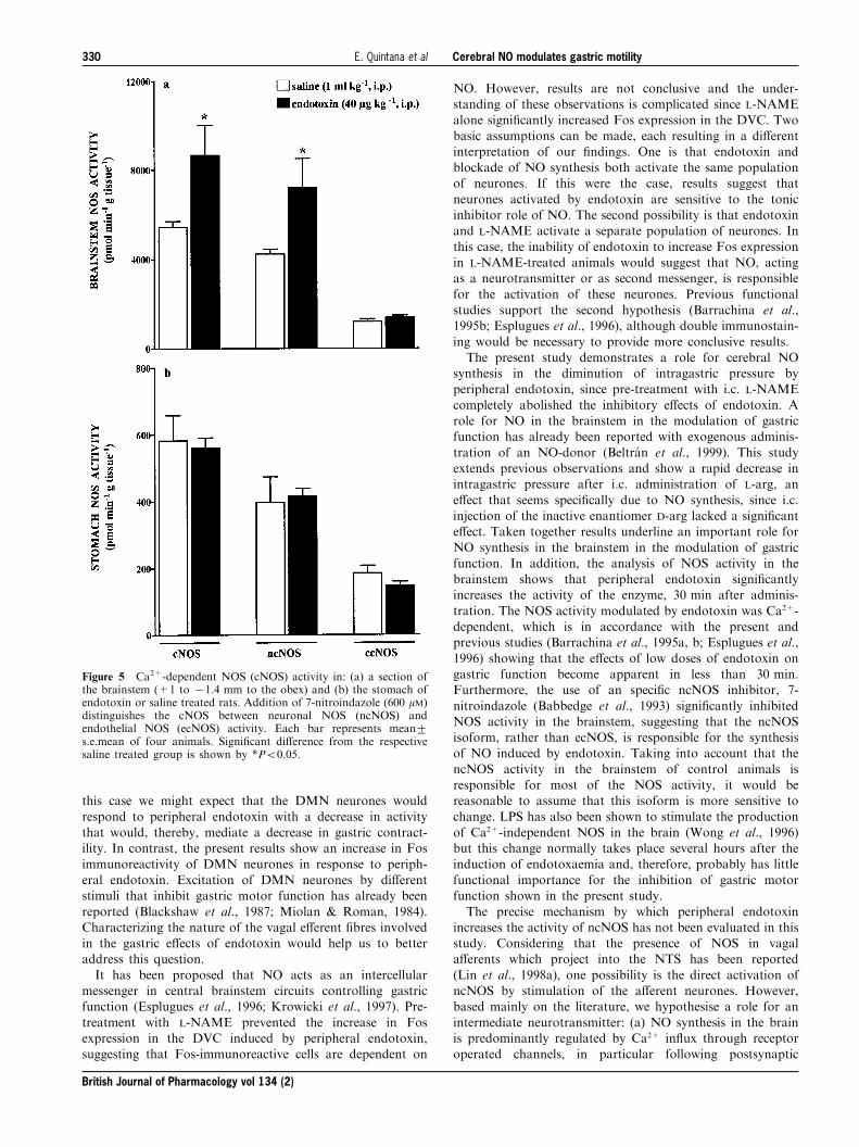
this case we might expect that the DMN neurones would
respond to peripheral endotoxin with a decrease in activitythat would, thereby, mediate a decrease in gastric contract-ility. In contrast, the present results show an increase in Fos
immunoreactivity of DMN neurones in response to periph-eral endotoxin. Excitation of DMN neurones by di�erentstimuli that inhibit gastric motor function has already been
reported (Blackshaw et al., 1987; Miolan & Roman, 1984).Characterizing the nature of the vagal e�erent ®bres involvedin the gastric e�ects of endotoxin would help us to betteraddress this question.
It has been proposed that NO acts as an intercellularmessenger in central brainstem circuits controlling gastricfunction (Esplugues et al., 1996; Krowicki et al., 1997). Pre-
treatment with L-NAME prevented the increase in Fosexpression in the DVC induced by peripheral endotoxin,suggesting that Fos-immunoreactive cells are dependent on
NO. However, results are not conclusive and the under-standing of these observations is complicated since L-NAMEalone signi®cantly increased Fos expression in the DVC. Two
basic assumptions can be made, each resulting in a di�erentinterpretation of our ®ndings. One is that endotoxin andblockade of NO synthesis both activate the same populationof neurones. If this were the case, results suggest that
neurones activated by endotoxin are sensitive to the tonicinhibitor role of NO. The second possibility is that endotoxinand L-NAME activate a separate population of neurones. In
this case, the inability of endotoxin to increase Fos expressionin L-NAME-treated animals would suggest that NO, actingas a neurotransmitter or as second messenger, is responsible
for the activation of these neurones. Previous functionalstudies support the second hypothesis (Barrachina et al.,1995b; Esplugues et al., 1996), although double immunostain-
ing would be necessary to provide more conclusive results.The present study demonstrates a role for cerebral NO
synthesis in the diminution of intragastric pressure byperipheral endotoxin, since pre-treatment with i.c. L-NAME
completely abolished the inhibitory e�ects of endotoxin. Arole for NO in the brainstem in the modulation of gastricfunction has already been reported with exogenous adminis-
tration of an NO-donor (Beltra n et al., 1999). This studyextends previous observations and show a rapid decrease inintragastric pressure after i.c. administration of L-arg, an
e�ect that seems speci®cally due to NO synthesis, since i.c.injection of the inactive enantiomer D-arg lacked a signi®cante�ect. Taken together results underline an important role for
NO synthesis in the brainstem in the modulation of gastricfunction. In addition, the analysis of NOS activity in thebrainstem shows that peripheral endotoxin signi®cantlyincreases the activity of the enzyme, 30 min after adminis-
tration. The NOS activity modulated by endotoxin was Ca2+-dependent, which is in accordance with the present andprevious studies (Barrachina et al., 1995a, b; Esplugues et al.,
1996) showing that the e�ects of low doses of endotoxin ongastric function become apparent in less than 30 min.Furthermore, the use of an speci®c ncNOS inhibitor, 7-
nitroindazole (Babbedge et al., 1993) signi®cantly inhibitedNOS activity in the brainstem, suggesting that the ncNOSisoform, rather than ecNOS, is responsible for the synthesisof NO induced by endotoxin. Taking into account that the
ncNOS activity in the brainstem of control animals isresponsible for most of the NOS activity, it would bereasonable to assume that this isoform is more sensitive to
change. LPS has also been shown to stimulate the productionof Ca2+-independent NOS in the brain (Wong et al., 1996)but this change normally takes place several hours after the
induction of endotoxaemia and, therefore, probably has littlefunctional importance for the inhibition of gastric motorfunction shown in the present study.
The precise mechanism by which peripheral endotoxinincreases the activity of ncNOS has not been evaluated in thisstudy. Considering that the presence of NOS in vagala�erents which project into the NTS has been reported
(Lin et al., 1998a), one possibility is the direct activation ofncNOS by stimulation of the a�erent neurones. However,based mainly on the literature, we hypothesise a role for an
intermediate neurotransmitter: (a) NO synthesis in the brainis predominantly regulated by Ca2+ in¯ux through receptoroperated channels, in particular following postsynaptic
Figure 5 Ca2+-dependent NOS (cNOS) activity in: (a) a section ofthe brainstem (+1 to 71.4 mm to the obex) and (b) the stomach ofendotoxin or saline treated rats. Addition of 7-nitroindazole (600 mM)distinguishes the cNOS between neuronal NOS (ncNOS) andendothelial NOS (ecNOS) activity. Each bar represents mean+s.e.mean of four animals. Signi®cant di�erence from the respectivesaline treated group is shown by *P50.05.
British Journal of Pharmacology vol 134 (2)
Cerebral NO modulates gastric motilityE. Quintana et al330
stimulation by the excitatory neurotransmitter glutamate(Bredt & Snyder, 1989; Garthwaite et al., 1989). (b) Recentstudies report that glutamate is the primary a�erent
neurotransmitter released in the NTS in the control of gastricfunction (Zheng et al., 1999). (c) The release of glutamate inthe brainstem by peripheral endotoxin has been reported(Mascarucci et al., 1998) and we have recently shown that
endogenous glutamate activates NO synthesis in the brain,which is in turn responsible for the inhibition of gastric acidsecretion by peripheral endotoxin (Garcõ a-Zaragoza et al.,
2000). These observations, considered with a previous studyreporting an inhibitory role on gastric motor function forglutamate acting in the NTS (Raybould et al., 1989) lead us
to believe that endogenous release of this neurotransmitter isinvolved in the activation of ncNOS in the brainstem byperipheral endotoxin.
Irrespective of the a�erent pathways involved, oncesynthesized in the brainstem, NO is involved in thediminution of intragastric pressure by endotoxin. NO in theDVC has been shown to modulate the ®ring rate of DMN
neurons (Travagli & Gillis, 1994). Such a modulation impliesactivation of e�erent vagal preganglionic ®bres that wouldsynapse with NANC inhibitory postganglionic neurones in
the myenteric plexus. NO has also been consolidated as oneof the non-adrenergic non-cholinergic (NANC) inhibitoryneurotransmitters of the gastrointestinal tract. In the present
study, systemic administration of L-NAME signi®cantlyprevented the reduction in intragastric pressure induced byendotoxin, suggesting a peripheral role for NO. However,
taking into account that i.v. administration of L-NAMEinduces Fos expression in the DVC of the brainstem, and
that similar doses of systemic L-NAME have been shown toa�ect the spontaneous discharge rate of NTS neurones (Maet al., 1995), we cannot deduce the release of NO in the
periphery. In addition, this role seems improbable sinceanalysis of NOS activity in the stomach shows no change inthe activity of any NOS isoform when evaluated 30 min afterinjection of endotoxin, a time interval in which changes in
gastric function are already apparent. Regardless of whetheror not activation of an inhibitory e�erent neural pathwaytakes place in the inhibition of gastric motor function by
endotoxin, the peripheral release of NO does not seem to beinvolved. Endogenous mediators such as VIP have also beenreported as inhibitory neurotransmitters of the NANC
innervation of the gastrointestinal tract. Further experimentsare required to address this question.In summary, the present results suggest that low doses of
endotoxin reduce gastric contractility through activation ofboth capsaicin-sensitive a�erent neurones and ncNOS in thebrainstem. This inhibition may be part of the mechanisminvolved in the delay in gastric emptying associated with
moderate endotoxaemia.
We would like to thank Prof C. Estrada and F. Portillo for theirvaluable help in the Fos immunohistochemistry. The present studyhas been supported by grants SAF 99-0114, SAF 98-0118 and1FD97-1029 from Ministerio de Educacio n y Cultura. E. Quintanaand E. Garcõ a-Zaragoza are the recipients of a fellowship fromMinisterio de Sanidad y Consumo and Ministerio de Educacio n yCultura, respectively. Part of this study has been supported by theRoyal Academy of Medicine of University of Valencia.
References
BABBEDGE, R.C., BLAND-WARD, P.A., HART, S.L. & MOORE, P.K.
(1993). Inhibition of rat cerebellar nitric oxide synthase by 7-nitroindazole and related substituted indazoles. Br. J. Pharma-col., 110, 225 ± 228.
BARRACHINA, M.D., CALATAYUD, S., MORENO, L., MARTIÂ NEZ-
CUESTA, M.A., WHITTLE, B.J.R. & ESPLUGUES, J.V. (1995a).Nitric oxide and sensory a�erent neurones modulate theprotective e�ects of low-dose endotoxin on rat gastric mucosaldamage. Eur. J. Pharmacol., 280, 339 ± 342.
BARRACHINA, M.D., MARTIÂ NEZ, V., WANG, L., WEI, J.Y. & TACHEÂ ,
Y. (1997). Synergistic interaction between leptin and cholecysto-kinin to reduce short-term food intake in lean mice. Proc. Natl.Acad. Sci. U.S.A., 94, 10455 ± 10460.
BARRACHINA, M.D., PANEÂ S, J. & ESPLUGUES, J.V. (2001). Role ofnitric oxide in gastrointestinal in¯ammatory and ulcerativediseases: perspective for drugs development. Curr. Pharmaceu-tical Design, 7, 31 ± 48.
BARRACHINA, M.D., WHITTLE, B.J.R., MONCADA, S. & ESPLU-
GUES, J.V. (1995b). Endotoxin inhibition of distension-stimu-lated gastric acid secretion in rat: mediation by NO in the centralnervous system. Br. J. Pharmacol., 114, 8 ± 12.
BELTRAÂ N, B., BARRACHINA, M.D., MEÂ NDEZ, A. & QUINTERO, E.
(1999). Synthesis of nitric oxide in the dorsal motor nucleus of thevagus mediates the inhibition of gastric acid secretion by centralbombesin. Br. J. Pharmacol., 127, 1603 ± 1610.
BLACKSHAW, L.A., GRUNDY, D. & SCRATCHERD, T. (1987).Involvement of gastrointestinal mechano- and intestinal chemor-eceptors in vagal re¯exes: an electrophysiological study. J. Aut.Nerv. Syst., 18, 225 ± 234.
BREDT, D.S. & SNYDER, S.H. (1989). Nitric oxide mediatesglutamate-linked enhancement of cGMP levels in the cerebellum.Proc. Natl. Acad. Sci. U.S.A., 86, 9030 ± 9033.
CALATAYUD, S., BARRACHINA, M.D., GARCIÂ A-ZARAGOZAÂ , E.,
QUINTANA, E. & ESPLUGUES, J.V. (2001). Endotoxin inhibitsgastric emptying in rats via a capsaicin-sensitive a�erent path-way. Naunyn-Smied. Arch. Pharmacol., 363, 276 ± 280.
ESPLUGUES, J.V., BARRACHINA, M.D., BELTRAÂ N, B., CALA-
TAYUD, S., WHITTLE, B.J.R. & MONCADA, S. (1996). Inhibitionof gastric acid secretion by stress: a protective re¯ex mediated bycerebral nitric oxide. Proc. Natl. Acad. Sci. U.S.A., 93, 14839 ±14844.
GARCIÂ A-ZARAGOZAÂ , E., BARRACHINA, M.D., MORENO, L. &
ESPLUGUES, J.V. (2000). Role of central glutamate receptors,nitric oxide and soluble guanylyl cyclase in the inhibition byendotoxin of rat gastric acid secretion. Br. J. Pharmacol., 130,1283 ± 1288.
GARTHWAITE, J., CHARLES, S.L. & CHESS-WILLIAMS, R. (1988).Endothelium derived relaxing factor release on activation ofNMDA receptors suggests a role as intercellular messenger in thebrain. Nature, 336, 385 ± 388.
GARTHWAITE, J., GARTHWAITE, G., PALMER, R.M. & MONCADA,
S. (1989). NMDA receptor activation induces nitric oxidesynthesis from arginine in rat brain slices. Eur. J. Pharmacol.,172, 413 ± 416.
HERMANN, G.E., EMCH, G.S., TOVAR, C.A. & ROGERS, R.C. (2001).C-fos generation in the dorsal vagal complex after systemicendotoxin is not dependent on the vagus nerve. Am. J. Physiol.,280, R289 ±R299.
HERMANN, G.E., TOVAR, C.A. & ROGERS, R.C. (1999). Induction ofendogenous tumor necrosis factor-a: suppression of centrallystimulated gastric motility. Am. J. Physiol., 276, R59 ±R68.
British Journal of Pharmacology vol 134 (2)
Cerebral NO modulates gastric motilityE. Quintana et al 331
HOLZER, P. (1988). Local e�ector functions of capsaicin-sensitivesensory nerve endings: involvement of tachykinins, calcitoningene-related peptide and other neurotransmitters. Neuroscience,24, 739 ± 768.
HOLZER, P. (1998). Neural emergency system in the stomach.Gastroenterology, 114, 823 ± 839.
HSU, S.M., RAINE, L. & FANGER, H. (1981). Use of avidin-biotin-peroxidase complex (ABC) in immunoperoxidase techniques: acomparison between ABC and unlabeled antibody (PAPprocedures). J. Histochem. Cytochem., 29, 577 ± 580.
KALIA, M. & MESULAM, M.M. (1980). Brain Stem projections ofsensory and motor components of the vagus complex in the cat:the cervical vagus and nodose ganglion. J. Comp. Neurol., 193,435 ± 465.
KROWICKI, Z.K. & HORNBY, P.J. (1996). The inhibitory e�ect ofsubstance P on gastric motor function in the nucleus rapheobscurus is mediated via nitric oxide in the dorsal vagal complex.J. Aut. Nerv. Syst., 58, 177 ± 180.
KROWICKI, Z.K., SHARKEY, K.A., SERRON, S.C., NATHAN, N.A. &
HORNBY, P.J. (1997). Distribution of nitric oxide synthase in ratdorsal vagal complex and e�ects of microinjection of nitric oxidecompounds upon gastric motor function. J. Comp. Neurol., 377,49 ± 69.
LIN, L.H., CASSELL, M.D., SANDRA, A. & TALMAN, W.T. (1998a).Direct evidence for nitric oxide synthase in vagal a�erents to thenucleus tractus solitarii. Neuroscience, 84, 549 ± 558.
LIN, H.C., WAN, F.J., KANG, B.H., WU, C.C. & TSENG, C.J. (1998b).Systemic administration of lipopolysaccharide induces release ofnitric oxide and glutamate and c-fos expression in the nucleustractus solitarii of rats. Hypertension, 33, 1218 ± 1224.
MA, S., ABBOUD, F.M. & FELDER, R.B. (1995). E�ects of L-arginine-derived nitric oxide synthesis on neuronal activity in nucleustractus solitarius. Am. J. Physiol., 268, R487 ±R491.
MARTIÂ NEZ-CUESTA, M.A., BARRACHINA, M.D., WHITTLE, B.J.R.,
PIQUEÂ , J.M. & ESPLUGUES, J.V. (1994). Involvement of neuronalprocesses and nitric oxide in the inhibition by endotoxin ofpentagastrin-stimulated gastric acid secretion. Naunyn-Smied.Arch. Pharmacol., 349, S23 ± S27.
MASCARUCCI, P., PEREGO, C., TERRAZZINO, S. & DE SIMONI, M.G.
(1998). Glutamate release in the nucleus tractus solitarius isinduced by peripheral lipopolysaccharide and interleukin-lb.Neuroscience, 86, 1285 ± 1290.
MIOLAN, J.P. & ROMAN, C. (1984). The role of esophageal andintestinal receptors in the control of gastric motility. J. Aut. Ner.Syst., 10, 235 ± 241.
MONCADA, S., PALMER, R.M. & HIGGS, E.A. (1991). Nitric oxide:physiology, pathophysiology and pharmacology. Pharmacol.Rev., 43, 109 ± 142.
RAYBOULD, H.E., JAKOBSEN, L.J., NOVIN, D. & TACHEÂ , Y. (1989).TRH stimulation and L-glutamic acid inhibition of proximalgastric motor activity in the rat dorsal vagal complex. Brain Res.,495, 319 ± 328.
SALTER, M., KNOWLES, R.G. & MONCADA, S. (1990). Widespreadtissue distribution, species distribution and changes in activity ofcalcium-dependent and calcium-independent nitric oxidesynthases. FEBS Lett., 291, 145 ± 149.
SWANSON, L.W. (1998). Brain maps: structure of the rat brain. Theneuroscience program; Elsevier, Los Angeles.
TRAVAGLI, R.A. & GILLIS, R.A. (1994). Nitric oxide mediatedexcitatory e�ect on neurones of dorsal motor nucleus of thevagus. Am. J. Physiol., 266, G154 ±G160.
URIBE, R.M., LEE, S. & RIVIER, C. (1999). Endotoxin stimulatesnitric oxide production in the paraventricular nucleus of thehypothalamus through nitric oxide synthase I: correlation withhypothalamic-pituitary-adrenal axis activation. Endocrinology,140, 5971 ± 5981.
WONG, M.L., RETTORI, V., AL-SHEKHLEE, A., BONGIORNO, P.B.,
CANTEROS, G., McCANN, S.M., GOLD, P.W. & LICINIO, J. (1996).Inducible nitric oxide synthase gene expression in the brainduring systemic in¯ammation. Nat. Med., 2, 581 ± 584.
ZHANG, X., RENETHAN, W.E. & FOGEL, R. (1998). Neurons in thevagal complex of the rat respond to mechanical and chemicalstimulation of the GI tract. Am. J. Physiol., 274, G331 ±G341.
ZHENG, H., KELLY, L., PATTERSON, L.M. & BERTHOUD, H.R.
(1999). E�ect of brainstem NMDA-receptor blockade by MK-801 on behavioural and fos responses to vagal satiety signals. Am.J. Physiol., 277, R1104 ±R1111.
(Received April 10, 2001Revised June 19, 2001
Accepted June 26, 2001)
British Journal of Pharmacology vol 134 (2)
Cerebral NO modulates gastric motilityE. Quintana et al332
Artículos de investigación
51
Artículo 2
“Transcriptional up-regulation of nNOS in
the dorsal vagal complex during low
endotoxemia”
Elsa Quintana, Carlos Hernández, Anthony P. Moran, Juan V. Esplugues &
María D. Barrachina
Neurogastroenterology and motility (2004, en revisión).
Neurogastroenterology & Motility (2004) Quintana et al. 1
Transcriptional up-regulation of nNOS in the dorsal
vagal complex during low endotoxemia
Elsa Quintana*, Carlos Hernández†, Anthony P. Moran††, Juan V. Esplugues† and María
D. Barrachina†
*Departamento de Farmacología and † Unidad Mixta CNIC-UVEG, Facultad de
Medicina, Universidad de Valencia, Valencia, Spain, and †† Department of
Microbiology, National University of Ireland, Galway, Ireland.
Short title: LPS up-regulates nNOS in the brainstem
Corresponding author: María D. Barrachina.
Department of Pharmacology, Faculty of Medicine,
Avd. Blasco Ibáñez 15, 46010 Valencia, Spain
E-mail: [email protected]
Fax: 34 96-3864625
Phone: 34 96-3983779
Neurogastroenterology & Motility (2004) Quintana et al. 2
ABSTRACT
The present study analyses neuronal nitric oxide synthase (nNOS) expression and
distribution in the brainstem of animals pre-treated with Escherichia coli or
Helicobacter pylori LPS, at doses that modulate gastric motor function. Systemic
administration of H. pylori LPS prevented in a dose-dependent manner (5, 40 and 100
µg kg-1, i.v) the increase in intragastric pressure induced by 2-deoxy-D-glucose (200 mg
kg-1, i.v.) in urethane-anaesthetized rats. Quantitative analysis showed a significant
increase in the amount of nNOS mRNA induced by E. coli or H. pylori LPS (2 h later),
in a section of the brainstem containing the dorsal vagal complex (DVC).
Immunohistochemical studies showed nNOS presence in the DVC of vehicle-treated
rats. Both E. coli (40 µg kg-1, i.p.) and H. pylori LPS (100 µg kg-1, i.p) significantly
increased (2 h later) the number of nNOS immunoreactive cells in this area, mainly at
the most rostral level. The present study shows that systemic administration of E. coli or
H. pylori LPS induces a transcriptional up-regulation of the nNOS in the DVC of the
brainstem and suggest a role for NO synthesis in this area in the control of gastric motor
function under endotoxemia.
Keywords: brain-gut; dorsal motor nucleus; dorsal vagal complex; gastric motor
function; neuronal nitric oxide synthase; nucleus of the solitary tract.
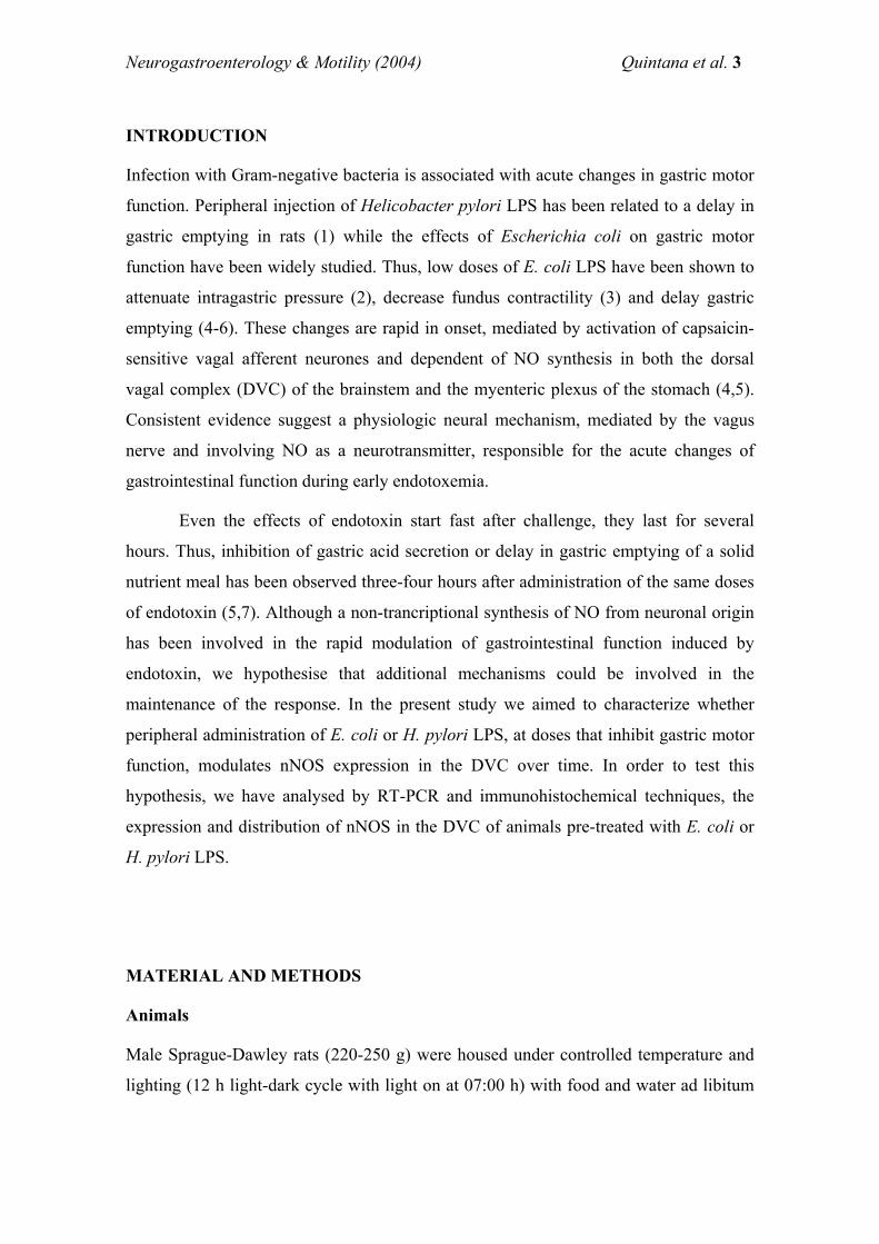
Neurogastroenterology & Motility (2004) Quintana et al. 3
INTRODUCTION
Infection with Gram-negative bacteria is associated with acute changes in gastric motor
function. Peripheral injection of Helicobacter pylori LPS has been related to a delay in
gastric emptying in rats (1) while the effects of Escherichia coli on gastric motor
function have been widely studied. Thus, low doses of E. coli LPS have been shown to
attenuate intragastric pressure (2), decrease fundus contractility (3) and delay gastric
emptying (4-6). These changes are rapid in onset, mediated by activation of capsaicin-
sensitive vagal afferent neurones and dependent of NO synthesis in both the dorsal
vagal complex (DVC) of the brainstem and the myenteric plexus of the stomach (4,5).
Consistent evidence suggest a physiologic neural mechanism, mediated by the vagus
nerve and involving NO as a neurotransmitter, responsible for the acute changes of
gastrointestinal function during early endotoxemia.
Even the effects of endotoxin start fast after challenge, they last for several
hours. Thus, inhibition of gastric acid secretion or delay in gastric emptying of a solid
nutrient meal has been observed three-four hours after administration of the same doses
of endotoxin (5,7). Although a non-trancriptional synthesis of NO from neuronal origin
has been involved in the rapid modulation of gastrointestinal function induced by
endotoxin, we hypothesise that additional mechanisms could be involved in the
maintenance of the response. In the present study we aimed to characterize whether
peripheral administration of E. coli or H. pylori LPS, at doses that inhibit gastric motor
function, modulates nNOS expression in the DVC over time. In order to test this
hypothesis, we have analysed by RT-PCR and immunohistochemical techniques, the
expression and distribution of nNOS in the DVC of animals pre-treated with E. coli or
H. pylori LPS.
MATERIAL AND METHODS
Animals
Male Sprague-Dawley rats (220-250 g) were housed under controlled temperature and
lighting (12 h light-dark cycle with light on at 07:00 h) with food and water ad libitum
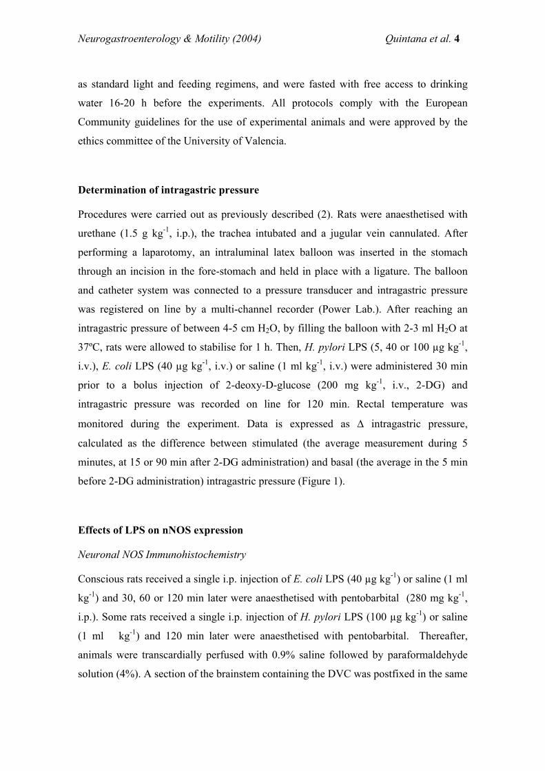
Neurogastroenterology & Motility (2004) Quintana et al. 4
as standard light and feeding regimens, and were fasted with free access to drinking
water 16-20 h before the experiments. All protocols comply with the European
Community guidelines for the use of experimental animals and were approved by the
ethics committee of the University of Valencia.
Determination of intragastric pressure
Procedures were carried out as previously described (2). Rats were anaesthetised with
urethane (1.5 g kg-1, i.p.), the trachea intubated and a jugular vein cannulated. After
performing a laparotomy, an intraluminal latex balloon was inserted in the stomach
through an incision in the fore-stomach and held in place with a ligature. The balloon
and catheter system was connected to a pressure transducer and intragastric pressure
was registered on line by a multi-channel recorder (Power Lab.). After reaching an
intragastric pressure of between 4-5 cm H2O, by filling the balloon with 2-3 ml H2O at
37ºC, rats were allowed to stabilise for 1 h. Then, H. pylori LPS (5, 40 or 100 µg kg-1,
i.v.), E. coli LPS (40 µg kg-1, i.v.) or saline (1 ml kg-1, i.v.) were administered 30 min
prior to a bolus injection of 2-deoxy-D-glucose (200 mg kg-1, i.v., 2-DG) and
intragastric pressure was recorded on line for 120 min. Rectal temperature was
monitored during the experiment. Data is expressed as ∆ intragastric pressure,
calculated as the difference between stimulated (the average measurement during 5
minutes, at 15 or 90 min after 2-DG administration) and basal (the average in the 5 min
before 2-DG administration) intragastric pressure (Figure 1).
Effects of LPS on nNOS expression
Neuronal NOS Immunohistochemistry
Conscious rats received a single i.p. injection of E. coli LPS (40 µg kg-1) or saline (1 ml
kg-1) and 30, 60 or 120 min later were anaesthetised with pentobarbital (280 mg kg-1,
i.p.). Some rats received a single i.p. injection of H. pylori LPS (100 µg kg-1) or saline
(1 ml kg-1) and 120 min later were anaesthetised with pentobarbital. Thereafter,
animals were transcardially perfused with 0.9% saline followed by paraformaldehyde
solution (4%). A section of the brainstem containing the DVC was postfixed in the same
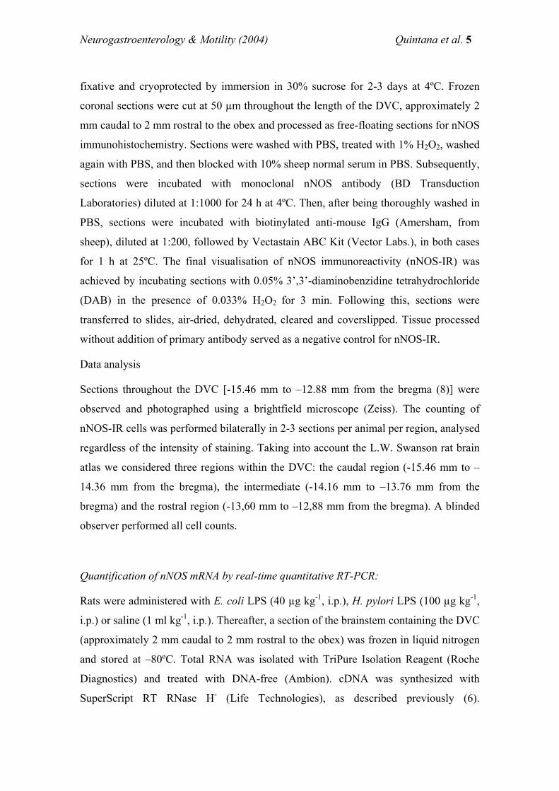
Neurogastroenterology & Motility (2004) Quintana et al. 5
fixative and cryoprotected by immersion in 30% sucrose for 2-3 days at 4ºC. Frozen
coronal sections were cut at 50 µm throughout the length of the DVC, approximately 2
mm caudal to 2 mm rostral to the obex and processed as free-floating sections for nNOS
immunohistochemistry. Sections were washed with PBS, treated with 1% H2O2, washed
again with PBS, and then blocked with 10% sheep normal serum in PBS. Subsequently,
sections were incubated with monoclonal nNOS antibody (BD Transduction
Laboratories) diluted at 1:1000 for 24 h at 4ºC. Then, after being thoroughly washed in
PBS, sections were incubated with biotinylated anti-mouse IgG (Amersham, from
sheep), diluted at 1:200, followed by Vectastain ABC Kit (Vector Labs.), in both cases
for 1 h at 25ºC. The final visualisation of nNOS immunoreactivity (nNOS-IR) was
achieved by incubating sections with 0.05% 3’,3’-diaminobenzidine tetrahydrochloride
(DAB) in the presence of 0.033% H2O2 for 3 min. Following this, sections were
transferred to slides, air-dried, dehydrated, cleared and coverslipped. Tissue processed
without addition of primary antibody served as a negative control for nNOS-IR.
Data analysis
Sections throughout the DVC [-15.46 mm to –12.88 mm from the bregma (8)] were
observed and photographed using a brightfield microscope (Zeiss). The counting of
nNOS-IR cells was performed bilaterally in 2-3 sections per animal per region, analysed
regardless of the intensity of staining. Taking into account the L.W. Swanson rat brain
atlas we considered three regions within the DVC: the caudal region (-15.46 mm to –
14.36 mm from the bregma), the intermediate (-14.16 mm to –13.76 mm from the
bregma) and the rostral region (-13,60 mm to –12,88 mm from the bregma). A blinded
observer performed all cell counts.
Quantification of nNOS mRNA by real-time quantitative RT-PCR:
Rats were administered with E. coli LPS (40 µg kg-1, i.p.), H. pylori LPS (100 µg kg-1,
i.p.) or saline (1 ml kg-1, i.p.). Thereafter, a section of the brainstem containing the DVC
(approximately 2 mm caudal to 2 mm rostral to the obex) was frozen in liquid nitrogen
and stored at –80ºC. Total RNA was isolated with TriPure Isolation Reagent (Roche
Diagnostics) and treated with DNA-free (Ambion). cDNA was synthesized with
SuperScript RT RNase H- (Life Technologies), as described previously (6).
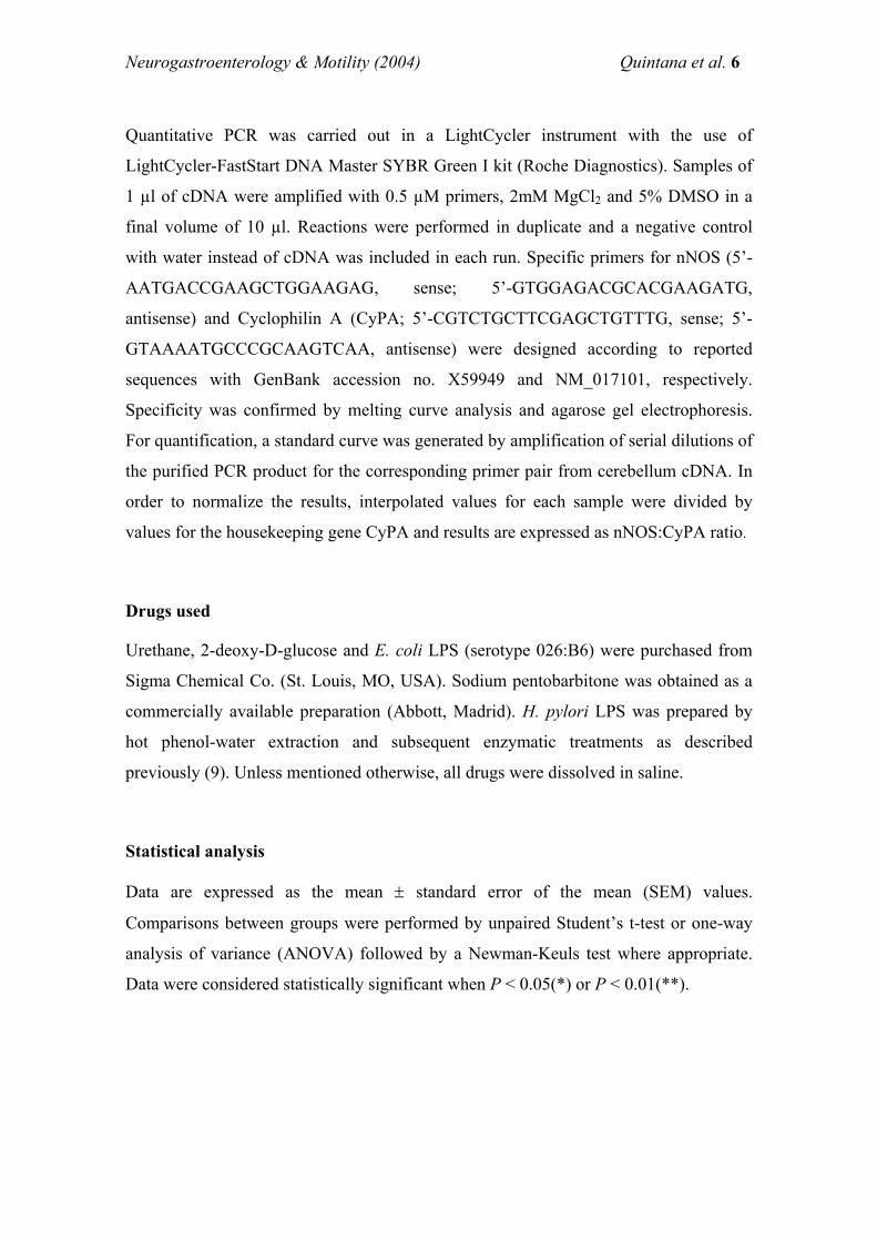
Neurogastroenterology & Motility (2004) Quintana et al. 6
Quantitative PCR was carried out in a LightCycler instrument with the use of
LightCycler-FastStart DNA Master SYBR Green I kit (Roche Diagnostics). Samples of
1 µl of cDNA were amplified with 0.5 µM primers, 2mM MgCl2 and 5% DMSO in a
final volume of 10 µl. Reactions were performed in duplicate and a negative control
with water instead of cDNA was included in each run. Specific primers for nNOS (5’-
AATGACCGAAGCTGGAAGAG, sense; 5’-GTGGAGACGCACGAAGATG,
antisense) and Cyclophilin A (CyPA; 5’-CGTCTGCTTCGAGCTGTTTG, sense; 5’-
GTAAAATGCCCGCAAGTCAA, antisense) were designed according to reported
sequences with GenBank accession no. X59949 and NM_017101, respectively.
Specificity was confirmed by melting curve analysis and agarose gel electrophoresis.
For quantification, a standard curve was generated by amplification of serial dilutions of
the purified PCR product for the corresponding primer pair from cerebellum cDNA. In
order to normalize the results, interpolated values for each sample were divided by
values for the housekeeping gene CyPA and results are expressed as nNOS:CyPA ratio.
Drugs used
Urethane, 2-deoxy-D-glucose and E. coli LPS (serotype 026:B6) were purchased from
Sigma Chemical Co. (St. Louis, MO, USA). Sodium pentobarbitone was obtained as a
commercially available preparation (Abbott, Madrid). H. pylori LPS was prepared by
hot phenol-water extraction and subsequent enzymatic treatments as described
previously (9). Unless mentioned otherwise, all drugs were dissolved in saline.
Statistical analysis
Data are expressed as the mean ± standard error of the mean (SEM) values.
Comparisons between groups were performed by unpaired Student’s t-test or one-way
analysis of variance (ANOVA) followed by a Newman-Keuls test where appropriate.
Data were considered statistically significant when P < 0.05(*) or P < 0.01(**).
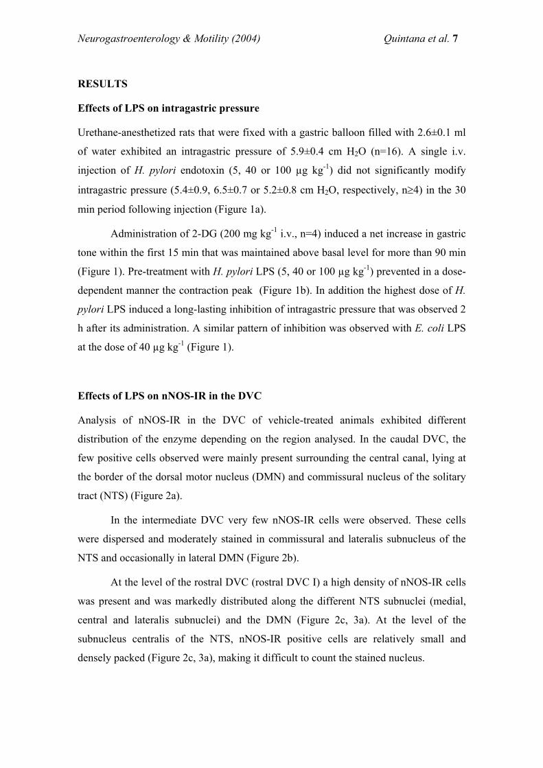
Neurogastroenterology & Motility (2004) Quintana et al. 7
RESULTS
Effects of LPS on intragastric pressure
Urethane-anesthetized rats that were fixed with a gastric balloon filled with 2.6±0.1 ml
of water exhibited an intragastric pressure of 5.9±0.4 cm H2O (n=16). A single i.v.
injection of H. pylori endotoxin (5, 40 or 100 µg kg-1) did not significantly modify
intragastric pressure (5.4±0.9, 6.5±0.7 or 5.2±0.8 cm H2O, respectively, n≥4) in the 30
min period following injection (Figure 1a).
Administration of 2-DG (200 mg kg-1 i.v., n=4) induced a net increase in gastric
tone within the first 15 min that was maintained above basal level for more than 90 min
(Figure 1). Pre-treatment with H. pylori LPS (5, 40 or 100 µg kg-1) prevented in a dose-
dependent manner the contraction peak (Figure 1b). In addition the highest dose of H.
pylori LPS induced a long-lasting inhibition of intragastric pressure that was observed 2
h after its administration. A similar pattern of inhibition was observed with E. coli LPS
at the dose of 40 µg kg-1 (Figure 1).
Effects of LPS on nNOS-IR in the DVC
Analysis of nNOS-IR in the DVC of vehicle-treated animals exhibited different
distribution of the enzyme depending on the region analysed. In the caudal DVC, the
few positive cells observed were mainly present surrounding the central canal, lying at
the border of the dorsal motor nucleus (DMN) and commissural nucleus of the solitary
tract (NTS) (Figure 2a).
In the intermediate DVC very few nNOS-IR cells were observed. These cells
were dispersed and moderately stained in commissural and lateralis subnucleus of the
NTS and occasionally in lateral DMN (Figure 2b).
At the level of the rostral DVC (rostral DVC I) a high density of nNOS-IR cells
was present and was markedly distributed along the different NTS subnuclei (medial,
central and lateralis subnuclei) and the DMN (Figure 2c, 3a). At the level of the
subnucleus centralis of the NTS, nNOS-IR positive cells are relatively small and
densely packed (Figure 2c, 3a), making it difficult to count the stained nucleus.
Neurogastroenterology & Motility (2004) Quintana et al. 8
At the level where the DVC is just beginning to migrate away from the fourth
ventricle (rostral DVC II), there were numerous densely stained nNOS-IR cells
throughout the DVC; however no apparent organisation of the positively stained cells
was observed and we proceeded with counting in the whole area (Figure 2d,e; 3b).
Systemic pre-treatment with endotoxin from E. coli (40 µg kg-1, i.p.) or H. pylori
(100 µg kg-1, i.p.) induced, 2 h later, a generalized increase in nNOS-IR in the DVC,
and was the greatest at the most rostral level (Figure 3). Thirty or sixty min after E. coli
or H. pylori LPS administration, there were not significant changes in nNOS-positive
cells in the DVC, compared with control animals (Figure 3g).
Effects of LPS on nNOS mRNA expression in the DVC
Expression of nNOS mRNA was detected by real-time quantitative RT-PCR in the
selected section of the brainstem containing the DVC. A single i.p. injection of
endotoxin from both E. coli (40 µg kg-1) or H. pylori (100 µg kg-1) induced a significant
increase 2 h later in the amount of nNOS mRNA in this section (Figure 4).
DISCUSSION
Lipopolysaccharides are a characteristic cell wall constituent of Gram-negative bacteria
which are important in the structure and function of the outer membrane (10). Both E.
coli and H. pylori are Gram-negative bacteria that cause pathophysiological disorders.
However, E. coli is usually present in the intestine and just after access to blood triggers
on a generalized systemic reaction, while H. pylori colonizes gastric mucosa and
constitutes a ethiophatogenic factor in gastric ulcer (11). In the present study we
demonstrate that both, E. coli and H. pylori LPS, at doses that inhibit gastric motor
function, induce a transcriptional up-regulation of nNOS in the DVC of the brainstem.
Systemic administration of H. pylori LPS to anaesthetized rats prevented in a
dose-dependent manner the increase in intragastric pressure induced by 2-DG. It seems
likely that the rapid diminution of gastric tone observed in the present study is involved
in the mechanism induced by H. pylori LPS to delay gastric emptying (1). Previous
Neurogastroenterology & Motility (2004) Quintana et al. 9
studies have reported a similar effect with E. coli LPS (2). Although differences in the
structure of H. pylori LPS from those of E. coli have been shown (12), in the present
study slightly higher doses of H. pylori LPS have been necessary in order to get a
similar degree of gastric inhibition. In line with these observations, the present study
demonstrates that both H. pylori and E. coli LPS, at doses that inhibit gastric motor
function induce a similar pattern of nNOS expression in the DVC of the brainstem. A
detailed analysis of the distribution of the nNOS-IR along the DVC of control animals
showed a majority of positive cells at the most rostral level. A single intraperitoneal
injection of both E. coli and H. pylori LPS induced two hours later a significant increase
in the number of nNOS-IR positive cells at the most rostral level, while it was not
observed earlier. Such an increase was correlated with a higher amount of nNOS mRNA
in the same area, suggesting that both H. pylori and E. coli LPS induce two hours later a
transcriptional up-regulation of the nNOS in the brainstem.
The DVC comprises the two interacting nuclei NTS and DMN, and the increase
in nNOS-IR induced by LPS has been observed in both. NTS constitutes the first point
of synapse of the vagus, carrying sensory information from numerous peripheral
structures (13) and strong evidence supports a role for the vagus nerve in brainstem
modulation induced by peripheral LPS, since: (a) endotoxin-induced c-fos in central
nuclei involved in autonomic functions is mediated by the vagus nerve (14,15); (b)
primary afferent neurones of the vagus nerve express fos immunoreactivity in response
to peripheral endotoxin (16); and (c) activation of capsaicin sensitive vagal afferent
neurones has been involved in the inhibition of gastric tone induced by peripheral
endotoxin (2). Although a direct effect of LPS or LPS-induced cytokines on nNOS gene
expression can not be ruled out, the rapid up-regulation of nNOS observed in the
present study led us to hypothesize that a nervous mechanism mediated by the vagus is
involved in the regulation of nNOS expression in the NTS by peripheral LPS. In
accordance with this hypothesis, a role for the vagus nerve in the regulation of nNOS
expression in the myenteric plexus has been reported (17).
In the NTS, pre- and post-synaptic structures, as well as intrinsic neurones have
been shown to express nNOS-IR (18). In the present study, the identity of the structures
expressing nNOS in the NTS of endotoxin-treated rats has not been analysed. However,
evidence suggests that these structures are not of vagal origin since: (a) pre-synaptic
Neurogastroenterology & Motility (2004) Quintana et al. 10
nNOS-IR terminals in the NTS are not vagal afferent in origin (18); (b) strong evidence
support a role for glutamate as the primary afferent neurotransmitter in the NTS (19);
(c) endotoxin, at the doses used in the present study, has been shown to increase the
release of glutamate in the brainstem (20) which in turn activates the nNOS (21); and
(d) intracisternal injection of glutamate has been shown to modulate gastric function
through an increased NO synthesis in the brainstem (21). Collectively, these results and
previous studies suggest that nNOS-IR in the NTS of endotoxin-treated animals could
be localized in other structures rather than vagal afferent terminals and modulation of
nNOS expression and activity in the NTS could be due to glutamate receptor
stimulation released from vagal afferent terminals (20).
In addition to the NTS, in the rostral DMN it was observed an increase in the
number of nNOS-IR positive cells, two hours after LPS administration. An increased in
nNOS protein amount in this nucleus has previously been related to changes in gastric
function induced by hyperthermia (22). In line with these observations, and taking into
account that inhibition of intragastric pressure is present two hours after challenge, a
correlation between up-regulation of nNOS in the DVC and inhibition of intragastric
pressure may be established. However it is important to note that even such a
correlation exists, alternative mechanisms must also be involved since the present study
demonstrates that diminution in intragastric pressure by LPS starts to be apparent thirty
minutes after challenge, moment in which no changes in nNOS IR in the DVC are
observed. Interestingly, functional and biochemical studies have demonstrated that a
non-transcriptional regulation of NO synthesis in the brainstem is involved in the rapid
(one hour) inhibition of intragastric pressure by systemic administration of E. coli LPS
(2). Although the mechanism of the transcriptional regulation of the nNOS has not been
analysed, it is possible that it is a consequence of the initial neural mechanism activated
by LPS (17). Further studies are required to address this question.
In summary, the present study shows a transcriptional up-regulation of nNOS in
the DVC of the brainstem induced by systemic administration of low doses of E. coli or
H. pylori LPS. Modulation of the nNOS activity in these nuclei could be part of the
mechanism activated by the host in order to eliminate the challenge and restore
homeostasis.
Neurogastroenterology & Motility (2004) Quintana et al. 11
ACKNOWLEDGMENTS
This study has been supported by grant FIS 01/1187 from the Ministerio de Sanidad y
Consumo and grant C 03/02 from Instituto de Salud Carlos III. Elsa Quintana is the
recipient of a fellowship from the Ministerio de Sanidad y Consumo.
REFERENCES
1 Okumura T, Shoji E, Takahashi N, Wakebe H, Imagawa K, Kikuchi M, Kohgo Y. Delayed gastric emptying by Helicobacter pylori lipopolysaccharide in conscious rats. Dig Dis Sci 1998; 43: 90-94.
2 Quintana E, Garcia-Zaragoza E, Martinez-Cuesta MA, Calatayud S, Esplugues JV, Barrachina MD. A cerebral nitrergic pathway modulates endotoxin-induced changes in gastric motility. Br J Pharmacol 2001; 134: 325-332.
3 Quintana E, Hernandez C, Alvarez-Barrientos A, Esplugues JV, Barrachina MD. Synthesis of nitric oxide in post-ganglionic myenteric neurones during endotoxemia:implications in gastric motor function. The FASEB J 2004; 18: 531-533.
4 van Miert AS, De la Parra DA. Inhibition of gastric emptying by endotoxin (bacterial lipopolysaccharide) in conscious rats and modification of this response by drugs affecting the autonomic nervous system. Arch Int Pharmacodyn Ther 1970; 184: 27-33.
5 Calatayud S, Barrachina MD, Garcia-Zaragoza E, Quintana E, Esplugues JV. Endotoxin inhibits gastric emptying in rats via a capsaicin-sensitive afferent pathway. Naunyn Schmiedebergs Arch Pharmacol 2001; 363: 276-280.
6 Calatayud S, Garcia-Zaragoza E, Hernandez C, Quintana E, Felipo V, Esplugues JV, Barrachina MD. Downregulation of nNOS and synthesis of PGs associated with endotoxin- induced delay in gastric emptying. Am J Physiol Gastrointest Liver Physiol 2002; 283: G1360-G1367.
7 Barrachina MD, Whittle BJ, Moncada S, Esplugues JV. Endotoxin inhibition of distension-stimulated gastric acid secretion in rat: mediation by NO in the central nervous system. Br J Pharmacol 1995; 114: 8-12.
8 Swanson LW. Brain maps: structure of the rat brain. The neuroscience program. Los Angeles: Elsevier, 1998.
9 Moran AP, Helander IM, Kosunen TU. Compositional analysis of Helicobacter pylori rough-form lipopolysaccharides. J Bacteriol 1992; 174: 1370-1377.
10 Erridge C, Bennett-Guerrero E, Poxton IR. Structure and function of lipopolysaccharides. Microbes Infect 2002; 4: 837-851.
Neurogastroenterology & Motility (2004) Quintana et al. 12
11 Taylor DN, Blaser MJ. The epidemiology of Helicobacter pylori infection. Epidemiol Rev 1991; 13: 42-59.
12 Moran AP, Lindner B, Walsh EJ. Structural characterization of the lipid A component of Helicobacter pylori rough- and smooth-form lipopolysaccharides. J Bacteriol 1997; 179: 6453-6463.
13 Barraco R, el Ridi M, Ergene E, Parizon M, Bradley D. An atlas of the rat subpostremal nucleus tractus solitarius. Brain Res Bull 1992; 29: 703-765.
14 Elmquist JK, Scammell TE, Jacobson CD, Saper CB. Distribution of Fos-like immunoreactivity in the rat brain following intravenous lipopolysaccharide administration. J Comp Neurol 1996; 371: 85-103.
15 Wan W, Wetmore L, Sorensen CM, Greenberg AH, Nance DM. Neural and biochemical mediators of endotoxin and stress-induced c-fos expression in the rat brain. Brain Res Bull 1994; 34: 7-14.
16 Gaykema RP, Goehler LE, Tilders FJ, Bol JG, McGorry M, Fleshner M, Maier SF, Watkins LR. Bacterial endotoxin induces fos immunoreactivity in primary afferent neurons of the vagus nerve. Neuroimmunomodulation 1998; 5: 234-240.
17 Nakamura K, Takahashi T, Taniuchi M, Hsu CX, Owyang C. Nicotinic receptor mediates nitric oxide synthase expression in the rat gastric myenteric plexus. J Clin Invest 1998; 101: 1479-1489.
18 Atkinson L, Batten TF, Corbett EK, Sinfield JK, Deuchars J. Subcellular localization of neuronal nitric oxide synthase in the rat nucleus of the solitary tract in relation to vagal afferent inputs. Neuroscience 2003; 118: 115-122.
19 Chen CY, Bonham AC. Non-NMDA and NMDA receptors transmit area postrema input to aortic baroreceptor neurons in NTS. Am J Physiol 1998; 275: H1695-H1706.
20 Mascarucci P, Perego C, Terrazzino S, De Simoni MG. Glutamate release in the nucleus tractus solitarius induced by peripheral lipopolysaccharide and interleukin-1 beta. Neuroscience 1998; 86: 1285-1290.
21 Garcia-Zaragoza E, Barrachina MD, Moreno L, Esplugues JV. Role of central glutamate receptors, nitric oxide and soluble guanylyl cyclase in the inhibition by endotoxin of rat gastric acid secretion. Br J Pharmacol 2000; 130: 1283-1288.
22 Esplugues JV, Barrachina MD, Beltran B, Calatayud S, Whittle BJ, Moncada S. Inhibition of gastric acid secretion by stress: a protective reflex mediated by cerebral nitric oxide. Proc Natl Acad Sci U S A 1996; 93: 14839-14844.
Neurogastroenterology & Motility (2004) Quintana et al. 13
Figure 1. (a) Original recording showing the effects of H. pylori LPS, E. coli LPS or
saline pre-treatment (30 min) on intragastric pressure induced by 2-DG. Graphs
represent the effects of H. pylori or E. coli LPS on intragastric pressure induced by 2-
DG (b) 45 min.- or (c) 120 min. after LPS treatment. Data is expressed as ∆ intragastric
pressure, calculated as the difference between stimulated (ii or iii, on recording) and
basal (i, on recording) intragastric pressure (cm H2O). Bars represent the mean±SEM
(n≥4). Comparisons between groups are performed by ANOVA followed by a
Newman-Keuls test. Significant difference from saline-treated rats is shown by
**P<0.01 or *P<0.05.
Figure 2. Series of line drawings (adapted from Swanson rat brain atlas) of coronal
sections from the brainstem of a control rat (saline, 1ml kg-1, i.p.) showing the
representative distribution of nNOS-IR cells (black triangles) along the rostro-caudal
axis of the dorsal vagal complex. (i) Drawing of a longitudinal section of the rat brain
showing the location of the coronal sections of the brainstem (grey area) represented.
The distance from the bregma (β) of each plate is indicated in mm. (a) Caudal section;
(b) intermediate section; (c) rostral I section; (d) and (e) rostral II section. AP, area
postrema; cc, central canal; ce, subnucleus centralis of the NTS; co, commissural NTS;
DMN, dorsal motor nucleus of the vagus; l, lateralis subnucleus of the NTS; m, medial
subnucleus of the NTS; NTS, nucleus tractus solitarius; V4, forth ventricle.
Figure 3. Representative microphotographs of coronal sections of the brainstem at the
rostral I (a,c,e; -13.60 mm from the bregma) and rostral II (b,d,f; -13.15 mm from the
bregma) regions showing nNOS-immunoreactivity in the NTS and DMN of the DVC,
2h after injection of (a,b) saline (1 ml kg-1, i.p.), (c,d) E. coli LPS (40 µg kg-1, i.p.) or
(e,f) H. pylori LPS (100 µg kg-1, i.p.); Scale bar=100 µm. (g) Graph showing the effects
of E. coli (30, 60 or 120 min before) or H. pylori (120 min before) LPS administration
on the number of nNOS-IR cells (number/section, bilateral) present in the most rostral
region (-13.28 to -13.15 mm from the bregma) of the DVC. Results are expressed as
mean ± S.E.M. (n≥3). *P<0.05 vs respective saline-treated rats (Student’s t-test).
Neurogastroenterology & Motility (2004) Quintana et al. 14
Figure 4. Real-time quantitative RT-PCR for nNOS mRNA in brainstem from rats
administered with saline, E. coli or H. pylori LPS, 2 hours beforehand. Relative
amounts of nNOS or CyPA mRNA were obtained from their standard curves. Results
are expressed as mean ± S.E.M. (n≥4). *P<0.05 or **P<0.01 vs saline-treated rats (one-
way ANOVA followed by Newman-Keuls test).
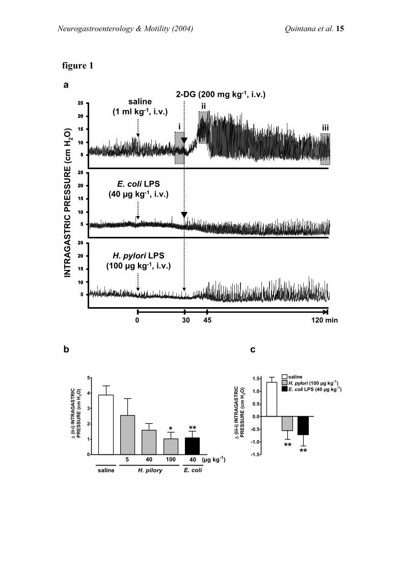
Neurogastroenterology & Motility (2004) Quintana et al. 15
5
10
15
20
25
5
10
15
20
25
5
10
15
20
25
5
10
15
20
25
5
10
15
20
25
5
10
15
20
25
5
10
15
20
25
5
10
15
20
25
5
10
15
20
25
INTR
AG
AST
RIC
PR
ESSU
RE
(cm
H2O
)
saline(1 ml kg-1, i.v.)
2-DG (200 mg kg-1, i.v.)
H. pylori LPS(100 µg kg-1, i.v.)
E. coli LPS(40 µg kg-1, i.v.)
i iii
120 min
a
b
ii
0 30 45
0
1
2
3
4
5
5 40 100
H. pilorysaline
* **
E. coli
40 (µg kg-1)
∆ (i
i-i) I
NTR
AG
AST
RIC
PRES
SUR
E (c
m H
2O)
-1.5
-1.0
-0.5
0.0
0.5
1.0
1.5 saline
E. coli LPS (40 µg kg-1)H. pylori (100 µg kg-1)
****
∆ (i
ii-i)
INTR
AG
AST
RIC
PRES
SUR
E (c
m H
2O)
c
figure 1
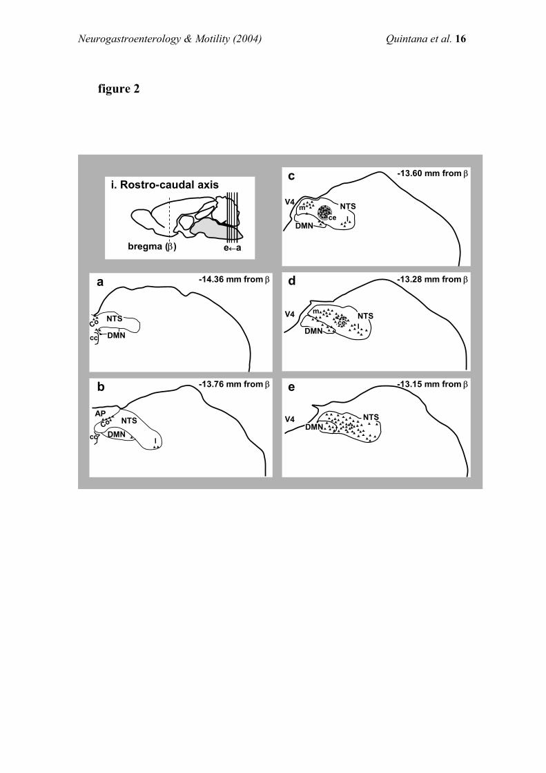
Neurogastroenterology & Motility (2004) Quintana et al. 16
Co
l
b
cc
AP
V4
-13.28 mm from βd
m
lce
-13.15 mm from βe
V4
-14.36 mm from β
Co
a
cc
-13.76 mm from β
-13.60 mm from βc
mlce
V4
V4
i. Rostro-caudal axis
e←abregma (β)
DMNNTS
DMN
NTS
DMN
NTS
DMN
NTS
DMNNTSCo
l
b
cc
AP
V4
-13.28 mm from βd
m
lcece
-13.15 mm from βe
V4
-14.36 mm from β
Co
a
cc
-13.76 mm from β
-13.60 mm from βc
mlcece
V4
V4
i. Rostro-caudal axis
e←abregma (β)
DMNDMNNTSNTS
DMNDMN
NTSNTS
DMNDMN
NTSNTS
DMNDMN
NTSNTS
DMNNTS
figure 2
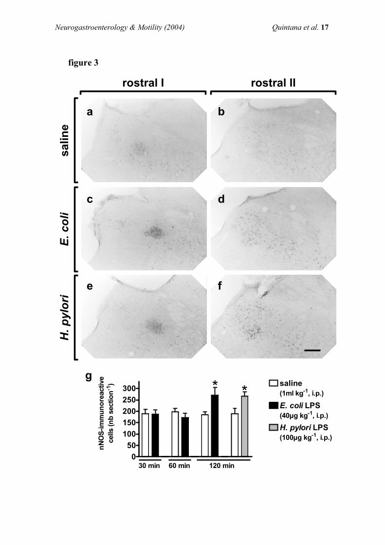
Neurogastroenterology & Motility (2004) Quintana et al. 17
rostral I rostral IIsa
line
E. c
oli
H. p
ylor
i
050
100150200250300 saline
(1ml kg-1, i.p.)
E. coli LPS(40µg kg-1, i.p.)
**
H. pylori LPS(100µg kg-1, i.p.)
30 min 60 min 120 min
nNO
S-im
mun
orea
ctiv
ece
lls (n
b se
ctio
n-1)
e
g
c
a
f
d
b
figure 3
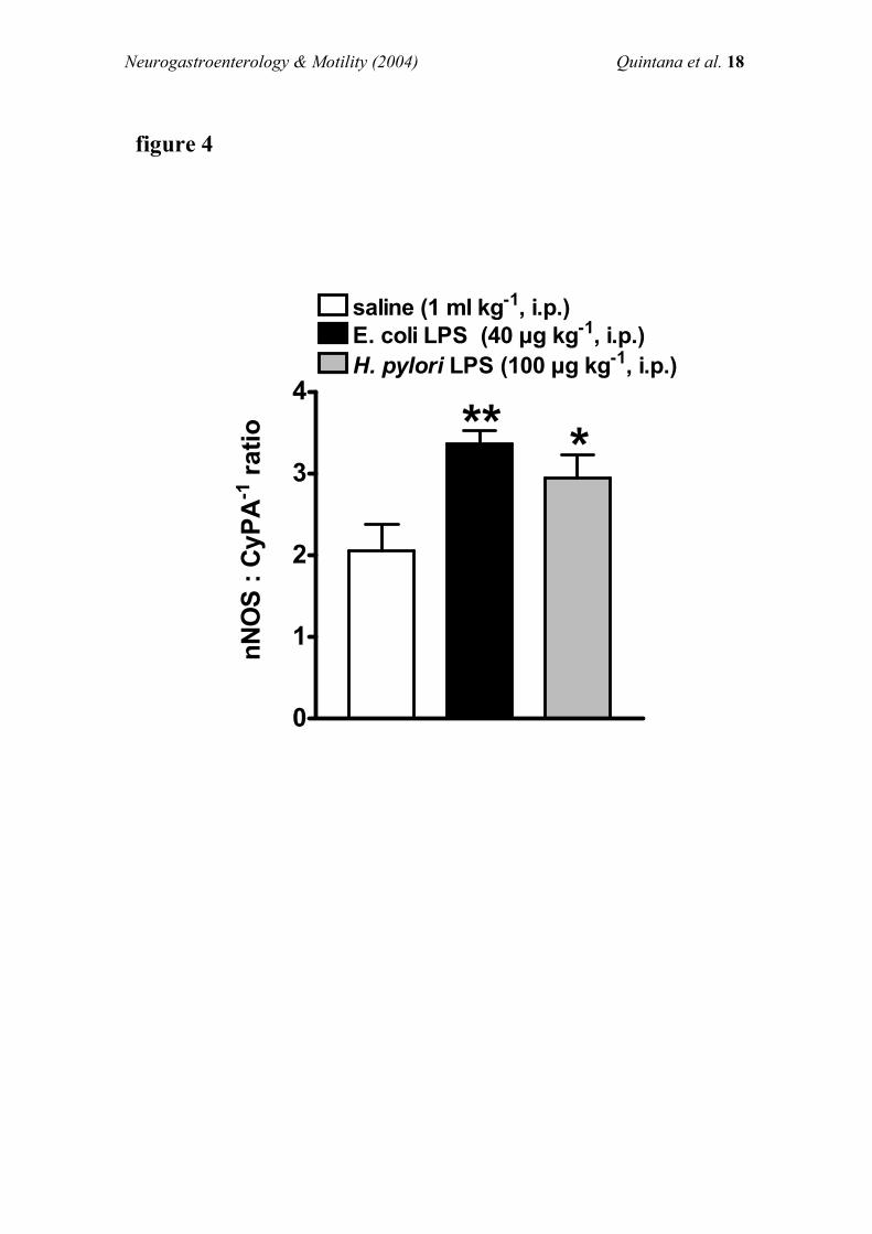
Neurogastroenterology & Motility (2004) Quintana et al. 18
0
1
2
3
4
saline (1 ml kg-1, i.p.)E. coli LPS (40 µg kg-1, i.p.)H. pylori LPS (100 µg kg-1, i.p.)
** *
nNO
S : C
yPA
-1 ra
tio
figure 4
Artículos de investigación
70
Artículo 3
“Synthesis of nitric oxide in post-
ganglionic myenteric neurons during
endotoxemia: implications for gastric
motor function”
Elsa Quintana, Carlos Hernández, Alberto Álvarez-Barrientos, Juan V.
Esplugues & María D. Barrachina
The FASEB Journal express article (2004)
a) Summary: The FASEB Journal (2004) 18: 531-533.
b) Full manuscript: The FASEB Journal (2004) 10.1096/fj.03-0596fje
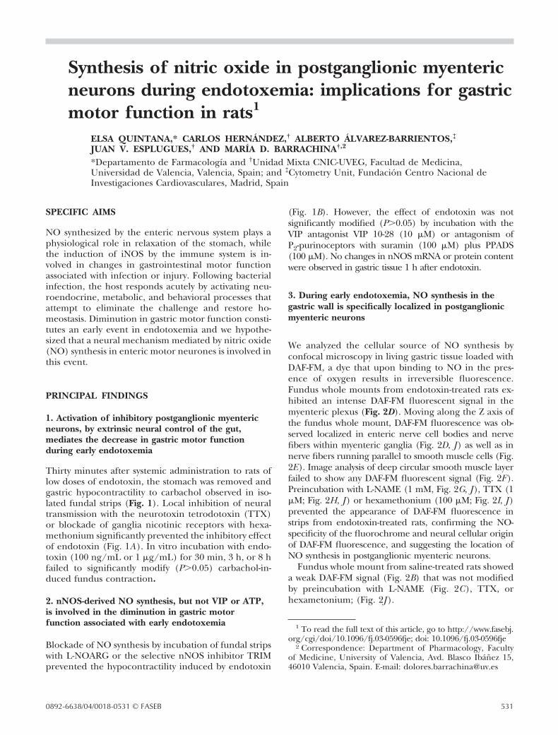
Synthesis of nitric oxide in postganglionic myentericneurons during endotoxemia: implications for gastricmotor function in rats1
ELSA QUINTANA,* CARLOS HERNANDEZ,† ALBERTO ALVAREZ-BARRIENTOS,‡
JUAN V. ESPLUGUES,† AND MARIA D. BARRACHINA†,2
*Departamento de Farmacologıa and †Unidad Mixta CNIC-UVEG, Facultad de Medicina,Universidad de Valencia, Valencia, Spain; and ‡Cytometry Unit, Fundacion Centro Nacional deInvestigaciones Cardiovasculares, Madrid, Spain
SPECIFIC AIMS
NO synthesized by the enteric nervous system plays aphysiological role in relaxation of the stomach, whilethe induction of iNOS by the immune system is in-volved in changes in gastrointestinal motor functionassociated with infection or injury. Following bacterialinfection, the host responds acutely by activating neu-roendocrine, metabolic, and behavioral processes thatattempt to eliminate the challenge and restore ho-meostasis. Diminution in gastric motor function consti-tutes an early event in endotoxemia and we hypothe-sized that a neural mechanism mediated by nitric oxide(NO) synthesis in enteric motor neurones is involved inthis event.
PRINCIPAL FINDINGS
1. Activation of inhibitory postganglionic myentericneurons, by extrinsic neural control of the gut,mediates the decrease in gastric motor functionduring early endotoxemia
Thirty minutes after systemic administration to rats oflow doses of endotoxin, the stomach was removed andgastric hypocontractility to carbachol observed in iso-lated fundal strips (Fig. 1). Local inhibition of neuraltransmission with the neurotoxin tetrodotoxin (TTX)or blockade of ganglia nicotinic receptors with hexa-methonium significantly prevented the inhibitory effectof endotoxin (Fig. 1A). In vitro incubation with endo-toxin (100 ng/mL or 1 �g/mL) for 30 min, 3 h, or 8 hfailed to significantly modify (P�0.05) carbachol-in-duced fundus contraction.
2. nNOS-derived NO synthesis, but not VIP or ATP,is involved in the diminution in gastric motorfunction associated with early endotoxemia
Blockade of NO synthesis by incubation of fundal stripswith L-NOARG or the selective nNOS inhibitor TRIMprevented the hypocontractility induced by endotoxin
(Fig. 1B). However, the effect of endotoxin was notsignificantly modified (P�0.05) by incubation with theVIP antagonist VIP 10-28 (10 �M) or antagonism ofP2-purinoceptors with suramin (100 �M) plus PPADS(100 �M). No changes in nNOS mRNA or protein contentwere observed in gastric tissue 1 h after endotoxin.
3. During early endotoxemia, NO synthesis in thegastric wall is specifically localized in postganglionicmyenteric neurons
We analyzed the cellular source of NO synthesis byconfocal microscopy in living gastric tissue loaded withDAF-FM, a dye that upon binding to NO in the pres-ence of oxygen results in irreversible fluorescence.Fundus whole mounts from endotoxin-treated rats ex-hibited an intense DAF-FM fluorescent signal in themyenteric plexus (Fig. 2D). Moving along the Z axis ofthe fundus whole mount, DAF-FM fluorescence was ob-served localized in enteric nerve cell bodies and nervefibers within myenteric ganglia (Fig. 2D, J ) as well as innerve fibers running parallel to smooth muscle cells (Fig.2E). Image analysis of deep circular smooth muscle layerfailed to show any DAF-FM fluorescent signal (Fig. 2F ).Preincubation with L-NAME (1 mM, Fig. 2G, J ), TTX (1�M; Fig. 2H, J ) or hexamethonium (100 �M; Fig. 2I, J )prevented the appearance of DAF-FM fluorescence instrips from endotoxin-treated rats, confirming the NO-specificity of the fluorochrome and neural cellular originof DAF-FM fluorescence, and suggesting the location ofNO synthesis in postganglionic myenteric neurons.
Fundus whole mount from saline-treated rats showeda weak DAF-FM signal (Fig. 2B) that was not modifiedby preincubation with L-NAME (Fig. 2C), TTX, orhexametonium; (Fig. 2J ).
1 To read the full text of this article, go to http://www.fasebj.org/cgi/doi/10.1096/fj.03-0596fje; doi: 10.1096/fj.03-0596fje
2 Correspondence: Department of Pharmacology, Facultyof Medicine, University of Valencia, Avd. Blasco Ibanez 15,46010 Valencia, Spain. E-mail: [email protected]
5310892-6638/04/0018-0531 © FASEB
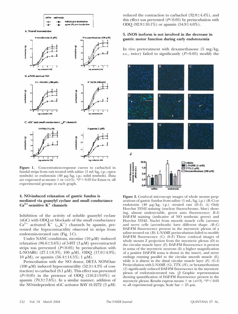
4. NO-induced relaxation of gastric fundus ismediated via guanylyl cyclase and small conductanceCa2�-sensitive K� channels
Inhibition of the activity of soluble guanylyl cyclase(sGC) with ODQ or blockade of the small conductanceCa2� activated K� (CaK
�) channels by apamin, pre-vented the hypocontractility observed in strips fromendotoxin-treated rats (Fig. 1C).
Under NANC conditions, nicotine (10 �M) -inducedrelaxation (96.6�3.6%) of 5-HT (3 �M) -precontractedstrips was prevented (P�0.05) by preincubation withL-NOARG (27.1�8.3%; 100 �M), ODQ (17.0�4.9%;10 �M), or apamin (56.4�14.5%; 1 �M).
Preincubation with the NO donor, DETA NONOate(100 �M) induced hypocontractility (52.3�4.3% of con-traction) to carbachol (0.1 �M). This effect was prevented(P�0.05) in the presence of ODQ (116.2�9.0%) orapamin (79.3�7.6%). In a similar manner, addition ofthe NO-independent sGC activator BAY 41-2272 (3 �M)
reduced the contraction to carbachol (32.8�4.4%), andthis effect was prevented (P�0.05) by preincubation withODQ (92.9�10.1%) or apamin (54.9�4.0%).
5. iNOS isoform is not involved in the decrease ingastric motor function during early endotoxemia
In vivo pretreatment with dexamethasone (5 mg/kg,s.c., twice) failed to significantly (P�0.05) modify the
Figure 1. Concentration-response curves to carbachol infundal strips from rats treated with saline (1 ml/kg, i.p.; opensymbols) or endotoxin (40 �g/kg, i.p.; solid symbols). Dataare expressed as means � se (n�5). *P � 0.05 for Emax vs. allexperimental groups in each graph.
Figure 2. Confocal microscopy images of whole mount prep-arations of gastric fundus from saline (1 mL/kg, i.p.) (B–C) orendotoxin (40 �g/kg, i.p.) -treated rats (D–I). A) OnlyHoechst 33342 staining (nuclear fluorochrome, blue) show-ing, almost undetectable, green auto fluorescence; B–I)DAF-FM staining (indicative of NO synthesis; green) andHoechst 33342. Nuclei from smooth muscle cells (arrows)and nerve cells (arrowheads) have different shape. (B–C)DAF-FM fluorescence present in the myenteric plexus of asaline-treated rat (B); L-NAME preincubation failed to modifyDAF-FM fluorescence (C). D–F) Three confocal images ofwhole mount Z projection from the myenteric plexus (D) tothe circular muscle layer (F). DAF-FM fluorescence is presentin soma of the myenteric neurons (D, a higher magnificationof a positive DAF-FM soma is shown in the insert), and nerveendings running parallel to the circular smooth muscle (E),while it is absent in the distal circular muscle layer (F). (G–I)Preincubation with L-NAME (G), TTX (H ), or hexamethonium(I ) significantly reduced DAF-FM fluorescence in the myentericplexus of endotoxin-treated rats. (J) Graphic representationshowing quantification of DAF-FM fluorescence present in themyenteric plexus. Results express means � se (n�3). *P � 0.05vs. all experimental groups. Scale bar � 25 �m.
532 Vol. 18 March 2004 QUINTANA ET AL.The FASEB Journal
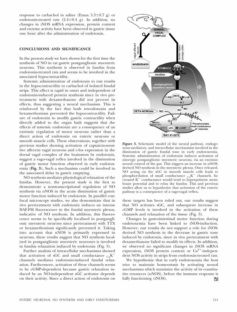
response to carbachol in saline (Emax 5.3�0.7 g) orendotoxin-treated rats (2.4�0.4 g). In addition, nochanges in iNOS mRNA expression, protein contentand enzyme activity have been observed in gastric tissueone hour after the administration of endotoxin.
CONCLUSIONS AND SIGNIFICANCE
In the present study we have shown for the first time thesynthesis of NO in rat gastric postganglionic myentericneurons. This synthesis is observed in fundus fromendotoxin-treated rats and seems to be involved in theassociated hypocontractility.
Systemic administration of endotoxin to rats resultsin the hypocontractility to carbachol of isolated fundalstrips. This effect is rapid in onset and independent ofendotoxin-induced protein synthesis since in vivo pre-treatment with dexamethasone did not prevent itseffects, thus suggesting a neural mechanism. This isreinforced by the fact that both tetrodotoxin andhexamethonium prevented the hypocontractility. Fail-ure of endotoxin to modify gastric contractility whendirectly added to the organ bath suggests that theeffects of systemic endotoxin are a consequence of anextrinsic regulation of motor neurons rather than adirect action of endotoxin on enteric neurons orsmooth muscle cells. These observations, together withprevious studies showing activation of capsaicin-sensi-tive afferent vagal neurons and c-fos expression in thedorsal vagal complex of the brainstem by endotoxin,suggest a vago-vagal reflex involved in the diminutionof gastric motor function observed in early endotox-emia (Fig. 3). Such a mechanism could be involved inthe associated delay in gastric emptying.
NO synthesis mediates physiological relaxation of thefundus. However, the present study is the first todemonstrate a nontranscriptional regulation of NOsynthesis via nNOS in the acute diminution of gastricmotor function induced by endotoxin. In parallel con-focal microscopy studies, we also demonstrate that invivo pretreatment with endotoxin induces an intenseDAF-FM fluorescence in the fundal myenteric plexus,indicative of NO synthesis. In addition, this fluores-cence seems to be specifically localized in postgangli-onic myenteric neurons since pretreatment with TTXor hexamethonium significantly prevented it. Takinginto account that nNOS is primarily expressed inneurons, these results suggest that NO synthesis local-ized in postganglionic myenteric neurones is involvedin fundus relaxation induced by endotoxin (Fig. 3).
Further analysis of intracellular mechanisms showedthat activation of sGC and small conductance CaK
�
channels mediates endotoxin-induced fundal relax-ation. Furthermore, activation of these channels seemsto be cGMP-dependent because gastric relaxation in-duced by an NO-independent sGC activator dependson their activity. Since a direct action of endotoxin on
these targets has been ruled out, our results suggestthat NO activates sGC, and subsequent increase incGMP levels is involved in the activation of thesechannels and relaxation of the tissue (Fig. 3).
Changes in gastrointestinal motor function duringendotoxemia have been linked to iNOS-induction.However, our results do not support a role for iNOS-derived NO synthesis in the decrease in gastric toneinduced by endotoxin, since in vivo pretreatment withdexamethasone failed to modify its effects. In addition,we observed no significant changes in iNOS mRNAexpression, iNOS protein content or Ca2�-indepen-dent NOS activity in strips from endotoxin-treated rats.
We hypothesize that in early endotoxemia the hosttries to maintain homeostasis by activating neuralmechanisms which maximize the activity of its constitu-tive resources (nNOS), before the immune response isfully functioning (iNOS).
Figure 3. Schematic model of the neural pathway, endoge-nous mediators, and intracellular mechanisms involved in thediminution of gastric fundal tone in early endotoxemia.Systemic administration of endotoxin induces activation ofnitrergic postganglionic myenteric neurons, via an extrinsicneural control of the gut. This triggers an increase in nNOS-derived NO synthesis in the myenteric plexus. Once released,NO acting on the sGC in smooth muscle cells leads tophosphorylation of small conductance CaK
� channels. In-creased K� conductance would tend to hyperpolarize mem-brane potential and to relax the fundus. This and previousstudies allow us to hypothesize that activation of the entericpathway is a consequence of a vago-vagal reflex.
533ENTERIC NEURONAL NO SYNTHESIS AND EARLY ENDOTOXEMIA
The FASEB Journal express article 10.1096/fj.03-0596fje. Published online January 8, 2004. Synthesis of nitric oxide in post-ganglionic myenteric neurons during endotoxemia: implications for gastric motor function Elsa Quintana,* Carlos Hernández,� Alberto Álvarez-Barrientos,� Juan V. Esplugues,� and María D. Barrachina�
*Departamento de Farmacología and �Unidad Mixta CNIC-UVEG, Facultad de Medicina, Universidad de Valencia, Avd. Blasco Ibáñez 15, 46010 Valencia, Spain; and �Cytometry Unit, Fundación Centro Nacional de Investigaciones Cardiovasculares (CNIC), Sinesio Delgado 4, 28029, Madrid, Spain Corresponding author: Juan V. Esplugues, Department of Pharmacology, Faculty of Medicine, University of Valencia, Avd. Blasco Ibáñez 15, 46010 Valencia, Spain. E-mail: [email protected]
ABSTRACT
We have investigated the mechanisms underlying acute changes in gastric motor function triggered by endotoxemia. In fundal strips from rats pre-treated with endotoxin (40 µg/kg, i.p. 30 min), mechanical activity was analyzed and the source of nitric oxide (NO) was visualized by confocal microscopy of tissue loaded with the fluorescent dye DAF-FM. NOS expression was determined by quantitative RT-PCR and Western blot, and enzyme activity by the citrulline assay. Strips from endotoxin-treated rats were hypo-contractile. This was prevented by pre-incubation with the neurotoxin tetrodotoxin, the gangliar blocker hexamethonium, or non-selective and neuronal-specific NOS inhibitors (L-NOARG and TRIM, respectively). The soluble guanylyl cyclase (sGC) inhibitor ODQ and the inhibitor of small conductance Ca2+-activated K+ channels apamin prevented relaxation induced by endotoxin, nicotine, exogenous NO (DETA-NONOate), and the NO-independent sGC activator BAY 41-2272. NO synthesis was observed in neuronal soma, axons, and nerve endings of the myenteric plexus in the fundus of endotoxin-treated rats and was prevented by L-NAME, tetrodotoxin, and hexamethonium. nNOS and iNOS mRNA and protein contents were unchanged. Our findings demonstrate synthesis of NO in post-ganglionic myenteric neurons during early endotoxemia that mediates gastric hypo-contractility. The effect of NO is mediated via sGC and small conductance Ca2+-activated K+channels.
Key words: myenteric plexus � motility � confocal microscopy � DAF � neuronal NOS
ollowing infection by endotoxin-producing bacteria, the host reacts by coordinating the peripheral immune response and by signaling to the central nervous system, where a cascade of events are orchestrated in what is collectively termed the acute-phase response.
A wide array of autonomic, endocrine, and behavioral changes occurs in this phase that is interpreted as an attempt to eliminate the challenge and restore homeostasis. Prominent among these is a substantial alteration in gastrointestinal motor function (1�3). In experimental animals,
F
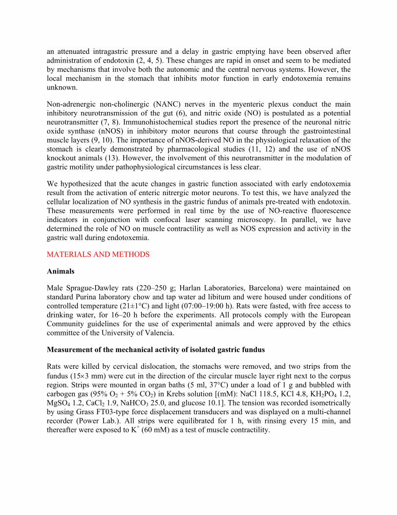
an attenuated intragastric pressure and a delay in gastric emptying have been observed after administration of endotoxin (2, 4, 5). These changes are rapid in onset and seem to be mediated by mechanisms that involve both the autonomic and the central nervous systems. However, the local mechanism in the stomach that inhibits motor function in early endotoxemia remains unknown.
Non-adrenergic non-cholinergic (NANC) nerves in the myenteric plexus conduct the main inhibitory neurotransmission of the gut (6), and nitric oxide (NO) is postulated as a potential neurotransmitter (7, 8). Immunohistochemical studies report the presence of the neuronal nitric oxide synthase (nNOS) in inhibitory motor neurons that course through the gastrointestinal muscle layers (9, 10). The importance of nNOS-derived NO in the physiological relaxation of the stomach is clearly demonstrated by pharmacological studies (11, 12) and the use of nNOS knockout animals (13). However, the involvement of this neurotransmitter in the modulation of gastric motility under pathophysiological circumstances is less clear.
We hypothesized that the acute changes in gastric function associated with early endotoxemia result from the activation of enteric nitrergic motor neurons. To test this, we have analyzed the cellular localization of NO synthesis in the gastric fundus of animals pre-treated with endotoxin. These measurements were performed in real time by the use of NO-reactive fluorescence indicators in conjunction with confocal laser scanning microscopy. In parallel, we have determined the role of NO on muscle contractility as well as NOS expression and activity in the gastric wall during endotoxemia.
MATERIALS AND METHODS
Animals
Male Sprague-Dawley rats (220�250 g; Harlan Laboratories, Barcelona) were maintained on standard Purina laboratory chow and tap water ad libitum and were housed under conditions of controlled temperature (21±1°C) and light (07:00�19:00 h). Rats were fasted, with free access to drinking water, for 16�20 h before the experiments. All protocols comply with the European Community guidelines for the use of experimental animals and were approved by the ethics committee of the University of Valencia.
Measurement of the mechanical activity of isolated gastric fundus
Rats were killed by cervical dislocation, the stomachs were removed, and two strips from the fundus (15×3 mm) were cut in the direction of the circular muscle layer right next to the corpus region. Strips were mounted in organ baths (5 ml, 37°C) under a load of 1 g and bubbled with carbogen gas (95% O2 + 5% CO2) in Krebs solution [(mM): NaCl 118.5, KCl 4.8, KH2PO4 1.2, MgSO4 1.2, CaCl2 1.9, NaHCO3 25.0, and glucose 10.1]. The tension was recorded isometrically by using Grass FT03-type force displacement transducers and was displayed on a multi-channel recorder (Power Lab.). All strips were equilibrated for 1 h, with rinsing every 15 min, and thereafter were exposed to K+ (60 mM) as a test of muscle contractility.
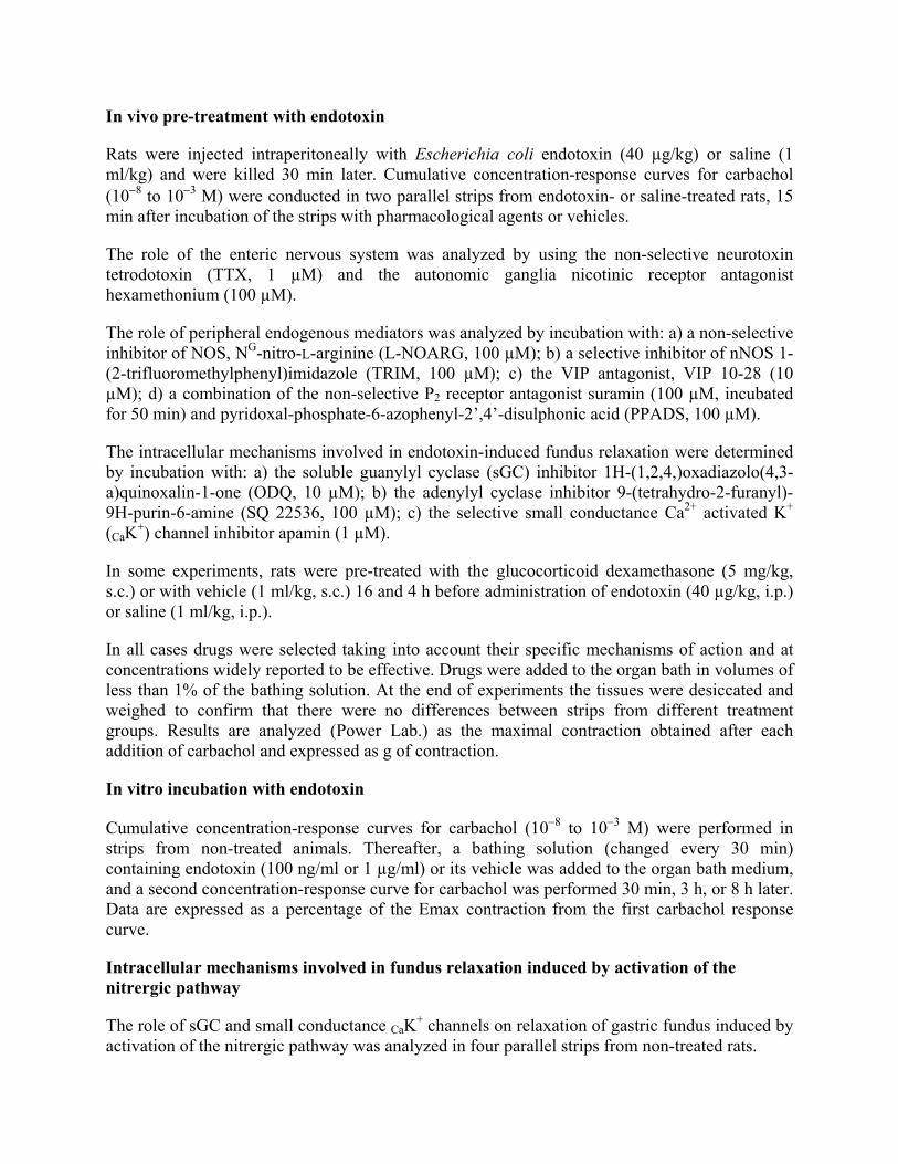
In vivo pre-treatment with endotoxin
Rats were injected intraperitoneally with Escherichia coli endotoxin (40 µg/kg) or saline (1 ml/kg) and were killed 30 min later. Cumulative concentration-response curves for carbachol (10−8 to 10−3 M) were conducted in two parallel strips from endotoxin- or saline-treated rats, 15 min after incubation of the strips with pharmacological agents or vehicles.
The role of the enteric nervous system was analyzed by using the non-selective neurotoxin tetrodotoxin (TTX, 1 µM) and the autonomic ganglia nicotinic receptor antagonist hexamethonium (100 µM).
The role of peripheral endogenous mediators was analyzed by incubation with: a) a non-selective inhibitor of NOS, NG-nitro-L-arginine (L-NOARG, 100 µM); b) a selective inhibitor of nNOS 1-(2-trifluoromethylphenyl)imidazole (TRIM, 100 µM); c) the VIP antagonist, VIP 10-28 (10 µM); d) a combination of the non-selective P2 receptor antagonist suramin (100 µM, incubated for 50 min) and pyridoxal-phosphate-6-azophenyl-2�,4�-disulphonic acid (PPADS, 100 µM).
The intracellular mechanisms involved in endotoxin-induced fundus relaxation were determined by incubation with: a) the soluble guanylyl cyclase (sGC) inhibitor 1H-(1,2,4,)oxadiazolo(4,3-a)quinoxalin-1-one (ODQ, 10 µM); b) the adenylyl cyclase inhibitor 9-(tetrahydro-2-furanyl)-9H-purin-6-amine (SQ 22536, 100 µM); c) the selective small conductance Ca2+ activated K+ (CaK+) channel inhibitor apamin (1 µM).
In some experiments, rats were pre-treated with the glucocorticoid dexamethasone (5 mg/kg, s.c.) or with vehicle (1 ml/kg, s.c.) 16 and 4 h before administration of endotoxin (40 µg/kg, i.p.) or saline (1 ml/kg, i.p.).
In all cases drugs were selected taking into account their specific mechanisms of action and at concentrations widely reported to be effective. Drugs were added to the organ bath in volumes of less than 1% of the bathing solution. At the end of experiments the tissues were desiccated and weighed to confirm that there were no differences between strips from different treatment groups. Results are analyzed (Power Lab.) as the maximal contraction obtained after each addition of carbachol and expressed as g of contraction.
In vitro incubation with endotoxin
Cumulative concentration-response curves for carbachol (10−8 to 10−3 M) were performed in strips from non-treated animals. Thereafter, a bathing solution (changed every 30 min) containing endotoxin (100 ng/ml or 1 µg/ml) or its vehicle was added to the organ bath medium, and a second concentration-response curve for carbachol was performed 30 min, 3 h, or 8 h later. Data are expressed as a percentage of the Emax contraction from the first carbachol response curve.
Intracellular mechanisms involved in fundus relaxation induced by activation of the nitrergic pathway
The role of sGC and small conductance CaK+ channels on relaxation of gastric fundus induced by activation of the nitrergic pathway was analyzed in four parallel strips from non-treated rats.
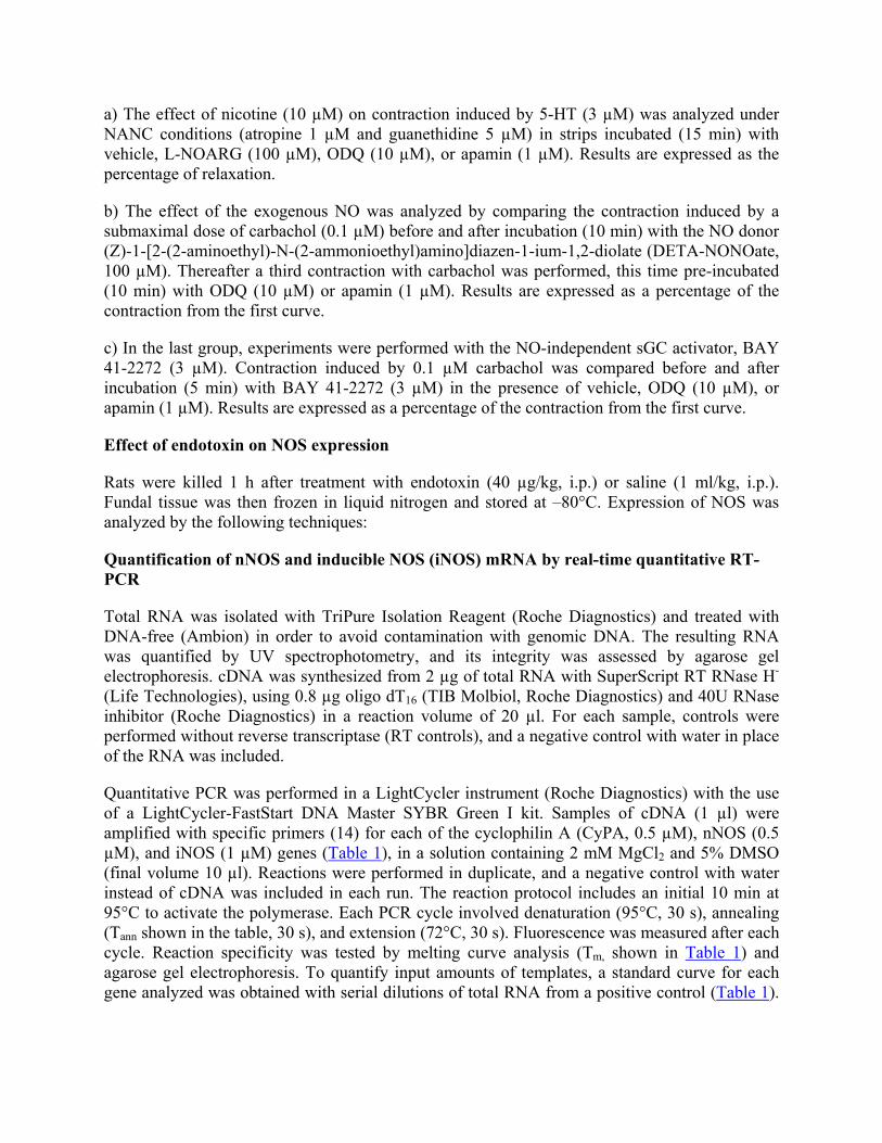
a) The effect of nicotine (10 µM) on contraction induced by 5-HT (3 µM) was analyzed under NANC conditions (atropine 1 µM and guanethidine 5 µM) in strips incubated (15 min) with vehicle, L-NOARG (100 µM), ODQ (10 µM), or apamin (1 µM). Results are expressed as the percentage of relaxation.
b) The effect of the exogenous NO was analyzed by comparing the contraction induced by a submaximal dose of carbachol (0.1 µM) before and after incubation (10 min) with the NO donor (Z)-1-[2-(2-aminoethyl)-N-(2-ammonioethyl)amino]diazen-1-ium-1,2-diolate (DETA-NONOate, 100 µM). Thereafter a third contraction with carbachol was performed, this time pre-incubated (10 min) with ODQ (10 µM) or apamin (1 µM). Results are expressed as a percentage of the contraction from the first curve.
c) In the last group, experiments were performed with the NO-independent sGC activator, BAY 41-2272 (3 µM). Contraction induced by 0.1 µM carbachol was compared before and after incubation (5 min) with BAY 41-2272 (3 µM) in the presence of vehicle, ODQ (10 µM), or apamin (1 µM). Results are expressed as a percentage of the contraction from the first curve.
Effect of endotoxin on NOS expression
Rats were killed 1 h after treatment with endotoxin (40 µg/kg, i.p.) or saline (1 ml/kg, i.p.). Fundal tissue was then frozen in liquid nitrogen and stored at �80°C. Expression of NOS was analyzed by the following techniques:
Quantification of nNOS and inducible NOS (iNOS) mRNA by real-time quantitative RT-PCR
Total RNA was isolated with TriPure Isolation Reagent (Roche Diagnostics) and treated with DNA-free (Ambion) in order to avoid contamination with genomic DNA. The resulting RNA was quantified by UV spectrophotometry, and its integrity was assessed by agarose gel electrophoresis. cDNA was synthesized from 2 µg of total RNA with SuperScript RT RNase H- (Life Technologies), using 0.8 µg oligo dT16 (TIB Molbiol, Roche Diagnostics) and 40U RNase inhibitor (Roche Diagnostics) in a reaction volume of 20 µl. For each sample, controls were performed without reverse transcriptase (RT controls), and a negative control with water in place of the RNA was included.
Quantitative PCR was performed in a LightCycler instrument (Roche Diagnostics) with the use of a LightCycler-FastStart DNA Master SYBR Green I kit. Samples of cDNA (1 µl) were amplified with specific primers (14) for each of the cyclophilin A (CyPA, 0.5 µM), nNOS (0.5 µM), and iNOS (1 µM) genes (Table 1), in a solution containing 2 mM MgCl2 and 5% DMSO (final volume 10 µl). Reactions were performed in duplicate, and a negative control with water instead of cDNA was included in each run. The reaction protocol includes an initial 10 min at 95°C to activate the polymerase. Each PCR cycle involved denaturation (95°C, 30 s), annealing (Tann shown in the table, 30 s), and extension (72°C, 30 s). Fluorescence was measured after each cycle. Reaction specificity was tested by melting curve analysis (Tm, shown in Table 1) and agarose gel electrophoresis. To quantify input amounts of templates, a standard curve for each gene analyzed was obtained with serial dilutions of total RNA from a positive control (Table 1).
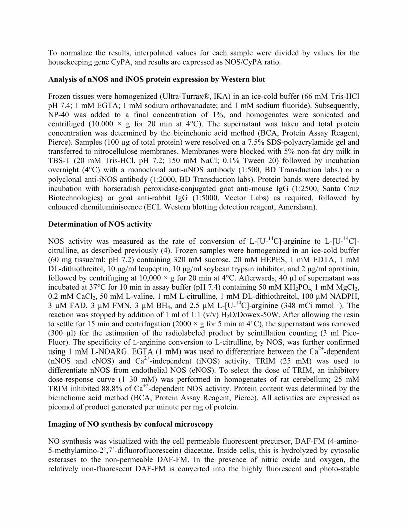
To normalize the results, interpolated values for each sample were divided by values for the housekeeping gene CyPA, and results are expressed as NOS/CyPA ratio.
Analysis of nNOS and iNOS protein expression by Western blot
Frozen tissues were homogenized (Ultra-Turrax®, IKA) in an ice-cold buffer (66 mM Tris-HCl pH 7.4; 1 mM EGTA; 1 mM sodium orthovanadate; and 1 mM sodium fluoride). Subsequently, NP-40 was added to a final concentration of 1%, and homogenates were sonicated and centrifuged (10.000 × g for 20 min at 4°C). The supernatant was taken and total protein concentration was determined by the bicinchonic acid method (BCA, Protein Assay Reagent, Pierce). Samples (100 µg of total protein) were resolved on a 7.5% SDS-polyacrylamide gel and transferred to nitrocellulose membranes. Membranes were blocked with 5% non-fat dry milk in TBS-T (20 mM Tris-HCl, pH 7.2; 150 mM NaCl; 0.1% Tween 20) followed by incubation overnight (4°C) with a monoclonal anti-nNOS antibody (1:500, BD Transduction labs.) or a polyclonal anti-iNOS antibody (1:2000, BD Transduction labs). Protein bands were detected by incubation with horseradish peroxidase-conjugated goat anti-mouse IgG (1:2500, Santa Cruz Biotechnologies) or goat anti-rabbit IgG (1:5000, Vector Labs) as required, followed by enhanced chemiluminiscence (ECL Western blotting detection reagent, Amersham).
Determination of NOS activity
NOS activity was measured as the rate of conversion of L-[U-14C]-arginine to L-[U-14C]-citrulline, as described previously (4). Frozen samples were homogenized in an ice-cold buffer (60 mg tissue/ml; pH 7.2) containing 320 mM sucrose, 20 mM HEPES, 1 mM EDTA, 1 mM DL-dithiothreitol, 10 µg/ml leupeptin, 10 µg/ml soybean trypsin inhibitor, and 2 µg/ml aprotinin, followed by centrifuging at 10,000 × g for 20 min at 4°C. Afterwards, 40 µl of supernatant was incubated at 37°C for 10 min in assay buffer (pH 7.4) containing 50 mM KH2PO4, 1 mM MgCl2, 0.2 mM CaCl2, 50 mM L-valine, 1 mM L-citrulline, 1 mM DL-dithiothreitol, 100 µM NADPH, 3 µM FAD, 3 µM FMN, 3 µM BH4, and 2.5 µM L-[U-14C]-arginine (348 mCi mmol�1). The reaction was stopped by addition of 1 ml of 1:1 (v/v) H2O/Dowex-50W. After allowing the resin to settle for 15 min and centrifugation (2000 × g for 5 min at 4°C), the supernatant was removed (300 µl) for the estimation of the radiolabeled product by scintillation counting (3 ml Pico-Fluor). The specificity of L-arginine conversion to L-citrulline, by NOS, was further confirmed using 1 mM L-NOARG. EGTA (1 mM) was used to differentiate between the Ca2+-dependent (nNOS and eNOS) and Ca2+-independent (iNOS) activity. TRIM (25 mM) was used to differentiate nNOS from endothelial NOS (eNOS). To select the dose of TRIM, an inhibitory dose-response curve (1�30 mM) was performed in homogenates of rat cerebellum; 25 mM TRIM inhibited 88.8% of Ca+2-dependent NOS activity. Protein content was determined by the bicinchonic acid method (BCA, Protein Assay Reagent, Pierce). All activities are expressed as picomol of product generated per minute per mg of protein.
Imaging of NO synthesis by confocal microscopy
NO synthesis was visualized with the cell permeable fluorescent precursor, DAF-FM (4-amino-5-methylamino-2�,7�-difluorofluorescein) diacetate. Inside cells, this is hydrolyzed by cytosolic esterases to the non-permeable DAF-FM. In the presence of nitric oxide and oxygen, the relatively non-fluorescent DAF-FM is converted into the highly fluorescent and photo-stable
triazole form, DAF-FM T, whose fluorescence intensity is directly proportional to the NO concentration. The spectra of the adduct of DAF-FM is independent of pH, and the detection limit of NO by this fluorochrome is 3 nM (15, 16).
Tissue staining and confocal microscopy visualization
Five fundal strips were obtained according to the same protocol as described above for functional studies. Four strips were pre-incubated (25 min, 37°C) with carbogenated-Krebs containing vehicle, the NO synthase inhibitor Nω-nitro-L-arginine methyl ester (L-NAME, 1 mM), TTX (1 µM), or hexamethonium (100 µM). Afterwards, both DAF-FM (1 µM) and the vital nuclear staining fluorochrome, Hoechst 33342 (1 µg/ml) were added and further incubated for 60 min at 37°C. To determine green basal autofluorescence and to establish confocal settings to obtain green fluorescence to minimal levels, Hoechst 33342 only was added to a vehicle-incubated strip. After rinsing, whole-mount preparations were mounted in a tissue slice chamber in carbogenated-Krebs solution. The slice chamber was set on a heated stage (Warner Instrument Corporation) at 37°C. Images were acquired with a Radiance 2100 confocal microscope (Bio-Rad, Hempel Hempstead, UK). A blue diode laser (405 nm) was used to excite Hoechst 33342, and a 488 nm laser line was used to excite DAF-FM. Fluorescence was acquired through 442/45 BP (Hoechst 33342) and 515/30 BP filters (DAF-FM). Confocal microscope settings were adjusted to produce the optimum signal-to-noise ratio. DETA-NONOate (100 µM) was added at the end of each experiment to test the responsiveness of the system. To compare fluorescence signals between different preparations, settings were fixed for all samples from the same animal. To analyze the spatial distribution of DAF-FM fluorescence in each preparation, a complete analysis along the whole-mount Z axis, from serosa to mucosa, was performed (0.6 µm optical section width, 5 µm step width). We used the morphology of cell nuclei to localize the different tissue layers in the gastric wall. Fluorescence analysis was performed by using LaserPix software (Bio-Rad).
Statistical analysis
Data are expressed as mean values ± SEM, and statistical comparison was performed by ANOVA followed by a Newman-Keuls test. Data were considered statistically significant when P<0.05. Cumulative concentration curves to carbachol were normalized by sigmoidal non-linear regression to obtain the pD2 and Emax values (Graphpad Prism 3.0).
Drugs used
Escherichia coli endotoxin (serotype 026:B6), carbamylcholine chloride, hexamethonium chloride, L-NOARG, L-NAME, TRIM, VIP 10-28, suramin, PPADS, ODQ, apamin, atropine, guanethidine, nicotine, Hoechst 33342, and all reagents used for determination of NOS activity were purchased from Sigma Chemical Co. (St. Louis, MO). SQ 22536, TTX, and DETA-NONOate were obtained from Calbiochem® (Darmstad, Germany), Tocris Coockson (Bristol, UK), and Alexis corporation (San Diego, CA), respectively. Dexamethasone (Fortecortin®) was used as a clinically available preparation. L-[U-14C]-arginine was obtained from Amersham Life Sciences (London, UK). The fluorochrome DAF-FM diacetate was purchased from Molecular Probes (Eugene, OR). BAY 41-2272 was the kind gift of Salvador Moncada at the Wolfson Institute for Biomedical Research (London, UK). Endotoxin was dissolved in saline, and ODQ,
TRIM, BAY 41-2272 and DAF-FM diacetate were dissolved in DMSO up to 10−2 M, 10−1 M, 10−3M, and 5 mM, respectively. Further dilutions of these compounds and solutions of all other drugs were in distilled water.
RESULTS
Effect of endotoxin on the mechanical activity of isolated gastric fundus
In vivo pre-treatment with endotoxin decreases fundus contractility
A concentration-response curve to carbachol was performed in fundal strips from saline-treated animals (pD2 = 6.2 ± 0.2, Emax = 6.3 ± 0.4 g, n = 5). Strips from rats treated with endotoxin (40 µg/kg, i.p.) exhibited a decrease in the Emax for carbachol (2.6 ± 0.6 g) with no change in the pD2 (6.2 ± 0.1, n = 5) (Fig. 1).
Involvement of post-ganglionic myenteric neurons in the effect of endotoxin
Inhibition of neural transmission with the neurotoxin TTX or blockade of gangliar nicotinic receptor with hexamethonium both significantly prevented the inhibitory effect of endotoxin on fundus contractility. Neither TTX nor hexamethonium modified the contractile effect of carbachol in control animals (Fig. 2A).
Pre-treatment with dexamethasone did not significantly modify the response to carbachol in strips from saline- or endotoxin-treated animals (Fig. 3)
NO synthesis by nNOS mediates the effect of endotoxin
Blockade of NO synthesis by incubation with L-NOARG or the selective nNOS inhibitor TRIM prevented hypo-contractility induced by endotoxin. Neither L-NOARG nor TRIM modified the response to carbachol in strips from control animals (Fig. 2B).
Contractility in strips from endotoxin- and saline-treated animals was not influenced by incubation with the VIP antagonist, VIP 10-28 (Fig. 4A), nor the P2-purinoceptor antagonists, suramin plus PPADS (Fig. 4B).
sGC and small conductance CaK+ channels mediate the effect of endotoxin
Inhibition of guanylyl cyclase activity with ODQ prevented the endotoxin-induced hypo-contractility, while having no effect in control rats (Fig. 2C). In addition, selective inhibition of the small conductance CaK+ channels by incubation with apamin blocked the inhibitory effect of endotoxin while leaving the control response unchanged (Fig. 2C).
Endotoxin-induced hypo-contractility (Emax 2.8±0.2 g) was not significantly modified (P>0.05) by inhibition of adenylyl cyclase activity with SQ 22536 (3.0±0.4 g). In fundus from saline-treated rats SQ 22536 did not significantly alter (5.5±1.1 g) the response to carbachol (5.9±1.2 g) n≥4.
None of the treatments above described modified the pD2 for carbachol. In addition, at the concentrations used, none of the inhibitors or vehicles significantly affected the basal tone of the fundus strips.
In vitro incubation with endotoxin fails to modify fundus contractility
Incubation with endotoxin (100 ng/ml) for 30 min, 3 h, or 8 h failed to significantly modify the contractile response to carbachol compared with vehicle-incubated strips (89.1 ± 6.8%, 130.8 ± 6.4%, or 134.2 ± 10.0%, respectively vs. 77.4 ± 6.3%, 140.2 ± 8.9%, and 131.4 ± 13.4%, respectively). In a similar manner, incubation with a higher dose of endotoxin (1 µg/ml) for 3 h had no effect (121.0 ± 8.3%) compared with control strips (131.4 ± 13.4%). When incubated for 8 h, this higher dose induced a non-significant (P>0.05) decrease in the contractile response to carbachol compared with vehicle-incubated strips (107.9 ± 16.7% vs. 131.4 ± 13.4%). In no case were changes observed in the pD2 (6.0 ± 0.2). In all cases n=5.
sGC and small conductance CaK+ channels are involved in the relaxation of gastric fundus induced by the nitrergic pathway
Nicotine-induced relaxation of strips pre-contracted with 5-HT was significantly prevented by pre-incubation with L-NOARG, ODQ, or apamin (Fig. 5A).
Hypo-contractility to carbachol induced by pre-incubation with DETA-NONOate or BAY 41-2272 was significantly reversed in the presence of ODQ or apamin. These drugs did not modify the contractile effect of carbachol in the respective control strips (Fig. 5B, C).
Effect of endotoxin on NOS expression
In vivo pre-treatment with endotoxin does not modify the expressed levels of nNOS or iNOS mRNA
nNOS and iNOS mRNA were both detected in the gastric fundus of saline-treated rats, as analyzed by real-time quantitative RT-PCR. In fundus obtained from endotoxin-treated rats no significant changes in the amount of nNOS or iNOS mRNA were observed compared with control rats (Table 2).
In vivo pre-treatment with endotoxin does not modify nNOS or iNOS protein content
In the gastric fundus of animals treated with endotoxin, the amount of nNOS protein was similar to that detected in the fundus of saline-treated rats (Fig. 6). iNOS protein was not detected in fundus homogenates of saline- or endotoxin-treated rats. As a positive control, iNOS protein was detected in lung and fundus extracted from a rat treated for 8 h with a higher dose of endotoxin (10 mg/kg, i.p.; Fig. 6).
In vivo pre-treatment with endotoxin does not modify NOS enzyme activity
Pre-treatment with endotoxin for 1 h did not modify total NOS activity in the gastric wall when compared with control rats (5.7±0.5 vs. 6.2±0.3 pmol/min mg protein). In both cases, this activity was due to Ca+2-dependent NOS isoforms, with Ca+2-independent NOS activity (iNOS)
barely detectable. The use of TRIM showed that the Ca+2-dependent NOS activity was mainly due to nNOS (Fig. 7).
In vivo pre-treatment with endotoxin increases NO synthesis in the myenteric plexus of the gastric fundus
Under our experimental conditions, control staining did not show significant green autofluorescence (Fig. 8A). After DAF-FM staining, strips from saline-treated rats exhibited a generalized weak green fluorescence signal (Fig. 8B, J). However, this fluorescence was not indicative of NO synthesis because addition of L-NAME did not reduce it (Fig. 8C, J). Furthermore, pre-incubation with TTX or hexamethonium also failed to modify the DAF-FM fluorescence signal (Fig. 8J).
Fundal strips from endotoxin-treated rats exhibited an intense DAF-FM fluorescent signal in the myenteric plexus (Fig. 8J). Moving along the fundus whole-mount Z axis, DAF-FM fluorescence was localized in enteric nerve cell bodies and nerve fibers within myenteric ganglia (Fig. 8D), as well as in nerve fibers running parallel to smooth muscle cells (Fig. 8E). There was only an extremely weak DAF-FM fluorescent signal in the deep circular smooth muscle layer (Fig. 8F).
Pre-incubation with L-NAME prevented the appearance of green fluorescence in strips from endotoxin-treated rats, confirming the NO-specificity (Fig. 8G, 8J). In a similar manner pre-incubation with TTX or hexamethonium impeded the green fluorescence induced by endotoxin-treatment, further confirming the neural cellular origin and suggesting the location of NO synthesis in post-ganglionic myenteric neurons (Fig. 8H�J, respectively).
DISCUSSION
In this study we describe the first visual detection of NO synthesis in rat gastric post-ganglionic myenteric neurons, in the fundus of rats treated with endotoxin. We further demonstrate that nitrergic signaling by these neurons mediates the gastric hypo-contractility in response to endotoxin. The effect of NO on gastric smooth muscle is mediated via activation of sGC and small conductance Ca2+-activated K+channels.
Enteric nervous system modulates gastric motor function during early endotoxemia
Endotoxemia caused by infection with gram-negative bacteria is associated with changes in gastric motor function such as a delayed gastric emptying (1, 2, 14). The present study shows that systemic administration of endotoxin to rats causes hypo-contractility to carbachol in isolated fundus strips. This effect is rapid in onset and independent of endotoxin-induced protein synthesis since in vivo pre-treatment with dexamethasone (14, 17) did not prevent its effect, thus suggesting a neural mechanism. This is reinforced by the fact that addition of tetrodotoxin to the organ bath prevented the hypo-contractility. Furthermore, this neural mechanism involves cholinergic synaptic transmission, since hexamethonium also prevented the effect of endotoxin. These observations, and the failure of endotoxin to modify gastric contractility when added to the organ bath, suggest that hypo-contractility is consequence of an extrinsic regulation of motor neurons rather than a direct action of endotoxin on enteric neurons or smooth muscle cells. It is important to note that the effect of endotoxin treatment is maintained after extrinsic denervation
of the gut, suggesting that once the challenge disappears, regulatory mechanisms at the synaptic transmission could be involved (18). Previous studies have reported a role for capsaicin-sensitive afferent vagal neurons in the delayed gastric emptying induced by systemic administration of low doses of endotoxin (2). In addition, these doses have been shown to activate the early-appearing gene product c-fos in the caudal part of the dorsal motor nucleus of the vagus nerve (4, 19) where pre-ganglionic neurons of the inhibitory motor pathway have been reported (20). Taken together the present results suggest that systemic administration of low doses of endotoxin activates a vago-vagal reflex in which the fundus of the stomach dilates. A similar mechanism has been observed in the receptive relaxation of the gastric fundus in response to food (11) or during early stages of vomiting, and might be involved in the delayed gastric emptying associated with endotoxemia.
nNOS-derived NO is involved in early endotoxemia
Inhibitory neurotransmission in the myenteric plexus is mainly mediated by the release of non-adrenergic non-cholinergic neurotransmitters. NO plays an essential role, both in intrinsic inhibitory reflexes that regulate adaptive relaxation of the fundus in response to small increases in intragastric pressure (12) and in extrinsic reflexes involved in the receptive relaxation of the fundus in response to food (11). In the present study, inhibition of NO synthesis in gastric tissue from control animals failed to alter the basal tone and the contractile response to carbachol. It seems that NO, in contrast to what happens in the intestine (21), does not modulate the resting tone of the rat gastric fundus. However, synthesis of NO takes place in fundus of endotoxin-treated rats, since addition of L-NOARG prevented hypo-contractility. This synthesis appears to be mediated by the nNOS isoform, as a selective nNOS inhibitor (22) also blocked the response to carbachol. In addition, nNOS-derived NO synthesis is not regulated at the transcriptional level, because endotoxin treatment did not increase nNOS mRNA or nNOS protein content. These results suggest that in vivo pre-treatment with endotoxin activates a neural mechanism that results in increased nNOS-mediated NO synthesis in the gastric wall. nNOS activity in the enteric nervous system is predominantly regulated by activation of nicotinic receptors and stimulation of a Ca2+-dependent protein kinase C pathway (8, 23). In the present study, analysis of the catalytic activity of NOS in control animals was significantly reduced by incubation with Ca2+-free medium, L-NAME, or TRIM, together suggesting that NO synthesis in the fundus is mainly due to the nNOS isoform. However, no differences in nNOS activity were observed between fundus homogenates from saline- or endotoxin-treated rats. As previously shown (24), regulatory mechanisms operating on constitutive NOS activity in intact tissue, but lost in tissue homogenates, can account for the differences between the results of functional and biochemical experiments.
In light of these discrepancies, we tried to visualize NO synthesis in the gastric fundus by a specific and sensitive technique which exploits the NO-reactive fluorescence indicator DAF-FM in conjunction with confocal laser scanning microscopy (15, 25�28). Previous immunohistochemical studies have demonstrated the presence of NOS in different cell types of the gastric wall (10, 29, 30). Furthermore, a chemiluminescence method allowing the indirect detection of NO in the myenteric plexus has been described (31). However, this is the first study to report the real-time visualization of intracellular NO production in gastric tissue by a direct and non-invasive method. This approach allows functional analysis of NO and offers valuable information about the cellular source of the NO. This is of special interest for this study, given
the wide expression of nNOS in gastric tissue (32, 33). Thus, in whole-mount fundus from control animals, only a slight non-specific background fluorescence signal was observed, suggesting that the basal release of NO was below the detection limit of the fluorochrome (3 nM), that implies a very low basal synthesis of NO in gastric fundus. This observation is consistent with the failure of NOS inhibitors to modulate resting tone and the low spontaneous neuronal activity described in circular muscle strips of the rat gastric fundus (34�36). By contrast, microscopic analysis of fundal strips from endotoxin-treated rats exhibited an intense DAF-FM fluorescence, which was indicative of NO synthesis because pre-incubation with L-NAME significantly prevented it. The fluorescent signal observed was significantly higher than that present in fundal strips from saline-treated rats, suggesting that NO synthesis was induced by endotoxin-treatment. This synthesis was specifically localized in neuronal soma and nerve endings that innervate the circular muscle, and was absent in smooth muscle cells. These observations, coupled with the fact that nNOS is primarily expressed in neurons (32), led us to conclude that neuronal cells are the source of NO synthesis involved in fundus relaxation. Furthermore, by combining microscopic techniques with pharmacological tools such as TTX and hexamethonium, our results locate NO synthesis induced by endotoxin to post-ganglionic myenteric neurons.
iNOS-derived NO is not involved in early endotoxemia
Induction of iNOS has been observed in a variety of tissues, such as liver and lung after in vivo administration of high doses of endotoxin (37). In addition, a role for iNOS-derived NO in the hypo-contractility induced by endotoxin has been shown in both ileal and vascular smooth muscle (30, 38). We, however, found no significant changes in iNOS mRNA expression, iNOS protein content, or Ca2+-independent NOS activity in strips from endotoxin-treated rats compared with controls. Furthermore, our results do not support a role for iNOS-derived NO synthesis in the decrease in gastric tone induced by endotoxin since in vivo pre-treatment with dexamethasone failed to modify the effects of endotoxin. The low doses of endotoxin used and the short time over which its effects are analyzed could explain the lack of iNOS expression, which normally is seen after longer exposure, even to higher doses of endotoxin (39, 40).
sGC and small conductance CaK+ channels mediate fundus hypo-contractility
Taken together, our results show that nNOS-mediated NO synthesis in gastric post-ganglionic myenteric neurons is involved in the hypo-contractility to carbachol induced by endotoxin. Most of the effects induced by the constitutive synthesis of NO, including relaxation of the circular muscle of the rat fundus, involve activation of sGC (41, 42). In the present study, inhibition of sGC activity by incubation with ODQ prevented the hypo-reactivity induced by endotoxin, suggesting that activation of the sGC is involved, and reinforcing the role of endogenous NO in the effect of endotoxin. In addition, activation of small conductance CaK+ channels has been reported in the relaxation of gastrointestinal tissue induced by endogenous and exogenous NO (10, 43). Results show that local blockade of small conductance CaK+ channels by apamin partially prevented the hypo-reactivity induced by endotoxin. Since a direct effect of endotoxin on these channels has been ruled out, these results suggest that it is NO released in response to endotoxin that acts on these channels. This observation is reinforced by the fact that relaxation of gastric fundus induced by exogenous NO (DETA-NONOate) or endogenous release of NO (nicotine) is also reversed by apamin. However, discrepancies still exist in the literature
regarding whether NO acts directly on CaK+ channels (44) or if it is a cGMP-dependent mechanism (45). The present study shows a role for small conductance CaK+ channels in the relaxation of the gastric fundus induced by an NO-independent sGC activator (46), suggesting that activation of these channels is due to cGMP rather than a direct action of NO. Taken together, these data suggest an intracellular mechanism of action of NO to relax the circular muscle of the rat gastric fundus based in a sequence of events: increase in cGMP and activation of small conductance CaK+ channels in the smooth muscle cell membrane.
In addition to NO, other transmitters such as VIP or ATP are implicated in NANC relaxations of the rat gastric fundus (7, 47, 48). The present results do not support a role for VIP or ATP in the hypo-contractility induced by endotoxin. Blockade of the VIP receptor of gastric fundus (49) did not significantly modify the effects of endotoxin. In addition, activation of intracellular adenylyl cyclase, which is involved in VIP-induced relaxation of the gastric fundus (50), does not mediate the effects of endotoxin. In a similar manner blockade of purinoceptors (51�54) also failed to alter the hypo-contractility induced by endotoxin, ruling out a role for ATP.
In conclusion, the present study shows that NO synthesis in gastric post-ganglionic myenteric neurons is involved in the hypo-contractility induced by in vivo pre-treatment with endotoxin. These results and previous studies suggest the existence of a vago-vagal reflex activated in early endotoxemia, which serves to maximize the activity of the constitutive resources before the immune response is fully functioning.
ACKNOWLEDGMENTS
This study has been supported by grant FIS 01/1187 from the Ministerio de Sanidad y Consumo, grant SAF 2001-0763 from the Comisión Interministerial de Ciencia y Tecnología, and grant C 03/02 from Instituto de Salud Carlos III. E. Q. is the recipient of a fellowship from the Ministerio de Sanidad y Consumo.
REFERENCES
1. van Miert, A. S., and De la Parra, D. A. (1970) Inhibition of gastric emptying by endotoxin (bacterial lipopolysaccharide) in conscious rats and modification of this response by drugs affecting the autonomic nervous system. Arch. Int. Pharmacodyn. Ther. 184, 27�33
2. Calatayud, S., Barrachina, M. D., Garcia-Zaragoza, E., Quintana, E., and Esplugues, J. V. (2001) Endotoxin inhibits gastric emptying in rats via a capsaicin-sensitive afferent pathway. Naunyn Schmiedebergs Arch. Pharmacol. 363, 276�280
3. Calatayud, S., Barrachina, M. D., Quintana, E., Ibiza, S., and Esplugues, J. V. (2003) Endotoxin stimulates fecal pellet output in rats through a neural mechanism. Naunyn Schmiedebergs Arch. Pharmacol. 367, 51�55
4. Quintana, E., Garcia-Zaragoza, E., Martinez-Cuesta, M. A., Calatayud, S., Esplugues, J. V., and Barrachina, M. D. (2001) A cerebral nitrergic pathway modulates endotoxin-induced changes in gastric motility. Br. J. Pharmacol. 134, 325�332
5. Takakura, K., Hasegawa, K., Goto, Y., and Muramatsu, I. (1997) Nitric oxide produced by inducible nitric oxide synthase delays gastric emptying in lipopolysaccharide-treated rats. Anesthesiology 87, 652�657
6. Furness, J. B. (2000) Types of neurons in the enteric nervous system. J. Auton. Nerv. Syst. 81, 87�96
7. Sanders, K. M., and Ward, S. M. (1992) Nitric oxide as a mediator of nonadrenergic noncholinergic neurotransmission. Am. J. Physiol. 262, G379�G392
8. Esplugues, J. V. (2002) NO as a signalling molecule in the nervous system. Br. J. Pharmacol. 135, 1079�1095
9. Berthoud, H. R. (1995) Anatomical demonstration of vagal input to nicotinamide acetamide dinucleotide phosphate diaphorase-positive (nitrergic) neurons in rat fundic stomach. J. Comp. Neurol. 358, 428�439
10. Shuttleworth, C. W., and Sanders, K. M. (1996) Involvement of nitric oxide in neuromuscular transmission in canine proximal colon. Proc. Soc. Exp. Biol. Med. 211, 16�23
11. Desai, K. M., Sessa, W. C., and Vane, J. R. (1991) Involvement of nitric oxide in the reflex relaxation of the stomach to accommodate food or fluid. Nature 351, 477�479
12. Desai, K. M., Zembowicz, A., Sessa, W. C., and Vane, J. R. (1991) Nitroxergic nerves mediate vagally induced relaxation in the isolated stomach of the guinea pig. Proc. Natl. Acad. Sci. USA 88, 11490�11494
13. Mashimo, H., Kjellin, A., and Goyal, R. K. (2000) Gastric stasis in neuronal nitric oxide synthase-deficient knockout mice. Gastroenterology 119, 766�773
14. Calatayud, S., Garcia-Zaragoza, E., Hernandez, C., Quintana, E., Felipo, V., Esplugues, J. V., and Barrachina, M. D. (2002) Downregulation of nNOS and synthesis of PGs associated with endotoxin- induced delay in gastric emptying. Am. J. Physiol. Gastrointest. Liver Physiol. 283, G1360�G1367
15. Nagano, T., and Yoshimura, T. (2002) Bioimaging of nitric oxide. Chem. Rev. 102, 1235�1270
16. Haugland, R. P. (2002) Handbook of Fluorescent Probes and Research Products. Molecular Probes, Inc.
17. Mc Call, T. B., Palmer, R. M., and Moncada, S. (1991) Induction of nitric oxide synthase in rat peritoneal neutrophis and its inhibition by dexamethasone. Eur. J. Immunol. 21, 2523�2527
18. Fossier, P., Tauc, L., and Baux, G. (1999) Calcium transients and neurotransmitter release at an identified synapse. Trends Neurosci. 22, 161�166
19. Elmquist, J. K., Scammell, T. E., Jacobson, C. D., and Saper, C. B. (1996) Distribution of Fos-like immunoreactivity in the rat brain following intravenous lipopolysaccharide administration. J. Comp. Neurol. 371, 85�103
20. Chang, H. Y., Mashimo, H., and Goyal, R. K. (2003) Musings on the wanderer: what's new in our understanding of vago-vagal reflex? IV. Current concepts of vagal efferent projections to the gut. Am. J. Physiol. Gastrointest. Liver Physiol. 284, G357�G366
21. Martinez-Cuesta, M. A., Esplugues, J. V., and Whittle, B. J. (1996) Modulation by nitric oxide of spontaneous motility of the rat isolated duodenum: role of tachykinins. Br. J. Pharmacol. 118, 1335�1340
22. Guilmard, C., Auguet, M., and Chabrier, P. E. (1998) Comparison between endothelial and neuronal nitric oxide pathways in rat aorta and gastric fundus. Nitric Oxide 2, 147�154
23. Nakamura, K., Takahashi, T., Taniuchi, M., Hsu, C. X., and Owyang, C. (1998) Nicotinic receptor mediates nitric oxide synthase expression in the rat gastric myenteric plexus. J. Clin. Invest. 101, 1479�1489
24. Calatayud, S., Canet, A., Bello, R., Hernandez, C., Marti, M., and Barrachina, M. D. (2003) Low endotoxemia prevents the reduction of gastric blood flow induced by NSAIDs: role of nitric oxide. Br. J. Pharmacol. 139, 263�270
25. Kojima, H., Hirata, M., Kudo, Y., Kikuchi, K., and Nagano, T. (2001) Visualization of oxygen-concentration-dependent production of nitric oxide in rat hippocampal slices during aglycemia. J. Neurochem. 76, 1404�1410
26. Willmott, N. J., Wong, K., and Strong, A. J. (2000) A fundamental role for the nitric oxide-G-kinase signaling pathway in mediating intercellular Ca(2+) waves in glia. J. Neurosci. 20, 1767�1779
27. Nakatsubo, N., Kojima, H., Kikuchi, K., Nagoshi, H., Hirata, Y., Maeda, D., Imai, Y., Irimura, T., and Nagano, T. (1998) Direct evidence of nitric oxide production from bovine aortic endothelial cells using new fluorescence indicators: diaminofluoresceins. FEBS Lett. 427, 263�266
28. Brown, L. A., Key, B. J., and Lovick, T. A. (2000) Fluorescent imaging of nitric oxide production in neuronal varicosities associated with intraparenchymal arterioles in rat hippocampal slices. Neurosci. Lett. 294, 9�12
29. Teng, B., Murthy, K. S., Kuemmerle, J. F., Grider, J. R., Sase, K., Michel, T., and Makhlouf, G. M. (1998) Expression of endothelial nitric oxide synthase in human and rabbit gastrointestinal smooth muscle cells. Am. J. Physiol. 275, G342�G351
30. Eskandari, M. K., Kalff, J. C., Billiar, T. R., Lee, K. K., and Bauer, A. J. (1999) LPS-induced muscularis macrophage nitric oxide suppresses rat jejunal circular muscle activity. Am. J. Physiol. 277, G478�G486
31. Wiklund, N. P., Cellek, S., Leone, A. M., Iversen, H. H., Gustafsson, L. E., Brundin, L., Furst, V. W., Flock, A., and Moncada, S. (1997) Visualisation of nitric oxide released by nerve stimulation. J. Neurosci. Res. 47, 224�232
32. Barrachina, M. D., Panes, J., and Esplugues, J. V. (2001) Role of nitric oxide in gastrointestinal inflammatory and ulcerative diseases: perspective for drugs development. Curr. Pharm. Des. 7, 31�48
33. Saur, D., Neuhuber, W. L., Gengenbach, B., Huber, A., Schusdziarra, V., and Allescher, H. D. (2002) Site-specific gene expression of nNOS variants in distinct functional regions of rat gastrointestinal tract. Am. J. Physiol. Gastrointest. Liver Physiol. 282, G349�G358
34. Ergun, Y., Ogulener, N., and Dikmen, A. (2001) Involvement of nitric oxide in non-adrenergic non-cholinergic relaxation and action of vasoactive intestinal polypeptide in circular muscle strips of the rat gastric fundus. Pharmacol. Res. 44, 221�228
35. Kamata, K., Kohzuki, M., Misawa, M., and Kasuya, Y. (1993) Involvement of nitric oxide pathway in non-adrenergic non-cholinergic (NANC) relaxation in the rat stomach: differential innervation of NANC nerves in the longitudinal and circular muscle of the fundus. Gen. Pharmacol. 24, 1403�1410
36. Li, C. G., and Rand, M. J. (1990) Nitric oxide and vasoactive intestinal polypeptide mediate non- adrenergic, non-cholinergic inhibitory transmission to smooth muscle of the rat gastric fundus. Eur. J. Pharmacol. 191, 303�309
37. Knowles, R. G., Merrett, M., Salter, M., and Moncada, S. (1990) Differential induction of brain, lung and liver nitric oxide synthase by endotoxin in the rat. Biochem. J. 270, 833�836
38. Hom, G. J., Grant, S. K., Wolfe, G., Bach, T. J., MacIntyre, D. E., and Hutchinson, N. I. (1995) Lipopolysaccharide-induced hypotension and vascular hyporeactivity in the rat: tissue analysis of nitric oxide synthase mRNA and protein expression in the presence and absence of dexamethasone, NG-monomethyl- L-arginine or indomethacin. J. Pharmacol. Exp. Ther. 272, 452�459
39. Helmer, K. S., West, S. D., Shipley, G. L., Chang, L., Cui, Y., Mailman, D., and Mercer, D. W. (2002) Gastric nitric oxide synthase expression during endotoxemia: implications in mucosal defense in rats. Gastroenterology 123, 173�186
40. Helmer, K. S., Chang, L., Cui, Y., and Mercer, D. W. (2002) Induction of NF-kappaB, IkappaB-alpha, and iNOS in rat gastric mucosa during endotoxemia. J. Surg. Res. 104, 46�52
41. Moncada, S., Palmer, R. M., and Higgs, E. A. (1991) Nitric oxide: physiology, pathophysiology, and pharmacology. Pharmacol. Rev. 43, 109�142
42. Lefebvre, R. A. (1998) Influence of a selective guanylate cyclase inhibitor, and of the contraction level, on nitrergic relaxations in the gastric fundus. Br. J. Pharmacol. 124, 1439�1448
43. Kitamura, K., Lian, Q., Carl, A., and Kuriyama, H. (1993) S-nitrosocysteine, but not sodium nitroprusside, produces apamin- sensitive hyperpolarization in rat gastric fundus. Br. J. Pharmacol. 109, 415�423
44. Bolotina, V. M., Najibi, S., Palacino, J. J., Pagano, P. J., and Cohen, R. A. (1994) Nitric oxide directly activates calcium-dependent potassium channels in vascular smooth muscle. Nature 368, 850�853
45. Robertson, B. E., Schubert, R., Hescheler, J., and Nelson, M. T. (1993) cGMP-dependent protein kinase activates Ca-activated K channels in cerebral artery smooth muscle cells. Am. J. Physiol. 265, C299�C303
46. Kalsi, J. S., Rees, R. W., Hobbs, A. J., Royle, M., Kell, P. D., Ralph, D. J., Moncada, S., and Cellek, S. (2003) BAY41-2272, a novel nitric oxide independent soluble guanylate cyclase activator, relaxes human and rabbit corpus cavernosum in vitro. J. Urol. 169, 761�766
47. Dick, J. M., and Lefebvre, R. A. (2000) Interplay between nitric oxide and vasoactive intestinal polypeptide in the pig gastric fundus smooth muscle. Eur. J. Pharmacol. 397, 389�397
48. D'Amato, M., Curro, D., and Montuschi, P. (1992) Evidence for dual components in the non-adrenergic non-cholinergic relaxation in the rat gastric fundus: role of endogenous nitric oxide and vasoactive intestinal polypeptide. J. Auton. Nerv. Syst. 37, 175�186
49. Grider, J. R., and River, J. R. (1990) Vasoactive Intestinal Peptide (VIP) as transmitter of inhibitory motor neurons of the gut: Evidence from the use of selective VIP antagonists and VIP antiserum. J. Pharmacol. Exp. Ther. 253, 738�741
50. Lefebvre, R. A., Smits, G. J., and Timmermans, J. P. (1995) Study of NO and VIP as non-adrenergic non-cholinergic neurotransmitters in the pig gastric fundus. Br. J. Pharmacol. 116, 2017�2026
51. Jenkinson, K. M., and Reid, J. J. (2000) Evidence that adenosine 5′-triphosphate is the third inhibitory non- adrenergic non-cholinergic neurotransmitter in the rat gastric fundus. Br. J. Pharmacol. 130, 1627�1631
52. Matharu, M. S., and Hollingsworth, M. (1992) Purinoceptors mediating relaxation and spasm in the rat gastric fundus. Br. J. Pharmacol. 106, 395�403
53. Voogd, T. E., Vansterkenburg, E. L., Wilting, J., and Janssen, L. H. (1993) Recent research on the biological activity of suramin. Pharmacol. Rev. 45, 177�203
54. Ishiguchi, T., Takahashi, T., Itoh, H., and Owyang, C. (2000) Nitrergic and purinergic regulation of the rat pylorus. Am. J. Physiol. Gastrointest. Liver Physiol. 279, G740�G747
Received August 4, 2003; accepted November 21, 2003.
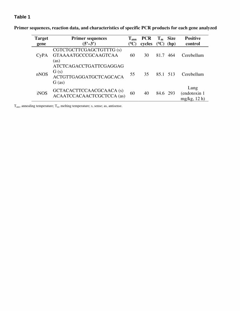
Table 1 Primer sequences, reaction data, and characteristics of specific PCR products for each gene analyzed
Target gene
Primer sequences (5’–3’)
Tann (ºC)
PCR cycles
Tm (ºC)
Size (bp)
Positive control
CyPA CGTCTGCTTCGAGCTGTTTG (s) GTAAAATGCCCGCAAGTCAA (as)
60 30 81.7 464 Cerebellum
nNOS
ATCTCAGACCTGATTCGAGGAGG (s) ACTGTTGAGGATGCTCAGCACAG (as)
55 35 85.1 513 Cerebellum
iNOS GCTACACTTCCAACGCAACA (s) ACAATCCACAACTCGCTCCA (as)
60 40 84.6 293 Lung
(endotoxin 1 mg/kg, 12 h)
Tann, annealing temperature; Tm, melting temperature; s, sense; as, antisense.
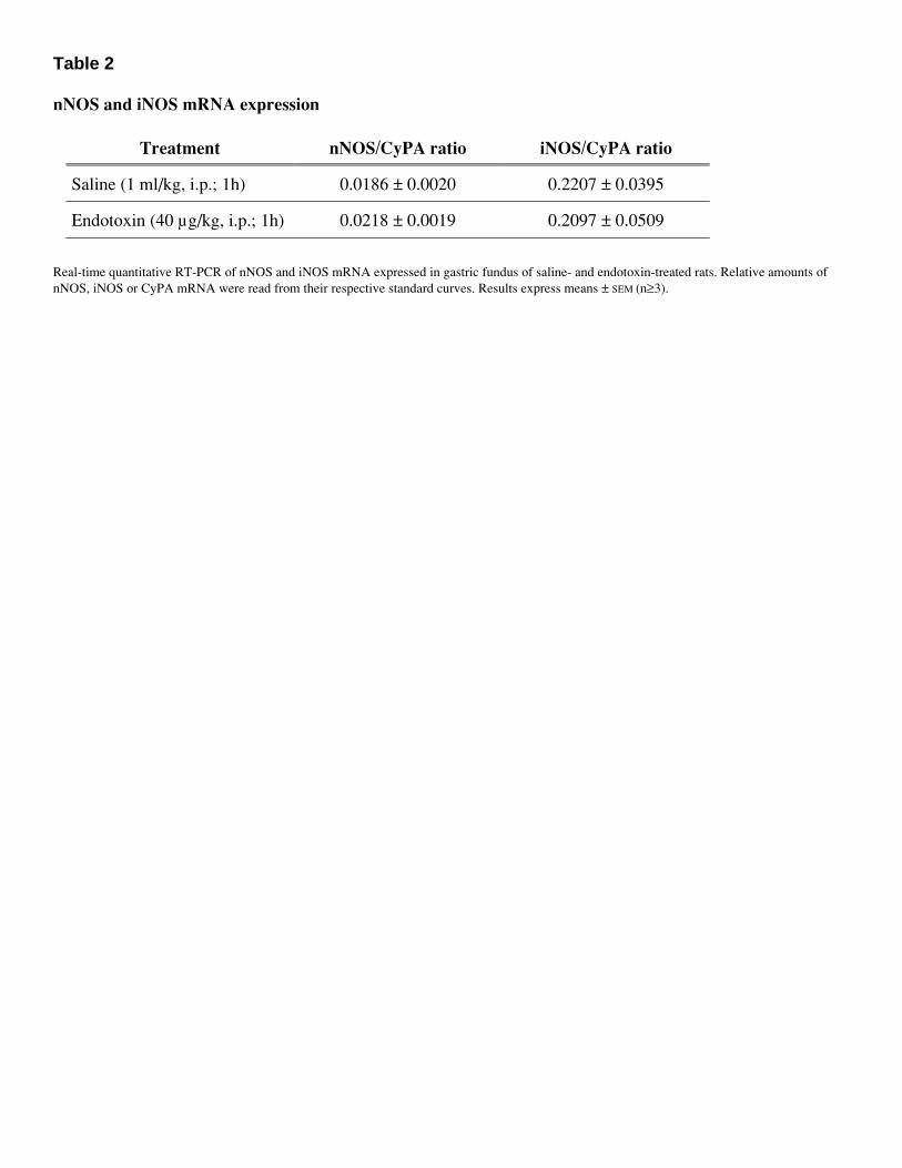
Table 2 nNOS and iNOS mRNA expression
Treatment nNOS/CyPA ratio iNOS/CyPA ratio
Saline (1 ml/kg, i.p.; 1h) 0.0186 ± 0.0020 0.2207 ± 0.0395
Endotoxin (40 µg/kg, i.p.; 1h) 0.0218 ± 0.0019 0.2097 ± 0.0509
Real-time quantitative RT-PCR of nNOS and iNOS mRNA expressed in gastric fundus of saline- and endotoxin-treated rats. Relative amounts of nNOS, iNOS or CyPA mRNA were read from their respective standard curves. Results express means ± SEM (n≥3).
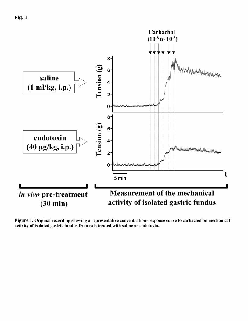
Fig. 1
Figure 1. Original recording showing a representative concentration–response curve to carbachol on mechanical activity of isolated gastric fundus from rats treated with saline or endotoxin.
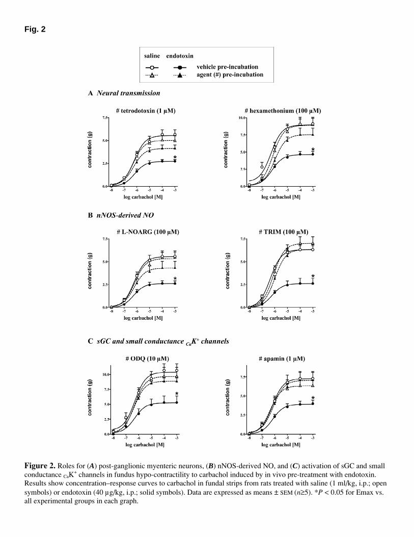
Fig. 2
Figure 2. Roles for (A) post-ganglionic myenteric neurons, (B) nNOS-derived NO, and (C) activation of sGC and small conductance CaK
+ channels in fundus hypo-contractility to carbachol induced by in vivo pre-treatment with endotoxin. Results show concentration–response curves to carbachol in fundal strips from rats treated with saline (1 ml/kg, i.p.; open symbols) or endotoxin (40 µg/kg, i.p.; solid symbols). Data are expressed as means ± SEM (n≥5). *P < 0.05 for Emax vs. all experimental groups in each graph.
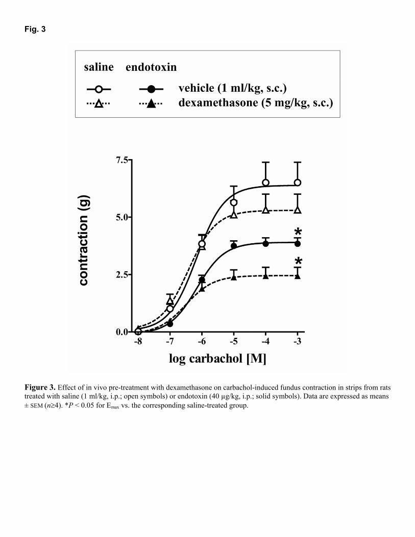
Fig. 3
Figure 3. Effect of in vivo pre-treatment with dexamethasone on carbachol-induced fundus contraction in strips from rats treated with saline (1 ml/kg, i.p.; open symbols) or endotoxin (40 µg/kg, i.p.; solid symbols). Data are expressed as means ± SEM (n≥4). *P < 0.05 for Emax vs. the corresponding saline-treated group.
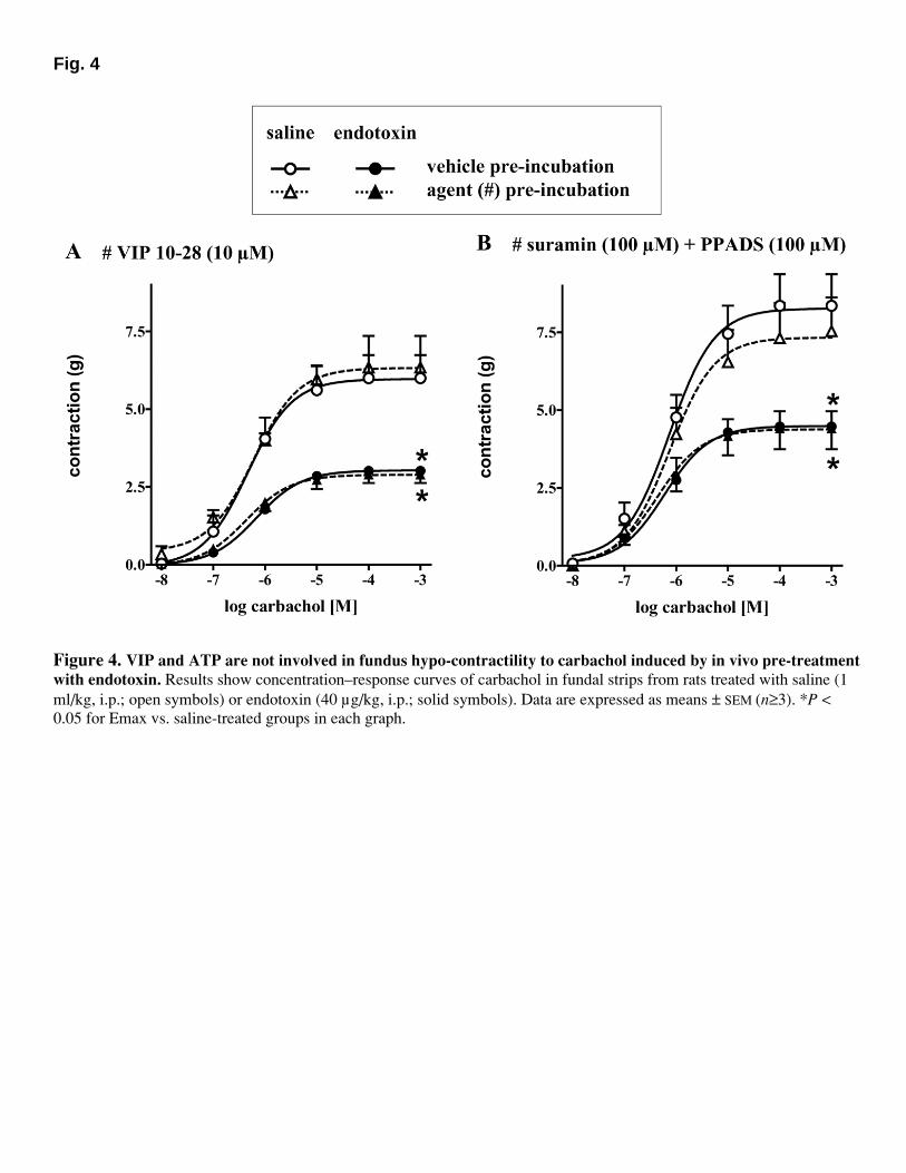
Fig. 4
Figure 4. VIP and ATP are not involved in fundus hypo-contractility to carbachol induced by in vivo pre-treatment with endotoxin. Results show concentration–response curves of carbachol in fundal strips from rats treated with saline (1 ml/kg, i.p.; open symbols) or endotoxin (40 µg/kg, i.p.; solid symbols). Data are expressed as means ± SEM (n≥3). *P < 0.05 for Emax vs. saline-treated groups in each graph.
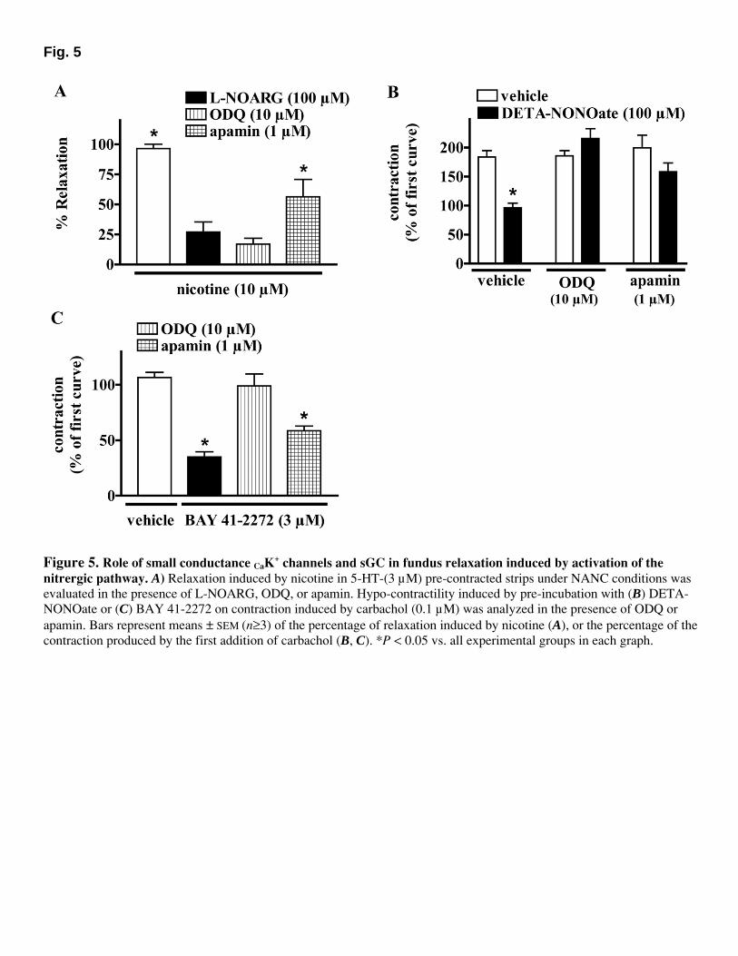
Fig. 5
Figure 5. Role of small conductance CaK
+ channels and sGC in fundus relaxation induced by activation of the nitrergic pathway. A) Relaxation induced by nicotine in 5-HT-(3 µM) pre-contracted strips under NANC conditions was evaluated in the presence of L-NOARG, ODQ, or apamin. Hypo-contractility induced by pre-incubation with (B) DETA-NONOate or (C) BAY 41-2272 on contraction induced by carbachol (0.1 µM) was analyzed in the presence of ODQ or apamin. Bars represent means ± SEM (n≥3) of the percentage of relaxation induced by nicotine (A), or the percentage of the contraction produced by the first addition of carbachol (B, C). *P < 0.05 vs. all experimental groups in each graph.
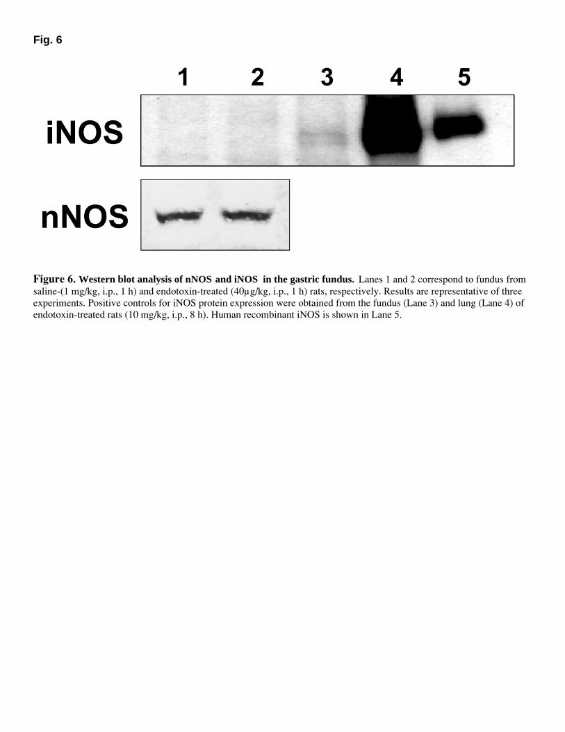
Fig. 6
Figure 6. Western blot analysis of nNOS and iNOS in the gastric fundus. Lanes 1 and 2 correspond to fundus from saline-(1 mg/kg, i.p., 1 h) and endotoxin-treated (40µg/kg, i.p., 1 h) rats, respectively. Results are representative of three experiments. Positive controls for iNOS protein expression were obtained from the fundus (Lane 3) and lung (Lane 4) of endotoxin-treated rats (10 mg/kg, i.p., 8 h). Human recombinant iNOS is shown in Lane 5.
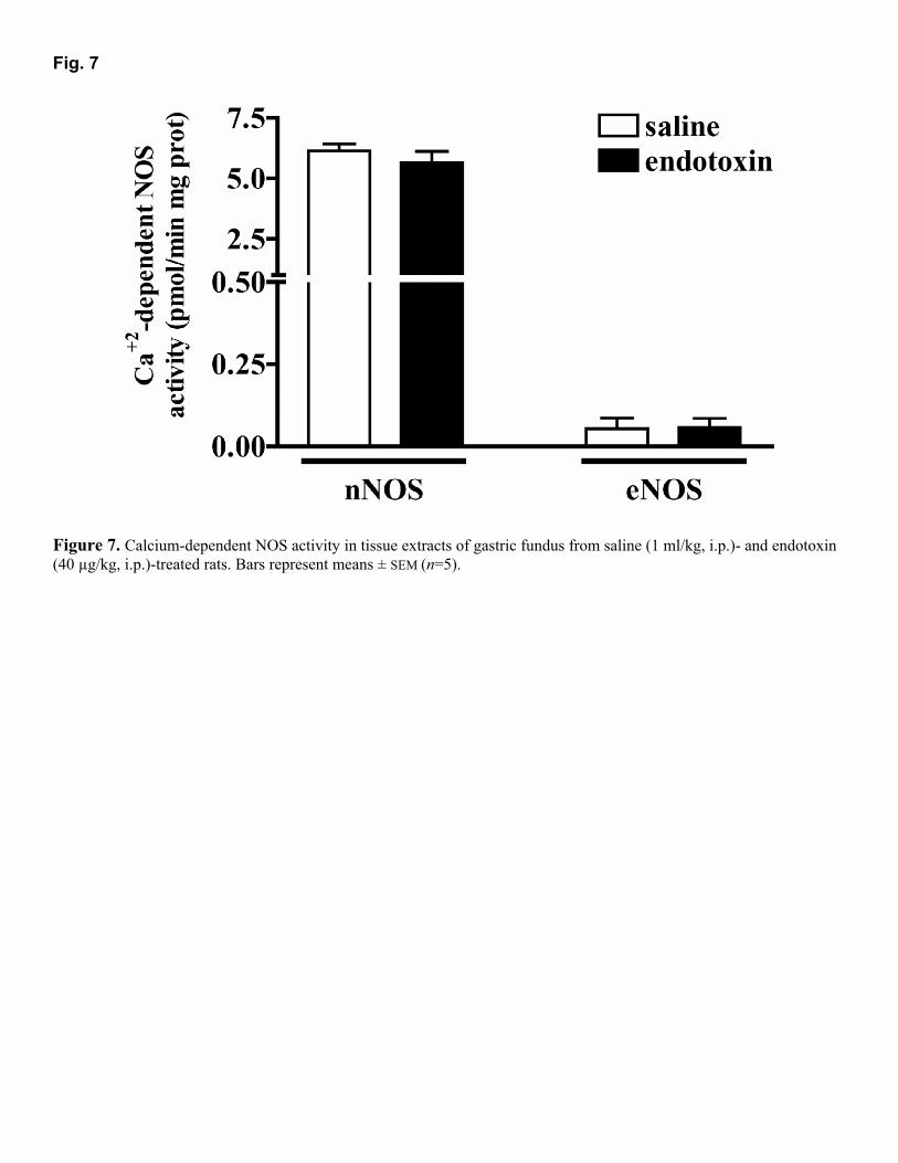
Fig. 7
Figure 7. Calcium-dependent NOS activity in tissue extracts of gastric fundus from saline (1 ml/kg, i.p.)- and endotoxin (40 µg/kg, i.p.)-treated rats. Bars represent means ± SEM (n=5).
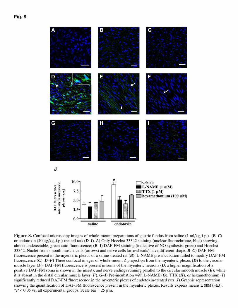
Fig. 8
Figure 8. Confocal microscopy images of whole-mount preparations of gastric fundus from saline (1 ml/kg, i.p.)- (B–C) or endotoxin (40 µg/kg, i.p.)-treated rats (D–I). A) Only Hoechst 33342 staining (nuclear fluorochrome, blue) showing, almost undetectable, green auto fluorescence; (B–I) DAF-FM staining (indicative of NO synthesis; green) and Hoechst 33342. Nuclei from smooth muscle cells (arrows) and nerve cells (arrowheads) have different shape. B–C) DAF-FM fluorescence present in the myenteric plexus of a saline-treated rat (B); L-NAME pre-incubation failed to modify DAF-FM fluorescence (C). D–F) Three confocal images of whole-mount Z projection from the myenteric plexus (D) to the circular muscle layer (F). DAF-FM fluorescence is present in soma of the myenteric neurons (D, a higher magnification of a positive DAF-FM soma is shown in the insert), and nerve endings running parallel to the circular smooth muscle (E), while it is absent in the distal circular muscle layer (F). G–I) Pre-incubation with L-NAME (G), TTX (H), or hexamethonium (I) significantly reduced DAF-FM fluorescence in the myenteric plexus of endotoxin-treated rats. J) Graphic representation showing the quantification of DAF-FM fluorescence present in the myenteric plexus. Results express means ± SEM (n≥3). *P < 0.05 vs. all experimental groups. Scale bar = 25 µm.
Artículos de investigación
100
Artículo 4
“Endotoxin inhibits gastric emptying in
rats via a capsaicin-sensitive afferent
pathway”
Sara Calatayud, María D. Barrachina, Eugenia García-Zaragozá, Elsa
Quintana & Juan V. Esplugues
Naunyn-Schmiedeberg’s Archives of Pharmacology (2001) 363:276-280.
Abstract The effects of endotoxin on gastric emptyingof a solid nutrient meal and the neural mechanisms in-volved in such a response were investigated in consciousrats.
The intraperitoneal (i.p.) administration of E. coli en-dotoxin (40 µg/kg) significantly reduced the 4-h rate ofgastric emptying of a standard solid nutrient meal. Abla-tion of primary afferent neurons by systemic administra-tion of high doses of capsaicin (20+30+50 mg/kg s.c.) toadult rats did not modify the rate of gastric emptying incontrol animals but prevented the delay in gastric transitinduced by endotoxin. Local application of capsaicin tothe vagus nerve rather than application of capsaicin to theceliac ganglion significantly repressed endotoxin-induceddelay in gastric emptying. Neither treatment modified therate of gastric emptying in vehicle-treated animals. Block-ade of CGRP receptors (CGRP 8–37, 100 µg/kg i.v.) didnot alter gastric emptying in control animals but signifi-cantly prevented endotoxin-induced inhibition of gastricemptying. In contrast, a tachykinin receptor antagonist([D-Pro2, D-Trp7,9]-substance P, 2 mg/kg i.p.) significantlyreduced the rate of gastric emptying in control animalsand did not modify the inhibitory effects of endotoxin.Adrenergic blockade with phentolamine (3 mg/kg i.p.) +propranolol (5 mg/kg i.p.) or muscarinic antagonism withatropine (0.1 mg/kg i.p.) failed to reverse the delay in gas-tric emptying induced by endotoxin.
These observations indicate that endotoxin-induceddelay in gastric emptying of a solid nutrient meal is medi-ated by capsaicin-sensitive afferent neurons.
Keywords Gastric emptying · Transit · Endotoxin · Capsaicin · CGRP · Tachykinins
Introduction
Administration of low doses of endotoxin to experimentalanimals induces acute changes in gastric function. Dosesof endotoxin that do not modify systemic arterial bloodpressure or rectal temperature selectively decrease gastricacid secretion (Barrachina et al. 1995a; Esplugues et al.1996; Martínez-Cuesta et al. 1994), increase the mucosalresistance to damage (Barrachina et al. 1995b) and de-crease gastric emptying of non-nutrient, usually liquid,meals (Collares 1997; Cullen et al. 1995; Takakura et al.1997; van Miert and de la Parra 1970). These effects ofendotoxin do not appear to be the consequence of a localmechanism and seem to be mediated by reflex pathwaysinvolving extrinsic innervation of the gut.
A delayed gastric emptying is commonly observed inmany pathological circumstances associated with endo-toxemia such as cirrhosis (Acalovschi et al. 1997; Isobe etal. 1994), diabetes, chronic renal failure (Kao et al. 1996),head injury (Kao et al. 1998), critical illness (Heyland etal. 1996) or the post-operative period (Ratych et al. 1991;Resnick et al. 1997; van Berge Henegouwen et al. 1997).In these conditions, a delay in the emptying of nutrientsfrom the stomach is the principal cause of dyspepsia, asignificant observation considering the lack of prior evi-dence about the gastric emptying of nutrient solid mealsin endotoxemia.
In the present study, we evaluated the effect of smalldoses of lipopolysaccharide of Escherichia coli on therate of gastric emptying of a standard solid meal in con-scious rats. In addition, we analyzed the afferent and theefferent neural pathways involved in endotoxin-inducedchanges in gastric emptying.
Materials and methods
General. Gastric emptying of solids was measured as previouslydescribed (Barrachina et al. 1997). Male Sprague-Dawley rats(250–300 g) which had fasted for 16 h were given access to pre-weighed food for 3 h. The food was then removed and the ani-mals were administered with endotoxin (E. coli lipopolysaccha-
Sara Calatayud · Maria Dolores Barrachina ·Eugenia García-Zaragozá · Elsa Quintana ·Juan V. Esplugues
Endotoxin inhibits gastric emptying in rats via a capsaicin-sensitive afferent pathway
Naunyn-Schmiedeberg’s Arch Pharmacol (2001) 363 :276–280DOI 10.1007/s002100000359
Received: 3 December 1999 / Accepted: 28 September 2000 / Published online: 1 December 2000
ORIGINAL ARTICLE
S. Calatayud · M. D. Barrachina (✉ ) · E. García-Zaragozá ·E. Quintana · J. V. EspluguesDepartment of Pharmacology, Faculty of Medicine, University of Valencia, Avda. Blasco Ibáñez 15, 46010 Valencia, Spain e-mail: [email protected], Fax: +34-96-3864625
© Springer-Verlag 2000
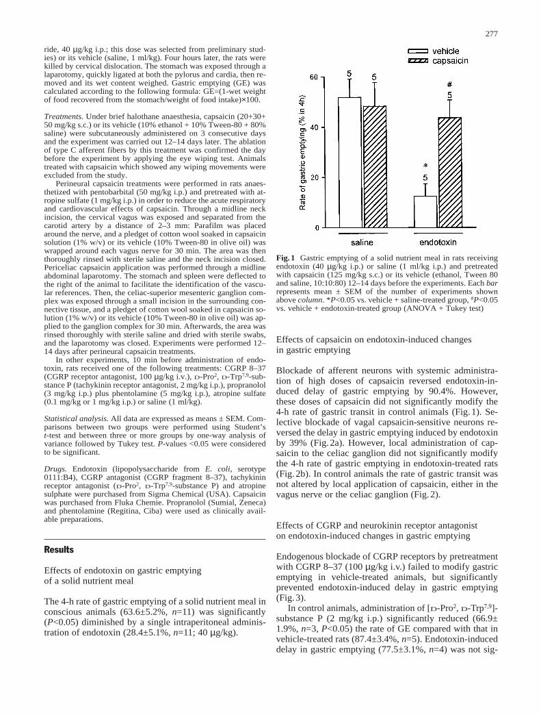
ride, 40 µg/kg i.p.; this dose was selected from preliminary stud-ies) or its vehicle (saline, 1 ml/kg). Four hours later, the rats werekilled by cervical dislocation. The stomach was exposed through alaparotomy, quickly ligated at both the pylorus and cardia, then re-moved and its wet content weighed. Gastric emptying (GE) wascalculated according to the following formula: GE=(1-wet weightof food recovered from the stomach/weight of food intake)×100.
Treatments. Under brief halothane anaesthesia, capsaicin (20+30+50 mg/kg s.c.) or its vehicle (10% ethanol + 10% Tween-80 + 80%saline) were subcutaneously administered on 3 consecutive daysand the experiment was carried out 12–14 days later. The ablationof type C afferent fibers by this treatment was confirmed the daybefore the experiment by applying the eye wiping test. Animalstreated with capsaicin which showed any wiping movements wereexcluded from the study.
Perineural capsaicin treatments were performed in rats anaes-thetized with pentobarbital (50 mg/kg i.p.) and pretreated with at-ropine sulfate (1 mg/kg i.p.) in order to reduce the acute respiratoryand cardiovascular effects of capsaicin. Through a midline neckincision, the cervical vagus was exposed and separated from thecarotid artery by a distance of 2–3 mm: Parafilm was placedaround the nerve, and a pledget of cotton wool soaked in capsaicinsolution (1% w/v) or its vehicle (10% Tween-80 in olive oil) waswrapped around each vagus nerve for 30 min. The area was thenthoroughly rinsed with sterile saline and the neck incision closed.Periceliac capsaicin application was performed through a midlineabdominal laparotomy. The stomach and spleen were deflected tothe right of the animal to facilitate the identification of the vascu-lar references. Then, the celiac-superior mesenteric ganglion com-plex was exposed through a small incision in the surrounding con-nective tissue, and a pledget of cotton wool soaked in capsaicin so-lution (1% w/v) or its vehicle (10% Tween-80 in olive oil) was ap-plied to the ganglion complex for 30 min. Afterwards, the area wasrinsed thoroughly with sterile saline and dried with sterile swabs,and the laparotomy was closed. Experiments were performed 12–14 days after perineural capsaicin treatments.
In other experiments, 10 min before administration of endo-toxin, rats received one of the following treatments: CGRP 8–37(CGRP receptor antagonist, 100 µg/kg i.v.), D-Pro2, D-Trp7,9-sub-stance P (tachykinin receptor antagonist, 2 mg/kg i.p.), propranolol(3 mg/kg i.p.) plus phentolamine (5 mg/kg i.p.), atropine sulfate(0.1 mg/kg or 1 mg/kg i.p.) or saline (1 ml/kg).
Statistical analysis. All data are expressed as means ± SEM. Com-parisons between two groups were performed using Student’s t-test and between three or more groups by one-way analysis ofvariance followed by Tukey test. P-values <0.05 were consideredto be significant.
Drugs. Endotoxin (lipopolysaccharide from E. coli, serotype0111:B4), CGRP antagonist (CGRP fragment 8–37), tachykininreceptor antagonist (D-Pro2, D-Trp7,9-substance P) and atropinesulphate were purchased from Sigma Chemical (USA). Capsaicinwas purchased from Fluka Chemie. Propranolol (Sumial, Zeneca)and phentolamine (Regitina, Ciba) were used as clinically avail-able preparations.
Results
Effects of endotoxin on gastric emptying of a solid nutrient meal
The 4-h rate of gastric emptying of a solid nutrient meal inconscious animals (63.6±5.2%, n=11) was significantly(P<0.05) diminished by a single intraperitoneal adminis-tration of endotoxin (28.4±5.1%, n=11; 40 µg/kg).
Effects of capsaicin on endotoxin-induced changes in gastric emptying
Blockade of afferent neurons with systemic administra-tion of high doses of capsaicin reversed endotoxin-in-duced delay of gastric emptying by 90.4%. However,these doses of capsaicin did not significantly modify the4-h rate of gastric transit in control animals (Fig.1). Se-lective blockade of vagal capsaicin-sensitive neurons re-versed the delay in gastric emptying induced by endotoxinby 39% (Fig.2a). However, local administration of cap-saicin to the celiac ganglion did not significantly modifythe 4-h rate of gastric emptying in endotoxin-treated rats(Fig.2b). In control animals the rate of gastric transit wasnot altered by local application of capsaicin, either in thevagus nerve or the celiac ganglion (Fig.2).
Effects of CGRP and neurokinin receptor antagonist on endotoxin-induced changes in gastric emptying
Endogenous blockade of CGRP receptors by pretreatmentwith CGRP 8–37 (100 µg/kg i.v.) failed to modify gastricemptying in vehicle-treated animals, but significantly prevented endotoxin-induced delay in gastric emptying(Fig.3).
In control animals, administration of [D-Pro2, D-Trp7,9]-substance P (2 mg/kg i.p.) significantly reduced (66.9±1.9%, n=3, P<0.05) the rate of GE compared with that invehicle-treated rats (87.4±3.4%, n=5). Endotoxin-induceddelay in gastric emptying (77.5±3.1%, n=4) was not sig-
277
Fig.1 Gastric emptying of a solid nutrient meal in rats receivingendotoxin (40 µg/kg i.p.) or saline (1 ml/kg i.p.) and pretreatedwith capsaicin (125 mg/kg s.c.) or its vehicle (ethanol, Tween 80and saline, 10:10:80) 12–14 days before the experiments. Each barrepresents mean ± SEM of the number of experiments shownabove column. *P<0.05 vs. vehicle + saline-treated group, #P<0.05vs. vehicle + endotoxin-treated group (ANOVA + Tukey test)
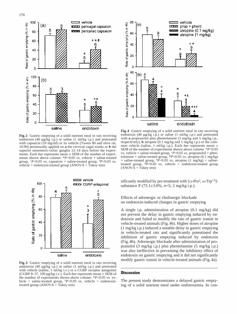
nificantly modified by pre-treatment with [D-Pro2, D-Trp7,9]-substance P (75.1±3.0%, n=5; 2 mg/kg i.p.).
Effects of adrenergic or cholinergic blockade on endotoxin-induced changes in gastric emptying
A single i.p. administration of atropine (0.1 mg/kg) didnot prevent the delay in gastric emptying induced by en-dotoxin and failed to modify the rate of gastric transit invehicle-treated animals (Fig.4b). Higher doses of atropine(1 mg/kg i.p.) induced a notable delay in gastric emptyingin vehicle-treated rats and significantly potentiated the inhibition of gastric emptying induced by endotoxin (Fig.4b). Adrenergic blockade after administration of pro-pranolol (3 mg/kg i.p.) plus phentolamine (5 mg/kg i.p.)was also ineffective in preventing the inhibitory effect ofendotoxin on gastric emptying and it did not significantlymodify gastric transit in vehicle-treated animals (Fig.4a).
Discussion
The present study demonstrates a delayed gastric empty-ing of a solid nutrient meal under endotoxemia. In con-
278
Fig.3 Gastric emptying of a solid nutrient meal in rats receivingendotoxin (40 µg/kg i.p.) or saline (1 ml/kg i.p.) and pretreatedwith vehicle (saline, 1 ml/kg i.v.) or a CGRP receptor antagonist(CGRP 8–37, 100 µg/kg i.v.). Each bar represents mean ± SEM ofthe number of experiments shown above column. *P<0.05 vs. ve-hicle + saline-treated group, #P<0.05 vs. vehicle + endotoxin-treated group (ANOVA + Tukey test)
Fig.2 Gastric emptying of a solid nutrient meal in rats receivingendotoxin (40 µg/kg i.p.) or saline (1 ml/kg i.p.) and pretreatedwith capsaicin (10 mg/ml) or its vehicle (Tween 80 and olive oil,10:90) perineurally applied on a the cervical vagal trunks or b thesuperior mesenteric-celiac ganglia 12–14 days before the experi-ments. Each bar represents mean ± SEM of the number of experi-ments shown above column. *P<0.05 vs. vehicle + saline-treatedgroup, +P<0.05 vs. capsaicin + saline-treated group, #P<0.05 vs.vehicle + endotoxin-treated group (ANOVA + Tukey test)
Fig.4 Gastric emptying of a solid nutrient meal in rats receivingendotoxin (40 µg/kg i.p.) or saline (1 ml/kg i.p.) and pretreatedwith a propranolol plus phentolamine (3 mg/kg and 5 mg/kg i.p.,respectively), b atropine (0.1 mg/kg and 1 mg/kg i.p.) or the com-mon vehicle (saline, 1 ml/kg i.p.). Each bar represents mean ±SEM of the number of experiments shown above column. *P<0.05vs. vehicle + saline-treated group, +P<0.05 vs. propranolol + phen-tolamine + saline-treated group, &P<0.05 vs. atropine (0.1 mg/kg)+ saline-treated group, #P<0.05 vs. atropine (1 mg/kg) + saline-treated group, $P<0.05 vs. vehicle + endotoxin-treated group(ANOVA + Tukey test)
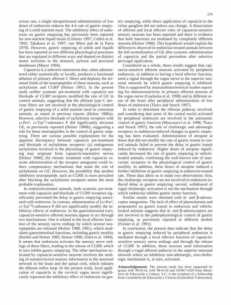
scious rats, a single intraperitoneal administration of lowdoses of endotoxin reduces the 4-h rate of gastric empty-ing of a solid nutrient meal. The inhibitory effect of endo-toxin on gastric emptying has previously been reportedfor non-nutrient liquid meals (Collares 1997; Cullen et al.1995; Takakura et al. 1997; van Miert and de la Parra1970). However, gastric emptying of solids and liquidshas been reported as two different physiological processesthat are regulated in different ways and depend on distinctmotor activities in the stomach, pylorus and proximalduodenum (Mayer 1994).
Capsaicin is a selective neurotoxin that, when adminis-tered either systemically or locally, produces a functionalablation of primary afferent C fibers and depletes the ter-minal fields of the neuropeptides of these neurons, such astachykinins and CGRP (Holzer 1991). In the presentstudy neither systemic pre-treatment with capsaicin norblockade of CGRP receptors modified the rate of GE incontrol animals, suggesting that the afferent type C ner-vous fibers are not involved in the physiological controlof gastric emptying of a solid nutrient meal in consciousanimals, as stated in previous reports (Holzer 1986a).However, selective blockade of tachykinins receptors withD-Pro2, D-Trp7,9-substance P did significantly decreaseGE, as previously reported (Holzer 1986b), suggesting arole for these neuropeptides in the control of gastric emp-tying. There are various possible explanations for the apparent discrepancy between capsaicin pre-treatmentand blockade of tachykinins receptors: (a) endogenoustachykinins involved in the physiology of gastric empty-ing may originate from capsaicin-insensitive nerves(Holzer 1988); (b) chronic treatment with capsaicin vs.acute administration of the receptor antagonist could in-duce compensatory mechanisms that mask the role oftachykinins on GE. However, the possibility that anotherinhibitory neuropeptide, such as CGRP, is more prevalentafter blocking the tachykinins receptors seems the mostprobable explanation.
In endotoxin-treated animals, both systemic pre-treat-ment with capsaicin and blockade of CGRP receptors sig-nificantly prevented the delay in gastric emptying associ-ated with endotoxin. In contrast, administration of [D-Pro2,D-Trp7,9]-substance P did not significantly modify the in-hibitory effects of endotoxin. In the gastrointestinal tract,capsaicin-sensitive afferent neurons appear to act throughtwo mechanisms. One is related to the local effector func-tion of the sensory nerve endings by which several neu-ropeptides are released (Holzer 1988, 1991), which mod-ulates gastrointestinal functions, including gastric motility(Barthó and Holzer 1985; Holzer 1998; Zittel et al. 1994).It seems that endotoxin activates the sensory nerve end-ings of these fibers, leading to the release of CGRP, whichin turn inhibit gastric emptying. The other mechanism ac-tivated by capsaicin-sensitive neurons involves the send-ing of somatovisceral sensory information to the neuronalnetwork in the brain and the spinal cord, which initiatesthe efferent reflex loop. In the present study, local appli-cation of capsaicin to the cervical vagus nerve signifi-cantly repressed the inhibitory effect of endotoxin on gas-
tric emptying, while direct application of capsaicin to theceliac ganglion did not induce any change. A dissociationof afferent and local effector roles of capsaicin-sensitivesensory neurons has been reported and there is evidencethat both functions are mediated by completely differentneurons (Holzer 1988). This hypothesis would explain thedifferences observed in endotoxin-treated animals betweenthe full normalization of GE after systemic administrationof capsaicin and the partial prevention after selectiveperivagal application.
Considered as a whole, these results suggest that cap-saicin-sensitive afferent neurons activated by peripheralendotoxin, in addition to having a local effector function,send a signal through the vagus nerve to the superior neu-ronal network by which gastric emptying is inhibited.This is supported by immunohistochemical studies report-ing fos immunoreactivity in primary afferent neurons ofthe vagus nerve (Gaykema et al. 1998) and in different ar-eas of the brain after peripheral administration of lowdoses of endotoxin (Tkacs and Strack 1997).
In order to determine the efferent pathway involved,and considering that some of the central nuclei activatedby peripheral endotoxin are involved in the autonomiccontrol of gastric function (Mascarucci et al. 1998; Tkacsand Strack 1997), the role of cholinergic and adrenergicreceptors in endotoxin-induced changes in gastric empty-ing has been evaluated. Administration of atropine atdoses that did not modify the rate of gastric transit in con-trol animals failed to prevent the delay in gastric transitinduced by endotoxin. Higher doses of atropine signifi-cantly decreased the rate of gastric emptying in vehicle-treated animals, confirming the well-known role of mus-carinic receptors in the physiological control of gastricmotility. In addition, these doses of atropine induced afurther inhibition of gastric emptying in endotoxin-treatedrats. These data allow us to make two observations: first,the cholinergic receptors are not involved in endotoxin-in-duced delay in gastric emptying; second, withdrawal ofvagal cholinergic activation is not the mechanism throughwhich endotoxin inhibits gastric motor function.
Similar results were obtained with α- and β-adreno-ceptor antagonists. The lack of effect of phentolamine andpropranolol on gastric transit in endotoxin and vehicle-treated animals suggests that α- and β-adrenoceptors arenot involved in the pathophysiological control of gastricemptying, as previously reported in different models(Forster et al. 1991).
In conclusion, the present data indicate that the delayin gastric emptying induced by peripheral endotoxin ismediated through a local effector function of capsaicin-sensitive sensory nerve endings and through the release of CGRP. In addition, these neurons send informationthrough a vagal afferent pathway to the superior neuronalnetwork where an inhibitory non-adrenergic, non-cholin-ergic mechanism is, in turn, activated.
Acknowledgements The present study has been supported bygrants SAF 99-0114, SAF 98-0118 and 1FD97-1029 from Minis-terio de Educación y Cultura. S.C. is the recipient of a fellowshipfrom Conselleria de Educación y Ciencia (Generalitat Valenciana),
279
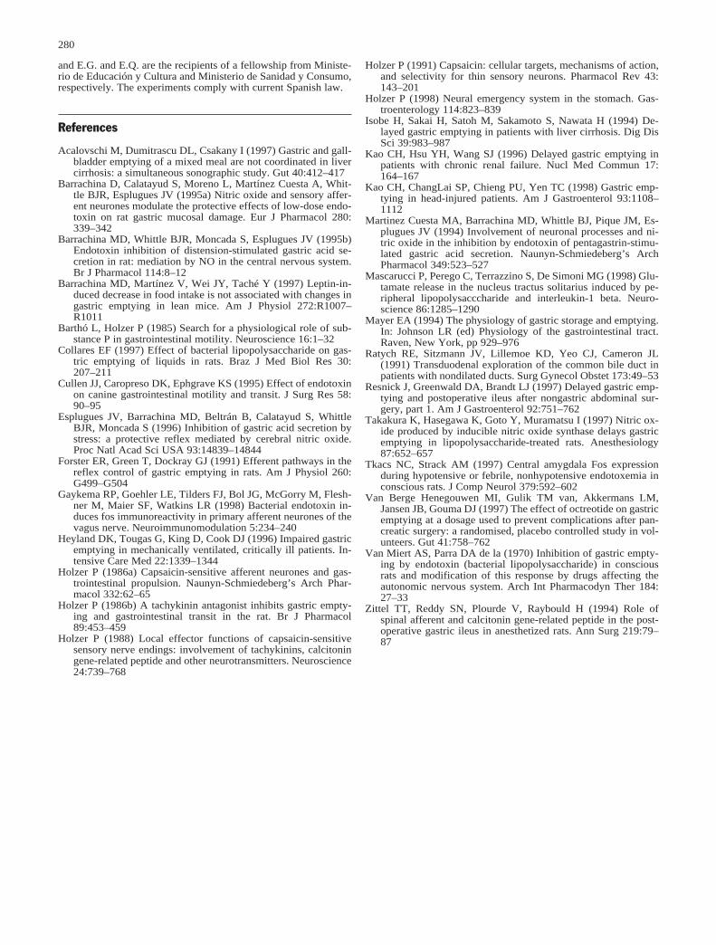
280
and E.G. and E.Q. are the recipients of a fellowship from Ministe-rio de Educación y Cultura and Ministerio de Sanidad y Consumo,respectively. The experiments comply with current Spanish law.
References
Acalovschi M, Dumitrascu DL, Csakany I (1997) Gastric and gall-bladder emptying of a mixed meal are not coordinated in livercirrhosis: a simultaneous sonographic study. Gut 40:412–417
Barrachina D, Calatayud S, Moreno L, Martínez Cuesta A, Whit-tle BJR, Esplugues JV (1995a) Nitric oxide and sensory affer-ent neurones modulate the protective effects of low-dose endo-toxin on rat gastric mucosal damage. Eur J Pharmacol 280:339–342
Barrachina MD, Whittle BJR, Moncada S, Esplugues JV (1995b)Endotoxin inhibition of distension-stimulated gastric acid se-cretion in rat: mediation by NO in the central nervous system.Br J Pharmacol 114:8–12
Barrachina MD, Martínez V, Wei JY, Taché Y (1997) Leptin-in-duced decrease in food intake is not associated with changes ingastric emptying in lean mice. Am J Physiol 272:R1007–R1011
Barthó L, Holzer P (1985) Search for a physiological role of sub-stance P in gastrointestinal motility. Neuroscience 16:1–32
Collares EF (1997) Effect of bacterial lipopolysaccharide on gas-tric emptying of liquids in rats. Braz J Med Biol Res 30:207–211
Cullen JJ, Caropreso DK, Ephgrave KS (1995) Effect of endotoxinon canine gastrointestinal motility and transit. J Surg Res 58:90–95
Esplugues JV, Barrachina MD, Beltrán B, Calatayud S, WhittleBJR, Moncada S (1996) Inhibition of gastric acid secretion bystress: a protective reflex mediated by cerebral nitric oxide.Proc Natl Acad Sci USA 93:14839–14844
Forster ER, Green T, Dockray GJ (1991) Efferent pathways in thereflex control of gastric emptying in rats. Am J Physiol 260:G499–G504
Gaykema RP, Goehler LE, Tilders FJ, Bol JG, McGorry M, Flesh-ner M, Maier SF, Watkins LR (1998) Bacterial endotoxin in-duces fos immunoreactivity in primary afferent neurones of thevagus nerve. Neuroimmunomodulation 5:234–240
Heyland DK, Tougas G, King D, Cook DJ (1996) Impaired gastricemptying in mechanically ventilated, critically ill patients. In-tensive Care Med 22:1339–1344
Holzer P (1986a) Capsaicin-sensitive afferent neurones and gas-trointestinal propulsion. Naunyn-Schmiedeberg’s Arch Phar-macol 332:62–65
Holzer P (1986b) A tachykinin antagonist inhibits gastric empty-ing and gastrointestinal transit in the rat. Br J Pharmacol89:453–459
Holzer P (1988) Local effector functions of capsaicin-sensitivesensory nerve endings: involvement of tachykinins, calcitoningene-related peptide and other neurotransmitters. Neuroscience24:739–768
Holzer P (1991) Capsaicin: cellular targets, mechanisms of action,and selectivity for thin sensory neurons. Pharmacol Rev 43:143–201
Holzer P (1998) Neural emergency system in the stomach. Gas-troenterology 114:823–839
Isobe H, Sakai H, Satoh M, Sakamoto S, Nawata H (1994) De-layed gastric emptying in patients with liver cirrhosis. Dig DisSci 39:983–987
Kao CH, Hsu YH, Wang SJ (1996) Delayed gastric emptying inpatients with chronic renal failure. Nucl Med Commun 17:164–167
Kao CH, ChangLai SP, Chieng PU, Yen TC (1998) Gastric emp-tying in head-injured patients. Am J Gastroenterol 93:1108–1112
Martinez Cuesta MA, Barrachina MD, Whittle BJ, Pique JM, Es-plugues JV (1994) Involvement of neuronal processes and ni-tric oxide in the inhibition by endotoxin of pentagastrin-stimu-lated gastric acid secretion. Naunyn-Schmiedeberg’s ArchPharmacol 349:523–527
Mascarucci P, Perego C, Terrazzino S, De Simoni MG (1998) Glu-tamate release in the nucleus tractus solitarius induced by pe-ripheral lipopolysacccharide and interleukin-1 beta. Neuro-science 86:1285–1290
Mayer EA (1994) The physiology of gastric storage and emptying.In: Johnson LR (ed) Physiology of the gastrointestinal tract.Raven, New York, pp 929–976
Ratych RE, Sitzmann JV, Lillemoe KD, Yeo CJ, Cameron JL(1991) Transduodenal exploration of the common bile duct inpatients with nondilated ducts. Surg Gynecol Obstet 173:49–53
Resnick J, Greenwald DA, Brandt LJ (1997) Delayed gastric emp-tying and postoperative ileus after nongastric abdominal sur-gery, part 1. Am J Gastroenterol 92:751–762
Takakura K, Hasegawa K, Goto Y, Muramatsu I (1997) Nitric ox-ide produced by inducible nitric oxide synthase delays gastricemptying in lipopolysaccharide-treated rats. Anesthesiology87:652–657
Tkacs NC, Strack AM (1997) Central amygdala Fos expressionduring hypotensive or febrile, nonhypotensive endotoxemia inconscious rats. J Comp Neurol 379:592–602
Van Berge Henegouwen MI, Gulik TM van, Akkermans LM,Jansen JB, Gouma DJ (1997) The effect of octreotide on gastricemptying at a dosage used to prevent complications after pan-creatic surgery: a randomised, placebo controlled study in vol-unteers. Gut 41:758–762
Van Miert AS, Parra DA de la (1970) Inhibition of gastric empty-ing by endotoxin (bacterial lipopolysaccharide) in consciousrats and modification of this response by drugs affecting theautonomic nervous system. Arch Int Pharmacodyn Ther 184:27–33
Zittel TT, Reddy SN, Plourde V, Raybould H (1994) Role ofspinal afferent and calcitonin gene-related peptide in the post-operative gastric ileus in anesthetized rats. Ann Surg 219:79–87
Artículos de investigación
106
Artículo 5
“Downregulation of nNOS and synthesis
of PGs associated with endotoxin-
induced delay in gastric emptying”
Sara Calatayud, Eugenia García-Zaragozá, Carlos Hernández, Elsa
Quintana, Vicente Felipo, Juan V. Esplugues & María D. Barrachina
American Journal of Physiology (2002) 283: G1360-G1367.
Downregulation of nNOS and synthesis of PGs associatedwith endotoxin-induced delay in gastric emptying
SARA CALATAYUD,2 EUGENIA GARCIA-ZARAGOZA,1 CARLOS HERNANDEZ,1
ELSA QUINTANA,1 VICENTE FELIPO,3 JUAN VICENTE ESPLUGUES,1
AND MARIA DOLORES BARRACHINA1
1Departmento de Farmacologıa, Universidad de Valencia; 2Unidad Mixta de Investigacion,Hospital Clınico/Universidad de Valencia; and 3Instituto de Investigaciones Citologicas,Fundacion Valenciana de Investigaciones Biomedicas, 46010 Valencia, SpainReceived 9 May 2002; accepted in final form 27 August 2002
Calatayud, Sara, Eugenia Garcıa-Zaragoza, CarlosHernandez, Elsa Quintana, Vicente Felipo, Juan Vi-cente Esplugues, and Marıa Dolores Barrachina.Downregulation of nNOS and synthesis of PGs associatedwith endotoxin-induced delay in gastric emptying. Am JPhysiol Gastrointest Liver Physiol 283: G1360–G1367, 2002;10.1152/ajpgi.00168.2002.—A single intraperitoneal injec-tion of endotoxin (40 �g/kg) significantly delayed gastricemptying of a solid nutrient meal. Blockade of nitric oxidesynthase (NOS) with 30 mg/kg ip NG-nitro-L-arginine methylester or 20 mg/kg ip 7-nitroindazole [neuronal NOS (nNOS)inhibitor] significantly delayed gastric emptying in controlanimals but failed to modify gastric emptying in endotoxin-treated rats. Administration of 2.5, 5, and 10 mg/kg ip N6-iminoethyl-L-lysine [inducible NOS (iNOS) inhibitor] had noeffect in either experimental group. Indomethacin (5 mg/kgsc), NS-398 (cyclooxygenase-2 inhibitor; 10 mg/kg ip), anddexamethasone (10 mg/kg sc) but not quinacrine (20 mg/kgip) significantly prevented delay in gastric emptying inducedby endotoxin but failed to modify gastric emptying in vehicle-treated animals. Ca2�-dependent NOS activity in the an-trum pylorus of the stomach was diminished by endotoxin,whereas Ca2�-independent NOS activity was not changed. Inaddition, decreased nNOS mRNA and protein were observedin the antrum pylorus of endotoxin-treated rats. Our resultssuggest that downregulation of nNOS in the antrum pylorusof the stomach and synthesis of prostaglandins mediate thedelay in gastric emptying of a solid nutrient meal induced byendotoxin.
nitric oxide; prostaglandins; antrum pylorus; nutrient meals
ENDOTOXEMIA AFTER INFECTION with gram-negative bacte-ria is associated with clinical abnormalities of gastricmotor function (such as vomiting) broadly character-ized by a decrease in gastric emptying. Experimentaladministration of the lipopolysaccharide of Escherichiacoli is known to delay gastric emptying (36, 37, 39), andcapsaicin-sensitive afferent neurons and nonadrener-gic noncholinergic mechanisms have recently beenshown to be involved (6).
Nitric oxide (NO) has a leading role as an inhibitorneurotransmitter of peripheral nonadrenergic noncho-linergic nerves (1, 8). In the gastrointestinal tract,neuronal NO plays a role in the physiology of gastricmotor function. NO is involved in the reflex relaxationof the gastric fundus to accommodate food or fluid (7)and mediates pyloric relaxation and intestinal feed-back regulation, thereby facilitating gastric emptying(2). A recent study (21) analyzing gastric emptying inneuronal NO synthase (nNOS) knockout mice points toa prominent role for this isoenzyme in the modulationof gastric motor function. However, little is knownabout the role of nNOS in changes in gastric emptyingassociated with pathophysiological circumstances suchas endotoxemia. Taking into account that endotoxinhas been widely shown to increase the expression ofthe inducible NOS (iNOS) in different tissues (5), thespecific role of nNOS and iNOS in endotoxin-induceddelay in gastric emptying has been evaluated.
Prostaglandins synthesized from arachidonic acidact as local regulatory agents that modulate gastricmotor function (32). Endogenous prostaglandins havebeen involved in the inhibition of gastric emptyinginduced by IL-1� (35), and exogenous administration ofthese prostanoids delays gastric emptying (33). Syn-thesis of prostaglandins is carried out by cyclooxygen-ase (COX), which exists as two isoenzymes, COX-1 and-2, and the role of prostaglandins on gastric emptyinghas been established mainly through the use of nonse-lective COX inhibitors. Both isoenzymes synthesizeprostanoids that mediate physiological functions (38).However, the formation of proinflammatory prosta-glandins is mostly catalyzed by COX-2, and expressionof this isoform is induced by a variety of stimuli, in-cluding endotoxin (12). This study also aims to deter-mine the role and the enzymatic source of prostaglan-dins in the delay of gastric emptying induced byendotoxin.
Address for reprint requests and other correspondence: M. D. Bar-rachina, Dept. of Pharmacology, Faculty of Medicine, Avd. BlascoIbanez, 15, 46010, Valencia, Spain (E-mail: [email protected]).
The costs of publication of this article were defrayed in part by thepayment of page charges. The article must therefore be herebymarked ‘‘advertisement’’ in accordance with 18 U.S.C. Section 1734solely to indicate this fact.
Am J Physiol Gastrointest Liver Physiol 283: G1360–G1367, 2002;10.1152/ajpgi.00168.2002.
0193-1857/02 $5.00 Copyright © 2002 the American Physiological Society http://www.ajpgi.orgG1360
MATERIALS AND METHODS
Animals
Male Sprague-Dawley rats (250–300 g) (Harlan Laborato-ries, Barcelona, Spain) were maintained ad libitum on stan-dard Purina laboratory chow and tap water and were housedunder conditions of controlled temperature (21 � 1°C), hu-midity (30–35%), and lighting (0700–1900 h). All experi-ments began between 0900 h and 1000 h and were performedin animals deprived of food for 16–18 h but with access towater before and during the experiments.
All protocols comply with the European Communityguidelines for the use of experimental animals and wereapproved by the ethics committee of the Faculty of Medi-cine of Valencia.
Measurement of Gastric Emptying
Gastric emptying of solids was measured as previouslydescribed (6). Rats were placed in individual cages and weregiven access to preweighed food for 3 h. Food was thenremoved and 40 �g/kg ip endotoxin (E. coli lipopolysaccha-ride) or 1 ml/kg ip saline was administered to the animals.Four hours later, the rats were killed by cervical dislocation.The stomach was exposed by laparotomy, quickly ligated atboth the pylorus and cardias and removed, and its wetcontent was weighed. Gastric emptying (GE) was calculatedaccording to the following formula: GE � (1 � wet wt of foodrecovered from stomach/weight of food intake) � 100.
Treatments
To analyze the role of NO in the rate of gastric emptying ofa solid nutrient meal, animals received a single intraperito-neal injection of 30 mg/kg N�-nitro-L-arginine methyl ester(L-NAME) (a NOS inhibitor), 20 mg/kg 7-nitroindazole (aselective nNOS inhibitor), or the respective vehicle (1 ml/kgsaline or 0.5 ml/kg DMSO) 2 h after the administration ofendotoxin or saline. Some rats received 2.5, 5, or 10 mg/kg ipN6-iminoethyl-L-lysine (L-NIL) (a selective iNOS inhibitor) orits vehicle (1 ml/kg ip saline) 15 min before endotoxin orsaline, and gastric emptying was determined as describedabove.
To determine the role of prostaglandins in the rate ofgastric emptying of a solid nutrient meal, rats were pre-treated with 5 mg/kg sc indomethacin (a dual inhibitor ofCOX-1 and -2), a COX-2-selective dose of 10 mg/kg ip NS-398(20), or their respective vehicles (1 ml/kg sc NaHCO3 5%, or0.5 ml/kg ip DMSO) 60 min before the administration ofendotoxin or saline. Some animals received a single intra-peritoneal injection of 20 mg/kg quinacrine (a phospholipase2 enzyme blocker) 15 min before endotoxin or saline.
In the last group, rats were treated with dexamethasone (5mg/kg sc, 16 h and 1 h before endotoxin) or saline (1 ml/kg sc),and the rate of gastric emptying was analyzed as mentionedabove.
Determination of NOS Activity
Rats were killed 4 h after the administration of endotoxin(40 �g/kg ip) or saline (1 ml/kg ip). In short, the antrumpylorus tissue was cut into small pieces, frozen in liquidnitrogen, and stored at �80°C. NOS activity was measuredas the rate of conversion of L-[U-14C]arginine to L-[U-14C]citrulline (31). Samples were homogenized (Ultra-Tur-rax) in an ice-cold buffer (330 mg/ml; pH 7.2) containing 320mM sucrose, 20 mM HEPES, 1 mM EDTA, 1 mM DL-dithio-threitol, 10 �g/ml leupeptin, 10 �g/ml soybean trypsin inhib-
itor, and 2 �g/ml aprotinin followed by centrifugation at10.000 g for 20 min at 4°C. Afterward, 40 �l of supernatantwas incubated at 37°C for 20 min in an assay buffer (pH 7.4)containing 50 mM KH2PO4, 1 mM MgCl2, 0.2 mM CaCl2, 50mM L-valine, 1 mM L-citrulline, 0.02 mM L-arginine, 1 mMDL-dithiothreitol, 100 �M NADPH, 3 �M FAD, 3 �M flavinmononucleotide, 3 �M tetrahydrobiopterin, and 950 nM L-[U-14C]arginine (348 mCi/mmol). The specificity of L-arginineconversion by NOS to L-citrulline was further confirmedusing 1 mM N�-nitro-L-arginine (an NOS inhibitor). To dif-ferentiate between the Ca2�-dependent and Ca2�-indepen-dent NOS (iNOS activity), 1 mM EGTA (a calcium chelatingagent) was used. All activities are expressed as picomoles ofproduct generated per minute per gram of tissue.
Determination of nNOS Protein Content by Western Blot
Rats were killed 4 h after the administration of endotoxin(40 �g/kg ip) or saline (1 ml/kg ip). The content of nNOSprotein in the stomach was determined by Western blotanalysis. In short, the antrum pylorus tissue was cut intosmall pieces and homogenized in a boiling medium contain-ing (in mM) 66 Tris �HCl (pH 7.4), 1 EGTA, 1 sodium or-thovanadate, and 1 sodium fluoride with 10% glycerol and 1%SDS. Homogenates were treated with ultrasound (cooled onice for 15 min) followed by centrifugation (10,000 g at 4°C) for20 min. The supernatant was brought to 30% saturation withammonium sulfate and stirred for 30 min in ice. After cen-trifugation for 5 min at 12,000 g, the pellets were resus-pended in the above medium. Lysates were boiled for 5 min,and protein was determined by the bicinchonic acid (proteinassay reagent; Pierce) method. Samples were subjected toSDS-PAGE, and immunoblotting was carried out as previ-ously described (11) by using an antibody against nNOS(1:500, Transduction). After development using anti-mouseIgG conjugated with alkaline phosphatase (Sigma) and alka-line phosphatase color development (Sigma), the image wascaptured using Gelprinter Plus System (TDI), and the den-sities of the spots were measured using the software Intelli-gent Quantifier version 2.5.0. (BioImage).
Quantification of nNOS mRNA and iNOS mRNA by RealTime Quantitative RT-PCR
Rats were administered endotoxin (40 �g/kg ip) or saline (1ml/kg ip) and killed by cervical dislocation 4 h later. In short,the antrum pylorus tissue was frozen in liquid nitrogen andstored at �80°C.
RNA extraction and cDNA synthesis. Total RNA from fro-zen gastric tissues was isolated with TriPure isolation re-agent (Roche Diagnostics) following the manufacturer’s pro-tocol. RNA was resuspended in diethylpyrocarbonate-treatedH2O and stored at �80°C. Total RNA was treated withDNA-free (Ambion) to eliminate traces of contaminatinggenomic DNA. Resulting total RNA was quantified by ultra-violet spectrophotometry, and its integrity was evaluated byagarose gel electrophoresis. RT from 2 �g of total RNA wascarried out with SuperScript RT RNase H� (Life Technolo-gies), using 0.8 �g oligo(dT16) (TIB Molbiol; Roche Diagnos-tics) and 40 units RNase inhibitor (Roche Diagnostics) in areaction volume of 20 �l. Controls without RT for eachsample and a negative control with water in place of RNAwere performed. Synthesized cDNA was stored at �20°C.
Quantitative PCR. Quantitative PCR was carried out in aLightCycler instrument (Roche Diagnostics) with the use ofLightCycler-FastStart DNA Master SYBR Green I kit. Sam-ples of 1 �l of cDNA were amplified through specific primersfor each gene, 0.5 �M cyclophilin A (CyPA), 0.5 �M nNOS, or
G1361ROLE OF NNOS, COX-2, AND ENDOTOXIN IN GASTRIC EMPTYING
AJP-Gastrointest Liver Physiol • VOL 283 • DECEMBER 2002 • www.ajpgi.org
1 �M iNOS, in a solution containing 2 mM MgCl2 and 5%DMSO (final volume 10 �l). Reactions were performed induplicate, and a negative control with water instead of cDNAwas included in each run. Protocol reaction includes aninitial period of 10 min at 95°C to activate the polymerase.Each PCR cycle involved denaturation at 95°C for 30 s,annealing at Tann (shown in Table 1) for 30 s and extension at72°C for 30 s. Fluorescence was measured at the end of eachcycle. Specificity of reactions was tested by analysis of themelting curve (Tm, shown in Table 1) and agarose gel elec-trophoresis. To quantify input amounts of templates, a stan-dard curve was obtained with serial dilutions of total RNA ofa positive control (Table 1) for each analyzed gene, also afterRT-PCR. To normalize the results, interpolated values foreach sample were divided by values for the housekeepinggene CyPA, and results are expressed as an NOS/CyPA ratio.
Statistical Analysis
All data are expressed as means � SE. Comparisons be-tween two groups were performed using the Student’s t-testand among three or more groups by one-way ANOVA fol-lowed by the Newman-Keuls test. P values 0.05 wereconsidered significant.
Drugs
Endotoxin (LPS from E. coli, serotype 0111:B4), L-NAME,L-NIL, 7-nitroindazole, NS-398, indomethacin, quinacrine,and all reagents used for determination of NOS activity werepurchased from Sigma (St. Louis, MO). L-[U-14C]argininewas obtained from Amersham Life Science (London, UK).Dexamethasone (Fortecortin) was used as a clinically avail-able preparation. Unless mentioned otherwise, all drugswere dissolved in saline.
RESULTS
Effects of Endotoxin on Gastric Emptying of a SolidNutrient Meal
The amount of rat chow eaten for 3 h by SpragueDawley rats after a 20-h fast was 5.1 � 0.2 g (n � 10).The 4-h rate of gastric emptying in animals treatedwith vehicle was significantly higher than that ob-served in endotoxin-treated rats (Fig. 1). This dose ofendotoxin has previously been shown to lack effects onrectal temperature and systemic blood pressure inanesthetized animals (9).
Role of NO. Nonisoform-selective blockade of NOsynthesis by pretreatment with L-NAME (30 mg/kg ip)significantly decreased the rate of gastric emptying in
saline-treated rats, whereas it did not significantlychange the rate of gastric emptying in endotoxin-treated animals (Fig. 1). In a similar manner, selectiveblockade of the nNOS isoform by pretreatment with7-nitroindazole (20 mg/kg ip) induced a pronouncedreduction in the rate of gastric emptying in saline-treated animals, whereas it did not modify the rate ofgastric emptying in animals receiving endotoxin (Fig.1). Selective inhibition of the iNOS isoform by theadministration of L-NIL (2.5, 5, and 10 mg/kg ip) didnot significantly modify the rate of gastric emptying invehicle- or endotoxin-treated animals (Fig. 2).
Role of prostaglandins. The delay in gastric empty-ing induced by endotoxin was significantly preventedby pretreatment with the nonselective COX-1 and -2inhibitor indomethacin (5 mg/kg sc; Fig. 3). In a similarmanner, selective inhibition of the COX-2 isoform bypretreatment with NS-398 (10 mg/kg ip) also signifi-cantly prevented the inhibitory effects of endotoxin ongastric emptying (Fig. 3). Both indomethacin and NS-398 when administered to saline-treated animalslacked any effect on the rate of gastric emptying (Fig.3). Blockade of arachidonic acid supply by pretreat-ment with a phospholipase 2 inhibitor, quinacrine (20mg/kg ip), did not alter the rate of gastric emptying inendotoxin- or saline-treated rats (Fig. 4).
Effects of dexamethasone. Pretreatment with dexa-methasone (10 mg/kg sc) significantly prevented theinhibition of gastric emptying by endotoxin but did notsignificantly change the rate of gastric emptying invehicle-treated animals (Fig. 4).
Effects of Endotoxin on NOS Activity
NOS activity analyzed by the rate of conversion ofL-arginine to L-citrulline was present in the antrumpylorus of saline-treated animals. With the use of acalcium chelating agent, we observed that NOS activ-ity in these conditions was mainly Ca2�-dependent,whereas Ca2�-independent NOS activity (iNOS) wasalmost nonapparent (Fig. 5). Pretreatment (4 h) withendotoxin induced a significant reduction of the Ca2�-dependent NOS activity (26.7%) in the antrum pylorusof the rats, whereas it did not significantly change theCa2�-independent NOS activity (iNOS), which wassimilar to that observed in saline-treated rats (Fig. 5).
Table 1. Primer sequences, reaction data, and characteristics of specific PCR products for each analyzed gene
Target Gene Primer Sequences (5-3) Tann, °C PCR, cycles Tm, °C Size, bp Positive Control
CyPA CGTCTGCTTCGAGCTGTTTG (s) 60 30 81.7 464 CerebellumGTAAAATGCCCGCAAGTCAA (as)
nNOS ATCTCAGACCTGATTCGAGGAGG (s) 55 35 85.1 513 CerebellumACTGTTGAGGATGCTCAGCACAG (as)
iNOS GCTACACTTCCAACGCAACA (s) 60 40 84.6 293 Lung, endotoxin, 1 mg/kg, 12 hACAATCCACAACTCGCTCCA (as)
Primers for neuronal nitric oxide synthase (nNOS) were taken as reference (24). Others were designed according to reported sequenceswith GenBank accession no. NM_017101 [cyclophilin A (CyPA)] and D12520 [inducible NOS (iNOS)]. Tann, annealing temperature; Tm,melting temperature.
G1362 ROLE OF NNOS, COX-2, AND ENDOTOXIN IN GASTRIC EMPTYING
AJP-Gastrointest Liver Physiol • VOL 283 • DECEMBER 2002 • www.ajpgi.org
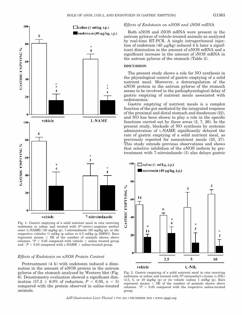
Effects of Endotoxin on nNOS Protein Content
Pretreatment (4 h) with endotoxin induced a dimi-nution in the amount of nNOS protein in the antrumpylorus of the stomach analyzed by Western blot (Fig.6). Densitometry evaluation showed a significant dim-inution (57.2 � 6.0% of reduction; P 0.05, n � 5)compared with the protein observed in saline-treatedanimals.
Effects of Endotoxin on nNOS and iNOS mRNA
Both nNOS and iNOS mRNA were present in theantrum pylorus of vehicle-treated animals as analyzedby real-time RT-PCR. A single intraperitoneal injec-tion of endotoxin (40 �g/kg) induced 4 h later a signif-icant diminution in the amount of nNOS mRNA and asignificant increase in the amount of iNOS mRNA inthe antrum pylorus of the stomach (Table 2).
DISCUSSION
The present study shows a role for NO synthesis inthe physiological control of gastric emptying of a solidnutrient meal. Moreover, a downregulation of thenNOS protein in the antrum pylorus of the stomachseems to be involved in the pathophysiological delay ofgastric emptying of nutrient meals associated withendotoxemia.
Gastric emptying of nutrient meals is a complexfunction of the gut mediated by the integrated responseof the proximal and distal stomach and duodenum (22),and NO has been shown to play a role in the specificfunctions carried out by these areas (2, 7, 26). In thepresent study, blockade of NO synthesis by systemicadministration of L-NAME significantly delayed therate of gastric emptying of a solid nutrient meal, aspreviously reported for nonnutrient meals (25, 27).This study extends previous observations and showsthat selective inhibition of the nNOS isoform by pre-treatment with 7-nitroindazole (3) also delays gastric
Fig. 1. Gastric emptying of a solid nutrient meal in rats receivingendotoxin or saline and treated with N�-nitro-L-arginine methylester (L-NAME) (30 mg/kg ip), 7-nitroindazole (20 mg/kg ip), or therespective vehicles (1 ml/kg ip saline or 0.5 ml/kg ip DMSO). Barsrepresent means � SE of the number of animals shown abovecolumns. *P 0.05 compared with vehicle � saline treated groupand �P 0.05 compared with L-NAME � saline-treated group.
Fig. 2. Gastric emptying of a solid nutrient meal in rats receivingendotoxin or saline and treated with N6-iminoethyl-L-lysine (L-NIL)(2.5, 5, or 10 mg/kg ip) or its vehicle (saline 1 ml/kg ip). Barsrepresent means � SE of the number of animals shown abovecolumns. *P 0.05 compared with the respective saline-treatedgroup.
G1363ROLE OF NNOS, COX-2, AND ENDOTOXIN IN GASTRIC EMPTYING
AJP-Gastrointest Liver Physiol • VOL 283 • DECEMBER 2002 • www.ajpgi.org
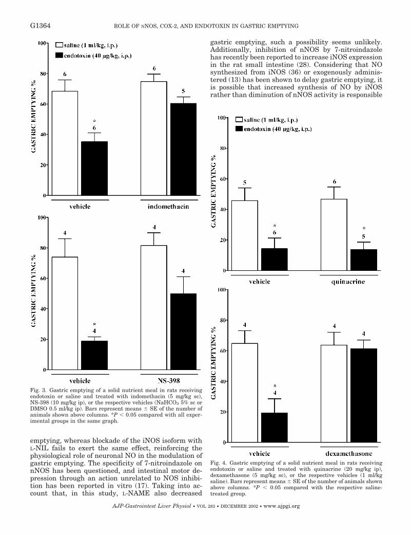
emptying, whereas blockade of the iNOS isoform withL-NIL fails to exert the same effect, reinforcing thephysiological role of neuronal NO in the modulation ofgastric emptying. The specificity of 7-nitroindazole onnNOS has been questioned, and intestinal motor de-pression through an action unrelated to NOS inhibi-tion has been reported in vitro (17). Taking into ac-count that, in this study, L-NAME also decreased
gastric emptying, such a possibility seems unlikely.Additionally, inhibition of nNOS by 7-nitroindazolehas recently been reported to increase iNOS expressionin the rat small intestine (28). Considering that NOsynthesized from iNOS (36) or exogenously adminis-tered (13) has been shown to delay gastric emptying, itis possible that increased synthesis of NO by iNOSrather than diminution of nNOS activity is responsible
Fig. 4. Gastric emptying of a solid nutrient meal in rats receivingendotoxin or saline and treated with quinacrine (20 mg/kg ip),dexamethasone (5 mg/kg sc), or the respective vehicles (1 ml/kgsaline). Bars represent means � SE of the number of animals shownabove columns. *P 0.05 compared with the respective saline-treated group.
Fig. 3. Gastric emptying of a solid nutrient meal in rats receivingendotoxin or saline and treated with indomethacin (5 mg/kg sc),NS-398 (10 mg/kg ip), or the respective vehicles (NaHCO3 5% sc orDMSO 0.5 ml/kg ip). Bars represent means � SE of the number ofanimals shown above columns. *P 0.05 compared with all exper-imental groups in the same graph.
G1364 ROLE OF NNOS, COX-2, AND ENDOTOXIN IN GASTRIC EMPTYING
AJP-Gastrointest Liver Physiol • VOL 283 • DECEMBER 2002 • www.ajpgi.org
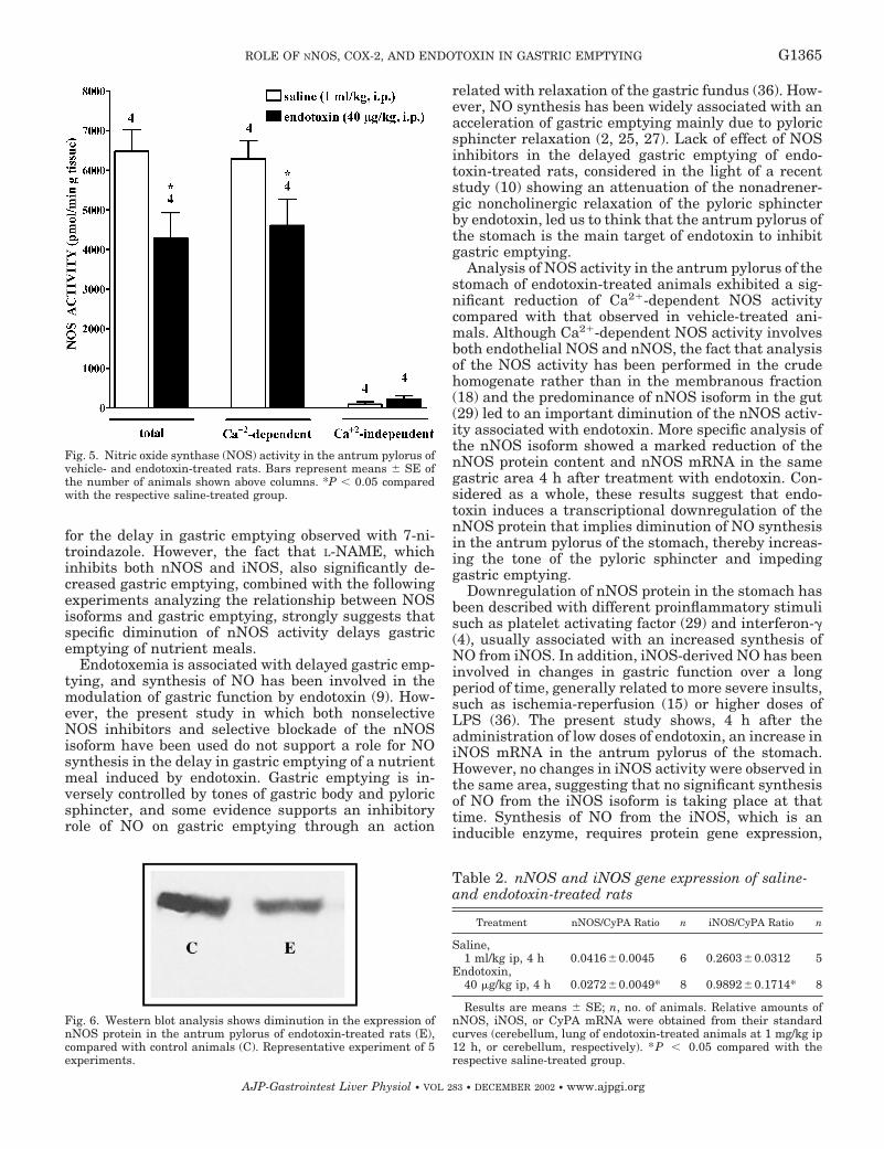
for the delay in gastric emptying observed with 7-ni-troindazole. However, the fact that L-NAME, whichinhibits both nNOS and iNOS, also significantly de-creased gastric emptying, combined with the followingexperiments analyzing the relationship between NOSisoforms and gastric emptying, strongly suggests thatspecific diminution of nNOS activity delays gastricemptying of nutrient meals.
Endotoxemia is associated with delayed gastric emp-tying, and synthesis of NO has been involved in themodulation of gastric function by endotoxin (9). How-ever, the present study in which both nonselectiveNOS inhibitors and selective blockade of the nNOSisoform have been used do not support a role for NOsynthesis in the delay in gastric emptying of a nutrientmeal induced by endotoxin. Gastric emptying is in-versely controlled by tones of gastric body and pyloricsphincter, and some evidence supports an inhibitoryrole of NO on gastric emptying through an action
related with relaxation of the gastric fundus (36). How-ever, NO synthesis has been widely associated with anacceleration of gastric emptying mainly due to pyloricsphincter relaxation (2, 25, 27). Lack of effect of NOSinhibitors in the delayed gastric emptying of endo-toxin-treated rats, considered in the light of a recentstudy (10) showing an attenuation of the nonadrener-gic noncholinergic relaxation of the pyloric sphincterby endotoxin, led us to think that the antrum pylorus ofthe stomach is the main target of endotoxin to inhibitgastric emptying.
Analysis of NOS activity in the antrum pylorus of thestomach of endotoxin-treated animals exhibited a sig-nificant reduction of Ca2�-dependent NOS activitycompared with that observed in vehicle-treated ani-mals. Although Ca2�-dependent NOS activity involvesboth endothelial NOS and nNOS, the fact that analysisof the NOS activity has been performed in the crudehomogenate rather than in the membranous fraction(18) and the predominance of nNOS isoform in the gut(29) led to an important diminution of the nNOS activ-ity associated with endotoxin. More specific analysis ofthe nNOS isoform showed a marked reduction of thenNOS protein content and nNOS mRNA in the samegastric area 4 h after treatment with endotoxin. Con-sidered as a whole, these results suggest that endo-toxin induces a transcriptional downregulation of thenNOS protein that implies diminution of NO synthesisin the antrum pylorus of the stomach, thereby increas-ing the tone of the pyloric sphincter and impedinggastric emptying.
Downregulation of nNOS protein in the stomach hasbeen described with different proinflammatory stimulisuch as platelet activating factor (29) and interferon-�(4), usually associated with an increased synthesis ofNO from iNOS. In addition, iNOS-derived NO has beeninvolved in changes in gastric function over a longperiod of time, generally related to more severe insults,such as ischemia-reperfusion (15) or higher doses ofLPS (36). The present study shows, 4 h after theadministration of low doses of endotoxin, an increase iniNOS mRNA in the antrum pylorus of the stomach.However, no changes in iNOS activity were observed inthe same area, suggesting that no significant synthesisof NO from the iNOS isoform is taking place at thattime. Synthesis of NO from the iNOS, which is aninducible enzyme, requires protein gene expression,
Fig. 6. Western blot analysis shows diminution in the expression ofnNOS protein in the antrum pylorus of endotoxin-treated rats (E),compared with control animals (C). Representative experiment of 5experiments.
Table 2. nNOS and iNOS gene expression of saline-and endotoxin-treated rats
Treatment nNOS/CyPA Ratio n iNOS/CyPA Ratio n
Saline,1 ml/kg ip, 4 h 0.0416�0.0045 6 0.2603�0.0312 5
Endotoxin,40 �g/kg ip, 4 h 0.0272�0.0049* 8 0.9892�0.1714* 8
Results are means � SE; n, no. of animals. Relative amounts ofnNOS, iNOS, or CyPA mRNA were obtained from their standardcurves (cerebellum, lung of endotoxin-treated animals at 1 mg/kg ip12 h, or cerebellum, respectively). *P � 0.05 compared with therespective saline-treated group.
Fig. 5. Nitric oxide synthase (NOS) activity in the antrum pylorus ofvehicle- and endotoxin-treated rats. Bars represent means � SE ofthe number of animals shown above columns. *P 0.05 comparedwith the respective saline-treated group.
G1365ROLE OF NNOS, COX-2, AND ENDOTOXIN IN GASTRIC EMPTYING
AJP-Gastrointest Liver Physiol • VOL 283 • DECEMBER 2002 • www.ajpgi.org
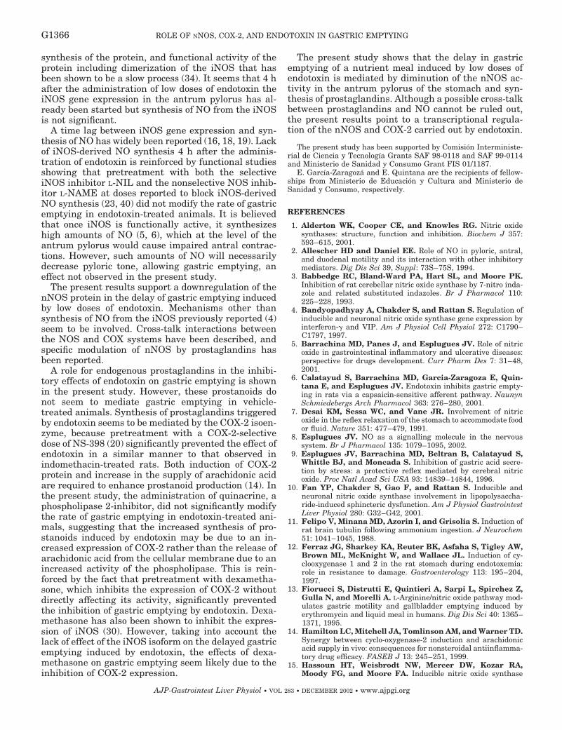
synthesis of the protein, and functional activity of theprotein including dimerization of the iNOS that hasbeen shown to be a slow process (34). It seems that 4 hafter the administration of low doses of endotoxin theiNOS gene expression in the antrum pylorus has al-ready been started but synthesis of NO from the iNOSis not significant.
A time lag between iNOS gene expression and syn-thesis of NO has widely been reported (16, 18, 19). Lackof iNOS-derived NO synthesis 4 h after the adminis-tration of endotoxin is reinforced by functional studiesshowing that pretreatment with both the selectiveiNOS inhibitor L-NIL and the nonselective NOS inhib-itor L-NAME at doses reported to block iNOS-derivedNO synthesis (23, 40) did not modify the rate of gastricemptying in endotoxin-treated animals. It is believedthat once iNOS is functionally active, it synthesizeshigh amounts of NO (5, 6), which at the level of theantrum pylorus would cause impaired antral contrac-tions. However, such amounts of NO will necessarilydecrease pyloric tone, allowing gastric emptying, aneffect not observed in the present study.
The present results support a downregulation of thenNOS protein in the delay of gastric emptying inducedby low doses of endotoxin. Mechanisms other thansynthesis of NO from the iNOS previously reported (4)seem to be involved. Cross-talk interactions betweenthe NOS and COX systems have been described, andspecific modulation of nNOS by prostaglandins hasbeen reported.
A role for endogenous prostaglandins in the inhibi-tory effects of endotoxin on gastric emptying is shownin the present study. However, these prostanoids donot seem to mediate gastric emptying in vehicle-treated animals. Synthesis of prostaglandins triggeredby endotoxin seems to be mediated by the COX-2 isoen-zyme, because pretreatment with a COX-2-selectivedose of NS-398 (20) significantly prevented the effect ofendotoxin in a similar manner to that observed inindomethacin-treated rats. Both induction of COX-2protein and increase in the supply of arachidonic acidare required to enhance prostanoid production (14). Inthe present study, the administration of quinacrine, aphospholipase 2-inhibitor, did not significantly modifythe rate of gastric emptying in endotoxin-treated ani-mals, suggesting that the increased synthesis of pro-stanoids induced by endotoxin may be due to an in-creased expression of COX-2 rather than the release ofarachidonic acid from the cellular membrane due to anincreased activity of the phospholipase. This is rein-forced by the fact that pretreatment with dexametha-sone, which inhibits the expression of COX-2 withoutdirectly affecting its activity, significantly preventedthe inhibition of gastric emptying by endotoxin. Dexa-methasone has also been shown to inhibit the expres-sion of iNOS (30). However, taking into account thelack of effect of the iNOS isoform on the delayed gastricemptying induced by endotoxin, the effects of dexa-methasone on gastric emptying seem likely due to theinhibition of COX-2 expression.
The present study shows that the delay in gastricemptying of a nutrient meal induced by low doses ofendotoxin is mediated by diminution of the nNOS ac-tivity in the antrum pylorus of the stomach and syn-thesis of prostaglandins. Although a possible cross-talkbetween prostaglandins and NO cannot be ruled out,the present results point to a transcriptional regula-tion of the nNOS and COX-2 carried out by endotoxin.
The present study has been supported by Comision Interministe-rial de Ciencia y Tecnologıa Grants SAF 98-0118 and SAF 99-0114and Ministerio de Sanidad y Consumo Grant FIS 01/1187.
E. Garcıa-Zaragoza and E. Quintana are the recipients of fellow-ships from Ministerio de Educacion y Cultura and Ministerio deSanidad y Consumo, respectively.
REFERENCES
1. Alderton WK, Cooper CE, and Knowles RG. Nitric oxidesynthases: structure, function and inhibition. Biochem J 357:593–615, 2001.
2. Allescher HD and Daniel EE. Role of NO in pyloric, antral,and duodenal motility and its interaction with other inhibitorymediators. Dig Dis Sci 39, Suppl: 73S–75S, 1994.
3. Babbedge RC, Bland-Ward PA, Hart SL, and Moore PK.Inhibition of rat cerebellar nitric oxide synthase by 7-nitro inda-zole and related substituted indazoles. Br J Pharmacol 110:225–228, 1993.
4. Bandyopadhyay A, Chakder S, and Rattan S. Regulation ofinducible and neuronal nitric oxide synthase gene expression byinterferon-� and VIP. Am J Physiol Cell Physiol 272: C1790–C1797, 1997.
5. Barrachina MD, Panes J, and Esplugues JV. Role of nitricoxide in gastrointestinal inflammatory and ulcerative diseases:perspective for drugs development. Curr Pharm Des 7: 31–48,2001.
6. Calatayud S, Barrachina MD, Garcia-Zaragoza E, Quin-tana E, and Esplugues JV. Endotoxin inhibits gastric empty-ing in rats via a capsaicin-sensitive afferent pathway. NaunynSchmiedebergs Arch Pharmacol 363: 276–280, 2001.
7. Desai KM, Sessa WC, and Vane JR. Involvement of nitricoxide in the reflex relaxation of the stomach to accommodate foodor fluid. Nature 351: 477–479, 1991.
8. Esplugues JV. NO as a signalling molecule in the nervoussystem. Br J Pharmacol 135: 1079–1095, 2002.
9. Esplugues JV, Barrachina MD, Beltran B, Calatayud S,Whittle BJ, and Moncada S. Inhibition of gastric acid secre-tion by stress: a protective reflex mediated by cerebral nitricoxide. Proc Natl Acad Sci USA 93: 14839–14844, 1996.
10. Fan YP, Chakder S, Gao F, and Rattan S. Inducible andneuronal nitric oxide synthase involvement in lipopolysaccha-ride-induced sphincteric dysfunction. Am J Physiol GastrointestLiver Physiol 280: G32–G42, 2001.
11. Felipo V, Minana MD, Azorin I, and Grisolia S. Induction ofrat brain tubulin following ammonium ingestion. J Neurochem51: 1041–1045, 1988.
12. Ferraz JG, Sharkey KA, Reuter BK, Asfaha S, Tigley AW,Brown ML, McKnight W, and Wallace JL. Induction of cy-clooxygenase 1 and 2 in the rat stomach during endotoxemia:role in resistance to damage. Gastroenterology 113: 195–204,1997.
13. Fiorucci S, Distrutti E, Quintieri A, Sarpi L, Spirchez Z,Gulla N, and Morelli A. L-Arginine/nitric oxide pathway mod-ulates gastric motility and gallbladder emptying induced byerythromycin and liquid meal in humans. Dig Dis Sci 40: 1365–1371, 1995.
14. Hamilton LC, Mitchell JA, Tomlinson AM, and Warner TD.Synergy between cyclo-oxygenase-2 induction and arachidonicacid supply in vivo: consequences for nonsteroidal antiinflamma-tory drug efficacy. FASEB J 13: 245–251, 1999.
15. Hassoun HT, Weisbrodt NW, Mercer DW, Kozar RA,Moody FG, and Moore FA. Inducible nitric oxide synthase
G1366 ROLE OF NNOS, COX-2, AND ENDOTOXIN IN GASTRIC EMPTYING
AJP-Gastrointest Liver Physiol • VOL 283 • DECEMBER 2002 • www.ajpgi.org
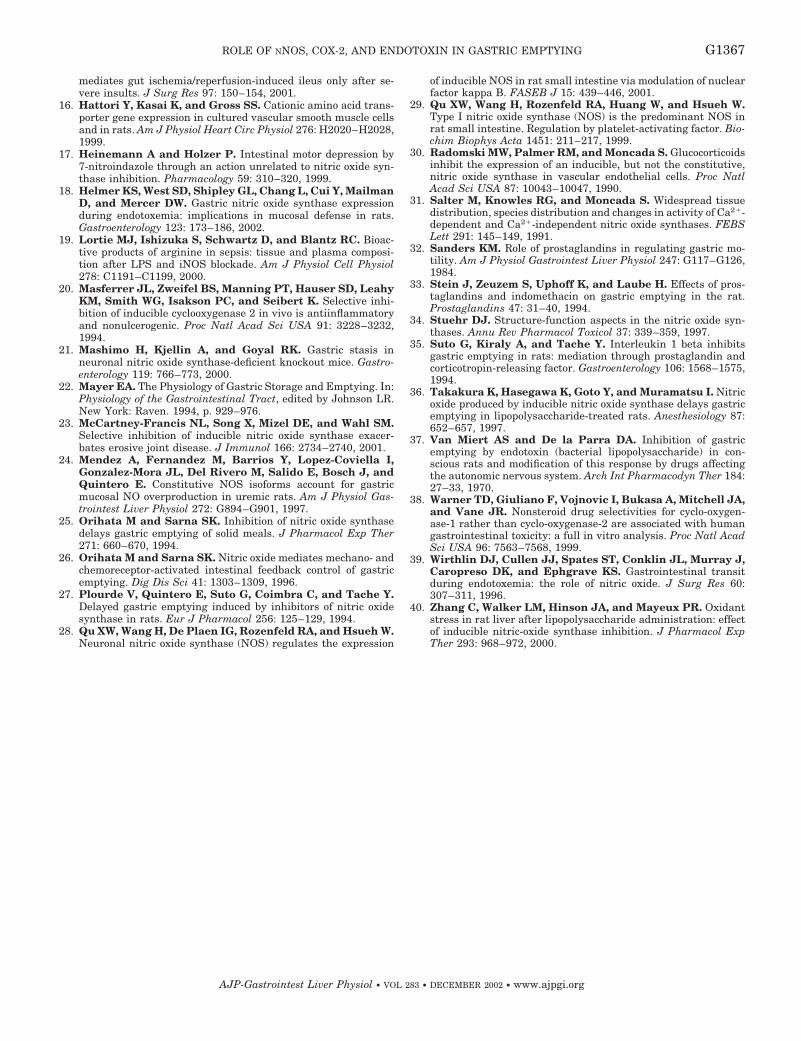
mediates gut ischemia/reperfusion-induced ileus only after se-vere insults. J Surg Res 97: 150–154, 2001.
16. Hattori Y, Kasai K, and Gross SS. Cationic amino acid trans-porter gene expression in cultured vascular smooth muscle cellsand in rats. Am J Physiol Heart Circ Physiol 276: H2020–H2028,1999.
17. Heinemann A and Holzer P. Intestinal motor depression by7-nitroindazole through an action unrelated to nitric oxide syn-thase inhibition. Pharmacology 59: 310–320, 1999.
18. Helmer KS, West SD, Shipley GL, Chang L, Cui Y, MailmanD, and Mercer DW. Gastric nitric oxide synthase expressionduring endotoxemia: implications in mucosal defense in rats.Gastroenterology 123: 173–186, 2002.
19. Lortie MJ, Ishizuka S, Schwartz D, and Blantz RC. Bioac-tive products of arginine in sepsis: tissue and plasma composi-tion after LPS and iNOS blockade. Am J Physiol Cell Physiol278: C1191–C1199, 2000.
20. Masferrer JL, Zweifel BS, Manning PT, Hauser SD, LeahyKM, Smith WG, Isakson PC, and Seibert K. Selective inhi-bition of inducible cyclooxygenase 2 in vivo is antiinflammatoryand nonulcerogenic. Proc Natl Acad Sci USA 91: 3228–3232,1994.
21. Mashimo H, Kjellin A, and Goyal RK. Gastric stasis inneuronal nitric oxide synthase-deficient knockout mice. Gastro-enterology 119: 766–773, 2000.
22. Mayer EA. The Physiology of Gastric Storage and Emptying. In:Physiology of the Gastrointestinal Tract, edited by Johnson LR.New York: Raven. 1994, p. 929–976.
23. McCartney-Francis NL, Song X, Mizel DE, and Wahl SM.Selective inhibition of inducible nitric oxide synthase exacer-bates erosive joint disease. J Immunol 166: 2734–2740, 2001.
24. Mendez A, Fernandez M, Barrios Y, Lopez-Coviella I,Gonzalez-Mora JL, Del Rivero M, Salido E, Bosch J, andQuintero E. Constitutive NOS isoforms account for gastricmucosal NO overproduction in uremic rats. Am J Physiol Gas-trointest Liver Physiol 272: G894–G901, 1997.
25. Orihata M and Sarna SK. Inhibition of nitric oxide synthasedelays gastric emptying of solid meals. J Pharmacol Exp Ther271: 660–670, 1994.
26. Orihata M and Sarna SK. Nitric oxide mediates mechano- andchemoreceptor-activated intestinal feedback control of gastricemptying. Dig Dis Sci 41: 1303–1309, 1996.
27. Plourde V, Quintero E, Suto G, Coimbra C, and Tache Y.Delayed gastric emptying induced by inhibitors of nitric oxidesynthase in rats. Eur J Pharmacol 256: 125–129, 1994.
28. Qu XW, Wang H, De Plaen IG, Rozenfeld RA, and Hsueh W.Neuronal nitric oxide synthase (NOS) regulates the expression
of inducible NOS in rat small intestine via modulation of nuclearfactor kappa B. FASEB J 15: 439–446, 2001.
29. Qu XW, Wang H, Rozenfeld RA, Huang W, and Hsueh W.Type I nitric oxide synthase (NOS) is the predominant NOS inrat small intestine. Regulation by platelet-activating factor. Bio-chim Biophys Acta 1451: 211–217, 1999.
30. Radomski MW, Palmer RM, and Moncada S. Glucocorticoidsinhibit the expression of an inducible, but not the constitutive,nitric oxide synthase in vascular endothelial cells. Proc NatlAcad Sci USA 87: 10043–10047, 1990.
31. Salter M, Knowles RG, and Moncada S. Widespread tissuedistribution, species distribution and changes in activity of Ca2�-dependent and Ca2�-independent nitric oxide synthases. FEBSLett 291: 145–149, 1991.
32. Sanders KM. Role of prostaglandins in regulating gastric mo-tility. Am J Physiol Gastrointest Liver Physiol 247: G117–G126,1984.
33. Stein J, Zeuzem S, Uphoff K, and Laube H. Effects of pros-taglandins and indomethacin on gastric emptying in the rat.Prostaglandins 47: 31–40, 1994.
34. Stuehr DJ. Structure-function aspects in the nitric oxide syn-thases. Annu Rev Pharmacol Toxicol 37: 339–359, 1997.
35. Suto G, Kiraly A, and Tache Y. Interleukin 1 beta inhibitsgastric emptying in rats: mediation through prostaglandin andcorticotropin-releasing factor. Gastroenterology 106: 1568–1575,1994.
36. Takakura K, Hasegawa K, Goto Y, and Muramatsu I. Nitricoxide produced by inducible nitric oxide synthase delays gastricemptying in lipopolysaccharide-treated rats. Anesthesiology 87:652–657, 1997.
37. Van Miert AS and De la Parra DA. Inhibition of gastricemptying by endotoxin (bacterial lipopolysaccharide) in con-scious rats and modification of this response by drugs affectingthe autonomic nervous system. Arch Int Pharmacodyn Ther 184:27–33, 1970.
38. Warner TD, Giuliano F, Vojnovic I, Bukasa A, Mitchell JA,and Vane JR. Nonsteroid drug selectivities for cyclo-oxygen-ase-1 rather than cyclo-oxygenase-2 are associated with humangastrointestinal toxicity: a full in vitro analysis. Proc Natl AcadSci USA 96: 7563–7568, 1999.
39. Wirthlin DJ, Cullen JJ, Spates ST, Conklin JL, Murray J,Caropreso DK, and Ephgrave KS. Gastrointestinal transitduring endotoxemia: the role of nitric oxide. J Surg Res 60:307–311, 1996.
40. Zhang C, Walker LM, Hinson JA, and Mayeux PR. Oxidantstress in rat liver after lipopolysaccharide administration: effectof inducible nitric-oxide synthase inhibition. J Pharmacol ExpTher 293: 968–972, 2000.
G1367ROLE OF NNOS, COX-2, AND ENDOTOXIN IN GASTRIC EMPTYING
AJP-Gastrointest Liver Physiol • VOL 283 • DECEMBER 2002 • www.ajpgi.org
Artículos de investigación
115
II. EFECTOS DE LA ENDOTOXINA SOBRE LA MOTILIDAD COLÓNICA
Por otra parte quisimos estudiar si en estas circunstancias también se altera
otro proceso fisiológico como es la excreción fecal. Para ello analizamos esta
función a lo largo del tiempo en ratas conscientes pretratadas con la endotoxina.
Estos experimentos han dado lugar a la publicación:
“Endotoxin stimulates fecal pellet output in rats through a neural
mechanism”. Naunyn-Schmiedeberg’s Archives of Pharmacology (2003)
367(1):51-5.
En ella demostramos que la endotoxina incrementa la excreción fecal de
forma aguda. Este efecto está mediado por la activación de fibras sensoriales
sensibles a la capsaicina y por la liberación de neurotransmisores excitadores
(Ach y SP) e inhibidores (NO) en la pared colónica, incrementando así la
velocidad del reflejo peristáltico.
Artículos de investigación
116
Artículo 6
“Endotoxin stimulates fecal pellet output
in rats through a neural mechanism”
Sara Calatayud, María D. Barrachina, Elsa Quintana, Sales Ibiza & Juan V.
Esplugues
Naunyn-Schmiedeberg’s Archives of Pharmacology (2003) 367(1):51-5.
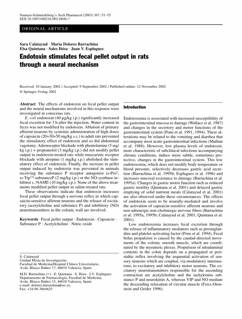
Abstract The effects of endotoxin on fecal pellet outputand the neural mechanisms involved in this response wereinvestigated in conscious rats.
E. coli endotoxin (40 µg/kg i.p.) significantly increasedfecal excretion for 3 h after the injection. Water content infeces was not modified by endotoxin. Ablation of primaryafferent neurons by systemic administration of high dosesof capsaicin (20+30+50 mg/kg s.c.) to adult rats preventedthe stimulatory effect of endotoxin and so did abdominalvagotomy. Adrenoceptor blockade with phentolamine (5 mg/kg i.p.) + propranolol (3 mg/kg i.p.) did not modify pelletoutput in endotoxin-treated rats while muscarinic receptorblockade with atropine (1 mg/kg i.p.) abolished the stim-ulatory effect of endotoxin. Finally, the increase in pelletoutput induced by endotoxin was prevented in animals receiving the substance P receptor antagonist D-Pro2,D-Trp7,9-substanceP (2 mg/kg i.p.) or the NO-synthase in-hibitor L-NAME (10 mg/kg i.p.). None of the above treat-ments modified pellet output in saline-treated rats.
These observations indicate that endotoxin increasesfecal pellet output through a nervous reflex in which cap-saicin-sensitive afferent neurons and the release of excita-tory (acetylcholine and substance P) and inhibitory (NO)neurotransmitters in the colonic wall are involved.
Keywords Fecal pellet output · Endotoxin · Capsaicin ·Substance P · Acetylcholine · Nitric oxide
Introduction
Endotoxemia is associated with increased susceptibility ofthe gastrointestinal mucosa to damage (Wallace et al. 1987)and changes in the secretory and motor functions of thegastrointestinal system (Pons et al. 1991, 1994). These al-terations may be related to the vomiting and diarrhea thataccompany most acute gastrointestinal infections (Mathanet al. 1988). However, low plasma levels of endotoxin,more characteristic of subclinical infections accompanyingchronic conditions, induce more subtle, sometimes pro-tective, changes in the gastrointestinal system. This lowendotoxemia, which does not modify body temperature orblood pressure, selectively decreases gastric acid secre-tion (Barrachina et al. 1995b; Esplugues et al. 1996) andincreases mucosal resistance to damage (Barrachina et al.1995a). Changes in gastric motor function such as reducedgastric motility (Quintana et al. 2001) and delayed gastricemptying of solid nutrient meals (Calatayud et al. 2001)are also observed under these circumstances. The effectsof endotoxin seem to be neurally-mediated and involvethe activation of capsaicin-sensitive afferent neurons andnon-adrenergic non-cholinergic nervous fibers (Barrachinaet al. 1995a, 1995b; Calatayud et al. 2001; Quintana et al.2001).
Low endotoxemia increases fecal excretion throughthe release of inflammatory mediators such as prostaglan-dins and platelet-activating factor (Pons et al. 1994). Fecalbolus propulsion is caused by the caudad-directed move-ments of the colonic smooth muscle, which are coordi-nated by the myenteric plexus. Propulsion of intraluminalcontents in the colon depends on a propagated or peri-staltic reflex involving the sequential activation of sen-sory neurons which are coupled, via modulatory interneu-rons, to excitatory and inhibitory motor neurons. The ex-citatory neurotransmitters responsible for the ascendingcontraction are acetylcholine and the tachykinins sub-stance P and neurokinin A, whereas VIP and NO mediatethe descending relaxation of circular muscle (Foxx-Oren-stein and Grider 1996).
Sara Calatayud · Maria Dolores Barrachina ·Elsa Quintana · Sales Ibiza · Juan V. Esplugues
Endotoxin stimulates fecal pellet output in rats through a neural mechanism
Naunyn-Schmiedeberg’s Arch Pharmacol (2003) 367 :51–55DOI 10.1007/s00210-002-0646-7
Received: 10 January 2002 / Accepted: 9 September 2002 / Published online: 12 November 2002
ORIGINAL ARTICLE
S. CalatayudUnidad Mixta de Investigación, Facultad de Medicina/Hospital Clínico Universitario, Avda. Blasco Ibáñez 17, 46010 Valencia, Spain
M.D. Barrachina (✉) · E. Quintana · S. Ibiza · J.V. EspluguesDepartamento de Farmacología, Facultad de Medicina, Avda. Blasco Ibáñez 15, 46010 Valencia, Spaine-mail: [email protected], Fax: +34-96-3864625
© Springer-Verlag 2002
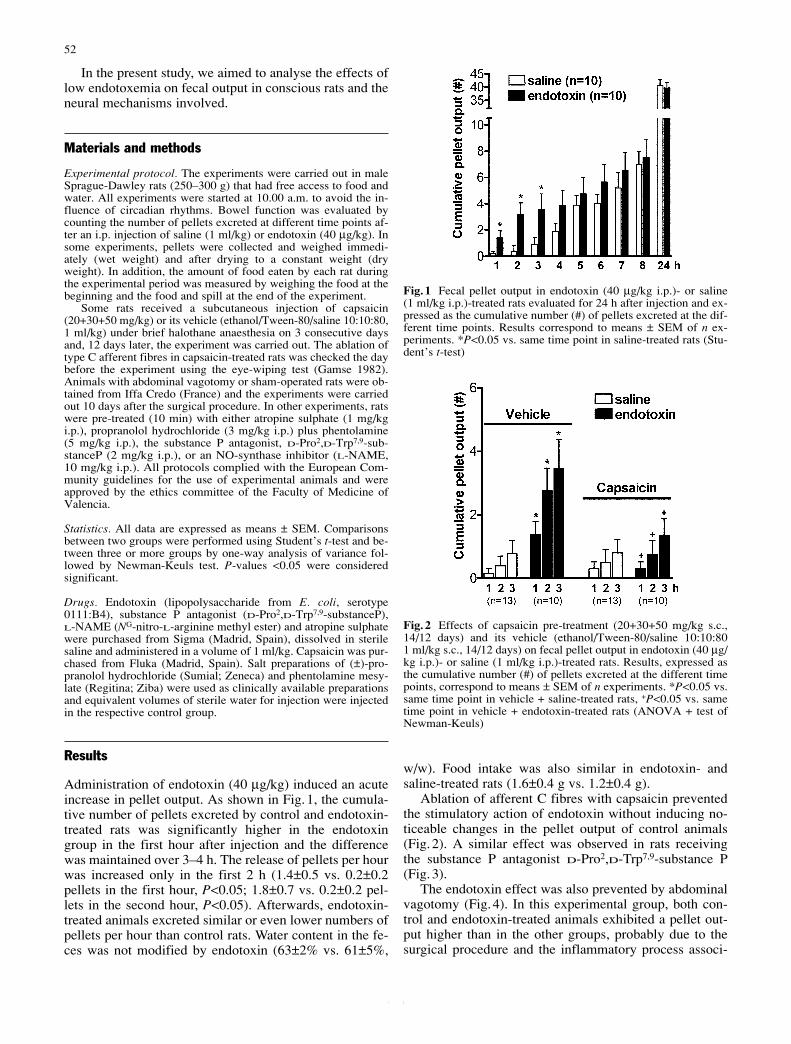
In the present study, we aimed to analyse the effects oflow endotoxemia on fecal output in conscious rats and theneural mechanisms involved.
Materials and methods
Experimental protocol. The experiments were carried out in maleSprague-Dawley rats (250–300 g) that had free access to food andwater. All experiments were started at 10.00 a.m. to avoid the in-fluence of circadian rhythms. Bowel function was evaluated bycounting the number of pellets excreted at different time points af-ter an i.p. injection of saline (1 ml/kg) or endotoxin (40 µg/kg). Insome experiments, pellets were collected and weighed immedi-ately (wet weight) and after drying to a constant weight (dryweight). In addition, the amount of food eaten by each rat duringthe experimental period was measured by weighing the food at thebeginning and the food and spill at the end of the experiment.
Some rats received a subcutaneous injection of capsaicin(20+30+50 mg/kg) or its vehicle (ethanol/Tween-80/saline 10:10:80,1 ml/kg) under brief halothane anaesthesia on 3 consecutive daysand, 12 days later, the experiment was carried out. The ablation oftype C afferent fibres in capsaicin-treated rats was checked the daybefore the experiment using the eye-wiping test (Gamse 1982).Animals with abdominal vagotomy or sham-operated rats were ob-tained from Iffa Credo (France) and the experiments were carriedout 10 days after the surgical procedure. In other experiments, ratswere pre-treated (10 min) with either atropine sulphate (1 mg/kgi.p.), propranolol hydrochloride (3 mg/kg i.p.) plus phentolamine(5 mg/kg i.p.), the substance P antagonist, D-Pro2,D-Trp7,9-sub-stanceP (2 mg/kg i.p.), or an NO-synthase inhibitor (L-NAME,10 mg/kg i.p.). All protocols complied with the European Com-munity guidelines for the use of experimental animals and wereapproved by the ethics committee of the Faculty of Medicine ofValencia.
Statistics. All data are expressed as means ± SEM. Comparisonsbetween two groups were performed using Student’s t-test and be-tween three or more groups by one-way analysis of variance fol-lowed by Newman-Keuls test. P-values <0.05 were consideredsignificant.
Drugs. Endotoxin (lipopolysaccharide from E. coli, serotype0111:B4), substance P antagonist (D-Pro2,D-Trp7,9-substanceP), L-NAME (NG-nitro-L-arginine methyl ester) and atropine sulphatewere purchased from Sigma (Madrid, Spain), dissolved in sterilesaline and administered in a volume of 1 ml/kg. Capsaicin was pur-chased from Fluka (Madrid, Spain). Salt preparations of (±)-pro-pranolol hydrochloride (Sumial; Zeneca) and phentolamine mesy-late (Regitina; Ziba) were used as clinically available preparationsand equivalent volumes of sterile water for injection were injectedin the respective control group.
Results
Administration of endotoxin (40 µg/kg) induced an acuteincrease in pellet output. As shown in Fig.1, the cumula-tive number of pellets excreted by control and endotoxin-treated rats was significantly higher in the endotoxingroup in the first hour after injection and the differencewas maintained over 3–4 h. The release of pellets per hourwas increased only in the first 2 h (1.4±0.5 vs. 0.2±0.2pellets in the first hour, P<0.05; 1.8±0.7 vs. 0.2±0.2 pel-lets in the second hour, P<0.05). Afterwards, endotoxin-treated animals excreted similar or even lower numbers ofpellets per hour than control rats. Water content in the fe-ces was not modified by endotoxin (63±2% vs. 61±5%,
w/w). Food intake was also similar in endotoxin- andsaline-treated rats (1.6±0.4 g vs. 1.2±0.4 g).
Ablation of afferent C fibres with capsaicin preventedthe stimulatory action of endotoxin without inducing no-ticeable changes in the pellet output of control animals(Fig.2). A similar effect was observed in rats receivingthe substance P antagonist D-Pro2,D-Trp7,9-substance P(Fig.3).
The endotoxin effect was also prevented by abdominalvagotomy (Fig.4). In this experimental group, both con-trol and endotoxin-treated animals exhibited a pellet out-put higher than in the other groups, probably due to thesurgical procedure and the inflammatory process associ-
52
Fig.1 Fecal pellet output in endotoxin (40 µg/kg i.p.)- or saline (1 ml/kg i.p.)-treated rats evaluated for 24 h after injection and ex-pressed as the cumulative number (#) of pellets excreted at the dif-ferent time points. Results correspond to means ± SEM of n ex-periments. *P<0.05 vs. same time point in saline-treated rats (Stu-dent’s t-test)
Fig.2 Effects of capsaicin pre-treatment (20+30+50 mg/kg s.c.,14/12 days) and its vehicle (ethanol/Tween-80/saline 10:10:80 1 ml/kg s.c., 14/12 days) on fecal pellet output in endotoxin (40 µg/kg i.p.)- or saline (1 ml/kg i.p.)-treated rats. Results, expressed asthe cumulative number (#) of pellets excreted at the different timepoints, correspond to means ± SEM of n experiments. *P<0.05 vs.same time point in vehicle + saline-treated rats, +P<0.05 vs. sametime point in vehicle + endotoxin-treated rats (ANOVA + test ofNewman-Keuls)
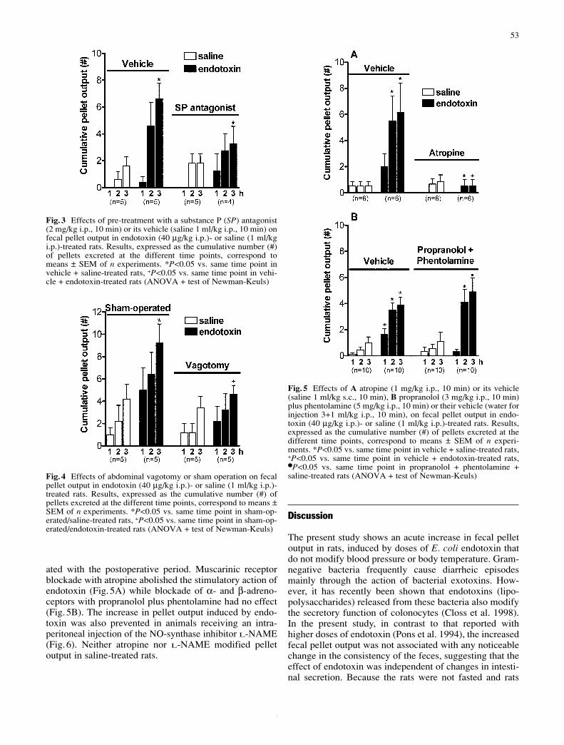
ated with the postoperative period. Muscarinic receptorblockade with atropine abolished the stimulatory action ofendotoxin (Fig.5A) while blockade of α- and β-adreno-ceptors with propranolol plus phentolamine had no effect(Fig.5B). The increase in pellet output induced by endo-toxin was also prevented in animals receiving an intra-peritoneal injection of the NO-synthase inhibitor L-NAME(Fig.6). Neither atropine nor L-NAME modified pelletoutput in saline-treated rats.
Discussion
The present study shows an acute increase in fecal pelletoutput in rats, induced by doses of E. coli endotoxin thatdo not modify blood pressure or body temperature. Gram-negative bacteria frequently cause diarrheic episodesmainly through the action of bacterial exotoxins. How-ever, it has recently been shown that endotoxins (lipo-polysaccharides) released from these bacteria also modifythe secretory function of colonocytes (Closs et al. 1998).In the present study, in contrast to that reported withhigher doses of endotoxin (Pons et al. 1994), the increasedfecal pellet output was not associated with any noticeablechange in the consistency of the feces, suggesting that theeffect of endotoxin was independent of changes in intesti-nal secretion. Because the rats were not fasted and rats
53
Fig.3 Effects of pre-treatment with a substance P (SP) antagonist(2 mg/kg i.p., 10 min) or its vehicle (saline 1 ml/kg i.p., 10 min) onfecal pellet output in endotoxin (40 µg/kg i.p.)- or saline (1 ml/kgi.p.)-treated rats. Results, expressed as the cumulative number (#)of pellets excreted at the different time points, correspond tomeans ± SEM of n experiments. *P<0.05 vs. same time point invehicle + saline-treated rats, +P<0.05 vs. same time point in vehi-cle + endotoxin-treated rats (ANOVA + test of Newman-Keuls)
Fig.4 Effects of abdominal vagotomy or sham operation on fecalpellet output in endotoxin (40 µg/kg i.p.)- or saline (1 ml/kg i.p.)-treated rats. Results, expressed as the cumulative number (#) ofpellets excreted at the different time points, correspond to means ±SEM of n experiments. *P<0.05 vs. same time point in sham-op-erated/saline-treated rats, +P<0.05 vs. same time point in sham-op-erated/endotoxin-treated rats (ANOVA + test of Newman-Keuls)
Fig.5 Effects of A atropine (1 mg/kg i.p., 10 min) or its vehicle(saline 1 ml/kg s.c., 10 min), B propranolol (3 mg/kg i.p., 10 min)plus phentolamine (5 mg/kg i.p., 10 min) or their vehicle (water forinjection 3+1 ml/kg i.p., 10 min), on fecal pellet output in endo-toxin (40 µg/kg i.p.)- or saline (1 ml/kg i.p.)-treated rats. Results,expressed as the cumulative number (#) of pellets excreted at thedifferent time points, correspond to means ± SEM of n experi-ments. *P<0.05 vs. same time point in vehicle + saline-treated rats,+P<0.05 vs. same time point in vehicle + endotoxin-treated rats,●P<0.05 vs. same time point in propranolol + phentolamine +saline-treated rats (ANOVA + test of Newman-Keuls)
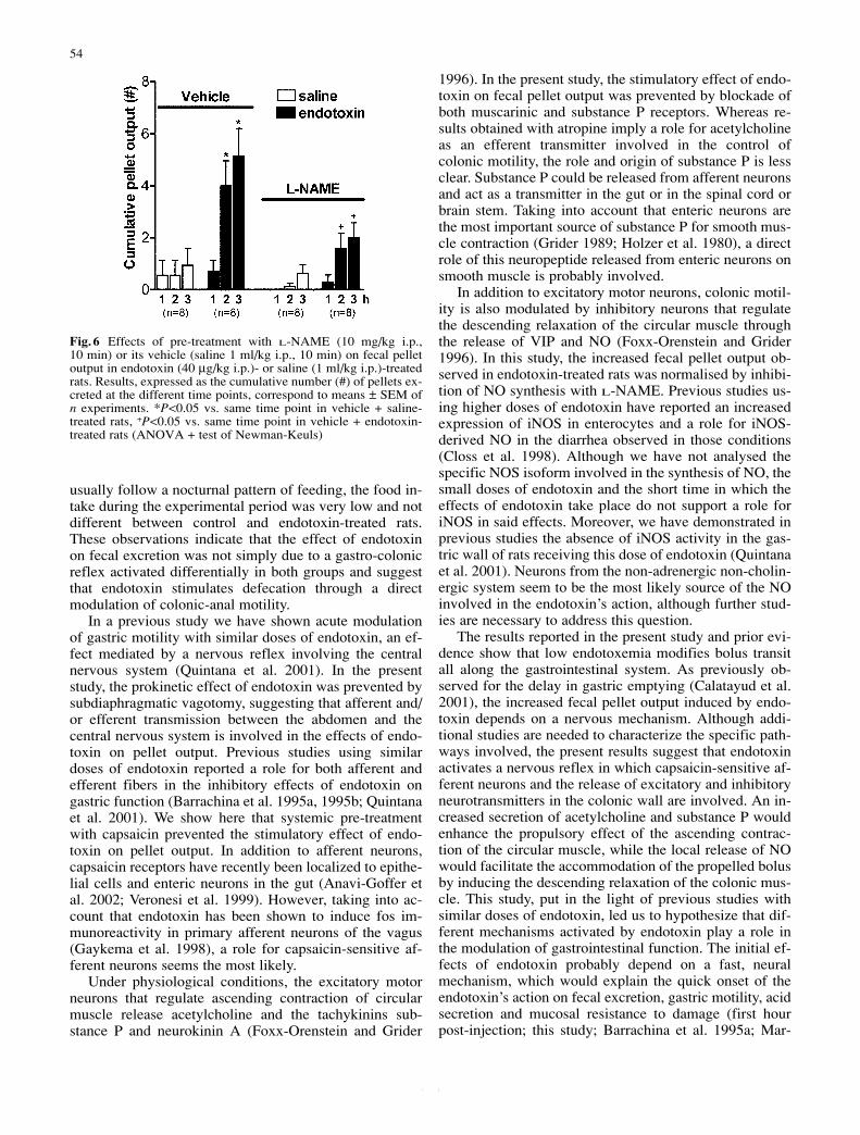
usually follow a nocturnal pattern of feeding, the food in-take during the experimental period was very low and notdifferent between control and endotoxin-treated rats.These observations indicate that the effect of endotoxinon fecal excretion was not simply due to a gastro-colonicreflex activated differentially in both groups and suggestthat endotoxin stimulates defecation through a directmodulation of colonic-anal motility.
In a previous study we have shown acute modulationof gastric motility with similar doses of endotoxin, an ef-fect mediated by a nervous reflex involving the centralnervous system (Quintana et al. 2001). In the presentstudy, the prokinetic effect of endotoxin was prevented bysubdiaphragmatic vagotomy, suggesting that afferent and/or efferent transmission between the abdomen and thecentral nervous system is involved in the effects of endo-toxin on pellet output. Previous studies using similardoses of endotoxin reported a role for both afferent andefferent fibers in the inhibitory effects of endotoxin ongastric function (Barrachina et al. 1995a, 1995b; Quintanaet al. 2001). We show here that systemic pre-treatmentwith capsaicin prevented the stimulatory effect of endo-toxin on pellet output. In addition to afferent neurons,capsaicin receptors have recently been localized to epithe-lial cells and enteric neurons in the gut (Anavi-Goffer etal. 2002; Veronesi et al. 1999). However, taking into ac-count that endotoxin has been shown to induce fos im-munoreactivity in primary afferent neurons of the vagus(Gaykema et al. 1998), a role for capsaicin-sensitive af-ferent neurons seems the most likely.
Under physiological conditions, the excitatory motorneurons that regulate ascending contraction of circularmuscle release acetylcholine and the tachykinins sub-stance P and neurokinin A (Foxx-Orenstein and Grider
1996). In the present study, the stimulatory effect of endo-toxin on fecal pellet output was prevented by blockade ofboth muscarinic and substance P receptors. Whereas re-sults obtained with atropine imply a role for acetylcholineas an efferent transmitter involved in the control ofcolonic motility, the role and origin of substance P is lessclear. Substance P could be released from afferent neuronsand act as a transmitter in the gut or in the spinal cord orbrain stem. Taking into account that enteric neurons arethe most important source of substance P for smooth mus-cle contraction (Grider 1989; Holzer et al. 1980), a directrole of this neuropeptide released from enteric neurons onsmooth muscle is probably involved.
In addition to excitatory motor neurons, colonic motil-ity is also modulated by inhibitory neurons that regulatethe descending relaxation of the circular muscle throughthe release of VIP and NO (Foxx-Orenstein and Grider1996). In this study, the increased fecal pellet output ob-served in endotoxin-treated rats was normalised by inhibi-tion of NO synthesis with L-NAME. Previous studies us-ing higher doses of endotoxin have reported an increasedexpression of iNOS in enterocytes and a role for iNOS-derived NO in the diarrhea observed in those conditions(Closs et al. 1998). Although we have not analysed thespecific NOS isoform involved in the synthesis of NO, thesmall doses of endotoxin and the short time in which theeffects of endotoxin take place do not support a role foriNOS in said effects. Moreover, we have demonstrated inprevious studies the absence of iNOS activity in the gas-tric wall of rats receiving this dose of endotoxin (Quintanaet al. 2001). Neurons from the non-adrenergic non-cholin-ergic system seem to be the most likely source of the NOinvolved in the endotoxin’s action, although further stud-ies are necessary to address this question.
The results reported in the present study and prior evi-dence show that low endotoxemia modifies bolus transitall along the gastrointestinal system. As previously ob-served for the delay in gastric emptying (Calatayud et al.2001), the increased fecal pellet output induced by endo-toxin depends on a nervous mechanism. Although addi-tional studies are needed to characterize the specific path-ways involved, the present results suggest that endotoxinactivates a nervous reflex in which capsaicin-sensitive af-ferent neurons and the release of excitatory and inhibitoryneurotransmitters in the colonic wall are involved. An in-creased secretion of acetylcholine and substance P wouldenhance the propulsory effect of the ascending contrac-tion of the circular muscle, while the local release of NOwould facilitate the accommodation of the propelled bolusby inducing the descending relaxation of the colonic mus-cle. This study, put in the light of previous studies withsimilar doses of endotoxin, led us to hypothesize that dif-ferent mechanisms activated by endotoxin play a role inthe modulation of gastrointestinal function. The initial ef-fects of endotoxin probably depend on a fast, neuralmechanism, which would explain the quick onset of theendotoxin’s action on fecal excretion, gastric motility, acidsecretion and mucosal resistance to damage (first hourpost-injection; this study; Barrachina et al. 1995a; Mar-
54
Fig.6 Effects of pre-treatment with L-NAME (10 mg/kg i.p., 10 min) or its vehicle (saline 1 ml/kg i.p., 10 min) on fecal pelletoutput in endotoxin (40 µg/kg i.p.)- or saline (1 ml/kg i.p.)-treatedrats. Results, expressed as the cumulative number (#) of pellets ex-creted at the different time points, correspond to means ± SEM ofn experiments. *P<0.05 vs. same time point in vehicle + saline-treated rats, +P<0.05 vs. same time point in vehicle + endotoxin-treated rats (ANOVA + test of Newman-Keuls)
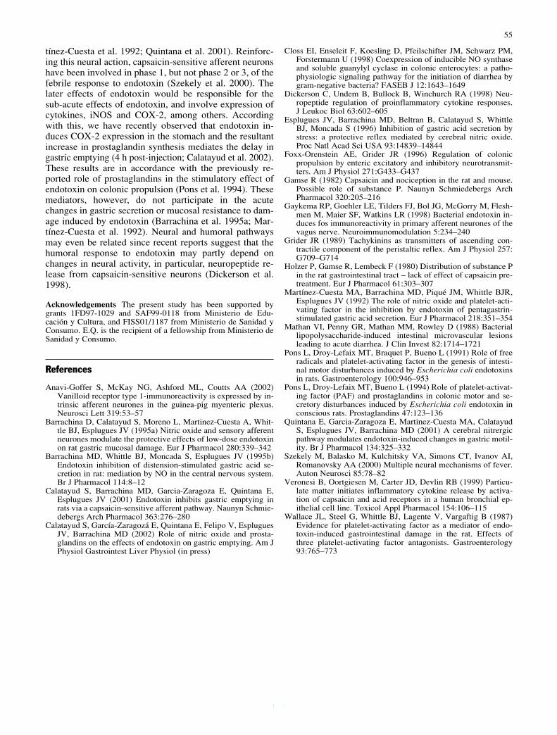
tínez-Cuesta et al. 1992; Quintana et al. 2001). Reinforc-ing this neural action, capsaicin-sensitive afferent neuronshave been involved in phase 1, but not phase 2 or 3, of thefebrile response to endotoxin (Szekely et al. 2000). Thelater effects of endotoxin would be responsible for thesub-acute effects of endotoxin, and involve expression ofcytokines, iNOS and COX-2, among others. Accordingwith this, we have recently observed that endotoxin in-duces COX-2 expression in the stomach and the resultantincrease in prostaglandin synthesis mediates the delay ingastric emptying (4 h post-injection; Calatayud et al. 2002).These results are in accordance with the previously re-ported role of prostaglandins in the stimulatory effect ofendotoxin on colonic propulsion (Pons et al. 1994). Thesemediators, however, do not participate in the acutechanges in gastric secretion or mucosal resistance to dam-age induced by endotoxin (Barrachina et al. 1995a; Mar-tínez-Cuesta et al. 1992). Neural and humoral pathwaysmay even be related since recent reports suggest that thehumoral response to endotoxin may partly depend onchanges in neural activity, in particular, neuropeptide re-lease from capsaicin-sensitive neurons (Dickerson et al.1998).
Acknowledgements The present study has been supported bygrants 1FD97-1029 and SAF99-0118 from Ministerio de Edu-cación y Cultura, and FISS01/1187 from Ministerio de Sanidad yConsumo. E.Q. is the recipient of a fellowship from Ministerio deSanidad y Consumo.
References
Anavi-Goffer S, McKay NG, Ashford ML, Coutts AA (2002)Vanilloid receptor type 1-immunoreactivity is expressed by in-trinsic afferent neurones in the guinea-pig myenteric plexus.Neurosci Lett 319:53–57
Barrachina D, Calatayud S, Moreno L, Martinez-Cuesta A, Whit-tle BJ, Esplugues JV (1995a) Nitric oxide and sensory afferentneurones modulate the protective effects of low-dose endotoxinon rat gastric mucosal damage. Eur J Pharmacol 280:339–342
Barrachina MD, Whittle BJ, Moncada S, Esplugues JV (1995b)Endotoxin inhibition of distension-stimulated gastric acid se-cretion in rat: mediation by NO in the central nervous system.Br J Pharmacol 114:8–12
Calatayud S, Barrachina MD, Garcia-Zaragoza E, Quintana E, Esplugues JV (2001) Endotoxin inhibits gastric emptying inrats via a capsaicin-sensitive afferent pathway. Naunyn Schmie-debergs Arch Pharmacol 363:276–280
Calatayud S, García-Zaragozá E, Quintana E, Felipo V, EspluguesJV, Barrachina MD (2002) Role of nitric oxide and prosta-glandins on the effects of endotoxin on gastric emptying. Am JPhysiol Gastrointest Liver Physiol (in press)
Closs EI, Enseleit F, Koesling D, Pfeilschifter JM, Schwarz PM,Forstermann U (1998) Coexpression of inducible NO synthaseand soluble guanylyl cyclase in colonic enterocytes: a patho-physiologic signaling pathway for the initiation of diarrhea bygram-negative bacteria? FASEB J 12:1643–1649
Dickerson C, Undem B, Bullock B, Winchurch RA (1998) Neu-ropeptide regulation of proinflammatory cytokine responses. J Leukoc Biol 63:602–605
Esplugues JV, Barrachina MD, Beltran B, Calatayud S, WhittleBJ, Moncada S (1996) Inhibition of gastric acid secretion bystress: a protective reflex mediated by cerebral nitric oxide.Proc Natl Acad Sci USA 93:14839–14844
Foxx-Orenstein AE, Grider JR (1996) Regulation of colonicpropulsion by enteric excitatory and inhibitory neurotransmit-ters. Am J Physiol 271:G433–G437
Gamse R (1982) Capsaicin and nociception in the rat and mouse.Possible role of substance P. Naunyn Schmiedebergs ArchPharmacol 320:205–216
Gaykema RP, Goehler LE, Tilders FJ, Bol JG, McGorry M, Flesh-men M, Maier SF, Watkins LR (1998) Bacterial endotoxin in-duces fos immunoreactivity in primary afferent neurones of thevagus nerve. Neuroimmunomodulation 5:234–240
Grider JR (1989) Tachykinins as transmitters of ascending con-tractile component of the peristaltic reflex. Am J Physiol 257:G709–G714
Holzer P, Gamse R, Lembeck F (1980) Distribution of substance Pin the rat gastrointestinal tract – lack of effect of capsaicin pre-treatment. Eur J Pharmacol 61:303–307
Martínez-Cuesta MA, Barrachina MD, Piqué JM, Whittle BJR,Esplugues JV (1992) The role of nitric oxide and platelet-acti-vating factor in the inhibition by endotoxin of pentagastrin-stimulated gastric acid secretion. Eur J Pharmacol 218:351–354
Mathan VI, Penny GR, Mathan MM, Rowley D (1988) Bacteriallipopolysaccharide-induced intestinal microvascular lesionsleading to acute diarrhea. J Clin Invest 82:1714–1721
Pons L, Droy-Lefaix MT, Braquet P, Bueno L (1991) Role of freeradicals and platelet-activating factor in the genesis of intesti-nal motor disturbances induced by Escherichia coli endotoxinsin rats. Gastroenterology 100:946–953
Pons L, Droy-Lefaix MT, Bueno L (1994) Role of platelet-activat-ing factor (PAF) and prostaglandins in colonic motor and se-cretory disturbances induced by Escherichia coli endotoxin inconscious rats. Prostaglandins 47:123–136
Quintana E, Garcia-Zaragoza E, Martinez-Cuesta MA, CalatayudS, Esplugues JV, Barrachina MD (2001) A cerebral nitrergicpathway modulates endotoxin-induced changes in gastric motil-ity. Br J Pharmacol 134:325–332
Szekely M, Balasko M, Kulchitsky VA, Simons CT, Ivanov AI,Romanovsky AA (2000) Multiple neural mechanisms of fever.Auton Neurosci 85:78–82
Veronesi B, Oortgiesen M, Carter JD, Devlin RB (1999) Particu-late matter initiates inflammatory cytokine release by activa-tion of capsaicin and acid receptors in a human bronchial ep-ithelial cell line. Toxicol Appl Pharmacol 154:106–115
Wallace JL, Steel G, Whittle BJ, Lagente V, Vargaftig B (1987)Evidence for platelet-activating factor as a mediator of endo-toxin-induced gastrointestinal damage in the rat. Effects ofthree platelet-activating factor antagonists. Gastroenterology93:765–773
55
Artículos de investigación
122
III. ANÁLISIS GLOBAL
Por último, se nos invitó a escribir una revisión que engloba todos los
resultados expuestos en la presente tesis junto con otros estudios previos
realizados en nuestro laboratorio.
Artículo 7
“Nitrergic modulation of gastrointestinal
function during early endotoxemia”
Elsa Quintana, María D. Barrachina & Juan V. Esplugues
Current Pharmaceutical Design (2004, en revisión)
Current Pharmaceutical Design (2004) Quintana et al. 1
NITRERGIC MODULATION OF GASTROINTESTINAL
FUNCTION DURING EARLY ENDOTOXEMIA
E. QUINTANA, M. D. BARRACHINA AND J. V. ESPLUGUES*
Department of Pharmacology, Faculty of Medicine, University of Valencia, Avd.
Blasco Ibáñez 15, 46010 Valencia, Spain.
Corresponding author:
M.D. Barrachina
Department of Pharmacology, Faculty of Medicine, University of Valencia
Avd. Blasco Ibáñez 15, 46010 Valencia, Spain.
E-mail: [email protected]
Phone: 34-96-3983779
Fax: 34-96-3864625
Ref. P-104667
Current Pharmaceutical Design (2004) Quintana et al. 2
ABSTRACT
After bacterial infection, the host reacts by signalling to the central nervous system
where a cascade of physiologic, neuroendocrine and behavioural processes is
orchestrated, collectively termed the acute phase response. Endotoxemia following
Gram-negative bacterial infection induces a wide array of effects, including fever, loss
of appetite and changes in gastrointestinal function that attempt to eliminate the
challenge and restore homeostasis. Systemic administration of low doses of endotoxin
(5-40 µg/kg) to rats is associated with changes in gastrointestinal motor function,
inhibition of gastric acid secretion and increase in the gastric mucosal resistance to
damage. These changes are rapid in onset (observed within one hour), not related to
vascular dysfunction, and appear to be mediated by mechanisms that involve the
peripheral and the central nervous system. Nitric oxide (NO) plays a central role in the
physiology of the gastrointestinal tract and its response to illness. Accumulated
evidence supports an increase of NO synthesis in the brainstem, as well as in the gastric
myenteric plexus thirty minutes after endotoxin administration. Such a synthesis is due
to constitutive nitric oxide synthase (NOS) and occurs before the induction of NOS
takes place. In this review we provide experimental evidence supporting the hypothesis
that activation of a physiologic mechanism, mediated by the autonomic and the central
nervous systems as well as constitutive NOS isoforms, is involved in acute changes of
gastrointestinal function during early endotoxemia.
Key words: neuronal nitric oxide synthase, endotoxemia, central nervous system, vagus
nerve, gastric motor function, brain-gut.
Current Pharmaceutical Design (2004) Quintana et al. 3
INTRODUCTION
The host is continuously exposed to pathogen agents and therefore has
developed innate mechanisms that orchestrate a rapid response to eliminate challenge. A
common set of physiologic, autonomic, neuroendocrine and behavioural responses are
triggered by the organism in what is collectively known as the acute phase response,
which is critical for host defence, helping to clear infection [1]. In most instances, the
process leads to the elimination of the microorganisms without producing clinically
detectable signs of inflammation. However, bacterial components, such as
lipopolysaccharide (LPS, or endotoxin) released by Gram-negative bacteria, may
activate host cells, such as monocytes/macrophages, neutrophils and endothelial cells,
and generate an inflammatory response that will result in phagocytosis and killing of the
microorganism [2]. Only under severe endotoxemia, regulatory control mechanisms are
overwhelmed and the inflammatory response leads to dysfunction of major organs
causing what is termed septic shock, which is characterized by hypotension and
vascular collapse, and may culminate in tissue injury, multiple organ failure and death
[2].
The acute phase response is clinically characterized by fever or hypothermia,
shivering, tachycardia, tachypnea and changes in gastrointestinal function. Experimental
work on the neurobiological basis of gastrointestinal function during infection has
focused on the examination of physiologic, neural and behavioural changes in rodent
models after systemic administration of bacterial endotoxins or cytokines as potential
mediators of the acute phase response.
In experimental studies, systemic administration to rats of doses of bacterial
endotoxin in the range of mg/kg has been reported to induce profound changes in
gastrointestinal function such as diarrhoea, reduction in gastric mucosal blood flow and
Current Pharmaceutical Design (2004) Quintana et al. 4
gastrointestinal mucosal damage [3,4]. These changes last for several hours after
challenge and are related with activation of the immune response and transcriptional
synthesis of proteins. In contrast, when low doses of endotoxin (in the range of µg/kg)
are administered to rats, gastrointestinal changes such as an increase in the mucosal
resistance to damage, diminution of gastric motility and secretion, and increase in
intestinal transit [5-9] are observed fast after challenge (30 min). These changes are
independent of transcriptional synthesis of proteins and are not related to any clinical
sign derived from vascular dysfunction. Strong experimental evidence suggests that
activation of physiologic mechanisms, including the autonomic and the central nervous
systems as well as constitutive enzymes, mediates acute changes in gastrointestinal
function [9-13]. The purpose of this review is to present an overview and new
developing hypothesis about the role of nitric oxide and the neural gastrointestinal
mechanism triggered by the host to expel the challenge and prevent massive
microorganism invasion and to defend itself against further aggression. Unless
mentioned otherwise, the review is centred on the gastrointestinal effects observed
within the first hour of administration of doses of endotoxin in the range of µg/kg to
rats.
INNERVATION OF THE GUT
The gut can be considered as a neurological organ as it contains the largest
collection of nerve cells outside the brain [14,15]. It is supplied by extrinsic and
intrinsic sensory neurons that, together with endocrine and immune cells form a
surveillance network that is essential to gastrointestinal function. While intrinsic
primary afferent neurones supply the enteric nervous system with information that is
required for its independent control of digestion, afferent fibres of the vagal and spinal
Current Pharmaceutical Design (2004) Quintana et al. 5
nerves convey information from the gut to the brain participating in autonomic and
neuroendocrine reflex circuits. Histochemical and functional evidence suggest complete
vagal innervation of the entire gastrointestinal tract. Vagal afferent input to the central
nervous system project to the medulla; these fibres have their cell bodies in the nodose
ganglion and terminate in the nucleus tractus solitarius (NTS) of the dorsal vagal
complex (DVC) at the brainstem, which is the first relay station for sensory signals
originating from viscera [16]. From the NTS, projections to other brainstem nuclei and
to the midbrain and hypothalamic nuclei are observed [17]. Among them, projections to
the dorsal motor nucleus, the nucleus ambiguous and the intermediolateral column of
the spinal cord form the basis for vagovagal and vagospinal reflexes involved in the
control of gastrointestinal functions such as gastric accommodation induced by
distension of the stomach [18-20].
The spinal afferent nerve fibres run with the splanchnic nerves and enter the
spinal cord generally through the dorsal roots. Some afferent fibres in visceral nerves
project only to prevertebral ganglia and not to the spinal cord and they are likely
involved in local autonomic reflexes. Some visceral sensory axons that do project to the
spinal cord also give collateral innervation to neurones in prevertebral ganglia and likely
also contribute to local control mechanisms as well as to visceral sensations [15].
Noradrenergic nerve endings in the gut wall emerge from the cell bodies located in
prevertebral sympathetic ganglia. While many noradrenergic fibres supply the ganglia
of the myenteric plexus, few fibres supply the muscle in non-sphincteric regions while
these fibres densely supply the pyloric region of the rat.
It has been widely described that vagal afferent neurones are activated by a
variety of physiological stimuli such as the appearance of cholecystokinin in the gastric
circulation or distension of the gastric wall while spinal afferent neurones are related
Current Pharmaceutical Design (2004) Quintana et al. 6
with stimuli that potentially are harmful to the tissue and challenge homeostasis [21-23].
However, increasing evidence suggest that vagal afferent neurones signalling
information to the central nervous system play a pivotal role in the mechanism triggered
by the host to maintain homeostasis after challenge with low doses of endotoxin
[5,10,24].
The central nervous system and endotoxemia
The central nervous system mediates multiple aspects of the acute phase
response to endotoxemia [1,25]. Immunohistochemical studies have shown the
expression of the early gene c-fos or its protein product Fos, markers of neuronal
activation, in key autonomic regulatory nuclear groups of the brain after peripheral
administration of LPS to rodent animals. Among these nuclei, an intense Fos expression
has been observed in the paraventricular nucleus of the hypothalamus, the rostral and
caudal levels of the ventrolateral medulla, the NTS and DMN of the DVC and
sympathetic preganglionic neurones in the intermediolateral cell column of the spinal
cord [10,26-36]. Accumulated evidence suggests that activation of glutamate receptors
may represent a final common pathway for the induction of Fos protein in the brain
induced by both endotoxin and stress [26]. In this line, several studies have reported a
rapid increase in glutamate release in the NTS after a single intraperitoneal injection of
LPS [37]. In addition, this effect was completely prevented by local perfusion in the
brain (microdialysis studies) of the neurotoxin tetrodotoxin, indicating that
glutamatergic nervous terminals in the NTS are activated by endotoxin. Neuronal
activation of the brain by peripheral LPS could be triggered by several peripheral
signals including immunomodulator transport across the blood brain barrier, humoral or
neural pathways [38,39]. Although a role for blood-borne cytokines induced by LPS

Current Pharmaceutical Design (2004) Quintana et al. 7
cannot be ruled out, the rapidity of the effects observed suggest peripheral sensory nerve
stimulation in this process.
Vagal afferent neural signals and endotoxemia
The vagus nerve is the major source of visceral innervation and constitutes the
primary neuroanatomic linkage between the brain and gut. For the last years, some
studies suggest that vagal sensory neurones play an essential role in signalling
information to the brain after intraperitoneal endotoxin administration since: a) c-fos
expression was shown in the cell bodies of vagal fibres located in the nodose ganglion,
after peripheral administration of endotoxin [29]; b) vagotomy completely blocked c-
Fos expression in the brainstem analysed two hours after the intraperitoneal
administration of 40 µg/kg of endotoxin to rats [26].
Reinforcing these observations, transection of the abdominal trunks of the vagus
nerve has been shown to attenuate or abrogate a number of illness responses to
administration of moderate doses of bacterial LPS, including sickness behaviour,
activation of the hypothalamic-pituitary axis, hyperalgesia or increase of slow-wave
sleep [39-41]. Evidence suggests that LPS activates vagal receptors within the
abdominal cavity, which in turn relay immune signal into the central nervous system. In
this line, functional studies support that modulation of gastrointestinal function by low
doses of LPS (µg/kg) involves a direct neural pathway from periphery to the brain via
sensory afferent neurons [5,10]. Indeed blockade of capsaicin-sensitive vagal afferent
neurons by local application of this neurotoxin on the vagus nerve, abolished inhibition
of gastrointestinal function induced by endotoxin [5,42]. It is important to note that the
role of the afferent vagus nerve in signalling information to the CNS has been mainly
reported when LPS is administered intraperitoneally to rats. However, some authors
Current Pharmaceutical Design (2004) Quintana et al. 8
using i.v. LPS have stated that the vagus nerve is not necessary to endotoxin-induced
fos expression in the brain [29,35,40], which suggest that alternative pathways may be
employed to inform the central nervous system about peripheral challenge.
Vagal efferent neural signals and endotoxemia
The cell bodies of pre-ganglionic motor neurons innervating the gastrointestinal
tract are located in the DMN. Fos expression after peripheral administration of LPS has
also been observed in the DMN [10], suggesting that LPS may involve this nucleus in
acute modulation of gastrointestinal function “Fig. (1)”. Excitation of DMN neurones
has been shown to decrease gastric motor function [43] and a differential distribution of
putative neurotransmitters/neuromodulators in the DMN has been reported [44,45];
however the biochemical coding of the DMN neurons involved in gastric inhibition
induced by endotoxin remains elusive. It is interesting to note that different hypothesis
have been proposed in the literature that could substantiate the inhibition of gastric
function by neuronal activation of the DMN: a) Fos expression in the DMN activated by
endotoxin would correspond with cholinergic pre-ganglionic neurons. Once modulated
by endotoxin, these fibres would synapse in the enteric ganglia and stimulate post-
ganglionic inhibitory motor fibres or inhibit excitatory post-ganglionic motor neurons;
the final release of inhibitory neurotransmitters from non-adrenergic non-cholinergic
(NANC) nerves such as NO or VIP and/or the decrease in the release of excitatory
neurotransmitters, such as acetylcholine (Ach) or substance P, from post-ganglionic
fibres would be involved in gastrointestinal inhibition. b) Fos expression in the DMN
activated by endotoxin would correspond with nitric oxide (NO)-containing neurons,
which will be the first in a sequence of nitrergic neurons innervating the gut, as it has
been hypothesized [46-48]. Although few nitric oxide synthase (NOS)-containing
Current Pharmaceutical Design (2004) Quintana et al. 9
neurons have been shown in the DMN, it has been argued that these neurons would be
in a “command” position controlling NANC vagally mediated release of NO in the
stomach [47]. Several pharmacological studies in rats have demonstrated that doses of
endotoxin in the range of µg/kg increase NO synthesis in the brainstem [10,24]. In
addition, a recent study has also shown NO synthesis in the stomach of rats treated with
the same doses of endotoxin [13]. However the specific relationship between central
and peripheral NO needs to be delineated.
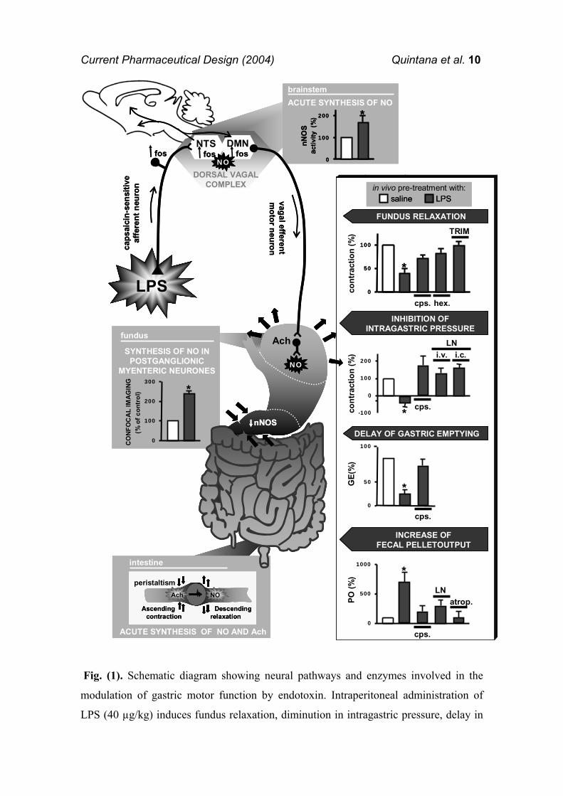
Current Pharmaceutical Design (2004) Quintana et al. 10
Fig. (1). Schematic diagram showing neural pathways and enzymes involved in the
modulation of gastric motor function by endotoxin. Intraperitoneal administration of
LPS (40 µg/kg) induces fundus relaxation, diminution in intragastric pressure, delay in
0
50
100
*
cps. hex.
TRIM
cont
ract
ion
(%)
-100
0
100
200
* cps.
i.v. i.c.LN
cont
ract
ion
(%)
0
500
1000*
cps.
LNatrop.PO
(%)
0
50 *
cps.
GE(
%)
saline LPSin vivo pre-treatment with:
INHIBITION OF INTRAGASTRIC PRESSURE
INCREASE OF FECAL PELLETOUTPUT
FUNDUS RELAXATION
100
DELAY OF GASTRIC EMPTYING
ACUTE SYNTHESIS OF NO AND Ach
intestine
NOAch
Ascendingcontraction
Descendingrelaxation
peristaltism
0
100
200
300*
(% o
fcon
trol
)
SYNTHESIS OF NO IN POSTGANGLIONIC
MYENTERIC NEURONES
CO
NFO
CA
L IM
AG
ING
fundus
fos
vagal efferent m
otor neuroncaps
aici
n-se
nsiti
veaf
fere
ntne
uron
LPS
NTS DMNfos fos
DORSAL VAGAL COMPLEX
NO
Ach
NO
nNOS
brainstemACUTE SYNTHESIS OF NObrainstem
0
100
200 *
nNO
S ac
tivity
(%
)
0
50
100
*
0
50
100
*
cps. hex.
TRIM
cont
ract
ion
(%)
-100
0
100
200
* cps.
i.v. i.c.LN
cont
ract
ion
(%)
0
500
1000*
cps.
LNatrop.PO
(%)
0
50 *
cps.
GE(
%)
saline LPSin vivo pre-treatment with:
salinesaline LPSLPSin vivo pre-treatment with:
INHIBITION OF INTRAGASTRIC PRESSURE
INHIBITION OF INTRAGASTRIC PRESSURE
INCREASE OF FECAL PELLETOUTPUT
INCREASE OF FECAL PELLETOUTPUT
FUNDUS RELAXATIONFUNDUS RELAXATION
100
DELAY OF GASTRIC EMPTYINGDELAY OF GASTRIC EMPTYING
ACUTE SYNTHESIS OF NO AND Ach
intestine
NOAch
Ascendingcontraction
Descendingrelaxation
peristaltism
ACUTE SYNTHESIS OF NO AND Ach
intestine
NOAch
Ascendingcontraction
Descendingrelaxation
peristaltismNOAch
Ascendingcontraction
Descendingrelaxation
NOAch
Ascendingcontraction
Descendingrelaxation
peristaltism
0
100
200
300*
(% o
fcon
trol
)
SYNTHESIS OF NO IN POSTGANGLIONIC
MYENTERIC NEURONES
CO
NFO
CA
L IM
AG
ING
fundus
0
100
200
300*
(% o
fcon
trol
)
SYNTHESIS OF NO IN POSTGANGLIONIC
MYENTERIC NEURONES
CO
NFO
CA
L IM
AG
ING
fundus
fos
vagal efferent m
otor neuroncaps
aici
n-se
nsiti
veaf
fere
ntne
uron
LPS
NTS DMNfosfos fosfos
DORSAL VAGAL COMPLEX
NONO
Ach
NONO
nNOSnNOS
brainstemACUTE SYNTHESIS OF NObrainstem
0
100
200 *
nNO
S ac
tivity
(%
)
brainstembrainstemACUTE SYNTHESIS OF NObrainstem
0
100
200 *
nNO
S ac
tivity
(%
)
0
100
200 *
nNO
S ac
tivity
(%
)
Current Pharmaceutical Design (2004) Quintana et al. 11
gastric emptying of solid meals and increase in intestinal transit, 30 min, 30 min, 4h and
1h later, respectively. It has been demonstrated that LPS induces the activation of: a)
capsaicin-sensitive afferent neurons; b) neurons of the dorsal vagal complex of the
brainstem and c) cholinergic pre-ganglionic motor fibres emerging from the DMN,
which in turn induce the release of inhibitory neurotransmitters in the myenteric plexus.
In addition, NO synthesis from nNOS has been observed by biochemical methods in the
brainstem and the myenteric plexus of LPS-treated animals and it has been functionally
related with changes in gastrointestinal function. Graphs show original data adapted
from [5,7,10,13,42] and represent mean±s.e.m. (n>3; *p<0.05). Atrop., atropine; cps.,
capsaicin; DMN, dorsal motor nucleus of the vagus; hex., hexamethonium; LN, L-
nitroarginine methyl ester; NTS, nucleus of the solitary tract; TRIM, 1-(2-
trifluoromethylphenyl)imidazole, a nNOS inhibitor.
NITRIC OXIDE
NO is synthesized from L-arginine by NOS, an enzyme that has been detected in
different cells of the gastrointestinal tract including endothelial, secretor, muscular and
nervous cells [49,50]. In addition, immunohistochemical studies have also reported the
presence of NOS in vagal afferents and central brainstem nuclei involved in modulation
of gastrointestinal function such as intrinsic neurons of the NTS and neurons of the
DMN [51,52]. Therefore it is not surprising that NO is an endogenous mediator
involved in the pathophysiological regulation of a variety of gastrointestinal functions
such as microcirculation and mucosal integrity maintenance, as well as modulation of
secretor and motor function. Three different NOS isoforms have been characterized in
mammalian cells [53,54]: the neuronal (nNOS) and endothelial (eNOS) NOS isoforms
that are constitutively expressed, widely distributed and synthesize NO at low
concentrations which regulates numerous physiological functions. The third isoform is
named inducible NOS (iNOS) as its expression requires transcriptional activation; it is
Current Pharmaceutical Design (2004) Quintana et al. 12
induced in many cell types including inflammatory cells, endothelium and neurons after
exposure to cytokines and LPS and it is continuously active during inflammation,
leading to an overproduction of NO, which presents cytotoxic activity. However, it is
now known that the terminology “constitutive” versus “inducible” is an
oversimplification as it has been stated that levels of gene expression of “constitutive”
isoforms, both eNOS and nNOS, may also be induced under different physiological
conditions [55] and, conversely, that iNOS may function as a “constitutive” enzyme in
some cells under physiological situations [56].
Nitric oxide and endotoxemia
It was early described that administration of LPS at doses in the range of mg/kg
induced a transcriptional increase in the expression of iNOS in a variety of tissues from
rodents [57]. Furthermore, elevated levels of circulating nitrite/nitrate, the stable NO
bio-reaction products, were reported in septic patients [58,59]. The massive release of
iNOS-derived NO during endotoxemia has been associated with vascular dysfunction
and severe changes in gastrointestinal function such as an increase in the epithelial
barrier function and gastrointestinal mucosal damage [60,61]. In contrast to these
effects, a gastrointestinal defensive mechanism is observed rapidly after administration
of doses in the range of µg/kg of endotoxin, and NO seems also to be involved.
However, a role for the iNOS isoform, as the one responsible for NO synthesis under
these circumstances, seems unlikely since iNOS is transcriptionally regulated, a process
that requires several hours [62]. Consistent with these observations are biochemical
studies showing, one hour after LPS administration (µg/kg), an increase in nNOS
enzymatic activity in the brainstem while no changes where observed in iNOS
enzymatic activity [10]. Furthermore, parallel experiments on the protein amount
Current Pharmaceutical Design (2004) Quintana et al. 13
showed no significant changes in nNOS protein in the brainstem at the same time point
(unpublished data). It was suggested that endotoxin induced in vivo a non-
transcriptional regulation of NO synthesis in the brainstem. This mechanism seems
extensible to other tissues and NO synthesis from constitutive sources has also been
observed in the stomach thirty minutes after administration of low doses of endotoxin
[13]. Recent studies using confocal microscopy provided direct evidence of NO
synthesis in the gastric fundus of rats treated with endotoxin, “Fig. (2)”; such a
synthesis was observed in the myenteric plexus and pharmacological tools allowed to
specifically localize the cellular source of NO synthesis in post-ganglionic myenteric
neurons. In a similar manner to that reported in the brainstem, such a synthesis was not
transcriptionally regulated, since no changes in nNOS mRNA nor iNOS mRNA
expression were observed in the gastric fundus one hour after LPS administration [13]
“Fig. (1)”.
It seems that nNOS isoform, which plays a main physiological role, may also
up-regulate its activity very rapidly after challenge with low doses of endotoxin. [10-
13,63]. This mechanism does not seem to be specific of the nNOS isoform and extends
to the other constitutive isoenzyme. In this line, NO synthesis has been also involved in
the increase in gastric mucosal resistance against deleterious agents induced by doses of
endotoxin in the range of µg/kg [11]. Such a synthesis is post-translationally regulated
since no changes in eNOS nor iNOS mRNA were observed in the gastric corpus of
endotoxin-treated rats at the time were the gastroprotective effect was observed [8]. A
recent study demonstrates that, these doses of endotoxin preserve the fall in gastric
blood flow induced by non steroidal anti-inflammatory drugs (NSAIDs) [8], a
mechanism that is probably involved in the gastroprotective effect of endotoxin.
Although no direct evidence of the specific NOS isoform involved was obtained, an
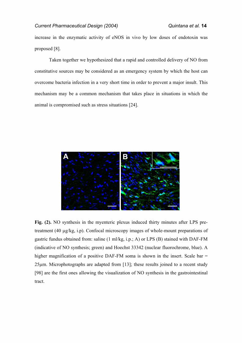
Current Pharmaceutical Design (2004) Quintana et al. 14
increase in the enzymatic activity of eNOS in vivo by low doses of endotoxin was
proposed [8].
Taken together we hypothesized that a rapid and controlled delivery of NO from
constitutive sources may be considered as an emergency system by which the host can
overcome bacteria infection in a very short time in order to prevent a major insult. This
mechanism may be a common mechanism that takes place in situations in which the
animal is compromised such as stress situations [24].
Fig. (2). NO synthesis in the myenteric plexus induced thirty minutes after LPS pre-
treatment (40 µg/kg, i.p). Confocal microscopy images of whole-mount preparations of
gastric fundus obtained from: saline (1 ml/kg, i.p.; A) or LPS (B) stained with DAF-FM
(indicative of NO synthesis; green) and Hoechst 33342 (nuclear fluorochrome, blue). A
higher magnification of a positive DAF-FM soma is shown in the insert. Scale bar =
25µm. Microphotographs are adapted from [13]; these results joined to a recent study
[98] are the first ones allowing the visualization of NO synthesis in the gastrointestinal
tract.
A BAA BB
Current Pharmaceutical Design (2004) Quintana et al. 15
CHANGES IN GASTROINTESTINAL FUNCTION IN ENDOTOXEMIA
Gastrointestinal Motility
Opposite changes between gastric and intestinal motility are observed in the
early phase of infection with Gram-negative bacteria. Inhibition of gastric motility and
stimulation of intestinal transit occur after administration of doses of endotoxin in the
range of µg/kg to rats [5,7,10,64-67].
Inhibition of gastric motor function
In experimental studies, diminution of the gastric tone, relaxation of the gastric
fundus and delay in gastric emptying are observed early after systemic administration of
bacterial endotoxin to rats. These changes are rapid in onset (30-60 min post-injection
of LPS) and mediated by capsaicin-sensitive afferent neurons, since neuronal blockade
of these fibres by pre-treatment with the selective neurotoxin capsaicin, completely
prevents gastric motor disturbances induced by LPS [5,10]. One of the physiological
roles played by these fibres includes a local efferent-like function that allows the release
of neuropeptides, a mechanism that seems to be involved in the inhibitory effect of
endotoxin, since blockade of CGRP receptors prevented the delay in gastric emptying
associated to this circumstance [5]. However, these fibres also trigger a sensory function
signalling information from periphery to superior neuronal network [68,69], a
mechanism that is also involved in the gastric effect of endotoxin since local application
of capsaicin to the cervical vagus nerve prevented such effect “Fig.(1)”.
The DVC of the brainstem plays an important role in physiological modulation
of gastric motor function. Although presence of different gastric inhibitory
neurotransmitters has been reported in this area, increasing evidence for the last fifteen
Current Pharmaceutical Design (2004) Quintana et al. 16
years support a crucial role for NO synthesis in the DVC on the inhibition of gastric
motor function [10,47]. It has been reported that gastric relaxation evoked by the central
nervous system is completely abolished by an NOS inhibitor in the DVC [70] while
administration of NO producing compounds in the DMN increase the spontaneous
firing rate of the neurons [71]; furthermore microinjection of L-arginine (the NO
synthesis precursor) into the DVC has been shown to decrease intragastric pressure
while L-NAME (a NOS inhibitor) increased it [47]. It is likely that a mechanism based
on the activation of the NOS in the DVC and the subsequent synthesis of NO is
triggered by peripheral endotoxin and plays a role in the diminution of gastric motor
function associated with this circumstance. In this line, NO synthesis in the brainstem
has been involved in endotoxin-induced diminution of intragastric pressure stimulated
by 2-deoxy-D-glucose [10]. These results, joined to the fact that no changes in the
amount of nNOS protein in the brainstem are observed under these circumstances,
suggest that a non-transcriptional regulation of NO synthesis from nNOS in this area
during early endotoxemia is involved in gastric motor inhibition “Fig. (1)”.
Previous studies have reported that inhibition of gastric motor function by NO
synthesis in the DVC is an effect mediated by the vagus nerve [47], although the nature
of the vagal efferent fibres involved has not been elucidated. In a recent study we have
reported that fundus relaxation induced by intraperitoneal administration of LPS is
mediated by a neural mechanism that involves extrinsic innervation of the gut and NO
synthesis in the myenteric plexus of the gastric fundus [13]. Such a synthesis is
mediated by the nNOS isoform since both L-NAME and a selective nNOS inhibitor,
TRIM, significantly prevented fundus relaxation induced by endotoxin. Different
experimental studies have demonstrated that NO synthesis in both the brainstem and
myenteric plexus is involved in gastric motor inhibition induced by low doses of
Current Pharmaceutical Design (2004) Quintana et al. 17
systemic LPS. These results led us to hypothesise that a sequence of nitrergic neurons
could be the efferent mechanism involved in the inhibitory effect of endotoxin on
gastric function, as previously reported [47]. However, it has also been reported that
endotoxin-induced NO synthesis in the myenteric plexus is regulated by nicotinic
receptors in the ganglia which support a role for vagal cholinergic pre-ganglionic fibres
in the effects of endotoxin [13]. These results ruled out the above hypothesis and make
necessary further studies to draw the specific efferent pathway activated by endotoxin
[10,72] “Fig. (1)”.
Most of the effects induced by the constitutive synthesis of NO, including
relaxation of the gastric smooth muscle involve activation of the soluble guanylate
cyclase (sGC) [53]. It has been reported that relaxation of the gastric fundus by low
doses of endotoxin is mediated by activation of the sGC. This intracellular messenger
reinforces the synthesis of NO by endotoxin and suggests a constitutive enzymatic
source [13]. In the same study it is shown that the inhibitory effect of LPS was also
mediated by activation of small conductance calcium sensitive K+ (caK+) channels [13],
which have been involved in gastrointestinal relaxation induced by endogenous and
exogenous NO [13,73-75]. In addition, by using pharmacological tools it is shown that
NO, once released from enteric neurons, would increase the levels of cGMP which in
turn phosphorylates the small conductance caK+ channels [76] which leads to relaxation
of the smooth muscle cell under early endotoxemia “Fig. (3)”.
In summary, consistent evidence supports that inhibition of gastric motor
function in early endotoxemia is mediated by a complex neural mechanism that
involves activation of the vagus nerve, synthesis of NO in the brainstem and the final
release of NO from enteric neurons acting on the smooth muscle cell. Such a
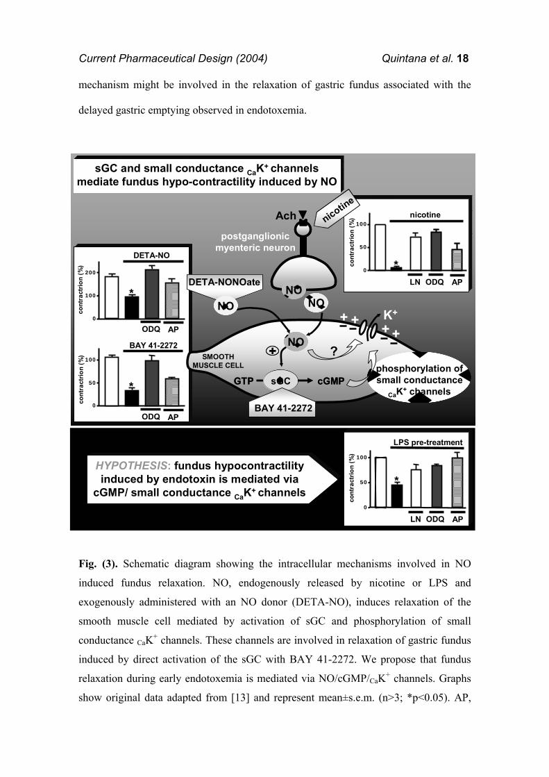
Current Pharmaceutical Design (2004) Quintana et al. 18
mechanism might be involved in the relaxation of gastric fundus associated with the
delayed gastric emptying observed in endotoxemia.
Fig. (3). Schematic diagram showing the intracellular mechanisms involved in NO
induced fundus relaxation. NO, endogenously released by nicotine or LPS and
exogenously administered with an NO donor (DETA-NO), induces relaxation of the
smooth muscle cell mediated by activation of sGC and phosphorylation of small
conductance CaK+ channels. These channels are involved in relaxation of gastric fundus
induced by direct activation of the sGC with BAY 41-2272. We propose that fundus
relaxation during early endotoxemia is mediated via NO/cGMP/CaK+ channels. Graphs
show original data adapted from [13] and represent mean±s.e.m. (n>3; *p<0.05). AP,
0
100
200
ODQ AP
DETA-NO
*
cont
ract
rion
(%)
0
50
100
ODQ AP
BAY 41-2272
*
cont
ract
rion
(%)
NO+
sGCGTP cGMP
K+
BAY 41-2272
NO
SMOOTH MUSCLE CELL
Ach
NO
postganglionicmyenteric neuron
?phosphorylation ofsmall conductance
CaK+ channels
DETA-NONOate
NO
sGC and small conductance CaK+ channelsmediate fundus hypo-contractility induced by NO
HYPOTHESIS: fundus hypocontractilityinduced by endotoxin is mediated via
cGMP/ small conductance CaK+ channels
LPS pre-treatment
0
50
100
APODQLN
cont
ract
rion
(%)
*
0
50
100
*APODQLN
nicotine
cont
ract
rion
(%)nicotin
e
0
100
200
ODQ AP
DETA-NO
*
cont
ract
rion
(%)
0
50
100
ODQ AP
BAY 41-2272
*
cont
ract
rion
(%)
0
100
200
ODQ AP
DETA-NO
*
cont
ract
rion
(%)
0
50
100
ODQ AP
BAY 41-2272
*
cont
ract
rion
(%)
NO++
sGCGTP cGMPsGCsGCGTP cGMP
K+
BAY 41-2272
NO
SMOOTH MUSCLE CELL
Ach
NO
postganglionicmyenteric neuron
?phosphorylation ofsmall conductance
CaK+ channels
DETA-NONOate
NO
sGC and small conductance CaK+ channelsmediate fundus hypo-contractility induced by NO
sGC and small conductance CaK+ channelsmediate fundus hypo-contractility induced by NO
HYPOTHESIS: fundus hypocontractilityinduced by endotoxin is mediated via
cGMP/ small conductance CaK+ channels
HYPOTHESIS: fundus hypocontractilityinduced by endotoxin is mediated via
cGMP/ small conductance CaK+ channels
LPS pre-treatment
0
50
100
APODQLN
cont
ract
rion
(%)
*
LPS pre-treatment
0
50
100
APODQLN
cont
ract
rion
(%)
*
0
50
100
*APODQLN
nicotine
cont
ract
rion
(%)
0
50
100
*APODQLN
nicotine
cont
ract
rion
(%)nicotin
e
nicotine
Current Pharmaceutical Design (2004) Quintana et al. 19
apamine; DETA-NO, (Z)-1-[2-(2-aminoethyl)-N-(2-ammonioethyl)amino]diazen-1-
ium-1,2-diolate); LN, L-nitroarginine methyl ester; ODQ, 1H-(1,2,4,)oxadiazolo(4,3-
a)quinoxalin-1-one).
Gastric emptying of meals is a complex function of the gut mediated by the
integrated response of the proximal (fundus) and distal (antrum-pylorus) stomach and
duodenum [77], and nitric oxide has been shown to play a role in the specific functions
carried out by these areas [78-80]. Gastric emptying is inversely controlled by tones of
gastric body and pyloric sphincter. In this context, some evidence supports an inhibitory
role of NO on gastric emptying through an action related with relaxation of the gastric
fundus [81] while NO synthesis has been associated with an acceleration of gastric
emptying mainly due to pyloric sphincter relaxation [78,82,83]. Experimental studies
have shown a delayed gastric emptying of a solid nutrient meal after administration of
doses of endotoxin in the range of µg/kg. Due to the intrinsic kinetic of this
physiological process, studies were performed four hours after endotoxin
administration. Interestingly, when constitutive NOS activity was analyzed in the
antrum-pylorus of the stomach, a significant reduction was observed parallel to delayed
gastric emptying [7]. In addition, nNOS mRNA expression and protein was also
significantly reduced, which suggest that these doses of endotoxin induce four hours
later a transcriptional down-regulation of the nNOS protein expression in the antrum-
pylorus; it was hypothesised that diminution of NO synthesis would increase the
sphincter tone and, therefore, delay gastric emptying “Fig. (1)”. It is interesting to note
the area-specific modulation of NO synthesis in the stomach induced by endotoxin: an
increase of NO synthesis in the gastric fundus is observed 30 min after challenge while
a trancriptional down-regulation of nNOS in the antrum pylorus is observed four hours
later. The specific relationship between these two mechanisms needs to be addressed.
Current Pharmaceutical Design (2004) Quintana et al. 20
Increase of intestinal transit
Besides the abnormal gastric motility, intestinal motility pattern is also altered
during early endotoxemia. Administration of doses of µg/kg of E. coli endotoxin induce
a significant increase of colonic motility and transit in conscious rats, an action that was
reported to be partly mediated by PAF and PGs [84]. In addition, a similar dose of
endotoxin has been shown to increase intestinal transit in anaesthetized rats and faecal
excretion in conscious rats within one hour of administration [42,85]. The effect of
endotoxin on fecal excretion was only observed within the first three hours after
challenge and was not associated with changes in the consistency of the faeces,
suggesting that the effect of endotoxin was independent of changes in intestinal
secretion, in contrast to that reported with slightly higher doses of intravenous
endotoxin [84]. Such a prokinetic effect of endotoxin on intestinal motility seems to be
mediated by a long nervous reflex, similar to what happens in the stomach, as endotoxin
effect on faecal excretion is prevented by subdiaphragmatic vagotomy or systemic pre-
treatment with capsaicin [42]. Systematic progression of intestinal contents is known to
be dependent of the peristaltic reflex, which consists of two sequential components:
descending relaxation caudal and ascending contraction oral to the site of stimulus. The
excitatory neurotransmitters responsible for ascending contraction are Ach and the
tachykinins substance P and neurokinin A, whereas VIP and NO mediate the
descending relaxation of circular muscle [86]. Once again the effects of early
endotoxemia on faecal excretion involve the activation of a physiological mechanism
based on the release of the excitatory neurotransmitters Ach and substance P and the
inhibitory neurotransmitter NO. Previous studies using higher doses of endotoxin have
reported an increased expression of iNOS in enterocytes and a role for iNOS-derived
NO in diarrhoea observed in those conditions [87]. However, the small doses of
Current Pharmaceutical Design (2004) Quintana et al. 21
endotoxin used and the short time in which its effects on faecal excretion are observed,
point towards the regulation of constitutive NOS isoform rather than an induction of
iNOS. As a whole, results suggest that the increase in faecal excretion induced by
endotoxin is consequence of a nervous mechanism in which capsaicin-sensitive afferent
neurons and the release of excitatory and inhibitory neurotransmitters in the colonic
wall are involved. An increased secretion of Ach and substance P would enhance the
propulsory effect of ascending contraction of the circular muscle, while local release of
NO would facilitate the accommodation of the propelled bolus by inducing the
descending relaxation of the colonic muscle “Fig. (1)”.
In conclusion we hypothesised that after bacterial invasion the host responds
acutely by maximizing its physiological resources, before the immune system is active,
in order to prevent further absorption of challenge. In this process, gastrointestinal
motility is wisely altered and opposing effects on motility depending on the region
analysed are observed. A hypo-contractility of proximal stomach and a delayed gastric
emptying would prevent access of possible toxins to the duodenum and absorption,
while increased intestinal propulsion and faecal excretion would help to expel
challenge. A perfect interplay between the central and enteric nervous systems is
necessary, and nitric oxide seems to be a crucial molecule in this process.
Gastric acid secretion
Gastric acid secretion constitutes one of the most important endogenous
aggressive factors for the gastric mucosa. Although hyper-secretion has been observed
in severe stress situations, inhibition of gastric acid secretion has been reported during
moderate clinical endotoxemia [88], a situation that has been reproduced after endotoxin
administration to rats [9,12,24,63,89]. Doses of LPS in the range of µg/kg inhibit gastric
Current Pharmaceutical Design (2004) Quintana et al. 22
acid secretion in a reversible way, indicative of a biological effect on gastric physiology
rather than a toxic or destructive effect on the parietal cell [90]. Early studies
demonstrated that inhibition of gastric acid secretion by endotoxin was rapid in onset
(observed 15 min post-injection), not related to vascular changes and independent of
protein synthesis [9,12,24]. In addition, experimental evidence demonstrated that a
neural mechanism involving the central nervous system and activation of non-
adrenergic non-cholinergic fibres was involved in endotoxin-induced gastric
hyposecretion [9]. Pharmacological studies using administration of NOS inhibitors into
the cisterna magna showed a role for central NO synthesis in the hypo-secretory effects
of endotoxin [12]. Further immunohistochemical and electrochemical studies localized
the enzyme responsible for NO synthesis in the DVC of the brainstem, suggesting that a
nervous mechanism involving the activation of the vagus nerve could mediate the
inhibitory effects of endotoxin on gastric acid secretion [24]. In line with this
observation, inhibition of pentagastrin-stimulated acid secretion by endotoxin was
prevented by cervical vagotomy [63]. Furthermore, the acid inhibitory effect of
endotoxin was also prevented by intracisternal administration of an NMDA glutamate
receptor antagonist, reinforcing the involvement of the afferent vagus nerve in the hypo-
secretion [63]. However, still little evidence exists regarding the efferent mechanism
that finally reduces gastric acid secretion in endotoxemia. Taking into consideration that
the hypo-secretory doses of endotoxin have been shown to increase NO synthesis in the
myenteric plexus [13], a direct role for NO acting on the parietal cell and inhibiting
gastric acid secretion might be inferred. Consistent with this observation is the fact that
NO donors have been shown to inhibit the secretory activity in rat isolated parietal cells
[91].
Current Pharmaceutical Design (2004) Quintana et al. 23
Endotoxin may be considered as a challenge to which the body responds with
stress. In this line, endotoxin has been shown to activate neurons immunoreactive to
stress peptides such as oxytocin or corticotrophin releasing factor in the brain [27,92]
and the endogenous release of oxytocin has been shown to play a key role in the hypo-
secretory response associated with endotoxemia [93]. Other experimental models of
moderate somatic stress such as mild hyperthermia or hypotension have been associated
with an acute and long-lasting inhibition of gastric acid secretion, and a nervous reflex
involving NO synthesis in the DVC [24] mediated such a response. It seems that acute
hypo-secretion associated with stress is a consequence of a physiological protective
response activated under these circumstances.
Gastric blood flow and mucosal integrity
Gastric mucosal lesions are important complications of septic shock. However,
administration of doses of endotoxin in the range of µg/kg to rats has been reported to
protect gastric mucosa against the damage induced by ulcerogenic stimuli such as stress,
NSAIDs and ethanol [6,8,11]. The gastroprotective action of LPS is observed one hour
after challenge, mediated by activation of capsaicin-sensitive afferent neurons and
independent of transcriptional synthesis of proteins. In addition NO synthesis-induced
by endotoxin seems to play an important role since blockade of the NOS significantly
prevented the gastroprotective effect. Such a synthesis was not related to the iNOS
isoform, since: a) iNOS mRNA expression was not detected in gastric corpus one hour
after LPS administration, time at which the gastroprotective effect was observed [8] and
b) dexamethasone pre-treatment did not modify LPS effect [11]. Once again, low doses
of endotoxin induce NO synthesis from constitutive sources, which is involved in
mucosal gastroprotection. Constitutive synthesis of NO by nNOS or eNOS isoforms has
Current Pharmaceutical Design (2004) Quintana et al. 24
been involved in the maintenance of the gastrointestinal mucosal integrity through
modulation of gastric mucosal blood flow, prevention of leukocyte adhesion to the
endothelium, epithelial secretion and barrier function [49,94,95]. Among these
mechanisms, modulation of gastric mucosal blood flow, [95] seems to play an essential
role. Neither, endogenously generated NO by endotoxin, nor exogenous NO liberated
by an NO donor, such as nitroglicerin, modifies resting gastrointestinal blood flow in
experimental animals. However, both situations have been shown to prevent the
reduction in gastric blood flow induced by an ulcerogenic dose of NSAIDs and
consequently counteract its detrimental effects [8,96]. The eNOS is presumably the
main isoform involved in the maintenance of gastric blood flow; however, synthesis of
NO in the myenteric plexus, probably due to nNOS isoform, which has been
demonstrated during early endotoxemia [13], could also be contributing to gastric
mucosal protection. In this line, evidence exists for a role of endogenous NO in gastric
mucosa protection following central vagal stimulation [97]. However, biochemical
studies trying to demonstrate an increase in the NOS activity of the constitutive
isoforms in the gastric corpus of endotoxin-treated rats showed no significant changes.
It is believed that factors increasing the activity of these enzymes that are only present
in vivo were responsible for the enhanced NO synthesis after endotoxin administration.
In summary, we believe that rapidly after bacterial infection the host tries to maintain
gastric mucosal integrity mainly by preserving the fall in microcirculation and, in order
to get this, maximizes the activity of its constitutive resources.
CONCLUDING REMARKS
Gastrointestinal function is modulated by endotoxin. The initial effects depend
on a fast, neural mechanism involving activation of capsaicin sensitive afferent neurons,
Current Pharmaceutical Design (2004) Quintana et al. 25
which explains the rapid onset (first hour post-injection) of the endotoxin effect on
faecal excretion, gastric motility, acid secretion and mucosal resistance to damage. In
such a response, NO synthesis from calcium-dependent NOS isoforms occurs in both,
the central (DVC) and enteric nervous systems. The perfect interplay of afferent/efferent
neurons and nNOS constitutes an emergency system, which allows the organism to
adapt to slight alterations of the physiology, and enhances the body ability to withstand
ulterior aggressions. Understanding the functional neuroanatomy by which the nervous
system responds to immune challenge would greatly help to control the deleterious and
promote the beneficial aspects of the acute phase reaction to infection.
ACNOWLEGDMENTS
Research in the authors’ laboratory pertaining to this topic was supported by
Grant FIS 01/1187 from the Ministerio de Sanidad y Consumo and grant C 03/02 from
Instituto de Salud Carlos III. Elsa Quintana is the recipient of a fellowship from the
Ministerio de Sanidad y Consumo.
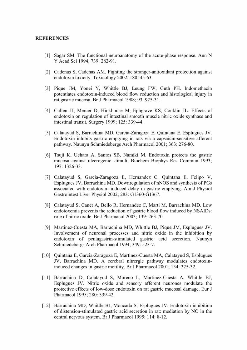
REFERENCES [1] Sagar SM. The functional neuroanatomy of the acute-phase response. Ann N
Y Acad Sci 1994; 739: 282-91.
[2] Cadenas S, Cadenas AM. Fighting the stranger-antioxidant protection against endotoxin toxicity. Toxicology 2002; 180: 45-63.
[3] Pique JM, Yonei Y, Whittle BJ, Leung FW, Guth PH. Indomethacin potentiates endotoxin-induced blood flow reduction and histological injury in rat gastric mucosa. Br J Pharmacol 1988; 93: 925-31.
[4] Cullen JJ, Mercer D, Hinkhouse M, Ephgrave KS, Conklin JL. Effects of endotoxin on regulation of intestinal smooth muscle nitric oxide synthase and intestinal transit. Surgery 1999; 125: 339-44.
[5] Calatayud S, Barrachina MD, Garcia-Zaragoza E, Quintana E, Esplugues JV. Endotoxin inhibits gastric emptying in rats via a capsaicin-sensitive afferent pathway. Naunyn Schmiedebergs Arch Pharmacol 2001; 363: 276-80.
[6] Tsuji K, Uehara A, Santos SB, Namiki M. Endotoxin protects the gastric mucosa against ulcerogenic stimuli. Biochem Biophys Res Commun 1993; 197: 1326-33.
[7] Calatayud S, Garcia-Zaragoza E, Hernandez C, Quintana E, Felipo V, Esplugues JV, Barrachina MD. Downregulation of nNOS and synthesis of PGs associated with endotoxin- induced delay in gastric emptying. Am J Physiol Gastrointest Liver Physiol 2002; 283: G1360-G1367.
[8] Calatayud S, Canet A, Bello R, Hernandez C, Marti M, Barrachina MD. Low endotoxemia prevents the reduction of gastric blood flow induced by NSAIDs: role of nitric oxide. Br J Pharmacol 2003; 139: 263-70.
[9] Martinez-Cuesta MA, Barrachina MD, Whittle BJ, Pique JM, Esplugues JV. Involvement of neuronal processes and nitric oxide in the inhibition by endotoxin of pentagastrin-stimulated gastric acid secretion. Naunyn Schmiedebergs Arch Pharmacol 1994; 349: 523-7.
[10] Quintana E, Garcia-Zaragoza E, Martinez-Cuesta MA, Calatayud S, Esplugues JV, Barrachina MD. A cerebral nitrergic pathway modulates endotoxin-induced changes in gastric motility. Br J Pharmacol 2001; 134: 325-32.
[11] Barrachina D, Calatayud S, Moreno L, Martinez-Cuesta A, Whittle BJ, Esplugues JV. Nitric oxide and sensory afferent neurones modulate the protective effects of low-dose endotoxin on rat gastric mucosal damage. Eur J Pharmacol 1995; 280: 339-42.
[12] Barrachina MD, Whittle BJ, Moncada S, Esplugues JV. Endotoxin inhibition of distension-stimulated gastric acid secretion in rat: mediation by NO in the central nervous system. Br J Pharmacol 1995; 114: 8-12.
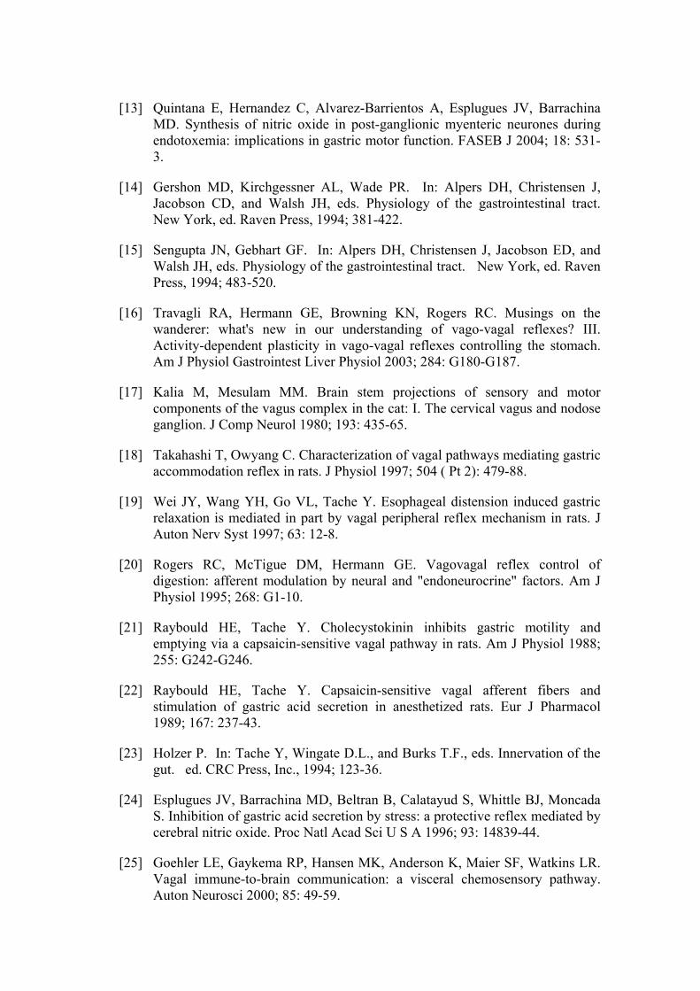
[13] Quintana E, Hernandez C, Alvarez-Barrientos A, Esplugues JV, Barrachina MD. Synthesis of nitric oxide in post-ganglionic myenteric neurones during endotoxemia: implications in gastric motor function. FASEB J 2004; 18: 531-3.
[14] Gershon MD, Kirchgessner AL, Wade PR. In: Alpers DH, Christensen J, Jacobson CD, and Walsh JH, eds. Physiology of the gastrointestinal tract. New York, ed. Raven Press, 1994; 381-422.
[15] Sengupta JN, Gebhart GF. In: Alpers DH, Christensen J, Jacobson ED, and Walsh JH, eds. Physiology of the gastrointestinal tract. New York, ed. Raven Press, 1994; 483-520.
[16] Travagli RA, Hermann GE, Browning KN, Rogers RC. Musings on the wanderer: what's new in our understanding of vago-vagal reflexes? III. Activity-dependent plasticity in vago-vagal reflexes controlling the stomach. Am J Physiol Gastrointest Liver Physiol 2003; 284: G180-G187.
[17] Kalia M, Mesulam MM. Brain stem projections of sensory and motor components of the vagus complex in the cat: I. The cervical vagus and nodose ganglion. J Comp Neurol 1980; 193: 435-65.
[18] Takahashi T, Owyang C. Characterization of vagal pathways mediating gastric accommodation reflex in rats. J Physiol 1997; 504 ( Pt 2): 479-88.
[19] Wei JY, Wang YH, Go VL, Tache Y. Esophageal distension induced gastric relaxation is mediated in part by vagal peripheral reflex mechanism in rats. J Auton Nerv Syst 1997; 63: 12-8.
[20] Rogers RC, McTigue DM, Hermann GE. Vagovagal reflex control of digestion: afferent modulation by neural and "endoneurocrine" factors. Am J Physiol 1995; 268: G1-10.
[21] Raybould HE, Tache Y. Cholecystokinin inhibits gastric motility and emptying via a capsaicin-sensitive vagal pathway in rats. Am J Physiol 1988; 255: G242-G246.
[22] Raybould HE, Tache Y. Capsaicin-sensitive vagal afferent fibers and stimulation of gastric acid secretion in anesthetized rats. Eur J Pharmacol 1989; 167: 237-43.
[23] Holzer P. In: Tache Y, Wingate D.L., and Burks T.F., eds. Innervation of the gut. ed. CRC Press, Inc., 1994; 123-36.
[24] Esplugues JV, Barrachina MD, Beltran B, Calatayud S, Whittle BJ, Moncada S. Inhibition of gastric acid secretion by stress: a protective reflex mediated by cerebral nitric oxide. Proc Natl Acad Sci U S A 1996; 93: 14839-44.
[25] Goehler LE, Gaykema RP, Hansen MK, Anderson K, Maier SF, Watkins LR. Vagal immune-to-brain communication: a visceral chemosensory pathway. Auton Neurosci 2000; 85: 49-59.
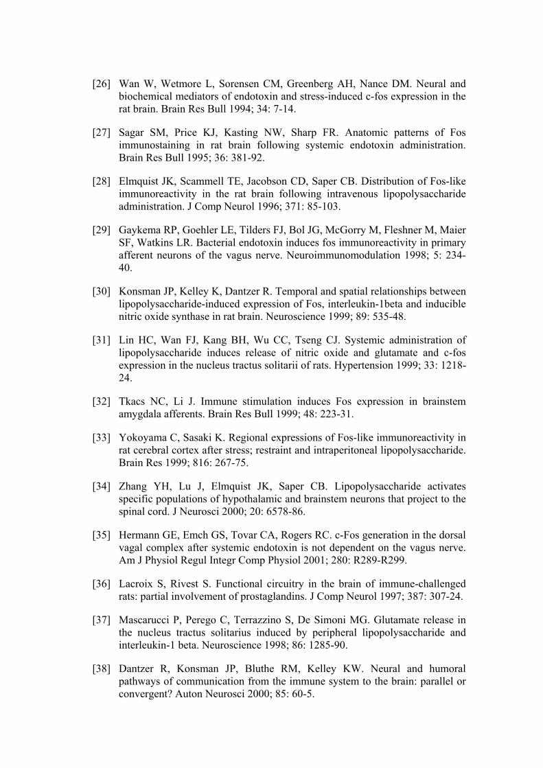
[26] Wan W, Wetmore L, Sorensen CM, Greenberg AH, Nance DM. Neural and biochemical mediators of endotoxin and stress-induced c-fos expression in the rat brain. Brain Res Bull 1994; 34: 7-14.
[27] Sagar SM, Price KJ, Kasting NW, Sharp FR. Anatomic patterns of Fos immunostaining in rat brain following systemic endotoxin administration. Brain Res Bull 1995; 36: 381-92.
[28] Elmquist JK, Scammell TE, Jacobson CD, Saper CB. Distribution of Fos-like immunoreactivity in the rat brain following intravenous lipopolysaccharide administration. J Comp Neurol 1996; 371: 85-103.
[29] Gaykema RP, Goehler LE, Tilders FJ, Bol JG, McGorry M, Fleshner M, Maier SF, Watkins LR. Bacterial endotoxin induces fos immunoreactivity in primary afferent neurons of the vagus nerve. Neuroimmunomodulation 1998; 5: 234-40.
[30] Konsman JP, Kelley K, Dantzer R. Temporal and spatial relationships between lipopolysaccharide-induced expression of Fos, interleukin-1beta and inducible nitric oxide synthase in rat brain. Neuroscience 1999; 89: 535-48.
[31] Lin HC, Wan FJ, Kang BH, Wu CC, Tseng CJ. Systemic administration of lipopolysaccharide induces release of nitric oxide and glutamate and c-fos expression in the nucleus tractus solitarii of rats. Hypertension 1999; 33: 1218-24.
[32] Tkacs NC, Li J. Immune stimulation induces Fos expression in brainstem amygdala afferents. Brain Res Bull 1999; 48: 223-31.
[33] Yokoyama C, Sasaki K. Regional expressions of Fos-like immunoreactivity in rat cerebral cortex after stress; restraint and intraperitoneal lipopolysaccharide. Brain Res 1999; 816: 267-75.
[34] Zhang YH, Lu J, Elmquist JK, Saper CB. Lipopolysaccharide activates specific populations of hypothalamic and brainstem neurons that project to the spinal cord. J Neurosci 2000; 20: 6578-86.
[35] Hermann GE, Emch GS, Tovar CA, Rogers RC. c-Fos generation in the dorsal vagal complex after systemic endotoxin is not dependent on the vagus nerve. Am J Physiol Regul Integr Comp Physiol 2001; 280: R289-R299.
[36] Lacroix S, Rivest S. Functional circuitry in the brain of immune-challenged rats: partial involvement of prostaglandins. J Comp Neurol 1997; 387: 307-24.
[37] Mascarucci P, Perego C, Terrazzino S, De Simoni MG. Glutamate release in the nucleus tractus solitarius induced by peripheral lipopolysaccharide and interleukin-1 beta. Neuroscience 1998; 86: 1285-90.
[38] Dantzer R, Konsman JP, Bluthe RM, Kelley KW. Neural and humoral pathways of communication from the immune system to the brain: parallel or convergent? Auton Neurosci 2000; 85: 60-5.
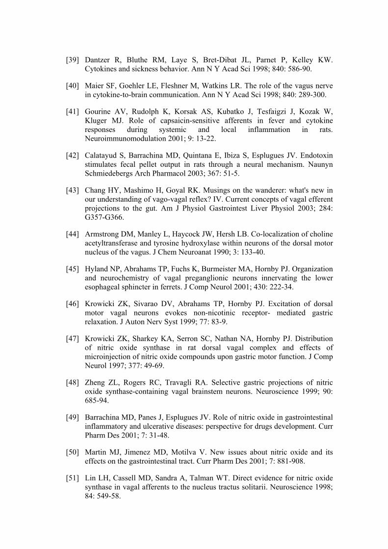
[39] Dantzer R, Bluthe RM, Laye S, Bret-Dibat JL, Parnet P, Kelley KW. Cytokines and sickness behavior. Ann N Y Acad Sci 1998; 840: 586-90.
[40] Maier SF, Goehler LE, Fleshner M, Watkins LR. The role of the vagus nerve in cytokine-to-brain communication. Ann N Y Acad Sci 1998; 840: 289-300.
[41] Gourine AV, Rudolph K, Korsak AS, Kubatko J, Tesfaigzi J, Kozak W, Kluger MJ. Role of capsaicin-sensitive afferents in fever and cytokine responses during systemic and local inflammation in rats. Neuroimmunomodulation 2001; 9: 13-22.
[42] Calatayud S, Barrachina MD, Quintana E, Ibiza S, Esplugues JV. Endotoxin stimulates fecal pellet output in rats through a neural mechanism. Naunyn Schmiedebergs Arch Pharmacol 2003; 367: 51-5.
[43] Chang HY, Mashimo H, Goyal RK. Musings on the wanderer: what's new in our understanding of vago-vagal reflex? IV. Current concepts of vagal efferent projections to the gut. Am J Physiol Gastrointest Liver Physiol 2003; 284: G357-G366.
[44] Armstrong DM, Manley L, Haycock JW, Hersh LB. Co-localization of choline acetyltransferase and tyrosine hydroxylase within neurons of the dorsal motor nucleus of the vagus. J Chem Neuroanat 1990; 3: 133-40.
[45] Hyland NP, Abrahams TP, Fuchs K, Burmeister MA, Hornby PJ. Organization and neurochemistry of vagal preganglionic neurons innervating the lower esophageal sphincter in ferrets. J Comp Neurol 2001; 430: 222-34.
[46] Krowicki ZK, Sivarao DV, Abrahams TP, Hornby PJ. Excitation of dorsal motor vagal neurons evokes non-nicotinic receptor- mediated gastric relaxation. J Auton Nerv Syst 1999; 77: 83-9.
[47] Krowicki ZK, Sharkey KA, Serron SC, Nathan NA, Hornby PJ. Distribution of nitric oxide synthase in rat dorsal vagal complex and effects of microinjection of nitric oxide compounds upon gastric motor function. J Comp Neurol 1997; 377: 49-69.
[48] Zheng ZL, Rogers RC, Travagli RA. Selective gastric projections of nitric oxide synthase-containing vagal brainstem neurons. Neuroscience 1999; 90: 685-94.
[49] Barrachina MD, Panes J, Esplugues JV. Role of nitric oxide in gastrointestinal inflammatory and ulcerative diseases: perspective for drugs development. Curr Pharm Des 2001; 7: 31-48.
[50] Martin MJ, Jimenez MD, Motilva V. New issues about nitric oxide and its effects on the gastrointestinal tract. Curr Pharm Des 2001; 7: 881-908.
[51] Lin LH, Cassell MD, Sandra A, Talman WT. Direct evidence for nitric oxide synthase in vagal afferents to the nucleus tractus solitarii. Neuroscience 1998; 84: 549-58.
[52] Zheng H, Kelly L, Patterson LM, Berthoud HR. Effect of brain stem NMDA-receptor blockade by MK-801 on behavioral and fos responses to vagal satiety signals. Am J Physiol 1999; 277: R1104-R1111.
[53] Moncada S, Palmer RM, Higgs EA. Nitric oxide: physiology, pathophysiology, and pharmacology. Pharmacol Rev 1991; 43: 109-42.
[54] Esplugues JV. NO as a signalling molecule in the nervous system. Br J Pharmacol 2002; 135: 1079-95.
[55] Forstermann U, Boissel JP, Kleinert H. Expressional control of the 'constitutive' isoforms of nitric oxide synthase (NOS I and NOS III). FASEB J 1998; 12: 773-90.
[56] Guo FH, De Raeve HR, Rice TW, Stuehr DJ, Thunnissen FB, Erzurum SC. Continuous nitric oxide synthesis by inducible nitric oxide synthase in normal human airway epithelium in vivo. Proc Natl Acad Sci U S A 1995; 92: 7809-13.
[57] Knowles RG, Merrett M, Salter M, Moncada S. Differential induction of brain, lung and liver nitric oxide synthase by endotoxin in the rat. Biochem J 1990; 270: 833-6.
[58] Gomez-Jimenez J, Salgado A, Mourelle M, Martin MC, Segura RM, Peracaula R, Moncada S. L-arginine: nitric oxide pathway in endotoxemia and human septic shock. Crit Care Med 1995; 23: 253-8.
[59] Evans T, Carpenter A, Kinderman H, Cohen J. Evidence of increased nitric oxide production in patients with the sepsis syndrome. Circ Shock 1993; 41: 77-81.
[60] Lamarque D, Moran AP, Szepes Z, Delchier JC, Whittle BJ. Cytotoxicity associated with induction of nitric oxide synthase in rat duodenal epithelial cells in vivo by lipopolysaccharide of Helicobacter pylori: inhibition by superoxide dismutase. Br J Pharmacol 2000; 130: 1531-8.
[61] Unno N, Wang H, Menconi MJ, Tytgat SH, Larkin V, Smith M, Morin MJ, Chavez A, Hodin RA, Fink MP. Inhibition of inducible nitric oxide synthase ameliorates endotoxin-induced gut mucosal barrier dysfunction in rats. Gastroenterology 1997; 113: 1246-57.
[62] Stuehr DJ. Structure-function aspects in the nitric oxide synthases. Annu Rev Pharmacol Toxicol 1997; 37: 339-59.
[63] Garcia-Zaragoza E, Barrachina MD, Moreno L, Esplugues JV. Role of central glutamate receptors, nitric oxide and soluble guanylyl cyclase in the inhibition by endotoxin of rat gastric acid secretion. Br J Pharmacol 2000; 130: 1283-8.
[64] Takakura K, Hasegawa K, Goto Y, Muramatsu I. Nitric oxide produced by inducible nitric oxide synthase delays gastric emptying in lipopolysaccharide-treated rats. Anesthesiology 1997; 87: 652-7.
[65] van Miert AS, De la Parra DA. Inhibition of gastric emptying by endotoxin (bacterial lipopolysaccharide) in conscious rats and modification of this response by drugs affecting the autonomic nervous system. Arch Int Pharmacodyn Ther 1970; 184: 27-33.
[66] Collares EF. Effect of bacterial lipopolysaccharide on gastric emptying of liquids in rats. Braz J Med Biol Res 1997; 30: 207-11.
[67] Cullen JJ, Caropreso DK, Ephgrave KS. Effect of endotoxin on canine gastrointestinal motility and transit. J Surg Res 1995; 58: 90-5.
[68] Holzer P. Local effector functions of capsaicin-sensitive sensory nerve endings: involvement of tachykinins, calcitonin gene-related peptide and other neuropeptides. Neuroscience 1988; 24: 739-68.
[69] Holzer P. Capsaicin: cellular targets, mechanisms of action, and selectivity for thin sensory neurons. Pharmacol Rev 1991; 43: 143-201.
[70] Krowicki ZK, Hornby PJ. The inhibitory effect of substance P on gastric motor function in the nucleus raphe obscurus is mediated via nitric oxide in the dorsal vagal complex. J Auton Nerv Syst 1996; 58: 177-80.
[71] Travagli RA, Gillis RA. Nitric oxide-mediated excitatory effect on neurons of dorsal motor nucleus of vagus. Am J Physiol 1994; 266: G154-G160.
[72] Ma S, Abboud FM, Felder RB. Effects of L-arginine-derived nitric oxide synthesis on neuronal activity in nucleus tractus solitarius. Am J Physiol 1995; 268: R487-R491.
[73] Bolotina VM, Najibi S, Palacino JJ, Pagano PJ, Cohen RA. Nitric oxide directly activates calcium-dependent potassium channels in vascular smooth muscle. Nature 1994; 368: 850-3.
[74] Shuttleworth CW, Sanders KM. Involvement of nitric oxide in neuromuscular transmission in canine proximal colon. Proc Soc Exp Biol Med 1996; 211: 16-23.
[75] Kitamura K, Lian Q, Carl A, Kuriyama H. S-nitrosocysteine, but not sodium nitroprusside, produces apamin- sensitive hyperpolarization in rat gastric fundus. Br J Pharmacol 1993; 109: 415-23.
[76] Robertson BE, Schubert R, Hescheler J, Nelson MT. cGMP-dependent protein kinase activates Ca-activated K channels in cerebral artery smooth muscle cells. Am J Physiol 1993; 265: C299-C303.
[77] Mayer EA. In: Johnson LR, ed. Physiology of the Gastrointestinal Tract. New York, ed. Raven Press, 1994; 929-76.
[78] Allescher HD, Daniel EE. Role of NO in pyloric, antral, and duodenal motility and its interaction with other inhibitory mediators. Dig Dis Sci 1994; 39: 73S-5S.
[79] Orihata M, Sarna SK. Nitric oxide mediates mechano- and chemoreceptor-activated intestinal feedback control of gastric emptying. Dig Dis Sci 1996; 41: 1303-9.
[80] Desai KM, Sessa WC, Vane JR. Involvement of nitric oxide in the reflex relaxation of the stomach to accommodate food or fluid. Nature 1991; 351: 477-9.
[81] Takakura K, Hasegawa K, Goto Y, Muramatsu I. Nitric oxide produced by inducible nitric oxide synthase delays gastric emptying in lipopolysaccharide-treated rats. Anesthesiology 1997; 87: 652-7.
[82] Orihata M, Sarna SK. Inhibition of nitric oxide synthase delays gastric emptying of solid meals. J Pharmacol Exp Ther 1994; 271: 660-70.
[83] Plourde V, Quintero E, Suto G, Coimbra C, Tache Y. Delayed gastric emptying induced by inhibitors of nitric oxide synthase in rats. Eur J Pharmacol 1994; 256: 125-9.
[84] Pons L, Droy-Lefaix MT, Bueno L. Role of platelet-activating factor (PAF) and prostaglandins in colonic motor and secretory disturbances induced by Escherichia coli endotoxin in conscious rats. Prostaglandins 1994; 47: 123-36.
[85] Martinez-Cuesta MA, Barrachina MD, Beltran B, Calatayud S, Esplugues J. Nitric oxide modulates the acute increase of gastrointestinal transit induced by endotoxin in rats: a possible role for tachykinins. J Pharm Pharmacol 1997; 49: 988-90.
[86] Foxx-Orenstein AE, Grider JR. Regulation of colonic propulsion by enteric excitatory and inhibitory neurotransmitters. Am J Physiol 1996; 271: G433-G437.
[87] Closs EI, Enseleit F, Koesling D, Pfeilschifter JM, Schwarz PM, Forstermann U. Coexpression of inducible NO synthase and soluble guanylyl cyclase in colonic enterocytes: a pathophysiologic signaling pathway for the initiation of diarrhea by gram-negative bacteria? FASEB J 1998; 12: 1643-9.
[88] Hurley JC. Endotoxemia: methods of detection and clinical correlates. Clin Microbiol Rev 1995; 8: 268-92.
[89] Calatayud S, Quintana E, Esplugues J, Barrachina MD. Role of central oxytocin in the inhibition by endotoxin of distension-stimulated gastric acid secretion. Naunyn Schmiedebergs Arch Pharmacol 1999; 360: 676-82.
[90] Uehara A, Okumura T, Tsuji K, Taniguchi Y, Kitamori S, Takasugi Y, Namiki M. Evidence that gastric antisecretory action of lipopolysaccharide is not due to a toxic effect on gastric parietal cells. Dig Dis Sci 1992; 37: 1039-44.
[91] Brown JF, Hanson PJ, Whittle BJ. The nitric oxide donor, S-nitroso-N-acetyl-penicillamine, inhibits secretory activity in rat isolated parietal cells. Biochem Biophys Res Commun 1993; 195: 1354-9.
[92] Rivest S, Laflamme N. Neuronal activity and neuropeptide gene transcription in the brains of immune-challenged rats. J Neuroendocrinol 1995; 7: 501-25.
[93] Vellucci SV, Parrott RF. Bacterial endotoxin-induced gene expression in the choroid plexus and paraventricular and supraoptic hypothalamic nuclei of the sheep. Brain Res Mol Brain Res 1996; 43: 41-50.
[94] Calatayud S, Barrachina D, Esplugues JV. Nitric oxide: relation to integrity, injury, and healing of the gastric mucosa. Microsc Res Tech 2001; 53: 325-35.
[95] Kubes P, Kanwar S, Niu XF, Gaboury JP. Nitric oxide synthesis inhibition induces leukocyte adhesion via superoxide and mast cells. FASEB J 1993; 7: 1293-9.
[96] Barrachina MD, Calatayud S, Canet A, Bello R, Diaz dR, Guth PH, Esplugues JV. Transdermal nitroglycerin prevents nonsteroidal anti-inflammatory drug gastropathy. Eur J Pharmacol 1995; 281: R3-R4.
[97] Kiraly A, Suto G, Tache Y. Role of nitric oxide in the gastric cytoprotection induced by central vagal stimulation. Eur J Pharmacol 1993; 240: 299-301.
[98] Kono T, Chisato N, Ebisawa Y, Asama T, Sugawara M, Ayabe T, Kogho Y,
Kasai S, Yoneda M, Takahashi T. Impaired nitric oxide production of the myenteric plexus in colitis detected by a new bioimaging system. J Surg Res 2004; 117: 329-38.
Resumen de resultados y discusión general
156
RESUMEN DE RESULTADOS
Y DISCUSIÓN GENERAL
Resumen de resultados y discusión general
157
El grupo de investigación al que pertenezco ha caracterizado durante los
últimos años los mecanismos involucrados en las alteraciones agudas de la
función gastrointestinal en la endotoxemia. A diferencia de lo que ocurre en
estadios más avanzados, en donde prevalecen los mecanismos inmunes, la
activación de vías nerviosas parece desempeñar un papel esencial en los
cambios gastrointestinales durante la endotoxemia temprana. De este modo, la
inhibición de la secreción ácida gástrica y el incremento del flujo sanguíneo y
resistencia de la mucosa gástrica, que acontecen de forma rápida tras la
administración de endotoxina a un animal de laboratorio, son mediados por un
mecanismo nervioso que involucra el sistema nervioso periférico y central
(Barrachina et al., 1995a y b; Calatayud et al., 2003; Esplugues et al., 1996;
Garcia-Zaragoza et al., 2000).
El presente trabajo ha sido diseñado para estudiar las alteraciones de la
motilidad gastrointestinal asociadas a los primeros estadios de la endotoxemia y
los mecanismos que la median. A continuación se resumen y discuten los
resultados obtenidos agrupándolos en dos partes generales según se refieran a
la alteración de la función motora gástrica o colónica.
I. EFECTOS DE LA ENDOTOXINA SOBRE LA MOTILIDAD GÁSTRICA
La administración periférica de endotoxina de E. coli, a dosis subsépticas,
produjo una inhibición aguda de la función motora gástrica caracterizada por
una hipocontractilidad del estómago proximal y un enlentecimiento del
vaciamiento gástrico de sólidos nutrientes (artículos 1-5).
Estos efectos se observan rápidamente tras la administración de la
endotoxina y son independientes de síntesis proteica lo que sugiere la
participación de mecanismos nerviosos. Gracias al uso de herramientas
farmacológicas y técnicas inmunohistoquímicas hemos demostrado la
Resumen de resultados y discusión general
158
participación de un reflejo nervioso que involucra el nervio vago y el SNC en las
alteraciones motoras inducidas por la endotoxina.
Análisis de la vía nerviosa aferente
La activación de fibras aferentes sensoriales sensibles a la capsaicina
participa en la inhibición de la presión intragástrica (PIG) y en el retraso del
vaciamiento gástrico inducido por la endotoxina, ya que el pretratamiento
sistémico con la capsaicina previno los efectos inhibidores de la endotoxina
(artículos 1,4).
Las neuronas aferentes primarias en el tracto gastrointestinal pueden actuar
a través de dos mecanismos. En primer lugar, su activación se ha relacionado
con un efecto local (reflejo axonal) a través del cual se liberan neuropéptidos,
tales como taquicininas y CGRP, que modulan la función gastrointestinal
(Holzer, 1988; Holzer, 1991). El bloqueo de los receptores del CGRP previno
significativamente el efecto de la endotoxina sobre el vaciamiento gástrico
sugiriendo por tanto que la activación de las terminaciones nerviosas
sensoriales de estas fibras está mediando dicho efecto (artículo 4). Por otra
parte, las fibras aferentes también mandan la información somatovisceral
sensorial a centros nerviosos superiores (Holzer, 1998). En nuestros
experimentos, la aplicación local de capsaicina en el nervio vago cervical
previno la inhibición del vaciamiento gástrico inducido por la endotoxina,
mientras que la aplicación directa de capsaicina en el ganglio celíaco no alteró
esta función en ningún caso (artículo 4). Estos resultados sugieren que un
reflejo nervioso largo, mediado por el nervio vago, que involucra la participación
del SNC también está implicado.
El conjunto de estos resultados proponen que la endotoxina altera la
motilidad gástrica mediante la activación de neuronas aferentes sensibles a la
Resumen de resultados y discusión general
159
capsaicina. Estas neuronas, además de tener una función efectora local, envían
a través del nervio vago la información aferente a centros nerviosos superiores.
Participación del sistema nervioso central
La administración periférica de endotoxina en la rata consciente induce un
incremento en la expresión de la proteína Fos, marcador de activación
neuronal, en diferentes núcleos del CDV, sugiriendo la participación del SNC
en los efectos de la endotoxina. Así, el pretratamiento con la endotoxina, a
dosis que modula la función motora gástrica, incrementó significativamente las
células inmunoreactivas a Fos en el CDV del tronco del encéfalo extraído de
ratas pretratadas con 2-DG (artículo 1). El CDV se compone del NTS, el cual
recibe las fibras sensoriales aferentes, y el NDV, que contiene los cuerpos de
las neuronas preganglionares motoras eferentes que inervan el tracto
gastrointestinal (Kalia & Mesulam, 1980). Estos núcleos participan en los
reflejos vago-vagales que regulan la función motora gástrica (Rogers et al.,
1995). Un análisis detallado de la localización de las células inmunoreactivas a
Fos demostró que el incremento se dio específicamente en el NTS y en el
NDV, implicando a ambos núcleos en la acción de la endotoxina.
La activación de neuronas del NTS sugiere que neuronas aferentes
primarias vagales se activan durante la endotoxemia. La activación de dicho
núcleo podría también deberse a un acceso directo de la endotoxina al SNC ya
que, aunque está descrito que a estas dosis es incapaz de atravesar la barrera
hematoencefálica (BHE), podría estar accediendo a través de pequeñas
estructuras centrales carentes de BHE como es el área postrema situada
también en el CDV. Sin embargo, la participación de las neuronas aferentes
sensibles a la capsaicina refuerza la hipótesis de que es un mecanismo
nervioso el responsable de la activación de núcleos centrales. En este sentido,
se ha descrito en la literatura que los cuerpos neuronales de fibras vagales
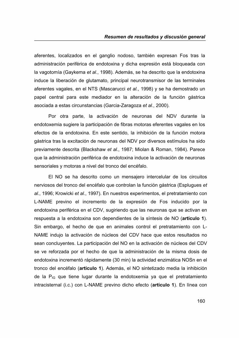
Resumen de resultados y discusión general
160
aferentes, localizados en el ganglio nodoso, también expresan Fos tras la
administración periférica de endotoxina y dicha expresión está bloqueada con
la vagotomía (Gaykema et al., 1998). Además, se ha descrito que la endotoxina
induce la liberación de glutamato, principal neurotransmisor de las terminales
aferentes vagales, en el NTS (Mascarucci et al., 1998) y se ha demostrado un
papel central para este mediador en la alteración de la función gástrica
asociada a estas circunstancias (Garcia-Zaragoza et al., 2000).
Por otra parte, la activación de neuronas del NDV durante la
endotoxemia sugiere la participación de fibras motoras eferentes vagales en los
efectos de la endotoxina. En este sentido, la inhibición de la función motora
gástrica tras la excitación de neuronas del NDV por diversos estímulos ha sido
previamente descrita (Blackshaw et al., 1987; Miolan & Roman, 1984). Parece
que la administración periférica de endotoxina induce la activación de neuronas
sensoriales y motoras a nivel del tronco del encéfalo.
El NO se ha descrito como un mensajero intercelular de los circuitos
nerviosos del tronco del encéfalo que controlan la función gástrica (Esplugues et
al., 1996; Krowicki et al., 1997). En nuestros experimentos, el pretratamiento con
L-NAME previno el incremento de la expresión de Fos inducido por la
endotoxina periférica en el CDV, sugiriendo que las neuronas que se activan en
respuesta a la endotoxina son dependientes de la síntesis de NO (artículo 1).
Sin embargo, el hecho de que en animales control el pretratamiento con L-
NAME indujo la activación de núcleos del CDV hace que estos resultados no
sean concluyentes. La participación del NO en la activación de núcleos del CDV
se ve reforzada por el hecho de que la administración de la misma dosis de
endotoxina incrementó rápidamente (30 min) la actividad enzimática NOSn en el
tronco del encéfalo (artículo 1). Además, el NO sintetizado media la inhibición
de la PIG que tiene lugar durante la endotoxemia ya que el pretratamiento
intracisternal (i.c.) con L-NAME previno dicho efecto (artículo 1). En línea con
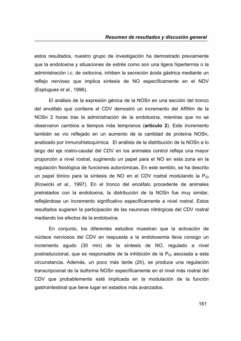
Resumen de resultados y discusión general
161
estos resultados, nuestro grupo de investigación ha demostrado previamente
que la endotoxina y situaciones de estrés como son una ligera hipertermia o la
administración i.c. de oxitocina, inhiben la secreción ácida gástrica mediante un
reflejo nervioso que implica síntesis de NO específicamente en el NDV
(Esplugues et al., 1996).
El análisis de la expresión génica de la NOSn en una sección del tronco
del encéfalo que contiene el CDV demostró un incremento del ARNm de la
NOSn 2 horas tras la administración de la endotoxina, mientras que no se
observaron cambios a tiempos más tempranos (artículo 2). Este incremento
también se vio reflejado en un aumento de la cantidad de proteína NOSn,
analizado por inmunohistoquímica. El análisis de la distribución de la NOSn a lo
largo del eje rostro-caudal del CDV en los animales control refleja una mayor
proporción a nivel rostral, sugiriendo un papel para el NO en esta zona en la
regulación fisiológica de funciones autonómicas. En este sentido, se ha descrito
un papel tónico para la síntesis de NO en el CDV rostral modulando la PIG
(Krowicki et al., 1997). En el tronco del encéfalo procedente de animales
pretratados con la endotoxina, la distribución de la NOSn fue muy similar,
reflejándose un incremento significativo específicamente a nivel rostral. Estos
resultados sugieren la participación de las neuronas nitrérgicas del CDV rostral
mediando los efectos de la endotoxina.
En conjunto, los diferentes estudios muestran que la activación de
núcleos nerviosos del CDV en respuesta a la endotoxemia lleva consigo un
incremento agudo (30 min) de la síntesis de NO, regulado a nivel
postraduccional, que es responsable de la inhibición de la PIG asociada a esta
circunstancia. Además, un poco más tarde (2h), se produce una regulación
transcripcional de la isoforma NOSn específicamente en el nivel más rostral del
CDV que probablemente esté implicada en la modulación de la función
gastrointestinal que tiene lugar en estadios más avanzados.
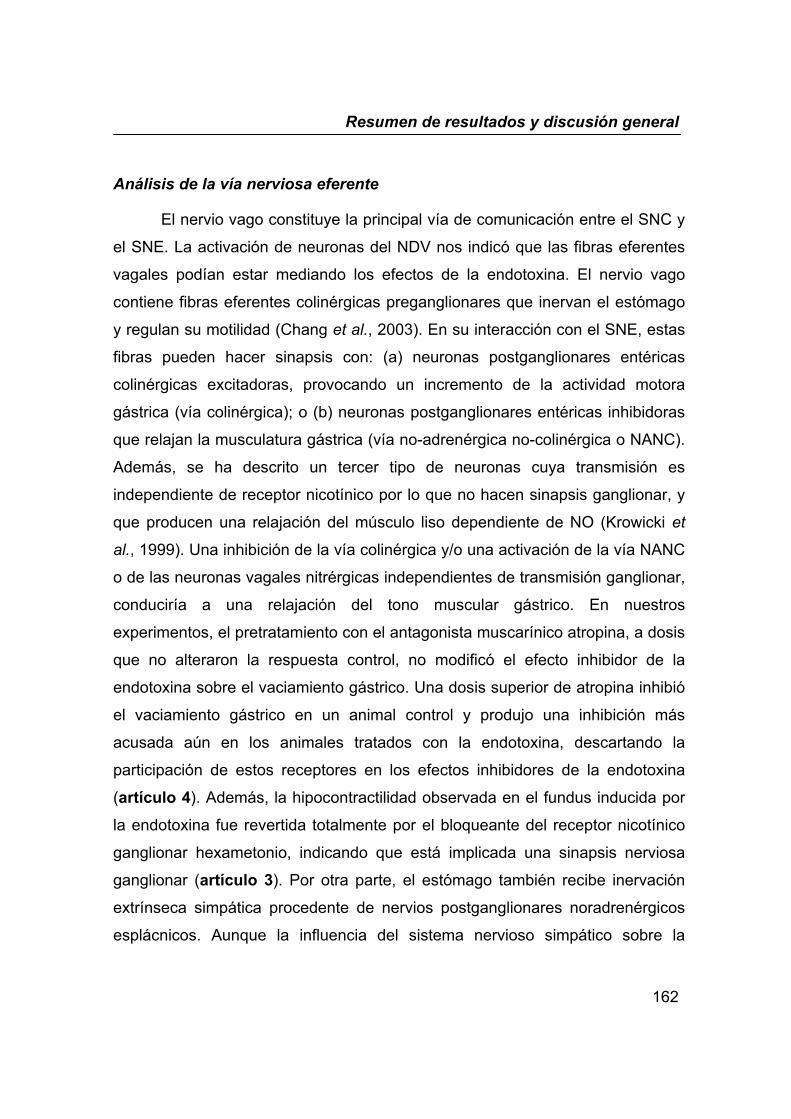
Resumen de resultados y discusión general
162
Análisis de la vía nerviosa eferente
El nervio vago constituye la principal vía de comunicación entre el SNC y
el SNE. La activación de neuronas del NDV nos indicó que las fibras eferentes
vagales podían estar mediando los efectos de la endotoxina. El nervio vago
contiene fibras eferentes colinérgicas preganglionares que inervan el estómago
y regulan su motilidad (Chang et al., 2003). En su interacción con el SNE, estas
fibras pueden hacer sinapsis con: (a) neuronas postganglionares entéricas
colinérgicas excitadoras, provocando un incremento de la actividad motora
gástrica (vía colinérgica); o (b) neuronas postganglionares entéricas inhibidoras
que relajan la musculatura gástrica (vía no-adrenérgica no-colinérgica o NANC).
Además, se ha descrito un tercer tipo de neuronas cuya transmisión es
independiente de receptor nicotínico por lo que no hacen sinapsis ganglionar, y
que producen una relajación del músculo liso dependiente de NO (Krowicki et
al., 1999). Una inhibición de la vía colinérgica y/o una activación de la vía NANC
o de las neuronas vagales nitrérgicas independientes de transmisión ganglionar,
conduciría a una relajación del tono muscular gástrico. En nuestros
experimentos, el pretratamiento con el antagonista muscarínico atropina, a dosis
que no alteraron la respuesta control, no modificó el efecto inhibidor de la
endotoxina sobre el vaciamiento gástrico. Una dosis superior de atropina inhibió
el vaciamiento gástrico en un animal control y produjo una inhibición más
acusada aún en los animales tratados con la endotoxina, descartando la
participación de estos receptores en los efectos inhibidores de la endotoxina
(artículo 4). Además, la hipocontractilidad observada en el fundus inducida por
la endotoxina fue revertida totalmente por el bloqueante del receptor nicotínico
ganglionar hexametonio, indicando que está implicada una sinapsis nerviosa
ganglionar (artículo 3). Por otra parte, el estómago también recibe inervación
extrínseca simpática procedente de nervios postganglionares noradrenérgicos
esplácnicos. Aunque la influencia del sistema nervioso simpático sobre la
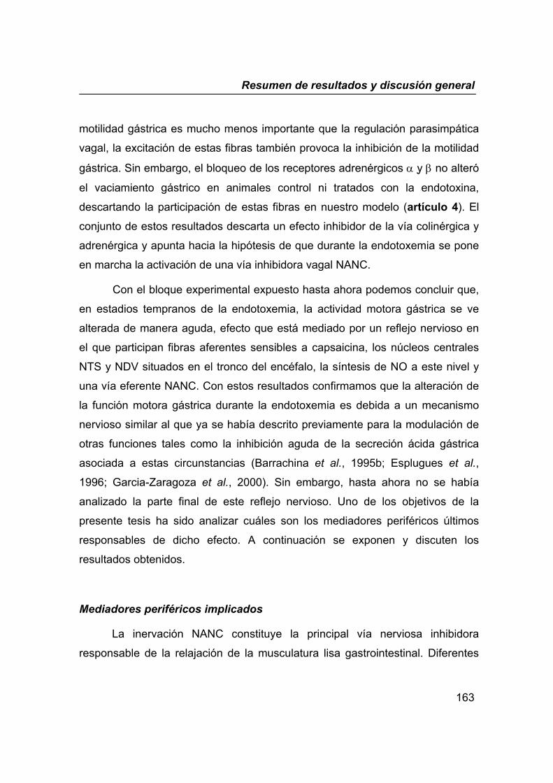
Resumen de resultados y discusión general
163
motilidad gástrica es mucho menos importante que la regulación parasimpática
vagal, la excitación de estas fibras también provoca la inhibición de la motilidad
gástrica. Sin embargo, el bloqueo de los receptores adrenérgicos α y β no alteró
el vaciamiento gástrico en animales control ni tratados con la endotoxina,
descartando la participación de estas fibras en nuestro modelo (artículo 4). El
conjunto de estos resultados descarta un efecto inhibidor de la vía colinérgica y
adrenérgica y apunta hacia la hipótesis de que durante la endotoxemia se pone
en marcha la activación de una vía inhibidora vagal NANC.
Con el bloque experimental expuesto hasta ahora podemos concluir que,
en estadios tempranos de la endotoxemia, la actividad motora gástrica se ve
alterada de manera aguda, efecto que está mediado por un reflejo nervioso en
el que participan fibras aferentes sensibles a capsaicina, los núcleos centrales
NTS y NDV situados en el tronco del encéfalo, la síntesis de NO a este nivel y
una vía eferente NANC. Con estos resultados confirmamos que la alteración de
la función motora gástrica durante la endotoxemia es debida a un mecanismo
nervioso similar al que ya se había descrito previamente para la modulación de
otras funciones tales como la inhibición aguda de la secreción ácida gástrica
asociada a estas circunstancias (Barrachina et al., 1995b; Esplugues et al.,
1996; Garcia-Zaragoza et al., 2000). Sin embargo, hasta ahora no se había
analizado la parte final de este reflejo nervioso. Uno de los objetivos de la
presente tesis ha sido analizar cuáles son los mediadores periféricos últimos
responsables de dicho efecto. A continuación se exponen y discuten los
resultados obtenidos.
Mediadores periféricos implicados
La inervación NANC constituye la principal vía nerviosa inhibidora
responsable de la relajación de la musculatura lisa gastrointestinal. Diferentes
Resumen de resultados y discusión general
164
estudios farmacológicos han demostrado que NO, VIP y ATP son los
mediadores finales de esta vía (Curro & Preziosi, 1998). En nuestros
experimentos in vivo, la administración intravenosa de L-NAME previno de
manera significativa la disminución de la PIG asociada a la endotoxemia,
sugiriendo por tanto que la síntesis periférica de NO está mediando dicho efecto
(artículo 1). Sin embargo, estos resultados no son concluyentes dado que el L-
NAME es capaz de atravesar la BHE inhibiendo así la síntesis de NO central,
que como ya hemos demostrado previamente, está involucrada. Con la finalidad
de analizar de manera aislada la parte final del reflejo nervioso, diseñamos un
protocolo experimental que combina el tratamiento in vivo con la endotoxina
durante 30 min, periodo en el cual se desencadena el reflejo nervioso, con el
análisis posterior de la capacidad contráctil del tejido aislado in vitro, mediante la
técnica de baño de órganos (artículo 3). De esta manera pudimos comprobar
que la hipocontractilidad fúndica derivada del pretratamiento con la endotoxina
fue revertida tras la incubación del tejido con la tetrodotoxina (TTX) o el
hexametonio. Además, cuando se incubó la endotoxina in vitro sobre el fundus
aislado de animales sin tratar no se vio afectada su capacidad contráctil,
descartando un efecto directo de la endotoxina sobre el territorio gástrico. El
conjunto de estos resultados demuestra la participación de una regulación
nerviosa extrínseca en nuestro modelo. Por otra parte, la inhibición local de la
síntesis de NO mediante la incubación con L-NOARG, o con TRIM (inhibidor
selectivo de la NOSn) revirtió por completo la hipotonicidad del fundus inducida
por la endotoxina. Así, obtuvimos la primera evidencia directa que demuestra
que la síntesis de NO en el fundus media los efectos de la endotoxina. El
bloqueo de los receptores del VIP o del ATP no modificó la respuesta contráctil
en ningún caso, descartando por tanto la participación de estos dos mediadores
en estas circunstancias.
Resumen de resultados y discusión general
165
El análisis de la cantidad de ARNm de NOSn en el fundus (1h tras el
tratamiento) nos indicó que la endotoxina no modifica la expresión génica de
esta isoenzima (artículo 3). Este resultado indica que la síntesis de NO inducida
por la endotoxina se debe a una regulación postranscripcional, hecho que no
sorprende dada la rapidez con la que se observan los cambios en la motilidad.
Con el afán de demostrar la síntesis de NO de una manera directa y de
analizar el origen celular empleamos la microscopía confocal, la cual nos
permitió visualizar en el tejido íntegro dicha síntesis gracias al empleo del DAF-
FM, un fluorocromo “vital” específico del NO (Nagano & Yoshimura, 2002)
(artículo 3). Este sistema permite observar directamente la síntesis intracelular
del NO in situ y en tiempo real proporcionando así una valiosa información de la
distribución espacial y temporal del NO en el organismo. Así, observamos que la
fluorescencia del DAF-FM, principalmente presente en células nerviosas, se vio
incrementada en el fundus de los animales pretratados in vivo con la
endotoxina. Además, dicho incremento fue revertido tras la incubación con L-
NAME, TTX y hexametonio. Estos resultados demuestran en su conjunto que la
endotoxemia induce la síntesis de NO en neuronas postganglionares del plexo
mientérico del fundus, y refuerzan los resultados funcionales que demuestran un
papel para dicha síntesis modulando el tono del fundus gástrico.
La principal diana del NO involucrada en la relajación del músculo liso
descrita hasta la fecha es la enzima GCs (Lefebvre, 1998; Moncada et al.,
1991). Además, los canales de potasio dependientes de calcio (canales CaK+)
también han sido relacionados con la relajación muscular inducida por el NO
(Kitamura et al., 1993; Shuttleworth & Sanders, 1996). En nuestros
experimentos (artículo 3) hemos comprobado que la relajación del fundus
inducida tanto por el NO exógeno (DETA-NO), como por el NO liberado
endógenamente (nicotina) fue revertida al bloquear la actividad enzimática de la
GCs con ODQ y al bloquear los canales CaK+ de baja conductancia con

Resumen de resultados y discusión general
166
apamina. Además, la hipocontractilidad del fundus inducida por la endotoxina
fue revertida también con el ODQ y con la apamina. Estos resultados
demuestran que el NO liberado durante la endotoxemia relaja el fundus gástrico
a través de la enzima GCs y de la hiperpolarización inducida por la apertura de
los canales CaK+ de baja conductancia. Actualmente en la literatura encontramos
discrepancias sobre si el NO activa directamente estos canales (Bolotina et al.,
1994; Serio et al., 2003) o si es una acción mediada por el propio GMPc
(Robertson et al., 1993). En este sentido, nosotros demostramos que la vía
GCs/canales CaK+ de baja conductancia media la relajación del fundus ya que la
relajación inducida por un activador directo de la enzima GCs es revertida por la
apamina. En base a estos resultados proponemos que la endotoxina relaja el
fundus gástrico mediante la vía NO/GMPc/canales CaK+; sin embargo, no
podemos descartar una acción directa del NO sobre estos canales.
Todos estos resultados sugieren que la administración de dosis bajas de
endotoxina activan un reflejo nervioso vagal en el que participa la síntesis
neuronal de NO en el tronco del encéfalo y en el plexo mientérico y como
consecuencia se induce rápidamente la relajación del fundus gástrico. Es
posible que este mecanismo pueda estar involucrado en el retraso del
vaciamiento gástrico que se observa en estas circunstancias. En la literatura se
ha descrito un papel inhibidor para el NO sobre el vaciamiento gástrico a través
de la relajación del fundus (Takakura et al., 1997).
El vaciamiento gástrico de sólidos nutrientes es una función compleja
regulada por la actuación integrada de la parte proximal (fundus) y distal (antro-
píloro) del estómago y del duodeno (Mayer, 1994). En el estómago, el tono de
la parte proximal y distal regulan de manera inversa el vaciamiento siendo el
tono del esfínter pilórico el último responsable que permite la salida del
contenido gástrico. Mediante técnicas de inmunohistoquímica se ha
demostrado una elevada densidad enzimática NOS en este esfínter (Feher et
Resumen de resultados y discusión general
167
al., 2001; Ward et al., 1994). Además, la síntesis de NO se ha asociado con
una aceleración del vaciamiento gástrico principalmente debido a la relajación
del píloro (Allescher & Daniel, 1994; Orihata & Sarna, 1994; Plourde et al.,
1994). En nuestros experimentos (artículo 5), el tratamiento con L-NAME o 7-
NI (inhibidor selectivo de la NOSn) inhibió de manera significativa el
vaciamiento de sólidos en animales control, reforzando un papel fisiológico
para la enzima NOSn en el control de esta función. Sin embargo, dicho
tratamiento no produjo una inhibición más acusada del vaciamiento en los
animales tratados con la endotoxina. Podríamos decir que la inhibición de la
síntesis de NO en condiciones fisiológicas mimetiza el efecto de la
endotoxemia sobre el vaciamiento gástrico. El análisis de la expresión génica,
cantidad proteica y actividad de la enzima NOSn en la región antro-pilórica
demostró que 4h tras el tratamiento con la endotoxina, tiempo en el cual está
inhibido el vaciamiento gástrico, se produce una disminución transcripcional de
la NOSn con la consecuente disminución de su actividad en esta región. La
inervación NANC juega un papel muy importante mediando la relajación del
píloro. Nosotros pensamos que dicha disminución de la NOSn en el píloro
puede estar causando una relajación NANC atenuada del esfínter con el
consiguiente incremento del tono, impidiendo así el vaciamiento del contenido
gástrico.
Resulta interesante la reacción del organismo mediante la cual está
modulando la síntesis del NO de manera diferencial dependiendo de la región
del estómago (incremento en la parte proximal y disminución en la parte distal),
retrasando así el vaciamiento gástrico, probablemente con la finalidad de
prevenir la absorción de toxinas.
Como se ha demostrado hasta ahora por distintas vías, la isoforma
implicada en todos estos efectos es la NOSn. Está ampliamente descrito que la
endotoxina incrementa la expresión de la NOSi (Barrachina et al., 2001;

Resumen de resultados y discusión general
168
Moncada et al., 1991) y se le ha atribuido un papel modulador de la motilidad a
esta isoforma (Eskandari et al., 1999; Takakura et al., 1997). Sin embargo, este
hecho está asociado a tiempos más largos y a dosis de endotoxina superiores
a la empleada en este estudio, generalmente relacionadas con un shock
endotóxico (Thiemermann, 1997). En nuestro modelo el análisis de la expresión
de la NOSi en el estómago 1h tras la administración de la endotoxina, tiempo
en el que la PIG y el tono del fundus están inhibidos, demuestra que esta
isoforma no está incrementada (artículos 1,3). Además, el pretratamiento con
la dexametasona no altera dicho efecto, hecho que nos indica que no existe
síntesis proteica (artículo 3). Por otra parte, el análisis de la expresión de la
NOSi en la región antro-pilórica 4h tras el tratamiento con la endotoxina, tiempo
en el cual observamos la inhibición del vaciamiento gástrico, sí revela un
incremento de la cantidad de ARNm de esta isoenzima (artículo 5). Sin
embargo, el análisis de la actividad enzimática NOSi no demuestra síntesis de
NO derivada de esta isoforma. Además, el tratamiento con un inhibidor
selectivo de la NOSi (L-NIL) no impidió el efecto de la endotoxina sobre el
vaciamiento gástrico. Estos resultados demuestran que 4h tras el tratamiento
con la endotoxina, la inducción de la NOS comienza a tener lugar, sin embargo
la síntesis de NO derivada todavía no existe y por lo tanto no se le puede
atribuir ningún papel funcional en la inhibición de la motilidad gástrica
observada en este momento.
Además del NO, las prostaglandinas (PGs) también actúan modulando de
manera importante la función motora gástrica (Sanders, 1984). La síntesis
endógena (Suto et al., 1994) o la administración exógena (Stein et al., 1994) de
PGs se ha asociado con la inhibición del vaciamiento gástrico. En nuestros
experimentos (artículo 5), la inhibición de la ciclooxigenasa (COX) con
indometacina no alteró el vaciamiento gástrico en los animales control
descartando un papel regulador de las PGs en condiciones fisiológicas. Sin
Resumen de resultados y discusión general
169
embargo, este tratamiento impidió la inhibición del vaciamiento gástrico
asociado a la endotoxemia indicando por tanto un papel para las PGs en estas
circunstancias. Además, dicha síntesis parece consecuencia de una inducción
de la isoforma COX-2 ya que el tratamiento con el inhibidor selectivo de esta
isoforma (NS-398) o con la dexametasona también previno el efecto de la
endotoxina.
II. EFECTOS DE LA ENDOTOXINA SOBRE LA MOTILIDAD COLÓNICA
Además de las alteraciones en la motilidad gástrica, la endotoxemia
también se caracteriza por una alteración de la función motora intestinal.
Estudios previos en nuestro laboratorio demuestran un incremento del tránsito
intestinal en estas circunstancias (Martinez-Cuesta et al., 1997). El presente
estudio amplía dichos resultados y demuestra que la endotoxina incrementa de
manera aguda (en las tres primeras horas) la excreción fecal (artículo 6).
Durante este proceso no se observó un cambio en la consistencia de las heces
lo que sugiere que el efecto de la endotoxina es independiente de alteraciones
en la secreción intestinal, hecho que se observa en los procesos diarreicos
característicos de una endotoxemia más severa (Closs et al., 1998; Pons et al.,
1994).
Nuestros experimentos demuestran que el efecto procinético inducido por la
endotoxina es consecuencia, al igual que en el caso anterior, de un reflejo
nervioso vagal en el que las neuronas aferentes sensibles a la capsaicina están
implicadas ya que el tratamiento con esta neurotoxina, así como una vagotomía
abdominal, revirtieron tal efecto.
El avance del bolo fecal está regulado por el reflejo peristáltico el cual se
compone de dos fases secuenciales: la relajación descendente caudal y

Resumen de resultados y discusión general
170
contracción ascendente oral. La coordinación de estas dos fases determinará la
velocidad del tránsito intestinal. En circunstancias fisiológicas, los
neurotransmisores excitadores responsables de la contracción ascendente son
principalmente Ach y SP mientras que el NO está implicado en la relajación
descendente. Nuestros resultados demuestran que durante la endotoxemia, la
liberación de Ach, SP y NO están mediando la aceleración de la excreción fecal.
La mayoría de los estudios realizados en este campo demuestran un papel
para la síntesis de NO derivada de la NOSi mediando la diarrea asociada a una
endotoxemia generalmente más severa que la que presentamos nosotros. Sin
embargo, aunque no hemos analizado en este caso la isoforma de la NOS
involucrada, las dosis bajas de endotoxina que usamos y el mecanismo nervioso
rápido que hemos demostrado está teniendo lugar, nos hace pensar
principalmente en una regulación postraduccional de la isoforma NOSn una vez
más.
Estos resultados sugieren que durante la endotoxemia se produce un
incremento agudo de la excreción fecal mediado por un reflejo nervioso en el
que neuronas aferentes sensibles a capsaicina y liberación de
neurotransmisores excitadores (Ach y SP) e inhibidores (NO) están
involucradas. Estudios adicionales harían falta para caracterizar la vía nerviosa
implicada. Nosotros proponemos que la liberación en la pared colónica de Ach y
SP promueve el efecto propulsor de la contracción ascendente, mientras que la
síntesis local de NO permite el avance del bolo fecal al mediar la relajación
descendente.
Resumen de resultados y discusión general
171
Recopilando toda la información reflejada en esta tesis podemos concluir
que durante una invasión bacteriana el organismo reacciona rápidamente
modulando la función motora gastrointestinal con la finalidad de prevenir la
absorción futura de agentes extraños. Así, una hipocontractilidad del estómago
proximal asociado a un enlentecimiento del vaciamiento gástrico prevendría el
acceso de posibles toxinas al duodeno, retrasando de esta manera su posible
absorción; en la misma línea, una aceleración de la excreción fecal facilitaría su
pronta expulsión. Esta rápida respuesta es posible gracias a la interacción y
perfecta sincronización de los sistemas nerviosos central y periférico. Así, el
SNC recibe la señal de “alerta” en la periferia gracias a las neuronas aferentes
sensibles a la capsaicina, rápidamente procesa esta información, principalmente
en el CDV, y envía el mensaje “ejecutor” a través de las neuronas eferentes
vagales que están en íntimo contacto con el SNE. De esta manera modula no
sólo la función motora gastrointestinal, sino que también desencadena una
inhibición de la secreción ácida gástrica, incrementa el flujo sanguíneo mucoso y
protege la mucosa gástrica frente a agresiones externas (artículo 7). Todas
estas acciones se pueden considerar como una reacción general de defensa
que pone en alerta al organismo, intenta mantener la homeostasis y le prepara
para una posible agresión posterior.
Además, hemos observado que la regulación postraduccional de la NOSn
tiene un papel protagonista en todo este proceso. Un incremento de la síntesis
de NO en el tronco del encéfalo, en neuronas postganglionares del plexo
mientérico del fundus gástrico y en la pared colónica sucede en los primeros
estadios de la endotoxemia. Pasado el efecto agudo de la endotoxina,
empezaría a observarse lo que se considera parte de una respuesta inmune
generalizada mediada por la expresión de las enzimas NOSi y COX-2. En
respuesta a una invasión bacteriana, parece que el organismo maximiza la
actividad de sus recursos fisiológicos mientras se alcanza la plena actividad del
sistema inmune.
Conclusiones
172
CONCLUSIONES
Conclusiones
173
I. Tras una invasión bacteriana se observan cambios agudos en la
función motora gastrointestinal caracterizados por un enlentecimiento
del vaciamiento gástrico y una aceleración de la excreción fecal. Este
comportamiento constituye un mecanismo de respuesta temprana
cuya finalidad podría ser evitar la absorción de toxinas y facilitar su
expulsión.
II. Estas alteraciones se producen rápidamente gracias a la actuación
del sistema nervioso, tanto central como periférico, mediante la
activación secuencial de fibras sensoriales aferentes, núcleos
centrales del complejo dorsal vagal, fibras eferentes vagales y
neuronas del plexo mientérico.
III. En este proceso, la síntesis aguda de óxido nítrico de origen
nervioso juega un papel esencial como neurotransmisor en el tronco
del encéfalo, y como molécula efectora en la unión neuro-muscular
en el territorio gastrointestinal.
Bibiografía
174
BIBLIOGRAFÍA
Bibiografía
175
AIMI, Y., KIMURA, H., KINOSHITA, T., MINAMI, Y., FUJIMURA, M. &
VINCENT, S.R. (1993). Histochemical localization of nitric oxide
synthase in rat enteric nervous system. Neuroscience 53, 553-560.
ALDERTON, W.K., COOPER, C.E. & KNOWLES, R.G. (2001). Nitric oxide
synthases: structure, function and inhibition. Biochem. J. 357, 593-615.
ALLESCHER, H.D., DANIEL, E.E. (1994). Role of NO in pyloric, antral, and
duodenal motility and its interaction with other inhibitory mediators. Dig.
Dis. Sci. 39, 73S-75S.
ALLESCHER, H.D., TOUGAS, G., VERGARA, P., LU, S. & DANIEL, E.E.
(1992). Nitric oxide as a putative nonadrenergic noncholinergic inhibitory
transmitter in the canine pylorus in vivo. Am. J. Physiol. 262, G695-
G702
ANDERSSON, K.E. (2003). Erectile physiological and pathophysiological
pathways involved in erectile dysfunction. J. Urol. 170, S6-13.
ANIKA, S.M., HOUPT, T.R. & HOUPT, K.A. (1977). Satiety elicited by
cholecystokinin in intact and vagotomized rats. Physiol. Behav. 19, 761-
766.
AZPIROZ, F., MALAGELADA, J.R. (1987). Importance of vagal input in
maintaining gastric tone in the dog. J. Physiol. 384, 511-524.
BARRACHINA, D., CALATAYUD, S., MORENO, L., MARTINEZ-CUESTA, A.,
WHITTLE, B.J. & ESPLUGUES, J.V. (1995a). Nitric oxide and sensory
afferent neurones modulate the protective effects of low-dose endotoxin
on rat gastric mucosal damage. Eur. J. Pharmacol. 280, 339-342.
Bibiografía
176
BARRACHINA, M.D., PANES, J. & ESPLUGUES, J.V. (2001). Role of nitric
oxide in gastrointestinal inflammatory and ulcerative diseases:
perspective for drugs development. Curr. Pharm. Des. 7, 31-48.
BARRACHINA, M.D., WHITTLE, B.J., MONCADA, S. & ESPLUGUES, J.V.
(1995b). Endotoxin inhibition of distension-stimulated gastric acid
secretion in rat: mediation by NO in the central nervous system. Br. J.
Pharmacol. 114, 8-12.
BECKMAN, J.S., KOPPENOL, W.H. (1996). Nitric oxide, superoxide, and
peroxynitrite: the good, the bad, and ugly. Am. J. Physiol. 271, C1424-
C1437
BELTRAN, B., BARRACHINA, M.D., MENDEZ, A., QUINTERO, E. &
ESPLUGUES, J.V. (1999). Synthesis of nitric oxide in the dorsal motor
nucleus of the vagus mediates the inhibition of gastric acid secretion by
central bombesin. Br. J. Pharmacol. 127, 1603-1610.
BELVISI, M.G., WARD, J.K., MITCHELL, J.A. & BARNES, P.J. (1995). Nitric
oxide as a neurotransmitter in human airways. Arch. Int. Pharmacodyn.
Ther. 329, 97-110.
BEUTLER, B., RIETSCHEL, E.T. (2003). Innate immune sensing and its roots:
the story of endotoxin. Nat. Rev. Immunol. 3, 169-176.
BLACKSHAW, L.A., GRUNDY, D. & SCRATCHERD, T. (1987). Involvement of
gastrointestinal mechano- and intestinal chemoreceptors in vagal
reflexes: an electrophysiological study. J. Auton. Nerv. Syst. 18, 225-
234.
Bibiografía
177
BLATTEIS, C.M., LI, S., LI, Z., PERLIK, V. & FELEDER, C. (2004). Signaling
the brain in systemic inflammation: the role of complement. Front.
Biosci. 9, 915-931.
BLATTEIS, C.M., SEHIC, E. & LI, S. (2000). Pyrogen sensing and signaling: old
views and new concepts. Clin. Infect. Dis. 31 Suppl 5, S168-S177
BLUTHE, R.M., WALTER, V., PARNET, P., LAYE, S., LESTAGE, J., VERRIER,
D., POOLE, S., STENNING, B.E., KELLEY, K.W. & DANTZER, R.
(1994). Lipopolysaccharide induces sickness behaviour in rats by a vagal
mediated mechanism. C. R. Acad. Sci. III 317, 499-503.
BOECKXSTAENS GE, PELCKMANS PA (1997). Nitric oxide and the non-
adrenergic non-cholinergic neurotransmission. Comp. Biochem. Physiol.
A. Physiol. 118, 925-937.
BOECKXSTAENS, G.E., PELCKMANS, P.A., RAMPART, M., VERBEUREN,
T.J., HERMAN, A.G. & VAN MAERCKE, Y.M. (1990). Nonadrenergic
noncholinergic mechanisms in the ileocolonic junction. Arch. Int.
Pharmacodyn. Ther. 303, 270-281.
BOJE, K.M. (2004). Nitric oxide neurotoxicity in neurodegenerative diseases.
Front. Biosci. 9, 763-776.
BOLOTINA, V.M., NAJIBI, S., PALACINO, J.J., PAGANO, P.J. & COHEN, R.A.
(1994). Nitric oxide directly activates calcium-dependent potassium
channels in vascular smooth muscle. Nature 368, 850-853.
BROWN, G.C. (2000). Nitric oxide as a competitive inhibitor of oxygen
consumption in the mitochondrial respiratory chain. Acta. Physiol.
Scand. 168, 667-674.
Bibiografía
178
BURNSTOCK, G. (1986). The non-adrenergic non-cholinergic nervous system.
Arch. Int. Pharmacodyn. Ther. 280, 1-15.
BURNSTOCK, G., CAMPBELL, G., BENNETT, M. & HOLMAN, M.E. (1963).
Inhibition of the smooth muscle on the taenia coli. Nature 200, 581-582.
CALATAYUD, S., CANET, A., BELLO, R., HERNANDEZ, C., MARTI, M. &
BARRACHINA, M.D. (2003). Low endotoxemia prevents the reduction of
gastric blood flow induced by NSAIDs: role of nitric oxide. Br. J.
Pharmacol. 139, 263-270.
CALATAYUD, S., QUINTANA, E., ESPLUGUES, J. & BARRACHINA, M.D.
(1999). Role of central oxytocin in the inhibition by endotoxin of
distension-stimulated gastric acid secretion. Naunyn Schmiedebergs
Arch. Pharmacol. 360, 676-682.
CARVAJAL, J.A., GERMAIN, A.M., HUIDOBRO-TORO, J.P. & WEINER, C.P.
(2000). Molecular mechanism of cGMP-mediated smooth muscle
relaxation. J. Cell. Physiol. 184, 409-420.
CHAKDER, S., RATTAN, S. (1993). Release of nitric oxide by activation of
nonadrenergic noncholinergic neurons of internal anal sphincter. Am. J.
Physiol. 264, G7-12.
CHANG, H.Y., MASHIMO, H. & GOYAL, R.K. (2003). Musings on the wanderer:
what's new in our understanding of vago-vagal reflex? IV. Current
concepts of vagal efferent projections to the gut. Am.J.Physiol
Gastrointest.Liver Physiol 284, G357-G366
CLOSS, E.I., ENSELEIT, F., KOESLING, D., PFEILSCHIFTER, J.M.,
SCHWARZ, P.M. & FORSTERMANN, U. (1998). Coexpression of
inducible NO synthase and soluble guanylyl cyclase in colonic
Bibiografía
179
enterocytes: a pathophysiologic signaling pathway for the initiation of
diarrhea by gram-negative bacteria? FASEB J. 12, 1643-1649.
CUDEIRO, J., RIVADULLA, C. (1999). Sight and insight--on the physiological
role of nitric oxide in the visual system. Trends Neurosci. 22, 109-116.
CURRO, D., PREZIOSI, P. (1998). Non-adrenergic non-cholinergic relaxation of
the rat stomach. Gen. Pharmacol. 31, 697-703.
DANIEL, E.E., BEREZIN, I., ALLESCHER, H.D., MANAKA, H. & POSEY-
DANIEL, V. (1989). Morphology of the canine pyloric sphincter in relation
to function. Can.J. Physiol. Pharmacol. 67, 1560-1573.
DE GROAT, W.C., YOSHIMURA, N. (2001). Pharmacology of the lower urinary
tract. Annu. Rev. Pharmacol. Toxicol. 41, 691-721.
DESAI, K.M., SESSA, W.C. & VANE, J.R. (1991a). Involvement of nitric oxide
in the reflex relaxation of the stomach to accommodate food or fluid.
Nature 351, 477-479.
DESAI, K.M., ZEMBOWICZ, A., SESSA, W.C. & VANE, J.R. (1991b).
Nitroxergic nerves mediate vagally induced relaxation in the isolated
stomach of the guinea pig. Proc. Natl. Acad. Sci. U.S.A 88, 11490-
11494.
DICKENS, E.J., EDWARDS, F.R. & HIRST, G.D. (2000). Vagal inhibition in the
antral region of guinea pig stomach. Am. J. Physiol. Gastrointest. Liver.
Physiol. 279, G388-G399
EKBLAD, E., ALM, P. & SUNDLER, F. (1994). Distribution, origin and
projections of nitric oxide synthase-containing neurons in gut and
pancreas. Neuroscience 63, 233-248.
Bibiografía
180
ESKANDARI, M.K., KALFF, J.C., BILLIAR, T.R., LEE, K.K. & BAUER, A.J.
(1999). LPS-induced muscularis macrophage nitric oxide suppresses rat
jejunal circular muscle activity. Am. J. Physiol. 277, G478-G486
ESPLUGUES, J.V. (2002). NO as a signalling molecule in the nervous system.
Br. J. Pharmacol. 135, 1079-1095.
ESPLUGUES, J.V., BARRACHINA, M.D., BELTRAN, B., CALATAYUD, S.,
WHITTLE, B.J. & MONCADA, S. (1996). Inhibition of gastric acid
secretion by stress: a protective reflex mediated by cerebral nitric oxide.
Proc. Natl. Acad. Sci. U.S.A 93, 14839-14844.
FEHER, E., ALTDORFER, K., DONATH, T. & FEHER, J. (2001).
Electronmicroscopical localization of nitric oxide synthase-containing
nerve elements in cat pylorus. Eur. J. Morphol. 39, 121-126.
FORSTERMANN, U., BOISSEL, J.P. & KLEINERT, H. (1998). Expressional
control of the 'constitutive' isoforms of nitric oxide synthase (NOS I and
NOS III). FASEB. J. 12, 773-790.
FOXX-ORENSTEIN, A.E., GRIDER, J.R. (1996). Regulation of colonic
propulsion by enteric excitatory and inhibitory neurotransmitters. Am. J.
Physiol. 271, G433-G437
GARCIA-ZARAGOZA, E., BARRACHINA, M.D., MORENO, L. & ESPLUGUES,
J.V. (2000). Role of central glutamate receptors, nitric oxide and soluble
guanylyl cyclase in the inhibition by endotoxin of rat gastric acid
secretion. Br. J. Pharmacol. 130, 1283-1288.
GAYKEMA, R.P., GOEHLER, L.E., TILDERS, F.J., BOL, J.G., MCGORRY, M.,
FLESHNER, M., MAIER, S.F. & WATKINS, L.R. (1998). Bacterial
Bibiografía
181
endotoxin induces fos immunoreactivity in primary afferent neurons of
the vagus nerve. Neuroimmunomodulation 5, 234-240.
GHAFOURIFAR, P., RICHTER, C. (1997). Nitric oxide synthase activity in
mitochondria. FEBS Letters 418, 291-296.
GIVALOIS, L., DORNAND, J., MEKAOUCHE, M., SOLIER, M.D., BRISTOW,
A.F., IXART, G., SIAUD, P., ASSENMACHER, I. & BARBANEL, G.
(1994). Temporal cascade of plasma level surges in ACTH,
corticosterone, and cytokines in endotoxin-challenged rats. Am. J.
Physiol. 267, R164-R170
GUO, F.H., DE RAEVE, H.R., RICE, T.W., STUEHR, D.J., THUNNISSEN, F.B.
& ERZURUM, S.C. (1995). Continuous nitric oxide synthesis by inducible
nitric oxide synthase in normal human airway epithelium in vivo. Proc.
Natl. Acad. Sci. U.S.A 92, 7809-7813.
HIRONAGA, K., HIROOKA, Y., MATSUO, I., SHIHARA, M., TAGAWA, T.,
HARASAWA, Y. & TAKESHITA, A. (1998). Role of Endogenous Nitric
Oxide in the Brain Stem on the Rapid AdaptatKion of Baroreflex.
Hypertension 31, 27-31.
HOLZER, P. (1988). Local effector functions of capsaicin-sensitive sensory
nerve endings: involvement of tachykinins, calcitonin gene-related
peptide and other neuropeptides. Neuroscience 24, 739-768.
HOLZER, P. (1991). Capsaicin: cellular targets, mechanisms of action, and
selectivity for thin sensory neurons. Pharmacol. Rev. 43, 143-201.
HOLZER, P. (1998). Neural emergency system in the stomach.
Gastroenterology 114, 823-839.
Bibiografía
182
HURLEY, J.C. (1995). Endotoxemia: methods of detection and clinical
correlates. Clin. Microbiol. Rev. 8, 268-292.
ISHIGUCHI, T., TAKAHASHI, T., ITOH, H. & OWYANG, C. (2000). Nitrergic
and purinergic regulation of the rat pylorus. Am. J. Physiol. Gastrointest.
Liver Physiol. 279, G740-G747
JANSKY, L., VYBIRAL, S., POSPISILOVA, D., ROTH, J., DORNAND, J.,
ZEISBERGER, E. & KAMINKOVA, J. (1995). Production of systemic and
hypothalamic cytokines during the early phase of endotoxin fever.
Neuroendocrinology 62, 55-61.
JONES, O.M., BRADING, A.F. & MORTENSEN, N.J. (2003). Role of nitric
oxide in anorectal function of normal and neuronal nitric oxide synthase
knockout mice: a novel approach to anorectal disease. Dis. Colon
Rectum. 46, 963-970.
KALIA, M., MESULAM, M.M. (1980). Brain stem projections of sensory and
motor components of the vagus complex in the cat: I. The cervical vagus
and nodose ganglion. J. Comp. Neurol. 193, 435-465.
KENDRICK, K.M., GUEVARA-GUZMAN, R., ZORRILLA, J., HINTON, M.R.,
BROAD, K.D., MIMMACK, M. & OHKURA, S. (1997). Formation of
olfactory memories mediated by nitric oxide. Nature 388, 670-674.
KITAMURA, K., LIAN, Q., CARL, A. & KURIYAMA, H. (1993). S-
nitrosocysteine, but not sodium nitroprusside, produces apamin-
sensitive hyperpolarization in rat gastric fundus. Br. J. Pharmacol. 109,
415-423.
KROWICKI, Z.K., SHARKEY, K.A., SERRON, S.C., NATHAN, N.A. &
HORNBY, P.J. (1997). Distribution of nitric oxide synthase in rat dorsal
Bibiografía
183
vagal complex and effects of microinjection of nitric oxide compounds
upon gastric motor function. J. Comp. Neurol. 377, 49-69.
KROWICKI, Z.K., SIVARAO, D.V., ABRAHAMS, T.P. & HORNBY, P.J. (1999).
Excitation of dorsal motor vagal neurons evokes non-nicotinic receptor-
mediated gastric relaxation. J. Auton. Nerv. Syst. 77, 83-89.
KUBES, P., SUZUKI, M. & GRANGER, D.N. (1991). Nitric oxide: an
endogenous modulator of leukocyte adhesion. Proc. Natl. Acad. Sci.
U.S.A 88, 4651-4655.
LEFEBVRE, R.A. (1998). Influence of a selective guanylate cyclase inhibitor,
and of the contraction level, on nitrergic relaxations in the gastric fundus.
Br. J. Pharmacol. 124, 1439-1448.
LI, S., SEHIC, E., WANG, Y., UNGAR, A.L. & BLATTEIS, C.M. (1999). Relation
between complement and the febrile response of guinea pigs to systemic
endotoxin. Am. J. Physiol. 277, R1635-R1645
LOPEZ-FIGUEROA, M.O., CAAMANO, C., MORANO, M.I., RONN, L.C., AKIL,
H. & WATSON, S.J. (2000). Direct evidence of nitric oxide presence
within mitochondria. Biochem. Biophys. Res. Commun. 272, 129-133.
LUO, Z.D., CIZKOVA, D. (2000). The role of nitric oxide in nociception. Curr.
Rev. Pain. 4, 459-466.
MAIER, S.F., GOEHLER, L.E., FLESHNER, M. & WATKINS, L.R. (1998). The
role of the vagus nerve in cytokine-to-brain communication. Ann. N.Y.
Acad. Sci. 840, 289-300.
MARTINEZ-CUESTA, M.A., BARRACHINA, M.D., BELTRAN, B., CALATAYUD,
S. & ESPLUGUES, J. (1997). Nitric oxide modulates the acute increase
Bibiografía
184
of gastrointestinal transit induced by endotoxin in rats: a possible role for
tachykinins. J. Pharm. Pharmacol. 49, 988-990.
MARTINEZ-CUESTA, M.A., BARRACHINA, M.D., WHITTLE, B.J., PIQUE, J.M.
& ESPLUGUES, J.V. (1994). Involvement of neuronal processes and
nitric oxide in the inhibition by endotoxin of pentagastrin-stimulated
gastric acid secretion. Naunyn Schmiedebergs Arch. Pharmacol. 349,
523-527.
MASCARUCCI, P., PEREGO, C., TERRAZZINO, S. & DE SIMONI, M.G.
(1998). Glutamate release in the nucleus tractus solitarius induced by
peripheral lipopolysaccharide and interleukin-1 beta. Neuroscience 86,
1285-1290.
MAYER E.A. (1994) The Physiology of Gastric Storage and Emptying. In
Physiology of the Gastrointestinal Tract. Third Edition Ed. L.R.Johnson.
New York: Raven Press. pp. 929-976.
MEARIN, F., MOURELLE, M., GUARNER, F., SALAS, A., RIVEROS-
MORENO, V., MONCADA, S. & MALAGELADA, J.R. (1993). Patients
with achalasia lack nitric oxide synthase in the gastro-oesophageal
junction. Eur. J. Clin. Invest. 23, 724-728.
MIOLAN, J.P., ROMAN, C. (1984). The role of oesophageal and intestinal
receptors in the control of gastric motility. J. Auton. Nerv. Syst. 10, 235-
241.
MONCADA, S., HIGGS, A. & FURCHGOTT, R. (1997). International Union of
Pharmacology Nomenclature in Nitric Oxide Research. Pharmacol.
Rev. 49, 137-142.
Bibiografía
185
MONCADA, S., PALMER, R.M. & HIGGS, E.A. (1991). Nitric oxide: physiology,
pathophysiology, and pharmacology. Pharmacol. Rev. 43, 109-142.
MORIMOTO, A., MURAKAMI, N., NAKAMORI, T. & WATANABE, T. (1987).
Evidence for separate mechanisms of induction of biphasic fever inside
and outside the blood-brain barrier in rabbits. J. Physiol. 383, 629-637.
MULLER, B.E.R.N., KLESCHYOV, A.L., ALENCAR, J.L., VANIN, A.N.A.T. &
STOCLET, J.C. (2002). Nitric Oxide Transport and Storage in the
Cardiovascular System. Ann. N.Y. Acad. Sci. 962, 131-139.
NAGANO, T., YOSHIMURA, T. (2002). Bioimaging of nitric oxide. Chem. Rev.
102, 1235-1270.
NISOLI, E., CLEMENTI, E., MONCADA, S. & CARRUBA, M.O. (2004).
Mitochondrial biogenesis as a cellular signaling framework. Biochem.
Pharmacol. 67, 1-15.
ORIHATA, M., SARNA, S.K. (1994). Inhibition of nitric oxide synthase delays
gastric emptying of solid meals. J. Pharmacol. Exp. Ther. 271, 660-670.
ORTEGA MATEO, A., AMAYA ALEIXANDRE DE ARTINANO, M. (2000). Nitric
oxide reactivity and mechanisms involved in its biological effects.
Pharmacol. Res. 42, 421-427.
PALMER, R.M., FERRIGE, A.G. & MONCADA, S. (1987). Nitric oxide release
accounts for the biological activity of endothelium-derived relaxing factor.
Nature 327, 524-526.
PATERSON, C.A., ANVARI, M., TOUGAS, G. & HUIZINGA, J.D. (2000).
Nitrergic and cholinergic vagal pathways involved in the regulation of
canine proximal gastric tone: an in vivo study. Neurogastroenterol. Motil.
12, 301-306.
Bibiografía
186
PLOURDE, V., QUINTERO, E., SUTO, G., COIMBRA, C. & TACHE, Y. (1994).
Delayed gastric emptying induced by inhibitors of nitric oxide synthase in
rats. Eur. J. Pharmacol. 256, 125-129.
PONS, L., DROY-LEFAIX, M.T. & BUENO, L. (1994). Role of platelet-activating
factor (PAF) and prostaglandins in colonic motor and secretory
disturbances induced by Escherichia coli endotoxin in conscious rats.
Prostaglandins 47, 123-136.
PRAST, H., PHILIPPU, A. (2001). Nitric oxide as modulator of neuronal
function. Progress in Neurobiology 64, 51-68.
RADOMSKI, M.W., PALMER, R.M. & MONCADA, S. (1987). Endogenous nitric
oxide inhibits human platelet adhesion to vascular endothelium. Lancet
2, 1057-1058.
ROBERTSON, B.E., SCHUBERT, R., HESCHELER, J. & NELSON, M.T.
(1993). cGMP-dependent protein kinase activates Ca-activated K
channels in cerebral artery smooth muscle cells. Am. J. Physiol 265,
C299-C303
ROGERS, R.C., MCTIGUE, D.M. & HERMANN, G.E. (1995). Vagovagal reflex
control of digestion: afferent modulation by neural and "endoneurocrine"
factors. Am. J. Physiol. 268, G1-10.
ROMANOVSKY, A.A. (2000). Thermoregulatory manifestations of systemic
inflammation: lessons from vagotomy. Auton. Neurosci. 85, 39-48.
ROMANOVSKY, A.A. (2004). Signaling the brain in the early sickness
syndrome: are sensory nerves involved? Front. Biosci. 9, 494-504.
ROTH, J., MARTIN, D., STORR, B. & ZEISBERGER, E. (1998). Neutralization
of pyrogen-induced tumour necrosis factor by its type 1 soluble receptor
Bibiografía
187
in guinea-pigs: effects on fever and interleukin-6 release. J. Physiol. 509 ( Pt 1), 267-275.
ROTHWELL, N.J., LUHESHI, G. (1994). Pharmacology of interleukin-1 actions
in the brain. Adv. Pharmacol. 25, 1-20.
SAKUMA, I., TOGASHI, H., YOSHIOKA, M., SAITO, H., YANAGIDA, M.,
TAMURA, M., KOBAYASHI, T., YASUDA, H., GROSS, S.S. & LEVI, R.
(1992). NG-methyl-L-arginine, an inhibitor of L-arginine-derived nitric
oxide synthesis, stimulates renal sympathetic nerve activity in vivo. A role
for nitric oxide in the central regulation of sympathetic tone? Circ. Res.
70, 607-611.
SALZMAN, A.L. (1995). Nitric oxide in the gut. New Horiz. 3, 33-45.
SANDERS, K.M. (1984). Role of prostaglandins in regulating gastric motility.
Am. J. Physiol. 247, G117-G126
SANDERS, K.M., WARD, S.M. (1992). Nitric oxide as a mediator of
nonadrenergic noncholinergic neurotransmission. Am. J. Physiol. 262,
G379-G392
SANGWAN, Y.P., SOLLA, J.A. (1998). Internal anal sphincter: advances and
insights. Dis. Colon Rectum 41, 1297-1311.
SCHOBITZ, B., DE KLOET, E.R. & HOLSBOER, F. (1994). Gene expression
and function of interleukin 1, interleukin 6 and tumor necrosis factor in
the brain. Prog. Neurobiol. 44, 397-432.
SERIO, R., GRAZIA ZIZZO, M. & MULE, F. (2003). Nitric oxide induces
muscular relaxation via cyclic GMP-dependent and -independent
mechanisms in the longitudinal muscle of the mouse duodenum. Nitric
Oxide 8, 48-52.
Bibiografía
188
SHUTTLEWORTH, C.W., SANDERS, K.M. (1996). Involvement of nitric oxide
in neuromuscular transmission in canine proximal colon. Proc. Soc. Exp.
Biol. Med. 211, 16-23.
SOUTHAM, E., GARTHWAITE, J. (1993). The nitric oxide-cyclic GMP signalling
pathway in rat brain. Neuropharmacology 32, 1267-1277.
STEIN, J., ZEUZEM, S., UPHOFF, K. & LAUBE, H. (1994). Effects of
prostaglandins and indomethacin on gastric emptying in the rat.
Prostaglandins 47, 31-40.
SUTO, G., KIRALY, A. & TACHE, Y. (1994). Interleukin 1 beta inhibits gastric
emptying in rats: mediation through prostaglandin and corticotropin-
releasing factor. Gastroenterology 106, 1568-1575.
TAKAKURA, K., HASEGAWA, K., GOTO, Y. & MURAMATSU, I. (1997). Nitric
oxide produced by inducible nitric oxide synthase delays gastric
emptying in lipopolysaccharide-treated rats. Anesthesiology 87, 652-657.
THIEMERMANN, C. (1997). Nitric oxide and septic shock. General
Pharmacology: The Vascular System 29, 159-166.
TODA, N., OKAMURA, T. (2003). The Pharmacology of Nitric Oxide in the
Peripheral Nervous System of Blood Vessels. Pharmacol. Rev. 55, 271-
324.
TOGASHI, H., SAKUMA, I., YOSHIOKA, M., KOBAYASHI, T., YASUDA, H.,
KITABATAKE, A., SAITO, H., GROSS, S.S. & LEVI, R. (1992). A central
nervous system action of nitric oxide in blood pressure regulation. J.
Pharmacol. Exp. Ther. 262, 343-347.
Bibiografía
189
TSUJI, K., UEHARA, A., SANTOS, S.B. & NAMIKI, M. (1993). Endotoxin
protects the gastric mucosa against ulcerogenic stimuli. Biochem.
Biophys. Res. Commun. 197, 1326-1333.
VANDERWINDEN, J.M., MAILLEUX, P., SCHIFFMANN, S.N.,
VANDERHAEGHEN, J.J. & DE LAET, M.H. (1992). Nitric oxide synthase
activity in infantile hypertrophic pyloric stenosis. N. Engl. J. Med. 327,
511-515.
WARD, S.M., XUE, C. & SANDERS, K.M. (1994). Localization of nitric oxide
synthase in canine ileocolonic and pyloric sphincters. Cell Tissue Res.
275, 513-527.
WATKINS, L.R., WIERTELAK, E.P., GOEHLER, L.E., MOONEY-HEIBERGER,
K., MARTINEZ, J., FURNESS, L., SMITH, K.P. & MAIER, S.F. (1994).
Neurocircuitry of illness-induced hyperalgesia. Brain Res. 639, 283-299.
WIESINGER, H. (2001). Arginine metabolism and the synthesis of nitric oxide in
the nervous system. Prog. Neurobiol. 64, 365-391.
WILLIAMS, C.L., MCGAUGH, J.L. (1993). Reversible lesions of the nucleus of
the solitary tract attenuate the memory-modulating effects of posttraining
epinephrine. Behav. Neurosci. 107, 955-962.
WOOD J.D. (1994) Physiology of the Enteric Nervous System. In Physiology of
the Gastrointestinal Tract. Third Edition Ed. Ed L.R.Johnson. New York:
Raven Press. pp. 423-482.
YUAN, S.Y., COSTA, M. & BROOKES, S.J. (2001). Neuronal control of the
pyloric sphincter of the guinea-pig. Neurogastroenterol. Motil. 13, 187-
198.


