Embed Size (px)
Citation preview

ARTICLE
Received 24 Feb 2014 | Accepted 23 Jul 2014 | Published 3 Sep 2014
Rugged and breathable forms of stretchableelectronics with adherent composite substratesfor transcutaneous monitoringKyung-In Jang1, Sang Youn Han1,2, Sheng Xu1, Kyle E. Mathewson3,4, Yihui Zhang5,6,7, Jae-Woong Jeong1,
Gwang-Tae Kim1, R. Chad Webb1, Jung Woo Lee1, Thomas J. Dawidczyk1, Rak Hwan Kim1, Young Min Song8,
Woon-Hong Yeo9, Stanley Kim1, Huanyu Cheng5,6, Sang Il Rhee1, Jeahoon Chung1, Byunggik Kim1, Ha Uk Chung1,
Dongjun Lee1, Yiyuan Yang1, Moongee Cho1, John G. Gaspar3,10, Ronald Carbonari3, Monica Fabiani3,10,
Gabriele Gratton3,10, Yonggang Huang5,6 & John A. Rogers1
Research in stretchable electronics involves fundamental scientific topics relevant to
applications with importance in human healthcare. Despite significant progress in active
components, routes to mechanically robust construction are lacking. Here, we introduce
materials and composite designs for thin, breathable, soft electronics that can adhere strongly
to the skin, with the ability to be applied and removed hundreds of times without damaging
the devices or the skin, even in regions with substantial topography and coverage of hair.
The approach combines thin, ultralow modulus, cellular silicone materials with elastic,
strain-limiting fabrics, to yield a compliant but rugged platform for stretchable electronics.
Theoretical and experimental studies highlight the mechanics of adhesion and elastic
deformation. Demonstrations include cutaneous optical, electrical and radio frequency
sensors for measuring hydration state, electrophysiological activity, pulse and cerebral
oximetry. Multipoint monitoring of a subject in an advanced driving simulator provides a
practical example.
DOI: 10.1038/ncomms5779
1 Department of Materials Science and Engineering, and Frederick Seitz Materials Research Laboratory, University of Illinois at Urbana-Champaign, Urbana,Illinois 61801, USA. 2 Samsung Display Co. Display R&D Center, Yongin-city, Gyeongki-do 446–711, Republic of Korea. 3 Beckman Institute for AdvancedScience and Technology, University of Illinois at Urbana-Champaign, Urbana, Illinois 61801, USA. 4 Department of Psychology, University of Alberta,Edmonton, Alberta, Canada T6G 2R3. 5 Department of Civil and Environmental Engineering, Center for Engineering and Health and Skin Disease ResearchCenter, Northwestern University, Evanston, Illinois 60208, USA. 6 Department of Mechanical Engineering, Center for Engineering and Health and Skin DiseaseResearch Center, Northwestern University, Evanston, Illinois 60208, USA. 7 Center for Mechanics and Materials, Tsinghua University, Beijing 100084, China.8 Department of Electronic Engineering, Pusan National University, Geumjeong-gu, Busan 609735, Republic of Korea. 9 Department of Mechanical andNuclear Engineering, VCU Massey Cancer Center, Virginia Commonwealth University, Richmond, Virginia 23284, USA. 10 Department of Psychology,University of Illinois at Urbana-Champaign, Urbana, Illinois 61801, USA. Correspondence and requests for materials should be addressed to J.A.R.(email: [email protected]).
NATURE COMMUNICATIONS | 5:4779 | DOI: 10.1038/ncomms5779 | www.nature.com/naturecommunications 1
& 2014 Macmillan Publishers Limited. All rights reserved.

Recent advances in stretchable electronics and sensors enabletheir intimate integration with the skin in ways that bypasslimitations of traditional technologies1–4. These systems
softly laminate onto the epidermis to yield highly functional,cutaneous interfaces that are mechanically, thermally andchemically ‘invisible’ to the user5,6 with potential for useoutside of hospitals and traditional laboratory settings.Opportunities in seamless, continuous assessment of health/wellness, advanced function in wound monitoring/care andhuman–machine control systems motivate research in thisfield7,8. Desirable physical attributes include a low modulus andbending stiffness with elastic mechanics for strains of tens ofpercent, or more, as well as a thin and lightweight layout with lowthermal mass, high water and gas permeability, robust and non-invasive adhesion to the skin, even in regions with significantcontours and hair, and a mechanically rugged, reusableconstruction. Achieving all of these features in a single systemis challenging. As an example, the mechanically rugged andreusable construction tends to favour choices in materials anddesigns that are at odds with the other attributes. Theconsequences have practical importance. For instance, removinga device that is strongly bonded to the skin demands forces thatcan lead to large, fracture-inducing strains in thin, ultralowmodulus platforms. A physically tough, high-modulus designavoids these types of failures, but does not allow soft integrationwith the skin. The results presented here resolve this apparentlyconflicting set of requirements by combining a thin, ultralowmodulus silicone elastomer with a stretchable, strain-limitingfabric to yield a thin, breathable composite substrate. Whenimplemented as a platform for electronics/sensors in open mesharchitectures, the result is a class of device that can be strongly,but reversibly, adhered to the skin, even in locations with hair,while simultaneously offering levels of mechanical robustness thatapproach those of commercial athletic apparel. An appealingaspect of the ideas is that they are compatible with a wide range ofpreviously reported stretchable active materials and devices3,9.Experimental demonstrations in various modes of physiologicalstatus monitoring and cerebral oximetry indicate an ability toform robust measurement interfaces to the body, relevant forhealthcare and non-healthcare applications alike.
In this paper, we present strategies for stretchable and robustintegration of soft materials, thin electronic modules and elasticfabrics into systems for physiological monitoring. Experimentaland theoretical studies indicate that this type of device hasexcellent breathability, reusability, washability, long-term usabil-ity and high adhesion. These features are not only attractive forskin-mounted devices, but also for textile-integrated electronics.
ResultsRugged and breathable forms of stretchable electronics.Figure 1a presents a schematic illustration of a device with a peel-away view at one of the corners to illustrate the multilayeredconstruction. The top layer is a thin (B5mm) electronic systemconsisting of metal, polymer and semiconductor materials in anopen mesh architecture composed of narrow filamentary ser-pentine (FS) traces (widths B60mm)9,10. The system shown hereconsists of three independent functional components; a wirelessthermal conductivity sensor that uses a stretchable radiofrequency (RF) antenna (Cu, 3 mm thickness) coupled to anelement for Joule heating (Au/Cr, 100/10 nm thickness); anoptical blood oximetry monitor that uses a microscale inorganicred light emitting diode (m-ILED; 150� 150 mm2, B2.5 mm thick;AlInGaP, with emission wavelength of 650 nm)11,12; and anelectrophysiological (EP) sensor based on three separateelectrodes, each in an FS mesh geometry (Supplementary
Figs 1 and 2)13. These components, in some cases with separateadditional external hardware, can be used to measure skinhydration (through thermal conductivity), blood oxygenation/perfusion (through optical scattering/absorption) and activityof the heart (electrocardiogram, ECG), muscle tissue (electro-myogram, EMG), eyes (electrooculogram, EOG) or brain(electroencephalography), all via a skin interface that does notrequire conductive gels, penetrating micro needles, tape or straps.
These devices bond to an ultralow modulus silicone, (which wewill refer to as UL-Sil) elastomer (Silbione RT Gel 4717 A/B,Bluestar Silicones, USA). The UL-Sil is an enabling material inthis context due to its low modulus (B3.0 kPa), excellentadhesion to the skin (B1.5 kPa, measured in a peeling mode asin Supplementary Fig. 3), and high permeability for transepi-dermal water loss (B10 gh� 1 m� 2 at a thickness of 100 mm,measured according to ASTM E96-95, as in SupplementaryFig. 4). The UL-Sil exists as a thin film coating on a stretchablefabric substrate, applied by mixing a base with a curing agent,spin casting and then curing at ambient temperature. Cross-linking by polyaddition promotes the formation of an opencellular architecture by release of hydrogen gas during thereaction (penetration: 170 mm/10, DIN ISO 2137). The porosityof UL-Sil is defined by the constituent chemistry, which caninclude H-donors, OH-containing moieties and H-bondingagents14. This open, porous geometry leads to an exceptionallylow modulus (much lower than the epidermis itself, B100–200 kPa), a high degree of elasticity (able to accommodateB250% tensile strain; Supplementary Fig. 5), excellent water andgas permeability and a soft, tacky but smooth surface. This last setof characteristics enables robust, reversible and non-irritatingadhesion to the skin, without other materials or mechanicalfixtures. The bonding, in fact, is sufficiently strong that the forcerequired to peel a device away from the skin would, with standardembodiments of stretchable electronics, lead to catastrophicfracture of the active components. An elastic fabric of aliphaticpolyamides (90%, nylon: condensation copolymers formed byreacting equal parts of a diamine and a dicarboxylic acid) andpolyurethane-polyurea copolymer (10%, spandex: a long-chainsynthetic polymeric fibre) addresses these and other practicalchallenges in ultralow modulus, skin-mounted electronics. Thefabric serves as a thin (B1 mm), breathable and physically toughsupport that also provides a stress/strain response with strongupward curvature. In other words, the modulus increases sharplywith strain above a certain threshold. Under low tensile strain,zig-zag configurations of the fibres of the fabric provide a loweffective modulus. Stretching beyond a level at which the fibresare deformed into straight geometries requires significant force.These behaviours combine soft mechanics at low strains, toenable non-constraining integration with the skin, and limiting,high-modulus behaviour at high strain, to prevent fracture of theelectronics. The assembly of these three mechanically andchemically distinct layers involves transfer of the electronics,fabricated in a releasable geometry on a temporary substrate, ontoa piece of fabric coated with UL-Sil. High yields in transfer arepossible by planarization of the fabric surface by the UL-Silcoating (for thicknesses larger than B30 mm), as shown inFig. 1b,c, and by its strongly adherent, tacky surface. Devicesdescribed in the following incorporate a B100 mm thick layer ofUL-Sil, as shown in Fig. 1d.
Supplementary Figure 2 summarizes the fabrication steps(details in the Methods section) and the stretchable, robustmechanics of a typical device. These systems can be applied toand removed from the skin, where they adhere strongly via thesoft surface of the UL-Sil, without degradation even on regionsthat present substantial coverage of hair (Fig. 1e,f). Repetitivetesting indicates an ability to apply and remove a single device to
ARTICLE NATURE COMMUNICATIONS | DOI: 10.1038/ncomms5779
2 NATURE COMMUNICATIONS | 5:4779 | DOI: 10.1038/ncomms5779 | www.nature.com/naturecommunications
& 2014 Macmillan Publishers Limited. All rights reserved.

and from the forearm more than B100 times without adverseeffect, either on the skin or the device (Supplementary Fig. 6).Similar manipulations without the fabric substrate lead toimmediate mechanical failure in the electronics, consistent withprevious observations15. In addition to strong, reversibleadhesion, this interface enables high quality recording of EPsignals, as shown for the cases of EMG collected from the medialor lateral side of the right proximal forearm in Fig. 2a; left/rightframes correspond to the flexor carpi radialis/extensor carpiradialis on the inside/outside of the forearm. Notably, the signalscollected in regions with hair (B20 mm long, with folliculardensity of around 60 per cm2, with about 2 mm spacing betweenadjacent follicular units) of Fig. 2b are comparable to thosewithout. Also, as shown on Supplementary Fig. 7a, the adhesionto the skin decreases by only a modest amount during the courseof a day. Such capabilities follow directly from the extremeconformality of the UL-Sil surface, and have significantconsequences for practical use. Figure 2c demonstrates anotherfeature that is important in this context: durability under vigorouswashing with soap and water. As shown in SupplementaryFig. 7b, even after 20 washing cycles, the device retains its originalshape. The results on the right in Fig. 2d illustrate that theoperating characteristics of a small array of m-ILEDs remainunchanged through a sequence of washing and drying steps.
The experimental and simulation results of Fig. 3 summarizekey characteristics of the materials and the composite construc-tion that lead to these favourable device properties. As shown inFig. 3 and Supplementary Figs 3–5, the UL-Sil provides strong,yet non-invasive and reversible, adhesion to human skin, in a waythat naturally accommodates motions of the skin as well astransepidermal water loss. In fact, the degree of adhesion to theskin, the permeability and the modulus are stronger (by B8,shown in Fig. 3a), higher (by B60, shown in Fig. 3b) and lower(by B1,000, shown in Fig. 3c), respectively, than those ofsilicones (Ecoflex, polydimethylsiloxane (PDMS) and Solaris)previously used in stretchable electronics6. Without the strain-limiting, stretchable fabric, however, these same favorableproperties in the UL-Sil would render it impractical as aplatform for the electronics. Figure 3d shows the stress/strainbehaviours of the fabric, with and without elastomer coatings andmounted electronics. The findings confirm the desiredbehaviours. In particular, the fabric has a modulus thatincreases with strain, its behaviour is elastic (at the lowfrequencies examined here; Supplementary Fig. 5b), and itsmechanics dominate the response of the integrated system. Dueto its low modulus, the UL-Sil material provides anotherimportant role in the system—it imposes only minimalconstraint on motions of the FS structures in the electronic
Electrophysiological sensor
Wireless thermal conductivity sensor Optical bloodoximetry monitor
Adhesive
Serpentine
t = 10 μm t = 30 μm
Figure 1 | Schematic illustration and images of rugged and stretchable electronic systems. (a) Illustrations of the various layers in a representative
system, including the active electronics (B5 mm thick), an ultralow modulus elastomer coating (B100mm thick) and a stretchable fabric (B1 mm thick;
90% nylon, 10% spandex). The active electronics layer includes a wireless thermal conductivity sensor, a blood flow monitor and an EP sensor. The
magnified view shows the FS structure of part of an EP sensor, as a coloured scanning electron micrograph (SEM; gold corresponds to the conducting
traces, scale bar, 100mm). SEM images (scale bar, 1 mm) of the surface of a stretchable fabric after coating with an UL-Sil at two different thicknesses:
(b) 10mm thickness and B200 nm roughness; (c) 30 mm thickness, B3 nm roughness; and (d) a device transfer printed onto a substrate like the one
shown in b. (e) Application and (f) detachment of the device from the skin.
NATURE COMMUNICATIONS | DOI: 10.1038/ncomms5779 ARTICLE
NATURE COMMUNICATIONS | 5:4779 | DOI: 10.1038/ncomms5779 | www.nature.com/naturecommunications 3
& 2014 Macmillan Publishers Limited. All rights reserved.

layer that occur during deformation of the entire system. Figure 4summarizes this unique feature and the underlying mechanicsthrough a series of three-dimensional finite element analysis(FEA) results and experimental measurements. Figure 4 andSupplementary Figs 8 and 9 show that upon uniaxial stretching,the FS interconnects in the electronics/UL-Sil/fabric system (inFig. 4a) deform via twisting and in- and out-of-plane bending
with little constraint imposed by the UL-Sil. By comparison,replacing the UL-Sil with PDMS significantly alters the nature ofthe FS deformations, as manifested by the resulting dominance ofin-plane bending and wrinkling at the regions of the arcs. As aresult, the maximum principal strains in the metal layers (Fig. 4b)are much smaller with UL-Sil than with PDMS. Quantitativeaccumulation of the metal strain is illustrated in Fig. 4c for both
Voltage (V)
First useAfter washed
Cur
rent
(m
A)
1
2
0 1 2
Time (s)0 10 20
Nor
mal
ized
ampl
itude
w/hair
w/o hair
Figure 2 | Capabilities for applying device to the skin with hairs and washing. EMG measurement setup (a) and data (b) from inside (w/o hair) and
outside of the forearm (w/hair). (c) Optical images (scale bar, 1 mm) of cleaning with soap and water: as-fabricated device (left), after contamination with
dirt (center) and after washing with soap and water (right). (d) Current–voltage characteristics of an AlInGaP microscale inorganic LED module
associated with the blood flow monitoring after first use and after washing. The image in the inset shows the device immersed in soapy water.
EcoflexSolaris
Str
ess
(kP
a)
Strain (%) Strain (%)
SilbionePDMS
0 0 5 10 1520 40 60 80 100
1
10
100
Adh
esio
n (k
Pa)
PDMS Ecoflex Solaris silbione0
1
2 15
100 200Thickness (μm)
300
Human skin
5
Tra
nsep
ider
mal
wat
erlo
ss (
TE
WS
, gh–1
m–2
)
0
Str
ess
(kP
a)
20
40
60
80
FabricFabric + SilbioneFabric + Silbione + deviceFabric + PDMSFabric + PDMS + device
10
SilbionePDMSEcoflexSolaris
Figure 3 | Physical properties of composite substrate materials. (a) Comparison of adhesion strength of four common silicone elastomers to the skin.
(b) Transepidermal water loss as a function of the thickness of four common silicone elastomers. (c) Stress–strain curves plotted in a semi-logarithmic
scale, for four common elastomers. Although each material exhibits linear elastic response up to strains of 100%, the UL-Sil has a modulus (B3 kPa) that is
nearly 1,000 times smaller than the most widely used elastomer for stretchable electronics (PDMS; B2 MPa). (d) Stress–strain curves for stretchable
fabrics with and without elastomer coatings and active electronics. The results indicate strain-limiting behaviour associated with the increasing effective
modulus of the fabric with increasing strain. Also, the mechanics of the fabric dominates the mechanics of the system, for all cases.
ARTICLE NATURE COMMUNICATIONS | DOI: 10.1038/ncomms5779
4 NATURE COMMUNICATIONS | 5:4779 | DOI: 10.1038/ncomms5779 | www.nature.com/naturecommunications
& 2014 Macmillan Publishers Limited. All rights reserved.

systems. For a representative yield strain of 0.3% for thecopper16,17, FEA predicts an elastic-stretchability of B60% forthe electronics/UL-Sil/fabric system, which is B15 times largerthan that (B4%) of an otherwise equivalent electronics/PDMS/fabric system. Techniques based on prestrain, previouslydemonstrated on conventional PDMS substrates18, can furtherenhance the stretchability. Figure 4d shows an example of B10%prestrain, in which the scanning electron micrograph images andFEA results show remarkable agreement for uniaxial strains up to90% stretching. Here, the elastic limit increases from B61 toB76%. FEA indicates, in fact, that the strain in the Cu remainso0.7%, much lower than the fracture strain19, even under 100%stretching.
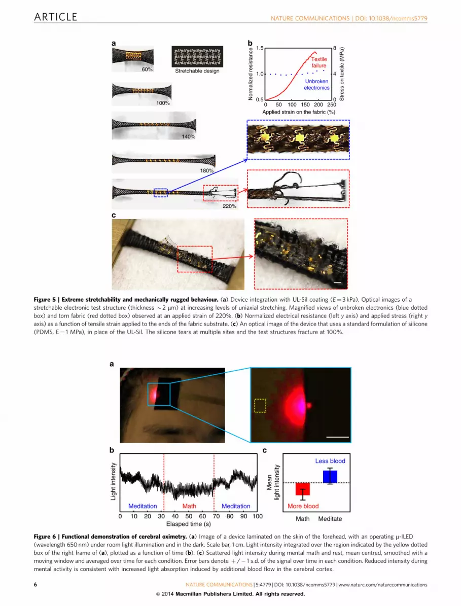
Stretchable electronics with ultra-soft elastomer coating.Experiments demonstrate that with appropriate designs, thestretchability is limited not by the electronics but by the fabricitself. Figure 5a shows optical images of a test structure of elec-tronics/UL-Sil/fabric (identical to that of Fig. 4a) under variousstates of uniaxial deformation (Supplementary Figs 8 and 9),along with data on its stress/strain and electrical/strain responses.At B220% strain, the system begins to fail, starting with tears atthe edge of the fabric near the location of clamping to thestretching fixture. The magnified images on the right highlight thelocation of this tearing, as well as the elastically deformedstructures in the center of the device. This robust mechanics isconsistent with separate measurements of electrical resistance as a
function of strain, summarized in Fig. 5b. Comparative results ina system that replaces UL-Sil with PDMS appear in Fig. 5c; herethe electronics and even the underlying PDMS exhibit multiple,severe fractures and regions of delamination and plastic defor-mation at strains of 100%.
Cerebral oximetery and wireless skin hydration monitoring.The UL-Sil coating and stretchable fabric allow facile, reversiblemounting of active electronics on the skin, in a way that estab-lishes intimate interfaces for high quality measurement. Examplesusing the three integrated sensor modules of the Fig. 1a device,for optical blood oximetry, wireless thermal conductivity andelectrophysiology, demonstrate some capabilities. Optical pulsemetrology is possible with a m-ILED that serves as a source oflight and a digital camera that enables measurements of thedistribution and temporal behaviour of interaction of this lightwith the skin. The system and demonstration results appear inSupplementary Fig. 10. A more sophisticated application of thistype of capability involves cerebral oximetry, as shown in Fig. 6.Here, light from a m-ILED (650 nm) diffuses into the depth of thetissue over the right prefrontal cortex; photodetection at aneighbouring region (dashed yellow box) captures scattered light,the majority of which passes unabsorbed through the scalp, theskull and the brain. Due to the optical absorption characteristicsof human tissue, blood is the primary absorber of light in the red-infrared (IR) range. Figure 6b shows results recorded while theparticipant alternates between resting and performing mental
Top view
Tilted view
Fully bondedserpentine
FEAExperiment
0%
90%
0
�max0.6
PI (t=2.4 μm, E=2.5GPa)
Cu (t=0.3 μm, E=119GPa)
Str
ain
on m
etal
(%
)
Strain (%)
PlasticElastic
SilbionePDMS
0 20 40 60 80 1000.0
0.2
0.4
0.6
0.8
1.0
0
3.0�max
0%
20%
40%
60%
Stiff medium(PDMS)
0%
20%
40%
60%
0
3.0�max
PI (t=2.4 μm, E=2.5GPa)
Silbione(t=100 μm, 3.0 kPa)
Fabric (t=500 μm, 391 kPa)
Soft medium(UL-Sil)
Figure 4 | Mechanics of materials and structures for stretchable electronics. (a) Cross-sectional illustration of representative layers in a stretchable
electronic system. (b) Deformations of a FS trace and distributions of maximum principal strains in the metal computed by FEA for a system consisting of
electronics/UL-Sil(3.0 kPa)/fabric and electronics/PDMS (B1–2 MPa)/fabric. (c) The computed maximum principal strains in the metal as a function of
the strain applied to the entire system show enhanced stretchability with the UL-Sil material compared with PDMS. (d) Scanning electron micrographs
(scale bar, 200mm) and corresponding FEA results of undeformed and uniaxially stretched (90%) configurations of a FS trace bonded to a UL-Sil/fabric
substrate.
NATURE COMMUNICATIONS | DOI: 10.1038/ncomms5779 ARTICLE
NATURE COMMUNICATIONS | 5:4779 | DOI: 10.1038/ncomms5779 | www.nature.com/naturecommunications 5
& 2014 Macmillan Publishers Limited. All rights reserved.
100%
140%
180%
220%
60% Stretchable design
Nor
mal
ized
res
ista
nce
Str
ess
on te
xtile
(M
Pa)
Applied strain on the fabric (%)0 50 100 150 200 250
0.5
1.0
1.5
Unbrokenelectronics
Textilefailure
0
4
8ba
c
Figure 5 | Extreme stretchability and mechanically rugged behaviour. (a) Device integration with UL-Sil coating (E¼ 3 kPa), Optical images of a
stretchable electronic test structure (thickness B2 mm) at increasing levels of uniaxial stretching. Magnified views of unbroken electronics (blue dotted
box) and torn fabric (red dotted box) observed at an applied strain of 220%. (b) Normalized electrical resistance (left y axis) and applied stress (right y
axis) as a function of tensile strain applied to the ends of the fabric substrate. (c) An optical image of the device that uses a standard formulation of silicone
(PDMS, E¼ 1 MPa), in place of the UL-Sil. The silicone tears at multiple sites and the test structures fracture at 100%.
Meditation Math Meditation
0 10 20 30 40 50 60 70 80 90 100Elasped time (s)
Ligh
t int
ensi
ty
More blood
Less blood
Math Meditate
Mea
nlig
ht in
tens
ity
Figure 6 | Functional demonstration of cerebral oximetry. (a) Image of a device laminated on the skin of the forehead, with an operating m-ILED
(wavelength 650 nm) under room light illumination and in the dark. Scale bar, 1 cm. Light intensity integrated over the region indicated by the yellow dotted
box of the right frame of (a), plotted as a function of time (b). (c) Scattered light intensity during mental math and rest, mean centred, smoothed with a
moving window and averaged over time for each condition. Error bars denote þ /� 1 s.d. of the signal over time in each condition. Reduced intensity during
mental activity is consistent with increased light absorption induced by additional blood flow in the cerebral cortex.
ARTICLE NATURE COMMUNICATIONS | DOI: 10.1038/ncomms5779
6 NATURE COMMUNICATIONS | 5:4779 | DOI: 10.1038/ncomms5779 | www.nature.com/naturecommunications
& 2014 Macmillan Publishers Limited. All rights reserved.

math exercises with their eyes closed (counting backwards by 7from 200). The data are mean centred for each condition, andthen smoothed by applying a 650 ms moving window average toeliminate the influence of the heart beat. The estimated cerebralblood flow is then averaged over time to yield the estimatedaverage blood flow in each condition shown in Fig. 6c. Asexpected20,21, the measurements indicate increased blood flowduring mental math compared with meditation.
Capabilities in wireless operation are important for manyenvisioned applications. A simple example of controlled, wirelessheating via far field exposure to RF radiation appears in IR imagesin Fig. 7a,b; Supplementary Fig. 11. Here, a stretchable RFantenna in a dipole geometry (30 mm for each branch, foroperation at B2.4 GHz in air, and at B0.9 GHz on skin, as inFig. 7c) with a FS mesh layout (Cu traces, 3 mm thick and 10 mmwide, encapsulated above and below with PI) absorbs incidentRF energy. The resulting induced current oscillates at theoperating frequency, to create Joule heating in a resistive element(Au/Cr, 100/15 nm thick) that has a much higher resistance(B500O) than the antenna (B5O). This element appears asbright spots in the centres of the dashed yellow boxes in Fig. 7a,b.Time-dependent measurements of the temperature, recordedelectrically or through analysis of the IR images, capturedynamics of heating and cooling that can yield quantitative
values for the thermal conductivity of the skin. The analysisinvolves characterizing the rise (and/or fall) times from data likethose in Fig. 7d and applying transient plane source analysis22.Figure 7e and Supplementary Fig. 12 show results of measure-ments of calibration standards, to verify the approach. Theconductivity correlates well to skin hydration, a physio-logical parameter of general interest, as illustrated in Fig. 7fthrough comparison with measurements using a commercialimpedance-based meter (Delfin, Finland).
Multiple site EP sensing. The materials and device constructionallow continuous use in realistic scenarios. Figure 8;Supplementary Fig. 13 present examples of EP measurements ona human subject engaged in an immersive, full-scale drivingsimulator. Here, as shown in Fig. 8a,b, several sensors placed onmultiple sites of the body simultaneously record EOG, for eyemovements, EMG, for movements of the chest/arms and ECG,with a ground electrode placed on the left hip. The signal analysisprocedures appear in the Methods section. As the participantdrives for 45 min under busy highway conditions with 100 othercars, some of which intermittently merge into or brake in front ofthe driver’s path, the EP information reveals various responses, asshown in Fig. 8c. Figure 8d shows normalized EOG (red), ECG/
S11
(dB
m)
Frequency (GHz)
On skin
In air
0 1 2 3–40
–30
–20
–10
0
Time (s)
Tem
pera
ture
(°C
)
40
36
32
280 40 80 120
RFoff
RFon
RFoff
0.0 0.1 0.2 0.3 0.4 0.5 0.6 0.7
0.0
0.5
1.0
1.5
2.0
Res
ista
nce
chan
ge (
Ω)
Time (s)
0% EG40% EG
100% EG
0 30 60 90
0.35
0.40
0.45
0.50
Mea
sure
d th
erm
alco
nduc
tivity
(W
mK
–1)
Measured hydration level
Figure 7 | Wireless evaluation of skin thermal properties. IR images of a wireless heating device, collected during exposure to RF energy, in a
free-standing state (a) and mounted on the wrist area (b). (c) S11 coefficient measured from the wireless heating element, evaluated in air and on human
skin. (d) Transient control of temperature on the skin using the wireless heating element, and measured using an IR camera. The temporal behaviour
during heating and during cooling define the thermal conductivity of the skin and, therefore, its hydration state. (e) Representative resistance changes
during transient heat pulse measurements on three different calibration solutions. (f) Transient heat source measurements, converted to thermal
conductivity, on skin. The hydration level corresponds to results of measurement with a commercially available skin moisture meter (Delfin, Finland), based
on skin impedance. Error bars correspond to average s.d. values during repeated calibration measurements.
NATURE COMMUNICATIONS | DOI: 10.1038/ncomms5779 ARTICLE
NATURE COMMUNICATIONS | 5:4779 | DOI: 10.1038/ncomms5779 | www.nature.com/naturecommunications 7
& 2014 Macmillan Publishers Limited. All rights reserved.

EMG (green) and EMG (blue) time-locked to a representativebraking event at t¼ 0. The data show that the driver reacts withquick eye movements to the car, muscle movements to move thesteering wheel and a decelerated heart rate in response to thischallenging driving event. Figure 8e shows the average event-related spectrogram for EOG, ECG and EMG for all 21 brakingand merging events, computed with a 4,096-point moving win-dow discrete Fourier transform of the 20 s before and after thebraking events. For each trial, subtraction of the average spectrafrom 2–5 s before the braking event isolates evoked changes inphysiology. The EOG activity indicates a sharp increase in eyemovements after the event, followed by a sustained high fre-quency set of movements. A device on the chest measures both anincrease in EMG activity of the chest muscles in response to thebraking event, as well as a peak at the frequency of the heart ratethat decreases in the period following a braking event. The EMGactivity indicates a sustained increase in high frequency muscleactivity as the participant moves their arms and the steering wheelin response to the braking car. Due to the thin, elastic con-struction of the devices, and their soft, reversible adhesion onto
the skin, all of these measurements occur without measurableeffect on natural response.
DiscussionThe results presented here indicate that heterogeneous integra-tion of semiconductors, metals and polymer dielectrics infilamentary, open mesh designs for active electronics and sensors,with ultralow modulus, permeable elastomer coatings forconformal skin adhesion, on breathable, stretchable fabrics asrugged substrates yields an attractive architecture for skin-mounted devices. The UL-Sil and the fabric are the criticalmaterials components in these systems. The former provides asmooth, conformable surface for strong adhesion and reversiblebonding to the skin, even in regions with substantial hair, and theelectronics, in a manner that imposes minimal mechanicalconstraints on their motion. The latter provides a robust, strainlimiting support for the UL-Sil, whose ultralow modulus andstrong adhesion would otherwise prevent its use in practicalapplications. These two materials form a composite substrate that
Braking
Merging
EP sensors on body
2
500
10
Time (s)
Fre
quen
cy (
Hz)
Fre
quen
cy (
Hz)
Fre
quen
cy (
Hz)
After an eventNormal
Eye
Chest
Arm
Eyes scanningscene
Heart beat changing
Chest moving
Turningwheel
0
500
10
2
500
10
After an eventNormal
Nor
mal
ized
am
plitu
de
Time (s)
Eye
Chest
Arm
0 2 4 6 6420–2 –2
Figure 8 | EP monitoring of a human subject in a driving simulator. (a) Schematic illustration of the locations of the EP sensors on the body.
(b) Configuration of a driving simulator that uses eight large screens surrounding a full-scale test vehicle to provide a realistic, but virtual, driving
experience. A driver wearing EP sensors is in the simulator car. (c) Representative driving scenarios for EP monitoring. (d) Typical EP signals recorded
during unexpected braking and merging events. (e) Spectrographs of the EP signals. A total 11 braking and 12 merging events occurred during 40 min,
while the multiple sensors continuously collected EP data.
ARTICLE NATURE COMMUNICATIONS | DOI: 10.1038/ncomms5779
8 NATURE COMMUNICATIONS | 5:4779 | DOI: 10.1038/ncomms5779 | www.nature.com/naturecommunications
& 2014 Macmillan Publishers Limited. All rights reserved.

is compatible with natural processes of transepidermal water loss,and offers capability for use not only with the types of activecomponents described here, but also with stretchable conductors,organic semiconductors and low-dimensional materials such ascarbon nanotubes, silver nanowires and graphene, reported else-where23–26. Although experiments, modelling results and demon-stration experiments indicate many appealing characteristics,there are additional opportunities for optimizing of properties ofthis substrate, through development of engineered fabrics andtailored elastomer coatings.
MethodsDevice fabrication. Spin casting formed thin layers of polydimethylsiloxane(PDMS; 10mm in thickness, Dow Corning, USA) and polyimide (PI; 1.2 mm inthickness, Sigma-Aldrich, USA) on a temporary glass substrate. Two m-ILEDsprepared according to previously described procedures11 were transfer printedonto the PI. Thermally curing the PI (2 h at 210 �C) yielded strong bonds tom-ILEDs. The wireless heater and EP sensors used photolithographically patternedbi-layers of Cr (15 nm)/Au (100 nm) formed by sputter deposition (AJA sputter,150 W/50 W). The RF antenna used Cu (3mm) by electron beam evaporation. Spincasting a layer of PI (2,000 r.p.m.) passivated and isolated the devices. Reactive ionetching (20 sccm O2, 200 W, 200 mTorr) through a photolithographically patternedhard mask (Cu, 100 nm thick) removed the PI in the regions between the devices.A water soluble tape (3 M, USA) allowed retrieval of the completed patterns fromthe glass substrate and delivery to an adhesive (UL-Sil or PDMS)-coated stretchabletextile substrate.
Finite element analysis. Three-dimensional FEA techniques allowed investigationof the mechanics of devices on UL-Sil/textile or PDMS/textile substrates. Eight-node three-dimensional solid elements and four-node shell elements were used forthe bi-layer substrate and serpentine interconnect, respectively, with refinedmeshes to ensure the accuracy. Displacement-type boundary conditions wereassigned to the side surfaces of substrate to apply to different levels of stretching.The Young’s modulus (E) and Poisson ratio (n) of the materials used in thesimulations include ECu¼ 119 GPa and nCu¼ 0.34 for copper; EPI¼ 2.5 GPa andnPI¼ 0.34 for PI; ETextile¼ 391 kPa and nTextile¼ 0.40 for textile (approximated, forpurposes of simulation, to have linear elastic properties); EUL-Sil¼ 3.0 kPa andnSilbione¼ 0.49 for UL-SIl; and EPDMS¼ 1 MPa and nPDMS¼ 0.49 for PDMS.
Wireless heater system. A network analyser (E5602, Agilent Technologies, USA)with calibration kit (85033E, Agilent Technologies, USA) enabled measurement ofthe return loss (S11) and the resonance frequency of the RF antennas. RF signalsprecisely controlled with an analogue signal generator (N5181A, Agilent Tech-nologies, USA), an amplifier (1119, EMPOWER RF System, USA), a DC powersupply (U8031A, Agilent Technologies, USA). Directional antenna with 10.5 dBigain (204411, Wilson Electronics, USA) and an RF power meter (43, Bird Tech-nologies, USA) were used to expose a wireless heater laminated on the arm (B2 kg)of an adult human subject. The power levels (o10 W) were maintained below FCCguidelines (8 W kg� 1 for partial body irradiation). The temperature distributionswere measured using an IR camera (A655SC, FLIR).
Measurements of skin hydration. Thermal conductivity was determined byanalysing the time constant for the rise in temperature determined using a resistivedetector after B0.7 s of Joule heating. Conversion to thermal conductivity relied oncalibration data collected from aqueous ethylene glycol solutions with knownthermal conductivities, as shown in Supplementary Fig. 12.
Measurements of EP signals. All experiments involving human subjects wereconducted under approval from Institutional Review Board at the University ofIllinois at Urbana-Champaign (protocol number: 13,229). There were six subjects(age: 21–32, all males). Research was carried out with informed consent. For thedata of Fig. 2b, the right wrist was extended every 5 s. Data were digitized at 256 Hz,with a 60 Hz notch filter to eliminate electrical noise. Data were amplified andtransmitted wirelessly to a recording PC using an amplifier (CleveMed, USA) as thedifference in voltage between two EP sensors on the device, spaced 40 mm apart.A ground electrode was placed over a bony protrusion of the right lateral ankle.The mean value was subtracted from original signal, and the data were divided bytheir maximum value to compute normalized EMG amplitude. SupplementaryMovie 1 demonstrates stretchability, reusability, washability of fabric-basedstretchable electronic devices. EMG measurements show capabilities for mountingon regions of the skin with hair.
Optical measurements of oximetry. An analysis programme stabilized all framesof video collected with a digital camera by template correlation with the first frame,and then determined the time variation of the average light intensity evaluated
inside a region of interest (dotted yellow box) for only the red RGB channel. Aband-pass filter between 0.5 and 3 Hz removed high frequency noise and lowfrequency drifts in the data. A discrete Fourier transform of the filtered signalrevealed the beat rate. For cerebral oximetry, a 3-mm diameter fibre-optic bundlewas held flush against the head to collect light and transmit it to a photomultipliertube detector (Imagent, USA).
Evaluations in the driving simulator. The experiments were conducted in theBeckman Institute Illinois Simulator Laboratory. A total of eight textile EP sensorswere placed on the skin at various locations across the body. EOG data wererecorded from bipolar electrodes placed superior (and slightly medial) and inferior(and slightly lateral) to the participant’s left eye, in a position sensitive to bothhorizontal and vertical eye movements. ECG data were recorded from bipolarelectrodes placed over the most medial areas of the left and right fourth intercostalspaces. EMG data were recorded from bipolar electrodes over the proximal leftforearm over the flexor carpi radialis muscle. All three pairs of bipolar textileelectrodes were used simultaneously with a common ground electrode placed onthe human subject’s left hip. Voltage differences between bipolar pairs of textileelectrodes were amplified and digitized with a Neuroscan SynAmps 32-Channelamplifier at 2,000-Hz with a 0.1–1,000 Hz online band-pass filter to remove slowdrifts and high frequency non-physiological noise, and a 60-Hz notch filter toattenuate electrical line noise from the simulator. The participant drove in aHyperdrive driving simulator for 45 min in a busy highway driving scenario with100 other cars. Cars intermittently merged into the driver’s path, or braked in frontof the driver, to simulate challenging driving scenarios. The normalized EOG (red),ECG (green) and EMG (blue) activity time-locked to the onset of a single brakingevent at time (0). In response to this challenging driving event, the driver reactedwith quick eye motions to the car, muscle activity to move the steering wheel and adecelerated heart rate. The average event-related spectrograms for EOG, ECG andEMG corresponds to responses for all 21 braking and merging events, computedwith a 4,096-point moving window DFT of the 20 s before and after the brakingevents. For each trial, the average spectra from 5–2 s before the braking event weresubtracted from each time point to isolate the evoked changes in physiology fromthe event.
References1. Kim, D. H. et al. Epidermal electronics. Science 333, 838–843 (2011).2. Kaltenbrunner, M. et al. An ultra-lightweight design for imperceptible plastic
electronics. Nature 499, 458–463 (2013).3. Xu, S. et al. Soft microfluidic assemblies of sensors, circuits, and radios for the
skin. Science 344, 70–74 (2014).4. Laulicht, B. et al. Quick-release medical tape. Proc. Natl Acad. Sci. USA 109,
18803–18808 (2012).5. Webb, R. C. et al. Ultrathin conformal devices for precise and continuous
thermal characterization of human skin. Nat. Mater. 12, 938–944 (2013).6. Yeo, W. H. et al. Multifunctional epidermal electronics printed directly onto
the skin. Adv. Mater. 25, 2773–2778 (2013).7. Dargaville, T. R. et al. Sensors and imaging for wound healing: a review.
Biosens. Bioelectron. 41, 30–42 (2013).8. Jeong, J.-W. et al. Materials and optimized designs for human-machine
interfaces via epidermal electronics. Adv. Mater. 25, 6839–6846 (2013).9. Someya, T. Stretchable Electronics (Wiley-VCH, 2013).10. Song, Y. M. et al. Digital cameras with designs inspired by the arthropod eye.
Nature 497, 95–99 (2013).11. Park, S. I. et al. Printed assemblies of inorganic light-emitting diodes for
deformable and semitransparent displays. Science 325, 977–981 (2009).12. Kim, R. H. et al. Flexible vertical light emitting diodes. Small. 8, 3123–3128
(2012).13. Berger, H. Uber das elektrenkephalogramm des menschen. Arch. Psychiatr.
Nervenkr. 87, 527–570 (1929).14. Phillip, M. D. et al. Composition I-II and products and uses thereof. US 2013/
0310781 A1.15. Cherenack, K. et al. Smart textiles: challenges and opportunities. J. Appl. Phys.
112, 091301 (2012).16. Riley, W. F. et al. Mechanics of Materials (Jon Wiley & Sons, 1999).17. Zhang, Y. et al. Buckling in serpentine microstructures and applications in
elastomer-supported ultra-stretchable electronics with high areal coverage. SoftMatter 9, 8062–8070 (2013).
18. Rogers, J. A. et al. Materials and mechanics for stretchable electronics. Science327, 1603–1607 (2010).
19. Davis, J. R. Copper and Copper Alloys (ASM International, 2001).20. Limongi, T. et al. Detecting mental calculation related frontal cortex
oxygenation changes for brain computer interface using multi-channelfunctional near infrared topography. Int. J. Bioelectromagn. 11, 86–90 (2009).
21. Khoa, T. Q. et al. Recognizing brain activities by functional near-infraredspectroscope signal analysis. Nonlinear Biomed. Phys. 2, 3 (2008).
22. Gustafsson, S. E. Transient plane source techniques for thermal conductivityand thermal diffusivity measurements of solid materials. Rev. Sci. Instrum. 62,797–804 (1991).
NATURE COMMUNICATIONS | DOI: 10.1038/ncomms5779 ARTICLE
NATURE COMMUNICATIONS | 5:4779 | DOI: 10.1038/ncomms5779 | www.nature.com/naturecommunications 9
& 2014 Macmillan Publishers Limited. All rights reserved.

23. Iijima, S. et al. Single-shell carbon nanotubes of 1-nm diameter. Nature 363,603–605 (1993).
24. Hu, J. et al. Chemistry and physics in one dimension: synthesis and propertiesof nanowires and nanotubes. Acc. Chem. Res. 32, 435–445 (1999).
25. Hu, L. et al. Scalable coating and properties of transparent, flexible, silvernanowire electrodes. ACS Nano 4, 2955–2963 (2010).
26. Novoselov, K. S. et al. Electrical field effect in atomically thin carbon films.Science 306, 666–669 (2004).
AcknowledgementsThis material is based on work supported by a NASA and used facilities in the FrederickSeitz Materials Research Laboratory and the Center for Microanalysis of Materials at theUniversity of Illinois at Urbana-Champaign. K.-I.J. acknowledges support from a BasicScience Research Program through the National Research Foundation of Korea (NRF)funded by the Ministry of Education (D00008). S.Y.H. acknowledges support fromSamsung Display Co. through a visiting research scholar programme. K.E.M.acknowledges postdoctoral fellowship funding from the Beckman Institute. H.C. is aHoward Hughes Medical Institute International Student Research fellow.
Author contributionsK.-I.J. and J.A.R. designed the project; S.Y.H., S.X., K.E.M., G.-T.K., J.-W.J., T.J.D.,R.C.W., J.W.L., J.C., B.K., M.C., H.U.C., D.L., Y.Y., R.C. and J.G.G. carried out experi-ments; K.E.M., Y.Z. and R.C.W. carried out simulations; and Y.H. and J.A.R. supervisedthe project. All of the co-authors contributed to writing the manuscript.
Additional informationSupplementary Information accompanies this paper at http://www.nature.com/naturecommunications
Competing financial interests: The authors declare no competing financial interests.
Reprints and permission information is available online at http://npg.nature.com/reprintsandpermissions/
How to cite this article: Jang, K.-I. et al. Rugged and breathable forms of stretchableelectronics with adherent composite substrates for transcutaneous monitoring.Nat. Commun. 5:4779 doi: 10.1038/ncomms5779 (2014).
ARTICLE NATURE COMMUNICATIONS | DOI: 10.1038/ncomms5779
10 NATURE COMMUNICATIONS | 5:4779 | DOI: 10.1038/ncomms5779 | www.nature.com/naturecommunications
& 2014 Macmillan Publishers Limited. All rights reserved.


