Embed Size (px)
Citation preview

Rutile and titanium particles differentially affect theproduction of osteoblastic local factors
Gema Valles,1 Pablo Gonzalez-Melendi,2 Laura Saldana,1,3 Mercedes Rodriguez,1,3 Luis Munuera,3,4
Nuria Vilaboa1,31Unidad de Investigacion, Hospital Universitario La Paz, Paseo de la Castellana 261, 28046 Madrid, Spain2Centro de Investigaciones Biologicas, CIB-CSIC, C/Ramiro de Maeztu 9, 28040 Madrid, Spain3Centro de Bioingenierıa, Biomateriales y Nanomedicina, CIBER-BBN, Zaragoza, Spain4Departamento de Traumatologıa y Cirugıa Ortopedica, Hospital Universitario La Paz, Paseo de la Castellana 261,28046 Madrid, Spain
Received 13 October 2006; revised 17 January 2007; accepted 30 January 2007Published online 2 July 2007 in Wiley InterScience (www.interscience.wiley.com). DOI: 10.1002/jbm.a.31315
Abstract: Titanium and its alloys are widely used asimplant materials for dental and orthopaedic applications.To improve their wear and corrosion resistance, severalsurface modifications that give rise to an outer ceramiclayer of rutile have been developed. It is expected that af-ter a long period of functional loading, rutile debris willarise from these modified surfaces. We have compared thein vitro biocompatibility of subcytotoxic doses of rutile andtitanium particles of phagocytosable size in primary cul-tures of human osteoblasts. Particles were visualized usinga spectral confocal microscope by reflection. Both types ofparticles aggregated in the culture media and were effi-ciently internalized by osteoblasts as agglomerates. Treat-ment of isolated cultures of osteoblasts with rutile particlesstimulated the release of IL-6, PGE2, and GM-CSF to a
lesser extent than titanium. The influence of macrophageson the particle-induced stimulation of those local factorswas analyzed by coculturing TPA-differentiated THP-1cells with osteoblasts. Under these conditions, levels of IL-6 and PGE2 after treatment of cocultured osteoblasts withrutile particles were lower than after exposure to titanium.These results indicate that rutile debris shows a lower bio-reactivity than titanium when tested in cultures of humanosteoblasts and support the improved biocompatibility oftitanium-based implants modified to create an outer layerof rutile on their surfaces. � 2007 Wiley Periodicals, Inc.J Biomed Mater Res 84A: 324–336, 2008
Key words: osteolysis; wear debris; osteoblast; cytokine;titanium; titanium oxide
INTRODUCTION
Aseptic loosening remains the most common long-term complication of major arthroplasties and is themain cause of implant revision surgery. The condi-tion, radiographically identified as radiolucent areasof osteolysis at the bone-implant interface, has a com-plex and multifactorial etiology.1 Extensive localized
bone resorption resulting in implant failure withoutsigns of infection or aseptic loosening was reported inthe 1970s.2,3 At that time, the observation of bonecement debris within the periprosthetic tissue led tothe term ‘‘bone cement disease’’ and promoted thelater development of cementless prostheses. However,radiolucent zones are often observed at the distal endof noncemented stems and the diverse chemistry ofparticles isolated from revision tissues has led to themore general concept of wear-mediated osteolysis.
The biological response to wear particles at thebone-implant interface is considered the main causeof periprosthetic osteolysis. The current model ofwear-mediated osteolysis hypothesizes that particlesproduced by wear at the articulating surfaces ofprostheses are phagocytosed by macrophages, whichare then stimulated to release inflammatory cyto-kines that activate the resorptive activity of osteo-clasts.4 Osteoblasts are also responsive to mediatorsreleased by macrophages and in turn induce the for-mation and activity of osteoclasts.5,6 Micromotion
Correspondence to: N. Vilaboa; e-mail: [email protected] grant sponsor: Comision Interministerial de
Ciencia y Tecnologıa; contract grant numbers: MAT2001/0019/CO2/O1 and MAT2006-12948-C04-02Contract grant sponsor: Fundacion Mutua MadrilenaContract grant sponsor: Fondo de Investigaciones Sanita-
rias; contract grant numbers: FIS 01/3027, FIS PI03/0036Contract grant sponsor: Program ‘‘Ramon y Cajal,’’
Spanish Ministry of Education and Science
' 2007 Wiley Periodicals, Inc.

and fretting at the distal end of metallic femoralstems has been suggested as a local source of partic-ulate debris that may directly affect osteoblastslocated at the bone-implant interface.7 In fact, metal-lic particles have been shown to have a direct effecton cells other than macrophages, such as fibroblastsand osteoblasts.8–16 In spite of the pivotal role of theosteoblasts in the regulation of bone remodeling,their participation in the wear-mediated osteolysisprocess has only recently begun to be elucidated.Incubation of cultured osteoblasts with particles ofdifferent chemistry and size affects osteoblastic phe-notype markers and induces the release of inflamma-tory factors such as interleuquin-6 (IL-6) and prosta-glandin E2 (PGE2).
8,11,12
Titanium (Ti) and Ti-based alloys are widely usedmetals for dental and orthopaedic implants due toseveral advantageous properties, such as good fa-tigue strength, machinability, and biocompatibility.17
Their good corrosion resistance is attributed to thespontaneous formation of an outer surface of non-stoichiometric amorphous or poorly crystallizedTiO2. Under continuous mechanical stress, Ti-basedimplants still give rise to large amounts of de-bris.18,19 Hence, there has been a very active searchfor surface modifications that improve both osseoin-tegration and wear resistance of these metallic mate-rials. Treatments of Ti-based implants such as micro-arc or thermal oxidation give rise to an outer rutilelayer that improves the corrosion resistance andreduces the friction coefficient in rubbing contact.20–22
Rutile surfaces improve osteoblast adhesion in vitroand increase the percentage of bone-to-implant con-tact in vivo.23–25 It is expected that after long-termfunctional loading, rutile debris will arise from thesemodified surfaces. We recently reported that rutileparticles exhibit a better biocompatibility than tita-nium, as tested in cultures of human macrophages.26
In this work, we aimed to study the behavior ofhuman osteoblasts exposed to these types of par-ticles. We investigated whether human osteoblastsare able to internalize rutile particles and respond tothem by modulating the secretion of factors associ-ated with periprosthetic osteolysis. For comparativepurposes, titanium particles were also used.
MATERIALS AND METHODS
Particles
Rutile (TiO2) and commercially pure Ti particles,obtained from Johnson Matthey (Ward Hill, MA), werecharacterized in the course of our previously reportedwork.26 The mean equivalent circle diameter (ECD) sizesof the particles were 0.45 6 0.26 lm for TiO2 (range was0.1–1.5 lm, 92% were lower than 0.9 lm) and 3.32 6
2.39 lm for Ti (range was 1–15 lm, 89% were lower than7 lm).26
TiO2 and Ti particles were weighed and sterilized byincubation in isopropanol at room temperature and driedunder UV light in a laminar flow hood. Prior to the addi-tion to the cells, particles were resuspended in the appro-priate culture medium (20 mg/mL) and sonicated at maxi-mum power for 10 min in a bath sonicator (Bransonic 12,Branson Ultrasonidos S. A. E., Barcelona, Spain).
Cell culture and treatments
Human osteoblastic cells (OB) were isolated from freshtrabecular bone explants obtained from patients (aged67 6 6 years old) undergoing total knee arthroplasty, aspreviously described.23 Patients enrolled in this researchsigned an Informed Consent form and all proceduresusing human tissue designated ‘‘surgical waste’’ wereapproved by the Human Research Committee of HospitalLa Paz (Date of Approval: 03-15-2001). Each bone samplewas processed in a separated primary culture and experi-ments were performed using cultures obtained from inde-pendent patients. OB were cultured in Dulbecco’s modi-fied Eagle’s medium (DMEM) supplemented with 15% (v/v)fetal bovine serum (FBS), 100 UI/mL penicillin, and0.1 mg/mL streptomycin in a humidified 5% CO2 atmosphereat 378C. Culture media were changed every 3 days untilconfluence was reached. For particle treatments, 2 3 105
OB were seeded in six-well plates (equivalent to a cell den-sity of 21 3 103 cells/cm2), cultured for 24 h, and thenwashed with phosphate-buffered saline (PBS). Appropriatevolumes of particle suspensions were added to cells toachieve doses of 0.5, 5, and 50 ng/cell in a final volume of2 mL and cells were cultured for further 24 h. As a control,OB were incubated in the absence of particles.
THP-1 cells (ECACC, Salisbury, Wiltshire, UK) weregrown in RPMI-1640 medium supplemented with 10% (v/v) heat-inactivated FBS, 500 UI/mL of penicillin, and 0.1mg/mL of streptomycin, in a humidified 5% CO2 atmos-phere at 378C. OB and TPA-differentiated THP-1 cellswere cocultured using a transwell insert system (Corning,Life Sciences, MA) that allows humoral contact of both celltypes, through a microporous membrane with a pore sizeof 0.4 lm, avoiding direct cell contact. About 2 3 105
THP-1 cells were seeded into six-well plates (equivalent toa cell density of 21 3 103 cells/cm2) and treated with10 ng/mL 12-O-tetradecanoyl phorbol 13-acetate (TPA)(Sigma, Madrid, Spain) for 12 h. Next, cells were thor-oughly washed with PBS and recovered in fresh mediumfor a further 24 h. THP-1 cells treated with TPA accordingto this procedure express high levels of the surface anti-gens CD11b and CD14, characteristics of the monocyte/macrophage lineage, and respond to titanium particles byincreased release of TNF-a, IL-6, and IL-1b.26 TPA-treatedTHP-1 cells adhered to the bottom of the wells were thenwashed with PBS and supplemented with 2.5 mL of a mix-ture of 50% RPMI and 50% DMEM, containing 12.5% (v/v) heat-inactivated FBS, 500 UI/mL of penicillin, and0.1 mg/mL of streptomycin. Inserts containing OB plated 24 hbefore at a cell density of 21 3 103 cells/cm2 were washedwith PBS and placed into the wells containing THP-1 cells.
OSTEOBLAST RESPONSE TO RUTILE AND TITANIUM PARTICLES 325
Journal of Biomedical Materials Research Part A DOI 10.1002/jbm.a

Immediately afterwards, appropriate volumes of particlessuspensions containing doses of 50 ng/cell, in a volume of0.5 mL, were added only to OB seeded in the inserts.Cocultured OB were incubated in the presence of the par-ticles for further 24 h. As a control, OB, THP-1 cells, andcocultures of OB and THP-1 were subjected to the samemanipulations but incubated in the absence of particles.
Endotoxin test
Particles and culture media were endotoxin-free as dem-onstrated by the Sigma E-TOXATE assay for detection andsemiquantification of endotoxins (Sigma). Particles and cul-ture media used in this study contained levels of endotox-ins below 0.015 EU/mL.
Confocal microscopy
Suspensions of TiO2 and Ti particles in culture mediawere prepared as described earlier to achieve the equiva-lent doses of 5 ng/cell and drops were mounted on mi-croscopy slides. The presence of the particles in the culturemedium was analyzed in a spectral confocal microscope(LEICA TCS-SP2-AOBS, Leica microsystems, HeidelbergGMBH, Germany) by reflection under excitation with thelaser lane of 488 nm and by collecting the emission in therange 490–510 nm. As a control, the particles were alsodirectly visualized by the Nomarski technique (or differen-tial interference contrast, DIC). Overlaid images of thesame fields showing particles observed by reflection andNomarski were used to compare both methods of detec-tion. To determine if the particles form aggregates in theculture media, we measured their areas by manually out-lining a region of interest (ROI) around each one and thenusing the ‘‘quantify’’ function of the LEICA software LCS,version 2.5 Build 1227. The corresponding EDCs weredetermined and represented in histograms.
OB were seeded in eight-well chambers (Nunc, Wiesba-den, Germany) at a density of 2 3 104 cells/well (equiva-lent to a cell density of 25 3 103 cells/cm2) and culturedfor 24 h in the presence or in the absence of particles. Afterextensive washing with PBS, OB were fixed for 45 minwith a solution of 2.5% glutaraldehyde in PBS in darkness.Cells were washed with PBS, placed onto slides, andmounted with a 1:1 mixture of glycerol:PBS. The speci-mens were observed in the spectral confocal microscopeunder a He/Ne laser. The autofluorescent signal inducedby glutaraldehyde fixation was collected in the emissionranges 490–540 nm (green) and 553–625 nm (red) (excita-tion lines 488 and 543 nm, respectively). The presence ofparticles within the cells was either inferred as nonfluores-cent, dark areas in an autofluorescent background,26 ordirectly visualized by reflection with the excitation line 488nm. Stacks of 0.5-lm optical sections spanning completedcells were recorded and the confocal images were ana-lyzed using the same LEICA software, which was alsoused for morphometric analysis and area measurement.The areas of intracellular dark regions of different sizeswere measured by manually outlining a ROI on the pro-jected maximum view of the confocal stack, as previously
described.26 The software is calibrated with the originaldata from the confocal stack collection to measure the realROI area and therefore, calculating the correspondingECD. For each experimental condition, individual cellswere examined to determine the number of internalizedparticles/cell. A significant number of measures were col-lected for each specimen.
Cytotoxicity and metabolic activity assays
Lactate dehydrogenase (LDH) release was assayed withthe Cyto Tox 96 (Promega, Madison, WI), following themanufacturer’s instructions. Metabolic activity wasassessed using the alamarBlue assay (Biosource, Nivelles,Belgium). Cells were incubated in DMEM containing 10%alamarBlue dye for 4 h and after excitation at 530 nm, thefluorescence emitted at 590 nm was quantified using aspectrofluorimeter (Victor2 Wallac 1420 Multilabel Counter,PerkinElmer, Turkin, Finland).
Immunoenzymatic assays
Culture media were collected, filtered, and centrifugedat 1.200 g for 10 min, supplemented with a mixture of pro-teases inhibitors (17.5 lg/mL phenylmethylsulfonyl fluo-ride, 1 lg/mL pepstatin A, 2 lg/mL aprotinin, 50 lg/mLbacitracin, all from Sigma), and frozen at �808C. Humanspecific ELISA kits were used to measure IL-6, soluble IL-6receptor (sIL-6R), and granulocyte-macrophage colonystimulating factor (GM-CSF) (Biosource International,Camarillo, CA), PGE2 (Cayman Chemical Company, AnnHarbor, MI), osteoprotegerin (OPG) (Biomedica GmbH,Vienna, Austria), and the soluble form of the receptor acti-vator of NF-jB ligand (sRANKL) (Inmundiagnostik, Ben-sheim, Germany). The detection limits of the kits were2 pg/mL for IL-6, 8 pg/mL for sIL-6R, 3 pg/mL for GM-CSF, 15 pg/mL for PGE2, 2.8 pg/mL for OPG, and 1.5 pg/mL for sRANKL. All procedures were performed follow-ing the manufacturer’s instructions.
Statistical analysis
The data are presented as mean 6 standard deviation(S.D.) of several independent experiments, each performedwith an individual primary culture. ECD of particles, num-ber of aggregates/cell, and secretion differences bet-ween isolated cultured and cocultured cells were analyzedusing one-way analysis of variance (ANOVA). Two-wayANOVA for repeated measures were performed for therest of the experiments. Post hoc comparisons were ana-lyzed by 95% confidence interval adjusted by the Bonferro-ni’s method. In all cases, we confirmed results by nonpara-metric pair-wise Friedman’s test with the appropriate posthoc comparisons. The p < 0.05 was considered to be statis-tically significant. All statistical analyses were performedusing personal computer-based statistical software (SPSSversion 10.0.1; SPSS, Chicago, IL).
326 VALLES ET AL.
Journal of Biomedical Materials Research Part A DOI 10.1002/jbm.a

RESULTS
Characterization of suspensions of particles
Two methods were used to visualize and charac-terize TiO2 and Ti particles: Nomarski [Fig. 1(A,D)]and reflection after excitation with the laser line of488 nm [Fig. 1(B,E)]. Some isolated particles togetherwith abundant aggregates of different sizes wereclearly identified in both cases. Merging of fluores-
cence and Nomarsky images showed an almost com-plete colocalization of the particles/aggregatesdetected by both methods [Fig. 1(C,F)]. The examina-tion of the suspensions under the confocal micro-scope suggested that the sizes of Ti aggregates werelarger than those of TiO2. ECD of isolated particlesand aggregates were measured and the correspond-ing distributions versus their relative frequencieswere represented in histograms for TiO2 [Fig. 1(G)]and Ti [Fig. 1(H)] particles. The mean particle sizes,
Figure 1. Characterization of particles resuspended in culture media. Confocal images of TiO2 (A–C) or Ti particles (D–F) resuspended in culture media. A and D show particles in suspension observed by the Nomarski technique, B and Eshow the detection of particles by reflection (green), and C and F show the overlapping of both images. Bars ¼ 40 lm.(G–H) ECD distribution of TiO2 (G) or Ti (H) particles resuspended in culture media (n ¼ 74).
OSTEOBLAST RESPONSE TO RUTILE AND TITANIUM PARTICLES 327
Journal of Biomedical Materials Research Part A DOI 10.1002/jbm.a
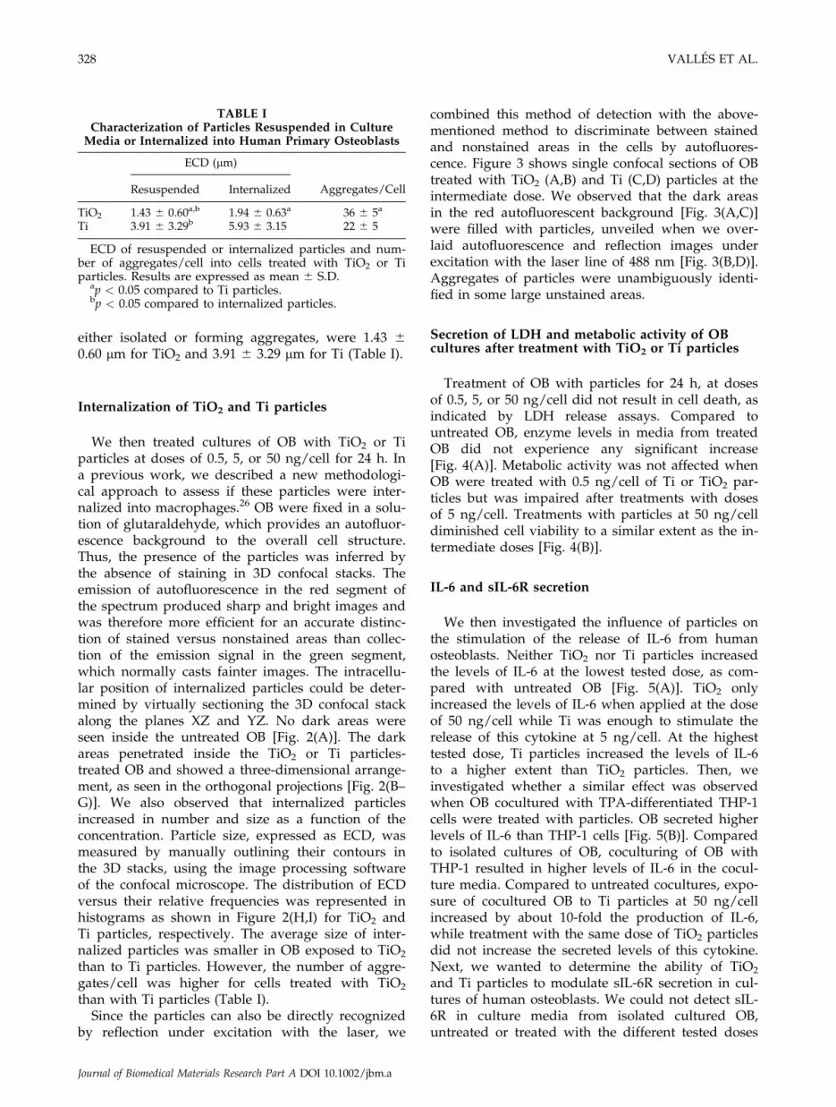
either isolated or forming aggregates, were 1.43 60.60 lm for TiO2 and 3.91 6 3.29 lm for Ti (Table I).
Internalization of TiO2 and Ti particles
We then treated cultures of OB with TiO2 or Tiparticles at doses of 0.5, 5, or 50 ng/cell for 24 h. Ina previous work, we described a new methodologi-cal approach to assess if these particles were inter-nalized into macrophages.26 OB were fixed in a solu-tion of glutaraldehyde, which provides an autofluor-escence background to the overall cell structure.Thus, the presence of the particles was inferred bythe absence of staining in 3D confocal stacks. Theemission of autofluorescence in the red segment ofthe spectrum produced sharp and bright images andwas therefore more efficient for an accurate distinc-tion of stained versus nonstained areas than collec-tion of the emission signal in the green segment,which normally casts fainter images. The intracellu-lar position of internalized particles could be deter-mined by virtually sectioning the 3D confocal stackalong the planes XZ and YZ. No dark areas wereseen inside the untreated OB [Fig. 2(A)]. The darkareas penetrated inside the TiO2 or Ti particles-treated OB and showed a three-dimensional arrange-ment, as seen in the orthogonal projections [Fig. 2(B–G)]. We also observed that internalized particlesincreased in number and size as a function of theconcentration. Particle size, expressed as ECD, wasmeasured by manually outlining their contours inthe 3D stacks, using the image processing softwareof the confocal microscope. The distribution of ECDversus their relative frequencies was represented inhistograms as shown in Figure 2(H,I) for TiO2 andTi particles, respectively. The average size of inter-nalized particles was smaller in OB exposed to TiO2
than to Ti particles. However, the number of aggre-gates/cell was higher for cells treated with TiO2
than with Ti particles (Table I).Since the particles can also be directly recognized
by reflection under excitation with the laser, we
combined this method of detection with the above-mentioned method to discriminate between stainedand nonstained areas in the cells by autofluores-cence. Figure 3 shows single confocal sections of OBtreated with TiO2 (A,B) and Ti (C,D) particles at theintermediate dose. We observed that the dark areasin the red autofluorescent background [Fig. 3(A,C)]were filled with particles, unveiled when we over-laid autofluorescence and reflection images underexcitation with the laser line of 488 nm [Fig. 3(B,D)].Aggregates of particles were unambiguously identi-fied in some large unstained areas.
Secretion of LDH and metabolic activity of OBcultures after treatment with TiO2 or Ti particles
Treatment of OB with particles for 24 h, at dosesof 0.5, 5, or 50 ng/cell did not result in cell death, asindicated by LDH release assays. Compared tountreated OB, enzyme levels in media from treatedOB did not experience any significant increase[Fig. 4(A)]. Metabolic activity was not affected whenOB were treated with 0.5 ng/cell of Ti or TiO2 par-ticles but was impaired after treatments with dosesof 5 ng/cell. Treatments with particles at 50 ng/celldiminished cell viability to a similar extent as the in-termediate doses [Fig. 4(B)].
IL-6 and sIL-6R secretion
We then investigated the influence of particles onthe stimulation of the release of IL-6 from humanosteoblasts. Neither TiO2 nor Ti particles increasedthe levels of IL-6 at the lowest tested dose, as com-pared with untreated OB [Fig. 5(A)]. TiO2 onlyincreased the levels of IL-6 when applied at the doseof 50 ng/cell while Ti was enough to stimulate therelease of this cytokine at 5 ng/cell. At the highesttested dose, Ti particles increased the levels of IL-6to a higher extent than TiO2 particles. Then, weinvestigated whether a similar effect was observedwhen OB cocultured with TPA-differentiated THP-1cells were treated with particles. OB secreted higherlevels of IL-6 than THP-1 cells [Fig. 5(B)]. Comparedto isolated cultures of OB, coculturing of OB withTHP-1 resulted in higher levels of IL-6 in the cocul-ture media. Compared to untreated cocultures, expo-sure of cocultured OB to Ti particles at 50 ng/cellincreased by about 10-fold the production of IL-6,while treatment with the same dose of TiO2 particlesdid not increase the secreted levels of this cytokine.Next, we wanted to determine the ability of TiO2
and Ti particles to modulate sIL-6R secretion in cul-tures of human osteoblasts. We could not detect sIL-6R in culture media from isolated cultured OB,untreated or treated with the different tested doses
TABLE ICharacterization of Particles Resuspended in Culture
Media or Internalized into Human Primary Osteoblasts
ECD (lm)
Aggregates/CellResuspended Internalized
TiO2 1.43 6 0.60a,b 1.94 6 0.63a 36 6 5a
Ti 3.91 6 3.29b 5.93 6 3.15 22 6 5
ECD of resuspended or internalized particles and num-ber of aggregates/cell into cells treated with TiO2 or Tiparticles. Results are expressed as mean 6 S.D.
ap < 0.05 compared to Ti particles.bp < 0.05 compared to internalized particles.
328 VALLES ET AL.
Journal of Biomedical Materials Research Part A DOI 10.1002/jbm.a

Figure 2. Internalization of TiO2 or Ti particles into OB. A–G: Orthogonal projections along the XZ (bottom panels) andYZ (right panels) at a random position of OB untreated (A) or treated for 24 h with TiO2 or Ti particles at doses of 0.5ng/cell (B or E), 5 ng/cell (C or F), and 50 ng/cell (D or G). Bars ¼ 40 lm. (H–I) ECD distribution of internalized particlesin OB treated with 5 ng/cell of TiO2 (H) or Ti (I) particles (n ¼ 74).
OSTEOBLAST RESPONSE TO RUTILE AND TITANIUM PARTICLES 329
Journal of Biomedical Materials Research Part A DOI 10.1002/jbm.a

of TiO2 or Ti particles (data not shown). No secre-tion was detected either in media from isolated cul-tures of THP-1 cells or from untreated cocultures[Fig. 5(C)]. However, sIL-6R secretion could bemeasured when cocultured OB were treated withTiO2 or Ti particles at 50 ng/cell. TiO2 particlesstimulated sIL-6R secretion to a higher extent thanTi particles.
PGE2 secretion
The influence of particles on the modulation of thesecretion of PGE2 was evaluated in isolated culturesof human osteoblasts [Fig. 6(A)]. Compared tountreated OB, treatment with TiO2 or Ti particles atall tested doses for 24 h stimulated the secretion ofPGE2. No differences were found between the levelsdetected in media from OB treated with TiO2 par-ticles at 0.5, 5, or 50 ng/cell. Levels in media fromcells treated with 0.5 or 5 ng/cell of Ti particleswere similar to those detected in cells treated withthe same doses of TiO2 particles. At the highesttested doses of 50 ng/cell, Ti particles increased thesecretion of PGE2 to a higher extent than TiO2. Asobserved for IL-6, OB secreted higher amounts ofPGE2 than THP-1 cells and coculturing OB withTHP-1 cells resulted in even greater levels of PGE2
in the media [Fig. 6(B)]. Treatment of cocultured OBwith 50 ng/cell of TiO2 or Ti particles furtherincreased PGE2 secretion, compared to untreated co-cultures. Treatment of cocultured OB with Ti par-
ticles induced PGE2 secretion to a higher extent thanTiO2.
GM-CSF secretion
GM-CSF was not detected in media from isolatedcultured OB untreated or treated with TiO2 particlesat any tested dose (data not shown). The secretion ofthis mediator could only be detected in OB treatedwith Ti particles at 50 ng/cell but not at lower doses.GM-CSF was not detected in cultures from TPA-dif-ferentiated THP-1 cells (data not shown), untreatedcocultures, or after treatment of cocultured OB with50 ng/cell of TiO2 or Ti particles (Table II).
OPG and sRANKL secretion
Treatment of isolated cultured OB with TiO2 or Tiparticles, at doses of 0.5 or 5 ng/cell did not affectOPG secretion when compared with levels detectedin untreated cells [Fig. 7(A)]. Incubation with thehighest tested doses decreased OPG levels by abouttwofold. No differences were found between the lev-els detected in media from OB treated with TiO2 orTi particles at 50 ng/cell. OPG was not detected inmedia from isolated cultured THP-1 cells [Fig. 7(B)].Compared to isolated cultures of OB, coculturingwith THP-1 cells resulted in higher levels of OPG inthe media. Treatment of cocultured OB with TiO2 orTi particles greatly diminished this cytokine releasecompared to untreated cocultures. Under these ex-
Figure 3. Visualization of TiO2 or Ti aggregates into OB. Single confocal sections of 0.5 lm showing stained and non-stained areas by autofluorescence (A and C) and overlapping of autofluorescence and reflection (B and D) in OB treatedwith TiO2 (A and B) or Ti (C and D) particles. The dark areas observed in the upper row are seen to contain aggregates ofparticles when combined with particle detection by reflection (green) (arrows in A–D). Bars ¼ 40 lm.
330 VALLES ET AL.
Journal of Biomedical Materials Research Part A DOI 10.1002/jbm.a

perimental conditions, OPG levels in the coculturemedia decreased to a similar extent after TiO2 or Tiparticles treatment. sRANKL release was notdetected in culture media from isolated or cocul-tured OB, untreated, or treated with particles.
DISCUSSION
In this work, we aimed to evaluate the in vitro bio-compatibility of rutile and titanium particles in cul-tures of human primary osteoblasts. Mean sizes ofindividual, dry rutile, and titanium particles used inour experiments were in a different size range, as
we showed in a previous study.26 Although rutileparticles exhibit a submicrometric size, titanium par-ticles are micrometric. Cell treatments were per-formed with equivalent amounts of each type of ma-terial and, in principle, osteoblasts would receive asubstantially higher number of rutile than titaniumparticles. However, examination of particles as sus-pensions in culture media, before being applied tothe cells, revealed in both cases the formation ofmicrometric aggregates. Cells were actually in con-tact with agglomerates of rutile or titanium particlesof a similar size range, rather than with individualparticles. Agglomeration of other kinds of particlesin the culture media has been previously detectedby other authors, who noticed that submicrometricdry alumina particles aggregated to the same extentas micrometric dry titania particles.27
Overlapping of Normarski and reflection imagesof the same fields of rutile and titanium suspensionsunder the spectral confocal microscopy showed analmost complete colocalization. This indicates thatwe can detect the particles with the reflectionmethod, which would ease their unequivocal recog-nition within the cells. Phagocytosis of titanium par-ticles by osteoblasts has been previously observed inMG-63 cells by means of time-consuming proceduresthat require higher technical skills, such as micropi-petting or observation of ultrathin sections on theelectron microscope.9,14 By contrast, we have em-ployed a simple procedure that avoids extensivemanipulations of the specimens. The average sizes ofthe internalized particles were slightly higher thanin the media, suggesting that further aggregation canoccur within the cells. Aggregates of particles couldbe located in phagocytosis vesicles, as some of themwere found at positions close to the plasma mem-brane, and early endosomes, the compartment whereendocytosis vesicles converge. The mechanisms ofintracellular vesicle trafficking of internalized par-ticles might contribute to aggregation of particlesinside the cells. Titanium formed bigger intracellularagglomerates than rutile, although the number ofaggregates was higher in cells treated with the latterparticles. Further experiments are required to deter-mine whether mechanisms of endocytosis are some-how sensitive to the chemistry of particles.
It has been previously reported that composition,size, and dose of particles may impair viability ofosteoblastic cells.8,9,27,28 Under our experimental con-ditions, treatment with titanium or rutile particlesdid not result in osteoblast death. Doses of titaniumparticles in a similar range as we used were notcytotoxic either for human osteoblast-like MG-63cells11,12 but severely decreased viability of rat osteo-blasts,9 suggesting that species-specific characteristicsmodulate the sensitivity of osteoblasts to wear par-ticles.
Figure 4. Effect of TiO2 or Ti particles on LDH releaseand metabolic activity. OB were untreated ( ) or treatedwith the indicated doses of TiO2 ( ) or Ti ( ) particles for24 h and LDH release (A) and metabolic activity (B) weredetermined. Each value represents the mean 6 S. D. of sixindependent experiments. *p < 0.05 compared to untreatedOB. LDH releases and metabolic activities are presented asfractions of the highest results, which were given the rela-tive value of 100.
OSTEOBLAST RESPONSE TO RUTILE AND TITANIUM PARTICLES 331
Journal of Biomedical Materials Research Part A DOI 10.1002/jbm.a

High levels of IL-6 and sIL-6R have been detectedin joint fluid of patients undergoing revision surgerydue to aseptic loosening of total hip arthroplasty.29
IL-6 is a pleiotropic cytokine that can mediate bothproresorptive and antiresorptive effects on osteo-clasts.30–32 IL-6 complexed with sIL-6R mediatesautocrine effects on osteoblasts, upregulating the
expression of effectors that activate osteoclastogene-sis as well as their own IL-6 expression.33 As previ-ously observed in MG-63 cells,11,13,34,35 isolated cul-tured human osteoblasts increased the IL-6 produc-tion in response to titanium particles. Compared totreatment with titanium, production of IL-6 waslower in cultures of isolated osteoblasts treated withrutile particles. Osteoblastic IL-6 production hasbeen shown to depend on the internalization of tita-nium particles, as indicated earlier experimentsusing inhibitors of phagocytosis and protein traffick-ing.11,35 It could then be hypothesized that the lowersize of intracellular agglomerates of rutile particlesled to reduced IL-6 secretion, compared to titanium.Although it is well established in vitro that wear par-ticles induce osteoblasts to increase the secretion ofIL-6, the influence on osteoblastic sIL-6R productionhas not been examined to date. Levels of sIL-6R inmedia from isolated cultured osteoblasts untreatedor treated with titanium or rutile particles wereunder the detection limits of our immunoassay. Astringent control of the production of sIL-6R hasbeen proposed as a mechanism to control the sensi-tivity of osteoblasts towards the autocrine effects ofIL-6.36 In the presence of macrophages, this mecha-nism seems to be bypassed and levels of sIL-6R
Figure 5. Effect of TiO2 and Ti particles on IL-6 and sIL-6R secretion. A: Levels of IL-6 were determined in mediafrom OB untreated ( ) or treated with the indicated dosesof TiO2 ( ) or Ti ( ) particles for 24 h. In these experi-ments, a relative secretion value of 100 corresponded toabout 110 6 13 ng IL-6 per mL of culture medium. Eachvalue represents the mean 6 S. D. of six independentexperiments *p < 0.05 compared to untreated OB; #p <0.05 compared to treatment with Ti particles, at the corre-sponding tested dose. B: Levels of IL-6 were determined inmedia from cultures of OB ( ), THP-1 cells, or coculturesof TPA-differentiated THP-1 cells and OB untreated ( ) ortreated with 50 ng/cell of TiO2 ( ) or Ti ( ) particles for24 h. (�) indicates the absence of the corresponding celltype; (þ) indicates that cocultured OB were treated withparticles. In these experiments, a relative secretion value of100 corresponded to about 453 6 74 ng of IL-6 per mL ofculture medium. Each value represents the mean 6 S. D.of six independent experiments. *p < 0.05 compared to iso-lated cultured OB; $p < 0.05 compared to isolated culturedOB or TPA-differentiated THP-1; #p < 0.05 compared tountreated cocultures or treated with TiO2 particles. C: Lev-els of sIL-6R were determined in media from OB, THP-1cells, or cocultures of TPA-differentiated THP-1 cells andOB untreated or treated with 50 ng/cell of TiO2 ( ) or Ti( ) particles for 24 h. (�) indicates the absence of the cor-responding cell type; (þ) indicates that cocultured OBwere treated with particles. In these experiments, a relativesecretion value of 100 corresponded to about 150 6 17 pgof sIL-6R per mL of culture medium. Each value repre-sents the mean 6 S. D. of five independent experiments. #p< 0.05 compared to treatment with Ti particles. N. D.: Notdetected.
332 VALLES ET AL.
Journal of Biomedical Materials Research Part A DOI 10.1002/jbm.a

raised after treatment of cocultured osteoblasts withboth types of particles, which would allow the for-mation of IL-6/sIL-6R complexes that amplify theeffects of IL-6. Indeed, autocrine upregulation of
osteoblastic IL-6 production seems to be operatingthrough IL-6/sIL-6R complexes after treatment ofcocultured osteoblasts with titanium particles butnot with rutile. Thus, IL-6 production increased byabout threefold after treatment of isolated culturedosteoblasts with titanium particles, while a 10-foldinduction was observed after same treatment ofcocultured osteoblasts. By contrast, IL-6 productionwas not enhanced in the coculture media after treat-ment with rutile, in spite of the fact that these par-ticles increased sIL-6R to a higher extent than tita-nium.
Increased amounts of PGE2 have been detected inperiprosthetic membranes of failed implants.37,38 Therole of PGE2 in titanium particles-induced osteoclas-togenesis and in osteolysis has been shown in anin vivo mouse calvaria resorption model.39 Data in thiswork show that titanium and rutile particles slightlyincreased the production of this prostanoid in iso-lated cultures of human osteoblasts. Treatment ofMG-63 cells with particles of different chemistry alsoresulted in a moderate enhancement of PGE2.
14,28 Atthe highest tested doses we used, treatment of iso-lated or cocultured osteoblasts with rutile particlesstimulated PGE2 secretion to a lower extent than tita-nium. IL-6 and PGE2 cooperate interactively in thebone microenvironment. Thus, osteoblasts respondedto IL-6 by increasing PGE2 production and PGE2
receptors expression40 while PGE2 stimulated osteo-blastic IL-6 expression and secretion.41 Titanium par-ticles seem to favor IL-6 and PGE2 crosstalk signal-ing, resulting in enhanced production of both localfactors. In the case of rutile, limited production ofIL-6 would also impair PGE2 secretion.
GM-CSF is a macrophage-inducing cytokine thatcan not only stimulates osteoclast progenitors prolif-eration42 but also inhibits osteoclastic differentia-tion.42,43 Titanium particles stimulated GM-CSFrelease from isolated cultures of osteoblasts, whichshould result in a decreased number of osteoclastsand attenuation of bone loss. However, it has beenreported that IL-6 is able to overcome GM-CSF inhi-
TABLE IIEffect of Treatment with TiO2 and Ti Particles
on GM-CSF Secretion
GM-CSF Secretion (pg/mL)
Isolated OB Cocultured OB
Untreated N.D. N.D.TiO2 N.D. N.D.Ti 51.06 6 7.32 N.D.
Isolated OB were untreated or treated with 50 ng/cell ofparticles for 24 h. OB cocultured with TPA-differentiatedTHP-1 cells were untreated or treated with 50 ng/cell ofTiO2 or Ti particles for 24 h. Results are expressed as themean 6 S.D. of five experiments.N.D.: Not detected.
Figure 6. Effect of TiO2 and Ti particles on PGE2 secretion.A: Levels of PGE2 were determined in media from OBuntreated ( ) or treated with the indicated doses of TiO2 ( )or Ti ( ) particles for 24 h. In these experiments, a relativesecretion value of 100 corresponded to about 7.3 6 1.1 ngPGE2 per mL of culture medium. Each value represents themean6 S. D. of five independent experiments *p< 0.05 com-pared to untreated OB; #p< 0.05 compared to treatment withTi particles, at the corresponding tested dose. B: Levels ofPGE2 were determined in media from OB ( ), THP-1 cells,or cocultures of TPA-differentiated THP-1 cells and OBuntreated ( ) or treated with 50 ng/cell of TiO2 ( ) or Ti ( )particles for 24 h. (�) indicates the absence of the corre-sponding cell type; (þ) indicates that cocultured OB weretreated with particles. In these experiments, a relative secre-tion value of 100 corresponded to about 41 6 6 ng of PGE2
per mL of culture medium. Each value represents the mean6 S. D. of five independent experiments. *p < 0.05 comparedto isolated cultured OB; $p < 0.05 compared to isolated cul-tured OB or TPA-differentiated THP-1; &p < 0.05 comparedto untreated cocultures; #p < 0.05 compared to treatmentwith Ti particles.
OSTEOBLAST RESPONSE TO RUTILE AND TITANIUM PARTICLES 333
Journal of Biomedical Materials Research Part A DOI 10.1002/jbm.a

bition of osteoclast formation.44 Compared to expo-sure to rutile particles, increased levels of IL-6 andGM-CSF upon exposure of osteoblasts to titaniumwould favor osteoclastogenesis. Interestingly, thepresence of macrophages in the cocultures totallyabrogated the titanium-induced stimulation of GM-CSF. This effect might be related to the higher levelsreached by PGE2 upon treatment with these par-
ticles, as it has been shown that PGE2 downregu-lated GM-CSF production by spleen cells and T lym-phocytes.45,46
OPG is as a potent inhibitor of osteoclast differen-tiation and activation that acts as a decoy receptorfor RANKL and prevents its interaction with thecognate receptor RANK expressed in osteoclasts pre-cursors.47 Particles of diverse chemistry have beenshown to diminish OPG expression and secretion15,48
but the influence of Ti particles on the expression ofthis cytokine has scarcely been addressed. OPGmRNA accumulation did not experience any signifi-cant change after treatment of human primary osteo-blasts for 24 h with a low dose of Ti particles, of asize similar to that employed by us.16 In our experi-ments, OPG release was not influenced by treatmentof isolated cultures of osteoblasts with low doses ofparticles. Instead, a dose of 50 ng/cell was requiredto reduce OPG release and no differences wereobserved between rutile and titanium particles. Inthe presence of TPA-differentiated cells, coculturedosteoblasts responded to these particles by depress-ing the levels of released OPG to a higher extentthan observed in cultures of isolated osteoblasts. Ithas recently been shown that human primary osteo-blasts respond to PGE2 by lowering the amounts ofreleased OPG.49 It is possible that, in addition todirect effects of particles on osteoblasts, particle-mediated induction of PGE2 contributes to decreasedlevels of OPG in coculture media. It is interesting tonote that sRANKL levels were not detected in any ofthe experimental conditions assayed in this work.Although we cannot exclude that rutile and titaniumparticles modulate the secretion of sRANKL in arange below the detection limits of our immunoas-say, it could also be hypothesized that these particlesfail to modulate the released levels of the solubleform of RANKL from osteoblasts, either isolated cul-tured or in the presence of macrophages.
In summary, our data indicate that human osteo-blasts may discriminate between rutile and titaniumparticles by differentially modulating the secretion ofseveral mediators involved in bone resorption. Bothtitanium and rutile particles are efficiently internal-ized by human osteoblasts as aggregates. Rutileshowed a lower bioreactivity than titanium in thiscell type, similar to that previously observed in cul-tures of human macrophages.26 Altogether, theseresults support the higher biocompatibility of tita-nium-based implants modified to create an outerlayer of rutile on their surfaces.
We thank Marıa Teresa Seisdedos and Silvia Hernandezof the confocal microscopy service of the CIB for excellenttechnical support. The authors thank the medical staff ofthe Orthopaedic Department (Hospital La Paz, Madrid,Spain) for providing us with bone samples.
Figure 7. Effect of TiO2 and Ti particles on OPG secre-tion. A: Levels of OPG were determined in media from OBuntreated ( ) or treated with the indicated doses of TiO2
( ) or Ti ( ) particles for 24 h. In these experiments, a rel-ative secretion value of 100 corresponded to about 832 6111 pg OPG per mL of culture medium. Each value repre-sents the mean 6 S. D. of six independent experiments. *p< 0.05 compared to untreated OB. B: Levels of OPG weredetermined in media from OB ( ), THP-1 cells or cocul-tures of TPA-differentiated THP-1 cells and OB untreated( ) or treated with 50 ng/cell of TiO2 ( ) or Ti ( ) par-ticles for 24 h. (�) indicates the absence of the correspond-ing cell type; (þ) indicates that cocultured OB were treatedwith particles. In these experiments, a relative secretionvalue of 100 corresponded to about 755 6 137 pg of OPGper mL of culture medium. Each value represents themean 6 S. D. of five independent experiments. $p < 0.05compared to isolated OB; &p < 0.05 compared to untreatedcocultures. N. D.: Not detected.
334 VALLES ET AL.
Journal of Biomedical Materials Research Part A DOI 10.1002/jbm.a

References
1. Sundfeldt M, Carlsson LV, Johansson CB, Thomsen P, Gret-zer C. Aseptic loosening, not only a question of wear: Areview of different theories. Acta Orthop 2006;77:177–197.
2. Charnley J. Fracture of femoral prostheses in total hipreplacement. A clinical study. Clin Orthop Relat Res 1975;111:105–120.
3. Harris WH, Schiller AL, Scholler JM, Freiberg RA, Scott R.Extensive localized bone resorption in the femur followingtotal hip replacement. J Bone Joint Surg Am 1976;58:612–618.
4. Wang ML, Sharkey PF, Tuan RS. Particle bioreactivity andwear-mediated osteolysis. J Arthroplasty 2004;19:1028–1038.
5. Horowitz SM, Rapuano BP, Lane JM, Burstein AH. The inter-action of the macrophage and the osteoblast in the patho-physiology of aseptic loosening of joint replacements. CalcifTissue Int 1994;54:320–324.
6. Horowitz SM, Purdon MA. Mechanisms of cellular recruit-ment in aseptic loosening of prosthetic joint implants. CalcifTissue Int 1995;57:301–305.
7. O’Connor DT, Choi MG, Kwon SY, Paul Sung KL. Newinsight into the mechanism of hip prosthesis loosening: Effectof titanium debris size on osteoblast function. J Orthop Res2004;22:229–236.
8. Allen MJ, Myer BJ, Millett PJ, Rushton N. The effects of par-ticulate cobalt, chromium and cobalt-chromium alloy onhuman osteoblast-like cells in vitro. J Bone Joint Surg Br1997;79:475–482.
9. Pioletti DP, Takei H, Kwon SY, Wood D, Sung KL. The cyto-toxic effect of titanium particles phagocytosed by osteoblasts.J Biomed Mater Res 1999;46:399–407.
10. Wei X, Zhang X, Zuscik MJ, Drissi MH, Schwarz EM,O’Keefe RJ. Fibroblasts express RANKL and support osteo-clastogenesis in a COX-2-dependent manner after stimulationwith titanium particles. J Bone Miner Res 2005;20:1136–1148.
11. Vermes C, Chandrasekaran R, Jacobs JJ, Galante JO, RoebuckKA, Glant TT. The effects of particulate wear debris, cyto-kines, and growth factors on the functions of MG-63 osteo-blasts. J Bone Joint Surg Am 2001;83:201–211.
12. Yao J, Cs-Szabo G, Jacobs JJ, Kuettner KE, Glant TT. Suppres-sion of osteoblast function by titanium particles. J Bone JointSurg Am 1997;79:107–112.
13. Takei H, Pioletti DP, Kwon SY, Sung KL. Combined effect oftitanium particles and TNF-a on the production of IL-6 byosteoblast-like cells. J Biomed Mater Res 2000;52:382–387.
14. Lohmann CH, Schwartz Z, Koster G, Jahn U, Buchhorn GH,
MacDougall MJ, Casasola D, Liu Y, Sylvia VL, Dean DD,
Boyan BD. Phagocytosis of wear debris by osteoblasts affects
differentiation and local factor production in a manner de-
pendent on particle composition. Biomaterials 2000;21:551–
561.
15. Granchi D, Ciapetti G, Amato I, Pagani S, Cenni E, Savarino
L, Avnet S, Peris JL, Pellacani A, Baldini N, Giunti A. The
influence of alumina and ultra-high molecular weight poly-
ethylene particles on osteoblast-osteoclast cooperation. Bio-
materials 2004;25:4037–4045.
16. Pioletti DP, Kottelat A. The influence of wear particles in theexpression of osteoclastogenesis factors by osteoblasts. Bio-materials 2004;25:5803–5808.
17. Liu X, Chu PK, Ding C. Surface modification of titanium, tita-nium alloys and related materials for biomedical applications.Mater Sci Eng 2004;47:49–121.
18. Buly RL, Huo MH, Salvati EA, Brien W, Bansal M. Titaniumwear debris in failed cemented total hip arthroplasty: Ananalysis of 71 cases. J Arthroplasty 1992;7:315–323.
19. Margevicius KJ, Bauer TW, Mc Mahon JT, Brown SA, MerrittK. Isolation and characterization of debris in membranes
around total joint prostheses. J Bone Joint Surg Am 1994;76:1664–1775.
20. Han Y, Xu KW. Photoexcited formation of bone apatite-likecoatings on micro-arc oxidized titanium. J Biomed Mater ResA 2004;71:608–614.
21. Guleryuz H, Cimenoglu H. Effect of thermal oxidation oncorrosion and corrosion-wear behaviour of a Ti-6Al-4V alloy.Biomaterials 2004;25:3325–3333.
22. Garcıa-Alonso MC, Saldana L, Valles G, Gonzalez-CarrascoJL, Gonzalez-Cabrero J, Martınez ME, Gil-Garay E, MunueraL. In vitro corrosion behaviour and osteoblast response ofthermally oxidised Ti6Al4V alloy. Biomaterials 2003;24:19–26.
23. Saldana L, Vilaboa N, Valles G, Gonzalez-Cabrero J, MunueraL. Osteoblast response to thermally oxidized Ti6Al4V alloy.J Biomed Mater Res A 2005;73:97–107.
24. Saldana L, Barranco V, Gonzalez-Carrasco JL, Rodrıguez M,Munuera L, Vilaboa N. Thermal oxidation enhances earlyinteractions between human osteoblasts and alumina blastedTi6Al4V alloy. J Biomed Mater Res A 2007;81A:334–346.
25. Kim YH, Koak JY, Chang IT, Wennerberg A, Heo SJ. A histo-morphometric analysis of the effects of various surface treat-ment methods on osseointegration. Int J Oral MaxillofacImplants 2003;18:349–356.
26. Valles G, Gonzalez-Melendi P, Gonzalez-Carrasco JL, SaldanaL, Sanchez-Sabate E, Munuera L, Vilaboa N. Differentialinflammatory macrophage response to rutile and titaniumparticles. Biomaterials 2006;27:5199–5211.
27. Gutwein LG, Webster TJ. Increased viable osteoblast density
in the presence of nanophase compared to conventional alu-
mina and titania particles. Biomaterials 2004;25:4175–4183.
28. Lohmann CH, Dean DD, Koster G, Casasola D, Buchhorn
GH, Fink U, Schwartz Z, Boyan BD. Ceramic and PMMA
particles differentially affect osteoblast phenotype. Biomateri-
als 2002;23:1855–1863.
29. Kim KJ, Hijikata H, Itoh T, Kumegawa M. Joint fluid frompatients with failed total hip arthroplasty stimulates pit for-mation by mouse osteoclasts on dentin slices. J Biomed MaterRes 1998;43:234–240.
30. Kudo O, Sabokbar A, Pocock A, Itonaga I, Fujikawa Y, Atha-nasou NA. Interleukin-6 and interleukin-11 support humanosteoclast formation by a RANKL-independent mechanism.Bone 2003;32:1–7.
31. Sims NA, Jenkins BJ, Quinn JM, Nakamura A, Glatt M, Gil-lespie MT, Ernst M, Martin TJ. Glycoprotein 130 regulatesbone turnover and bone size by distinct downstream signal-ing pathways. J Clin Invest 2004;113:379–389.
32. Ohishi M, Matsumura Y, Aki D, Mashima R, Taniguchi K,Kobayashi T, Kukita T, Iwamoto Y, Yoshimura A. Suppres-sors of cytokine signaling-1 and -3 regulate osteoclastogenesisin the presence of inflammatory cytokines. J Immunol 2005;174:3024–3231.
33. Franchimont N, Rydziel S, Canalis E. Interleukin 6 is autore-gulated by transcriptional mechanisms in cultures of ratosteoblastic cells. J Clin Invest 1997;100:1797–1803.
34. Vermes C, Roebuck KA, Chandrasekaran R, Dobai JG, JacobsJJ, Glant TT. Particulate wear debris activates protein tyrosinekinases and nuclear factor kappaB, which down-regulatestype I collagen synthesis in human osteoblasts. J Bone MinerRes 2000;15:1756–1765.
35. Shida J, Trindade MC, Goodman SB, Schurman DJ, Smith RL.Induction of interleukin-6 release in human osteoblast-likecells exposed to titanium particles in vitro. Calcif Tissue Int2000;67:151–155.
36. Vermes C, Jacobs JJ, Zhang J, Firneisz G, Roebuck KA,Glant TT. Shedding of the interleukin-6 (IL-6) receptor(gp80) determines the ability of IL-6 to induce gp130 phos-phorylation in human osteoblasts. J Biol Chem 2002;277:16879–16887.
OSTEOBLAST RESPONSE TO RUTILE AND TITANIUM PARTICLES 335
Journal of Biomedical Materials Research Part A DOI 10.1002/jbm.a
37. Sedel L, Simeon J, Meunier A, Villette JM, Launay SM. Pros-taglandin E2 level in tissue surrounding aseptic failed totalhips. Effects of materials. Arch Orthop Trauma Surg 1992;111:255–258.
38. Perry MJ, Ponsford FM, Mortuza FY, Learmonth ID, AtkinsRM, Elson CJ. Osteolytic properties of the synovial-like tissuefrom aseptically failed joint prostheses. Br J Rheumatol 1996;35:943–950.
39. Zhang X, Morham SG, Langenbach R, Young DA, Xing L,Boyce BF, Puzas EJ, Rosier RN, O’Keefe RJ, Schwarz EM.Evidence for a direct role of cyclo-oxygenase 2 in implantwear debris-induced osteolysis. J Bone Miner Res 2001;16:660–670.
40. Liu XH, Kirschenbaum A, Yao S, Levine AC. Cross-talkbetween the interleukin-6 and prostaglandin E(2) signalingsystems results in enhancement of osteoclastogenesis througheffects on the osteoprotegerin/receptor activator of nuclearfactor-{j}B (RANK) ligand/RANK system. Endocrinology2005;146:1991–1998.
41. Gruber R, Nothegger G, Ho GM, Willheim M, Peterlik M.Differential stimulation by PGE2 and calcemic hormones ofIL-6 in stromal/osteoblastic cells. Biochem Biophys Res Com-mun 2000;270:1080–1085.
42. Miyamoto T, Ohneda O, Arai F, Iwamoto K, Okada S, TakagiK, Anderson DM, Suda T. Bifurcation of osteoclasts and dendri-tic cells from common progenitors. Blood 2001;98:2544–2554.
43. Quinn JM, Elliott J, Gillespie MT, Martin TJ. A combinationof osteoclast differentiation factor and macrophage-colony
stimulating factor is sufficient for both human and mouseosteoclast formation in vitro. Endocrinology 1998;139:4424–4427.
44. Gorny G, Shaw A, Oursler MJ. IL-6, LIF and TNF-a regula-tion of GM-CSF inhibition of osteoclastogenesis in vitro. ExpCell Res 2004;294:149–158.
45. Morgan H, Tumber A, Hill PA. Breast cancer cells induceosteoclast formation by stimulating host IL-11 productionand downregulating granulocyte/macrophage colony-stimu-lating factor. Int J Cancer 2004;109:653–660.
46. Borger P, Kauffman HF, Vijgen JL, Postma DS, Vellenga E.Activation of the cAMP-dependent signaling pathway down-regulates the expression of interleukin-3 and granulocyte-macrophage colony-stimulating factor in activated human Tlymphocytes. Exp Hematol 1996;24:108–115.
47. Theoleyre S, Wittrant Y, Tat SK, Fortun Y, Redini F,Heymann D. The molecular triad OPG/RANK/RANKL:Involvement in the orchestration of pathophysiologicalbone remodeling. Cytokine Growth Factor Rev 2004;15:457–475.
48. Lavigne P, Shi Q, Lajeunesse D, Dehnade F, Fernandes JC.Metabolic activity of osteoblasts retrieved from osteoarthriticpatients after stimulation with mediators involved in peri-prosthetic loosening. Bone 2004;34:478–486.
49. Moreau M, Boileau C, Martel-Pelletier J, Brunet J, Laufer S,Pelletier JP. Implication of prostaglandin receptors in theaccumulation of osteoprotegerin in human osteoblast cul-tures. J Rheumatol 2006;33:1167–1175.
336 VALLES ET AL.
Journal of Biomedical Materials Research Part A DOI 10.1002/jbm.a


](https://img.pdfslide.net/doc/110x75/6336ac0eb5f91cb18a0beec4/adsorption-of-bi-isonicotinic-acid-on-rutile-tiosub-2110.jpg)


