Embed Size (px)
Citation preview
University of Massachusetts Amherst University of Massachusetts Amherst
ScholarWorks@UMass Amherst ScholarWorks@UMass Amherst
Doctoral Dissertations Dissertations and Theses
November 2015
Responsive Supramolecular Assemblies Based on Amphiphilic Responsive Supramolecular Assemblies Based on Amphiphilic
Polymers and Hybrid Materials Polymers and Hybrid Materials
Longyu Li University of Massachusetts Amherst
Follow this and additional works at: https://scholarworks.umass.edu/dissertations_2
Part of the Biochemistry Commons, Inorganic Chemistry Commons, Materials Chemistry Commons,
Medicinal-Pharmaceutical Chemistry Commons, Organic Chemistry Commons, and the Polymer
Chemistry Commons
Recommended Citation Recommended Citation Li, Longyu, "Responsive Supramolecular Assemblies Based on Amphiphilic Polymers and Hybrid Materials" (2015). Doctoral Dissertations. 431. https://doi.org/10.7275/7193691.0 https://scholarworks.umass.edu/dissertations_2/431
This Open Access Dissertation is brought to you for free and open access by the Dissertations and Theses at ScholarWorks@UMass Amherst. It has been accepted for inclusion in Doctoral Dissertations by an authorized administrator of ScholarWorks@UMass Amherst. For more information, please contact [email protected].
RESPONSIVE SUPRAMOLECULAR ASSEMBLIES BASED ON AMPHIPHILIC
POLYMERS AND HYBRID MATERIALS
A Dissertation Presented
by
LONGYU LI
Submitted to the Graduate School of the
University of Massachusetts Amherst in partial fulfillment
of the requirements for the degree of
DOCTOR OF PHILOSOPHY
September 2015
Department of Chemistry

© Copyright by Longyu Li 2015
All Rights Reserved
RESPONSIVE SUPRAMOLECULAR ASSEMBLIES BASED ON AMPHIPHILIC
POLYMERS AND HYBRID MATERIALS
A Dissertation Presented
by
LONGYU LI
Approved as to style and content by:
_________________________________________
Sankaran Thayumanvan, Chair
_________________________________________
Min Chen, Member
_________________________________________
Kevin R. Kittilstved, Member
_________________________________________
Thomas J. McCarthy, Member
______________________________________
Craig T. Martin, Department Head
Chemistry

DEDICATION
To my wife Zhu, my parents, and my elder sister
v
ACKNOWLEDGMENTS
It was very intense and difficult to have five years for PhD degree in Chemistry Department
at University of Massachusetts Amherst. Without support and help from many people, this thesis
would not have been achieved.
First of all, I am most grateful to my advisor and mentor, Professor Sankaran Thayumanavan,
for his continuous guidance, support, and encouragement throughtout this process. Thank you for
giving me the chance to come to Amherst and do research in such a great research group. I
greatly appreciate the things he taught me in pursuing a scientific career, which will be definitely
a source of wealth for my career in the future. Thank you for being a great advisor.
Next, I am deeply grateful to Professor Xinlin Yang, who was my advisor and mentor when
I was an undergraduate student in Nankai Univeristy. Without his patience, guidance, and support,
I would not have taken scientific career as my life goal, decided to come to USA, and finally
finished my PhD degree. I also thank Professor Changchun Wang in Fudan Univeristy. He did
not blame or stop me when I left his group for USA. He always treats me as his student and keeps
encouraging me to work hard.
I would also like to thank my committee member Professor Min Chen, Professor Kevin R.
Kittilstved, and Professor Thomas J. McCarthy for their valuable time and input to my research.
Thank you for always allowing me to quickly fix the meeting time during my Prospectus, ORP
and data defense. I especially thank Professor Kevin R. Kittilstved again for his great suggestions
and help in my recent project about hollow metal-organic particles.
I also thank my past collaborators: Dr. Ja-Hyoung Ryu, Dr. Conghui Yuan, Dr. Alexander E.
Ribbe, Kishore Raghupathi, Cunfeng Song, Dongming Zhou, Jiaming Zhuang, Priyaa Prasad,
Mine Canakci, Mallory Gordon, and Matthew Jennings. I especially thank Dr. Ja-Hyoung Ryu for
his initial help when I started my project in Thai group, and Dr. Conghui Yuan for being an
unbelievable friend.
vi
I would also like to take this opportunity to express my thanks to all the past and present
Thai group members: Dr. Byron Collins, Dr. Deepak V. Dharmangadan, Dr. Ayyagari Venkata
Subrahmanyam, Dr. Feng Wang, Dr. Jing Guo, Dr. Diego Amado Torres, Dr. Andrea Della Pelle,
Dr. Bhooshan Popere, Dr. Ambata Poe, Dr. Wei bai, Dr. Mijanur Rahaman Molla, Hui Wang, Bin
Liu, Huan He, and Bo Zhao, etc for their help. I am espically grateful to Hui Wang who joined
Thai group at the same time and being there with me as one of my great friend.
I will not forget our group assistant Karen Hakala, and I thank her very much for her
continuous help and support. Many thanks are also expressed to UMass staff, Weiguo Hu, Louis
Raboin, Dennis Glick and J.M. Stowe.
I thank my classmates: Hui Wang, Xian Wang, Hanwei Zhao, Ying Jiang, Xuni Li, Kevin
Dagbay, Jack Fuller, Timothy Gehan, Adam Gann, Devon McCarthy, Rubul Mout, Gustavo
Elberto Epalza Sanchez, and so on. You all made me feel comfortable in USA.
I also need to thank all friends from Umass: Yinyong Li, Wenxu Zhang, Tao Feng,
Changjiang Ye, Ran Liu, Songhua Liu, Yingqing Deng, Lin Gui, Sainan Lin, Dan Zhang, Yubo
Huang, Zhe Zhang, Shengkai Li, Lei Zhang, Leqi Cui, Qingzhao Wang and Boqin Sun. Thank
you all for the great time we had in the past five years. I will definitely remember the friendship
with you all. May all your dreams and wishes come true!
I really want to thank my parents, Ping Sun and Changshu Li for always supporting without
any conditions throughout my life. Although I can not become the doctor you wished, I wish this
doctor degree still expresses my sincere thanks for your trust in me. I also thank my elder sister,
Ting Li, for taking care of the family when I have been out of town since 2005. I thank her
husband and little son for bringing more happinesses into the family. Wish I will hold him soon!
Finally I would like to thank my wife, Zhu Lin, for her patience and unconditional love. I
must thank my father-in-law, Hongfeng Lin, and mother-in law, Shuping Xia for their trust. There
are no words that can express the gratitude to my wife. Zhu, you are the apple of my eyes!
vii
ABSTRACT
RESPONSIVE SUPRAMOLECULAR ASSEMBLIES BASED ON AMPHIPHILIC
POLYMERS AND HYBRID MATERIALS
SEPTEMBER 2015
LONGYU LI, B.A., NANKAI UNIVERSITY
Ph.D., UNIVERSITY OF MASSACHUSETTS AMHERST
Directed by: Professor Sankaran Thayumanavan
The design and synthesis of responsive supramolecular assemblies are of great interest due
to their applications in a variety of areas such as drug delivery and sensing. We have developed a
facile method to prepare self-crosslinking disulfide-based nanogels derived from an amphiphilic
random copolymer containing a hydrophilic oligo-(ethylene glycol)-based side-chain
functionality and a hydrophobic pyridyl disulfide functional group. This thesis first provides a
concept of studying the influence of Hofmeister ions on the size and guest encapsulation stability
of a polymeric nanogel. The size and core density of nanogel can be fine-tuned through the
addition of both chaotropes and kosmotropes during nanogel formation. We demonstrate that the
change in core density can affect the guest encapsulation stability and stimuli-responsive
character of the nanogel. Fluorescence resonance energy transfer (FRET) has been used as the
tool to interrogate the guest-exchange process among varieties of host-guest assemblies, which
has proved to be quite a robust method to gain insights regarding the guest encapsulation stability
in these host assemblies. We studied the effect of host and guest environment upon the guest-
exchange dynamics. By systematically comparing the behavior of pH-sensitive and pH-
insensitive nanogels, we show that size, concentration, and hydrophobicity can all play a critical
role in guest-exchange dynamics. More importantly, these studies reveal that the dominant
mechanism of guest exchange can intimately depend on environmental factors. Nanocarriers that
can be effectively transported across cellular membranes have potential in a variety of biomedical
applications. We report a facile route to prepare nanogels, which generate surface charge with pH
viii
as stimulus due to the slightly acidic conditions observed in the extracellular environment of solid
tumor. We show that the pH at which the charge is generated, i.e. the isoelectric point (pI) of the
nanogel, can be easily adjusted. Intracellular delivery of these nanogels was greatly enhanced in
an acidic pH environment due to the surface charge generation. This study demonstrates the
versatile nature of the nanogels to introduce specific functionalities with relative ease to achieve
desired functional behavior. Further, we have taken advantage of photo-induced heterodisulfide
metathesis to develop a reagent-free synthetic method to generate self-crosslinking disulfide-
based nanogels crosslinked polymer nanoparticles.
In addition, we report on a simple method to prepare monodisperse polymeric nanoparticles
through sequential boronate esterification of boronic acids and bifunctional catechols under
ambient conditions. Our results suggest that the initial polymer formation, serving as the nucleus
for monodisperse nanoparticle assembly, involves a cooperative polymerization, wherein the
dative bond between the nitrogen in the imine building blocks and the boron in the boronate ester
plays a critical role. The dynamic nature of the dative interaction in this equilibrium self-
assembly has been shown to endow these nanoparticles with thermal responsive characteristics.
Further, hollow metal-organic nanoparticles (MOPs) were synthesized from these polymeric
nanoparticles using a simple metal-comonomer exchange process in a single step. The Kirkendall
effect has been identified as the underlying mechanism for the formation of these hollow MOPs,
which also allows a unique opportunity to tune the shell thickness of the MOPs. The generality of
the methodology is evident from that it is applied for a variety of metal ions with different
coordination geometries.
ix
TABLE OF CONTENTS
Page
ACKNOWLEDGMENTS ............................................................................................................... v
ABSTRACT ................................................................................................................................... vii
LIST OF TABLES ......................................................................................................................... xv
LIST OF FIGURES ...................................................................................................................... xvi
LIST OF SCHEMES................................................................................................................... xxiii
CHAPTER
1. INTRODUCTION ....................................................................................................................... 1
1.1 Introduction ................................................................................................................... 1
1.2 Simple nanogels based on amphiphilic random copolymers ........................................ 2
1.3 Complex polymeric aggregates based on amphiphilic random copolymers ................. 9
1.4 Thesis overview .......................................................................................................... 13
1.5 References ................................................................................................................... 16
2. EFFECT OF HOFMEISTER IONS ON THE SIZE AND ENCAPSULATION
STABILITY OF POLYMER NANOGELS .................................................................................. 19
2.1 Introduction ................................................................................................................. 19
2.2 Results and Discussion ............................................................................................... 20
2.2.1 Effect of Hofmeister ions on the LCST of polymers .................................. 20
2.2.2 Effect of Hofmeister ions on the size of nanogels ...................................... 22
2.2.3 Effect of Hofmeister ions on the encapsulation stability of nanogels ......... 24
2.2.4 Effect of Hofmeister ions on the release behavior of nanogels .................. 29
2.3 Summary ..................................................................................................................... 29
2.4 Experimental .............................................................................................................. 30
2.4.1 General ........................................................................................................ 30

x
2.4.2 Synthesis of Random Copolymer. .............................................................. 31
2.4.3 Synthesis of Nanogels Containing DiI/DiO. ............................................... 31
2.4.4 Dynamic Light Scattering Measurement. ................................................... 32
2.5 References ................................................................................................................... 32
3. ENVIRONMENT-DEPENDENT GUEST EXCHANGE IN SUPRAMOLECULAR
HOSTS .......................................................................................................................................... 35
3.1 Introduction ................................................................................................................. 35
3.2 Results and Discussion ............................................................................................... 37
3.2.1 Design and synthesis of nanogels ............................................................... 37
3.2.2 Collision-based mechanism for control nanogels NG-C ............................ 38
3.2.3 pH-dependent guest-exchange mechanism ................................................. 39
3.3 Summary ..................................................................................................................... 47
3.4 Experimental ............................................................................................................... 47
3.4.1 General Methods ......................................................................................... 47
3.4.2 Synthesis of Random Copolymer ............................................................... 48
3.4.3 Preparation of Nanogels Contaning DiI/DiO .............................................. 49
3.4.4 Mixing of Nanogel-Encapsulated Dyes ...................................................... 49
3.5 References ................................................................................................................... 50
4. SURFACE CHARGE GENERATION IN NANOGELS FOR ACTIVATED
CELLULAR UPTAKE AT TUMOR-RELEVANT PH ................................................................ 53
4.1 Introduction ................................................................................................................. 53
4.2 Results and Discussion ............................................................................................... 54
4.2.1 Design and synthesis of pH-sensitive nanogels .......................................... 54
4.2.2 Effect of monomer ratio on pI .................................................................... 58
4.2.3 Effect of crosslink density on pI ................................................................. 60
4.2.4 Effect of preparation condition on pI .......................................................... 61
xi
4.2.5 pH-dependent cellular uptake ..................................................................... 62
4.2.6 Encapsulation capability and redox-responsive behavior ........................... 64
4.3 Summary ..................................................................................................................... 67
4.4 Experimental ............................................................................................................... 68
4.4.1 General ........................................................................................................ 68
4.4.2 Synthesis of random copolymer P1-P4 ....................................................... 69
4.4.3 Synthesis of random copolymer P5 without DPA units ............................. 69
4.4.4 Synthesis of random copolymer with primary amine groups ..................... 70
4.4.5 Encapsulation of DiI/DiO in nanogels ........................................................ 70
4.4.6 Synthesis of fluorescein-labeled DPA nanogels ......................................... 70
4.4.7 DLS measurement ....................................................................................... 70
4.4.8 Dye exchange experiment ........................................................................... 71
4.4.9 DiI release experiment ................................................................................ 71
4.4.10 Confocal experiments ............................................................................... 71
4.5 References ................................................................................................................... 72
5. PHOTOINDUCED HETERODISULFIDE METATHESIS FOR REAGENT-FREE
SYNTHESIS OF POLYMER NANOPARTICLES ...................................................................... 76
5.1 Introduction ................................................................................................................. 76
5.2 Results and Discussion ............................................................................................... 77
5.2.1 Photo-induced disulfide metathesis of 2-hydroxyethyl-o-pyridyl
disulfide ......................................................................................................... 77
5.2.2 Preparation of nanogels via photo-induced crosslinking reaction .............. 82
5.2.3 Encapsulation capability and redox-responsive behavior ........................... 85
5.3 Summary ..................................................................................................................... 86
5.4 Experimental ............................................................................................................... 87
5.4.1 General Methods ......................................................................................... 87
xii
5.4.2 Synthesis of 2-hydroxyethyl 2-pyridyl disulfide (PDS-OH) and 2-
thiopyridone .................................................................................................. 88
5.4.3 Synthesis of PDS monomer ........................................................................ 88
5.4.4 Synthesis of random copolymer containing PDS groups............................ 89
5.4.5 Time-dependent 1H NMR measurement ..................................................... 90
5.4.6 Nanoparticles preparation ........................................................................... 90
5.4.7 Determination of crosslinking density ........................................................ 90
5.4.8 Redox-responsive guest release experiment ............................................... 91
5.4.9 FRET experiment ........................................................................................ 91
5.5 References ................................................................................................................... 91
6. THERMORESPONSIVE POLYMERIC NANOPARTICLES: NUCLEATION FROM
COOPERATIVE POLYMERIZATION DRIVEN BY DATIVE BONDS ................................... 95
6.1 Introduction ................................................................................................................. 95
6.2 Results and Discussion ............................................................................................... 96
6.2.1 Design and preparation of nanoparticles ..................................................... 96
6.2.2 Mechanism study of the particle formation .............................................. 101
6.2.3 Responsive behavior of BNP based on dative N→B bond ....................... 108
6.3 Summary ................................................................................................................... 110
6.4 Experimental ............................................................................................................. 111
6.4.1 General Procedures ................................................................................... 111
6.4.2 Synthesis of Im-Ca .................................................................................... 111
6.4.3 Synthesis of Im-BA .................................................................................. 112
6.4.4 Synthesis of mono-Im-Ca ......................................................................... 113
6.4.5 Synthesis of Im-Ca2 .................................................................................. 113
6.4.6 Synthesis of Im-Ca6 .................................................................................. 114
6.4.7 Synthesis of St-BA and St-Ca ................................................................... 114
xiii
6.4.8 Synthesis of tetraethyl (1,4-
phenylenebis(methylene))bis(phosphonate) ................................................ 115
6.4.9 Protection of 4-formylphenylboronic acid ................................................ 115
6.4.10 Protection of 3, 4-dihydroxybenzaldehyde ............................................. 116
6.4.11 Synthesis of St-BA .................................................................................. 116
6.4.12 Synthesis of St-Ca ................................................................................... 117
6.4.13 Preparation of Nanoparticles via dropwise addition ............................... 118
6.4.14 Stability of nanoparticles in solution and in solid state. ......................... 119
6.5 References ................................................................................................................. 121
7. HOLLOW METAL ORGANIC NANOPARTICLES FROM POLYMERIC
NANOPARTICLES USING KIRKENDALL EFFECT ............................................................. 125
7.1 Introduction ............................................................................................................... 125
7.2 Results and Discussion ............................................................................................. 126
7.2.1 Preparation and characterization of hollow metal organic
nanoparticles (MOPs) .................................................................................. 126
7.2.2 General method for hollow metal organic nanoparticles (MOP) .............. 129
7.2.3 Electron paramagnetic resonance spectroscopy characterization ............. 131
7.2.4 Mechanism study of the hollow MOP formation...................................... 132
7.3 Summary ................................................................................................................... 138
7.4 Experimental ............................................................................................................. 138
7.4.1 General ...................................................................................................... 138
7.4.2 Synthesis of Im-Ca .................................................................................... 139
7.4.3 Synthesis of Im-BA .................................................................................. 140
7.4.4 Preparation of Hollow Metal-Organic Particles........................................ 140
7.4.5 Preparation of Hollow Fe@C particles ..................................................... 140
7.4.6 Adsorption of methylene blue (MB) from aqueus solution ...................... 141
7.4.7 Release of methylene blue (MB) from Fe@C particles ............................ 141
xiv
7.5 References ................................................................................................................. 141
8. SUMMARY AND FUTURE DIRECTIONS .......................................................................... 144
8.1 Summary ................................................................................................................... 144
8.2 Future directions ....................................................................................................... 147
8.2.1 Redox-activatable 19
F-MRI nanoprobe ..................................................... 147
8.2.2 Hollow carbon particles loaded with metal nanoparticles ........................ 152
8.3 References ................................................................................................................. 156
BIBLIOGRAPHY ........................................................................................................................ 160
xv
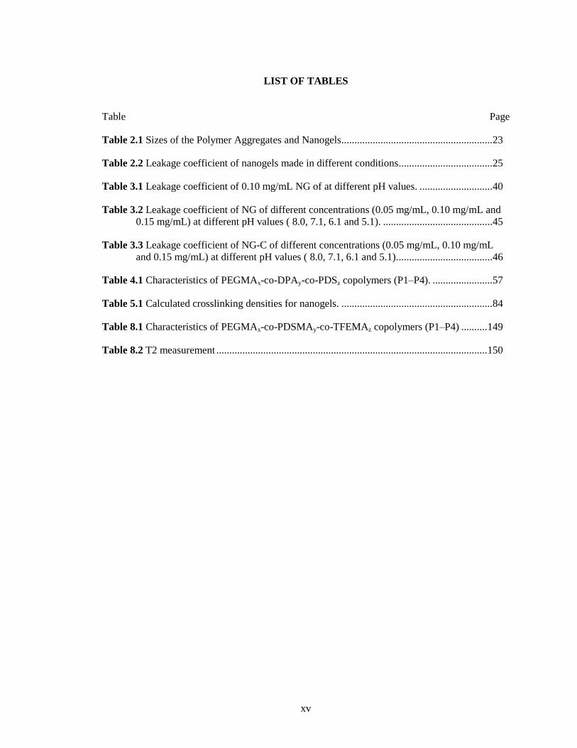
LIST OF TABLES
Table Page
Table 2.1 Sizes of the Polymer Aggregates and Nanogels .......................................................... 23
Table 2.2 Leakage coefficient of nanogels made in different conditions .................................... 25
Table 3.1 Leakage coefficient of 0.10 mg/mL NG of at different pH values. ............................ 40
Table 3.2 Leakage coefficient of NG of different concentrations (0.05 mg/mL, 0.10 mg/mL and
0.15 mg/mL) at different pH values ( 8.0, 7.1, 6.1 and 5.1). .......................................... 45
Table 3.3 Leakage coefficient of NG-C of different concentrations (0.05 mg/mL, 0.10 mg/mL
and 0.15 mg/mL) at different pH values ( 8.0, 7.1, 6.1 and 5.1). .................................... 46
Table 4.1 Characteristics of PEGMAx-co-DPAy-co-PDSz copolymers (P1–P4). ....................... 57
Table 5.1 Calculated crosslinking densities for nanogels. .......................................................... 84
Table 8.1 Characteristics of PEGMAx-co-PDSMAy-co-TFEMAz copolymers (P1–P4) .......... 149
Table 8.2 T2 measurement ........................................................................................................ 150
xvi
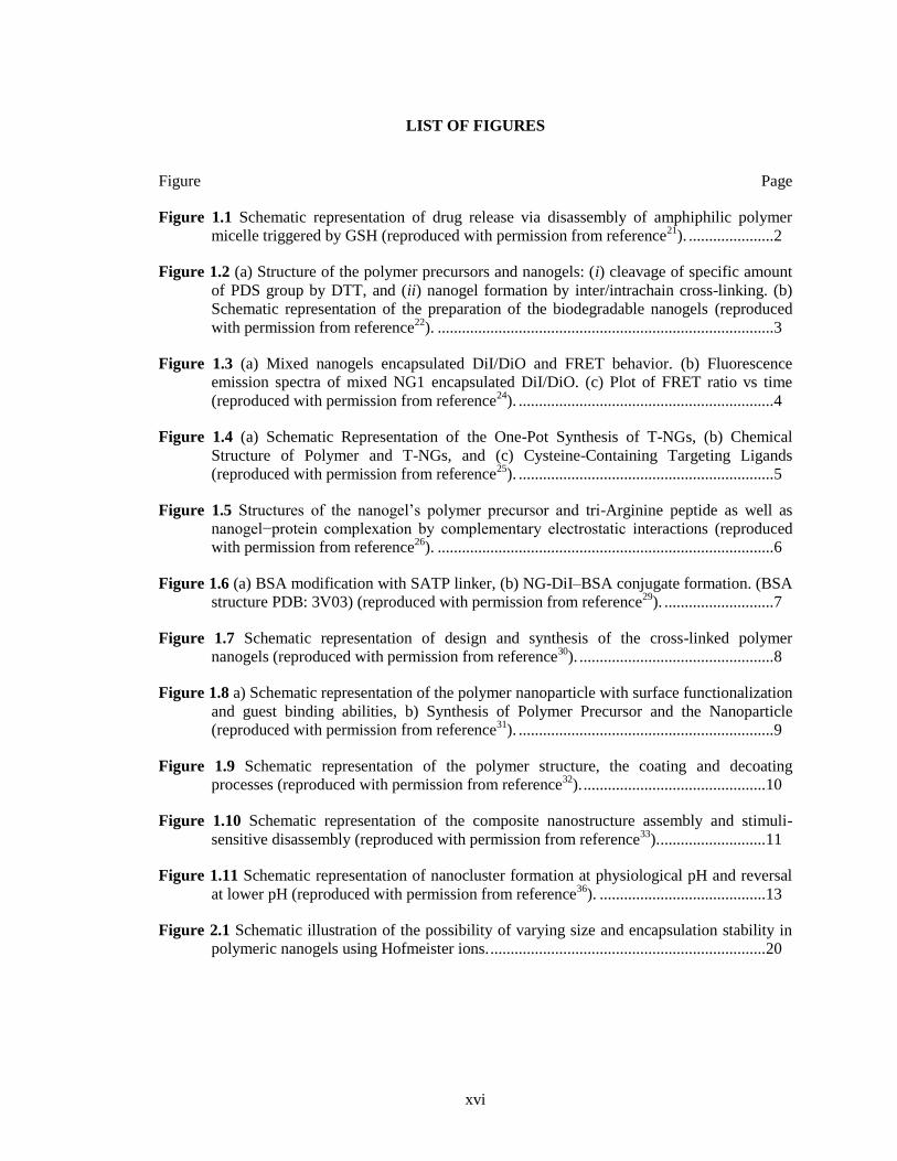
LIST OF FIGURES
Figure Page
Figure 1.1 Schematic representation of drug release via disassembly of amphiphilic polymer
micelle triggered by GSH (reproduced with permission from reference21
). ..................... 2
Figure 1.2 (a) Structure of the polymer precursors and nanogels: (i) cleavage of specific amount
of PDS group by DTT, and (ii) nanogel formation by inter/intrachain cross-linking. (b)
Schematic representation of the preparation of the biodegradable nanogels (reproduced
with permission from reference22
). ................................................................................... 3
Figure 1.3 (a) Mixed nanogels encapsulated DiI/DiO and FRET behavior. (b) Fluorescence
emission spectra of mixed NG1 encapsulated DiI/DiO. (c) Plot of FRET ratio vs time
(reproduced with permission from reference24
). ............................................................... 4
Figure 1.4 (a) Schematic Representation of the One-Pot Synthesis of T-NGs, (b) Chemical
Structure of Polymer and T-NGs, and (c) Cysteine-Containing Targeting Ligands
(reproduced with permission from reference25
). ............................................................... 5
Figure 1.5 Structures of the nanogel’s polymer precursor and tri-Arginine peptide as well as
nanogel−protein complexation by complementary electrostatic interactions (reproduced
with permission from reference26
). ................................................................................... 6
Figure 1.6 (a) BSA modification with SATP linker, (b) NG-DiI–BSA conjugate formation. (BSA
structure PDB: 3V03) (reproduced with permission from reference29
). ........................... 7
Figure 1.7 Schematic representation of design and synthesis of the cross-linked polymer
nanogels (reproduced with permission from reference30
). ................................................ 8
Figure 1.8 a) Schematic representation of the polymer nanoparticle with surface functionalization
and guest binding abilities, b) Synthesis of Polymer Precursor and the Nanoparticle
(reproduced with permission from reference31
). ............................................................... 9
Figure 1.9 Schematic representation of the polymer structure, the coating and decoating
processes (reproduced with permission from reference32
). ............................................. 10
Figure 1.10 Schematic representation of the composite nanostructure assembly and stimuli-
sensitive disassembly (reproduced with permission from reference33
). .......................... 11
Figure 1.11 Schematic representation of nanocluster formation at physiological pH and reversal
at lower pH (reproduced with permission from reference36
). ......................................... 13
Figure 2.1 Schematic illustration of the possibility of varying size and encapsulation stability in
polymeric nanogels using Hofmeister ions. .................................................................... 20
xvii
Figure 2.2 (a) LCST trend of polymer in salts with different anions solution, the salt
concentration is 30 mM/mL (b)LCST trend of polymer in salts with different cations
solution, the salt concentration is 30 mM/mL (c) LCST trend of polymer in Na2SO4
solution with different concentrations (d) LCST trend of polymer in NaSCN solution with
different concentrations. All the concentration of polymer solution is 10 mg/mL. ........ 22
Figure 2.3 Size of polymer aggregates (a) in Na2SO4 solution (b) in NaSCN solution; Size of
nanogels after dialysis (c) made in Na2SO4 solution (d) made in NaSCN solution. ....... 23
Figure 2.4 Fluorescence emission spectra of mixed NGs encapsulated DiI/DiO, (a) NG made in
100 mM Na2SO4(aq), (b) NG made in 100 mM NaSCN(aq). (c) Comparing the dynamics
of leakage/interchange of NGs made in different solution. ............................................ 24
Figure 2.5 Absorbance of the nanogel loaded with DiI after the addition of Na2SO4 or NaSCN.
Nanogel was made in salt free water, and concentration of both the measured nanogel
solutions is 0.2 mg/mL. (a-b) Nanogels with 6% crosslinking degree, (c-d) Nanogels with
13% crosslinking degree, (e-f) Nanogels with 25% crosslinking degree. ...................... 26
Figure 2.6 Change in morphology of the nanogels due to the presence of salts (a) nanogel
without salts (b) nanogel after adding Na2SO4 (c) nanogel after adding NaSCN from
AFM(height mode). ........................................................................................................ 27
Figure 2.7 Analysis of the nanogel AFM height images of the nanogels (a) in the absence of salt
(b) in the presence of Na2SO4 (c) in the presence of NaSCN. ........................................ 28
Figure 2.8 GSH-induced (10mM) DiI release rate from the nanogels made in different salt
solutions. ......................................................................................................................... 29
Figure 3.1 Schematic representation of two possible mechanisms for guest-exchange between
cross-linked nanogels. a) Collision – Exchange – Separation Mechanism, also called as
Collision- based mechanism b) Exit – Re - entry Mechanism, also called as Diffusion-
based mechanism. ........................................................................................................... 36
Figure 3.2 Size distribution of nanogel at different pH values via DLS measurements. a) NG with
30% crosslinking density, b) NG-C with 20% crosslinking density. .............................. 38
Figure 3.3 Fluorescence evolution when NG-C containing DiI and DiO are mixed in solutions at
pH 7.1. a) 0.05 mg/mL, b) 0.10 mg/mL, c) 0.15 mg/mL. d) Comparison of the dynamics
of guest-exchange between NG-C with different concentration at pH 7.1. .................... 39
Figure 3.4 Fluorescence evolution when NG containing DiI and DiO are mixed in solutions at
different pH values. a) pH 8.0, b) pH 6.4, c) pH 5.1. d) Guest-exchange rates between NG
at different pH values. The concentration of each nanogel was 0.10 mg/mL. ................ 40
Figure 3.5 Comparison of the dynamics of guest-exchange between NG-C with 0.10 mg/mL at
different pH values. ......................................................................................................... 42
Figure 3.6 Effect of pH on the hydrophobicity. a) Fluorescence emission spectrum measured for
nanogel NG-C loading 2 wt% pyrene and b) calculated I1/I3 ratios for nanogels at
different pH values, (black, NG; red, NG-C). ................................................................. 43
xviii
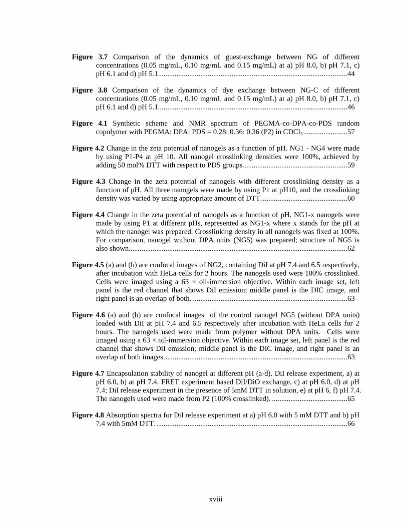
Figure 3.7 Comparison of the dynamics of guest-exchange between NG of different
concentrations (0.05 mg/mL, 0.10 mg/mL and 0.15 mg/mL) at a) pH 8.0, b) pH 7.1, c)
pH 6.1 and d) pH 5.1 ....................................................................................................... 44
Figure 3.8 Comparison of the dynamics of dye exchange between NG-C of different
concentrations (0.05 mg/mL, 0.10 mg/mL and 0.15 mg/mL) at a) pH 8.0, b) pH 7.1, c)
pH 6.1 and d) pH 5.1 ....................................................................................................... 46
Figure 4.1 Synthetic scheme and NMR spectrum of PEGMA-co-DPA-co-PDS random
copolymer with PEGMA: DPA: PDS = 0.28: 0.36: 0.36 (P2) in CDCl3 ........................ 57
Figure 4.2 Change in the zeta potential of nanogels as a function of pH. NG1 - NG4 were made
by using P1-P4 at pH 10. All nanogel crosslinking densities were 100%, achieved by
adding 50 mol% DTT with respect to PDS groups. ........................................................ 59
Figure 4.3 Change in the zeta potential of nanogels with different crosslinking density as a
function of pH. All three nanogels were made by using P1 at pH10, and the crosslinking
density was varied by using appropriate amount of DTT. .............................................. 60
Figure 4.4 Change in the zeta potential of nanogels as a function of pH. NG1-x nanogels were
made by using P1 at different pHs, represented as NG1-x where x stands for the pH at
which the nanogel was prepared. Crosslinking density in all nanogels was fixed at 100%.
For comparison, nanogel without DPA units (NG5) was prepared; structure of NG5 is
also shown....................................................................................................................... 62
Figure 4.5 (a) and (b) are confocal images of NG2, containing DiI at pH 7.4 and 6.5 respectively,
after incubation with HeLa cells for 2 hours. The nanogels used were 100% crosslinked.
Cells were imaged using a 63 × oil-immersion objective. Within each image set, left
panel is the red channel that shows DiI emission; middle panel is the DIC image, and
right panel is an overlap of both. .................................................................................... 63
Figure 4.6 (a) and (b) are confocal images of the control nanogel NG5 (without DPA units)
loaded with DiI at pH 7.4 and 6.5 respectively after incubation with HeLa cells for 2
hours. The nanogels used were made from polymer without DPA units. Cells were
imaged using a 63 × oil-immersion objective. Within each image set, left panel is the red
channel that shows DiI emission; middle panel is the DIC image, and right panel is an
overlap of both images. ................................................................................................... 63
Figure 4.7 Encapsulation stability of nanogel at different pH (a-d). DiI release experiment, a) at
pH 6.0, b) at pH 7.4. FRET experiment based DiI/DiO exchange, c) at pH 6.0, d) at pH
7.4; DiI release experiment in the presence of 5mM DTT in solution, e) at pH 6, f) pH 7.4.
The nanogels used were made from P2 (100% crosslinked). ......................................... 65
Figure 4.8 Absorption spectra for DiI release experiment at a) pH 6.0 with 5 mM DTT and b) pH
7.4 with 5mM DTT. ........................................................................................................ 66
xix
Figure 4.9 (a) and (b) are confocal images of fluorescein-labeled DPA nanogel at pH 7.4 and 6.5
after incubation with HeLa cells for 2 hours. The concentration of the nanogels used was
1mg/mL. Cells were imaged using a 63 × oil-immersion objective. Within each image set,
left panel is the green channel that shows FITC emission; middle panel is the DIC image,
and right panel is an overlap of both. .............................................................................. 67
Figure 5.1 Time-dependent 1H NMR spectra of the photo-induced reaction of 2-hydroxyethyl 2-
pyridyl disulfide (PDS-OH, 1). Control spectra of bis(2-hydroxyethyl) disulfide (DPDS,
2), 2, 2’-dipyridyl disulfide (3) and 2-thiopyridone were also included. The concentration
was 2.5 mg/mL. The final product after photoirradiation was determined to be pyridine-2-
sulfonic acid (4). ............................................................................................................. 78
Figure 5.2 Concentration dependent of reaction rate of PDS under photoirradiation. ................ 79
Figure 5.3 Time-dependent 1H NMR spectra of the photo-induced disulfide metathesis of 2-
hydroxyethyl 2-pyridyl disulfide (PDS-OH, 1) under anaerobic condition. Control spectra
of bis(2-hydroxyethyl) disulfide (2), 2, 2’-dipyridyl disulfide (DPDS, 3) and 2-
thiopyridone were also included. The concentration was 2.5 mg/mL. ........................... 80
Figure 5.4 Time-dependent 1H NMR spectra of 2, 2’-dipyridyl disulfide (DPDS) under the
photoirradiation without O2. ....................................................................................... 81
Figure 5.5 DLS sizes in hydrodynamic diameter, a) micelle aggregates prepared with polymer
solutions with different concentrations, b) nanogels prepared with polymer solutions with
different concentrations, the photoirradiation time was 2 h for all samples. .................. 83
Figure 5.6 a) Absorption spectra of pyridothione in UV-vis. Pyridothione, which is a byproduct
by the reaction between PDS with DTT and shows characteristic absorption at 343 nm
wavelength, is monitored in each nanogel (2 mg/mL) prepared. b) Time dependent
crosslinking density of the nanogels with different concentrations of polymer solution,
from 1 mg/mL to 10 mg/mL. .......................................................................................... 84
Figure 5.7 Dye release from the nanogels prepared via photoirradiation for 2 h in response to
varied GSH concentrations, a) 0 mM, b) 10 mM. Sample concentration was 0.1 mg/mL.
FRET experiment based DiI/DiO exchange, c) micelle aggregates, d) nanogels prepared
via photoirradiation for 1 h, e) nanogels prepared via photoirradiation for 2 h. Sample
concentration was 0.1 mg/mL. ........................................................................................ 86
Figure 6.1 Synthesis and characterization of boronate nanoparticle (BNP). a) Scheme presents
the syntheses of Im-Ca and Im-BA monomers, as well as polymer P1 via the
polyboronate esterification between Im-Ca and Im-BA. b) TEM image of the BNP, the
scale bar is 500 nm. c) SEM image of the BNP, the scale bar is 5μm. Conditions:
dropwise added 1 mL 5 mg/mL Im-Ca methanol solution into 1 mL 5 mg/mL Im-BA
methanol solution with stirring at room temperature. The TEM and SEM samples were
taken immediately after the addition. d) FTIR spectra of molecule Im-BA, Im-Ca and
solid BNP, and e) Zoomed-in FTIR spectra from (d). .................................................... 97
xx
Figure 6.2 Particle size and absorption characteristics variation. a) DLS of the BNP prepared
with different monomer concentration in methanol. TEM images of the BNP, prepared
with different monomer concentration in methanol, b) 1.5 mg/mL, c) 3.0 mg/mL, d) 5
mg/mL. The scale bar is 500 nm. e) Normalized absorption spectra for Im-BA, Im-Ca
and BNP prepared with different monomer concentration in methanol. Temporal
evolution of absorption spectra for BNP prepared with f) 3 mg/mL in methanol, g) 5
mg/mL in methanol. All samples were prepared by adding 0.01 mL reaction mixture into
1 mL methanol. ............................................................................................................... 98
Figure 6.3 PXRD pattern of the BNP. A broad bump at about 2θ=20 was resulted from
crystalline parts of polymers. .......................................................................................... 99
Figure 6.4 Absorption spectra for Im-BA and Im-Ca as the time. The concentration for both
solutions is 0.02 mg/mL. ............................................................................................... 100
Figure 6.5 Mechanism study of the particle formation. a) Photographs of the BNP solution with
different ratios of Im-Ca into Im-BA in methanol. b) DLS of the BNP prepared with
different addition volumes of Im-Ca into 1 mL of Im-BA solution. TEM images of the
corresponding solutions, c) 0.10 mL Im-Ca + 1.0 mL Im-BA, d) 0.30 mL Im-Ca + 1.0
mL Im-BA, e) 0.65 mL Im-Ca + 1.0 mL Im-BA, f) 1.0 mL Im-Ca + 1.0 mL Im-BA.
The scale bar is 500 nm. Absorption spectra of the reaction mixture with different initial
concentrations during the dropwise addition, g) 1 mg/mL, h) 2 mg/mL, i) 3 mg/mL. Note
that there were flat lines between 500 and 525 nm, because samples had an absorbance of
more than dynamic range of instrument. ...................................................................... 102
Figure 6.6 Solid 11
B-NMR spectra of Im-BA, boronate ester from 4-formylphenylboronic acid
and 3,4-dihydroxybenzaldehyde, and solid BNP. ......................................................... 105
Figure 6.7 UV-vis spectra of a), St-BA, St-Ca, Im-BA and Im-Ca in ethanol; b), their one to one
mixture in ethanol. ........................................................................................................ 106
Figure 6.8 a)Typical photos of the mixtures using Im-BA react with Im-Ca2 and Im-Ca6 in
methanol, and their TEM pictures of BNP using b) Im-Ca2, c) Im-Ca6. The scale bar is
500 nm. ......................................................................................................................... 106
Figure 6.9 Solvent effects on the particle formation. TEM images of the BNP prepared with
methanol (M) and DMSO (D) mixture solution, a) M/D=4/1, b) M/D=6/1, c) M/D=9/1, d)
Pure M. The scale bar is 500 nm................................................................................... 107
Figure 6.10 Responsive behavior of BNP based on dative N→B bond. a) Photographs of the
BNP with the addition of pyridine, imidazole, 4-dimethylaminopyridine, b) Photographs
of the BNP with changing temperature and their TEM images of the BNP, c) before
heating, d) after first heat-cool cycle, e) after second heat-cool cycle, f) after third heat-
cool cycle. The scale bars are 500 nm. ......................................................................... 108
Figure 6.11 TEM images of the BNP, a) before the heating and cooling at room temperature for
b) 1 min, c) 5 mins, d) 10 mins. .................................................................................... 110
Figure 6.12 Aging of the BNP in methanol. TEM images of the samples taken after a) 5 mins
after the mixing, size is 330 nm, b) 3 days after the mixing, size is 320 nm. The scale bar
is 500 nm. ...................................................................................................................... 119
xxi
Figure 6.13 NMR spectra showing stability of the imine bond as time. ................................... 120
Figure 6.14 TGA curves of Im-BA, Im-Ca and solid BNP. .................................................... 121
Figure 7.1 a) TEM image of the BNP; b) TEM image of the hollow Fe3+
-MOP; c) SEM image of
the hollow Fe3+
-MOP; Inset is a SEM image of one broken hollow Fe3+
-MOP. All the
scale bar is 500 nm. ....................................................................................................... 127
Figure 7.2 a) EELS mapped images of BNP; b) EELS mapped images of hollow Fe3+
-MOP; All
the scale bar is 500 nm. ................................................................................................. 127
Figure 7.3 Fe, C, N and O line-scanned EELS profiles of two nanoparticles in Figure 1f. EELS
images and line-scan profiles confirm the hollow structure of hollow Fe3+
-MOP. ...... 128
Figure 7.4 Bright-field STEM images and EDS maps of hollow Fe3+
-MOP, element C and Fe
included. ........................................................................................................................ 129
Figure 7.5 a) TEM image of the hollow Cu2+
-MOP by adding Cu2+
; b) TEM image of the hollow
Sn4+
-MOP adding Sn4+
; c) TEM image of the hollow Al3+
-MOP adding Al3+
; All the
scale bar is 500 nm. ....................................................................................................... 130
Figure 7.6 a) Bright-field STEM images and EDS maps of hollow Sn4+
-MOP, element C and Sn
included; b) HAADF STEM images and EDS maps of dual Fe3+
/Sn4+
-hollow MOP,
element C, Fe and Sn included. .................................................................................... 130
Figure 7.7 EPR spectra of BNP and hollow MOP. 295 K, X-band (9.8 GHz) EPR spectrum
measured at room temperature for BNP (black), hollow Al3+
-MOP (green), hollow Fe3+
-
MOP (blue), dual hollow Fe3+
/Al3+
-MOP (red) (starting composition was 0.05 mol
fraction of FeCl3/AlCl3). Inset: g' ~ 9.6 transition of the dual hollow Fe3+
/Al3+
-MOPs on
an expanded scale. ........................................................................................................ 132
Figure 7.8 TEM images taken at a) 0 min, b) 20 min, c) 1 h and d) 6 h after the addition of FeCl3
into BNP; the scale bar is 500 nm. Conditions: 0.1 mL FeCl3 methanol solution was
added into 1 mL BNP methanol solution prepared using 3 mg/mL Im-BA and Im-Ca, the
ratio of Fe3+
to Im-Ca was 1/2. The TEM samples were taken by drop-casting. The scale
bar is 500 nm................................................................................................................. 133
Figure 7.9 Time dependent TEM measurement, taken after 30 min, 1 h, and 2 h. Conditions: 0.1
mL SnCl4 methanol solution was added into 1 mL BNP methanol solution prepared using
3 mg/mL Im-BA and Im-Ca, the ratio of Sn4+
to Im-Ca was 1/2. The TEM samples were
taken by drop-casting. The scale bar is 500 nm. ........................................................... 133
Figure 7.10 The thickness of shell could be tuned by changing the ratio of Fe3+
to Im-Ca, a) 1/8,
b) 1/2, c) 3/4.However, when the ratio increased up to 1/1, only solid MOPs were
observed as shown TEM in figure d). EDS maps of solid Fe3+
-MOP, element C (e) and
Fe (f) included. , all scale bar is 500 nm except e) and f) with 250 nm scale bar. ....... 134
Figure 7.11 The size of hollow MOP could be varied by tuning the size of BNP used, a, d) 1.5
mg/mL monomers, b, e) 3.0 mg/mL monomers, c, f) 5.0 mg/mL monomers. The ratio of
Fe3+
to Im-Ca was 1/2; all scale bar is 500 nm. ............................................................ 135
xxii
Figure 7.12 Preparation and application of hollow Fe@C particles. TEM measurement of a)
hollow Fe@C particles after the temperature thermal treatment of MOPs, b) hollow
carbon particles after the acid treatment of Fe@C; c) Optical photograph of the MB
solution without (left) and with (right) the addition of Fe@C applying an external magnet;
d) UV-vis absorption measurement before and after the magnetic separation. ............ 136
Figure 7.13 HAADF STEM images and EDX line scanning data. ........................................... 137
Figure 8.1 Schematic representation of the functionalities for an effective drug delivery system
after the injection. ...................................................................................................................... 144
Figure 8.2 a) pH-activatable ON/OFF 19
F-MRI nanoprobes from ionizable diblock copolymers.
At pH > pKa, the hydrophobic segments selfassemble into a micelle core leading to 19
F
signal suppression as a result of restricted polymer chain motion. Upon pH activation (pH
< pKa), micelle disassembly leads to dissociated unimers and a strong 19
F signal. b)
Structural formula of three representative diblock copolymers containing different pH
responsive segments and 19
F reporter moieties, their pKa values and 19
F chemical shifts
(in ppm, relative to trifluoroacetic acid, or TFA), respectively, are shown in parenthesis
(reproduced with permission from reference15
). ........................................................... 148
Figure 8.3 GPC results of P1-P3. .............................................................................................. 149
Figure 8.4 Typical NMR spectrum of P2. ................................................................................. 150
Figure 8.5 19
F NMR spectra were taken for all three polymers before and after the addition of
GSH in water containing 10% D2O. ............................................................................. 151
Figure 8.6 Hollow carbon particles loaded with metal nanoparticles (M@Fe), a) Fe@C, b)
Cu@C, c) Sn@C. .......................................................................................................... 152
Figure 8.7 XRD results of M@C. Typical peaks were marked. ............................................... 153
Figure 8.8 TGA result, a) MOPs and BNP in N2, b) M@C in air. ........................................... 154
Figure 8.9 The time-dependent absorption spectra of the reaction solution in the presence of 0.3
mg of Cu@C. ................................................................................................................ 155
xxiii
LIST OF SCHEMES
Scheme Page
Scheme 2.1 Structure of the polymer and the nanogel containing OEG units. ........................... 21
Scheme 3.1 Structure of polymers P1 and P2; nanogels NG-C and NG; and the dye molecules
DiO and DiI. ................................................................................................................... 37
Scheme 4.1 Schematic illustration and chemical structure of polymeric nanogels for pH-induced
surface charge generation and activated cellular uptake. ................................................ 55
Scheme 5.1 Hypothesized reaction scheme of photo-induced disulfide metathesis of 2-pyridyl
disulfide (PDS-OH) ........................................................................................................ 78
Scheme 5.2 Scheme of photo-induced chemistry reaction of PDS-OH. ..................................... 79
Scheme 5.3 Scheme of photo-induced chemistry reaction of 2-pyridyl disulfide (PDS-OH) under
anaerobic condition. ........................................................................................................ 81
Scheme 5.4 Representation of photo-induced crosslinking reaction of random copolymers
containing PDS groups. .................................................................................................. 82
Scheme 6.1 Structure of St-Ca, St-BA, Im-Ca2 and Im-Ca6. ................................................ 106
Scheme 6.2 Synthesis of Im-Ca. ............................................................................................... 111
Scheme 6.3 Synthesis of Im-BA. .............................................................................................. 112
Scheme 6.4 Synthesis of mono-Im-Ca. .................................................................................... 113
Scheme 6.5 Synthesis of Im-Ca2. ............................................................................................. 113
Scheme 6.6 Synthesis of Im-Ca6. ............................................................................................. 114
Scheme 6.7 Synthesis of tetraethyl (1,4-phenylenebis(methylene))bis(phosphonate). ............. 115
Scheme 6.8 Protection of 4-formylphenylboronic acid. ............................................................ 115
Scheme 6.9 Protection of 3, 4-dihydroxybenzaldehyde. ........................................................... 116
Scheme 6.10 Synthesis of St-BA. ............................................................................................. 116
Scheme 6.11 Synthesis of St-Ca. .............................................................................................. 117
Scheme 7.1 Scheme presents the syntheses of BNP via Im-Ca and Im-BA, and the preparation of
hollow MOP by adding metal chloride salts to the BNP solution. .............................. 126
Scheme 7.2 Schematic illustration showing the transformation of solid polymer nanoparticles to
hollow or solid metal organic particles, depending on the relative diffusivities........... 134

xxiv
Scheme 8.1 Synthetic scheme of PEGMAx-co-PDSMAy-co-TFEMAz random copolymer. .... 149
1
CHAPTER 1
INTRODUCTION
Li, L.; Raghupathi, K.; Song, C.; Prasad, P.; Thayumanavan, S. Self-assembly of random
copolymers. Chem. commun. 2014, 50, 13417-13432. - Reproduced by permission of The Royal
Society of Chemistry
http://pubs.rsc.org/en/content/articlehtml/2014/cc/c4cc03688c
1.1 Introduction
Self-assembly of amphiphilic copolymers are of great interest for many decades because
these materials are able to offer a rich variety of morphologies and transitions as well as their
potential applications in many fields, such as biomedical, micro-electronic, photoelectric and
optical materials.1–5
Significant progress has been made in the design and synthesis of a variety of
amphiphilic copolymers owing to advances in controlled polymerization techniques, such as
nitroxide mediated radical polymerization (NMP), atom transfer radical polymerization (ATRP),
reversible addition–fragmentation chain transfer polymerization (RAFT) and ring-opening
mediated radical polymerization (ROMP).6–11
Copolymers can be broadly classified into two
categories: block copolymers and random copolymers. In the case of block copolymers, the
monomers are arranged systematically in the form of blocks where each block is a repetition of a
certain monomer species, whereas in the case of random copolymers different monomeric
components of the polymer are randomly arranged where the probability of finding a given
monomeric unit at any given location on the polymer is independent of the nature of the adjacent
units. Until recently, much focus has been directed towards understanding the self-assembly of
block copolymers, due to their unique and excellent assembly behaviours.12–17
However, their
synthetic methods can be tedious and time-consuming, as it involves sequential controlled
polymerization or post-polymerization treatments such as grafting, substitution, hydrolysis and
‘‘click’’ chemistries.18–20
Compared to block copolymers, preparation of random copolymers is
2
relatively easy, as they are typically achieved in a one step co-polymerization of two (or more)
different monomers. Therefore, it is intriguing to highlight the supramolecular capabilities of
amphiphilic random copolymers in self-assembly.
1.2 Simple nanogels based on amphiphilic random copolymers
Our group has had a longstanding interest in using random copolymers for self-assembly.
Amphiphilic random copolymers, containing triethylene glycol as the hydrophilic part and an
alkyl chain connected by the disulfide bond as the hydrophobic part, were prepared by free
radical polymerization.21
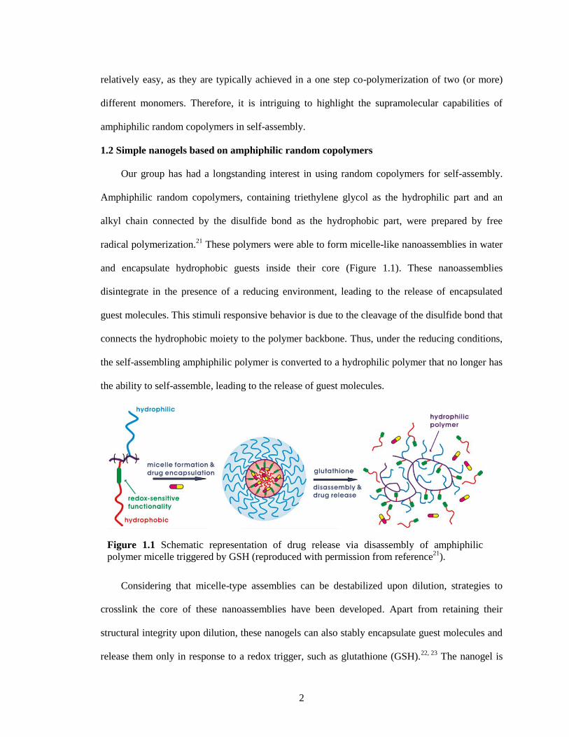
These polymers were able to form micelle-like nanoassemblies in water
and encapsulate hydrophobic guests inside their core (Figure 1.1). These nanoassemblies
disintegrate in the presence of a reducing environment, leading to the release of encapsulated
guest molecules. This stimuli responsive behavior is due to the cleavage of the disulfide bond that
connects the hydrophobic moiety to the polymer backbone. Thus, under the reducing conditions,
the self-assembling amphiphilic polymer is converted to a hydrophilic polymer that no longer has
the ability to self-assemble, leading to the release of guest molecules.
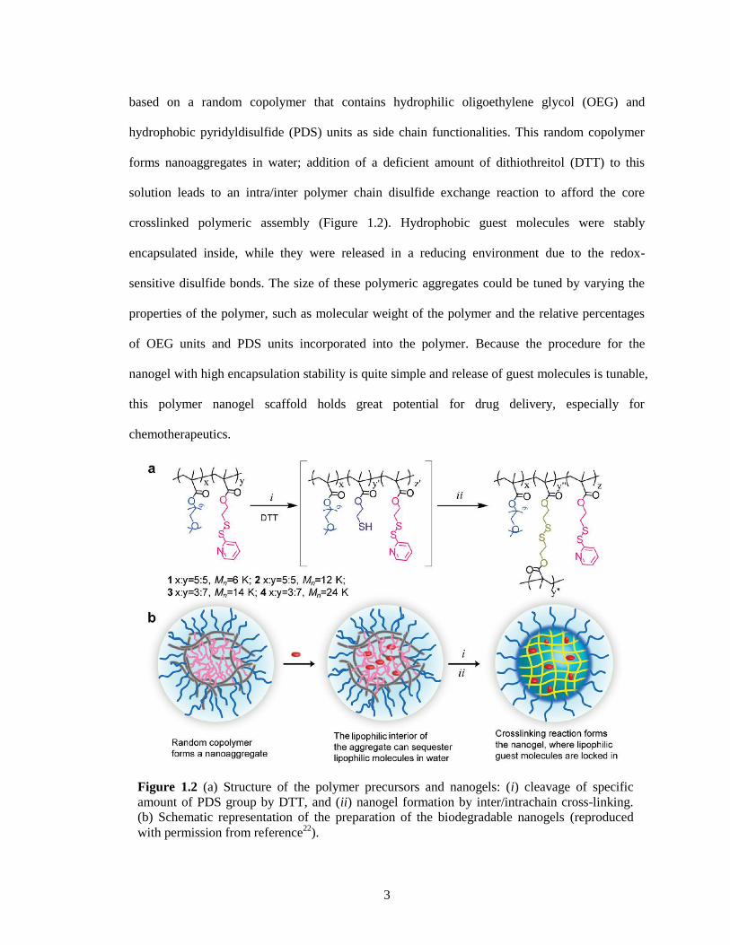
Considering that micelle-type assemblies can be destabilized upon dilution, strategies to
crosslink the core of these nanoassemblies have been developed. Apart from retaining their
structural integrity upon dilution, these nanogels can also stably encapsulate guest molecules and
release them only in response to a redox trigger, such as glutathione (GSH).22, 23
The nanogel is
Figure 1.1 Schematic representation of drug release via disassembly of amphiphilic
polymer micelle triggered by GSH (reproduced with permission from reference21
).
3
based on a random copolymer that contains hydrophilic oligoethylene glycol (OEG) and
hydrophobic pyridyldisulfide (PDS) units as side chain functionalities. This random copolymer
forms nanoaggregates in water; addition of a deficient amount of dithiothreitol (DTT) to this
solution leads to an intra/inter polymer chain disulfide exchange reaction to afford the core
crosslinked polymeric assembly (Figure 1.2). Hydrophobic guest molecules were stably
encapsulated inside, while they were released in a reducing environment due to the redox-
sensitive disulfide bonds. The size of these polymeric aggregates could be tuned by varying the
properties of the polymer, such as molecular weight of the polymer and the relative percentages
of OEG units and PDS units incorporated into the polymer. Because the procedure for the
nanogel with high encapsulation stability is quite simple and release of guest molecules is tunable,
this polymer nanogel scaffold holds great potential for drug delivery, especially for
chemotherapeutics.
Figure 1.2 (a) Structure of the polymer precursors and nanogels: (i) cleavage of specific
amount of PDS group by DTT, and (ii) nanogel formation by inter/intrachain cross-linking.
(b) Schematic representation of the preparation of the biodegradable nanogels (reproduced
with permission from reference22
).
4
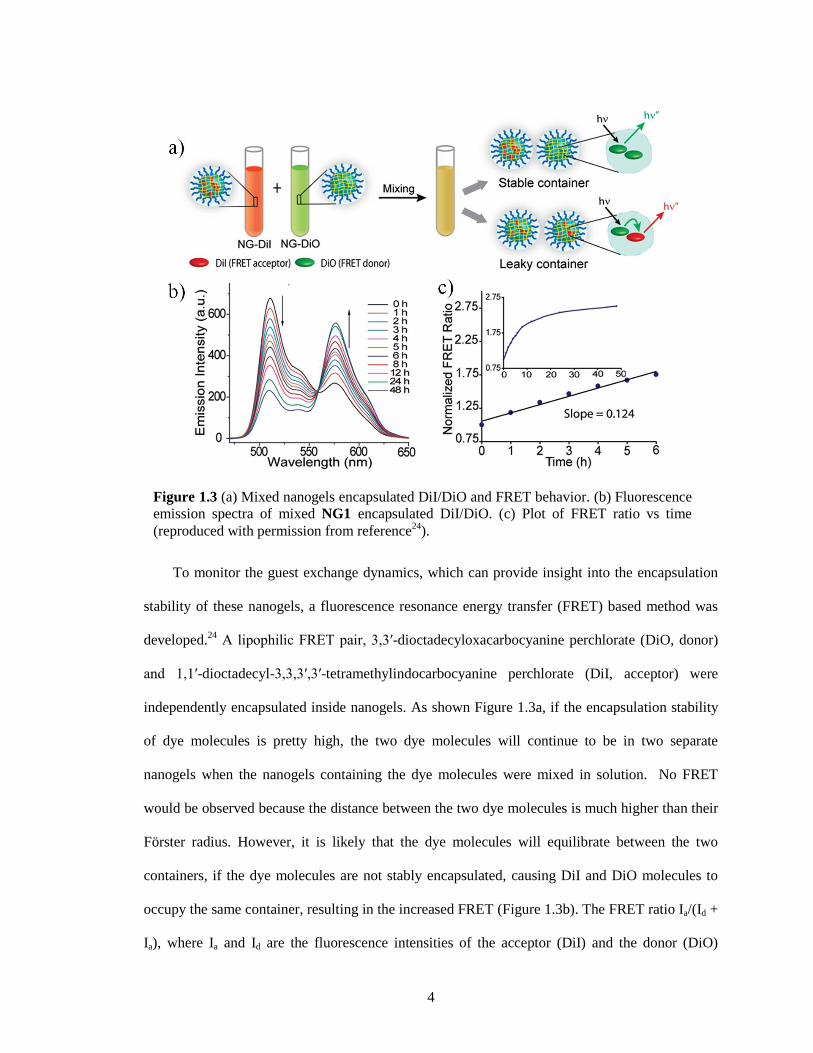
To monitor the guest exchange dynamics, which can provide insight into the encapsulation
stability of these nanogels, a fluorescence resonance energy transfer (FRET) based method was
developed.24
A lipophilic FRET pair, 3,3′-dioctadecyloxacarbocyanine perchlorate (DiO, donor)
and 1,1′-dioctadecyl-3,3,3′,3′-tetramethylindocarbocyanine perchlorate (DiI, acceptor) were
independently encapsulated inside nanogels. As shown Figure 1.3a, if the encapsulation stability
of dye molecules is pretty high, the two dye molecules will continue to be in two separate
nanogels when the nanogels containing the dye molecules were mixed in solution. No FRET
would be observed because the distance between the two dye molecules is much higher than their
Förster radius. However, it is likely that the dye molecules will equilibrate between the two
containers, if the dye molecules are not stably encapsulated, causing DiI and DiO molecules to
occupy the same container, resulting in the increased FRET (Figure 1.3b). The FRET ratio Ia/(Id +
Ia), where Ia and Id are the fluorescence intensities of the acceptor (DiI) and the donor (DiO)
Figure 1.3 (a) Mixed nanogels encapsulated DiI/DiO and FRET behavior. (b) Fluorescence
emission spectra of mixed NG1 encapsulated DiI/DiO. (c) Plot of FRET ratio vs time
(reproduced with permission from reference24
).
5
respectively, was plotted against time. The slope of the linear fit is defined as the leakage
coefficient (Λ), which shows the dynamics of the guest exchange (Figure 1.3c). It showed that the
encapsulation stability of nanogels depends on the degree of cross-linking.
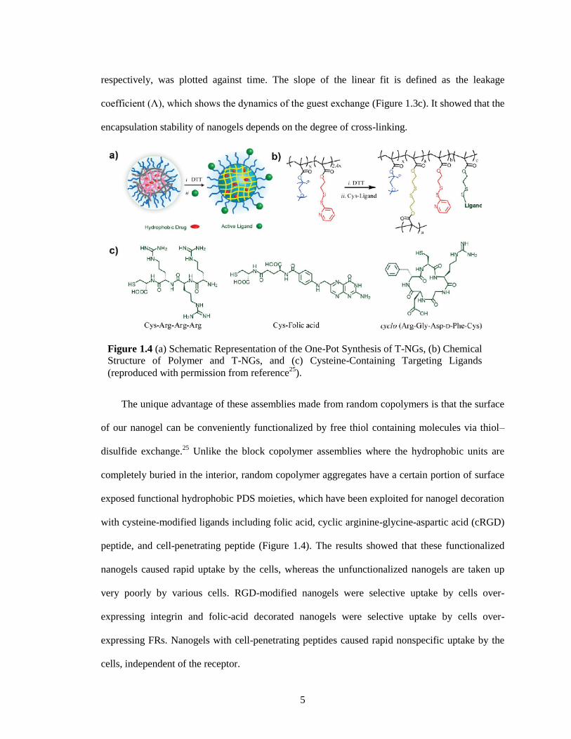
The unique advantage of these assemblies made from random copolymers is that the surface
of our nanogel can be conveniently functionalized by free thiol containing molecules via thiol–
disulfide exchange.25
Unlike the block copolymer assemblies where the hydrophobic units are
completely buried in the interior, random copolymer aggregates have a certain portion of surface
exposed functional hydrophobic PDS moieties, which have been exploited for nanogel decoration
with cysteine-modified ligands including folic acid, cyclic arginine-glycine-aspartic acid (cRGD)
peptide, and cell-penetrating peptide (Figure 1.4). The results showed that these functionalized
nanogels caused rapid uptake by the cells, whereas the unfunctionalized nanogels are taken up
very poorly by various cells. RGD-modified nanogels were selective uptake by cells over-
expressing integrin and folic-acid decorated nanogels were selective uptake by cells over-
expressing FRs. Nanogels with cell-penetrating peptides caused rapid nonspecific uptake by the
cells, independent of the receptor.
Figure 1.4 (a) Schematic Representation of the One-Pot Synthesis of T-NGs, (b) Chemical
Structure of Polymer and T-NGs, and (c) Cysteine-Containing Targeting Ligands
(reproduced with permission from reference25
).
6
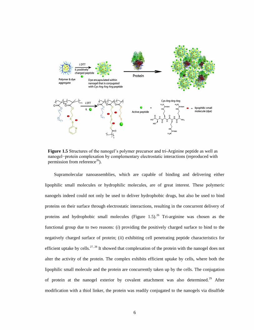
Supramolecular nanoassemblies, which are capable of binding and delivering either
lipophilic small molecules or hydrophilic molecules, are of great interest. These polymeric
nanogels indeed could not only be used to deliver hydrophobic drugs, but also be used to bind
proteins on their surface through electrostatic interactions, resulting in the concurrent delivery of
proteins and hydrophobic small molecules (Figure 1.5).26
Tri-arginine was chosen as the
functional group due to two reasons: (i) providing the positively charged surface to bind to the
negatively charged surface of protein; (ii) exhibiting cell penetrating peptide characteristics for
efficient uptake by cells.27, 28
It showed that complexation of the protein with the nanogel does not
alter the activity of the protein. The complex exhibits efficient uptake by cells, where both the
lipophilic small molecule and the protein are concurrently taken up by the cells. The conjugation
of protein at the nanogel exterior by covalent attachment was also determined.29
After
modification with a thiol linker, the protein was readily conjugated to the nanogels via disulfide
Figure 1.5 Structures of the nanogel’s polymer precursor and tri-Arginine peptide as well as
nanogel−protein complexation by complementary electrostatic interactions (reproduced with
permission from reference26
).
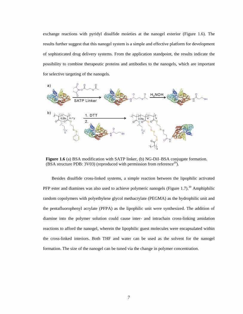
7
exchange reactions with pyridyl disulfide moieties at the nanogel exterior (Figure 1.6). The
results further suggest that this nanogel system is a simple and effective platform for development
of sophisticated drug delivery systems. From the application standpoint, the results indicate the
possibility to combine therapeutic proteins and antibodies to the nanogels, which are important
for selective targeting of the nanogels.
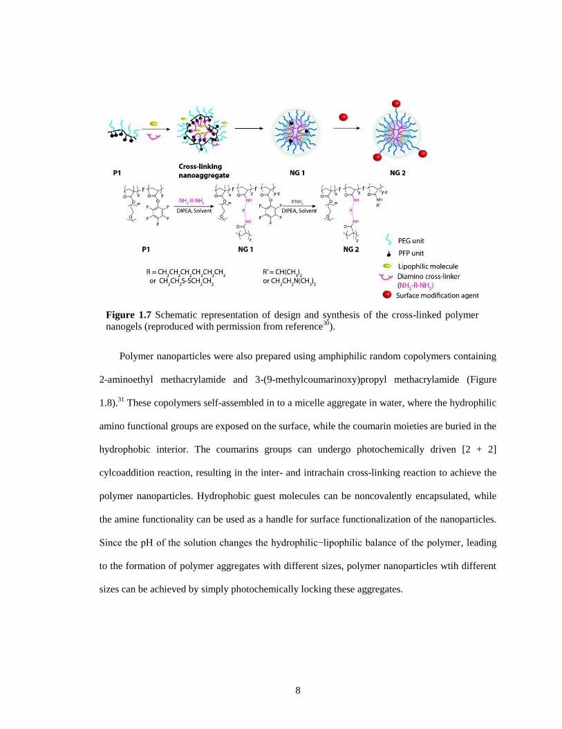
Besides disulfide cross-linked systems, a simple reaction between the lipophilic activated
PFP ester and diamines was also used to achieve polymeric nanogels (Figure 1.7).30
Amphiphilic
random copolymers with polyethylene glycol methacrylate (PEGMA) as the hydrophilic unit and
the pentafluorophenyl acrylate (PFPA) as the lipophilic unit were synthesized. The addition of
diamine into the polymer solution could cause inter- and intrachain cross-linking amidation
reactions to afford the nanogel, wherein the lipophilic guest molecules were encapsulated within
the cross-linked interiors. Both THF and water can be used as the solvent for the nanogel
formation. The size of the nanogel can be tuned via the change in polymer concentration.
Figure 1.6 (a) BSA modification with SATP linker, (b) NG-DiI–BSA conjugate formation.
(BSA structure PDB: 3V03) (reproduced with permission from reference29
).
8
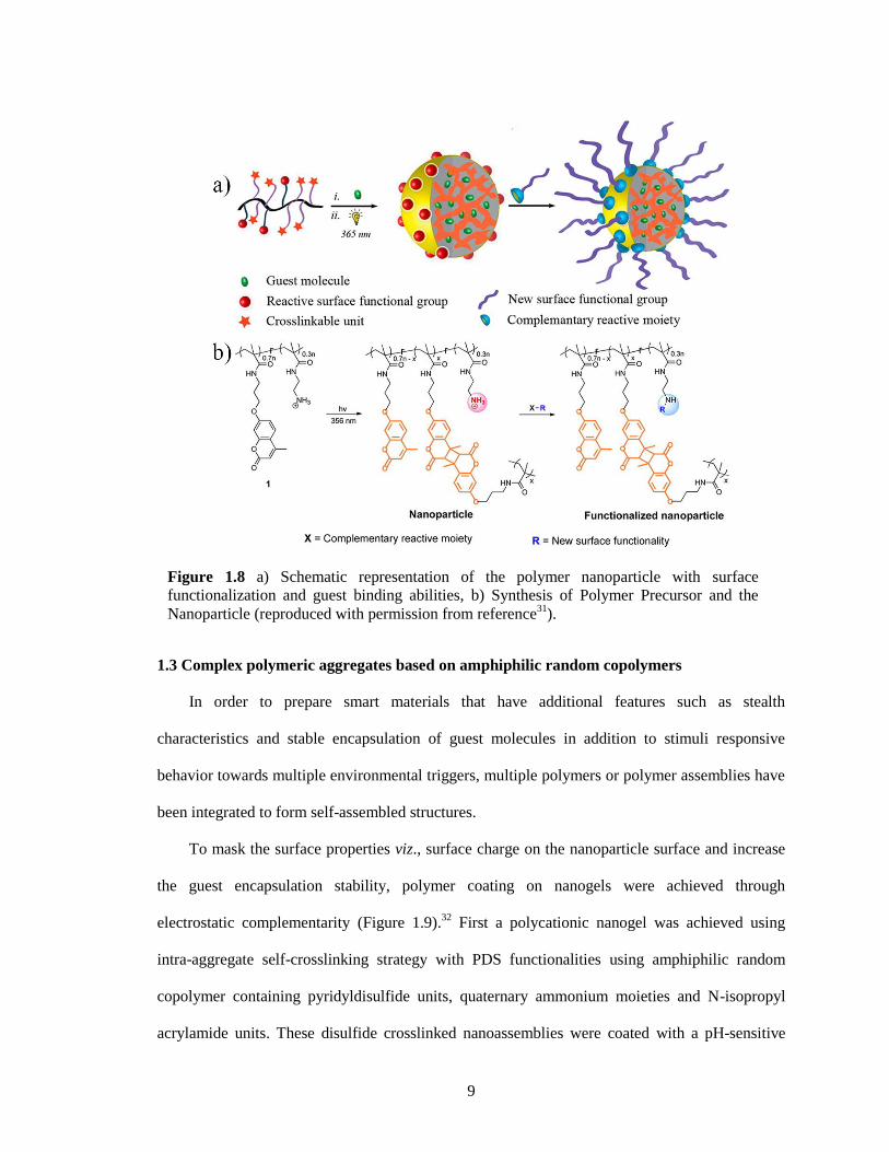
Polymer nanoparticles were also prepared using amphiphilic random copolymers containing
2-aminoethyl methacrylamide and 3-(9-methylcoumarinoxy)propyl methacrylamide (Figure
1.8).31
These copolymers self-assembled in to a micelle aggregate in water, where the hydrophilic
amino functional groups are exposed on the surface, while the coumarin moieties are buried in the
hydrophobic interior. The coumarins groups can undergo photochemically driven [2 + 2]
cylcoaddition reaction, resulting in the inter- and intrachain cross-linking reaction to achieve the
polymer nanoparticles. Hydrophobic guest molecules can be noncovalently encapsulated, while
the amine functionality can be used as a handle for surface functionalization of the nanoparticles.
Since the pH of the solution changes the hydrophilic−lipophilic balance of the polymer, leading
to the formation of polymer aggregates with different sizes, polymer nanoparticles wtih different
sizes can be achieved by simply photochemically locking these aggregates.
Figure 1.7 Schematic representation of design and synthesis of the cross-linked polymer
nanogels (reproduced with permission from reference30
).
9
1.3 Complex polymeric aggregates based on amphiphilic random copolymers
In order to prepare smart materials that have additional features such as stealth
characteristics and stable encapsulation of guest molecules in addition to stimuli responsive
behavior towards multiple environmental triggers, multiple polymers or polymer assemblies have
been integrated to form self-assembled structures.
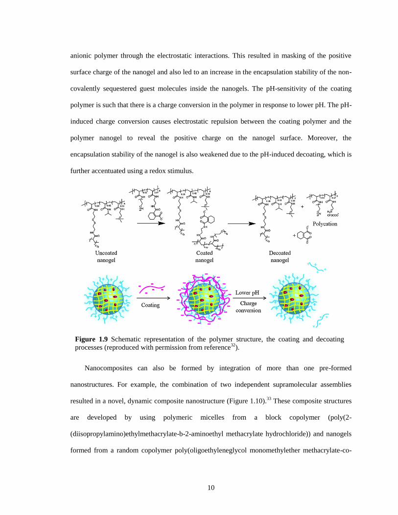
To mask the surface properties viz., surface charge on the nanoparticle surface and increase
the guest encapsulation stability, polymer coating on nanogels were achieved through
electrostatic complementarity (Figure 1.9).32
First a polycationic nanogel was achieved using
intra-aggregate self-crosslinking strategy with PDS functionalities using amphiphilic random
copolymer containing pyridyldisulfide units, quaternary ammonium moieties and N-isopropyl
acrylamide units. These disulfide crosslinked nanoassemblies were coated with a pH-sensitive
Figure 1.8 a) Schematic representation of the polymer nanoparticle with surface
functionalization and guest binding abilities, b) Synthesis of Polymer Precursor and the
Nanoparticle (reproduced with permission from reference31
).
10
anionic polymer through the electrostatic interactions. This resulted in masking of the positive
surface charge of the nanogel and also led to an increase in the encapsulation stability of the non-
covalently sequestered guest molecules inside the nanogels. The pH-sensitivity of the coating
polymer is such that there is a charge conversion in the polymer in response to lower pH. The pH-
induced charge conversion causes electrostatic repulsion between the coating polymer and the
polymer nanogel to reveal the positive charge on the nanogel surface. Moreover, the
encapsulation stability of the nanogel is also weakened due to the pH-induced decoating, which is
further accentuated using a redox stimulus.
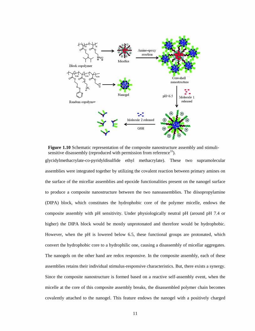
Nanocomposites can also be formed by integration of more than one pre-formed
nanostructures. For example, the combination of two independent supramolecular assemblies
resulted in a novel, dynamic composite nanostructure (Figure 1.10).33
These composite structures
are developed by using polymeric micelles from a block copolymer (poly(2-
(diisopropylamino)ethylmethacrylate-b-2-aminoethyl methacrylate hydrochloride)) and nanogels
formed from a random copolymer poly(oligoethyleneglycol monomethylether methacrylate-co-
Figure 1.9 Schematic representation of the polymer structure, the coating and decoating
processes (reproduced with permission from reference32
).
11
glycidylmethacrylate-co-pyridyldisulfide ethyl methacrylate). These two supramolecular
assemblies were integrated together by utilizing the covalent reaction between primary amines on
the surface of the micellar assemblies and epoxide functionalities present on the nanogel surface
to produce a composite nanostructure between the two nanoassemblies. The diisopropylamine
(DIPA) block, which constitutes the hydrophobic core of the polymer micelle, endows the
composite assembly with pH sensitivity. Under physiologically neutral pH (around pH 7.4 or
higher) the DIPA block would be mostly unprotonated and therefore would be hydrophobic.
However, when the pH is lowered below 6.5, these functional groups are protonated, which
convert the hydrophobic core to a hydrophilic one, causing a disassembly of micellar aggregates.
The nanogels on the other hand are redox responsive. In the composite assembly, each of these
assemblies retains their individual stimulus-responsive characteristics. But, there exists a synergy.
Since the composite nanostructure is formed based on a reactive self-assembly event, when the
micelle at the core of this composite assembly breaks, the disassembled polymer chain becomes
covalently attached to the nanogel. This feature endows the nanogel with a positively charged
Figure 1.10 Schematic representation of the composite nanostructure assembly and stimuli-
sensitive disassembly (reproduced with permission from reference33
).
12
surface that was previously unavailable on the nanogel. The pH-induced charge generation that
leads to rapid cellular uptake and the possibility of encapsulating and releasing two different
molecules at two different times and locations potentially lend themselves for applications in
cancer therapy.
Considering the significant role that the size of drug delivery vehicles plays in enhanced
permeability and retention (EPR) effect based tumour targeting,34, 35
it is interesting to be able to
design composite nanostructures that change size in response to microenvironments that are
unique to cancer tissues. For example, a larger nanoparticle size is desired for tumour homing,
while much smaller nanoparticle sizes are desired for tissue penetration. This combined with the
fact that the tumour pH is lower; it is interesting to design a system that exhibits one size at
neutral pH and reduces in size when subjected to lower pH conditions. Accordingly, a system of
complex aggregates was designed to exhibit variations in size and charge in response to slight
changes in pH.36
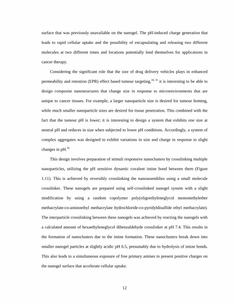
This design involves preparation of stimuli responsive nanoclusters by crosslinking multiple
nanoparticles, utilizing the pH sensitive dynamic covalent imine bond between them (Figure
1.11). This is achieved by reversibly crosslinking the nanoassemblies using a small molecule
crosslinker. These nanogels are prepared using self-crosslinked nanogel system with a slight
modification by using a random copolymer poly(oligoethyleneglycol monomethylether
methacrylate-co-aminoethyl methacrylate hydrochloride-co-pyridyldisulfide ethyl methacrylate).
The interparticle crosslinking between these nanogels was achieved by reacting the nanogels with
a calculated amount of hexaethyleneglycol dibenzaldehyde crosslinker at pH 7.4. This results in
the formation of nanoclusters due to the imine formation. These nanoclusters break down into
smaller nanogel particles at slightly acidic pH 6.5, presumably due to hydrolysis of imine bonds.
This also leads to a simultaneous exposure of free primary amines to present positive charges on
the nanogel surface that accelerate cellular uptake.
13
1.4 Thesis overview
This thesis work focuses on responsive supramolecular assemblies based on amphiphilic
random copolymers. In Chapter 2, influence of Hofmeister ions has been investigated on the size
and guest encapsulation stability of a polymeric nanogel. While variations in macroscopic phase
transitions have been observed in response to the presence of salts, changes in the size and
host−guest behavior of polymeric aggregates in the presence of salts have not been explored in
any detail. The size and core density of nanogel can be fine-tuned through the addition of both
chaotropes and kosmotropes during nanogel formation. The change in core density affects the
guest encapsulation stability and stimuli-responsive character of the nanogel. These studies not
only have practical applications in areas such as drug delivery and sensing but also have
Figure 1.11 Schematic representation of nanocluster formation at physiological pH and
reversal at lower pH (reproduced with permission from reference36
).
14
fundamental implications in the future design and syntheses of polymer nanoassemblies and
nanoparticles.
The guest-exchange mechanism in supramolecular hosts was studied to probe the possible
pathways for guest exchange in these polymeric nanogels in Chapter 3. Dynamic exchange of
guest molecules, encapsulated in host assemblies, is a phenomenon in supramolecular chemistry
that has important implications in several applications. While the mechanism of exchange in
micellar assemblies has been previously investigated, the effect of host and guest environment
upon the guestexchange dynamics has received little attention, if any. By systematically
comparing the behavior of pH-sensitive nanogels along with pH-insensitive nanogels as a control,
size, concentration, and hydrophobicity can all play a critical role in guest-exchange dynamics.
More importantly, these studies reveal that the dominant mechanism of guest exchange can
intimately depend on environmental factors.
Nanocarriers that can be effectively transported across cellular membranes have potential in
a variety of biomedical applications. Among these, materials that are capable of changing their
surface properties and thus gain entry into a cell, in response to a specific tissue environment, are
of particular interest. In Chapter 4, a facile route was designed to prepare nanogels, which
generate surface charge with pH as stimulus. This is achieved by designing a polymeric nanogel
containing 2-diisopropylamino (DPA) moieties. The pH at which the charge is generated, i.e. the
isoelectric point (pI) of the nanogel, can be adjusted by varying the percentage of DPA units in
the nanogel, its preparation process and crosslinking density. Intracellular delivery of these
nanogels was greatly enhanced in an acidic pH environment due to the surface charge generation.
This study demonstrates the versatile nature of the nanogels to introduce specific functionalities
with relative ease to achieve desired functional behavior.
Photo-crosslinking chemistry has significant importance owing to its advantages, such as no
crosslinking agents or reaction catalysts are needed and the photo-crosslinking procedure can be
15
easily tuned by turning on or off the light. However, the problem is that the de-crosslinking for
the current methods always involves the use of high-energy ultra-violet (UV) or visible light,
which limits the practical application of these nanocarriers. In Chapter 5, photoinduced
heterodisulfide metathesis of pyridyl disulfide (PDS) functionalities is an irreversible reaction.
The micelle aggregates based on random copolymer containing PDS can be successfully
crosslinked to form polymer nanoparticles based on redox-responsive disulfide bonds. Since the
crosslinks generated here are biologically relevant, this method could also have implications in
generating new materials of interest in biology and medicine.
This thesis also describes a novel and facile approach to generate well-defined, hollow
metal-organic nanoparticles (MOPs) from polymeric organic nanoparticles. In Chapter 6,
polymeric organic nanoparticles were prepared through sequential boronate esterification of
boronic acids and bifunctional catechols under ambient conditions. The nucleation step involves
the cooperative polymerization, wherein the covalent dative interaction between nitrogen in imine
and boron center from boronate ester was used as the secondary. In Chapter 7, these polymeric
organic nanoparticles based on boronate ester were extended to prepare the hollow MOPs using a
simple metal-comonomer exchange process in a single step. The Kirkendall effect has been
identified as the underlying mechanism for the formation of these hollow MOPs. This non-
reciprocal diffusion process based effect has not been utilized for hollow metal-organic
composites, which are of great interest in a variety of applications because of their low density
and high surface area.
Finally, in Chapter 8 general conclusions and several future research directions are provided.
We hope that our work will have some impact on the design of responsive supramolecular
assemblies based on amphiphilic polymers. Our ability to reliably generate organic-inorganic
hybrid nanoparticles using polymeric nanostructure templates will open up a fount of
opportunities for hybrid materials nanotechnology.
16
1.5 References
1. Ikkala, O.; ten Brinke, G. Functional materials based on self-assembly of polymeric
supramolecules. Science, 2002, 295, 2407–2409.
2. Janata, J.; Josowicz, M. Conducting polymers in electronic chemical sensors. Nat. Mater.,
2003, 2, 19–24.
3. Zotti, G.; Vercelli, B.; Berlin, A. Monolayers and multilayers of conjugated polymers as
nanosized electronic components. Acc. Chem. Res., 2008, 41, 1098–1109.
4. Aida, T.; Meijer, E. W.; Stupp, S. I. Functional supramolecular polymers. Science, 2012, 335,
813–817.
5. Rösler, A.; Vandermeulen, G. W. M.; Klok, H.-A. Advanced drug delivery devices via self-
assembly of amphiphilic block copolymers. Adv. Drug Delivery Rev., 2012, 64, 270–279.
6. Nakatani, K.; Ogura, Y.; Koda, Y.; Terashima, T.; Sawamoto, M. Sequence-regulated
copolymers via tandem catalysis of living radical polymerization and in situ transesterification. J.
Am. Chem. Soc., 2012, 134, 4373–4383.
7. Hawker, C. J.; Bosman, A. W.; Harth, E. New polymer synthesis by nitroxide mediated living
radical polymerizations. Chem. Rev., 2001, 101, 3661–3688.
8. Moad, G.; Rizzardo, E.; Thang, S. H. Living radical polymerization by the RAFT process–a
third update. Aust. J. Chem., 2012, 65, 985–1076.
9. Hilf, S.; Kilbinger, A. F. M. Functional end groups for polymers prepared using ring-opening
metathesis polymerization. Nat. Chem., 2009, 1, 537–546.
10. Matyjaszewski, K.; Tsarevsky, N. V. Macromolecular engineering by atom transfer radical
polymerization. J. Am. Chem. Soc., 2014, 136, 6513–6533.
11. Rosebrugh, L. E.; Marx, V. M.; Keitz, B. K.; Grubbs, R. H. Synthesis of highly cis,
syndiotactic polymers via ring-opening metathesis polymerization using ruthenium metathesis
catalysts. J. Am. Chem. Soc., 2013, 135, 10032–10035.
12. Kim, J. K.; Yang, S. Y.; Lee, Y.; Kim, Y. Functional nanomaterials based on block
copolymer self-assembly. Prog. Polym. Sci., 2010, 35, 1325–1349.
13. Zhang, Q.; Ko, N. R.; Oh, J. K. Recent advances in stimuli-responsive degradable block
copolymer micelles: synthesis and controlled drug delivery applications. Chem. Commun., 2012,
48, 7542–7552.
14. Ge, Z.; Liu, S. Functional block copolymer assemblies responsive to tumor and intracellular
microenvironments for site-specific drug delivery and enhanced imaging performance. Chem. Soc.
Rev., 2013, 42, 7289–7325.
15. Ruzette, A.-V.; Leibler, L. Block copolymers in tomorrow's plastics. Nat. Mater., 2005, 4,
19–31.
17
16. Mai, Y.; Eisenberg, A. Self-assembly of block copolymers. Chem. Soc. Rev., 2012, 41, 5969–
5985.
17. Kim, H. -C.; Park, S.-M.; Hinsberg, W. D. Block copolymer based nanostructures: materials,
processes, and applications to electronics. Chem. Rev., 2009, 110, 146–177.
18. Quémener, D.; Davis, T. P.; Barner-Kowollik, C.; Stenzel, M. H. RAFT and click chemistry:
A versatile approach to well-defined block copolymers. Chem. Commun., 2006, 42, 5051–5053.
19. Wong, C. -H.; Zimmerman, S. C. Orthogonality in organic, polymer, and supramolecular
chemistry: from Merrifield to click chemistry. Chem. Commun., 2013, 49, 1679–1695.
20. Johnson, J. A.; Lu, Y. Y.; Burts, A. O.; Lim, Y. -H.; Finn, M. G.; Koberstein, J. T.; Turro, N.
J.; Tirrell, D. A.; Grubbs, R. H. Core-clickable PEG-branch-azide bivalent-bottle-brush polymers
by ROMP: grafting-through and clicking-to. J. Am. Chem. Soc., 2010, 133, 559–566.
21. Ryu, J. -H.; Roy, R.; Ventura, J.; Thayumanavan, S. Redox-sensitive disassembly of
amphiphilic copolymer based micelles. Langmuir 2010, 26, 7086–7092.
22. Ryu, J. -H.; Chacko, R. T.; Jiwpanich, S.; Bickerton, S.; Babu R. P.; Thayumanavan, S. Self-
cross-linked polymer nanogels: a versatile nanoscopic drug delivery platform. J. Am. Chem. Soc.,
2010, 132, 17227–17235.
23. Ryu, J. -H.; Jiwpanich, S.; Chacko, R.; Bickerton S.; Thayumanavan, S. Surface-
functionalizable polymer nanogels with facile hydrophobic guest encapsulation capabilities. J.
Am. Chem. Soc., 2010, 132, 8246–8247.
24. Jiwpanich, S.; Ryu, J. -H.; Bickerton, S.; Thayumanavan, S. Noncovalent encapsulation
stabilities in supramolecular nanoassemblies. J. Am. Chem. Soc., 2010, 132, 10683-10685.
25. Ryu, J. -H.; Bickerton, S.; Zhuang, J.; Thayumanavan, S. Ligand-decorated nanogels: fast
one-pot synthesis and cellular targeting. Biomacromolecules 2012, 13, 1515-1522.
26. González-Toro, D. C.; Ryu, J. -H.; Chacko, R.; Zhuang, J.; Thayumanavan, S. Concurrent
binding and delivery of proteins and lipophilic small molecules using polymeric nanogels. J. Am.
Chem. Soc., 2012, 134, 6964-6967.
27. Rothbard, J. B.; Jessop, T. C.; Lewis, R. S.; Murray, B. A.; Wender, P. A. Role of membrane
potential and hydrogen bonding in the mechanism of translocation of guanidinium-rich peptides
into cells. J. Am. Chem. Soc. 2004, 126, 9506−9507.
28. Nakase, I.; Takeuchi, T.; Tanaka, G.; Futaki, S. Methodological and cellular aspects that
govern the internalization mechanisms of arginine-rich cell-penetrating peptides. Adv. Drug
Delivery Rev. 2008, 60, 598−607.
29. Matsumoto, N. M.; González-Toro, D. C.; Chacko, R. T.; Maynard, H. D.; Thayumanavan. S.
Synthesis of nanogel–protein conjugates. Polym. Chem., 2013, 4, 2464-2469.
30. Zhuang, J.; Jiwpanich, S.; Deepak, V. D.; Thayumanavan, S. Facile preparation of nanogels
using activated ester containing polymers. ACS Macro Lett., 2012, 1, 175−179.
18
31. Wang, H.; Zhuang, J.; Thayumanavan, S. Functionalizable Amine-Based Polymer
Nanoparticles. ACS Macro Lett. 2013, 2, 948−951.
32. Zhuang, J.; Chacko, R. T.; Amado Torres, D. F.; Wang, H.; Thayumanavan, S. Dual Stimuli-
Dual Response Nanoassemblies Prepared from a Simple Homopolymer. ACS Macro Lett. 2014, 3,
1−5.
33. Yuan, C.; Raghupathi, K.; Popere, B. C.; Ventura, J.; Dai, L.; Thayumanavan, S. Composite
supramolecular nanoassemblies with independent stimulus sensitivities. Chem. Sci., 2014, 5, 229-
234.
34. Maeda, H.; Wu, J.; Sawa, T.; Matsumura Y.; Hori, K. Tumor vascular permeability and the
EPR effect in macromolecular therapeutics: a review. J. Controlled Release, 2000, 65, 271-284.
35. Maeda, H.; Fang, J.; Inutsuka T.; Kitamoto, Y. Vascular permeability enhancement in solid
tumor: various factors, mechanisms involved and its implications. Int. Immunopharmacol., 2003,
3, 319–328.
36. Raghupathi, K.; Li, L.; Ventura, J.; Jennings M.; Thayumanavan, S. pH responsive soft
nanoclusters with size and charge variation features. Polym. Chem., 2014, 5, 1737–1742.
19
CHAPTER 2
EFFECT OF HOFMEISTER IONS ON THE SIZE AND ENCAPSULATION STABILITY
OF POLYMER NANOGELS
Reprinted with permission from Li, L.; Ryu, J. -H.; Thayumanavan, S. Effect of Hofmeister
ions on the size and encapsulation stability of polymer nanogels. Langmuir 2013, 29, 50-55.
Copyright © 2015 American Chemical Society.
2.1 Introduction
Inorganic salts have the ability to enhance solubilization or precipitate proteins in an
aqueous solution, based on a phenomenon observed by Hofmeister in the late 19th century.
1−3
While biological applications of these findings are well-established,4−6
its implications in
chemical applications are relatively underexplored.7 For example, it has been shown that addition
of salts can significantly alter the lower critical solution temperature (LCST) of water-soluble
polymers and that the effect is dependent on the nature of the salt; that is, they follow the
Hofmeister series.8−11
The LCST behavior, or the macroscopic phase separation of the polymer
from the aqueous phase, is attributed to the reduced hydrogen bonding driven interaction between
water and the polymer at elevated temperatures. While it is well-studied in macroscopic
hydrogels and surfaces,12,13
we do not know of any studies that take advantage of the subtle
changes that are likely to occur due to altered hydration or dehydration of functional groups in the
presence of Hofmeister ions in nanosized gels. We have been interested in probing this
phenomenon, with fine-tuning the size and guest encapsulation stability in oligo(ethylene glycol)-
functionalized polymer nanogels as the context.
We hypothesized that although LCST transitions in polymers are often sharp, it is likely that
there are also subtle changes in the aggregation states of these polymers at temperatures well
below their LCST. Considering that Hofmeister ions could influence LCST, it is also likely that
these ions would influence these aggregation states and thus the nanogel itself. We then
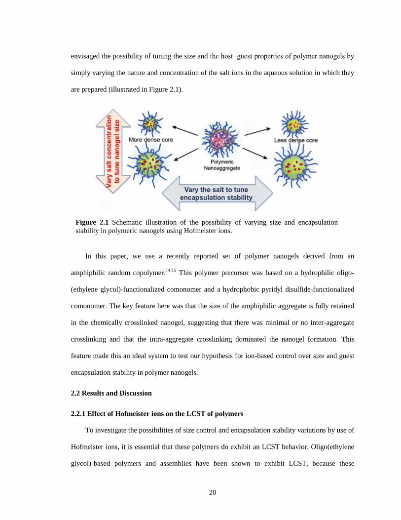
20
envisaged the possibility of tuning the size and the host−guest properties of polymer nanogels by
simply varying the nature and concentration of the salt ions in the aqueous solution in which they
are prepared (illustrated in Figure 2.1).
In this paper, we use a recently reported set of polymer nanogels derived from an
amphiphilic random copolymer.14,15
This polymer precursor was based on a hydrophilic oligo-
(ethylene glycol)-functionalized comonomer and a hydrophobic pyridyl disulfide-functionalized
comonomer. The key feature here was that the size of the amphiphilic aggregate is fully retained
in the chemically crosslinked nanogel, suggesting that there was minimal or no inter-aggregate
crosslinking and that the intra-aggregate crosslinking dominated the nanogel formation. This
feature made this an ideal system to test our hypothesis for ion-based control over size and guest
encapsulation stability in polymer nanogels.
2.2 Results and Discussion
2.2.1 Effect of Hofmeister ions on the LCST of polymers
To investigate the possibilities of size control and encapsulation stability variations by use of
Hofmeister ions, it is essential that these polymers do exhibit an LCST behavior. Oligo(ethylene
glycol)-based polymers and assemblies have been shown to exhibit LCST, because these
Figure 2.1 Schematic illustration of the possibility of varying size and encapsulation
stability in polymeric nanogels using Hofmeister ions.
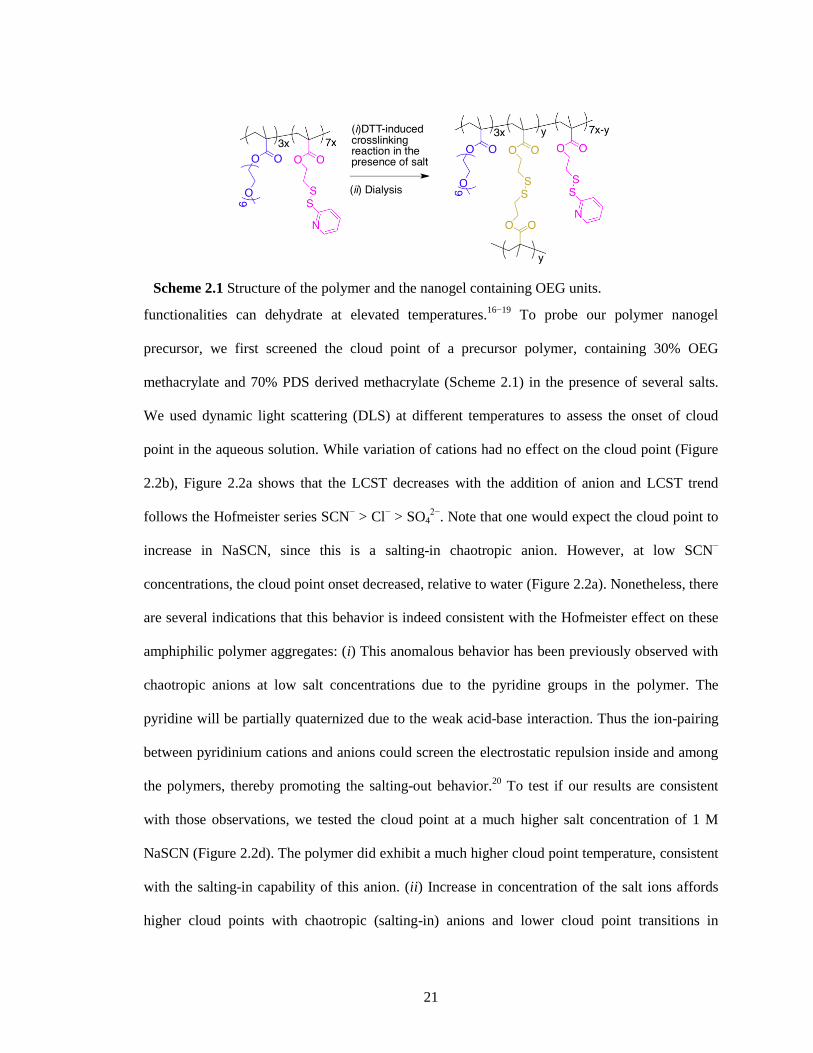
21
functionalities can dehydrate at elevated temperatures.16−19
To probe our polymer nanogel
precursor, we first screened the cloud point of a precursor polymer, containing 30% OEG
methacrylate and 70% PDS derived methacrylate (Scheme 2.1) in the presence of several salts.
We used dynamic light scattering (DLS) at different temperatures to assess the onset of cloud
point in the aqueous solution. While variation of cations had no effect on the cloud point (Figure
2.2b), Figure 2.2a shows that the LCST decreases with the addition of anion and LCST trend
follows the Hofmeister series SCN− > Cl
− > SO4
2−. Note that one would expect the cloud point to
increase in NaSCN, since this is a salting-in chaotropic anion. However, at low SCN−
concentrations, the cloud point onset decreased, relative to water (Figure 2.2a). Nonetheless, there
are several indications that this behavior is indeed consistent with the Hofmeister effect on these
amphiphilic polymer aggregates: (i) This anomalous behavior has been previously observed with
chaotropic anions at low salt concentrations due to the pyridine groups in the polymer. The
pyridine will be partially quaternized due to the weak acid-base interaction. Thus the ion-pairing
between pyridinium cations and anions could screen the electrostatic repulsion inside and among
the polymers, thereby promoting the salting-out behavior.20
To test if our results are consistent
with those observations, we tested the cloud point at a much higher salt concentration of 1 M
NaSCN (Figure 2.2d). The polymer did exhibit a much higher cloud point temperature, consistent
with the salting-in capability of this anion. (ii) Increase in concentration of the salt ions affords
higher cloud points with chaotropic (salting-in) anions and lower cloud point transitions in
Scheme 2.1 Structure of the polymer and the nanogel containing OEG units.
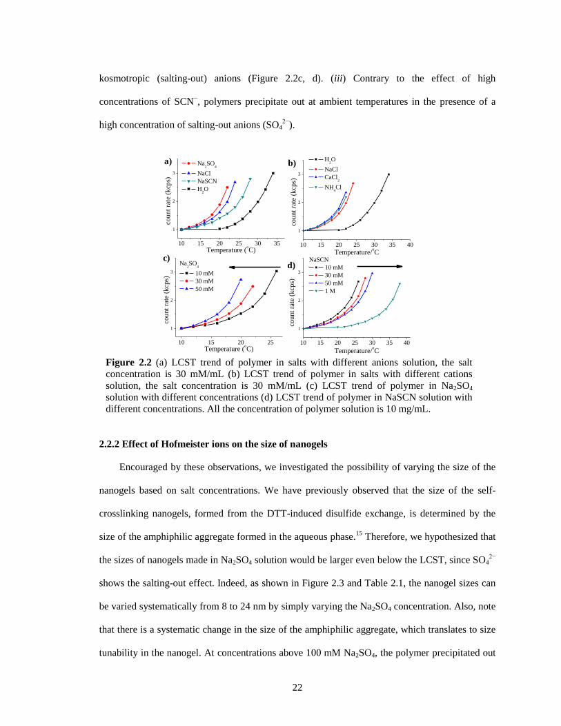
22
kosmotropic (salting-out) anions (Figure 2.2c, d). (iii) Contrary to the effect of high
concentrations of SCN−, polymers precipitate out at ambient temperatures in the presence of a
high concentration of salting-out anions (SO42−
).
2.2.2 Effect of Hofmeister ions on the size of nanogels
Encouraged by these observations, we investigated the possibility of varying the size of the
nanogels based on salt concentrations. We have previously observed that the size of the self-
crosslinking nanogels, formed from the DTT-induced disulfide exchange, is determined by the
size of the amphiphilic aggregate formed in the aqueous phase.15
Therefore, we hypothesized that
the sizes of nanogels made in Na2SO4 solution would be larger even below the LCST, since SO42−
shows the salting-out effect. Indeed, as shown in Figure 2.3 and Table 2.1, the nanogel sizes can
be varied systematically from 8 to 24 nm by simply varying the Na2SO4 concentration. Also, note
that there is a systematic change in the size of the amphiphilic aggregate, which translates to size
tunability in the nanogel. At concentrations above 100 mM Na2SO4, the polymer precipitated out
Figure 2.2 (a) LCST trend of polymer in salts with different anions solution, the salt
concentration is 30 mM/mL (b) LCST trend of polymer in salts with different cations
solution, the salt concentration is 30 mM/mL (c) LCST trend of polymer in Na2SO4
solution with different concentrations (d) LCST trend of polymer in NaSCN solution with
different concentrations. All the concentration of polymer solution is 10 mg/mL.
10 15 20 25 30 35 40
1
2
3
cou
nt
rate
(k
cps)
Temperature/oC
H2O
NaCl
CaCl2
NH4Cl
10 15 20 25
1
2
3
cou
nt
rate
(k
cps)
Temperature (oC)
Na2SO
4
10 mM
30 mM
50 mM
10 15 20 25 30 35
1
2
3co
un
t ra
te (
kcp
s)
Temperature (oC)
Na2SO
4
NaCl
NaSCN
H2O
a) b)
c)d)
10 15 20 25 30 35 40
1
2
3
cou
nt
rate
(k
cps)
Temperature/oC
NaSCN
10 mM
30 mM
50 mM
1 M
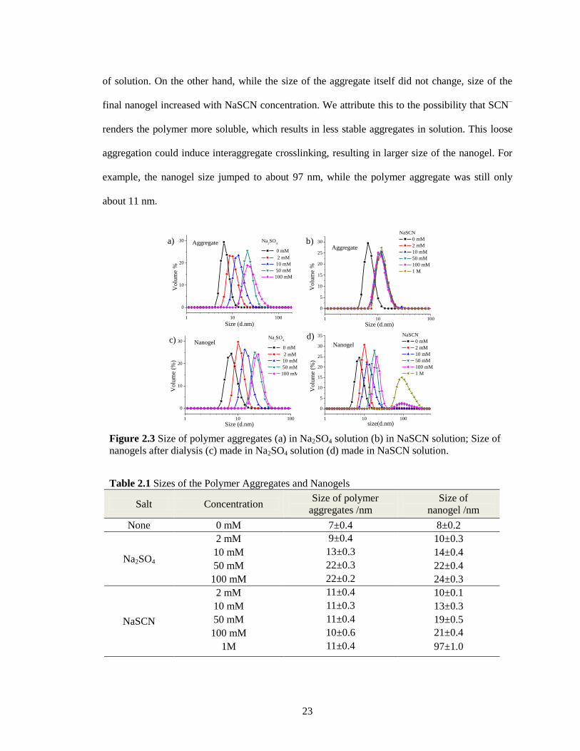
23
of solution. On the other hand, while the size of the aggregate itself did not change, size of the
final nanogel increased with NaSCN concentration. We attribute this to the possibility that SCN−
renders the polymer more soluble, which results in less stable aggregates in solution. This loose
aggregation could induce interaggregate crosslinking, resulting in larger size of the nanogel. For
example, the nanogel size jumped to about 97 nm, while the polymer aggregate was still only
about 11 nm.
Table 2.1 Sizes of the Polymer Aggregates and Nanogels
Salt Concentration Size of polymer
aggregates /nm
Size of
nanogel /nm
None 0 mM 7±0.4 8±0.2
Na2SO4
2 mM 9±0.4 10±0.3
10 mM 13±0.3 14±0.4
50 mM 22±0.3 22±0.4
100 mM 22±0.2 24±0.3
NaSCN
2 mM 11±0.4 10±0.1
10 mM 11±0.3 13±0.3
50 mM 11±0.4 19±0.5
100 mM 10±0.6 21±0.4
±
1M 11±0.4 97±1.0
Figure 2.3 Size of polymer aggregates (a) in Na2SO4 solution (b) in NaSCN solution; Size of
nanogels after dialysis (c) made in Na2SO4 solution (d) made in NaSCN solution.
1 10 100
0
10
20
30
Vo
lum
e (%
)
Size (d.nm)
Na2SO
4
0 mM
2 mM
10 mM
50 mM
100 mM
Nanogel
1 10 100
0
5
10
15
20
25
30
35
Nanogel
Vo
lum
e (%
)
size(d.nm)
NaSCN
0 mM
2 mM
10 mM
50 mM
100 mM
1 M
c) d)
1 10 100
0
10
20
30 Na2SO
4
0 mM
2 mM
10 mM
50 mM
100 mM
Vo
lum
e %
Size (d.nm)
Aggregatea)
1 10 100
0
5
10
15
20
25
30
Aggregate
Vo
lum
e %
Size (d.nm)
NaSCN
0 mM
2 mM
10 mM
50 mM
100 mM
1 M
b)
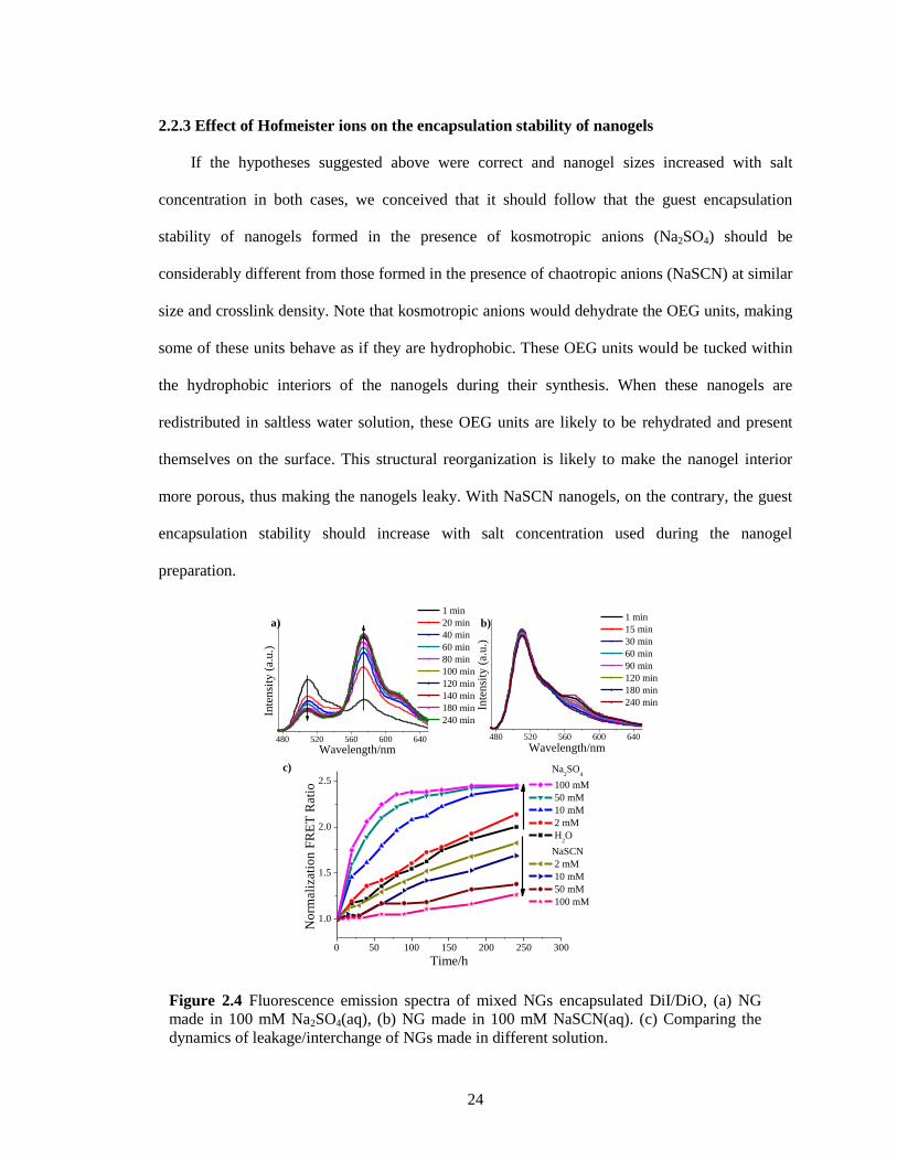
24
2.2.3 Effect of Hofmeister ions on the encapsulation stability of nanogels
If the hypotheses suggested above were correct and nanogel sizes increased with salt
concentration in both cases, we conceived that it should follow that the guest encapsulation
stability of nanogels formed in the presence of kosmotropic anions (Na2SO4) should be
considerably different from those formed in the presence of chaotropic anions (NaSCN) at similar
size and crosslink density. Note that kosmotropic anions would dehydrate the OEG units, making
some of these units behave as if they are hydrophobic. These OEG units would be tucked within
the hydrophobic interiors of the nanogels during their synthesis. When these nanogels are
redistributed in saltless water solution, these OEG units are likely to be rehydrated and present
themselves on the surface. This structural reorganization is likely to make the nanogel interior
more porous, thus making the nanogels leaky. With NaSCN nanogels, on the contrary, the guest
encapsulation stability should increase with salt concentration used during the nanogel
preparation.
Figure 2.4 Fluorescence emission spectra of mixed NGs encapsulated DiI/DiO, (a) NG
made in 100 mM Na2SO4(aq), (b) NG made in 100 mM NaSCN(aq). (c) Comparing the
dynamics of leakage/interchange of NGs made in different solution.
0 50 100 150 200 250 300
1.0
1.5
2.0
2.5
No
rmal
izat
ion
FR
ET
Rat
io
Time/h
Na2SO
4
100 mM
50 mM
10 mM
2 mM
H2O
NaSCN
2 mM
10 mM
50 mM
100 mM
480 520 560 600 640
Inte
nsi
ty (
a.u
.)
Wavelength/nm
1 min
20 min
40 min
60 min
80 min
100 min
120 min
140 min
180 min
240 min
480 520 560 600 640
Inte
nsi
ty (
a.u
.)
Wavelength/nm
1 min
15 min
30 min
60 min
90 min
120 min
180 min
240 min
a) b)
c)
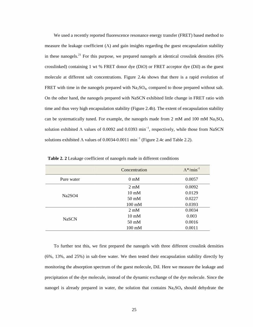
25
We used a recently reported fluorescence resonance energy transfer (FRET) based method to
measure the leakage coefficient (Λ) and gain insights regarding the guest encapsulation stability
in these nanogels.21
For this purpose, we prepared nanogels at identical crosslink densities (6%
crosslinked) containing 1 wt % FRET donor dye (DiO) or FRET acceptor dye (DiI) as the guest
molecule at different salt concentrations. Figure 2.4a shows that there is a rapid evolution of
FRET with time in the nanogels prepared with Na2SO4, compared to those prepared without salt.
On the other hand, the nanogels prepared with NaSCN exhibited little change in FRET ratio with
time and thus very high encapsulation stability (Figure 2.4b). The extent of encapsulation stability
can be systematically tuned. For example, the nanogels made from 2 mM and 100 mM Na2SO4
solution exhibited Λ values of 0.0092 and 0.0393 min−1
, respectively, while those from NaSCN
solutions exhibited Λ values of 0.0034-0.0011 min−1
(Figure 2.4c and Table 2.2).
To further test this, we first prepared the nanogels with three different crosslink densities
(6%, 13%, and 25%) in salt-free water. We then tested their encapsulation stability directly by
monitoring the absorption spectrum of the guest molecule, DiI. Here we measure the leakage and
precipitation of the dye molecule, instead of the dynamic exchange of the dye molecule. Since the
nanogel is already prepared in water, the solution that contains Na2SO4 should dehydrate the
Table 2. 2 Leakage coefficient of nanogels made in different conditions
Concentration Λ*/min-1
Pure water 0 mM 0.0057
Na2SO4
2 mM 0.0092
10 mM 0.0129
50 mM 0.0227
100 mM 0.0393
NaSCN
2 mM 0.0034
10 mM 0.003
50 mM 0.0016
100 mM 0.0011
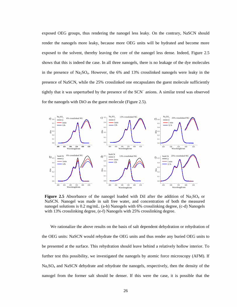
26
exposed OEG groups, thus rendering the nanogel less leaky. On the contrary, NaSCN should
render the nanogels more leaky, because more OEG units will be hydrated and become more
exposed to the solvent, thereby leaving the core of the nanogel less dense. Indeed, Figure 2.5
shows that this is indeed the case. In all three nanogels, there is no leakage of the dye molecules
in the presence of Na2SO4. However, the 6% and 13% crosslinked nanogels were leaky in the
presence of NaSCN, while the 25% crosslinked one encapsulates the guest molecule sufficiently
tightly that it was unperturbed by the presence of the SCN− anions. A similar trend was observed
for the nanogels with DiO as the guest molecule (Figure 2.5).
We rationalize the above results on the basis of salt dependent dehydration or rehydration of
the OEG units: NaSCN would rehydrate the OEG units and thus render any buried OEG units to
be presented at the surface. This rehydration should leave behind a relatively hollow interior. To
further test this possibility, we investigated the nanogels by atomic force microscopy (AFM). If
Na2SO4 and NaSCN dehydrate and rehydrate the nanogels, respectively, then the density of the
nanogel from the former salt should be denser. If this were the case, it is possible that the
Figure 2.5 Absorbance of the nanogel loaded with DiI after the addition of Na2SO4 or
NaSCN. Nanogel was made in salt free water, and concentration of both the measured
nanogel solutions is 0.2 mg/mL. (a-b) Nanogels with 6% crosslinking degree, (c-d) Nanogels
with 13% crosslinking degree, (e-f) Nanogels with 25% crosslinking degree.
450 500 550 600 650
0.0
0.1
0.2
0.3
0.4
0.5
0.625% crosslinked NG
Ab
s
Wavelength/nm
Na2SO
4
0
1 min
12 h
e)
450 500 550 600 650
0.0
0.1
0.2
0.3
0.4
0.5
25% crosslinked NGA
bs
Wavelength/nm
NaSCN
0
1 min
12 h
f)
400 450 500 550 600 650
0.0
0.1
0.2
0.3
0.4
0.5
6% crosslinked NG
Ab
s
Wavelength/nm
NaSCN
0
1min
12h
b)
400 450 500 550 600 650
0.0
0.1
0.2
0.3
0.4
0.5 13% crosslinked NG
Ab
s
Wavelength/nm
NaSCN
0
1min
12h
d)
400 450 500 550 600 650
0.0
0.1
0.2
0.3
0.4
0.5 13% crosslinked NG
Ab
s
Wavelength/nm
Na2SO
4
0
1min
12h
c)
400 450 500 550 600 650
0.0
0.1
0.2
0.3
0.4
0.5
0.6
Ab
s
Wavelength/nm
Na2SO
4
0
1min
12h
a) 6% crosslinked NG
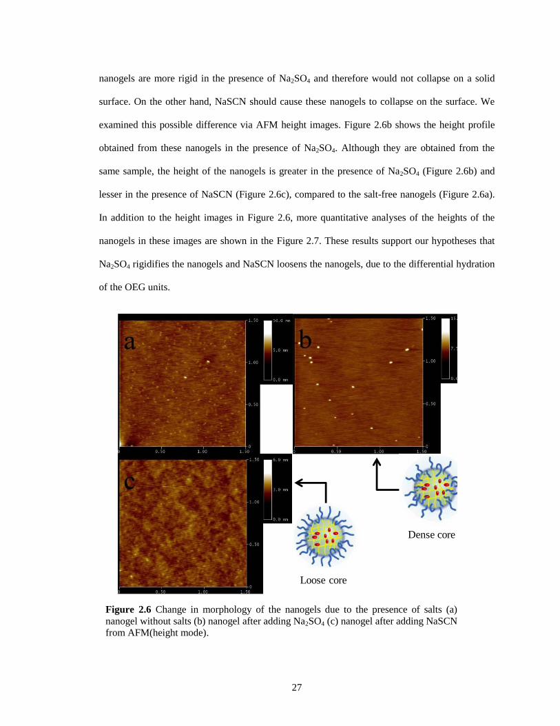
27
nanogels are more rigid in the presence of Na2SO4 and therefore would not collapse on a solid
surface. On the other hand, NaSCN should cause these nanogels to collapse on the surface. We
examined this possible difference via AFM height images. Figure 2.6b shows the height profile
obtained from these nanogels in the presence of Na2SO4. Although they are obtained from the
same sample, the height of the nanogels is greater in the presence of Na2SO4 (Figure 2.6b) and
lesser in the presence of NaSCN (Figure 2.6c), compared to the salt-free nanogels (Figure 2.6a).
In addition to the height images in Figure 2.6, more quantitative analyses of the heights of the
nanogels in these images are shown in the Figure 2.7. These results support our hypotheses that
Na2SO4 rigidifies the nanogels and NaSCN loosens the nanogels, due to the differential hydration
of the OEG units.
Figure 2.6 Change in morphology of the nanogels due to the presence of salts (a)
nanogel without salts (b) nanogel after adding Na2SO4 (c) nanogel after adding NaSCN
from AFM(height mode).
Loose core
Dense core
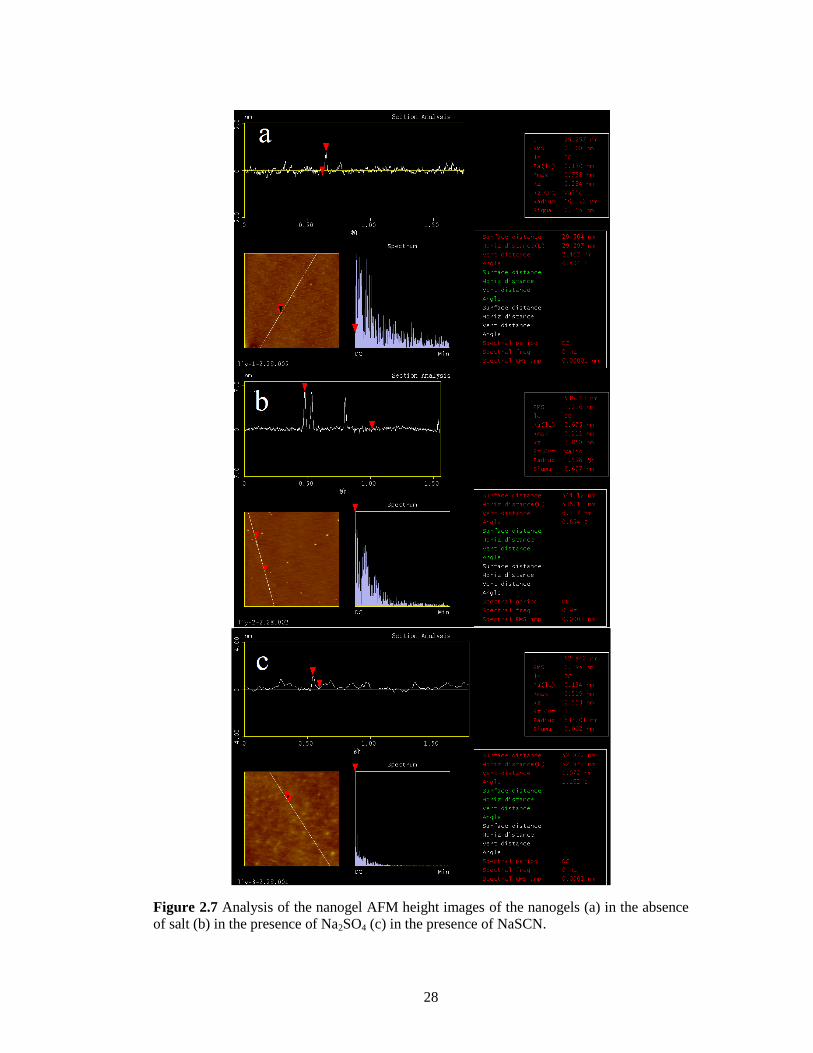
28
Figure 2.7 Analysis of the nanogel AFM height images of the nanogels (a) in the absence
of salt (b) in the presence of Na2SO4 (c) in the presence of NaSCN.
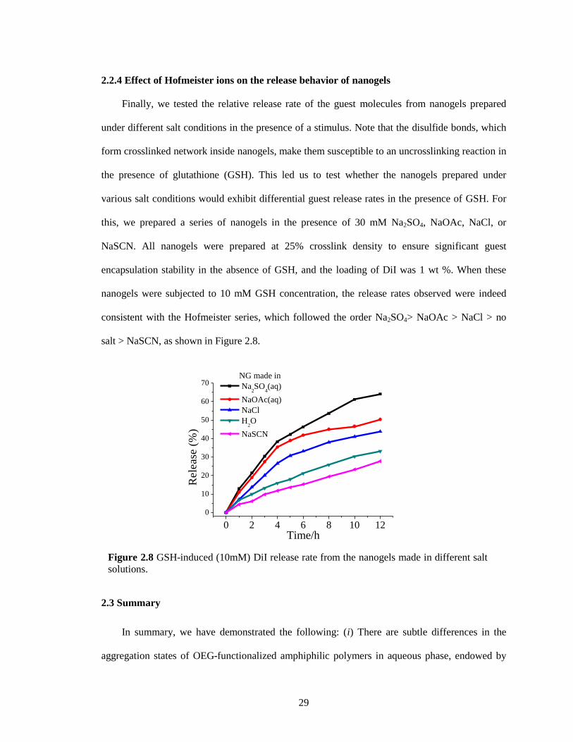
29
2.2.4 Effect of Hofmeister ions on the release behavior of nanogels
Finally, we tested the relative release rate of the guest molecules from nanogels prepared
under different salt conditions in the presence of a stimulus. Note that the disulfide bonds, which
form crosslinked network inside nanogels, make them susceptible to an uncrosslinking reaction in
the presence of glutathione (GSH). This led us to test whether the nanogels prepared under
various salt conditions would exhibit differential guest release rates in the presence of GSH. For
this, we prepared a series of nanogels in the presence of 30 mM Na2SO4, NaOAc, NaCl, or
NaSCN. All nanogels were prepared at 25% crosslink density to ensure significant guest
encapsulation stability in the absence of GSH, and the loading of DiI was 1 wt %. When these
nanogels were subjected to 10 mM GSH concentration, the release rates observed were indeed
consistent with the Hofmeister series, which followed the order Na2SO4> NaOAc > NaCl > no
salt > NaSCN, as shown in Figure 2.8.
2.3 Summary
In summary, we have demonstrated the following: (i) There are subtle differences in the
aggregation states of OEG-functionalized amphiphilic polymers in aqueous phase, endowed by
Figure 2.8 GSH-induced (10mM) DiI release rate from the nanogels made in different salt
solutions.
0 2 4 6 8 10 12
0
10
20
30
40
50
60
70
Rel
ease
(%
)
Time/h
NG made in
Na2SO
4(aq)
NaOAc(aq)
NaCl
H2O
NaSCN
30
the presence of various salts. (ii) While anions provide variations as predicted by the Hofmeister
effect, variation in cations does not affect the aggregation of the polymer. (iii) In the case of
kosmotropic SO42−
, the size of the nanogel can be systematically varied with concentration, since
the size is determined by the aggregate size. (iv) In the case of chaotropic anion SCN-,
interaggregate crosslinking occurs due to the rather loose nature of the aggregates. (v)
Encapsulation stability of guest molecules is dependent on the nature of the salt used for
preparation of the nanogels; chaotropic anions afford nanogels with greater guest encapsulation
stability. (vi) Salt-dependent hydration of the OEG units in the nanogel causes variations in
density, which then determine the leaky character of the nanogel. From a practical perspective,
utilizing Hofmeister ions provides unprecedented control in fine-tuning the size of the polymer
nanogel. In addition to size, we have concurrent control over altering the guest encapsulation
stabilities. These studies not only have practical applications in areas such as drug delivery and
sensing but also have fundamental implications in the future design and syntheses of polymer
nanoassemblies and nanoparticles.
2.4 Experimental
2.4.1 General
2,2′-Dithiodipyridine, 2-mercaptoethanol, poly(ethylene glycol) monomethyl ether
methacrylate (MW 450), D,L-dithiothreitol (DTT), 1,1′-dioctadecyl-3,3,3′,3′-
tetramethylindocarbocyanine perchlorate (DiI), 3,3′-dioctadecyloxacarbocyanine perchlorate
(DiO), 4-cyano-4-(phenylcarbonothioylthio)pentanoic acid, and other conventional reagents were
obtained from commercial sources and were used as received, unless otherwise mentioned.
Pyridyl disulfide ethyl methacrylate (PDSEMA) was prepared by a previously reported
procedure.22
1H NMR spectra were recorded on a 400 MHz Bruker NMR spectrometer with the
residual proton resonance of the solvent as the internal standard. Chemical shifts are reported in
parts per million (ppm). Molecular weights of the polymers were estimated by gel-permeation
31
chromatography (GPC) with a refractive index detector, with poly(methyl methacrylate) (PMMA)
as a standard . Dynamic light scattering (DLS) measurements were performed on a Malvern
Nanozetasizer. UV−visible absorption spectra were recorded on a Varian (model EL 01125047)
spectrophotometer. Fluorescence spectra were obtained from a Jasco FP-6500 spectrofluorometer.
Atomic force microscopy (AFM) images were collected on a Digital Instruments Nanoscope III
in tapping mode under ambient conditions by use of silicon cantilievers (spring constant 0.58
N/m).
2.4.2 Synthesis of Random Copolymer.
Polymers were prepared by reversible addition−fragmentation chain transfer (RAFT)
polymerization. A mixture of 4-cyano-4-(phenylcarbonothioylthio)pentanoic acid (3.0 mg, 0.02
mmol), PDSEMA (1.25 g, 4.9 mmol), poly(ethylene glycol) monomethyl ether methacrylate (1.0
g, 2.1 mmol), and 2,2′-azobis(isobutyronitrile) (AIBN; 2.5 mg, 0.014 mmol) was dissolved in
tetrahydrofuran (THF) (5 mL) and degassed by performing three freeze−pump−thaw cycles. The
reaction mixture was sealed and then transferred into a preheated oil bath at 65 °C for 10 h. To
remove unreactive monomers, the resultant mixture was precipitated in cold ethyl ether (20 mL)
to yield the random copolymer as a waxy liquid. GPC (THF) Mn, 13 kDa; polydispersity, 1.3. 1H
NMR (400 MHz, CDCl3) δ 8.46, 7.68, 7.11, 4.35-4.09, 3.94-3.37, 3.03, 2.04-1.64, 1.43-0.87. The
molar ratio between two blocks was determined by integrating the methoxy proton in the poly-
(ethylene glycol) unit and the aromatic proton in the pyridine and was found to be 3:7
(PEO:PDSEMA).
2.4.3 Synthesis of Nanogels Containing DiI/DiO.
As shown in Scheme 1, the polymer (10 mg) was dissolved in 1 mL of water or salt solution.
A 0.02 mL DiI stock solution (5 mg/mL) or 0.04 mL DiO stock solution (2.5 mg/mL) in acetone
was added into the polymer solution. The mixture was stirred overnight at room temperature,
open to the atmosphere to allow the organic solvent to evaporate. Then, to control the
32
crosslinking density of nanogel (6%, 13%, and 25% by molar equivalents), a measured amount of
DTT (0.4, 0.8, and 2.0 μmol for 10, 20, and 50 mol % against PDS groups, respectively) was
added. After 4 h of stirring, any insoluble DiI/DiO residue was removed by filtration and
pyridothione was removed from the nanogel solution by extensive dialysis through a membrane
with a molecular weight cutoff of 7000 g/mol.
2.4.4 Dynamic Light Scattering Measurement.
Dynamic light scattering experiments were performed by using a Malvern Nanozetasizer.
The scattered light is detected at an angle of 173°. For cloud point measurement, the polymer (10
mg/mL) in water or salt solution was used. The temperature was increased from 10 to 60 °C in
2 °C increments. Equilibration time at each temperature was 10 min. Above the LCST, the
polymer chains collapse into a globule and produce a more compressed particle with a higher
refractive index, so scattering intensity was monitored as a function of temperature to determine
the LCST behavior of polymer solution. For size distribution measurement, we used the polymer
(10 mg/mL) or nanogel (1 mg/mL) solution in water or each salt solution, which contains a
specific salt concentration. The measurement was kept constant at 25 °C throughout the
experiment. Dust was eliminated by filtering the solution through 0.45 μm membrane filter.
Nanogel Encapsulated Dye Mixing. A solution of nanogel containing DiI (100 μL) was mixed
with a solution of nanogel containing DiO (100 μL) in a cuvette, and then Milli-Q water (800 μL)
was added to adjust the volume. The fluorescence intensity was recorded (λexc = 450 nm).
2.5 References
1. Hofmeister, F. On the understanding of the effects of salts, second report. On irregularities in
the precipitating effect of salts and their relationship to their physical behavior. Arch. Exp. Pathol.
Pharmocol. 1888, 24, 247-260.
2. Von Hippel, P. H.; Wong. K. Y. Neutral salts: the generality of their effects on the stability of
macromolecular conformations. Science 1964, 145, 577-580.
3. Baldwin, R. L. How Hofmeister ion interactions affect protein stability. Biophys. J. 1996, 71,
2056-2063.
33
4. Arakawa, T.; Timasheff, S. N. Preferential interactions of proteins with salts in concentrated
solutions. Biochemistry. 1982, 21, 6545-6552.
5. Pinna, M. C.; Bauduin, P.; Touraud, D.; Monduzzi, M.; Ninham, B. W.; Kunz, W. Hofmeister
effects in biology: effect of choline addition on the salt-induced super activity of horseradish
peroxidase and its implication for salt resistance of plants. J. Phys. Chem. B, 2005, 109, 16511-
16514.
6. Nostro, P. L.; Ninham, B. W. Hofmeister phenomena: an update on ion specificity in biology.
Chem. Rev. 2012, 112, 2286-2322.
7. Zhang, Y. J.; Cremer, P. S. Chemistry of Hofmeister anions and osmolytes. Ann. Rev. Phys.
Chem. 2010, 61, 63-83.
8. Zhang, Y. J.; Furyk, S.; Bergbreiter, D. E.; Cremer, P. S. Specific ion effects on the water
solubility of macromolecules: PNIPAM and the Hofmeister Series. J. Am. Chem.Soc. 2005, 127,
14505–14510.
9. Magnusson, J. P.; Khan, A.; Pasparakis, G.; Saeed, A. O.; Wang, W. X.; Alexander, C. Ion-
sensitive isothermal responsive polymers prepared in water. J. Am. Chem. Soc. 2008, 130, 10852–
10853.
10. Suwa, K.; Yamamoto, K.; Akashi, M.; Takano, K.; Tanaka, N.; Kunugi, S. Effects of salt on
the temperature and pressure responsive properties of poly(N-vinylisobutyramide) aqueous
solutions. Colloid Polym. Sci. 1998, 276, 529-533.
11. Zhang, Y.; Furyk, S.; Sagle, L. B.; Cho, Y.; Bergbreiter, D. E.; Cremer, P. S. Effects of
Hofmeister anions on the LCST of PNIPAM as a function of molecular weight. J. Phys. Chem. C.
2007, 111, 8916-8924.
12. He, Y.; Shao, Q.; Chen, S. F.; Jiang, S. Y. Water mobility: a bridge between the Hofmeister
series of ions and the friction of zwitterionic surfaces in aqueous environments. J. Phys. Chem. C.
2011, 115, 15525–15531.
13. Swann, J. M. G.; Bras, W.; Topham, P. D.; Howse, J. R.; Ryan, A. J. Effect of the Hofmeister
anions upon the swelling of a self-assembled ph-responsive hydrogel. Langmuir 2010, 26,
10191–10197.
14. Ryu, J. -H.; Jiwpanich, S.; Chacko, R.; Bickerton, S.; Thayumanavan, S. Surface-
functionalizable polymer nanogels with facile hydrophobic guest encapsulation capabilities. J.
Am. Chem. Soc. 2010, 132, 8246-8247.
15. Ryu, J. -H.; Chacko, R. T.; Jiwpanich, S.; Bickerton, S.; Babu, R. P.; Thayumanavan, S. Self-
cross-linked polymer nanogels: a versatile nanoscopic drug delivery platform. J. Am. Chem. Soc.
2010, 132, 17227-17235.
16. Lutz, J. -F. Thermo-switchable materials prepared using the OEGMA-platform. Adv. Mater.
2011, 23, 2237-2243.
34
17. Lutz, J. F.; Akdemir, O.; Hoth, A. Point by point comparison of two thermosensitive
polymers exhibiting a similar LCST: is the age of poly(NIPAM) Over? J. Am. Chem. Soc. 2006,
128, 13046-13047.
18. Qin, S.; Geng, Y.; Discher, D. E.; Yang, S. Temperature-controlled assembly and release
from polymer vesicles of poly(ethylene oxide)-block- poly(N-isopropylacrylamide). Adv. Mater.
2006, 18, 2905-2909.
19. Aathimanikandan, S. V.; Savariar, E. N.; Thayumanavan, S. Temperature-sensitive dendritic
micelles. J. Am. Chem. Soc. 2005, 127, 14922-14929.
20. Liu, X. Y.; Mu, X. R.; Liu, Y.; Liu, H. J.; Chen, Y.; Cheng, F.; Jiang, S. C. Hyperbranched
polymers with thermoresponsive property highly sensitive to ions. Langmuir 2012, 10, 4867-
4876.
21. Jiwpanich, S.; Ryu J. -H.; Bickerton S.; Thayumanavan S. Noncovalent encapsulation
stabilities in supramolecular nanoassemblies. J. Am. Chem. Soc. 2010, 132, 10683–10685.
22. Ghosh, S.; Basu, S.; Thayumanavan, S. Simultaneous and reversible functionalization of
copolymers for biological application. Macromolecules 2006, 39, 5595-5597.
35
CHAPTER 3
ENVIRONMENT-DEPENDENT GUEST EXCHANGE IN SUPRAMOLECULAR HOSTS
Reprinted with permission from Li, L.; Thayumanavan, S. Environment-dependent guest
exchange in supramolecular hosts. Langmuir 2014, 30, 12384-12390. Copyright © 2015
American Chemical Society.
3.1 Introduction
Nanocontainers that can bind and hold hydrophobic molecules have attracted significant
interest due to implications in areas such as self-healing materials and drug delivery.1−3
Among
the factors that are taken into account while arriving at a molecular design for these containers,
encapsulation stability is often considered a critical one.4,5
Encapsulation itself is often defined by
the loading capacity of the host, i.e., the amount of guest molecule that a host assembly can hold.
This capacity is dictated by the thermodynamic distribution coefficient of the guest molecule
between the host and the solvent.6−8
A feature that has not received much attention involves the
guest exchange dynamics. This is important, as this is a direct and arguably the most rigorous
indicator of encapsulation stability of a host assembly.9 Considering the diversity of the
environments that a supramolecular assembly encounters in a typical biological system, it is
important that we understand the influence of these external environmental factors upon
encapsulation stability. In this paper, we show that the mechanism of guest-exchange dynamics
and thus the factors that affect encapsulation stability can greatly vary with environmental
changes.
Three limiting scenarios exist for the mechanism of guestexchange: (i)
collision−exchange−separation mechanism, (ii) exit−re-entry mechanism, and (iii)
fission−recombination mechanism.10−16
The first mechanism is collision-based, where the guest
exchange occurs only because of a collision between two host assemblies. The rate of this process
mostly depends on the effective collision frequency. The second pathway is diffusion-based,
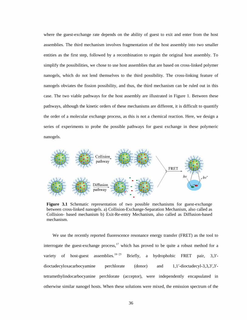
36
where the guest-exchange rate depends on the ability of guest to exit and enter from the host
assemblies. The third mechanism involves fragmentation of the host assembly into two smaller
entities as the first step, followed by a recombination to regain the original host assembly. To
simplify the possibilities, we chose to use host assemblies that are based on cross-linked polymer
nanogels, which do not lend themselves to the third possibility. The cross-linking feature of
nanogels obviates the fission possibility, and thus, the third mechanism can be ruled out in this
case. The two viable pathways for the host assembly are illustrated in Figure 1. Between these
pathways, although the kinetic orders of these mechanisms are different, it is difficult to quantify
the order of a molecular exchange process, as this is not a chemical reaction. Here, we design a
series of experiments to probe the possible pathways for guest exchange in these polymeric
nanogels.
We use the recently reported fluorescence resonance energy transfer (FRET) as the tool to
interrogate the guest-exchange process,17
which has proved to be quite a robust method for a
variety of host-guest assemblies.18−25
Briefly, a hydrophobic FRET pair, 3,3′-
dioctadecyloxacarbocyamine perchlorate (donor) and 1,1′-dioctadecyl-3,3,3′,3′-
tetramethylindocarbocyanine perchlorate (acceptor), were independently encapsulated in
otherwise similar nanogel hosts. When these solutions were mixed, the emission spectrum of the
Figure 3.1 Schematic representation of two possible mechanisms for guest-exchange
between cross-linked nanogels. a) Collision-Exchange-Separation Mechanism, also called as
Collision- based mechanism b) Exit-Re-entry Mechanism, also called as Diffusion-based
mechanism.
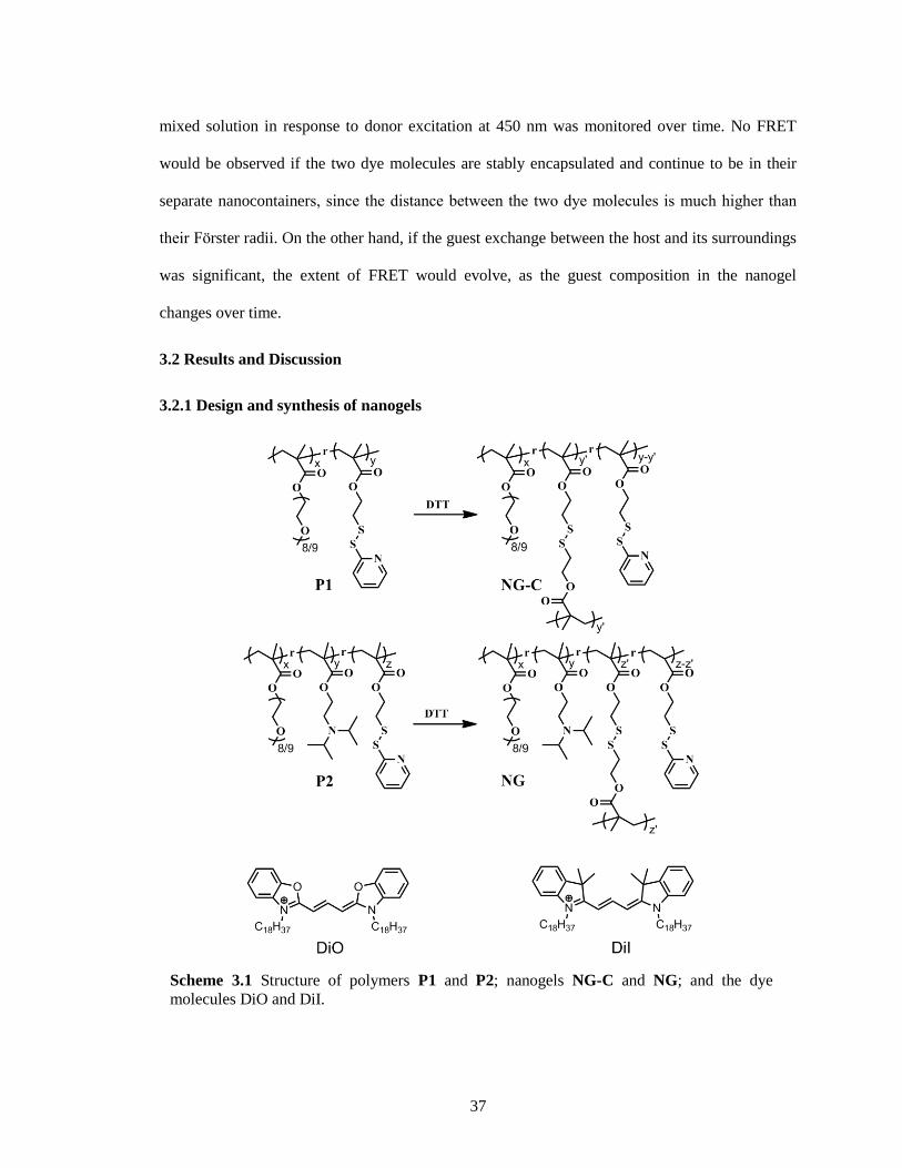
37
mixed solution in response to donor excitation at 450 nm was monitored over time. No FRET
would be observed if the two dye molecules are stably encapsulated and continue to be in their
separate nanocontainers, since the distance between the two dye molecules is much higher than
their F rster radii. On the other hand, if the guest exchange between the host and its surroundings
was significant, the extent of FRET would evolve, as the guest composition in the nanogel
changes over time.
3.2 Results and Discussion
3.2.1 Design and synthesis of nanogels
Scheme 3.1 Structure of polymers P1 and P2; nanogels NG-C and NG; and the dye
molecules DiO and DiI.
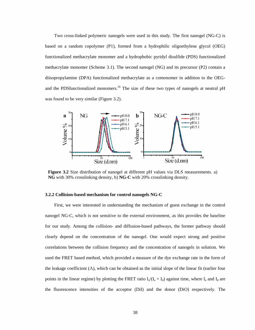
38
Two cross-linked polymeric nanogels were used in this study. The first nanogel (NG-C) is
based on a random copolymer (P1), formed from a hydrophilic oligoethylene glycol (OEG)
functionalized methacrylate monomer and a hydrophobic pyridyl disulfide (PDS) functionalized
methacrylate monomer (Scheme 3.1). The second nanogel (NG) and its precursor (P2) contain a
diisopropylamine (DPA) functionalized methacrylate as a comonomer in addition to the OEG-
and the PDSfunctionalized monomers.26
The size of these two types of nanogels at neutral pH
was found to be very similar (Figure 3.2).
3.2.2 Collision-based mechanism for control nanogels NG-C
First, we were interested in understanding the mechanism of guest exchange in the control
nanogel NG-C, which is not sensitive to the external environment, as this provides the baseline
for our study. Among the collision- and diffusion-based pathways, the former pathway should
clearly depend on the concentration of the nanogel. One would expect strong and positive
correlations between the collision frequency and the concentration of nanogels in solution. We
used the FRET based method, which provided a measure of the dye exchange rate in the form of
the leakage coefficient (Λ), which can be obtained as the initial slope of the linear fit (earlier four
points in the linear regime) by plotting the FRET ratio Ia/(Ia + Id) against time, where Ia and Id are
the fluorescence intensities of the acceptor (DiI) and the donor (DiO) respectively. The
Figure 3.2 Size distribution of nanogel at different pH values via DLS measurements. a)
NG with 30% crosslinking density, b) NG-C with 20% crosslinking density.
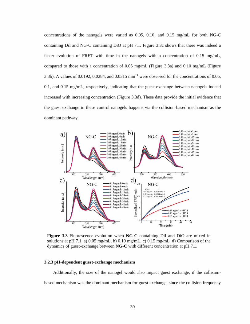
39
concentrations of the nanogels were varied as 0.05, 0.10, and 0.15 mg/mL for both NG-C
containing DiI and NG-C containing DiO at pH 7.1. Figure 3.3c shows that there was indeed a
faster evolution of FRET with time in the nanogels with a concentration of 0.15 mg/mL,
compared to those with a concentration of 0.05 mg/mL (Figure 3.3a) and 0.10 mg/mL (Figure
3.3b). Λ values of 0.0192, 0.0284, and 0.0315 min−1
were observed for the concentrations of 0.05,
0.1, and 0.15 mg/mL, respectively, indicating that the guest exchange between nanogels indeed
increased with increasing concentration (Figure 3.3d). These data provide the initial evidence that
the guest exchange in these control nanogels happens via the collision-based mechanism as the
dominant pathway.
3.2.3 pH-dependent guest-exchange mechanism
Additionally, the size of the nanogel would also impact guest exchange, if the collision-
based mechanism was the dominant mechanism for guest exchange, since the collision frequency
Figure 3.3 Fluorescence evolution when NG-C containing DiI and DiO are mixed in
solutions at pH 7.1. a) 0.05 mg/mL, b) 0.10 mg/mL, c) 0.15 mg/mL. d) Comparison of the
dynamics of guest-exchange between NG-C with different concentration at pH 7.1.
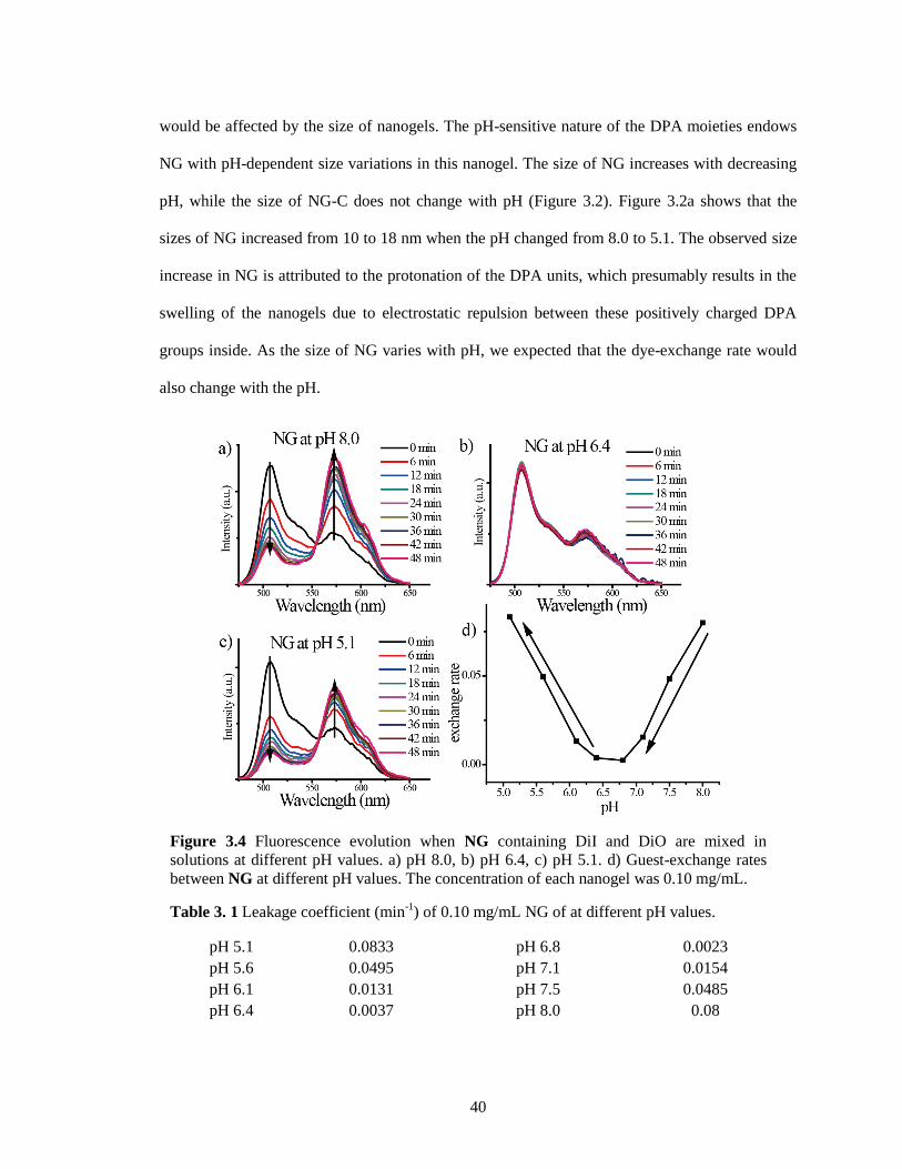
40
would be affected by the size of nanogels. The pH-sensitive nature of the DPA moieties endows
NG with pH-dependent size variations in this nanogel. The size of NG increases with decreasing
pH, while the size of NG-C does not change with pH (Figure 3.2). Figure 3.2a shows that the
sizes of NG increased from 10 to 18 nm when the pH changed from 8.0 to 5.1. The observed size
increase in NG is attributed to the protonation of the DPA units, which presumably results in the
swelling of the nanogels due to electrostatic repulsion between these positively charged DPA
groups inside. As the size of NG varies with pH, we expected that the dye-exchange rate would
also change with the pH.
Figure 3.4 Fluorescence evolution when NG containing DiI and DiO are mixed in
solutions at different pH values. a) pH 8.0, b) pH 6.4, c) pH 5.1. d) Guest-exchange rates
between NG at different pH values. The concentration of each nanogel was 0.10 mg/mL.
Table 3. 1 Leakage coefficient (min-1
) of 0.10 mg/mL NG of at different pH values.
pH 5.1 0.0833 pH 6.8 0.0023
pH 5.6 0.0495 pH 7.1 0.0154
pH 6.1 0.0131 pH 7.5 0.0485
pH 6.4 0.0037 pH 8.0 0.08
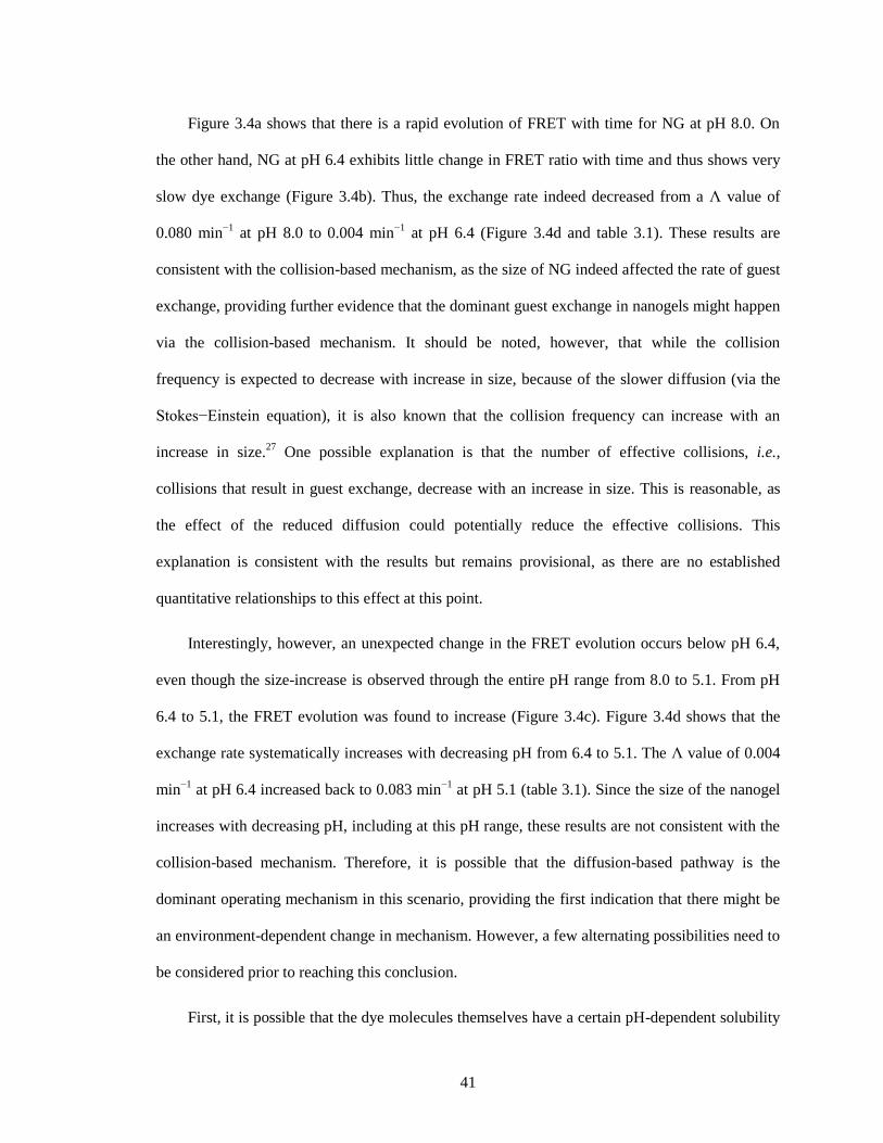
41
Figure 3.4a shows that there is a rapid evolution of FRET with time for NG at pH 8.0. On
the other hand, NG at pH 6.4 exhibits little change in FRET ratio with time and thus shows very
slow dye exchange (Figure 3.4b). Thus, the exchange rate indeed decreased from a Λ value of
0.080 min−1
at pH 8.0 to 0.004 min−1
at pH 6.4 (Figure 3.4d and table 3.1). These results are
consistent with the collision-based mechanism, as the size of NG indeed affected the rate of guest
exchange, providing further evidence that the dominant guest exchange in nanogels might happen
via the collision-based mechanism. It should be noted, however, that while the collision
frequency is expected to decrease with increase in size, because of the slower diffusion (via the
Stokes−Einstein equation), it is also known that the collision frequency can increase with an
increase in size.27
One possible explanation is that the number of effective collisions, i.e.,
collisions that result in guest exchange, decrease with an increase in size. This is reasonable, as
the effect of the reduced diffusion could potentially reduce the effective collisions. This
explanation is consistent with the results but remains provisional, as there are no established
quantitative relationships to this effect at this point.
Interestingly, however, an unexpected change in the FRET evolution occurs below pH 6.4,
even though the size-increase is observed through the entire pH range from 8.0 to 5.1. From pH
6.4 to 5.1, the FRET evolution was found to increase (Figure 3.4c). Figure 3.4d shows that the
exchange rate systematically increases with decreasing pH from 6.4 to 5.1. The Λ value of 0.004
min−1
at pH 6.4 increased back to 0.083 min−1
at pH 5.1 (table 3.1). Since the size of the nanogel
increases with decreasing pH, including at this pH range, these results are not consistent with the
collision-based mechanism. Therefore, it is possible that the diffusion-based pathway is the
dominant operating mechanism in this scenario, providing the first indication that there might be
an environment-dependent change in mechanism. However, a few alternating possibilities need to
be considered prior to reaching this conclusion.
First, it is possible that the dye molecules themselves have a certain pH-dependent solubility
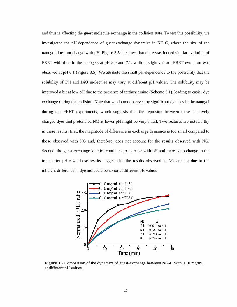
42
and thus is affecting the guest molecule exchange in the collision state. To test this possibility, we
investigated the pH-dependence of guest-exchange dynamics in NG-C, where the size of the
nanogel does not change with pH. Figure 3.5a,b shows that there was indeed similar evolution of
FRET with time in the nanogels at pH 8.0 and 7.1, while a slightly faster FRET evolution was
observed at pH 6.1 (Figure 3.5). We attribute the small pH-dependence to the possibility that the
solubility of DiI and DiO molecules may vary at different pH values. The solubility may be
improved a bit at low pH due to the presence of tertiary amine (Scheme 3.1), leading to easier dye
exchange during the collision. Note that we do not observe any significant dye loss in the nanogel
during our FRET experiments, which suggests that the repulsion between these positively
charged dyes and protonated NG at lower pH might be very small. Two features are noteworthy
in these results: first, the magnitude of difference in exchange dynamics is too small compared to
those observed with NG and, therefore, does not account for the results observed with NG.
Second, the guest-exchange kinetics continues to increase with pH and there is no change in the
trend after pH 6.4. These results suggest that the results observed in NG are not due to the
inherent difference in dye molecule behavior at different pH values.
Figure 3.5 Comparison of the dynamics of guest-exchange between NG-C with 0.10 mg/mL
at different pH values.
43
We hypothesized next that the observed change in pH dependence trend at pH 6.4 could
perhaps be explained by a change in the hydrophobicity of the nanogel interior, i.e., the host
properties. It is reasonable to anticipate that the protonation of DPA groups would decrease the
hydrophobicity of the nanogel.28
Pyrene’s fluorescence properties are greatly dependent on its
microenvironment.29, 30
Specifically, the ratio of the intensities between the first and the third
peaks (I1/I3) in the pyrene emission spectrum can be used to determine the polarity of the dye’s
microenvironment. The value of this ratio can range from 1.9 in polar solvents to 0.6 in certain
hydrocarbon solvents. We utilized this dye as the guest molecule to probe the hydrophobicity of
the microenvironment within the nanogel interiors. Aqueous buffer solutions of NG containing 2
wt% pyrene were prepared for this purpose at different pH values. A sample fluorescence
spectrum of nanogel is shown in Figure 3.6a. The hydrophobicity of nanogel at different pH
values was studied by calculating the intensity ratio of the first and third emission peaks of pyrene,
commonly referred to as the I1/I3 ratio. Figure 3.6b shows the variations in I1/I3 of pyrene
encapsulated in NG at different pH values. When the pH was reduced from 8.0 to about 6.4, the
I1/I3 remained constant around 1.25. This value suggests that the nanogel interior is quite
hydrophobic. However, when the pH was further decreased, a steady increase in the I1/I3 value
was observed, suggesting that the microenvironment of the dye is becoming increasingly polar.
The I1/I3 ratio at pH 5.1 was found to be about 1.37. On the contrary, pyrene in the control
Figure 3.6 Effect of pH on the hydrophobicity. a) Fluorescence emission spectrum
measured for nanogel NG-C loading 2 wt% pyrene and b) calculated I1/I3 ratios for
nanogels at different pH values, (black, NG; red, NG-C).
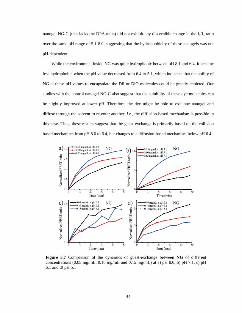
44
nanogel NG-C (that lacks the DPA units) did not exhibit any discernible change in the I1/I3 ratio
over the same pH range of 5.1-8.0, suggesting that the hydrophobicity of these nanogels was not
pH-dependent.
While the environment inside NG was quite hydrophobic between pH 8.1 and 6.4, it became
less hydrophobic when the pH value decreased from 6.4 to 5.1, which indicates that the ability of
NG at these pH values to encapsulate the DiI or DiO molecules could be greatly depleted. Our
studies with the control nanogel NG-C also suggest that the solubility of these dye molecules can
be slightly improved at lower pH. Therefore, the dye might be able to exit one nanogel and
diffuse through the solvent to re-enter another; i.e., the diffusion-based mechanism is possible in
this case. Thus, these results suggest that the guest exchange is primarily based on the collision
based mechanism from pH 8.0 to 6.4, but changes to a diffusion-based mechanism below pH 6.4.
Figure 3.7 Comparison of the dynamics of guest-exchange between NG of different
concentrations (0.05 mg/mL, 0.10 mg/mL and 0.15 mg/mL) at a) pH 8.0, b) pH 7.1, c) pH
6.1 and d) pH 5.1
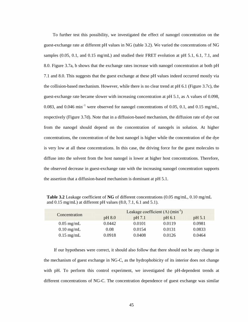
45
To further test this possibility, we investigated the effect of nanogel concentration on the
guest-exchange rate at different pH values in NG (table 3.2). We varied the concentrations of NG
samples (0.05, 0.1, and 0.15 mg/mL) and studied their FRET evolution at pH 5.1, 6.1, 7.1, and
8.0. Figure 3.7a, b shows that the exchange rates increase with nanogel concentration at both pH
7.1 and 8.0. This suggests that the guest exchange at these pH values indeed occurred mostly via
the collision-based mechanism. However, while there is no clear trend at pH 6.1 (Figure 3.7c), the
guest-exchange rate became slower with increasing concentration at pH 5.1, as Λ values of 0.098,
0.083, and 0.046 min−1
were observed for nanogel concentrations of 0.05, 0.1, and 0.15 mg/mL,
respectively (Figure 3.7d). Note that in a diffusion-based mechanism, the diffusion rate of dye out
from the nanogel should depend on the concentration of nanogels in solution. At higher
concentrations, the concentration of the host nanogel is higher while the concentration of the dye
is very low at all these concentrations. In this case, the driving force for the guest molecules to
diffuse into the solvent from the host nanogel is lower at higher host concentrations. Therefore,
the observed decrease in guest-exchange rate with the increasing nanogel concentration supports
the assertion that a diffusion-based mechanism is dominant at pH 5.1.
If our hypotheses were correct, it should also follow that there should not be any change in
the mechanism of guest exchange in NG-C, as the hydrophobicity of its interior does not change
with pH. To perform this control experiment, we investigated the pH-dependent trends at
different concentrations of NG-C. The concentration dependence of guest exchange was similar
Table 3.2 Leakage coefficient of NG of different concentrations (0.05 mg/mL, 0.10 mg/mL
and 0.15 mg/mL) at different pH values (8.0, 7.1, 6.1 and 5.1).
Concentration Leakage coefficient (Λ) (min
-1)
pH 8.0 pH 7.1 pH 6.1 pH 5.1
0.05 mg/mL 0.0442 0.0101 0.0119 0.0981
0.10 mg/mL 0.08 0.0154 0.0131 0.0833
0.15 mg/mL 0.0918 0.0408 0.0126 0.0464
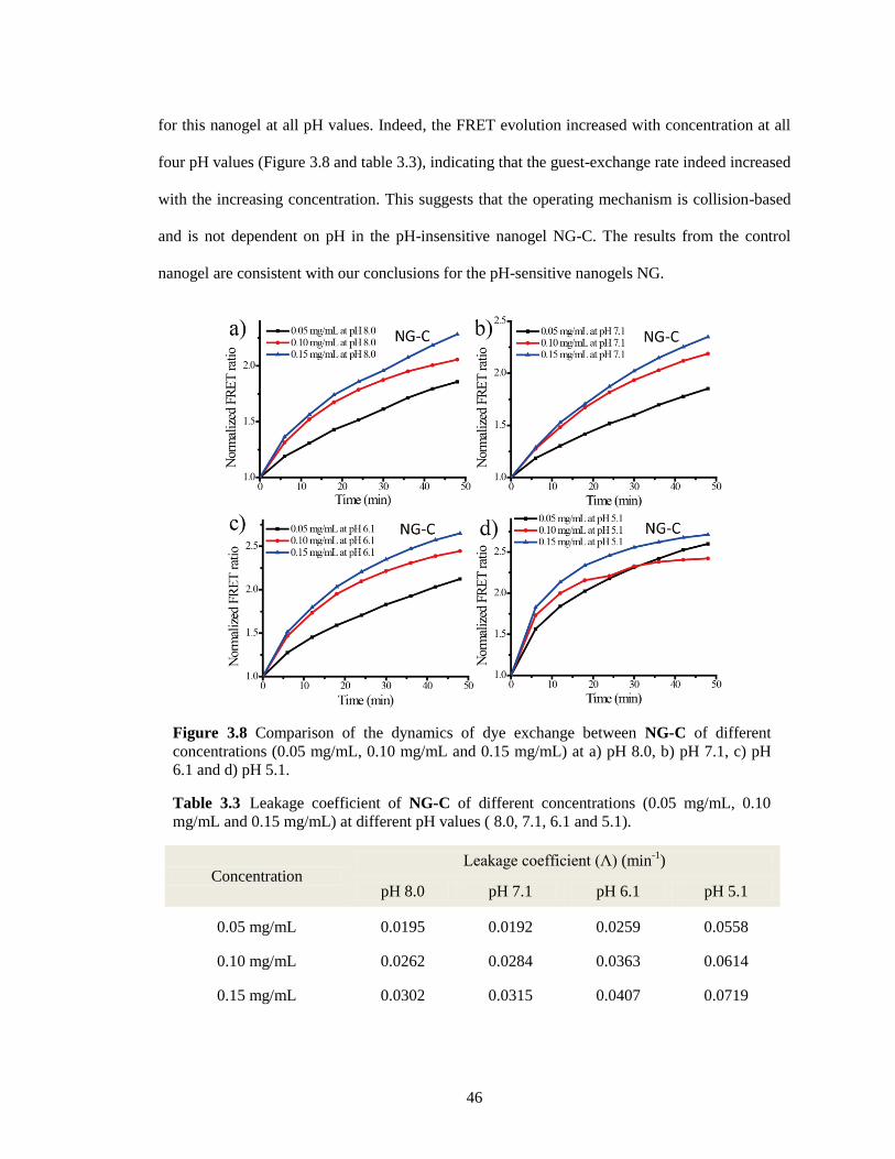
46
for this nanogel at all pH values. Indeed, the FRET evolution increased with concentration at all
four pH values (Figure 3.8 and table 3.3), indicating that the guest-exchange rate indeed increased
with the increasing concentration. This suggests that the operating mechanism is collision-based
and is not dependent on pH in the pH-insensitive nanogel NG-C. The results from the control
nanogel are consistent with our conclusions for the pH-sensitive nanogels NG.
Figure 3.8 Comparison of the dynamics of dye exchange between NG-C of different
concentrations (0.05 mg/mL, 0.10 mg/mL and 0.15 mg/mL) at a) pH 8.0, b) pH 7.1, c) pH
6.1 and d) pH 5.1.
Table 3.3 Leakage coefficient of NG-C of different concentrations (0.05 mg/mL, 0.10
mg/mL and 0.15 mg/mL) at different pH values ( 8.0, 7.1, 6.1 and 5.1).
Concentration Leakage coefficient (Λ) (min
-1)
pH 8.0 pH 7.1 pH 6.1 pH 5.1
0.05 mg/mL 0.0195 0.0192 0.0259 0.0558
0.10 mg/mL 0.0262 0.0284 0.0363 0.0614
0.15 mg/mL 0.0302 0.0315 0.0407 0.0719
47
3.3 Summary
In summary, we have designed a series of experiments to study the guest-exchange
mechanism in pH-sensitive nanogels along with pH-insensitive nanogels as a control. We have
shown that the dominant mechanism for guest exchange in these pH insensitive nanogel hosts is
collision-based. Perhaps, the most important take-home message of this work is that the
mechanism of guest exchange in the pH-sensitive host-guest assemblies can change on the basis
of the microenvironment of the host. When the nanogel interior is hydrophobic, the collision-
based mechanism is the dominant pathway. However, when the interior is sufficiently hydrophilic,
the dominant mechanism changes to a diffusion-based one. From an even broader perspective, it
is important to recognize that both the intrinsic factors, such as size and interior environment of
the host assemblies, and the extrinsic factors, such as pH and concentration, can have significant
impact on encapsulation stability. From the application standpoint, these findings could have
implications in many areas. For example, in drug delivery applications, the drug-loaded
nanocarrier experiences both concentration and environmental changes upon biodistribution to
diseased tissues. The mechanistic variations in two different environments provide insights into
molecular designs that can afford stable encapsulation in one environment and release of the
molecules in another.31,32
Similarly, the results from this study may also provide new
opportunities for designing nanoreactors33,34
in which catalysts encapsulated in a host can be used
to reversibly turn a chemical reaction on or off, because of the environment-dependent diffusion
of reactant molecules.
3.4 Experimental
3.4.1 General Methods
2,2′-Dithiodipyridine, 2-mercaptoethanol, poly(ethylene glycol) monomethyl ether
methacrylate (PEGMA, Mw 450 g/mol), 2-(diisopropylamino)ethyl methacrylate (DPA), D,L-
dithiothreitol (DTT), 1,1′-dioctadecyl-3,3,3′,3′-tetramethylindocarbocyanine perchlorate (DiI),
48
3,3′-dioctadecyloxacarbocyanine perchlorate (DiO), 4-cyano-4-
(phenylcarbonothioylthio)pentanoic acid, and other conventional reagents were obtained from
commercial sources and were used as received without further purification. 2,2′-
Azobis(isobutyronitrile) (AIBN) was purified by recrystallization from ethanol. Pyridyl disulfide
ethyl methacrylate (PDSEMA) was prepared using a previously reported procedure.35
Phosphate
buffer solutions were prepared by using monosodium phosphate and disodium phosphate. The
buffer strength was constant at 10 mM. The pH value was determined via the Accumet AB
15/15+ benchtop pH meter. 1H NMR spectra were recorded on a 400 MHz Bruker NMR
spectrometer using the residual proton resonance of the solvent as the internal standard. Chemical
shifts are reported in parts per million (ppm). Molecular weights of the polymers were estimated
by gel permeation chromatography (GPC) using PMMA standard with a refractive index detector.
Dynamic light scattering (DLS) measurements were performed using a Malvern Nanozetasizer.
UV−visible absorption spectra were recorded on a Varian (model EL 01125047)
spectrophotometer. The fluorescence spectra were obtained from a JASCO FP-6500
spectrofluorimeter. Transmission electron microscopy (TEM) images were taken using a JEOL
2000FX at 200 kV. All experiments were carried out at ambient temperature, unless otherwise
mentioned.
3.4.2 Synthesis of Random Copolymer
Random copolymer P2, containing DPA groups, was synthesized according to a previously
reported procedure.26
A mixture of 4-cyano-4-(phenylcarbonothioylthio)pentanoic acid (12.0 mg,
0.043 mmol), PDSMA (150.0 mg, 0.588 mmol), PEGMA (215.0 mg, 0.453 mmol), DPAMA
(130.0 mg, 0.609 mmol), and AIBN (1.5 mg, 0.010 mmol) was dissolved in THF (1.0 mL) and
degassed by performing three freeze-pump-thaw cycles. The reaction mixture was sealed and
then transferred into a preheated oil bath at 65 °C and stirred for 10 h. To remove unreacted
monomers, the resultant mixture was precipitated in cold diethyl ether (20 mL) and redispersed in
49
THF three times to yield the random copolymer P2 as a waxy solid. GPC (THF) Mn: 10.6 kDa. Đ:
1.44. 1H NMR (400 MHz, CDCl3) δ: 8.46, 7.67, 7.10, 4.35-4.09, 3.94-3.37, 3.02, 2.62, 2.04-1.64,
1.43-0.87. The molar ratio (PDS/PEG/DPA) was determined to be 34%/28%/38% by integrating
the methoxy protons in the polyethylene glycol unit (δHd = 3.37 ppm), the aromatic protons in the
pyridine (δHa = 8.46, δHb = 7.67, δHc = 7.10), and the methylene protons next to the nitrogen in
DPA units (δHe = 2.62, δHf = 3.02).
3.4.3 Preparation of Nanogels Contaning DiI/DiO
Polymer (10 mg) was dissolved in 1 mL of water, and the pH was adjusted to around 10
through the addition of NaOH aqueous solution (1 M). Then 0.04 mL of DiI in acetone stock
solution (5 mg/mL) or 0.08 mL of DiO in acetone stock solution (2.5 mg/mL) was added into the
polymer solution (0.2 mg of each dye for 10 mg of polymer in 1 mL of water). The final dye
concentration was about 0.2 mmol/L. The mixed solution was stirred overnight at room
temperature, open to the atmosphere to allow the organic solvent to evaporate. Then a measured
amount of DTT (0.2 μmol for 15 mol % against PDS groups) was added. After stirring for 4 h,
insoluble DiI/DiO was removed by filtration and the pyridothione byproduct was removed from
the nanogel solution by extensive dialysis using a membrane with a molecular weight cutoff of
7000 g/mol. Finally, nanogel stock solutions with a concentration of 1 mg/mL were prepared for
further studies.
3.4.4 Mixing of Nanogel-Encapsulated Dyes
Nanogel containing DiI and nanogel containing DiO were mixed in phosphate buffer
solution at ambient temperature and at a certain pH value such that the total volume was 1 mL.
The pH did not change after mixing. The fluorescence spectra were recorded using a 450 nm
excitation wavelength. At the same time, absorption spectra were also measured for all samples to
ensure that there is no loss of dye molecules due to precipitation. The FRET ratio Ia/(Ia + Id),
where Ia and Id are the fluorescence intensities of the acceptor (DiI) and the donor (DiO),
50
respectively, was plotted against time to show the dynamics of the dye exchange in nanogel
solution. The leakage coefficients (Λ) were calculated on the basis of the slopes from four earlier
points in the linear regime. In the case of very fast guest exchange, the time frame for obtaining
these four data points is shorter.
3.5 References
1. Allen, T. M.; Cullis, P. R. Drug delivery systems: entering the mainstream. Science 2004, 303,
1818-1822.
2. Peer, D.; Karp, J. M.; Hong, S.; Farokhzad, O. C.; Margalit, R.; Langer, R. Nanocarriers as an
emerging platform for cancer therapy. Nat. Nanotechnol. 2007, 2, 751-760.
3. Oh, J. K.; Drumright, R.; Siegwart, D. J.; Matyjaszewski, K. The development of
microgels/nanogels for drug delivery applications. Prog. Polym. Sci. 2008, 33, 448-477.
4. Savic, R.; Eisenberg, A.; Maysinger, D. Block copolymer micelles as delivery vehicles of
hydrophobic drugs: Micelle-cell interactions. J. Drug Target. 2006, 14, 343-355.
5. Liu, K. C.; Yeo, Y. Extracellular stability of nanoparticulate drug carriers. Arch. Pharmacal
Res. 2014, 37, 16-23.
6. Kumar, R.; Chen, M. H.; Parmar, V. S.; Samuelson, L. A.; Kumar, J.; Nicolosi, R.;
Yoganathan, S.; Watterson, A. C. Supramolecular assemblies based on copolymers of PEG600
and functionalized aromatic diesters for drug delivery applications. J. Am. Chem. Soc., 2004, 126,
10640-10644.
7. Pan, L.; He, Q.; Liu, J.; Chen, Y.; Ma, M.; Zhang, L.; Shi, J. Nuclear-targeted drug delivery of
TAT peptide-conjugated monodisperse mesoporous silica nanoparticles. J. Am. Chem. Soc. 2012,
134, 5722-5725.
8. Shen, Y.; Jin, E.; Zhang, B.; Murphy, C. J.; Sui, M.; Zhao, J.; Wang, J.; Tang, J.; Fan, M.; Kirk,
E. V.; Murdoch, W. J. Prodrugs forming high drug loading multifunctional nanocapsules for
intracellular cancer drug delivery. J. Am. Chem. Soc., 2010, 132, 4259-4265.
9. Zhu, J.; Tan, M.; Zhang, L.; Yin, Q. Elongation flow-triggered morphology transitions of
dendritic polyethylene amphiphilic assemblies: host−guest implications. Soft Matter, 2014, 10,
6506-6513.
10. Rharbi, Y.; Winnik, M. A. Solute exchange between surfactant micelles by micelle
fragmentation and fusion. Adv. Colloid Interface Sci. 2001, 89, 25-46.
11. Piñeiro, L.; Freire, S.; Bordello, J.; Novo, M.; Al-Soufi, W. Dye exchange in micellar
solutions. Quantitative analysis of bulk and single molecule fluorescence titrations. Soft Matter
2013, 9, 10779-10790.
51
12. Rharbi, Y.; Li, M.; Winnik, M. A.; Hahn, K. G. Temperature dependence of fusion and
fragmentation kinetics of Triton X-100 Micelles. J. Am. Chem. Soc. 2000, 122, 6242-6251.
13. Rharbi, Y.; Chen, L.; Winnik, M. A. Exchange mechanisms for sodium dodecyl sulfate
micelles: high salt concentration. J. Am. Chem. Soc. 2004, 126, 6025-6034.
14. Novo, M.; Felekyan, S.; Seidel, C. A.; Al-Soufi, W. Dye-exchange dynamics in micellar
solutions studied by fluorescence correlation spectroscopy. J. Phys. Chem. B 2007, 111, 3614-
3624.
15. Bordello, J.; Novo, M.; Al-Soufi, W. Exchange-dynamics of a neutral hydrophobic dye in
micellar solutions studied by fluorescence correlation spectroscopy. J. Colloid Interface Sci. 2010,
345, 369-376.
16. Gehlen, M. H.; De Schryver, F. C. Time-resolved fluorescence quenching in micellar
assemblies. Chem. Rev. 1993, 93, 199-221.
17. Jiwpanich, S.; Ryu, J. -H.; Bickerton, S.; Thayumanavan, S. Noncovalent encapsulation
stabilities in supramolecular nanoassemblies. J. Am. Chem. Soc. 2010, 132, 10683-10685.
18. Lu, J.; Owen, S. C.; Shoichet, M. S. Stability of self-assembled polymeric micelles in serum.
Macromolecules 2011, 44, 6002-6008.
19. Dong, H.; Shu, J. Y.; Dube, N.; Ma, Y.; Tirrell, M. V.; Downing, K. H.; Xu, T. 3-Helix
micelles stabilized by polymer springs. J. Am. Chem. Soc. 2012, 134, 11807-11814.
20. Kim, K.; Bae, B.; Kang, Y. J.; Nam, J.-M.; Kang, S.; Ryu, J. -H. Natural polypeptide-based
supramolecular nanogels for stable noncovalent encapsulation. Biomacromolecules 2013, 14,
3515-3522.
21. Zou, P.; Chen, H.; Paholak, H. J.; Sun, D. Noninvasive fluorescence resonance energy
transfer imaging of in vivo premature drug release from polymeric nanoparticles. Mol.
Pharmaceutics 2013, 10, 4185-4194.
22. Dan, K.; Rajdev, P.; Deb, J.; Jana, S. S.; Ghosh, S. Remarkably stable amphiphilic random
copolymer assemblies: A structure–property relationship study. J. Polym. Sci. A Polym. Chem.
2013, 51, 4932-4943.
23. Li, L.; Ryu, J. -H.; Thayumanavan, S. Effect of Hofmeister ions on the size and encapsulation
stability of polymer nanogels. Langmuir 2013, 29, 50-55.
24. Fuller, J. M.; Raghupathi, K. R.; Ramireddy, R. R.; Subrahmanyam, A. V.; Yesilyurt, V.;
Thayumanavan, S. Temperature-sensitive transitions below LCST in amphiphilic dendritic
assemblies: host–guest implications. J. Am. Chem. Soc. 2013, 135, 8947-8954;
25. Swaminathan, S.; Fowley, C.; McCaughan, B.; Cusido, J.; Callan, J. F.; Raymo. F. M.
Intracellular guest-exchange between dynamic supramolecular hosts. J. Am. Chem. Soc., 2014,
136, 7907-7913.
26. Li, L.; Raghupathi, K.; Yuan, C.; Thayumanavan, S. Surface charge generation in nanogels
for activated cellular uptake at tumor-relevant pH. Chem. Sci. 2013, 4, 3654-3360.
52
27. Gjaltema, A.; Van Loosdrecht, M. C. M.; Heijnen, J. Abrasion of suspended biofilm pellets in
airlift reactors: Effect of particle size. J. Biotechnol. Bioeng. 1997, 55, 206-215
28. Yu, S.; Hu, J.; Pan, X.; Yao, P.; Jiang, M. Stable and pH-sensitive nanogels prepared by self-
assembly of chitosan and ovalbumin. Langmuir 2006, 22, 2754-2759.
29. Kalyanasundaram, K.; Thomas, J. Environmental effects on vibronic band intensities in
pyrene monomer fluorescence and their application in studies of micellar systems. J. Am. Chem.
Soc. 1977, 99, 2039-2044.
30. Bickerton, S.; Jiwpanich, S.; Thayumanavan, S. Interconnected roles of scaffold
hydrophobicity, drug loading, and encapsulation stability in polymeric nanocarriers. Mol.
Pharmaceutics 2012, 9, 3569-3578.
31. Du, J. Z.; Sun, T. M.; Song, W. J.; Wu, J.; Wang, J. A tumor-acidity-activated charge-
conversional nanogel as an intelligent vehicle for promoted tumoral-cell uptake and drug delivery.
Angew. Chem. Int. Ed., 2010, 49, 3621-3626.
32. Poon, Z.; Chang, D.; Zhao, X. Y.; Hammond, P. T. Layer-by-layer nanoparticles with a pH-
sheddable layer for in vivo targeting of tumor hypoxia. ACS Nano., 2011, 5, 4284-4292.
33. Wang, X.; Liu, G.; Hu, J.; Zhang, G.; Liu, S. Concurrent block copolymer polymersome
stabilization and bilayer permeabilization by stimuli-regulated traceless crosslinking. Angew.
Chem. Int. Ed., 2014, 53, 3138-3142.
34. Yuan, C.; Luo, W.; Zhong, L.; Deng, H.; Liu, J.; Xu, Y.; Dai, L. Au @ polymer
nanostructures with tunable permeability shells for selective catalysis. Angew. Chem. Int. Ed.
2011, 51, 3515-3519.
35. Ghosh, S.; Basu, S.; Thayumanavan, S. Simultaneous and reversible functionalization of
copolymers for biological applications. Macromolecules 2006, 39, 5595-5597.
53
CHAPTER 4
SURFACE CHARGE GENERATION IN NANOGELS FOR ACTIVATED CELLULAR
UPTAKE AT TUMOR-RELEVANT PH
Li, L.; Raghupathi, K.; Yuan, C.; Thayumanavan, S. Surface charge generation in nanogels
for activated cellular uptake at tumor-relevant pH. Chem. Sci. 2013, 4, 3654-3360. - Reproduced
by permission of The Royal Society of Chemistry
http://pubs.rsc.org/en/content/articlehtml/2013/sc/c3sc50899d
4.1 Introduction
Nanocarriers with cationic surface charge are readily internalized by the cells because of
their higher affinity for the negatively charged phospholipid bilayer of cell membranes.1,2
Since
cellular internalization can often be a stumbling block in therapeutic approaches, positively
charged nanoparticles are often used to enhance cellular uptake.3 However, the positively charged
nature of carriers may also induce non-specific interactions in serum, leading to their severe
aggregation and rapid clearance from the circulation by the reticuloendothelial system following
systemic administration.4 To address this problem, recent efforts have been dedicated to the
development of stimuli-responsive charge conversional nanoparticles.5–7
Typically, it is desirable
that the surface charge of nanoparticles be neutral or even negatively charged under normal
physiological conditions. Then, under the aberrant conditions that simulate the disease site, the
surface charge of these environment responsive nanoparticles become positive. The hypothesis is
that such a change could result in rapid uptake of the nanocarriers by the cells.
Environmental stimuli that were exploited in the development of stimuli-responsive drug
delivery systems include pH,8–10
temperature,11–13
enzymatic expression,14–16
redox reaction, 17–19
and light.20–22
Many of these designs were motivated by the environment around solid tumors and
thus have been targeted to ultimately impact cancer therapy. Among these, pH responsiveness is
most frequently utilized, because the extracellular pH (pHe) of tumor tissue (~6.5) is slightly
54
lower than that of normal tissue and blood (~7.4), which is due to the hypoxia-induced production
of excess lactic acid in the tumor microenvironments.23–25
For example, nanocarriers that are
capable of reversing the surface charge from negative to positive at pHe have been designed,
where the pH-induced cleavage of the amide bond between an amino and 2,3-dimethylmaleic
anhydride causes the charge conversion.26–28
Similarly, nanoparticles containing a pH-sensitive
outer stealth layer have been designed using layer-by-layer techniques. In this case, the
nanoparticle sheds a layer in response to acidity and reveals a charged nanoparticle surface that
can be readily internalized by tumor cells.29
In this manuscript, we report on a system that contains a few important and new
complementary features. In our design, we stipulated that: (i) the system should be a charge
generation process (neutral to positive), instead of a charge conversion process (negative to
positive). This design has the potential to further reduce non-specific binding, prior to the charge-
generation step; (ii) the charge generation should be rapid; (iii) the pH-induced charge-generation
step is reversible, i.e. the surface charge is environment-dependent, but not permanent; (iv) the
scaffold that exhibits these features should be nanoscale and be able to non-covalently bind
hydrophobic guest molecules; and (v) the charge-generation step should not produce any small
molecule by-products. Polymer nanogels have the potential to satisfy these requirements and thus
afford pH-activated cellular uptake.
4.2 Results and Discussion
4.2.1 Design and synthesis of pH-sensitive nanogels
To afford pH-activated cellular uptake and satisfy the requirements above, we utilize our
recently reported self-crosslinking disulfide-based nanogels as the scaffold.30–33
Disulfide based
nanogels systems have gained significant interest because of their triggered encapsulated guest
release capabilities in the cytosol.34
Our polymeric disulfide nanogels can be achieved by taking
advantage of amphiphilic random copolymers that form nanoscale aggregates and by trapping
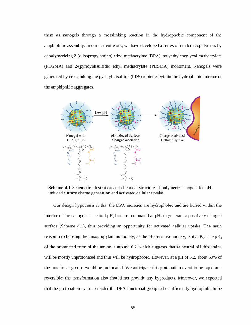
55
them as nanogels through a crosslinking reaction in the hydrophobic component of the
amphiphilic assembly. In our current work, we have developed a series of random copolymers by
copolymerizing 2-(diisopropylamino) ethyl methacrylate (DPA), polyethyleneglycol methacrylate
(PEGMA) and 2-(pyridyldisulfide) ethyl methacrylate (PDSMA) monomers. Nanogels were
generated by crosslinking the pyridyl disulfide (PDS) moieties within the hydrophobic interior of
the amphiphilic aggregates.
Our design hypothesis is that the DPA moieties are hydrophobic and are buried within the
interior of the nanogels at neutral pH, but are protonated at pHe to generate a positively charged
surface (Scheme 4.1), thus providing an opportunity for activated cellular uptake. The main
reason for choosing the diisopropylamino moiety, as the pH-sensitive moiety, is its pKa. The pKa
of the protonated form of the amine is around 6.2, which suggests that at neutral pH this amine
will be mostly unprotonated and thus will be hydrophobic. However, at a pH of 6.2, about 50% of
the functional groups would be protonated. We anticipate this protonation event to be rapid and
reversible; the transformation also should not provide any byproducts. Moreover, we expected
that the protonation event to render the DPA functional group to be sufficiently hydrophilic to be
Scheme 4.1 Schematic illustration and chemical structure of polymeric nanogels for pH-
induced surface charge generation and activated cellular uptake.
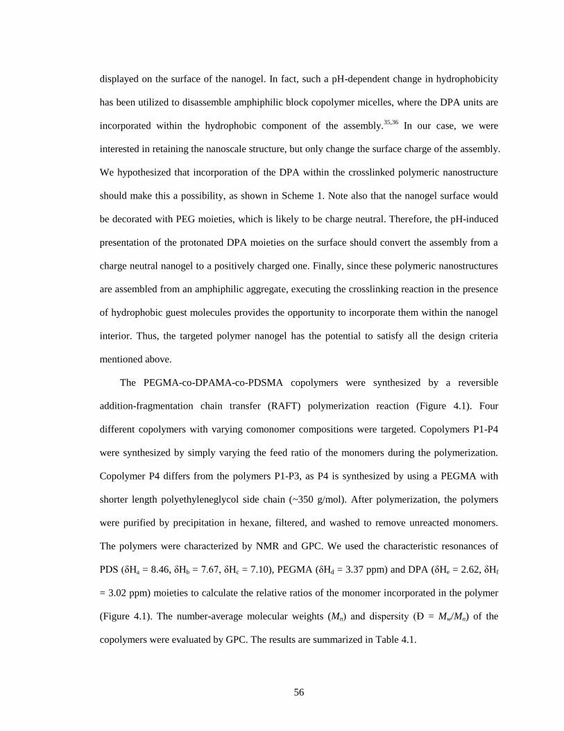
56
displayed on the surface of the nanogel. In fact, such a pH-dependent change in hydrophobicity
has been utilized to disassemble amphiphilic block copolymer micelles, where the DPA units are
incorporated within the hydrophobic component of the assembly.35,36
In our case, we were
interested in retaining the nanoscale structure, but only change the surface charge of the assembly.
We hypothesized that incorporation of the DPA within the crosslinked polymeric nanostructure
should make this a possibility, as shown in Scheme 1. Note also that the nanogel surface would
be decorated with PEG moieties, which is likely to be charge neutral. Therefore, the pH-induced
presentation of the protonated DPA moieties on the surface should convert the assembly from a
charge neutral nanogel to a positively charged one. Finally, since these polymeric nanostructures
are assembled from an amphiphilic aggregate, executing the crosslinking reaction in the presence
of hydrophobic guest molecules provides the opportunity to incorporate them within the nanogel
interior. Thus, the targeted polymer nanogel has the potential to satisfy all the design criteria
mentioned above.
The PEGMA-co-DPAMA-co-PDSMA copolymers were synthesized by a reversible
addition-fragmentation chain transfer (RAFT) polymerization reaction (Figure 4.1). Four
different copolymers with varying comonomer compositions were targeted. Copolymers P1-P4
were synthesized by simply varying the feed ratio of the monomers during the polymerization.
Copolymer P4 differs from the polymers P1-P3, as P4 is synthesized by using a PEGMA with
shorter length polyethyleneglycol side chain (~350 g/mol). After polymerization, the polymers
were purified by precipitation in hexane, filtered, and washed to remove unreacted monomers.
The polymers were characterized by NMR and GPC. We used the characteristic resonances of
PDS (δHa = 8.46, δHb = 7.67, δHc = 7.10), PEGMA (δHd = 3.37 ppm) and DPA (δHe = 2.62, δHf
= 3.02 ppm) moieties to calculate the relative ratios of the monomer incorporated in the polymer
(Figure 4.1). The number-average molecular weights (Mn) and dispersity (Đ = Mw/Mn) of the
copolymers were evaluated by GPC. The results are summarized in Table 4.1.
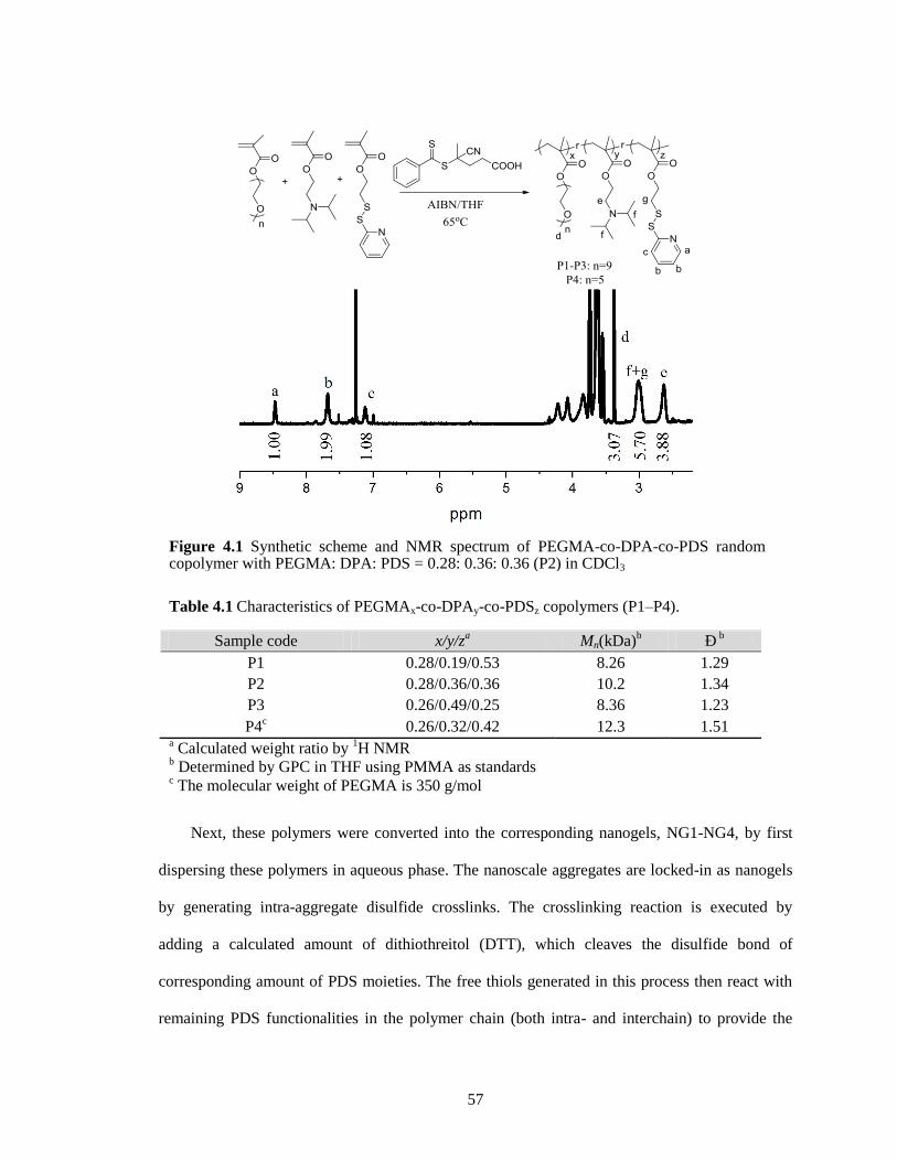
57
Next, these polymers were converted into the corresponding nanogels, NG1-NG4, by first
dispersing these polymers in aqueous phase. The nanoscale aggregates are locked-in as nanogels
by generating intra-aggregate disulfide crosslinks. The crosslinking reaction is executed by
adding a calculated amount of dithiothreitol (DTT), which cleaves the disulfide bond of
corresponding amount of PDS moieties. The free thiols generated in this process then react with
remaining PDS functionalities in the polymer chain (both intra- and interchain) to provide the
Table 4.1 Characteristics of PEGMAx-co-DPAy-co-PDSz copolymers (P1–P4).
Sample code x/y/za Mn(kDa)
b Ð
b
P1 0.28/0.19/0.53 8.26 1.29
P2 0.28/0.36/0.36 10.2 1.34
P3 0.26/0.49/0.25 8.36 1.23
P4c 0.26/0.32/0.42 12.3 1.51
a Calculated weight ratio by
1H NMR
b Determined by GPC in THF using PMMA as standards
c The molecular weight of PEGMA is 350 g/mol
Figure 4.1 Synthetic scheme and NMR spectrum of PEGMA-co-DPA-co-PDS random copolymer with PEGMA: DPA: PDS = 0.28: 0.36: 0.36 (P2) in CDCl3
58
crosslinked polymer nanogel. The intra-aggregate nature of the crosslinking reaction insures that
the nanogel size is tightly controlled by the size of the nanoscale aggregate. In all our nanogels,
we used about 50% DTT (relative to the PDS units) to cause the PDS units to be exhausted in the
crosslinking reaction; the extent of the PDS cleavage is conveniently monitored by the absorption
peak that corresponds to the 2-thiopyridone by-product.
Next, we investigated the pH-dependent charge-generation features in these nanogels
through zeta potential measurements. The zeta potential of the PEG-decorated nanogel at neutral
pH is negative. Although this seems surprising, there have been several reports that suggest that
the surface charge of PEG-decorated nanostructures are indeed negative.37,38
The origins of this
charge are not clear. Upon decreasing the pH of the solution, the surface charge of the nanogel
changes to positive with the transition from negative to positive charge at a pH ~ 6.2. Considering
that there is no formal charge in the PEG moieties, we hesitate to ascertain that the generation of
the pH induced positive charge generation as charge-conversional. Nonetheless, the apparent
negative surface charge of the PEG moieties on the surface of the nanogel served as a useful
starting point for us to conveniently ascertain the pH at which the surface charge of the nanogel
becomes positive.
4.2.2 Effect of monomer ratio on pI
We first hypothesized that the pH at which the surface charge is generated in the nanogel can
be conveniently tuned by simply altering the ratio of DPA unit in the copolymer. To test this
possibility, we used the nanogels NG1-NG3 and investigated their isoelectric point (pI). Due to
the apparent negative charge on the PEG surface, we named the pH at which the surface charge of
the nanogel becomes neutral as the nanogel's pI. We found that the pI indeed increased with the
ratio of DPA in the copolymer, as shown in Figure 4.2. The pI of the NG1 was found to be 6.2,
while those of the NG2 and NG3 were found to be 6.7 and 6.8 respectively. These results are
taken to suggest that the surface charge generation is driven by the protonation induced charge
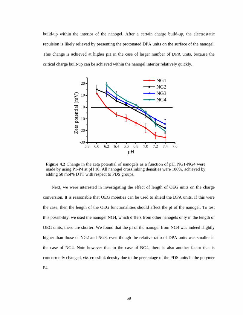
59
build-up within the interior of the nanogel. After a certain charge build-up, the electrostatic
repulsion is likely relieved by presenting the protonated DPA units on the surface of the nanogel.
This change is achieved at higher pH in the case of larger number of DPA units, because the
critical charge built-up can be achieved within the nanogel interior relatively quickly.
Next, we were interested in investigating the effect of length of OEG units on the charge
conversion. It is reasonable that OEG moieties can be used to shield the DPA units. If this were
the case, then the length of the OEG functionalities should affect the pI of the nanogel. To test
this possibility, we used the nanogel NG4, which differs from other nanogels only in the length of
OEG units; these are shorter. We found that the pI of the nanogel from NG4 was indeed slightly
higher than those of NG2 and NG3, even though the relative ratio of DPA units was smaller in
the case of NG4. Note however that in the case of NG4, there is also another factor that is
concurrently changed, viz. crosslink density due to the percentage of the PDS units in the polymer
P4.
Figure 4.2 Change in the zeta potential of nanogels as a function of pH. NG1-NG4 were
made by using P1-P4 at pH 10. All nanogel crosslinking densities were 100%, achieved by
adding 50 mol% DTT with respect to PDS groups.
5.8 6.0 6.2 6.4 6.6 6.8 7.0 7.2 7.4 7.6-30
-20
-10
0
10
20
Zet
a p
ote
nti
al (
mV
)
pH
NG1
NG2
NG3
NG4
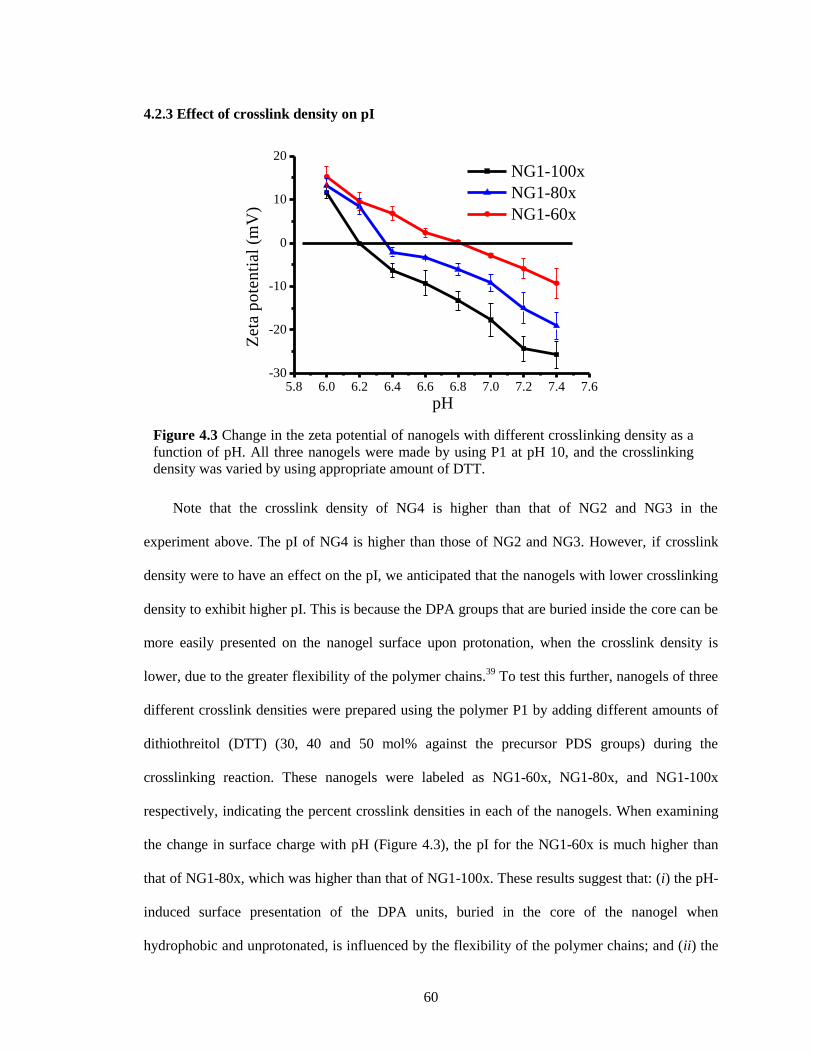
60
4.2.3 Effect of crosslink density on pI
Note that the crosslink density of NG4 is higher than that of NG2 and NG3 in the
experiment above. The pI of NG4 is higher than those of NG2 and NG3. However, if crosslink
density were to have an effect on the pI, we anticipated that the nanogels with lower crosslinking
density to exhibit higher pI. This is because the DPA groups that are buried inside the core can be
more easily presented on the nanogel surface upon protonation, when the crosslink density is
lower, due to the greater flexibility of the polymer chains.39
To test this further, nanogels of three
different crosslink densities were prepared using the polymer P1 by adding different amounts of
dithiothreitol (DTT) (30, 40 and 50 mol% against the precursor PDS groups) during the
crosslinking reaction. These nanogels were labeled as NG1-60x, NG1-80x, and NG1-100x
respectively, indicating the percent crosslink densities in each of the nanogels. When examining
the change in surface charge with pH (Figure 4.3), the pI for the NG1-60x is much higher than
that of NG1-80x, which was higher than that of NG1-100x. These results suggest that: (i) the pH-
induced surface presentation of the DPA units, buried in the core of the nanogel when
hydrophobic and unprotonated, is influenced by the flexibility of the polymer chains; and (ii) the
Figure 4.3 Change in the zeta potential of nanogels with different crosslinking density as a
function of pH. All three nanogels were made by using P1 at pH 10, and the crosslinking
density was varied by using appropriate amount of DTT.
5.8 6.0 6.2 6.4 6.6 6.8 7.0 7.2 7.4 7.6-30
-20
-10
0
10
20
Zet
a p
ote
nti
al (
mV
)
pH
NG1-100x
NG1-80x
NG1-60x
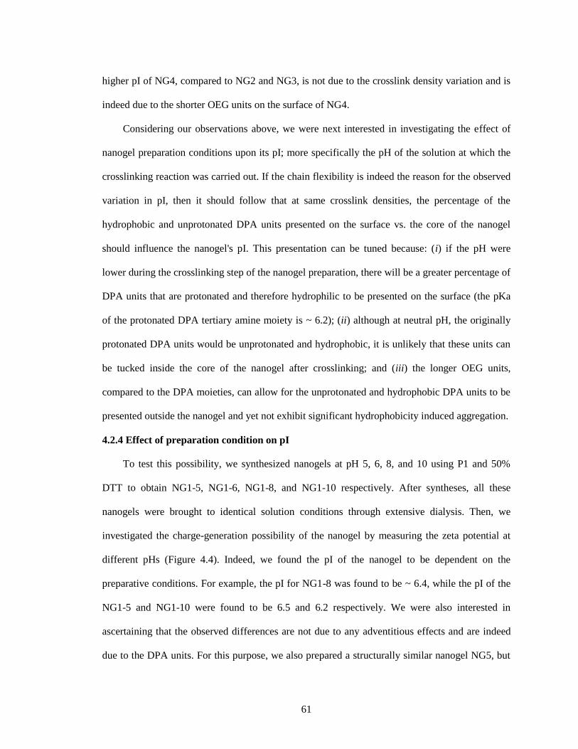
61
higher pI of NG4, compared to NG2 and NG3, is not due to the crosslink density variation and is
indeed due to the shorter OEG units on the surface of NG4.
Considering our observations above, we were next interested in investigating the effect of
nanogel preparation conditions upon its pI; more specifically the pH of the solution at which the
crosslinking reaction was carried out. If the chain flexibility is indeed the reason for the observed
variation in pI, then it should follow that at same crosslink densities, the percentage of the
hydrophobic and unprotonated DPA units presented on the surface vs. the core of the nanogel
should influence the nanogel's pI. This presentation can be tuned because: (i) if the pH were
lower during the crosslinking step of the nanogel preparation, there will be a greater percentage of
DPA units that are protonated and therefore hydrophilic to be presented on the surface (the pKa
of the protonated DPA tertiary amine moiety is ~ 6.2); (ii) although at neutral pH, the originally
protonated DPA units would be unprotonated and hydrophobic, it is unlikely that these units can
be tucked inside the core of the nanogel after crosslinking; and (iii) the longer OEG units,
compared to the DPA moieties, can allow for the unprotonated and hydrophobic DPA units to be
presented outside the nanogel and yet not exhibit significant hydrophobicity induced aggregation.
4.2.4 Effect of preparation condition on pI
To test this possibility, we synthesized nanogels at pH 5, 6, 8, and 10 using P1 and 50%
DTT to obtain NG1-5, NG1-6, NG1-8, and NG1-10 respectively. After syntheses, all these
nanogels were brought to identical solution conditions through extensive dialysis. Then, we
investigated the charge-generation possibility of the nanogel by measuring the zeta potential at
different pHs (Figure 4.4). Indeed, we found the pI of the nanogel to be dependent on the
preparative conditions. For example, the pI for NG1-8 was found to be ~ 6.4, while the pI of the
NG1-5 and NG1-10 were found to be 6.5 and 6.2 respectively. We were also interested in
ascertaining that the observed differences are not due to any adventitious effects and are indeed
due to the DPA units. For this purpose, we also prepared a structurally similar nanogel NG5, but
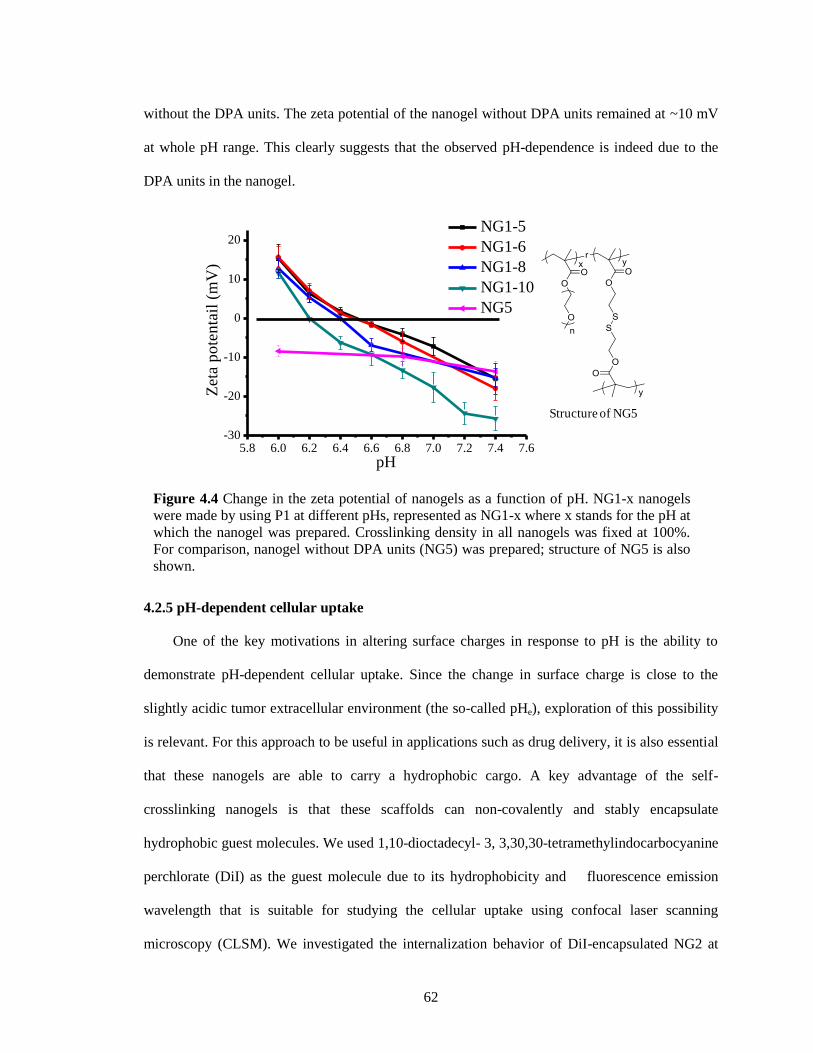
62
without the DPA units. The zeta potential of the nanogel without DPA units remained at ~10 mV
at whole pH range. This clearly suggests that the observed pH-dependence is indeed due to the
DPA units in the nanogel.
4.2.5 pH-dependent cellular uptake
One of the key motivations in altering surface charges in response to pH is the ability to
demonstrate pH-dependent cellular uptake. Since the change in surface charge is close to the
slightly acidic tumor extracellular environment (the so-called pHe), exploration of this possibility
is relevant. For this approach to be useful in applications such as drug delivery, it is also essential
that these nanogels are able to carry a hydrophobic cargo. A key advantage of the self-
crosslinking nanogels is that these scaffolds can non-covalently and stably encapsulate
hydrophobic guest molecules. We used 1,10-dioctadecyl- 3, 3,30,30-tetramethylindocarbocyanine
perchlorate (DiI) as the guest molecule due to its hydrophobicity and fluorescence emission
wavelength that is suitable for studying the cellular uptake using confocal laser scanning
microscopy (CLSM). We investigated the internalization behavior of DiI-encapsulated NG2 at
Figure 4.4 Change in the zeta potential of nanogels as a function of pH. NG1-x nanogels
were made by using P1 at different pHs, represented as NG1-x where x stands for the pH at
which the nanogel was prepared. Crosslinking density in all nanogels was fixed at 100%.
For comparison, nanogel without DPA units (NG5) was prepared; structure of NG5 is also
shown.
5.8 6.0 6.2 6.4 6.6 6.8 7.0 7.2 7.4 7.6-30
-20
-10
0
10
20
Zet
a p
ote
nta
il (
mV
)
pH
NG1-5
NG1-6
NG1-8
NG1-10
NG5
Structure of NG5
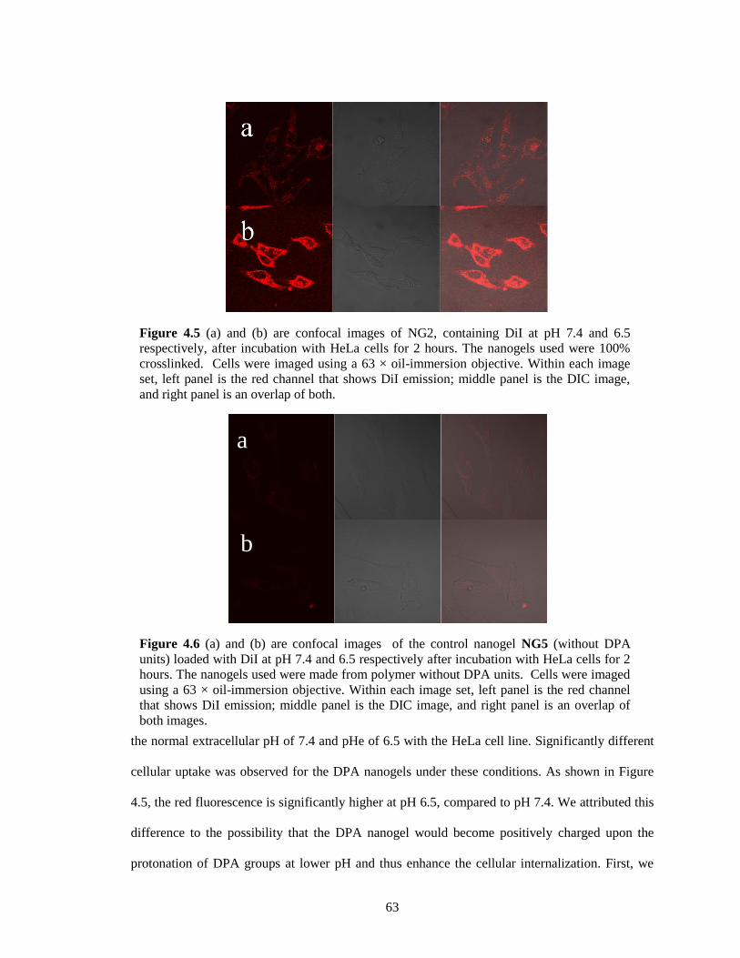
63
the normal extracellular pH of 7.4 and pHe of 6.5 with the HeLa cell line. Significantly different
cellular uptake was observed for the DPA nanogels under these conditions. As shown in Figure
4.5, the red fluorescence is significantly higher at pH 6.5, compared to pH 7.4. We attributed this
difference to the possibility that the DPA nanogel would become positively charged upon the
protonation of DPA groups at lower pH and thus enhance the cellular internalization. First, we
Figure 4.5 (a) and (b) are confocal images of NG2, containing DiI at pH 7.4 and 6.5
respectively, after incubation with HeLa cells for 2 hours. The nanogels used were 100%
crosslinked. Cells were imaged using a 63 × oil-immersion objective. Within each image
set, left panel is the red channel that shows DiI emission; middle panel is the DIC image,
and right panel is an overlap of both.
Figure 4.6 (a) and (b) are confocal images of the control nanogel NG5 (without DPA
units) loaded with DiI at pH 7.4 and 6.5 respectively after incubation with HeLa cells for 2
hours. The nanogels used were made from polymer without DPA units. Cells were imaged
using a 63 × oil-immersion objective. Within each image set, left panel is the red channel
that shows DiI emission; middle panel is the DIC image, and right panel is an overlap of
both images.
a
b
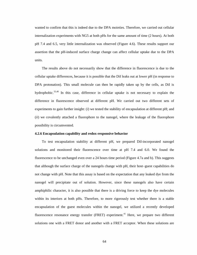
64
wanted to confirm that this is indeed due to the DPA moieties. Therefore, we carried out cellular
internalization experiments with NG5 at both pHs for the same amount of time (2 hours). At both
pH 7.4 and 6.5, very little internalization was observed (Figure 4.6). These results support our
assertion that the pH-induced surface charge change can affect cellular uptake due to the DPA
units.
The results above do not necessarily show that the difference in fluorescence is due to the
cellular uptake differences, because it is possible that the DiI leaks out at lower pH (in response to
DPA protonation). This small molecule can then be rapidly taken up by the cells, as DiI is
hydrophobic.33,40
In this case, difference in cellular uptake is not necessary to explain the
difference in fluorescence observed at different pH. We carried out two different sets of
experiments to gain further insight: (i) we tested the stability of encapsulation at different pH; and
(ii) we covalently attached a fluorophore to the nanogel, where the leakage of the fluorophore
possibility is circumvented.
4.2.6 Encapsulation capability and redox-responsive behavior
To test encapsulation stability at different pH, we prepared DiI-incorporated nanogel
solutions and monitored their fluorescence over time at pH 7.4 and 6.0. We found the
fluorescence to be unchanged even over a 24 hours time period (Figure 4.7a and b). This suggests
that although the surface charge of the nanogels change with pH, their host–guest capabilities do
not change with pH. Note that this assay is based on the expectation that any leaked dye from the
nanogel will precipitate out of solution. However, since these nanogels also have certain
amphiphilic character, it is also possible that there is a driving force to keep the dye molecules
within its interiors at both pHs. Therefore, to more rigorously test whether there is a stable
encapsulation of the guest molecules within the nanogel, we utilized a recently developed
fluorescence resonance energy transfer (FRET) experiment.41
Here, we prepare two different
solutions one with a FRET donor and another with a FRET acceptor. When these solutions are
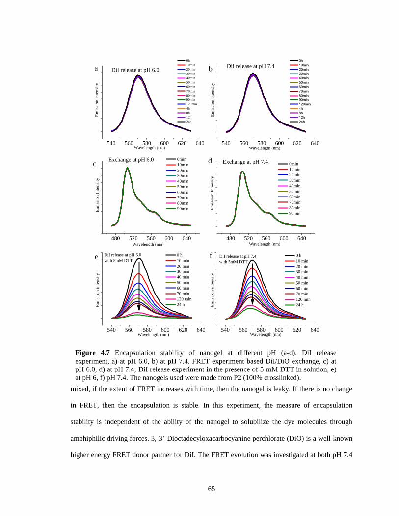
65
mixed, if the extent of FRET increases with time, then the nanogel is leaky. If there is no change
in FRET, then the encapsulation is stable. In this experiment, the measure of encapsulation
stability is independent of the ability of the nanogel to solubilize the dye molecules through
amphiphilic driving forces. 3, 3’-Dioctadecyloxacarbocyanine perchlorate (DiO) is a well-known
higher energy FRET donor partner for DiI. The FRET evolution was investigated at both pH 7.4
Figure 4.7 Encapsulation stability of nanogel at different pH (a-d). DiI release
experiment, a) at pH 6.0, b) at pH 7.4. FRET experiment based DiI/DiO exchange, c) at
pH 6.0, d) at pH 7.4; DiI release experiment in the presence of 5 mM DTT in solution, e)
at pH 6, f) pH 7.4. The nanogels used were made from P2 (100% crosslinked).
540 560 580 600 620 640
Em
issi
on
in
ten
sity
Wavelength (nm)
0h
10min
20min
30min
40min
50min
60min
70min
80min
90min
120min
4h
8h
12h
24h
DiI release at pH 6.0a
540 560 580 600 620 640
Em
issi
on
in
ten
sity
Wavelength (nm)
0h
10min
20min
30min
40min
50min
60min
70min
80min
90min
120min
4h
8h
12h
24h
DiI release at pH 7.4b
480 520 560 600 640
Exchange at pH 6.0
Em
issi
on
In
ten
sity
Wavelength (nm)
0min
10min
20min
30min
40min
50min
60min
70min
80min
90min
c
480 520 560 600 640
Exchange at pH 7.4
Em
issi
on
In
ten
sity
Wavelength (nm)
0min
10min
20min
30min
40min
50min
60min
70min
80min
90min
d
540 560 580 600 620 640
DiI release at pH 6.0
with 5mM DTT
Em
issi
on
in
ten
sity
Wavelength (nm)
0 h
10 min
20 min
30 min
40 min
50 min
60 min
70 min
120 min
24 h
e
540 560 580 600 620 640
f DiI release at pH 7.4
with 5mM DTT
Em
issi
on
in
ten
sity
Wavelength (nm)
0 h
10 min
20 min
30 min
40 min
50 min
60 min
70 min
120 min
24 h
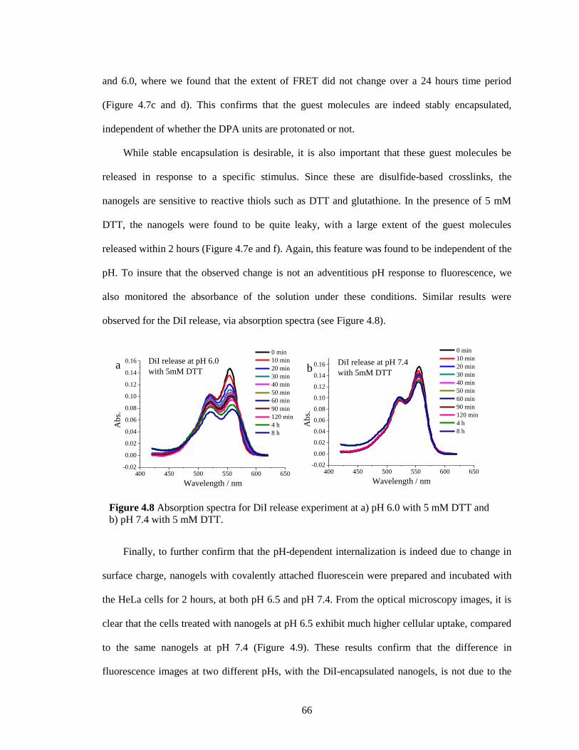
66
and 6.0, where we found that the extent of FRET did not change over a 24 hours time period
(Figure 4.7c and d). This confirms that the guest molecules are indeed stably encapsulated,
independent of whether the DPA units are protonated or not.
While stable encapsulation is desirable, it is also important that these guest molecules be
released in response to a specific stimulus. Since these are disulfide-based crosslinks, the
nanogels are sensitive to reactive thiols such as DTT and glutathione. In the presence of 5 mM
DTT, the nanogels were found to be quite leaky, with a large extent of the guest molecules
released within 2 hours (Figure 4.7e and f). Again, this feature was found to be independent of the
pH. To insure that the observed change is not an adventitious pH response to fluorescence, we
also monitored the absorbance of the solution under these conditions. Similar results were
observed for the DiI release, via absorption spectra (see Figure 4.8).
Finally, to further confirm that the pH-dependent internalization is indeed due to change in
surface charge, nanogels with covalently attached fluorescein were prepared and incubated with
the HeLa cells for 2 hours, at both pH 6.5 and pH 7.4. From the optical microscopy images, it is
clear that the cells treated with nanogels at pH 6.5 exhibit much higher cellular uptake, compared
to the same nanogels at pH 7.4 (Figure 4.9). These results confirm that the difference in
fluorescence images at two different pHs, with the DiI-encapsulated nanogels, is not due to the
Figure 4.8 Absorption spectra for DiI release experiment at a) pH 6.0 with 5 mM DTT and
b) pH 7.4 with 5 mM DTT.
400 450 500 550 600 650-0.02
0.00
0.02
0.04
0.06
0.08
0.10
0.12
0.14
0.16 DiI release at pH 6.0
with 5mM DTT
Ab
s.
Wavelength / nm
0 min
10 min
20 min
30 min
40 min
50 min
60 min
90 min
120 min
4 h
8 h
a
400 450 500 550 600 650-0.02
0.00
0.02
0.04
0.06
0.08
0.10
0.12
0.14
0.16 DiI release at pH 7.4
with 5mM DTT
Ab
s.
Wavelength / nm
0 min
10 min
20 min
30 min
40 min
50 min
60 min
90 min
120 min
4 h
8 h
b
67
non-covalently encapsulated guest leakage. These results also support our assertion that the
difference in cellular uptake is indeed due to the greater nanogel uptake at pH 6.5, most likely due
to the difference in surface charge. These results also suggest that the current nanogel system can
be used in drug delivery via both physical encapsulation and chemical conjugation.
4.3 Summary
In summary, we report on a nanogel that is capable of transforming into a positively charged
nanogel at a pH that is relevant to the slightly acidic tumor extracellular environment. We have
shown that: (i) the co-incorporation of DPA units to PEG based nanogels can lead to effective
pH-dependent charge generation; (ii) the pI of the DPA nanogel can be systematically tuned by
varying the (a) percentage DPA units present in the nanogel, (b) percentage of DPA units that are
initially exposed on the nanogel surface, and (c) crosslink density; (iii) the charge generation
process can be utilized to enhance cellular uptake at pHe, compared to the normal extracellular
pH. It is remarkable that this charge generation event results in enhanced cellular uptake, even
though the length of the OEG units are much higher than the side chains that contain the DPA
moieties; and (iv) non-covalently encapsulated guest molecules are stably encapsulated at both
pH and are thus effectively taken up by the cells at lower pH without any leakage. Considering
Figure 4.9 (a) and (b) are confocal images of fluorescein-labeled DPA nanogel at pH 7.4
and 6.5 after incubation with HeLa cells for 2 hours. The concentration of the nanogels
used was 1mg/mL. Cells were imaged using a 63 × oil-immersion objective. Within each
image set, left panel is the green channel that shows FITC emission; middle panel is the
DIC image, and right panel is an overlap of both.
Structure of FITC labled NG
68
that there exist a number of ternary amines with very different pKa from 5 to 7, it is also possible
to prepare nanogels with diverse pI. Thus, the surface charge can be selectively modulated not
only for cellular uptake, but also for different endocytic compartments such as early endosomes
(pH 5.9-6.2) and lysosomes (5.0-5.5). Overall, the reported facile synthetic method for preparing
the pHe-responsive charge generation nanogels, combined with stable encapsulation and tunable
pI, should open up new avenues for the design of the next generation nanogels.
4.4 Experimental
4.4.1 General
2,2′-dithiodipyridine, 2-mercaptoethanol, polyethylene glycol monomethylether
methacrylate (MW 450 g/mol and 350 g/mol) (OEG), 2-(diisopropylamino) ethyl methacrylate
(DPA), D,L-dithiothreitol (DTT), 1,1'-dioctadecyl-3,3,3',3'-tetramethyl indocarbocyanine
perchlorate (DiI) and 3,3'-dioctadecyloxacarbocyanine perchlorate (DiO), 4-cyano-4-
(phenylcarbonothioylthio)pentanoic acid, fluorescein isothiocyanate (FITC) and other
conventional reagents were obtained from commercial sources and were used as received, unless
otherwise mentioned. Polymers were synthesized using RAFT polymerization and then purified
by precipitation. Pyridyl disulfide ethyl methacrylate (PDS) was prepared using a previously
reported procedure.42
1H-NMR spectra were recorded on a 400 MHz Bruker NMR spectrometer
using the residual proton resonance of the solvent as the internal standard. Chemical shifts are
reported in parts per million (ppm). Molecular weights of the polymers were estimated by gel
permeation chromatography (GPC) using PMMA standard with a refractive index detector and
THF as the elution. Dynamic light scattering (DLS) measurements and zeta potential
measurements were performed using a Malvern Nanozetasizer. UV-visible absorption spectra
were recorded on a Varian (model EL 01125047) spectrophotometer. The fluorescence spectra
were obtained from a JASCO FP-6500 spectrofluorimeter.
69
4.4.2 Synthesis of random copolymer P1-P4
A mixture of 4-cyano-4-(phenylcarbonothioylthio) pentanoic acid (5.9 mg, 0.021 mmol),
PDS (107.4 mg, 0.421 mmol), polyethylene glycol monomethyl ether methacrylate (100 mg, 0.21
mmol), 2-(diisopropylamino) ethyl methacrylate (27mg, 0.126 mmol) and AIBN (0.7 mg,
0.00421 mmol) was dissolved in THF (0.5 mL) and degassed by performing three freeze-pump-
thaw cycles. The reaction mixture was sealed and then transferred into a pre-heated oil bath at
65 °C and stirred for 10 h. To remove unreactive monomers, the resultant mixture was
precipitated in cold ethyl ether (20 mL) to yield the random copolymer P1 as a waxy solid. GPC
(THF) Mn: 8.3 KDa. D: 1.3. 1H NMR (400 MHz, CDCl3) δ: 8.46, 7.67, 7.10, 4.35-4.09, 3.94-3.37,
3.02, 2.62, 2.04-1.64, 1.43-0.87. Other polymers P2-P4 were prepared by a similar method using
the appropriate molar ratio of the co-monomers. The molar ratio between three monomer units
was determined by integrating the methoxy protons in the polyethylene glycol unit, the aromatic
protons in the pyridine and methylene protons contacted to amino groups.
4.4.3 Synthesis of random copolymer P5 without DPA units
A mixture of 4-cyano-4-(phenylcarbonothioylthio)pentanoic acid (3.0 mg, 0.02 mmol), PDS
(1.25 g, 4.9 mmol), polyethylene glycol monomethyl ether methacrylate (1.0 g, 2.1 mmol) and
AIBN (2.5 mg, 0.014 mmol) was dissolved in THF (5 mL) and degassed by performing three
freeze-pump-thaw cycles. The reaction mixture was sealed and then transferred into a pre-heated
oil bath at 65 °C and stirred for 10 h. To remove unreactive monomers, the resultant mixture was
precipitated in cold ethyl ether (20 mL) to yield the random copolymer as a waxy liquid. GPC
(THF) Mn: 13 kDa; PD: 1.3. 1H NMR (400 MHz, CDCl3) δ: 8.46, 7.68, 7.11, 4.35-4.09, 3.94-3.37,
3.03, 2.04-1.64, 1.43-0.87. The molar ratio between two monomer units was determined by
integrating the methoxy protons in the polyethyleneglycol unit and the aromatic protons in the
pyridine and found to be 3:7 (PEG:PDS).
70
4.4.4 Synthesis of random copolymer with primary amine groups
A mixture of 4-cyano-4-(phenylcarbonothioylthio) pentanoic acid (5.5 mg), PDS (75 mg),
polyethylene glycol monomethyl ether methacrylate (107 mg), 2-(diisopropylamino) ethyl
methacrylate (65 mg), 2-Aminoethyl methacrylate hydrochloride (4mg) and AIBN (0.6 mg) was
dissolved in DMF (0.5 mL) and degassed by performing three freeze-pump-thaw cycles. The
reaction mixture was sealed and then transferred into a pre-heated oil bath at 65 °C and stirred for
10 h. To remove unreactive monomers, the resultant mixture was precipitated in cold ethyl ether
(20 mL) to yield the random copolymer as a waxy solid.
4.4.5 Encapsulation of DiI/DiO in nanogels
The polymer (10 mg) was dissolved in 1 mL water. To this solution, either a 0.02 mL of DiI
stock solution (5mg/mL in acetone) or 0.04 mL DiO stock solution (2.5 mg/mL in acetone) was
added into the polymer solution. The pH was adjusted by adding drops of HCl or NaOH solution
(1M). The mixed solution was stirred overnight at room temperature, open to the atmosphere
allowing the organic solvent to evaporate. Then a measured amount of DTT was added. After
stirring for 4 hours, insoluble DiI/DiO was removed by filtration and pyridinethione was removed
from the nanogel solution by extensive dialysis using a membrane with a molecular weight cutoff
of 7,000 g/mol.
4.4.6 Synthesis of fluorescein-labeled DPA nanogels
The blank DPA nanogel was made by using amine group containing polymer (10 mg/mL)
using the procedures outlined above. Then excess FITC was added into the nanogel solution.
After stirring for 24 hours, excess FITC was removed from the nanogel solution by extensive
dialysis using a membrane with a molecular weight cutoff of 7,000 g/mol.
4.4.7 DLS measurement
Dynamic light scattering experiments were performed by using a digital correlator and
goniometer. The light source was solid-stat laser system, operating at 514 nm. For size
71
measurements, the nanogels in deionized water (1 mg/mL) was kept constant at 25 °C throughout
the experiment. Dust was eliminated by filtering the solution through 0.45 μm polycarbonate
filter. All measurements were done at a correlation time of 30 seconds. For zeta potential
measurements, the pH was adjusted by adding drops of HCl or NaOH solution (1M). The
nanogels solution was also kept constant at 25 °C throughout the experiment.
4.4.8 Dye exchange experiment
A solution of nanogel containing DiI (100 μL) was mixed with a solution of nanogel
containing DiO (100 μL) in a cuvette, and then milliQ water (800 μL) was added to adjust the
volume. The fluorescence spectra were recorded using the excitation wavelength of 450 nm.
4.4.9 DiI release experiment
A solution of nanogel containing DiI (100 μL) was mixed with a buffer solution with pH 6
or 7.4 (800 μL) in a cuvette, and then DTT stock solution (100 μL) was added. The fluorescence
spectra were recorded using the excitation wavelength of 530 nm.
4.4.10 Confocal experiments
Confocal experiments were done using Zeiss510 META confocal laser scanning microscope.
100k HeLa cells were incubated in glass bottomed dishes, purchased from Mat tek corp, at 37 °C
overnight in 5% CO2 incubator allowing the cells stick to the bottom of dish. Then, the nutrient
medium was taken out and cells were washed with 5 mL of pH 7.4 PBS buffer. In one of the
confocal dish, the pH of 1 mL of nutrient media was adjusted to 6.5. Then, 100uL of 1mg/mL
solution of pH sensitive nanogels were added to each of the confocal dishes maintained at pH 6.5
and at pH 7.4. The cells were incubated for 2 hrs at 37 °C and the florescence was observed at
560 nm by laser excitation at 543 nm for the DiI encapsulated nanogels and florescence was
observed at 501nm by exciting at 480nm for fluorescein-attached nanogels. All images were
taken using 63x oil immersion objective.
72
4.5 References
1. Mutsaers, S. E.; John M. P. Surface charge of macrophages and their interaction with charged
particles. J. Leukocyte Biol., 1988, 44, 17-26.
2. Richard, J. P.; Melikov, K.; Vives, E.; Ramos, C.; Verbeure, B.; Gait, M. J.; Chernomordik, L.
V.; Lebleu, B. Cell-penetrating peptides A reevaluation of the mechanism of cellular uptake. J.
Biol. Chem., 2003, 278, 585-590.
3. Ulbrich, K.; Subr, V. Polymeric anticancer drugs with pH-controlled activation. Adv. Drug
Delivery Rev. 2004, 56, 1023-1050;
4. Lee, H. J.; Pardridge, W. M. Monoclonal antibody radiopharmaceuticals: cationization,
pegylation, radiometal chelation, pharmacokinetics, and tumor imaging. Bioconjugate Chem.
2003, 14, 546-553;
5. Gu, J.; Cheng, W. P.; Liu, J.; Lo, S. Y.; Smith, D.; Qu, X.; Yang, Z. pH-triggered reversible
stealth polycationic micelles. Biomacromolecules 2008, 9, 255-262;
6. Gao, Y.; Yang, C.; Liu, X.; Ma, R.; Kong, D.; Shi, L. A multifunctional nanocarrier based on
nanogated mesoporous silica for enhanced tumor-specific uptake and intracellular delivery.
Macromol Biosci. 2012, 12, 251-259;
7. Ding, C. X.; Gu, J. X.; Qu, X. Z.; Yang, Z. Z. Preparation of multifunctional drug carrier for
tumor-specific uptake and enhanced intracellular delivery through the conjugation of weak acid
labile linker. Bioconjugate Chem. 2009, 20, 1163-1170;
8. Bae, Y.; Fukushima, S.; Harada, A.; Kataoka, K. Design of environment-sensitive
supramolecular assemblies for intracellular drug delivery: Polymeric micelles that are responsive
to intracellular pH change. Angew. Chem., Int. Ed., 2003, 42, 4640-4643.
9. Griset, A. P.; Walpole, J.; Liu, R.; Gaffey, A.; Colson Y. L.; Grinstaff, M. W. Expansile
nanoparticles: synthesis, characterization, and in vivo efficacy of an acid-responsive polymeric
drug delivery system. J. Am. Chem. Soc., 2009, 131, 2469-2471;
10. Liu, S.; Weaver, J. V. M.; Tang, Y.; Billingham, N. C.; Armes, S. P.; Tribe. K. Synthesis of
shell cross-linked micelles with pH-responsive cores using ABC triblock copolymers.
Macromolecules, 2002, 35, 6121-6131.
11. Choi, S. W.; Zhang, Y.; Xia, Y. N. A temperature-sensitive drug release system based on
phase-change materials. Angew. Chem., Int. Ed., 2010, 49, 7904-7908.
12. Bikram, M; West, J. L. Thermo-responsive systems for controlled drug delivery. Expert Opin
Drug Deliv. 2008, 10, 1077-1091;
13. Nakayama, M.; Okano, T.; Miyazaki, T.; Kohori, F.; Sakai, K.; Yokoyama, M. Molecular
design of biodegradable polymeric micelles for temperature-responsive drug release. J.
Controlled Release, 2006, 115, 46–56.
73
14. Lee, Y.; Fukushima, S.; Bae, Y.; Hiki, S.; Ishii, T.; Kataoka, K. A protein nanocarrier from
charge-conversion polymer in response to endosomal pH. J. Am. Chem. Soc. 2007, 129, 5362-
5363;
15. Xiong, M. H.; Bao, Y.; Yang, X. Z.; Wang, Y. C.; Sun, B. L.; Wang, J. lipase-sensitive
polymeric triple-layered nanogel for on-demand drug delivery. J. Am. Chem. Soc., 2012, 134,
4355-4362;
16. Wang, C.; Chen, Q.S.; Wang, Z. Q.; Zhang, X. An enzyme-responsive polymeric
superamphiphile. Angew. Chem. Int. Ed., 2010, 49, 8612-8615;
17. Dong, W.; Kishimura, A.; Anraku, Y.; Chuanoi, S.; Kataoka, K. Monodispersed polymeric
nanocapsules: spontaneous evolution and morphology transition from reducible hetero-PEG
PICmicelles by controlled degradation. J. Am. Chem. Soc., 2009, 131, 3804-3805.
18. Liu, J. Y.; Pang, Y.; Huang, W.; Zhu, Z. Y.; Zhu, X. Y.; Zhou, Y. F.; Yan, D. Y. Redox-
responsive polyphosphate nanosized assemblies: a smart drug delivery platform for cancer
therapy. Biomacromolecules, 2011, 12, 2407-2415;
19. Cerritelli, S.; Velluto, D.; Hubbell, J. A. PEG-SS-PPS: reduction-sensitive disulfide block
copolymer vesicles for intracellular drug delivery. Biomacromolecules, 2007, 8, 1966-1972.
20. Tong, R.; Hemmati, H. D.; Langer, R.; Kohane, D. S. Photoswitchable nanoparticles for
triggered tissue penetration and drug delivery. J. Am. Chem. Soc., 2012, 134, 8848-8855;
21. McCoy, C. P.; Rooney, C.; Edwards, C. R.; Jones, D. S.; Gorman, S. P. Light-triggered
molecule-scale drug dosing devices. J. Am. Chem. Soc., 2007, 129, 9572-9573;
22. Hu, L. C.; Yonamine, Y.; Lee, S. H.; Van der Veer, W. E.; Shea, K. J. Light-triggered charge
reversal of organic-silica hybrid nanoparticles. J. Am. Chem. Soc., 2012, 134, 11072-11075;
23. Yamagata, M.; Hasuda, K.; Stamato, T.; Tannock, I. F. The contribution of lactic acid to
acidification of tumours: studies of variant cells lacking lactate dehydrogenase. Br. J. Cancer,
1998, 77, 1726-1731;
24. Helmlinger, G.; Schell, A.; Dellian, M.; Forbes, N. S.; Jain, R. K. Acid production in
glycolysis-impaired tumors provides new insights into tumor metabolism. Clin. Cancer Res. 2002,
8, 1284-1291;
25. Vander, H. M. G.; Cantley, L. C.; Thompson, C. B. Understanding the Warburg effect: the
metabolic requirements of cell proliferation. Science, 2009, 324, 1029-1033;
26. Du, J. Z.; Sun, T. M.; Song, W. J.; Wu, J.; Wang, J. A tumor-acidity-activated charge-
conversional nanogel as an intelligent vehicle for promoted tumoral-cell uptake and drug delivery.
Angew. Chem. Int. Ed., 2010, 49, 3621-3626;
27. Du, J. Z.; Du, X. J.; Mao, C. Q.; Wang, J. Tailor-made dual pH-sensitive
polymer_doxorubicin nanoparticles for efficient anticancer drug delivery. J. Am. Chem. Soc.,
2011, 133, 17560-17563;
74
28. Yuan, Y. Y.; Mao, C. Q.; Du, X. J.; Du, J. Z.; Wang, F.; Wang, J. Surface charge switchable
nanoparticles based on zwitterionic polymer for enhanced drug delivery to tumor. Adv. Mater.,
2012, 24, 5476-5480;
29. Poon, Z.; Chang, D.; Zhao, X. Y.; Hammond, P. T. Layer-by-layer nanoparticles with a pH-
sheddable layer for in vivo targeting of tumor hypoxia. ACS Nano, 2011, 5, 4284-4292;
30. Ryu, J. -H.; Chacko, R.; Jiwpanich, S.; Bickerton, S.; Babu, R. P.; Thayumanavan, S. Self-
crosslinked polymer nanogels: a versatile nanoscopic drug delivery platform. J. Am. Chem. Soc.
2010, 132, 17227-17235;
31. Ryu, J. -H.; Jiwpanich, S.; Chacko, R.; Bickerton, S.; Thayumanavan, S. Surface-
functionalizable polymer nanogels with facile hydrophobic guest encapsulation capabilities J. Am.
Chem. Soc. 2010, 132, 8246-824;
32. González-Toro, D. C.; Ryu, J. -H.; Chacko, R. T.; Zhuang, J. M.; Thayumanavan, S.
Concurrent binding and delivery of proteins and lipophilic small molecules using polymeric
nanogels. J. Am. Chem. Soc., 2012, 134, 6964-6967;
33. Ryu, J. H.; Bickerton, S.; Zhuang, J. M.; Thayumanavan, S. Ligand-decorated nanogels: fast
one-pot synthesis and cellular targeting. Biomacromolecules, 2012, 13, 1515-1522;
34. Oh, J. K.; Siegwart, D. J.; Lee, H.; Sherwood, G.; Peteanu, L.; Hollinger, J. O.; Kataoka, K.;
Matyjaszewski, K. Biodegradable nanogels prepared by atom transfer radical polymerization as
potential drug delivery carriers: synthesis, biodegradation, in vitro release, and bioconjugation. J.
Am. Chem. Soc., 2007, 129, 5939-5945.
35. Hu, Y. Q.; Kim, M. S.; Kim, B. S.; Lee, D. S. Synthesis and pH-dependent micellization of 2-
(diisopropylamino)ethyl methacrylate based amphiphilic diblock copolymers via RAFT
polymerization. Polymer, 2007, 48, 3437-3443;
36. Du, J. Z.; Tang, Y. Q.; Lewis, A. L.; Armes, S. P. pH-Sensitive vesicles based on a
biocompatible zwitterionic diblock copolymer. J. Am. Chem. Soc., 2005, 127, 17982-17983;
37. Radovic-Moreno, A. F.; Lu, T. K.; Puscasu, V. A.; Yoon, C. J.; Langer, R.; Farokhzad, O. C.
Surface charge-switching polymeric nanoparticles for bacterial cell wall-targeted delivery of
antibiotics. ACS Nano, 2012, 6, 4279-4287.
38. Bahadur, K. C. R.; Xu, P. Multicompartment intracellular self-expanding nanogel for targeted
delivery of drug cocktail. Adv. Mater., 2012, 24, 6479-6483.
39. Li, L.; Ryu, J. -H.; Thayumanavan, S. Effect of Hofmeister ions on the size and encapsulation
stability of polymer nanogels. Langmuir, 2013, 29, 50-55.
40. Li, Y.; Song, Y.; Zhao, L.; Gaidosh, G.; Laties, A. M.; Wen, R. Direct labeling and
visualization of blood vessels with lipophilic carbocyanine dye DiI. Nat. Protoc., 2008, 3, 1703-
1708.
41. Jiwpanich, S.; Ryu, J. H.; Bickerton, S.; Thayumanavan, S. Noncovalent encapsulation
stabilities in supramolecular nanoassemblies. J. Am. Chem. Soc., 2010, 132, 10683-10685;
75
42. Ghosh, S.; Basu, S.; Thayumanavan, S. Simultaneous and reversible functionalization of
copolymers for biological applications. Macromolecules 2006, 39, 5595-5597.
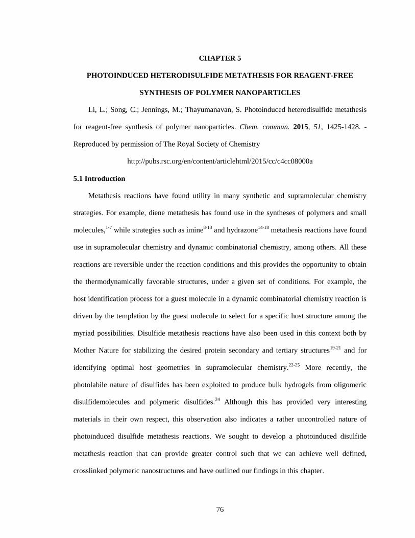
76
CHAPTER 5
PHOTOINDUCED HETERODISULFIDE METATHESIS FOR REAGENT-FREE
SYNTHESIS OF POLYMER NANOPARTICLES
Li, L.; Song, C.; Jennings, M.; Thayumanavan, S. Photoinduced heterodisulfide metathesis
for reagent-free synthesis of polymer nanoparticles. Chem. commun. 2015, 51, 1425-1428. -
Reproduced by permission of The Royal Society of Chemistry
http://pubs.rsc.org/en/content/articlehtml/2015/cc/c4cc08000a
5.1 Introduction
Metathesis reactions have found utility in many synthetic and supramolecular chemistry
strategies. For example, diene metathesis has found use in the syntheses of polymers and small
molecules,1-7
while strategies such as imine8-13
and hydrazone14-18
metathesis reactions have found
use in supramolecular chemistry and dynamic combinatorial chemistry, among others. All these
reactions are reversible under the reaction conditions and this provides the opportunity to obtain
the thermodynamically favorable structures, under a given set of conditions. For example, the
host identification process for a guest molecule in a dynamic combinatorial chemistry reaction is
driven by the templation by the guest molecule to select for a specific host structure among the
myriad possibilities. Disulfide metathesis reactions have also been used in this context both by
Mother Nature for stabilizing the desired protein secondary and tertiary structures19-21
and for
identifying optimal host geometries in supramolecular chemistry.22-25
More recently, the
photolabile nature of disulfides has been exploited to produce bulk hydrogels from oligomeric
disulfidemolecules and polymeric disulfides.24
Although this has provided very interesting
materials in their own respect, this observation also indicates a rather uncontrolled nature of
photoinduced disulfide metathesis reactions. We sought to develop a photoinduced disulfide
metathesis reaction that can provide greater control such that we can achieve well defined,
crosslinked polymeric nanostructures and have outlined our findings in this chapter.
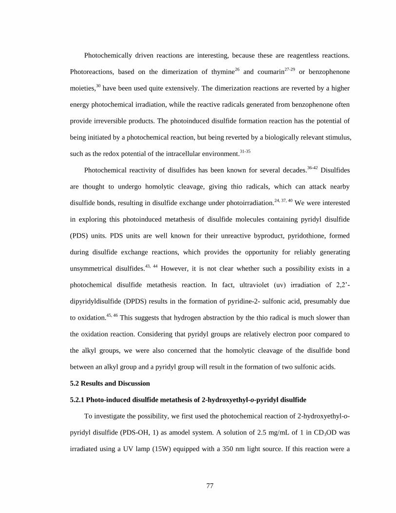
77
Photochemically driven reactions are interesting, because these are reagentless reactions.
Photoreactions, based on the dimerization of thymine26
and coumarin27-29
or benzophenone
moieties,30
have been used quite extensively. The dimerization reactions are reverted by a higher
energy photochemical irradiation, while the reactive radicals generated from benzophenone often
provide irreversible products. The photoinduced disulfide formation reaction has the potential of
being initiated by a photochemical reaction, but being reverted by a biologically relevant stimulus,
such as the redox potential of the intracellular environment.31-35
Photochemical reactivity of disulfides has been known for several decades.36-42
Disulfides
are thought to undergo homolytic cleavage, giving thio radicals, which can attack nearby
disulfide bonds, resulting in disulfide exchange under photoirradiation.24, 37, 40
We were interested
in exploring this photoinduced metathesis of disulfide molecules containing pyridyl disulfide
(PDS) units. PDS units are well known for their unreactive byproduct, pyridothione, formed
during disulfide exchange reactions, which provides the opportunity for reliably generating
unsymmetrical disulfides.43, 44
However, it is not clear whether such a possibility exists in a
photochemical disulfide metathesis reaction. In fact, ultraviolet (uv) irradiation of 2,2’-
dipyridyldisulfide (DPDS) results in the formation of pyridine-2- sulfonic acid, presumably due
to oxidation.45, 46
This suggests that hydrogen abstraction by the thio radical is much slower than
the oxidation reaction. Considering that pyridyl groups are relatively electron poor compared to
the alkyl groups, we were also concerned that the homolytic cleavage of the disulfide bond
between an alkyl group and a pyridyl group will result in the formation of two sulfonic acids.
5.2 Results and Discussion
5.2.1 Photo-induced disulfide metathesis of 2-hydroxyethyl-o-pyridyl disulfide
To investigate the possibility, we first used the photochemical reaction of 2-hydroxyethyl-o-
pyridyl disulfide (PDS-OH, 1) as amodel system. A solution of 2.5 mg/mL of 1 in CD3OD was
irradiated using a UV lamp (15W) equipped with a 350 nm light source. If this reaction were a
78
purely disulfide exchange reaction due to thio radical formation and then recombination,
molecule 1 would have been in equilibrium with bis(2-hydroxyethyl) disulfide (2) and DPDS (3).
Ideally, a statistical ratio of 2 : 1 : 1 of products 1-3 would be obtained (Scheme 5.1). However,
the starting material 1 was being continuously consumed with a concurrent increase in the
concentration of 2. As shown in the 1H NMR spectra over the irradiation time (Figure 5.1), the
integrated signal intensity of the triplet at 2.94 ppm, arising from the CH2 protons attached to
sulfur in 1, decreased with the corresponding increase in the intensity of the triplet at 2.83 ppm
(the same CH2 protons in 2). Finally, the disappearance of the peak at 2.94 ppm showed that no
starting material (1) remained in the reaction mixture. Also, there is no evidence of the formation
Figure 5.1 Time-dependent 1H NMR spectra of the photo-induced reaction of 2-
hydroxyethyl 2-pyridyl disulfide (PDS-OH, 1). Control spectra of bis(2-hydroxyethyl)
disulfide (DPDS, 2), 2, 2’-dipyridyl disulfide (3) and 2-thiopyridone were also included.
The concentration was 2.5 mg/mL. The final product after photoirradiation was determined
to be pyridine-2-sulfonic acid (4).
Scheme 5.1 Hypothesized reaction scheme of photo-induced disulfide metathesis of 2-
pyridyl disulfide (PDS-OH).
79
of DPDS (3) in the reaction mixture. However, we did find that the formation of 2 was
accompanied by the formation of pyridine-2-sulfonic acid (4) (Scheme 5.2). Finally, we found
that the percent conversion in this reaction was lower at higher concentrations (Figure 5.2). This
is typical of photochemical reactions as the fluence of light was kept the same in all these
reactions. Two features are readily discerned from these observations: (i) no equilibrium is
observed in this photoinduced disulfide metathesis and (ii) while the pyridylthio radical is readily
oxidized, the alkylthio radical undergoes a radical recombination reaction to produce 2. These
initial observations are encouraging for the use of heterodisulfides containing PDS units for
controlled polymer nanoparticle synthesis.
Prior to utilizing this in nanoparticle synthesis however, we were interested in investigating
the reasons for the observed oxidation of the pyridylthio radical. Presumably, this oxidation is
Scheme 5.2 Scheme of photo-induced chemistry reaction of PDS-OH.
Figure 5.2 Concentration dependent of reaction rate of PDS under photoirradiation.
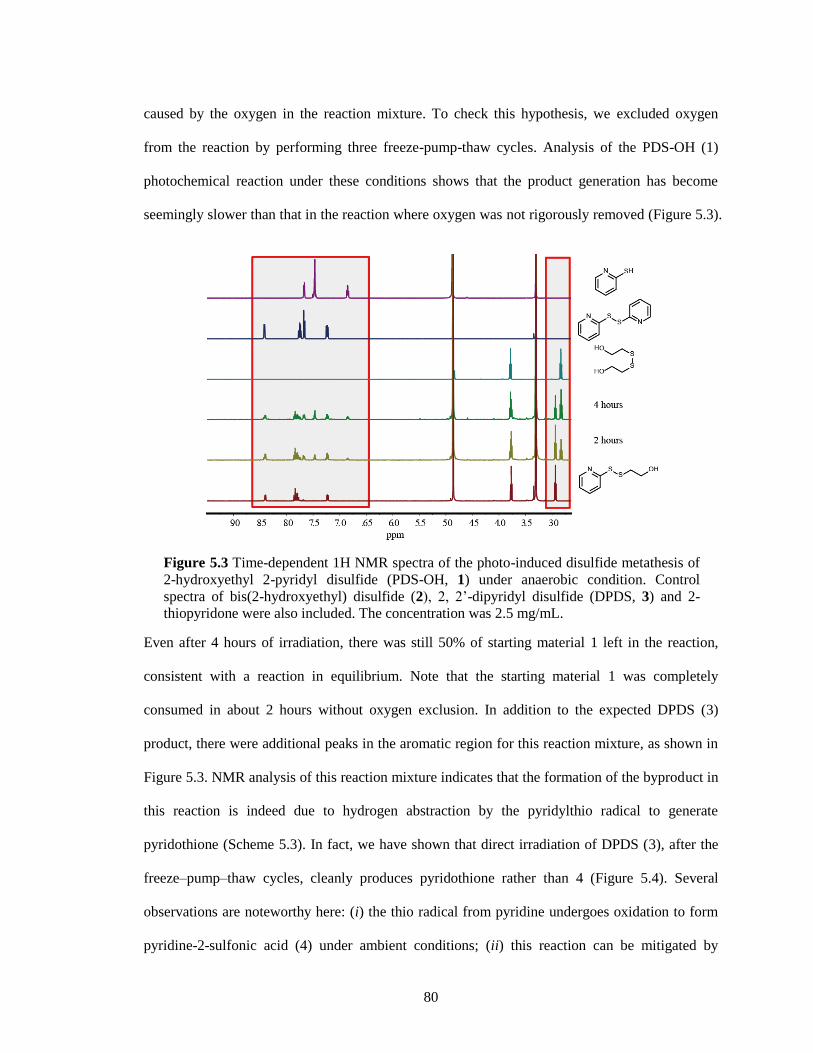
80
caused by the oxygen in the reaction mixture. To check this hypothesis, we excluded oxygen
from the reaction by performing three freeze-pump-thaw cycles. Analysis of the PDS-OH (1)
photochemical reaction under these conditions shows that the product generation has become
seemingly slower than that in the reaction where oxygen was not rigorously removed (Figure 5.3).
Even after 4 hours of irradiation, there was still 50% of starting material 1 left in the reaction,
consistent with a reaction in equilibrium. Note that the starting material 1 was completely
consumed in about 2 hours without oxygen exclusion. In addition to the expected DPDS (3)
product, there were additional peaks in the aromatic region for this reaction mixture, as shown in
Figure 5.3. NMR analysis of this reaction mixture indicates that the formation of the byproduct in
this reaction is indeed due to hydrogen abstraction by the pyridylthio radical to generate
pyridothione (Scheme 5.3). In fact, we have shown that direct irradiation of DPDS (3), after the
freeze–pump–thaw cycles, cleanly produces pyridothione rather than 4 (Figure 5.4). Several
observations are noteworthy here: (i) the thio radical from pyridine undergoes oxidation to form
pyridine-2-sulfonic acid (4) under ambient conditions; (ii) this reaction can be mitigated by
Figure 5.3 Time-dependent 1H NMR spectra of the photo-induced disulfide metathesis of
2-hydroxyethyl 2-pyridyl disulfide (PDS-OH, 1) under anaerobic condition. Control
spectra of bis(2-hydroxyethyl) disulfide (2), 2, 2’-dipyridyl disulfide (DPDS, 3) and 2-
thiopyridone were also included. The concentration was 2.5 mg/mL.
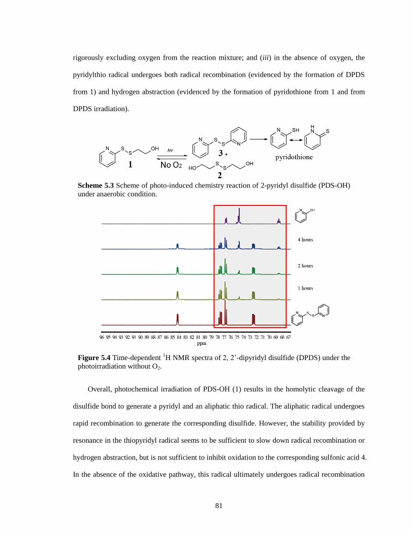
81
rigorously excluding oxygen from the reaction mixture; and (iii) in the absence of oxygen, the
pyridylthio radical undergoes both radical recombination (evidenced by the formation of DPDS
from 1) and hydrogen abstraction (evidenced by the formation of pyridothione from 1 and from
DPDS irradiation).
Overall, photochemical irradiation of PDS-OH (1) results in the homolytic cleavage of the
disulfide bond to generate a pyridyl and an aliphatic thio radical. The aliphatic radical undergoes
rapid recombination to generate the corresponding disulfide. However, the stability provided by
resonance in the thiopyridyl radical seems to be sufficient to slow down radical recombination or
hydrogen abstraction, but is not sufficient to inhibit oxidation to the corresponding sulfonic acid 4.
In the absence of the oxidative pathway, this radical ultimately undergoes radical recombination
Scheme 5.3 Scheme of photo-induced chemistry reaction of 2-pyridyl disulfide (PDS-OH)
under anaerobic condition.
Figure 5.4 Time-dependent 1H NMR spectra of 2, 2’-dipyridyl disulfide (DPDS) under the
photoirradiation without O2.
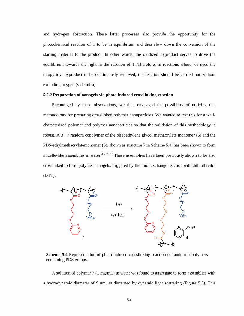
82
and hydrogen abstraction. These latter processes also provide the opportunity for the
photochemical reaction of 1 to be in equilibrium and thus slow down the conversion of the
starting material to the product. In other words, the oxidized byproduct serves to drive the
equilibrium towards the right in the reaction of 1. Therefore, in reactions where we need the
thiopyridyl byproduct to be continuously removed, the reaction should be carried out without
excluding oxygen (vide infra).
5.2.2 Preparation of nanogels via photo-induced crosslinking reaction
Encouraged by these observations, we then envisaged the possibility of utilizing this
methodology for preparing crosslinked polymer nanoparticles. We wanted to test this for a well-
characterized polymer and polymer nanoparticles so that the validation of this methodology is
robust. A 3 : 7 random copolymer of the oligoethylene glycol methacrylate monomer (5) and the
PDS-ethylmethacrylatemonomer (6), shown as structure 7 in Scheme 5.4, has been shown to form
micelle-like assemblies in water.33, 44, 47
These assemblies have been previously shown to be also
crosslinked to form polymer nanogels, triggered by the thiol exchange reaction with dithiothreitol
(DTT).
A solution of polymer 7 (1 mg/mL) in water was found to aggregate to form assemblies with
a hydrodynamic diameter of 9 nm, as discerned by dynamic light scattering (Figure 5.5). This
Scheme 5.4 Representation of photo-induced crosslinking reaction of random copolymers
containing PDS groups.
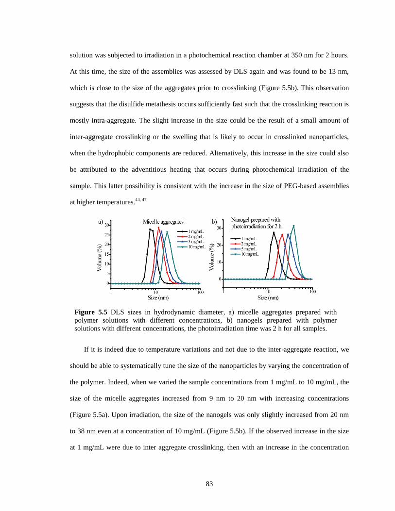
83
solution was subjected to irradiation in a photochemical reaction chamber at 350 nm for 2 hours.
At this time, the size of the assemblies was assessed by DLS again and was found to be 13 nm,
which is close to the size of the aggregates prior to crosslinking (Figure 5.5b). This observation
suggests that the disulfide metathesis occurs sufficiently fast such that the crosslinking reaction is
mostly intra-aggregate. The slight increase in the size could be the result of a small amount of
inter-aggregate crosslinking or the swelling that is likely to occur in crosslinked nanoparticles,
when the hydrophobic components are reduced. Alternatively, this increase in the size could also
be attributed to the adventitious heating that occurs during photochemical irradiation of the
sample. This latter possibility is consistent with the increase in the size of PEG-based assemblies
at higher temperatures.44, 47
If it is indeed due to temperature variations and not due to the inter-aggregate reaction, we
should be able to systematically tune the size of the nanoparticles by varying the concentration of
the polymer. Indeed, when we varied the sample concentrations from 1 mg/mL to 10 mg/mL, the
size of the micelle aggregates increased from 9 nm to 20 nm with increasing concentrations
(Figure 5.5a). Upon irradiation, the size of the nanogels was only slightly increased from 20 nm
to 38 nm even at a concentration of 10 mg/mL (Figure 5.5b). If the observed increase in the size
at 1 mg/mL were due to inter aggregate crosslinking, then with an increase in the concentration
Figure 5.5 DLS sizes in hydrodynamic diameter, a) micelle aggregates prepared with
polymer solutions with different concentrations, b) nanogels prepared with polymer
solutions with different concentrations, the photoirradiation time was 2 h for all samples.
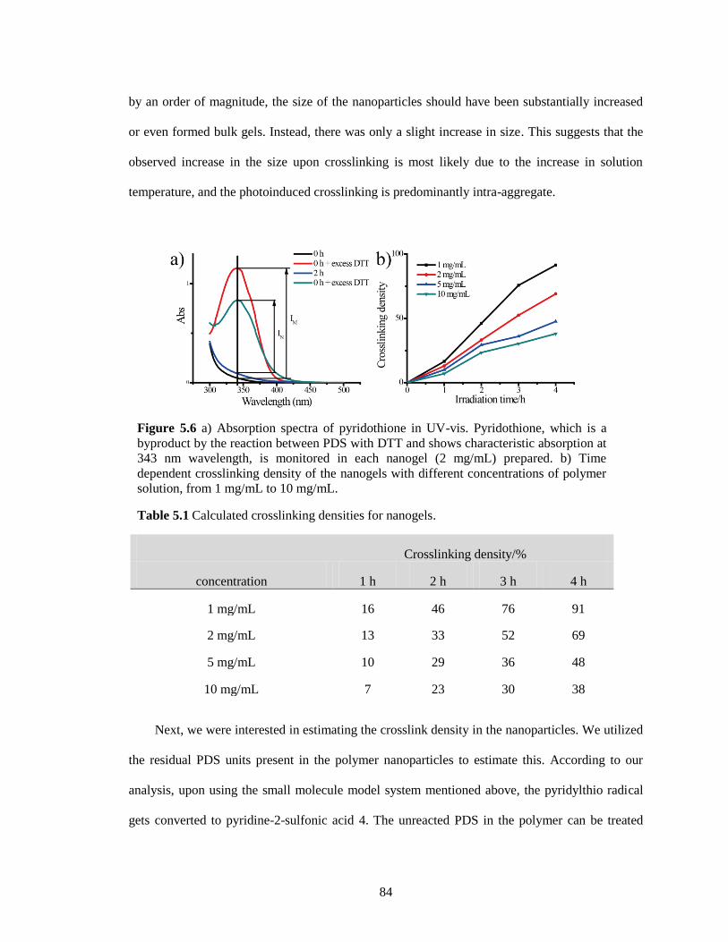
84
by an order of magnitude, the size of the nanoparticles should have been substantially increased
or even formed bulk gels. Instead, there was only a slight increase in size. This suggests that the
observed increase in the size upon crosslinking is most likely due to the increase in solution
temperature, and the photoinduced crosslinking is predominantly intra-aggregate.
Next, we were interested in estimating the crosslink density in the nanoparticles. We utilized
the residual PDS units present in the polymer nanoparticles to estimate this. According to our
analysis, upon using the small molecule model system mentioned above, the pyridylthio radical
gets converted to pyridine-2-sulfonic acid 4. The unreacted PDS in the polymer can be treated
Figure 5.6 a) Absorption spectra of pyridothione in UV-vis. Pyridothione, which is a
byproduct by the reaction between PDS with DTT and shows characteristic absorption at
343 nm wavelength, is monitored in each nanogel (2 mg/mL) prepared. b) Time
dependent crosslinking density of the nanogels with different concentrations of polymer
solution, from 1 mg/mL to 10 mg/mL.
Table 5.1 Calculated crosslinking densities for nanogels.
Crosslinking density/%
concentration 1 h 2 h 3 h 4 h
1 mg/mL 16 46 76 91
2 mg/mL 13 33 52 69
5 mg/mL 10 29 36 48
10 mg/mL 7 23 30 38
85
with a thiol to reliably generate pyridothione, which can be readily monitored spectroscopically
due to its distinct absorption peak at 343 nm.43
The concentration of pyridothione generated then
gives us the amount of unreacted PDS in the mixture. As shown in Figure 5.6a, there was very
little difference between the absorption spectra before and after photoirradiation. However, the
addition of excess DTT into both samples resulted in a significant difference between the
intensities of the absorption peak at 343 nm due to the difference in the extent of pyridothione
generated. The crosslinking density was then calculated using the ratio of these intensities. If IN is
the increased intensity at 343 nm of the irradiated sample obtained by adding excess DTT, while
IM is that of the unirradiated sample, then the crosslinking density was calculated from IN/IM.
Thus, the crosslinking density of the nanoparticles was found to be 33% after photoirradiation for
2 hours, when the concentration of the polymer was 2 mg/mL. The crosslinking density could be
systematically varied using different irradiation times, as shown in Figure 5.6b and table 5.1.
Interestingly, the crosslink density scales much slower with irradiation time at higher
concentrations (Figure 5.6b). This might seem to be counterintuitive at first, because reactions
seem to be slower at higher concentrations. This is indeed consistent with photochemical
reactions between small molecules (Figure 5.2), because we are measuring the percent conversion
of the starting material and the reagent here is light, which is constant at all different
concentrations.
5.2.3 Encapsulation capability and redox-responsive behavior
To test if the difference in the extent of PDS generation is indeed due to crosslinking, we
incorporated hydrophobic guest molecules inside these nanogels prior to the photoirradiation.
With the dye molecules, DiI, incorporated into the nanoparticles, the guest molecules were found
to be stably encapsulated in the nanoparticles over a time period of 24 hours, even after a 10-fold
dilution (Figure 5.7a). However, when glutathione (GSH) was added to this solution at 10 mM
concentration, the guest molecules seem to be releasing over time (Figure 5.7b).
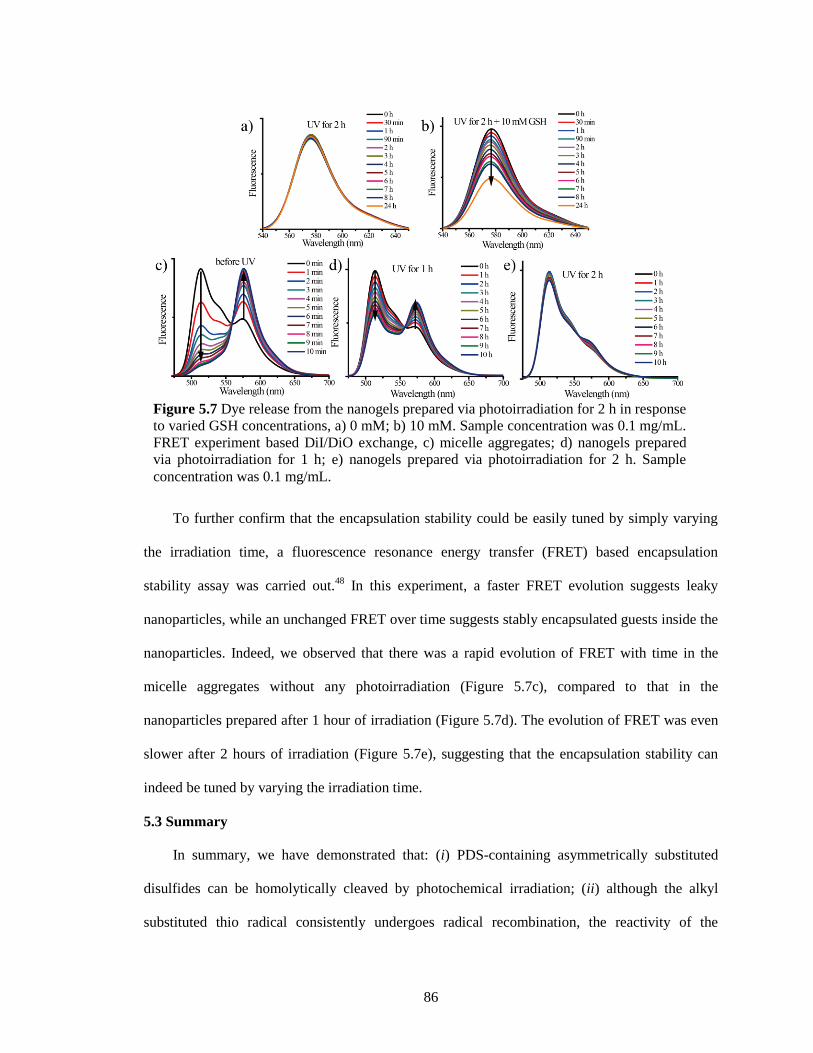
86
To further confirm that the encapsulation stability could be easily tuned by simply varying
the irradiation time, a fluorescence resonance energy transfer (FRET) based encapsulation
stability assay was carried out.48
In this experiment, a faster FRET evolution suggests leaky
nanoparticles, while an unchanged FRET over time suggests stably encapsulated guests inside the
nanoparticles. Indeed, we observed that there was a rapid evolution of FRET with time in the
micelle aggregates without any photoirradiation (Figure 5.7c), compared to that in the
nanoparticles prepared after 1 hour of irradiation (Figure 5.7d). The evolution of FRET was even
slower after 2 hours of irradiation (Figure 5.7e), suggesting that the encapsulation stability can
indeed be tuned by varying the irradiation time.
5.3 Summary
In summary, we have demonstrated that: (i) PDS-containing asymmetrically substituted
disulfides can be homolytically cleaved by photochemical irradiation; (ii) although the alkyl
substituted thio radical consistently undergoes radical recombination, the reactivity of the
Figure 5.7 Dye release from the nanogels prepared via photoirradiation for 2 h in response
to varied GSH concentrations, a) 0 mM; b) 10 mM. Sample concentration was 0.1 mg/mL.
FRET experiment based DiI/DiO exchange, c) micelle aggregates; d) nanogels prepared
via photoirradiation for 1 h; e) nanogels prepared via photoirradiation for 2 h. Sample
concentration was 0.1 mg/mL.
87
pyridylthio radical is dependent on the presence of oxygen; (iii) in the presence of oxygen, the
pyridylthio radical gets converted to pyridine-2-sulfonic acid, while it undergoes hydrogen
abstraction or radical recombination in deoxygenated solutions; (iv) the oxidation reaction also
provides a pathway for driving the equilibrium towards the desired product; (v) this feature was
used for the reagentless synthesis of crosslinked polymer nanoparticles; (vi) the size of the
nanoparticles can be conveniently varied by tuning the size of the nanoaggregates, because the
photo-induced crosslinking was predominantly intra-aggregate; and (vii) the crosslink density and
encapsulation stability of these nanoparticles can be simply varied by altering the irradiation time.
In addition to the typical advantages of a reagentless reaction, the fact that this reaction is carried
out in water and that the byproduct of this reaction is a highly water-soluble sulfonic acid that can
be easily removed by dialysis suggests that this methodology offers a promising new approach for
nanoparticle synthesis. Since the crosslinks generated here are biologically relevant, this method
could also have implications in generating new materials of interest in biology and medicine.
5.4 Experimental
5.4.1 General Methods
2,2-Dithiodipyridine, 2-mercaptoethanol, polyethyleneglycol monomethylether methacrylate
(PEGMA, MW 450 g/mol), D,L-dithiothreitol (DTT), 1,1'-dioctadecyl-3,3,3',3'-
tetramethylindocarbocyanine perchlorate (DiI) and 3,3'-dioctadecyloxacarbocyanine perchlorate
(DiO), 4-Cyano-4-(phenylcarbonothioylthio)-pentanoic acid and other conventional reagents
were obtained from commercial sources and were used as received without further purification.
The 2,2’-azobisisobutyronitrile (AIBN) was purified by recrystallization from ethanol. 1H-NMR
spectra were recorded on a 400 MHz Bruker NMR spectrometer using the residual proton
resonance of the solvent as the internal standard. Chemical shifts are reported in parts per million
(ppm). Molecular weights of the polymers were estimated by gel permeation chromatography
(GPC) using PMMA standard with a refractive index detector. Dynamic light scattering (DLS)
88
measurements were performed using a Malvern Nanozetasizer. UV-visible absorption spectra
were recorded on a Varian (model EL 01125047) spectrophotometer. The fluorescence spectra
were obtained from a JASCO FP-6500 spectrofluorimeter.
5.4.2 Synthesis of 2-hydroxyethyl 2-pyridyl disulfide (PDS-OH) and 2-thiopyridone
PDS-OH was synthesized according to the previous report.49
2, 2’-dipyridyl disulfide (DPDS)
(10.3 g, 0.047 mol) was dissolved in 100 mL of methanol. Then 1.2 mL of glacial acetic acid,
used as catalyst, was added. To this mixture, a solution of mercaptoethanol (4.4 g, 0.056 mol, 4
mL) in 100 mL methanol was added drop-wise at room temperature with continuous stirring.
Once the addition was over, the reaction mixture was stirred at room temperature overnight. The
stirring was stopped; solvent was evaporated to get the crude product as yellow oil which was
purified by flash column chromatography using silica gel as stationary phase and mixture of ethyl
acetate/hexane as eluent. When the polarity of the eluent was increased to 40% ethyl
acetate/hexane, the desired product came out as colorless oil. Yield : 80 % 1H NMR: (CD3OD,
400 MHz), δ (ppm): 8.40 (m, 1H, aromatic proton ortho-N), 7.84 (m, 1H, aromatic proton meta-
N), 7.80 (m, 1H, aromatic proton para-N), 7.22 (m, 1H, aromatic proton, ortho-disulfide linkage),
3.77(t, 2H, -CH2OH), 2.94 (t, 2H, -SS-CH2-).
2-thiopyridone can also be prepared at the same time by continually increasing the polarity
of the eluent up to 55% ethyl acetate/hexane to get the desired product. After concentrating the
solution, light yellow Needle-shaped crystals were got. 1H NMR: (CD3OD, 400 MHz), δ (ppm):
7.67 (m, 1H), 7.47 (m, 2H), 6.85 (m, 1H).
5.4.3 Synthesis of PDS monomer
PDS monomer was also synthesized according to the previous report.49
To a solution of
PDS-OH (12 g, 64.1 mmol) in 50 mL of dry dichloromethane was added 7.8 g (76.9 mmol) of
triethylamine. The mixture was firstly cooled in an ice-bath. To this cold mixture, a solution of
methacryloyl chloride (6.7 g, 64.1 mmol) in 25 mL dichloromethane was then added drop-wise
89
with continuous stirring. After the addition was over the reaction mixture was stirred at room
temperature overnight. The stirring was stopped and the reaction mixture was washed with 3x100
mL distilled water and then with 100 mL of brine. The organic layer was collected, dried over
anhydrous Na2SO4 and concentrated to get the crude product as yellow oil. It was purified by
column chromatography using silica gel as stationary phase and mixture of ethyl acetate/hexane
as eluent. The pure product was collected at 30 % ethylacetate/hexane. Yield: 80 % 1H NMR:
(CDCl3, 400 MHz), δ (ppm): 8.52 (m, 1H, aromatic proton ortho-N), 7.62-7.83 (m, 2H, aromatic
proton meta-N and para-N), 7.15 (m, 1H, aromatic proton, orthodisulfide linkage), 6.23 (d, 1H,
vinylic proton, cis-ester), 5.65(d, 1H, vinylic proton, trans-ester) 4.41 (t, 2H, -S-S-CH2CH2O-),
3.13 (t, 2H, -S-S-CH2CH2O-), 1.91(s, 3H, methyl proton of the methacryloyl group).
5.4.4 Synthesis of random copolymer containing PDS groups
Random copolymers were prepared by reversible addition−fragmentation chain transfer
(RAFT) polymerization.47
A mixture of 4-cyano-4-(phenylcarbonothioylthio)pentanoic acid (24
mg, 0.086 mmol), poly(ethylene glycol) monomethyl ether methacrylate (0.4 g, 0.84 mmol), PDS
(0.537 g, 2.1 mmol), and 2,2′-azobis(isobutyronitrile) (AIBN; 2.8 mg, 0.017 mmol) was dissolved
in tetrahydrofuran (THF) (2 mL) and degassed by performing three freeze-pump-thaw cycles.
The reaction mixture was sealed and then transferred into a preheated oil bath at 65 °C for 10 h.
To remove unreactive monomers, the resultant mixture was precipitated in cold ethyl ether (20
mL) to yield the random copolymer as a waxy liquid. GPC (THF) Mw, 12 kDa; polydispersity, 1.2.
1H NMR (400 MHz, CDCl3) δ 8.46, 7.68, 7.11, 4.35-4.09, 3.94-3.37, 3.03, 2.04-1.64, 1.43-0.87.
The molar ratio between two blocks was determined by integrating the methoxy proton in the
poly-(ethylene glycol) unit and the aromatic proton in the pyridine and was found to be 1:2.70
(PEO:PDS).
90
5.4.5 Time-dependent 1H NMR measurement
In 5mm NMR tubes, 1 mL of samples with different concentrations was added. 1H-NMR
spectra were recorded on a 400 MHz Bruker NMR spectrometer using the residual proton
resonance of the solvent as the internal standard. These tubes were put inside UV chamber
(SYLVANIA F15T8/350/BL, 15w). After each 1 hour’s photoirradiation, another NMR spectrum
for each sample was taken with the same parameters. The integrations of peak at 2.94 ppm and
peak at 2.83 ppm were used to determine the percent of remaining PDS-OH.
5.4.6 Nanoparticles preparation
1 mL of polymer solution of different concentrations was added to a 7 mL vial. These vials
were then put inside an UV chamber (SYLVANIA F15T8/350/BL, 15w). After photoirradiation
for certain times, nanoparticles with different sizes were formed in solutions. The crosslinking
density could be determined by taking some volume of polymer solution from the above solutions
into cuvettes for UV-vis measurement. The concentration of these samples was all 0.1 mg/mL.
Absorption spectra were taken first. Then, excess DTT were added into these samples and then
the solution was observed using absorption spectroscopy again after 3 hours, during which all
PDS functionalities could be changed to 2-thiopyridine by DTT.
5.4.7 Determination of crosslinking density
In 7mL small glass vial, 1 mL of polymer solution with different concentrations was added.
From them, certain volume of polymer solution was taken and then added into cuvettes for UV-
vis measurement. The volume was made to 1 mL by adding water. The concentration of these
samples was all 0.1 mg/mL. Absorption spectra were taken. Excess DTT were added into these
samples, one more absorption spectra were taken after 3 hours, during which all PDS
functionalities could be changed to 2-thiopyridine by DTT.
Then these vials were put inside UV chamber (SYLVANIA F15T8/350/BL, 15w). After
each 1 hour’s photoirradiation, certain volume of polymer solution was taken from the above
91
solutions and then added into cuvettes for UV-vis measurement. The volume was made to 1 mL
by adding water. The concentration of these samples was all 0.1 mg/mL. Absorption spectra were
taken. Excess DTT were added into these samples, one more absorption spectra were taken after
3 hours, during which all PDS functionalities could be changed to 2-thiopyridine by DTT.
5.4.8 Redox-responsive guest release experiment
A solution of nanogel containing DiI (100 µL) was mixed with water (800 µL) in a cuvette,
and then GSH stock solution (100 µL) was added. The fluorescence spectra were recorded using
the excitation wavelength of 530 nm.
For control experiment, A solution of nanogel containing DiI (100 µL) was mixed with
water (900 µL) in a cuvette. The fluorescence spectra were recorded using the excitation
wavelength of 530 nm.
5.4.9 FRET experiment
A solution of nanogel containing DiI (100 µL) was mixed with a solution of nanogel
containing DiO (100 µL) in a cuvette, and then milliQ water (800 µL) was added to adjust the
volume. The fluorescence spectra were recorded using the excitation wavelength of 450 nm.
5.5 References
1. Fu, G. C.; Nguyen, S. T.; Grubbs, G. H. Catalytic ring-closing metathesis of functionalized
dienes by a ruthenium carbene complex. J. Am. Chem. Soc. 1993, 115, 9856-9857;
2. La, D. S.; Alexander, J. B.; Cefalo, D. R.; Graf, D. D.; Hoveyda, A. H.; Schrock, R. R. Mo-
catalyzed asymmetric synthesis of dihydrofurans. Catalytic kinetic resolution and enantioselective
desymmetrization through ring-closing metathesis. J. Am. Chem. Soc. 1998, 120, 9720-9721;
3. Connon, S. J.; Blechert, S.. Recent developments in olefin cross-metathesis. Angew. Chem. Int.
Ed. 2003, 42, 1900-1923;
4. Mutlu, H.; de Espinosa, L. M.; Meier, M. A. R. Acyclic diene metathesis: a versatile tool for
the construction of defined polymer architectures. Chem. Soc. Rev. 2011, 40, 1404-1445;
5. Wagener, K. B., Boncella, J. M.; Nel, J. G. Acyclic diene metathesis (ADMET) polymerization.
Macromolecules 1991, 24, 2649-2657;
6. Lu, Y.-X.; Guan, Z. Olefin metathesis for effective polymer healing via dynamic exchange of
strong carbon–carbon double bonds. J. Am. Chem. Soc. 2012, 134, 14226-14231;
92
7. Koh, M. J.; Khan, R. K. M.; Torker, S.; Hoveyda, A. H. Broadly applicable Z- and
diastereoselective ring-opening/cross-metathesis catalyzed by a dithiolate Ru complex. Angew.
Chem. Int. Ed. 2014, 53, 1968-1972.
8. Oh, K.; Jeong, K.-S.; Moore, J. S. Folding-driven synthesis of oligomers. Nature 2001, 414,
889-893;
9. Nishinaga, T.; Tanatani, A.; Oh, K.; Moore, J. S. The size-selective synthesis of folded
oligomers by dynamic templation. J. Am. Chem. Soc. 2002, 124, 5934-5935;
10. Cantrell, G. K.; Meyer, T. Y. Catalytic CN bond formation by metal-imide-mediated imine
metathesis. J. Am. Chem. Soc. 1998, 120, 8035-8042;
11. Meyer, C. D.; Joiner, C. S.; Stoddart, J. F. Template-directed synthesis employing reversible
imine bond formation. Chem. Soc. Rev. 2007, 36, 1705-1723;
12. Janeliunas, D.; van Rijn, P.; Boekhoven, J.; Minkenberg, C. B.; van Esch, J. H.; Eelkema, R.
Aggregation-driven reversible formation of conjugated polymers in water. Angew. Chem. Int. Ed.
2013, 52, 1998-2001;
13. Taynton, P.; Yu, K.; Shoemaker, R. K.; Jin, Y.; Qi, H. J.; Zhang, W. Heat- or water- driven
malleability in a highly recyclable covalent network polymer. Adv. Mater. 2014, 26, 3938–3942.
14. Hirsch, A. K. H.; Buhler, E.; Lehn, J. -M. Biodynamers: self-organization-driven formation of
doubly dynamic proteoids. J. Am. Chem. Soc. 2012, 134, 4177-4183.
15. Folmer-Andersen, J. F.; Lehn, J. -M. Thermoresponsive dynamers: thermally induced,
reversible chain elongation of amphiphilic poly (acylhydrazones). J. Am. Chem. Soc. 2011, 133,
10966-10973.
16. Barrell, M. J.; Campaña, A. G.; von Delius, M.; Geertsema, E. M.; Leigh, D. A. Light-driven
transport of a molecular walker in either direction along a molecular track. Angew. Chem. Int. Ed.
2011, 50, 285-290;
17. Dirksen, A.; Dirksen, S.; Hackeng, T. M.; Dawson. P. E.. Nucleophilic catalysis of
hydrazone formation and transimination: implications for dynamic covalent chemistry. J. Am.
Chem. Soc. 2006, 128, 15602-15603;
18. Chung, M.-K.; Hebling, C. M.; Jorgenson, J. W.; Severin, K.; Lee, S. J.; Gagné, M. R.
Deracemization of a dynamic combinatorial library induced by (−)-cytidine and (−)-2-
thiocytidine. J. Am. Chem. Soc. 2008, 130, 11819-11827.
19. Markus, G.; Karush, F. The disulfide bonds of human serum albumin and bovine γ-globulin1.
J. Am. Chem. Soc. 1957, 79, 134-139;
20. Rueping, M.; Jaun, B.; Seebach, D. NMR Structure in methanol of a β-hexapeptide with a
disulfide clamp. Chem. Commun. 2000, 22, 2267-2268;
21. Wedemeyer, W. J.; Welker, E.; Narayan, M.; Scheraga, H. A. Disulfide bonds and protein
folding. Biochemistry 2000, 39, 4207-4216.
93
22. Otto, S.; Furlan, R. L. E.; Sanders, J. K. M. Dynamic combinatorial libraries of macrocyclic
disulfides in water. J. Am. Chem. Soc. 2000, 122, 12063-12064;
23. Otto, S.; Furlan, R. L. E.; Sanders, J. K. M. Selection and amplification of hosts from
dynamic combinatorial libraries of macrocyclic disulfides. Science 2002, 297, 590-593;
24. Li, J.; Carnall, J.; Stuart, M. C. A.; Otto, S. Hydrogel formation upon photoinduced covalent
capture of macrocycle stacks from dynamic combinatorial libraries. Angew. Chem. Int. Ed. 2011,
50, 8384-8386;
25. Basak, D.; Kumar, R.; Ghosh, S. Telechelic poly(disulfide)s and related block copolymer.
Macromol. Rapid Commun., 2014, 35, 1340-1344.
26. Kuang, H.; He, H.; Hou, J.; Xie, Z.; Jing, X.; Huang, Y. Thymine modified amphiphilic
biodegradable copolymers for photo-cross-linked micelles as stable drug carriers. Macromol.
Biosci. 2013, 13, 1593-1600.
27. Chung, J. W.; Lee, K.; Neikirk, C.; Nelson, C. M.; Priestley, R. D. Photoresponsive
coumarin-stabilized polymeric nanoparticles as a detectable drug carrier. Small 2012, 8, 1693-
1700;
28. He, J.; Tong, X.; Zhao, Y. Photoresponsive nanogels based on photocontrollable cross-links.
Macromolecules 2009, 42, 4845-4852;
29. Jiang, J.; Qi, B.; Lepage, M.; Zhao, Y. Polymer micelles stabilization on demand through
reversible photo-cross-linking. Macromolecules 2007, 40, 790-792.
30. Kim, J. S.; Youk, J. H. Preparation of core cross-linked micelles using a photo-cross-linking
agent. Polymer 2009, 50, 2204-2208.
31. Chakravarthi, S.; Jessop, C. E.; Bulleid, N. J. The role of glutathione in disulphide bond
formation and endoplasmic-reticulum-generated oxidative stress. EMBO reports 2006, 7, 271-275;
32. Luo, Z.; Cai, K.; Hu, Y.; Zhao, L.; Liu, P.; Duan, L.; Yang, W. Mesoporous silica
nanoparticles end‐capped with collagen: redox‐responsive nanoreservoirs for targeted drug
delivery. Angew. Chem. Int. Ed. 2011, 50, 640-643;
33. Ryu, J.-H.; Jiwpanich, S.; Chacko, R.; Bickerton, S.; Thayumanavan, S. Surface-
functionalizable polymer nanogels with facile hydrophobic guest encapsulation capabilities. J.
Am. Chem. Soc. 2010, 132, 8246-8247;
34. Li, L.; Raghupathi, K.; Yuan, C.; Thayumanavan, S. Surface charge generation in nanogels
for activated cellular uptake at tumor-relevant pH. Chem. Sci. 2013, 4, 3654-3660;
35. Kabanov, A. V.; Vinogradov, S. V. Nanogels as pharmaceutical carriers: finite networks of
infinite capabilities. Angew. Chem. Int. Ed. 2009, 48, 5418-5429.
36. Milligan, B.; Rivett, D. E.; Savige, W. E. The photolysis of dialkyl sulphides, disulphides,
and trisulphides. Aust. J. Chem. 1963, 16, 1020-1029;
94
37. Nelander, B.; Sunner, S. Cogwheel effect in dialkyl disulfides. J. Am. Chem. Soc. 1972, 94,
3576-3577;
38. Lee, Y. R.; Chiu, C. L.; Lin, S. M. Ultraviolet photodissociation study of CH3SCH3 and
CH3SSCH3. J. Chem. Phys. 1994, 100, 7376-7384;
39. Gupta, D.; Knight, A. R. Reactions of thiyl radicals. XIII. Photochemically induced exchange
reactions of liquid alkyl disulfides. Can. J. Chem. 1980, 58, 1350-1354;
40. Otsuka, H.; Nagano, S.; Kobashi, Y.; Maeda, T.; Takahara, A. A dynamic covalent polymer
driven by disulfide metathesis under photoirradiation. Chem. Commun. 2010, 46, 1150-1152;
41. Robert-Banchereau, E.; Lacombe, S.; Ollivier, J. Solution reactivity of thiyl radicals with
molecular oxygen: Unsensitized photooxidation of dimethyldisulfide. Tetrahedron Lett. 1995, 36,
8197-8200;
42. Robert-Banchereau, E.; Lacombe, S.; Ollivier, J. Unsensitized photooxidation of sulfur
compounds with molecular oxygen in solution. Tetrahedron 1997, 53, 2087-2102.
43. Kakizawa, Y.; Harada, A.; Kataoka, K. Environment-sensitive stabilization of core-shell
structured polyion complex micelle by reversible cross-linking of the core through disulfide bond.
J. Am. Chem. Soc. 1999, 121, 11247-11248;
44. Ryu, J. -H.; Chacko, R. T.; Jiwpanich, S.; Bickerton, S.; Babu, R. P.; Thayumanavan, S. Self-
cross-linked polymer nanogels: a versatile nanoscopic drug delivery platform. J. Am. Chem. Soc.
2010, 132, 17227-17235.
45. Adam, W.; Grimm, G. N.; Marquardt, S.; Saha-Möller, C. R. Are pyridinethiones reliable
photochemical oxyl-radical sources for photobiological studies? The importance of secondary
photolysis products in the guanine oxidation of 2'-deoxyguanosine and cell-free DNA. J. Am.
Chem. Soc. 1999, 121, 1179-1185;
46. Lobana, T. S.; Kinoshita, I.; Kimura, K.; Nishioka, T.; Shiomi, D.; Isobe, K. Pyridine-2-
sulfonates as versatile ligands for the synthesis of novel coordinative and hydrogen-bonded
supramolecules. Eur. J. Inorg. Chem. 2004, 2004, 356-367;
47. Li, L.; Ryu, J. -H.; Thayumanavan, S. Effect of hofmeister ions on the size and encapsulation
stability of polymer nanogels. Langmuir 2013, 29, 50-55.
48. Jiwpanich, S.; Ryu, J. -H.; Bickerton, S.; Thayumanavan, S. Noncovalent encapsulation
stabilities in supramolecular nanoassemblies. J. Am. Chem. Soc. 2010, 132, 10683-10685.
49. Ghosh, S.; Basu, S.; Thayumanavan, S. Simultaneous and reversible functionalization of
copolymers for biological applications. Macromolecules 2006, 39, 5595-5597.
95
CHAPTER 6
THERMORESPONSIVE POLYMERIC NANOPARTICLES: NUCLEATION FROM
COOPERATIVE POLYMERIZATION DRIVEN BY DATIVE BONDS
Reprinted with permission from Li, L.; Yuan, C.; Dai, L.; Thayumanavan, S.
Thermoresponsive Polymeric Nanoparticles: Nucleation from Cooperative Polymerization Driven
by Dative Bonds. Macromolecules 2014, 47, 5869. Copyright © 2015 American Chemical
Society
6.1 Introduction
Cooperative polymerization, also called nucleation-elongation (N-E) polymerization, has
become a new and interesting paradigm in the field of supramolecular materials.1-3
Although it
has been observed and relatively well studied in biological systems (e.g. actin, tubulin, bacterial
flagellin, and tobacco mosaic virus),4, 5
these are not common in artificial material assemblies. It
is believed that systems governed by the N-E mechanism can provide access to materials with
novel properties, which are otherwise not available with classical synthetic polymerization
techniques.6-8
For example, although a polymer is achieved through a condensation-type reaction,
the N-E system could produce polymers with substantially high molecular weights even when the
stoichiometry of the monomers is imbalanced.9 The molecular origin of cooperative growth of
supramolecular polymers is regarded as the result of secondary interaction between units, which
usually involves a non-covalent interaction arising due to electronic (both short-range
polarization and longer-range electrostatic effects), structural (e.g. helix formation and allosteric
conformational changes), or hydrophobic effects.1 However, there are no reports on utilizing
dative bonds, a covalent interaction, as the driving force for the cooperative polymerization.
Demonstration of such a possibility will greatly expand the repertoire of nucleation-elongation
polymerization in materials science, as exemplified in this report.
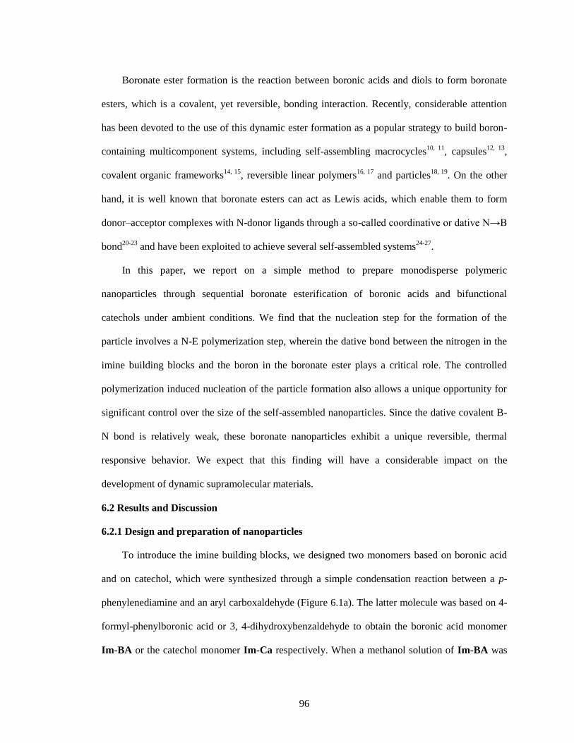
96
Boronate ester formation is the reaction between boronic acids and diols to form boronate
esters, which is a covalent, yet reversible, bonding interaction. Recently, considerable attention
has been devoted to the use of this dynamic ester formation as a popular strategy to build boron-
containing multicomponent systems, including self-assembling macrocycles10, 11
, capsules12, 13
,
covalent organic frameworks14, 15
, reversible linear polymers16, 17
and particles18, 19
. On the other
hand, it is well known that boronate esters can act as Lewis acids, which enable them to form
donor–acceptor complexes with N-donor ligands through a so-called coordinative or dative N→B
bond20-23
and have been exploited to achieve several self-assembled systems24-27
.
In this paper, we report on a simple method to prepare monodisperse polymeric
nanoparticles through sequential boronate esterification of boronic acids and bifunctional
catechols under ambient conditions. We find that the nucleation step for the formation of the
particle involves a N-E polymerization step, wherein the dative bond between the nitrogen in the
imine building blocks and the boron in the boronate ester plays a critical role. The controlled
polymerization induced nucleation of the particle formation also allows a unique opportunity for
significant control over the size of the self-assembled nanoparticles. Since the dative covalent B-
N bond is relatively weak, these boronate nanoparticles exhibit a unique reversible, thermal
responsive behavior. We expect that this finding will have a considerable impact on the
development of dynamic supramolecular materials.
6.2 Results and Discussion
6.2.1 Design and preparation of nanoparticles
To introduce the imine building blocks, we designed two monomers based on boronic acid
and on catechol, which were synthesized through a simple condensation reaction between a p-
phenylenediamine and an aryl carboxaldehyde (Figure 6.1a). The latter molecule was based on 4-
formyl-phenylboronic acid or 3, 4-dihydroxybenzaldehyde to obtain the boronic acid monomer
Im-BA or the catechol monomer Im-Ca respectively. When a methanol solution of Im-BA was
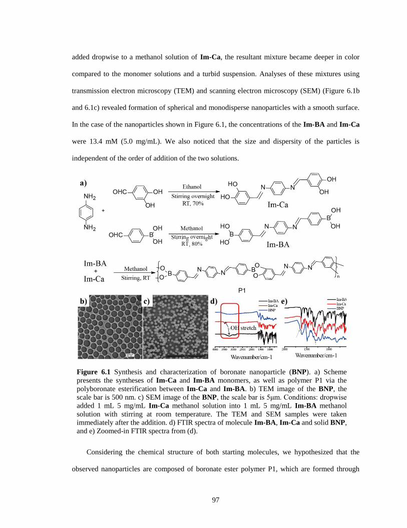
97
added dropwise to a methanol solution of Im-Ca, the resultant mixture became deeper in color
compared to the monomer solutions and a turbid suspension. Analyses of these mixtures using
transmission electron microscopy (TEM) and scanning electron microscopy (SEM) (Figure 6.1b
and 6.1c) revealed formation of spherical and monodisperse nanoparticles with a smooth surface.
In the case of the nanoparticles shown in Figure 6.1, the concentrations of the Im-BA and Im-Ca
were 13.4 mM (5.0 mg/mL). We also noticed that the size and dispersity of the particles is
independent of the order of addition of the two solutions.
Considering the chemical structure of both starting molecules, we hypothesized that the
observed nanoparticles are composed of boronate ester polymer P1, which are formed through
Figure 6.1 Synthesis and characterization of boronate nanoparticle (BNP). a) Scheme
presents the syntheses of Im-Ca and Im-BA monomers, as well as polymer P1 via the
polyboronate esterification between Im-Ca and Im-BA. b) TEM image of the BNP, the
scale bar is 500 nm. c) SEM image of the BNP, the scale bar is 5μm. Conditions: dropwise
added 1 mL 5 mg/mL Im-Ca methanol solution into 1 mL 5 mg/mL Im-BA methanol
solution with stirring at room temperature. The TEM and SEM samples were taken
immediately after the addition. d) FTIR spectra of molecule Im-BA, Im-Ca and solid BNP,
and e) Zoomed-in FTIR spectra from (d).
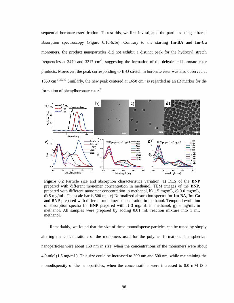
98
sequential boronate esterification. To test this, we first investigated the particles using infrared
absorption spectroscopy (Figure 6.1d-6.1e). Contrary to the starting Im-BA and Im-Ca
monomers, the product nanoparticles did not exhibit a distinct peak for the hydroxyl stretch
frequencies at 3470 and 3217 cm-1
, suggesting the formation of the dehydrated boronate ester
products. Moreover, the peak corresponding to B-O stretch in boronate ester was also observed at
1350 cm-1
.29, 30
Similarly, the new peak centered at 1658 cm-1
is regarded as an IR marker for the
formation of phenylboronate ester.31
Remarkably, we found that the size of these monodisperse particles can be tuned by simply
altering the concentrations of the monomers used for the polymer formation. The spherical
nanoparticles were about 150 nm in size, when the concentrations of the monomers were about
4.0 mM (1.5 mg/mL). This size could be increased to 300 nm and 500 nm, while maintaining the
monodispersity of the nanoparticles, when the concentrations were increased to 8.0 mM (3.0
Figure 6.2 Particle size and absorption characteristics variation. a) DLS of the BNP
prepared with different monomer concentration in methanol. TEM images of the BNP,
prepared with different monomer concentration in methanol, b) 1.5 mg/mL, c) 3.0 mg/mL,
d) 5 mg/mL. The scale bar is 500 nm. e) Normalized absorption spectra for Im-BA, Im-Ca
and BNP prepared with different monomer concentration in methanol. Temporal evolution
of absorption spectra for BNP prepared with f) 3 mg/mL in methanol, g) 5 mg/mL in
methanol. All samples were prepared by adding 0.01 mL reaction mixture into 1 mL
methanol.
99
mg/mL) and 13.4 mM (5.0 mg/mL) respectively. Thus, we found that the sizes of the polymer
nanoparticles systematically increase with concentration. The rather uniform dispersity of the
particles is evident from the TEM images shown in Figure 6.2. These results were also consistent
with the size measurements in solution using dynamic light scattering (DLS) (Figure 6.2a). Lack
of diffraction peaks, except for an ill-defined broad peak at about 2θ=20, in powder X-ray
diffraction (PXRD) (Figure 6.3) suggests that these particles were mostly amorphous.
Interestingly, there is a need for a critical concentration, below which there was no discernible
nanoparticle formation. We noticed that particles could not be formed when the initial
concentration of both monomers (1:1 ratio) was lower than 1.0 mg/mL, as discerned from TEM.
The poor correlation function, observed in DLS, further confirmed this assertion.
The absorbance spectra for these particles in methanol are also shown in the Figure 6.2e.
Compared to the starting Im-BA and Im-Ca, the nanoparticle solutions exhibit new peaks at
longer wavelengths. This is taken to be an indicator for the formation of boronate ester between
Im-BA and Im-Ca, which could lead to a longer π-conjugated polymer. Conjugated systems
extending through the boronate ester is reasonable. For example, poly (dioxaborole)s, in which
Figure 6.3 PXRD pattern of the BNP. A broad bump at about 2θ=20 was resulted from
crystalline parts of polymers.
0 20 40 60 80 100
Inte
nsi
ty
2-Theta (o)
BNP-1.5 mg/mL
BNP-3 mg/mL
BNP-5 mg/mL
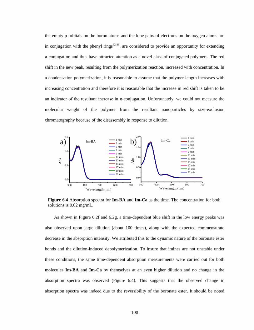
100
the empty p-orbitals on the boron atoms and the lone pairs of electrons on the oxygen atoms are
in conjugation with the phenyl rings32-36
, are considered to provide an opportunity for extending
π-conjugation and thus have attracted attention as a novel class of conjugated polymers. The red
shift in the new peak, resulting from the polymerization reaction, increased with concentration. In
a condensation polymerization, it is reasonable to assume that the polymer length increases with
increasing concentration and therefore it is reasonable that the increase in red shift is taken to be
an indicator of the resultant increase in π-conjugation. Unfortunately, we could not measure the
molecular weight of the polymer from the resultant nanoparticles by size-exclusion
chromatography because of the disassembly in response to dilution.
As shown in Figure 6.2f and 6.2g, a time-dependent blue shift in the low energy peaks was
also observed upon large dilution (about 100 times), along with the expected commensurate
decrease in the absorption intensity. We attributed this to the dynamic nature of the boronate ester
bonds and the dilution-induced depolymerization. To insure that imines are not unstable under
these conditions, the same time-dependent absorption measurements were carried out for both
molecules Im-BA and Im-Ca by themselves at an even higher dilution and no change in the
absorption spectra was observed (Figure 6.4). This suggests that the observed change in
absorption spectra was indeed due to the reversibility of the boronate ester. It should be noted
Figure 6.4 Absorption spectra for Im-BA and Im-Ca as the time. The concentration for both
solutions is 0.02 mg/mL.
300 400 500 600 700
0.0
0.5
1.0
1.5
Ab
s
Wavelength (nm)
1 min
3 min
5 min
7 min
9 min
11 min
13 min
15 min
17 min
19 min
21 min
Im-BA
300 400 500 600 700
0.0
0.5
1.0
1.5
2.0
Ab
s
Wavelength (nm)
1 min
3 min
5 min
7 min
9 min
11 min
13 min
15 min
17 min
19 min
21 min
Im-Caa) b)
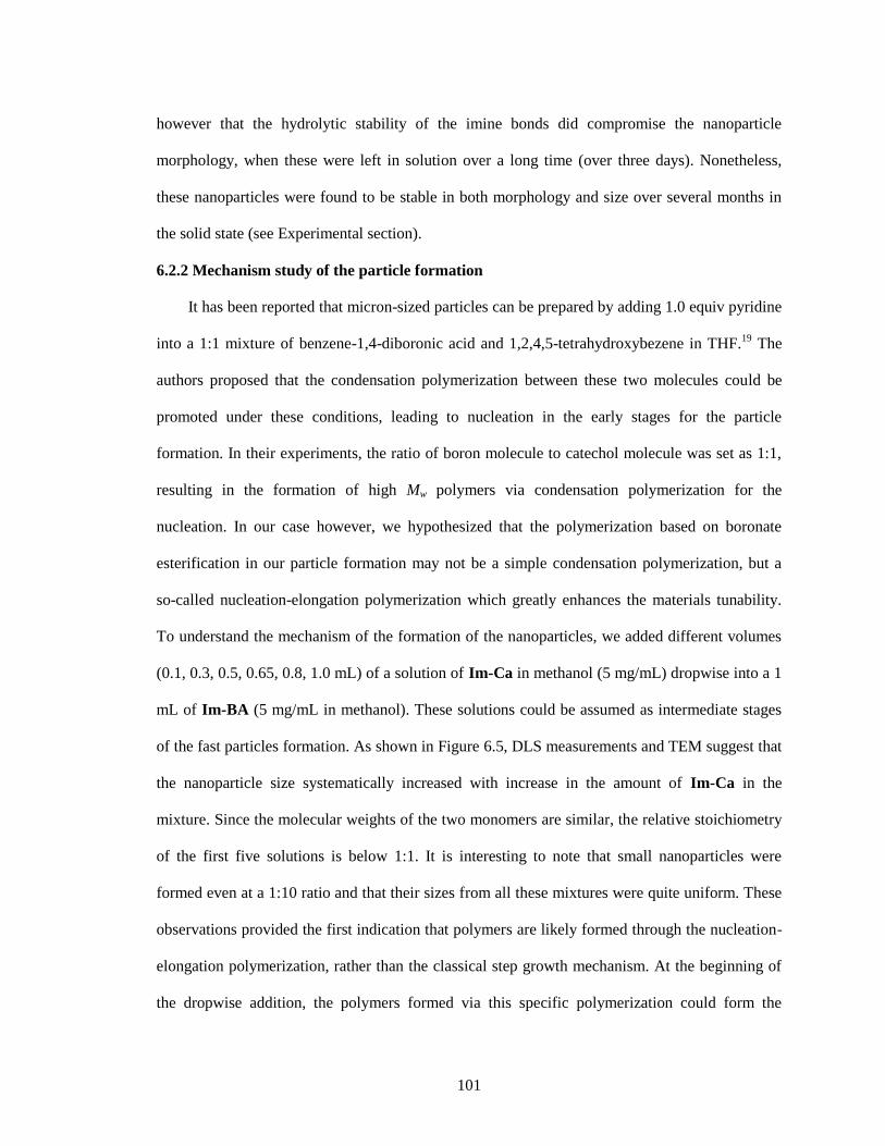
101
however that the hydrolytic stability of the imine bonds did compromise the nanoparticle
morphology, when these were left in solution over a long time (over three days). Nonetheless,
these nanoparticles were found to be stable in both morphology and size over several months in
the solid state (see Experimental section).
6.2.2 Mechanism study of the particle formation
It has been reported that micron-sized particles can be prepared by adding 1.0 equiv pyridine
into a 1:1 mixture of benzene-1,4-diboronic acid and 1,2,4,5-tetrahydroxybezene in THF.19
The
authors proposed that the condensation polymerization between these two molecules could be
promoted under these conditions, leading to nucleation in the early stages for the particle
formation. In their experiments, the ratio of boron molecule to catechol molecule was set as 1:1,
resulting in the formation of high Mw polymers via condensation polymerization for the
nucleation. In our case however, we hypothesized that the polymerization based on boronate
esterification in our particle formation may not be a simple condensation polymerization, but a
so-called nucleation-elongation polymerization which greatly enhances the materials tunability.
To understand the mechanism of the formation of the nanoparticles, we added different volumes
(0.1, 0.3, 0.5, 0.65, 0.8, 1.0 mL) of a solution of Im-Ca in methanol (5 mg/mL) dropwise into a 1
mL of Im-BA (5 mg/mL in methanol). These solutions could be assumed as intermediate stages
of the fast particles formation. As shown in Figure 6.5, DLS measurements and TEM suggest that
the nanoparticle size systematically increased with increase in the amount of Im-Ca in the
mixture. Since the molecular weights of the two monomers are similar, the relative stoichiometry
of the first five solutions is below 1:1. It is interesting to note that small nanoparticles were
formed even at a 1:10 ratio and that their sizes from all these mixtures were quite uniform. These
observations provided the first indication that polymers are likely formed through the nucleation-
elongation polymerization, rather than the classical step growth mechanism. At the beginning of
the dropwise addition, the polymers formed via this specific polymerization could form the
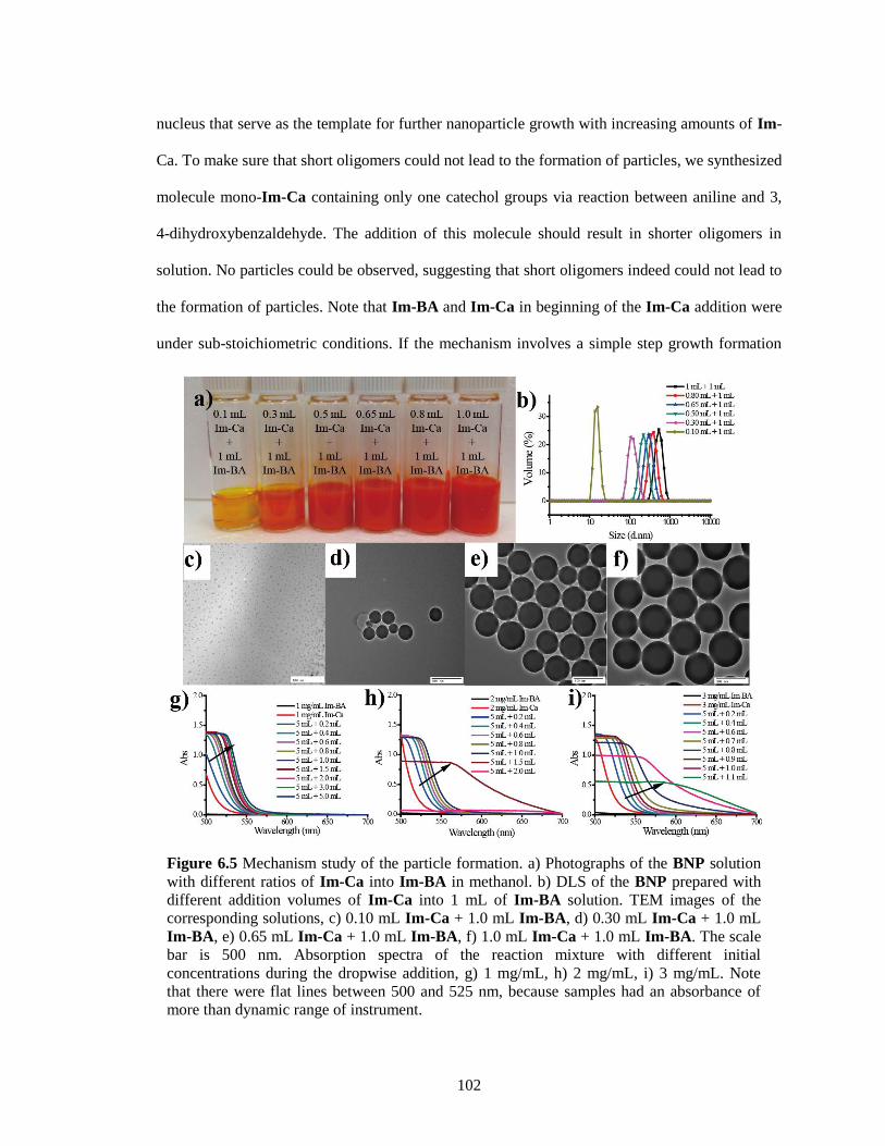
102
nucleus that serve as the template for further nanoparticle growth with increasing amounts of Im-
Ca. To make sure that short oligomers could not lead to the formation of particles, we synthesized
molecule mono-Im-Ca containing only one catechol groups via reaction between aniline and 3,
4-dihydroxybenzaldehyde. The addition of this molecule should result in shorter oligomers in
solution. No particles could be observed, suggesting that short oligomers indeed could not lead to
the formation of particles. Note that Im-BA and Im-Ca in beginning of the Im-Ca addition were
under sub-stoichiometric conditions. If the mechanism involves a simple step growth formation
Figure 6.5 Mechanism study of the particle formation. a) Photographs of the BNP solution
with different ratios of Im-Ca into Im-BA in methanol. b) DLS of the BNP prepared with
different addition volumes of Im-Ca into 1 mL of Im-BA solution. TEM images of the
corresponding solutions, c) 0.10 mL Im-Ca + 1.0 mL Im-BA, d) 0.30 mL Im-Ca + 1.0 mL
Im-BA, e) 0.65 mL Im-Ca + 1.0 mL Im-BA, f) 1.0 mL Im-Ca + 1.0 mL Im-BA. The scale
bar is 500 nm. Absorption spectra of the reaction mixture with different initial
concentrations during the dropwise addition, g) 1 mg/mL, h) 2 mg/mL, i) 3 mg/mL. Note
that there were flat lines between 500 and 525 nm, because samples had an absorbance of
more than dynamic range of instrument.
103
of the boronate ester, then only oligomers would form in solution and there should not be
significant nanoparticle formation at imbalanced ratios of the starting monomers.
To further determine that it is the polymer formed but not oligomer formation, leading to the
nucleation step, we investigated the absorption spectra of these reaction mixtures after small
additions of Im-Ca into Im-BA, since the red shift in absorption likely indicates increase in
conjugation, i.e. polymer chain length. First, a solution of Im-Ca (1 mg/mL) was added in 0.1 mL
increments to a 5 mL solution of Im-BA (1mg/mL) and the evolution of the absorption spectrum
of the resultant solution was monitored after each addition. As shown in Figure 6.5g, Im-BA
solution with 1 mg/mL did not have any absorption while Im-Ca solution with 1 mg/mL had a
small absorption from 500 nm to 550 nm. After 0.2 mL Im-Ca was added into 5 mL Im-BA, a
significant absorption was noticed even though the concentration of Im-Ca in this mixture was
only around 0.04 mg/mL, suggesting the formation of oligomers, which have a much stronger
absorption from 500 nm to 550 nm. As more Im-Ca was added, the absorption intensity of the
low energy peak between 500 nm and 550 nm keeps slowly increasing and red-shifting
throughout the addition process. Interestingly however, no particle formation was observed via
both DLS and TEM at this monomer concentration. The solution was also clear, although the
color became deep orange. This is taken to suggest that the 1 mg/mL concentrations of the
monomers are lower than the critical concentration for the nucleation step in the nucleation-
elongation polymerization. On the contrary, when the concentrations of the monomers are 2
mg/mL, there was a sharp increase in absorption after 550 nm at a sub-stoichiometric ratio of
monomers (2 mL of Im-Ca into 5 mL of Im-BA, the ratio of Im-Ca to Im-BA is 0.4/1), as shown
in Figure 6.5h, suggesting the formation of polymers with larger conjugation. At this ratio, the
solution became a turbid suspension and small spherical particles were also observed by TEM.
Similar results were found at an even lower stoichiometry of Im-Ca (only 0.9 mL of Im-Ca into
5 mL of Im-BA), when the initial concentration was 3 mg/mL (Figure 6.5i).
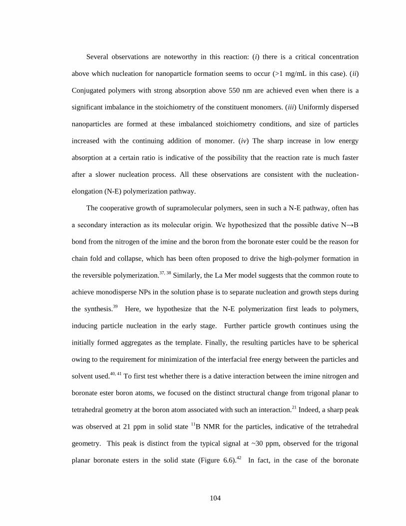
104
Several observations are noteworthy in this reaction: (i) there is a critical concentration
above which nucleation for nanoparticle formation seems to occur (>1 mg/mL in this case). (ii)
Conjugated polymers with strong absorption above 550 nm are achieved even when there is a
significant imbalance in the stoichiometry of the constituent monomers. (iii) Uniformly dispersed
nanoparticles are formed at these imbalanced stoichiometry conditions, and size of particles
increased with the continuing addition of monomer. (iv) The sharp increase in low energy
absorption at a certain ratio is indicative of the possibility that the reaction rate is much faster
after a slower nucleation process. All these observations are consistent with the nucleation-
elongation (N-E) polymerization pathway.
The cooperative growth of supramolecular polymers, seen in such a N-E pathway, often has
a secondary interaction as its molecular origin. We hypothesized that the possible dative N→B
bond from the nitrogen of the imine and the boron from the boronate ester could be the reason for
chain fold and collapse, which has been often proposed to drive the high-polymer formation in
the reversible polymerization.37, 38
Similarly, the La Mer model suggests that the common route to
achieve monodisperse NPs in the solution phase is to separate nucleation and growth steps during
the synthesis.39
Here, we hypothesize that the N-E polymerization first leads to polymers,
inducing particle nucleation in the early stage. Further particle growth continues using the
initially formed aggregates as the template. Finally, the resulting particles have to be spherical
owing to the requirement for minimization of the interfacial free energy between the particles and
solvent used.40, 41
To first test whether there is a dative interaction between the imine nitrogen and
boronate ester boron atoms, we focused on the distinct structural change from trigonal planar to
tetrahedral geometry at the boron atom associated with such an interaction.21
Indeed, a sharp peak
was observed at 21 ppm in solid state 11
B NMR for the particles, indicative of the tetrahedral
geometry. This peak is distinct from the typical signal at ~30 ppm, observed for the trigonal
planar boronate esters in the solid state (Figure 6.6).42
In fact, in the case of the boronate
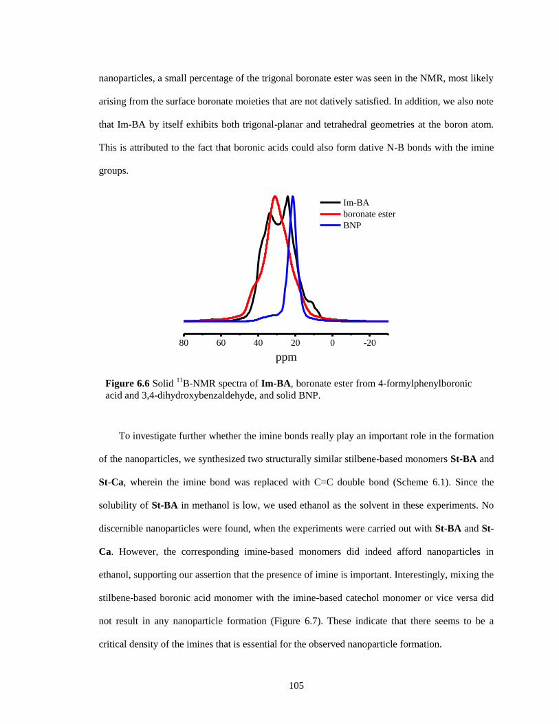
105
nanoparticles, a small percentage of the trigonal boronate ester was seen in the NMR, most likely
arising from the surface boronate moieties that are not datively satisfied. In addition, we also note
that Im-BA by itself exhibits both trigonal-planar and tetrahedral geometries at the boron atom.
This is attributed to the fact that boronic acids could also form dative N-B bonds with the imine
groups.
To investigate further whether the imine bonds really play an important role in the formation
of the nanoparticles, we synthesized two structurally similar stilbene-based monomers St-BA and
St-Ca, wherein the imine bond was replaced with C=C double bond (Scheme 6.1). Since the
solubility of St-BA in methanol is low, we used ethanol as the solvent in these experiments. No
discernible nanoparticles were found, when the experiments were carried out with St-BA and St-
Ca. However, the corresponding imine-based monomers did indeed afford nanoparticles in
ethanol, supporting our assertion that the presence of imine is important. Interestingly, mixing the
stilbene-based boronic acid monomer with the imine-based catechol monomer or vice versa did
not result in any nanoparticle formation (Figure 6.7). These indicate that there seems to be a
critical density of the imines that is essential for the observed nanoparticle formation.
Figure 6.6 Solid 11
B-NMR spectra of Im-BA, boronate ester from 4-formylphenylboronic
acid and 3,4-dihydroxybenzaldehyde, and solid BNP.
80 60 40 20 0 -20
ppm
Im-BA
boronate ester
BNP
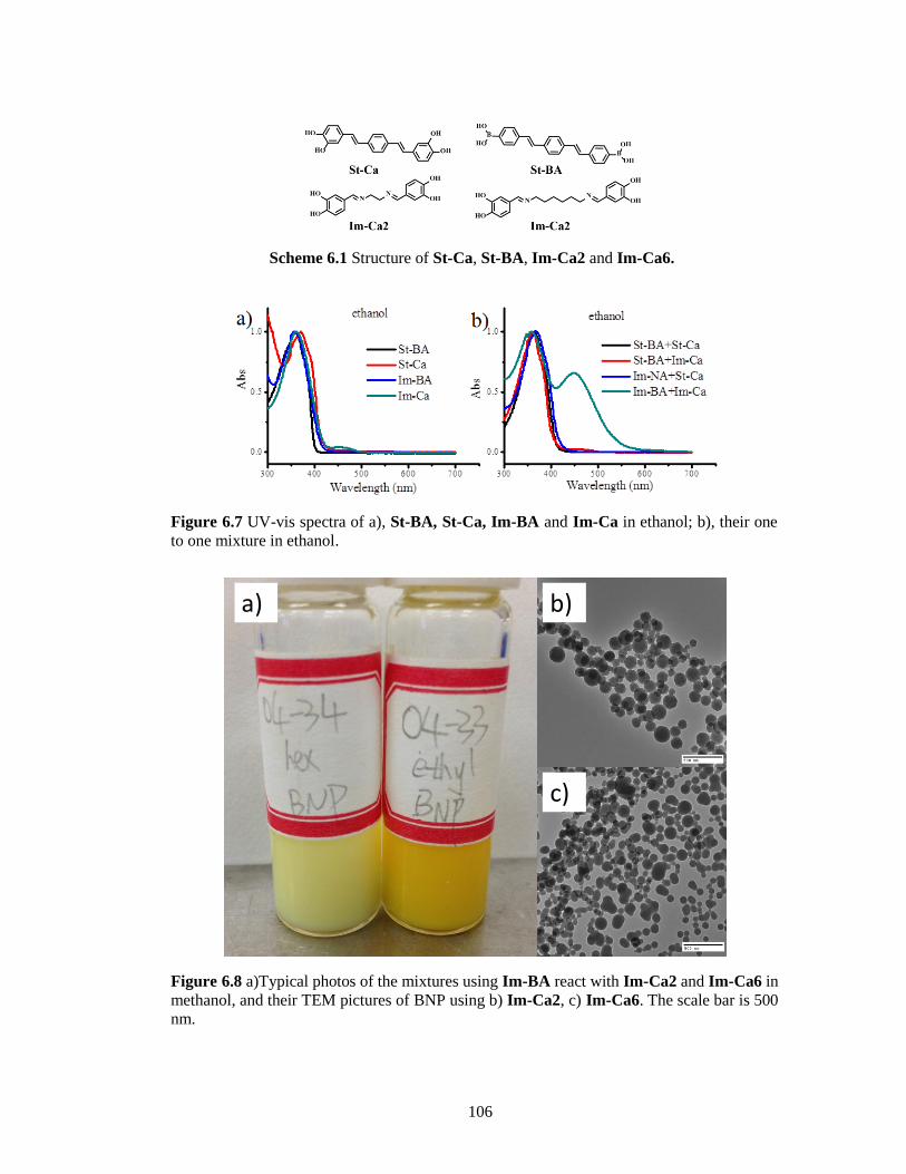
106
Figure 6.8 a)Typical photos of the mixtures using Im-BA react with Im-Ca2 and Im-Ca6 in
methanol, and their TEM pictures of BNP using b) Im-Ca2, c) Im-Ca6. The scale bar is 500
nm.
a) b)
c)
Figure 6.7 UV-vis spectra of a), St-BA, St-Ca, Im-BA and Im-Ca in ethanol; b), their one
to one mixture in ethanol.
Scheme 6.1 Structure of St-Ca, St-BA, Im-Ca2 and Im-Ca6.
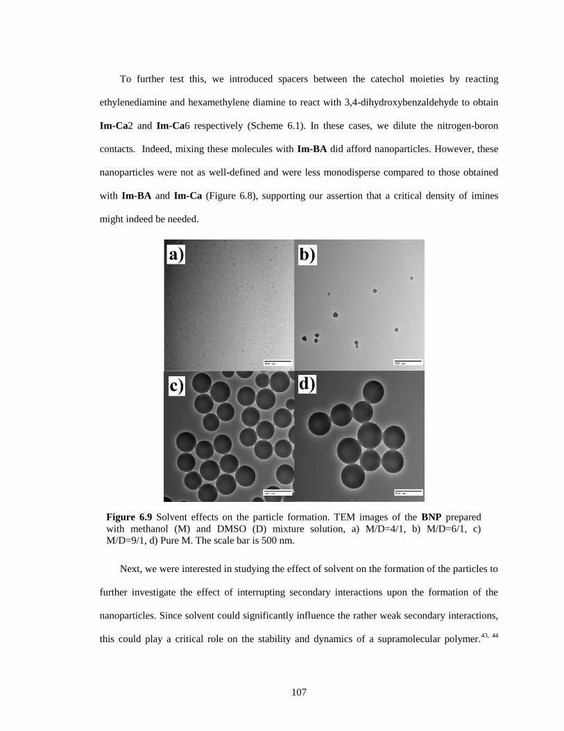
107
To further test this, we introduced spacers between the catechol moieties by reacting
ethylenediamine and hexamethylene diamine to react with 3,4-dihydroxybenzaldehyde to obtain
Im-Ca2 and Im-Ca6 respectively (Scheme 6.1). In these cases, we dilute the nitrogen-boron
contacts. Indeed, mixing these molecules with Im-BA did afford nanoparticles. However, these
nanoparticles were not as well-defined and were less monodisperse compared to those obtained
with Im-BA and Im-Ca (Figure 6.8), supporting our assertion that a critical density of imines
might indeed be needed.
Next, we were interested in studying the effect of solvent on the formation of the particles to
further investigate the effect of interrupting secondary interactions upon the formation of the
nanoparticles. Since solvent could significantly influence the rather weak secondary interactions,
this could play a critical role on the stability and dynamics of a supramolecular polymer.43, 44
Figure 6.9 Solvent effects on the particle formation. TEM images of the BNP prepared
with methanol (M) and DMSO (D) mixture solution, a) M/D=4/1, b) M/D=6/1, c)
M/D=9/1, d) Pure M. The scale bar is 500 nm.
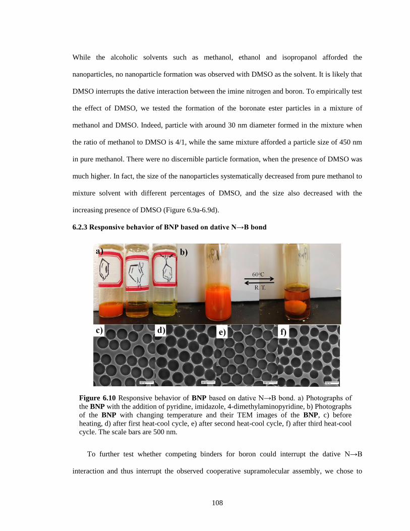
108
While the alcoholic solvents such as methanol, ethanol and isopropanol afforded the
nanoparticles, no nanoparticle formation was observed with DMSO as the solvent. It is likely that
DMSO interrupts the dative interaction between the imine nitrogen and boron. To empirically test
the effect of DMSO, we tested the formation of the boronate ester particles in a mixture of
methanol and DMSO. Indeed, particle with around 30 nm diameter formed in the mixture when
the ratio of methanol to DMSO is 4/1, while the same mixture afforded a particle size of 450 nm
in pure methanol. There were no discernible particle formation, when the presence of DMSO was
much higher. In fact, the size of the nanoparticles systematically decreased from pure methanol to
mixture solvent with different percentages of DMSO, and the size also decreased with the
increasing presence of DMSO (Figure 6.9a-6.9d).
6.2.3 Responsive behavior of BNP based on dative N→B bond
To further test whether competing binders for boron could interrupt the dative N→B
interaction and thus interrupt the observed cooperative supramolecular assembly, we chose to
Figure 6.10 Responsive behavior of BNP based on dative N→B bond. a) Photographs of
the BNP with the addition of pyridine, imidazole, 4-dimethylaminopyridine, b) Photographs
of the BNP with changing temperature and their TEM images of the BNP, c) before
heating, d) after first heat-cool cycle, e) after second heat-cool cycle, f) after third heat-cool
cycle. The scale bars are 500 nm.
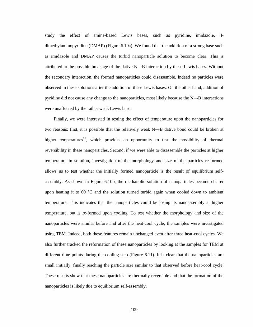
109
study the effect of amine-based Lewis bases, such as pyridine, imidazole, 4-
dimethylaminopyridine (DMAP) (Figure 6.10a). We found that the addition of a strong base such
as imidazole and DMAP causes the turbid nanoparticle solution to become clear. This is
attributed to the possible breakage of the dative N→B interaction by these Lewis bases. Without
the secondary interaction, the formed nanoparticles could disassemble. Indeed no particles were
observed in these solutions after the addition of these Lewis bases. On the other hand, addition of
pyridine did not cause any change to the nanoparticles, most likely because the N→B interactions
were unaffected by the rather weak Lewis base.
Finally, we were interested in testing the effect of temperature upon the nanoparticles for
two reasons: first, it is possible that the relatively weak N→B dative bond could be broken at
higher temperatures26
, which provides an opportunity to test the possibility of thermal
reversibility in these nanoparticles. Second, if we were able to disassemble the particles at higher
temperature in solution, investigation of the morphology and size of the particles re-formed
allows us to test whether the initially formed nanoparticle is the result of equilibrium self-
assembly. As shown in Figure 6.10b, the methanolic solution of nanoparticles became clearer
upon heating it to 60 °C and the solution turned turbid again when cooled down to ambient
temperature. This indicates that the nanoparticles could be losing its nanoassembly at higher
temperature, but is re-formed upon cooling. To test whether the morphology and size of the
nanoparticles were similar before and after the heat-cool cycle, the samples were investigated
using TEM. Indeed, both these features remain unchanged even after three heat-cool cycles. We
also further tracked the reformation of these nanoparticles by looking at the samples for TEM at
different time points during the cooling step (Figure 6.11). It is clear that the nanoparticles are
small initially, finally reaching the particle size similar to that observed before heat-cool cycle.
These results show that these nanoparticles are thermally reversible and that the formation of the
nanoparticles is likely due to equilibrium self-assembly.
110
6.3 Summary
In summary, we report on a supramolecular nanoparticle that is autonomously self-
assembled by conjugated polymers based on boronate ester. We have shown that: (i) the polymers
inducing nucleation for the formation of the particle involves a N-E polymerization step; (ii)
dative bond between the nitrogen in the imine building blocks and the boron in the boronate ester
is regarded as the necessary secondary interaction for the specific reaction; (iii) their particle size
could be easily adjusted by controlling the mixing concentration; (iv) imine density and solvent
can affect the particle formation; and (v) these boronate nanoparticles exhibit thermally
responsive disassembly behavior due to the relatively weak dative covalent N→B bond. To our
knowledge, this is the first time for a cooperative polymerization to use covalent dative bond as
Figure 6.11 TEM images of the BNP, a) before the heating and cooling at room
temperature for b) 1 min, c) 5 mins, d) 10 mins.
1 mins
5 mins 10 mins
a) b)
c) d)

111
the driving force. We believe that this will open new doors for autonomous self-assembly
strategies to achieve not only monodisperse, but also functionally diverse nanoassemblies.
6.4 Experimental
6.4.1 General Procedures
p-Phenylenediamine, ethylenediamine, hexamethylene diamine, 3,4-dihydroxybenzaldehyde,
4-formylphenylboronic acid, tert-butyldimethylsilyl chloride, imidazole, 4-
Dimethylaminopyridine, pinacol, 1,4-bis (bromomethyl) benzene, triethylphosphite and other
conventional reagents were obtained from commercial sources and were used as received unless
otherwise mentioned. 1H-NMR spectra were recorded on a Bruker 400 MHz NMR spectrometer
using the residual proton resonance of the solvent as the internal standard. 13
C-NMR spectra were
recorded on a Bruker 100 MHz NMR spectrometer using the carbon signal of the deuterated
solvent as the internal standard. Solid state 11
B-NMR spectra were recorded at 13 kHz on a
Bruker DSX300 spectrometer with 4 mm rotor. Mass spectra were taken on Bruker MicrOTOF-
II. Dynamic light scattering (DLS) measurements were performed by using a Malvern
Nanozetasizer. UV/visible absorption spectra of the samples in solutions were measured on a
Unico UV/Vis 2802PCS instrument. Transmission electron microscopy (TEM) images were
taken from JEOL 2000FX at 200 KV. FT-IR spectra were acquired in a Bruker Alpha-P
Diamond ATR-FTIR. Scanning electron microscopy (SEM) images were taken from FESEM
Magellan 400. Thermogravimetric analysis (TGA) was performed in a nitrogen atmosphere on a
DuPont TGA 2950 at a heating rate of 10 °C/min.
6.4.2 Synthesis of Im-Ca
Scheme 6.2 Synthesis of Im-Ca.
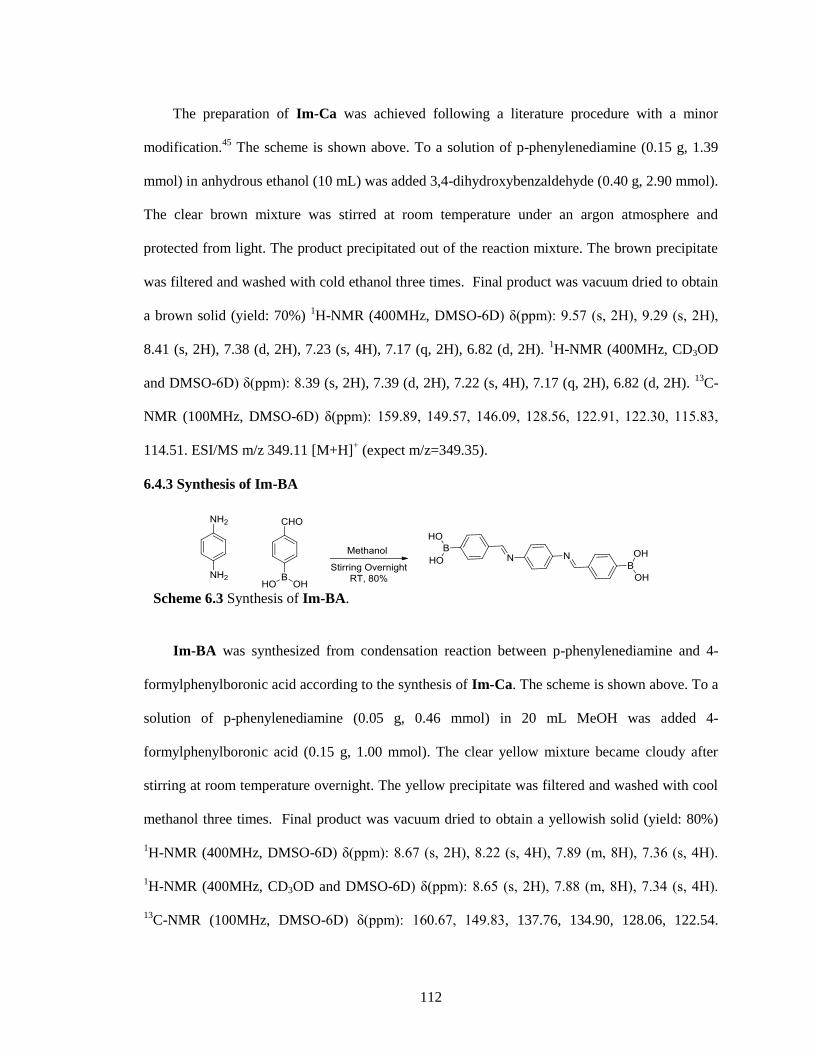
112
The preparation of Im-Ca was achieved following a literature procedure with a minor
modification.45
The scheme is shown above. To a solution of p-phenylenediamine (0.15 g, 1.39
mmol) in anhydrous ethanol (10 mL) was added 3,4-dihydroxybenzaldehyde (0.40 g, 2.90 mmol).
The clear brown mixture was stirred at room temperature under an argon atmosphere and
protected from light. The product precipitated out of the reaction mixture. The brown precipitate
was filtered and washed with cold ethanol three times. Final product was vacuum dried to obtain
a brown solid (yield: 70%) 1H-NMR (400MHz, DMSO-6D) δ(ppm): 9.57 (s, 2H), 9.29 (s, 2H),
8.41 (s, 2H), 7.38 (d, 2H), 7.23 (s, 4H), 7.17 (q, 2H), 6.82 (d, 2H). 1H-NMR (400MHz, CD3OD
and DMSO-6D) δ(ppm): 8.39 (s, 2H), 7.39 (d, 2H), 7.22 (s, 4H), 7.17 (q, 2H), 6.82 (d, 2H). 13
C-
NMR (100MHz, DMSO-6D) δ(ppm): 159.89, 149.57, 146.09, 128.56, 122.91, 122.30, 115.83,
114.51. ESI/MS m/z 349.11 [M+H]+ (expect m/z=349.35).
6.4.3 Synthesis of Im-BA
Im-BA was synthesized from condensation reaction between p-phenylenediamine and 4-
formylphenylboronic acid according to the synthesis of Im-Ca. The scheme is shown above. To a
solution of p-phenylenediamine (0.05 g, 0.46 mmol) in 20 mL MeOH was added 4-
formylphenylboronic acid (0.15 g, 1.00 mmol). The clear yellow mixture became cloudy after
stirring at room temperature overnight. The yellow precipitate was filtered and washed with cool
methanol three times. Final product was vacuum dried to obtain a yellowish solid (yield: 80%)
1H-NMR (400MHz, DMSO-6D) δ(ppm): 8.67 (s, 2H), 8.22 (s, 4H), 7.89 (m, 8H), 7.36 (s, 4H).
1H-NMR (400MHz, CD3OD and DMSO-6D) δ(ppm): 8.65 (s, 2H), 7.88 (m, 8H), 7.34 (s, 4H).
13C-NMR (100MHz, DMSO-6D) δ(ppm): 160.67, 149.83, 137.76, 134.90, 128.06, 122.54.
Scheme 6.3 Synthesis of Im-BA.
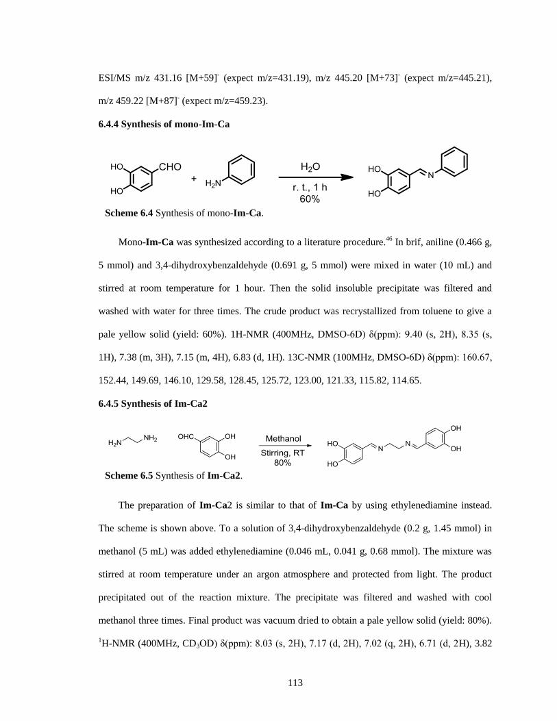
113
ESI/MS m/z 431.16 [M+59]- (expect m/z=431.19), m/z 445.20 [M+73]
- (expect m/z=445.21),
m/z 459.22 [M+87]- (expect m/z=459.23).
6.4.4 Synthesis of mono-Im-Ca
Mono-Im-Ca was synthesized according to a literature procedure.46
In brif, aniline (0.466 g,
5 mmol) and 3,4-dihydroxybenzaldehyde (0.691 g, 5 mmol) were mixed in water (10 mL) and
stirred at room temperature for 1 hour. Then the solid insoluble precipitate was filtered and
washed with water for three times. The crude product was recrystallized from toluene to give a
pale yellow solid (yield: 60%). 1H-NMR (400MHz, DMSO-6D) δ(ppm): 9.40 (s, 2H), 8.35 (s,
1H), 7.38 (m, 3H), 7.15 (m, 4H), 6.83 (d, 1H). 13C-NMR (100MHz, DMSO-6D) δ(ppm): 160.67,
152.44, 149.69, 146.10, 129.58, 128.45, 125.72, 123.00, 121.33, 115.82, 114.65.
6.4.5 Synthesis of Im-Ca2
The preparation of Im-Ca2 is similar to that of Im-Ca by using ethylenediamine instead.
The scheme is shown above. To a solution of 3,4-dihydroxybenzaldehyde (0.2 g, 1.45 mmol) in
methanol (5 mL) was added ethylenediamine (0.046 mL, 0.041 g, 0.68 mmol). The mixture was
stirred at room temperature under an argon atmosphere and protected from light. The product
precipitated out of the reaction mixture. The precipitate was filtered and washed with cool
methanol three times. Final product was vacuum dried to obtain a pale yellow solid (yield: 80%).
1H-NMR (400MHz, CD3OD) δ(ppm): 8.03 (s, 2H), 7.17 (d, 2H), 7.02 (q, 2H), 6.71 (d, 2H), 3.82
Scheme 6.5 Synthesis of Im-Ca2.
Scheme 6.4 Synthesis of mono-Im-Ca.
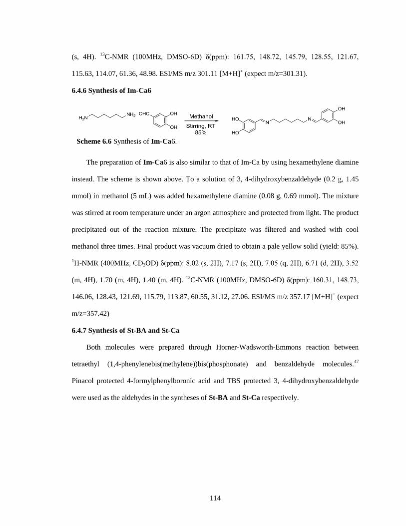
114
(s, 4H). 13
C-NMR (100MHz, DMSO-6D) δ(ppm): 161.75, 148.72, 145.79, 128.55, 121.67,
115.63, 114.07, 61.36, 48.98. ESI/MS m/z 301.11 [M+H]+ (expect m/z=301.31).
6.4.6 Synthesis of Im-Ca6
The preparation of Im-Ca6 is also similar to that of Im-Ca by using hexamethylene diamine
instead. The scheme is shown above. To a solution of 3, 4-dihydroxybenzaldehyde (0.2 g, 1.45
mmol) in methanol (5 mL) was added hexamethylene diamine (0.08 g, 0.69 mmol). The mixture
was stirred at room temperature under an argon atmosphere and protected from light. The product
precipitated out of the reaction mixture. The precipitate was filtered and washed with cool
methanol three times. Final product was vacuum dried to obtain a pale yellow solid (yield: 85%).
1H-NMR (400MHz, CD3OD) δ(ppm): 8.02 (s, 2H), 7.17 (s, 2H), 7.05 (q, 2H), 6.71 (d, 2H), 3.52
(m, 4H), 1.70 (m, 4H), 1.40 (m, 4H). 13
C-NMR (100MHz, DMSO-6D) δ(ppm): 160.31, 148.73,
146.06, 128.43, 121.69, 115.79, 113.87, 60.55, 31.12, 27.06. ESI/MS m/z 357.17 [M+H]+ (expect
m/z=357.42)
6.4.7 Synthesis of St-BA and St-Ca
Both molecules were prepared through Horner-Wadsworth-Emmons reaction between
tetraethyl (1,4-phenylenebis(methylene))bis(phosphonate) and benzaldehyde molecules.47
Pinacol protected 4-formylphenylboronic acid and TBS protected 3, 4-dihydroxybenzaldehyde
were used as the aldehydes in the syntheses of St-BA and St-Ca respectively.
Scheme 6.6 Synthesis of Im-Ca6.
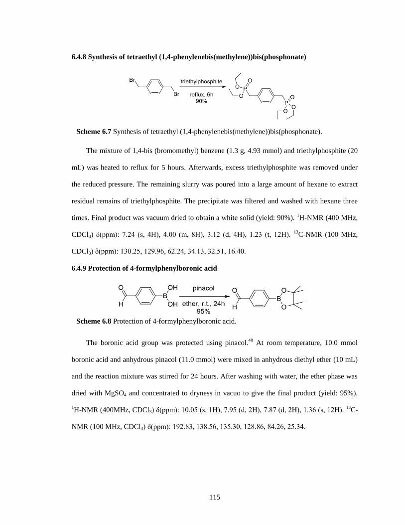
115
6.4.8 Synthesis of tetraethyl (1,4-phenylenebis(methylene))bis(phosphonate)
The mixture of 1,4-bis (bromomethyl) benzene (1.3 g, 4.93 mmol) and triethylphosphite (20
mL) was heated to reflux for 5 hours. Afterwards, excess triethylphosphite was removed under
the reduced pressure. The remaining slurry was poured into a large amount of hexane to extract
residual remains of triethylphosphite. The precipitate was filtered and washed with hexane three
times. Final product was vacuum dried to obtain a white solid (yield: 90%). 1H-NMR (400 MHz,
CDCl3) δ(ppm): 7.24 (s, 4H), 4.00 (m, 8H), 3.12 (d, 4H), 1.23 (t, 12H). 13
C-NMR (100 MHz,
CDCl3) δ(ppm): 130.25, 129.96, 62.24, 34.13, 32.51, 16.40.
6.4.9 Protection of 4-formylphenylboronic acid
The boronic acid group was protected using pinacol.48
At room temperature, 10.0 mmol
boronic acid and anhydrous pinacol (11.0 mmol) were mixed in anhydrous diethyl ether (10 mL)
and the reaction mixture was stirred for 24 hours. After washing with water, the ether phase was
dried with MgSO4 and concentrated to dryness in vacuo to give the final product (yield: 95%).
1H-NMR (400MHz, CDCl3) δ(ppm): 10.05 (s, 1H), 7.95 (d, 2H), 7.87 (d, 2H), 1.36 (s, 12H).
13C-
NMR (100 MHz, CDCl3) δ(ppm): 192.83, 138.56, 135.30, 128.86, 84.26, 25.34.
Scheme 6.8 Protection of 4-formylphenylboronic acid.
Scheme 6.7 Synthesis of tetraethyl (1,4-phenylenebis(methylene))bis(phosphonate).
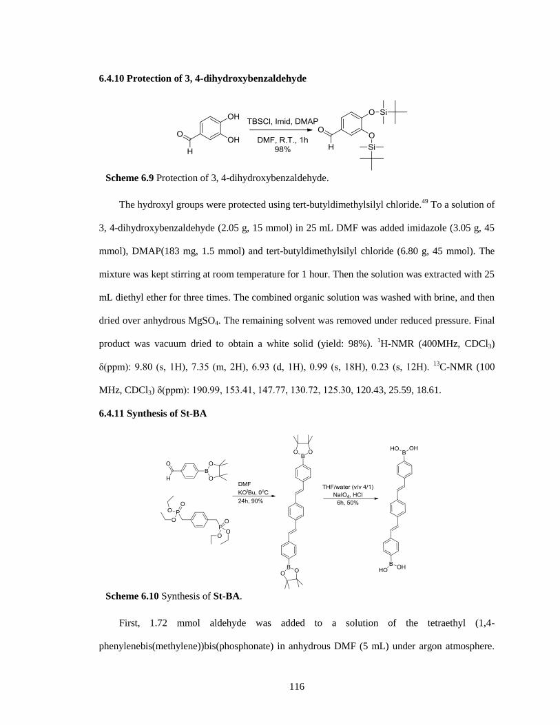
116
6.4.10 Protection of 3, 4-dihydroxybenzaldehyde
The hydroxyl groups were protected using tert-butyldimethylsilyl chloride.49
To a solution of
3, 4-dihydroxybenzaldehyde (2.05 g, 15 mmol) in 25 mL DMF was added imidazole (3.05 g, 45
mmol), DMAP(183 mg, 1.5 mmol) and tert-butyldimethylsilyl chloride (6.80 g, 45 mmol). The
mixture was kept stirring at room temperature for 1 hour. Then the solution was extracted with 25
mL diethyl ether for three times. The combined organic solution was washed with brine, and then
dried over anhydrous MgSO4. The remaining solvent was removed under reduced pressure. Final
product was vacuum dried to obtain a white solid (yield: 98%). 1H-NMR (400MHz, CDCl3)
δ(ppm): 9.80 (s, 1H), 7.35 (m, 2H), 6.93 (d, 1H), 0.99 (s, 18H), 0.23 (s, 12H). 13
C-NMR (100
MHz, CDCl3) δ(ppm): 190.99, 153.41, 147.77, 130.72, 125.30, 120.43, 25.59, 18.61.
6.4.11 Synthesis of St-BA
First, 1.72 mmol aldehyde was added to a solution of the tetraethyl (1,4-
phenylenebis(methylene))bis(phosphonate) in anhydrous DMF (5 mL) under argon atmosphere.
Scheme 6.10 Synthesis of St-BA.
Scheme 6.9 Protection of 3, 4-dihydroxybenzaldehyde.
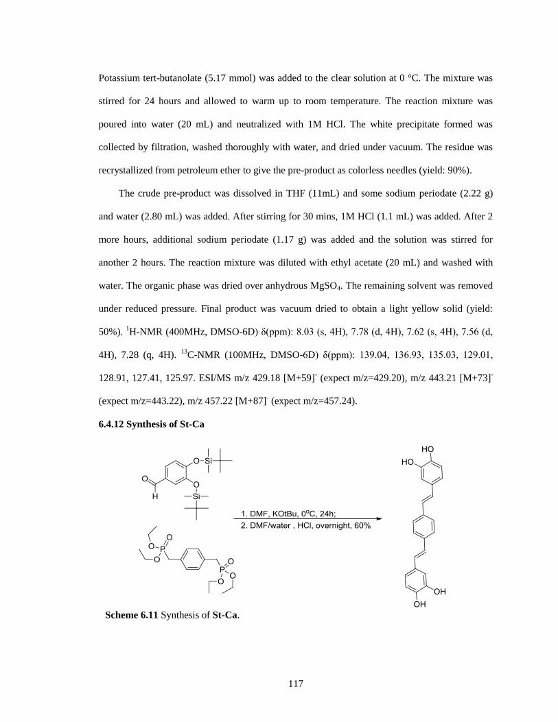
117
Potassium tert-butanolate (5.17 mmol) was added to the clear solution at 0 °C. The mixture was
stirred for 24 hours and allowed to warm up to room temperature. The reaction mixture was
poured into water (20 mL) and neutralized with 1M HCl. The white precipitate formed was
collected by filtration, washed thoroughly with water, and dried under vacuum. The residue was
recrystallized from petroleum ether to give the pre-product as colorless needles (yield: 90%).
The crude pre-product was dissolved in THF (11mL) and some sodium periodate (2.22 g)
and water (2.80 mL) was added. After stirring for 30 mins, 1M HCl (1.1 mL) was added. After 2
more hours, additional sodium periodate (1.17 g) was added and the solution was stirred for
another 2 hours. The reaction mixture was diluted with ethyl acetate (20 mL) and washed with
water. The organic phase was dried over anhydrous MgSO4. The remaining solvent was removed
under reduced pressure. Final product was vacuum dried to obtain a light yellow solid (yield:
50%). 1H-NMR (400MHz, DMSO-6D) δ(ppm): 8.03 (s, 4H), 7.78 (d, 4H), 7.62 (s, 4H), 7.56 (d,
4H), 7.28 (q, 4H). 13
C-NMR (100MHz, DMSO-6D) δ(ppm): 139.04, 136.93, 135.03, 129.01,
128.91, 127.41, 125.97. ESI/MS m/z 429.18 [M+59]- (expect m/z=429.20), m/z 443.21 [M+73]
-
(expect m/z=443.22), m/z 457.22 [M+87]- (expect m/z=457.24).
6.4.12 Synthesis of St-Ca
Scheme 6.11 Synthesis of St-Ca.
118
The preparation of St-Ca is similar to that of St-BA by using protected 3, 4-
dihydroxybenzaldehyde instead. 1.06 mmol aldehyde was added to a solution of tetraethyl (1,4-
phenylenebis(methylene))bis(phosphonate) in anhydrous DMF (5 mL) under argon atmosphere.
Potassium tert-butoxide (3.17 mmol) was added to the clear solution at 0 °C. The mixture was
stirred for 24 hours and allowed to warm up to room temperature. The reaction mixture was
poured into water (20 mL) and neutralized with 1M HCl. The mixture was kept stirring overnight.
The white precipitate formed was collected by filtration, washed thoroughly with water, and dried
under vacuum. The residue was recrystallized from petrolether to give the pre-product as
colorless needles (yield: 60%). 1H-NMR (400MHz, DMSO-6D) δ(ppm): 9.10 (s, 2H), 8.94 (s,
2H), 7.49 (s, 4H), 6.90 (m, 8H), 6.72 (d, 2H), 13
C-NMR (100MHz, DMSO-6D) δ(ppm): 146.11,
145.84, 136.60, 129.15, 128.85, 126.80, 125.13, 119.10, 116.13, 113.68. ESI/MS m/z 369.10
[M+Na]+ (expect m/z=369.38).
6.4.13 Preparation of Nanoparticles via dropwise addition
First, Im-Ca and Im-BA were dissolved in methanol with the help of heating. After cooling
down by setting at room temperature, one solution was added into the solution drop by drop, the
mixture solution because deep orange and suspension was finally got. The resultant particles were
purified by repeating centrifugation, decantation and resuspension. The composite particles were
then dried in a vacuum oven at 50 oC overnight.
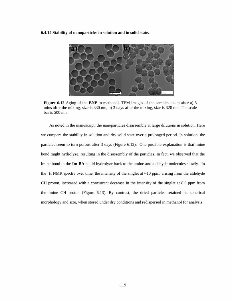
119
6.4.14 Stability of nanoparticles in solution and in solid state.
As noted in the manuscript, the nanoparticles disassemble at large dilutions in solution. Here
we compare the stability in solution and dry solid state over a prolonged period. In solution, the
particles seem to turn porous after 3 days (Figure 6.12). One possible explanation is that imine
bond might hydrolyze, resulting in the disassembly of the particles. In fact, we observed that the
imine bond in the Im-BA could hydrolyze back to the amine and aldehyde molecules slowly. In
the 1H NMR spectra over time, the intensity of the singlet at ~10 ppm, arising from the aldehyde
CH proton, increased with a concurrent decrease in the intensity of the singlet at 8.6 ppm from
the imine CH proton (Figure 6.13). By contrast, the dried particles retained its spherical
morphology and size, when stored under dry conditions and redispersed in methanol for analysis.
Figure 6.12 Aging of the BNP in methanol. TEM images of the samples taken after a) 5
mins after the mixing, size is 330 nm, b) 3 days after the mixing, size is 320 nm. The scale
bar is 500 nm.
a) b)
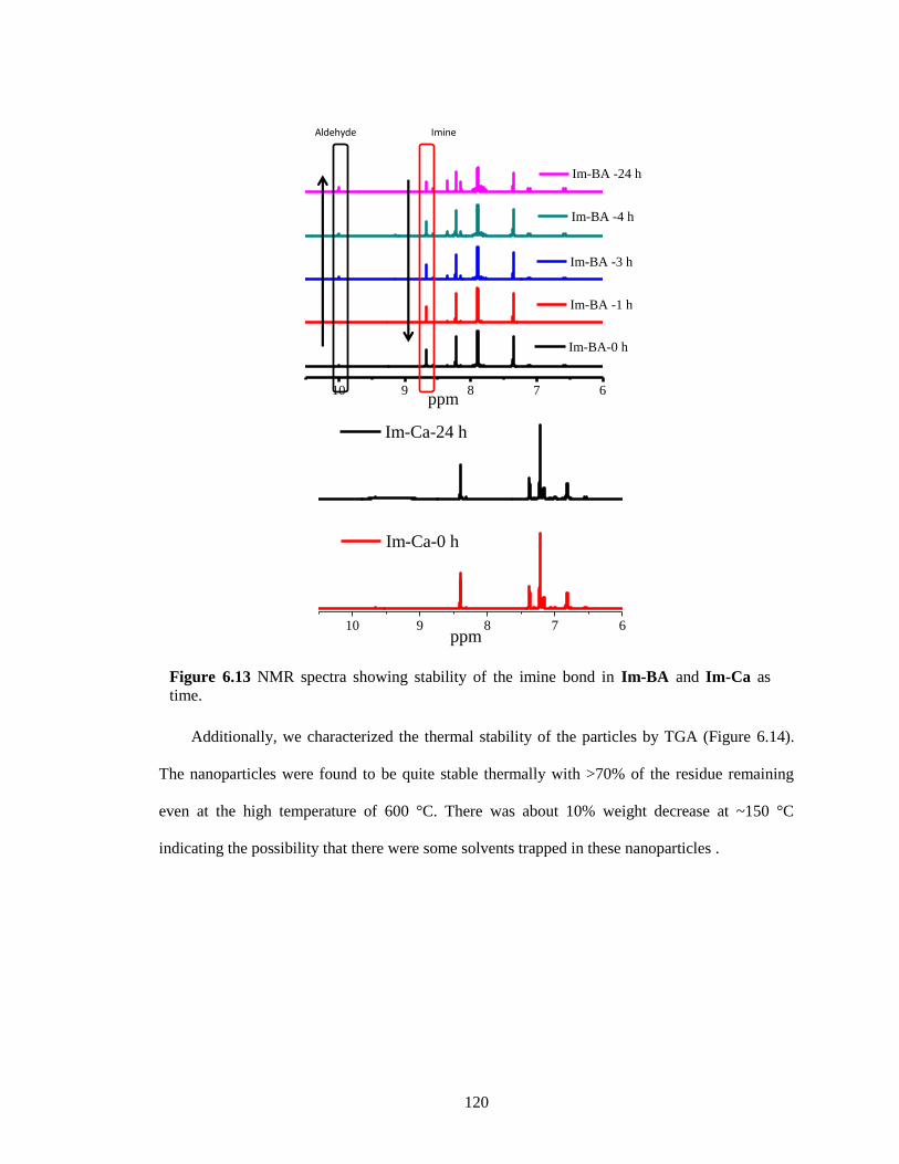
120
Additionally, we characterized the thermal stability of the particles by TGA (Figure 6.14).
The nanoparticles were found to be quite stable thermally with >70% of the residue remaining
even at the high temperature of 600 °C. There was about 10% weight decrease at ~150 °C
indicating the possibility that there were some solvents trapped in these nanoparticles .
Figure 6.13 NMR spectra showing stability of the imine bond in Im-BA and Im-Ca as
time.
10 9 8 7 6
Im-BA -24 h
Im-BA -4 h
Im-BA -3 h
Im-BA -1 h
Im-BA-0 h
ppm
Aldehyde Imine
10 9 8 7 6
Im-Ca-0 h
ppm
Im-Ca-24 h
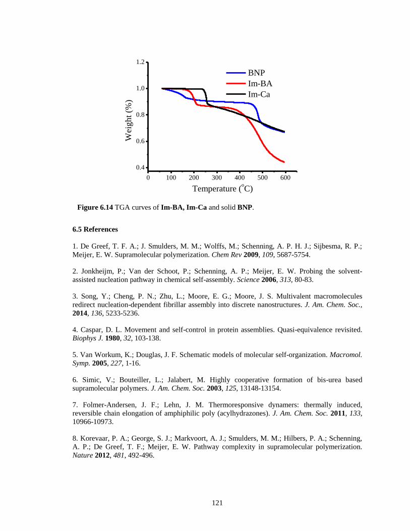
121
6.5 References
1. De Greef, T. F. A.; J. Smulders, M. M.; Wolffs, M.; Schenning, A. P. H. J.; Sijbesma, R. P.;
Meijer, E. W. Supramolecular polymerization. Chem Rev 2009, 109, 5687-5754.
2. Jonkheijm, P.; Van der Schoot, P.; Schenning, A. P.; Meijer, E. W. Probing the solvent-
assisted nucleation pathway in chemical self-assembly. Science 2006, 313, 80-83.
3. Song, Y.; Cheng, P. N.; Zhu, L.; Moore, E. G.; Moore, J. S. Multivalent macromolecules
redirect nucleation-dependent fibrillar assembly into discrete nanostructures. J. Am. Chem. Soc.,
2014, 136, 5233-5236.
4. Caspar, D. L. Movement and self-control in protein assemblies. Quasi-equivalence revisited.
Biophys J. 1980, 32, 103-138.
5. Van Workum, K.; Douglas, J. F. Schematic models of molecular self-organization. Macromol.
Symp. 2005, 227, 1-16.
6. Simic, V.; Bouteiller, L.; Jalabert, M. Highly cooperative formation of bis-urea based
supramolecular polymers. J. Am. Chem. Soc. 2003, 125, 13148-13154.
7. Folmer-Andersen, J. F.; Lehn, J. M. Thermoresponsive dynamers: thermally induced,
reversible chain elongation of amphiphilic poly (acylhydrazones). J. Am. Chem. Soc. 2011, 133,
10966-10973.
8. Korevaar, P. A.; George, S. J.; Markvoort, A. J.; Smulders, M. M.; Hilbers, P. A.; Schenning,
A. P.; De Greef, T. F.; Meijer, E. W. Pathway complexity in supramolecular polymerization.
Nature 2012, 481, 492-496.
Figure 6.14 TGA curves of Im-BA, Im-Ca and solid BNP.
0 100 200 300 400 500 600
0.4
0.6
0.8
1.0
1.2
Wei
gh
t (%
)
Temperature (oC)
BNP
Im-BA
Im-Ca
122
9. Zhao, D.; Moore, J. S. Nucleation-elongation polymerization under imbalanced stoichiometry.
J. Am. Chem. Soc. 2003, 125, 16294-16299.
10. Iwasawa, N.; Takahagi, H. Boronic esters as a system for crystallization-induced dynamic
self-assembly equipped with an on-off switch for equilibration. J. Am. Chem. Soc. 2007, 129,
7754-7755.
11. Christinat, N.; Scopelliti, R.; Severin, K. Multicomponent assembly of boronic acid based
macrocycles and cages. Angew. Chem. Int. Ed. 2008, 47, 1848-1852.
12. Nishimura, N.; Kobayashi, K. Self-assembly of a cavitand-based capsule by dynamic boronic
ester formation. Angew. Chem. Int. Ed. 2008, 47, 6255-6258.
13. Nishimura, N.; Yoza, K.; Kobayashi, K. Guest-encapsulation properties of a self-assembled
capsule by dynamic boronic ester bonds. J. Am. Chem. Soc. 2009, 132, 777-790.
14. El-Kaderi, H. M.; Hunt, J. R.; Mendoza-Cortes, J. L.; Cote, A. P.; Taylor, R. E.; O'Keeffe, M.;
Yaghi, O. M. Designed synthesis of 3D covalent organic frameworks. Science 2007, 316, 268-
272.
15. Colson, J. W.; Woll, A. R.; Mukherjee, A.; Levendorf, M. P.; Spitler, E. L.; Shields, V. B.;
Spencer, M. G.; Park, J.; Dichtel, W. R. Oriented 2D covalent organic framework thin films on
single-layer grapheme. Science 2011, 332, 228-231.
16. Nakazawa, I.; Suda, S.; Masuda, M.; Asai, M.; Shimizu, T. pH-Dependent reversible
polymers formed from cyclic sugar-and aromatic boronic acid-based bolaamphiphiles. Chem.
Commun., 2000, 10, 881-882.
17. Niu, W.; Smith, M. D.; Lavigne, J. J. Self-assembling poly (dioxaborole) s as blue-emissive
materials. J. Am. Chem. Soc. 2006, 128, 16466-16467.
18. Bapat, A. P.; Roy, D.; Ray, J. G.; Savin, D. A.; Sumerlin, B. S. Dynamic-covalent
macromolecular stars with boronic ester linkages. J. Am. Chem. Soc. 2011, 133, 19832-19838.
19. Nishiyabu, R.; Teraoka, S.; Matsushima, Y.; Kubo, Y. Fabrication of soft submicrospheres by
sequential boronate esterification and their dynamic behavior. ChemPlusChem 2012, 77, 201.
20. Höpfl, H. J. The tetrahedral character of the boron atom newly defined—a useful tool to
evaluate the N→ B bond. Organomet. Chem. 1999, 581, 129-149.
21. Zhu, L.; Shabbir, S. H.; Gray, M.; Lynch, V. M.; Sorey, S.; Anslyn, E. V. A structural
investigation of the NB interaction in an o-(N, N-dialkylaminomethyl) arylboronate system. J.
Am. Chem. Soc. 2006, 128, 1222-1232.
22. Franzen, S.; Ni, W.; Wang, B. Study of the mechanism of electron-transfer quenching by
boron-nitrogen adducts in fluorescent sensors. J. Phys. Chem. B 2003, 107, 12942-12948.
23. Ni, W.; Kaur, G.; Springsteen, G.; Wang, B.; Franzen, S. Regulating the fluorescence
intensity of an anthracene boronic acid system: a B–N bond or a hydrolysis mechanism? Bioorg.
Chem., 2004, 32, 571-581.
123
24. Christinat, N.; Scopelliti, R.; Severin, K. Multicomponent assembly of boron-based dendritic
nanostructures. J. Org. Chem. 2007, 72, 2192-2200.
25. Cruz-Huerta, J.; Salazar-Mendoza, D.; Hernandez-Paredes, J.; Hernandez Ahuactzi, I. F.;
Hopfl, H. N-containing boronic esters as self-complementary building blocks for the assembly of
2D and 3D molecular networks. Chem. Commun. 2012, 48, 4241-4243.
26. Sheepwash, E.; Krampl, V.; Scopelliti, R.; Sereda, O.; Neels, A.; Severin, K. Molecular
networks based on dative boron-nitrogen bonds. Angew. Chem. Int. Ed. 2011, 50, 3034-3037.
27. Zhang, Y.; Li, M.; Chandrasekaran, S.; Gao, X.; Fang, X.; Lee, H. W.; Hardcastle, K.; Yang,
J.; Wang, B. A unique quinolineboronic acid-based supramolecular structure that relies on double
intermolecular B-N bonds for self-assembly in solid state and in solution. Tetrahedron, 2007, 63,
3287-3292.
28. Kiviranta, P. H.; Leppänen, J.; Kyrylenko, S.; Salo, H. S.; Lahtela-Kakkonen, M.; Tervo, A. J.;
Wittekindt, C.; Suuronen, T.; Kuusisto, E.; Järvinen, T. N, N'-bisbenzylidenebenzene-1, 4-
diamines and N, N'-bisbenzylidenenaphthalene-1, 4-diamines as sirtuin type 2 (SIRT2) inhibitors.
J. Med. Chem. 2006, 49, 7907-7911.
29. Spitler, E. L.; Dichtel, W. R. Lewis acid-catalysed formation of two-dimensional
phthalocyanine covalent organic frameworks. Nat. Chem., 2010, 2, 672-677.
30. Spitler, E. L.; Giovino, M. R.; White, S. L.; Dichtel, W. R. A mechanistic study of Lewis
acid-catalyzed covalent organic framework formation. Chem. Sci., 2011, 2, 1588-1593.
31. Brewer, S. H.; Allen, A. M.; Lappi, S. E.; Chasse, T. L.; Briggman, K. A.; Gorman, C. B.;
Franzen, S. Infrared detection of a phenylboronic acid terminated alkane thiol monolayer on gold
surfaces. Langmuir 2004, 20, 5512-5520.
32. Li, H.; Jäkle, F. Universal scaffold for fluorescent conjugated organoborane polymers. Angew.
Chem. Int. Ed., 2009, 48, 2313-2316.
33. Jäkle, F.; Lewis acidic organoboron polymers. Coord. Chem. Rev., 2006, 250, 1107-1121.
34. Hall, L. W.; Odom, J. D.; Ellis, P. D. Nuclear magnetic resonance investigation of the nature
of the boron-carbon bond in some vinylboranes. J. Am. Chem. Soc. 1975, 97, 4527-4531.
35. Liu, W.; Pink, M.; Lee, D. Conjugated polymer sensors built on π-extended borasiloxane
cages. J. Am. Chem. Soc. 2009, 131, 8703-8707.
36. Bachler, V.; Metzler-Nolte, N. The π-electron-accepting ability of the boron atom in
ethynylboranes and related compounds-an approximate weight computation for resonance
structures. Eur. J. Inorg. Chem. 1998, 6, 733-744;
37. Zhao, D.; Moore, J. S. Reversible polymerization driven by folding. J. Am. Chem. Soc. 2002,
124, 9996-9997.
38. Oh, K.; Jeong, K.-S.; Moore, J. S. Folding-driven synthesis of oligomers. Nature 2001, 414,
889.
124
39. LaMer, V. K.; Dinegar, R. H. Theory, production and mechanism of formation of
monodispersed hydrosols. J. Am. Chem. Soc. 1950, 72, 4847-4854.
40. Oh, M.; Mirkin, C. A. Chemically tailorable colloidal particles from infinite coordination
polymers. Nature 2005, 438, 651-654.
41. Maeda, H.; Hasegawa, M.; Hashimoto, T.; Kakimoto, T.; Nishio, S.; Nakanishi, T. Nanoscale
spherical architectures fabricated by metal coordination of multiple dipyrrin moieties. J. Am.
Chem. Soc. 2006, 128, 10024-10025.
42. Niu, W.; Smith, M. D.; Lavigne, J. J. Substituent effects on the structure and supramolecular
assembly of bis (dioxaborole) s derived from 1, 2, 4, 5-tetrahydroxybenzene. Cryst. Growth Des.
2006, 6, 1274-1277.
43. Korevaar, P. A.; Schaefer, C.; De Greef, T. F.; Meijer, E. W. Controlling chemical self-
assembly by solvent-dependent dynamics. J. Am. Chem. Soc. 2012, 134, 13482-13491.
44. England, J. L.; Haran, G. Role of solvation effects in protein denaturation: from
thermodynamics to single molecules and back. Annu. Rev. Phys. Chem. 2011, 62, 257-277.
45. Kiviranta, P. H.; Leppänen, J.; Kyrylenko, S.; Salo, H. S.; Lahtela-Kakkonen, M.; Tervo, A. J.;
Wittekindt, C.; Suuronen, T.; Kuusisto, E.; Järvinen, T. N, N'-bisbenzylidenebenzene-1, 4-
diamines and N, N'-bisbenzylidenenaphthalene-1, 4-diamines as sirtuin type 2 (SIRT2) inhibitors.
J. Med. Chem. 2006, 49, 7907-7911.
46. Cheng, L. X.; Tang, J. J.; Luo, H.; Jin, X. L.; Dai, F.; Yang, J.; Qian, Y. P.; Li, X. Z.; Zhou, B.
Antioxidant and antiproliferative activities of hydroxyl-substituted Schiff bases. Bioorg. Med.
Chem. Lett. 2010, 20, 2417-2420.
47. Ha, Y. M.; Lee, H. J.; Park, D.; Jeong, H. O.; Park, J. Y.; Park, Y. J.; Lee, K. J.; Lee, J. Y.;
Moon, H. R.; Chung, H. Y. Molecular docking studies of (1E,3E,5E)-1,6-bis(substituted
phenyl)hexa-1,3,5-triene and 1,4-bis(substituted trans-styryl)benzene analogs as novel tyrosinase
inhibitors. Biol. Pharm. Bull. 2012, 36, 55-65.
48. Oehlke, A.; Auer, A. A.; Jahre, I.; Walfort, B.; Rüffer, T.; Zoufalá, P.; Lang, H.; Spange, S.
Nitro-substituted stilbeneboronate pinacol esters and their fluoro-adducts. fluoride ion induced
polarity enhancement of arylboronate esters. J. Org. Chem. 2007, 72, 4328-4339.
49. Uchiyama, M.; Ozawa, H.; Takuma, K.; Matsumoto, Y.; Yonehara, M.; Hiroya, K.; Sakamoto,
T. Regiocontrolled intramolecular cyclizations of carboxylic acids to carbon-carbon triple bonds
promoted by acid or base catalyst. Org. Lett. 2006, 8, 5517-5520.
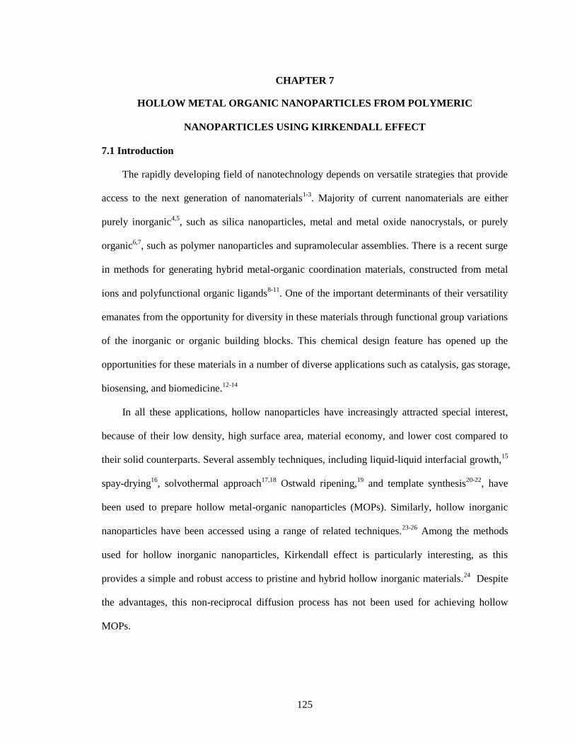
125
CHAPTER 7
HOLLOW METAL ORGANIC NANOPARTICLES FROM POLYMERIC
NANOPARTICLES USING KIRKENDALL EFFECT
7.1 Introduction
The rapidly developing field of nanotechnology depends on versatile strategies that provide
access to the next generation of nanomaterials1-3
. Majority of current nanomaterials are either
purely inorganic4,5
, such as silica nanoparticles, metal and metal oxide nanocrystals, or purely
organic6,7
, such as polymer nanoparticles and supramolecular assemblies. There is a recent surge
in methods for generating hybrid metal-organic coordination materials, constructed from metal
ions and polyfunctional organic ligands8-11
. One of the important determinants of their versatility
emanates from the opportunity for diversity in these materials through functional group variations
of the inorganic or organic building blocks. This chemical design feature has opened up the
opportunities for these materials in a number of diverse applications such as catalysis, gas storage,
biosensing, and biomedicine.12-14
In all these applications, hollow nanoparticles have increasingly attracted special interest,
because of their low density, high surface area, material economy, and lower cost compared to
their solid counterparts. Several assembly techniques, including liquid-liquid interfacial growth,15
spay-drying16
, solvothermal approach17,18
Ostwald ripening,19
and template synthesis20-22
, have
been used to prepare hollow metal-organic nanoparticles (MOPs). Similarly, hollow inorganic
nanoparticles have been accessed using a range of related techniques.23-26
Among the methods
used for hollow inorganic nanoparticles, Kirkendall effect is particularly interesting, as this
provides a simple and robust access to pristine and hybrid hollow inorganic materials.24
Despite
the advantages, this non-reciprocal diffusion process has not been used for achieving hollow
MOPs.
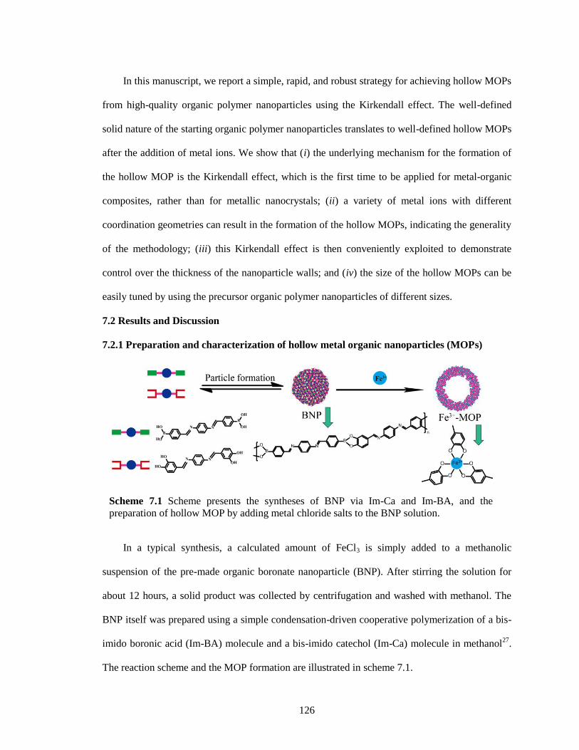
126
In this manuscript, we report a simple, rapid, and robust strategy for achieving hollow MOPs
from high-quality organic polymer nanoparticles using the Kirkendall effect. The well-defined
solid nature of the starting organic polymer nanoparticles translates to well-defined hollow MOPs
after the addition of metal ions. We show that (i) the underlying mechanism for the formation of
the hollow MOP is the Kirkendall effect, which is the first time to be applied for metal-organic
composites, rather than for metallic nanocrystals; (ii) a variety of metal ions with different
coordination geometries can result in the formation of the hollow MOPs, indicating the generality
of the methodology; (iii) this Kirkendall effect is then conveniently exploited to demonstrate
control over the thickness of the nanoparticle walls; and (iv) the size of the hollow MOPs can be
easily tuned by using the precursor organic polymer nanoparticles of different sizes.
7.2 Results and Discussion
7.2.1 Preparation and characterization of hollow metal organic nanoparticles (MOPs)
In a typical synthesis, a calculated amount of FeCl3 is simply added to a methanolic
suspension of the pre-made organic boronate nanoparticle (BNP). After stirring the solution for
about 12 hours, a solid product was collected by centrifugation and washed with methanol. The
BNP itself was prepared using a simple condensation-driven cooperative polymerization of a bis-
imido boronic acid (Im-BA) molecule and a bis-imido catechol (Im-Ca) molecule in methanol27
.
The reaction scheme and the MOP formation are illustrated in scheme 7.1.
Scheme 7.1 Scheme presents the syntheses of BNP via Im-Ca and Im-BA, and the
preparation of hollow MOP by adding metal chloride salts to the BNP solution.
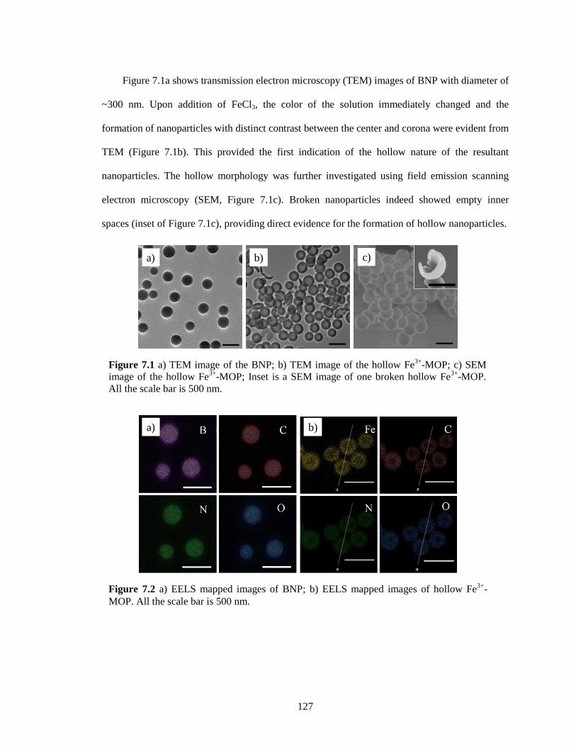
127
Figure 7.1a shows transmission electron microscopy (TEM) images of BNP with diameter of
~300 nm. Upon addition of FeCl3, the color of the solution immediately changed and the
formation of nanoparticles with distinct contrast between the center and corona were evident from
TEM (Figure 7.1b). This provided the first indication of the hollow nature of the resultant
nanoparticles. The hollow morphology was further investigated using field emission scanning
electron microscopy (SEM, Figure 7.1c). Broken nanoparticles indeed showed empty inner
spaces (inset of Figure 7.1c), providing direct evidence for the formation of hollow nanoparticles.
Figure 7.2 a) EELS mapped images of BNP; b) EELS mapped images of hollow Fe3+
-
MOP. All the scale bar is 500 nm.
b)a)
Figure 7.1 a) TEM image of the BNP; b) TEM image of the hollow Fe3+
-MOP; c) SEM
image of the hollow Fe3+
-MOP; Inset is a SEM image of one broken hollow Fe3+
-MOP.
All the scale bar is 500 nm.
a) b) Dc)
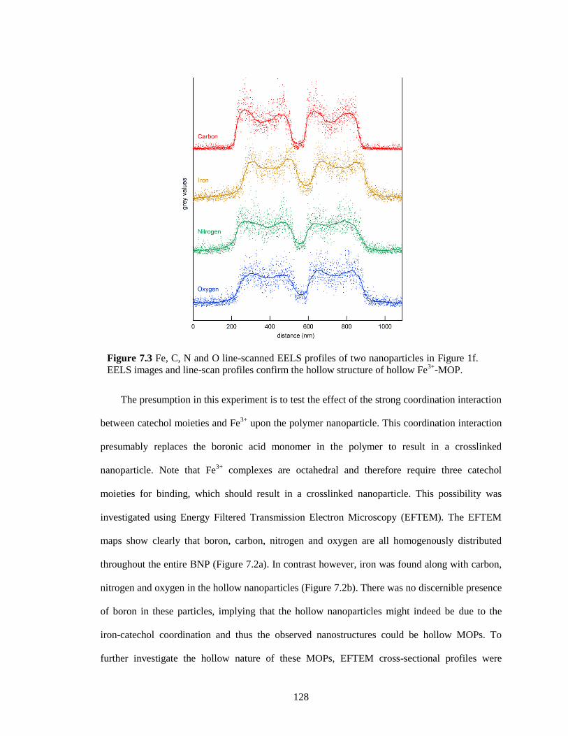
128
The presumption in this experiment is to test the effect of the strong coordination interaction
between catechol moieties and Fe3+
upon the polymer nanoparticle. This coordination interaction
presumably replaces the boronic acid monomer in the polymer to result in a crosslinked
nanoparticle. Note that Fe3+
complexes are octahedral and therefore require three catechol
moieties for binding, which should result in a crosslinked nanoparticle. This possibility was
investigated using Energy Filtered Transmission Electron Microscopy (EFTEM). The EFTEM
maps show clearly that boron, carbon, nitrogen and oxygen are all homogenously distributed
throughout the entire BNP (Figure 7.2a). In contrast however, iron was found along with carbon,
nitrogen and oxygen in the hollow nanoparticles (Figure 7.2b). There was no discernible presence
of boron in these particles, implying that the hollow nanoparticles might indeed be due to the
iron-catechol coordination and thus the observed nanostructures could be hollow MOPs. To
further investigate the hollow nature of these MOPs, EFTEM cross-sectional profiles were
Figure 7.3 Fe, C, N and O line-scanned EELS profiles of two nanoparticles in Figure 1f.
EELS images and line-scan profiles confirm the hollow structure of hollow Fe3+
-MOP.
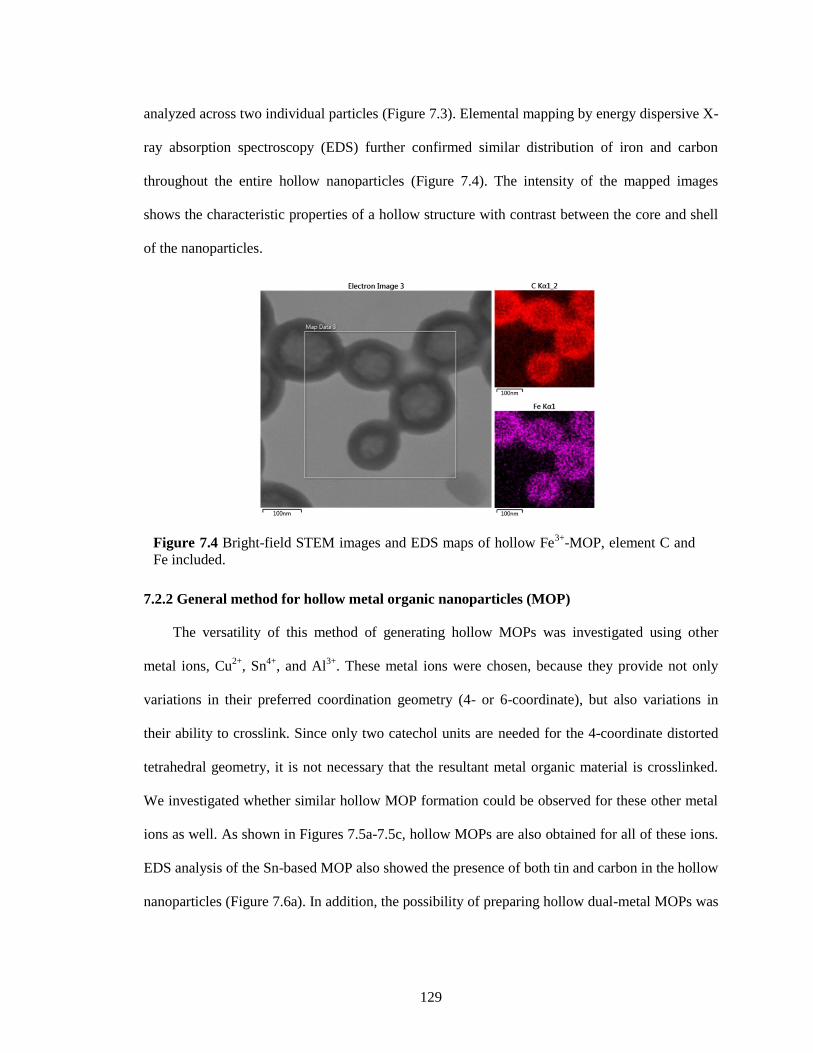
129
analyzed across two individual particles (Figure 7.3). Elemental mapping by energy dispersive X-
ray absorption spectroscopy (EDS) further confirmed similar distribution of iron and carbon
throughout the entire hollow nanoparticles (Figure 7.4). The intensity of the mapped images
shows the characteristic properties of a hollow structure with contrast between the core and shell
of the nanoparticles.
7.2.2 General method for hollow metal organic nanoparticles (MOP)
The versatility of this method of generating hollow MOPs was investigated using other
metal ions, Cu2+
, Sn4+
, and Al3+
. These metal ions were chosen, because they provide not only
variations in their preferred coordination geometry (4- or 6-coordinate), but also variations in
their ability to crosslink. Since only two catechol units are needed for the 4-coordinate distorted
tetrahedral geometry, it is not necessary that the resultant metal organic material is crosslinked.
We investigated whether similar hollow MOP formation could be observed for these other metal
ions as well. As shown in Figures 7.5a-7.5c, hollow MOPs are also obtained for all of these ions.
EDS analysis of the Sn-based MOP also showed the presence of both tin and carbon in the hollow
nanoparticles (Figure 7.6a). In addition, the possibility of preparing hollow dual-metal MOPs was
Figure 7.4 Bright-field STEM images and EDS maps of hollow Fe3+
-MOP, element C and
Fe included.
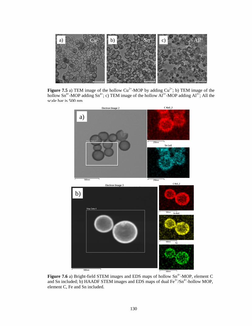
130
Figure 7.6 a) Bright-field STEM images and EDS maps of hollow Sn
4+-MOP, element C
and Sn included; b) HAADF STEM images and EDS maps of dual Fe3+
/Sn4+
-hollow MOP,
element C, Fe and Sn included.
a)
b)
Figure 7.5 a) TEM image of the hollow Cu2+
-MOP by adding Cu2+
; b) TEM image of the
hollow Sn4+
-MOP adding Sn4+
; c) TEM image of the hollow Al3+
-MOP adding Al3+
; All the
scale bar is 500 nm.
Cu2+ Sn4+ Al3+a) b) c)
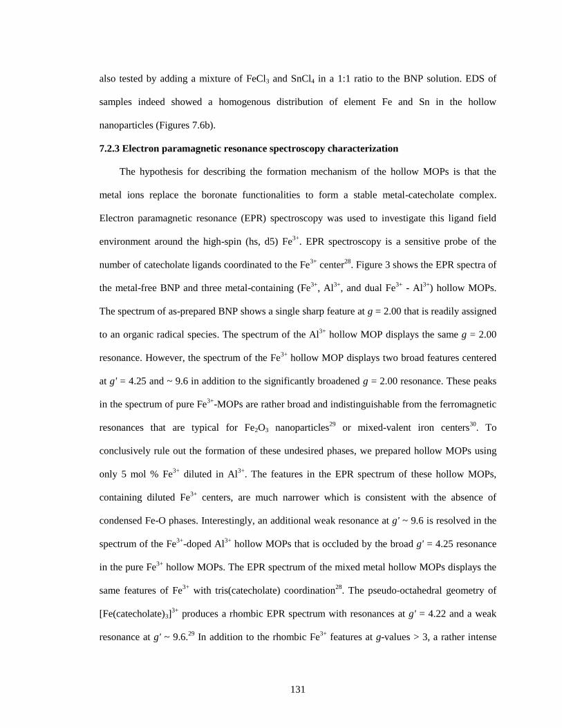
131
also tested by adding a mixture of FeCl3 and SnCl4 in a 1:1 ratio to the BNP solution. EDS of
samples indeed showed a homogenous distribution of element Fe and Sn in the hollow
nanoparticles (Figures 7.6b).
7.2.3 Electron paramagnetic resonance spectroscopy characterization
The hypothesis for describing the formation mechanism of the hollow MOPs is that the
metal ions replace the boronate functionalities to form a stable metal-catecholate complex.
Electron paramagnetic resonance (EPR) spectroscopy was used to investigate this ligand field
environment around the high-spin (hs, d5) Fe3+
. EPR spectroscopy is a sensitive probe of the
number of catecholate ligands coordinated to the Fe3+
center28
. Figure 3 shows the EPR spectra of
the metal-free BNP and three metal-containing (Fe3+
, Al3+
, and dual Fe3+
- Al3+
) hollow MOPs.
The spectrum of as-prepared BNP shows a single sharp feature at g = 2.00 that is readily assigned
to an organic radical species. The spectrum of the Al3+
hollow MOP displays the same g = 2.00
resonance. However, the spectrum of the Fe3+
hollow MOP displays two broad features centered
at g' = 4.25 and ~ 9.6 in addition to the significantly broadened g = 2.00 resonance. These peaks
in the spectrum of pure Fe3+
-MOPs are rather broad and indistinguishable from the ferromagnetic
resonances that are typical for Fe2O3 nanoparticles29
or mixed-valent iron centers30
. To
conclusively rule out the formation of these undesired phases, we prepared hollow MOPs using
only 5 mol % Fe3+
diluted in Al3+
. The features in the EPR spectrum of these hollow MOPs,
containing diluted Fe3+
centers, are much narrower which is consistent with the absence of
condensed Fe-O phases. Interestingly, an additional weak resonance at g' ~ 9.6 is resolved in the
spectrum of the Fe3+
-doped Al3+
hollow MOPs that is occluded by the broad g' = 4.25 resonance
in the pure Fe3+
hollow MOPs. The EPR spectrum of the mixed metal hollow MOPs displays the
same features of Fe3+
with tris(catecholate) coordination28
. The pseudo-octahedral geometry of
[Fe(catecholate)3]3+
produces a rhombic EPR spectrum with resonances at g' = 4.22 and a weak
resonance at g' ~ 9.6.29
In addition to the rhombic Fe3+
features at g-values > 3, a rather intense
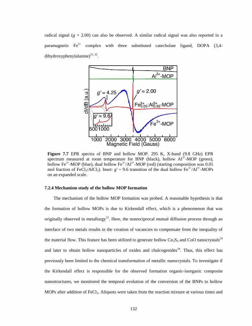
132
radical signal (g = 2.00) can also be observed. A similar radical signal was also reported in a
paramagnetic Fe3+
complex with three substituted catecholate ligand, DOPA (3,4-
dihydroxyphenylalanine)31, 32
.
7.2.4 Mechanism study of the hollow MOP formation
The mechanism of the hollow MOP formation was probed. A reasonable hypothesis is that
the formation of hollow MOPs is due to Kirkendall effect, which is a phenomenon that was
originally observed in metallurgy33
. Here, the nonreciprocal mutual diffusion process through an
interface of two metals results in the creation of vacancies to compensate from the inequality of
the material flow. This feature has been utilized to generate hollow Co3S4 and CoO nanocrystals24
and later to obtain hollow nanoparticles of oxides and chalcogenides34
. Thus, this effect has
previously been limited to the chemical transformation of metallic nanocrystals. To investigate if
the Kirkendall effect is responsible for the observed formation organic-inorganic composite
nanostructures, we monitored the temporal evolution of the conversion of the BNPs to hollow
MOPs after addition of FeCl3. Aliquots were taken from the reaction mixture at various times and
Figure 7.7 EPR spectra of BNP and hollow MOP. 295 K, X-band (9.8 GHz) EPR
spectrum measured at room temperature for BNP (black), hollow Al3+
-MOP (green),
hollow Fe3+
-MOP (blue), dual hollow Fe3+
/Al3+
-MOP (red) (starting composition was 0.05
mol fraction of FeCl3/AlCl3). Inset: g' ~ 9.6 transition of the dual hollow Fe3+
/Al3+
-MOPs
on an expanded scale.
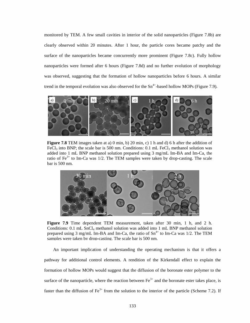
133
monitored by TEM. A few small cavities in interior of the solid nanoparticles (Figure 7.8b) are
clearly observed within 20 minutes. After 1 hour, the particle cores became patchy and the
surface of the nanoparticles became concurrently more prominent (Figure 7.8c). Fully hollow
nanoparticles were formed after 6 hours (Figure 7.8d) and no further evolution of morphology
was observed, suggesting that the formation of hollow nanoparticles before 6 hours. A similar
trend in the temporal evolution was also observed for the Sn4+
-based hollow MOPs (Figure 7.9).
An important implication of understanding the operating mechanism is that it offers a
pathway for additional control elements. A rendition of the Kirkendall effect to explain the
formation of hollow MOPs would suggest that the diffusion of the boronate ester polymer to the
surface of the nanoparticle, where the reaction between Fe3+
and the boronate ester takes place, is
faster than the diffusion of Fe3+
from the solution to the interior of the particle (Scheme 7.2). If
Figure 7.8 TEM images taken at a) 0 min, b) 20 min, c) 1 h and d) 6 h after the addition of
FeCl3 into BNP; the scale bar is 500 nm. Conditions: 0.1 mL FeCl3 methanol solution was
added into 1 mL BNP methanol solution prepared using 3 mg/mL Im-BA and Im-Ca, the
ratio of Fe3+
to Im-Ca was 1/2. The TEM samples were taken by drop-casting. The scale
bar is 500 nm.
Figure 7.9 Time dependent TEM measurement, taken after 30 min, 1 h, and 2 h.
Conditions: 0.1 mL SnCl4 methanol solution was added into 1 mL BNP methanol solution
prepared using 3 mg/mL Im-BA and Im-Ca, the ratio of Sn4+
to Im-Ca was 1/2. The TEM
samples were taken by drop-casting. The scale bar is 500 nm.
20 min 1 hb) d)c)a) 0 min 6 h
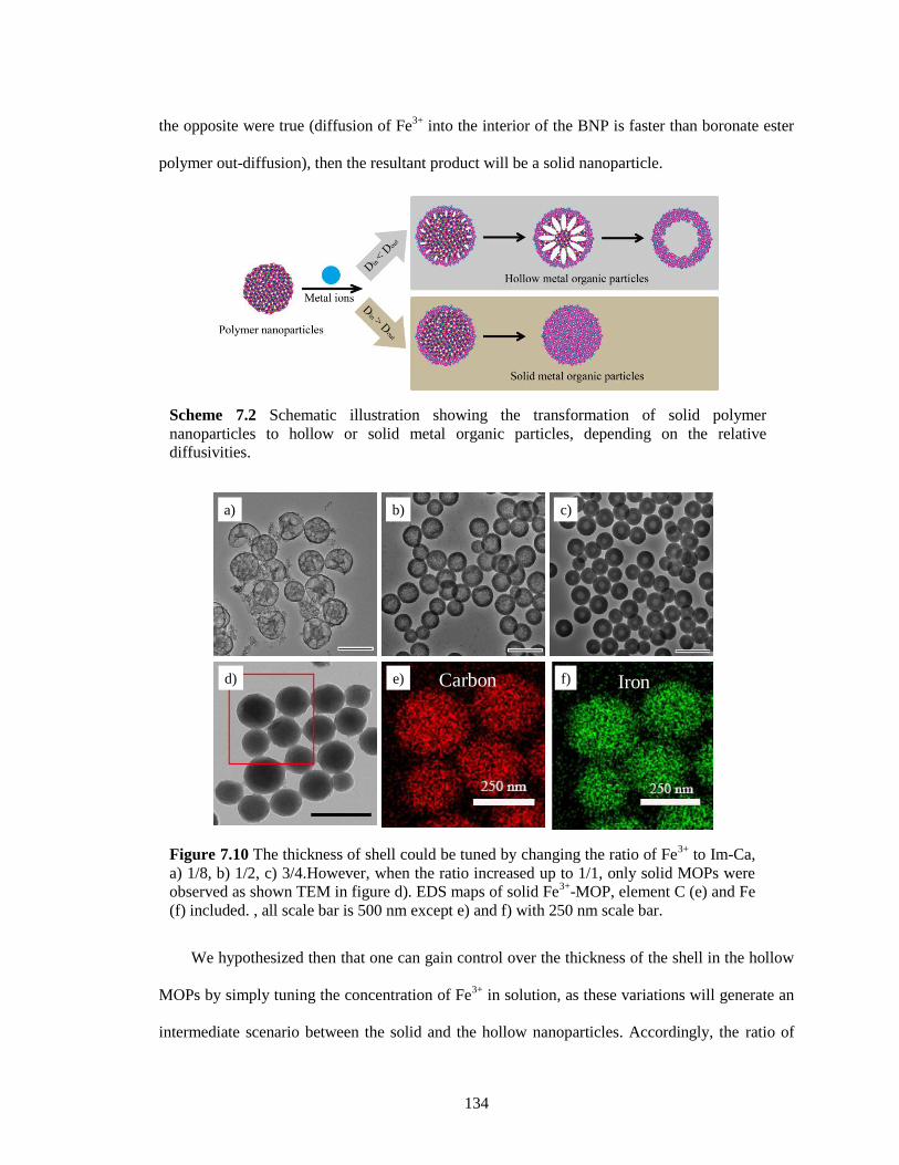
134
the opposite were true (diffusion of Fe3+
into the interior of the BNP is faster than boronate ester
polymer out-diffusion), then the resultant product will be a solid nanoparticle.
We hypothesized then that one can gain control over the thickness of the shell in the hollow
MOPs by simply tuning the concentration of Fe3+
in solution, as these variations will generate an
intermediate scenario between the solid and the hollow nanoparticles. Accordingly, the ratio of
Figure 7.10 The thickness of shell could be tuned by changing the ratio of Fe3+
to Im-Ca,
a) 1/8, b) 1/2, c) 3/4.However, when the ratio increased up to 1/1, only solid MOPs were
observed as shown TEM in figure d). EDS maps of solid Fe3+
-MOP, element C (e) and Fe
(f) included. , all scale bar is 500 nm except e) and f) with 250 nm scale bar.
a)
d)
b)
e)
c)
f)Carbon Iron
Scheme 7.2 Schematic illustration showing the transformation of solid polymer
nanoparticles to hollow or solid metal organic particles, depending on the relative
diffusivities.
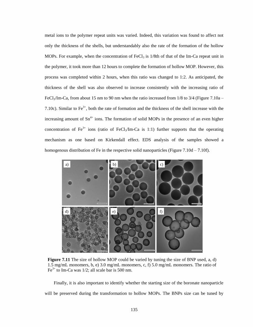
135
metal ions to the polymer repeat units was varied. Indeed, this variation was found to affect not
only the thickness of the shells, but understandably also the rate of the formation of the hollow
MOPs. For example, when the concentration of FeCl3 is 1/8th of that of the Im-Ca repeat unit in
the polymer, it took more than 12 hours to complete the formation of hollow MOP. However, this
process was completed within 2 hours, when this ratio was changed to 1:2. As anticipated, the
thickness of the shell was also observed to increase consistently with the increasing ratio of
FeCl3/Im-Ca, from about 15 nm to 90 nm when the ratio increased from 1/8 to 3/4 (Figure 7.10a –
7.10c). Similar to Fe3+
, both the rate of formation and the thickness of the shell increase with the
increasing amount of Sn4+
ions. The formation of solid MOPs in the presence of an even higher
concentration of Fe3+
ions (ratio of FeCl3/Im-Ca is 1:1) further supports that the operating
mechanism as one based on Kirkendall effect. EDS analysis of the samples showed a
homogenous distribution of Fe in the respective solid nanoparticles (Figure 7.10d – 7.10f).
Finally, it is also important to identify whether the starting size of the boronate nanoparticle
will be preserved during the transformation to hollow MOPs. The BNPs size can be tuned by
Figure 7.11 The size of hollow MOP could be varied by tuning the size of BNP used, a, d)
1.5 mg/mL monomers, b, e) 3.0 mg/mL monomers, c, f) 5.0 mg/mL monomers. The ratio of
Fe3+
to Im-Ca was 1/2; all scale bar is 500 nm.
a) b) c)
d) e) f)
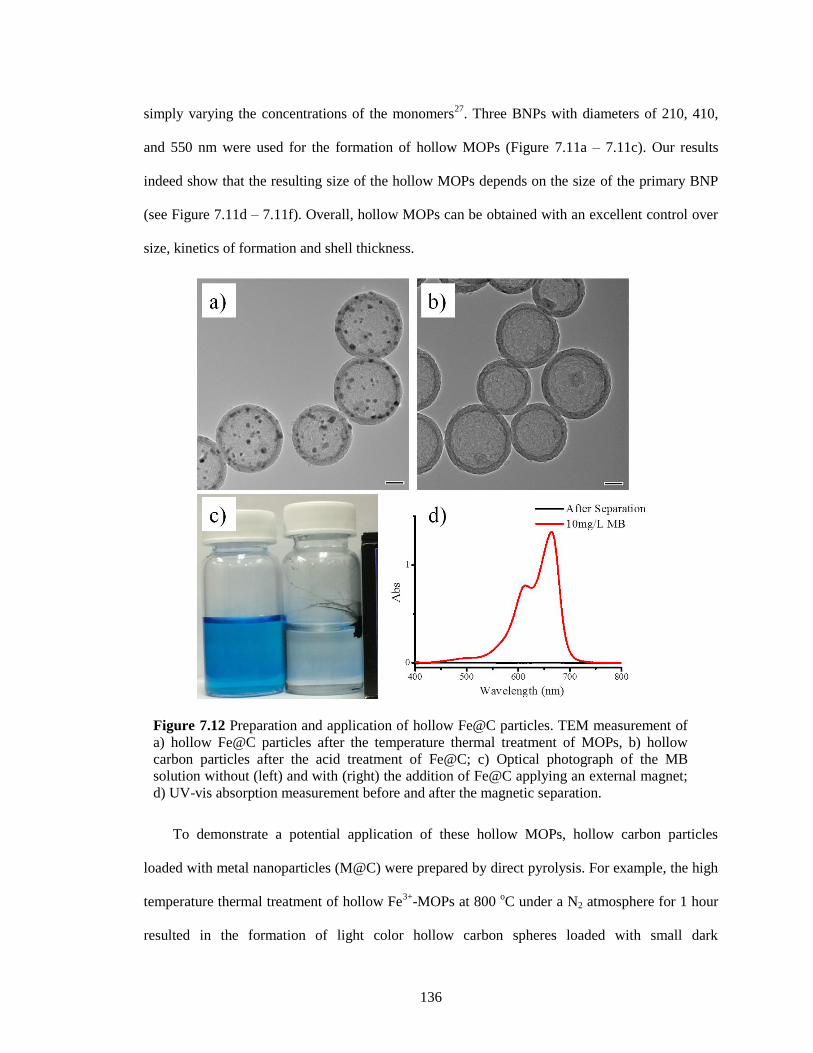
136
simply varying the concentrations of the monomers27
. Three BNPs with diameters of 210, 410,
and 550 nm were used for the formation of hollow MOPs (Figure 7.11a – 7.11c). Our results
indeed show that the resulting size of the hollow MOPs depends on the size of the primary BNP
(see Figure 7.11d – 7.11f). Overall, hollow MOPs can be obtained with an excellent control over
size, kinetics of formation and shell thickness.
To demonstrate a potential application of these hollow MOPs, hollow carbon particles
loaded with metal nanoparticles (M@C) were prepared by direct pyrolysis. For example, the high
temperature thermal treatment of hollow Fe3+
-MOPs at 800 oC under a N2 atmosphere for 1 hour
resulted in the formation of light color hollow carbon spheres loaded with small dark
Figure 7.12 Preparation and application of hollow Fe@C particles. TEM measurement of
a) hollow Fe@C particles after the temperature thermal treatment of MOPs, b) hollow
carbon particles after the acid treatment of Fe@C; c) Optical photograph of the MB
solution without (left) and with (right) the addition of Fe@C applying an external magnet;
d) UV-vis absorption measurement before and after the magnetic separation.
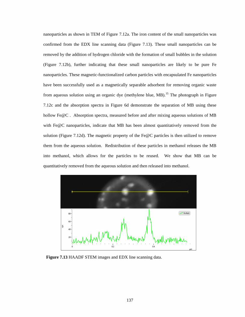
137
nanoparticles as shown in TEM of Figure 7.12a. The iron content of the small nanoparticles was
confirmed from the EDX line scanning data (Figure 7.13). These small nanoparticles can be
removed by the addition of hydrogen chloride with the formation of small bubbles in the solution
(Figure 7.12b), further indicating that these small nanoparticles are likely to be pure Fe
nanoparticles. These magnetic-functionalized carbon particles with encapsulated Fe nanoparticles
have been successfully used as a magnetically separable adsorbent for removing organic waste
from aqueous solution using an organic dye (methylene blue, MB).35
The photograph in Figure
7.12c and the absorption spectra in Figure 6d demonstrate the separation of MB using these
hollow Fe@C . Absorption spectra, measured before and after mixing aqueous solutions of MB
with Fe@C nanoparticles, indicate that MB has been almost quantitatively removed from the
solution (Figure 7.12d). The magnetic property of the Fe@C particles is then utilized to remove
them from the aqueous solution. Redistribution of these particles in methanol releases the MB
into methanol, which allows for the particles to be reused. We show that MB can be
quantitatively removed from the aqueous solution and then released into methanol.
Figure 7.13 HAADF STEM images and EDX line scanning data.
138
7.3 Summary
In conclusion, a simple and versatile strategy for achieving hollow metal organic
nanoparticles has been developed, where boronate ester based solid organic nanoparticles have
been used as the precursor. The size of the hollow MOPs is dictated by the size of the precursor
BNP, which itself exhibits excellent tunability. The formation of these hollow MOPs seem to be
independent of the valence and the geometry of the metal ion, which also allows for the
incorporation of more than one type of metal ion into the MOPs. Kirkendall effect has been
shown to be the underlying mechanism for the formation of these hollow MOPs. For the first time,
Kirkendall effect has been responsible for the formation of hollow metal organic nanoparticles
from organic polymer nanoparticles. This understanding further lends itself to control in the shell
thickness of the MOPs. Overall, the method reported here for achieving hollow MOPs with a high
degree of control has the versatility to impact applications such as catalysis, sensing, storage, and
biomedicine, as this approach should be extendable to other organic-inorganic molecular
architectures. Moreover, the fields of polymer synthesis and self-assembly are orthogonally and
rapidly evolving with its own versatility and control elements36
. Our ability to reliably generate
hollow organic-inorganic hybrid nanoparticles using polymeric nanostructure templates will open
up a fount of opportunities for hybrid materials nanotechnology.
7.4 Experimental
7.4.1 General
p-Phenylenediamine, 3,4-dihydroxybenzaldehyde, 4-formylphenylboronic acid, Iron(III)
chloride, Copper(II) chloride, Tin(IV) chloride pentahydrate, Aluminum chloride and other
conventional reagents were obtained from commercial sources and used as received unless
otherwise mentioned. 1H-NMR spectra were recorded on a Bruker 400 MHz NMR spectrometer
using the residual proton resonance of the solvent as the internal standard. 13
C-NMR spectra were
recorded on a Bruker 100 MHz NMR spectrometer using the carbon signal of the deuterated
139
solvent as the internal standard. Mass spectra were taken on Bruker micrOTOF-II. Convential
Bright-Filed Transmission electron microscopy (TEM) images were taken using a JEOL JEM-
2000FX operating at 200 KV. Scanning electron microscopy (SEM) images were taken using a
FEI Magellan 400 FESEM in SE imaging mode. Engery Filtered Transmission Electron
Microscopy was performed using a JEO JEM-2200FS Energy Filtered Field Emission TEM
operating at 200kV. Prior to generating the element distribution images (EFTEM maps) the
presence of the respective elements was verfiied using Elelectron Energy Lass Spectroscopy
(EELS) in the same instrument. Subsequently EFTEM maps were acquired using the three-
window method in wide-field mode, which is based on the subtraction of two pre-edge images
from one post-edge image for back-ground substraction1. The following element edges where
used: BK= 165eV, CK=284eV, NK = 401eV, OK = 532eV, FeL2,3 = 708eV and an Energy Window
mapping was performrmed in the same instrument in STEM mode utilizing an Oxford X-MAX
80mm2 detector and a probe size of 1.5nm. EDS maps were acquired until sufficient cotrast was
obtained (typically 15-30 min). Room temperature EPR spectra were collected on powdered
samples using the TE102 mode of a X-band resonator (9.81 GHz, Bruker ELEXSYS-500).
7.4.2 Synthesis of Im-Ca
The preparation of Im-Ca was achieved following our previous report27
. To a solution of p-
phenylenediamine (0.15 g, 1.39 mmol) in anhydrous ethanol (10 mL) was added 3,4-
dihydroxybenzaldehyde (0.40 g, 2.90 mmol). The clear brown mixture was stirred at room
temperature under an argon atmosphere and protected from light. The product precipitated out of
the reaction mixture. The brown precipitate was filtered and washed with cold ethanol three times.
Final product was vacuum dried to obtain a brown solid (yield: 70%) 1H-NMR (400 MHz,
DMSO-6D) δ(ppm): 9.57 (s, 2H), 9.29 (s, 2H), 8.41 (s, 2H), 7.38 (d, 2H), 7.23 (s, 4H), 7.17 (q,
2H), 6.82 (d, 2H). 1H-NMR (400 MHz, CD3OD and DMSO-6D) δ(ppm): 8.39 (s, 2H), 7.39 (d,
140
2H), 7.22 (s, 4H), 7.17 (q, 2H), 6.82 (d, 2H). 13
C-NMR (100MHz, DMSO-6D) δ(ppm): 159.89,
149.57, 146.09, 128.56, 122.91, 122.30, 115.83, 114.51. ESI/MS m/z 349.11 [M+H]+ (expect m/z
349.35).
7.4.3 Synthesis of Im-BA
Im-BA was synthesized from condensation reaction between p-phenylenediamine and 4-
formylphenylboronic acid. To a solution of p-phenylenediamine (0.05 g, 0.46 mmol) in 20 mL
MeOH was added 4-formylphenylboronic acid (0.15 g, 1.00 mmol). The clear yellow mixture
became cloudy after stirring at room temperature overnight. The yellow precipitate was filtered
and washed with cool methanol three times. Final product was vacuum dried to obtain a
yellowish solid (yield: 80%) 1H-NMR (400MHz, DMSO-6D) δ(ppm): 8.67 (s, 2H), 8.22 (s, 4H),
7.89 (m, 8H), 7.36 (s, 4H). 1H-NMR (400MHz, CD3OD and DMSO-6D) δ(ppm): 8.65 (s, 2H),
7.88 (m, 8H), 7.34 (s, 4H). 13
C-NMR (100MHz, DMSO-6D) δ(ppm): 160.67, 149.83, 137.76,
134.90, 128.06, 122.54. ESI/MS m/z 431.16 [M+59]- (expect m/z=431.19), m/z 445.20 [M+73]
-
(expect m/z=445.21), m/z 459.22 [M+87]- (expect m/z=459.23).
7.4.4 Preparation of Hollow Metal-Organic Particles
The preparation of hollow nanoparticles could be watched as the movie S1. First, Im-Ca and
Im-BA were dissolved in methanol with the help of heating. After cooling down by setting at
room temperature, one solution was added into the solution drop by drop, the mixture solution
because deep orange and suspension was finally got. Then a methanolic solution of metal
chloride salts (FeCl3 in this example) was added to the suspension of BNPs once. After stirring
for given time, the resultant hollow metal-organic particles were washed with methanol, collected
by centrifugation, and dried in a vacuum oven at 50 oC overnight.
7.4.5 Preparation of Hollow Fe@C particles
Hollow Fe3+
-MOPs were placed in a tube furnace and let the nitrogen gas flow for 30 mins
at room temperature. Then the furnace was calcined up to 800 °C quickly. After 1 hours, the
141
furnace was turned off and the resulting particles were cooled to room temperature with the
nitrogen gas flow.
7.4.6 Adsorption of methylene blue (MB) from aqueus solution
Hollow Fe@C (2 mg) was added to 10 mL of MB aqueous solution (10 mg/L) and was
sonticated to form the dispersed solution. After 5 mins, an external magnet was applied to
removed the Fe@C particles from the aqueous solutions. UV-Vis spectra were measured before
and after the separation.
7.4.7 Release of methylene blue (MB) from Fe@C particles
After collecting the Fe@C particles from the aqueous solution containing MB, methanol was
used to wash the MB moelcules out from these carbon based particles for four times. Each time,
Fe@C particles were removed from the solution by applying external magnet, and methanol
solution containing MB could be collected. The Fe@C particles can then be used for adsorption
of MB again.
7.5 References
1. Martin, C. R. Nanomaterials-a membrane-based synthetic approach. Science 1994, 266, 1961.
2. Huang, Y.; Duan, X.; Wei, Q.; Lieber, C. M. Functional nanoscale electronic devices
assembled using silicon nanowire building blocks. Science 2001, 291, 630.
3. Suteewong, T.; Sai, H.; Hovden, R.; Muller, D.; Bradbury, M. S.; Gruner, S. M.; Wiesner, U.
Multicompartment mesoporous silica nanoparticles with branched shapes: An epitaxial growth
mechanism. Science 2013, 340, 337.
4. Wang, X.; Zhuang, J.; Peng, Q.; Li, Y. A general strategy for nanocrystal synthesis. Nature
2005, 437, 121.
5. Howes, P. D.; Chandrawati, R.; Stevens, M. M. Colloidal nanoparticles as advanced biological
sensors. Science 2014, 346, 1247390.
6. Savić, R.; Luo, L.; Eisenberg, A.; Maysinger, D. Micellar nanocontainers distribute to defined
cytoplasmic organelles. Science 2003, 300, 615.
7. Jones, J. T. A.; Hasell, T.; Wu, X.; Bacsa, J.; Jelfs, K. E.; Schmidtmann, M.; Chong, S. Y.;
Adams, D. J.; Trewin, A.; Schiffman, F.; Cora, F.; Slater, B.; Steiner, A.; Day, G. M.; Cooper, A.
I. Modular and predictable assembly of porous organic molecular crystals. Nature 2011, 474, 367.
142
8. Furukawa, H.; Cordova, K. E.; O’Keeffe, M.; Yaghi, O. M. The chemistry and applications of
metal-organic frameworks. Science 2013, 341, 1230444.
9. Oh, M.; Mirkin, C. A. Chemically tailorable colloidal particles from infinite coordination
polymers. Nature 2005, 438, 651.
10. Spokoyny, A. M.; Kim, D.; Sumrein, A.; Mirkin, C. A. Infinite coordination polymer nano-
and microparticle structures. Chem. Soc. Rev., 2009, 38, 1218.
11. Carné, A.; Carbonell, C.; Imaz, I.; Maspoch, D. Nanoscale metal–organic materials. Chem.
Soc. Rev., 2011, 40, 291–305
12. Seo, J. S.; Whang, D.; Lee, H.; Jun, S.I. ; Oh, J.; Jeon, Y. J.; Kim, K. A homochiral metal–
organic porous material for enantioselective separation and catalysis. Nature 2000, 404, 982.
13. Banerjee, R.; Phan, A.; Wang, B.; Knobler, C.; Furukawa, H.; O'Keeffe, M.; Yaghi, O. M. High-throughput synthesis of zeolitic imidazolate frameworks and application to CO2 capture.
Science 2008, 319, 939.
14. Horcajada, P.; Gref, R.; Baati, T.; Allan, P. K.; Maurin, G.; Couvreur, P.; Ferey, G.; Morris, R.
E.; Serre, C. Metal–organic frameworks in biomedicine. Chem. Rev. 2011, 112, 1232.
15. Ameloot, R.; Vermoortele, F.; Vanhove, W.; Roeffaers, M. B.J. ; Sels, B. F.; De Vos, D. E.
Interfacial synthesis of hollow metal–organic framework capsules demonstrating selective
permeability. Nat. Chem. 2011, 3, 382.
16. Carné-Sánchez, A.; Imaz, I.; Cano-Sarabia, M.; Maspoch, D. A spray-drying strategy for
synthesis of nanoscale metal–organic frameworks and their assembly into hollow superstructures.
Nat. Chem. 2013, 5, 203.
17. Pang, M.; Cairns, A. J.; Liu, Y.; Belmabkhout, Y.; Zeng, H. C.; Eddaoudi, M. Synthesis and
integration of Fe-soc-MOF cubes into colloidosomes via a single-step emulsion-based approach.
J. Am. Chem. Soc. 2013, 135, 10234.
18. Zhang, Z.; Chen, Y.; Zhang, J.; Xu, X.; Yang, Y.; Nosheen, F.; Saleem, F.; He, W.; Wang, X.
Hierarchical Zn/Ni-MOF-2 nanosheet-assembled hollow nanocubes for multicomponent catalytic
reactions. Angew. Chem. Int. Ed. 2014, 53, 12517.
19. Huo, J.; Wang, L.; Irran, E.; Yu, H.; Gao, J.; Fan, D.; Li, B.; Wang, J.; Ding, W.; Amin, A.
M.; Li, C.; Ma, L. Hollow ferrocenyl coordination polymer microspheres with micropores in
shells prepared by Ostwald ripening. Angew. Chem. Int. Ed. 2010, 49, 9237.
20. Ejima, H.; Richardson, J. J.; Liang, K.; Best, J. P.; van Koeverden, M. P.; Such, G. K.; Cui, J.;
Caruso, F. One-step assembly of coordination complexes for versatile film and particle
engineering. Science 2013, 341, 154.
21. Zhang, Z.; Chen, Y.; Xu, X.; Zhang, J.; Xiang, G.; He, W.; Wang, X. Well-defined metal-
organic framework hollow nanocages. Angew. Chem. Int. Ed. 2014, 53, 429.
143
22. Zhang, F.; Wei, Y.; Wu, X.; Jiang, H.; Wang, W.; Li, H. Hollow zeolitic imidazolate
framework nanospheres as highly efficient cooperative catalysts for [3 + 3] cycloaddition
reactions. J. Am. Chem. Soc. 2014, 136, 13963.
23. Caruso, F.; Caruso, R. A.; Möhwald, H. Nanoengineering of inorganic and hybrid hollow
spheres by colloidal templating. Science 1998, 282, 1111.
24. Yin, Y.; Rioux, R. M.; Erdonmez, C. K.; Hughes, S.; Somorjai, G. A.; Alivisatos, A. P.
Formation of hollow nanocrystals through the nanoscale Kirkendall effect. Science 2004, 304,
711.
25. Oh, M. H.; Yu, T.; Yu, S.-H.; Lim, B.; Ko, K.-T.; Willinger, M.-G.; Seo, D.-H.; Kim, B. H.;
Cho, M. G.; Park, J.-H. Galvanic replacement reactions in metal oxide nanocrystals. Science 2013,
340, 964.
26. Lou, X. W.; Wang, Y.; Yuan, C.; Lee, J. Y.; Archer, L. A. Template-free synthesis of SnO2
hollow nanostructures with high lithium storage capacity. Adv. Mater. 2006, 18, 2325.
27. Li, L.; Yuan, C.; Dai, L.; Thayumanavan, S. Thermoresponsive Polymeric Nanoparticles:
Nucleation from Cooperative Polymerization Driven by Dative Bonds. Macromolecules 2014, 47,
5869.
28. Weisser, J. T.; Nilges, M. J.; Sever, M. J.; Wilker, J. J. EPR investigation and spectral
simulations of iron-catecholate complexes and iron-peptide models of marine adhesive cross-
links. Inorg. Chem. 2006, 45, 7736.
29. Koksharov, Y. A.; Gubin, S.P.; Kosobudsky, I. D.; Beltran, M.; Khodorkovsky, Y.; Tishin, A.
M. Low temperature electron paramagnetic resonance anomalies in Fe-based nanoparticles. J.
Appl. Phys. 2000, 88, 1587.
30. Nikiforov, V. N.; Koksharov, Y. A.; Polyakov, S. N.; Malakho, A. P.; Volkov, A. V.;
Moskvina, M. A.; Khomutov, G. B.; Irkhin, V. Y. Magnetism and Verwey transition in magnetite
nanoparticles in thin polymer film. J. Alloys Compd. 2013, 569, 58.
31. Sever, M. J.; Weisser, J. T.; Monahan, J.; Srinivasan, S.; Wilker, J. J. Metal-mediated cross-
linking in the generation of a marine-mussel adhesive. Angew. Chem. Int. Ed. 2004, 43, 448.
32. Sever, M. J.; Wilker, J. J. Absorption spectroscopy and binding constants for first-row
transition metal complexes of a DOPA-containing peptide. Dalton Trans. 2006, 813.
33. Kirkendall, E. Diffusion of zinc in alpha brass. Aime Trans. 1942, 147, 104.
34. Yin, Y.; Erdonmez, C. K.; Cabot, A.; Hughes, S.; Alivisatos, A. P. Colloidal synthesis of
hollow cobalt sulfide nanocrystals. Adv. Funct. Mater. 2006, 16, 1389.
35. Lee, H. J.; Choi, S. C.; Oh, M. Well-dispersed hollow porous carbon spheres synthesized by
direct pyrolysis of core-shell type metal-organic frameworks and their sorption properties. Chem.
Commun., 2014, 50, 4492.
36. Hawker, C. J.; Wooley, K. L. The convergence of synthetic organic and polymer chemistries.
Science 2005, 309, 1200.
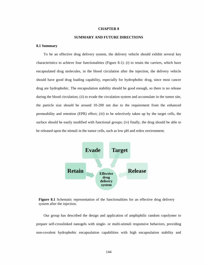
144
CHAPTER 8
SUMMARY AND FUTURE DIRECTIONS
8.1 Summary
To be an effective drug delivery system, the delivery vehicle should exhibit several key
characteristics to achieve four functionalities (Figure 8.1): (i) to retain the carriers, which have
encapsulated drug molecules, in the blood circulation after the injection, the delivery vehicle
should have good drug loading capability, especially for hydrophobic drug, since most cancer
drug are hydrophobic. The encapsulation stability should be good enough, so there is no release
during the blood circulation; (ii) to evade the circulation system and accumulate in the tumor site,
the particle size should be around 10-200 nm due to the requirement from the enhanced
permeability and retention (EPR) effect; (iii) to be selectively taken up by the target cells, the
surface should be easily modified with functional groups; (iv) finally, the drug should be able to
be released upon the stimuli in the tumor cells, such as low pH and redox environment.
Our group has described the design and application of amphiphilic random copolymer to
prepare self-crosslinked nanogels with single- or multi-stimuli responsive behaviors, providing
non-covalent hydrophobic encapsulation capabilities with high encapsulation stability and
Figure 8.1 Schematic representation of the functionalities for an effective drug delivery
system after the injection.
Effective drug
delivery system
Retain
Evade Target
Release
145
controlled release of the payload for drug delivery. In this thesis, we further fundamentally
studied the structure-property correlations in self-crosslinked polymer nanogels and developed
improved functionalities for drug delivery applications. In Chapter 2, we highlight unprecedented
control in fine-tuning the sizes and encapsulation stability using Hofmeister ions. The size of the
nanogel can be systematically varied with concentration of both kosmotropic and chaotropic
anion. Encapsulation stability of guest molecules is also dependent on the nature of the salt used
for preparation of the nanogels; chaotropic anions afford nanogels with greater guest
encapsulation stability. The guest release kinetics of nanogels made with ions were also
consistent with the Hofmeister series.
Fluorescence resonance energy transfer (FRET) based method has been applied to evaluate
exchange dynamics of lipophilic guest molecules, providing important sights on the evaluation of
the stability of these drug delivery vehicles. In Chapter 3, we have shown that the dominant
mechanism for guest exchange in nanogel hosts is collision-based. In addition, the mechanism of
guest exchange can change on the basis of the microenvironment of the host. When the nanogel
interior is hydrophobic, the collision-based mechanism is the dominant pathway. However, when
the interior is sufficiently hydrophilic, the dominant mechanism changes to a diffusion-based one.
In drug delivery applications, the drug-loaded nanocarrier experiences both concentration and
environmental changes upon bio-distribution to diseased tissues. The mechanistic variations
based on environments provide insights into molecular designs that can afford stable
encapsulation in one environment and release of the molecules in another.
It is important to have nanocarriers that can be effectively transported across cellular
membranes for biomedical applications. In Chapter 4, we have developed a simple method to
prepare a nanogel that is capable of transforming from a negatively charged form into a positively
charged in the slightly acidic tumor extracellular environment. The reported facile synthetic
method for preparing the pHe-responsive charge conversional nanogels, combined with stable
146
encapsulation and tunable pI, should open up new avenues in the design of advanced drug carriers
for intelligent drug delivery. The study also highlights the opportunities to develop other
functional nanogels using units with other functional group.
Reagent-free synthetic methods are needed for nanogel formation due to their simplicity and
implications in green chemistry. In Chapter 5, we have demonstrated a new pathway for the
preparation of disulfide based self-crosslinking nanogels using photoirradiation. The most
important take-home message of this work is that pyridyl disulfide might be used as a photo-
crosslinkable functional group, while this crosslinks would be redox responsive, which should
play an important role in the design of drug delivery nanocarriers. From the application
standpoint, changing the DTT-induced crosslinking method to photo-induced crosslinking
method makes our nanogel preparation more cost-effective. The scale up of the process should
become possible considering that the diffusion of crosslinking agents into the large volume of
solution may result in heterogeneous crosslinking density.
Hybrid metal-organic coordination materials, constructed from metal ions and
polyfunctional organic ligands, have received great attention due to their interesting applications.
We have developed a simple, rapid, and robust method to produce hollow metal-organic particles
with well controlled size and thickness from organic polymer particles. In Chapter 6, we prepared
organic polymer particles through boronate esterification. Their size, absorption characteristic can
be adjusted by controlling the mixing concentration. We pointed out that the nucleation step
involves a nucleation elongation polymerization with N→B interaction as the driving force for
the collapsing. In Chapter 7, we have demonstrated that hollow metal organic nanoparticles can
be prepared by simply adding metal ions into purely organic nanoparticles. The Kirkendall effect
is responsible for the formation of the hollow structure as shown by time dependent TEM
measurements. The tunability of the original organic nanoparticles makes it easy to control the
size of hollow nanoparticles, while the thickness can be tuned by simply altering the ratio of
147
metal ions to the catechol monomers. We further showed that various metal ions can be used to
form hollow nanoparticles. We believe that this approach can be extended to the development of
more diverse hollow metal organic nanoparticles.
8.2 Future directions
In this section future directions for two more projects are described.
8.2.1 Redox-activatable 19
F-MRI nanoprobe
Magnetic resonance imaging (MRI) is of great interest in both basic biological research and
clinical diagnosis of cancer and other diseases.1-3
19
F MRI has received significant attention in
MR imaging and spectroscopy studies because of two reasons: (i) 19
F-MRI has little biological
background, offering a means to detect diseases in deep tissue since there is low levels of
endogenous fluorine in the body; (ii) 19
F has 100% natural abundance and its gyromagentic ratio
(40.06 MHz/T) is second only to 1H, making it more sensitive for detection over other nuclei.
4
Here we are interested in redox-responsive 19
F MRI nanoprobe, since it is important to monitor
biological redox reactions.5-8
Actually, biological redox reactions play important roles in many
biological processes. Abnormal levels of redox reactions can result in various disease including
liver damage, skin lesions, and slow growth.9-12
A stimulus-responsive 19
F-MRI nanoprobes have been usually prepared based on the
strategy of using changes in spin-spin relaxations between the aggregation and free states to turn
ON/OFF 19
F signals in response to the pH values.13-15
For example, pH-activatable 19
F-MRI
nanoprobes were prepared by introducing the fluoro groups into the ionizable block (PR) of
amphiphilic copolymers containing a hydrophilic poly(ethylene oxide) (PEO) segment and
tertiary amine/ammonium segment (Figure 8.2).15
At pH > pKa, the formation of micelle highly
restricted chain motions, resulting in short spin–spin relaxation times (almost 0), which
effectively broadened and eliminated the 19
F signals. However, protonation of ammonium groups
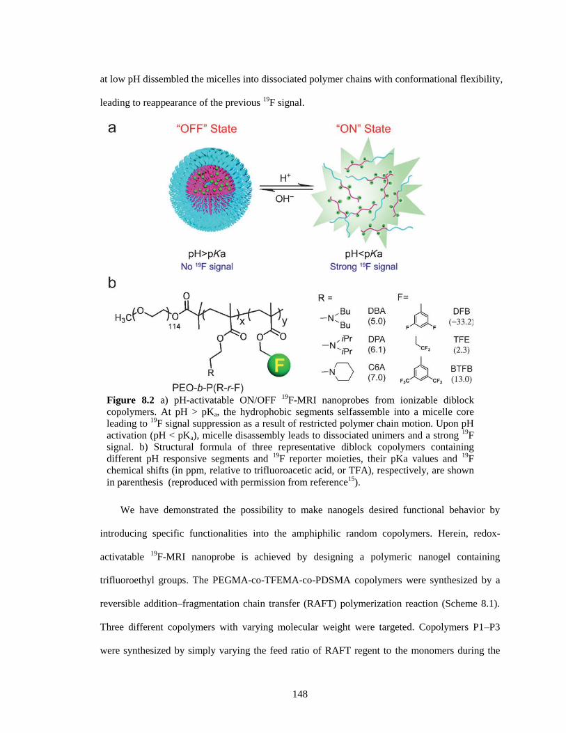
148
at low pH dissembled the micelles into dissociated polymer chains with conformational flexibility,
leading to reappearance of the previous 19
F signal.
We have demonstrated the possibility to make nanogels desired functional behavior by
introducing specific functionalities into the amphiphilic random copolymers. Herein, redox-
activatable 19
F-MRI nanoprobe is achieved by designing a polymeric nanogel containing
trifluoroethyl groups. The PEGMA-co-TFEMA-co-PDSMA copolymers were synthesized by a
reversible addition–fragmentation chain transfer (RAFT) polymerization reaction (Scheme 8.1).
Three different copolymers with varying molecular weight were targeted. Copolymers P1–P3
were synthesized by simply varying the feed ratio of RAFT regent to the monomers during the
Figure 8.2 a) pH-activatable ON/OFF
19F-MRI nanoprobes from ionizable diblock
copolymers. At pH > pKa, the hydrophobic segments selfassemble into a micelle core
leading to 19
F signal suppression as a result of restricted polymer chain motion. Upon pH
activation (pH < pKa), micelle disassembly leads to dissociated unimers and a strong 19
F
signal. b) Structural formula of three representative diblock copolymers containing
different pH responsive segments and 19
F reporter moieties, their pKa values and 19
F
chemical shifts (in ppm, relative to trifluoroacetic acid, or TFA), respectively, are shown
in parenthesis (reproduced with permission from reference15
).
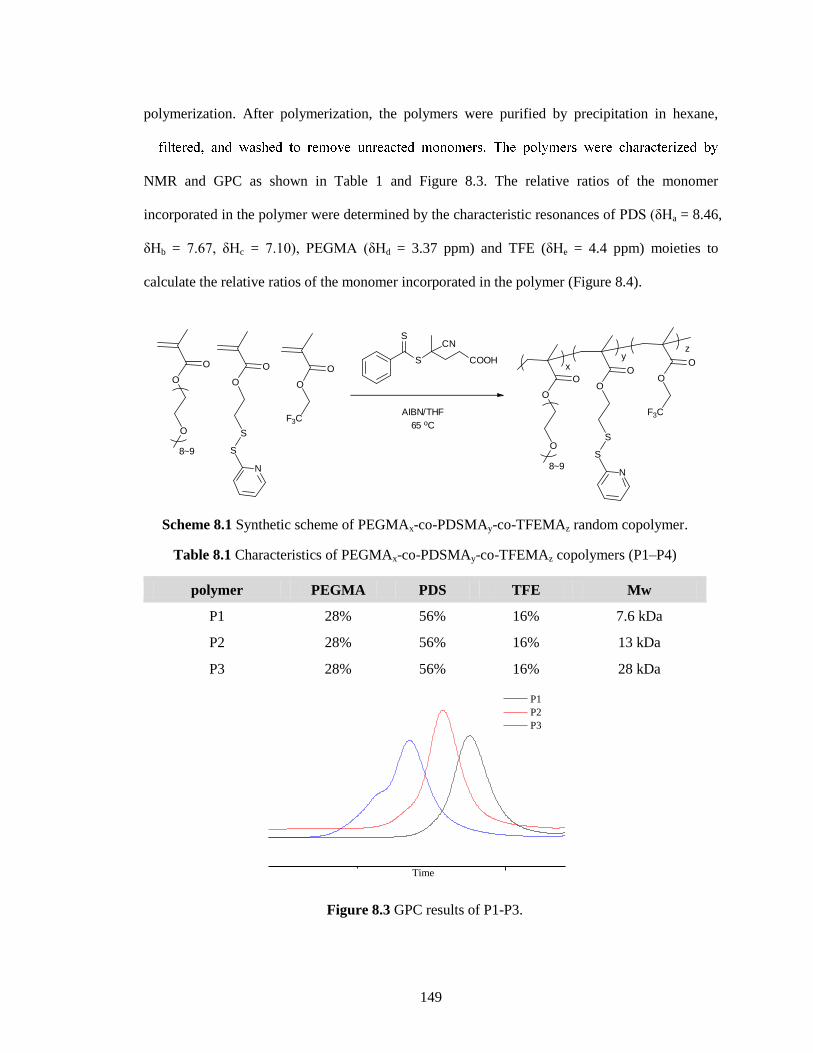
149
polymerization. After polymerization, the polymers were purified by precipitation in hexane,
NMR and GPC as shown in Table 1 and Figure 8.3. The relative ratios of the monomer
incorporated in the polymer were determined by the characteristic resonances of PDS (δHa = 8.46,
δHb = 7.67, δHc = 7.10), PEGMA (δHd = 3.37 ppm) and TFE (δHe = 4.4 ppm) moieties to
calculate the relative ratios of the monomer incorporated in the polymer (Figure 8.4).
Scheme 8.1 Synthetic scheme of PEGMAx-co-PDSMAy-co-TFEMAz random copolymer.
Table 8.1 Characteristics of PEGMAx-co-PDSMAy-co-TFEMAz copolymers (P1–P4)
polymer PEGMA PDS TFE Mw
P1 28% 56% 16% 7.6 kDa
P2 28% 56% 16% 13 kDa
P3 28% 56% 16% 28 kDa
Figure 8.3 GPC results of P1-P3.
O
O
O
O
O
S
S
N
O
O
F3C
8~9
S
SCN
COOH
AIBN/THF
65 oC
O
O
O
8~9
O
O
S
S
N
O
O
F3C
yz
x
Time
P1
P2
P3
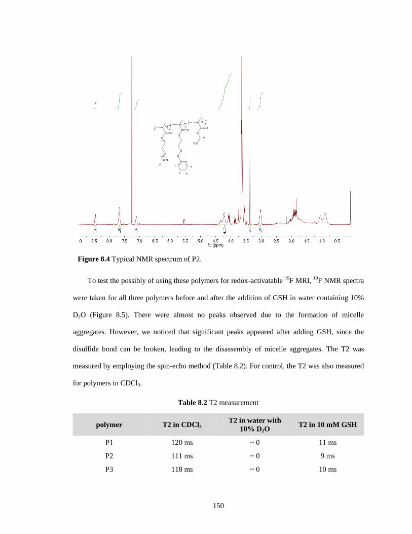
150
To test the possibly of using these polymers for redox-activatable 19
F MRI, 19
F NMR spectra
were taken for all three polymers before and after the addition of GSH in water containing 10%
D2O (Figure 8.5). There were almost no peaks observed due to the formation of micelle
aggregates. However, we noticed that significant peaks appeared after adding GSH, since the
disulfide bond can be broken, leading to the disassembly of micelle aggregates. The T2 was
measured by employing the spin-echo method (Table 8.2). For control, the T2 was also measured
for polymers in CDCl3.
Table 8.2 T2 measurement
polymer T2 in CDCl3 T2 in water with
10% D2O T2 in 10 mM GSH
P1 120 ms ~ 0 11 ms
P2 111 ms ~ 0 9 ms
P3 118 ms ~ 0 10 ms
Figure 8.4 Typical NMR spectrum of P2.
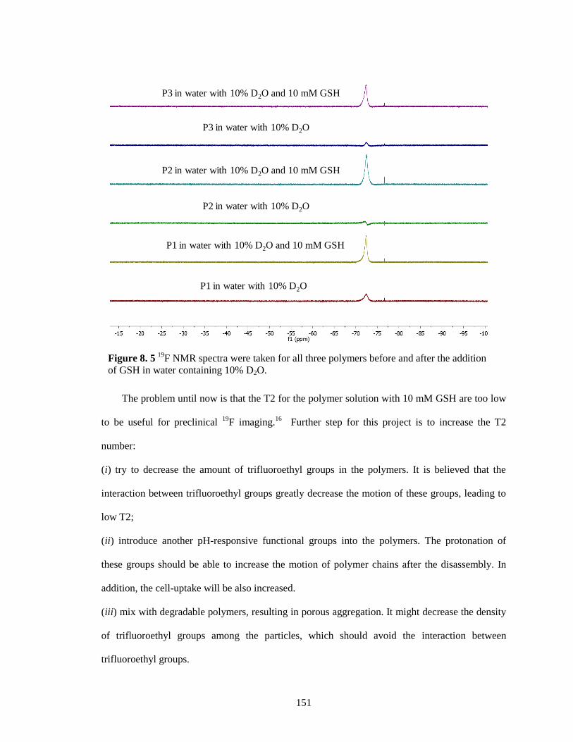
151
The problem until now is that the T2 for the polymer solution with 10 mM GSH are too low
to be useful for preclinical 19
F imaging.16
Further step for this project is to increase the T2
number:
(i) try to decrease the amount of trifluoroethyl groups in the polymers. It is believed that the
interaction between trifluoroethyl groups greatly decrease the motion of these groups, leading to
low T2;
(ii) introduce another pH-responsive functional groups into the polymers. The protonation of
these groups should be able to increase the motion of polymer chains after the disassembly. In
addition, the cell-uptake will be also increased.
(iii) mix with degradable polymers, resulting in porous aggregation. It might decrease the density
of trifluoroethyl groups among the particles, which should avoid the interaction between
trifluoroethyl groups.
Figure 8. 5 19F NMR spectra were taken for all three polymers before and after the addition
of GSH in water containing 10% D2O.
P1 in water with 10% D2O
P1 in water with 10% D2O and 10 mM GSH
P2 in water with 10% D2O
P3 in water with 10% D2O
P2 in water with 10% D2O and 10 mM GSH
P3 in water with 10% D2O and 10 mM GSH
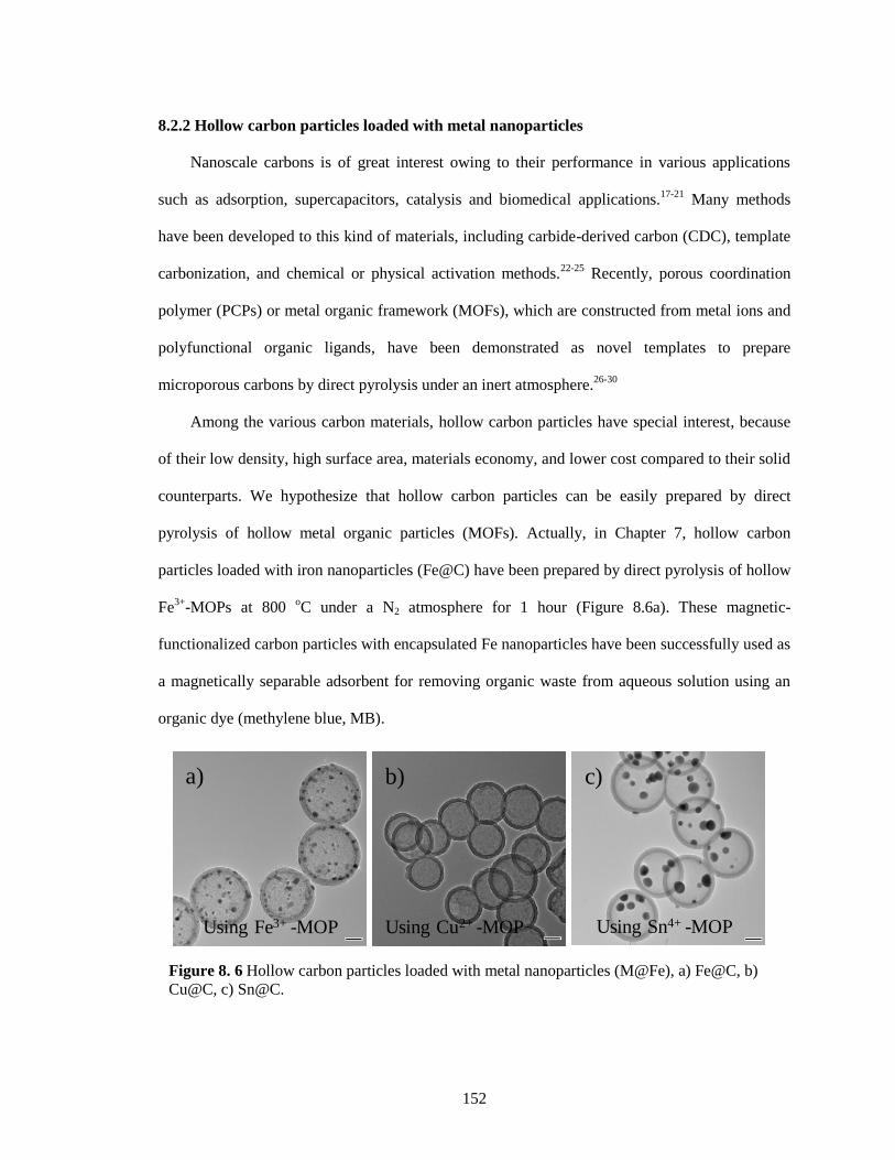
152
8.2.2 Hollow carbon particles loaded with metal nanoparticles
Nanoscale carbons is of great interest owing to their performance in various applications
such as adsorption, supercapacitors, catalysis and biomedical applications.17-21
Many methods
have been developed to this kind of materials, including carbide-derived carbon (CDC), template
carbonization, and chemical or physical activation methods.22-25
Recently, porous coordination
polymer (PCPs) or metal organic framework (MOFs), which are constructed from metal ions and
polyfunctional organic ligands, have been demonstrated as novel templates to prepare
microporous carbons by direct pyrolysis under an inert atmosphere.26-30
Among the various carbon materials, hollow carbon particles have special interest, because
of their low density, high surface area, materials economy, and lower cost compared to their solid
counterparts. We hypothesize that hollow carbon particles can be easily prepared by direct
pyrolysis of hollow metal organic particles (MOFs). Actually, in Chapter 7, hollow carbon
particles loaded with iron nanoparticles (Fe@C) have been prepared by direct pyrolysis of hollow
Fe3+
-MOPs at 800 oC under a N2 atmosphere for 1 hour (Figure 8.6a). These magnetic-
functionalized carbon particles with encapsulated Fe nanoparticles have been successfully used as
a magnetically separable adsorbent for removing organic waste from aqueous solution using an
organic dye (methylene blue, MB).
Figure 8. 6 Hollow carbon particles loaded with metal nanoparticles (M@Fe), a) Fe@C, b)
Cu@C, c) Sn@C.
Using Sn4+ -MOPUsing Cu2+ -MOPUsing Fe3+ -MOP
a) b) c)
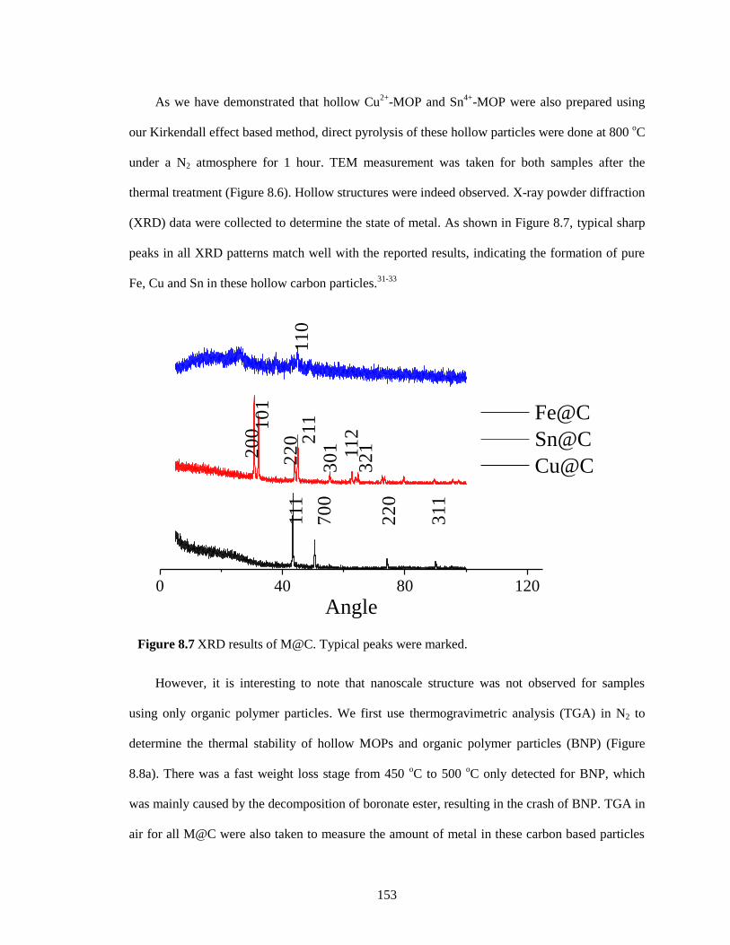
153
As we have demonstrated that hollow Cu2+
-MOP and Sn4+
-MOP were also prepared using
our Kirkendall effect based method, direct pyrolysis of these hollow particles were done at 800 oC
under a N2 atmosphere for 1 hour. TEM measurement was taken for both samples after the
thermal treatment (Figure 8.6). Hollow structures were indeed observed. X-ray powder diffraction
(XRD) data were collected to determine the state of metal. As shown in Figure 8.7, typical sharp
peaks in all XRD patterns match well with the reported results, indicating the formation of pure
Fe, Cu and Sn in these hollow carbon particles.31-33
However, it is interesting to note that nanoscale structure was not observed for samples
using only organic polymer particles. We first use thermogravimetric analysis (TGA) in N2 to
determine the thermal stability of hollow MOPs and organic polymer particles (BNP) (Figure
8.8a). There was a fast weight loss stage from 450 oC to 500
oC only detected for BNP, which
was mainly caused by the decomposition of boronate ester, resulting in the crash of BNP. TGA in
air for all M@C were also taken to measure the amount of metal in these carbon based particles
Figure 8.7 XRD results of M@C. Typical peaks were marked.
0 40 80 120
Angle
Fe@C
Sn@C
Cu@C
11
0111
70
0
22
0
311
20
0 10
1
22
0 211
30
1 11
23
21
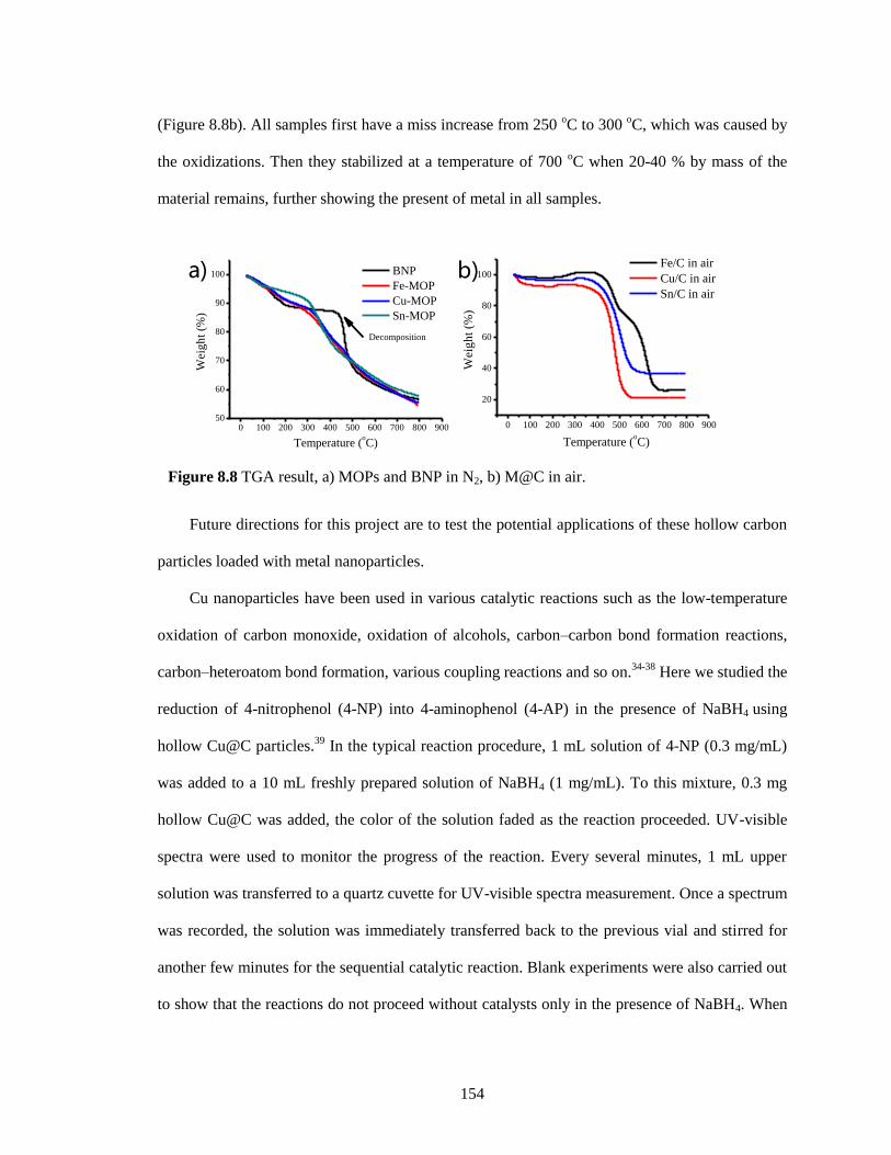
154
(Figure 8.8b). All samples first have a miss increase from 250 oC to 300
oC, which was caused by
the oxidizations. Then they stabilized at a temperature of 700 oC when 20-40 % by mass of the
material remains, further showing the present of metal in all samples.
Future directions for this project are to test the potential applications of these hollow carbon
particles loaded with metal nanoparticles.
Cu nanoparticles have been used in various catalytic reactions such as the low-temperature
oxidation of carbon monoxide, oxidation of alcohols, carbon–carbon bond formation reactions,
carbon–heteroatom bond formation, various coupling reactions and so on.34-38
Here we studied the
reduction of 4-nitrophenol (4-NP) into 4-aminophenol (4-AP) in the presence of NaBH4 using
hollow Cu@C particles.39
In the typical reaction procedure, 1 mL solution of 4-NP (0.3 mg/mL)
was added to a 10 mL freshly prepared solution of NaBH4 (1 mg/mL). To this mixture, 0.3 mg
hollow Cu@C was added, the color of the solution faded as the reaction proceeded. UV-visible
spectra were used to monitor the progress of the reaction. Every several minutes, 1 mL upper
solution was transferred to a quartz cuvette for UV-visible spectra measurement. Once a spectrum
was recorded, the solution was immediately transferred back to the previous vial and stirred for
another few minutes for the sequential catalytic reaction. Blank experiments were also carried out
to show that the reactions do not proceed without catalysts only in the presence of NaBH4. When
Figure 8.8 TGA result, a) MOPs and BNP in N2, b) M@C in air.
0 100 200 300 400 500 600 700 800 90050
60
70
80
90
100
Wei
gh
t (%
)
Temperature (oC)
BNP
Fe-MOP
Cu-MOP
Sn-MOP
Decomposition
0 100 200 300 400 500 600 700 800 900
20
40
60
80
100
Wei
gh
t (%
)
Temperature (oC)
Fe/C in air
Cu/C in air
Sn/C in air
a) b)
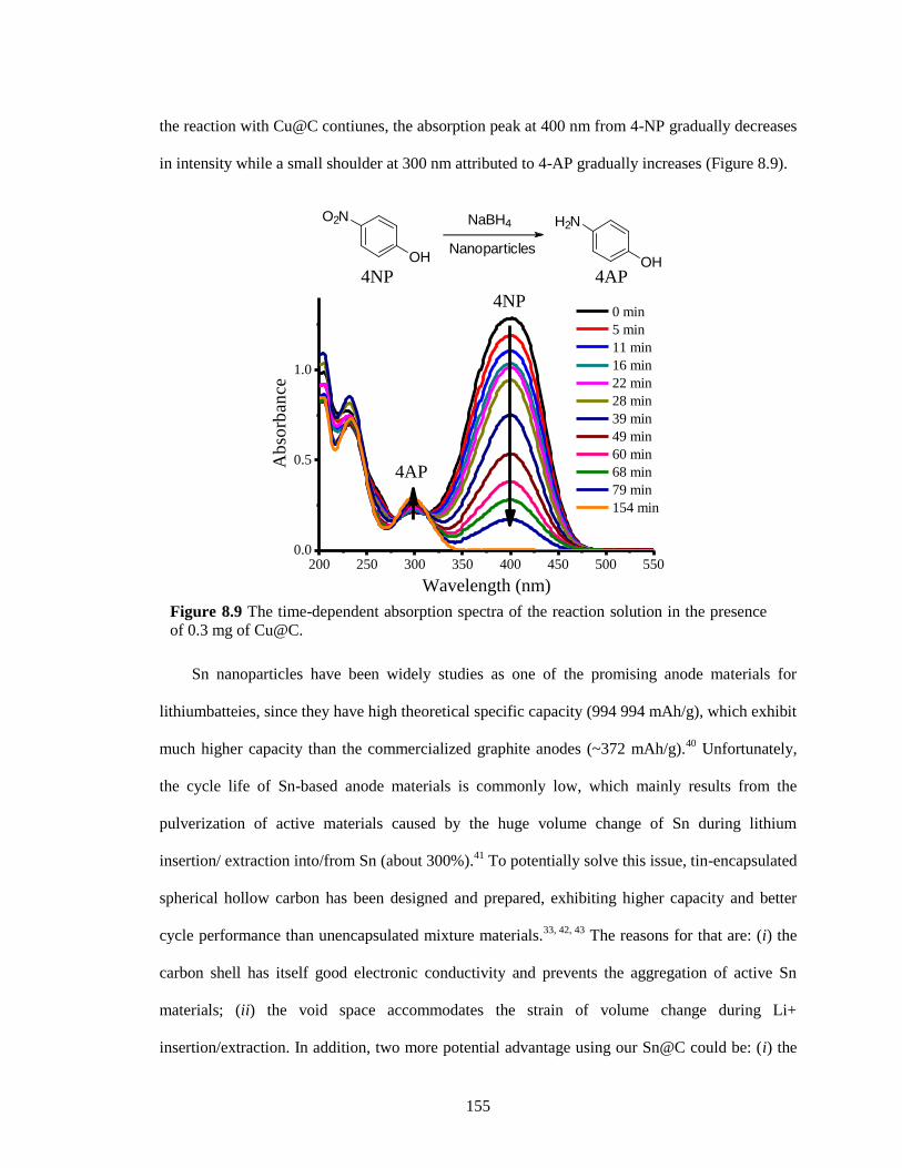
155
the reaction with Cu@C contiunes, the absorption peak at 400 nm from 4-NP gradually decreases
in intensity while a small shoulder at 300 nm attributed to 4-AP gradually increases (Figure 8.9).
Sn nanoparticles have been widely studies as one of the promising anode materials for
lithiumbatteies, since they have high theoretical specific capacity (994 994 mAh/g), which exhibit
much higher capacity than the commercialized graphite anodes (~372 mAh/g).40
Unfortunately,
the cycle life of Sn-based anode materials is commonly low, which mainly results from the
pulverization of active materials caused by the huge volume change of Sn during lithium
insertion/ extraction into/from Sn (about 300%).41
To potentially solve this issue, tin-encapsulated
spherical hollow carbon has been designed and prepared, exhibiting higher capacity and better
cycle performance than unencapsulated mixture materials.33, 42, 43
The reasons for that are: (i) the
carbon shell has itself good electronic conductivity and prevents the aggregation of active Sn
materials; (ii) the void space accommodates the strain of volume change during Li+
insertion/extraction. In addition, two more potential advantage using our Sn@C could be: (i) the
Figure 8.9 The time-dependent absorption spectra of the reaction solution in the presence
of 0.3 mg of Cu@C.
200 250 300 350 400 450 500 5500.0
0.5
1.0
4AP4NP
O2N
OH
NaBH4
Nanoparticles
H2N
OH
4NP
Ab
sorb
ance
Wavelength (nm)
0 min
5 min
11 min
16 min
22 min
28 min
39 min
49 min
60 min
68 min
79 min
154 min
4AP
156
carbon may be nitrogen-doped concerning the molecules used, which can improve the
electrochemical performance, exhibiting superhigh capacity and excellent rate capability44
; (ii)
Sn-based intermetallic compounds (MxSn; M = Cu, Fe, etc) and multiphase Sn–M alloys have
been explored to show better performance45
.
8.3 References
1. Caravan, P.; Ellison, J. J.; McMurry, T. J.; Lauffer, Randall B. Gadolinium (III) chelates as
MRI contrast agents: structure, dynamics, and applications. Chem. Rev. 1999, 99, 2293-2352;
2. Laurent, S.; Forge, D.; Port, M.; Roch, A.; Robic, C.; Vander Elst, L.; Muller, R. N. Magnetic
iron oxide nanoparticles: synthesis, stabilization, vectorization, physicochemical characterizations,
and biological applications. Chem. Rev. 2008, 108, 2064-2110;
3. Thorek, D. L. J.; Ulmert, D.; Diop, N.-F. M.; Lupu, M. E.; Doran, M. G.; Huang, R.; Abou, D.
S.; Larson, S. M.; Grimm, J. Non-invasive mapping of deep-tissue lymph nodes in live animals
using a multimodal PET/MRI nanoparticle. Nat. Commun. 2014, 5, 1-9;
4. Ruiz-Cabello, J.; Barnett, B. P.; Bottomley, P. A.; Bulte, J. W. M. Fluorine (19F) MRS and
MRI in biomedicine. NMR Biomed. 2011, 24, 114-129
5. Hwang, C.; Sinskey, A. J.; Lodish, H. F. Oxidized redox state of glutathione in the
endoplasmic reticulum. Science 1992, 257, 1496-1502.
6. Chen, X.; Zhou, Y.; Peng, X.; Yoon, J.; Fluorescent and colorimetric probes for detection of
thiols. Chem. Soc. Rev. 2010, 39, 2120-2135.
7. Yuan, L.; Lin, W.; Zhao, S.; Gao, W.; Chen, B.; He, L.; Zhu, S. A unique approach to
development of near-infrared fluorescent sensors for in vivo imaging. J. Am. Chem. Soc.2012,
134, 13510-135123.
8. Jung, H. S.; Chen, X.; Kim, J. S.; Yoon, J. Recent progress in luminescent and colorimetric
chemosensors for detection of thiols. Chem. Soc. Rev. 2013, 42, 6019-6031.
9. Herzenberg, L. A.; De Rosa, S. C.; Dubs, J. G.; Roederer, M.; Anderson, M. T.; Ela, S. W.;
Deresinski, S. C.; Herzenberg, L. A. Glutathione deficiency is associated with impaired survival
in HIV disease. Proc. Natl. Acad. Sci. U. S. A. 1997, 94, 1967-1972.
10. Shahrokhian, S. Lead phthalocyanine as a selective carrier for preparation of a cysteine-
selective electrode. Anal. Chem. 2001, 73, 5972-5978.
11. Barnham, K. J.; Masters, C. L.; Bush, A. I. Neurodegenerative diseases and oxidative stress.
Nat. Rev. Drug Discov. 2004, 3, 205-214.
12. Valko, M.; Leibfritz, D.; Moncol, J.; Cronin, M. T. D.; Mazur, M.; Telser, J. Free radicals and
antioxidants in normal physiological functions and human disease. Int. J. Biochem. Cell Biol.
2007, 39, 44-84.
157
13. Oishi, M.; Sumitani, S.; Nagasaki, Y. On-off regulation of 19F magnetic resonance signals
based on pH-sensitive PEGylated nanogels for potential tumor-specific smart 19
F MRI probes.
Bioconjugate Chem. 2007, 18, 1379-1382;
14. Takaoka, Y.; Sakamoto, T.; Tsukiji, S.; Narazaki, M.; Matsuda, T.; Tochio, H.; Shirakawa,
M.; Hamachi. I. Self-assembling nanoprobes that display off/on 19F nuclear magnetic resonance
signals for protein detection and imaging. Nat. Chem. 2009, 1, 557-561;
15. Huang, X.; Huang, G.; Zhang, S.; Sagiyama, K; Togao, O.; Ma, X.; Wang, Y.; Li, Y.; Soesbe,
T. C.; Sumer, B. D.; Takahashi, M.; Sherry, A. D.; Gao, J. Multi‐chromatic pH‐activatable 19
F‐MRI nanoprobes with binary ON/OFF pH transitions and chemical‐shift barcodes. Angew.
Chem. Int. Ed., 2013, 52, 8074-8078;
16. Thurecht, K. J.; Blakey, I.; Peng, H.; Squires, O.; Hsu, S.; Alexander, C.; Whittaker, A. K.
Functional hyperbranched polymers: toward targeted in vivo 19F magnetic resonance imaging
using designed macromolecules. J. Am. Chem. Soc. 2010, 132, 5336.
17. Zhang, S.; Chen, L.; Zhou, S.; Zhao, D.; Wu. L. Facile synthesis of hierarchically ordered
porous carbon via in situ self-assembly of colloidal polymer and silica spheres and its use as a
catalyst support. Chem. Mater., 2010, 22, 3433-3440;
18. Lee, J. -S.; Kim, S. -I.; Yoon, J. -C.; Jang, J. -H. Chemical vapor deposition of mesoporous
graphene nanoballs for supercapacitor. ACS Nano, 2013, 7, 6047-6055;
19. Wang, L.; Sun, Q.; Wang, X.; Wen, T.; Yin, J.; Wang, P.; Bai, R.; Zhang, X.; Zhang, L.; Lu,
A.; Chen, C. Using hollow carbon nanospheres as a light-induced free radical generator to
overcome chemotherapy resistance. J. Am. Chem. Soc., 2015, 137, 1947–1955;
20. Schaefer, Z. L.; Gross, M. L.; Hickner, M. A.; Schaak, R. E. Uniform hollow carbon shells:
nanostructured graphitic supports for improved oxygen-reduction catalysis. Angew. Chem. Int.
Ed., 2010, 49, 7045-7048;
21. Liu, Z.; Robinson, J. T.; Tabakman, S. M.; Yang, K.; Dai, H. Carbon materials for drug
delivery & cancer therapy. Mater. Today2011, 14, 316-323.
22. Wang, H.; Gao, Q.; Hu, J. High hydrogen storage capacity of porous carbons prepared by
using activated carbon. J. Am. Chem. Soc., 2009, 131, 7016-22.
23. Jänes, A.; Thomberg, T.; Lust, E. Synthesis and characterisation of nanoporous carbide-
derived carbon by chlorination of vanadium carbide. Carbon, 2007, 45, 2717-2722.
24. Yang, Z.; Xia, Y.; Mokaya, R. Enhanced hydrogen storage capacity of high surface area
zeolite-like carbon materials. J. Am. Chem. Soc. 2007, 129, 1673-1679.
25. Ania, C. O.; Khomenko, V.; Raymundo‐Piñero, E.; Parra, J. B.; Beguin, F. The large
electrochemical capacitance of microporous doped carbon obtained by using a zeolite template.
Adv. Funct. Mater., 2007, 17, 1828-1836.
26. Hu, J.; Wang, H.; Gao, Q.; Guo, H. Porous carbons prepared by using metal–organic
framework as the precursor for supercapacitors. Carbon, 2010, 48, 3599-3606.
158
27. Yang, S. J.; Park, C. R. Preparation of Highly Moisture-Resistant Black-Colored Metal
Organic Frameworks. Adv. Mater., 2012, 24, 4010-4013.
28. Aijaz, A.; Fujiwara, N.; Xu, Q. From metal–organic framework to nitrogen-decorated
nanoporous carbons: high CO2 uptake and efficient catalytic oxygen reduction. J. Am. Chem.
Soc., 2014, 136, 6790-6793.
29. Hu, M.; Reboul, J.; Furukawa, S.; Torad, N. L.; Ji, Q.; Srinivasu, P.; Ariga, K.; Kitagawa, S.;
Yamauchi, Y. Direct carbonization of Al-based porous coordination polymer for synthesis of
nanoporous carbon. J. Am. Chem. Soc., 2012, 134, 2864-2867.
30. Lee, H. J.; Choi, S.; Oh, M.; Well-dispersed hollow porous carbon spheres synthesized by
direct pyrolysis of core-shell type metal-organic frameworks and their sorption properties. Chem.
Commun., 2014, 50, 4492-4495.
31. Wang, H.; Yan, N.; Li, Y.; Zhou, X.; Chen, J.; Yu, B.; Gong, M.; Chen, Q. Fe nanoparticle-
functionalized multi-walled carbon nanotubes: one-pot synthesis and their applications in
magnetic removal of heavy metal ions. J. Mater. Chem., 2012, 22, 9230-9236.
32. Patra, A. K.; Dutta, A.; Bhaumik, A. Cu nanorods and nanospheres and their excellent
catalytic activity in chemoselective reduction of nitrobenzenes. Cataly. Commun. 2010, 11, 651-
655.
33. Ni, W.; Wang, Y.; Xu, R. Formation of Sn@ C yolk-shell nanospheres and core-sheath
nanowires for highly reversible lithium storage. Part. Part. Syst. Charact. 2013, 30, 873-880.
34. Tu, C. H.; Wang, A. Q.; Zheng, M. Y.; Wang X. D.; Zhang, T. Factors influencing the
catalytic activity of SBA-15-supported copper nanoparticles in CO oxidation. Appl. Catal., A,
2006, 297, 40-47.
35. Kalbasi, R. J.; Nourbakhsh, A. A.; Zia, M. Aerobic oxidation of alcohols catalyzed by copper
nanoparticle-polyacrylamide/SBA-15 as novel polymer-inorganic hybrid. J. Inorg. Organomet.
Polym., 2012, 22, 536-542.
36. Ranu, B. C.; Dey, R.; Chatterjee T. ; Ahammed, S. Copper nanoparticle-
carbon and carbon heteroatom bond formation with a greener perspective. ChemSusChem, 2012,
5, 22-44.
37. Ahammed, S.; Saha, A.; Ranu, B. C. Hydrogenation of azides over copper nanoparticle
surface using ammonium formate in water. J. Org. Chem., 2011, 76, 7235-7239.
38. Dulle, J.; Thirunavukkarasu, K.; Mittelmeijer-Hazeleger, M. C.; Andreeva, D. V.; Shiju, N. R.;
Rothenberg, G. Efficient three-component coupling catalysed by mesoporous copper-aluminum
based nanocomposites. Green Chem., 2013, 15, 1238-1243.
39. Deka, P.; Deka, R. C.; Bharali, P. In situ generated copper nanoparticle catalyzed reduction of
4-nitrophenol. New J.Chem., 2014, 38,1789.
40. Winter, M.; Besenhard, J. O. Electrochemical lithiation of tin and tin-based intermetallics and
composites. Electrochim. Acta 1999, 45, 31.
159
41. Vaughey, J. T.; O'Hara, J.; Thackeray, M. M. Intermetallic Insertion Electrodes with a Zinc
Blende-Type Structure for Li Batteries: A Study of Li x InSb (0≤ x≤ 3). Electrochem. Solid-State
Lett., 2000, 3,13–16.
42. Lee, K. T.; Jung, Y. S.; Oh, S. M. Synthesis of tin-encapsulated spherical hollow carbon for
anode material in lithium secondary batteries. J. Am. Chem. Soc., 2003, 125, 5652-5653.
43. Zhang, W.; Hu, J. -S.; Guo, Y. -G.; Zheng, S. -F.; Zhong, L. -S.; Song, W. -G.; Wan, L. -J.
Tin-nanoparticles encapsulated in elastic hollow carbon spheres for high-performance anode
material in lithium-ion batteries. Adv. Mater., 2008, 20, 1160-1165.
44. Qie, L.; Chen, W.; Wang, Z.; Shao, Q.; Li, X.; Yuan, L.; Hu, X.; Zhang, W.; Huang, Y.
Nitrogen-doped porous carbon nanofiber webs as anodes for lithium ion batteries with a
superhigh capacity and rate capability. Adv. Mater., 2012, 24, 2047-2050.
45. Park, C. -M.; Kim, J. -H.; Kim, H.; Sohn, H. -J.; Li-alloy based anode materials for Li
secondary batteries. Chem. Soc. Rev., 2010, 39, 3115–3141.
160
BIBLIOGRAPHY
Aathimanikandan, S. V.; Savariar, E. N.; Thayumanavan, S. Temperature-sensitive dendritic
micelles. J. Am. Chem. Soc. 2005, 127, 14922-14929.
Adam, W.; Grimm, G. N.; Marquardt, S.; Saha-Möller, C. R. Are pyridinethiones reliable
photochemical oxyl-radical sources for photobiological studies? The importance of secondary
photolysis products in the guanine oxidation of 2'-deoxyguanosine and cell-free DNA. J. Am.
Chem. Soc. 1999, 121, 1179-1185;
Ahammed, S.; Saha, A.; Ranu, B. C. Hydrogenation of azides over copper nanoparticle surface
using ammonium formate in water. J. Org. Chem., 2011, 76, 7235–7239.
Aida, T.; Meijer, E. W.; Stupp, S. I. Functional supramolecular polymers. Science, 2012, 335,
813–817
Aijaz, A.; Fujiwara, N.; Xu, Q. From metal–organic framework to nitrogen-decorated nanoporous
carbons: high CO2 uptake and efficient catalytic oxygen reduction. J. Am. Chem. Soc., 2014, 136,
6790-6793.
Allen, T. M.; Cullis, P. R. Drug delivery systems: entering the mainstream. Science 2004, 303,
1818-1822.
Ameloot, R.; Vermoortele, F.; Vanhove, W.; Roeffaers, M. B.J. ; Sels, B. F.; De Vos, D. E.
Interfacial synthesis of hollow metal–organic framework capsules demonstrating selective
permeability. Nat. Chem. 2011, 3, 382.
Ania, C. O.; Khomenko, V.; Raymundo‐Piñero, E.; Parra, J. B.; Beguin, F. The large
electrochemical capacitance of microporous doped carbon obtained by using a zeolite template.
Adv. Funct. Mater., 2007, 17, 1828-1836.
Arakawa, T.; Timasheff, S. N. Preferential interactions of proteins with salts in concentrated
solutions. Biochemistry. 1982, 21, 6545-6552.
Bachler, V.; Metzler-Nolte, N. The π-electron-accepting ability of the boron atom in
ethynylboranes and related compounds-an approximate weight computation for resonance
structures. Eur. J. Inorg. Chem. 1998, 6, 733-744;
Bae, Y.; Fukushima, S.; Harada, A.; Kataoka, K. Design of environment-sensitive supramolecular
assemblies for intracellular drug delivery: Polymeric micelles that are responsive to intracellular
pH change. Angew. Chem., Int. Ed., 2003, 42, 4640–4643.
Bahadur, K. C. R.; Xu, P. Multicompartment intracellular self‐expanding nanogel for targeted
delivery of drug cocktail. Adv. Mater., 2012, 24, 6479–6483.
Baldwin, R. L. How Hofmeister ion interactions affect protein stability. Biophys. J. 1996, 71,
2056-2063.
Robert-Banchereau, E.; Lacombe, S.; Ollivier, J. Solution reactivity of thiyl radicals with
molecular oxygen: Unsensitized photooxidation of dimethyldisulfide. Tetrahedron Lett. 1995, 36,
8197-8200;
161
Banerjee, R.; Phan, A.; Wang, B.; Knobler, C.; Furukawa, H.; O'Keeffe, M.; Yaghi, O. M. High-
throughput synthesis of zeolitic imidazolate frameworks and application to CO2 capture. Science
2008, 319, 939.
Bapat, A. P.; Roy, D.; Ray, J. G.; Savin, D. A.; Sumerlin, B. S. Dynamic-covalent
macromolecular stars with boronic ester linkages. J. Am. Chem. Soc. 2011, 133, 19832-19838.
Barnham, K. J.; Masters, C. L.; Bush, A. I. Neurodegenerative diseases and oxidative stress. Nat.
Rev. Drug Discov. 2004, 3, 205-214.
Barrell, M. J.; Campaña, A. G.; von Delius, M.; Geertsema, E. M.; Leigh, D. A. Light-driven
transport of a molecular walker in either direction along a molecular track. Angew. Chem. Int. Ed.
2011, 50, 285-290;
Basak, D.; Kumar, R.; Ghosh, S. Telechelic poly(disulfide)s and related block copolymer.
Macromol. Rapid Commun., 2014, 35, 1340–1344.
Bickerton, S.; Jiwpanich, S.; Thayumanavan, S. Interconnected roles of scaffold
hydrophobicity, drug loading, and encapsulation stability in polymeric nanocarriers. Mol.
Pharmaceutics 2012, 9, 3569-3578.
Bikram, M; West, J. L. Thermo-responsive systems for controlled drug delivery. Expert Opin
Drug Deliv. 2008, 10, 1077-1091;
Bordello, J.; Novo, M.; Al-Soufi, W. Exchange-dynamics of a neutral hydrophobic dye in
micellar solutions studied by fluorescence correlation spectroscopy. J. Colloid Interface Sci.
2010, 345, 369-376.
Brewer, S. H.; Allen, A. M.; Lappi, S. E.; Chasse, T. L.; Briggman, K. A.; Gorman, C. B.;
Franzen, S. Infrared detection of a phenylboronic acid terminated alkane thiol monolayer on gold
surfaces. Langmuir 2004, 20, 5512-5520.
Cantrell, G. K.; Meyer, T. Y. Catalytic CN bond formation by metal-imide-mediated imine
metathesis. J. Am. Chem. Soc. 1998, 120, 8035-8042;
Caravan, P.; Ellison, J. J.; McMurry, T. J.; Lauffer, Randall B. Gadolinium (III) chelates as MRI
contrast agents: structure, dynamics, and applications. Chem. Rev. 1999, 99, 2293 – 2352;
Carné, A.; Carbonell, C.; Imaz, I.; Maspoch, D. Nanoscale metal–organic materials. Chem. Soc.
Rev., 2011, 40, 291–305
Carné-Sánchez, A.; Imaz, I.; Cano-Sarabia, M.; Maspoch, D. A spray-drying strategy for
synthesis of nanoscale metal–organic frameworks and their assembly into hollow superstructures.
Nat. Chem. 2013, 5, 203.
Caruso, F.; Caruso, R. A.; Möhwald, H. Nanoengineering of inorganic and hybrid hollow spheres
by colloidal templating. Science 1998, 282, 1111.
Caspar, D. L. Movement and self-control in protein assemblies. Quasi-equivalence revisited.
Biophys J. 1980, 32, 103-138.
162
Cerritelli, S.; Velluto, D.; Hubbell, J. A. PEG-SS-PPS: reduction-sensitive disulfide block
copolymer vesicles for intracellular drug delivery. Biomacromolecules, 2007, 8, 1966–1972.
Chakravarthi, S.; Jessop, C. E.; Bulleid, N. J. The role of glutathione in disulphide bond
formation and endoplasmic-reticulum-generated oxidative stress. EMBO reports 2006, 7, 271-275;
Chen, X.; Zhou, Y.; Peng, X.; Yoon, J.; Fluorescent and colorimetric probes for detection of
thiols. Chem. Soc. Rev. 2010, 39, 2120–2135.
Cheng, L. X.; Tang, J. J.; Luo, H.; Jin, X. L.; Dai, F.; Yang, J.; Qian, Y. P.; Li, X. Z.; Zhou, B.
Antioxidant and antiproliferative activities of hydroxyl-substituted Schiff bases. Bioorg. Med.
Chem. Lett. 2010, 20, 2417-2420.
Choi, S. W.; Zhang, Y.; Xia, Y. N. A temperature-sensitive drug release system based on phase-
change materials. Angew. Chem., Int. Ed., 2010, 49, 7904–7908.
Christinat, N.; Scopelliti, R.; Severin, K. Multicomponent assembly of boron-based dendritic
nanostructures. J. Org. Chem. 2007, 72, 2192-2200.
Christinat, N.; Scopelliti, R.; Severin, K. Multicomponent assembly of boronic acid based
macrocycles and cages. Angew. Chem. Int. Ed. 2008, 47, 1848-1852.
Chung, J. W.; Lee, K.; Neikirk, C.; Nelson, C. M.; Priestley, R. D. Photoresponsive coumarin-
stabilized polymeric nanoparticles as a detectable drug carrier. Small 2012, 8, 1693-1700;
Chung, M.-K.; Hebling, C. M.; Jorgenson, J. W.; Severin, K.; Lee, S. J.; Gagné, M. R.
Deracemization of a dynamic combinatorial library induced by (−)-cytidine and (−)-2-
thiocytidine. J. Am. Chem. Soc. 2008, 130, 11819-11827.
Colson, J. W.; Woll, A. R.; Mukherjee, A.; Levendorf, M. P.; Spitler, E. L.; Shields, V. B.;
Spencer, M. G.; Park, J.; Dichtel, W. R. Oriented 2D covalent organic framework thin films on
single-layer grapheme. Science 2011, 332, 228-231.
Connon, S. J.; Blechert, S.. Recent developments in olefin cross-metathesis. Angew. Chem. Int.
Ed. 2003, 42, 1900-1923;
Cruz-Huerta, J.; Salazar-Mendoza, D.; Hernandez-Paredes, J.; Hernandez Ahuactzi, I. F.; Hopfl,
H. N-containing boronic esters as self-complementary building blocks for the assembly of 2D and
3D molecular networks. Chem. Commun. 2012, 48, 4241-4243.
Huang, Y.; Duan, X.; Wei, Q.; Lieber, C. M. Functional nanoscale electronic devices assembled
using silicon nanowire building blocks. Science 2001, 291, 630.
Dan, K.; Rajdev, P.; Deb, J.; Jana, S. S.; Ghosh, S. Remarkably stable amphiphilic random
copolymer assemblies: A structure–property relationship study. J. Polym. Sci. A Polym.
Chem. 2013, 51, 4932-4943.
De Greef, T. F. A.; J. Smulders, M. M.; Wolffs, M.; Schenning, A. P. H. J.; Sijbesma, R. P.;
Meijer, E. W. Supramolecular polymerization. Chem Rev 2009, 109, 5687-5754.
163
Deka, P.; Deka, R. C.; Bharali, P. In situ generated copper nanoparticle catalyzed reduction of 4-
nitrophenol. New J.Chem., 2014, 38,1789.
Ding, C. X.; Gu, J. X.; Qu, X. Z.; Yang, Z. Z. Preparation of multifunctional drug carrier for
tumor-specific uptake and enhanced intracellular delivery through the conjugation of weak acid
labile linker. Bioconjugate Chem. 2009, 20, 1163–1170;
Dirksen, A.; Dirksen, S.; Hackeng, T. M.; Dawson. P. E.. Nucleophilic catalysis of hydrazone
formation and transimination: implications for dynamic covalent chemistry. J. Am. Chem. Soc.
2006, 128, 15602-15603;
Dong, H.; Shu, J. Y.; Dube, N.; Ma, Y.; Tirrell, M. V.; Downing, K. H.; Xu, T. 3-Helix
micelles stabilized by polymer springs. J. Am. Chem. Soc. 2012, 134, 11807-11814.
Dong, W.; Kishimura, A.; Anraku, Y.; Chuanoi, S.; Kataoka, K. Monodispersed polymeric
nanocapsules: spontaneous evolution and morphology transition from reducible hetero-PEG
PICmicelles by controlled degradation. J. Am. Chem. Soc., 2009, 131, 3804–3805.
Du, J. Z.; Tang, Y. Q.; Lewis, A. L.; Armes, S. P. pH-Sensitive vesicles based on a biocompatible
zwitterionic diblock copolymer. J. Am. Chem. Soc., 2005, 127, 17982–17983;
Du, J. Z.; Sun, T. M.; Song, W. J.; Wu, J.; Wang, J. A tumor-acidity-activated charge-
conversional nanogel as an intelligent vehicle for promoted tumoral-cell uptake and drug
delivery. Angew. Chem. Int. Ed., 2010, 49, 3621-3626.
Du, J. Z.; Du, X. J.; Mao, C. Q.; Wang, J. Tailor-made dual pH-sensitive polymer_doxorubicin
nanoparticles for efficient anticancer drug delivery. J. Am. Chem. Soc., 2011, 133, 17560–17563;
Dulle, J.; Thirunavukkarasu, K.; Mittelmeijer-Hazeleger, M. C.; Andreeva, D. V.; Shiju, N. R.;
Rothenberg, G. Efficient three-component coupling catalysed by mesoporous copper-aluminum
based nanocomposites. Green Chem., 2013, 15, 1238–1243.
Ejima, H.; Richardson, J. J.; Liang, K.; Best, J. P.; van Koeverden, M. P.; Such, G. K.; Cui, J.;
Caruso, F. One-step assembly of coordination complexes for versatile film and particle
engineering. Science 2013, 341, 154.
El-Kaderi, H. M.; Hunt, J. R.; Mendoza-Cortes, J. L.; Cote, A. P.; Taylor, R. E.; O'Keeffe, M.;
Yaghi, O. M. Designed synthesis of 3D covalent organic frameworks. Science 2007, 316, 268-
272.
England, J. L.; Haran, G. Role of solvation effects in protein denaturation: from thermodynamics
to single molecules and back. Annu. Rev. Phys. Chem. 2011, 62, 257-277.
Folmer-Andersen, J. F.; Lehn, J.-M. Thermoresponsive dynamers: thermally induced, reversible
chain elongation of amphiphilic poly (acylhydrazones). J. Am. Chem. Soc. 2011, 133, 10966-
10973.
Franzen, S.; Ni, W.; Wang, B. Study of the mechanism of electron-transfer quenching by boron-
nitrogen adducts in fluorescent sensors. J. Phys. Chem. B 2003, 107, 12942-12948.
164
Fu, G. C.; Nguyen, S. T.; Grubbs, G. H. Catalytic ring-closing metathesis of functionalized dienes
by a ruthenium carbene complex. J. Am. Chem. Soc. 1993, 115, 9856-9857;
Fuller, J. M.; Raghupathi, K. R.; Ramireddy, R. R.; Subrahmanyam, A. V.; Yesilyurt, V.;
Thayumanavan, S. Temperature-sensitive transitions below LCST in amphiphilic dendritic
assemblies: host–guest implications. J. Am. Chem. Soc. 2013, 135, 8947-8954;
Furukawa, H.; Cordova, K. E.; O’Keeffe, M.; Yaghi, O. M. The chemistry and applications of
metal-organic frameworks. Science 2013, 341, 1230444.
Gao, Y.; Yang, C.; Liu, X.; Ma, R.; Kong, D.; Shi, L. A multifunctional nanocarrier based on
nanogated mesoporous silica for enhanced tumor-specific uptake and intracellular delivery.
Macromol Biosci. 2012, 12, 251-259;
Ge, Z.; Liu, S. Functional block copolymer assemblies responsive to tumor and intracellular
microenvironments for site-specific drug delivery and enhanced imaging performance. Chem. Soc.
Rev., 2013, 42, 7289–7325.
Gehlen, M. H.; De Schryver, F. C. Time-resolved fluorescence quenching in micellar
assemblies. Chem. Rev. 1993, 93, 199-221.
Ghosh, S.; Basu, S.; Thayumanavan, S. Simultaneous and reversible functionalization of
copolymers for biological applications. Macromolecules 2006, 39, 5595-5597.
Gjaltema, A.; Van Loosdrecht, M. C. M.; Heijnen, J. Abrasion of suspended biofilm pellets
in airlift reactors: Effect of particle size. J. Biotechnol. Bioeng. 1997, 55, 206-215
González-Toro, D. C.; Ryu, J. H.; Chacko, R. T.; Zhuang, J. M.; Thayumanavan, S. Concurrent
binding and delivery of proteins and lipophilic small molecules using polymeric nanogels. J. Am.
Chem. Soc., 2012, 134, 6964–6967;
Griset, A. P.; Walpole, J.; Liu, R.; Gaffey, A.; Colson Y. L.; Grinstaff, M. W. Expansile
nanoparticles: synthesis, characterization, and in vivo efficacy of an acid-responsive polymeric
drug delivery system. J. Am. Chem. Soc., 2009, 131, 2469–2471;
Gu, J.; Cheng, W. P.; Liu, J.; Lo, S. Y.; Smith, D.; Qu, X.; Yang, Z. pH-triggered reversible
stealth polycationic micelles. Biomacromolecules 2008, 9, 255-262;
Gupta, D.; Knight, A. R. Reactions of thiyl radicals. XIII. Photochemically induced exchange
reactions of liquid alkyl disulfides. Can. J. Chem. 1980, 58, 1350-1354;
Ha, Y. M.; Lee, H. J.; Park, D.; Jeong, H. O.; Park, J. Y.; Park, Y. J.; Lee, K. J.; Lee, J. Y.; Moon,
H. R.; Chung, H. Y. Molecular docking studies of (1E,3E,5E)-1,6-bis(substituted phenyl)hexa-
1,3,5-triene and 1,4-bis(substituted trans-styryl)benzene analogs as novel tyrosinase inhibitors.
Biol. Pharm. Bull. 2012, 36, 55-65.
Hall, L. W.; Odom, J. D.; Ellis, P. D. Nuclear magnetic resonance investigation of the nature of
the boron-carbon bond in some vinylboranes. J. Am. Chem. Soc. 1975, 97, 4527-4531.
Hawker, C. J.; Bosman, A. W.; Harth, E. New polymer synthesis by nitroxide mediated living
radical polymerizations. Chem. Rev., 2001, 101, 3661–3688.
165
Hawker, C. J.; Wooley, K. L. The convergence of synthetic organic and polymer chemistries.
Science 2005, 309, 1200.
He, J.; Tong, X.; Zhao, Y. Photoresponsive nanogels based on photocontrollable cross-links.
Macromolecules 2009, 42, 4845-4852;
He, Y.; Shao, Q.; Chen, S. F.; Jiang, S. Y. Water mobility: a bridge between the Hofmeister series
of ions and the friction of zwitterionic surfaces in aqueous environments. J. Phys. Chem. C. 2011,
115, 15525–15531.
Helmlinger, G.; Schell, A.; Dellian, M.; Forbes, N. S.; Jain, R. K. Acid production in glycolysis-
impaired tumors provides new insights into tumor metabolism. Clin. Cancer Res. 2002, 8, 1284–
1291;
Herzenberg, L. A.; De Rosa, S. C.; Dubs, J. G.; Roederer, M.; Anderson, M. T.; Ela, S. W.;
Deresinski, S. C.; Herzenberg, L. A. Glutathione deficiency is associated with impaired survival
in HIV disease. Proc. Natl. Acad. Sci. U. S. A. 1997, 94, 1967–1972.
Hilf, S.; Kilbinger, A. F. M. Functional end groups for polymers prepared using ring-opening
metathesis polymerization. Nat. Chem., 2009, 1, 537–546.
Hirsch, A. K. H.; Buhler, E.; Lehn, J.-M. Biodynamers: self-organization-driven formation of
doubly dynamic proteoids. J. Am. Chem. Soc. 2012, 134, 4177-4183.
Hofmeister, F. On the understanding of the effects of salts, second report. On irregularities in the
precipitating effect of salts and their relationship to their physical behavior. Arch. Exp. Pathol.
Pharmocol. 1888, 24, 247-260.
Höpfl, H. J. The tetrahedral character of the boron atom newly defined—a useful tool to evaluate
the N→ B bond. Organomet. Chem. 1999, 581, 129-149.
Horcajada, P.; Gref, R.; Baati, T.; Allan, P. K.; Maurin, G.; Couvreur, P.; Ferey, G.; Morris, R.
E.; Serre, C. Metal–organic frameworks in biomedicine. Chem. Rev. 2011, 112, 1232.
Howes, P. D.; Chandrawati, R.; Stevens, M. M. Colloidal nanoparticles as advanced biological
sensors. Science 2014, 346, 1247390.
Hu, J.; Wang, H.; Gao, Q.; Guo, H. Porous carbons prepared by using metal–organic framework
as the precursor for supercapacitors. Carbon, 2010, 48, 3599-3606.
Hu, L. C.; Yonamine, Y.; Lee, S. H.; Van der Veer, W. E.; Shea, K. J. Light-triggered charge
reversal of organic–silica hybrid nanoparticles. J. Am. Chem. Soc., 2012, 134, 11072–11075;
Hu, M.; Reboul, J.; Furukawa, S.; Torad, N. L.; Ji, Q.; Srinivasu, P.; Ariga, K.; Kitagawa, S.;
Yamauchi, Y. Direct carbonization of Al-based porous coordination polymer for synthesis of
nanoporous carbon. J. Am. Chem. Soc., 2012, 134, 2864-2867.
Hu, Y. Q.; Kim, M. S.; Kim, B. S.; Lee, D. S. Synthesis and pH-dependent micellization of 2-
(diisopropylamino)ethyl methacrylate based amphiphilic diblock copolymers via RAFT
polymerization. Polymer, 2007, 48, 3437–3443;
166
Huang, X.; Huang, G.; Zhang, S.; Sagiyama, K; Togao, O.; Ma, X.; Wang, Y.; Li, Y.; Soesbe, T.
C.; Sumer, B. D.; Takahashi, M.; Sherry, A. D.; Gao, J. Multi‐chromatic pH‐activatable
19F‐MRI nanoprobes with binary ON/OFF pH transitions and chemical‐shift barcodes. Angew.
Chem. Int. Ed., 2013, 52, 8074-8078;
Huo, J.; Wang, L.; Irran, E.; Yu, H.; Gao, J.; Fan, D.; Li, B.; Wang, J.; Ding, W.; Amin, A. M.; Li,
C.; Ma, L. Hollow ferrocenyl coordination polymer microspheres with micropores in shells
prepared by Ostwald ripening. Angew. Chem. Int. Ed. 2010, 49, 9237.
Hwang, C.; Sinskey, A. J.; Lodish, H. F. Oxidized redox state of glutathione in the endoplasmic
reticulum. Science 1992, 257, 1496–1502.
Ikkala, O.; ten Brinke, G. Functional materials based on self-assembly of polymeric
supramolecules. Science, 2002, 295, 2407–2409.
Iwasawa, N.; Takahagi, H. Boronic esters as a system for crystallization-induced dynamic self-
assembly equipped with an on-off switch for equilibration. J. Am. Chem. Soc. 2007, 129, 7754-
7755.
Jäkle, F.; Lewis acidic organoboron polymers. Coord. Chem. Rev., 2006, 250, 1107-1121.
Janata, J.; Josowicz, M. Conducting polymers in electronic chemical sensors. Nat. Mater., 2003,
2, 19–24.
Janeliunas, D.; van Rijn, P.; Boekhoven, J.; Minkenberg, C. B.; van Esch, J. H.; Eelkema, R.
Aggregation-driven reversible formation of conjugated polymers in water. Angew. Chem. Int. Ed.
2013, 52, 1998-2001;
Jänes, A.; Thomberg, T.; Lust, E. Synthesis and characterisation of nanoporous carbide-derived
carbon by chlorination of vanadium carbide. Carbon, 2007, 45, 2717-2722.
Jiang, J.; Qi, B.; Lepage, M.; Zhao, Y. Polymer micelles stabilization on demand through
reversible photo-cross-linking. Macromolecules 2007, 40, 790-792.
Jiwpanich, S.; Ryu, J.-H.; Bickerton, S.; Thayumanavan, S. Noncovalent encapsulation stabilities
in supramolecular nanoassemblies. J. Am. Chem. Soc. 2010, 132, 10683-10685.
Johnson, J. A.; Lu, Y. Y.; Burts, A. O.; Lim, Y.-H.; Finn, M. G.; Koberstein, J. T.; Turro, N. J.;
Tirrell, D. A.; Grubbs, R. H. Core-clickable PEG-branch-azide bivalent-bottle-brush polymers by
ROMP: grafting-through and clicking-to. J. Am. Chem. Soc., 2010, 133, 559–566.
Jones, J. T. A.; Hasell, T.; Wu, X.; Bacsa, J.; Jelfs, K. E.; Schmidtmann, M.; Chong, S. Y.;
Adams, D. J.; Trewin, A.; Schiffman, F.; Cora, F.; Slater, B.; Steiner, A.; Day, G. M.; Cooper, A.
I. Modular and predictable assembly of porous organic molecular crystals. Nature 2011, 474, 367.
Jonkheijm, P.; Van der Schoot, P.; Schenning, A. P.; Meijer, E. W. Probing the solvent-assisted
nucleation pathway in chemical self-assembly. Science 2006, 313, 80-83.
Jung, H. S.; Chen, X.; Kim, J. S.; Yoon, J. Recent progress in luminescent and colorimetric
chemosensors for detection of thiols. Chem. Soc. Rev. 2013, 42, 6019-6031.
167
Kabanov, A. V.; Vinogradov, S. V. Nanogels as pharmaceutical carriers: finite networks of
infinite capabilities. Angew. Chem. Int. Ed. 2009, 48, 5418-5429.
Kakizawa, Y.; Harada, A.; Kataoka, K. Environment-sensitive stabilization of core-shell
structured polyion complex micelle by reversible cross-linking of the core through disulfide bond.
J. Am. Chem. Soc. 1999, 121, 11247-11248;
Kalbasi, R. J.; Nourbakhsh, A. A.; Zia, M. Aerobic oxidation of alcohols catalyzed by copper
nanoparticle-polyacrylamide/SBA-15 as novel polymer-inorganic hybrid. J. Inorg. Organomet.
Polym., 2012, 22, 536–542.
Kalyanasundaram, K.; Thomas, J. Environmental effects on vibronic band intensities in
pyrene monomer fluorescence and their application in studies of micellar systems. J. Am.
Chem. Soc. 1977, 99, 2039-2044.
Kim, H.-C.; Park, S.-M.; Hinsberg, W. D. Block copolymer based nanostructures: materials,
processes, and applications to electronics. Chem. Rev., 2009, 110, 146–177.
Kim, J. K.; Yang, S. Y.; Lee, Y.; Kim, Y. Functional nanomaterials based on block copolymer
self-assembly. Prog. Polym. Sci., 2010, 35, 1325–1349.
Kim, J. S.; Youk, J. H. Preparation of core cross-linked micelles using a photo-cross-linking
agent. Polymer 2009, 50, 2204-2208.
Kim, K.; Bae, B.; Kang, Y. J.; Nam, J.-M.; Kang, S.; Ryu, J.-H. Natural polypeptide-based
supramolecular nanogels for stable noncovalent encapsulation. Biomacromolecules 2013, 14,
3515-3522.
Kirkendall, E. Diffusion of zinc in alpha brass. Aime Trans. 1942, 147, 104.
Kiviranta, P. H.; Leppänen, J.; Kyrylenko, S.; Salo, H. S.; Lahtela-Kakkonen, M.; Tervo, A. J.;
Wittekindt, C.; Suuronen, T.; Kuusisto, E.; Järvinen, T. N, N'-bisbenzylidenebenzene-1, 4-
diamines and N, N'-bisbenzylidenenaphthalene-1, 4-diamines as sirtuin type 2 (SIRT2) inhibitors.
J. Med. Chem. 2006, 49, 7907-7911.
Koh, M. J.; Khan, R. K. M.; Torker, S.; Hoveyda, A. H. Broadly applicable Z- and
diastereoselective ring-opening/cross-metathesis catalyzed by a dithiolate Ru complex. Angew.
Chem. Int. Ed. 2014, 53, 1968-1972.
Koksharov, Y. A.; Gubin, S.P.; Kosobudsky, I. D.; Beltran, M.; Khodorkovsky, Y.; Tishin, A. M.
Low temperature electron paramagnetic resonance anomalies in Fe-based nanoparticles. J. Appl.
Phys. 2000, 88, 1587.
Korevaar, P. A.; George, S. J.; Markvoort, A. J.; Smulders, M. M.; Hilbers, P. A.; Schenning, A.
P.; De Greef, T. F.; Meijer, E. W. Pathway complexity in supramolecular polymerization. Nature
2012, 481, 492-496.
Korevaar, P. A.; Schaefer, C.; De Greef, T. F.; Meijer, E. W. Controlling chemical self-assembly
by solvent-dependent dynamics. J. Am. Chem. Soc. 2012, 134, 13482-13491.
168
Kuang, H.; He, H.; Hou, J.; Xie, Z.; Jing, X.; Huang, Y. Thymine modified amphiphilic
biodegradable copolymers for photo-cross-linked micelles as stable drug carriers. Macromol.
Biosci. 2013, 13, 1593-1600.
Kumar, R.; Chen, M. H.; Parmar, V. S.; Samuelson, L. A.; Kumar, J.; Nicolosi, R.;
Yoganathan, S.; Watterson, A. C. Supramolecular assemblies based on copolymers of
PEG600 and functionalized aromatic diesters for drug delivery applications. J. Am. Chem.
Soc., 2004, 126, 10640-10644.
La, D. S.; Alexander, J. B.; Cefalo, D. R.; Graf, D. D.; Hoveyda, A. H.; Schrock, R. R. Mo-
catalyzed asymmetric synthesis of dihydrofurans. Catalytic kinetic resolution and enantioselective
desymmetrization through ring-closing metathesis. J. Am. Chem. Soc. 1998, 120, 9720-9721;
LaMer, V. K.; Dinegar, R. H. Theory, production and mechanism of formation of monodispersed
hydrosols. J. Am. Chem. Soc. 1950, 72, 4847-4854.
Laurent, S.; Forge, D.; Port, M.; Roch, A.; Robic, C.; Vander Elst, L.; Muller, R. N. Magnetic
iron oxide nanoparticles: synthesis, stabilization, vectorization, physicochemical characterizations,
and biological applications. Chem. Rev. 2008, 108, 2064 – 2110;
Lee, H. J.; Choi, S.; Oh, M.; Well-dispersed hollow porous carbon spheres synthesized by direct
pyrolysis of core-shell type metal-organic frameworks and their sorption properties. Chem.
Commun., 2014, 50, 4492-4495.
Lee, H. J.; Pardridge, W. M. Monoclonal antibody radiopharmaceuticals: cationization,
pegylation, radiometal chelation, pharmacokinetics, and tumor imaging. Bioconjugate Chem.
2003, 14, 546-553;
Lee, J. -S.; Kim, S. -I.; Yoon, J. -C.; Jang, J. -H. Chemical vapor deposition of mesoporous
graphene nanoballs for supercapacitor. ACS Nano, 2013, 7, 6047–6055;
Lee, K. T.; Jung, Y. S.; Oh, S. M. Synthesis of tin-encapsulated spherical hollow carbon for
anode material in lithium secondary batteries. J. Am. Chem. Soc., 2003, 125, 5652-5653.
Lee, Y. R.; Chiu, C. L.; Lin, S. M. Ultraviolet photodissociation study of CH3SCH3 and
CH3SSCH3. J. Chem. Phys. 1994, 100, 7376-7384;
Lee, Y.; Fukushima, S.; Bae, Y.; Hiki, S.; Ishii, T.; Kataoka, K. A protein nanocarrier from
charge-conversion polymer in response to endosomal pH. J. Am. Chem. Soc. 2007, 129, 5362 –
5363;
Li, H.; Jäkle, F. Universal scaffold for fluorescent conjugated organoborane polymers. Angew.
Chem. Int. Ed., 2009, 48, 2313-2316.
Li, J.; Carnall, J.; Stuart, M. C. A.; Otto, S. Hydrogel formation upon photoinduced covalent
capture of macrocycle stacks from dynamic combinatorial libraries. Angew. Chem. Int. Ed. 2011,
50, 8384-8386;
Li, L.; Raghupathi, K.; Song, C.; Prasad, P.; Thayumanavan, S. Self-assembly of random
copolymers. Chem. commun. 2014, 50, 13417-13432.
169
Li, L.; Raghupathi, K.; Yuan, C.; Thayumanavan, S. Surface charge generation in nanogels for
activated cellular uptake at tumor-relevant pH. Chem. Sci. 2013, 4, 3654-3660;
Li, L.; Ryu, J.-H.; Thayumanavan, S. Effect of hofmeister ions on the size and encapsulation
stability of polymer nanogels. Langmuir 2013, 29, 50-55.
Li, L.; Song, C.; Jennings, M.; Thayumanavan, S. Photoinduced heterodisulfide metathesis for
reagent-free synthesis of polymer nanoparticles. Chem. commun. 2015, 51, 1425-1428.
Li, L.; Thayumanavan, S. Environment-dependent guest exchange in supramolecular
hosts. Langmuir 2014, 30, 12384-12390.
Li, L.; Yuan, C.; Dai, L.; Thayumanavan, S. Thermoresponsive Polymeric Nanoparticles:
Nucleation from Cooperative Polymerization Driven by Dative Bonds. Macromolecules 2014, 47,
5869.
Li, Y.; Song, Y.; Zhao, L.; Gaidosh, G.; Laties, A. M.; Wen, R. Direct labeling and visualization
of blood vessels with lipophilic carbocyanine dye DiI. Nat. Protoc., 2008, 3, 1703–1708.
Liu, J. Y.; Pang, Y.; Huang, W.; Zhu, Z. Y.; Zhu, X. Y.; Zhou, Y. F.; Yan, D. Y. Redox-
responsive polyphosphate nanosized assemblies: a smart drug delivery platform for cancer
therapy. Biomacromolecules, 2011, 12, 2407–2415;
Liu, K. C.; Yeo, Y. Extracellular stability of nanoparticulate drug carriers. Arch. Pharmacal
Res. 2014, 37, 16-23.
Liu, S.; Weaver, J. V. M.; Tang, Y.; Billingham, N. C.; Armes, S. P.; Tribe. K. Synthesis of shell
cross-linked micelles with pH-responsive cores using ABC triblock copolymers. Macromolecules,
2002, 35, 6121–6131.
Liu, W.; Pink, M.; Lee, D. Conjugated polymer sensors built on π-extended borasiloxane cages. J.
Am. Chem. Soc. 2009, 131, 8703-8707.
Liu, X. Y.; Mu, X. R.; Liu, Y.; Liu, H. J.; Chen, Y.; Cheng, F.; Jiang, S. C. Hyperbranched
polymers with thermoresponsive property highly sensitive to ions. Langmuir 2012, 10, 4867-
4876.
Liu, Z.; Robinson, J. T.; Tabakman, S. M.; Yang, K.; Dai, H. Carbon materials for drug delivery
& cancer therapy. Mater. Today 2011, 14, 316-323.
Nostro, P. L.; Ninham, B. W. Hofmeister phenomena: an update on ion specificity in biology.
Chem. Rev. 2012, 112, 2286-2322.
Lobana, T. S.; Kinoshita, I.; Kimura, K.; Nishioka, T.; Shiomi, D.; Isobe, K. Pyridine-2-
sulfonates as versatile ligands for the synthesis of novel coordinative and hydrogen-bonded
supramolecules. Eur. J. Inorg. Chem. 2004, 2004, 356-367;
Lou, X. W.; Wang, Y.; Yuan, C.; Lee, J. Y.; Archer, L. A. Template-free synthesis of SnO2
hollow nanostructures with high lithium storage capacity. Adv. Mater. 2006, 18, 2325.
170
Lu, J.; Owen, S. C.; Shoichet, M. S. Stability of self-assembled polymeric micelles in serum.
Macromolecules 2011, 44, 6002-6008.
Lu, Y.-X.; Guan, Z. Olefin metathesis for effective polymer healing via dynamic exchange of
strong carbon–carbon double bonds. J. Am. Chem. Soc. 2012, 134, 14226-14231;
Luo, Z.; Cai, K.; Hu, Y.; Zhao, L.; Liu, P.; Duan, L.; Yang, W. Mesoporous silica nanoparticles
end‐capped with collagen: redox‐responsive nanoreservoirs for targeted drug delivery. Angew.
Chem. Int. Ed. 2011, 50, 640-643;
Lutz, J.-F. Thermo-switchable materials prepared using the OEGMA-platform. Adv. Mater. 2011,
23, 2237-2243.
Lutz, J. F.; Akdemir, O.; Hoth, A. Point by point comparison of two thermosensitive polymers
exhibiting a similar LCST: is the age of poly(NIPAM) Over? J. Am. Chem. Soc. 2006, 128,
13046-13047.
Maeda, H.; Fang, J.; Inutsuka T.; Kitamoto, Y. Vascular permeability enhancement in solid tumor:
various factors, mechanisms involved and its implications. Int. Immunopharmacol., 2003, 3, 319–
328.
Maeda, H.; Hasegawa, M.; Hashimoto, T.; Kakimoto, T.; Nishio, S.; Nakanishi, T. Nanoscale
spherical architectures fabricated by metal coordination of multiple dipyrrin moieties. J. Am.
Chem. Soc. 2006, 128, 10024-10025.
Maeda, H.; Wu, J.; Sawa, T.; Matsumura Y.; Hori, K. Tumor vascular permeability and the EPR
effect in macromolecular therapeutics: a review. J. Controlled Release, 2000, 65, 271–284.
Magnusson, J. P.; Khan, A.; Pasparakis, G.; Saeed, A. O.; Wang, W. X.; Alexander, C. Ion-
sensitive isothermal responsive polymers prepared in water. J. Am. Chem. Soc. 2008, 130, 10852–
10853.
Mai, Y.; Eisenberg, A. Self-assembly of block copolymers. Chem. Soc. Rev., 2012, 41, 5969–
5985.
Markus, G.; Karush, F. The disulfide bonds of human serum albumin and bovine γ-globulin1. J.
Am. Chem. Soc. 1957, 79, 134-139;
Martin, C. R. Nanomaterials-a membrane-based synthetic approach. Science 1994, 266, 1961.
Matsumoto, N. M.; González-Toro, D. C.; Chacko, R. T.; Maynard, H. D.; Thayumanavan. S.
Synthesis of nanogel–protein conjugates. Polym. Chem., 2013, 4, 2464-2469.
Matyjaszewski, K.; Tsarevsky, N. V. Macromolecular engineering by atom transfer radical
polymerization. J. Am. Chem. Soc., 2014, 136, 6513–6533.
McCoy, C. P.; Rooney, C.; Edwards, C. R.; Jones, D. S.; Gorman, S. P. Light-triggered molecule-
scale drug dosing devices. J. Am. Chem. Soc., 2007, 129, 9572–9573;
Meyer, C. D.; Joiner, C. S.; Stoddart, J. F. Template-directed synthesis employing reversible
imine bond formation. Chem. Soc. Rev. 2007, 36, 1705-1723;
171
Milligan, B.; Rivett, D. E.; Savige, W. E. The photolysis of dialkyl sulphides, disulphides, and
trisulphides. Aust. J. Chem. 1963, 16, 1020-1029;
Moad, G.; Rizzardo, E.; Thang, S. H. Living radical polymerization by the RAFT process–a third
update. Aust. J. Chem., 2012, 65, 985–1076.
Mutlu, H.; de Espinosa, L. M.; Meier, M. A. R. Acyclic diene metathesis: a versatile tool for the
construction of defined polymer architectures. Chem. Soc. Rev. 2011, 40, 1404-1445;
Mutsaers, S. E.; John M. P. Surface charge of macrophages and their interaction with charged
particles. J. Leukocyte Biol., 1988, 44, 17–26.
Nakase, I.; Takeuchi, T.; Tanaka, G.; Futaki, S. Methodological and cellular aspects that govern
the internalization mechanisms of arginine-rich cell-penetrating peptides. Adv. Drug Delivery Rev.
2008, 60, 598−607.
Nakatani, K.; Ogura, Y.; Koda, Y.; Terashima, T.; Sawamoto, M. Sequence-regulated
copolymers via tandem catalysis of living radical polymerization and in situ transesterification. J.
Am. Chem. Soc., 2012, 134, 4373–4383.
Nakayama, M.; Okano, T.; Miyazaki, T.; Kohori, F.; Sakai, K.; Yokoyama, M. Molecular design
of biodegradable polymeric micelles for temperature-responsive drug release. J. Controlled
Release, 2006, 115, 46–56.
Nakazawa, I.; Suda, S.; Masuda, M.; Asai, M.; Shimizu, T. pH-Dependent reversible polymers
formed from cyclic sugar-and aromatic boronic acid-based bolaamphiphiles. Chem. Commun.,
2000, 10, 881-882.
Nelander, B.; Sunner, S. Cogwheel effect in dialkyl disulfides. J. Am. Chem. Soc. 1972, 94, 3576-
3577;
Ni, W.; Kaur, G.; Springsteen, G.; Wang, B.; Franzen, S. Regulating the fluorescence intensity of
an anthracene boronic acid system: a B–N bond or a hydrolysis mechanism? Bioorg. Chem., 2004,
32, 571-581.
Ni, W.; Wang, Y.; Xu, R. Formation of Sn@ C yolk-shell nanospheres and core-sheath nanowires
for highly reversible lithium storage. Part. Part. Syst. Charact. 2013, 30, 873–880.
Nikiforov, V. N.; Koksharov, Y. A.; Polyakov, S. N.; Malakho, A. P.; Volkov, A. V.; Moskvina,
M. A.; Khomutov, G. B.; Irkhin, V. Y. Magnetism and Verwey transition in magnetite
nanoparticles in thin polymer film. J. Alloys Compd. 2013, 569, 58.
Nishimura, N.; Kobayashi, K. Self-assembly of a cavitand-based capsule by dynamic boronic
ester formation. Angew. Chem. Int. Ed. 2008, 47, 6255-6258.
Nishimura, N.; Yoza, K.; Kobayashi, K. Guest-encapsulation properties of a self-assembled
capsule by dynamic boronic ester bonds. J. Am. Chem. Soc. 2009, 132, 777-790.
Nishinaga, T.; Tanatani, A.; Oh, K.; Moore, J. S. The size-selective synthesis of folded oligomers
by dynamic templation. J. Am. Chem. Soc. 2002, 124, 5934-5935;
172
Nishiyabu, R.; Teraoka, S.; Matsushima, Y.; Kubo, Y. Fabrication of soft submicrospheres by
sequential boronate esterification and their dynamic behavior. ChemPlusChem 2012, 77, 201.
Niu, W.; Smith, M. D.; Lavigne, J. J. Self-assembling poly (dioxaborole) s as blue-emissive
materials. J. Am. Chem. Soc. 2006, 128, 16466-16467.
Niu, W.; Smith, M. D.; Lavigne, J. J. Substituent effects on the structure and supramolecular
assembly of bis (dioxaborole) s derived from 1, 2, 4, 5-tetrahydroxybenzene. Cryst. Growth Des.
2006, 6, 1274-1277.
Novo, M.; Felekyan, S.; Seidel, C. A.; Al-Soufi, W. Dye-exchange dynamics in micellar
solutions studied by fluorescence correlation spectroscopy. J. Phys. Chem. B 2007, 111,
3614-3624.
Oehlke, A.; Auer, A. A.; Jahre, I.; Walfort, B.; Rüffer, T.; Zoufalá, P.; Lang, H.; Spange, S.
Nitro-substituted stilbeneboronate pinacol esters and their fluoro-adducts. fluoride ion induced
polarity enhancement of arylboronate esters. J. Org. Chem. 2007, 72, 4328-4339.
Oh, J. K.; Drumright, R.; Siegwart, D. J.; Matyjaszewski, K. The development of
microgels/nanogels for drug delivery applications. Prog. Polym. Sci. 2008, 33, 448-477.
Oh, J. K.; Siegwart, D. J.; Lee, H.; Sherwood, G.; Peteanu, L.; Hollinger, J. O.; Kataoka, K.;
Matyjaszewski, K. Biodegradable nanogels prepared by atom transfer radical polymerization as
potential drug delivery carriers: synthesis, biodegradation, in vitro release, and bioconjugation. J.
Am. Chem. Soc., 2007, 129, 5939–5945.
Oh, K.; Jeong, K.-S.; Moore, J. S. Folding-driven synthesis of oligomers. Nature 2001, 414, 889-
893;
Oh, M.; Mirkin, C. A. Chemically tailorable colloidal particles from infinite coordination
polymers. Nature 2005, 438, 651-654.
Oh, M. H.; Yu, T.; Yu, S.-H.; Lim, B.; Ko, K.-T.; Willinger, M.-G.; Seo, D.-H.; Kim, B. H.; Cho,
M. G.; Park, J.-H. Galvanic replacement reactions in metal oxide nanocrystals. Science 2013, 340,
964.
Oishi, M.; Sumitani, S.; Nagasaki, Y. On-off regulation of 19F magnetic resonance signals based
on pH-sensitive PEGylated nanogels for potential tumor-specific smart 19F MRI probes.
Bioconjugate Chem. 2007, 18, 1379 – 1382;
Otsuka, H.; Nagano, S.; Kobashi, Y.; Maeda, T.; Takahara, A. A dynamic covalent polymer
driven by disulfide metathesis under photoirradiation. Chem. Commun. 2010, 46, 1150-1152;
Otto, S.; Furlan, R. L. E.; Sanders, J. K. M. Dynamic combinatorial libraries of macrocyclic
disulfides in water. J. Am. Chem. Soc. 2000, 122, 12063-12064;
Otto, S.; Furlan, R. L. E.; Sanders, J. K. M. Selection and amplification of hosts from dynamic
combinatorial libraries of macrocyclic disulfides. Science 2002, 297, 590-593;
173
Pan, L.; He, Q.; Liu, J.; Chen, Y.; Ma, M.; Zhang, L.; Shi, J. Nuclear-targeted drug delivery
of TAT peptide-conjugated monodisperse mesoporous silica nanoparticles. J. Am. Chem.
Soc. 2012, 134, 5722-5725.
Pang, M.; Cairns, A. J.; Liu, Y.; Belmabkhout, Y.; Zeng, H. C.; Eddaoudi, M. Synthesis and
integration of Fe-soc-MOF cubes into colloidosomes via a single-step emulsion-based approach.
J. Am. Chem. Soc. 2013, 135, 10234.
Park, C. –M.; Kim, J. –H.; Kim, H.; Sohn, H. -J.; Li-alloy based anode materials for Li secondary
batteries. Chem. Soc. Rev., 2010, 39, 3115–3141.
Patra, A. K.; Dutta, A.; Bhaumik, A. Cu nanorods and nanospheres and their excellent catalytic
activity in chemoselective reduction of nitrobenzenes. Cataly. Commun. 2010, 11, 651–655.
Peer, D.; Karp, J. M.; Hong, S.; Farokhzad, O. C.; Margalit, R.; Langer, R. Nanocarriers as
an emerging platform for cancer therapy. Nat. Nanotechnol. 2007, 2, 751-760.
Piñeiro, L.; Freire, S.; Bordello, J.; Novo, M.; Al-Soufi, W. Dye exchange in micellar
solutions. Quantitative analysis of bulk and single molecule fluorescence titrations. Soft
Matter 2013, 9, 10779-10790.
Pinna, M. C.; Bauduin, P.; Touraud, D.; Monduzzi, M.; Ninham, B. W.; Kunz, W. Hofmeister
effects in biology: effect of choline addition on the salt-induced super activity of horseradish
peroxidase and its implication for salt resistance of plants. J. Phys. Chem. B, 2005, 109, 16511-
16514.
Poon, Z.; Chang, D.; Zhao, X. Y.; Hammond, P. T. Layer-by-layer nanoparticles with a pH-
sheddable layer for in vivo targeting of tumor hypoxia. ACS Nano., 2011, 5, 4284-4292.
Qie, L.; Chen, W.; Wang, Z.; Shao, Q.; Li, X.; Yuan, L.; Hu, X.; Zhang, W.; Huang, Y. Nitrogen-
doped porous carbon nanofiber webs as anodes for lithium ion batteries with a superhigh capacity
and rate capability. Adv. Mater., 2012, 24, 2047-2050.
Qin, S.; Geng, Y.; Discher, D. E.; Yang, S. Temperature-controlled assembly and release from
polymer vesicles of poly(ethylene oxide)-block- poly(N-isopropylacrylamide). Adv. Mater. 2006,
18, 2905-2909.
Quémener, D.; Davis, T. P.; Barner-Kowollik, C.; Stenzel, M. H. RAFT and click chemistry: A
versatile approach to well-defined block copolymers. Chem. Commun., 2006, 42, 5051–5053.
Radovic-Moreno, A. F.; Lu, T. K.; Puscasu, V. A.; Yoon, C. J.; Langer, R.; Farokhzad, O. C.
Surface charge-switching polymeric nanoparticles for bacterial cell wall-targeted delivery of
antibiotics. ACS Nano, 2012, 6, 4279–4287.
Raghupathi, K.; Li, L.; Ventura, J.; Jennings M.; Thayumanavan, S. pH responsive soft
nanoclusters with size and charge variation features. Polym. Chem., 2014, 5, 1737–1742.
Ranu, B. C.; Dey, R.; Chatterjee T. ; Ahammed, S. Copper nanoparticle-
carbon and carbon heteroatom bond formation with a greener perspective. ChemSusChem, 2012,
5, 22–44.
174
Rharbi, Y.; Chen, L.; Winnik, M. A. Exchange mechanisms for sodium dodecyl sulfate
micelles: high salt concentration. J. Am. Chem. Soc. 2004, 126, 6025-6034.
Rharbi, Y.; Li, M.; Winnik, M. A.; Hahn, K. G. Temperature dependence of fusion and
fragmentation kinetics of Triton X-100 Micelles. J. Am. Chem. Soc. 2000, 122, 6242-6251.
Rharbi, Y.; Winnik, M. A. Solute exchange between surfactant micelles by micelle
fragmentation and fusion. Adv. Colloid Interface Sci. 2001, 89, 25-46.
Richard, J. P.; Melikov, K.; Vives, E.; Ramos, C.; Verbeure, B.; Gait, M. J.; Chernomordik, L. V.;
Lebleu, B. Cell-penetrating peptides A reevaluation of the mechanism of cellular uptake. J. Biol.
Chem., 2003, 278, 585–590.
Robert-Banchereau, E.; Lacombe, S.; Ollivier, J. Unsensitized photooxidation of sulfur
compounds with molecular oxygen in solution. Tetrahedron 1997, 53, 2087-2102.
Rosebrugh, L. E.; Marx, V. M.; Keitz, B. K.; Grubbs, R. H. Synthesis of highly cis, syndiotactic
polymers via ring-opening metathesis polymerization using ruthenium metathesis catalysts. J. Am.
Chem. Soc., 2013, 135, 10032–10035.
Rösler, A.; Vandermeulen, G. W. M.; Klok, H.-A. Advanced drug delivery devices via self-
assembly of amphiphilic block copolymers. Adv. Drug Delivery Rev., 2012, 64, 270–279.
Rothbard, J. B.; Jessop, T. C.; Lewis, R. S.; Murray, B. A.; Wender, P. A. Role of membrane
potential and hydrogen bonding in the mechanism of translocation of guanidinium-rich peptides
into cells. J. Am. Chem. Soc. 2004, 126, 9506−9507.
Rueping, M.; Jaun, B.; Seebach, D. NMR Structure in methanol of a β-hexapeptide with a
disulfide clamp. Chem. Commun. 2000, 22, 2267-2268;
Ruiz-Cabello, J.; Barnett, B. P.; Bottomley, P. A.; Bulte, J. W. M. Fluorine (19F) MRS and MRI
in biomedicine. NMR Biomed. 2011, 24, 114 – 129
Ruzette, A.-V.; Leibler, L. Block copolymers in tomorrow's plastics. Nat. Mater., 2005, 4, 19–31.
Ryu, J. –H.; Bickerton, S.; Zhuang, J.; Thayumanavan, S. Ligand-decorated nanogels: fast one-
pot synthesis and cellular targeting. Biomacromolecules 2012, 13, 1515-1522.
Ryu, J. H.; Chacko, R. T.; Jiwpanich, S.; Bickerton, S.; Babu, R. P.; Thayumanavan, S. Self-
cross-linked polymer nanogels: a versatile nanoscopic drug delivery platform. J. Am. Chem. Soc.
2010, 132, 17227-17235.
Ryu, J. H.; Jiwpanich, S.; Chacko, R.; Bickerton, S.; Thayumanavan, S. Surface-functionalizable
polymer nanogels with facile hydrophobic guest encapsulation capabilities J. Am. Chem. Soc.
2010, 132, 8246-824;
Ryu, J. –H.; Roy, R.; Ventura, J.; Thayumanavan, S. Redox-sensitive disassembly of amphiphilic
copolymer based micelles. Langmuir 2010, 26, 7086–7092.
Savic, R.; Eisenberg, A.; Maysinger, D. Block copolymer micelles as delivery vehicles of
hydrophobic drugs: Micelle–cell interactions. J. Drug Target. 2006, 14, 343-355.
175
Savić, R.; Luo, L.; Eisenberg, A.; Maysinger, D. Micellar nanocontainers distribute to defined
cytoplasmic organelles. Science 2003, 300, 615.
Schaefer, Z. L.; Gross, M. L.; Hickner, M. A.; Schaak, R. E. Uniform hollow carbon shells:
nanostructured graphitic supports for improved oxygen-reduction catalysis. Angew. Chem. Int.
Ed., 2010, 49, 7045-7048;
Seo, J. S.; Whang, D.; Lee, H.; Jun, S.I. ; Oh, J.; Jeon, Y. J.; Kim, K. A homochiral metal–
organic porous material for enantioselective separation and catalysis. Nature 2000, 404, 982.
Sever, M. J.; Weisser, J. T.; Monahan, J.; Srinivasan, S.; Wilker, J. J. Metal-mediated cross-
linking in the generation of a marine-mussel adhesive. Angew. Chem. Int. Ed. 2004, 43, 448.
Sever, M. J.; Wilker, J. J. Absorption spectroscopy and binding constants for first-row transition
metal complexes of a DOPA-containing peptide. Dalton Trans. 2006, 813.
Shahrokhian, S. Lead phthalocyanine as a selective carrier for preparation of a cysteine-selective
electrode. Anal. Chem. 2001, 73, 5972–5978.
Sheepwash, E.; Krampl, V.; Scopelliti, R.; Sereda, O.; Neels, A.; Severin, K. Molecular networks
based on dative boron-nitrogen bonds. Angew. Chem. Int. Ed. 2011, 50, 3034-3037.
Shen, Y.; Jin, E.; Zhang, B.; Murphy, C. J.; Sui, M.; Zhao, J.; Wang, J.; Tang, J.; Fan, M.;
Kirk, E. V.; Murdoch, W. J. Prodrugs forming high drug loading multifunctional
nanocapsules for intracellular cancer drug delivery. J. Am. Chem. Soc., 2010, 132, 4259-
4265.
Simic, V.; Bouteiller, L.; Jalabert, M. Highly cooperative formation of bis-urea based
supramolecular polymers. J. Am. Chem. Soc. 2003, 125, 13148-13154.
Song, Y.; Cheng, P. N.; Zhu, L.; Moore, E. G.; Moore, J. S. Multivalent macromolecules redirect
nucleation-dependent fibrillar assembly into discrete nanostructures. J. Am. Chem. Soc., 2014,
136, 5233-5236.
Spitler, E. L.; Dichtel, W. R. Lewis acid-catalysed formation of two-dimensional phthalocyanine
covalent organic frameworks. Nat. Chem., 2010, 2, 672-677.
Spitler, E. L.; Giovino, M. R.; White, S. L.; Dichtel, W. R. A mechanistic study of Lewis acid-
catalyzed covalent organic framework formation. Chem. Sci., 2011, 2, 1588-1593.
Spokoyny, A. M.; Kim, D.; Sumrein, A.; Mirkin, C. A. Infinite coordination polymer nano-and
microparticle structures. Chem. Soc. Rev., 2009, 38, 1218.
Suteewong, T.; Sai, H.; Hovden, R.; Muller, D.; Bradbury, M. S.; Gruner, S. M.; Wiesner, U.
Multicompartment mesoporous silica nanoparticles with branched shapes: An epitaxial growth
mechanism. Science 2013, 340, 337.
Suwa, K.; Yamamoto, K.; Akashi, M.; Takano, K.; Tanaka, N.; Kunugi, S. Effects of salt on the
temperature and pressure responsive properties of poly(N-vinylisobutyramide) aqueous solutions.
Colloid Polym. Sci. 1998, 276, 529-533.
176
Swaminathan, S.; Fowley, C.; McCaughan, B.; Cusido, J.; Callan, J. F.; Raymo. F. M.
Intracellular guest-exchange between dynamic supramolecular hosts. J. Am. Chem. Soc.,
2014, 136, 7907-7913.
Swann, J. M. G.; Bras, W.; Topham, P. D.; Howse, J. R.; Ryan, A. J. Effect of the Hofmeister
anions upon the swelling of a self-assembled ph-responsive hydrogel. Langmuir 2010, 26,
10191–10197.
Takaoka, Y.; Sakamoto, T.; Tsukiji, S.; Narazaki, M.; Matsuda, T.; Tochio, H.; Shirakawa, M.;
Hamachi. I. Self-assembling nanoprobes that display off/on 19F nuclear magnetic resonance
signals for protein detection and imaging. Nat. Chem. 2009, 1, 557 – 561;
Taynton, P.; Yu, K.; Shoemaker, R. K.; Jin, Y.; Qi, H. J.; Zhang, W. Heat- or water- driven
malleability in a highly recyclable covalent network polymer. Adv. Mater. 2014, 26, 3938–3942.
Thorek, D. L. J.; Ulmert, D.; Diop, N.-F. M.; Lupu, M. E.; Doran, M. G.; Huang, R.; Abou, D. S.;
Larson, S. M.; Grimm, J. Non-invasive mapping of deep-tissue lymph nodes in live animals using
a multimodal PET/MRI nanoparticle. Nat. Commun. 2014, 5, 1-9;
Thurecht, K. J.; Blakey, I.; Peng, H.; Squires, O.; Hsu, S.; Alexander, C.; Whittaker, A. K.
Functional hyperbranched polymers: toward targeted in vivo 19F magnetic resonance imaging
using designed macromolecules. J. Am. Chem. Soc. 2010, 132, 5336.
Tong, R.; Hemmati, H. D.; Langer, R.; Kohane, D. S. Photoswitchable nanoparticles for triggered
tissue penetration and drug delivery. J. Am. Chem. Soc., 2012, 134, 8848–8855;
Tu, C. H.; Wang, A. Q.; Zheng, M. Y.; Wang X. D.; Zhang, T. Factors influencing the catalytic
activity of SBA-15-supported copper nanoparticles in CO oxidation. Appl. Catal., A, 2006, 297,
40–47.
Uchiyama, M.; Ozawa, H.; Takuma, K.; Matsumoto, Y.; Yonehara, M.; Hiroya, K.; Sakamoto, T.
Regiocontrolled intramolecular cyclizations of carboxylic acids to carbon-carbon triple bonds
promoted by acid or base catalyst. Org. Lett. 2006, 8, 5517-5520.
Ulbrich, K.; Subr, V. Polymeric anticancer drugs with pH-controlled activation. Adv. Drug
Delivery Rev. 2004, 56, 1023-1050;
Valko, M.; Leibfritz, D.; Moncol, J.; Cronin, M. T. D.; Mazur, M.; Telser, J. Free radicals and
antioxidants in normal physiological functions and human disease. Int. J. Biochem. Cell Biol.
2007, 39, 44-84.
Van Workum, K.; Douglas, J. F. Schematic models of molecular self-organization. Macromol.
Symp. 2005, 227, 1-16.
Vander, H. M. G.; Cantley, L. C.; Thompson, C. B. Understanding the Warburg effect: the
metabolic requirements of cell proliferation. Science, 2009, 324, 1029–1033;
Vaughey, J. T.; O'Hara, J.; Thackeray, M. M. Intermetallic Insertion Electrodes with a Zinc
Blende-Type Structure for Li Batteries: A Study of Li x InSb (0≤ x≤ 3). Electrochem. Solid-State
Lett., 2000, 3,13–16.
177
Von Hippel, P. H.; Wong. K. Y. Neutral salts: the generality of their effects on the stability of
macromolecular conformations. Science 1964, 145, 577-580.
Wagener, K. B., Boncella, J. M.; Nel, J. G. Acyclic diene metathesis (ADMET) polymerization.
Macromolecules 1991, 24, 2649-2657;
Wang, C.; Chen, Q.S.; Wang, Z. Q.; Zhang, X. An enzyme-responsive polymeric
superamphiphile. Angew. Chem. Int. Ed., 2010, 49, 8612-8615;
Wang, H.; Gao, Q.; Hu, J. High hydrogen storage capacity of porous carbons prepared by using
activated carbon. J. Am. Chem. Soc., 2009, 131, 7016–22.
Wang, H.; Yan, N.; Li, Y.; Zhou, X.; Chen, J.; Yu, B.; Gong, M.; Chen, Q. Fe nanoparticle-
functionalized multi-walled carbon nanotubes: one-pot synthesis and their applications in
magnetic removal of heavy metal ions. J. Mater. Chem., 2012, 22, 9230-9236.
Wang, H.; Zhuang, J.; Thayumanavan, S. Functionalizable Amine-Based Polymer Nanoparticles.
ACS Macro Lett. 2013, 2, 948−951.
Wang, X.; Liu, G.; Hu, J.; Zhang, G.; Liu, S. Concurrent block copolymer polymersome
stabilization and bilayer permeabilization by stimuli-regulated traceless crosslinking. Angew.
Chem. Int. Ed., 2014, 53, 3138-3142.
Wang, X.; Zhuang, J.; Peng, Q.; Li, Y. A general strategy for nanocrystal synthesis. Nature 2005,
437, 121.
Wedemeyer, W. J.; Welker, E.; Narayan, M.; Scheraga, H. A. Disulfide bonds and protein folding.
Biochemistry 2000, 39, 4207-4216.
Weisser, J. T.; Nilges, M. J.; Sever, M. J.; Wilker, J. J. EPR investigation and spectral simulations
of iron-catecholate complexes and iron-peptide models of marine adhesive cross-links. Inorg.
Chem. 2006, 45, 7736.
Winter, M.; Besenhard, J. O. Electrochemical lithiation of tin and tin-based intermetallics and
composites. Electrochim. Acta 1999, 45, 31.
Wong, C.-H.; Zimmerman, S. C. Orthogonality in organic, polymer, and supramolecular
chemistry: from Merrifield to click chemistry. Chem. Commun., 2013, 49, 1679–1695.
Xiong, M. H.; Bao, Y.; Yang, X. Z.; Wang, Y. C.; Sun, B. L.; Wang, J. lipase-sensitive polymeric
triple-layered nanogel for on-demand drug delivery. J. Am. Chem. Soc., 2012, 134, 4355–4362;
Yamagata, M.; Hasuda, K.; Stamato, T.; Tannock, I. F. The contribution of lactic acid to
acidification of tumours: studies of variant cells lacking lactate dehydrogenase. Br. J. Cancer,
1998, 77, 1726–1731;
Yang, S. J.; Park, C. R. Preparation of Highly Moisture-Resistant Black-Colored Metal Organic
Frameworks. Adv. Mater., 2012, 24, 4010-4013.
Yang, Z.; Xia, Y.; Mokaya, R. Enhanced hydrogen storage capacity of high surface area zeolite-
like carbon materials. J. Am. Chem. Soc. 2007, 129, 1673–1679.
178
Yin, Y.; Erdonmez, C. K.; Cabot, A.; Hughes, S.; Alivisatos, A. P. Colloidal synthesis of hollow
cobalt sulfide nanocrystals. Adv. Funct. Mater. 2006, 16, 1389.
Yin, Y.; Rioux, R. M.; Erdonmez, C. K.; Hughes, S.; Somorjai, G. A.; Alivisatos, A. P.
Formation of hollow nanocrystals through the nanoscale Kirkendall effect. Science 2004, 304,
711.
Yuan, C.; Luo, W.; Zhong, L.; Deng, H.; Liu, J.; Xu, Y.; Dai, L. Au @ polymer
nanostructures with tunable permeability shells for selective catalysis. Angew. Chem. Int. Ed.
2011, 51, 3515-3519.
Yuan, C.; Raghupathi, K.; Popere, B. C.; Ventura, J.; Dai, L.; Thayumanavan, S. Composite
supramolecular nanoassemblies with independent stimulus sensitivities. Chem. Sci., 2014, 5, 229-
234.
Yuan, L.; Lin, W.; Zhao, S.; Gao, W.; Chen, B.; He, L.; Zhu, S. A unique approach to
development of near-infrared fluorescent sensors for in vivo imaging. J. Am. Chem. Soc.2012,
134, 13510–135123.
Yuan, Y. Y.; Mao, C. Q.; Du, X. J.; Du, J. Z.; Wang, F.; Wang, J. Surface charge switchable
nanoparticles based on zwitterionic polymer for enhanced drug delivery to tumor. Adv. Mater.,
2012, 24, 5476–5480;
Zhang, F.; Wei, Y.; Wu, X.; Jiang, H.; Wang, W.; Li, H. Hollow zeolitic imidazolate framework
nanospheres as highly efficient cooperative catalysts for [3 + 3] cycloaddition reactions. J. Am.
Chem. Soc. 2014, 136, 13963.
Zhang, Q.; Ko, N. R.; Oh, J. K. Recent advances in stimuli-responsive degradable block
copolymer micelles: synthesis and controlled drug delivery applications. Chem. Commun., 2012,
48, 7542–7552.
Zhang, S.; Chen, L.; Zhou, S.; Zhao, D.; Wu. L. Facile synthesis of hierarchically ordered porous
carbon via in situ self-assembly of colloidal polymer and silica spheres and its use as a catalyst
support. Chem. Mater., 2010, 22, 3433–3440;
Zhang, W.; Hu, J. –S.; Guo, Y. –G.; Zheng, S. -F.; Zhong, L. -S.; Song, W. -G.; Wan, L. -J. Tin-
nanoparticles encapsulated in elastic hollow carbon spheres for high-performance anode material
in lithium-ion batteries. Adv. Mater., 2008, 20, 1160-1165.
Zhang, Y. J.; Cremer, P. S. Chemistry of Hofmeister anions and osmolytes. Ann. Rev. Phys.
Chem. 2010, 61, 63-83.
Zhang, Y. J.; Furyk, S.; Bergbreiter, D. E.; Cremer, P. S. Specific ion effects on the water
solubility of macromolecules: PNIPAM and the Hofmeister Series. J. Am. Chem.Soc. 2005, 127,
14505–14510.
Zhang, Y.; Furyk, S.; Sagle, L. B.; Cho, Y.; Bergbreiter, D. E.; Cremer, P. S. Effects of
Hofmeister anions on the LCST of PNIPAM as a function of molecular weight. J. Phys. Chem. C.
2007, 111, 8916-8924.
179
Zhang, Y.; Li, M.; Chandrasekaran, S.; Gao, X.; Fang, X.; Lee, H. W.; Hardcastle, K.; Yang, J.;
Wang, B. A unique quinolineboronic acid-based supramolecular structure that relies on double
intermolecular B-N bonds for self-assembly in solid state and in solution. Tetrahedron, 2007, 63,
3287-3292.
Zhang, Z.; Chen, Y.; Zhang, J.; Xu, X.; Yang, Y.; Nosheen, F.; Saleem, F.; He, W.; Wang, X.
Hierarchical Zn/Ni-MOF-2 nanosheet-assembled hollow nanocubes for multicomponent catalytic
reactions. Angew. Chem. Int. Ed. 2014, 53, 12517.
Zhang, Z.; Chen, Y.; Xu, X.; Zhang, J.; Xiang, G.; He, W.; Wang, X. Well-defined metal-organic
framework hollow nanocages. Angew. Chem. Int. Ed. 2014, 53, 429.
Zhao, D.; Moore, J. S. Reversible polymerization driven by folding. J. Am. Chem. Soc. 2002, 124,
9996-9997.
Zhao, D.; Moore, J. S. Nucleation-elongation polymerization under imbalanced stoichiometry. J.
Am. Chem. Soc. 2003, 125, 16294-16299.
Zhu, J.; Tan, M.; Zhang, L.; Yin, Q. Elongation flow-triggered morphology transitions of
dendritic polyethylene amphiphilic assemblies: host−guest implications. Soft Matter, 2014,
10, 6506-6513.
Zhu, L.; Shabbir, S. H.; Gray, M.; Lynch, V. M.; Sorey, S.; Anslyn, E. V. A structural
investigation of the NB interaction in an o-(N, N-dialkylaminomethyl) arylboronate system. J.
Am. Chem. Soc. 2006, 128, 1222-1232.
Zhuang, J.; Chacko, R. T.; Amado Torres, D. F.; Wang, H.; Thayumanavan, S. Dual Stimuli–
Dual Response Nanoassemblies Prepared from a Simple Homopolymer. ACS Macro Lett. 2014, 3,
1−5.
Zhuang, J.; Jiwpanich, S.; Deepak, V. D.; Thayumanavan, S. Facile preparation of nanogels using
activated ester containing polymers. ACS Macro Lett., 2012, 1, 175−179.
Zotti, G.; Vercelli, B.; Berlin, A. Monolayers and multilayers of conjugated polymers as
nanosized electronic components. Acc. Chem. Res., 2008, 41, 1098–1109.
Zou, P.; Chen, H.; Paholak, H. J.; Sun, D. Noninvasive fluorescence resonance energy
transfer imaging of in vivo premature drug release from polymeric nanoparticles. Mol.
Pharmaceutics 2013, 10, 4185-4194.