Embed Size (px)
Citation preview
THE UNIVERSITY OF KANSAS
PALEONTOLOGICAL CONTRIBUTIONS
ECHINODERMATA
ARTICLE 6
Pages 1-16, Plates 1-4, Text-figures 1-8
A LIVING SOMASTEROID, PLATASTERIASLATIRADIATA GRAY
By H. BARRACLOUGH FELL
UNIVERSITY OF KANSAS PUBLICATIONS
JULY 9, 1962

THE UNIVERSITY OF KANSAS PALEONTOLOGICAL CONTRIBUTIONS
Echinodermata, Article 6, pages 1-16, Plates 1-4, Text-figures 1-8
A LIVING SOMASTEROID, PLAT ASTERIASLATIRADIATA GRAY
By H. BARRACLOUGH FELL
Department of Zoology, Victoria University of Wellington, New Zealand
CONTENTS
PAGE
ABSTRACT 4
INTRODUCTION 4
MATERIAL EXAMINED 5
BODY SHAPE
5ARM STRUCTURE
6
METAPINNULES 11
BUCCAL SKELETON 12
PAGE
PINNATE FASCIOLAR GROOVES 13NUTRITION 14ABORAL SKELETON 14SYSTEMATIC POSITION 15REFERENCES 15ADDENDUM 16
ILLUSTRATIONS
PLATE FACING PAGE FIGURE
4
1. Platasterias latiradiata GRAY. Aboral aspect ofdisc and one arm. Specimen from Corinto,Nicaragua
2. Platasterias latiradiata GRAY. Adorai aspect ofdisc and one arm of specimen illustrated inPlate 1
3. Platasterias latiradiata GRAY. External aspect of
ventral skeleton after exposure. Specimen fromCorinto, Nicaragua
4. Platasterias latiradiata GRAY. Oblique ventralaspect of part of a regenerating arm, partiallydissected to expose the skeleton. Holotype, from
5 Tehuantepec, Southern Mexico, now in BritishMuseum of Natural History, London
12
13
FIGURE PAGE
1. Chinianaster levyi THORAL (Chinianasteridae)
2. Villebrunaster thorali SPENCER (Chinianasteri-dae)
3. Archegonaster pentagona SPENCER (Archegon-asteridae)
4. Platasterias latiradiata GRAY (Platasteriidae):A, virgalia in ventral oblique aspect; B, trans-verse section of base of arm
5. Platasterias latiradiata: internal aspect of ven-tral skeleton of basal region of arm
FIG URE
Platasterias latiradiata: dissection of buccalskeleton and arm-base 10Platasterias latiradiata: A, furrow aspect of fur-row-wall; B, block-diagram of virgalia and fas-ciolar channels; C, transverse section of arm,near tip, in regenerating region (holotype) 11Paxillae: A, Platasterias latiradiata GRAY (Plata-steriidae, Somasteroidea); B, Psilaster acumina-tus SLADEN (Astropectinidae, Asteroidea); C,Luidia neozelanica MORTENSEN (Luidiidae,Asteroidea) 15
5 6.
6
7.
7
8.
8
9
ABSTRACT
Platasterias latiradiata GRAY, hitherto supposed to bean aberrant luidiid asteroid, is shown to have funda-mentally the same structure as the Cambro-OrdovicianSomasteroidea, and must accordingly be referred to thatgroup of Asterozoa. The pinnate character of the skele-ton of the somasteroid arm, first elucidated by SPENCER(1951), is shown to extend also to the external alimentary-respiratory system, and to have been the basis from whichthe asteroid morphological features arose, by successivechanges in axial gradients during development. Featuresnot conserved in fossil somasteroids include paired erectilewebs on either side of the inter-pinnular fasciolar grooves,incorporating serially arranged cover-plates, closely simu-lating those of crinoids. The buccal skeleton, though ap-proximating that of asteroids, is demonstrably of pinnatestructure, which is elucidated. Enlarged external cupulesaccommodate the tube-feet when retracted, and the sup-
posed evidence of external ampullae in fossil somasteroidsis considered to have been a misinterpretation. SPENCER ' Sinferred homologies between somasteroid virgalia andasteroid adambulacral and marginal plates are confirmed,and the superambulacral plate is also shown to be anoccluded virgalium. A series of adductor muscles be-tween the ambulacral plates permits partial, temporaryerection of a shallow furrow, approximately of asteroidtype; the triangular depressions in which these musclesare inserted are represented by corresponding structuresin Chinianaster, and accordingly it is considered that ashallow, temporary furrow must have been erected duringfeeding activity in at least some Paleozoic Chinianasteri-dae. The family Platasteriidae Caso is redefined. Possiblederivation of all astroradiate extant echinoderms fromcrinoids is postulated, on the basis of the marked resem-blances to crinoids now evident in somasteroids.
INTRODUCTION
As recently announced, Platasterias latiradiata isa surviving member of the Somasteroidea, an archaicgroup of star-shaped echinoderms hitherto thoughtto have become extinct early in the Ordovician Period(FELL, 1961). A brief account of the analyticalmethod by which the affinities of the animal were firstrealized has already been given (FELL, 1962). Thiscontribution is limited to brief discussion of the majorfeatures of somasteroid anatomy, as illustrated byPlatasterias, with the stress mainly upon endoskeletalstructures of paleontological significance. A more de-tailed discussion of axial gradients and their theoreti-cal implications will be presented elsewhere.
Somasteroids differ from all other echinoderms inhaving an extremely flattened, star-shaped body, inwhich the skeleton of the arms resembles that of bi-serial arms of crinoids, the former being built ofelongate rods (called virgalia) arranged in obliquelytransverse rows on either side of the axial series ofambulacral ossicles. The rows of virgalia in the moreunspecialized somasteroids are arranged like the pin-nules of a biserial crinoid arm, and form the adjacentwalls of intervening grooves, which apparently con-vey water-currents to the radial food-groove, and
thence to the small, central mouth. The ambulacralossicles are not erected to form an inverted-V, as inasteroids, but are placed in a more or less recumbentattitude; thus the radial furrow is not invaginatedinto the arm, but is merely a shallow, narrow grooveunderlying the ambulacral ossicles. As can now beinferred from the living example, a Y-shaped depres-sion on the median aspect of the ambulacral ossicleshouses a transverse adductor muscle, capable of pro-ducing a partial, temporary elevation of the medianventral surface of the arm, during selective detritalfeeding; the presence of similar Y-shaped depressionsin at least Chinianaster, among the lower Paleozoicmembers, suggests that this muscle, and the mode offeeding it facilitates, may be a more generalized char-acter of early somasteroids. As we can also infer fromthe living example, somasteroids are characterized byhaving simple tube-feet, without suckers, and a blindgut resembling that of ophiuroids and luidiid aster-oids. SPENCER (1951) regarded the somasteroids asancestral to both asteroids and ophiuroids, and rankedthem as a subclass of Asterozoa. This view is sup-ported, but Platasterias is clearly seen to lie near theline of descent which led to luidiid asteroids.
EXPLANATION OF PLATE 1
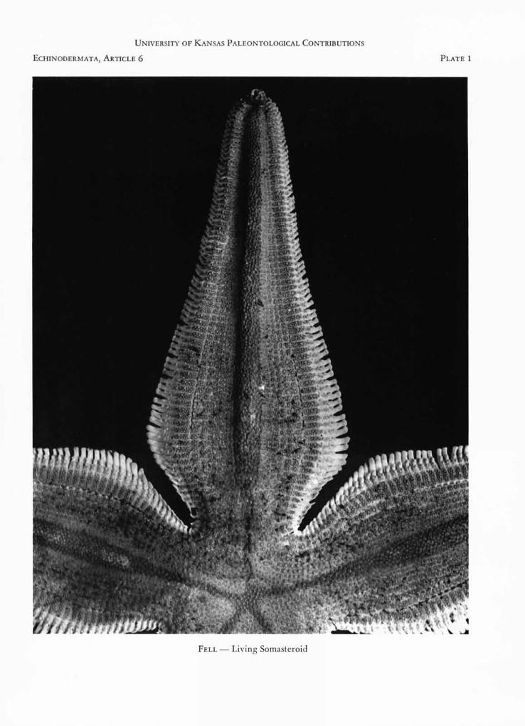
FIGURE 1. Platasterias latiradiata GRAY. Aboral aspect of disc and one arm. Specimen from Corinto, Nicaragua, AllanHancock Foundation, Velero III Station 962-39, 2-5 m, 4 May 1939. R, 64 mm., r, 11 mm. Photo M. D. KING.
UNIVERSITY OF KANSAS PALEONTOLOGICAL CONTRIBUTIONS
ECHINODERMATA, ARTICLE 6
PLATE 1
FELL - Living Somasteroid
UNIVERSITY OF KANSAS PALEONTOLOGICAL CONTRIBUTIONS
PLATE 2
ECHINODERMATA, ARTICLE 6
FELL - Living Somasteroid
LIVING SOMASTEROID 5
Photographs accompanying this paper were madeby M. D. KING, Victoria University of Wellington.Text-figures (except Figures 1-3) were drawn by me.
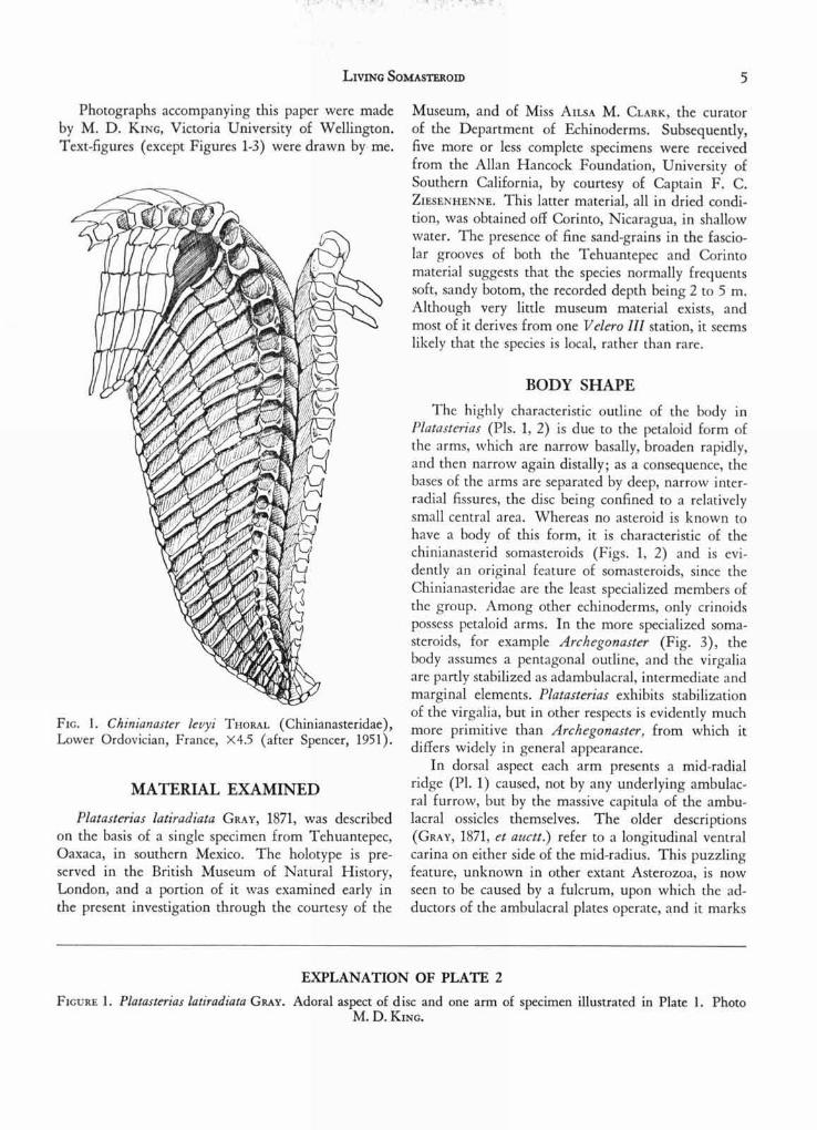
FIG. 1. Chinianaster levyi THORAL (Chinianasteridae),Lower Ordovician, France, x4.5 (after Spencer, 1951).
MATERIAL EXAMINED
Platasterias latiradiata GRAY, 1871, was describedon the basis of a single specimen from Tehuantepec,Oaxaca, in southern Mexico. The holotype is pre-served in the British Museum of Natural History,London, and a portion of it was examined early inthe present investigation through the courtesy of the
Museum, and of Miss AILSA M. CLARK, the curatorof the Department of Echinoderms. Subsequently,five more or less complete specimens were receivedfrom the Allan Hancock Foundation, University ofSouthern California, by courtesy of Captain F. C.ZIESENHENNE. This latter material, all in dried condi-tion, was obtained off Corinto, Nicaragua, in shallowwater. The presence of fine sand-grains in the fascio-lar grooves of both the Tehuantepec and Corintomaterial suggests that the species normally frequentssoft, sandy botom, the recorded depth being 2 to 5 m.Although very little museum material exists, andmost of it derives from one Velcro Ill station, it seemslikely that the species is local, rather than rare.
BODY SHAPEThe highly characteristic outline of the body in
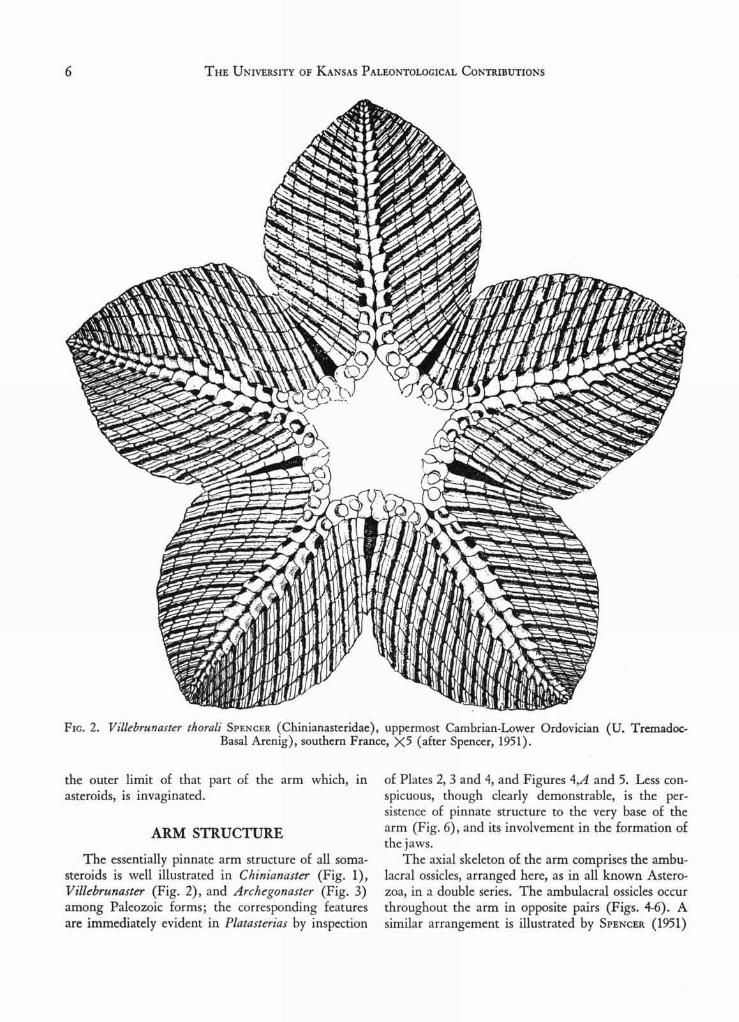
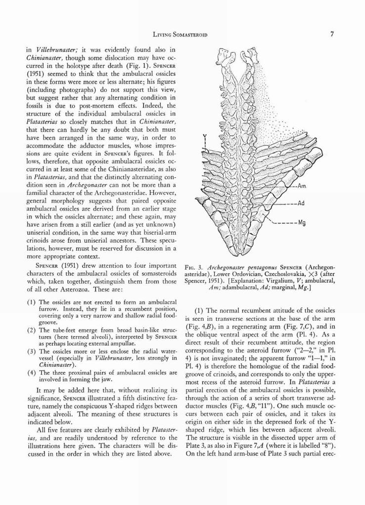
Platasterias (Pls. 1, 2) is due to the petaloid form ofthe arms, which are narrow basally, broaden rapidly,and then narrow again distally; as a consequence, thebases of the arms are separated by deep, narrow inter-radial fissures, the disc being confined to a relativelysmall central area. Whereas no asteroid is known tohave a body of this form, it is characteristic of thechinianasterid somasteroids (Figs. 1, 2) and is evi-dently an original feature of somasteroids, since theChinianasteridae are the least specialized members ofthe group. Among other echinoderms, only crinoidspossess petaloid arms. In the more specialized soma-steroids, for example Archegonaster (Fig. 3), thebody assumes a pentagonal outline, and the virgaliaare partly stabilized as adambulacral, intermediate andmarginal elements. Platasterias exhibits stabilizationof the virgalia, but in other respects is evidently muchmore primitive than Archegonaster, from which itdiffers widely in general appearance.
In dorsal aspect each arm presents a mid-radialridge (Pl. 1) caused, not by any underlying ambulac-ral furrow, but by the massive capitula of the ambu-lacral ossicles themselves. The older descriptions(GRAY, 1871, et auctt.) refer to a longitudinal ventralcaria on either side of the mid-radius. This puzzlingfeature, unknown in other extant Asterozoa, is nowseen to be caused by a fulcrum, upon which the ad-ductors of the ambulacral plates operate, and it marks
EXPLANATION OF PLATE 2
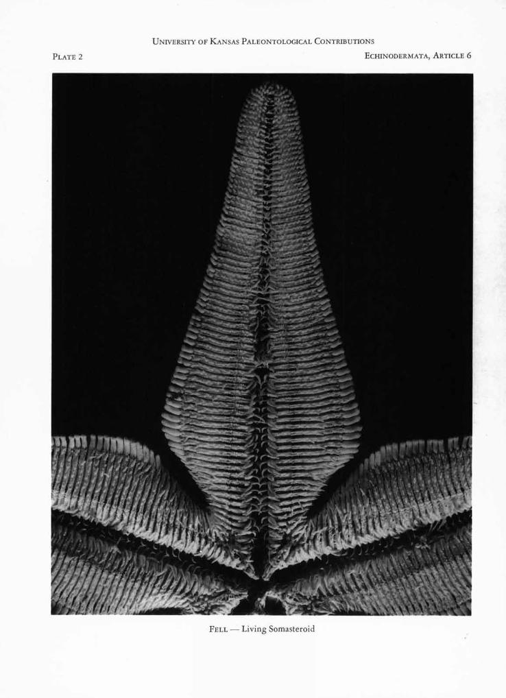
FIGURE 1. Platasterias latiradtata GRAY. Adoral aspect of disc and one arm of specimen illustrated in Plate 1. PhotoM. D. KING.
THE UNIVERSITY OF KANSAS PALEONTOLOGICAL CONTRIBUTIONS6
4,alfs).\\V‘kaN\44\14***IV4*
000111.„n1111:1n1111n111.100
FIG. 2. Villebrunaster thorali SPENCER (Chinianasteridae), uppermost Cambrian-Lower Ordovician (U. Tremadoc-Basal Arenig), southern France, X5 (after Spencer, 1951).
the outer limit of that part of the arm which, inasteroids, is invaginated.
ARM STRUCTURE
The essentially pinnate arm structure of all soma-steroids is well illustrated in Chinianaster (Fig. 1),Villebrunaster (Fig. 2), and Archegonaster (Fig. 3)among Paleozoic forms; the corresponding featuresare immediately evident in Platasterias by inspection
of Plates 2, 3 and 4, and Figures 4,A and 5. Less con-spicuous, though clearly demonstrable, is the per-sistence of pinnate structure to the very base of thearm (Fig. 6), and its involvement in the formation ofthe jaws.
The axial skeleton of the arm comprises the ambu-lacral ossicles, arranged here, as in all known Astero-zoa, in a double series. The ambulacral ossicles occurthroughout the arm in opposite pairs (Figs. 4-6). Asimilar arrangement is illustrated by SPENCER (1951)
LIVING SOMASTEROID
7
in V illebrunaster; it was evidently found also inChinianaster, though some dislocation may have oc-curred in the holotype after death (Fig. 1). SPENCER(1951) seemed to think that the ambulacral ossiclesin these forms were more or less alternate; his figures(including photographs) do not support this view,but suggest rather that any alternating condition infossils is due to post-mortem effects. Indeed, thestructure of the individual ambulacral ossicles inPlatasterias so closely matches that in Chinianaster,that there can hardly be any doubt that both musthave been arranged in the same way, in order toaccommodate the adductor muscles, whose impres-sions are quite evident in SPENCER ' S figures. It fol-lows, therefore, that opposite ambulacral ossicles oc-curred in at least some of the Chinianasteridae, as alsoin Platasterias, and that the distinctly alternating con-dition seen in Archegonaster can not be more than afamilial character of the Archegonasteridae. However,general morphology suggests that paired oppositeambulacral ossicles are derived from an earlier stagein which the ossicles alternate; and these again, mayhave arisen from a still earlier (and as yet unknown)uniserial condition, in the same way that biserial-armcrinoids arose from uniserial ancestors. These specu-lations, however, must be reserved for discussion in amore appropriate context.
SPENCER (1951) drew attention to four importantcharacters of the ambulacral ossicles of somasteroidswhich, taken together, distinguish them from thoseof all other Asterozoa. These are:
(1) The ossicles are not erected to form an ambulacralfurrow. Instead, they lie in a recumbent position,covering only a very narrow and shallow radial food-groove.
(2) The tube-feet emerge from broad basin-like struc-tures (here termed alveoli), interpreted by SPENCERas perhaps locating external ampullae.
(3) The ossicles more or less enclose the radial water-vessel (especially in Villebrunaster, less strongly inChinianaster).
(4) The three proximal pairs of ambulacral ossicles areinvolved in forming the jaw.
It may be added here that, without realizing itssignificance, SPENCER illustrated a fifth distinctive fea-ture, namely the conspicuous Y-shaped ridges betweenadjacent alveoli. The meaning of these structures isindicated below.
All five features are clearly exhibited by Plataster-jas, and are readily understood by reference to theillustrations here given. The characters will be dis-cussed in the order in which they are listed above.
FIG. 3. Archegonaster pentagonus SPENCER (Archegon-asteridae), Lower Ordovician, Czechoslovakia, X3 (afterSpencer, 1951). [Explanation: Virgalium, V; ambulacral,
Am; adambulacral, Ad; marginal, Mg.]
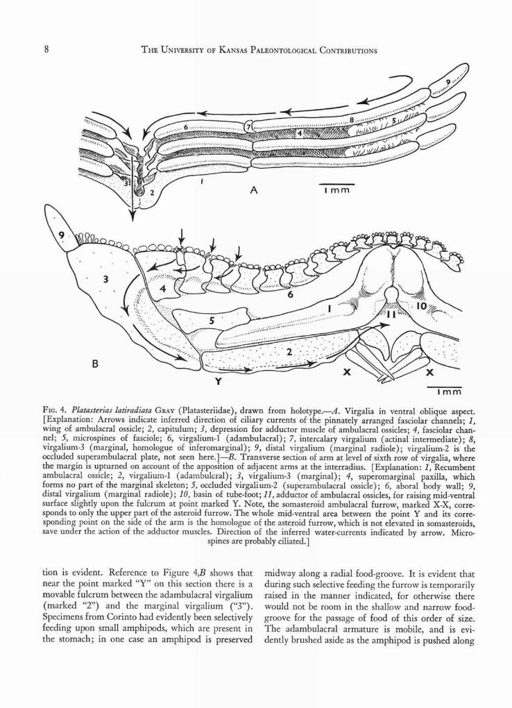
(1) The normal recumbent attitude of the ossiclesis seen in transverse sections at the base of the arm(Fig. 4,B), in a regenerating arm (Fig. 7,C), and inthe oblique ventral aspect of the arm (Pl. 4). As adirect result of their recumbent attitude, the regioncorresponding to the asteroid furrow ("2-2," in Pl.4) is not invaginated; the apparent furrow "1-1," inPI. 4) is therefore the homologue of the radial food-groove of crinoids, and corresponds to only the upper-most recess of the asteroid furrow. In Platasterias apartial erection of the ambulacral ossicles is possible,through the action of a series of short transverse ad-ductor muscles (Fig. 4,B, "11"). One such muscle oc-curs between each pair of ossicles, and it takes itsorigin on either side in the depressed fork of the Y-shaped ridge, which lies between adjacent alveoli.The structure is visible in the dissected upper arm ofPlate 3, as also in Figure 7,A (where it is labelled "8").On the left hand arm-base of Plate 3 such partial erec-
THE UNIVERSITY OF KANSAS PALEONTOLOGICAL CONTRIBUTIONS
.s,„vmittg...ib...kwQN4t:akg.V4ZrNSW''''47tktwkW;7r'jaYff
-z'''z '''zz<kaw.4m .v.z(ffgay/-foli kkle(w.
• • 4110,,,,
4
I M M
8
Flo. 4. Platasterias latiradiata GRAY (Platasteriidae), drawn from holotype.—A. Virgalia in ventral oblique aspect.[Explanation: Arrows indicate inferred direction of ciliary currents of the pinnately arranged fasciolar channels; 1,wing of ambulacral ossicle; 2, capitulum; 3, depression for adductor muscle of ambulacral ossicles; 4, fasciolar chan-nel; 5, microspines of fasciole; 6, virgalium-1 (adambulacral); 7, intercalary virgalium (actinal intermediate); 8,virgalium-3 (marginal, homologue of inferomarginal); 9, distal virgalium (marginal radiole); virgalium-2 is theoccluded superambulacral plate, not seen here.]—B. Transverse section of arm at level of sixth row of virgalia, wherethe margin is upturned on account of the apposition of adjacent arms at the interradius. [Explanation: 1, Recumbentambulacral ossicle; 2, virgalium-1 (adambulcral); 3, virgalium-3 (marginal); 4, superomarginal paxilla, whichforms no part of the marginal skeleton; 5, occluded virgali um-2 (superambulacral ossicle); 6, aboral body wall; 9,distal virgalium (marginal radiole); 10, basin of tube-foot; 11, adductor of ambulacral ossicles, for raising mid-ventralsurface slightly upon the fulcrum at point marked Y. Note, the somasteroid ambulacral furrow, marked X-X, corre-sponds to only the upper part of the asteroid furrow. The whole mid-ventral area between the point Y and its corre-sponding point on the side of the arm is the homologue of the asteroid furrow, which is not elevated in somasteroids,save under the action of the adductor muscles. Direction of the inferred water-currents indicated by arrow. Micro-
spines are probably ciliated.]
tion is evident. Reference to Figure 4,B shows thatnear the point marked "Y" on this section there is amovable fulcrum between the adambulacral virgalium(marked "2") and the marginal virgalium ("3").Specimens from Corinto had evidently been selectivelyfeeding upon small amphipods, which are present inthe stomach; in one case an amphipod is preserved
midway along a radial food-groove. It is evident thatduring such selective feeding the furrow is temporarilyraised in the manner indicated, for otherwise therewould not be room in the shallow and narrow food-groove for the passage of food of this order of size.The adambulacral armature is mobile, and is evi-dently brushed aside as the amphipod is pushed along
LIVING SOMASTEROID 9
ow'ow'doof
4
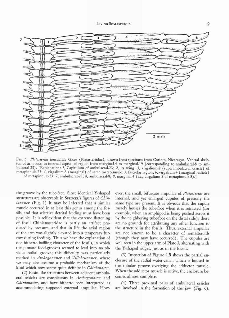
FIG. 5. Platasterias latiradiata GRAY (Platasteriidae), drawn from specimen from Corinto, Nicaragua. Ventral skele-ton of arm-base, in internal aspect, of region from marginal-4 to marginal-19 (corresponding to ambulacral-8 to am-bulacral-23). [Explanation: 1, Capitulum of ambulacral-23; 2, its wing; 3, virgalium -2 (superambulacral ossicle) ofmetapinnule-23; 4, virgalium-3 (marginal) of same metapinnule; 5, fasciolar region; 6, virgalium-4 (marginal radiole)
of metapinnule-23; 7, ambulacral-23; 8, ambulacral-8; 9, marginal-4 (i.e., virgalium-8 of metapinnule-8).]
the groove by the tube-feet. Since identical Y-shapedstructures are observable in SPENCER ' S figures of Chin-ianaster (Fig. 1) it may be inferred that a similarmuscle occurred in at least this genus among the fos-sils, and that selective detrital feeding must have beenpossible. It is self-evident that the extreme flatteningof fossil Chinianasteridae is partly an artifact pro-duced by pressure, and that in life the axial regionof the arm was slightly elevated into a temporary fur-row during feeding. Thus we have the explanation ofone hitherto baffling character of the fossils, in whichthe pinnate food-grooves seemed to lead into no ob-vious radial groove; this difficulty was particularlymarked in Archegonaster and V illebrunaster, wherewe may also assume a probable mechanism of thekind which now seems quite definite in Chinianaster.
(2) Basin-like structures between adjacent ambula-cral ossicles are conspicuous in Archegonaster andChinianaster, and have hitherto been interpreted asaccommodating supposed external ampullae. How-
ever, the small, bifurcate ampullae of Platasterias areinternal, and yet enlarged cupules of precisely thesame type are present. It is obvious that the cupulemerely houses the tube-foot when it is retracted (forexample, when an amphipod is being pushed across itby the neighboring tube-foot on the distal side); thereare no grounds for attributing any other function tothe structure in the fossils. Thus, external ampullaeare not known to be a character of somasteroids(though they may have occurred). The cupules arewell seen in the upper arm of Plate 3, alternating withthe Y-shaped ridges, just as in the fossils.
(3) Inspection of Figure 4,B shows the partial en-closure of the radial water-canal, which is housed inthe tubular groove overlying the adductor muscle.When the adductor muscle is active, the enclosure be-comes almost complete.
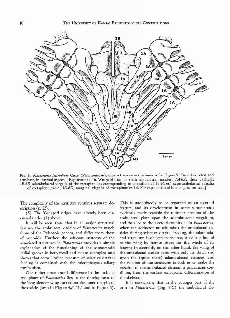
(4) Three proximal pairs of ambulacral ossiclesare involved in the formation of the jaw (Fig. 6).
10 THE UNIVERSITY OF KANSAS PALEONTOLOGICAL CONTRIBUTIONS
FIG. 6. Platasterias latiradiata GRAY (Platasteriidae), drawn from same specimen as for Figure 5. Buccal skeleton andarm-base, in internal aspect. [Explanation: 1-6, Wings of first to sixth ambulacral ossicles; 1A-6A, their capitula;1B-6B, adambulacral virgalia of the metapinnules corresponding to ambulacrals-1-6; 4C-6C, superambulacral virgalia
of metapinnules-4-6; 5D-6D, marginal virgalia of metapinnules-5-6. For explanation of homologies, see text.]
The complexity of the structure requires separate de-scription (p. 12).
(5) The Y-shaped ridges have already been dis-cussed under (1) above.
It will be seen, thus, that in all major structuralfeatures the ambulacral ossicles of Platasterias matchthose of the Paleozoic genera, and differ from thoseof asteroids. Further, the soft-part anatomy of theassociated structures in Platasterias provides a simpleexplanation of the functioning of the somasteroidradial groove in both fossil and extant examples, andshows that some limited measure of selective detritalfeeding is combined with the microphagous ciliarymechanism.
One rather pronounced difference in the ambula-cral plates of Platasterias lies in the development ofthe long slender wing carried on the outer margin ofthe ossicle (seen in Figure 4,B, "1," and in Figure 6).
This is undoubtedly to be regarded as an asteroidfeature, and its development in some somasteroidsevidently made possible the ultimate erection of theambulacral plate upon the adambulacral virgalium,and thus led to the asteroid condition. In Platasterias,when the adductor muscle raises the ambulacral os-sides during selective detrital feeding, the adambula-cral virgalium is obliged to rise too, since it is boundto the wing by fibrous tissue for the whole of itslength; in asteroids, on the other hand, the wing ofthe ambulacral ossicle rests with only its distal endupon the (quite short) adambulacral element, andthe relation of the structures is such as to make theerection of the ambulacral element a permanent con-dition, from the earliest embryonic differentiation ofthe skeleton.
It is noteworthy that in the younger part of thearm in Platasterias (Fig. 7,C) the ambulacral ele-
A
LIVING SOMASTEROID
11
I m m
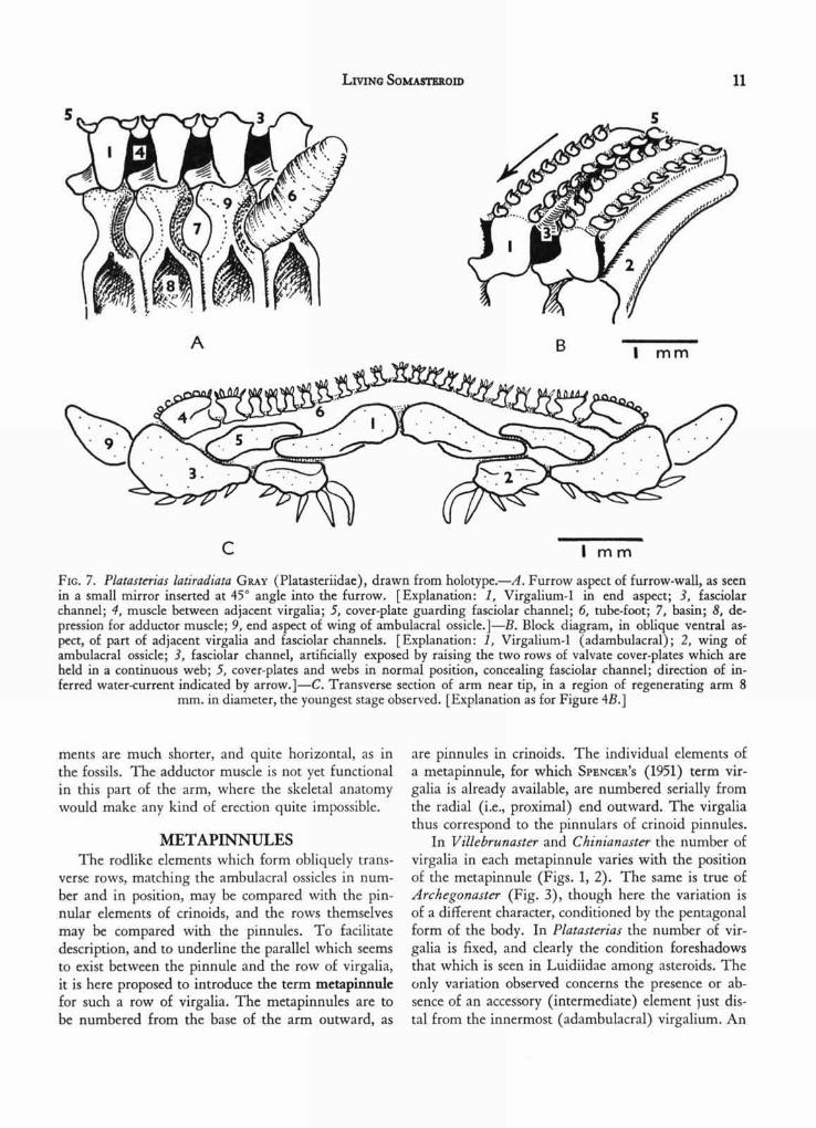
FIG. 7. Platasterias latiradiata GRAY (Platasteriidae), drawn from holotype.—A. Furrow aspect of furrow-wall, as seenin a small mirror inserted at 45° angle into the furrow. [Explanation: 1, Virgalium-1 in end aspect; 3, fasciolarchannel; 4, muscle between adjacent virgalia; 5, cover-plate guarding fasciolar channel; 6, tube-foot; 7, basin; 8, de-pression for adductor muscle; 9, end aspect of wing of ambulacral ossicle.]—B. Block diagram, in oblique ventral as-pect, of part of adjacent virgalia and fasciolar channels. [Explanation: 1, Virgalium-1 (adambulacral); 2, wing ofambulacral ossicle; 3, fasciolar channel, artificially exposed by raising the two rows of valvate cover-plates which areheld in a continuous web; 5, cover-plates and webs in normal position, concealing fasciolar channel; direction of in-ferred water-current indicated by arrow.]—C. Transverse section of arm near tip, in a region of regenerating arm 8
mm. in diameter, the youngest stage observed. [Explanation as for Figure 4B.]
ments are much shorter, and quite horizontal, as inthe fossils. The adductor muscle is not yet functionalin this part of the arm, where the skeletal anatomywould make any kind of erection quite impossible.
METAPINNULESThe rodlike elements which form obliquely trans-
verse rows, matching the ambulacral ossicles in num-ber and in position, may be compared with the pin-nular elements of crinoids, and the rows themselvesmay be compared with the pinnules. To facilitatedescription, and to underline the parallel which seemsto exist between the pinnule and the row of virgalia,it is here proposed to introduce the term metapinnulefor such a row of virgalia. The metapinnules are tobe numbered from the base of the arm outward, as
are pinnules in crinoids. The individual elements ofa metapinnule, for which SPENCER'S (1951) term vir-galia is already available, are numbered serially fromthe radial (i.e., proximal) end outward. The virgaliathus correspond to the pinnulars of crinoid pinnules.
In V illebrunaster and Chinianaster the number ofvirgalia in each metapinnule varies with the positionof the metapinnule (Figs. 1, 2). The same is true ofArchegonaster (Fig. 3), though here the variation isof a different character, conditioned by the pentagonalform of the body. In Platasterias the number of vir-galia is fixed, and clearly the condition foreshadowsthat which is seen in Luidiidae among asteroids. Theonly variation observed concerns the presence or ab-sence of an accessory (intermediate) element just dis-tal from the innermost (adambulacral) virgalium. An
12 THE UNIVERSITY OF KANSAS PALEONTOLOGICAL CONTRIBUTIONS
external view of the metapinnules is seen in Figure4,A (after dissection), and Figure 5 illustrates theinternal aspect; sectional views are seen in Figure4,B, and in Plate 4. It seems rather evident from Fig-ure 5 that the rodlike superambulacral ossicles ("3")are probably occluded virgalia, and this inference issupported by sections of the embryonic part of an armwhich is regenerating (Fig. 7,C). In the latter, thedisposition of the ambulacral ossicle is such as to im-ply that it has become wedged between the firstvirgalium (adambulacral) and the superambulacralelement. In other words, the superambulacral ele-ment must be the second virgalium. No youngerstage has as yet been investigated. As already stated(FELL, 1962), it was the occurrence of rodlike super-ambulacral ossicles in certain astropectinid and luidiidforms which first led to the suspicion that super-ambulacral plates might really represent virgalia, forthey presented some resemblance to the intermediatevirgalia of Arch egonaster, and were found to sharethe same axial gradients as the adambulacral and mar-ginal plates in Luidiidae. Thus, these plates playeda critical part in the first steps which led ultimately tothe isolation and recognition of Platasterias as asomasteroid.
The logical nomenclature for the virgalia of anymetapinnule in Platasterias, and the correspondinghomologies with asteroids, will be as follows: vir-galium 1=adambulacral; virgalium 2=superambula-cral plate; virgalium 3=marginal (inferomarginal);virgalium 4=marginal radiole.
Owing to the exceptional length of the wing ofthe ambulacral ossicle in Platasterias, it seemed plaus-ible that the wing might represent a proximal vir-galium, fused to the original ambulacral ossicle(which would in that case correspond only to thecapitulum of the ossicle). However, no evidence insupport of this view has been obtained, whereas thedevelopment of the ambulacral ossicle, as seen inFigure 7,C, yields rather strong evidence against it.
Accordingly, in the foregoing description, the winghas not been counted as a virgalium.
The virgalia, in addition to providing the funda-mental unit for the entire endoskeleton (apart fromthe axial skeleton and paxillae) also play an importantpart in the formation of the pinnate fasciolar food-grooves, which evidently serve the functions of micro-phagous feeding and of respiration. Before describ-ing these, however, it will be more convenient totrace the metapinnules into the base of the arm, andso elucidate the buccal skeleton.
BUCCAL SKELETON
As will now be shown, the entire buccal skeletonof Platasterias has been derived from the pinnateskeletal system, involving the three proximal pairs ofambulacral ossicles in each arm, together with theirassociated metapinnules.
Casual inspection of the ventral surface of the arm-base in Platasterias (Pl. 3) suggests that the pairedoral plates are merely the somewhat enlarged adam-bulacral virgalia of the first metapinnule. Dissectionof the internal surface of the jaw-region, however, dis-closes that this is not so, and that the real situationis more complex (Fig. 6). The pinnate structure ofthe arm continues without significant alteration(apart from a reduction in size of the distal virgalia)so far as the innermost marginal. As can be seenfrom Figure 6, this is the outer element of the meta-pinnule of the fifth ambulacral ossicle. It is clear,from inspection, that the next row of plates repre-sents the fourth metapinnule, which develops no mar-ginal element, but terminates in an internal super-ambulacral element (Fig. 6 "4C"). Continuing proxi-mally, we reach the third metapinnule, now reducedto only one element (Fig. 6, "3B") which is theadambulacral virgalium; this element is the shortadambulacral which lies next to the oral plates, andappears in ventral aspect (Pl. 3) to be the second
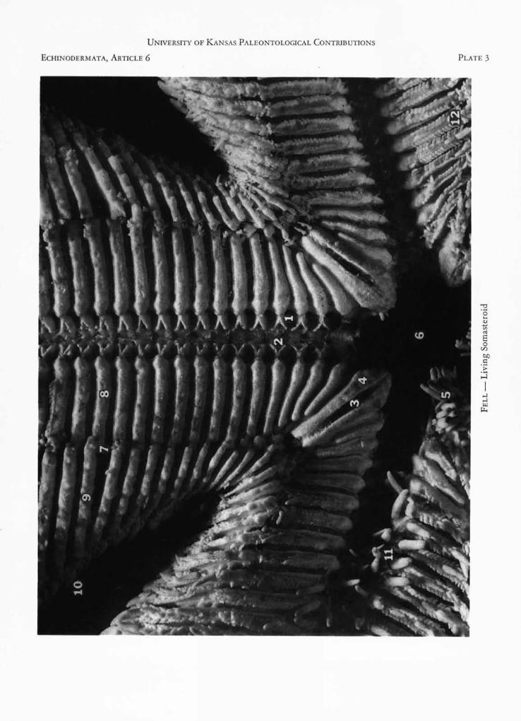
EXPLANATION OF PLATE 3
FIGURE 1. Platasterias latiradiata GRAY. Ventral skeletonafter exposure by dissection, seen in external aspectfrom below. Photo by M. D. KING. [Explanation: I,alveolus of tube-foot; 2, Y-shaped ridge enclosing de-pression for origin of adductor muscle of ambulacralplates; 3, interradial fasciolar groove between oralvirgalia; 4, oral virgalium (second metapinnule); 5,
oral armature; 6, mouth; 7, fasciolar food-groove; 8,adambulacral virgalium of fifteenth metapinnule; 9,marginal virgalium of same metapinnule; 10, distalvirgalium of same metapinnule; 11, adambulacralarmature; 12, adambulacral virgalium with cover-plates still in situ, protecting food-grooves on eitherside. The longer edge of the field measures 25 mm.]
ECHINODERMATA, ARTICLE 6
PLATE 3
UNIVERSITY OF KANSAS PALEONTOLOGICAL CONTRIBUTIONS
UNIVERSITY OF KANSAS PALEONTOLOGICAL CONTRIBUTIONS
PLATE 4
ECHINODERMATA, ARTICLE 6
LIVING SOMASTEROID
13
adambulacral, which it is not. The capitulum (Fig. 6,"3A") of the third ambulacral ossicle is fused to thatof the second ambulacral, as in luidiid and astero-pectinid asteroids. The wing of the second ambulacral(Fig. 6, "2") is a discrete element, again as in luidiidand astropectinid asteroids, and is directed inwardto rest upon the oral plate of its side (Fig. 6, "2B").The latter can therefore be recognized as the adam-bulacral virgalium of the second ambulacral; it isthus the vestige of the second metapinnule. The re-maining structures, forming a T-shaped compoundplate in the interradius, can easily be recognized asthe occluded first ambulacral plates of either side(Fig. 6, "1," "1A"), to which are fused distally twosmall elements, one on either side (Fig. 6, "lB") cor-responding to the adambulacral element of the firstmel.apinnule. Earlier in the investigation a similar T-shaped plate was found in the jaw-apparatus of luidiidand some asteropectinid asteroids; here, however, itsreduction has been carried further, and when theplate was originally noticed, it was thought to repre-sent only the first ambulacral elements, fused to-gether. The function of the plate, obscure in luidiidsand astropectinids, is obvious enough in Platasterias,where it is relatively larger; it serves for the origin ofmusculo-fibrous tissue which unites the two oral ele-ments to it, and thus strengthens the otherwise fragile,bipartite jaw. In asteroids, on the other hand, thegreater development of the oral plates renders theT-shaped element superfluous, and it is accordinglyreduced or, more generally, lost altogether. Thehomologies of the three ambulacral elements takingpart in the jaw, with the three ambulacral elementsobserved by SPENCER in fossil somasteroids, is evident.The entry of the adambulacral virgalium of the sec-ond metapinnule into the jaw-structure to form theoral plate, is an advanced feature, obviously leadingto asteroids and ophiuroids. The relatively simplestate of the jaw, however, is underlined by the uni-form appearance of all the adambulacral elements ex-ternally visible, and by the fact that fasciolar grooves
and covering webs, with cover-plates, extend un-altered throughout the entire series; thus an unpairedfasciolar groove occurs between the two oral plates(Pl. 3).
It will be observed that the general principles bywhich the arm-structure has been modified to giverise to the jaw-structure present remarkable parallelsto that which is already known to have occurred inophiuroids. Evidence will shortly be presented else-where showing that the structure of the arm of cer-tain ophiuroids is fundamentally pinnate, as in som-asteroids, and that the condition seen in other ophiur-oids is a specialization of the earlier pinnate pattern.The whole pattern of skeletal development in allAsterozoa—somasteroids, asteroids, and ophiuroids-is therefore considered to be fundamentally the same,namely, the pinnate pattern initiated by crinoids(FELL, 1962a).
PINNATE FASCIOLAR GROOVESSPENCER (1951) inferred from the structure of
Chinianasteridae that a system of fasciolar food-grooves, set between the adjacent metapinnules, ledinto the radial food-groove, and he drew a parallelwith the observed tegumental fascioles in the asteroidPorania, which is known to be a potentially micro-phagous form, when detrital food supplies fail. Thestructure of Platasterias confirms SPENCER'S inferences,for such food-grooves exist, and the ciliary mechanismis probably located on the microspines placed on theanterior and posterior faces of the free portions of themarginal virgalia (Fig. 4,A, "5"). The proximal halfof the marginal virgalium, and the whole of theadambulacral virgalium, is united by muscle to thecorresponding structures of the adjacent metapin-nules. This muscle forms the roof of the food-groove.
A supposed difficulty in SPENCER'S interpretationlay in the apparent lack of any floor to the food-grooveunless, indeed, the Chinianasteridae lay upon the ab-oral surface, with the oral surface directed upward,to receive the plankton-fall.
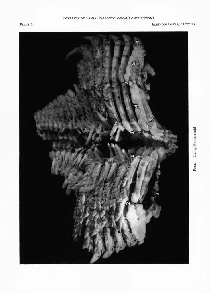
EXPLANATION OF PLATE 4
FIGURE 1. Platasterias latiradiata GRAY. Arm in obliqueventral aspect, partially dissected to expose skeleton.Holotype, from Tehuantepec, southern Mexico, nowin the British Museum of Natural History, London.The region shown extends from the sixth to thetwelfth marginal, plus regenerating portion. Photo by
M. D. KING. [Explanation: 1-1, radial food-groove;2-2, uninvaginated region which in asteroids is erectedinto the furrow; 3, recumbent ambulacral ossicle;4, adambulacral virgalium; 5, marginal virgalium; 6,distal virgalium; 7, aboral body-wall. Portion shownmeasures 20 mm. across.]
14 THE UNIVERSITY OF KANSAS PALEONTOLOGICAL CONTRIBUTIONS
This difficulty is now seen to be imaginary. Thefood-grooves do in fact possess a floor. It is formedby two longitudinal webs which link a series of cover-plates on either side of each food-groove (Pl. 3 andFig. 7,A,B). The webs are erectile, and when erected,occupy the same position as, and closely simulate, thefood-grooves of the pinnules of crinoids. Such erectedwebs are visible on the arm shown in ventral aspectin Plate 2. Their detailed structure has not yet beeninvestigated, but the fact that they can be raised andlowered implies a muscular mechanism and suggeststhat a nerve strand may perhaps traverse the ventralsurface of the metapinnule. The general likeness tocrinoids is astonishing. Between the two rows ofcover-plates on each metapinnule, there is an irregularseries of median spinules, forming a ventral armature.The three furrow-spines represent a specialization ofsuch spinules. On the other hand the so-called mar-ginal spine (in earlier descriptions of Platasterias) isan entirely different structure, for it is obviously thedistal virgalium of the metapinnule itself. A similarsituation exists in the ophiuroid genus Asteronyx(FELL, 1962a).
NUTRITIONIt is apparent that two types of nutrition occur in
Platasterias and, in the light of the foregoing discus-sion of the ambulacral structure and its musculature,the same two types of nutrition must have occurredin the Paleozoic chinianasterids. These are:(1) Microphagous ciliary feeding, performed by the
action of the pinnate food-grooves. The currentsof water (indicated by heavy arrows in Figure 4)would enter between the lateral paxillae, traversethe food-groove, be directed into the radial food-groove, and so pass to the mouth. The mobileflattened furrow-spine seen in Figure 4,B prob-ably acts like a baffle; if placed at a 45° angle itwould automatically direct the current towardsthe mouth. The water-current would also sub-serve the function of respiration, especially as itpassed over the tube-feet.
(2) Selective detrital feeding, in which small amphi-pods (no larger than can pass along the radialfood-groove) are captured by the tube-feet, andpassed along the groove to the mouth. As notedabove, one specimen from Corinto is preservedin which an amphipod is held in this position, andseveral specimens are seen to have an amphipodwithin the jaws. This would be the limit towhich "carnivorous" feeding could extend, therelatively small mouth making it impossible for
voracious feeding of the general asteroid type tooccur. In general, this second type of feeding isdirectly comparable with what is found in manyophiuroids, save that in these latter, the mobilearms are the food-gathering agents, rather thanthe tube-feet.
ABORAL SKELETONLittle is known of the aboral skeleton of fossil
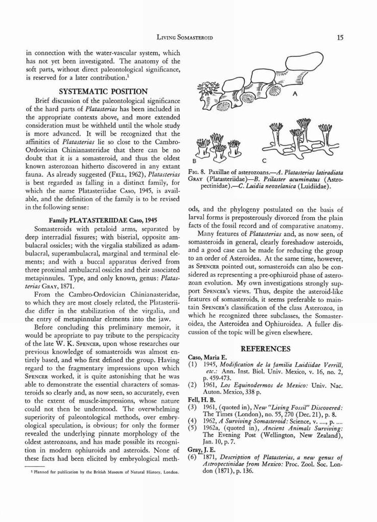
somasteroids. SPENCER (1951) has described three-radiate and four-radiate structures, with an elevatedcentral column, scattered in the aboral integument.He compared them, most inappropriately, with thetriradiate spicules which occur in embryonic echin-oids. The latter structures, which actually occur inall embryonic echinoderms, are in no way compar-able, since they are minute sclerites which subse-quently fuse to form the crystalline mesh of the adultplates. The structures observed by SPENCER in fossilsomasteroids ought to have been termed paxillae, andon first inspection, they are seen to be directly com-parable with the four-radiate paxillae of Luidia, thecentral elevation being the tabulum of the paxilla,characteristically elongate in this genus (Fig. 7,C).The more common type of paxilla in the paxillosephanerozonid asteroids is that illustrated in Figure7,B, where the base exhibits no radiate structure, andthe tabulum is shorter and more robust. Before Platas-terias had been examined in the present investigation,I had already come to the conclusion that the Luidii-dae are the most archaic surviving asteroids, andaccordingly the similarity between the aboral paxillaeof luidiids and chinianasterids was readily under-standable. Platasterias conforms to the essential planof luidiid paxillar structure, but in the part of the armadjoining the marginals, the paxillae are much en-larged (Fig. 7,A). The central paxillar spinules areconverted into granules. In the outer part of the arm(Fig. 7,C) the paxillae are slender, four-radiate orfive-radiate, and closely resemble the structures de-scribed for fossil somasteroids. The outermost paxillais the evident homologue of the superomarginal ofLuidia, and hence of all other asteroids. Thus theluidiid paxilla is seen to be a direct inheritance fromsomasteroid ancestors, and the basis from which allother aboral skeletal structures of asteroids can easilybe derived (Fig. 8).
There remains the madreporite, an element of theaboral skeleton in Platasterias. It is small in the mate-rial at my disposal, and covered by granules, simu-lating a paxilla. Further information cannot at thisstage be given, as the madreporite is better considered
LIVING SOMASTEROID
15
in connection with the water-vascular system, whichhas not yet been investigated. The anatomy of thesoft parts, without direct paleontological significance,is reserved for a later contribution.'
SYSTEMATIC POSITIONBrief discussion of the paleontological significance
of the hard parts of Platasterias has been included inthe appropriate contexts above, and more extendedconsideration must be withheld until the whole studyis more advanced. It will be recognized that theaffinities of Platasterias lie so close to the Cambro-Ordovician Chinianasteridae that there can be nodoubt that it is a somasteroid, and thus the oldestknown asterozoan hitherto discovered in any extantfauna. As already suggested (FELL, 1962), Platasteriasis best regarded as falling in a distinct family, forwhich the name Platasteriidae CASO, 1945, is avail-able, and the definition of the family is to be revisedin the following sense:
Family PLATASTERIIDAE Caso, 1945Somasteroids with petaloid arms, separated by
deep interradial fissures; with biserial, opposite am-bulacral ossicles; with the virgalia stabilized as adam-bulacral, superambulacral, marginal and terminal ele-ments; and with a buccal apparatus derived fromthree proximal ambulacral ossicles and their associatedmetapinnules. Type, and only known, genus: Platas-terias GRAY, 1871.
From the Cambro-Ordovician Chinianasteridae,to which they are most closely related, the Platasterii-dae differ in the stabilization of the virgalia, andthe entry of metapinnular elements into the jaw.
Before concluding this preliminary memoir, itwould be apropriate to pay tribute to the perspicacityof the late W. K. SPENCER, upon whose researches ourprevious knowledge of somasteroids was almost en-tirely based, and who first defined the group. Havingregard to the fragmentary impressions upon whichSPENCER worked, it is quite astonishing that he wasable to demonstrate the essential characters of somas-teroids so clearly and, as now seen, so accurately, evento the extent of muscle-impressions, whose naturecould not then be understood. The overwhelmingsuperiority of paleontological methods, over embry-ological speculation, is obvious; for only the formerrevealed the underlying pinnate morphology of theoldest asterozoans, and has made possible its recogni-tion in modern ophiuroids and asteroids. None ofthese facts had been elicited by embryological meth-
1 Planned for publication by the British Museum of Natural History, London.
FIG. 8. Paxillae of asterozoans.—A. Platasterias latiradiataGRAY (Platasteriidae)—B. Psilaster acuminatus (Astro-
pectinidae).—C. Luidia neozelanica (Luidiidae).
ods, and the phylogeny postulated on the basis oflarval forms is preposterously divorced from the plainfacts of the fossil record and of comparative anatomy.
Many features of Platasterias and, as now seen, ofsomasteroids in general, clearly foreshadow asteroids,and a good case can be made for reducing the groupto an order of Asteroidea. At the same time, however,as SPENCER pointed out, somasteroids can also be con-sidered as representing a pre-ophiuroid phase of astero-zoan evolution. My own investigations strongly sup-port SPENCER'S views. Thus, despite the asteroid-likefeatures of somasteroids, it seems preferable to main-tain SPENCER'S classification of the class Asterozoa, inwhich he recognized three subclasses, the Somaster-oidea, the Asteroidea and Ophiuroidea. A fuller dis-cussion of the topic will be given elsewhere.
REFERENCESCaso, Maria E.(1) 1945, Modification de la familia Luidiidae Verrill,
etc.: Ann. Inst. Biol. Univ. Mexico, v. 16, no. 2,p. 459-473.
(2) 1961, Los Equinodermos de Mexico: Univ. Nac.Auton. Mexico, 338 p.
Fell, H. B.(3) 1961, (quoted in), New "Living Fossil" Discovered:
The Times (London), no. 55, 270 (Dec. 21), p. 8.(4) 1962, A Surviving Somasteroid: Science, v....., p.(5) 1962a, (quoted in), Ancient Animals Surviving:
The Evening Post (Wellington, New Zealand),Jan. 10, p. 7.
GraEJ. E.(6) 1871, Description of Platasterias, a new genus of
Astropectinidae from Mexico: Proc. Zool. Soc. Lon-don (1871), p. 136.
16 THE UNIVERSITY OF KANSAS PALEONTOLOGICAL CONTRIBUTIONS
Spencer, W. K.(7) 1951, Early Palaeozoic Starfish: Philos. Trans., ser.
B, v. 235, no. 623, p. 87-129 (London).
ADDENDUM
Since the foregoing was written, I have been able,through the kindness of Professor GEORGES UBAGHS,Université de Liège, to examine latex molds of theoriginal types of Chinianaster and V illebrunaster.There are no marginal elements in Chinianaster; Fig-
ure 1, here reproduced from SPENCER (1951), is there-
fore partly in error, for the metapinnules terminateeach in a free, acuminate radiole, as in V illebrunaster.The condition is thus nearer to Platasterias than wassupposed. The ambulacral ossicles are essentially op-posite, as inferred above, though they become alter-nate at the arm-tip, and are alternate in a young speci-men. The virgalia are comparable in form to piecesof a tram-rail, having a flattened base, and a flangedkeel; thus they too are more comparable with Platas-terias than was apparent from SPENCER'S account. De-tails will be given elsewhere.