Embed Size (px)
Citation preview
Kardiologia Polska 2006; 64: 1
Selective ablation or isolation of all pulmonary veins in atrial fibrillation – when and for whom?
FFrraanncciisszzeekk WWaallcczzaakk11,, ££uukkaasszz SSzzuummoowwsskkii11,, PPiioottrr UUrrbbaanneekk11,, EEwwaa SSzzuuffllaaddoowwiicczz11,, PPaawwee³³ DDeerreejjkkoo11,, PPiioottrrKKuu³³aakkoowwsskkii22,, RRaaffaa³³ BBaarraannoowwsskkii22,, RRoobbeerrtt BBooddaallsskkii22,, RRoommaann KKêêppsskkii22,, MMaaggddaalleennaa ZZaaggrrooddzzkkaa33,, KKaarriinnaa OOnniisshh44,,IIwwoonnaa BBeessttrryy44,, MMaarreekk KKoonnkkaa,, BBeeaattaa KKuuœœmmiieerrcczzyykk,, AAggnniieesszzkkaa MMaarryynniiaakk
1National Institute of Cardiology, Warsaw, Poland2Department of Cardiology, Postgraduate Medical School, Warsaw, Poland3Military Institute of the Health Services, Warsaw, Poland4Tuberculosis and Lung Diseases Institute, Warsaw, Poland
Original article
Address for correspondence:
prof. Franciszek Walczak, Instytut Kardiologii, ul. Alpejska 42, 04-628 Warszawa, Poland, tel./fax: +48 34 34 417, e-mail: [email protected]
RReecceeiivveedd:: 24 January 2005. AAcccceepptteedd:: 22 August 2005.
Abstract
IInnttrroodduuccttiioonn:: Targeted treatment of atrial fibrillation (AF) involves circumferential isolation of all pulmonary veins (PV) orisolation of electrical connections within their ostia. Only in some cases are the real localisation and number of triggering foci, theanatomy of venous ostia as well as the form of AF (paroxysmal, persistent, chronic, primary or secondary) taken into consideration.
AAiimm:: To compare the results of selective electrical isolation (1-3 PV ostia or ablation of a single focus in other veins oratrium) versus isolation of all pulmonary veins.
MMeetthhooddss:: RF ablation was performed in eighty patients (51 men, 29 women) with symptomatic, drug-refractory AF. Fifty-ninepatients had paroxysmal AF (PAF), 16 persistent (AFpers), and 5 chronic AF (AFchro). Selective ablation was carried out in thosepatients who had detectable AF triggers during sinus rhythm – supraventricular extrasystolic beats (SVEB) of 1 to 3 morphologies(group I). Extended ablation – isolation of all 4-5 PV – was performed in patients with multiple SVEB morphologies andheterogeneous electrical connections within all PV (group II). Group I consisted of 60 patients (22 females) aged 46±14 years,whereas group II comprised 20 patients (7 females) aged 52±13 years. In 24 patients (18 from group I and 6 from group II) withconcomitant typical atrial flutter, an ablation line in the cavo-tricuspid isthmus was also performed. Long-term results were assessed17±15.6 (4-105) months after the procedure based on routine ECG, ambulatory 24-hour ECG monitoring, clinical evaluation andregular phone calls. In patients with PAF, left atrial diameter <4.2 cm and evidence of successful ablation, antiarrhythmic agentswere withheld. In patients with AFpers and AFchro, antiarrhythmic drugs were discontinued 3 to 6 months after successful ablation.
RReessuullttss:: Complete procedural success was achieved in 61 (76%) patients, and significant clinical improvement wasobserved in another 9 (11%) patients. Effective ablation significantly improved quality of life. In group I the procedure wasentirely successful or a marked improvement was reported (single, transient palpitation episodes and/or atrialtachyarrhythmias lasting up to 30 seconds) in 54 (90%) patients. Among 48 (80%) patients with complete success, 25 (42%)did not receive any antiarrhythmic drugs during follow-up, 12 (20%) with arterial hypertension received β-blockers, and 11 (18%)continued β-blocker + class I antiarrhythmic drug. In another 6 (10%) patients a significant clinical improvement in arrhythmiacontrol was observed. In Group II the procedure was fully effective or a significant improvement was observed in 16 (80%)patients. Among 13 (65%) patients with complete success, 5 (25%) did not require any antiarrhythmic drugs, 4 (20%) who hadhypertension continued β-blockers, and another 4 (20%) continued β-blocker + I class antiarrhythmic drug. A significant clinicalimprovement of arrhythmia control was observed in another 3 (15%) patients.
CCoonncclluussiioonnss:: In patients with a limited number of triggering foci and limited AF substrate, selective ablation effectivelyeliminates AF with a low risk of complications. Detailed electrophysiological assessment (standard ECG, 12-lead Holter ECGmonitoring and endocardial mapping) allows precise identification of this group of patients. In patients with chronic andpersistent AF benefits occur with some delay which is associated with a delayed reversal of atrial remodelling.
KKeeyy wwoorrddss:: atrial fibrillation, ablation
Kardiol Pol 2006; 64: 26-35
Kardiologia Polska 2006; 64: 1
Ablation of atrial fibrillation 27
from its beginning, fibrillation is a kind of gusty, electrical storm
for the atria, devastating even in a period of limited clinical symptoms
IntroductionAtrial fibrillation (AF) can appear in each decade of
life. Predisposing and underlying triggering factors,maintenance and self-termination of the arrhythmiachange with age and other circumstances. There arevarious AF triggering factors – primary or secondary,and focal or non-focal. Impulses initiating AF can begenerated in the pulmonary veins (PV), in other veinsdraining to the atrium or within the atria (focal ortriggered by anatomical or functional reentrant loop).
Various triggering mechanisms have been shown tocontribute to the development of focal AF (micro-reentry, pathological automaticity or triggered activity).Initiating electrical impulses usually originate from 1-3centres, less frequently from four or more sites [1-13].Paroxysmal AF is most commonly initiated by triggersfrom more than one PV; however, there is also evidencethat only a single triggering focus may be found in PVor in another vein draining to the atrium.
Rapid development of interventional procedures forAF treatment was stimulated by the limited efficacyand side effects of pharmacotherapy. Currently, themost promising technique is radiofrequency (RF)ablation. This method consists of electrical isolation orablation of triggering foci (triggers or drivers) whichproduce anisotropic conduction in limited areas,leading to symptomatic AF. Other, non-focal triggeringfactors such as typical atrial flutter, may also exist.
At present, isolation of all PV and performingadditional ablation lines, regardless of the actuallocalisation and the number of triggering foci as well asthe type of arrhythmia substrate, is the most widelyused approach. However, the question arises in whichAF patients can the procedure be limited to a single ora few PV or two different veins, in whom it is reallynecessary to isolate all PV, and who needs additionalablation lines. Accordingly, the aim of the present studywas to compare the results of selective versus non-selective ablation of triggering foci in patients with AF.
MethodsPatientsEighty patients with highly symptomatic, drug-
refractory AF underwent RF ablation. Fifty-six hadparoxysmal atrial fibrillation (PAF), 16 persistent AF (AFpers), and the remaining 5 chronic AF (AF chro). In 65patients no obvious structural heart disease could be
detected, 23 patients had hypertension, and 8 hadcoronary artery disease (CAD). Patients with hypertensionand CAD were treated with β-blockers, ACE-I and statinsboth before and after RF ablation. Two of these patientsunderwent coronary angioplasty with stent implantation.
Based on standard ECG recordings, 12-leadambulatory 24-hour Holter ECG monitoring, long-termECG recordings using computerised ECG systems (Bardor EP-MED) as well as electrophysiological testing withisoproterenol and adenosine, a selective (Group I) orextended (all PV – Group II) ablation procedures wereperformed.
Group I consisted of 60 patients (45-PAF, 12-AFpers, 3-AFchro) aged 46±14 years (22 women, 38 men). Meanleft atrial (LA) diameter was 3.7±0.6 cm, and leftventricular ejection fraction (LVEF) was 64±10.1%.
Group II consisted of 20 patients (14-PAF, 4-AFpers, 2-AFchro) aged 52±13 years (7 women, 13 men). MeanLA diameter was 3.9±0.5 cm and LVEF was 65±5.9%.
All patients received prolonged anticoagulation for atleast three months, which was discontinued three daysbefore the procedure and replaced by enoxaparineadministered in a dose of 2 mg/kg. In patients with PAF allantiarrhythmic drugs were withdrawn. Spiral CT andtransoesophageal echocardiography were performedbefore the procedure in order to visualise and measure PV.Patients with enlarged LA received ACE-I after the ablation.
RF ablationElectrodes/ablation catheters were introduced to LA
by transseptal puncture or through the patent foramenovale. This was usually performed under fluoroscopicguidance; however, in six patients intracardiacechocardiography (ICE) was used and in one patientwith atypical septum anatomy transoesophagealechocardiography was performed. Next, a heparinbolus was given, followed by repeated doses injected atone hour intervals during the procedure. In all patients,PV venography was performed. In patients with sinusrhythm, an electrophysiological study was performeddirectly before the procedure, including programmed,incremental and rapid pacing (up to the couplingintervals below refractory periods) from the coronarysinus (CS), LA appendage (LAA) and venous ostium orits proximal segment (PV or superior vena cava (SVC)).
The ablation procedure was performed usingHaissaguerre's method. RF applications, lasting 30-120seconds, were delivered proximal to the lasso electrodewhich was situated in the venous ostium. The nominalRF generator settings were: temperature of 50-55°Cand RF energy of 30-35 W for upper PV and 25 W forinferior PV. Electrical isolation was confirmed after theprocedure using the above-described pacing protocol.In the last 30 patients the end point was the inability to
Kardiologia Polska 2006; 64: 1
28 Franciszek Walczak et al
induce AF lasting >30 seconds by rapid pacing fromthree sites (CS, LAA or high right atrium (HRA)), bothfrom the lasso electrode and from the electrode locatedin the proximal part of PV or SVC.
In selected patients (usually AFpers or AFchrono)electroanatomic mapping was performed using theCARTO system.
In 20 patients (Group II) extended ablation wasperformed (4-5 PV, fifth – middle cardiac vein oraccessory vein) – all of them presented heterogeneouselectrical connections in all PV.
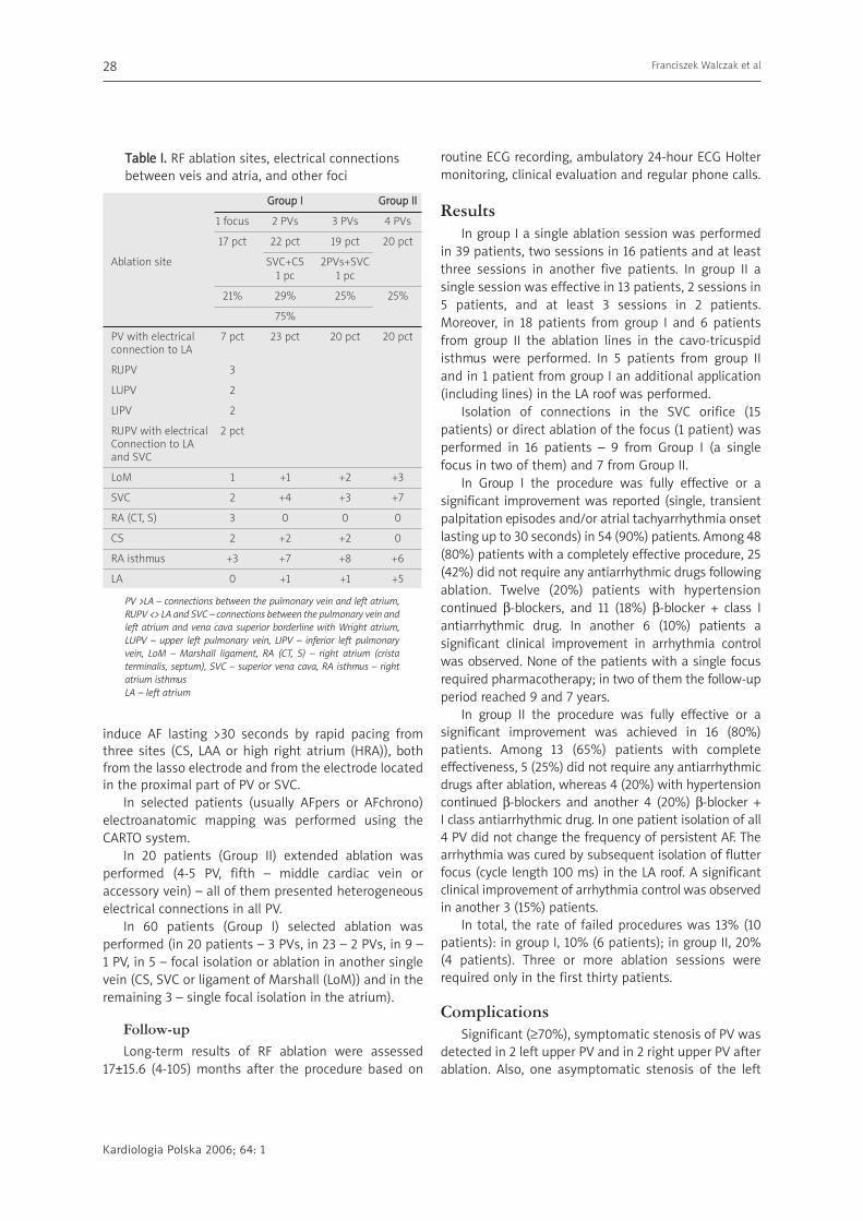
In 60 patients (Group I) selected ablation wasperformed (in 20 patients – 3 PVs, in 23 – 2 PVs, in 9 –1 PV, in 5 – focal isolation or ablation in another singlevein (CS, SVC or ligament of Marshall (LoM)) and in theremaining 3 – single focal isolation in the atrium).
Follow-upLong-term results of RF ablation were assessed
17±15.6 (4-105) months after the procedure based on
routine ECG recording, ambulatory 24-hour ECG Holtermonitoring, clinical evaluation and regular phone calls.
ResultsIn group I a single ablation session was performed
in 39 patients, two sessions in 16 patients and at leastthree sessions in another five patients. In group II asingle session was effective in 13 patients, 2 sessions in5 patients, and at least 3 sessions in 2 patients.Moreover, in 18 patients from group I and 6 patientsfrom group II the ablation lines in the cavo-tricuspidisthmus were performed. In 5 patients from group IIand in 1 patient from group I an additional application(including lines) in the LA roof was performed.
Isolation of connections in the SVC orifice (15patients) or direct ablation of the focus (1 patient) wasperformed in 16 patients – 9 from Group I (a singlefocus in two of them) and 7 from Group II.
In Group I the procedure was fully effective or asignificant improvement was reported (single, transientpalpitation episodes and/or atrial tachyarrhythmia onsetlasting up to 30 seconds) in 54 (90%) patients. Among 48(80%) patients with a completely effective procedure, 25(42%) did not require any antiarrhythmic drugs followingablation. Twelve (20%) patients with hypertensioncontinued β-blockers, and 11 (18%) β-blocker + class Iantiarrhythmic drug. In another 6 (10%) patients asignificant clinical improvement in arrhythmia controlwas observed. None of the patients with a single focusrequired pharmacotherapy; in two of them the follow-upperiod reached 9 and 7 years.
In group II the procedure was fully effective or asignificant improvement was achieved in 16 (80%)patients. Among 13 (65%) patients with completeeffectiveness, 5 (25%) did not require any antiarrhythmicdrugs after ablation, whereas 4 (20%) with hypertensioncontinued β-blockers and another 4 (20%) β-blocker + I class antiarrhythmic drug. In one patient isolation of all4 PV did not change the frequency of persistent AF. Thearrhythmia was cured by subsequent isolation of flutterfocus (cycle length 100 ms) in the LA roof. A significantclinical improvement of arrhythmia control was observedin another 3 (15%) patients.
In total, the rate of failed procedures was 13% (10patients): in group I, 10% (6 patients); in group II, 20%(4 patients). Three or more ablation sessions wererequired only in the first thirty patients.
ComplicationsSignificant (≥70%), symptomatic stenosis of PV was
detected in 2 left upper PV and in 2 right upper PV afterablation. Also, one asymptomatic stenosis of the left
GGrroouupp II GGrroouupp IIII
1 focus 2 PVs 3 PVs 4 PVs
17 pct 22 pct 19 pct 20 pct
Ablation site SVC+CS 2PVs+SVC1 pc 1 pc
21% 29% 25% 25%
75%
PV with electrical 7 pct 23 pct 20 pct 20 pctconnection to LA
RUPV 3
LUPV 2
LIPV 2
RUPV with electrical 2 pctConnection to LA and SVC
LoM 1 +1 +2 +3
SVC 2 +4 +3 +7
RA (CT, S) 3 0 0 0
CS 2 +2 +2 0
RA isthmus +3 +7 +8 +6
LA 0 +1 +1 +5
TTaabbllee II.. RF ablation sites, electrical connectionsbetween veis and atria, and other foci
PV >LA – connections between the pulmonary vein and left atrium,
RUPV <> LA and SVC – connections between the pulmonary vein and
left atrium and vena cava superior borderline with Wright atrium,
LUPV – upper left pulmonary vein, LIPV – inferior left pulmonary
vein, LoM – Marshall ligament, RA (CT, S) – right atrium (crista
terminalis, septum), SVC – superior vena cava, RA isthmus – right
atrium isthmus
LA – left atrium
Kardiologia Polska 2006; 64: 1
Ablation of atrial fibrillation 29
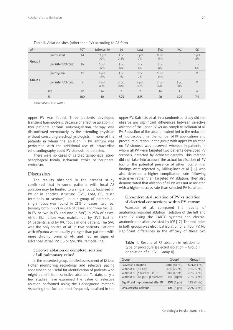
AAFF PPCCTT IIsstthhmmuuss RRAA LLAA LLooMM SSVVCC IIVVCC CCSS
ppaarrooxxiissmmaall 44 12 pct 1 pc 3 pct 8 pct 0 5 pct
GGrroouupp II27% 2.4% 7% 18% 11%
ppeerrssiisstteenntt//cchhrroonniicc 16 6 pct 1 pc 1 pc 1 pc 0 1 pc37% 6% 6% 6% 6%
ppaarrooxxyyssmmaall 15 2 pct 1 pc 1 pc 5 pct 0 0
GGrroouupp IIII13% 7% 7% 33%
ppeerrssiisstteenntt//cchhrroonniicc 5 4 pct 4 pct 2 pct 2 pct 1 pc 080% 80% 40% 40% 20%
PPcctt 80 24 7 7 16 1 6
%% 110000 3300 88..7755 88..7755 2200 11..2255 77..55
TTaabbllee IIII.. Ablation sites (other than PV) according to AF form
Abbreviations: as in Table I.
upper PV was found. Three patients developedtransient haemoptysis. Because of effective ablation, intwo patients chronic anticoagulation therapy wasdiscontinued prematurely by the attending physicianwithout consulting electrophysiologists. In none of thepatients in whom the ablation in PV antrum wasperformed with the additional use of intracardiacechocardiography could PV stenosis be detected.
There were no cases of cardiac tamponade, atrio-oesophageal fistula, ischaemic stroke or peripheralembolism.
DiscussionThe results obtained in the present study
confirmed that in some patients with focal AFablation may be limited to a single focus, localised inPV or in another structure (SVC, LoM, CS, cristaterminalis or septum). In our group of patients, asingle focus was found in 21% of cases, two foci(usually both in PV) in 29% of cases, and three foci (allin PV or two in PV and one in SVC) in 25% of cases.Atrial fibrillation was maintained by SVC foci in 14 patients, and by IVC focus in one patient. The SVCwas the only source of AF in two patients. Patientswith AFparox were usually younger than patients withmore chronic forms of AF, and had no signs ofadvanced atrial, PV, CS or SVC/IVC remodelling.
Selective ablation or complete isolation of all pulmonary veins? In the presented group, detailed assessment of 12-lead
Holter monitoring recordings and selective pacingappeared to be useful for identification of patients whomight benefit from selective ablation. To date, only afew studies have examined the value of selectiveablation performed using the Haisseguerre method.Assuming that foci are most frequently localised in the
upper PV, Katritsis et al. in a randomised study did notobserve any significant differences between selectiveablation of the upper PV versus complete isolation of allPV. Reduction of the ablation extent led to the reductionof fluoroscopy time, the number of RF applications andprocedure duration. In the group with upper PV ablationno PV stenosis was observed, whereas in patients inwhom all PV were targeted two patients developed PVstenosis, detected by echocardiography. This methoddid not take into account the actual localisation of PVfoci or the potential presence of other foci. Similarfindings were reported by Dilling-Boer et al. [16], whoalso detected a higher complication rate followingextensive rather than targeted PV ablation. They alsodemonstrated that ablation of all PV was not associatedwith a higher success rate than selected PV isolation.
Circumferential isolation of PV vs isolation of electrical connections within PV antrumMansour et al. compared the results of
anatomically-guided ablation (isolation of the left andright PV using the CARTO system) and electro-anatomical ablation assisted by ICE [17]. The end pointin both groups was electrical isolation of all four PV. Nosignificant differences in the efficacy of these two
GGrroouupp GGrroouupp II GGrroouupp IIII
SSuucccceessssffuull aabbllaattiioonn 8800%% (48 pts) 6655%% (13 pts)
Without AF (No AA)* 42% (25 pts) 25% (5 pts)
Without AF (β-blocker – HT)* 20% (12 pts) 20% (4 pts)
Without AF (AA gr. I + β-blocker)* 18% (11pts) 20% (4 pts)
SSiiggnniiffiiccaanntt iimmpprroovveemmeenntt aafftteerr RRFF 1100%% (6 pts) 1155%% (3 pts)
UUnnssuucccceessssffuull aabbllaattiioonn 1100%% (6 pts) 2200%% (4 pts)
TTaabbllee IIIIII.. Results of RF ablation in relation totype of procedure (selected isolation – Group Ior ablation of all PV – Group II)
30 Franciszek Walczak et al
GGrroouupp TToottaall AAFF ppaarrooxxyyssmmaall AAFF cchhrroonniicc aanndd ppeerrssiisstteenntt
%% ((ppcctt)) 110000%% ((8800)) 7744%% ((5599)) 2266%% ((2211))
RRFF aabbllaattiioonn eeffffeeccttiivvee 7766%% ((6611)) 8855%% ((5500)) 5533%% ((1111))
Without AF (No AA)* 37% (30) 42% (25) 24% (5)
Without AF (β-blocker – HT)* 20% (16) 26% (15) 5% (1)
Without AF (AA gr. I + β-blocker)* 19%(15) 17%(10) 24%(5)
IImmpprroovveemmeenntt aafftteerr RRFF 1111%% ((99)) 88%% ((55)) 1199%% ((44))
RRFF aabbllaattiioonn uunnssuucccceessssffuull 1133%% ((1100)) 77%% ((44)) 2288%% ((66))
TTaabbllee IIVV.. Ablation efficacy in relation to AF type
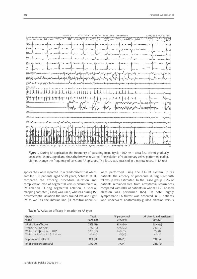
FFiigguurree 11.. During RF application the frequency of pulsating focus (cycle ~100 ms – ultra fast driver) graduallydecreased, then stopped and sinus rhythm was restored. The isolation of 4 pulmonary veins, performed earlier,did not change the frequency of constant AF episodes. The focus was localised in a narrow recess in LA roof
approaches were reported. In a randomised trial whichenrolled 100 patients aged 58±9 years, Schmitt et al.compared the efficacy, procedure duration andcomplication rate of segmental versus circumferentialPV ablation. During segmental ablation, a specialmapping catheter (Lasso) was used, whereas during PVcircumferential ablation the lines around left and rightPV as well as the inferior line (LLPV-mitral annulus)
were performed using the CARTO system. In 93patients the efficacy of procedure during six-monthfollow-up was estimated. In the Lasso group, 89% ofpatients remained free from arrhythmia recurrencescompared with 80% of patients in whom CARTO-basedablation was performed (NS). Of note, highlysymptomatic LA flutter was observed in 13 patientswho underwent anatomically-guided ablation versus
Kardiologia Polska 2006; 64: 1
Kardiologia Polska 2006; 64: 1
Ablation of atrial fibrillation 31
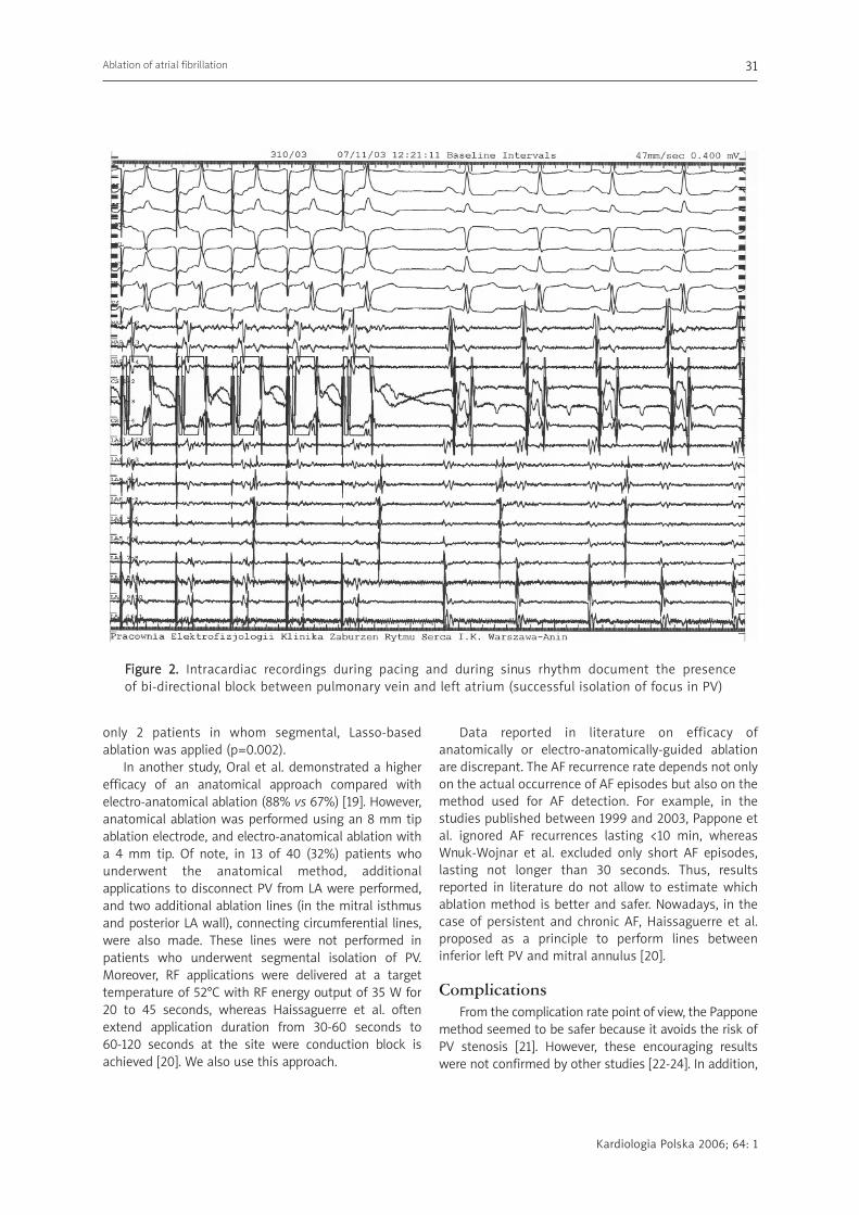
FFiigguurree 22.. Intracardiac recordings during pacing and during sinus rhythm document the presence of bi-directional block between pulmonary vein and left atrium (successful isolation of focus in PV)
only 2 patients in whom segmental, Lasso-basedablation was applied (p=0.002).
In another study, Oral et al. demonstrated a higherefficacy of an anatomical approach compared withelectro-anatomical ablation (88% vs 67%) [19]. However,anatomical ablation was performed using an 8 mm tipablation electrode, and electro-anatomical ablation witha 4 mm tip. Of note, in 13 of 40 (32%) patients whounderwent the anatomical method, additionalapplications to disconnect PV from LA were performed,and two additional ablation lines (in the mitral isthmusand posterior LA wall), connecting circumferential lines,were also made. These lines were not performed inpatients who underwent segmental isolation of PV.Moreover, RF applications were delivered at a targettemperature of 52°C with RF energy output of 35 W for20 to 45 seconds, whereas Haissaguerre et al. oftenextend application duration from 30-60 seconds to 60-120 seconds at the site were conduction block isachieved [20]. We also use this approach.
Data reported in literature on efficacy ofanatomically or electro-anatomically-guided ablationare discrepant. The AF recurrence rate depends not onlyon the actual occurrence of AF episodes but also on themethod used for AF detection. For example, in thestudies published between 1999 and 2003, Pappone etal. ignored AF recurrences lasting <10 min, whereasWnuk-Wojnar et al. excluded only short AF episodes,lasting not longer than 30 seconds. Thus, resultsreported in literature do not allow to estimate whichablation method is better and safer. Nowadays, in thecase of persistent and chronic AF, Haissaguerre et al.proposed as a principle to perform lines betweeninferior left PV and mitral annulus [20].
ComplicationsFrom the complication rate point of view, the Pappone
method seemed to be safer because it avoids the risk ofPV stenosis [21]. However, these encouraging resultswere not confirmed by other studies [22-24]. In addition,
Kardiologia Polska 2006; 64: 1
a life-threatening complication of circumferential PVablation – an atrio-oesophageal fistula with subsequentmultiple stroke and sepsis – was reported by two veryexperienced centres. As a result, the strategy of theprocedure was changed – energy and temperaturesettings were decreased and RF application at the sites inclose proximity to the oesophagus was abandoned [25].
Both methods carry the risk of atrial wallperforation during transseptal puncture (1-6% of cases)which may be complicated by cardiac tamponade,requiring immediate decompression, and sometimeseven cardiac surgery [26-28].
One of the main tools to prevent PV occlusion is thepossibility to use ICE, which visualises the borderbetween the atrium and vein, as well as indicating thepossible risk of microembolisation [10].
Assessment of ablation efficacy and asymptomatic episodes of AFAs in the present study, long-term evaluation in the
majority of studies is based on the evaluation of clinicalsymptoms (palpitation confirmed by ECG as AF andrehospitalisation rate) as well as routine 24-hour HolterECG monitoring. Before ablation all patients suffer fromhighly symptomatic, long-lasting episodes of atrialtachyarrhythmias. After ablation, there is usually asignificant decrease in the AF burden. However, the realrisk of asymptomatic AF recurrences is definitelyunderestimated. For example, Schmitt et al. showedthat the proportion of AF-free patients is significantlylower when 7-day Holter ECG monitoring is used,compared with a standard 24-hour Holter. In that study,the percentage of AF-free patients decreased from 89to 52% in the segmental ablation group and from 80 to47% in the anatomically-guided ablation group [18].Thus, it is very likely that the AF recurrence rate(especially of asymptomatic episodes) is in fact higherthan that usually reported.
An opposite opinion was presented by Oral et al. [29].They studied 60 patients who had a mean of 19±13 AFepisodes before ablation and assessed AF recurrencerate 6 months after ablation, using a 30-daytranstelephonic ECG. The recorder was activated by apatient every day at various times and always whensymptoms suggesting AF were present. During thisperiod, 7 (12%) patients experienced palpitations, whichwere identified by ECG recordings as AF episodes. Themean number of AF episodes per month decreased from19±14 preablation to 3±1 postablation. Among the 53asymptomatic patients following ablation, only twoepisodes of AF were captured in 1 (2%), which promptedthe authors' conclusion that asymptomatic episodes ofAF after apparently successful RF ablation are infrequent.
Wnuk-Wojnar et al. analysed 20 highly symptomaticpatients, who underwent ablation according to thePappone technique. Six months after the procedure,asymptomatic patients (11 – 55%) did not presentepisodes of AF in either test. Among 9 (45%)symptomatic patients, in 5 (25%) an episode of AF wascaptured in 24-hour Holter monitoring, and in another 4(20%) between the 2nd and 7th day of monitoring [30].Evidently, the number of AF episodes and AF burden werehigher in the 7-day Holter recordings, respectively: 2±3.4vs 22±40 (p <0.005) and 2.6±4 h vs 37±29 h (p <0.005).
In a selected group of 297 patients, Pappone et al.evaluated the incidence of AF episodes (lasting at least30 s – symptomatic and asymptomatic) during one-yearfollow-up using 48- Holter monitoring performedmonthly and transtelephonic ECG tracings carried outfour times daily [31]. Asymptomatic episodes of AF weredocumented in 29 (9.6%) patients. In 15 of them,symptomatic AF episodes were also observed (50%).None of them presented an asymptomatic AF episode intranstelephonic ECG tracings. Thus, the authorssuggested that individual asymptomatic AF episodes areinfrequent (14 patients; 5%). This issue, however,requires further studies [32-33]. Asymptomatic AFepisodes are observed mainly during sleeping hours orduring activity in patients with depressed AV conductionwhen the ventricular rate does not exceed 80-100 perminute. Such a situation is rare in young patients.
High efficacy of selective ablation in patients withparoxysmal AF is mainly achieved in patients with singleor two triggering foci (outside PV in some patients).Immediately after the procedure, the majority of them donot require antiarrythmic agents. We anticipate adecrease in antiarrhythmic drug usage in our patients – currently we continue antiarrhythmic therapy afterablation in patients with chronic or persistent AF duringthe early remodelling period [4, 6, 34-38].
Limitations of the studyIn the first 21 patients we had no access to the
Lasso catheter. Post-ablation assessment of conductionbetween LA and PV, CS and SVC/IVC was performedonly in the last 40 cases. We did not perform 7-dayHolter recordings. In the first patients we ablated onlyarrhythmogenic foci, whereas later on we changed ourstrategy and started to isolate electrical connections inthe PV ostia. In selected cases we performed ablationof extra-PV foci as well as RF applications in the sites ofanisotropic conduction (fibrinogenic nests).
Conclusions1. RF ablation is an effective method of treatment of focal
AF, and its efficacy depends on the number and
32 Franciszek Walczak et al
Kardiologia Polska 2006; 64: 1
Ablation of atrial fibrillation 33
localisation of foci, operator experience and techniquesavailable for direct assessment of the efficacy ofablation.
2. A careful analysis of ECG and 12-lead 24-hour Holtermonitoring recording as well as selective pacingallow localisation of high-activity foci, responsible forAF initiation.
3. In selected patients (approximately 20% of AFpatients) only one triggering focus is present, whichallows selected isolation of the focus without the needfor extensive, potentially risky anatomical ablation.
4. Atrial flutter – an additional trigger and driver of AF– coexisted in 30% of our patients.
5. The superior vena cava was the site responsible forAF maintenance in 17.5% of patients, and in 2.5%was the only triggering focus.
6. Arterial hypertension is one of the most frequentfactors predisposing to AF and needs to be wellcontrolled after ablation in order to prevent thedevelopment of new AF foci and substrate.
RReeffeerreenncceess
1. Haissaguerre M, Jais P, Shah DC, et al. Spontaneous initiationof atrial fibrillation by ectopic beats originating in thepulmonary veins. N Engl J Med 1998; 339: 659-66.
2. Chen SA, Hsieh MH, Tai CT, et al. Initiation of atrial fibrillationby ectopic beats originating from the pulmonary veins:electrophysiological characteristics, pharmacologicalresponses, and effects of radiofrequency ablation. Circulation
1999; 100: 1879-86.3. Lin WS, Tai CT, Hsieh MH, et al. Catheter ablation of
paroxysmal atrial fibrillation initiated by non-pulmonary veinectopy. Circulation 2003; 107: 3176-83.
4. Walczak F, Jaworska K, Szufladowicz E, et al. Ogniskowe migotanieprzedsionków – opis przypadku. Kardiol Pol 1998; 49: 547-52.
5. Haissaguerre M, Jais P, Shah DC, et al. Electrophysiological endpoint for catheter ablation of atrial fibrillation initiated frommultiple pulmonary venous foci. Circulation 2000; 101: 1409-17.
6. Walczak F, Szufladowicz E, Baranowski R, et al. ¯y³a p³ucnapunktem wyjœcia migotania przedsionków. Kardiol Pol 2000;52: 475-8.
7. Walczak F, Urbanek P, Szumowski £, et al. Ogniskowemigotanie przedsionków czy zespó³ tachy-bradykardii? Kardiol
Pol 2004; (Suppl.). 8. Haissaguerre M, Sanders P, Hocini M, et al. Changes in atrial
fibrillation cycle length and inducibility during catheter ablationand their relation to outcome. Circulation 2004; 109: 3007-13.
9. Pachon M JC, Pachon M EI, Pachon M JC, et al. A newtreatment for atrial fibrillation based on spectral analysis toguide the catheter RF-ablation. Europace 2004; 6: 590-601.
10. Marrouche NF, Martin DO, Wazni O, et al. Phased-arrayintracardiac echocardiography monitoring during pulmonaryvein isolation in patients with atrial fibrillation: impact onoutcome and complications. Circulation 2003; 107: 2710-6.
11. Bhargava M, Marrouche NF, Martin DO, et al. Impact of age onthe outcome of pulmonary vein isolation for atrial fibrillation
using circular mapping technique and cooled-tip ablationcatheter. J Cardiovasc Electrophysiol 2004; 15: 8-13.
12. Pappone C, Rosanio S, Augello G, et al. Mortality, morbidity, andquality of life after circumferential pulmonary vein ablation foratrial fibrillation: outcomes from a controlled nonrandomizedlong-term study. J Am Coll Cardiol 2003; 42: 185-97.
13. Jais P, Hocini M, Li-Fern H. Individualized atrial fibrillationablation with the end point of non inducibility: A prospectivestudy. Heart Rhythm 2004; Suppl. 1: A852.
14. Mesas CE, Pappone C, Lang CC, et al. Left atrial tachycardiaafter circumferential pulmonary vein ablation for atrialfibrillation: electroanatomic characterization and treatment. JAm Coll Cardiol 2004; 44: 1071-9.
15. Katritsis DG, Ellenbogen KA, Panagiotakos DB, et al. Ablationof superior pulmonary veins compared to ablation of all fourpulmonary veins. J Cardiovasc Electrophysiol 2004; 15: 641-5.
16. Dilling-Boer D, Van Der Merwe N, Adams J, et al. Ablation offocally induced atrial fibrillation: selective or extensive? J
Cardiovasc Electrophysiol 2004; 15: 200-5.17. Mansour M, Ruskin J, Keane D. Efficacy and safety of
segmental ostial versus circumferential extra-ostial pulmonaryvein isolation for atrial fibrillation. J Cardiovasc Electrophysiol
2004; 15: 532-7.18. Schmitt C, Deisenhofer I, Schreieck J, et al. Symptomatic and
asymptomatic recurrence of atrial fibrillation after ablation:randomized comparison of segmental pulmonary veinablation with circumferential pulmonary vein ablation. Eur
Heart J 2004; 25 Abstr. Suppl. 277: P1645.19. Oral H, Scharf C, Chugh A, et al. Catheter ablation for paroxysmal
atrial fibrillation: segmental pulmonary vein ostial ablationversus left atrial ablation. Circulation 2003; 108: 2355-60.
20. Hocini M, Sanders P, Jais P, et al. Techniques for curativetreatment of atrial fibrillation. J Cardiovasc Electrophysiol 2004;15): 1467-71.
21. Oral H. Pulmonary vein occlusion/stenosis after pulmonaryvein ablation for atrial fibrillation. J Cardiovasc Electrophysiol
2003; 14: 371-2.22. Ernst S, Ouyang F, Goya M, et al. Total pulmonary vein
occlusion as a consequence of catheter ablation for atrialfibrillation mimicking primary lung disease. J Cardiovasc
Electrophysiol 2003; 14: 366-70.23. Vasamreddy CR, Jayam V, Bluemke DA, et al. Pulmonary vein
occlusion: an unanticipated complication of catheter ablationof atrial fibrillation using the anatomic circumferentialapproach. Heart Rhythm 2004; 1: 78-81.
24. Drzewiecka-Gerber A, Rybicka A, Krauze J, et al. The doppler-
echo evaluation of pulmonary vein flow and left ventricularfunction after circumferential RF catheter ablation ofpulmonary veins due to paroxysmal atrial fibrillation.Miêdzynarodowy Kongres PTK, Warszawa 2004.
25. Pappone C, Oral H, Santinelli V, et al. Atrio-esophageal fistulaas a complication of percutaneous transcatheter ablation ofatrial fibrillation. Circulation 2004; 109: 2724-6.
26. Hsu LF, Jais P, Hocini M, et al. Minimizing cardiac perforationduring linear ablation for atrial fibrillation. Heart Rhythm
2004; Abstr. Suppl. 1: S141, 444.27. Vasamreddy CR, Lickfett L, Jayam VK, et al. Predictors of
recurrence following catheter ablation of atrial fibrillation
Kardiologia Polska 2006; 64: 1
using an irrigated-tip ablation catheter. J Cardiovasc
Electrophysiol 2004; 15: 692-7.28. KoŸluk E, Lodziñski P, Kiliszek M, et al. Ablacja pod³o¿a
ogniskowego migotania przedsionków. Kardiol Pol 2004; 61: 127.29. Oral H, Veerareddy S, Good E, et al. Prevalence of
asymptomatic recurrences of atrial fibrillation after successfulradiofrequency catheter ablation. J Cardiovasc Electrophysiol
2004; 15: 920-4.30. Wnuk-Wojnar AM, Trusz-Gluza M, Szyd³o KT, et al. Usefulness
of 7-days Holter recording in assessment of long-term resultsof circumferential pulmonary veins RF catheter ablation inpatients with paroxysmal atrial fibrillation. Kardiol Pol 2004;61 (Suppl. III): 41, P167.
31. Pappone C, Santinelli V, Manguso F, et al. pulmonary veindenervation enhances long-term benefit after circumferentialablation for paroxysmal atrial fibrillation. Circulation 2004;109: 327-34.
32. Senatore G, Stabile G, Bertaglia E. Evaluation of asymptomaticatrial fibrillation recurrences after radiofrequency ablation:Multicenter experience. Heart Rhythm 2004; 1: Suppl. A272.
33. Piórkowski K, Hindricks G, Kobza R. Change of AF perceptionafter left atrial catheter ablation – Implication on success rateand follow-up. Heart Rhythm 2004; (Suppl. 1); A274.
34. Walczak F, Szumowski £, Szufladowicz E, et al. Ogniskowemigotanie przedsionków u chorego z zespo³em Wolffa,Parkinsona i White'a – skuteczna izolacja ogniska w ¿ylep³ucnej dolnej lewej. Kardiol Pol 2003; 59: 323-7.
35. Walczak F, Szumowski £. Wskazania do ablacji w migotaniuprzedsionków. Kardiol Pol 2004; 60: 415-28.
36. Walczak F, Szumowski £, Urbanek P, et al. Skuteczna ablacjaogniskowego migotania przedsionków usuwa zaburzeniaautomatyzmu i przewodzenia wywo³uj¹ce epizody utratyprzytomnoœci. Kardiol Pol 2003; 59: 567-73.
37. Walczak F, Szumowski £, Urbanek P, et al. Wp³yw ujœcia ¿y³yp³ucnej i leków AA na zmianê postaci napadów – zd³ugotrwa³ego migotania na krótkie, nawracaj¹ce trzepotanielub czêstoskurcz przedsionkowy. Kardiol Pol 2004; 60: 407-14.
38. Walczak F, Szumowski £, Bodalski R, et al. Ustawiczne napadyogniskowego migotania oraz nawrotnego trzepotaniaprzedsionków. Izolacja przepustów w ujœciach górnych ¿y³p³ucnych przekszta³ca migotanie w nawrotne trzepotanie, azamkniêcie cieœni dolnej przywraca rytm zatokowy. Kardiol Pol
2004; 61: 405-13.
34 Franciszek Walczak et al
Kardiologia Polska 2006; 64: 1
35
Wybiórcza ablacja a izolacja wszystkich żył płucnych w migotaniu przedsionków – kiedy i komu?
FFrraanncciisszzeekk WWaallcczzaakk11,, ££uukkaasszz SSzzuummoowwsskkii11,, PPiioottrr UUrrbbaanneekk11,, EEwwaa SSzzuuffllaaddoowwiicczz11,, PPaawwee³³ DDeerreejjkkoo11,, PPiioottrr KKuu³³aakkoowwsskkii22,, RRaaffaa³³ BBaarraannoowwsskkii22,, RRoobbeerrtt BBooddaallsskkii22,, RRoommaann KKêêppsskkii22,, MMaaggddaalleennaa ZZaaggrrooddzzkkaa33,, KKaarriinnaa OOnniisshh44,, IIwwoonnaa BBeessttrryy44,, MMaarreekk KKoonnkkaa,, BBeeaattaa KKuuœœmmiieerrcczzyykk,, AAggnniieesszzkkaa MMaarryynniiaakk
1Instytut Kardiologii, Warszawa2Klinika Kardiologii CMKP, Warszawa3Wojskowy Instytut Medyczny Centralny Szpital Kliniczny MON, Warszawa4Instytut GruŸlicy i Chorób P³uc, Warszawa
Adres do korespondencji:
prof. Franciszek Walczak, Instytut Kardiologii, ul. Alpejska 42, 04-628 Warszawa, Poland, tel./fax: +48 34 34 417, e-mail: [email protected]
PPrraaccaa wwpp³³yynnêê³³aa:: 24.01.2005. ZZaaaakkcceeppttoowwaannoo ddoo ddrruukkuu:: 22.08.2005.
Streszczenie
WWssttêêpp:: W przyczynowym leczeniu migotania przedsionków (AF) stosuje siê zwykle z za³o¿enia izolacjê okrê¿n¹ wokó³wszystkich ¿y³ p³ucnych (PV) lub izolacjê elektrycznych przepustów w ich ujœciach. Tylko w pojedynczych przypadkach uwzglêd-nia siê faktyczn¹ lokalizacjê i liczbê ognisk wyzwalania, liczbê i osobnicz¹ anatomiê ujœæ ¿y³ p³ucnych oraz charakter AF (napa-dowe, przetrwa³e czy przewlek³e oraz pierwotne czy wtórne).
CCeell:: Porównanie wyników ablacji u pacjentów, u których wykonano wybiórcz¹ izolacjê przepustów w ujœciu 1–3 PV lub ablacjêogniska poza PV (grupa I) oraz u chorych, u których wykonano izolacjê przepustów w ujœciu wszystkich ¿y³ p³ucnych (grupa II).
MMeettooddyykkaa:: Ablacji poddano 80 chorych (51 mê¿czyzn, 29 kobiet) z objawowym, opornym na leczenie farmakologiczne AF.Napadowe AF (PAF stwierdzano u 59 chorych), przetrwa³e AF (AFpers.) – u 16 osób, a przewlek³e AF (AFchro) – u 5 osób. Abla-cjê wybiórcz¹ wykonano u chorych z ekstrasystoli¹ nadkomorow¹ (SVEB) od 1 do 3 morfologii (grupa I). Ablacjê rozszerzon¹(4–5 PV) wykonano u chorych ze SVEB o mnogiej morfologii i niejednorodnymi przepustami we wszystkich PV (grupa II). Do gru-py I w³¹czono 60 chorych w wieku 46±14 lat (22 kobiety), a do grupy II – 20 chorych w wieku 52±13 lat (7 kobiet). U chorychz napadami typowego trzepotania przedsionków wykonano równie¿ liniê ablacyjn¹ w cieœni dolnej – w grupie I u 18 chorych,a w grupie II u 6 pacjentów. Wyniki odleg³e oceniono na podstawie holterowskich zapisów EKG, wizyt oraz rozmów telefonicz-nych w okresie 17±15.6 (zakres 4–103) miesiêcy od ablacji. U chorych z PAF, wymiarem lewego przedsionka <4.2 cm oraz para-metrami wskazuj¹cymi na skutecznoœæ ablacji nie powracano do leków antyarytmicznych. U chorych z AFpers lub AFchro lekiantyarytmiczne odstawiano 3–6 miesiêcy po skutecznej ablacji.
WWyynniikkii:: Zabieg by³ w pe³ni skuteczny u 61 (76%) chorych, a znaczna poprawa nastapi³a u dalszych 9 (11%) chorych. Sku-teczna ablacja wp³ywa³a istotnie na poprawê jakoœci ¿ycia.
W grupie I zabieg by³ w pe³ni skuteczny lub ma miejsce znaczna poprawa u 54 (90%) pacjentów. Wœród 48 (80%) osób beznawrotu AF podczas obserwacji d³ugoterminowej, 25 (42%) chorych nie przyjmowa³o leków antyarytmicznych, 12 (20%) cho-rych z nadciœnieniem têtniczym otrzymywa³o β-bloker, a 11 (18%) chorych nadal leczonych by³o β-blokerem i lekiem antyaryt-micznym klasy I. Istotn¹ poprawê kontroli rytmu obserwowano u 6 (10%) pacjentów. W grupie II zabieg by³ skuteczny lub mamiejsce znaczna poprawa u 16 (80%) chorych. Wœród 13 (65%) chorych z pe³n¹ skutecznoœci¹ ablacji, 5 (25%) osób nie przyj-mowa³o leków, 4 (20%) chorych z nadciœnieniem otrzymywa³o β-bloker, a pozosta³ych 4 (20%) chorych kontynuowa³o β-blokeri lek antyarytmiczny klasy I. Istotn¹ kliniczn¹ poprawê kontrolê rytmu obserwowano u 3 (15%) pacjentów.
WWnniioosskkii:: U chorych z ograniczon¹ liczb¹ czynników wyzwalania i ograniczonym pod³o¿em podtrzymania AF, wybiórcza abla-cja RF jest wystarczaj¹cym, skutecznym i zmniejszaj¹cym ryzyko powik³añ zabiegiem eliminuj¹cym AF. Szczegó³owa ocena elek-trofizjologiczna (EKG, Holter 12-odprowadzeniowy i maping stymulacyjny) pozwala dok³adn¹ identyfikacjê tej grupy chorych.U chorych z przetrwa³ym i przewlek³ym AF korzystny wynik pojawia siê z opóŸnieniem zwi¹zanym z regresj¹ przebudowy miê-œnia przedsionków.
SS³³oowwaa kklluucczzoowwee:: migotanie przedsionków, wybiórcza ablacja RF
Kardiol Pol 2006; 64: 26-35


