Embed Size (px)
Citation preview
Self-Induced Docking Site of a Deeply Embedded PeripheralMembrane Protein
Simon Jaud,* Douglas J. Tobias,* Joseph J. Falke,y and Stephen H. Whitez
*Department of Chemistry, and zDepartment of Physiology and Biophysics, University of California, Irvine, California; andyMolecular Biophysics Program and The Department of Chemistry and Biochemistry, University of Colorado, Boulder, Colorado
ABSTRACT As a first step toward understanding the principles of the targeting of C2 domains to membranes, we have carriedout a molecular dynamics simulation of the C2 domain of cytosolic phospholipase A2 (cPLA2-C2) in a 1-palmitoyl-2-oleoyl-phosphatidylcholine bilayer at constant pressure and temperature (NPT, 300 K and 1 atm). Using the high-resolution crystalstructure of cPLA2-C2 as a starting point, we embedded two copies of the C2 domain into a preequilibrated membrane at thedepth and orientation previously defined by electron paramagnetic resonance (EPR). Noting that in the membrane-bound statethe three calcium binding loops are complexed to two calcium ions, we initially restrained the calcium ions at the membrane depthdetermined by EPR. But the depth and orientation of the domains remained within EPR experimental errors when the restraintswere later removed. We find that the thermally disordered, chemically heterogeneous interfacial zones of phosphatidylcholinebilayers allow local lipid remodeling to produce a nearly perfect match to the shape and polarity of the C2 domain, therebyenabling the C2 domain to assemble and optimize its own lipid docking site. The result is a cuplike docking site with a hydro-phobic bottom and hydrophilic rim. Contrary to expectations, we did not find direct interactions between the protein-bound calciumions and lipid headgroups, which were sterically excluded from the calcium binding cleft. Rather, the lipid phosphate groupsprovided outer-sphere calcium coordination through intervening water molecules. These results show that the combined useof high-resolution protein structures, EPR measurements, and molecular dynamics simulations provides a general approachfor analyzing the molecular interactions between membrane-docked proteins and lipid bilayers.
INTRODUCTION
Cytosolic phospholipase A2 (cPLA2) is an 85-kDa enzyme
that initiates the synthesis of leukotrienes and prostaglandin,
important mediators of inflammation (1–3). It consists of two
domains, both structurally and functionally independent,
separated by a flexible linker. The 121-residue C2 domain
(cPLA2-C2) docks to phosphatidylcholine-rich intracellular
membranes in response to a second messenger calcium signal
(1–3) to establish membrane proximity of the 600-residue
catalytic domain (Fig. 1 a), which hydrolytically liberates
arachidonic acid from zwitterionic phospholipids after dock-
ing (1–3). Widespread in eukaryotic signaling pathways, C2
domains exhibit two conserved architectural features: an
eight-strand antiparallel b-sandwich, and three negatively
charged calcium-binding loops (CBLs) that typically bind
two or three calcium ions and drive membrane docking (4–
8). Despite these similarities, C2 motifs possess low se-
quence identity and target different intracellular membranes.
Although crystal (9) and solution (10) structures of the
cPLA2-C2 domain and a crystal structure of the entire cPLA2
protein (3) have been determined, little is known about the
interactions of the cPLA2-C2 domain with membranes at the
atomic level. To gain insights into these interactions, we
have carried out molecular dynamics (MD) simulations of
cPLA2-C2 domains docked to lipid bilayers.
The high-resolution structures show that the CBLs of
cPLA2-C2, which provide multiple aspartate and asparagine
side chains for the coordination of two calcium ions, are
tipped with hydrophobic residues. Mechanistic studies
(11,12) have shown that hydrophobic interactions, presum-
ably involving these residues, are important in the docking of
cPLA-C2 into phosphatidylcholine-rich target membranes.
More generally, comparisons of different C2 domains
suggest specialized membrane docking mechanisms that
are dominated by electrostatic interactions in some cases,
and hydrophobic interactions in others (12). This wide range
of mechanisms underscores the central role of specialized
protein–lipid interactions in the targeting of C2 domains to
different intracellular membrane surfaces, as required by
their different cellular functions. However, the molecular
origins of these specialized interactions are poorly charac-
terized, because high-resolution structures of membrane-em-
bedded proteins and the surrounding, thermally disordered
lipids cannot be obtained. Understanding the molecular inter-
actions between the membrane-bound cPLA2-C2 and the
surrounding lipids is essential for elucidating the general
principles by which C2 domains stably associate with spe-
cific target membranes.
Several groups have investigated the depth and orientation
of the membrane-bound cPLA2-C2 domain using fluores-
cence (13), NMR (10), and electron paramagnetic resonance
(EPR) (14–16) methods. Each of these studies has concluded
that the CBLs dominate contacts with the membrane, as
predicted by the mechanistic studies. Several of these studies
Submitted June 6, 2006, and accepted for publication October 5, 2006.
Address reprint requests to Stephen H. White, University of California at
Irvine, Dept. of Physiology and Biophysics, Med. Sci. I–D346, Irvine, CA
92697-4560. E-mail: [email protected].
� 2007 by the Biophysical Society
0006-3495/07/01/517/08 $2.00 doi: 10.1529/biophysj.106.090704
Biophysical Journal Volume 92 January 2007 517–524 517
also propose that the two calcium ions bound by the CBLs
are directly coordinated by phospholipid headgroups in the
membrane-docked state. The most extensive study to date is
that of Malmberg et al. (16), who used EPR measurements
along with the crystal structure of the calcium-occupied
cPLA2-C2 domain (9) to define the depth and orientation of
the membrane-inserted domain relative to the bilayer sur-
face. Their study examined EPR spin labels at 24 different
C2 domain positions (16). Nine positions, all on the CBLs,
were observed to penetrate significantly into the membrane,
whereas the other 15 positions were located in or near
the aqueous phase. Distance constraints determined for the
membrane-embedded positions defined the depth of the do-
main in the bilayer and yielded angular orientations of the b-strands relative to the membrane surface.
Recent x-ray reflectivity studies (17) yielded a similar
depth of the domain in the bilayer, although the data were
consistent with several possible orientations of the domain
relative to the membrane surface. One of these orientations
closely matches the EPR-derived geometry, and the EPR
data disfavor the other orientations. The published model
that differs most from the EPR-derived geometry is one based
on NMR chemical shift studies of cPLA2-C2 bound to
dodecylphosphocholine (DPPC) micelles (10). This model,
which supplements the NMR data with structural analysis of
other choline-binding proteins, proposes that the bound cal-
cium ions of the C2 domain are directly coordinated by the
PC headgroup, as in the calcium-bridge model (6).
Despite these extensive previous studies, our understanding
of the molecular interactions between membrane-embedded
C2 domains and the surrounding lipid molecules remains
incomplete. An important issue is that the membrane models
used in the previous studies are static, low-resolution slab
models that cannot provide any information about the spe-
cific nature of the critical lipid-protein interactions. Thus, to
obtain the atomic, dynamic details of these interactions, we
have carried out all-atom, dynamic computer simulations based
upon the EPR membrane depth measurements (16) and the
crystal structure (9) of cPLA2-C2.
Setup and technical details of the simulation
MD simulations used in concert with experimental data can,
in principle, provide information about the structures and
motions of the lipids surrounding a membrane-embedded
protein. We considered the possibility of using the solution
NMR structure (10) as the starting structure for the simu-
lations, but we found that the conformations of the CBLs
differ significantly from those of the crystal structure. We
ultimately chose the crystal structure (PDB 1RLW (9)),
because its CBLs are properly constrained by their native
calcium coordination bonds (such bonds are invisible to
NMR), its resolution is higher, and the EPR membrane-
docking model was developed using the 1RLW structure.
Hence, we carried out MD simulations of cPLA2-C2 docked
to a lipid bilayer in the geometry specified by the EPR model
(16), which was generated by studies of the native protein-
bilayer complex by means of a large number of experimental
distance constraints. Using the membrane depth and geom-
etry obtained from these constraints, we embedded the
crystal coordinates of the cPLA2 C2 into a bilayer made of
pure 1-palmitoyl-2-oleoyl-phosphatidylcholine (POPC), which
was chosen because of this domain’s strong preference for
PC-rich membranes (12).
We built the system from a previously hydrated and
equilibrated POPC bilayer. To improve statistics and to
prevent asymmetric distortions of the bilayer, we inserted a
cPLA2-C2 domain into each bilayer leaflet (Fig. 1 b), whichrequired removal of twenty-two overlapping lipids. The
volume of the simulation cell was ;90 3 90 3 125 A3,
containing 266 lipids, four calcium ions, eight chloride
counter ions (to make the system electroneutral), two pro-
teins of 121 residues each, and 19,492 water molecules, for
a total of 98,110 atoms. We positioned the two domains in
an offset fashion to avoid inadvertent interactions with each
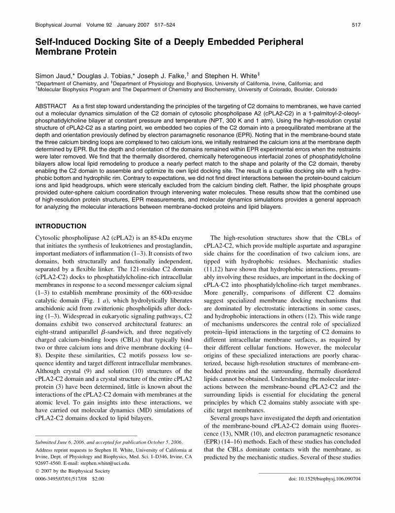
FIGURE 1 Cytosolic phospholipase A2 (cPLA2) and the interactions of
its C2 domain with a POPC bilayer. (a) The complete cPLA2 molecule (3)
(PDB 1CJY), with the C2 domain shown in blue and the catalytic domain in
green. The Ca21 ions are colored purple. The three Ca21 binding loops
(CBL) numbered 1, 2, and 3 are colored red, orange, and yellow, respectively.
(b) The simulated system, inwhich two isolated cPLA2C2 domains (9) (PDB
1RLW) are embedded in opposite sides of a POPC bilayer, and positioned
using electron paramagnetic resonance data (16).
518 Jaud et al.
Biophysical Journal 92(2) 517–524
other as a result of their direct proximity and the periodic
boundary conditions used in the simulations. Fig. 2 reveals
the lack of significant cross-bilayer overlap. The initial place-
ment positioned the calcium ions at the level of the phosphate
groups, whereas CBL1 and CBL3, which possess the most
apolar side chains, were in contact with both the headgroup
and hydrocarbon core regions of the membrane.
The system was set up and relaxed through a methodology
comparable to that described previously (18), with one impor-
tant exception: strong harmonic constraints (100 kcal/mol)
were constantly applied on the position of the calcium ions
throughout the production run to assure that the proteins did
not stray from the experimental results. We removed these
constraints at the end of the production run to verify that the
system parameters remained stable and within the experi-
mentally observed values, which was the case (Fig. 3). After
protein insertion into the bilayer, a 5-ns equilibration and a
9-ns production run were performed with periodic boundary
conditions using a multiple time step integrator (19,20) with
an elementary time step of 1 fs. Nonbonded and electrostatic
interactions were calculated every two and four time steps,
respectively, with a cutoff of 11 A. The SHAKE algorithm
(21) was used to constrain the lengths of bonds involving
hydrogen atoms. The particle mesh Ewald summation (22)
was employed in the calculation of Coulomb interactions.
The temperature was kept constant using Langevin dynam-
ics, and a Nose-Hoover Langevin piston (23,24) was em-
ployed for pressure control (NPT, 1 bar and 300 K). The MD
simulation was performed with the NAMDprogram (25) using
the CHARMM22 protein force field (26) and CHARMM 27
lipid force field (27).
During the simulation, backbone structures of the mem-
brane-embedded CBLs exhibited only small fluctuations
away from their crystal structure conformations (Fig. 4, a
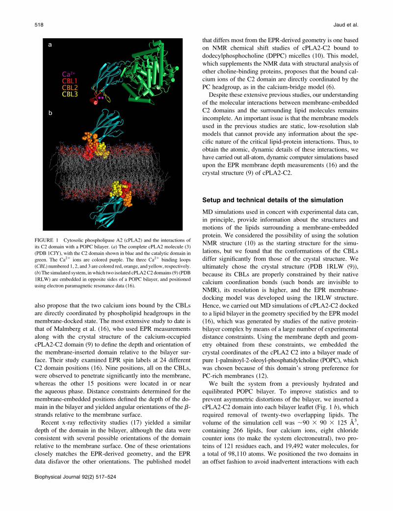
FIGURE 2 Lipids within 10 and 20 A of the center-of-masses (COM)
of the CBLs on the two bilayer surfaces, indicated by yellow and orange
coloring, respectively. The two calcium ions on the membrane surface
facing the viewer are shown in purple. There is no significant overlap of the
C2 domains or their associated lipids across the bilayer, confirmed by an
examination of the lipid order parameters in the vicinities of the two domains
(Fig. 6).
FIGURE 3 Average time-dependent COM (a) and orientation (b) of the
C2 domains. The ‘‘constrained run’’ curves indicate that the constraints on
the calcium ions successfully restrained the C2 domains to be within
experimental uncertainties during the production run. At the end of the run,
we released the constraints to ensure that they were not forcing the domains
to be into a dramatically unfavorable state. After 8 ns of unconstrained
simulations, the fluctuations of the COM and the orientation increase, as
expected, but remain within experimental uncertainties. This indicates that
the constraints on the calcium ions had no undesirable effect on the system.
(a) The gray zones correspond to the experimental values and their ex-
perimental uncertainties. Malmberg et al. (16) calculated the C2 domain
insertion depth with respect to the phosphate plane, representing the average
depth of the headgroup phosphorous atom. The apparent experimental
uncertainty of the depth was taken directly from the Malmberg et al. (16)
article. In a dynamic bilayer, the thermally disordered phosphate groups
exhibit a normal distribution along the transmembrane axis, indicated by the
fuzzy sky-blue zone. This zone shows the inherent uncertainty of the po-
sitions of the phosphorous atoms normal to the bilayer. Because of this
uncertainty, we calculated the protein COM with respect to the center of the
bilayer rather than with respect to the phosphate plane. The average number
of waters that penetrated between the protein and lipids is plotted in orange.
This penetration is limited largely to the region of the phosphates. (b) The
protein orientation is defined as the angle between the transmembrane axis
and a vector from the protein COM to the protein b-sheets COM. The vector
was chosen this way because of the remarkable structural stability of its
starting and ending points. The uncertainty in orientation was taken as the
square root of the sum of the squares of the two orientational uncertainties
provided by Malmberg et al. (16).
C2 Domain Docking Site 519
Biophysical Journal 92(2) 517–524
and c). By contrast, the interstrand loops that are not
membrane-bound exhibited much greater flexibility and
larger variations from the crystal structure (Fig. 4 d). In otherwords, the bilayer and multivalent Ca21 coordination damp
the movement of the CBLs. However, this damping did not
prevent the side chains of the CBLs from changing orien-
tation (Fig. 4 b).
Structure of the self-induced socking site
During equilibration, the bilayer underwent a remarkable
self-recovery from the deeply perturbing initial placement of
the motifs, and generated a cavity ;25 A deep and 30 A
wide for each C2 domain (Fig. 5 a). This induced docking
site has the shape of a cup with a hydrophobic basin formed
from the lipid alkyl chains and a hydrophilic rim formed
from lipid phosphate, choline, and carbonyl groups. Each C2
domain was able to induce its own docking site, because the
bilayer acts like an agitated sea of lipids that bend, twist, and
reach out to contact the domain to maximize energetically
favorable interactions with protein side chains (Fig. 5 b).Although polar interactions are extensive, there are also
numerous apolar contacts located at the tips of CBL1 and
CLB3. The polar collar of the protein (G33, G36, T41, K32,
and D37 of CBL1; N64 and N65 of CBL2; and N95, Y96,
T101, D99, and E100 of CBL3) is in contact with polar lipid
headgroup components, whereas apolar side chains (A34,
F35, M38, and L39 in CBL1 and V97 and M98 of CLB3)
promote nonpolar interactions in the basin of the cavity (Fig.
5 c). But there is also a zone in which polar and apolar lipid
components are in contact with both polar and apolar side
chains (N95, Y96, V97, and M98 of CBL3). The ensemble
of these interactions dictates the behavior of the phospho-
lipids around the domain (Fig. 5 d). The lipid alkyl chains
closest to CBL1 and CBL3 literally wrap around the hydro-
phobic residues of these loops. But this creates a void under
the domain that becomes filled by extended lipid chains from
the opposing leaflet. Therefore, when compared to lipids
remote from the C2 domain, the lipids next to the domain are
more disordered and twisted, whereas the lipids in the op-
posing leaflet are more oriented and stretched.
The average conformations of the lipid alkyl chains can be
described in terms of the orientational order parameters SCH(Fig. 6). The distorted lipids around the periphery of the
protein have lower order parameters than the average lipid in
a pure lipid bilayer, whereas the extended opposing lipids
from the other leaflet have higher order parameters (Fig. 5 d).Thus, the disturbance of the C2 domain extends across the
full thickness of the bilayer in its vicinity, which would not
be apparent in x-ray reflectivity measurements (17) due to
the low resolution of the method. Because some lipids are
less ordered and others are more ordered in the vicinity of the
C2 domains, lipid order parameters calculated for the whole
membrane system fall within the range observed for lipids in
the pure bilayer.
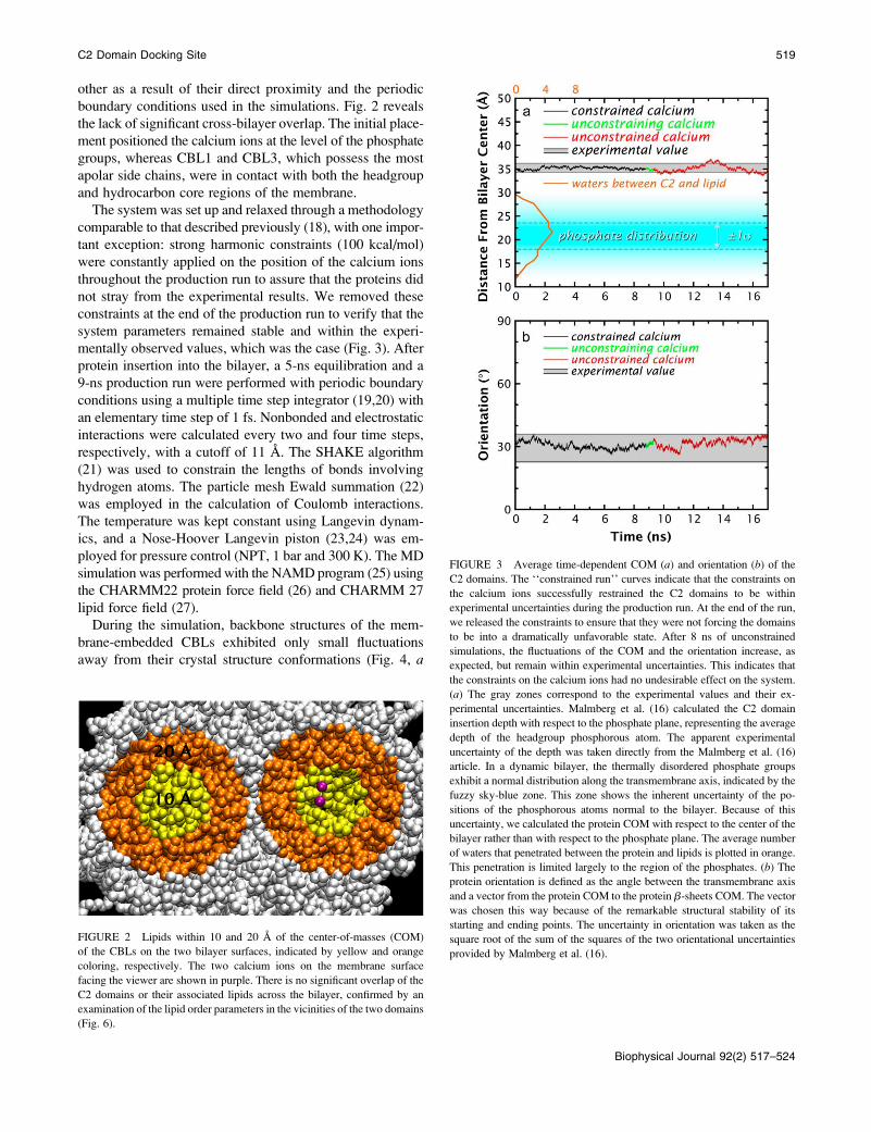
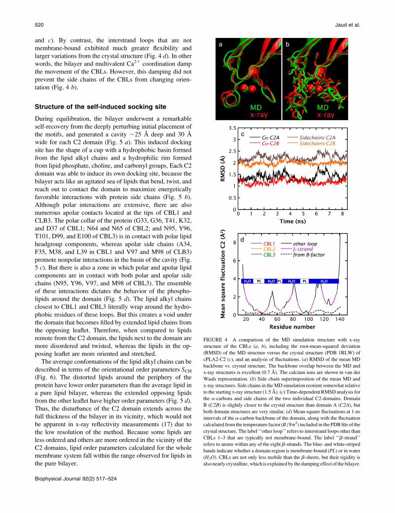
FIGURE 4 A comparison of the MD simulation structure with x-ray
structure of the CBLs (a, b), including the root-mean-squared deviation
(RMSD) of the MD structure versus the crystal structure (PDB 1RLW) of
cPLA2-C2 (c), and an analysis of fluctuations. (a) RMSD of the mean MD
backbone vs. crystal structure. The backbone overlap between the MD and
x-ray structures is excellent (0.7 A). The calcium ions are shown in van der
Waals representation. (b) Side chain superimposition of the mean MD and
x-ray structures. Side chains in theMD simulation reorient somewhat relative
to the starting x-ray structure (1.5 A). (c) Time-dependent RMSD analysis for
the a-carbons and side chains of the two individual C2-domains. Domain
B (C2B) is slightly closer to the crystal structure than domain A (C2A), but
both domain structures are very similar. (d) Mean-square fluctuations at 1-ns
intervals of the a-carbon backbone of the domain, along with the fluctuation
calculated from the temperature factor (B / 8p2) included in the PDBfile of the
crystal structure. The label ‘‘other loop’’ refers to interstrand loops other than
CBLs 1–3 that are typically not membrane-bound. The label ‘‘b-strand’’
refers to atoms within any of the eight b-strands. The blue- and white-striped
bands indicate whether a domain region is membrane-bound (PL) or in water
(H2O). CBLs are not only less mobile than the b-sheets, but their rigidity is
also nearly crystalline,which is explained by the damping effect of the bilayer.
520 Jaud et al.
Biophysical Journal 92(2) 517–524
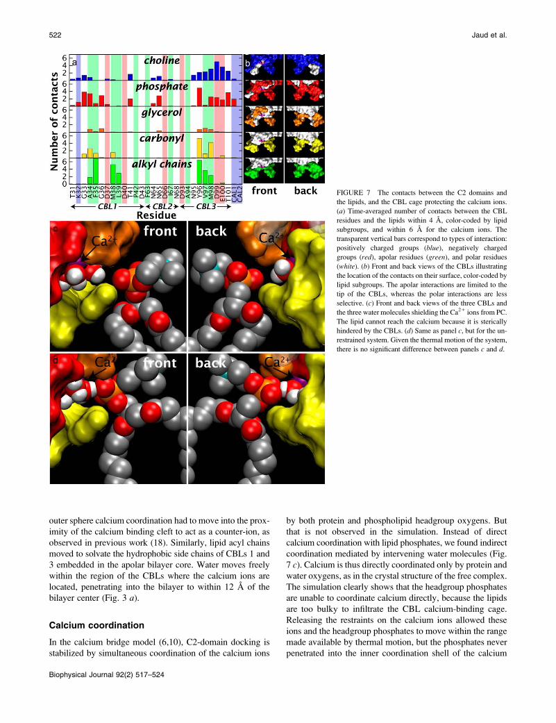
To quantify the interactions underlying this behavior, the
average numbers of contacts between each CBL residue and
the headgroup components (choline, phosphate, glycerol,
carbonyl, alkyl chains) were calculated (Fig. 7 a). The resultsconfirm the high density of apolar contacts at the tips of
CBL1 and CBL3 (Fig. 7 b), and reveal that lipid hydrocarbonchains interact solely with CBL1 and CBL3, where they clus-
ter around F35 and V97, while avoiding close interactions
with charged residues. The carbonyl contacts are very similar
to those of the alkyl chains but show a weak preference for
CBL3. The glycerols exhibit the fewest contacts, and are
distributed with remarkable evenness among all three CBLs.
Because CBL2 lacks the hydrophobic residues of CBL1 and
CBL3, most of the lipid contacts with CBL2 involve the
charged choline and phosphate moieties. In addition, the pos-
itively charged cholines exhibit an important distribution
centered around the acidic residues D99 and E100 on CBL3.
Despite their negative charge, phosphates also exhibit an
important concentration around CBL3, in some cases because
their positions are correlated with the cholines. The phos-
phates also exhibit a significant presence near the basic K32
and near one of the positively charged calcium ions.
Our results also show that the bilayer plays a central role in
the docking process by adapting its shape and polarity to the
topology of the domain, to accommodate charged, polar, and
apolar groups. For instance, the phosphate group that provides
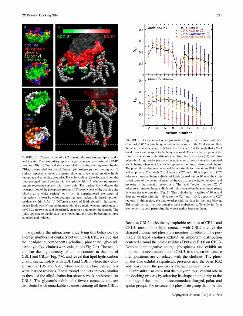
FIGURE 5 Close-up view of a C2 domain, the surrounding lipids, and a
docking site. The molecular graphics images were prepared using the VMD
program (30). (a) Top and side views of the docking site organized by the
CBLs, color-coded for the different lipid subgroups constituting it. (b)Surface representation of a domain, showing a few representative lipids
wrapping and extending around it. The color coding of the domain shows the
time-averaged type of contact with the lipids within 4 A, whereas transparent
regions represent contacts with water only. The dashed line indicates the
mean position of the phosphate groups. (c) Two top views of the docking site
(shown as a white surface) on which is superimposed the types of
interactions (shown by color coding) that each makes with nearby protein
residues (within 6 A). (d) Different classes of lipids found in the system:
distant lipids (far left) never interact with the domain whereas lipids next to
the CBLs are twisted and disordered, creating a void under the domain. The
lipids opposite to the domain have moved into this void by becoming more
extended and ordered.
FIGURE 6 Orientational order parameters SCH of the palmitic and oleic
chains of POPC in pure bilayers and in the vicinity of the C2 domains. Here
the order parameter is SCH ¼ 12Æ3cos2u� 1æ, where u is the angle that a C–H
bond makes with respect to the bilayer normal. The error bars represent the
standard deviations of the data obtained from block averages (31) over 1-ns
intervals. A high order parameter is indicative of more extended, oriented
alkyl chains, whereas a low value represents nonlinear, disordered chains.
The pure bilayer data were obtained from a simulation containing 864 lipids
and no protein. The labels ‘‘10 A next to C2’’ and ‘‘10 A opposite to C2’’
refer to a transmembrane cylinder of lipids located within 10 A of the (x,y)-
coordinates of the center of mass of the CBLs, on the leaflet adjacent and
opposite to the domain, respectively. The label ‘‘region between C2’s’’
refers to a transmembrane cylinder of lipids located on the membrane plane,
between the two domains (Fig. 2). This cylinder has a radius of 10 A and
does not overlap with the ‘‘10 A next to C2’’ and ‘‘10 A opposite to C2’’
regions. In this region, the data overlap with the data for the pure bilayer.
This confirms that the two domains were embedded sufficiently far from
each other to avoid perturbing the whole region between them.
C2 Domain Docking Site 521
Biophysical Journal 92(2) 517–524
outer sphere calcium coordination had to move into the prox-
imity of the calcium binding cleft to act as a counter-ion, as
observed in previous work (18). Similarly, lipid acyl chains
moved to solvate the hydrophobic side chains of CBLs 1 and
3 embedded in the apolar bilayer core. Water moves freely
within the region of the CBLs where the calcium ions are
located, penetrating into the bilayer to within 12 A of the
bilayer center (Fig. 3 a).
Calcium coordination
In the calcium bridge model (6,10), C2-domain docking is
stabilized by simultaneous coordination of the calcium ions
by both protein and phospholipid headgroup oxygens. But
that is not observed in the simulation. Instead of direct
calcium coordination with lipid phosphates, we found indirect
coordination mediated by intervening water molecules (Fig.
7 c). Calcium is thus directly coordinated only by protein and
water oxygens, as in the crystal structure of the free complex.
The simulation clearly shows that the headgroup phosphates
are unable to coordinate calcium directly, because the lipids
are too bulky to infiltrate the CBL calcium-binding cage.
Releasing the restraints on the calcium ions allowed these
ions and the headgroup phosphates to move within the range
made available by thermal motion, but the phosphates never
penetrated into the inner coordination shell of the calcium
FIGURE 7 The contacts between the C2 domains and
the lipids, and the CBL cage protecting the calcium ions.
(a) Time-averaged number of contacts between the CBL
residues and the lipids within 4 A, color-coded by lipid
subgroups, and within 6 A for the calcium ions. The
transparent vertical bars correspond to types of interaction:
positively charged groups (blue), negatively charged
groups (red), apolar residues (green), and polar residues
(white). (b) Front and back views of the CBLs illustrating
the location of the contacts on their surface, color-coded by
lipid subgroups. The apolar interactions are limited to the
tip of the CBLs, whereas the polar interactions are less
selective. (c) Front and back views of the three CBLs and
the three water molecules shielding the Ca21 ions from PC.
The lipid cannot reach the calcium because it is sterically
hindered by the CBLs. (d) Same as panel c, but for the un-
restrained system. Given the thermal motion of the system,
there is no significant difference between panels c and d.
522 Jaud et al.
Biophysical Journal 92(2) 517–524
ions (Fig. 7 d). This is in contrast to the previous NMR-
derived model (10), which predicted that there was sufficient
space to fit glycerophosphocholine (GPC), a PC headgroup
analog, between the CBLs. But this PC headgroup analog is
much shorter than a typical phospholipid, because it lacks
long alkyl chains. This, and the fact that the calcium ions are
farther apart in the solution NMR structure (5.6 A) than in
the crystal structure (4.2 A), might explain why it was pos-
sible to introduce GPC between the CBLs in the NMR
model.
The observation that the calcium ions are not directly
coordinated by lipid headgroups strongly supports the elec-
trostatic switch model of Murray and Honig (28) for the
calcium activation of cPLA2-C2 membrane docking. These
authors have demonstrated through electrostatic calculations
that the neighborhood of the calcium-free CBLs is strongly
negatively charged, which would prevent docking to the
membrane surface. Upon calcium binding, the CBLs become
overall neutral, thereby allowing membrane docking and
penetration into the bilayer core. In the simplest version of
this model, the calcium ions serve only as an electrostatic
switch and do not directly interact with lipid headgroups
to stabilize the membrane docking, as observed in the sim-
ulation.
CONCLUSIONS
Structural studies of pure bilayers carried out by combined
x-ray and neutron diffraction have revealed that the 15-A-
thick interfacial zones of lipid bilayers, dominated by the
phospholipid headgroups, are highly dynamic regions of
tumultuous chemical heterogeneity (29). This remarkable
property allows the lipids to reorganize themselves easily in
the vicinity of embedded proteins to optimize the interac-
tions between protein and lipid, creating in this case a cavity
in the lipid bilayer that mirrors the properties of the C2
domain. We suggest that this idea underlies the targeting and
membrane docking of C2 domains, and of peripheral mem-
brane proteins in general. Each type of conserved C2 domain
likely requires specific lipids, or mixtures of lipids, and a
specific penetration depth to create the optimal complemen-
tary contacts between the protein surface and the targeted
membrane. Our results show that these important details of
targeted docking can be obtained from MD simulations used
in concert with crystallographic and EPR data. But this is
only a beginning; several other types of C2-domain must be
studied before their targeting can be fully understood in
terms of protein-lipid interactions. Future simulations must
examine the importance of mixtures of lipids—particularly
cholesterol—in determining bilayer fluidity, adaptability,
and protein specificity of the interfacial region. An interest-
ing question that demands closer attention is whether the
transbilayer influence of the C2 domains on the opposing
bilayer leaflet will be a persistent finding, independent of lipid
composition.
We thank Dr. Alfredo Freites for supplying the trajectory of the neat lipid
bilayer.
This research was supported by grants from the National Institute of
General Medical Sciences to S.H.W. and J.J.F., from the National Center
for Research Resources to S.H.W., and from the National Science
Foundation to D.J.T. ‘‘Le Fond Quebecois de la Recherche sur la Nature
et les Technologies’’ provided a graduate fellowship for S.J.
REFERENCES
1. Leslie, C. C. 2004. Regulation of the specific release of arachidonicacid by cytosolic phospholipase A2. Prostaglandins Leukot. Essent.Fatty Acids. 70:373–376.
2. Clark, J. D., A. R. Schievella, E. A. Nalefski, and L.-L. Lin. 1995.Cytosolic phospholipase A2. J. Lipid Mediat. Cell Signal. 12:83–117.
3. Dessen, A., J. Tang, H. Schmidt, M. Stahl, J. D. Clark, J. Seehra, andW. S. Somers. 1999. Crystal structure of human cytosolic phospholipaseA2 reveals a novel topology and catalytic mechanism.Cell. 97:349–360.
4. Cho, W., and R. V. Stahelin. 2005. Membrane-protein interactions incell signaling and membrane trafficking. Annu. Rev. Biophys. Biomol.Struct. 34:119–151.
5. Bai, J., and E. R. Chapman. 2004. The C2 domains of synaptotag-min—partners in exocytosis. Trends Biochem. Sci. 29:143–151.
6. Hurley, J. H., and S. Misra. 2000. Signaling and subcellular targeting bymembrane-bindingdomains.Annu.Rev.Biophys.Biomol. Struct.29:49–79.
7. Rizo, J., and T. C. Sudhof. 1998. C2-domains, structure and function ofa universal Ca21-binding domain. J. Biol. Chem. 273:15879–15882.
8. Nalefski, E. A., M. M. Slazas, and J. J. Falke. 1997. Ca21-signalingcycle of a membrane-docking C2 domain. Biochemistry. 36:12011–12018.
9. Perisic, O., S. Fong, D. E. Lynch, M. Bycroft, and R. L. Williams.1998. Crystal structure of a calcium-phospholipid binding domain fromcytosolic phospholipase A2. J. Biol. Chem. 273:1596–1604.
10. Xu,G.-Y., T.McDonagh,H.-A.Yu, E.A.Nalefski, J. D.Clark, andD.A.Cumming. 1998. Solution structure and membrane interactions of theC2 domain of cytosolic phospholipase A2. J. Mol. Biol. 280:485–500.
11. Perisic, O., H. F. Paterson, G. Mosedale, S. Lara-Gonzalez, and R. L.Williams. 1999. Mapping the phospholipid-binding surface and trans-location determinants of the C2 domain from cytosolic phospholipaseA2. J. Biol. Chem. 274:14979–14987.
12. Nalefski, E. A., M. A. Wisner, J. Z. Chen, S. R. Sprang, M. Fukuda,K. Mikoshiba, and J. J. Falke. 2001. C2 domains from differentCa21 signaling pathways display functional and mechanistic diversity.Biochemistry. 40:3089–3100.
13. Nalefski, E. A., and J. J. Falke. 1998. Location of the membrane-docking face on the Ca21-activated C2 domain of cytosoic phospho-lipase A2. Biochemistry. 37:17642–17650.
14. Frazier, A. A., M. A. Wisner, N. J. Malmberg, K. G. Victor, G. E.Fanucci, E. A. Nalefski, J. J. Falke, and D. S. Cafiso. 2002. Membraneorientation and position of the C2 domain from cPLA2 by site-directedspin labeling. Biochemistry. 41:6282–6292.
15. Nielsen, R. D., K. Che, M. H. Gelb, and B. H. Robinson. 2005. A rulerfor determining the position of proteins in membranes. J. Am. Chem.Soc. 127:6430–6442.
16. Malmberg, N. J., D. R. Van Buskirk, and J. J. Falke. 2003. Membrane-docking loops of the cPLA2 C2 domain: Detailed structural analysis ofthe protein-membrane interface via site-directed spin-labeling. Bio-chemistry. 42:13227–13240.
17. Malkova, S., F. Long, R. V. Stahelin, S. V. Pingali, D. Murray, W. Cho,and M. L. Schlossman. 2005. X-ray reflectivity studies of cPLA2a-C2domains adsorbed onto Langmuir monolayers of SOPC. Biophys. J.89:1861–1873.
18. Freites, J. A., D. J. Tobias, G. von Heijne, and S. H. White. 2005.Interface connections of a transmembrane voltage sensor. Proc. Natl.Acad. Sci. USA. 102:15059–15064.
C2 Domain Docking Site 523
Biophysical Journal 92(2) 517–524
19. Tuckerman, M., and B. J. Berne. 1992. Reversible multiple time scalemolecular dynamics. J. Chem. Phys. 97:1990–2001.
20. Grubmuller, H., H. Heller, A. Windemuth, and K. Schulten. 1991.Generalized Verlet algorithm for efficient molecular dynamics simu-lations with long-range interactions. Mol. Simul. 6:121–142.
21. Ryckaert, J.-P., G. Ciccotti, and H. J. C. Berendsen. 1977. Numericalintegration of the Cartesian equations of motion of a system withconstraints: molecular dynamics of n-alkanes. J. Comput. Phys.23:327–341.
22. Darden, T., D. York, and L. Pedersen. 1993. Particle mesh Ewald: AnN�log(N) method for Ewald sums in large systems. J. Chem. Phys.98:10089–10092.
23. Martyna, G. J., D. J. Tobias, and M. L. Klein. 1994. Constant-pressuremolecular-dynamics algorithms. J. Chem. Phys. 101:4177–4189.
24. Feller, S. E., Y. Zhang, R. W. Pastor, and B. R. Brooks. 1995. Constantpressure molecular dynamics simulation: the Langevin piston method.J. Chem. Phys. 103:4613–4621.
25. Kale, L., R. Skeel, M. Bhandarkar, R. Brunner, A. Gursoy, N. Krawetz,J. Phillips, A. Shinozaki, K. Varadarajan, and K. Schulten. 1999.
NAMD2: Greater scalability for parallel molecular dynamics. J. Comput.Phys. 151:283–312.
26. MacKerell, A. D. Jr., D. Bashford, M. Bellott, R. L. Dunbrack Jr., J. D.Evanseck, M. J. Field, S. Fischer, J. Gao, H. Guo, S. Ha, D. Joseph-McCarthy, L. Kuchnir, et al. 1998. All-atom empirical potential formolecular modeling and dynamics studies of proteins. J. Phys. Chem.B. 102:3586–3616.
27. Feller, S. E., and A. D. MacKerell Jr. 2000. An improved empiricalpotential energy function for molecular simulations of phospholipids.J. Phys. Chem. B. 104:7510–7515.
28. Murray, D., and B. Honig. 2002. Electrostatic control of the membranetargeting of C2 domains. Mol. Cell. 9:145–154.
29. Wiener, M. C., and S. H. White. 1992. Structure of a fluid dioleoylphos-phatidylcholine bilayer determined by joint refinement of x-ray andneutron diffraction data. III. Complete structure. Biophys. J. 61:434–447.
30. Humphrey, W., W. Dalke, and K. Schulten. 1996. VMD: visualmolecular dynamics. J. Mol. Graph. 14:33–38.
31. Flyvbjerg, H., and H. G. Petersen. 1989. Error estimates on averages ofcorrelated data. J. Chem. Phys. 91:461–466.
524 Jaud et al.
Biophysical Journal 92(2) 517–524


