Embed Size (px)
Citation preview

4313Development 125, 4313-4323 (1998)Printed in Great Britain © The Company of Biologists Limited 1998DEV1331
Semaphorins III and IV repel hippocampal axons via two distinct receptors
Alain Chédotal 1,*, Jose A. Del Rio 4, Monica Ruiz 4, Zhigang He 2, Victor Borrell 4, Fernando de Castro 1,Frédéric Ezan 1, Corey S. Goodman 3, Marc Tessier-Lavigne 2, Constantino Sotelo 1 and Eduardo Soriano 4
1INSERM U106, Hôpital de la Salpêtrière, 75013 Paris, France 2HHMI, Department of Anatomy, UCSF, San Francisco, CA, USA3HHMI, Department of Molecular and Cell Biology, UC Berkeley, Berkeley, CA, USA4Department of Animal and Plant Cell Biology, University of Barcelona, Barcelona 08028, Spain*Author for correspondence (e-mail: [email protected])
Accepted 13 August; published on WWW 30 September 1998
The semaphorins are the largest family of repulsive axonguidance molecules. Secreted semaphorins bind neuropilinreceptors and repel sensory, sympathetic and motor axons.Here we show that CA1, CA3 and dentate gyrus axons fromE15-E17 mouse embryo explants are selectively repelled byentorhinal cortex and neocortex. The secreted semaphorinsSema III and Sema IV and their receptors Neuropilin-1and -2 are expressed in the hippocampal formation duringappropriate stages. Sema III and Sema IV strongly repelCA1, CA3 and dentate gyrus axons; entorhinal axons are
only repelled by Sema III. An antibody against Neuropilin-1 blocks the repulsive action of Sema III and the entorhinalcortex, but has no effect on Sema IV-induced repulsion.Thus, chemorepulsion plays a role in axon guidance in thehippocampus, secreted semaphorins are likely to beresponsible for this action, and the same axons can berepelled by two distinct semaphorins via two differentreceptors.
Key words: Semaphorin, Neuropilin, Hippocampus, Cortex, Mouse
SUMMARY
nsasratoredandmsne
, orr-esin-o.,flyinut
inestorave
nghis),ou
INTRODUCTION
Mounting evidence indicates that in the developing centnervous system, growth cones can be guided at a distancdiffusible molecules secreted by non-target cells (TessiLavigne and Goodman, 1996). Many of these factors functas chemorepellents: they induce growth cone collapse oriented axonal outgrowth away from the source of the facSince the demonstration 5 years ago by Pini (1993) that septum in the embryonic rat produces a diffusible factor threpels olfactory bulb axons, several studies have indicated chemorepulsive molecules are produced in a variety of cennervous system (CNS) regions, such as the ventral spinal c(Fitzgerald et al., 1993), the floor plate (Colomarino anTessier-Lavigne, 1995; Tamada et al., 1995; Varela-Echavaet al., 1997) or the thalamus (Tuttle et al., 1998). Potenchemorepellents have been identified in two gene families, netrins and the semaphorins (Culotti and Kolodkin, 199Tessier-Lavigne and Goodman, 1996). Floor plate-derivNetrin-1 has been shown to repel motor axons from ttrochlear, trigeminal, facial and glossopharyngeal crannuclei (Colomarino and Tessier-Lavigne, 1995; VarelEchavarria et al., 1997). Most of the secreted chemorepelleidentified to date belong to the semaphorin family, which acontains transmembrane members (Kolodkin et al., 1993; Market al., 1997). Sema III/D (hereafter referred to as Sema III) aits avian ortholog Collapsin-1, which is expressed in tembryonic ventral spinal cord, are known to cause in vitro tcollapse of sensory axons growing from explants of dorsal r
rale byer-ionandtor.theat
thattralordd
rriatialthe6;edheiala-nts
lso
ndheheoot
ganglia (Luo et al., 1995) and can also repel these axo(Messersmith et al., 1995; Püschel et al., 1995). Sema III halso been reported to act as a chemorepellent for most cranial motor axons (Varela-Echavarria et al., 1997) and folfactory axons (Kobayashi et al., 1997). Two other secretsemaphorins, Sema A and Sema E/Collapsin-3, can repel cause the collapse of axons from sympathetic ganglia (Adaet al., 1997; Koppel et al., 1997). Recently, the transmembraprotein Neuropilin-1 has been been shown to be a receptora component of a receptor, for Sema III (He and TessieLavigne, 1997; Kolodkin et al., 1997). Other studies havdemonstrated that the secreted semaphorins Sema E/collap3, Collapsin-2, Collapsin-5 and Sema IV also bind tNeuropilin-1 with apparently equivalent affinities (Chen et al1997; Feiner et al., 1997). Neuropilin-2, a homolog oNeuropilin-1, exists in at least six isoforms, that are probabgenerated by alternative splicing (Chen et al., 1997; Kolodket al., 1997). Neuropilin-2 can bind Sema E and Sema IV, bnot Sema III with high-affinity (Chen et al., 1997) but theinvolvement of Neuropilin-2 in mediating a semaphorinresponse remains to be demonstrated.
Although semaphorins and their receptors are expressedmany brain regions during development, functional responshave been reported only for sensory, sympathetic and moaxons (see above for references). In the present study we hinvestigated the role of diffusible semaphorins in the patterniof the main hippocampal connections. We have selected tregion not only because Neuropilin-1 (Kawakami et al., 1996Neuropilin-2 (Chen et al., 1997) and several semaphorins (Zh

4314
50-er.erces
andseealenorhnen),
al.lyurs
).or
orre
fes
th
at),
esith
ind
e
todreal
ntsachithst,
eseal
A. Chédotal and others
et al., 1997; Skaliora et al., 1998) are expressed in this areaalso because the hippocampus has a simple pattern of affeconnections. The main afferents arise from the entorhinal coand from the CA3/hilar hippocampal fields, and terminate inlayer-specific fashion. Entorhinal axons, through the perforpathway, innervate the outer dendritic segments of the princneurons (the stratum lacunosum-moleculare and the omolecular layer), and commissural/associational afferents frCA3/hilar regions innervate inner dendritic segments in tstratum oriens, stratum radiatum and inner molecular layer (Fig. 1A; Amaral and Witter, 1995).
Tracing studies in mouse embryos have shown tdeveloping hippocampal afferents invade their appropritarget region and layers in a highly specific fashion (Supèr Soriano, 1994; Supèr et al., 1998). Such stereotyped grosuggests the involvement of both long-range cues influencearly axonal trajectories, and membrane- or substrate-anchcues, providing layer-specific positional information. Analysof mutant mice indicate that the diffusible factor Netrin-1 anits receptor, Deleted in Colorectal Cancer, are involved in formation of commissural connections (Serafini et al., 199Keino-Masu et al., 1996). In addition, it has been shown tthe Cajal-Retzius cells and the extracellular protein Reeregulate the elaboration and targeting of entorhinal affere(Del Rio et al., 1997; Supèr et al., 1998; Borrell et aunpublished). In the present study, we show in the entorhcortex and neocortex of mouse embryos the presence of lorange repellent cues, which repel hippocampal axons. We demonstrate that several members of the semaphorin neuropilin families are expressed in the hippocampal formatwhen connections are being formed. We further show that secreted semaphorins Sema III and Sema IV, but not SemSema E or Sema H, exert potent repulsive effects hippocampal axonal growth. Finally we found that Sema Iinduced repulsion is blocked by anti-Neuropilin-1 antibodiewhich have no apparent effect on Sema IV-induced repulsi
MATERIALS AND METHODS
AnimalsOF1 albino mice (IFFA-Credo, Lyon, France) were used for tculture experiments and for in situ hybridization studies. The daywhich a vaginal plug was detected was considered embryonic da(E0), and the day of birth postnatal day 0 (P0).
In situ hybridizationE15-E18 embryos and P0 mice (2-3 animals each) were perfused4% paraformaldehyde in 0.1 M phosphate buffer, pH 7.4 (PFBrains were postfixed in 4% PFA, cryoprotected with 30% sucroand sectioned at 50 µm. Antisense riboprobes were labelled witdigoxigenin-d-UTP (Boehringer-Mannheim) by in vitro transcriptioof mouse cDNAs encoding netrin-1 (Keino-Masu et al., 1996), semaIII (Messersmith et al., 1995), sema IV(a kind gift of Dr HarryDrabkin), sema E(Chen et al., 1997), sema A(gift of Dr AndreasPüschel; Püschel et al., 1995) and sema H(a gift of Dr Christensen;Christensen et al., 1998), or neuropilin-1 (He and Tessier-Lavigne,1997) and neuropilin-2(Chen et al., 1997). In situ hybridization waperformed on free-floating sections as described elsewhere (de Let al., 1997). Controls including hybridization with sense riboprobprevented hybridization signals.
Explant cultures and coculturesThe hippocampus, entorhinal cortex and dorsal parietal cortex,
, butrent
rtex a
antipaluteromhesee
hatateandwthingoredesd
the6;
hatlinntsl.,inalng-
alsoandionthea A,onII-s,on.
he ony 0
withA).se,hn
seceaes
all
from E15-E17 embryos, were dissected out in a single piece, and 2350 µm thick horizontal slices were obtained using a tissue choppSelected slices containing the hippocampal region were furthdissected using fine tungsten needles to get small tissue pie(approximately 300-500 µm across) from the entorhinal cortex,dentate gyrus, and CA3 and CA1 areas; in most cases the fimbria the marginal zone were taken out and excluded from the explants (Fig. 1B). Other brains were sectioned coronally to obtain neocorticexplants. Cocultures consisted either of all combinations betweentorhinal cortex, CA3, CA1, dentate gyrus and neocortex, entorhinal cortex, CA3, CA1 and dentate gyrus cocultured witaggregates of COS cells transfected with secreted alkaliphosphatase (AP), using the AP-Tag-4 vector (gift of Dr J. Flanagaor Human sema III-myc(Messermith et al., 1995), Human sema IV-AP (Chen et al., 1997), mouse sema E-AP(Chen et al., 1997), mouseSema A-mycor mouse Sema H-myc. Those last two constructs wereobtained by amplifying by PCR cDNAs encoding mouse sema Aandsema H, then subcloned into PCDNA3.1 myc/his (Invitrogen).Expression was controlled on western blots using the monoclon9E10 anti-myc antibody or a polyclonal anti-AP antibody (Dako)Explants were embedded in rat-tail collagen gel as previousdescribed (Lumsden and Davies, 1986), and cultured for 48-72 hoin Neurobasal medium supplemented with L-Glutamine, NaHCO3, D-glucose and B27 supplement (all from Gibco Life TechnologiesCocultures were incubated either with 5% or 1% horse serum, serum-free, in a 5% CO2, 95% humidity incubator at 37°C. Nodifferences were observed whether explants were cultured in 1%5% serum or in serum-free medium, so most cocultures weincubated in a low-serum medium.
Explants were fixed in ice-cold 4% PFA. For the visualization oneuronal processes, cultures were fixed for 1 hour, rinsed several timin 0.1 M PBS, blocked with 10% normal goat serum, incubated wia neuron-specific anti-class III β-tubulin antibody (1:3000; clone TUJ-1, Babco; Moody et al., 1989), followed by an HRP-conjugated goanti-mouse antibody (1:2000; Jackson Immunological Laboratoriesand developed with a diaminobenzidine reaction. Other coculturwere kept in 4% PFA and one explant in each pair was injected wa small crystal of lipophilic tracer DiI (1,1′ dioctadecyl-3,3,3′,3′tetramethylindocarbocynanine; Molecular Probes). After 4-6 days the dark to allow the diffusion of the tracer, explants were recordeand photographed under rhodamine fluorescence optics.
For function-blocking experiments, explants were cultured in thpresence of 10 µg/ml of anti-Neuropilin-1 immunoglobulins (IgG). Atsuch a concentration this antibody has been previously shown abolish completely Sema III-induced collapse of DRG axons (He anTessier-Lavigne, 1997). For control experiments either no IgGs weadded, or some FitC-conjugated rabbit IgGs (Jackson ImmunologicLaboratories) at a similar concentration.
QuantificationAfter β-tubulin immunostaining or DiI labeling the neurite length ofthe explants was measured in the proximal, distal and lateral quadra(four different axons among the longest ones were measured in equadrant for each culture) using a millimetric eyepiece (Messersmet al., 1995). Data were statistically analyzed using the LSD teStudent’s t-test or Mann-Whitney Rank Sum test. Individual cultureswere additionally classified as described in Table 1.
RESULTS
Hippocampal axons are repelled by diffusiblefactors released from the entorhinal cortexTo investigate the existence of long-range repulsive influencin the growth and patterning of hippocampal connections wtook advantage of the assay system in three-dimension
4315Semaphorins III and IV repel hippocampal axons
st inrethere
collagen gels (Lumsden and Davies, 1986) and cocultuexplants from the entorhinal cortex and from sevehippocampal regions (Figs 2, 3; Table 1). We selected E15-hippocampal tissue because these stages correspond t
ldl.,
s).hensal
ofs1
theve).yatge
aleinalllf
Instalig.
ofrusx
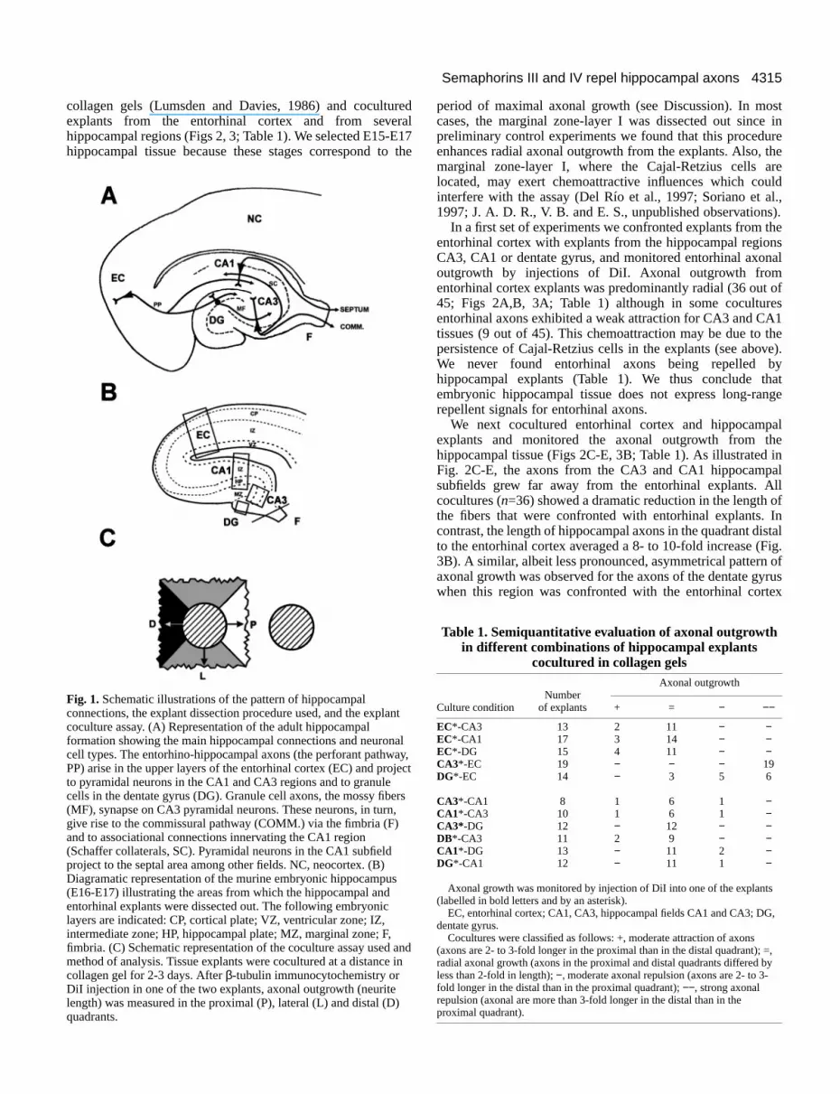
Fig. 1.Schematic illustrations of the pattern of hippocampalconnections, the explant dissection procedure used, and the explacoculture assay. (A) Representation of the adult hippocampalformation showing the main hippocampal connections and neuroncell types. The entorhino-hippocampal axons (the perforant pathwPP) arise in the upper layers of the entorhinal cortex (EC) and proto pyramidal neurons in the CA1 and CA3 regions and to granulecells in the dentate gyrus (DG). Granule cell axons, the mossy fib(MF), synapse on CA3 pyramidal neurons. These neurons, in turngive rise to the commissural pathway (COMM.) via the fimbria (F)and to associational connections innervating the CA1 region(Schaffer collaterals, SC). Pyramidal neurons in the CA1 subfieldproject to the septal area among other fields. NC, neocortex. (B)Diagramatic representation of the murine embryonic hippocampus(E16-E17) illustrating the areas from which the hippocampal andentorhinal explants were dissected out. The following embryoniclayers are indicated: CP, cortical plate; VZ, ventricular zone; IZ,intermediate zone; HP, hippocampal plate; MZ, marginal zone; F,fimbria. (C) Schematic representation of the coculture assay usedmethod of analysis. Tissue explants were cocultured at a distancecollagen gel for 2-3 days. After β-tubulin immunocytochemistry orDiI injection in one of the two explants, axonal outgrowth (neuritelength) was measured in the proximal (P), lateral (L) and distal (Dquadrants.
redralE17o the
period of maximal axonal growth (see Discussion). In mocases, the marginal zone-layer I was dissected out sincepreliminary control experiments we found that this proceduenhances radial axonal outgrowth from the explants. Also, marginal zone-layer I, where the Cajal-Retzius cells alocated, may exert chemoattractive influences which couinterfere with the assay (Del Río et al., 1997; Soriano et a1997; J. A. D. R., V. B. and E. S., unpublished observation
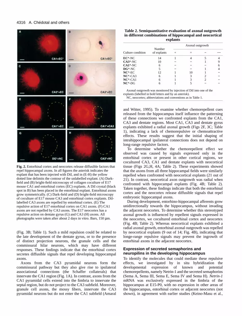
In a first set of experiments we confronted explants from tentorhinal cortex with explants from the hippocampal regioCA3, CA1 or dentate gyrus, and monitored entorhinal axonoutgrowth by injections of DiI. Axonal outgrowth fromentorhinal cortex explants was predominantly radial (36 out45; Figs 2A,B, 3A; Table 1) although in some cocultureentorhinal axons exhibited a weak attraction for CA3 and CAtissues (9 out of 45). This chemoattraction may be due to persistence of Cajal-Retzius cells in the explants (see aboWe never found entorhinal axons being repelled bhippocampal explants (Table 1). We thus conclude thembryonic hippocampal tissue does not express long-ranrepellent signals for entorhinal axons.
We next cocultured entorhinal cortex and hippocampexplants and monitored the axonal outgrowth from thhippocampal tissue (Figs 2C-E, 3B; Table 1). As illustrated Fig. 2C-E, the axons from the CA3 and CA1 hippocampsubfields grew far away from the entorhinal explants. Acocultures (n=36) showed a dramatic reduction in the length othe fibers that were confronted with entorhinal explants. contrast, the length of hippocampal axons in the quadrant dito the entorhinal cortex averaged a 8- to 10-fold increase (F3B). A similar, albeit less pronounced, asymmetrical patternaxonal growth was observed for the axons of the dentate gywhen this region was confronted with the entorhinal corte
nt
alay,ject
ers,
and in
)
Table 1. Semiquantitative evaluation of axonal outgrowthin different combinations of hippocampal explants
cocultured in collagen gelsAxonal outgrowth
NumberCulture condition of explants + = − −−
EC*-CA3 13 2 11 − −EC*-CA1 17 3 14 − −EC*-DG 15 4 11 − −CA3*-EC 19 − − − 19DG*-EC 14 − 3 5 6
CA3*-CA1 8 1 6 1 −CA1*-CA3 10 1 6 1 −CA3* -DG 12 − 12 − −DB*-CA3 11 2 9 − −CA1* -DG 13 − 11 2 −DG*-CA1 12 − 11 1 −
Axonal growth was monitored by injection of DiI into one of the explants(labelled in bold letters and by an asterisk).
EC, entorhinal cortex; CA1, CA3, hippocampal fields CA1 and CA3; DG,dentate gyrus.
Cocultures were classified as follows: +, moderate attraction of axons(axons are 2- to 3-fold longer in the proximal than in the distal quadrant); =,radial axonal growth (axons in the proximal and distal quadrants differed byless than 2-fold in length); −, moderate axonal repulsion (axons are 2- to 3-fold longer in the distal than in the proximal quadrant); −−, strong axonalrepulsion (axonal are more than 3-fold longer in the distal than in theproximal quadrant).
4316
esng1,usleeofon
etheealedlyofn
).nalpel
wged
intex adtof
ve
lrins
e ofnotl.,
A. Chédotal and others
Fig. 2.Entorhinal cortex and neocortex release diffusible factors threpel hippocampal axons. In all figures the asterisk indicates theexplant that has been injected with DiI, and in (E-H) the yellowdotted line delimits the contour of the unlabelled explant. (A) Darkfield and (B) bright-field microscopy of collagen coculture of E17mouse CA1 and entorhinal cortex (EC) explants. A DiI crystal (blaspot in B) has been placed in the entorhinal explant. Entorhinal axgrow symmetrically. (C) Dark-field and (D) bright-field microscopyof coculture of E17 mouse CA3 and entorhinal cortex explants. Dlabelled CA3 axons are repelled by entorhinal cortex. (E) Therepulsive action of E17 entorhinal cortex on CA1 axons. (F) CA1axons are not repelled by CA3 axons. The E17 neocortex has arepulsive action on dentate gyrus (G) and CA3 (H) axons. Allphotographs were taken after about 2 days in vitro. Bars, 150 µm.
Table 2. Semiquantitative evaluation of axonal outgrowthin different combinations of hippocampal and neocortical
explantsAxonal outgrowth
NumberCulture condition of explants + = − −−
EC*-NC 14 1 4 5 4CA3*-NC 10 − − 1 9CA1*-NC 6 − − − 6DG*-NC 5 − − 1 4NC*-EC 12 1 10 1 −NC*-CA3 6 1 3 2 −NC*-CA1 6 1 3 2 −NC*-DG 6 1 5 − −
Axonal outgrowth was monitored by injection of DiI into one of theexplants (labelled in bold letters and by an asterisk).
NC, neocortex; abbreviations and conventions as in Table 1.
(Fig. 3B; Table 1). Such a mild repulsion could be relatedthe late development of the dentate gyrus, or to the preseof distinct projection neurons, the granule cells and tcommissural hilar neurons, which may have differeresponses. These findings indicate that the entorhinal cosecretes diffusible signals that repel developing hippocamaxons.
Axons from the CA3 pyramidal neurons form thcommissural pathway but they also give rise to ipsilateassociational connections (the Schaffer collaterals) tinnervate the CA1 region (Fig. 1A). In contrast, axons from tCA1 pyramidal cells extend into the fimbria to innervate tseptal region, but do not project to the CA3 subfield. Moreovgranule cell axons, the mossy fibers, innervate the Cpyramidal neurons but do not enter the CA1 subfield (Ama
toncehentrtexpal
eralhatheheer,A3ral
and Witter, 1995). To examine whether chemorepellent cureleased from the hippocampus itself influence the patterniof these connections we confronted explants from the CACA3 and dentate regions. Most CA1, CA3 and dentate gyrexplants exhibited a radial axonal growth (Figs 2F, 3C; Tab1), indicating a lack of chemorepulsive or chemoattractiveffects. These results suggest that the initial shaping intrahippocampal ipsilateral connections does not depend long-range repulsive factors.
To determine whether the chemorepellent effect wobserved was caused by signals expressed only in entorhinal cortex or present in other cortical regions, wcocultured CA3, CA1 and dentate explants with neocortictissue (Figs 2G,H, 4A; Table 2). These experiments showthat the axons from all three hippocampal fields were similarrepelled when confronted with neocortical explants (21 out 21). In contrast, neocortical axon outgrowth was radial wheconfronted with hippocampal explants (Fig. 4B; Table 2Taken together, these findings indicate that both the entorhicortex and the neocortex release diffusible signals that reembryonic hippocampal axons.
During development, entorhino-hippocampal afferents grounidirectionally towards the hippocampus, without invadinthe adjacent neocortex. To determine whether this stereotypaxonal growth is influenced by repellent signals expressedthe neocortex, we cocultured entorhinal cortex and neocor(Fig. 4B; Table 2). Whereas neocortical explants exhibitedradial axonal growth, entorhinal axonal outgrowth was repelleby neocortical explants (9 out of 14; Fig. 4B), indicating thalonge-range repulsive signals may prevent the ingrowth entorhinal axons in the adjacent neocortex.
Expression of secreted semaphorins andneuropilins in the developing hippocampusTo identify the molecules that could mediate these repulsieffects, we investigated by in situ hybridization thedevelopmental expression of known and potentiachemorepellents, namely Netrin-1 and the secreted semapho(Sema A, Sema III, Sema E, Sema IV and Sema H). Netrin-1mRNA was exclusively expressed in the fimbria of thhippocampus at E15-P0, with no expression in other areasthe hippocampus, entorhinal cortex or adjacent neocortex (shown), in agreement with earlier studies (Keino-Masu et a
at
-
ckons
iI-

4317Semaphorins III and IV repel hippocampal axons
enthegns
(inea,he
1996). This pattern of expression indicates that Netrin-1highly unlikely to play a repulsive role in the patterning of thmain hippocampal connections (J. A. D. R., V. B. and E. unpublished observations). A similar conclusion can be drafor Sema A, whose mRNA is not expressed in the hippocamregion between E15 and P0 (not shown).
In contrast, sema IV mRNA was uniformly expressed
nedhethe
ne,0,
inalhe
nalese
h,ects
the
Fig. 3.Histograms showing the axonal length (mean ± s.e.m.) inseveral combinations of hippocampal and entorhinal explantslabelled with DiI. Explants injected with DiI are marked by anasterisk. Axonal growth was measured in the distal (filled bars),lateral (grey bars) and proximal (white bars) quadrants. For statistanalysis, axonal growth in the distal quadrant was compared to thin the lateral and proximal quadrants. (A) entorhinal cortex axonsgrow symmetrically when confronted with CA3, CA1 or dentategyrus explants. (B) On the contrary axons from CA3, CA1 anddentate gyrus explants confronted with entorhinal cortex explantswere significantly shorter in the lateral and proximal quadrantscompared to the distal quadrant, indicating that the entorhinal corexerces a repulsive action on these axons. (C) Axonal growth wasmeasured in several combinations of hippocampal cocultures. Nosignificant differences were observed between axonal length in thquadrants, indicating the absence of long-range diffusible cues. nvalues are in parentheses. *P<0.05; **P<0.01; ***P<0.001.
iseS.,wnpal
through the hippocampus, entorhinal cortex and the adjacneocortex at E15-P0. The expression signal is low in tproliferating ventricular zone, and higher in the differentiatinfields (the cortical and hippocampal plates) of these regio(Fig. 5A,B). At E15, sema IIImRNA was widely expressed inthe cortical plate and intermediate zone of the neocortex agreement with Catalano et al., 1998) and entorhinal arwhereas hybridization signals were virtually absent from thippocampus (Fig. 5C). At P0 the expression pattern remaiessentially the same, with high levels of expression in tneocortex and entorhinal area, and weak signals in both hippocampus proper and dentate gyrus (Fig. 5D). At E15, semaE mRNA was exclusively expressed in the intermediate zoboth in the hippocampal region and neocortex (Fig. 5E). At Plow expression was detected in the neocortex and entorharea, whereas hybridization signals were still very high in thippocampus (data not shown). At E15-P0, sema HmRNA wasexpressed in the cortical plate of the neocortex, entorhicortex and in the hippocampus (Fig. 5F). Taken together, thexpression studies show that the secreted semaphorins semaIII , sema IV, sema Eand sema H are expressed in thehippocampal region at the time of axonal outgrowtsuggesting that these factors could mediate the repulsive effdescribed above.
Next we investigated the developmental expression of
icalat
tex
e 3
Fig. 4.Histograms illustrating the axonal length (mean ± s.e.m.) incocultures of hippocampal and neocortical explants. Conventions asin Fig. 3. In (A) the axonal growth of hippocampal and entorhinalcortex explants was monitored when confronted with the neocortex.In all cases axons were significantly shorter in lateral and mostproximal quadrants compared to the distal quadrant, indicating thatthe neocortex releases soluble activities that repel those axons. In (B)we recorded the growth of neocortical explants confronted withhippocampal tissue. No significant difference was observed betweenaxonal length in the three quadrants. n values are in parentheses.*P<0.05; **P<0.01; ***P<0.001.

4318
nalsters
eat
uteeeednddnd
yedn).w-sen).
lls Inereg
se
1,OS
eomrlyB),nd
A. Chédotal and others
Fig. 5.Expression pattern of secreted semaphorins and their receNeuropilin-2, in the developing entorhino-hippocampal system anadjacent neocortex. Hybridizations were performed withdigoxigenin-labelled riboprobes on horizontal sections of E15 andmouse brains. (A,B) Expression pattern of sema IVmRNA at E15and P0 showing wide expression throughout the entorhino-hippocampal system and neocortex, mainly in the cortical plate.(C,D) Expression pattern of sema IIImRNA at E15 and P0illustrating high expression levels in the entorhinal cortex andneocortex and very low expression in the hippocampus itself, excat P0 in the dentate gyrus. Note that sema IIIexpression stopssharply at the junction between EC and hippocampus (arrowhead(E) sema EmRNA expression at E15 demonstrating high expressiolevels in the intermediate zone of the entorhinohippocampal systeand neocortex. (F) Illustration of sema HmRNA expression in thehippocampus and cortex of P0 mouse. (G,H) The strong expressiof neuropilin-2mRNA in the EC and hippocampus at E15 and P0.Also note the faint labelling in the intermediate zone of the neocor(arrowheads) at E15. EC, entorhinal cortex; NC, neocortex; CA1 aCA3, hippocampal fields CA1 and CA3; DG, dentate gyrus. Bars,400 µm (A,C,E,G) and 600 µm (B,D,F,H.)
Table 3. Semiquantitative evaluation of the effect of SemaIII and Sema IV on axonal outgrowth of hippocampal and
entorhinal cortex explantsAxonal outgrowth
NumberCulture condition of explants + = − −−
CA1+Sema III 23 − − − 23CA3+Sema III 19 − − − 19DG+Sema III 13 − 3 3 7EC+Sema III 15 − 3 2 10
CA1+Sema IV 44 − − 3 41CA3+Sema IV 41 − − 1 40DG+Sema IV 15 − 3 2 10EC+Sema IV 22 − 21 1 −
CA1+Control 30 − 28 2 −CA3+Control 29 − 25 4 −DG+Control 5 − 4 1 −EC+Control 8 − 8 − −
Explants were labelled using anti-β-tubulin antibodies. Abbreviations and conventions as in Table 1.
known semaphorin receptors Neuropilin-1 and Neuropilin-At E15-P0, neuropilin-1mRNA was heavily expressed in thehippocampus and neocortex, and at lower levels in tentorhinal area (not shown, but see Kawakami et al., 1996).E15 neuropilin-2 was heavily expressed in the hippocampuand entorhinal cortex, especially in the differentiating fieldincluding the cortical and hippocampal plates (Fig. 5G). contrast, low levels of expression could be detected in tintermediate zone of the adjacent neocortex. At P0 t
2.
he Atss
Inhehe
expression pattern remained the same, though increased sigwere detected in the hippocampus, particularly in the dentagyrus (Fig. 5H). We conclude that the semaphorin receptoNeuropilin-1 and Neuropilin-2 are expressed in thhippocampus and entorhinal cortex at the time thhippocampal connections are being formed.
Sema III and Sema IV repel hippocampal axons invitroTo test directly whether secreted semaphorins can contribto the chemorepulsive effect of the entorhinal cortex, wcultured CA1 and CA3 explants (from E15-E17 mousembryos) with COS cells that had been transiently transfectwith expression constructs for all five known mammaliasecreted semaphorins (A, III/D, E, IV, H). We also coculturethose cells with explants from the entorhinal cortex anneocortex. Explants were cultured for 48-72 hours, fixed astained with an anti-class III β-tubulin antibody that labels theentire population of axons growing from the explant (Moodet al., 1989). The expression of the diverse epitope-taggsemaphorins was verified by western blotting (data not show
Axons from CA1, CA3 and dentate gyrus explants grosymmetrically when cultured with control COS cells, mocktransfected or transfected with an alkaline-phosphataexpression construct (see Fig. 6C,F; Table 3 and not showFurthermore we could not detect any effect of COS-cesecreting Sema A, Sema E or Sema H (data not shown).contrast, axons from CA1, CA3 and dentate gyrus explants wstrongly repelled when confronted with COS cells expressinSema III-myc (Fig. 6A,D; Table 3; see also Fig. 8A). Axongrew almost exclusively from the distal quadrant opposite thCOS-cell aggregate. An equivalent strong repulsion of CACA3 and dentate gyrus axons was observed when using Ccells transfected with sema IV-AP(Figs 6B,E, 8B; Table 3 andnot shown). Axons that exited the lateral quadrant of thexplants, or explants that were placed at a long distance frCOS-cell aggregates producing Sema III or Sema IV, cleaturned away from the aggregates (see for example Fig. 6demonstrating that when present as a gradient Sema IV a
ptord
P0
ept
s).nm
on
texnd

4319Semaphorins III and IV repel hippocampal axons
IV,ingendthebygsndal
epulsion of hippocampal and entorhinal axons by Sema III and/or. E16-E17 CA1 (A-C), CA3 (D-F) and entorhinal (G-I) explants wereed for 48-72 hours at a distance from aggregates of COS cellsted with H-sema III(A,D,G), H-sema IV(B,E,H) or alkaline-atase(C,F,I). All explants were fixed and stained with anti-β-tubulinies. CA1 (A), CA3 (D) and entorhinal (G) axons are strongly repelleda III. Sema IV also exerces a robust repulsive action on CA1 (B) and) axons, but has no effect on entorhinal axons (H). Axons from CA13 (F) and entorhinal cortex (I) grow symmetrically when confrontedS cells secreting only alkaline phosphatase. Ent. Cx. entorhinal cortex;s., alkaline phosphatase. Bars, 280 µm.
Sema III do not inhibit, but rather orientate, the growth hippocampal axons. In addition, we found that Sema III aexercises a repulsive action on entorhinal axons, whereas SIV had no apparent effect on these axons (Figs 6G-I, 8A; Ta3). Axons of neocortex and cerebellar explants from E16 moembryos (two other regions where several secreted semaphand neuropilins are expressed; not shown) grew symmetricwhen confronted with Sema III and Sema IV-expressing Ccell aggregates, or with cells secreting other semaphorins shown). These results show that CA1, CA3 anddentate gyrus axons can be repelled by two distinctsecreting semaphorins, Sema III and Sema IV,whereas entorhinal axons are exclusively repelled bySema III.
Neuropilin-1 antibodies block Sema III andentorhinal cortex-induced chemorepulsionof hippocampal axonsIt has been shown recently that members of theneuropilin family are high-affinity receptors forsecreted semaphorins: Sema IV binds to Neuropilin-1and Neuropilin-2 with high affinity, whereas Sema IIIonly binds to Neuropilin-1 with high affinity (Chen etal., 1997; He and Tessier-Lavigne, 1997; Kolodkin etal., 1997). An antibody (purified IgGs) directed againstthe MAM and b1-b2 domains of Neuropilin-1 has beenshown to abolish completely Sema III-inducedrepulsion and collapse of DRG axons at a concentrationof 10 µg/ml (He and Tessier-Lavigne, 1997).
We first tried to determine whether we could blockSema III-induced repulsion of hippocampal andentorhinal axons by using the anti-Neuropilin-1antibody. In the presence of 10 µg/ml of anti-Neuropilin-1 we found that Sema III-inducedrepulsion was significantly reduced (Figs 7A,B, 8A).Axonal growth was restored in the lateral andproximal quadrants to levels almost identical to thatof the distal quadrant.
We next examined whether Neuropilin-1 was alsoinvolved in Sema IV-induced repulsion ofhippocampal axons. No significant differences wereobserved between CA1 and CA3 explants culturednext to COS cells expressing Sema IV in the presenceor absence of anti-Neuropilin-1 antibodies (Figs 7C,8B). Thus, Sema IV induced-repulsion does notinvolve Neuropilin-1.
Finally, we cocultured CA1 and entorhinalexplants with 10 µg/ml of anti-Neuropilin-1antibodies and found that explants grew almostsymmetrically (Figs 7D, 8C), suggesting thatNeuropilin-1 is involved in mediating to a large extentthe entorhinal cortex-induced repulsion ofhippocampal axons.
DISCUSSION
In this study we have shown that, at the time offormation of hippocampal connections in vivo, thereis a strong chemorepulsion between tissue explantsfrom distinct hippocampal subfields. Concomitantly,
Fig. 6. RSema IVcoculturtransfecphosphantibodby SemCA3 (E(C), CAwith COAlk. Pho
oflsoemableuseorinsallyOS(not
several secreted semaphorins, including Sema III and Semaand their receptors neuropilin, are expressed in the develophippocampal formation. More importantly, the repulsiveffects on hippocampal axons are mimicked by Sema III aSema IV, but not by other secreted semaphorins. Finally, repulsive action of Sema III, but not Sema IV, is mediated Neuropilin-1 receptors. To our knowledge the present findinare the first demonstration of the involvement of Sema III aits receptor, Neuropilin-1, in the patterning of neuron

4320
owerts
tal3n4;
ingg
theatus,
A. Chédotal and others
Fig. 7.Neuropilin-1 is involved in Sema III-and entorhinal cortex-induced repulsions of hippocampal axons. E15-E17 hippocampal(A,C,D) or entorhinal (B) explants were cocultured in collagen gel nextto COS cells secreting Sema III (A-B) or Sema IV (C), or next toentorhinal cortex explant (D), in the presence of 10 µg/ml of anti-Neuropilin-1 IgGs. Explants were stained with anti-β-tubulin antibodies(A-C) or DiI (D). Sema III-induced repulsion of CA3 (A) andentorhinal (B) axons is prevented by the antiboby. On the contrary, CA3axons are still repelled by Sema IV in the presence of anti-Neuropilin-1IgGs (C). (D) Two DiI-labelled CA1 explants were cocultured next toan entorhinal cortex explant (dashed line) in the presence of anti-Neuropilin-1 antibody. DiI-labelled CA1axons are growing toward theentorhinal explant, showing that neuropilin-1 is involved in entorhinal-induced repulsion of CA1 axons (compare to Fig. 2E). Bars, 100 µm(A-C) and 180 µm (D).
Fig. 8.Histograms showing the axonal length (mean ± s.e.m.) inseveral combinations of hippocampal and entorhinal explants and/orCOS cells aggregates expressing Sema III or Sema IV, in the absenceor presence of anti-neuropilin-1 antibody. Explants were labelledwith anti-β-tubulin antibodies (A-B) or DiI (asterisks in C). Axonallength was measured in the distal (filled bars), lateral (grey bars) andproximal (white bars) quadrants. For statistical analysis, axonallengths in the different quadrants, with or without antibody, werecompared. Anti-Neuropilin-1 significantly inhibits Sema III-inducedrepulsion of CA1, CA3 and entorhinal axons (A), but does not affectSema IV-induced repulsion of hippocampal axons (B). (C) Anti-Neuropilin-1 also blocks the repulsive effect of the entorhinal cortexon CA1. n values are in parentheses. *P<0.05; **P<0.01;*** P<0.001.
connections in the forebrain. In addition, our data also shfor the first time that the secreted semaphorin, Sema IV, exa potent repulsive response on developing axons, and thatresponse is likely to be mediated by Neuropilin-2.
Involvement of chemorepulsion in the patterning ofhippocampal connectionsIn mouse embryos the earliest entorhinal axons leave the coby E14, being directed towards the hippocampus; by Eentorhinal axons reach the hippocampal intermediate zone,1 day later they invade their target layer, the outer margizone (prospective stratum lacunosum-molecularConcomitantly, the first axons from the pyramidal neuronsthe CA3 and CA1 hippocampal fields grow rostrally throug
this
rtex15 andnale). inh
the fimbria to form the commissural and hippocampo-seppathways. In addition, by E16-E17, axons from the CApyramidal neurons innervate the ipsilateral CA1 regio(associational afferents; Figs 1A, 9; Supèr and Soriano, 199Supèr et al., 1998). Such a stereotyped pattern of developconnections suggest the involvement of very specific guidincues.
We have shown here that, at these embryonic stages, entorhinal cortex releases long-range diffusible factors threpel axons arising from several regions of the hippocamp

4321Semaphorins III and IV repel hippocampal axons
e
ede.yale
alry).eglin
aalendn
sg-g
ereceds,ixalet
inngn.ne
nin
soorl.,sin eta ete ann3,
Fig. 9.Schematic summary of the data obtained in this study on thformation of hippocampal connections in mouse embryos. Entorhiaxons project to the hippocampus intermediate zone and invade thtarget layer, the outer marginal zone (prospective stratum lacunosmoleculare). Axons from the CA3 and CA1 hippocampal fields grorostrally through the fimbria to form the commissural andhippocampo-septal pathways. In addition, axons from the CA3pyramidal neurons innervate the ipsilateral CA1 region (associatioafferents), but the axons of either the CA3 or the CA1 regions nevinvade the entorhinal cortex or the neocortex. Neuropilins areexpressed by entorhinal and hippocampal neurons. Sema IIImRNAis expressed at high levels in the neocortex and entorhinal area anbarely detectable in the hippocampus itself. This cortical-derivedSema III would prevent the penetration of hippocampal axons in thentorhinal cortex and neocortex. The high expression of Sema IV the hippocampal plate may direct and confine pyramidal cell axonneighbouring layers, such as the intermediate zone and stratumradiatum. The repulsion of entorhinal axons by the neocortex is wand could depend on the different responsiveness of distinctentorhinal projection neurons (see Discussion). −, repulsion; CP,cortical plate; IZ, intermediate zone; VZ, ventricular zone; DG,dentate gyrus.
including the CA1, CA3 and dentate fields. In contrast, reciprocal repulsive effect of the embryonic hippocamptissue on entorhinal axons was observed. Since the majoritpostmitotic neurons present in the CA1-CA3 hippocampusE15-E17 are pyramidal neurons, most axons being repellethe explant coculture assay must arise from the pyramiprojection neurons. For the dentate gyrus, repelled axprobably come from the commissural/associational neuronsthe hilus and from the granule cells (Stanfield and Cow1988).
These in vitro results are in agreement with the eapatterning and trajectory followed by the developinhippocampal axons in vivo, in which axons from the CA3 aCA1 pyramidal neurons grow through the fimbria towards thippocampal commissure, but never invade the entorhicortex (Supèr and Soriano, 1994; Amaral and Witter, 199Supèr et al., 1998). Thus, our data suggest that endogerepulsive signals released by the embryonic entorhinal corprevent the ingrowth of hippocampal axons into the entorhiarea. In addition, the entorhinal-derived repulsive cues micontribute to pushing away the hippocampal axons and,conjunction with chemoattractive cues present in the fimband hippocampal commissure such as Netrin-1 (Serafini et
noaly of atd indalons in
an,
rlygndhenal5;
noustexnalght inria al.,
1996), help to direct them towards the telencephalic midlin(Fig. 9).
Hippocampal axons are also dramatically repelled by thembryonic neocortex, indicating that both the neocortex anentorhinal cortex release diffusible signals that prevent thingrowth of hippocampal axons into these cortical fieldsMoreover, we found that entorhinal axons are repelled bembryonic necortical tissue, whereas neocortical axonoutgrowth is unaffected by nearby hippocampal explants. Thentorhinal cortex receives convergent inputs from severneocortical areas while the entorhinal cortex projects only vesparsely to some cortical areas (Amaral and Witter, 1995Again, the present in vitro findings are consistent with thpattern of neocortical/entorhinal connections in the developinand adult brain, and with the preferential growth of entorhinaaxons towards the hippocampus. However, although the maoutput projection from the entorhinal area is theentorhinohippocampal pathway (perforant pathway), this aresends sparse projections to different subcortical nuclei (Amarand Witter, 1995; Supèr et al., 1994), suggesting that threlatively weak repulsive response of entorhinal axons wheconfronted with neocortical explants (Fig. 4A), may be relateto the different responsiveness of distinct entorhinal projectioneurons.
In this study we did not find evidence for repulsive responsein hippocampus/hippocampus cocultures, suggesting that lonrange repellent cues are unlikely to play a role in the patterninof ipsilateral hippocampal connections, such as the Schaffcollaterals or the mossy fibers. The development of thesintrahippocampal connections, as well as their layer-specifitargeting, could be determined by cues other than secretsemaphorins (but see below), which might include ephrinCAMS, transmembrane semaphorins, and extracellular matrmolecules that are expressed in the developing hippocampsystem (e.g. Gao et al., 1996; Zhang et al., 1996; Serafini al., 1996; Del Río et al., 1997).
Role of Sema III and Sema IV in the development ofhippocampal connectionsIn insects, it is clear that semaphorins play important roles the development of the nervous system. They function igrowth cone guidance, preventing fasciculation and inhibitinaxon branching, but also in synaptic terminal arbor formatio(Kolodkin et al., 1993; Matthes et al., 1995; Yu et al., 1998)Interestingly, G-Sema I also apparently functions as aattractive-permissive guidance cue for growth cones of thsubgenual organ in the limb bud (Wong et al., 1997). Imammals, the function of transmembrane semaphorins in bradevelopment is still unknown, but secreted semaphorins alact as repulsive molecules. Sema III/Collapsin-1 repels mot(Varela-Echavarria et al., 1997) and sensory axons (Luo et a1995; Messersmith et al., 1995; Püschel et al., 1995; Kobayaet al., 1997) in vitro, and Sema A and Sema E/Collapsin-3 carepel and cause the collapse of sympathetic axons (Adamsal., 1997; Koppel et al., 1997). In addition, Sema Z1a, zebrafish semaphorin, also collapses sensory axons (Shojial., 1998). The present findings demonstrate for the first timthat Sema III has an action on forebrain neurons, indicatingmore widespread function for Sema III in axon guidance thahas been shown previously. Sema IV was first identified ihumans and localized to the region 3p21.3 of chromosome
enaleir
um-w
naler
d
eins to
eak

4322
nsis
er,ma
,er
isenatutrin
al
of,
).t
ngl.,areotalralrusllynal
s
ofts
deinant
by
s.
of
hIII.
A. Chédotal and others
where several lung cancer cell lines exhibit homozygodeletions indicative of a tumor supressor gene (Roche et 1996). Nevertheless, the function of Sema IV remainunknown. We show here that Sema IV has a strong repulsaction on hippocampal axons. Since Sema III and Sema IVhighly expressed in the embryonic hippocampal formatiwhen the main axonal trajectories are established, and repulsive responses of hippocampal and entorhinal axonsmimicked by them, we propose that both semaphorins involved in the formation of hippocampal connections.
The pattern of sema III mRNA distribution, with highexpression levels in the neocortex and entorhinal area barely detectable hybridization signals in the hippocampitself, is consistent with the notion that Sema III may be tcortical-derived, endogenous repulsive factor causirepulsion of developing hippocampal axons (Fig. 9). Tcontribution of Sema IV to the patterning of hippocampconnections may be more debatable, since the high expreslevels of sema IVin the hippocampus seems to be inconsistewith the in vitro analyses, in which we never observerepulsive responses when hippocampus/hippocampus explwere cocultured. One major limitation of these interpretatioresides in the absence of data on the localization of semaphproteins, their concentration level or release sites. Howevermammals, secreted semaphorins have a short stretch of bamino acids at their carboxy terminus. This highly chargdomain is likely to limit the diffusion of semaphorins, whiccould act as short-range or local guiding cues (Culotti aKolodkin, 1996). If this is the case, the high expression Sema IV in the hippocampal plate may direct and confipyramidal cell axons to neighbouring layers, such as intermediate zone and stratum radiatum (Fig. 9).
Recently two groups have obtained knock-out mice in whithe sema III gene has been inactivated by homologorecombination (Behar et al., 1996; Taniguchi et al., 1997). Tphenotypic analysis of those mice has shown that the Clooks remarkably normal and that neurons that are stronrepelled in vitro by Sema III project normally in the mutanCNS (Behar et al., 1996; Taniguchi et al., 1997; Catalanoal., 1998). Moreover, neuropilin-1mutant mice also display noabnormalities in the CNS projections of sensory axons, simto the lack of phenotype for these projections in the sema IIImutant mice. However, when neurons from neuropilin-1mutant mice are tested in in vitro explants, they are no lonrepelled by Sema III (Kitsukawa et al., 1997). This lack ofphenotype in the CNS of the sema IIIand neuropilin-1mutantmice has been tentatively explained by both the existenceother semaphorins in the developing CNS, and potentiaother neuropilin/semaphorin receptors on CNS axons (or otguidance cues). Our results clearly show that two distinsemaphorins can have a similar repulsive effect on the saaxons at the same time, and that two distinct receptors medthese effects (see below). This could explain the absencestrong brain defects in sema IIIand neuropilin-1 knock-outs.
Involvement of Neuropilin-1 in axonal repulsion inthe hippocampusThe expression pattern of the semaphorin receptors Neurop1 and Neuropilin-2, with high expression in the entorhincortex and in the hippocampus itself, suggests that the repulresponses decribed in this study are mediated by these rece
usal.,edive
areonthe
areare
andushenghealsionntd
antsnsorin, inasic
edhndofnethe
chusheNSglyt et
ilar
ger a
ofllyherctmeiate of
ilin-alsiveptors
(Fig. 9). Furthermore, the finding that anti-Neuropilin-1antibodies block the repulsive responses of hippocampal axoversus the entorhinal cortex, indicates that this receptor directly involved in the signaling of endogenous repulsivfactors. The contribution of Neuropilin-2 receptors, howeveremains more elusive. Binding studies have shown that SeIII only binds with high affinity to Neuropilin-1, whereas SemaIV binds to both Neuropilin-1 and Neuropilin-2 (Chen et al.1997). Nevertheless, Sema IV appears to bind with highaffinity to Neuropilin-2 than Neuropilin-1 (the KD is about 10-fold smaller), which suggested that the Sema IV signal mediated principally by Neuropilin-2 (Chen et al., 1997). Thabsence of blocking effect of anti-Neuropilin-1 antibodies oSema IV induced-repulsion further supports the idea thNeuropilin-2 might mediate the action of this semaphorin. Bone cannot exclude the possibility that another semaphoreceptor is involved. In addition, we found that Neuropilin-1antibodies block Sema III-induced repulsion of hippocampaxons therefore confirming, for the first time in a systemdifferent from the sensory axons, an essential role Neuropilin-1 in mediating Sema III action. Unfortunatelyneuropilin-1 knock-out mice die around E12.5, which is tooearly to study hippocampal projections (Kitsukawa et al., 1997
Finally, our results confirm that semaphorin binding is nosufficient to trigger a response in growth cones expressineuropilins, as previously shown in the DRG (Koppel et a1997). Thus, the secreted semaphorins E and H, which expressed in the hippocampus and bind neuropilins, do nhave detectable guiding effects on embryonic hippocampaxons. Nevertheless, since the development of neuconnections in the hippocampus proper and dentate gycontinues postnatally, secreted semaphorins could potentiaplay a role at later developmental stages, for instance in axoarbor formation and synaptogenesis.
We would like to thank Dr Flanagan for the ApTag4 vector, DrDrabkin and Christensen for providing us respectively with mousesema IVand sema H. C. S. G. and M. T. L. are investigators of theHoward Hughes Medical Institute. Z. H. is a postdoctoral associatethe Howard Hughes Medical Institute. E. S. is supported by granSAF98-106 (CICYT), the Ramón Areces Foundation, the Marató TV3 Foundation and the International Institute for Research Paraplegia. A. C. and C. S. are supported by INSERM, and the grBIO4-CT960-774 from the EU.
REFERENCES
Adams, R. H., Lohrum, M., Klostermann, A., Betz, H. and Püschel A. W.(1997). The chemorepulsive activity of secreted semaphorins is regulatedfurin-dependent proteolytic processing. EMBO J.16, 6077-6086.
Amaral, D. G., and Witter, M. P. (1995). Hippocampal formation. In The RatNervous System(ed. G. Paxinos), pp. 443-493. New York: Academic Pres
Behar, O., Golden, J. A., Mashimo, H., Schoen, F. J. and Fishman, M. C.(1996). Semaphorin III is needed for normal patterning and growth nerves, bones and heart. Nature 383, 525-528.
Catalano S., Messersmith, E. K., Goodman, C. S., Shatz, C. J. andChédotal, A. (1998). Many major CNS axon projections develop normallyin the absence of semaphorin III. Mol. Cell Neurosci.11, 173-182.
Chen, H., Chédotal, A., He, Z., Goodman, C. S. and Tessier-Lavigne, M.(1997). Neuropilin-2, a novel member of the neuropilin family, is a higaffinity receptor for the semaphorins sema E and sema IV but not sema Neuron 19, 547-559.
Christensen, C. R., Klingelhofer, J., Tarabykina, S., Hulgaard, E. F.,Kramerov, D. and Lukanidin, E. (1998). Transcription of a novel mouse
4323Semaphorins III and IV repel hippocampal axons
-
s
d
m.
.
.
,I.
n
ain.
.
s
st
sempahorin gene, M-sema H, correlates with the metastatic ability of motumor cell lines. Cancer Res.58, 1238-1244.
Colomarino, S. and Tessier-Lavigne, M.(1995). The axonal chemoattractannetrin-1 is also a chemorepellant for trochlear motor axons. Cell 81, 621-629.
Culotti, J. G. and Kolodkin, A. L. (1996). Function of netrins andsemaphorins in axon guidance. Curr. Opin. Neurobiol.6, 81-88.
De Lecea, L., Del Río, J. A, Criado, J. R., Alcantara, S., Morales, M.,Danielson, P. E., Henriksen, S. J., Soriano, E. and Sutcliffe, G.(1997).Cortistatin is expressed in a distinct subset of cortical interneuronsJ.Neurosci.17, 5868-5880
Del Río, J. A., Heimrich, B,., Borrell, V., Forster, E., Drakew, A.,Alcántara, S., Nakajima, K., Miyata, T., Ogawa, M., Mikoshiba, K. etal. (1997). A role for Cajal-Retzius cells and reelin in the developmenthippocampal connections. Nature385, 70-74.
Feiner, L., Koppel, A. M., Kobayashi, H. and Raper, J.(1997). Secretedchick semaphorins bind recombinant neuropilin with similar affinities bbind different subsets of neurons in situ. Neuron19, 539-545.
Fitzgerald, M., Kwiat, G. C., Middleton, J. and Pini, A. (1993). Ventralspinal cord inhibition of neurite outgrowth from embryonic rat dorsal roganglia. Development117, 1377-1384.
Gao, P. P., Zhang, J. H., Yokohama, M., Racey, B., Dreyfus, C. F., Black,I. B. and Zhou, R. (1996). Regulation of topographic projection in thebrain: Elf-1 in the hippocamposeptal system. Proc. Nat. Acad. Sci USA93,11161-11166.
He, Z. and Tessier-Lavigne M.(1997). Neuropilin is a receptor for the axonachemorepellent semaphorin III. Cell 90, 739-751
Kawakami, A., Kitsukawa, T., Takagi, S. and Fujisawa, H. (1996).Developmentally regulated expression of a cell surface protein, neuropin the mouse nervous system. J. Neurobiol.29, 1-17.
Keino-Masu, K., Masu, M., Hinck, L., Leonardo, E. D., Chan, S.-S.,Culotti, J. G. and Tessier-Lavigne, M. (1996). Deleted in ColorectalCancer (DCC) encodes a netrin receptor. Cell 87, 175-185.
Kitsukawa, T., Shimizu, M., Sanbo, M., Hirata, T., Taniguchi, M., Bekku,Y., Yagi, T. and Fujisawa, H. (1997). Neuropilin-semaphorin III/D-mediated chemorepulsive signals play a crucial role in peripheral neprojection in mice. Neuron19, 995-1005.
Kobayashi, H., Koppel, A. M., Luo, Y. and Raper, A.(1997). A role forcollapsin-1 in olfactory and cranial sensory axon guidance. J. Neurosci.17,8339-8352.
Kolodkin, A. L., Levengood, D. V., Rowe, E. G., Tai, Y.-T., Giger, R. J. andGinty, D. D. (1997). Neuropilin is a semaphorin III receptor. Cell 90, 753-762
Kolodkin, A. L., Matthes, D. J. and Goodman, C. S.(1993). The semaphoringenes encode a family of transmembrane and secreted growth cone guidmolecules. Cell 75, 1389-1399.
Koppel, A. M., Feiner, L., Kobayashi, H. and Raper, J.(1997). A 70 aminoacid region within the semaphorin domain activates specific celluresponse of semaphorin family members. Neuron19, 531-537.
Lumsden, A. G. S. and Davies, A. M.(1986). Chemotropic effect of specifictarget epithelium in the development of the mammalian central nervsystem. Nature323, 538-539.
Luo, Y., Shepherd, I., Li, J., Renzi, M. J., Chang, S. and Raper, J. A.(1995). A family of molecules related to collapsin in the embryonic chinervous system.Neuron14, 1131-1140.
Mark, M. D., Lohrum, M. and Püschel, A. (1997). Patterning neuronalconnections by chemorepulsion: the semaphorins. Cell Tiss. Res.290, 299-306.
Matthes, D. J., Sink, H., Kolodkin, A. L. and Goodman, C. S.(1995).Semaphorin III can function as a selective inhibitor of specific synaparborizations. Cell 81, 631-639.
Messersmith, E. K., Leonardo, E. D., Shatz, C. J., Tessier-Lavigne, M.,Goodman, C. S. and Kolodkin, A. L.(1995). Semaphorin III can functionas a selective chemorepellant to pattern sensory projections in the scord. Neuron 14, 949-959.
use
t
.
of
ut
ot
l
ilin,
rve
ance
lar
ous
ck
tic
pinal
Moody, S. A., Quigg, M. S. and Frankfurter, A. (1989). Development of the peripheral trigeminal system in the chick revealed by an isotypespecific anti-β-tubulin monoclonal antibody. J. Comp. Neurol.346, 97-118.
Pini, A. (1993). Chemorepulsion of axons in the developing mammaliancentral nervous system. Science261, 34-98.
Püschel, A. W., Adams, R. H. and Betz, H.(1995). Murine semaphorinD/collapsin is a member of a diverse gene family and creates domaininhibitory for axonal extension. Neuron14, 941-948.
Roche, J., Boldog, F., Robinson, M., Varella-Garcia, M., Swanton, M.,Waggoner, B., Fishel, R., Franklin, W., Gemmill, R. and Drabkin, H.(1996). Distinct 3p21.3 deletions in lung cancer, analysis of deletegenes and identification of a new human semaphorin. Oncogene12, 1289-1297.
Serafini, T., Colomarino, S. A., Leonardo, E. D., Wang, H., Beddington,R., Skarnes, W. C. and Tessier-Lavigne, M.(1996). Netrin-1 is requiredfor commisural axon guidance in the developing vertebrate nervous systeCell 87, 1001-1014.
Shoji, W., Yee, C. S. and Kuwada, J. Y.(1998). Zebrafish semaphorin Z1acollapses specific growth cones and alters their pathway in vivoDevelopment125, 1275-1283.
Skaliora, I., Singer, W., Betz, H. and Püschel, A. W.(1998). Differentialpatterns of semaphorin expression in the developing rat brain. Eur. J.Neurosci.10, 1215-1229.
Soriano, E., Alvarado-Mallart, R. M., Dumesnil, N., Del Río, J. A. andSotelo, C.(1997). Cajal-Retzius cells regulate the radial glia phenotype inthe adult and developing cerebellum and alter granulle cell migrationNeuron18, 563-577.
Stanfield, B. B. and Cowan, W. M. (1988). The development of thehippocampal region. In The Cerebral Cortex, vol. 7 (ed. E. G. Jones and A.Peters), pp. 91-131. New York, New York, Plenum Press.
Supèr, H. and Soriano, E.(1994). The organization of the embryonic andearly postnatal murine hippocampus. II. Development of entorhinalcommissural, and septal connections studied with the lipophilic tracer DiJ. Comp. Neurol.344, 101-120.
Supèr, H., Martínez, A., Del Río, J. A. and Soriano, E.(1998). Involvementof distinct pionner neurons in the formation of layer-specific connections ithe hippocampus. J. Neurosci. 18, 4616-4626.
Tamada A., Shirasaki, R. and Murakami(1995). Floor plate chemoattractscrossed axons and chemorepels uncrossed axons in the vertebrate brNeuron14, 1083-1093.
Taniguchi, M., Yuasa, S., Fujisawa, H., Naruse, I., Saga, S., Mishina, M.and Yagi, T. (1997). Disruption of semaphorin III/D gene causes severeabnormality in peripheral nerve projection. Neuron19, 519-530
Tessier-Lavigne, M. and Goodman, C. S.(1996). The molecular biology ofaxon guidance. Science 274, 1123-1133.
Tuttle, R., Braisted, J. E., Richards, L. J. and O’Leary, D. D. M.(1998).Retinal axon guidance by region-specific cues in diencephalonDevelopment125, 791-801.
Varela-Echavarria, A., Tucker, A., Püschel, A. W. and Guthrie, S.(1997).Motor axon subpopulations respond differentially to the chemorepellentnetrin-1 and semaphorin D. Neuron18, 193-207.
Wong, J. T. W., Yu, W. T. C. and O’Connor, T. P.(1997). Transmembranegrasshopper semaphorin I promotes axon outgrowth in vivo. Development124, 3597-3607.
Yu, H.-H., Araj., H. H., Ralls, S. A. and Kolodkin, A. L. (1998). Thetransmembrane semaphorin sema I is required in Drosophilafor embryonicmotor and CNS axon guidance. Neuron20, 207-220.
Zhang, J. H., Cerreti, D. P., Yu, T., Flanagan, J. G. and Zhou, R.(1996).Detection of ligands in regions anatomically connected to neuronexpressing the EpH receptor BNsk: potential roles in neuron-targeinteraction. J. Neurosci.16, 7182-7192.
Zhou, L., White, A., Lentz, S. I., Wright, D. E., Fisher, D. A. and Snider,W. D. (1997). Cloning and expression of a novel murine semaphorin withstructure similarity to insect semaphorin 1. Mol. Cell Neurosci.9, 26-41.


