Embed Size (px)
Citation preview

Cytotherapy, 2015; 17: 1524e1535
Serum-free process development: improving the yield and consistencyof human mesenchymal stromal cell production
THOMAS R.J. HEATHMAN1, ALEXANDRA STOLZING1, CLAIRE FABIAN2,3,QASIM A. RAFIQ1,4, KAREN COOPMAN1, ALVIN W. NIENOW1,5,BO KARA6 & CHRISTOPHER J. HEWITT1,4
1Centre for Biological Engineering, Loughborough University, Leicestershire, United Kingdom, 2Fraunhofer Institute forCell Therapy and Immunology, Leipzig, Germany, 3Translational Centre for Regenerative Medicine, LeipzigUniversity, Leipzig, Germany, 4Aston Medical Research Institute, School of Life and Health Sciences, Aston University,Aston Triangle, Birmingham, United Kingdom, 5Centre for Bioprocess Engineering, University of Birmingham,Birmingham, United Kingdom, and 6FUJIFILM Diosynth Biotechnologies, Billingham, United Kingdom.
AbstractBackground aims. The cost-effective production of human mesenchymal stromal cells (hMSCs) for off-the-shelf and patientspecific therapies will require an increasing focus on improving product yield and driving manufacturing consistency.Methods. Bone marrowederived hMSCs (BM-hMSCs) from two donors were expanded for 36 days in monolayer withmedium supplemented with either fetal bovine serum (FBS) or PRIME-XV serum-free medium (SFM). Cells were assessedthroughout culture for proliferation, mean cell diameter, colony-forming potential, osteogenic potential, gene expression andmetabolites. Results. Expansion of BM-hMSCs in PRIME-XV SFM resulted in a significantly higher growth rate (P < 0.001)and increased consistency between donors compared with FBS-based culture. FBS-based culture showed an inter-batchproduction range of 0.9 and 5 days per dose compared with 0.5 and 0.6 days in SFM for each BM-hMSC donor line.The consistency between donors was also improved by the use of PRIME-XV SFM, with a production range of 0.9 dayscompared with 19.4 days in FBS-based culture. Mean cell diameter has also been demonstrated as a process metric for BM-hMSC growth rate and senescence through a correlation (R2 ¼ 0.8705) across all conditions. PRIME-XV SFM has alsoshown increased consistency in BM-hMSC characteristics such as per cell metabolite utilization, in vitro colony-formingpotential and osteogenic potential despite the higher number of population doublings. Conclusions. We have increased theyield and consistency of BM-hMSC expansion between donors, demonstrating a level of control over the product, which hasthe potential to increase the cost-effectiveness and reduce the risk in these manufacturing processes.
Key Words: cell-based therapy, comparability, consistency, human mesenchymal stromal cell, manufacturing, regenerative medicine,serum-free, yield
Introduction
The successful development of cell-based therapieshas the potential to address a number of currentlyunmet clinical indications and to improve patientcare across the world. Growing interest in thisemerging field is evident by the large number ofrecent acquisitions of cell-based therapy companiesby larger biopharmaceutical multinationals; forexample, FUJIFILM Holdings Corporation (TSE:4901) recently acquired Cellular Dynamics Interna-tional (NASDAQ: ICEL), a developer and manu-facturer of induced pluripotent stem cells. However,despite the progress, there are a number of
Correspondence: Christopher J. Hewitt, MD, Aston Medical Research InstituBirmingham B4 7ET, UK. E-mail: [email protected]
(Received 18 June 2015; accepted 3 August 2015)
ISSN 1465-3249 Copyright � 2015, International Society for Cellular Therapy.license (http://creativecommons.org/licenses/by/4.0/).http://dx.doi.org/10.1016/j.jcyt.2015.08.002
challenges that remain before cell-based therapiescan be incorporated into routine clinical practice andtheir full potential realized.
Human mesenchymal stromal cells (hMSCs)have demonstrated the potential to target a numberof these currently unmet conditions, with clinicaltrials currently underway for indications such asacute myocardial infarction, stroke and a host of in-flammatory and immune disorders [1]. For the ma-jority of these clinical indications, however, thein vitro expansion of cells is required to deliver aneffective therapeutic dose. The intention of thisexpansion step is to manufacture a sufficient number
te, School of Life and Health Sciences, Aston University, Aston Triangle,
Published by Elsevier Inc. This is an open access article under the CC BY

Improving yield and consistency of hMSC production 1525
of cells to deliver therapeutic benefit without having adetrimental impact on the quality of the cell atdecreasing production costs. Understanding anddefining the quality attributes of hMSC therapies willbe critical for their successful manufacture. This isproving difficult, however, owing to their complex,multifaceted and poorly understood in vivo mecha-nism of action [2].
Cell-based therapies can be broadly divided intotwo categories: patient-specific therapies (autolo-gous) and off-the-shelf therapies (allogeneic).Traditional biopharmaceutical manufacture is pre-dominantly focused on universal treatments in whichmultiple patients can be treated from a single batch.The manufacture of patient-specific therapies, how-ever, will require the careful consideration of regu-latory challenges as well as the distribution anddelivery of a safe, effective and affordable cell-basedtherapy [3]. This also introduces a range of addi-tional challenges, not least of all how a cell therapymanufacturing process can be developed to consis-tently manufacture products from multiple donors[4]. This will also be necessary for off-the-shelfproducts because cellular senescence will limit theirexpansion potential [5]. The key difference betweenoff-the-shelf and patient-specific therapies, however,is that a donor selection process can be used for off-the-shelf products to select donor cell lines that aresimilar on the basis of expansion potential anddesired quality attributes.
A crucial factor determining the economic suc-cess of off-the-shelf cell-based therapies in terms ofaffordability probably will hinge on whether the pa-tient receiving the hMSC therapy will requireimmunosuppressive medication, increasing theoverall lifetime cost of the treatment, although mostclinical trials do not currently use them [6]. It hasbeen demonstrated previously [7,8] that the use ofserum during cell culture processes can lead to anundesired increase in immune response in vivo, andtherefore the use of serum-free alternatives has thepotential to reduce the requirement for post-infusionimmunosuppressive medication. The developmentof defined medium formulations for specific cell-based therapies can also use this type of clinicaloutput as a basis for their development. These long-term considerations for hMSC product manufactureand delivery will be important to drive the develop-ment of cost-effective and reimbursable therapies,which has proved difficult to date.
Achieving the consistent manufacture of medici-nal products is a key requirement for regulatoryapproval and begins with assessing and reducingprocess variation when possible [9]. Driving aconsistent process will demonstrate a state of controlover the product and provides a foundation for
comparability, whereby process changes duringclinical development can be validated and allows forthe product to be manufactured at multiple sites. Akey aspect of reducing variation in the process will bereducing and eventually eliminating the use of fetalbovine serum (FBS) from the cell culture medium[10]. In addition to lot-to-lot variability, there arefurther process constraints on the use of FBS such aslimited supply [11], spiraling cost, potential forpathogen transmission, increased risk of recipientimmune reaction [12] and reduced scope for processoptimization. All of these considerations mean thatmoving toward a serum-free process would bebeneficial in achieving scalable, tunable and consis-tent hMSC manufacturing processes. In addition,serum-free culture has been shown to be amenable toscalable expansion technology such as micro-carriersand stirred bioreactors, producing higher hMSCyields per time unit than serum-based processes,which will be important for driving down the pro-duction cost of hMSC therapies [13,14] and is a keyfocus for our group. That said, the current cost ofserum-free medium (SFM) for research is generallyhigher than serum-based medium; however, as thedemand increases and higher yield processes can bedeveloped, these costs probably will be reduced overtime.
Considering the innate biological variability thatexists between donors and the importance ofensuring a consistent manufacturing process, drivingthis philosophy into process development at an earlystage is critical. Therefore, the aim of this study wasto demonstrate how the development of a serum-freeexpansion process can drive increased consistencyand yield of hMSC manufacture between donors andthe benefits that this can bring as the process scaleincreases.
Methods
Monolayer culture
Human MSCs were isolated from bone marrowaspirate purchased from Lonza obtained from twohealthy donors with informed consent: BM-hMSC 1(lot: 071313B) and BM-hMSC 2 (lot: 071281D).The local ethics committee approved the use of thesample for research. Cells from passage 1 were cry-opreserved at a density of 1e2 � 106 cells/mL in afreeze medium containing 90% (vol/vol) FBS(Hyclone) and 10% (vol/vol) dimethyl sulfoxide(Sigma-Aldrich). For serum-free experiments,hMSCs cryopreserved in serum underwent oneadaptation passage in SFM. Cells were grown in T-flasks seeded at 5000 cells/cm2 at 37�C in humidifiedair containing 5% CO2. For serum-based culture,

1526 T. R.J. Heathman et al.
Dulbecco’s modified Eagle’s medium (DMEM, 1 g/L glucose; Lonza) supplemented with 10% (vol/vol)FBS (Hyclone) and 2 mmol/L UltraGlutamine(Lonza) was exchanged every 3 days. For serum-freeculture, the growth surface of T-flasks was coatedwith 0.4 mg/cm2 PRIME-XV human fibronectin(Irvine Scientific) and cultured in PRIME-XV SFM(Irvine Scientific) according to the manufacturer’sinstructions. On passage, the hMSCs were washedwith phosphate-buffered saline (PBS) without Caþ
or Mgþ and incubated for 4 min with trypsin (0.25%)/ethylene diamine tetra-acetic acid (Lonza) forserum-based culture or TrypLE Express (Invitrogen)for serum-free culture. Dissociation reagents wereinactivated by the addition of appropriate growthmedium, and the cell suspension was centrifuged at220g for 5 min. The supernatant was discarded andthe remaining pellet was re-suspended in an appro-priate volume of culture medium.
Analytical techniques
Analysis of glucose and lactate concentrations in thespent medium was performed with the use of aCedex Bio-HT (Roche). Cell counting, mean celldiameter and viability (by use of acridine orangeuptake and 40-6-diamidino-2-phenylindole exclu-sion) was performed with the use of a Nucleo-Counter NC-3000 automated mammalian cellcounter (Chemometec). The following parameterswere obtained:
Specific growth rate
Specific growth rate; m ¼ ln�CxðtÞ=Cxð0Þ
�Dt
where m is the net specific growth rate (h�1), Cx(t)and Cx(0) are the cell numbers at the end and start ofthe exponential growth phase, respectively, and t istime (h).
Population doublings
Population doublings; Pd ¼ 1logð2Þ,log
�CxðtÞCxð0Þ
�
where Pd is the number of population doublings andCx(t) and Cx(0) are the cell numbers at the end andstart of the exponential growth phase, respectively.
Specific metabolite consumption/production rate
Specific metabolite flux;
qmet ¼�
m
Cxð0Þ�,
�CmetðtÞ � Cmetð0Þ
emt � 1
�
where qmet is the net specific metabolite consumptionor production rate, m is the specific growth rate (h�1),
Cx(0) is the cell number at the end of the exponentialgrowth phase, Cmet(t) and Cmet(0) are the metaboliteconcentrations at the end and start of the exponentialgrowth phase, respectively, and t is time (h).
Quantitative osteogenesis assay
Osteogenesis was quantified by means of hMSCcollagen production with the use of the Sircol assay(Biocolour) after osteogenic differentiation. Collagenstandards of acid-soluble collagen type I at 0, 0.1, 0.2and 0.4 g/L were used to quantify the collagen pro-duction. Bone marrowederived hMSCs (BM-hMSCs) were seeded at 10,000 cells/cm2 in a wellplate with previously described cell culture medium;after 3 days, culture growth medium was exchangedto osteogenic medium (Irvine Scientific) andcultured for 9 days with a medium exchange takingplace every 3 days. To quantify the collagen pro-duction, cells were fixed with a solution of 5% aceticacid (vol/vol) (Sigma) and 9% formaldehyde (vol/vol)(Sigma) for 30 min at room temperature. Themonolayer was washed, and Sircol dye reagent(Biocolour) was added to each well for 30 min,removed and the cell monolayer was washed withAcid-Salt Wash Reagent (Biocolour). Alkali Reagent(Biocolour) was added to each well to release thecollagen-bound Sircol Dye Reagent, and the result-ing solution along with the collagen standard wasquantified on a microplate reader (BMG Labtech) atan absorbance of 555 nm.
Colony-forming unit fibroblast efficiency
To assess the colony-forming unit fibroblast (CFU-f)efficiency, BM-hMSCs were seeded in a T-flask at10 cells/cm2 and cultured with a medium exchangeevery 3 to 5 days. After 14 days of culture, cells werewashed with PBS and fixed in 4% formaldehyde (vol/vol) (Sigma) for 30 min. Colonies were stained with1% crystal violet (Sigma) in 100% methanol (wt/vol)for 30 min. Stained colonies that were made up ofmore than 25 cells were recorded as CFUs.
RNA isolation
Cells were harvested for RNA isolation at thebeginning, middle and end of the expansion process.Total RNA was collected with the use of TriFastReagent (Peqlab) according to the manufacturer’sinstructions. Potential genomic DNA contaminationwas removed by digestion with DNase (Life Tech-nologies) followed by reverse transcription at 50�Cfor 60 min with the use of Superscript III (LifeTechnologies) and 250 ng Oligo(dT)18-primer (LifeTechnologies).

Figure 1. Growth kinetics of the two BM-hMSC lines over 36 days of expansion. (A) Box-and-whisker plots demonstrate the increasedconsistency and cumulative population doublings in SFM- compared with FBS-containing medium (P < 0.001); (B) specific growth rate ofhMSCs in FBS and SFM. Data are shown as mean � standard deviation (n ¼ 4).
Improving yield and consistency of hMSC production 1527
Quantitative real-time polymerase chain reactionanalysis
Quantitative real-time polymerase chain reaction(PCR) was performed with the use of SYBRGreenER quantitative PCR Supermix Universal(Life Technologies); 1� SybrGreen I (Life Tech-nologies) and 0.2 mmol/L primer each on the DNAengine Opticon2 (Biorad) were additionally added,through the use of these cycling conditions: primarydenaturation at 95�C for 3 min, followed by 35 cy-cles: 95�C for 30 s, 60�C for 30 s (36B4, p21,CCL2)/55 �C (Oct4) and 72�C for 30 s followed byfluorescence measurement. The following primersfor cell markers were used: CCL2 (recruits mono-cytes, memory T cells and dendritic cells to the sitesof inflammation produced by either tissue injury orinfection, NM_002982.3) (Fw) 50-CCA AGG GCTCGC TCA GCC AGA TGC-30, (Re) 50-CGG AGTTTG GGT TTG CTT GTC CAGG-30; p21 (reg-ulates the cell cycle and mediates cellular senescence,NM_000389.4) (Fw) 50-CCG CCT GCC TCCTCC CAA CT-30, (Re) 50-GAG GCC CGT GAGCGA TGG AA-30, OCT4 (pluripotent markerassociated with self-renewal of undifferentiated cells,NM_002701.4) (Fw) 50-GAG GAG TCC CAGGAC ATC AA-30, (Re) 50-CAT CGG CCT GTGTAT ATC CC-30 and vascular endothelial growthfactor (VEGF) (associated with vascularizationand growth of blood vessels, NM_001171623.1)(Fw) 50-GGAAGGAGCCTCCCTCAGGGTTTCG-30, (Re) 5¢-GCCGGAGTCTCGCCCTCCGG -3¢. Serial dilutions of plasmid standards wereused as positive controls and for quantification.Expression was normalized to the referencegene 36B4 (ribosomal protein large P0 RPLP0,NM_001002.3).
Human MSC characterization
Immunophenotype analysis was performed by meansof multi-parameter flow cytometry before and after
the hMSC expansion process with the use of a pre-viously developed protocol [15]. Short-tandemrepeat analysis was completed by means of LGCStandards (UK) under their cell line authenticationprogram. Morphology images were obtained with theuse of a light microscope (Nikon Eclipse TS-100).
The hMSC differentiation was induced throughthe use of PRIME-XV differentiation SFM (IrvineScientific) according to the manufacturer’s in-structions. After 21 days, the differentiation mediawere removed and cells were rinsed with PBS fixedwith 4% (vol/vol) paraformaldehyde (PFA) at roomtemperature. Adipocytes were stained with 1% (wt/vol)oil red O (Sigma-Aldrich) in isopropanol at roomtemperature and rinsed with distilled water. Osteo-blasts were incubated with 2.5% (vol/vol) silver nitrate(Sigma-Aldrich) under ultraviolet light (30-min expo-sure), rinsed with distilled water and stained with fastviolet solution (Sigma-Aldrich) containing 4% (vol/vol)napthol AS-MX phosphate alkaline (Sigma-Aldrich)for 45 min at room temperature in the dark. Chon-drocytes were stained with 1% (wt/vol) alcian blue(Sigma-Aldrich) in 0.1 mol/L hydrochloric acid(Sigma-Aldrich). After 30-min incubation, cells wererinsed 3 times with 0.1 mol/L HCl. After staining,differentiated cells were visualized under a light mi-croscope (Nikon Eclipse TS-100).
Statistical analysis
Results were deemed to be significant at a value ofP < 0.05 with the use of a two-tailed Student’s t-test.
Results
Human MSC growth across multiple donor lines
Human MSCs are currently under clinical investi-gation for the treatment of many diseases, with themajority of these off-the-shelf therapies typicallyrequiring more than one billion cells per patient

Table I. Process time required to manufacture a theoretical batchof 350M hMSCs, demonstrating variation in process time betweenand within donor material that is reduced in the serum-free process.
Condition hMSC line
Per dose of 350M hMSCs
Process time(days)
Inter-batchrange (days)
Inter-donorrange (days)
FBS BM-hMSC 1 23.5 0.9 19.4BM-hMSC 2 39.5 5.0
SFM BM-hMSC 1 12.9 0.5 0.9BM-hMSC 2 12.4 0.6
Assumptions: Starting population of 2M hMSCs expanded in theT-flask process presented in this study.
1528 T. R.J. Heathman et al.
[16,17]. For manufacturing processes to meet thisdemand, a large cell expansion ratio will be requiredto treat many patients from the same batch. Figure 1shows the relative difference between a SFM,PRIME-XV, and media supplemented with FBS interms of hMSC growth. It is clear that the SFM of-fers a significantly higher proliferation rate over FBS(P < 0.001), with a maximum specific growth rate of0.471 � 0.009 per day compared with 0.244 � 0.018per day during the 36-day expansion process. Thisincreased growth rate corresponds to a final mediancumulative population doubling level of 18.15 in theSFM compared with 8.93 in FBS culture across thetwo donors at the end of the 36-day expansion pro-cess. This represents an increase of approximately600 times the number of cells under the SFM culturecompared with FBS culture over this expansionperiod, which dramatically increases the effectiveproduct yield and potential scalability of the SFMprocess to meet the needs of a large-scale, off-the-shelf, cell-based therapy. Cells that have nutrientdeprivation typically spend longer in the G1 phase ofthe cell cycle, resulting in slower proliferation [18].The fact that the PRIME-XV medium supported amore rapid cell growth therefore indicates that it mayprovide a better nutritional balance or activatesgrowth regulation pathways, such as the PI(3)Kpathway, than the FBS-containing medium underthe medium exchange regimen used here.
One of the key driving factors for the overall pro-duction cost of an hMSC therapy, as well as mediumcost, probably will be in the level of donor-to-donorvariability in play during the manufacturing process,owing to increased process time and risk of batchfailure. The donor lines selected for this study hadpreviously demonstrated large differences in cellularcharacteristics [4], which will particularly affect thesuccessful development of patient-specific hMSCtherapies. Figure 1A shows the inconsistency betweenthe growth characteristics of these hMSC lines, with arange of 4.86 population doublings after the 36-dayFBS expansion process, demonstrating a divergingprocess that occurs in FBS culture. In contrast to thisvariance experienced in FBS culture and despite ahigher number of cumulative population doublings,the SFM culture process had a corresponding range ofonly 1.45 population doublings between these donorsand batch runs after 36 days in culture.
For the development of a patient-specific, cell-based therapy, in which the inter-donor variabilitymust be accounted for during manufacture, thisreduction in product variance has a significantbenefit to the process. As discussed previously [4],reducing the divergence in donor cell characteristicswill alleviate potential bottlenecks in the isolation,expansion and delivery process, an important
consideration for cell-based therapy process devel-opment. In conjunction with this, a more consistentexpansion process will reduce the risk of productbatch failure, which for a patient-specific therapy willmean that the patient will not be treated. Table Ishows the variability between the two BM-hMSClines for a hypothetical process requiring a batchsize of 350 million cells from a starting population oftwo million cells [1]. This demonstrates theincreased consistency that can be achieved in theSFM process between production batches and be-tween donors, with a reduction in range from 19.4days in the FBS process to 0.9 days in the serum-freeprocess. This also has advantages for off-the-shelftherapies because it is possible to select material forthe expansion process by pre-screening hMSCs anddiscarding those that do not display sufficient growthkinetics. Under the SFM condition, both of the BM-hMSC lines displayed similar process times betweendonors and a reduced inter-donor range. This hasthe potential to lead to reduced costs in the processdevelopment phase because fewer donor lines will beexcluded (and therefore require testing) comparedwith FBS culture and will be available for the pro-duction process. The reduced process time to ach-ieve this batch size in the serum-free condition willalso be advantageous for reducing the overall me-dium costs because product batches can be manu-factured in reduced time. Furthermore, an increasein achievable cell number per batch will also increasethe material available for quality release testing,which is an important consideration of patient-specific therapies because each patient batch mustbe independently tested before release. A study byDeskins et al. [19] also demonstrated that three in-dependent in vitro assays that were based on growthrate, proliferative potential and ATP content wereable to predict in vivo performance, with hMSCsperforming above average in all three assays havingincreased in vivo regenerative abilities.
Associated with the reduction in inter-donorvariability, further reductions in process input
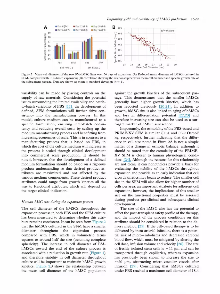
Figure 2. Mean cell diameter of the two BM-hMSC lines over 36 days of expansion. (A) Reduced mean diameter of hMSCs cultured inSFM- compared with FBS-based expansion; (B) correlation showing the relationship between mean cell diameter and specific growth rate ofthe subsequent passage. Data are shown as mean � standard deviation (n ¼ 4).
Improving yield and consistency of hMSC production 1529
variability can be made by placing controls on thesupply of raw materials. Considering the potentialissues surrounding the limited availability and batch-to-batch variability of FBS [11], the development ofdefined, SFM formulations will further drive con-sistency into the manufacturing process. In thismodel, culture medium can be manufactured to aspecific formulation, ensuring inter-batch consis-tency and reducing overall costs by scaling up themedium manufacturing process and benefitting fromincreasing economies of scale. This is in contrast to amanufacturing process that is based on FBS, inwhich the cost of the culture medium will increase asthe process is scaled through clinical developmentinto commercial scale production. It should benoted, however, that the development of a definedmedium formulation should be based on a rigorousproduct understanding so that desired product at-tributes are maximized and not affected by thevarious medium components. These desired productattributes could range from growth kinetics all theway to functional attributes, which will depend onthe target clinical indication.
Human MSC size during the expansion process
The cell diameter of the hMSCs throughout theexpansion process in both FBS and the SFM culturehas been measured to determine whether this attri-bute remains consistent. It can be seen from Figure 2that the hMSCs cultured in the SFM have a smallerdiameter throughout the expansion processcompared with FBS, which in volumetric termsequates to around half the size (assuming completesphericity). The increase in cell diameter of BM-hMSCs toward the end of the culture process isassociated with a reduction in growth rate (Figure 1)and therefore stability in cell diameter throughoutculture will be important to maintain hMSC growthkinetics. Figure 2B shows the relationship betweenthe mean cell diameter of the hMSC population
against the growth kinetics of the subsequent pas-sage. This demonstrates that the smaller hMSCsgenerally have higher growth kinetics, which hasbeen reported previously [20,21]. In addition togrowth, hMSC size is also linked to aging of hMSCsand loss in differentiation potential [22,23] andtherefore increasing size can also be used as a sur-rogate marker of hMSC senescence.
Importantly, the osmolality of the FBS-based andPRIME-XV SFM is similar (0.31 and 0.29 Osmol/kg, respectively), further indicating that the differ-ence in cell size noted in Fiure 2A is not a simplematter of a change in osmotic balance, although itshould be noted that the osmolality of the PRIME-XV SFM is closer to human physiological condi-tions [24]. Although the reasons for this relationshipare not clear, it can nonetheless provide a basis forevaluating the stability of the hMSCs throughoutexpansion and provide as an early indication that cellgrowth kinetics may begin to reduce. The smaller cellsize in the SFM will also allow for higher number ofcells per area, an important attribute for adherent cellexpansion; however, the implications of this smallersize on the functional properties must be assessedduring product pre-clinical and subsequent clinicaldevelopment.
The size of the hMSC also has the potential toaffect the post-transplant safety profile of the therapy,and the impact of the process conditions on thisattribute should be considered in relation to the de-livery method [25]. If the cell-based therapy is to bedelivered by intra-arterial infusion, there is a poten-tial risk of micro-embolisms and decreased cerebralblood flow, which must be mitigated by altering thecell dose, infusion volume and velocity [26]. The sizeof freshly isolated stem cells is w11 mm and can betransported through capillaries, whereas expansionhas previously been shown to increase the size tow20 mm, obstructing micro-vascular vessels afterinfusion [27]. Considering that hMSCs culturedunder FBS reached a maximum cell diameter of 18.4
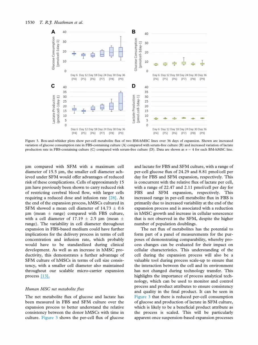
Figure 3. Box-and-whisker plots show per-cell metabolite flux of two BM-hMSC lines over 36 days of expansion. Shown are increasedvariation of glucose consumption rate in FBS-containing culture (A) compared with serum-free culture (B) and increased variation of lactateproduction rate in FBS-containing culture (C) compared with serum-free culture (D). Data are shown as n ¼ 4 for each BM-hMSC line.
1530 T. R.J. Heathman et al.
mm compared with SFM with a maximum celldiameter of 15.5 mm, the smaller cell diameter ach-ieved under SFM would offer advantages of reducedrisk of these complications. Cells of approximately 15mm have previously been shown to carry reduced riskof restricting cerebral blood flow, with larger cellsrequiring a reduced dose and infusion rate [28]. Atthe end of the expansion process, hMSCs cultured inSFM showed a mean cell diameter of 14.73 � 0.6mm (mean � range) compared with FBS culture,with a cell diameter of 17.19 � 2.5 mm (mean �range). The variability in cell diameter throughoutexpansion in FBS-based medium could have furtherimplications for the delivery process in terms of cellconcentration and infusion rate, which probablywould have to be standardized during clinicaldevelopment. As well as an increase in hMSC pro-ductivity, this demonstrates a further advantage ofSFM culture of hMSCs in terms of cell size consis-tency, with a smaller cell diameter also maintainedthroughout our scalable micro-carrier expansionprocess [13].
Human MSC net metabolite flux
The net metabolite flux of glucose and lactate hasbeen measured in FBS and SFM culture over theexpansion process to better understand the relativeconsistency between the donor hMSCs with time inculture. Figure 3 shows the per-cell flux of glucose
and lactate for FBS and SFM culture, with a range ofper-cell glucose flux of 24.29 and 8.81 pmol/cell perday for FBS and SFM expansion, respectively. Thisis concurrent with the relative flux of lactate per cell,with a range of 22.47 and 2.11 pmol/cell per day forFBS and SFM expansion, respectively. Thisincreased range in per-cell metabolite flux in FBS isprimarily due to increased variability at the end of theexpansion process and is associated with a reductionin hMSC growth and increase in cellular senescencethat is not observed in the SFM, despite the highernumber of population doublings.
The net flux of metabolites has the potential toform part of a panel of measurements for the pur-poses of demonstrating comparability, whereby pro-cess changes can be evaluated for their impact oncellular characteristics. This understanding of thecell during the expansion process will also be avaluable tool during process scale-up to ensure thatthe interaction between the cell and its environmenthas not changed during technology transfer. Thishighlights the importance of process analytical tech-nology, which can be used to monitor and controlprocess and product attributes to ensure consistencyand quality in the final product. It can be seen inFigure 3 that there is reduced per-cell consumptionof glucose and production of lactate in SFM culture,which is likely to be a beneficial product attribute asthe process is scaled. This will be particularlyapparent once suspension-based expansion processes

Figure 4. Colony-forming efficiency and osteogenic potential of two BM-hMSC lines against number of population doublings throughoutthe expansion process. (A) Colony-forming efficiency demonstrates increased consistency between BM-hMSC lines in SFM- compared withFBS-based medium; (B) level of collagen deposition demonstrates maintenance in osteogenic potential of both BM-hMSC lines in SFM at ahigh number of population doublings.
Improving yield and consistency of hMSC production 1531
routinely reach high cell densities (in excess of 1 �106 cell/mL), in which the buildup of waste productssuch as lactate has the potential to inhibit theexpansion process. Considering that the culturemedium is likely to contribute to a significant portionof the cost of goods for hMSC production, productattributes that reduce the usage of culture mediumwill be increasingly beneficial at the large scale.
Discussion
The increased consistency between donor hMSClines in terms of per-cell metabolite flux is a furtherdemonstration that the cells cultured in the SFMconditions show reduced inter-donor variation, animportant consideration, given the large amount ofvariation in cell-based therapy manufacture.Although this has been demonstrated in the presentstudy for basic metabolites, there is scope to extendthis analysis to a larger panel of metabolic in-termediates to provide a detailed understanding ofthe impact of the process on the hMSC metaboliccharacteristics during scale-up. The reason for thisincreased consistency in the SFM has yet to beexplored in the literature; however, the combinationof hMSCs cultured on fibronectin with serum-freegrowth medium has been previously shown to acti-vate the platelet-derived growth factor receptor,which is essential for cell migration [29]. Activationof hMSCs in this way provides a potential mecha-nism for the cell characteristics to converge becausethe hMSCs are actively forced to utilize specificcellular pathways, as opposed to serum-based cul-ture, in which an abundance of various proteins areavailable to the cells. This combination of a fibro-nectin coating with growth medium supplementedwith platelet-derived growth factor has been usedpreviously to positively select for smaller, highlyproliferative cell populations from bone marrow,termed multipotent adult progenitor cells [30]. This
positive selection process could also be contributingto the increase in consistency measured in severaldifferent characteristics under serum-free conditions,as discussed further below.
Ensuring consistency in hMSC characteristics
There is currently much discussion about the trueidentity and desired characteristics of hMSCs forclinical applications and how they elicit their thera-peutic mechanism of action [31]. Despite this, somehints come from a graft-versus-host disease studyshowing that just a few passages can make a signifi-cant difference. Human MSCs from passages 1e2compared with passages 3e4 showed a decrease inpatient survival and response, whereas no in vitrodifferences were found [32]. Despite this uncertaintyin the application of hMSCs, there is still a need torigorously characterize the cellular product duringthe development of an expansion process to ensurethe process itself is not having a detrimental impacton the product characteristics while yield isincreased. It is important that assay developmenttakes place in parallel with clinical development sothat the prediction of clinical effect for a specifictarget indication can be correlated to process mea-surements in vitro.
As hMSC expansion processes move through thedevelopment phase, there is an increasing need toassess the characteristics of the product in relation tothe number of population doublings the cells haveundergone and is favorable under the current regu-latory guidelines. This has the benefit of normalizingcell expansion data because passage number does nottake into account the seeding density of the cells orthe relative expansion level of the product in eachcondition. The use of population-doubling levelversus cellular characteristic in this way allows for afair comparison between conditions and is far moreamenable to comparisons with scale-up technology

Figure 5. Quantitative real-time PCR analysis shows RNA expression of four hMSC genes for the two BM-hMSC lines in FBS (blue) andSFM (green) expansion. Shown are maintained expression of CCL2 (A), P21 (B) and Oct4 (C) and a reduction in expression of VEGF-A(D) in SFM culture. Complementary DNA is normalized to housekeeping gene 36B4. Solid line indicates BM-hMSC 1; dashed line, BM-hMSC 2.
1532 T. R.J. Heathman et al.
such as bioreactors, when the term “passage” doesnot readily apply. Figure 4 shows the colony-formingpotential and osteogenic potential of the hMSCdonor lines under FBS and SFM culture against thenumber of population doublings. This demonstratesthat both product attributes decrease through theexpansion process as the number of populationdoublings increases, which hints at the challengesthat exist when developing large-scale manufacturingprocesses, which are likely to require lot sizes inexcess of a trillion cells [33].
Despite this, hMSCs cultured in SFM retained asimilar level of colony-forming potential at a highnumber of population doublings, and, importantly,the consistency between the two hMSC donor lineswas far greater than in FBS. Our data thereforesuggest that the SFM condition used, PRIME-XV,in conjunction with growth on fibronectin, is ableto support the generation of a more homogenous cellpopulation in terms of colony-forming potential aswell as cell size and growth rate, possibly through apositive selection process or the maintenance ofasymmetric division. Indeed, an increased presenceof CFU-F in an MSC population has previously beennoted when an optimized defined medium formula-tion is used as compared with DMEM/FBS [34].Furthermore, Wagner et al. [35], who compared twoserum-containing media, also noted that one wasable to support a more homogenous morphologythan the other. This maintenance of consistency
between donors will be important for both patient-specific and off-the-shelf therapies and will reducethe likelihood of product batch failure during qualitytesting. This will result in a reduced cost at the largescale because the capital invested per batch will behigh, but, more importantly, for patient-specifictherapies, a batch failure would result in a patientgoing without treatment, which would be highlyundesirable.
It is also important that the hMSCs retain theexpression of key genes throughout the expansionprocess because they are likely to play a key role inthe product performing its function in vivo. Figure 5shows the RNA expression of the donor hMSC linesin FBS and the SFM culture against population-doubling level. It is clear that despite the increasedpopulation-doubling level, hMSCs cultured underSFM conditions retained the expression of all fourgenes analyzed, indicating that the positive selectionwe believe is occurring to generate the more ho-mogenous population is not affecting the expressionof key genes. CCL2 has been implemented in therecruitment of T-cells, monocytes and dendritic cellsto the sites of inflammation [36] and is therefore animportant gene to maintain for clinical indicationsthat require a level of immune modulation.Figure 5B shows that the relative expression of P21, agene relating to cellular aging and senescence, hasnot unduly increased throughout the expansionprocess in SFM or FBS culture. Oct4 is a marker of

Figure 6. Phase-contrast images of the two BM-hMSC lines show the increased consistency in morphology between the cell lines underSFM expansion compared with FBS.
Improving yield and consistency of hMSC production 1533
pluripotency, mainly associated with embryonic stemcells, but has previously demonstrated expression inhMSCs [37]. Maintenance of Oct4 under SFMexpansion demonstrates the continued ability of thecells to self-proliferate at high population-doublinglevels and will be important for the clinical applica-tion of cell-based products that undergo cellulardifferentiation. VEGF has been shown to be a highlyimportant gene in the promotion of angiogenesis byhMSCs [38], which will be important in a number ofclinical indications, particularly for cardiac repair[39], a key target for a number of hMSC-basedtherapies. Despite a higher relative expression ofVEGF in hMSCs cultured in SFM, there is adecrease in the expression of VEGF as the number ofpopulation doublings increases, which should befurther investigated if the hMSC product requires ahigh level of cumulative population doublings and isto be used for clinical indications requiringangiogenesis.
Figure 6 shows the difference in hMSCmorphology between donor lines in FBS-based cul-ture that is reduced in SFM culture, with smallerspindle-shaped cells. This increased consistency be-tween donors in cellular morphology observed inSFM has benefits for the development ofmanufacturing processes that are based on a fixedsurface area. This will be particularly apparent forpatient-specific therapies, in which the number of
obtainable cells per square centimeter will determinethe final cell yield of the product batch. Consideringthat manufacturing processes for these cell-basedtherapies probably will have a minimum number ofcells per dose, this reduced variation under SFM willgreatly reduce the risk of product batch failure,increasing the cost-efficiency of the process. As wellas morphology, the hMSCs have demonstrated thedesired immunophenotype and tri-lineage differen-tiation potential in FBS and SFM culture throughoutthe entire expansion process (SupplementaryFigure 1). Additionally, hMSCs in both conditionshave demonstrated the correct genotype profile at theend of the expansion process, as determined bymeans of short-tandem repeat analysis for eachdonor cell line (Supplementary Figure 2).
Conclusions
The development of consistent manufacturingprocesses remains a key challenge that must beovercome to ensure the successful translation ofcell-based therapies. SFM has the potential to reducethe variability of input material to these processes,which will allow for increased control over processconsistency. By developing a serum-free process, wehave increased the yield and consistency of hMSCexpansion between donors, which offers large

1534 T. R.J. Heathman et al.
advantages in the development of both off-the-shelfand patient-specific cell-based therapies. The con-vergence of hMSC characteristics throughout anexpansion process demonstrates a level of controlover the product manufacture, which has the po-tential to increase the cost-effectiveness and reducethe risk in these processes.
Acknowledgments
This study was funded by the Engineering andPhysical Sciences Research Council (EPSRC) andFUJIFILM Diosynth Biotechnologies.
Disclosure of interests: The authors have nocommercial, proprietary, or financial interest in theproducts or companies described in this article.
References
[1] Heathman TRJ, Nienow AW, McCall MJ, Coopman K,Kara B, Hewitt CJ. The translation of cell-based therapies:clinical landscape and manufacturing challenges. Regen Med2015;10:49e64.
[2] Carmen J, Burger SR, McCaman M, Rowley JA. Developingassays to address identity, potency, purity and safety: cellcharacterization in cell therapy process development. RegenMed 2012;7:85e100.
[3] Hourd P, Chandra A, Medcalf N, Williams DJ. Regulatorychallenges for the manufacture and scale-out of autologouscell therapies. StemBook. 2014. Paul Hourd, Amit Chandra,Nick Medcalf and David J. Williams, Cambridge MA, 2014.
[4] Heathman TRJ, Rafiq QA, Chan AKC, Coopman K,Nienow AW, Kara B, et al. Characterization of humanmesenchymal stem cells from multiple donors and theimplications for large scale bioprocess development. BiochemEngineer J 205 (Epub ahead of print; http://dx.doi.org/10.1016/j.bej.2015.06.018).
[5] Estrada JC, Torres Y, Benguria A, Dopazo A, Roche E,Carrera-Quintanar L, et al. Human mesenchymal stem cell-replicative senescence and oxidative stress are closely linkedto aneuploidy. Cell Death Dis 2013;4:e691.
[6] Golpanian S, El-Khorazaty J, Mendizabal A, DiFede DL,Suncion VY, Karantalis V, et al. Effect of aging on humanmesenchymal stem cell therapy in ischemic cardiomyopathypatients. J Am Coll Cardiol 2015;65:125e32.
[7] Kadri N, Potiron N, Ouary M, Jegou D, Gouin E, Bach JM,et al. Fetal calf serum-primed dendritic cells induce a stronganti-fetal calf serum immune response and diabetes protec-tion in the non-obese diabetic mouse. Immunol Lett 2007;108:129e36.
[8] Haase C, Ejrnaes M, Juedes AE, Wolfe T, Markholst H, vonHerrath MG. Immunomodulatory dendritic cells requireautologous serum to circumvent nonspecific immunosup-pressive activity in vivo. Blood 2005;106:4225e33.
[9] Williams DJ, Thomas RJ, Hourd PC, Chandra A, Ratcliffe E,Liu Y, et al. Precision manufacturing for clinical-qualityregenerative medicines. Philos Trans A Math Phys Eng Sci2012;370:3924e49.
[10] Wappler J, Rath B, Laufer T, Heidenreich A, Montzka K.Eliminating the need of serum testing using low serumculture conditions for human bone marrow-derived
mesenchymal stromal cell expansion. Biomed Eng Online2013;12:15.
[11] Brindley DA, Davie NL, Culme-Seymour EJ, Mason C,Smith DW, Rowley JA. Peak serum: implications of serumsupply for cell therapy manufacturing. Regen Med 2012;7:7e13.
[12] Spees JL, Gregory CA, Singh H, Tucker HA, Peister A,Lynch PJ, et al. Internalized antigens must be removed toprepare hypoimmunogenic mesenchymal stem cells for celland gene therapy, Molecular therapy. Mol Ther 2004;9:747e56.
[13] Heathman TRJ, Glyn VAM, Picken A, Rafiq QA, Coopman K,Nienow AW, et al. Expansion, harvest and cryopreservationof human mesenchymal stem cells in a serum-free microcarrierprocess. Biotechnol Bioeng 2015;112:1696e707.
[14] dos Santos F, Andrade PZ, Abecasis MM, Gimble JM,Chase LG, Campbell AM, et al. Toward a Clinical-GradeExpansion of Mesenchymal Stem Cells from Human Sour-ces: A Microcarrier-Based Culture System Under Xeno-FreeConditions. Tissue Eng Part C Methods 2011;17:1201e10.
[15] Chan AK, Heathman TR, Coopman K, Hewitt CJ. Multi-parameter flow cytometry for the characterisation of extra-cellular markers on human mesenchymal stem cells.Biotechnol Lett 2014;36:731e41.
[16] Maziarz RT, Devos T, Bachier CR, Goldstein SC, Leis JF,Devine SM, et al. Single and multiple dose MultiStem(multipotent adult progenitor cell) therapy prophylaxis ofacute graft-versus-host disease in myeloablative allogeneichematopoietic cell transplantation: a phase 1 trial. Biol BloodMarrow Transplant 2015;21:720e8.
[17] Prasad VK, Lucas KG, Kleiner GI, Talano JA, Jacobsohn D,Broadwater G, et al. Efficacy and safety of ex vivo culturedadult human mesenchymal stem cells (Prochymal) in pedi-atric patients with severe refractory acute graft-versus-hostdisease in a compassionate use study. Biol Blood MarrowTransplant 2011;17:534e41.
[18] Jorgensen P, Tyers M. How cells coordinate growth anddivision. Curr Biol 2004;14:R1014e27.
[19] Deskins DL, Bastakoty D, Saraswati S, Shinar A, Holt GE,Young PP. Human Mesenchymal Stromal Cells: IdentifyingAssays to Predict Potency for Therapeutic Selection. StemCells Transl Med 2013;2:151e8.
[20] Christodoulou I, Kolisis FN, Papaevangeliou D,Zoumpourlis V. Comparative Evaluation of HumanMesenchymal Stem Cells of Fetal (Wharton’s Jelly) andAdult (Adipose Tissue) Origin during Prolonged In VitroExpansion: Considerations for Cytotherapy. Stem Cells Int2013;2013:246134.
[21] Majore I, Moretti P, Hass R, Kasper C. Identification ofsubpopulations in mesenchymal stem cell-like cultures fromhuman umbilical cord, Cell Communication and Signaling.Cell Commun Signal 2009;7:6.
[22] Wagner W, Ho AD, Zenke M. Different facets of aging inhuman mesenchymal stem cells. Tissue Eng Part B Rev2010;16:445e53.
[23] Stolzing A, Scutt A. Age-related impairment of mesenchymalprogenitor cell function. Aging cell 2006;5:213e24.
[24] Cheuvront SN, Kenefick RW, Heavens KR, Spitz MG.A Comparison of Whole Blood and Plasma Osmolality andOsmolarity. J Clin Lab Anal 2014;28:368e73.
[25] Ge J, Guo L, Wang S, Zhang Y, Cai T, Zhao RC, et al. Thesize of mesenchymal stem cells is a significant cause ofvascular obstructions and stroke. Stem Cell Rev 2014;10:295e303.
[26] Cui LL, Kerkela E, Bakreen A, Nitzsche F, Andrzejewska A,Nowakowski A, et al. The cerebral embolism evoked byintra-arterial delivery of allogeneic bone marrow

Improving yield and consistency of hMSC production 1535
mesenchymal stem cells in rats is related to cell dose andinfusion velocity. Stem Cell Res Ther 2015;6:11.
[27] Moelker AD, Baks T, Wever KM, Spitskovsky D,Wielopolski PA, van Beusekom HM, et al. Intracoronary de-livery of umbilical cord blood derived unrestricted somatic stemcells is not suitable to improve LV function after myocardialinfarction in swine. J Mol Cell Cardiol 2007;42:735e45.
[28] Janowski M, Lyczek A, Engels C, Xu J, Lukomska B,Bulte JW, et al. Cell size and velocity of injection are majordeterminants of the safety of intracarotid stem cell trans-plantation. J Cereb Blood Flow Metab 2013;33:921e7.
[29] Veevers-Lowe J, Ball SG, Shuttleworth A, Kielty CM.Mesenchymal stem cell migration is regulated by fibronectinthrough alpha5beta1-integrin-mediated activation ofPDGFR-beta and potentiation of growth factor signals. J CellSci 2011;124:1288e300.
[30] Breyer A, Estharabadi N, Oki M, Ulloa F, Nelson-Holte M,Lien L, et al. Multipotent adult progenitor cell isolation andculture procedures. Exp Hematol 2006;34:1596e601.
[31] Bianco P, Cao X, Frenette PS, Mao JJ, Robey PG,Simmons PJ, et al. The meaning, the sense and the signifi-cance: translating the science of mesenchymal stem cells intomedicine. Nat Med 2013;19:35e42.
[32] von Bahr L, Batsis I, Moll G, Hagg M, Szakos A,Sundberg B, et al. Analysis of tissues following mesenchymalstromal cell therapy in humans indicates limited long-termengraftment and no ectopic tissue formation (Dayton,Ohio). Stem cells 2012;30:1575e8.
[33] Rowley J, Abraham E, Campbell A, Brandwein H, Oh S.Meeting Lot-Size Challenges of Manufacturing AdherentCells for Therapy. BioProcess International 2012;10:16e22.
[34] Jung S, Sen A, Rosenberg L, Behie LA. Identification ofgrowth and attachment factors for the serum-free isolation
and expansion of human mesenchymal stromal cells. Cyto-therapy 2010;12:637e57.
[35] Wagner W, Feldmann RE Jr, Seckinger A, Maurer MH,Wein F, Blake J, et al. The heterogeneity of human mesen-chymal stem cell preparationseevidence from simultaneousanalysis of proteomes and transcriptomes. Exp Hematol2006;34:536e48.
[36] Guilloton F, Caron G, Menard C, Pangault C, Ame-Thomas P, Dulong J, et al. Mesenchymal stromal cellsorchestrate follicular lymphoma cell niche through theCCL2-dependent recruitment and polarization of mono-cytes. Blood 2012;119:2556e67.
[37] Riekstina U, Cakstina I, Parfejevs V, Hoogduijn M,Jankovskis G, Muiznieks I, et al. Embryonic stem cell markerexpression pattern in human mesenchymal stem cells derivedfrom bone marrow, adipose tissue, heart and dermis. StemCell Rev 2009;5:378e86.
[38] Beckermann BM, Kallifatidis G, Groth A, Frommhold D,Apel A, Mattern J, et al. VEGF expression by mesenchymalstem cells contributes to angiogenesis in pancreatic carci-noma. Br J Cancer 2008;99:622e31.
[39] Gao F, He T, Wang H, Yu S, Yi D, Liu W, et al.A promising strategy for the treatment of ischemic heartdisease: Mesenchymal stem cell-mediated vascular endo-thelial growth factor gene transfer in rats. Can J Cardiol2007;23:891e8.
Supplementary data
Supplementary data related to this article canbe found at http://dx.doi.org/10.1016/j.jcyt.2015.08.002.


