Embed Size (px)
Citation preview


9 3 8 | S A L A E T A L . | M O L M E D 1 8 : 9 3 8 - 9 4 7 , 2 0 1 2
INTRODUCTIONRat sarcoma (RAS) belongs to the fam-
ily of small G proteins, low molecularweight (20–40 kDa) guanine nucleotide-binding proteins. These proteins wereidentified in 1980 in a sarcoma virus (1)and constitute molecular switches in-volved in transmitting extracellular stim-uli downstream into the cell. Small Gproteins exist in two conformations: aninactive form, in which they bind guano-sine diphosphate (GDP), and an activeform, interacting with guanosine triphos-phate (GTP). Extracellular stimuli inducedissociation of GDP and binding of GTP,which prompts a conformational changeand consequent binding with down-stream effectors. Since both the sponta-
neous exchange GDP/GTP and the hy-drolysis of GTP are inefficient processes,regulatory proteins are needed for themto proceed: guanine nucleotide exchangefactors (GEFs) activate the small G pro-tein, while GTPase activating proteins(GAPs) (such as neurofibromin 1 andRASA1 [RAS p21 protein activator (GTPase activating protein) 1]) stimulatethe hydrolysis of GTP. Induction of GEFactivity is promoted by growth factors,which activate tyrosine kinase receptors(RTKs). Activation of RTKs may be nega-tively regulated by casitas b-lineage lym-phoma (CBL) ubiquitin ligase. Phospho-tyrosines are binding sites for adapterproteins, like SHC/growth factor recep-tor-bound 2 (GRB2) complex, which bind
the cytosolic protein son of sevenless(SOS), the most important GEF of RAS.SH2 domain-containing protein tyrosinephosphatase (SHP2) is also a RAS regula-tor; in fact it interacts with RTKs andadapter proteins like GRB2 and GRB2- associated-binding protein (GAB), induc-ing RAS signaling. RAS activates, inturn, a cascade of downstream kinases:RAF, mitogen-activated protein kinasekinase (MEK) and mitogen-activatedprotein kinase (MAPK). Scaffold pro-teins, like SHOC2, link the RAS to theRAF family. RAF activity can be pre-vented by SPRED1 (sprouty-related,EVH1 domain containing 1) binding.RAFs, when induced, phosphorylateMEK, which finally activates MAPKs.The molecular targets of the MAPK fam-ily are usually nuclear transcription fac-tors, cytoplasmic factors responsible forthe initiation of translation and apoptosisregulators (2,3).
Hypertrophic CardiomyopathyThe response of the heart to meet the
change in demands of circulation is dependent primarily on myocardial
Signaling to Cardiac Hypertrophy: Insights from Human andMouse RASopathies
Valentina Sala,1 Simona Gallo,1 Christian Leo,1 Stefano Gatti,1 Bruce D Gelb,2 and Tiziana Crepaldi1
1Department of Anatomy, Pharmacology and Forensic Medicine, University of Turin, Turin, Italy; 2Child Health and DevelopmentInstitute, Mount Sinai School of Medicine, New York, New York, United States of America
Cardiac hypertrophy is the heart’s response to a variety of extrinsic and intrinsic stimuli, some of which might finally lead up toa maladaptive state. An integral part of the pathogenesis of the hypertrophic cardiomyopathy disease (HCM) is the activationof the rat sarcoma (RAS)/RAF/MEK (mitogen-activated protein kinase kinase)/MAPK (mitogen-activated protein kinase) cascade.Therefore, the molecular signaling involving RAS has been the subject of intense research efforts, particularly after the identifica-tion of the RASopathies. These constitute a class of developmental disorders caused by germline mutations affecting proteinscontributing to the RAS pathway. Among other phenotypic features, a subset of these syndromes is characterized by HCM,prompting researchers and clinicians to delve into the chief signaling constituents of cardiac hypertrophy. In this review, we sum-marize current advances in the knowledge of the molecular signaling events involved in the pathogenesis of cardiac hypertro-phy through work completed on patients and on genetically manipulated animals with HCM and RASopathies. Important insightsare drawn from the recognition of parallels between cardiac hypertrophy and cancer. Future research promises to further eluci-date the complex molecular interactions responsible for cardiac hypertrophy, possibly pointing the way for the identification ofnew specific targets for the treatment of HCM.Online address: http://www.molmed.orgdoi: 10.2119/molmed.2011.00512
Address correspondence to Tiziana Crepaldi, Department of Anatomy, Pharmacology
and Forensic Medicine, Corso Massimo D’Azeglio 52, 10126 Turin, Italy. Phone: +39-011-
6707773; Fax: +39-011-2367773; E-mail: [email protected].
Submitted December 28, 2011; Accepted for publication May 4, 2012; Epub
(www.molmed.org) ahead of print May 4, 2012.
R E V I E W A R T I C L E
M O L M E D 1 8 : 9 3 8 - 9 4 7 , 2 0 1 2 | S A L A E T A L . | 9 3 9
growth. The heart enhances its contrac-tile function in response to increasedworkload by increasing cardiomyocytesize and force production (4). For exam-ple, physiological hypertrophy is oftendeveloped by athletes: the increase ofheart muscle mass reduces ventricularwall stress and compensates for the in-creased hemodynamic demand, improv-ing heart contractility. In contrast,pathological signals may lead to mal-adaptive hypertrophy, with diminishedcardiac output and enhanced risk ofsudden death, arrhythmia and heartfailure. Pathological signals may be ex-trinsic, like arterial hypertension, myo-cardial infarction and aortic stenosis, or
intrinsic to the cardiomyocyte, like ge-netic hypertrophic cardiomyopathy(HCM). Usually the genetic causes ofHCM are autosomal dominant muta-tions on proteins of the sarcomere. Asecondary form of HCM may arise aspart of a series of dysfunctions in a con-genital syndrome. Irrespective of theorigin of the pathological hypertrophy,typical features are reexpression of fetalgenes and sarcomeric remodeling. Inmost cases, ventricular hypertrophy isdetectable as ventricular wall thicken-ing; however, hypertrophy can also leadto dilation of the ventricular chambersand cause contractile dysfunction andapoptosis.
RAS Pathway Mutations inRASopathies Associated with HCM
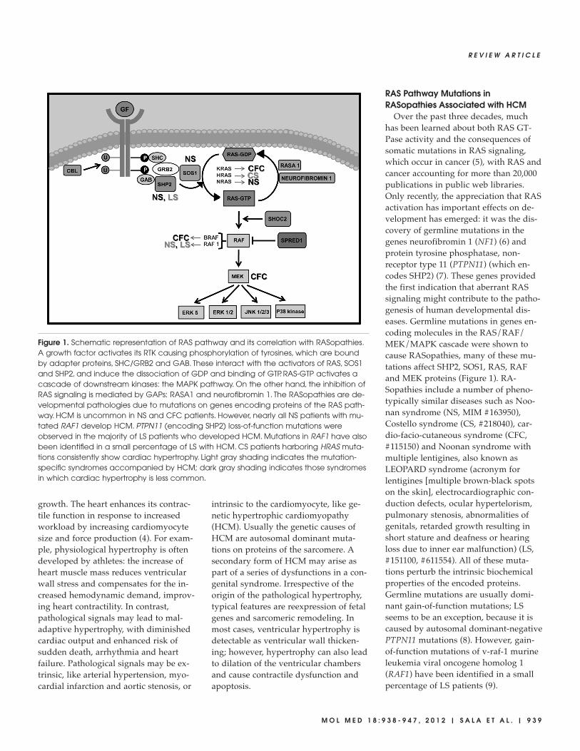
Over the past three decades, muchhas been learned about both RAS GT-Pase activity and the consequences ofsomatic mutations in RAS signaling,which occur in cancer (5), with RAS andcancer accounting for more than 20,000publications in public web libraries.Only recently, the appreciation that RASactivation has important effects on de-velopment has emerged: it was the dis-covery of germline mutations in thegenes neurofibromin 1 (NF1) (6) andprotein tyrosine phosphatase, non- receptor type 11 (PTPN11) (which en-codes SHP2) (7). These genes providedthe first indication that aberrant RASsignaling might contribute to the patho-genesis of human developmental dis-eases. Germline mutations in genes en-coding molecules in the RAS/RAF/MEK/ MAPK cascade were shown tocause RASopathies, many of these mu-tations affect SHP2, SOS1, RAS, RAFand MEK proteins (Figure 1). RA-Sopathies include a number of pheno-typically similar diseases such as Noo-nan syndrome (NS, MIM #163950),Costello syndrome (CS, #218040), car-dio-facio-cutaneous syndrome (CFC,#115150) and Noonan syndrome withmultiple lentigines, also known as LEOPARD syndrome (acronym forlentigines [multiple brown-black spotson the skin], electrocardiographic con-duction defects, ocular hypertelorism,pulmonary stenosis, abnormalities ofgenitals, retarded growth resulting inshort stature and deafness or hearingloss due to inner ear malfunction) (LS,#151100, #611554). All of these muta-tions perturb the intrinsic biochemicalproperties of the encoded proteins.Germline mutations are usually domi-nant gain-of-function mutations; LSseems to be an exception, because it iscaused by autosomal dominant-negativePTPN11 mutations (8). However, gain-of-function mutations of v-raf-1 murineleukemia viral oncogene homolog 1(RAF1) have been identified in a smallpercentage of LS patients (9).
Figure 1. Schematic representation of RAS pathway and its correlation with RASopathies.A growth factor activates its RTK causing phosphorylation of tyrosines, which are boundby adapter proteins, SHC/GRB2 and GAB. These interact with the activators of RAS, SOS1and SHP2, and induce the dissociation of GDP and binding of GTP. RAS-GTP activates acascade of downstream kinases: the MAPK pathway. On the other hand, the inhibition ofRAS signaling is mediated by GAPs: RASA1 and neurofibromin 1. The RASopathies are de-velopmental pathologies due to mutations on genes encoding proteins of the RAS path-way. HCM is uncommon in NS and CFC patients. However, nearly all NS patients with mu-tated RAF1 develop HCM. PTPN11 (encoding SHP2) loss-of-function mutations wereobserved in the majority of LS patients who developed HCM. Mutations in RAF1 have alsobeen identified in a small percentage of LS with HCM. CS patients harboring HRAS muta-tions consistently show cardiac hypertrophy. Light gray shading indicates the mutation-specific syndromes accompanied by HCM; dark gray shading indicates those syndromesin which cardiac hypertrophy is less common.
9 4 0 | S A L A E T A L . | M O L M E D 1 8 : 9 3 8 - 9 4 7 , 2 0 1 2
R A S S I G N A L I N G T O C A R D I A C H Y P E R T R O P H Y
The common underlying pathogeneticmechanism involved in RASopathiesbrings about a significant overlap in phe-notype, which includes craniofacial dys-morphology and heart defects. The car-diac abnormalities in CS, NS and CFCsyndromes are quite similar, althoughthey vary in severity and frequency. Congenital cardiac defects include pul-monary valve and aortic stenosis, atrialand ventricular septal defects and mitralinsufficiency. Among the RASopathies,congenital heart defects are the mostcommon in NS (PTPN11 or SOS1). Theyalso occur in CFC syndrome (v-rafmurine sarcoma viral oncogene homologB1 [BRAF]) and NS (RAF1). Conductionabnormalities in patients with an RAF1mutation, CS and LS are also well de-scribed. Vascular abnormalities (that is,aortic dilation, coronary artery dilationand peripheral aneurysms) have alsobeen reported in patients with NS andLS (10). HCM occurs with similar frequency in CS (10,11) and LS (12),whereas it is less common in CFC syn-drome (13–15) and NS patients (16–19).In approximately one-third of CS pa-tients, HCM is chronically severe, wors-ened or stable. However, the resolutionor regression of HCM on echocardio-graphic follow-up was also reported (10).
The fundamental role of several RTKs,like epidermal growth factor receptor(EGFR) (also known as ErbB), fibroblastGFR and vascular endothelial GFR, hasbeen described in heart development,from the induction of cardiac mesodermto the formation of cardiac cushions inthe atrioventricular (AV) canal and theoutflow tract (OFT) and the epithelial-to-mesenchymal transition process leadingto valve formation (20). The RAS signal-ing mutations causing defective AV andOFT development and valvulogenesis(that is, atrioventricular septal defectsand pulmonary valvular stenosis) in asubset of RASopathies (21) probably af-fect multiple populations of cell progeni-tors. Indeed, various cell lineages con-tribute at different stages to OFT and AVchannels, including the so-called secondheart field (22,23). Despite the clear in-
volvement of RAS signaling in the prena-tal development of the heart, importanteffects of this signaling should also beconsidered in the postnatal response tostress. In this review, we emphasize therole of the RAS pathway in HCM. Theage at which HCM is diagnosed rangesfrom infancy, in patients with a severeform of the rare neonatal phenotype, tochildhood, the more common age ofpresentation (10). In studies focused onfetuses, investigators failed to diagnoseprenatal onset of HCM, at least in CS(24,25). So far, hypertrophy has seemedto develop as a counteraction of the car-diomyocyte to increased hemodynamicdemands. However, it remains to be es-tablished whether the pathological phe-notype may result from RAS signalingpathway alterations in fetal cardio -myocytes and, if so, how this occurs.
Pathophysiological Signaling:Experimental Relevance of theRAS/RAF/MEK/MAPK Pathway in HCM
The discovery of mutated RAS-relatedgenes in RASopathy patients has under-lined the relevance of this signaling toHCM (Figure 1). In addition to geneticlesions, pathological stimuli, like biome-chanical stress and neurohumoral fac-tors, lead to a hypertrophic state of theheart (26–28). The altered stimulation ofmembrane receptors, such as RTKs, is acrucial initiating step leading to the finalactivation of MAPKs, which thereby induce the hypertrophic response(27,29–33). By altering the levels and ac-tivities of cardiac transcription factors(for example, GATA binding protein 4[GATA4], myocyte enhancer factor 2[MEF2] and nuclear factor of activatedT cells [NFAT]), the RAS/RAF/MEK/MAPK pathway leads to reexpression offetal cardiac genes (34). In particular, theupregulation of β-MHC (β myosin heavychain) and atrial natriuretic factor andthe downregulation of SERCA (sarco -plasmic reticulum Ca2+ ATPase) genesoccur in the pathological hypertrophy,with consequent loss of efficient contrac-tion and calcium cycling (30,33,34). Suchtranscriptional remodeling correlates
with the loss of cardiac function; con-versely, the improvement of cardiac func-tion in response to drugs or implantationof an assist device is accompanied by thenormalization of gene expression (35,36).Studies using genetically engineeredmice have suggested that direct targetingof the hypertrophic response itself is ben-eficial and may provide a suitable thera-peutic option in such cases (4,37,38).Thus, strategies to normalize cardiacgene expression by controlling its up-stream signaling offer an attractive ap-proach for HCM therapy.
To this aim, a considerable number ofanimal models, mainly murine, havebeen generated to reproduce a hyper-trophic phenotype due to hyperactiva-tion of RTKs, RAS and RAS-downstreamproteins. The attenuation of hypertrophicresponses by the use of inhibitors ofMEK1/2, dominant-negative RAF1 orMEK1 and antisense oligonucleotidesagainst extracellular signal-regulated ki-nase 1 and 2 (ERK1/2) definitely sub-stantiated the important role of this cas-cade for cardiomyocyte hypertrophy(27,30,33). All of the findings accumu-lated over the past couple of decades underscore the importance of both RAS-downstream ERK-mediated and ERK- independent pathways in cardiac hyper-trophy. The earliest evidence of acorrelation between RAS and hypertro-phy came from experiments on culturedmyocytes (39,40). Then, a link betweenRAS expression levels and the severity ofhypertrophy was given by RAS mRNAmeasurements among HCM patients(41). However, the in vivo proof of its rolein hypertrophy derives from transgenicmice in which oncogenic RAS is ex-pressed in the cardiac ventricular cham-ber (42). Mutations on the v-Ha-ras Har-vey rat sarcoma viral oncogene homolog(HRAS) gene have been found in the ma-jority of patients affected by CS withsigns of HCM (11). A mouse and a zebra -fish model for HRAS-related CS havebeen produced so far. However, althoughthe mouse model showed signs of car-diac hypertrophy (43), mutant fish didnot (44), revealing an inconsistency with

R E V I E W A R T I C L E
M O L M E D 1 8 : 9 3 8 - 9 4 7 , 2 0 1 2 | S A L A E T A L . | 9 4 1
the human phenotype. The involvementof aberrant RTK signaling in the patho-genesis of cardiac hypertrophy has beenrecently demonstrated by our group withthe use of a transgenic conditional mousemodel expressing the constitutively ac-tive Met tyrosine kinase (45). RTK activa-tion engenders RAS signaling to beturned on through GRB2, which hasbeen implicated in the development ofhypertrophy (46). In turn, GRB2 medi-ates the activation of SOS1, which is mu-tated in NS patients and mouse modelsleading to HCM (47). Also, SHP2 is a keycomponent of multiple RTK, cytokine re-ceptor and integrin signaling cascades.The animal model carrying an NS- associated Ptpn11 mutation does not de-velop cardiac hypertrophy (48), in linewith the observation that HCM occurs inless than 5%–10% of NS patients withPTPN11 mutations (19). In contrast, adominant-negative mutation with cat-alytic loss of function affecting SHP2leads to HCM in both LS patients andmice (49). As expected, the LS mice showreduced agonist-evoked ERK/MAPKsignaling, with a concomitant enhance-ment of protein kinase B (AKT)– mammalian target of rapamycin (mTOR) activity.
The importance of excessive phospho-inositide 3-kinase (PI3K)-AKT-mTOR sig-naling in the pathogenesis of HCM in LSwas further proved by rapamycin treat-ment, which reverses LS cardiac defects.Indeed, even though direct mutations onPI3K or AKT have not been found, it isknown that the prolonged constitutiveactivation of PI3K in the heart results inhypertrophy (50). In contrast, temporallycontrolled overexpression of cardiacPI3K in adult transgenic mice does notinduce hypertrophy, but results in in-creased contractility (51). In fact, in thecardiac context, PI3K plays importantroles in cardiac growth and exercise- induced hypertrophy and exerts protec-tive effects against pathological stimuli(52–54). Similarly, short-term activationof AKT in cardiomyocytes results inphysiological adaptive hypertrophy,whereas chronic activation of AKT leads
to pathological hypertrophy (55). Thus,the PI3K-AKT pathway shows a high de-gree of complexity, with different roles inregulating cardiac hypertrophy. RAS acti-vation results in the stimulation of RAFfamily members, which have been impli-cated in the development of hypertrophy(56,57) and of HCM in RASopathies(9,13,14). RAF1 mutations have beenmore frequently found to be associatedwith HCM, and nearly all NS patientswith RAF1 mutations exhibit HCM (58).Like NS patients, mice heterozygous forthe NS-associated RAF1 mutation exhibiteccentric cardiac hypertrophy (59). Amouse model of a constitutively activeform of BRAF has also been created (60).However, mutant mice show an in-creased total number of cardiomyocytes,rather than alterations in size, in contrastto data from CFC patients with mutatedBRAF who do develop HCM (61).
RAF activity results in the subsequentphosphorylation of MEKs. MutatedMEK1 was found in a few CFC patientswith signs of HCM (13). Evidence fromcultured cardiac myocytes exposed to aMEK1-specific inhibitor demonstratedthe critical contribution of the ERK path-way to hypertrophy (62,63). In vivo, inhi-bition of MEK attenuated cardiac growthin both induced and genetic models ofhypertrophy (48,59,64,65). Final activa-tion of MAPK has also been documentedin different heart diseases (66,67).Sprouty1, an endogenous inhibitor ofERK, was reported to be induced inhuman hearts during hypertrophy regres-sion after implantation of an assist device(68). These observations are in agreementwith reports about MAPK phosphatase-1inhibiting effects on cardiac hypertrophy,in vitro and in vivo (69). However, micelacking ERK1 and one ERK2 allele showa normal hypertrophic response to pres-sure overload and exercise (70), suggest-ing that ERK1/2 may not be mandatoryfor cardiomyocyte growth. Consistent re-sults were raised in mice with cardiac-specific inhibition of ERK1/2 activity(70–72). These proofs underpin the hy-pothesis that cardiac hypertrophy can de-velop independently of ERK1/2.
Recently, a new regulatory mechanismfor ERK2 in cardiac hypertrophy was de-scribed: Thr188 autophosphorylation onERK2 directs it to the nucleus, leading tophosphorylation of nuclear targets, with-out an overall increase in the activity ofERK1/2 (73). Thus, Thr188 phosphoryla-tion might represent a specific switch to-ward hypertrophic signaling, uncoupledfrom physiological signaling, and henceembody a good target for therapy. More-over, because overall ERK1/2 activity remains unchanged, ERK1/2 functionsother than the hypertrophic one might beunaffected, reducing massive side effects.
Cooperative Effects on the RASPathway: Cross-Links in Hypertrophy
A first suggestion that RAS is in frontof a network rather than in a top-downpathway actually comes from geneticsyndromes with HCM. For example,whereas both RAS and MEK1 gain-of-function approaches result in a hyper-trophic response, only MEK activationhas no cardiotoxic effects (74). This dis-crepancy might be explained by ERK- independent RAS pathways or by activated V12H RAS recruiting of modu-latory proteins, for example, Gβγ- subunits, which could influence itsdownstream cascade (75). Accordingly,the autophosphorylation of ERK2 atThr188 requires the integrated activationof the RAF/MEK/ERK1/2 cascade, aswell as that of Gαq-coupled receptors(73). Also, activation of β-adrenergic re-ceptors regulates the activity of smallGTPases involved in cardiac hypertro-phy (76,77): a novel family of exchangeproteins directly activated by cyclic AMP(EPAC) (78) has been shown to linkG protein–coupled receptors (GPCRs)and RAS signaling (Figure 2). EPAC actsas a GEF on RAS-like small GTPasesRap1/2 (79,80), participating in multiplecellular events initiated by GPCRs(76,81,82). In turn, Rap1 activates BRAFand has been involved in cardiac hyper-trophy (77). In addition, signaling viaGPCRs promotes cardiac hypertrophyvia ERK (83,84). Likewise, other GPCRs,including α-adrenergic receptors (28,85),

9 4 2 | S A L A E T A L . | M O L M E D 1 8 : 9 3 8 - 9 4 7 , 2 0 1 2
R A S S I G N A L I N G T O C A R D I A C H Y P E R T R O P H Y
angiotensin receptors (86) and endothe-lin receptors (63,87), signal through ERKto promote cardiomyocyte hypertrophy.Moreover, nuclear targeted α-adrenergicreceptors might activate ERK located incaveolae (88).
Several studies have also implicatedfocal adhesion kinase (FAK), a nonrecep-tor tyrosine kinase, in the first phase ofthe hypertrophic response to stretch incardiomyocytes (89) by regulating the activation of MEF2 and JUN N-terminalkinase (JNK)-JUN pathways, which areearly activators of the hypertrophic ge-netic program (90).
Irrespective of the original stimulus,these pathways converge to the activa-tion in the nucleus of transcriptional fac-tors, which have earlier roles during de-velopment, including GATA4 and MEF2(Figure 2). In the normal adult myo -cardium, only basal MEF2 activity is re-quired for the maintenance of contractileproperties, whereas stress stimuli,through MAPK, stimulate its activity(Figure 2) (91,92). In the adult, these fac-tors induce a fetal gene program, which
might be beneficial in adapting to stress,in principle, but then leads to alteredcontractility, uncontrolled calcium tran-sients and inadequate energetic and, fi-nally, maladaptative changes (93–95).
Gene expression profiling from tempo-rally regulated V12H RAS transgenichearts has suggested that induction ofearly response genes, loss of mitochondr-ial function and altered ionic channelproteins are the likely culprits of thepathological changes in extracellular ma-trix remodeling, cardiac output and elec-trophysiological parameters (96). Recentwork has suggested that the selective in-duction of Gαi in the RAS transgenicheart contributes to impaired sarcoplas-mic reticulum calcium cycling (97), and anumber of other studies have led to de-scriptions of RAS-induced alterations incalcium transients (98–100). Interestingly,abnormalities in delicate calcium homeo-stasis can trigger cardiac hypertrophythrough mechanisms that have not beenfully elucidated (101). The major link be-tween calcium signaling and expressionof fetal genes is represented by NFATs.
NFATs are activated by calcineurinthrough its dephosphorylating action,then migrate into the nucleus, wherethey interact with MEF2 and GATA4(102,103). Moreover, GSK3 mediates ex-port of NFATs from the nucleus and ter-mination of transcription. GSK3 is aknown effector of the PI3K-AKT path-way, and activation of AKT leads to inhi-bition of GSK3, thus crossing calcineurin,RAS and PI3K pathways (Figure 2). In-deed, the involvement of PI3K signalingwas underlined in cardiac pathogenesis(50) and hypertrophy (51,57). Finally,phosphorylation of RAF1 through PI3Kand P21-activated kinase (PAK, Figure 2)provides a costimulatory signal, which,together with RAS, leads to strong acti-vation of RAF1 kinase (104).
What is clear, in light of this excitingprogress, is that an intricate web of inter-connected signaling modules existsaround the hub of RAS, in which RASand fluctuations in calcium concentra-tions interplay and regulate each other.
Lessons from Cancer to CardiacHypertrophy
Interesting insights from the role ofRAS mutations in tumorigenesis mightprovide a new perspective for consider-ing the role of hyperactive RAS in thedevelopment of HCM in RAS syn-dromes. Parallels between cancer andhypertrophy have already been sug-gested, concerning both leading causesand therapy (105). Inadequately acti-vated RTKs are implicated in many pro-liferative disorders and have thereforereceived considerable attention. More re-cently, the same pathway was shown tobe involved in the pathogenesis of car-diac hypertrophy, prompting scientists totake advantage of the experience gainedin cancer for the cardiovascular field. Forexample, an unanticipated role for mu-tated CBL in the pathogenesis of a clini-cally variable condition with features fit-ting or partially overlapping NS wassuggested (17): CBL is a small E3 ubiqui-tin ligase that negatively regulates intra-cellular signaling downstream of RTKs.Moreover, cardiac-specific expression of
Figure 2. RAS Network: cross-links in hypertrophy. The cardiac hypertrophic response impli-cates signal transduction pathways initiated by ligand-stimulated membrane-bound re-ceptors (RTKs, GPCRs) and biomechanical stress sensors (integrins). Various signaling effec-tors interact with the RAS/MEK/MAPK pathway. GPCR receptors activate RAS proteinsthrough EPAC, induce release of internal Ca2+ stores and elicit pathological hypertrophythrough calcineurin/NFAT. GPCRs also act through ERK activation. GSK3 kinase negativelyregulates NFAT and, in turn, is inactivated by the PI3K-AKT pathway stimulated by RAS.Stimuli acting on integrins prompt cardiac hypertrophy through FAK activation and con-siderable cross-talk with RTK-mediated signaling. In addition, PAK regulates RAF1 activity.All these pathways converge on the modulation of transcriptional factors (MEF2, JUN andGATA4), which induce the expression of genes of the hypertrophic program.

R E V I E W A R T I C L E
M O L M E D 1 8 : 9 3 8 - 9 4 7 , 2 0 1 2 | S A L A E T A L . | 9 4 3
Tpr-Met, the oncogenic fusion protein ofhepatocyte growth factor receptor, wasshown to result in concentric hypertro-phy and congestive heart failure (45).
A first important suggestion comesfrom the analysis of the strength in de-gree and duration of the activity of mu-tated proteins. Activating RAS mutationsoccur in ~30% of human cancers with apattern skewed with respect to tissuetype and isoform. Interestingly, the so-matic mutations triggering cancer tend tobe more malignant and less variablethan the germline mutations found in RASopathies. Indeed, RAS-related devel-opmental disorders seem to be caused bymoderately hyperactivated proteins thatcan be tolerated in the germline, whereassome, if not most, of the mutations foundin cancer are incompatible with develop-ment, as suggested by G12D v-Ki-ras2Kirsten rat sarcoma viral oncogene ho-molog (KRAS) lethality during embryo-genesis (106). This might be true also inthe case of HCM, which develops in thepresence of germline mutants that can begenerally considered mild hypermorphs,whereas strong gain-of-function proteinswould lead to lethal malformations ofthe heart, or even cancer, as happens inthe Von Hippel-Lindau knock-out mouse(107).
Spontaneous regression is another in-teresting feature of RAS syndromes: thediagnosis of RASopathies primarily de-pends on clinical features, but the preva-lence of the different characteristicsamong affected individuals greatly de-pends on age. The facial features, for ex-ample, generally become more difficultto detect in later adolescence and adult-hood. The example of juvenile myelo -monocytic leukemia is particularly intriguing in this respect, because myelo -proliferative disorders that occur in NSinfants frequently regress without treat-ment. Consistently, in some cases (14% ofCS patients with HCM and one patientwith mutated KRAS) a natural regressionof HCM occurs with time (10). Elucidat-ing the mechanisms of spontaneous re-gression in cancer, still not well under-stood, might be very useful to stimulate
cardiomyocytes to escape from HCM aswell.
Interestingly, the tissues that are per-turbed in CS (the nervous and muscu-loskeletal systems) greatly overlap withthe types of malignancies that are ob-served (rhabdomyosarcoma, neuroblas-toma, ganglioneuroblastoma and bladdercancer). Because HRAS mutations accountonly for <1% of all cancer- associated RASmutations, emerging considerations arethat HRAS is transforming only in thosetissues in which it exerts a major roleand/or the gene is expressed at low levelsin most types of cancer-initiating cells.The concept of tumor addiction might beeasily transferred to RASopathies as well:specific tissues depend on the sustainedactivity of specific proteins to grow andsurvive, as it has been described for Metkinase (108). Accordingly, HRAS, which ismutated in CS, may create a dependenceon cardiac cells for their proliferation andsurvival, so CS patients develop HCMmore frequently than patients with otherRASopathies. Moreover, for each tissue,or even cell type, there might be a thresh-old in the response to gain and loss inRAS signaling, dictated by the fact thatoncogenes may convey both prosurvivaland proapoptotic signals. This idea mightexplain why either decreasing or increas-ing SHP2 phosphatase activity has delete-rious developmental consequences (109).Finally, cardiomyocytes might be less sen-sitive to a mutation for which the valvesare more sensitive. This may explain why40% of patients with mutated RAS do notdevelop HCM. For this reason, efforts stillhave to be addressed to study the role ofeach causative molecule not only inpathological but also in physiological con-ditions.
It is also notable that the kinase activi-ties of some of the mutant BRAF proteinsfound in CFC syndrome are comparableto BRAF oncoproteins; yet the germlineCFC mutations do not predispose totumor formation at all. These observa-tions suggest that the relatively low riskof cancer in many of these syndromes isnot entirely due to the degree of biochem-ical activity of the mutant protein. Indeed,
the observation that many individualswith CS do not develop cancer providesevidence that cooperating mutations orconcurrent microenvironmental cues areneeded for tumorigenesis. This observa-tion should be kept in mind in analysis ofmodels designed to mimic hypertrophy inRASopathies: these syndromes are dis-eases of complex systems, in which theparadigm one gene one phenotype mightbe far from being valid. In fact, in humansas well as in mice, different ERK1/2 func-tions are selectively switched on (for ex-ample, the protective and antiapoptoticfunctions of ERK1/2 more than their hy-pertrophic functions) in response to acombination of upstream signals that alto-gether would differently influence the car-diac phenotype. Furthermore, the consti-tutive activation of ERK1/2 in thesetransgenic models may be distinct fromthe carefully tuned activation pattern ofendogenous kinases (33,71), even if one ofthem carries an activating mutation.
Considering all of these findings, whywould germline mutations affectingsingle genes in RAS signaling be suffi-cient to confer an overt clinical pheno-type? Once again, cancer docet. The phe-nomenon of drug resistance in targettherapy has been extensively described,that is to say that cancer cells find newpathways to bypass a signaling blockade.Thus, because the germline mutationsare present throughout development,there is a substantial amount of time forbody cells to adapt to them, activatingregulatory feedback loops as well. How-ever, not all the cancers are able to findan escape and those that do not can beeradicated. Correspondingly, the tissuesthat are perturbed in RAS syndromesmay be not only highly sensitive to theRAS/RAF/MEK/MAPK pathway tospecify their cell fates, but also relativelyincapable of adjusting to overthresholdquantitative variations. It is as if hyper-trophic cardiomyocytes were like cancercells that could not find an escape.
Future Directions for TherapyTreatment of cardiac manifestations is
generally the same in RAS syndrome pa-

9 4 4 | S A L A E T A L . | M O L M E D 1 8 : 9 3 8 - 9 4 7 , 2 0 1 2
R A S S I G N A L I N G T O C A R D I A C H Y P E R T R O P H Y
tients as in the general population. Surgi-cal and pharmacologic treatment (like ve-rapamil, an inhibitor of calcium channels,and β-blockers, inhibitors of adrenergicreceptors) are generally used to alleviatesevere cardiac hypertrophy. TheRAS/MAPK pathway has long been adrug target because of its involvement inmalignant tumors (110). As pointed out inthis review, efficient therapeutic strategiesthat directly target the RAF-MEK-ERK1/2cascade might be the best tool against car-diac hypertrophy (30,33,111). Possibly, ad-vanced therapies could be based on anti-sense oligonucleotides that inhibit thetranslation of the altered genetic portion,or on gene-targeted therapies.
Direct inhibitors of the pathway,which have already been identified inanticancer studies, might represent analternative. Unfortunately, anticancerdrugs designed to specifically targetmolecules in the RAS pathway haveshown considerable side effects. Selec-tive inhibitors of RAF, such as PLX4032,have remarkable clinical activity in pa-tients with mela nomas who carry mu-tated BRAF (112). However, as for otherinhibitors of oncogenic kinases (113,114),response to PLX4032 is profound butoften temporary, because of the loss ofaddiction and the onset of resistance(115). Moreover, the functional role ofMAPKs in the heart itself presents a po-tential dilemma to any heart diseasetherapies targeting this pathway: thephysiological role of RAS signalingshould not be forgotten. In cultured my-ocytes, MAPK-inducing agents haveprotective effects against starvation, hy-poxia or reoxygenation injury (116,117).MEK transgenic hearts are protectedfrom ischemia/reperfusion and apopto-sis; similarly, inactivation of ERK2 pro-motes damage and death in response toischemia/reperfusion (118), whereas in-hibition of RAF1 promotes myocyte apo-ptosis and heart failure (58,119). In fact,the RAS pathway modulates the activityof regulators of apoptosis (120–123), aswell as of protein kinase Cε and p53(124). Thus, although activation of RASsignaling may trigger HCM, inhibiting
the pathway could render hearts morevulnerable to stress-induced myocytedeath, considerably increasing the riskof worsening heart disease.
Clinical efficacy and feasibility wouldcertainly be implemented whether mal-adaptive hypertrophic signals could beselectively blocked, without affecting thephysiological positive effects. It followsthat only detailed knowledge about thedifferential molecular mechanisms ofERK1/2 activation could lead to realistictherapeutic opportunities. The identifica-tion of a Thr188-phosphorylation site onERK2 might be a first step in this direc-tion: this phosphorylation can selectivelyactivate hypertrophic ERK1/2 functionsand, therefore, may be targeted withoutdangerous side effects. Mutant mice thatlack this phosphorylation site show pre-served cardiac structure and functionand attenuated hypertrophic response,indeed demonstrating that ERK1/2 canbe selectively targeted, at least in theadult patient (59).
The latest recommended goals fortreating hypertrophy may require multi-ple drugs. Lower doses in combinationmay render more efficacy and safetythan highest doses of single agents. Thechoice of added agents should greatlydepend on both cost and compliance.Currently, generic statins offer severalcost benefits. These inhibitors of theposttranscriptional lipid modifications ofRAS are likely to be effective, becausethey have been shown to exert antitu-moral action in different cancer cells withmutations on RAS (125,126). Indeed,statins reduced cardiac hypertrophy in atransgenic model of HCM (127,128). Acombination of low doses of statinsand specific inhibitors of pathologicalERK1/2 signaling represents, at the mo-ment, a pretty exciting possibility still tobe explored.
CONCLUSIONThe RAS/RAF/MEK/MAPK pathway
is involved in both proliferation and dif-ferentiation of different cellular lines, thecardiac lineage included, and thus playsa pivotal role during both adulthood and
embryogenesis; this characteristic finallyexcludes the possibility of completely in-hibiting this signaling cascade in youngpatients (for example, pediatric patientswith RASopathy-associated HCM). Al-though the need of introducing noveldrugs in the pediatric clinics is evident,the balance between risk and benefitshould be carefully considered. Finally,the dose used to treat children should bereduced as much as possible, consideringthe possible adverse impact of even amodest degree of toxicity. We suggestthat the severity and degree of cardiovas-cular impairment in pediatric patientswith RASopathies should be subjected toaccurate stratification; this could enablethe minimization of drug dosage to sparepatients with mild manifestations ofHCM from treatment-related morbidityand mortality. On the other hand, themost severe cases should be treated withtargeted and rationally designed thera-pies aimed at chronicizing a disease thatwould be otherwise lethal.
ACKNOWLEDGMENTSWe kindly acknowledge the constant
support of the Association Francaise con-tre les Myopathies (AFM) and the Sev-enth Framework Programme. V Sala wasa Fellow of Università Italo Francese in2011 (UIF, Cap. III Progetto Vinci 2008).The fellowships of V Sala and C Leowere granted by the FP7-2010-ICT-GC“EM-SAFETY” project no. 265772. Wegratefully thank GB Ferrero for helpfuldiscussion.
DISCLOSUREThe authors declare that they have no
competing interests as defined by Molec-ular Medicine, or other interests thatmight be perceived to influence the re-sults and discussion reported in thispaper.
REFERENCES1. Shih TY, Papageorge AG, Stokes PE, Weeks MO,
Scolnick EM. (1980) Guanine nucleotide-bindingand autophosphorylating activities associatedwith the p21src protein of Harvey murine sar-coma virus. Nature. 287:686–91.
2. Raman M, Chen W, Cobb MH. (2007) Differential
R E V I E W A R T I C L E
M O L M E D 1 8 : 9 3 8 - 9 4 7 , 2 0 1 2 | S A L A E T A L . | 9 4 5
regulation and properties of MAPKs. Oncogene.26:3100–12.
3. Yoon S, Seger R. (2006) The extracellular signal-regulated kinase: multiple substrates regulate di-verse cellular functions. Growth Factors. 24:21–44.
4. Hill JA, Olson EN. (2008) Mechanisms of disease:cardiac plasticity. New. Engl. J. Med. 358:1370–80.
5. Takai Y, Sasaki T, Matozaki T. (2001) Small GTP-binding proteins. Physiol. Rev. 81:153–208.
6. Xu GF, et al. (1990) The Neurofibromatosis Type-1 gene encodes a protein related to GAP. Cell.62:599–608.
7. Tartaglia M, et al. (2001) Mutations in PTPN11,encoding the protein tyrosine phosphatase SHP-2, cause Noonan syndrome. Nat. Genet. 29:465–8.
8. Kontaridis MI, Swanson KD, David FS, BarfordD, Neel BG. (2006) PTPN11 (Shp2) mutations inLEOPARD syndrome have dominant negative,not activating, effects. J. Biol. Chem. 281:6785–92.
9. Pandit B, et al. (2007) Gain-of-function RAF1 mu-tations cause Noonan and LEOPARD syndromeswith hypertrophic cardiomyopathy. Nat. Genet.39:1007–12.
10. Lin AE, et al. (2011) Clinical, pathological, andmolecular analyses of cardiovascular abnormali-ties in Costello Syndrome: a Ras/MAPK pathwaysyndrome. Am. J. Med. Genet. A. 155A:486–507.
11. Gripp KW, et al. (2006) HRAS mutation analysisin Costello syndrome: genotype and phenotypecorrelation. Am. J. Med. Genet. A. 140A:1–7.
12. Sarkozy A, Digilio MC, Dallapiccola B. (2008)Leopard syndrome. Orphanet J. Rare Dis. 3:13.
13. Gripp KW, et al. (2007) Further delineation of thephenotype resulting from BRAF or MEK1germline mutations helps differentiate cardio-facio-cutaneous syndrome from Costello syn-drome. Am. J. Med. Genet. A. 143A:1472–80.
14. Niihori T, et al. (2006) Germline KRAS and BRAFmutations in cardio-facio-cutaneous syndrome.Nat. Genet. 38:294–6.
15. Rodriguez-Viciana P. et al. (2006) Germline mutations in genes within the MAPK pathwaycause cardio-facio-cutaneous syndrome. Science.311:1287–90.
16. Cirstea IC, et al. (2010) A restricted spectrum ofNRAS mutations causes Noonan syndrome. Nat.Genet. 42:27–9.
17. Martinelli S, et al. (2010) Heterozygous germlinemutations in the CBL tumor-suppressor genecause a Noonan syndrome-like phenotype. Am. J.Hum. Genet. 87:250–7.
18. Roberts AE, et al. (2007) Germline gain-of- function mutations in SOS1 cause Noonan syn-drome. Nat. Genet. 39:70–4.
19. Tartaglia M, et al. (2002) PTPN11 mutations inNoonan syndrome: molecular spectrum, genotype-phenotype correlation, and phenotypicheterogeneity. Am. J. Hum. Genet. 70:1555–63.
20. Rose BA, Force T, Wang YB. (2010) Mitogen- activated protein kinase signaling in the heart:angels versus demons in a heart-breaking tale.Physiol. Rev. 90:1507–46.
21. Yutzey KE, Colbert M, Robbins J (2005) Ras-
related signaling pathways in valve develop-ment: ebb and flow. Physiol. 20:390–7.
22. Rochais F, Mesbah K, Kelly RG (2009) Signalingpathways controlling second heart field develop-ment. Circ. Res. 104:933–42.
23. Snarr BS, Kern CB, Wessels A (2008) Origin andfate of cardiac mesenchyme. Dev. Dyn. 237:2804–19.
24. Lin AE, et al. (2009) Prenatal features of Costellosyndrome: ultrasonographic findings and atrialtachycardia. Prenatal Diag. 29:682–90.
25. Smith LP, Podraza J, Proud VK. (2009) Polyhy-dramnios, fetal overgrowth, and macrocephaly:prenatal ultrasound findings of Costello syn-drome. Am. J. Med. Genet. A. 149A: 779–84.
26. Dorn GW II, Brown JH. (1999) Gq signaling incardiac adaptation and maladaptation. TrendsCardiovasc. Med. 9:26–34.
27. Heineke J, Molkentin JD. (2006) Regulation ofcardiac hypertrophy by intracellular signallingpathways. Nat. Rev. Mol. Cell. Biol. 7:589–600.
28. Xiao L, et al. (2001) MEK1/2-ERK1/2 mediatesalpha(1)-adrenergic receptor-stimulated hyper-trophy in adult rat ventricular myocytes. J. Mol.Cell. Cardiol. 33:779–87.
29. Dwyer JP, et al. (2008) Myocardial gene expres-sion associated with genetic cardiac hypertrophyin the absence of hypertension. Hypertens. Res.31:941–55.
30. Muslin AJ. (2008) MAPK signalling in cardiovas-cular health and disease: molecular mechanismsand therapeutic targets. Clin. Sci. 115:203–18.
31. Onan D, Pipolo L, Yang E, Hannan RD, ThomasWG. (2004) Urotensin II promotes hypertrophyof cardiac myocytes via mitogen-activated pro-tein kinases. Mol. Endocrinol. 18:2344–54.
32. Samarel AM et al. (2001) Src and multiple MAPkinase activation in cardiac hypertrophy andcongestive heart failure under chronic pressure-overload: comparison with acute mechanicalstretch. J. Mol. Cell. Cardiol. 33:1637–48.
33. Wang YB. (2007) Mitogen-activated protein ki-nases in heart development and diseases. Circula-tion. 116:1413–23.
34. Olson EN, Schneider MD. (2003) Sizing up theheart: development redux in disease. Genes Dev.17:1937–56.
35. Blaxall BC, Tschannen-Moran BM, Milano CA,Koch WJ. (2003) Differential gene expression andgenomic patient stratification following left ven-tricular assist device support. J. Am. Coll. Cardiol.41:1096–06.
36. Lowes BD, et al. (2002) Myocardial gene expres-sion in dilated cardiomyopathy treated with beta-blocking agents. New Engl. J. Med. 346:1357–65.
37. Esposito G, et al. (2002) Genetic alterations thatinhibit in vivo pressure-overload hypertrophyprevent cardiac dysfunction despite increasedwall stress. Circulation. 105:85–92.
38. Frey N, Katus HA, Olson EN, Hill JA. (2004) Hy-pertrophy of the heart: a new therapeutic target?Circulation. 109:1580–9.
39. Fuller SJ, Finn SG, Downward J, Sugden PH.(1998) Stimulation of gene expression in neonatal
rat ventricular myocytes by Ras is mediated byRal guanine nucleotide dissociation stimulator(Ral.GDS) and phosphatidylinositol 3-kinase inaddition to Raf. Biochem. J. 335:241–6.
40. Thorburn A, et al. (1993) Hras-dependent path-ways can activate morphological and genetic-markers of cardiac-muscle cell hypertrophy.J. Biol. Chem. 268:2244–9.
41. Kai H, et al. (1998) Expression of proto-oncogenesand gene mutation of sarcomeric proteins in pa-tients with hypertrophic cardiomyopathy. Circ.Res. 83:594–601.
42. Hunter JJ, Tanaka N, Rockman HA, Ross J, ChienKR. (1995) Ventricular expression of A Mlc-2V-Ras fusion gene induces cardiac-hypertrophyand selective diastolic dysfunction in transgenicmice. J. Biol. Chem. 270:23173–8.
43. Schuhmacher AJ, et al. (2008) A mouse model forCostello syndrome reveals an Ang II-mediated hy-pertensive condition. J. Clin. Invest. 118:2169–79.
44. Santoriello C, et al. (2009) Expression ofH-RASV12 in a zebrafish model of Costello syndrome causes cellular senescence in adultproliferating cells. Dis. Model Mech. 2:56–67.
45. Leo C, et al. (2011) Activated Met signalling inthe developing mouse heart leads to cardiac dis-ease. Plos One. 6:e14675.
46. Zhang SS, et al. (2003) The role of the Grb2-p38MAPK signaling pathway in cardiac hypertro-phy and fibrosis. J. Clin. Invest. 111:833–41.
47. Chen PC, et al. (2010) Activation of multiple sig-naling pathways causes developmental defects inmice with a Noonan syndrome-associated Sos1mutation. J. Clin. Invest. 120:4353–65.
48. Araki T, et al. (2004) Mouse model of Noonansyndrome reveals cell type- and gene dosage- dependent effects of Ptpn11 mutation. Nat. Med.10:849–57.
49. Marin TM, et al. (2011) Rapamycin reverses hy-pertrophic cardiomyopathy in a mouse model ofLEOPARD syndrome-associated PTPN11 muta-tion. J. Clin. Invest. 121:1026–43.
50. Oudit GY, Penninger JM. (2009) Cardiac regula-tion by phosphoinositide 3-kinases and PTEN.Cardiovasc. Res. 82:250–60.
51. Yano N, et al. (2008) Temporally controlled over-expression of cardiac-specific PI3K alpha inducesenhanced myocardial contractility–a new trans-genic model. Am. J. Physiol. Heart Circ. Physiol.295:H1690–4.
52. McMullen JR, et al. (2003) Phosphoinositide 3-ki-nase(p110alpha) plays a critical role for the in-duction of physiological, but not pathological,cardiac hypertrophy. Proc. Natl. Acad. Sci. U. S. A.100:12355–60.
53. McMullen JR, et al. (2007) Protective effects of ex-ercise and phosphoinositide 3-kinase(p110 alpha)si3naling in dilated and hypertrophic cardiomy-opathy. Proc. Natl. Acad. Sci. U. S. A. 104:612–7.
54. Ruan HM, et al. (2009) Inducible and cardiac spe-cific PTEN inactivation protects ischemia/reper-fusion injury. J. Mol. Cell. Cardiol. 46:193–200.
55. Shiojima I, et al. (2005) Disruption of coordinated

9 4 6 | S A L A E T A L . | M O L M E D 1 8 : 9 3 8 - 9 4 7 , 2 0 1 2
R A S S I G N A L I N G T O C A R D I A C H Y P E R T R O P H Y
cardiac hypertrophy and angiogenesis con-tributes to the transition to heart failure. J. Clin.Invest. 115:2108–18.
56. Harris IS, et al. (2004) Raf-1 kinase is required forcardiac hypertrophy and cardiomyocyte survivalin response to pressure overload. Circulation.110:718–23.
57. Klein M et al. (2008) Combined tyrosine and ser-ine/threonine kinase inhibition by sorafenib pre-vents progression of experimental pulmonaryhypertension and myocardial remodeling. Circu-lation. 118:2081–90.
58. Kobayashi T, et al. (2010) Molecular and clinicalanalysis of RAF1 in Noonan syndrome and re-lated disorders: dephosphorylation of serine 259as the essential mechanism for mutant activation.Hum. Mutat. 31:284–94.
59. Wu X. (2011) MEK-ERK pathway modulationameliorates disease phenotypes in a mousemodel of Noonan syndrome associated with theRaf1L613V mutation. J. Clin. Invest. 121:1009–25.
60. Urosevic J, et al. (2011) Constitutive activation ofB-Raf in the mouse germ line provides a modelfor human cardio-facio-cutaneous syndrome.Proc. Natl. Acad. Sci. U. S. A. 108:5015–20.
61. Allanson JE et al. (2011) Cardio-Facio-Cutaneoussyndrome: does genotype predict phenotype? Am.J. Med. Genet. C. Semin. Med. Genet. 157:129–35.
62. Clerk A, Aggeli IKS, Stathopoulou K, SugdenPH. (2006) Peptide growth factors signal differ-entially through protein kinase C to extracellularsignal-regulated kinases in neonatal cardiomy-ocytes. Cell. Signal. 18:225–35.
63. Kennedy RA, Kemp TJ, Sugden PH, Clerk A.(2006) Using U0126 to dissect the role of the ex-tracellular signal-regulated kinase 1/2 (ERK1/2)cascade in the regulation of gene expression byendothelin-1 in cardiac myocytes. J. Mol. Cell.Cardiol. 41:236–47.
64. Sanada S, et al. (2003) Long-acting Ca2+ blockersprevent myocardial remodeling induced by chronicNO inhibition in rats. Hypertension. 41:963–7.
65. Yue TL, et al. (2000) Extracellular signal-regulatedkinase plays an essential role in hypertrophic ag-onists, endothelin-1 and phenylephrine-inducedcardiomyocyte hypertrophy. J. Biol. Chem. 275:37895–901.
66. Armstrong SC. (2004) Protein kinase activationand myocardial ischemia/reperfusion injury.Cardiovasc. Res. 61:427–36.
67. Takeishi Y, et al. (2002) Activation of mitogen- activated protein kinases and p90 ribosomal S6kinase in failing human hearts with dilated car-diomyopathy. Cardiovasc. Res. 53:131–7.
68. Huebert RC, et al. (2004) Identification and regu-lation of Sprouty1, a negative inhibitor of theERK cascade, in the human heart. Physiol. Ge-nomics. 18:284–9.
69. Bueno OF, et al. (2001) The dual-specificity phos-phatase MKP-1 limits the cardiac hypertrophicresponse in vitro and in vivo. Circ. Res. 88:88–96.
70. Purcell NH, et al. (2007) Genetic inhibition ofcardiac ERK1/2 promotes stress-induced apo-
ptosis and heart failure but has no effect on hy-pertrophy in vivo. Proc. Natl. Acad. Sci. U. S. A.104: 14074–9.
71. Luttrell DK, Luttrell LM. (2003) Signaling in timeand space: G protein-coupled receptors and mi-togen-activated protein kinases. Assay Drug Dev.Techn. 1:327–38.
72. Owens DM, Keyse SM. (2007) Differential regula-tion of MAP kinase signalling by dual-specificityprotein phosphatases. Oncogene. 26:3203–13.
73. Lorenz K, Schmitt JP, Schmitteckert EM, LohseMJ. (2009) A new type of ERK1/2 autophospho-rylation causes cardiac hypertrophy. Nat. Med.15:75–83.
74. Bueno OF, et al. (2000) The MEK1-ERK1/2 signal-ing pathway promotes compensated cardiac hy-pertrophy in transgenic mice. Embo. J. 19:6341–50.
75. Slupsky JR, et al. (1999) Binding of G beta gammasubunits to cRaf1 downregulates G-protein- coupled receptor signalling. Curr. Biol. 9:971–4.
76. Metrich M, et al. (2008) Epac mediates beta-adrenergic receptor-induced cardiomyocyte hy-pertrophy. Circ. Res. 102:959–65.
77. Morel E, et al. (2005) cAMP-binding protein Epacinduces cardiomyocyte hypertrophy. Circ. Res.97:1296–304.
78. Ponsioen B, et al. (2004) Detecting cAMP-inducedEpac activation by fluorescence resonance energytransfer: Epac as a novel cAMP indicator. Embo.Rep. 5:1176–80.
79. de Rooij J, et al. (1998) Epac is a Rap1 guanine-nucleotide-exchange factor directly activated bycyclic AMP. Nature. 396:474–77.
80. Kawasaki H, et al. (1998) A family of cAMP- binding proteins that directly activate Rap1. Science. 282:2275–9.
81. Holz GG, Kang G, Harbeck M, Roe MW, Chep-urny OG. (2006) Cell physiology of cAMP sensorEpac. J. Physiol. 577:5–15.
82. Schmidt M, Sand C, Jakobs KH, Michel MC,Weernink PA. (2007) Epac and the cardiovascularsystem. Curr. Opin. Pharmacol. 7:193–200.
83. Barki-Harrington L, Perrino C, Rockman HA.(2004) Network integration of the adrenergic system in cardiac hypertrophy. Cardiovasc. Res.63:391–402.
84. Salazar NC, Chen J, Rockman HA. (2007) CardiacGPCRs: GPCR signaling in healthy and failinghearts. Biochim. Biophys. Acta. 1768:1006–18.
85. Kuster GM, et al. (2005) Alpha-adrenergic recep-tor-stimulated hypertrophy in adult rat ventricu-lar myocytes is mediated via thioredoxin-1- sensitive oxidative modification of thiols on Ras.Circulation. 111:1192–8.
86. Aoki H, Richmond M, Izumo S, Sadoshima J.(2000) Specific role of the extracellular signal- regulated kinase pathway in angiotensin II-in-duced cardiac hypertrophy in vitro. Biochem. J.347:275–84.
87. Cullingford TE, et al. (2008) Temporal regulationof expression of immediate early and secondphase transcripts by endothelin-1 in cardiomy-ocytes. Genome Biol. 9:R32.
88. Wright CD, et al. (2008) Nuclear alpha 1-adrener-gic receptors signal activated ERK localization tocaveolae in adult cardiac myocytes. Circ. Res.103:992–1000.
89. Torsoni AS, Constancio SS, Nadruz W, HanksSK, Franchini KG. (2003) Focal adhesion kinaseis activated and mediates the early hyper-trophic response to stretch in cardiac myocytes.Circ. Res. 93:140–7.
90. Nadruz W, Corat MAF, Marin TM, PereiraGAG, Franchini KG. (2005) Focal adhesion ki-nase mediates MEF2 and c-Jun activation bystretch: role in the activation of the cardiac hy-pertrophic genetic program. Cardiovasc. Res.68:87–97.
91. Passier R, et al. (2000) CaM kinase signaling in-duces cardiac hypertrophy and activates theMEF2 transcription factor in vivo. J. Clin. Invest.105:1395–406 .
92. McKinsey TA, Zhang CL, Olson EN. (2002)MEF2: a calcium-dependent regulator of cell di-vision, differentiation and death. TrendsBiochem. Sci. 27:40–7.
93. Miyata S, Minobe W, Bristow MR, LeinwandLA. (2000) Myosin heavy chain isoform expres-sion in the failing and nonfailing human heart.Circ. Res. 86:386–90.
94. Abraham WT, et al. (2002) Coordinate changesin myosin heavy chain isoform gene expressionare selectively associated with alterations in di-lated cardiomyopathy phenotype. Mol. Med.8:750–60.
95. Braunwald E, Bristow MR. (2000) Congestiveheart failure: fifty years of progress. Circulation.102:14–23.
96. Mitchell S, et al. (2006) Distinct gene expressionprofiles in adult mouse heart following targetedMAP kinase activation. Physiol. Genomics. 25:50–9.
97. Ruan HM, et al. (2007) Gi alpha 1-mediated car-diac electrophysiological remodeling and ar-rhythmia in hypertrophic cardiomyopathy. Circulation. 116:596–605.
98. Chen J, et al. (1998) Selective requirement ofmyosin light chain 2v in embryonic heart func-tion. J. Biol. Chem. 273:1252–6.
99. Ho PD, et al. (2001) Ras reduces L-type calciumchannel current in cardiac myocytes - correctiveeffects of L-channels and SERCA2 on [Ca2+](i)regulation and cell morphology. Circ. Res. 88:63–9.
100. Zheng MZ, et al. (2004) Sarcoplasmic reticulumcalcium defect in Ras-induced hypertrophic car-diomyopathy heart. Am. J. Physiol. Heart Circ.Physiol. 286: H424-33.
101. Frey N, McKinsey TA, Olson EN. (2000) Decod-ing calcium signals involved in cardiac growthand function. Nat. Med. 6:1221–7.
102. Crabtree GR, Olson EN. (2002) NFAT signaling:choreographing the social lives of cells. Cell.109: S67-79.
103. Molkentin JD, et al. (1998) A calcineurin- dependent transcriptional pathway for cardiachypertrophy. Cell. 93:215–28.
104. Chaudhary A, et al. (2000) Phosphatidylinositol

R E V I E W A R T I C L E
M O L M E D 1 8 : 9 3 8 - 9 4 7 , 2 0 1 2 | S A L A E T A L . | 9 4 7
3-kinase regulates Raf1 through Pak phospho-rylation of serine 338. Curr. Biol. 10:551–4.
105. Grimminger F, Schermuly RT, Ghofrani HA.(2010) Targeting non-malignant disorders withtyrosine kinase inhibitors. Nat. Rev. Drug. Dis-cov. 9:956–70.
106. Tuveson DA, et al. (2004) Endogenous onco-genic K-ras(G12D) stimulates proliferation andwidespread neoplastic and developmental de-fects. Cancer Cell. 5:375–87.
107. Lei L et al. (2008) Hypoxia-inducible factor- dependent degeneration, failure, and malignanttransformation of the heart in the absence of thevon Hippel-Lindau protein. Mol. Cell. Biol.28:3790–803.
108. Comoglio PM, Giordano S, Trusolino L. (2008)Drug development of MET inhibitors: targetingoncogene addiction and expedience. Nat. Rev.Drug. Discov. 7:504–16.
109. Grossmann KS. (2011) The tyrosine phosphataseShp2 in development and cancer. Adv. CancerRes. 106:53–89.
110. Young A, et al. (2009) Ras signaling and thera-pies. Adv Cancer Res. 102:1–17.
111. Gutkind JS, Offermanns S. (2009) A new Gq- initiated MAPK signaling pathway in the heart.Dev. Cell. 16:163–4.
112. Flaherty KT, et al. (2010) Inhibition of mutated,activated BRAF in metastatic melanoma. NewEngl. J. Med. 363:809–19.
113. Garrett JT, Arteaga CL. (2011) Resistance toHER2-directed antibodies and tyrosine kinaseinhibitors. Cancer Biol. Ther. 11:793–800.
114. Poulikakos PI, Solit DB. (2011) Resistance toMEK inhibitors: should we co-target upstream?Sci. Signal. 4:pe16.
115. Solit DB, Rosen N. (2011) Resistance to BRAFinhibition in melanomas. New Engl. J. Med.364:772–4.
116. Bueno OF, Molkentin JD. (2002) Involvement ofextracellular signal-regulated kinases 1/2 incardiac hypertrophy and cell death. Circ. Res.91:776–81.
117. Sugden PH & Clerk A. (2006) Oxidative stressand growth-regulating intracellular signalingpathways in cardiac myocytes. Antioxid. RedoxSignal. 8:2111–24.
118. Lips DJ, et al. (2004) MEK1-ERK2 signalingpathway protects myocardium from ischemicinjury in vivo. Circulation. 109:1938–41.
119. Yamaguchi O, et al. (2004) Cardiac-specific dis-ruption of the c-raf-1 gene induces cardiac dys-function and apoptosis. J. Clin. Invest.114:937–43.
120. Le Mellay V, Troppmair J, Benz R, Rapp UR.(2002) Negative regulation of mitochondrialVDAC channels by C-Raf kinase. BMC Cell Biol.3:14.
121. O’Neill E. (2011) Role of the kinase MST2 insuppression of apoptosis by the proto-oncogeneproduct Raf-1. Science. 306:2267–70.
122. Rapp UR, Rennefahrt U, Troppmair J. (2004) Bcl-2 proteins: master switches at the intersection of
death signaling and the survival control by Rafkinases. Biochim Biophys Acta 1644:149–58.
123. Tian S, et al. (2006) Interaction and stabilizationof X-linked inhibitor of apoptosis by Raf-1 pro-tein kinase. Int. J. Oncol. 29:861–7.
124. Cox AD, Der CJ. (2003) The dark side of Ras:regulation of apoptosis. Oncogene 22:8999–9006.
125. Park IH, Kim JY, Jung JI, Han JY. (2010) Lovas-tatin overcomes gefitinib resistance in humannon-small cell lung cancer cells with K-Ras mu-tations. Invest. New Drugs. 28:791–9.
126. Perchellet JP, et al. (2009) Novel synthetic inhibitors of 3-hydroxy-3-methylglutaryl- coenzyme A (HMG-CoA) reductase activity thatinhibit tumor cell proliferation and are struc-turally unrelated to existing statins. Int. J. Mol.Med. 24:633–43.
127. Patel R, et al. (2001) Simvastatin induces regres-sion of cardiac hypertrophy and fibrosis andimproves cardiac function in a transgenic rabbitmodel of human hypertrophic cardiomyopathy.Circulation. 104:317–24.
128. Senthil V, et al. (2005) Prevention of cardiac hy-pertrophy by atorvastatin in a transgenic rabbitmodel of human hypertrophic cardiomyopathy.Circ. Res. 97:285–92.


