Embed Size (px)
Citation preview
Site-Directed Fluorescence Studies of a Prokaryotic ClC Antiporter†
Susan P. Bell,‡ Patricia K. Curran, Sean Choi,§ and Joseph A. Mindell*
Membrane Transport Biophysics Unit, Porter Neuroscience Center, National Institute of Neurological Disorders and Stroke,National Institutes of Health, Bethesda, Maryland 20892
ReceiVed NoVember 21, 2005; ReVised Manuscript ReceiVed April 6, 2006
ABSTRACT: Channels and transporters of the ClC family serve a variety of physiological functions.Understanding of their gating and transport mechanisms remains incomplete, with disagreement over theextent of protein conformational change involved. Using site-directed fluorescence labeling, we probeClC-ec1, a prokaryotic ClC, for transport-related structural rearrangements. We specifically label cysteinesintroduced at several positions in the R helix of ClC-ec1 with AlexaFluor 488, an environment-sensitivefluorophore, and demonstrate that the labeled mutants show H+/Cl- transport activity indistinguishablefrom that of the wild-type protein. At each position that we examined we observe fluorescence changesupon acidification over the same pH range that is known to activate transport. The fluorescence changeis also sensitive to Cl- concentration; furthermore, the Cl- and H+ dependencies are coupled as would beexpected if the fluorescence change reflected a conformational change required for transport. Together,the results suggest that the changes in fluorescence report protein conformational changes underlying thetransport process. Labeled transporters mutated to remove a glutamate critical to proton-coupled chloridetransport retain pH-dependent fluorescence changes, suggesting that multiple residues confer pH dependenceon the transport mechanism. These results have implications for models of transport and gating in ClCchannels and transporters.
The ClCs represent a broad and functionally polymorphicfamily of anion transport proteins. ClC genes abound in allkingdoms; in mammals, these proteins perform a variety ofphysiological functions, from maintaining the resting mem-brane potential in muscle to mediating transepithelial Cl-
secretion in kidney (1). Understanding of these proteins hasadvanced rapidly in recent years, with the determination ofX-ray crystal structures of two prokaryotic ClCs (2) as wellas the elucidation of functional roles for many of theseproteins using genetic approaches (3). Most surprising wasthe recent revelation that theEscherichia coliClC is not achloride channel but, rather, functions as a H+/Cl- antiporter(4). Despite this progress, our understanding of the physicalprocesses underlying ClC function remains sketchy andincomplete.
Gating in ClC proteins is complex and incompletelyunderstood. The ClCs are homodimeric proteins with twodistinct transport pathways, one in each subunit. The best-studied ClC channels, ClC-0 and ClC-1, display two distinctgating processes, a “fast” gate which gates the two poresindependently and a “slow” gate that closes both pores
simultaneously. Recently, Dutzler and MacKinnon proposeda model for the fast gate based on X-ray crystal structuresof prokaryotic ClCs (5). In the original structures, a glutamateresidue (position 148 in ClC-ec1, in contact with theextracellular aqueous compartment) occludes the presumedchloride permeation pathway. In contrast, the structure of amutant in which glutamate 148 is replaced with glutaminereveals that residue’s side chain has moved away from thepore and has been replaced with a chloride ion. On the basisof these observations, the authors proposed that the move-ments of this glutamate in and out of the permeation pathwayaccount for the fast gating behavior of ClC channels. Indeed,experiments in that paper, as well as in subsequent publica-tions (5-7), demonstrate that mutations at that position havea strong influence on the pH dependence and rectificationproperties of eukaryotic ClCs. Data from other experimentalapproaches, however, argue that the fast gate may be morecomplicated than the movement of a single glutamate. Hintscome from the state dependence of block by cytoplasmicagents (8, 9), as well as the limited effects on ClC-0 channelproperties of mutating a conserved, Cl--coordinating residue(Tyr512 in ClC-0 corresponding to Tyr445 in ClC-ec1)(10).
Thus, the structural model proposed for the ClC fast gatecannot account for all of the functional observations. Do thesedifferences result from differences in mechanism betweenClC channels and transporters? Or is the “gating glutamate”a component of a more complex gating machinery in all ofthe ClCs? If so, what other protein rearrangements arerequired for ClC gating? Neither the protein movementscomprising any conformational changes aside from the gating
† This work was supported by the Intramural Program of the NationalInstitute of Neurological Disorders and Stroke, National Institutes ofHealth.
* To whom correspondence should be addressed: MTBU, NINDS,NIH, 35 Convent Dr., Bldg. 35, MSC 3701, Bethesda, MD 20892.Phone: (301) 402-3473. Fax: (301) 480-1693. E-mail: [email protected].
‡ Current address: Department of Medicine, Johns Hopkins Uni-versity, Baltimore, MD 21287.
§ Current address: Department of Biomedical Engineering, JohnsHopkins University, Baltimore, MD 21287.
6773Biochemistry2006,45, 6773-6782
10.1021/bi0523815 This article not subject to U.S. Copyright. Published 2006 by the American Chemical SocietyPublished on Web 05/10/2006
glutamate nor their presence or absence in a prokaryotic ClChas been examined.
We have begun to address these questions by applyingsite-directed fluorescence experiments to a prokaryotic ClC.We sought to determine whether regions of the proteinbeyond glutamate 148 are moving during transport. Theseexperiments involve introducing a cysteine at a given locationin the protein, covalently coupling a fluorescent probe, andmonitoring changes in fluorescence under conditions thatactivate the transport process. The fluorophore we used,AlexaFluor 488, is sensitive to its local solvent environment(11) and has been used to detect protein conformationalchanges (12). If the protein undergoes a conformationalchange upon transporting ions, we might therefore expectto find one or more positions where the fluorescence dependson the activation state of the transporter. This approach hasbeen used successfully to monitor conformational changesin other membrane proteins, including theâ-adrenergicreceptor (13) and potassium channels (14-16), among others.
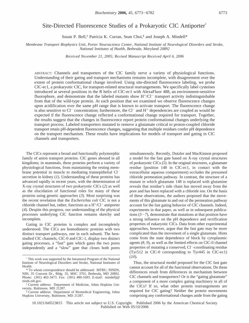
For these experiments, we labeled residues on or near theR helix of ClC-ec1 (Figure 1, yellow helix); this unusuallypositionedR helix begins deep in the presumably membrane-embedded portion of the protein and extends into thecytoplasmic aqueous compartment. The R helix has beenproposed as a possible site of transporter regulation byintracellular processes (2) because Tyr445, on its N-terminalend, is involved in coordinating a permeant ion even as thehelix extends into the intracellular compartment. One faceof helix R lines the inner vestibule of the presumed Cl-
transport pathway, and tyrosine 445 also at least partiallyoccludes access to the central Cl--binding site. We reasonedthat since the R helix bridges the protein core and thecytoplasm, even a small conformational change might moveat least one residue across the protein-water boundary andcause a detectable change in fluorescence. Indeed, we findthat fluorescent labels attached at multiple positions in helixR demonstrate fluorescence changes upon acidification,suggesting that this helix or nearby parts of the protein
undergo a conformational change. Our results suggest thatconformational changes in ClC-ec1 extend beyond glutamate148 and propagate to the intracellular side of the protein.Furthermore, we find evidence that the conformationalchange could involve unexpected parts of the protein. ThepH dependence of the change, though coupled to the gatingglutamate, may also reflect the titration of other residues.
EXPERIMENTAL PROCEDURES
Chemicals. E. colipolar lipid extract was purchased fromAvanti Polar Lipids.n-Decyl D-maltopyranoside was pur-chased from Anatrace. x3-[(3-Cholamidopropyl)dimethy-lammonio]-1-propanesulfonate was purchased from PierceChemical. Endopeptidase LysC was obtained from RocheChemicals. AlexaFluor 488 C5-maleimide (Alexa488)1 wasfrom Invitrogen. Other chemicals were purchased fromSigma Chemical.
Recombinant DNA, Cell Growth, and Protein Purification.The pASK-derived plasmid containing ClC-ec1 with aC-terminal LysC site and His tag was obtained from A.Accardi (7). Mutations were generated using standard PCR-based methods; inserts were sequenced in their entirety toverify the presence of the desired mutation and the absenceof others. Cultures of freshly transformed BL21(DE3) cellswere grown in Terrific Broth to mid-log phase (A600 ) 1.0)at 37 °C and then induced with anhydrotetracycline at 22°C for 16 h. Cells were spun, resuspended in 50 mM Tris,100 mM NaCl, and 5 mMâME (pH 7.5), and disrupted bysonication. Membrane proteins were extracted with 50 mMDM for 2 h. His tag purification was performed on a cobaltaffinity column, eluting with 500 mM imidazole. Afterconcentration (Millipore UltraFree) and LysC proteolysis (toremove the His tag), the protein was run on a Superdex 20010/300 GL column in 10 mM Tris, 150 mM NaCl, 5 mMtris(2-carboxyethyl)phosphine hydrochloride, and 10 mMDM (pH 7.5). Peak fractions were collected and used forlabeling with the fluorophore. Assays for free thiol used akit based on the reduction of cysteamine (Molecular Probes).
Labeling. Alexa488 was dissolved in water, aliquoted,dried in a speed-vac, and stored at-20 °C for future use.Sephadex G-50 (Amersham) was swelled overnight in anexcess of flux buffer [10 mM HEPES (pH 7.5), 50 mM NaCl,and 2 mM CaCl2] containing 10 mM DM. The purifiedprotein concentration was determined by the absorbance atOD280 using a calculated extinction coefficient. Protein andall tubes were degassed for 1 h and then placed undernitrogen. A 10-fold molar excess of Alexa488 was added toprotein and the mixture incubated for 2 h at room temper-ature. Excess unreacted label was removed on a desaltingcolumn. Polystyrene columns (Pierce) filled with swelledSephadex G-50 were centrifuged to remove excess buffer,loaded with labeled protein, and centrifuged again to separateunbound Alexa488. The protein concentration and labelingefficiency were determined by absorption spectroscopy.
Reconstitution into Liposomes. E. colipolar lipids, driedunder N2 and washed with pentane, were suspended bysonication to a final concentration of 10 mg/mL in flux buffer
1 Abbreviations: DM,n-decyl â-D-maltopyranoside; CHAPS, x3-[(3-cholamidopropyl)dimethylammonio]-1-propanesulfonate;âME, 2-mer-captoethanol; Alexa488, AlexaFluor 488 C5-maleimide; CCCP, carbonylcyanidem-chlorophenyl hydrazone.
FIGURE 1: Structure of ClC-ec1. The protein backbone from ProteinData Bank entry 1OTS is viewed parallel to the membrane planein cyan, with the R helix highlighted in yellow (Fab fragments arenot shown). The side chain of glutamate 148 is colored red, andthat of tyrosine 445 is colored blue. At the bottom, an energy-minimized structure of Alexa488 maleimide is shown drawn to scalefor size comparison. The cyan portion of the protein backbone issemitransparent to facilitate visualization of internal structures.Structures were viewed in VMD (23, 24) and rendered in POVray(www.povray.org); the fluorophore was drawn and energy-minimized in ChemDraw (CambridgeSoft) and viewed in VMD.
6774 Biochemistry, Vol. 45, No. 22, 2006 Bell et al.
containing 35 mM CHAPS; 100µg of labeled protein wasincubated with 10 mg of lipids for 30 min at roomtemperature and then the mixture dialyzed overnight toremove the detergent. Samples, frozen in a dry ice/ethanolbath and thawed for three cycles, were then extruded with aminiextruder (Avanti Polar Lipids) for 13 passes through 100nm membranes and used immediately.
Flux assay reconstitutions were similar except that theinitial buffer was 350 mM KCl, 50 mM citric acid, 20 mMsodium phosphate, and 35 mM CHAPS (pH 7.0); the protein-to-lipid ratio was 5µg of protein/mg of lipid. After dialysisand freezing and thawing, the mixture was acidified to pH4.5 with phosphoric acid and sonicated; the outside solutionwas then changed by spinning the sample through a SephadexG-50 column equilibrated with 300 mM potassium sulfate,3 mM KCl, and 2 mML-glutamic acid (pH 4.8) (LC buffer).The choice of detergent for the lipid solubilization is critical;reconstitution using DM-solubilized lipids produces uselesslyleaky vesicles.
Fluorescence Measurement.Fluorescence measurementswere performed on a Fluoromax-3 (Jobin Yvon) fluorescencespectrophotometer, equipped with Peltier temperature controland polarizers. For experiments with liposomes, dilutionswere in FB in the presence of 5µM carbonyl cyanidem-chlorophenyl hydrazone (CCCP). The pH was loweredby adding 1 M citric acid (final concentration of up to 5mM) and increased with 1 M Tris (pH 9) (final concentrationof up to 10 mM). Solubilized, labeled protein was diluted inFB and 10 mM anagrade DM. For anion experiments, theconcentrated anions and protein were diluted in low-salt FB[8 mM NaCl, 1 mM CaCl2, and 10 mM HEPES (pH 7.5)].For all experiments, the excitation wavelength was 493 nm;fluorescence was recorded in a 10 mm quartz cell at 22°C.All additions were followed by thorough mixing. For freeprobe experiments (Figure 2), probe concentrations were0.5-2 µM. Final protein concentrations were 2-5 and 15-25 nM for the detergent-solublized protein and liposome-reconstituted protein, respectively. For each condition, threespectra were acquired and averaged. Data from independentexperiments were further averaged for presentation. Datawere corrected for volume changes; these never exceeded5% and never altered the qualitative result.
Flux Experiment for MoVement of Protons DriVen by aChloride Gradient.Flux experiments were performed es-sentially as described in ref7. Liposomes containing 350mM KCl, 50 mM citrate, and 20 mM phosphate (pH 7.0) inLC buffer were added to 2 mL of LC buffer in a tube, andthe pH was monitored using a pH electrode (Corning) andrecorded on a chart recorder. Transport was initiated withthe addition of 1µg/mL valinomycin. The experiment wasterminated by collapsing the proton gradient with the additionof 0.25 µg/mL CCCP or carbonyl cyanidep-(trifluo-romethoxy)phenylhydrazone. Chart records were scanned andprocessed using Adobe Photoshop to remove the backgroundgrid and enhance the contrast of the recorded trace.
RESULTS
Specific Labeling of Mutant Proteins.We sought evidenceof conformational changes in the prokaryotic ClC formerlyknown as EriC, now called ClC-ec1. This protein is readilyexpressed and purified, and the majority of published
structural and functional analyses utilize this protein. We useda construct (kindly provided by A. Accardi) similar to thatin ref 17 but with a C-terminal histidine tag instead of theoriginal N-terminal tag. Proteins with R helix mutationsgenerally expressed well, in the range of 0.5-1.5 mg/L ofculture. Proteins were purified using a protocol based on the
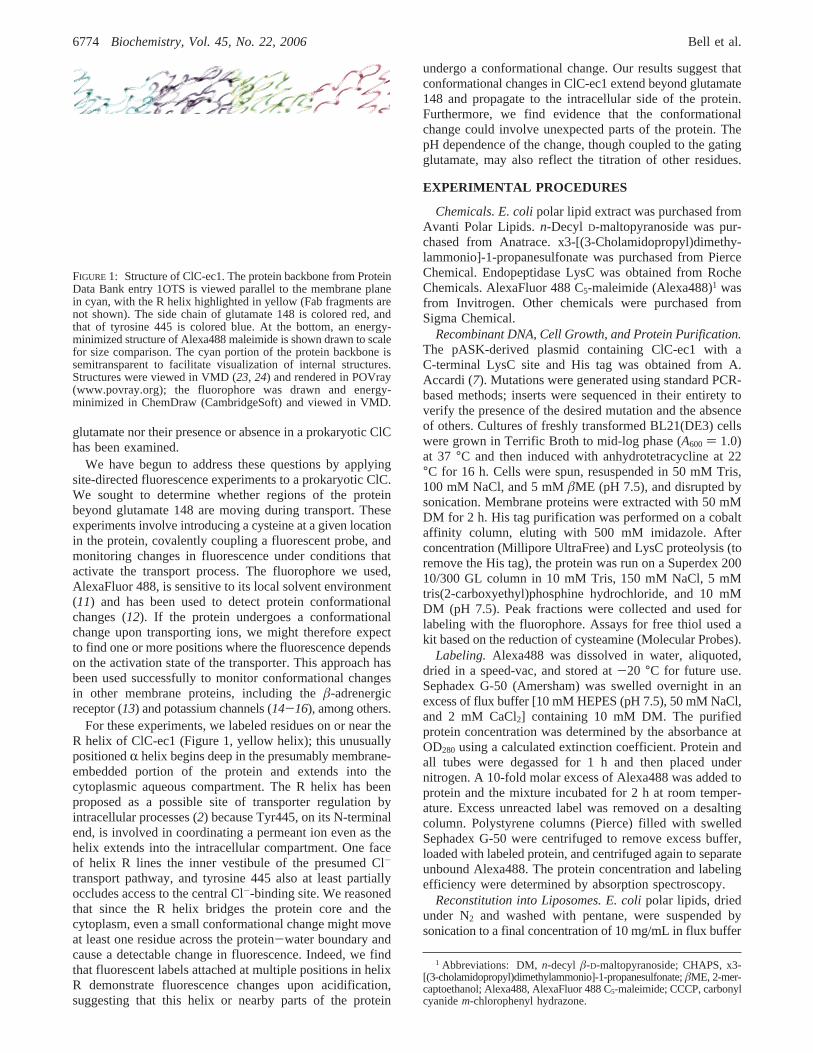
FIGURE 2: Properties of the fluorophore. The structure of Alexa488is shown in panel A. Emission spectra of the free probe weremeasured at a series of pH’s. (B) Fluorophore emission spectrawere measured at pH 7.5 (white circles); subsequently, the cuvettepH was lowered to 4.2 with citric acid (black triangles) and thenincreased to pH∼7.5 with the addition of Tris (gray squares). Eachexperiment was normalized to an initial peak emission of 1.0 atpH 7.5. Averaged data from three separate experiments at eachpH are shown, with error bars indicating the standard error of themean. In panel C, emission spectra are shown for equal concentra-tions of Alexa488 in different solvents. Single spectra are shownfor each solvent, including water (black circles), methanol (whitesquares), and ethanol (gray diamonds) in order of decreasingpolarity. The aqueous spectrum was measured in flux buffer (seethe text). Spectra were normalized to the aqueous maximum. Frepresents the normalized fluorescence emission in this andsubsequent figures.
Site-Directed ClC Fluorescence Biochemistry, Vol. 45, No. 22, 20066775
method of ref2, with an initial Co2+ chelating columnfollowed by tag cleavage (with lysC protease) and gelfiltration. The solutions in our protocol were modified slightlyfrom those published previously; introduced cysteines werekept reduced withâME added in early steps and replacedwith tris(2-carboxyethyl)phosphine hydrochloride during gelfiltration. This protocol yields a single protein band on anoverloaded Coomassie-stained gel (data not shown). All ofthe mutant proteins reported here gave symmetric peaks onthe gel filtration column and eluted in the fractions expectedfor dimeric ClC-ec1 (data not shown). Since we are not usingthese proteins for electrophysiology, no attempts to removetrace contaminating porins have been made (7).
Our approach is to label the protein with a fluorescentprobe and to manipulate the solution conditions (buffer pHand ion concentrations) in ways that are known to affecttransport activity and thus might affect the protein conforma-tion. If the protein undergoes a conformational change thatalters the local environment of the fluorophore, we wouldexpect to see changes in the fluorescence emission. To beuseful in these experiments, a fluorophore’s emission proper-ties must (1) depend on the local solvent (i.e., dielectric)environment and (2) be independent of pH and ionic strengthin the ranges of interest. We performed controls to test theseproperties using fluorophores that had been pre-reacted withâME, to approximate the protein-conjugated state. Figure 2presents the results of these controls for Alexa488, thereagent we used for all experiments presented here. ForAlexa488 (chemical structure shown in Figure 2A), both theλmax and the quantum yield depend on the nature of thesolvent. The emission intensity depends monotonically onthe polarity of the solvent and will be used below as ameasure of fluorophore environment. In contrast, theλmax
shifts to a slightly longer wavelength between water [Figure2C (black circles)] and methanol (white squares) and thenback toward shorter wavelengths in the more hydrophobicethanol (gray diamonds). This observation suggests that theλmax will not be useful in attempts to interpret fluorescencechanges. Experiments shown in Figure 2B, which showsspectra for the free probe at pH 7.5 (white circles) and 4.2(black triangles) and once the pH has been restored to 7.5(gray squares) in a single experiment, also demonstrate thatthe probe’s emission is independent of pH between 7 and4.2. Results demonstrating that the free probe is alsoessentially independent of Cl- concentration in the range ofinterest are shown in Figure 7D to allow comparison withthe relevant experiments.
The wild-type ClC-ec1 protein contains three cysteineresidues. Given that simultaneously mutating all three ofthese residues results in low expression yields (M. Maduke,personal communication), we evaluated the feasibility ofusing the native protein as a target for cysteine mutagenesis.On the basis of their locations in the structure, the nativecysteines are not expected to be accessible from aqueoussolution: one of them is exposed to the hydrophobic coreof the membrane, and two are buried in the protein core.Thiol assays on the detergent-solubilized wild-type proteinrevealed no accessible SH groups per protein monomer (notshown). In contrast, similar assays on the single-cysteinemutant, L449C, quantified a single accessible cysteine permonomer (not shown), further suggesting that the nativecysteines are inaccessible to aqueous reagents. In addition,
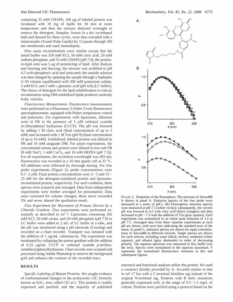
the water-soluble fluorescent probe, Alexa488, reacts stronglywith the L449C mutant but minimally with the wild-typeprotein to form a covalent adduct as shown in Figure 3. Thisfigure shows absorbance spectra of wild-type (dashed line)and L449C (solid line) proteins after reaction with a 10-fold molar excess of Alexa488 for 2 h and removal of theexcess unreacted fluorophore. The low absorbance of thewild-type protein around 500 nm, the absorption peak of thefluorophore, demonstrates that the native protein is poorlylabeled and that our spin column efficiently removes theexcess unreacted probe. The fluorophore absorbs lightprimarily in the visible spectrum, allowing us to use itsknown extinction coefficient together with that of the proteinto determine the extent of labeling for a given preparation.For most constructs, we routinely obtain labeling efficienciesof 85-95%. In general, proteins labeled at>40% are usablein fluorescence measurements in liposomes; at lower levels,it can be difficult to separate the fluorescence emission fromscattered light, even with the use of polarizers.
ActiVity Measurements on Labeled Proteins.We measuredthe transport activity of each labeled mutant to confirm thatthe presence of the fluorescent probe does not preventfunctional transport. The chloride-driven proton uptake assayis a stringent assay for a properly functioning protein; weuse the method described by Accardi and Miller (4). Wecreate a large, outwardly directed Cl- gradient (350 meq/Linside, 3 meq/L outside) across the membranes of proteoli-posomes containing reconstituted, labeled ClC-ec1 mutants.In the presence of functional transporters, this gradient willdrive protons into the vesicles against their concentrationgradient. The outside solution is minimally buffered, so smallpH changes are easily measured with a standard pH electrodeand documented on a chart recorder. These assays are shownfor the wild-type protein and the mutants in Figure 4. Notethat since the transporter is electrogenic, we initially observeno proton flux upon addition of vesicles to the cuvette. Onlyafter valinomycin is added to shunt the electrical potential
FIGURE 3: Specific labeling of introduced cysteines. Wild-type(WT) and I448C proteins were exposed to equal concentrations ofAlexa488 maleimide for 2 h and then spun through an∼3 mLSephadex G-50 column to remove unreacted probe. Absorptionspectra are shown for the column eluates with WT represented bythe dashed line and I448C by the solid line. Spectra were normalizedto an A280 of 1.0. The mutant protein spectrum has a strongabsorbance maximum at∼490 nm, which represents covalentlyattached Alexa488, whereas the WT has a minimal 490 nm peak.The minimal fluorophore peak for the WT sample demonstratesthat the three native cysteines are essentially inaccessible to thereagent and that the G-50 column is sufficient for removal of thebulk of the unreacted fluorophore.
6776 Biochemistry, Vol. 45, No. 22, 2006 Bell et al.
across the membrane does the external pH rise, reflectingthe Cl--driven uptake of protons into the vesicles. After theproton flux is allowed to stabilize, the proton ionophoreCCCP is added; the sequestered protons are thereby re-released, and the pH returns to near its starting value. Oncetransport is activated, though, all of the mutants are able tosupport Cl--driven proton uptake that is similar in magnitude
and rate when compared with that of the wild-type trans-porter. These labeled mutants had between 40 and 95%labeling, so if the labeled mutants were not active, we wouldexpect a similar decrease in the measured activity. Ourcomparisons are not quantitative enough to determine if thereare modest changes in the activity of the labeled proteinsbut do support the conclusion that they are capable of Cl-/H+ antiport and that they are therefore useful models forthe behavior of the wild-type protein.
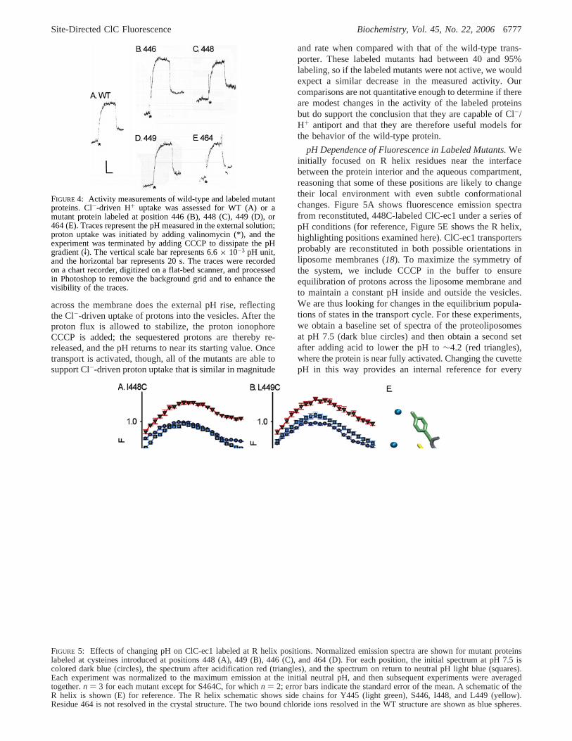
pH Dependence of Fluorescence in Labeled Mutants.Weinitially focused on R helix residues near the interfacebetween the protein interior and the aqueous compartment,reasoning that some of these positions are likely to changetheir local environment with even subtle conformationalchanges. Figure 5A shows fluorescence emission spectrafrom reconstituted, 448C-labeled ClC-ec1 under a series ofpH conditions (for reference, Figure 5E shows the R helix,highlighting positions examined here). ClC-ec1 transportersprobably are reconstituted in both possible orientations inliposome membranes (18). To maximize the symmetry ofthe system, we include CCCP in the buffer to ensureequilibration of protons across the liposome membrane andto maintain a constant pH inside and outside the vesicles.We are thus looking for changes in the equilibrium popula-tions of states in the transport cycle. For these experiments,we obtain a baseline set of spectra of the proteoliposomesat pH 7.5 (dark blue circles) and then obtain a second setafter adding acid to lower the pH to∼4.2 (red triangles),where the protein is near fully activated. Changing the cuvettepH in this way provides an internal reference for every
FIGURE 4: Activity measurements of wild-type and labeled mutantproteins. Cl--driven H+ uptake was assessed for WT (A) or amutant protein labeled at position 446 (B), 448 (C), 449 (D), or464 (E). Traces represent the pH measured in the external solution;proton uptake was initiated by adding valinomycin (*), and theexperiment was terminated by adding CCCP to dissipate the pHgradient (V). The vertical scale bar represents 6.6× 10⁻³ pH unit,and the horizontal bar represents 20 s. The traces were recordedon a chart recorder, digitized on a flat-bed scanner, and processedin Photoshop to remove the background grid and to enhance thevisibility of the traces.
FIGURE 5: Effects of changing pH on ClC-ec1 labeled at R helix positions. Normalized emission spectra are shown for mutant proteinslabeled at cysteines introduced at positions 448 (A), 449 (B), 446 (C), and 464 (D). For each position, the initial spectrum at pH 7.5 iscolored dark blue (circles), the spectrum after acidification red (triangles), and the spectrum on return to neutral pH light blue (squares).Each experiment was normalized to the maximum emission at the initial neutral pH, and then subsequent experiments were averagedtogether.n ) 3 for each mutant except for S464C, for whichn ) 2; error bars indicate the standard error of the mean. A schematic of theR helix is shown (E) for reference. The R helix schematic shows side chains for Y445 (light green), S446, I448, and L449 (yellow).Residue 464 is not resolved in the crystal structure. The two bound chloride ions resolved in the WT structure are shown as blue spheres.
Site-Directed ClC Fluorescence Biochemistry, Vol. 45, No. 22, 20066777
experiment. After measuring the pH, we add base, returningto near the starting pH (light blue squares), to evaluate thereversibility of the spectral changes.
Figure 5A reveals that for labeled 448C there is an increasein the fluorescence emission yield upon acidification, achange which is reversed fully upon return to neutral pH.This result suggests that the fluorophore is reversiblychanging its local environment; if its behavior when co-valently linked to a protein matches that in free solvent, thenit is moving to a more polar locale upon acidification. Asimilar, but weaker, effect is observed when protein labeledat the adjacent residue, position 449, is examined (Figure5B). Fluorescence changes still occur when the label islocated deeper within the protein. With the mutant labeledat S446C, a residue adjoining the Cl--coordinating Y445,acidification causes the fluorescence quantum yield toreversibly decrease(Figure 5C), suggesting an oppositechange in exposure, decreasing the local polarity experiencedby the probe.
We also labeled the mutant S464C. This residue, thepenultimate position in our construct, is unresolved in thecrystal structure (the last resolved residue in Figure 5E isresidue 460); we reasoned that it might be distant from anyenvironmental and/or conformational change. Even with this(labeled) mutant, we see a pronounced, but reversible, changein fluorescence emission upon acidification (Figure 5D); aswith S446C, the sign of the effect at position 464 suggeststhat the probe is moving to a more hydrophobic milieu atlow pH.
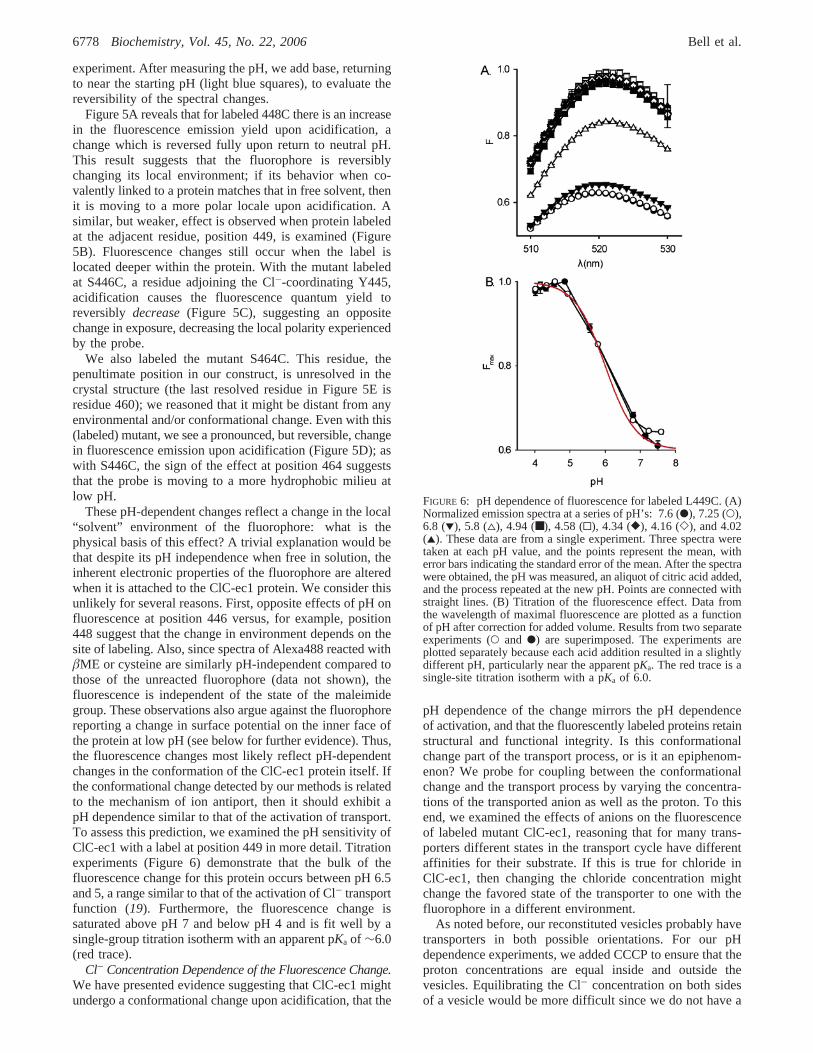
These pH-dependent changes reflect a change in the local“solvent” environment of the fluorophore: what is thephysical basis of this effect? A trivial explanation would bethat despite its pH independence when free in solution, theinherent electronic properties of the fluorophore are alteredwhen it is attached to the ClC-ec1 protein. We consider thisunlikely for several reasons. First, opposite effects of pH onfluorescence at position 446 versus, for example, position448 suggest that the change in environment depends on thesite of labeling. Also, since spectra of Alexa488 reacted withâME or cysteine are similarly pH-independent compared tothose of the unreacted fluorophore (data not shown), thefluorescence is independent of the state of the maleimidegroup. These observations also argue against the fluorophorereporting a change in surface potential on the inner face ofthe protein at low pH (see below for further evidence). Thus,the fluorescence changes most likely reflect pH-dependentchanges in the conformation of the ClC-ec1 protein itself. Ifthe conformational change detected by our methods is relatedto the mechanism of ion antiport, then it should exhibit apH dependence similar to that of the activation of transport.To assess this prediction, we examined the pH sensitivity ofClC-ec1 with a label at position 449 in more detail. Titrationexperiments (Figure 6) demonstrate that the bulk of thefluorescence change for this protein occurs between pH 6.5and 5, a range similar to that of the activation of Cl- transportfunction (19). Furthermore, the fluorescence change issaturated above pH 7 and below pH 4 and is fit well by asingle-group titration isotherm with an apparent pKa of ∼6.0(red trace).
Cl- Concentration Dependence of the Fluorescence Change.We have presented evidence suggesting that ClC-ec1 mightundergo a conformational change upon acidification, that the
pH dependence of the change mirrors the pH dependenceof activation, and that the fluorescently labeled proteins retainstructural and functional integrity. Is this conformationalchange part of the transport process, or is it an epiphenom-enon? We probe for coupling between the conformationalchange and the transport process by varying the concentra-tions of the transported anion as well as the proton. To thisend, we examined the effects of anions on the fluorescenceof labeled mutant ClC-ec1, reasoning that for many trans-porters different states in the transport cycle have differentaffinities for their substrate. If this is true for chloride inClC-ec1, then changing the chloride concentration mightchange the favored state of the transporter to one with thefluorophore in a different environment.
As noted before, our reconstituted vesicles probably havetransporters in both possible orientations. For our pHdependence experiments, we added CCCP to ensure that theproton concentrations are equal inside and outside thevesicles. Equilibrating the Cl- concentration on both sidesof a vesicle would be more difficult since we do not have a
FIGURE 6: pH dependence of fluorescence for labeled L449C. (A)Normalized emission spectra at a series of pH’s: 7.6 (b), 7.25 (O),6.8 (1), 5.8 (4), 4.94 (■), 4.58 (0), 4.34 ([), 4.16 (]), and 4.02(2). These data are from a single experiment. Three spectra weretaken at each pH value, and the points represent the mean, witherror bars indicating the standard error of the mean. After the spectrawere obtained, the pH was measured, an aliquot of citric acid added,and the process repeated at the new pH. Points are connected withstraight lines. (B) Titration of the fluorescence effect. Data fromthe wavelength of maximal fluorescence are plotted as a functionof pH after correction for added volume. Results from two separateexperiments (O and b) are superimposed. The experiments areplotted separately because each acid addition resulted in a slightlydifferent pH, particularly near the apparent pKa. The red trace is asingle-site titration isotherm with a pKa of 6.0.
6778 Biochemistry, Vol. 45, No. 22, 2006 Bell et al.
chloride ionophore. Instead, we studied the influence of Cl-
concentration using protein solubilized in detergent micelles.The detergent-solubilized protein runs as a symmetric, sharppeak on gel filtration (data not shown); in addition, pHdependence experiments on fluorescently labeled detergent-solubilized mutants, similar to those in Figure 5, reveal pH-dependent fluorescence changes similar to those we see withreconstituted protein (data not shown). Together, these resultssuggest that the protein in detergent (DM) remains intactand capable of the conformational change.
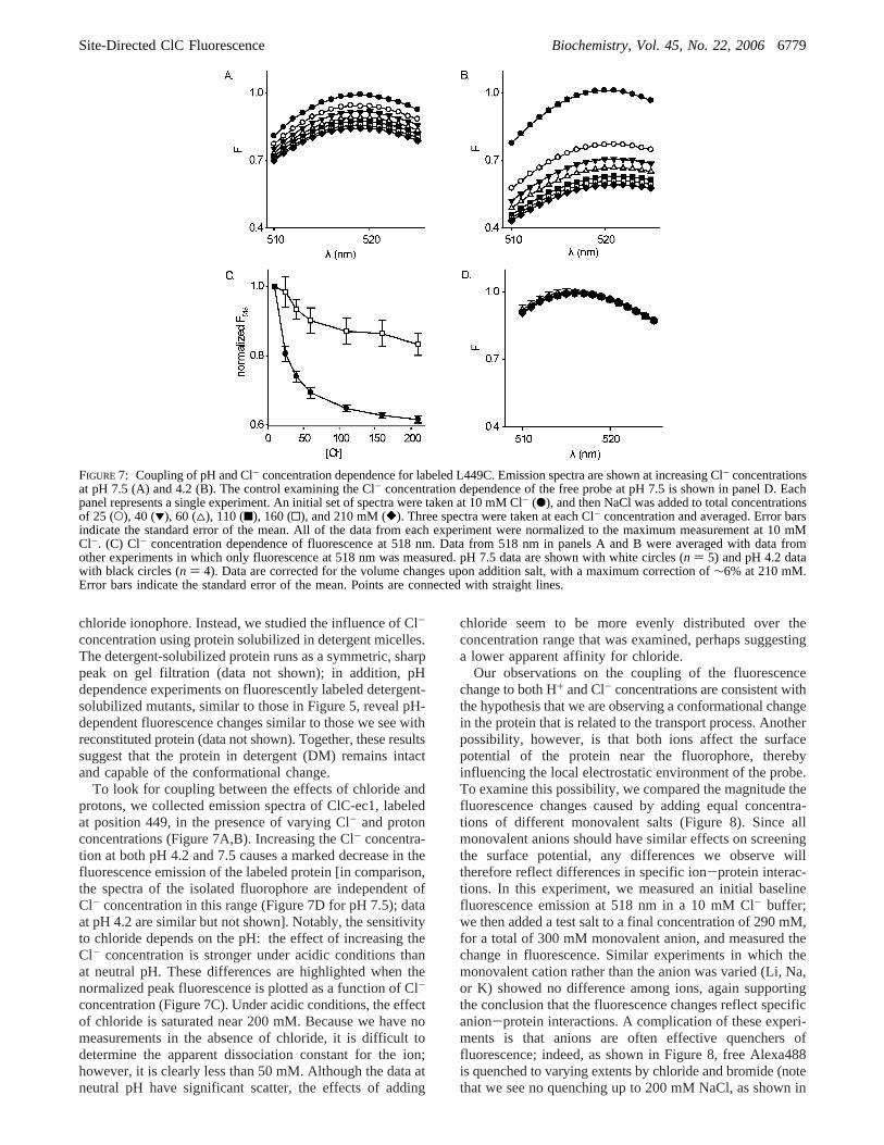
To look for coupling between the effects of chloride andprotons, we collected emission spectra of ClC-ec1, labeledat position 449, in the presence of varying Cl- and protonconcentrations (Figure 7A,B). Increasing the Cl- concentra-tion at both pH 4.2 and 7.5 causes a marked decrease in thefluorescence emission of the labeled protein [in comparison,the spectra of the isolated fluorophore are independent ofCl- concentration in this range (Figure 7D for pH 7.5); dataat pH 4.2 are similar but not shown]. Notably, the sensitivityto chloride depends on the pH: the effect of increasing theCl- concentration is stronger under acidic conditions thanat neutral pH. These differences are highlighted when thenormalized peak fluorescence is plotted as a function of Cl-
concentration (Figure 7C). Under acidic conditions, the effectof chloride is saturated near 200 mM. Because we have nomeasurements in the absence of chloride, it is difficult todetermine the apparent dissociation constant for the ion;however, it is clearly less than 50 mM. Although the data atneutral pH have significant scatter, the effects of adding
chloride seem to be more evenly distributed over theconcentration range that was examined, perhaps suggestinga lower apparent affinity for chloride.
Our observations on the coupling of the fluorescencechange to both H+ and Cl- concentrations are consistent withthe hypothesis that we are observing a conformational changein the protein that is related to the transport process. Anotherpossibility, however, is that both ions affect the surfacepotential of the protein near the fluorophore, therebyinfluencing the local electrostatic environment of the probe.To examine this possibility, we compared the magnitude thefluorescence changes caused by adding equal concentra-tions of different monovalent salts (Figure 8). Since allmonovalent anions should have similar effects on screeningthe surface potential, any differences we observe willtherefore reflect differences in specific ion-protein interac-tions. In this experiment, we measured an initial baselinefluorescence emission at 518 nm in a 10 mM Cl- buffer;we then added a test salt to a final concentration of 290 mM,for a total of 300 mM monovalent anion, and measured thechange in fluorescence. Similar experiments in which themonovalent cation rather than the anion was varied (Li, Na,or K) showed no difference among ions, again supportingthe conclusion that the fluorescence changes reflect specificanion-protein interactions. A complication of these experi-ments is that anions are often effective quenchers offluorescence; indeed, as shown in Figure 8, free Alexa488is quenched to varying extents by chloride and bromide (notethat we see no quenching up to 200 mM NaCl, as shown in
FIGURE 7: Coupling of pH and Cl- concentration dependence for labeled L449C. Emission spectra are shown at increasing Cl- concentrationsat pH 7.5 (A) and 4.2 (B). The control examining the Cl- concentration dependence of the free probe at pH 7.5 is shown in panel D. Eachpanel represents a single experiment. An initial set of spectra were taken at 10 mM Cl- (b), and then NaCl was added to total concentrationsof 25 (O), 40 (1), 60 (4), 110 (9), 160 (0), and 210 mM ([). Three spectra were taken at each Cl- concentration and averaged. Error barsindicate the standard error of the mean. All of the data from each experiment were normalized to the maximum measurement at 10 mMCl-. (C) Cl- concentration dependence of fluorescence at 518 nm. Data from 518 nm in panels A and B were averaged with data fromother experiments in which only fluorescence at 518 nm was measured. pH 7.5 data are shown with white circles (n ) 5) and pH 4.2 datawith black circles (n ) 4). Data are corrected for the volume changes upon addition salt, with a maximum correction of∼6% at 210 mM.Error bars indicate the standard error of the mean. Points are connected with straight lines.
Site-Directed ClC Fluorescence Biochemistry, Vol. 45, No. 22, 20066779
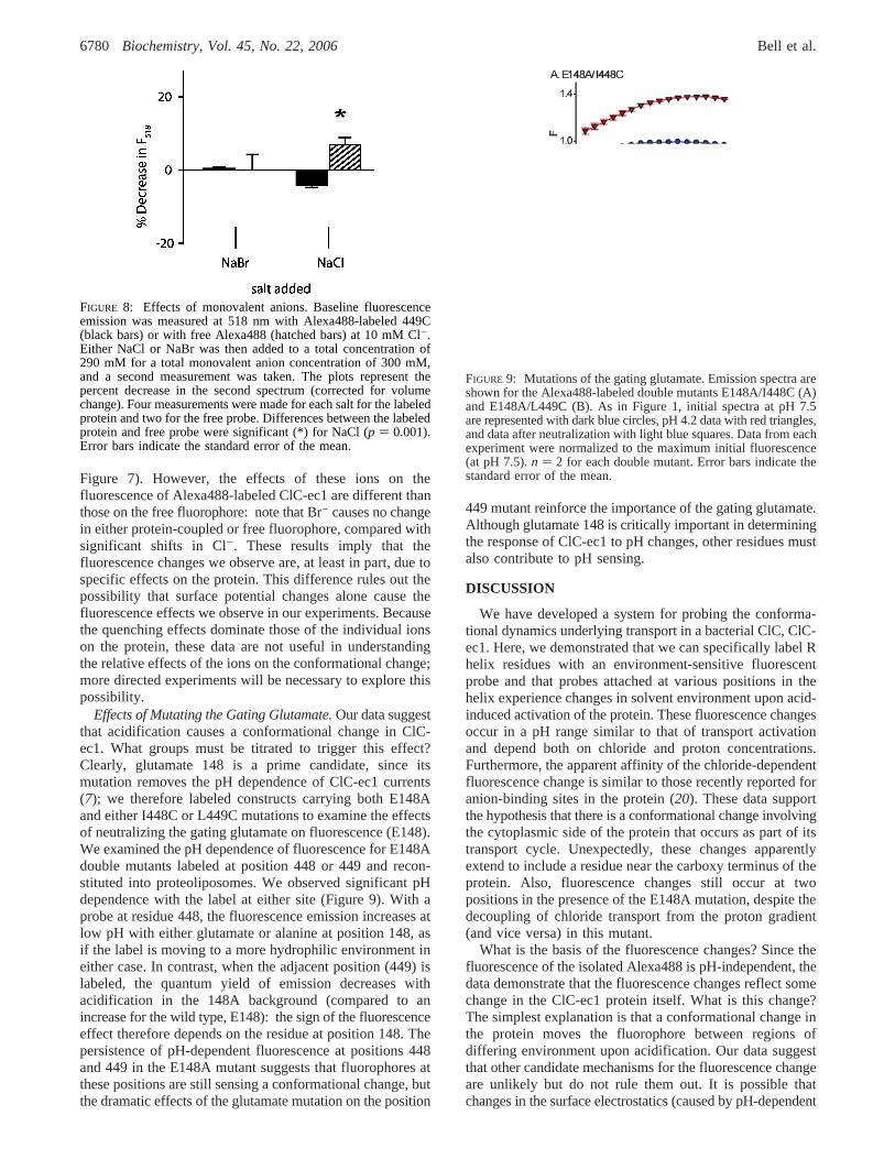
Figure 7). However, the effects of these ions on thefluorescence of Alexa488-labeled ClC-ec1 are different thanthose on the free fluorophore: note that Br- causes no changein either protein-coupled or free fluorophore, compared withsignificant shifts in Cl-. These results imply that thefluorescence changes we observe are, at least in part, due tospecific effects on the protein. This difference rules out thepossibility that surface potential changes alone cause thefluorescence effects we observe in our experiments. Becausethe quenching effects dominate those of the individual ionson the protein, these data are not useful in understandingthe relative effects of the ions on the conformational change;more directed experiments will be necessary to explore thispossibility.
Effects of Mutating the Gating Glutamate.Our data suggestthat acidification causes a conformational change in ClC-ec1. What groups must be titrated to trigger this effect?Clearly, glutamate 148 is a prime candidate, since itsmutation removes the pH dependence of ClC-ec1 currents(7); we therefore labeled constructs carrying both E148Aand either I448C or L449C mutations to examine the effectsof neutralizing the gating glutamate on fluorescence (E148).We examined the pH dependence of fluorescence for E148Adouble mutants labeled at position 448 or 449 and recon-stituted into proteoliposomes. We observed significant pHdependence with the label at either site (Figure 9). With aprobe at residue 448, the fluorescence emission increases atlow pH with either glutamate or alanine at position 148, asif the label is moving to a more hydrophilic environment ineither case. In contrast, when the adjacent position (449) islabeled, the quantum yield of emission decreases withacidification in the 148A background (compared to anincrease for the wild type, E148): the sign of the fluorescenceeffect therefore depends on the residue at position 148. Thepersistence of pH-dependent fluorescence at positions 448and 449 in the E148A mutant suggests that fluorophores atthese positions are still sensing a conformational change, butthe dramatic effects of the glutamate mutation on the position
449 mutant reinforce the importance of the gating glutamate.Although glutamate 148 is critically important in determiningthe response of ClC-ec1 to pH changes, other residues mustalso contribute to pH sensing.
DISCUSSION
We have developed a system for probing the conforma-tional dynamics underlying transport in a bacterial ClC, ClC-ec1. Here, we demonstrated that we can specifically label Rhelix residues with an environment-sensitive fluorescentprobe and that probes attached at various positions in thehelix experience changes in solvent environment upon acid-induced activation of the protein. These fluorescence changesoccur in a pH range similar to that of transport activationand depend both on chloride and proton concentrations.Furthermore, the apparent affinity of the chloride-dependentfluorescence change is similar to those recently reported foranion-binding sites in the protein (20). These data supportthe hypothesis that there is a conformational change involvingthe cytoplasmic side of the protein that occurs as part of itstransport cycle. Unexpectedly, these changes apparentlyextend to include a residue near the carboxy terminus of theprotein. Also, fluorescence changes still occur at twopositions in the presence of the E148A mutation, despite thedecoupling of chloride transport from the proton gradient(and vice versa) in this mutant.
What is the basis of the fluorescence changes? Since thefluorescence of the isolated Alexa488 is pH-independent, thedata demonstrate that the fluorescence changes reflect somechange in the ClC-ec1 protein itself. What is this change?The simplest explanation is that a conformational change inthe protein moves the fluorophore between regions ofdiffering environment upon acidification. Our data suggestthat other candidate mechanisms for the fluorescence changeare unlikely but do not rule them out. It is possible thatchanges in the surface electrostatics (caused by pH-dependent
FIGURE 8: Effects of monovalent anions. Baseline fluorescenceemission was measured at 518 nm with Alexa488-labeled 449C(black bars) or with free Alexa488 (hatched bars) at 10 mM Cl-.Either NaCl or NaBr was then added to a total concentration of290 mM for a total monovalent anion concentration of 300 mM,and a second measurement was taken. The plots represent thepercent decrease in the second spectrum (corrected for volumechange). Four measurements were made for each salt for the labeledprotein and two for the free probe. Differences between the labeledprotein and free probe were significant (*) for NaCl (p ) 0.001).Error bars indicate the standard error of the mean.
FIGURE 9: Mutations of the gating glutamate. Emission spectra areshown for the Alexa488-labeled double mutants E148A/I448C (A)and E148A/L449C (B). As in Figure 1, initial spectra at pH 7.5are represented with dark blue circles, pH 4.2 data with red triangles,and data after neutralization with light blue squares. Data from eachexperiment were normalized to the maximum initial fluorescence(at pH 7.5).n ) 2 for each double mutant. Error bars indicate thestandard error of the mean.
6780 Biochemistry, Vol. 45, No. 22, 2006 Bell et al.

titration of charged group) of the protein could change theexcited state of the fluorophore and thereby change itsemission properties. However, our observations of salt-dependent fluorescence effects (Figure 8) argue against asurface charge-based electrostatic mechanism. The effectsof mutating glutamate 148, fairly distant from the fluoro-phore, also support the argument that the fluorescence changereflects a conformational change, since this mutation shouldhave a minimal effect on the surface potential of thecytoplasmic side of the protein, yet we see substantialalterations in the fluorescence change between the antiportercontaining a wild-type glutamate at position 148 and theE148A mutant.
Fluorescence changes occur with probes at each R helixresidue labeled in these experiments. These may reflect aconformational change moving this helix in relation to therest of the protein; however, other possibilities must also beconsidered. One alternative is that other parts of the protein,helix D or J, for example, move to shift the exposure ofhelix R. Nothing in our data precludes this option. Giventhe relatively long linker (five methylenes) between themaleimide and the fluorophore in Alexa488, we must alsocontemplate the possibility that acidic conditions expose abinding site for the fluorophore; rather than sensing shiftsin solvent access, the fluorescence changes measured wouldthen reflect binding to this site. Common to all of thesealternatives is the presence of an underlying conformationalchange in the protein.
We were surprised to find that the protein labeled atposition 464 demonstrated a large fluorescence change uponacidification, since a rather large conformational changewould be required to change the solvent exposure of thispart of the protein. Without such a negative control, it isdifficult to conclusively demonstrate the existence of aconformational change; however, the results discussed aboverender alternative explanations unlikely.
Several lines of evidence suggest that the acid-inducedfluorescence changes we observe likely reflect a conforma-tional change which contributes to the transport process inClC-ec1. First, the changes occur with a pH dependence(Figure 6) essentially identical to that of transport activation,as reported by Iyer et al. (19). Second, the change is sensitiveto the chloride concentration (Figure 8) but is inconsistentwith a simple surface potential effect. Finally, the chloridedependence and pH dependence are coupled (Figure 7), asexpected for a Cl-/H+ antiporter.
Given the pH independence of transport in the mutantneutralizing glutamate 148 (7), we were initially surprisedto find that the neutralization of this residue did not alleviatethe pH dependence of the conformational change. Uponfurther consideration, though, we realized that since ClC-ec1 is indeed a proton transporter, there are probably multipletitratable groups on the pathway taken by H+ through theprotein, a conclusion supported by recent mutagenesis andstructural results (21). In this light, even though mutation ofE148 removes a critical group that mediates a rate-limitingstep in transport, other parts of the transport cycle, includingother pH-dependent steps, could remain intact. We proposethat the pH dependence observed in the E148A mutantsresults from conformational changes related to these othertransport steps.
Detailed inferences regarding the nature of the conforma-tional change are limited by complexities of the transportmechanism. The recent revelation that ClC-ec1 is a H+/Cl-
antiporter, rather than a Cl- channel, implies that, whenactive, the transporter is continually cycling through a seriesof states at rates in the neighborhood of 103 s-1 (A. Accardi,personal communication). The fluorescence signal we mea-sure under a given set of conditions therefore represents aweighted average of contributions from multiple conforma-tional states (e.g., external vs internal exposure, bound vsunbound ions). In this light, changes in the fluorescencesignal upon activation at low pH reflect changes in therelative occupancies of the various states contributing to thetransport cycle resulting, in turn, from changes in the rate-limiting steps in the cycle. Given that measured fluorescencereflects an average of the true fluorescence of multipleconformations, our measurements likely underestimate themaximal change.
What is the magnitude of the ClC-ec1 conformationalchange? Even though the fluorescence changes we measureare relatively large, it is difficult to infer from these thedistances of the underlying movements. At best, fluorescencechanges reflect many influences and are not expected to belinear indicators of the position of the fluorophore (22). Therelatively long linker (CH2 × 5) connecting the maleimideand the ring system adds further uncertainty to the measure-ment. On the other hand, we observe changes through thelength of the R helix, observations consistent with move-ments affecting the entire helix. The largest effect we haveobserved is at position 464. This residue is unresolved inthe X-ray crystal structure of ClC-ec1 but seems to beexposed to a more hydrophobic environment (on average)upon acidification.
Our results support the proposition that a conformationalchange involving the R helix contributes to the transportcycle in ClC-ec1. This implies a complexity in the ClCtransport cycle/gating process and is consistent with dataimplicating an R helix residue in eukaryotic ClC gating (10).The hypothesis that a simple side chain movement explainsClC gating was proposed before the diagnosis of dualfunctional personalities in the ClC family: though such amodel could in principle explain channel gating, it is difficultto imagine a mechanism for coupled transport based on sucha simple movement. Transporter mechanisms, whether mod-eled with alternating access (large conformational change)or dual gates (smaller, more localized conformationalchanges), must limit access of ions from one side of themembrane while simultaneously providing access from theother: coordinated movements on both sides of the bindingsite are therefore necessary. Experimentally, while the criticalrole of glutamate 148 in coupled transport is clear (4, 5, 7),the persistence of pH-dependent fluorescence changes inmutants neutralizing this residue supports the idea ofmultiple, coordinated movements underlying transport. Ineukaryotic ClCs also, the role of the residue correspondingto glutamate 148 in fast gating has been well established;we speculate that the conformational changes reported herecould reflect aspects of the slow gating process. Delineatingthe details of movement in the ClC-ec1 transporter willprovide a framework for more detailed explorations ofconformational changes associated with gating in eukaryoticClC channels.
Site-Directed ClC Fluorescence Biochemistry, Vol. 45, No. 22, 20066781
ACKNOWLEDGMENT
Alessio Accardi kindly provided the wild-type ClC-ec1expression construct. We thank Miguel Holmgren, MerrittMaduke, Kenton Swartz, and Chris Miller for helpfuldiscussions.
REFERENCES
1. Jentsch, T. J., Stein, V., Weinreich, F., and Zdebik, A. A. (2002)Molecular structure and physiological function of chloride chan-nels,Physiol. ReV. 82, 503-568.
2. Dutzler, R., Campbell, E. B., Cadene, M., Chait, B. T., andMacKinnon, R. (2002) X-ray structure of a ClC chloride channelat 3.0 Å reveals the molecular basis of anion selectivity,Nature415, 287-294.
3. Jentsch, T. J., Poet, M., Fuhrmann, J. C., and Zdebik, A. A. (2005)Physiological functions of CLC Cl- channels gleaned from humangenetic disease and mouse models,Annu. ReV. Physiol. 67, 779-807.
4. Accardi, A., and Miller, C. (2004) Secondary active transportmediated by a prokaryotic homologue of ClC Cl- channels,Nature427, 803-807.
5. Dutzler, R., Campbell, E. B., and MacKinnon, R. (2003) Gatingthe selectivity filter in ClC chloride channels,Science 300, 108-112.
6. Niemeyer, M. I., Cid, L. P., Zuniga, L., Catalan, M., andSepulveda, F. V. (2003) A conserved pore-lining glutamate as avoltage- and chloride-dependent gate in the ClC-2 chloridechannel,J. Physiol. 553, 873-879.
7. Accardi, A., Kolmakova-Partensky, L., Williams, C., and Miller,C. (2004) Ionic currents mediated by a prokaryotic homologueof CLC Cl- channels,J. Gen. Physiol. 123, 109-119.
8. Estevez, R., Schroeder, B. C., Accardi, A., Jentsch, T. J., andPusch, M. (2003) Conservation of chloride channel structurerevealed by an inhibitor binding site in ClC-1,Neuron 38, 47-59.
9. Traverso, S., Elia, L., and Pusch, M. (2003) Gating competenceof constitutively open CLC-0 mutants revealed by the interactionwith a small organic Inhibitor,J. Gen. Physiol. 122, 295-306.
10. Accardi, A., and Pusch, M. (2003) Conformational changes in thepore of CLC-0,J. Gen. Physiol. 122, 277-293.
11. Lakowicz, J. (1999)Principles of Fluorescence Spectroscopy, 2nded., Kluwer Academic Publishers, New York.
12. Rajagopalan, P. T., Zhang, Z., McCourt, L., Dwyer, M., Benkovic,S. J., and Hammes, G. G. (2002) Interaction of dihydrofolatereductase with methotrexate: Ensemble and single-moleculekinetics,Proc. Natl. Acad. Sci. U.S.A. 99, 13481-13486.
13. Gether, U., Lin, S., and Kobilka, B. K. (1995) Fluorescent labelingof purified â2 adrenergic receptor. Evidence for ligand-specificconformational changes,J. Biol. Chem. 270, 28268-28275.
14. Mannuzzu, L. M., Moronne, M. M., and Isacoff, E. Y. (1996)Direct physical measure of conformational rearrangement underly-ing potassium channel gating,Science 271, 213-216.
15. Cha, A., and Bezanilla, F. (1997) Characterizing voltage-dependentconformational changes in the Shaker K+ channel with fluores-cence,Neuron 19, 1127-1140.
16. Larsson, H. P., Tzingounis, A. V., Koch, H. P., and Kavanaugh,M. P. (2004) Fluorometric measurements of conformationalchanges in glutamate transporters,Proc. Natl. Acad. Sci. U.S.A.101, 3951-3956.
17. Maduke, M., Pheasant, D. J., and Miller, C. (1999) High-levelexpression, functional reconstitution, and quaternary structure ofa prokaryotic ClC-type chloride channel,J. Gen. Physiol. 114,713-722.
18. Matulef, K., and Maduke, M. (2005) Side-dependent inhibitionof a prokaryotic ClC by DIDS,Biophys. J.
19. Iyer, R., Iverson, T. M., Accardi, A., and Miller, C. (2002) Abiological role for prokaryotic ClC chloride channels,Nature 419,715-718.
20. Lobet, S., and Dutzler, R. (2006) Ion-binding properties of theClC chloride selectivity filter,EMBO J. 25, 24-33.
21. Accardi, A., Walden, M., Nguitragool, W., Jayaram, H., Williams,C., and Miller, C. (2005) Separate ion pathways in a Cl-/H+
exchanger,J. Gen. Physiol. 126, 563-570.22. Kobilka, B. K., and Gether, U. (2002) Use of fluorescence
spectroscopy to study conformational changes in theâ2-adreno-ceptor,Methods Enzymol. 343, 170-182.
23. Humphrey, W., Dalke, A., and Schulten, K. (1996) VMD: Visualmolecular dynamics,J. Mol. Graphics 14, 33-38.
24. Humphrey, W., Dalke, A., and Schulten, K. (1996) VMD: Visualmolecular dynamics,J. Mol. Graphics 14, 27-38
BI0523815
6782 Biochemistry, Vol. 45, No. 22, 2006 Bell et al.
![Redshift-Distance Survey of Early-Type Galaxies. II. The [ITAL]D[/ITAL][TINF][CLC][ITAL]n[/ITAL][/CLC][/TINF]-σ Relation](https://img.pdfslide.net/doc/110x75/63196a26d4191f2f93079742/redshift-distance-survey-of-early-type-galaxies-ii-the-italditaltinfclcitalnitalclctinf-s.jpg)
![The Type I[CLC]a[/CLC] Supernova 1999[CLC]aw[/CLC]: A Probable 1999[CLC]aa[/CLC]-like Event in a Low-Luminosity Host Galaxy](https://img.pdfslide.net/doc/110x75/6349c0c82cd4c1a3540daaa2/the-type-iclcaclc-supernova-1999clcawclc-a-probable-1999clcaaclc-like.jpg)
![Optical and Infrared Investigation toward the [CLC][ITAL]z[/ITAL][/CLC] = 3.8 Quasar Pair PC 1643+4631A, B](https://img.pdfslide.net/doc/110x75/635142a28624f8c9710ee8e7/optical-and-infrared-investigation-toward-the-clcitalzitalclc-38-quasar.jpg)
![The Peculiar Type II Supernova 1997D: A Case for a Very Low [TSUP]56[/TSUP]N[CLC]i[/CLC] Mass](https://img.pdfslide.net/doc/110x75/632035e018429976e40619e0/the-peculiar-type-ii-supernova-1997d-a-case-for-a-very-low-tsup56tsupnclciclc.jpg)
![Colors of 2625 Quasars at 0 \u003c [ITAL][CLC]z[/CLC][/ITAL] \u003c 5 Measured in the Sloan Digital Sky Survey Photometric System](https://img.pdfslide.net/doc/110x75/6356dcb73fe58b1a610624d5/colors-of-2625-quasars-at-0-u003c-italclczclcital-u003c-5-measured-in.jpg)
![Optical and Infrared Photometry of the Nearby Type I[CLC]a[/CLC] Supernova 2001[CLC]el[/CLC](https://img.pdfslide.net/doc/110x75/631b4cb2d5372c006e03e0f3/optical-and-infrared-photometry-of-the-nearby-type-iclcaclc-supernova-2001clcelclc.jpg)


![RX J1716.6+6708: A Young Cluster at [CLC][ITAL]z[/ITAL][/CLC] = 0.81](https://img.pdfslide.net/doc/110x75/633b8b27d7265336d80bd2f7/rx-j171666708-a-young-cluster-at-clcitalzitalclc-081.jpg)


