Embed Size (px)
Citation preview
Skin sensitization: Langerhans’ cell mobilization, cytokine
regulation and immunomodulation by lactoferrin.
A thesis submitted to the University of Manchester for the degree
of Doctor of Philosophy in the Faculty of Life Sciences
2014
Aleksandra Metryka
Faculty of Life Sciences
2
Table of Contents
Table of Contents .............................................................................................................. 2
List of Figures ................................................................................................................... 9
List of Tables ................................................................................................................. 13
Abbreviations ................................................................................................................. 14
Abstract ................................................................................................................. 17
Declaration ................................................................................................................. 18
Copy right statement ....................................................................................................... 19
Acknowledgements ......................................................................................................... 20
1 Introduction ............................................................................................. 21
1.1 General background................................................................................. 21
1.2 Anatomy of the skin ................................................................................ 21
1.3 Contact dermatitis .................................................................................... 22
1.4 Allergens.................................................................................................. 23
1.5 Allergic contact dermatitis ...................................................................... 24
1.5.1 Mechanism of sensitization ..................................................................... 26
1.5.1.1 Formation of the protein-hapten complexes ............................................ 28
1.5.1.2 Danger signals ......................................................................................... 29
1.6 Dendritic cells .......................................................................................... 35
1.6.1 Cutaneous dendritic cell subsets .............................................................. 37
1.6.1.1 Ontogeny of Langerhans’ cells and marker expression .......................... 39
1.6.1.2 Ontogeny of dermal dendritic cells and marker expression .................... 41
1.6.2 Roles of skin DC: sensing danger, taking up antigen and
transporting antigen ................................................................................. 43
1.6.3 Polarization of immune responses by DC ............................................... 45
1.6.3.1 Allergen dependent polarization of immune response ............................ 48
3
1.7 Langerhans’ cell migration ...................................................................... 50
1.8 Role of cutaneous DC populations in the development of CHS ............. 52
1.9 Tolerogenic role of Langerhans’ cells ..................................................... 55
1.10 Langerhans’ cells functions: cross-presentation and activation of
Th17 cells ................................................................................................ 57
1.11 Keratinocytes ........................................................................................... 59
1.12 Role of innate immune cells in CHS reactions ........................................ 61
1.12.1 NKT cells................................................................................................. 61
1.12.2 Mast cells ................................................................................................. 61
1.12.3 Neutrophils .............................................................................................. 64
1.12.4 Skin resident γδ T cells ............................................................................ 65
1.13 Characteristics of lactoferrin ................................................................... 68
1.14 Microbiocidal effects of lactoferrin ......................................................... 70
1.15 Immunomodulatory functions of lactoferrin ........................................... 71
1.15.1 The impact of lactoferrin on Langerhans’ cell migration ........................ 73
1.16 Lactoferrin receptors ............................................................................... 75
1.17 Characterization of molecular mechanisms of LF activity ...................... 77
1.18 Lactoferrin derived peptides .................................................................... 79
1.19 Hypothesis, aims and experimental approach ......................................... 81
1.19.1 Hypothesis ............................................................................................... 81
1.19.2 Aims ........................................................................................................ 81
1.19.3 Experimental approach ............................................................................ 81
2 Methods ................................................................................................... 83
2.1 Tissue culture........................................................................................... 83
2.1.1 THP-1 cell line maintenance ................................................................... 83
2.1.2 Differentiation of THP-1 cells into macrophages ................................... 83
2.1.3 Stimulation of THP-1 cells ...................................................................... 83
4
2.1.4 Stimulation of THP-1 macrophages ........................................................ 84
2.1.5 HaCaT and PAM 212 cell line maintenance ........................................... 84
2.1.5.1 HaCaT cell culture ................................................................................... 85
2.1.5.2 PAM 212 cell culture............................................................................... 85
2.1.6 Primary keratinocyte preparation ............................................................ 86
2.1.6.1 Primary keratinocyte culture ................................................................... 86
2.2 Immunohistochemistry ............................................................................ 87
2.3 Mycoplasma testing ................................................................................. 87
2.4 Enzyme-linked immunosorbent assay ..................................................... 88
2.4.1 Human TNF-α ELISA ............................................................................. 88
2.4.2 Human IL-8 ELISA ................................................................................. 89
2.4.3 Human IL-1β ELISA ............................................................................... 90
2.4.4 Mouse TNF-α ELISA .............................................................................. 90
2.4.5 Mouse IL-1β ELISA ................................................................................ 91
2.4.6 Mouse IL-1α ELISA ................................................................................ 92
2.4.7 Mouse IFN-γ ELISA ............................................................................... 93
2.4.8 Mouse IL-13 ELISA ................................................................................ 93
2.4.9 Mouse IL-17A ELISA ............................................................................. 94
2.4.10 Mouse IL-17F ELISA.............................................................................. 95
2.4.11 Mouse CXCL9 ELISA ............................................................................ 96
2.4.12 Mouse CXCL10 ELISA .......................................................................... 96
2.5 Animal work ............................................................................................ 97
2.5.1 Animal source and maintenance .............................................................. 97
2.5.2 Chemicals and exposure .......................................................................... 98
2.5.3 Exposure to lactoferrin ............................................................................ 98
2.5.4 Cytokine treatment .................................................................................. 98
2.5.5 Antibody administration .......................................................................... 99
5
2.6 Preparation of epidermal sheets and LC enumeration ............................. 99
2.7 Homogenization of ear tissue .................................................................. 99
2.8 Preparation and analysis of lymph nodes for cytokine production ....... 100
2.8.1 Analysis of lymph node cell proliferation ............................................. 100
2.9 Isolation of messenger RNA ................................................................. 100
2.10 Synthesis of complementary DNA ........................................................ 101
2.11 Quantitative PCR ................................................................................... 102
2.12 Analysis of cytokine production by explants ........................................ 104
2.13 Statistical analyses ................................................................................. 104
3 In vivo effects of lactoferrin .................................................................. 105
3.1 Introduction ........................................................................................... 105
3.2 Results ................................................................................................... 106
3.2.1 Impact of lactoferrin on allergen-induced cutaneous cytokine
expression .............................................................................................. 107
3.2.2 Lactoferrin did not markedly affect baseline dermal cytokine
expression .............................................................................................. 109
3.2.3 The effect of lactoferrin on allergen-induced lymph node activation ... 113
3.2.4 The influence of lactoferrin on cytokine-induced responses in the
skin ........................................................................................................ 119
3.3 Discussion.............................................................................................. 128
4 Investigation of the immunomodulatory potential of lactoferrin in
THP-1 monocytes and macrophages ..................................................... 147
4.1 Introduction ........................................................................................... 147
4.2 Results ................................................................................................... 148
4.2.1 Dose response of THP-1 monocytes to PGN ........................................ 148
4.2.2 PGN-activated THP-1 macrophages: dose response ............................. 150
4.2.3 The influence of lactoferrin on TNF-α production by THP-1
macrophages .......................................................................................... 152
6
4.2.4 The influence of PMA on THP-1 macrophage responsiveness to LF ... 154
4.2.5 Stimulatory effect of lactoferrin was specific and endotoxin
independent............................................................................................ 159
4.2.6 Lactoferrin stimulated TNF-α and IL-8 production via the
membrane nucleolin receptor. ............................................................... 163
4.2.7 The stimulatory effect of lactoferrin was lost over time. ...................... 164
4.3 Discussion.............................................................................................. 166
5 Effects of lactoferrin on keratinocytes .................................................. 176
5.1 Introduction ........................................................................................... 176
5.2 Results ................................................................................................... 177
5.2.1 PAM 212 and HaCaT cell lines and primary murine keratinocytes
expressed keratin-14 .............................................................................. 177
5.2.2 PAM cells did not respond to TLR ligands ........................................... 179
5.2.3 PAM cells produced IL-1α and IL-1β ................................................... 180
5.2.4 PAM cells responded to stimulation with IL-1α and IL-1β .................. 182
5.2.5 Lactoferrin did not modulate cytokine stimulated gene expression
by PAM cells ......................................................................................... 184
5.2.6 PGN induced TNF-α production by primary keratinocytes .................. 187
5.2.7 Primary murine keratinocytes responded to stimulation with IL-1α
and IL-1β ............................................................................................... 190
5.2.8 Lactoferrin did not modulate cytokine stimulated gene expression
by primary keratinocytes ....................................................................... 191
5.2.9 HaCaT cells produced TNF-α, IL-8 and IL-1β following
stimulation with PGN ............................................................................ 193
5.2.10 HaCaT cells responded to stimulation with IL-1β ................................ 194
5.2.11 Lactoferrin did not impact on IL-1β-stimulated gene expression by
HaCaT cells ........................................................................................... 197
5.2.12 HaCaT cells responded to stimulation with TNF-α ............................... 198
7
5.2.13 Cytokine triggered gene expression by HaCaT cells was modulated
by dexamethasone and thioredoxin peptide .......................................... 199
5.3 Discussion.............................................................................................. 204
6 Differential cytokine requirements for mobilization of LC induced
by oxazolone and DNCB ....................................................................... 218
6.1 Introduction ........................................................................................... 218
6.2 Results ................................................................................................... 220
6.2.1 Contact allergens (oxazolone and DNCB) induced LC migration ........ 220
6.2.2 Lactoferrin inhibited oxazolone-induced LC migration ........................ 223
6.2.3 The N-lobe of lactoferrin was not responsible for inhibition of LC
migration................................................................................................ 227
6.2.4 Differential effect of lactoferrin on LC migration induced by
oxazolone and DNCB ............................................................................ 228
6.2.5 DNCB-mediated LC migration was IL-1β dependent and TNF-α
independent............................................................................................ 233
6.2.6 Members of dinitrohalobenzene family induced LC migration in
the absence of TNF-α signalling ........................................................... 239
6.2.7 Oxazolone and DNCB induced divergent cutaneous cytokine
expression profiles ................................................................................. 241
6.2.8 LC migration induced by high dose of oxazolone is TNF-α
independent............................................................................................ 247
6.2.9 Chemicals provoked distinct cutaneous cytokine expression
profiles ................................................................................................... 249
6.3 Discussion.............................................................................................. 253
7 General discussion ................................................................................. 270
7.1 Background............................................................................................ 270
7.2 Main findings......................................................................................... 271
7.2.1 Lactoferrin has a dual immunomodulatory potential ............................ 271
8
7.2.2 Keratinocytes might not be the source of TNF-α during the process
of LC migration ..................................................................................... 273
7.2.3 Divergence in the contact allergen family revealed a crucial role of
IL-1β ...................................................................................................... 276
7.3 Future Aims ........................................................................................... 278
7.4 Conclusions ........................................................................................... 280
8 References ............................................................................................. 282
Word count: 82.413
9
List of Figures
Figure 1.1 Schematic view of the events during the sensitization phase of ACD. ......... 27
Figure 1.2 Subsets of dermal (d)DC in mouse and human skin. .................................... 36
Figure 1.3 Ontogeny of cutaneous DC. ........................................................................... 42
Figure 1.4 Induction of T helper cell subsets. ................................................................. 47
Figure 1.5 Divergent polarization cytokine profiles triggered by DNCB and TMA. ..... 49
Figure 1.6 Structure of human lactoferrin. ...................................................................... 69
Figure 3.1 The influence of LF on a cutaneous cytokine production following
treatment with oxazolone. .......................................................................... 108
Figure 3.2 Lactoferrin induced time dependent changes in cytokine expression. ........ 110
Figure 3.3 The impact of LF on oxazolone-induced changes in dermal cytokine
mRNA expression. ..................................................................................... 112
Figure 3.4 Oxazolone-induced lymph node cell activation: the influence of LF.......... 114
Figure 3.5 Impact of LF on allergen-actviated lymph node cell cytokine
production................................................................................................... 116
Figure 3.6 IL-17 expression by lymph node cells: the impact of disaggregation
method and LF. .......................................................................................... 118
Figure 3.7 Lactoferrin inhibited IL-1β mediated LC migration. ................................... 121
Figure 3.8 Lactoferrin significantly inhibited IL-17F expression following
injection of homologous, recombinant IL-1β. ........................................... 123
Figure 3.9 Lactoferrin did not affect cytokine expression following injection of
homologous, recombinant TNF-α. ............................................................ 125
Figure 3.10 Lactoferrin inhibited Cxcl1 expression following injection of
homologous, recombinant IL-1α. ............................................................... 127
Figure 4.1 THP-1 monocytes responded to PGN with TNF-α and IL-8 production. ... 149
Figure 4.2 Lactoferrin did not modulate PGN-induced TNF-α secretion by
monocytes................................................................................................... 150
Figure 4.3 THP-1 macrophages responded to PGN with TNF-α production. .............. 151
Figure 4.4 Lactoferrin did not modulate TNF-α and IL-8 baseline secretion by
THP-1 macrophages. .................................................................................. 152
Figure 4.5 Lack of inhibition of PGN-induced THP-1 macrophage TNF-α and
IL-8 secretion by lactoferrin. ...................................................................... 153
10
Figure 4.6 Following treatment with PMA THP-1 monocytes were differentiated
into macrophages........................................................................................ 154
Figure 4.7 Lactoferrin induced TNF-α and IL-8 secretion by THP-1 macrophages. ... 157
Figure 4.8 Lactoferrin did not inhibit PGN-induced TNF-α production by THP-1
macrophages. .............................................................................................. 158
Figure 4.9 Transferrin does not induce TNF- α release by THP-1 macrophages. ........ 159
Figure 4.10 Lactoferrin did not induce TNF-α release from THP-1 monocytes. ......... 160
Figure 4.11 The stimulatory effect of LF was not dependent on endotoxin. ................ 162
Figure 4.12 The stimulatory effect of LF was mediated via its membrane receptor
– nucleolin. ................................................................................................. 164
Figure 4.13 The immunostimulatory effect of lactoferrin was lost over time. ............. 165
Figure 5.1 Primary keratinocytes and keratinocyte cell lines expressed keratin 14. .... 178
Figure 5.2 Effects of homologous, recombinant IL-1α and IL-1β on IL-1α and
IL-1β production by PAM cells. ................................................................ 181
Figure 5.3 Homologous, recombinant IL-1α and IL-1β-induced dose dependent
cytokine and Icam-1 expression by PAM cells. ....................................... 183
Figure 5.4 Homologous, recombinant IL-1α and IL-1β-induced time dependent
increases in cytokine and Icam-1 expression by PAM cells. ..................... 184
Figure 5.5 Lactoferrin did not affect IL-1β or IL-1α-induced Tnf-α or Icam-1
expression in PAM cells. ............................................................................ 186
Figure 5.6 Lactoferrin significantly inhibited expression of Tnf-α and Icam-1
induced by lower concentration of IL-1β. .................................................. 187
Figure 5.7 Primary keratinocytes secreted TNF-α following PGN stimulation. .......... 188
Figure 5.8 Primary murine keratinocytes expressed Tnf-α and Il-1β upon TLR2
ligand stimulation. ...................................................................................... 189
Figure 5.9 IL-1α and IL-1β-induced cytokine and Icam-1 expression by murine
primary keratinocytes. ................................................................................ 190
Figure 5.10 Lactoferrin did not affect Tnf-α, Il-1β or Icam-1 expression following
2, 8 or 24h cytokine stimulation in primary keratinocytes. ........................ 192
Figure 5.11 Stimulation of HaCaT cells with a TLR2 ligand resulted in a
production of inflammatory cytokines. ...................................................... 193
Figure 5.12 Peptidoglycan-induced cytokine expression by HaCaT cells. ................... 194
Figure 5.13 Homologous IL-1β-induced time and dose dependent changes in
TNF-α, IL-1β, IL-8 and ICAM-1 expression by HaCaT cells. .................. 195
11
Figure 5.14 Lactoferrin did not affect IL-1β-induced TNF-α, IL-1β, IL-8 and
ICAM-1 expression. ................................................................................... 196
Figure 5.15 Lactoferrin did not inhibit TNF-α, IL-1β and IL-8 expression induced
by the lower concentration of IL-1β. .......................................................... 198
Figure 5.16 Effects of homologous TNF-α on time dependent changes in TNF-α,
IL-1β, IL-8 and ICAM-1 mRNA expression by HaCaT cells.................... 199
Figure 5.17 Pep-1 peptide did not significantly affect TNF-α, IL-β and IL-8
expression following stimulation with homologous TNF-α and IL-1β. .... 201
Figure 5.18 Thioredoxin peptide did not impact on IL-1β-induced gene
expression. .................................................................................................. 202
Figure 5.19 Thioredoxin peptide decreased significantly TNF-α-induced IL-1β
expression. .................................................................................................. 203
Figure 6.1 Treatment with DNCB and oxazolone resulted in LC migration. ............... 222
Figure 6.2 Lactoferrin inhibited oxazolone-induced migration of LC. ......................... 224
Figure 6.3 Lactoferrin was active over a range of concentrations. ............................... 225
Figure 6.4 The inhibitory effect of LF on oxazolone-induced LC migration was
transient. ..................................................................................................... 226
Figure 6.5 The N-lobe of LF was not responsible for inhibition of oxazolone-
induced LC migration................................................................................. 228
Figure 6.6 Lactoferrin was without effect on DNCB-induced LC migration. .............. 230
Figure 6.7 Lactoferrin did not inhibit DNCB-induced migration of LC at low or
high doses of allergen. ................................................................................ 232
Figure 6.8 DNCB-induced LC migration was TNF-α independent but IL-1β
dependent. .................................................................................................. 235
Figure 6.9 DNCB induced the same level of LC migration at different
concentrations............................................................................................. 237
Figure 6.10 Dinitrohalobenzenes (DNCB and DNFB) did not require TNF-α
signalling to trigger LC migration. ............................................................. 240
Figure 6.11 Oxazolone and DNCB induced cutaneous cytokine expression in
BALB/c strain mice. ................................................................................... 242
Figure 6.12 Differential induction of cutaneous Il-1β and Cxcl1 expression by
DNCB compared with oxazolone. ............................................................. 244
Figure 6.13 Allergen treatment provoked release of cytokines from the skin. ............. 245
12
Figure 6.14 Cutaneous cytokine release induced by contact allergens was dose
dependent. .................................................................................................. 247
Figure 6.15 Lactoferrin did not inhibit LC migration caused by high concentration
of oxazolone. .............................................................................................. 248
Figure 6.16 TMA-induced skin cytokine profile. ......................................................... 250
Figure 6.17 SLS-induced skin cytokine profile. ........................................................... 251
Figure 6.18 DNTB-induced skin cytokine profile. ....................................................... 252
Figure 7.1 Mechanism of the immunomodulatory action of LF. .................................. 280

13
List of Tables
Table 1.1 Marker expression by mouse dDC subsets. .................................................... 38
Table 1.2 Marker expression by human dDC subsets. ................................................... 38
Table 1.3 Comparison of marker expression by mouse and human LC. ........................ 39
Table 2.1 PCR run parameters for mycoplasma detection reaction. ............................... 88
Table 2.2 Components of the cDNA synthesis reaction. ............................................. 101
Table 2.3 Cycling conditions required for the cDNA synthesis reaction. .................... 102
Table 2.4 Components of the qPCR reaction. ............................................................... 102
Table 2.5 Cycling conditions required for qPCR reaction. ........................................... 103
Table 2.6 Example of the comparative Ct method calculations. .................................. 103
Table 3.1 Results of the analysis of statistical significance# of differences in gene
expression induced by oxazolone and untreated naive controls (t=0). ...... 113
Table 5.1 PAM 212 cells did not secrete TNF-α following incubation with a
variety of stimuli. ....................................................................................... 179
14
Abbreviations
ACD allergic contact dermatitis
AhR aryl hydrocarbon receptor
AMP antimicrobial peptide
AOO acetone:olive oil
APC antigen presenting cells
ARE antioxidant response element
ASC apoptosis associated speck-like protein containing a CARD
ASK apoptosis-stimulated kinase 1
ATP adenosine triphosphate
bFGF basic fibroblast endothelial growth factor
BrdU bromodeoxyuridine
CD cluster of differentiation
cDNA complementary DNA
CDP common DC precursor
CDP common dendritic cell precursor
CHO chinese hamster ovary
CHS contact hypersensitivity
CLEC9 C-type lectin domain family 9 member A
CMP common myeloid progenitor
DAMP damage associated molecular pattern
DAPI 4',6-diamidino-2-phenylindole
DC dendritic cell
DC-SIGN dendritic cell-specific intercellular adhesion molecule-3-grabbing non-
integrin
dDC dermal dendritic cell
DETC dendritic epidermal γδ T cell
DMF dimethylformamide
DMSO dimethyl sulfoxide
DNCB 2,4-dinitrochlorobenzene
DNFB 2,4-dinitrofluorobenzene
DNP dinitrophenol
DNTB dinitrothiocyanobenzene
DPC diphenylcyclopropenone
dpm disintegrations per min
DT dyptheria toxin
DTA dyphteria toxin subunit A
DTH delayed-type hypersensitivity
DTR dyphteria toxin receptor
ECM extracellular matrix
EDTA ethylenediaminetetraacetic acid
EGFP enhanced green fluorescent protein
ELISA enzyme-linked immunosorbent assay
EU endotoxin unit
FADD fas-associated death domain
FITC fluorescein isothiocyanate
GM-CSF granulocyte-macrophage colony-stimulating factor
GOI gene of interest
GSH glutathione
15
HA hyaluronan
HSA human serum albumin
HBSS Hanks balanced salt solution
HIV human immunodeficiency virus
HMW high molecular weight
HPRT hypoxanthine-guanine phosphoribosyltransferase
HSV herpes simplex virus
HUVEC human umbilical vein endothelial cell
ICAM-1 intercellular adhesion molecule 1
IcD intracellular domain
ICD irritant contact dermatitis
IFN interferon
IGF-1 insulin-like growth factor 1
IHC immunohistochemistry
IKK IκB kinase
IL interleukin
IL-1Ra interleukin 1 receptor antagonist
JNK c-Jun NH2-terminal kinase
K keratin
KEAP 1 kelch-like ECH-associated protein 1
KO knock out
LC Langerhans’ cell
LF lactoferrin
LFA-1 lymphocyte function-associated antigen 1
LFampin lactoferrampin
LFcinB bovine lactoferricin
LFcinH human lactoferricin
LFR lactoferrin receptor
LMW low molecular weight
LN lymph node
LNC lymph node cell
LPS lipopolysaccharide
LT leukotriene
M-CSF macrophage colony-stimulating factor
MCP-1 monocyte chemoattractant protein-1
MDP macrophage/dendritic cell precursor
MHC major histocompatibility complex
MIP-3α macrophage inflammatory protein 3α
MMP matrix metalloprotease
mRNA messenger RNA
NGS normal goat serum
NKT natural killer T cell
NLR nucleotide-binding domain leucine-rich repeat containing receptor
NO nitric oxide
NRS normal rabbit serum
OD optical density
OVA ovalbumin
Oxazolone 4-ethoxymethylene-2-phenyl-2-oxazolin-5-one
PAMP pathogen associated molecular pattern
PBMC peripheral blood mononuclear cell
16
PBS phosphate buffered saline
PG prostaglandin
PGN peptidoglycan
PLA2 phospholipase A2
PMA phorbol-12-myristate-13-acetate
PMB polymixin B
PPD paraphenylendiamine
qPCR quantitative PCR
RA rheumatoid arthritis
RIP receptor-interacting protein
ROS reactive oxygen species
RPM revolutions per minute
R/T room temperature
RT reverse transcriptase
S seconds
SCR scrambled control
siRNA small interfering RNA
SIRPα signal regulatory protein α
SLS sodium lauryl sulfate
SODD silencer of death domains
SRBC sheep red blood cells
Strep-HRP streptavidin conjugated to horseradish-peroxidase
TACE tumour necrosis factor α converting enzyme
TBS tris-buffered saline
Tc T cytotoxic cell
TCR T cell receptors
TGF-β transforming growth factor β
Th T helper cell
TLF talactoferrin-α
TLR toll-like receptors
TMA trimellitic anhydride
TMB 3,3’,5,5’-tetramethylbenzidine
TNCB 2,4,6-trinitrochlorobenzene
TNF-α tumour necrosis factor α
TPA 12-o-tetradecanoylphorbol-13-acetate
TRADD tumour necrosis factor receptor–associated death domain
TRAF tumour necrosis factor receptor–associated factor
Treg regulatory T cell
TRF transferrin
TRIF Toll/IL-1R-containing adaptor-inducing interferon-β
TRITC tetramethylrhodamine
TRX thioredoxin peptide
TSLP thymic stromal lymphopoietin
TSST Toxic Shock Syndrome Toxin
UTP uridine-5'-triphosphate
UV ultraviolet
VEGF vascular endothelial growth factor
WB Western blot
WT wild type
17
Abstract Aleksandra Metryka, University of Manchester
PhD in the Faculty of Life Sciences, 2014
Skin sensitization: Langerhans’ cell mobilization, cytokine regulation and
immunomodulation by lactoferrin.
Allergic contact dermatitis is an important occupational health disease. It represents a
useful experimental paradigm in which the mechanisms and characteristics of cutaneous
immune responses can be investigated. This thesis has focused on the sensitization
phase of contact allergy, including Langerhans’ cell (LC) migration, cytokine
expression and the ability of the protein lactoferrin (LF) to modulate aspects of these
processes. Lactoferrin was originally identified as an antimicrobial protein. However, it
is being recognized increasingly to have immunomodulatory effects on the cells of the
immune system. Migration of LC in mice and in humans is mediated via two
independent cytokine signals delivered by tumour necrosis factor (TNF)-α and
interleukin (IL)-1β, which were thought to derive from keratinocytes and LC,
respectively. Further, topical application of LF was shown to inhibit LC migration in
both man and mouse potentially through the inhibition of de novo TNF-α production.
The inhibitory effect of LF on LC mobilization induced by the contact allergen 4-
ethoxymethylene-2-phenyl-2-oxazolin-5-one (oxazolone) has been confirmed in these
investigations. Conversely, LF did not inhibit LC migration triggered by another contact
allergen, 2,4-dinitrochlorobenzene (DNCB). That result prompted a comparison
between oxazolone and DNCB with respect to their ability to induce LC migration and
to provoke cutaneous cytokine production. It was discovered that DNCB induced LC
mobilization in the absence of TNF-α signalling. Moreover, exposure to superoptimal
doses of oxazolone resulted in TNF-α independent LC migration. Further experiments
revealed that TNF-α independence might be mediated partially by the elevated
concentration of IL-1β produced in the skin following exposure to DNCB and these
superoptimal concentrations of oxazolone.
Investigations of the immunomodulatory mechanism of LF in vitro demonstrated
that it did not inhibit TNF-α production by THP-1 macrophages. On the contrary, LF
was shown to stimulate TNF-α and IL-8 release by THP-1 macrophages in a dose
dependent manner, via endotoxin-independent and nucleolin-dependent mechanism.
Subsequently, the role of LF in modulation of keratinocyte activation was investigated.
Keratinocytes expressed high levels of inducible TNF-α mRNA, however, this was not
modulated specifically by LF. Additional examination of the effects of LF in vivo
revealed that it inhibited cutaneous IL-17 and CXCL1 mRNA expression, induced by
IL-1β and IL-1α, respectively. Lactoferrin treatment did not affect oxazolone-induced
lymph node (LN) cell proliferation. However, it was demonstrated to decrease IL-17
production by LN cells 24h following exposure to oxazolone, which may be important
in driving the vigour and/or quality of response to the contact allergen.
Overall, these investigations have demonstrated a divergence within the family of
contact allergens with regard to the requirement for TNF-α signalling for LC
mobilization. It was established that when elevated concentrations of IL-1β are present
LC migration can occur in the absence of TNF-α signalling. Moreover, a dual nature of
LF, which can act in a stimulatory as well as inhibitory manner, was confirmed. These
investigations have revealed a potential role for CXCL1 and IL-17 in the process of LC
migration. Furthermore, it was shown that the inhibitory effect of LF on oxazolone
induced LC migration might be mediated via its effect on IL-17.

18
Declaration
No portion of the work referred to in the thesis has been submitted in support of an
application for another degree or qualification of this or any other university or other
institute of learning.
19
Copy right statement
i. The author of this thesis (including any appendices and/or schedules to this thesis)
owns certain copyright or related rights in it (the “Copyright”) and s/he has given The
University of Manchester certain rights to use such Copyright, including for
administrative purposes.
ii. Copies of this thesis, either in full or in extracts and whether in hard or electronic
copy, may be made only in accordance with the Copyright, Designs and Patents Act
1988 (as amended) and regulations issued under it or, where appropriate, in accordance
with licensing agreements which the University has from time to time. This page must
form part of any such copies made.
iii. The ownership of certain Copyright, patents, designs, trade marks and other
intellectual property (the “Intellectual Property”) and any reproductions of copyright
works in the thesis, for example graphs and tables (“Reproductions”), which may be
described in this thesis, may not be owned by the author and may be owned by third
parties. Such Intellectual Property and Reproductions cannot and must not be made
available for use without the prior written permission of the owner(s) of the relevant
Intellectual Property and/or Reproductions.
iv. Further information on the conditions under which disclosure, publication and
commercialisation of this thesis, the Copyright and any Intellectual Property and/or
Reproductions described in it may take place is available in the University IP Policy
(see http://documents.manchester.ac.uk/DocuInfo.aspx?DocID=487), in any relevant
Thesis restriction declarations deposited in the University Library, The University
Library’s regulations (see http://www.manchester.ac.uk/library/aboutus/regulations) and
in The University’s policy on Presentation of Theses.
20
Acknowledgements
This thesis is a culmination of 4 years of work. It would not have been written if it was
not for the help and support of many people.
I would like to thank my supervisors Dr. Rebecca Dearman, Prof. Ian Kimber and Prof.
Ruth Roberts. My supervisors gave me a lot of support and guidance during the time of
my studies and I learned a lot from working as part of their team. I especially would like
to thank Dr. Rebecca Dearman for spending a lot of time with me discussing the data
and helping me with the first draft of this thesis.
I also would like to thank my advisor Dr. Sheena Cruickshank for her assistance during
the time of my study.
I am grateful to Medical Research Council and Astra Zeneca for providing the funding
for these investigations.
Additionally, I would like to thank the members of the lab, past and present, for
welcoming me and sharing their experience and knowledge. I am especially grateful to
Dr. Laura Eaton for her help and support. We have worked together on a publication
and some of the experiments performed by Dr. Laura Eaton for said publication were
used for this thesis. Specifically she performed injections for the experiment presented
in the Figure 6.8A, she conducted the experiment presented in the Figure 6.8B,
TNFRII-/- part of the experiment depicted in the Figure 6.9 and the experiment
presented in the Figure 6.10A.
I would like to thank my friends for enduring me during the writing up period. Lastly, I
would like to express my gratitude to my family and my husband, Michał Dudek. They
have been the source of encouragement throughout my studies. My husband has given
me tremendous support and I could not have completed this journey without him.
21
1 Introduction
1.1 General background
Dermatitis is inflammation of the skin, and contact dermatitis is caused by contact with
a substance that causes the cutaneous inflammation. Further characterization of contact
dermatitis distinguishes two forms: allergic and irritant contact dermatitis (ACD and
ICD, respectively). In this work the particular focus will be on ACD. Allergic contact
dermatitis is characterized by two distinct phases: symptomless sensitization and
elicitation where clinical manifestations occur. During sensitization, the organism
acquires the ability to respond to an allergic substance, whereas elicitation is
characterized by a rapid inflammatory response to a second exposure to the same
allergen (Peiser et al., 2012). Skin sensitization resulting in ACD is the most common
form of immunotoxicity in humans and this thesis will focus on the mechanisms of
sensitization to chemical contact allergens, its regulation by cytokines and on exploring
the immunomodulatory potential of the protein lactoferrin (LF). Given that the skin is
the focus of this thesis, the immunobiology of the skin and its cellular components will
be considered next. Additionally, the hurdles which chemical allergens must overcome
and their mechanism of action will be explored. Finally, more detailed information will
be provided about LF, and its many versatile functions.
1.2 Anatomy of the skin
Human skin in adults has a total surface area of around 2m2 and can constitute
approximately 6% of total body weight, making it the largest organ in the body. Its role
is to protect the organism from mechanical, chemical and microbial insults, prevent
dehydration, maintain temperature, and to serve as a sensory organ (Tobin, 2006). The
skin executes these tasks through its composition that combines durability and a finely
tuned network of nervous and immune systems (Kanitakis, 2002). Mammalian skin has
a stratified structure that is divided into epidermis, dermis and hypodermis. The
epidermis is comprised of four layers (from the bottom): basal, spinous, granular and
cornified or horny. The most abundant cells of the epidermis are keratinocytes. They
originate from the basal layer. Basal keratinocytes divide, and undergo morphological
22
changes, in a continuous process called keratinization, while migrating through the
spinous and granular layers until they reach the last epidermal sheet known also as
stratum corneum (from Latin corneum=’horny’). At that point keratinocytes have lost
all of their cytoplasmic organelles but are still biochemically active and are called
corneocytes. They are flattened and mainly composed of keratin (K) and lipids. Keratin,
a fibrous protein, from the intermediate filament family, is the main component of the
keratinocyte cytoskeleton (Kanitakis, 2002). The mouse epidermis is home to two
important immune cell types, Langerhans’ cells (LC) and dendritic γδ T cells (DETC)
(Romani et al., 1985), whereas the human epidermis contains LC, αβ and γδ T cells
(Bos et al., 1990; Dupuy et al., 1990). The non-cellular basal membrane divides the
epidermis from the dermis. Fibroblasts are the predominate cell in the dermis. They
produce and degrade components of extracellular matrix (ECM), namely collagen that
comprises 90% of dermal proteins (Rook, 2010). Similarly, there are versatile immune
cells present in the dermis during steady state, such as various dendritic cell (DC)
populations, γδ T cells and mast cells. Thus, skin is the home to a myriad of immune
cells that participate in both the innate and adaptive immune responses (Nestle et al.,
2009). Their role in the allergic inflammation of the skin will be considered in
subsequent sections.
1.3 Contact dermatitis
As described above, the skin is an important immune organ and is responsible for
providing protection against infection and malignant disease. However, there are certain
circumstances in which an inappropriate specific immune response develops in the skin
to relatively benign materials, for instance chemicals. This is known as contact
hypersensitivity (CHS) or allergy. The events induced by contact allergens are part of
the normal response of the immune system, which is designed to eliminate invading
pathogens. Chemical allergens are haptens with low molecular weight (LMW)
(<500Da), which are not large enough to be recognized as foreign by the immune
system, unless associated with a carrier protein, which subsequently triggers a chain of
events in the skin and in the local lymph node (LN). This ultimately results in the
acquisition of sensitization (Kaplan et al., 2012) and will be described in detail in the
following sections.
23
Exposure to chemical allergens either in the home or at work (occupational exposure)
can cause allergy or hypersensitivity reactions, either skin sensitization or sensitization
of the respiratory tract. Inflammation of the skin is relatively prevalent and of particular
importance as a work related health issue. In the UK in the period 2006-2012 there were
an estimated 30,000 new diagnoses of contact dermatitis per year. The occupations with
highest risk of occurrence of ACD are florists, hairdressers, cooks, beauticians and
metal workers (HSE, 2013). It is estimated that 15-20 % of the European population
suffers from hypersensitivity to at least one sensitizing chemical (Peiser et al., 2012).
Clinical manifestations of ACD and ICD are very similar and include presence of rash,
itchiness, erythema and oedema at the sight of exposure. Dermatitis can occur anywhere
on the body but the hands are most often affected (emedicinehealth, 2014; Thyssen et
al., 2007). Due to its high prevalence and the impact on the work force, it is important
that the mechanisms involved in the initiation and orchestration of skin sensitization and
ACD are understood.
1.4 Allergens
The list of compounds capable of causing ACD in human comprises of at least 4300
chemicals (De Groot, 2008). These compounds can be from different product families,
e.g. metals: nickel, chromium; ingredients of cosmetics: paraphenylenediamine (PPD)
(present in hair dyes), balsam of Peru, fragrances; medications: antibiotics and steroids
(Fonacier et al., 2010). Among these different groups of contact allergens there are a
number of compounds that are routinely utilized in experimental investigations of CHS,
these include 4-ethoxymethylene-2-phenyl-2-oxazolin-5-one (oxazolone);
2,4-dinitrofluorobenzene (DNFB); 2,4-dinitrochlorobenzene (DNCB);
2,4,6-trinitrochlorobenzene (TNCB or picryl chloride); diphenylcyclopropenone (DPC).
Out of these experimental allergens, data regarding human exposure are available only
for DNCB and DPC. Indeed, a considerable body of data has been collected following
exposure to DNCB, revealing that the proportion of individuals that will become
sensitized is directly related to the application dose (Friedmann et al., 1983), depends on
the area of exposure (when the area is below 1cm2) (Rees et al., 1990), and the sex of
the subject, with females being more susceptible to sensitization than men (Rees et al.,
1989). Human exposure data to the chemicals DNCB and DPC is available due to their
use in the treatment of alopecia (El-Zawahry et al., 2010; Singh and Lavanya, 2010).
24
1.5 Allergic contact dermatitis
As discussed previously, the development of ACD occurs over two distinct phases:
sensitization and elicitation. In the sensitization phase, a chemical with properties
described before gains access to the viable skin for the first time and is sensed by skin
resident DC and their broad pattern recognition receptors. Given that the appropriate
inflammatory milieu is present DC then migrate from the skin to the draining LN where
they present antigen to cluster of differentiation (CD)4+ and CD8
+ T cells in the LN (Xu
et al., 1996). Process of antigen presentation will be described in detail in the following
section.
Subsequent exposure to the same contact allergen results in the elicitation phase where
the clinical manifestations are observed. Effector T cells are recruited to the skin in
response to hapten-mediated inflammation within 24h following exposure to contact
sensitizer. The next step involves activation of hapten specific CD4+ and CD8
+ T cells
generated during the sensitization phase, which leads to production of interferon (IFN)-γ
and interleukin (IL)-4 by infiltrating T cells. These cytokines have potent effects on
keratinocytes, stimulating the expression of chemokines, such as CXCL9, CXCL10 and
CXCL11, for further recruitment of T cells, neutrophils and macrophages, which
contribute to the morphological changes to the skin, specific for ACD (Toebak et al.,
2009; Vocanson et al., 2009).
Allergic contact dermatitis was once considered to be driven mainly by T helper (h) 1
(CD4+) cells as it is a delayed type hypersensitivity reaction (Black, 1999). T helper
cells arise from CD4+ T cells upon recognition of antigen within the context of major
histocompatibility complex (MHC) II molecules. Another subset of T cells, CD8+ cells,
gives rise to T cytotoxic (c) cells following interaction with antigen bound to MHC I
molecules. Additionally, Th or Tc cell subsets are further divided into type 1, 2 or 17
classes based on their cytokine production profile (Neefjes et al., 2011). This process
will be described in more detail in the next section.
Recent studies have established that ACD reactions are in fact characterized by both
CD4+ and CD8
+ T cell subset activation, with the CD8
+ population being the main
effector cells and CD4+ subset acting as regulators of CHS. Thus, it was demonstrated
25
that depletion of CD4+
or CD8+ subsets prior to sensitization with DNFB or oxazolone
resulted in exaggerated and reduced ear swelling following challenge, respectively (Xu
et al., 1996). Indeed, sensitization with both allergens resulted in the production of IFN-
γ but not IL-4 or IL-10 production by hapten specific CD8+ cells, while hapten specific
CD4+ cells released IL-4 and IL-10, but little IFN-γ, suggesting a Tc1 and a Th2
phenotype of CD8+ and CD4
+ populations, respectively (Xu et al., 1996). Similarly,
MHC II knock out (KO) mice displayed an amplified allergic reaction (Bour et al.,
1995) due to the absence of CD4+ subset. Conversely, when the contact allergen DNCB
was utilized it was demonstrated that IFN-γ was produced by both CD4+ and CD8
+ cell
subsets, suggesting the presence of both Th1 and Tc1 cell populations (Dearman et al.,
1996b). Therefore, it could be concluded that both CD4+ and CD8
+ T cell subsets could
be effectors of the ACD, depending on the inducing allergen; whilst regulation of the
reaction is due to CD4+ T cells. Additionally, it has been shown that the cellular
infiltrate during the elicitation phase was capable of IL-17 production. Subsequently, it
was demonstrated that Th17 (Albanesi et al., 1999; Larsen et al., 2009) and Tc17 (Zhao
et al., 2009) cells could be found in the skin of allergic individuals upon allergen
exposure.
After reaching its peak, inflammation resolves via the production of IL-10, transforming
growth factor (TGF)-β and prostaglandin (PG) E2. Thus, naturally occurring T cell
derived regulatory (Treg) subset cells have been identified in blood of nickel sensitive
individuals, as well as in healthy subjects. It was shown that secretion of IL-10 by Treg
cells down regulated the nickel mediated immune response by blocking the maturation
of DC, thus preventing further T cell activation (Cavani et al., 2000). In addition to Treg
cells, keratinocytes and macrophages secrete IL-10, with keratinocytes also producing
PGE2 and TGF-β that inhibit, respectively, cytokine production and adhesion molecule
expression (Toebak et al., 2009). CD4+CD25
+FoxP3
+ T cells (Treg cells) originating
from the thymus are also engaged in regulating the CHS response. Ring et al. (2006)
found that injection of Treg cells prior to challenge with TNCB inhibited leukocyte
adhesion and rolling via release of IL-10 (Ring et al., 2006). After resolving
inflammation, the skin retains resident T cells (CD4+CCR10
+) at the site of allergic
reaction due to CCL27 (CCR10+ ligand) production by keratinocytes. This observation
may explain the clinically relevant reactions of ‘flaring up’ when antigen is encountered
again, e.g. via ingestion (Moed et al., 2004).
26
1.5.1 Mechanism of sensitization
This PhD project has focused only on the sensitization phase of the ACD in which the
main events include access of the allergen to the viable epidermis and formation of the
protein-hapten complexes that will interact with DC and provide them with danger
signals that will facilitate their maturation and migration to the LN to present the
antigen to T cells. Subsequently, specific T cells will proliferate and clonally expand
thus providing memory and effector cells to cause the clinical manifestations when
allergen is encountered the next time. The steps leading to efficient sensitization will be
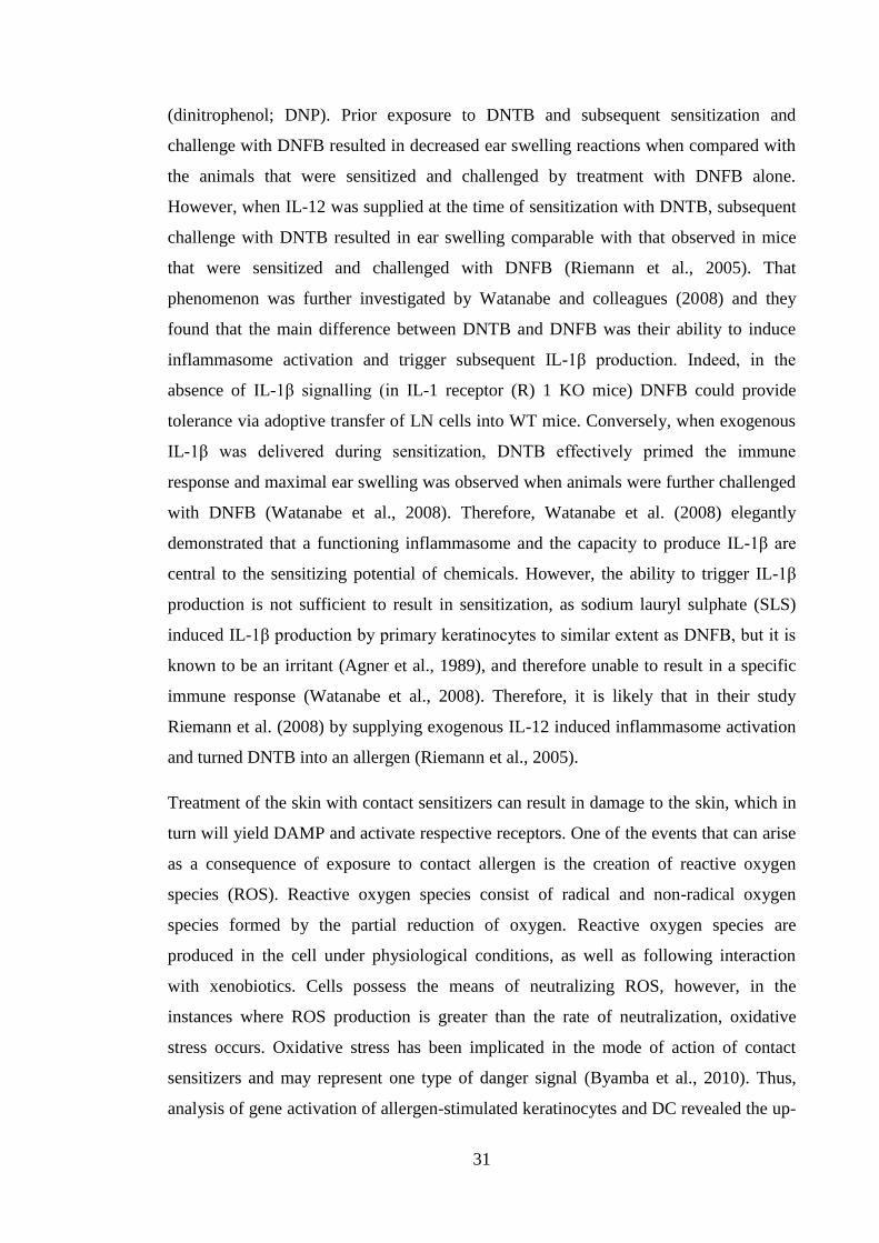
now described in detail and are illustrated in the Figure 1.1.
27
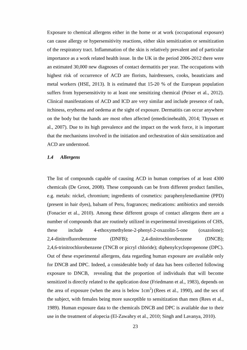
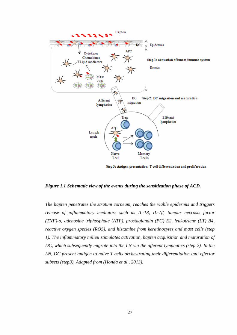
Figure 1.1 Schematic view of the events during the sensitization phase of ACD.
The hapten penetrates the stratum corneum, reaches the viable epidermis and triggers
release of inflammatory mediators such as IL-18, IL-1β, tumour necrosis factor
(TNF)-α, adenosine triphosphate (ATP), prostaglandin (PG) E2, leukotriene (LT) B4,
reactive oxygen species (ROS), and histamine from keratinocytes and mast cells (step
1). The inflammatory milieu stimulates activation, hapten acquisition and maturation of
DC, which subsequently migrate into the LN via the afferent lymphatics (step 2). In the
LN, DC present antigen to naive T cells orchestrating their differentiation into effector
subsets (step3). Adapted from (Honda et al., 2013).
28
1.5.1.1 Formation of the protein-hapten complexes
To sensitize the organism haptens have to be bioavailable, that is, they have to cross the
stratum corneum and create complexes with proteins via a process called haptenization
(Figure 1.1). The ability of sensitizers and proteins to form adducts stems from their
electrophilic and nucleophilic nature, respectively, whereby the nucleophile donates an
electron pair to electrophile, thereby forming a chemical bond (Gerberick et al., 2008).
There are instances when a nonallergic parent compounds can become activated to
become sensitizing chemicals via oxidation or interaction with phase I or II enzymes
present in the skin (so called prohaptens). Phase I and II enzymes include, among
others, cytochrome P450 mixed-function oxidase system and acyltransferases,
glutathione (GSH) S-transferases, respectively (Karlberg et al., 2008). It is still not
known, however, whether the hapten-protein complexes are created with proteins on the
surface and/or inside keratinocytes and/or DC or maybe with extracellular proteins.
Certain contact sensitizers have been demonstrated to induce DC maturation both in
vivo (Aiba and Katz, 1990) and in vitro (Aiba et al., 1997). However, despite much
being known regarding the ability of haptens to cause ACD and activate DC, their exact
fate in the skin still remains largely unknown. DNCB is thought to be able to enter the
cell directly and to be processed with endogenous proteins and presented through the
MHC I complex (Toebak et al., 2009). Studies by Hopkins et al. (2005) have shown that
when incubated with cellular and serum proteins DNCB binds preferentially to cellular
proteins, reinforcing the theory of MHC I presentation. Investigations of the mode of
action of PPD demonstrated that human serum albumin (HSA) was modified by PPD in
vitro. Adducts of HSA and PPD were capable of stimulating proliferation of
lymphocytes from individuals sensitized to PPD (Jenkinson et al., 2010). In addition to
modifying non cell bound proteins, allergens have been observed to interact with cell
membrane markers, e.g CD54, CD86 that induce maturation of model DC cell lines
(THP-1, U937) (Ashikaga et al., 2006; Python et al., 2007); as well as result in the
production of cytokines, including tumour necrosis factor (TNF)-α, IL-1β, IL-1α, IL-8
and IL-18 among others (Teunis et al., 2013; Van Och et al., 2005). In a recent study
that set out to identify targets of haptenation, bromobianes, fluorescent chemical
compounds known for their strong reactivity with thiol groups, were utilized. It was
29
found, using microscopy, that in human skin exposed ex vivo, bromobianes were bound
preferentially to basal keratinocytes (Simonsson et al., 2011). Further investigations
revealed that fluorescent hapten targeted specifically K5 and K14 (Simonsson et al.,
2011). Overall, the receptors and direct modes of action of the majority of contact
allergens remain unknown.
1.5.1.2 Danger signals
Creation of hapten-protein complexes is only the first step of allergen interactions
within the skin and with cutaneous antigen presenting cells (APC), i.e. various DC
populations. In order for sensitization to take place there has to be present an
environment of inflammation to insure that the immune response is not mounted in the
absence of danger. Therefore, it has been speculated that in addition to antigen signal,
there is a requirement for a non-specific inflammatory signal to stimulate DC
mobilization and lymph node cell (LNC) proliferation. Such non-specific inflammation
was given the term “danger signal”. The concept was suggested by Matzinger (1994),
who postulated that the immune system has the ability to detect and react to the damage
of the organism (Matzinger, 1994). Consequently, danger signals can be divided into
two broad ranges of stimuli: pathogen associated molecular patterns (PAMP) and
damage associated molecular patterns (DAMP) that are capable of triggering immune
responses and subsequently facilitating maturation of DC. In the context of ACD the
presence of danger signals is crucial, as maturation and migration of DC is a pivotal
step in ensuring sensitization to allergens, and this will be expanded on in the next
section.
One group of receptors capable of recognizing danger signals are toll-like receptors
(TLR). Indeed, it has been shown recently that in humans one of the common
sensitizing metals, nickel, utilized the TLR4 receptor, which is best known for its ability
to recognize a component of Gram negative bacterial cell walls, lipopolysaccharide
(LPS) (Trinchieri and Sher, 2007). Schmidt et al. (2010) confirmed the interaction of
nickel with histidine amino acids within TLR4 and showed that this contributes to the
development of ACD in humans, but not in mice (as mice do not have corresponding
histidine residues), supplying a direct inflammatory signal to DC and triggering their
activation (Schmidt et al., 2010). Conversely, Klekotka et al. (2010) demonstrated that
30
the CHS reaction to DNFB was apparently independent of signals from TLR 2, 3, 4, 6,
and 9 as Toll/IL-1R-containing adaptor-inducing interferon-β (TRIF) (TRIF is an
accessory molecule TLR 2,3,4,6 and 9) deficient mice exhibited normal levels of CHS
(Klekotka et al., 2010).
Another important family of receptors are the nucleotide-binding domain leucine-rich
repeat containing receptors (NLR). NLR participate in responding to PAMP such as
LPS, flagellin, bacterial and viral nucleic acids, as well as DAMP, including adenosine
triphosphate (ATP), uric acid and hyaluronan (HA). Following activation, the NLR
forms a complex with apoptosis associated speck-like protein containing a CARD
(ASC). This complex is called the inflammasome. The inflammasome plays a critical
part in the activation of the protease, caspase-1, which participates in the creation of
biologically active IL-1β and IL-18 from biologically inactive precursor forms (Jha and
Ting, 2009).
One of the danger signals implicated in inflammasome signalling is ATP. It is present
extracellularly only at low concentrations in resting conditions, which can increase
dramatically when cells are stressed or dying. One of ATP receptors is P2X7, which is
involved in the processing of biologically active IL-1β and IL-18 via activation of
NLRP3 inflammasome. The role of P2X7 signalling in skin sensitization was revealed
in the study by Weber et al. (2010). They found that mice deficient in P2X7 receptor
exhibited impaired CHS responses, which were reversed by the delivery of exogenous
IL-1β (Weber et al., 2010). Indeed, the importance of caspase-1 in the sensitization
phase of CHS was explored and confirmed by Antonopoulos et al. (2001). They showed
that DNFB and oxazolone induced CHS reactions were attenuated in caspase-1 KO
mice in comparison with wild type (WT) counterparts. Similarly, topical application of
caspase-1 inhibitor, YVAD, inhibited ear swelling in response to DNFB challenge.
Further, no LC migration was observed in caspase-1 deficient mice in response to
treatment with 0.5% DNFB or TNF-α. However, LC migration was observed when
exogenous IL-1β was administered (Antonopoulos et al., 2001).
The importance of inflammasome activation is particularly apparent when considering
dinitrothiocyanobenzene (DNTB), a chemical from the same family as DNFB and
DNCB, that is, however, unable to initiate immune or inflammatory responses. DNTB
and the dinitrohalobenzene allergens DNFB and DNCB share the same epitope
31
(dinitrophenol; DNP). Prior exposure to DNTB and subsequent sensitization and
challenge with DNFB resulted in decreased ear swelling reactions when compared with
the animals that were sensitized and challenged by treatment with DNFB alone.
However, when IL-12 was supplied at the time of sensitization with DNTB, subsequent
challenge with DNTB resulted in ear swelling comparable with that observed in mice
that were sensitized and challenged with DNFB (Riemann et al., 2005). That
phenomenon was further investigated by Watanabe and colleagues (2008) and they
found that the main difference between DNTB and DNFB was their ability to induce
inflammasome activation and trigger subsequent IL-1β production. Indeed, in the
absence of IL-1β signalling (in IL-1 receptor (R) 1 KO mice) DNFB could provide
tolerance via adoptive transfer of LN cells into WT mice. Conversely, when exogenous
IL-1β was delivered during sensitization, DNTB effectively primed the immune
response and maximal ear swelling was observed when animals were further challenged
with DNFB (Watanabe et al., 2008). Therefore, Watanabe et al. (2008) elegantly
demonstrated that a functioning inflammasome and the capacity to produce IL-1β are
central to the sensitizing potential of chemicals. However, the ability to trigger IL-1β
production is not sufficient to result in sensitization, as sodium lauryl sulphate (SLS)
induced IL-1β production by primary keratinocytes to similar extent as DNFB, but it is
known to be an irritant (Agner et al., 1989), and therefore unable to result in a specific
immune response (Watanabe et al., 2008). Therefore, it is likely that in their study
Riemann et al. (2008) by supplying exogenous IL-12 induced inflammasome activation
and turned DNTB into an allergen (Riemann et al., 2005).
Treatment of the skin with contact sensitizers can result in damage to the skin, which in
turn will yield DAMP and activate respective receptors. One of the events that can arise
as a consequence of exposure to contact allergen is the creation of reactive oxygen
species (ROS). Reactive oxygen species consist of radical and non-radical oxygen
species formed by the partial reduction of oxygen. Reactive oxygen species are
produced in the cell under physiological conditions, as well as following interaction
with xenobiotics. Cells possess the means of neutralizing ROS, however, in the
instances where ROS production is greater than the rate of neutralization, oxidative
stress occurs. Oxidative stress has been implicated in the mode of action of contact
sensitizers and may represent one type of danger signal (Byamba et al., 2010). Thus,
analysis of gene activation of allergen-stimulated keratinocytes and DC revealed the up-
32
regulation of genes in pathways that are in charge of oxidative stress management
(Natsch and Emter, 2008). The Kelch-like ECH-associated protein 1 (Keap1)/Nrf2-
signaling pathway is known to be important during oxidative stress. Keap1 is a sensor
protein, rich in reactive cysteine residues. These cysteine moieties are modified via
interaction with electrophilic compounds. In turn, this event releases Nrf2, which
translocates to the nucleus, binds to antioxidant response element (ARE) and triggers
expression of phase II detoxification enzymes (such as catalase, heme oxygenase-1,
GSH S-transferase) (Dinkova-Kostova et al., 2005). A comprehensive analysis of the
Nrf2 pathway was conducted by Natsch et al. (2008). They investigated a range of
strong and moderate sensitizers for their ability to induce the ARE-responsive gene,
quinine reductase, in a hepatic mouse cell line, Hepa1C1C7, or an activation of
luciferase gene under the ARE-dependent promoter in the reporter cell line AREc32, a
stable cell line derived from the human MCF7 breast carcinoma cell line. It was found
that Nrf2 pathway was activated by sensitizers but not by non-sensitizing chemicals.
This implies that sensitizers induce oxidative stress, and secondly that this feature could
be utilized in the in vitro tests to distinguish between sensitizing and non-sensitizing
chemicals (Natsch and Emter, 2008). Additionally, DNCB was demonstrated to increase
Nrf2 accumulation via depletion of GSH and covalent modification of Keap1 in mouse
liver cells (Chia et al., 2010). Moreover, the expression of Nfr2-dependent genes
(HMOX1 and NQO1) by both human peripheral blood mononuclear cell (PBMC)
derived DC and the THP-1 cell line, has been shown to be induced specifically by
treatment with a range of sensitizing chemicals, but not by non-sensitizers (Ade et al.,
2009). Furthermore, prior incubation of THP-1 cells with N-acetylcysteine (a GSH
precursor) inhibited DNCB induced expression of HMOX1 and NQO1 genes (Ade et al.,
2009), suggesting a specific ability of contact sensitizers to induce oxidative stress.
Within the immune system, ROS have been shown to participate in the activation of
NLRP3 inflammasome. Moreover, production of ROS was reported to be important
during antigen specific interaction of DC and T cells. Thus, administration of a potent
antioxidant (ebselen) to mice, either after sensitization or after challenge with
oxazolone, significantly inhibited the development of CHS, measured as a function of
challenge induced ear swelling responses, compared with untreated mice (Matsue et al.,
2003).
33
Reactive oxygen species and TLR are required for the formation of, and response to,
another important danger signalling molecule, HA. Hyaluronan is a component of
ECM, produced mainly by dermal fibroblasts and keratinocytes. Hyaluronan is a high
molecular weight glycosaminoglycan, composed of disaccharide repeats of d-glucuronic
acid and d-N-acetylglucosamine, connected via alternating β-1,4 and β-1,3 glycosidic
bonds (Mummert, 2005). A receptor for HA has been identified as CD44, which was
demonstrated to mediate cell adhesion to ECM (Aruffo et al., 1990) and lymphocyte
extravasation from the blood into tissues (DeGrendele et al., 1996). Hyaluronan under
physiological conditions is present in the skin as a high molecular weight (HMW)
molecule. It was found that when mice were exposed to HMW HA, it exerted anti-
inflammatory properties, as manifested by reduced DNFB challenge-induced changes in
ear thickness and decreased scarring and scaling recorded for CHS reactions provoked
by DNFB (Kim et al., 2008). Conversely, LMW HA fragments were shown to induce
inflammation and to mediate the production of inflammatory cytokines, such as TNF-α
by human monocyte derived DC, to a similar extent to that observed for LPS. The
immunostimulatory action of LMW HA has been reported to depend on TLR4
signalling, as bone marrow derived DC from mice with defective TLR4 receptors
(C3H/HeJ strain mice and TLR4 KO on C57BL/10 background) failed to secrete TNF-α
in response to LMW HA when compared with their WT counterparts (Termeer et al.,
2002). Similarly, it has been found that in a human melanoma cell line exposure to
LMW HA resulted in matrix metalloproteinase (MMP) 2 and IL-8 mRNA expression
and protein production (Voelcker et al., 2008). Matrix metalloproteinase 2 and MMP9
enzymes are expressed on the surface of cutaneous DC and are important in facilitating
DC migration from the skin to lymphatic vessels (Ratzinger et al., 2002). These changes
in MMP2 and IL-8 production were also mediated via interaction with TLR4, as when
TLR4 was silenced by transfection with small interfering (si) RNA, the stimulatory
effect of LMW HA was no longer observed (Voelcker et al., 2008). Further involvement
of TLR2 in HA signalling was observed by Scheibner et al. (2006). Degradation of
HMW to LMW HA is mediated via enzymes called hyaluronidases; a family of endo-
glycosidases that hydrolyze the β-1,4 linkages between N-acetyl-hexosamines and
glucuronic acid (Petrey and de la Motte, 2014). Degradation of HA also has been
observed via direct interaction with ROS, which could be inhibited by extracellular
superoxide dismutase (Gao et al., 2008). Moreover, it was demonstrated that in addition
34
to direct degradation of HA, ROS were capable of indirectly affecting HA degradation
via increasing the expression of hyaluronidase 2 (Monzon et al., 2010).
The roles of both ROS and products of HA degradation on the development of ACD
were investigated by Esser et al. (2012). They found that TNCB-induced CHS was
completely absent in double TLR2 and TLR4 KO mice. However, CHS reactions were
induced successfully in germ free TLR2 and TLR4 competent mice, suggesting the
involvement of endogenous TLR agonists in this process. In further experiments, Esser
and colleagues confirmed that allergen induced the production of ROS in vitro in the
PAM212 keratinocyte cell line, and in vivo in the ear (Esser et al., 2012). Allergen-
induced degradation of HA in the skin was confirmed by electrophoresis of
homogenized skin samples following 4h and 24h exposure to TNCB. It was shown that
ROS mediated HA degradation was inhibited in the presence of antioxidants,
N-acetylcysteine or plant derived antioxidant, RF-40. Additional evidence for the
importance of ROS in the pathology of CHS came from experiments in which topical
pre-treatment with a range of antioxidants prior to sensitization decreased the extent of
ear swelling (Esser et al., 2012). Furthermore, it was reported that exposure to TNCB
augmented hyaluronidase activity in a ROS dependent manner. Finally, treatment with
the hyaluronidase inhibitor, aristolochic acid, before sensitization prevented the CHS
reaction, confirming the involvement of ROS and degraded HA in contact allergic
reactions (Esser et al., 2012).
In this section the participation of danger signals in the induction phase of contact
sensitivity has been described. Danger signals arise in the skin following exposure to
allergens and in response to other traumatic events or local cell or tissue damage. They
can be versatile and stimulate divergent receptors. Thus far, the literature suggests that
danger signals are uniformly required for successful sensitization. The necessity for a
non-specific inflammation triggered by danger signals is a sophisticated mechanism of
two-step control of the immune system that does not allow adaptive responses to take
place in the absence of sufficient threat, like in the case of DNTB. It seems that one of
the main purposes of danger signalling is the stimulation of production of biologically
active IL-1β, which confers to DC the ability to transfer hapten responsiveness or
stimulate T cell proliferation. Indeed, IL-1β plays a crucial role during the migration
35
and maturation of cutaneous DC and will be described in greater detail in the next
section.
1.6 Dendritic cells
Antigen presentation is mediated by professional APC, DC. Dendritic cells are bone
marrow derived cells of the innate immune system. They can reside in the lymphoid and
non-lymphoid tissues. They act as sentinels of the immune system. Dendritic cell
populations detect pathogens and tissue damage. They are located in tissues where the
body comes into closest contact with the external environment, including in the gut,
lung and skin. Additionally, DC are resident in the spleen or in the LN, where they
effectively sample blood and lymph, respectively, in search of self and non-self
antigens. The role of the DC is to sense danger and to carry antigens to the draining LN,
where they can encounter other components of the immune system. Therefore they
effectively act as a bridge between the innate and adaptive immune systems (Merad et
al., 2013).
36
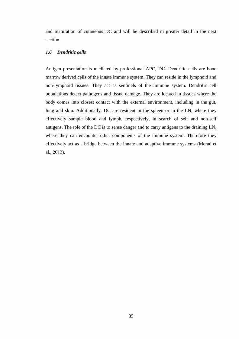
Figure 1.2 Subsets of dermal (d)DC in mouse and human skin.
Mouse skin contains LC in the epidermis and three dDC subsets in the dermis, which
are: migratory LC on their way to the lymphatic vessels, langerin+
dDC and langerin-
dDC (majority population). Human skin also contains LC in the epidermis. There are
four dDC populations in the human dermis, which include: migratory LC, CD141+ dDC
considered to be the equivalent of mouse langerin+ dDC population; CD14
+ dDC and
CD1c+ dDC. It is hypothesized that there might be diversity within the mouse langerin
-
dDC subset to accommodate the two populations of human dDC without mouse
counterparts. Contrary to dDC found in the mouse, only migratory LC found in the
human dermis express langerin.
37
1.6.1 Cutaneous dendritic cell subsets
Skin DC are heterogeneous and comprise of LC and dermal DC (dDC); the breadth of
DC populations in mouse and human skin are illustrated in Figure 1.2. The role of DC
in the skin is to maintain tolerance to commensal bacteria found on the surface of the
skin and to self-antigens, as well as to initiate immune responses to invading pathogens
and assaulting chemical compounds. The function of skin resident DC depends on their
ability to migrate to the LN, where they can orchestrate the type and the extent of
adaptive immune responses. As such, they are involved in sensing whether a particular
cutaneous insult is potentially dangerous, taking up that signal and transporting it (Helft
et al., 2010). As discussed previously, contact allergens are misread by the immune
system as potential pathogens, thus stimulating an inflammatory and allergic response.
Within the mouse dDC subset, 2 main populations are distinguished: langerin+ dDC and
langerin- dDC (Merad et al., 2008) (Table 1.1; Figure 1.2). In humans, 3 cell subsets
can be found in the dermis: CD14+ dDC and DC1c
+ dDC (Klechevsky et al., 2008) and
CD141+ dDC (Haniffa et al., 2012) (Table 1.2; Figure 1.2). Functional similarities can
be found between mouse and human dDC subsets, namely mouse langerin+ dDC are
thought to be an equivalent of CD141+ human dDC (Haniffa et al., 2013; Haniffa et al.,
2012; Heath and Carbone, 2013). Similarly, it is considered that there is a diversity
within the mouse langerin- dDC population to accommodate human populations of
CD14+ dDC and CD1c
+ dDC (Haniffa et al., 2012; Heath and Carbone, 2013) (Figure
1.2).
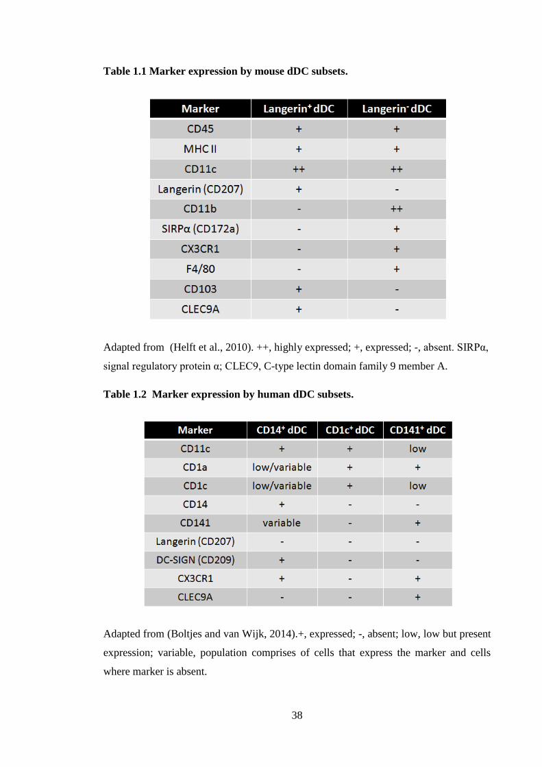
38
Table 1.1 Marker expression by mouse dDC subsets.
Adapted from (Helft et al., 2010). ++, highly expressed; +, expressed; -, absent. SIRPα,
signal regulatory protein α; CLEC9, C-type lectin domain family 9 member A.
Table 1.2 Marker expression by human dDC subsets.
Adapted from (Boltjes and van Wijk, 2014).+, expressed; -, absent; low, low but present
expression; variable, population comprises of cells that express the marker and cells
where marker is absent.
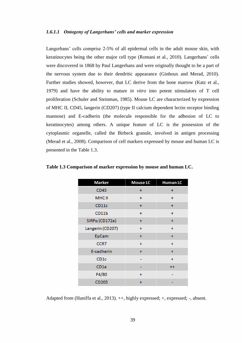
39
1.6.1.1 Ontogeny of Langerhans’ cells and marker expression
Langerhans’ cells comprise 2-5% of all epidermal cells in the adult mouse skin, with
keratinocytes being the other major cell type (Romani et al., 2010). Langerhans’ cells
were discovered in 1868 by Paul Langerhans and were originally thought to be a part of
the nervous system due to their dendritic appearance (Ginhoux and Merad, 2010).
Further studies showed, however, that LC derive from the bone marrow (Katz et al.,
1979) and have the ability to mature in vitro into potent stimulators of T cell
proliferation (Schuler and Steinman, 1985). Mouse LC are characterized by expression
of MHC II, CD45, langerin (CD207) (type II calcium dependent lectin receptor binding
mannose) and E-cadherin (the molecule responsible for the adhesion of LC to
keratinocytes) among others. A unique feature of LC is the possession of the
cytoplasmic organelle, called the Birbeck granule, involved in antigen processing
(Merad et al., 2008). Comparison of cell markers expressed by mouse and human LC is
presented in the Table 1.3.
Table 1.3 Comparison of marker expression by mouse and human LC.
Adapted from (Haniffa et al., 2013). ++, highly expressed; +, expressed; -, absent.
40
Langerhans’ cells are the only DC present in the epidermis. They appear in the
epidermis of mice from 18 day of embryonic development (Chorro et al., 2009). Crucial
for the development of LC is the presence of TGF-β. The presence of the TGF-β is
required for in vitro differentiation of human CD34+ precursors into langerin expressing
LC (Caux et al., 1999; Guironnet et al., 2002). Indeed, it had been observed that TGF-β
KO mice do not have MHC II+ cells present in the epidermis (Borkowski et al., 1996;
Borkowski et al., 1997). Mice deficient in TGF-β presented with inflammation and an
early death. To exclude the possibility that the persistent inflammation was responsible
for the absence of LC, mice were treated with rapamycin that inhibited the inflammation
and prolonged the life of KO animals. However, rapamycin did not reinstate LC in the
epidermis of these animals, suggesting that their absence was not caused by cutaneous
inflammation, but rather was a result of a defective LC development, which is
dependent upon TGF-β (Borkowski et al., 1996).
Langerhans’ cells turnover in the epidermis in the steady state is independent of bone
marrow precursors and is maintained in situ by a population of skin resident cells.
Langerhans’ cells are resistant to ionizing radiation and remain of host origin after bone
marrow transplant or in congenic parabiotic (with shared circulation) mice. However,
following ultraviolet (UV) light radiation (inflammatory conditions) LC can be derived
from precursors in the blood. Indeed, in a severely inflamed skin, where LC and their
progenitors are lost and skin integrity is compromised, LC are repopulated by
circulating monocytes that respond to chemokines secreted by endothelial cells, namely
monocyte chemoattractant protein-1 (MCP-1). MCP-1 is the ligand for CCR2 that
drives monocytes to the dermis. Another signal from keratinocyte-derived macrophage
inflammatory protein (MIP) 3α, a CCR6 ligand, controls their entry into the epidermis
(Ginhoux and Merad, 2010). The half-life of LC is much longer than other DC (7-8
days) and is thought to be between 53-78 days. Moreover, between 1 and 2% of all LC
were demonstrated to be able to divide at any given time compared with 5% for other
DC subsets (Merad et al., 2002).
41
1.6.1.2 Ontogeny of dermal dendritic cells and marker expression
In contrast to LC, tissue DC have a shorter life span and have to be replenished by
circulating precursors from the blood (Liu et al., 2007; Merad et al., 2002). Turnover of
DC in non-lymphoid tissues was assessed in mice following bromodeoxyuridine (BrdU)
injection. Bromodeoxyuridine is a synthetic analogue of thymidine, used to label
dividing cells. It was demonstrated that 12h following BrdU injection 5–10% of DC in
the dermis and lung, and 10–20% of DC in the liver and kidney were labelled with
BrdU (Ginhoux et al., 2009).
Dendritic cell-restricted precursors that give rise to tissue resident DC have been
identified in the bone marrow. Multipotent precursor cells in the bone marrow divide
into two distinct lineages lymphoid and myeloid, which give rise to lymphocytes and
granulocytes, erythrocytes, megakaryocytes, monocytes, respectively. Dendritic cells
can differentiate from macrophage/DC precursors (MDP) which differentiate into
monocytes and common DC precursors (CDP) (illustrated in the Figure 1.3). The
common DC precursor subsequently becomes a DC restricted precursor, called pre-DC
(Liu et al., 2007). Pre-DC leave the bone marrow, circulate in the blood and home to
lymphoid organs, where they become lymphoid tissue-resident DC or settle in the non
lymphoid tissues, such as liver, skin, kidneys or lungs (Ginhoux et al., 2009; Helft et al.,
2010). In addition to DC derived from pre-DC precursors, DC can arise from
monocytes. Indeed, it was found that out of two monocyte populations present in
mouse blood, Ly6Chi
CCR2+CX3CR1
lowCD62L
+ and Ly6C
- CCR2
-CX3CR1
hiCD62L
-,
the former is recruited to inflamed tissues, whereas the latter is recruited into non-
inflamed tissues via a CX3CR1--dependent mechanism (Geissmann et al., 2003).
Subsequently, it was discovered that Ly6Chi
and Ly6C- monocytes gave rise to CD103
+
DC and CD103-
DC, respectively, in the lung (Jakubzick et al., 2008). Mouse
monocytes Ly6Chi
and Ly6C- subsets were found to correspond with human monocytes
CD14+CD16
- and CD14
-CD16
+ populations, respectively (León et al., 2005). The ability
of monocytes to give rise to DC the presence of granulocyte-macrophage colony-
stimulating factor (GM-CSF) and IL-4 has greatly facilitated DC investigation of in
vitro derived DC (Sallusto and Lanzavecchia, 1994).
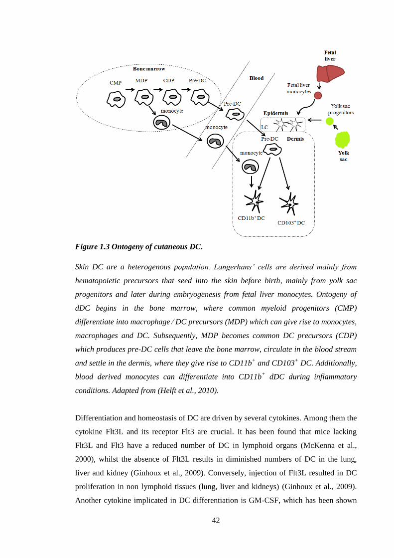
42
Figure 1.3 Ontogeny of cutaneous DC.
Skin DC are a heterogenous population. Langerhans’ cells are derived mainly from
hematopoietic precursors that seed into the skin before birth, mainly from yolk sac
progenitors and later during embryogenesis from fetal liver monocytes. Ontogeny of
dDC begins in the bone marrow, where common myeloid progenitors (CMP)
differentiate into macrophage ⁄ DC precursors (MDP) which can give rise to monocytes,
macrophages and DC. Subsequently, MDP becomes common DC precursors (CDP)
which produces pre-DC cells that leave the bone marrow, circulate in the blood stream
and settle in the dermis, where they give rise to CD11b+ and CD103
+ DC. Additionally,
blood derived monocytes can differentiate into CD11b+ dDC during inflammatory
conditions. Adapted from (Helft et al., 2010).
Differentiation and homeostasis of DC are driven by several cytokines. Among them the
cytokine Flt3L and its receptor Flt3 are crucial. It has been found that mice lacking
Flt3L and Flt3 have a reduced number of DC in lymphoid organs (McKenna et al.,
2000), whilst the absence of Flt3L results in diminished numbers of DC in the lung,
liver and kidney (Ginhoux et al., 2009). Conversely, injection of Flt3L resulted in DC
proliferation in non lymphoid tissues (lung, liver and kidneys) (Ginhoux et al., 2009).
Another cytokine implicated in DC differentiation is GM-CSF, which has been shown
43
to play a role in the development of CD11b+, but not CD11b
- DC in the dermis
(Kingston et al., 2009). Macrophage colony-stimulating factor (M-CSF) was originally
considered to be required for the development of macrophages and it has been
demonstrated recently to facilitate the development of LC, such that mice lacking
M-CSF receptor did not develop LC (Ginhoux et al., 2006). Additionally, as previously
mentioned, TGF-β has been found to be required for the formation of LC (Borkowski et
al., 1996).
1.6.2 Roles of skin DC: sensing danger, taking up antigen and transporting antigen
The main function of DC is to bridge the gap between innate and adaptive immune
responses by sensing danger, as described in the section about danger signals, and
triggering specific responses by T cells. Dendritic cells are superbly equipped to fulfil
these roles. Firstly, their name stems from their morphologic appearance of
characteristic long or veil-like protrusions, which serve to maximize the surface area of
the cell on which the antigens might be presented (high surface area to volume ratio)
(Banchereau and Steinman, 1998). Immature DC are proficient in sensing danger,
antigen capture and processing. They lack expression of T cell co-stimulatory molecules
CD40, CD54, CD80 and CD86. Conversely, they possess receptors required for antigen
acquisition. Dendritic cells utilize phagocytosis and macropinocytosis to capture
microbes and the soluble antigens in the extracellular environment, respectively. In
addition to these two methods of antigen sampling, DC utilize receptor mediated
endocytosis via receptors recognizing Fc portions of antibodies (eg. Fcγ and Fcɛ), heat
shock protein receptors and scavenger receptors, C-type lectin receptors, such as the
mannose receptor, CD205 receptor (Guermonprez et al., 2002), dendritic cell-specific
intercellular adhesion molecule-3-grabbing non-integrin (DC-SIGN; CD209)
(Geijtenbeek et al., 2000) and langerin (CD207) (Valladeau et al., 2000). These methods
of antigen acquisition are effective even at picomolar and nanomolar levels (Sallusto et
al., 1995). Captured antigen is then loaded onto MHC to form antigen-MHC complexes
that are presented on the DC surface for subsequent recognition by T cell receptors
(TCR) (Guermonprez et al., 2002). Cells down-regulate their expression of antigen
acquisition receptors and begin to undergo changes known collectively as maturation.
Mature DC migrate via efferent lymphatics to the LN where they localize in the
paracortex region (Randolph et al., 2005).
44
The sequence of events in DC migration is triggered by variety of factors of biological
(Haley et al., 2012; Shankar et al., 1996), chemical (Cumberbatch et al., 1992a) and
physical (Ghaznawie et al., 1999) nature. Langerhans’ cells have a complicated path to
follow during migration to the LN. It has been described as a two step model
(Villablanca and Mora, 2008), where the first leg of LC journey is from the epidermis to
dermis (Ouwehand et al., 2008), and the second part entails mobilization from the
dermis to the lymphatic vessels. Langerhans’ cell migration is dependent on two
compulsory signals: IL-1β and TNF-α. Upon recognition of danger signals LC secrete
IL-1β that in turn stimulates LC via IL-1R1 and triggers the release of TNF-α,
presumably from surrounding keratinocytes. TNF-α acts on LC through type 2 receptor
(TNFRII) delivering a second stimulus necessary for LC migration (Cumberbatch et al.,
1997a, b, 1999c).
Cumberbatch et al. (1997) investigated the influence of antibodies that neutralize TNF-α
and IL-1β on LC migration and LN DC accumulation caused by topical administration
of oxazolone or by intra-dermal delivery of TNF-α or IL-1β. Thus, systemic
(intraperitoneal) delivery of anti-IL-1β or anti-TNF-α neutralizing antibody reduced the
DC influx to the LN normally observed after exposure to oxazolone. The reduction in
LC frequency in the murine ear epidermis usually observed following TNF-α intra-
dermal administration was also inhibited by anti-IL-1β antibodies. Correspondingly,
anti-TNF-α antibody prevented IL-1β triggered LC migration and LN DC accumulation
(Cumberbatch et al., 1997b).
Others have also demonstrated the requirements for IL-1β and TNF-α cytokine signals
in the process of LC mobilization. Wang et al. (1997) found that following exposure to
fluorescein isothiocyanate (FITC) in mice deficient in TNFRII, the only TNF-α receptor
present on LC (Peschon et al., 1998), there was a decreased number of antigen bearing
cells in the LN in comparison with WT mice. Furthermore, it was reported that TNFRII
KO mice were less responsive to oxazolone induced CHS, as measured by reduced
challenge-induced ear swelling (Wang et al., 1996).
It was found that DC migration in humans was also dependent on TNF-α and IL-1β
signalling. Stoitzner et al. (1999) reported that inhibition of TNF-α via exposure to anti-
TNF-α antibody inhibited spontaneous LC migration in human skin explants. Moreover,
it was established that delivery of exogenous IL-1α, IL-1β and TNF-α enhanced DC
45
migration from skin explants in comparison with spontaneous migration (Stoitzner et
al., 1999).
Further investigations of cytokine requirements for LC migration were performed by
Cumberbatch and colleagues. A role for IL-18 was observed, in addition to requirement
for TNF-α and IL-1β signalling. Intradermal delivery of exogenous IL-18 resulted in LC
mobilisation that was significantly inhibited in the presence of anti- TNF-α, anti-IL-1R1
antibody and in caspase-1 KO mice. Moreover, oxazolone induced decrease in LC
frequency was inhibited by pre-treatment with anti-IL-18 antibody. These data
suggested that IL-18 signals upstream from TNF-α and IL-1β (Cumberbatch et al.,
2001).
Once in the LN, mature DC cannot capture antigens; however, they possess the unique
ability to induce the activation of naive T cells. In the LN, when antigen carried by the
DC is recognized by a T cell, two cells interact, creating an immunological synapse
(Dustin et al., 2010). In the LN, circulating T cells sample antigens on DC via
interaction between lymphocyte function-associated antigen 1 (LFA-1) and intercellular
adhesion molecule-1 (ICAM-1), present on T cells and DC, respectively. Recognition of
antigen-MHC complex on DC by TCR drives conformational change in the LFA-1
which stabilizes interaction between T cells and the DC (Hogg et al., 2002).
Recognition of antigen-MHC complex by TCR delivers a first signal to naive T cells. A
second signal is delivered by interaction of the DC co-stimulatory molecules CD80 and
CD86 with T cell receptor, CD28 (Jenkins et al., 1991). Activation of CD28 receptor
specifically stimulates production of IL-2, which further drives its own production (Jain
et al., 1995). Interleukin 2 is crucial for T cell growth, proliferation and differentiation
(Smith, 1988). Additionally, interaction between CD40 and CD40L on DC and T cells,
respectively, has been demonstrated to up-regulate expression of MHC II, CD80 and
CD86 (Caux et al., 1994), and stimulate IL-12 release by DC (Cella et al., 1996).
1.6.3 Polarization of immune responses by DC
A third signal is delivered by the DC, which induces T cell polarization and
development into either type 1 or type 2 cells (Gutcher and Becher, 2007) (Figure 1.4).
The paradigm of Th1 and Th2 responses was proposed in 1986 by Mosmann et al.
(1986) and argued that CD4+
T cells could develop into functionally distinctive
46
subpopulations that display differential cytokine profiles and diverse roles in adaptive
immunity (Mosmann et al., 1986). It was further discovered that Th1 subset could be
characterized by production of IL-2, IL-3, TNF-α, and most notably IFN-γ and
participation in a cell-mediated immunity, whereas the Th2 population secreted mainly
IL-4, IL-5, and IL-13 and aided humoral immunity (Coffman, 2006; Zhou et al., 2009).
The polarization of T cell response is dependent on the cytokines secreted by DC, which
in turn are determined by the type of antigen encountered by the DC. The presence of
intracellular pathogens causes IL-12 release by the DC and subsequent polarization of
CD4+ cells into Th1 populations. Conversely, helminths and other extracellular
pathogens trigger production of IL-4 and subsequent polarization of CD4+ cells into Th2
cells (Walsh and Mills, 2013; Zhou et al., 2009).
Similarly to CD4+ cell populations, type 1 and type 2 cells are also present among CD8
+
cytotoxic T cells (Tc1 and Tc2, respectively), and are characterized by similar cytokine
secretion profiles to those displayed by Th cell subsets (Vukmanovic-Stejic et al.,
2000). Generation of Tc1 and Tc2 subsets is also regulated by IL-12 and IL-4 release by
DC, respectively (Woodland and Dutton, 2003). These and other Th cell subsets are
displayed in the Figure 1.4.
Recently, another Th subset has been described, namely Th17 (Figure 1.4). Th17 cells
are involved in responding to bacterial and fungal infections (Yamane and Paul, 2013).
They release IL-17A, IL-17F, IL-21 and IL-22 cytokines. Their polarization is
controlled by TGF-β, IL-1, IL-6 and IL-23 (Korn et al., 2009; Mangan et al., 2006). The
Th17 cell subset was identified in the cellular infiltrate of human skin undergoing the
elicitation phase of ACD. Furthermore, enhanced expression of mRNA for signature
Th17cytokines IL-17A, IL-17F and IL-23 was detected in the skin during the elicitation
phase of ACD. Increased levels of with IFN-γ and IL-4 mRNA also were observed in
allergen challenged skin, which were characteristic of Th1 and Th2 response,
respectively (Zhao et al., 2009). Further clues as to the importance of IL-17 in the skin
came from IL-17 KO mice, which exhibited impaired CHS to DNFB, as measured by
diminished ear swelling. There were no differences in the TNF-α, IL-1α and IL-1β
cytokine levels in the ear following the challenge, suggesting that the reduced CHS
reaction was due to the absence of IL-17, and not because of the secondary effects on
IL-1 or TNF-α (Nakae et al., 2002).
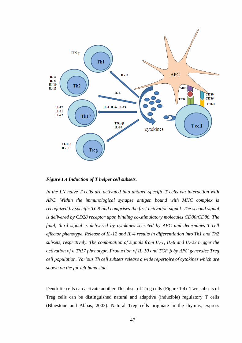
47
Figure 1.4 Induction of T helper cell subsets.
In the LN naive T cells are activated into antigen-specific T cells via interaction with
APC. Within the immunological synapse antigen bound with MHC complex is
recognized by specific TCR and comprises the first activation signal. The second signal
is delivered by CD28 receptor upon binding co-stimulatory molecules CD80/CD86. The
final, third signal is delivered by cytokines secreted by APC and determines T cell
effector phenotype. Release of IL-12 and IL-4 results in differentiation into Th1 and Th2
subsets, respectively. The combination of signals from IL-1, IL-6 and IL-23 trigger the
activation of a Th17 phenotype. Production of IL-10 and TGF-β by APC generates Treg
cell population. Various Th cell subsets release a wide repertoire of cytokines which are
shown on the far left hand side.
Dendritic cells can activate another Th subset of Treg cells (Figure 1.4). Two subsets of
Treg cells can be distinguished natural and adaptive (inducible) regulatory T cells
(Bluestone and Abbas, 2003). Natural Treg cells originate in the thymus, express
48
CD4+CD25
+Foxp3
+ markers and are involved in maintaining central and peripheral
tolerance to autoantigens (Bach and François Bach, 2003). Adaptive regulatory T cells
are created in the periphery from naive T cells by interaction with immature or partially
matured DC in the absence of sufficient co-stimulatory molecule expression (Walsh and
Mills, 2013). A major role in the development of adaptive regulatory T cells is played
by TGF-β and IL-10. Interleukin 10 interferes with DC function and inhibits their full
maturation; it inhibits T cell CD28 signalling and in turn promotes differentiation into
IL-10 producing T cells (Joss et al., 2000; Sato et al., 2003; Steinbrink et al., 1997).
Similarly, TGF-β prevents maturation of DC (Steinman et al., 2003) and inhibits
activation of T cells (Coffman et al., 1989; Kehrl et al., 1986). Adaptive regulatory T
cells are associated with mucosal tolerance towards non-pathogenic microflora and are
thought to aid regulation of autoimmunity (de Jong et al., 2005).
1.6.3.1 Allergen dependent polarization of immune response
As mentioned previously, the immune system can misread allergens as pathogens,
therefore, allergens are capable of polarizing the immune response in the same way as
just has been described for biological insults. Indeed, exposure to different classes of
allergens was demonstrated to result in distinct polarization profiles. Work with
reference contact allergen DNCB and respiratory allergen trimellitic anhydride (TMA)
in mice has shown that these two allergens are capable of inducing opposing
polarization of immune response into Th1/Tc1 and Th2, respectively. It has been known
that inhalation of certain acid anhydrides and aromatic isocyanates can result in airway
hypersensitivity in humans (Bernstein et al., 1982; Danks et al., 1981; Moller et al.,
1985). It was shown that inhalation of TMA, but not of DNCB, induced IgE antibody
production in BALB/c strain mice (Dearman et al., 1991). Further work revealed that
topical exposure of BALB/c strain mice to doses of DNCB and TMA was capable of
inducing equivalent levels of immune response, as measured by the ability to induce
LNC proliferation, resulted in IgE antibody production only in mice treated with TMA
(Dearman and Kimber, 1991). Production of IgE is associated with Th2 response and is
mediated by IL-4 and strongly inhibited by IFN-γ (Coffman and Carty, 1986; Coffman
et al., 1986; Snapper and Paul, 1987). Indeed, further investigations have revealed that
chronic exposure to oxazolone or DNFB (contact allergens) and TMA or toluene
diisocyanate (respiratory allergens) resulted in production of IFN-γ and IL-4,
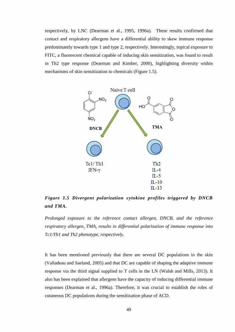
49
respectively, by LNC (Dearman et al., 1995, 1996a). These results confirmed that
contact and respiratory allergens have a differential ability to skew immune response
predominantly towards type 1 and type 2, respectively. Interestingly, topical exposure to
FITC, a fluorescent chemical capable of inducing skin sensitization, was found to result
in Th2 type response (Dearman and Kimber, 2000), highlighting diversity within
mechanisms of skin sensitization to chemicals (Figure 1.5).
Figure 1.5 Divergent polarization cytokine profiles triggered by DNCB
and TMA.
Prolonged exposure to the reference contact allergen, DNCB, and the reference
respiratory allergen, TMA, results in differential polarization of immune response into
Tc1/Th1 and Th2 phenotype, respectively.
It has been mentioned previously that there are several DC populations in the skin
(Valladeau and Saeland, 2005) and that DC are capable of shaping the adaptive immune
response via the third signal supplied to T cells in the LN (Walsh and Mills, 2013). It
also has been explained that allergens have the capacity of inducing differential immune
responses (Dearman et al., 1996a). Therefore, it was crucial to establish the roles of
cutaneous DC populations during the sensitization phase of ACD.
50
1.7 Langerhans’ cell migration
Langerhans’ cell migration has been one of the main endpoints of interest in the current
investigations. Additionally, LC migration is a more complex process than dDC
migration due to the fact that LC have a two step path to follow, that is from the
epidermis to dermis, and from the dermis to lymphatic vessels. Therefore, it is
important to understand the process of LC migration in greater detail.
Upon exposure to inflammatory stimuli, LC migrate towards the draining LN which
they reach after approximately 72h, where they present antigen to T cells
(Kissenpfennig et al., 2005). In order to reach the lymphatic vessels the LC have to
dissociate from the tightly packed network of keratinocytes. Interaction between
keratinocytes and LC is mediated by the adhesion molecule E-cadherin, which is
expressed by all keratinocytes, with approximately half of the cells exhibiting very high
levels of this molecule. E-cadherin mediates homophilic interactions between different
adjacent cell types in both mice (Tang et al., 1993) and in humans (Blauvelt et al.,
1995). Upon activation, LC are induced to decrease their expression of E-cadherin to
disengage from surrounding keratinocytes. Indeed, it was demonstrated that in clusters
of fetal skin dermal DC, the triggers that induce LC migration in vivo (IL-1α and IL-1β,
TNF-α and LPS), also resulted in a rapid (after 4h) down-regulation of E-cadherin
expression and increased levels of MHC II, CD40 and CD86 expression (Jakob and
Udey, 1998). Recently, it was reported that in addition to keeping LC ‘in place’, E-
cadherin expression by keratinocytes was required for LC differentiation from immature
precursors, which takes place under inflammatory conditions, when LC have to be
recruited to the epidermis from blood precursors (Mayumi et al., 2013).
The first step of LC migration was investigated by Ouwehand et al. (2008). They found
that human LC migrated ex vivo following exposure of the skin to NiSO4. Furthermore,
when human epidermal sheets were exposed to non-toxic concentrations of NiSO4 in a
transwell system, there was a decrease in the number of LC. When fibroblasts were
placed in the lower chamber of the transwell system, the extent of LC mobilization was
even higher. Later it was discovered that the LC migration towards fibroblasts could be
reversed by addition of the anti-CXCL12 antibody; suggesting that fibroblasts released
CXCL12 that attracted LC out of the epidermis. Subsequent experiments with the
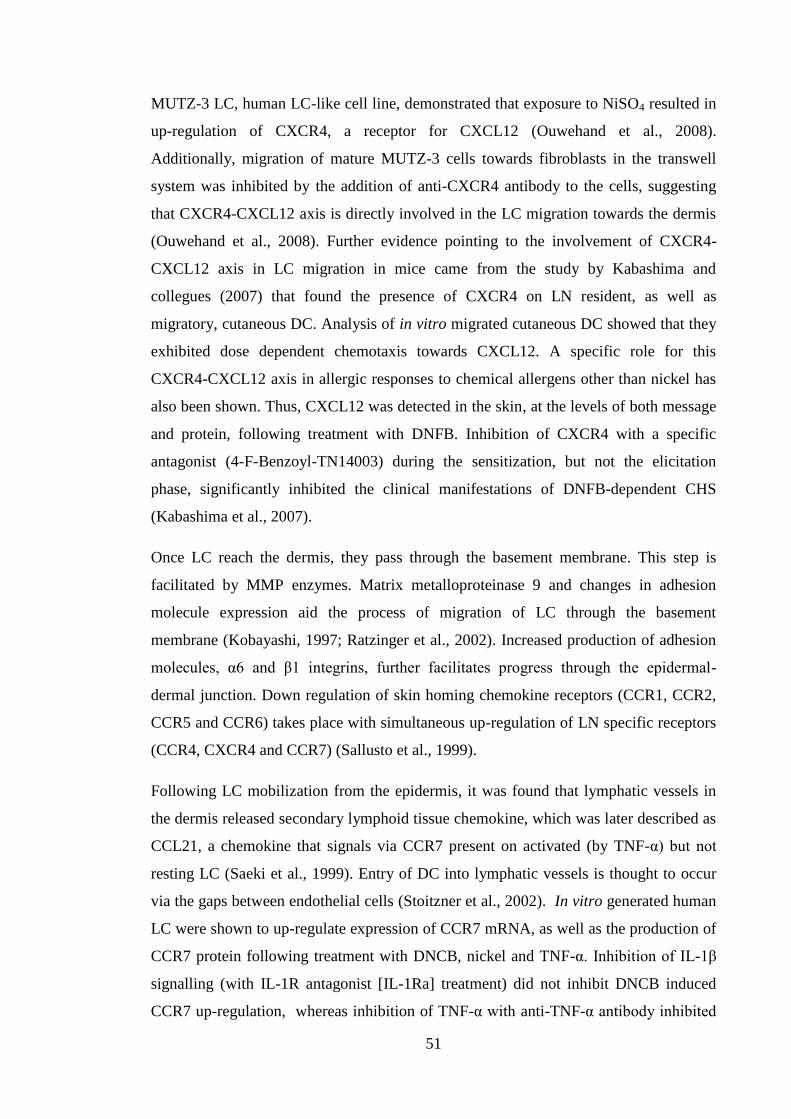
51
MUTZ-3 LC, human LC-like cell line, demonstrated that exposure to NiSO4 resulted in
up-regulation of CXCR4, a receptor for CXCL12 (Ouwehand et al., 2008).
Additionally, migration of mature MUTZ-3 cells towards fibroblasts in the transwell
system was inhibited by the addition of anti-CXCR4 antibody to the cells, suggesting
that CXCR4-CXCL12 axis is directly involved in the LC migration towards the dermis
(Ouwehand et al., 2008). Further evidence pointing to the involvement of CXCR4-
CXCL12 axis in LC migration in mice came from the study by Kabashima and
collegues (2007) that found the presence of CXCR4 on LN resident, as well as
migratory, cutaneous DC. Analysis of in vitro migrated cutaneous DC showed that they
exhibited dose dependent chemotaxis towards CXCL12. A specific role for this
CXCR4-CXCL12 axis in allergic responses to chemical allergens other than nickel has
also been shown. Thus, CXCL12 was detected in the skin, at the levels of both message
and protein, following treatment with DNFB. Inhibition of CXCR4 with a specific
antagonist (4-F-Benzoyl-TN14003) during the sensitization, but not the elicitation
phase, significantly inhibited the clinical manifestations of DNFB-dependent CHS
(Kabashima et al., 2007).
Once LC reach the dermis, they pass through the basement membrane. This step is
facilitated by MMP enzymes. Matrix metalloproteinase 9 and changes in adhesion
molecule expression aid the process of migration of LC through the basement
membrane (Kobayashi, 1997; Ratzinger et al., 2002). Increased production of adhesion
molecules, α6 and β1 integrins, further facilitates progress through the epidermal-
dermal junction. Down regulation of skin homing chemokine receptors (CCR1, CCR2,
CCR5 and CCR6) takes place with simultaneous up-regulation of LN specific receptors
(CCR4, CXCR4 and CCR7) (Sallusto et al., 1999).
Following LC mobilization from the epidermis, it was found that lymphatic vessels in
the dermis released secondary lymphoid tissue chemokine, which was later described as
CCL21, a chemokine that signals via CCR7 present on activated (by TNF-α) but not
resting LC (Saeki et al., 1999). Entry of DC into lymphatic vessels is thought to occur
via the gaps between endothelial cells (Stoitzner et al., 2002). In vitro generated human
LC were shown to up-regulate expression of CCR7 mRNA, as well as the production of
CCR7 protein following treatment with DNCB, nickel and TNF-α. Inhibition of IL-1β
signalling (with IL-1R antagonist [IL-1Ra] treatment) did not inhibit DNCB induced
CCR7 up-regulation, whereas inhibition of TNF-α with anti-TNF-α antibody inhibited
52
the induction of CCR7 (Boislève et al., 2004). The switching on and off of certain
chemokine receptors appears to be crucial for DC responsiveness. It has been shown by
Sallusto et al. (1998) that during maturation (following treatment with LPS) of human
DC derived from PBMC, CCR1 and CCR5 were rapidly (within 3h) down-regulated.
Conversely, expression of CXCR4 and CCR7 was augmented (Sallusto et al., 1998),
further confirming the role of CXCR4-CXCL12 axis in LC migration from the
epidermis to dermis and subsequent CCR7-CCL21 axis mediating LC chemotaxis
towards the LN. Another CCR7 ligand, CCL19, is produced by lymphatic vessels, as
well as by mature DC in the LN. Chemokine CCL19 attracts DC and naive T cells that
also express CCR7, to facilitate the encounter of naive T cells with antigen bearing DC
(Gunn et al., 1998; Willimann et al., 1998).
1.8 Role of cutaneous DC populations in the development of CHS
The skin represents a particularly rich source of different DC populations. As the
existence of these different populations has become apparent, there has been a
considerable interest in examining the roles of these different subsets in the
development of cutaneous immune responses, including skin allergic responses. Indeed,
contact allergens represent a relatively simple system in which the role of these DC in
skin immunity could be probed. Use of conditional and constitutive KO mouse models
allows for a selective depletion of specific DC populations and investigation of their
functions (Kaplan, 2010; Kaplan et al., 2008; Romani et al., 2010). There have been
many attempts to achieve this and as a result there were often conflicting results
regarding the role of LC and dDC in the CHS response. One such model utilized
langerin-DTR mice which had a knock in of human diphtheria toxin (DT) receptor
(DTR) under the control of the langerin promoter. Upon delivery of DT to these mice
depletion of all langerin-expressing cells took place within 24h of toxin injection.
Epidermal LC did not repopulate the epidermis until 4 weeks after toxin injection
(Bennett et al., 2005) leaving a window where langerin-DTR mice were deficient in
langerin+ cells. It was confirmed that depletion of langerin
+ cells per se did not result in
inflammation and that skin homeostasis was maintained by visualization of undisturbed
DETC in the epidermis. Following sensitization with 1% TNCB and challenge with
0.25% TNCB, CHS reactions in langerin-DTR mice were significantly diminished,
compared with WT mice, although still detectable. These results suggested that
53
langerin-expressing cells were sufficient to induce CHS reaction (Bennett et al., 2005).
However, a study by Kissenpfennig et al. (2005) also utilized the same langerin-DTR
mice and found, in contrast to a report by Bennett et al. (2005), that the CHS reaction in
mice deficient in langerin-expressing cells was the same as in control, WT animals
(Kissenpfennig et al., 2005). Another available model of LC ablation was described by
Kaplan et al. (2005). In that model a bacterial artificial chromosome containing the DT
subunit A (DTA) under the control of human langerin was constitutively expressed,
making these transgenic mice lack langerin+ cells from birth. Interestingly, in these mice
(langerin-DTA) there were langerin+
cells present in the LN, however, that population
of cells was missing in the mouse model described by Bennett et al. (2005). When CHS
responses were examined in langerin-DTA mice that are deficient in LC from birth, it
was found that the response to oxazolone and DNFB was exaggerated in comparison
with WT counterparts. Moreover, it was found that the enhanced CHS response from
langerin-DTA mice could have been adoptively transferred into WT mice. These data
suggested that LC play a regulatory role.
What Bennett et al. and Kissenpfennig and colleagues (2005) were not aware of at the
time, was that upon delivery of DT two populations of cells were depleted: LC and
langerin+ dDC; the latter were identified in 2007 and described in a publication by
Bursch et al. (2007). Therefore, in retrospect, only the ablation model described by
Kaplan et al. (2005) was indeed the one that could investigate the role of LC in the
CHS, as those were the only cells that were depleted in the model that he described;
while in models used by Kissenpfennig et al. (2005) and Bennett et al. (2005) two cell
populations were depleted: LC and langerin+ dDC. A novel population of dermis
resident, radiosensitive langerin+ dDC was identified by crossing the two depletion
models, langerin-DTR and langerin-DTA (Bursch et al., 2007). It was shown that this
novel subset of cells could be distinguished from epidermal LC by the absence of
CD11b and the presence of CD103. In addition to the dermis, langerin+ DC were
detected in the lungs and in the liver. Investigations of the role of langerin+ dDC in the
CHS reaction demonstrated that the complete ablation of langerin+ cells resulted in
decreased challenge responses, whereas in the situation when only LC were absent CHS
reaction to DNFB was not different from that observed in mice with intact populations
of cutaneous DC (Bursch et al., 2007).
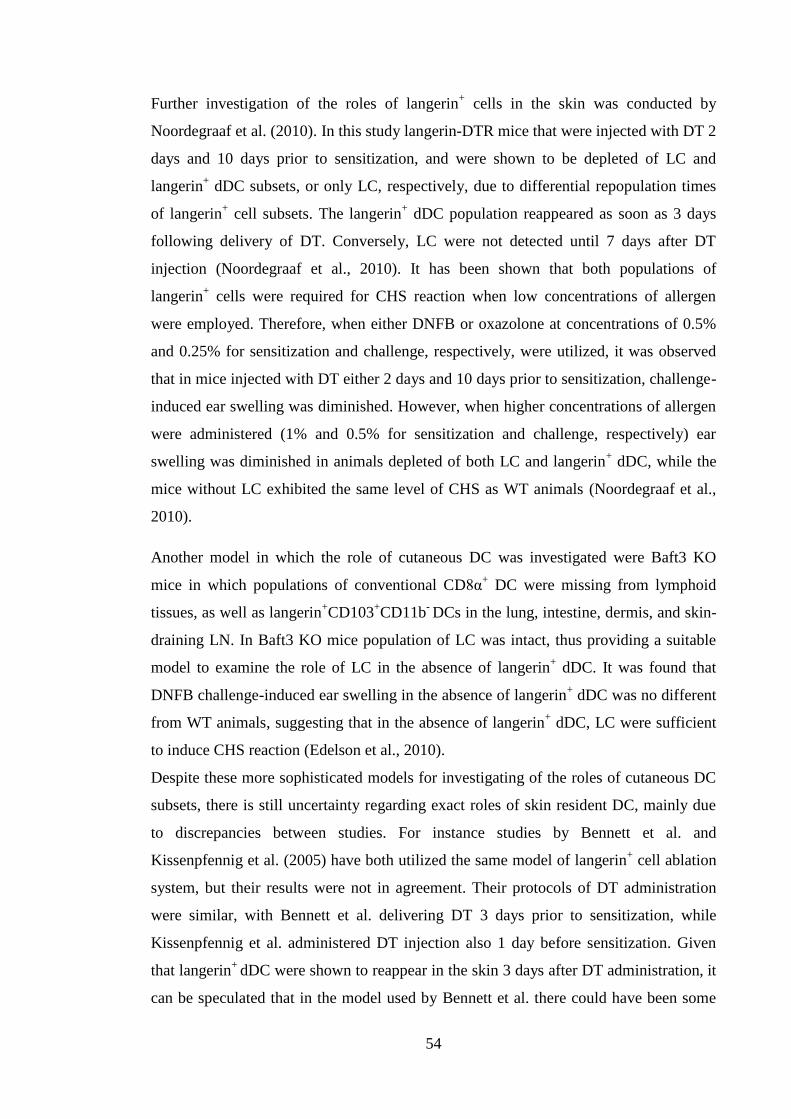
54
Further investigation of the roles of langerin+ cells in the skin was conducted by
Noordegraaf et al. (2010). In this study langerin-DTR mice that were injected with DT 2
days and 10 days prior to sensitization, and were shown to be depleted of LC and
langerin+ dDC subsets, or only LC, respectively, due to differential repopulation times
of langerin+ cell subsets. The langerin
+ dDC population reappeared as soon as 3 days
following delivery of DT. Conversely, LC were not detected until 7 days after DT
injection (Noordegraaf et al., 2010). It has been shown that both populations of
langerin+ cells were required for CHS reaction when low concentrations of allergen
were employed. Therefore, when either DNFB or oxazolone at concentrations of 0.5%
and 0.25% for sensitization and challenge, respectively, were utilized, it was observed
that in mice injected with DT either 2 days and 10 days prior to sensitization, challenge-
induced ear swelling was diminished. However, when higher concentrations of allergen
were administered (1% and 0.5% for sensitization and challenge, respectively) ear
swelling was diminished in animals depleted of both LC and langerin+ dDC, while the
mice without LC exhibited the same level of CHS as WT animals (Noordegraaf et al.,
2010).
Another model in which the role of cutaneous DC was investigated were Baft3 KO
mice in which populations of conventional CD8α+ DC were missing from lymphoid
tissues, as well as langerin+CD103
+CD11b
- DCs in the lung, intestine, dermis, and skin-
draining LN. In Baft3 KO mice population of LC was intact, thus providing a suitable
model to examine the role of LC in the absence of langerin+ dDC. It was found that
DNFB challenge-induced ear swelling in the absence of langerin+ dDC was no different
from WT animals, suggesting that in the absence of langerin+ dDC, LC were sufficient
to induce CHS reaction (Edelson et al., 2010).
Despite these more sophisticated models for investigating of the roles of cutaneous DC
subsets, there is still uncertainty regarding exact roles of skin resident DC, mainly due
to discrepancies between studies. For instance studies by Bennett et al. and
Kissenpfennig et al. (2005) have both utilized the same model of langerin+ cell ablation
system, but their results were not in agreement. Their protocols of DT administration
were similar, with Bennett et al. delivering DT 3 days prior to sensitization, while
Kissenpfennig et al. administered DT injection also 1 day before sensitization. Given
that langerin+
dDC were shown to reappear in the skin 3 days after DT administration, it
can be speculated that in the model used by Bennett et al. there could have been some
55
langerin+
dDC returning to the dermis at the time of sensitization. However, that still
does not explain divergent results, as Bennett et al. observed decreased CHS, which is
in line with the belief that langerin+
dDC are the main DC population responsible for
antigen delivery to the LN (which suggests that the langerin+ dDC were not there). One
other possibility for the differences between the two studies was the allergens that were
used. TNCB and DNFB were utilized by Bennett et al. (2005) and Kissenpfennig et al.
(2005), respectively, and it is possible that different chemical allergens may display
divergent needs for classes of skin DC.
Taken together, these data demonstrate that there has been somewhat of a paradigm
shift in the understanding of the role of LC. Some years ago, LC were considered to be
the chief APC responsible for antigen presentation to naive T cells in the skin draining
LN. Recently, it has become clear that in common with other aspects of the immune
system, there is some redundancy with respect to cutaneous DC subsets and antigen
presentation. Indeed, under some circumstances LC may not be the most efficient skin
APC, although when allergen is limiting, for example, they may play a more major role.
Furthermore, there is increasing evidence that LC may have other functions, including
immunoregulatory activities.
1.9 Tolerogenic role of Langerhans’ cells
The evidence obtained from langerin-DTA mice suggested that LC played a regulatory
role in the CHS response, whereby in their absence the symptoms in response to both
FITC and DNFB were more pronounced than in the WT mice. Further investigation of
this system revealed that the immunomodulatory effects of LC required the presence of
MHC II on the surface of the LC. Thus, increased ear swelling was observed in animals
with selective depletion of MHC II in the LC population, suggesting the role for MHC
II in LC-mediated immunosuppression. Furthermore, it has been shown that the lack of
LC-derived IL-10 in langerin-DTA mice contributed also to the augmented clinical
manifestation of CHS (Igyarto et al., 2009).
More clues about the additional roles of LC in the immune response came from the
study by Gomez de Agüero and colleagues (2012). They used DNFB and the
chemically related molecule DNTB. DNTB has been shown to be tolerizing (see section
1.5.1.2), with respect to DNFB, that is, prior topical exposure of mice to DNTB renders
56
the mice less sensitive to challenge with DNFB (Riemann et al., 2005). Following
treatment with DNTB, LC migrated to the LN and presented antigen to CD8+ T cells,
suggesting their ability to cross-present antigen. It was found that by interacting with
naive CD8+ T cells LC were able to render them unresponsive to subsequent exposure
to DNFB. To exert their tolerogenic effect, LC required the presence of
CD4+CD25
+Foxp3
+ cells, such that in the mice where that population of cells was
absent, the tolerizing effect of DNTB was no longer observed (Gomez de Agüero et al.,
2012).
To add further support to a potential immunoregulatory function of LC in contact
sensitization, there are other reports suggesting that LC play a role as mediators of
tolerance in other types of skin immune response, for example non-responsiveness
towards commensal, skin resident bacteria (van der Aar et al., 2013). Van der Aar and
colleagues demonstrated that human LC mediated tolerance towards bacterial flora via
inefficient bacteria engulfment and subsequent, ineffective antigen presentation. As a
consequence, LC induced bacteria specific Treg cells, instead of activating antibacterial
effector T cells (van der Aar et al., 2013).
Similarly, investigations of human LC revealed that LC, but not dDC, have the ability to
induce proliferation of skin resident memory T cells, the majority (~84%) of which
expressed markers specific for Treg cells. Further experiments demonstrated that LC
mediated induction of memory T cell proliferation depended on direct cell to cell
contact of the T cells with LC. However, when a pathogenic stimulus of heat inactivated
Candida albicans (C.albicans) was added to the culture, LC induced the proliferation of
effector memory T cells, which produced IFN-γ and IL-17 (Seneschal et al., 2012),
which was in accordance with data reported by Igyarto et al. (2011). Overall, the data
presented by Seneschal et al. (2012) suggested that under physiological conditions LC
exhibited a tolerogenic function by mediating the proliferation of Treg cells, but in the
presence of danger signal, i.e. C. albicans, LC had the ability to induce expansion of
effector memory T cells.
It is appears that LC can act as professional APC, however, under certain circumstances
they can play a tolerogenic role. Additionally, it has been recently suggested that LC
might mediate Th17 responses, which have been implicated in skin inflammation and
ACD (Peiser et al., 2012). Moreover, there are reports that suggest that LC are the main
57
subset responsible for antigen presentation to CD8+ T cells via the process of cross-
presentation.
1.10 Langerhans’ cells functions: cross-presentation and activation of Th17 cells
Langerhans’ cells are the outermost antigen presenting cells that are the first line of
contact of the skin with biological, chemical and physical insults as well self antigens
(Igyártó and Kaplan, 2013). In addition to their conventional role as APC, whereby LC
present antigens in the context of MHC II molecules to CD4+ T cells, LC have been
shown to possess cross-presentation abilities. Cross-presentation refers to the
presentation of antigens in the context of MHC I molecules. It is responsible for the
development of Tc mediated immunity directed against parasites and tumours that do
not infect APC, thus allowing presentation through the classical endogenous MHC class
I pathway, as well as maintaining tolerance (Joffre et al., 2012). There are conflicting
reports regarding the role of LC as the most efficient inducers of CD8+ T cell responses.
In 2008 Klechevsky and colleagues showed that cutaneous human DC have distinct
functions with regard to efficient antigen cross-presentation and the generation of an
effective Tc response. Langerhans’ cells (both isolated from the human skin, as well as
created in vitro) were superior at inducing CD8+ T cell proliferation and the induction of
antigen-specific CD8+ T cell responses, than were CD14
+ or CD1a
+ dDC (Klechevsky
et al., 2008), which are equivalents of mouse dDC populations (Boltjes and van Wijk,
2014; Guilliams et al., 2010) . Moreover, Polak et al. (2012) demonstrated that human
LC were more efficient than dDC at presenting Epstein Barr virus to CD8+ T cells, as
measured by their ability to proliferate and secrete IFN-γ (Polak et al., 2012). Similar
results were obtained by Stoitzner et al. (2006) regarding mouse LC (Stoitzner et al.,
2006).
However, there are data suggesting that LC might not be the main DC subset
responsible for the antigen cross-presentation in cutaneous immune responses. In a
study that employed an in vivo C. albicans infection model, it was found that LC
promoted the development of Th17 cell subsets, whereas langerin+ dDC induced Tc and
Th1 responses and were capable of inhibiting the LC-triggered Th17 response. Mice
that lacked specific subsets of cutaneous DC were utilized to investigate the distinct
roles of these subsets in T cell activation (Igyártó et al., 2011). Expression of Th17
58
derived cytokines (IL-17, IL-21 and IL-22) has been detected in ACD patients
following exposure to contact allergen (Larsen et al., 2009; Peiser et al., 2012; Simon et
al., 2014). Therefore, LC could have a role in the orchestration of ACD via its impact
on Th17 mediated responses.
In the study by Igyarto et al. (2011) mice were injected initially with OT I cells, a MHC
I restricted, ovalbumin (OVA)-specific CD8+ T cell line (Clarke et al., 2000).
Subsequently, mice were infected with a transgenic C. albicans strain that was
engineered to express the OVA-derived peptide to which the OT I cells respond. It was
found that the level of proliferation of OT I cells in mice in which only LC were absent
(Langerin-DTA) was not different from that observed in WT mice. In contrast, mice
that lacked both LC and langerin+ dDC subsets (Langerin-DTR) exhibited decreased
levels of OT I cell proliferation, suggesting that the langerin+ dDC population was
responsible for cross-presentation of the OVA antigen (Igyártó et al., 2011). The results
of Igyarto et al. (2011) were in accordance with the study by Bedoui and colleagues
(2009) who showed that migratory langerin+ dDC isolated from mice that expressed
OVA under the K5 gene constitutive promoter (hence OVA was expressed by
keratinocytes), preferentially induced proliferation of OT I cells. Conversely, LC or
langerin- dDC did not have the capacity to trigger division of OT I cells (Bedoui et al.,
2009). Similarly, migratory langerin+ dDC, but not LC, from animals infected with
herpes simplex virus (HSV) were the most potent at inducing proliferation of gBT-I
cells (Bedoui et al., 2009), a MHC I restricted CD8+ T cell line specific for HSV
derived antigen (Mueller et al., 2002). Lastly, studies of human cutaneous DC subsets
revealed that CD141hi
DC cells pulsed with Hepatitis B surface antigen were more
effective in triggering Tc responses (measured by IFN-γ production) by CD8+
T cells
restricted to Hepatitis B surface antigen. Moreover, CD141hi
dDC [a subtype of CD1a
dDC (Boltjes and van Wijk, 2014)] from human skin were shown, via transcriptional
analysis, to be the equivalent of langerin+ dDC in mouse (Haniffa et al., 2012), that
were also demonstrated to cross-present antigens (Bedoui et al., 2009; Igyártó et al.,
2011).
However, there is still some controversy regarding the main cell subtype responsible for
antigen cross-presentation in the skin. Recent approaches utilizing langerin-DTR mice
revealed that expansion of OVA specific OT I cells was differentially regulated
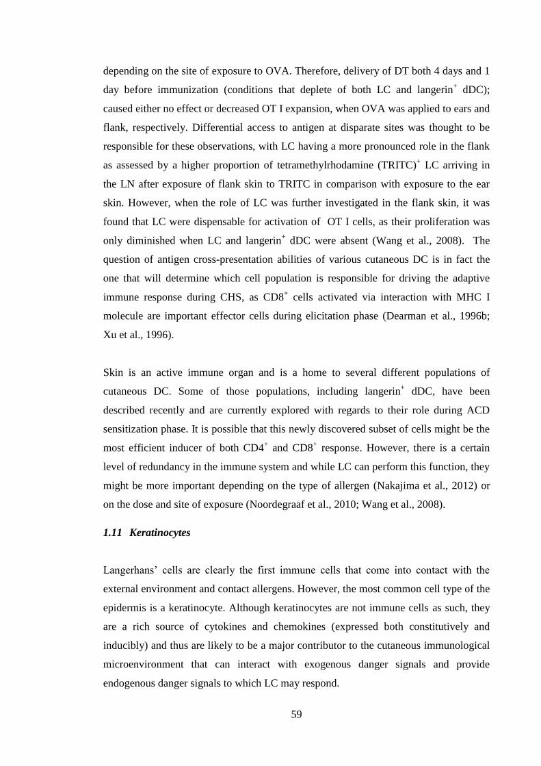
59
depending on the site of exposure to OVA. Therefore, delivery of DT both 4 days and 1
day before immunization (conditions that deplete of both LC and langerin+ dDC);
caused either no effect or decreased OT I expansion, when OVA was applied to ears and
flank, respectively. Differential access to antigen at disparate sites was thought to be
responsible for these observations, with LC having a more pronounced role in the flank
as assessed by a higher proportion of tetramethylrhodamine (TRITC)+ LC arriving in
the LN after exposure of flank skin to TRITC in comparison with exposure to the ear
skin. However, when the role of LC was further investigated in the flank skin, it was
found that LC were dispensable for activation of OT I cells, as their proliferation was
only diminished when LC and langerin+ dDC were absent (Wang et al., 2008). The
question of antigen cross-presentation abilities of various cutaneous DC is in fact the
one that will determine which cell population is responsible for driving the adaptive
immune response during CHS, as CD8+ cells activated via interaction with MHC I
molecule are important effector cells during elicitation phase (Dearman et al., 1996b;
Xu et al., 1996).
Skin is an active immune organ and is a home to several different populations of
cutaneous DC. Some of those populations, including langerin+ dDC, have been
described recently and are currently explored with regards to their role during ACD
sensitization phase. It is possible that this newly discovered subset of cells might be the
most efficient inducer of both CD4+ and CD8
+ response. However, there is a certain
level of redundancy in the immune system and while LC can perform this function, they
might be more important depending on the type of allergen (Nakajima et al., 2012) or
on the dose and site of exposure (Noordegraaf et al., 2010; Wang et al., 2008).
1.11 Keratinocytes
Langerhans’ cells are clearly the first immune cells that come into contact with the
external environment and contact allergens. However, the most common cell type of the
epidermis is a keratinocyte. Although keratinocytes are not immune cells as such, they
are a rich source of cytokines and chemokines (expressed both constitutively and
inducibly) and thus are likely to be a major contributor to the cutaneous immunological
microenvironment that can interact with exogenous danger signals and provide
endogenous danger signals to which LC may respond.
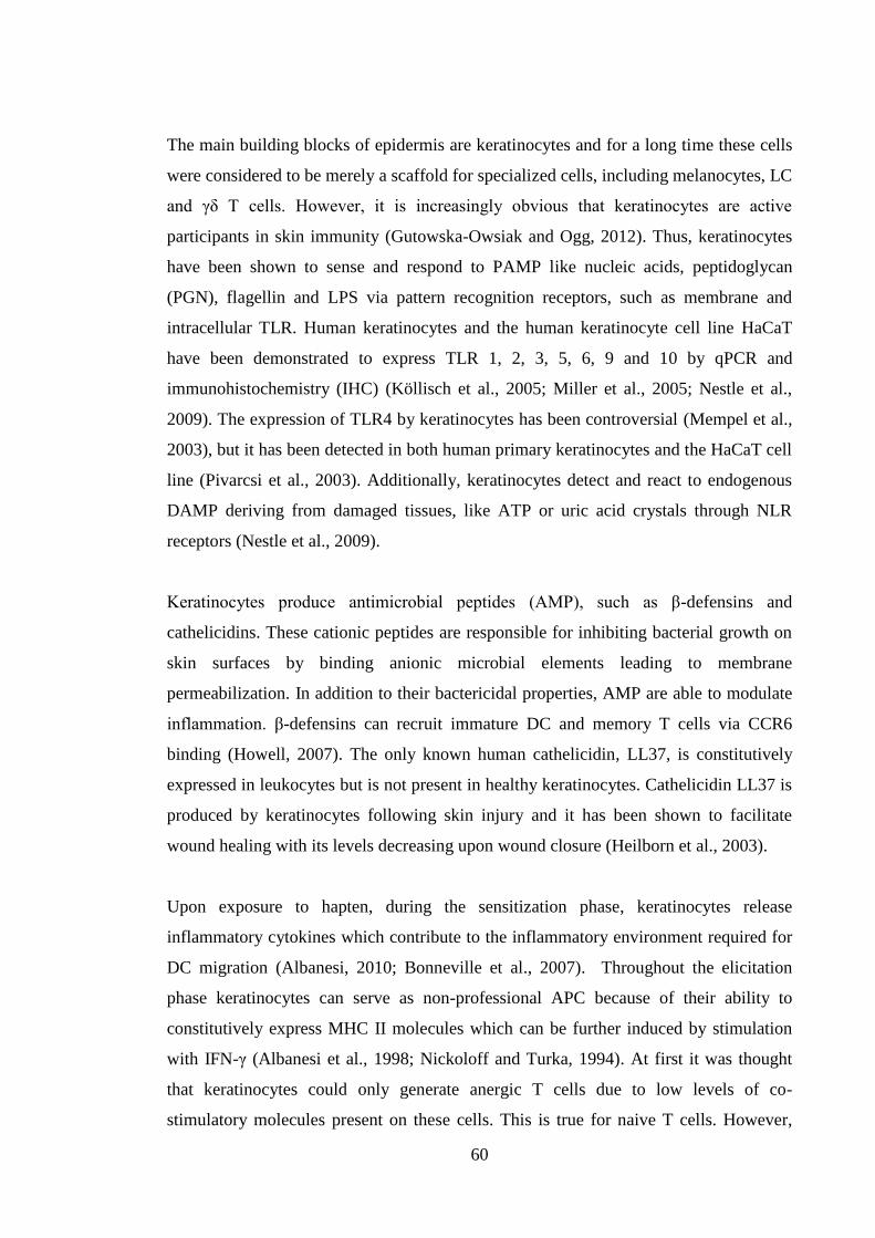
60
The main building blocks of epidermis are keratinocytes and for a long time these cells
were considered to be merely a scaffold for specialized cells, including melanocytes, LC
and γδ T cells. However, it is increasingly obvious that keratinocytes are active
participants in skin immunity (Gutowska-Owsiak and Ogg, 2012). Thus, keratinocytes
have been shown to sense and respond to PAMP like nucleic acids, peptidoglycan
(PGN), flagellin and LPS via pattern recognition receptors, such as membrane and
intracellular TLR. Human keratinocytes and the human keratinocyte cell line HaCaT
have been demonstrated to express TLR 1, 2, 3, 5, 6, 9 and 10 by qPCR and
immunohistochemistry (IHC) (Köllisch et al., 2005; Miller et al., 2005; Nestle et al.,
2009). The expression of TLR4 by keratinocytes has been controversial (Mempel et al.,
2003), but it has been detected in both human primary keratinocytes and the HaCaT cell
line (Pivarcsi et al., 2003). Additionally, keratinocytes detect and react to endogenous
DAMP deriving from damaged tissues, like ATP or uric acid crystals through NLR
receptors (Nestle et al., 2009).
Keratinocytes produce antimicrobial peptides (AMP), such as β-defensins and
cathelicidins. These cationic peptides are responsible for inhibiting bacterial growth on
skin surfaces by binding anionic microbial elements leading to membrane
permeabilization. In addition to their bactericidal properties, AMP are able to modulate
inflammation. β-defensins can recruit immature DC and memory T cells via CCR6
binding (Howell, 2007). The only known human cathelicidin, LL37, is constitutively
expressed in leukocytes but is not present in healthy keratinocytes. Cathelicidin LL37 is
produced by keratinocytes following skin injury and it has been shown to facilitate
wound healing with its levels decreasing upon wound closure (Heilborn et al., 2003).
Upon exposure to hapten, during the sensitization phase, keratinocytes release
inflammatory cytokines which contribute to the inflammatory environment required for
DC migration (Albanesi, 2010; Bonneville et al., 2007). Throughout the elicitation
phase keratinocytes can serve as non-professional APC because of their ability to
constitutively express MHC II molecules which can be further induced by stimulation
with IFN-γ (Albanesi et al., 1998; Nickoloff and Turka, 1994). At first it was thought
that keratinocytes could only generate anergic T cells due to low levels of co-
stimulatory molecules present on these cells. This is true for naive T cells. However,
61
Black et al.,(2007) demonstrated that keratinocytes were capable of eliciting responses
from antigen-experienced CD4+ and CD8
+ T cells.
Keratinocytes are an abundant source of cytokines and chemokines that act in an
autocrine and paracrine manner and recruit T cells and neutrophils. Keratinocytes have
been shown to express IL-1, TNF-α, IL-6, IL-8, IL-7, IL-15, IL-10, IL-12 and IL-18, as
well as following cytokine receptors: IL-1RI and II, TNF-R, IL-6R, IL-18R, IL-10R,
IL-4R, IL-13R, IL-17R, IL-12R (Gröne, 2002).
1.12 Role of innate immune cells in CHS reactions
As described above, ACD is a reaction which is mediated by members of adaptive
immune system, CD4+ and CD8
+ T cells in particular. However, cells of an innate
immune response, such as natural killer T cells (NKT) cells, mast cells, neutrophils and
γδ T cells also contribute to cutaneous immune and allergic responses, and are likely to
be particularly important in the early events of contact sensitization, before the adaptive
immune response has developed. Their roles are considered in subsequent sections.
1.12.1 NKT cells
In addition to priming naive T cells in local LN, another key event during skin
sensitization is the production of hapten-specific IgM antibodies by the B1 B cell subset
(Tsuji et al., 2002). The B1 subset is stimulated by IL-4 derived from CD1d restricted
NKT. Natural killer T cells have been shown to be crucial for initiating ACD reactions.
For example, depletion of NKT cells or cells expressing the CD1d molecule resulted in
inhibition of the CHS reaction (Nieuwenhuis et al., 2005). It was demonstrated that
sensitization with TNCB resulted in rapid (within 2h) expansion of NKT cells in the
liver. No elicitation of CHS was observed in NKT deficient mice or mice reconstituted
with NKT cells from IL-4 KO mice (Campos et al., 2003).
1.12.2 Mast cells
Mast cells reside in connective tissues in the body and in the mucosa. In the skin, they
mainly localize around the perivascular area and in the proximity of nerve endings.
Mast cells are important in mediating the IgE (immediate type) allergic response by
releasing contents of their abundant cytoplasmic granules upon crosslinking of the high
62
affinity receptor for IgE (FcεRI). These granules contain plethora of mediators, namely
histamine, serotonin, proteases, cytokines and chemokines (Debenedictis et al., 2001).
Mast cells also play a key role in initiating a rapid (antibody-independent) response to
pathogens like bacteria and fungi, and recently it was found that they secrete histamine
and serotonin after encountering ROS, independently of FcεRI (Chodaczek et al., 2009),
so they are able to respond to exogenous danger signals (PAMP) with the production of
endogenous danger signals (DAMP).
Mouse and human LC have been shown to express histamine receptor H4, thus are able
to respond to mast cell derived DAMP. Furthermore, LC migration was observed
following exposure to H4 agonist, 4-methylhistamine (Gschwandtner et al., 2010),
suggesting that there is an interplay between LC and mast cells. Indirect involvement of
mast cells in the CHS was observed by Bryce et al. (2004). In IgE deficient mice,
challenge with either oxazolone or DNFB resulted in diminished challenge-induced
oedema in comparison with WT mice. Adoptive transfer experiments demonstrated that
the presence of IgE was required during the sensitization phase. Normal ear swelling
was restored by the delivery of exogenous IgE prior to sensitization; it was not
necessary for the IgE to be specific for the hapten. For instance, in the study by Bryce
and colleagues, IgE specific for trinitrophenol, DNP and OVA (all unrelated allergens)
were all equally effective in restoring CHS in response to oxazolone. Additionally, in
the absence of IgE, no LC migration was observed 18h following exposure to
oxazolone. The ability of LC to mobilize was restored by a passive transfer of IgE.
Further investigations demonstrated that a deficiency of mast cells or of FcɛRI also
resulted in diminished ear swelling following challenge with oxazolone (Bryce et al.,
2004). The requirement for the presence of mast cells during the sensitization phase of
CHS was confirmed in transgenic mice where mast cells were ablated by exposure to
DT (MaS TRECK Tg mice). Migration of DC to the LN following exposure to DNFB
was diminished in MaS TRECK Tg mice compared with WT animals. Furthermore, the
ability of DC from these transgenic mice to induce T cell proliferation was significantly
diminished. In vitro investigations of the interactions between mast cells and DC have
shown that the presence of mast cells and direct cell contact was required for optimal
expression of co-stimulatory molecules CD40, CD80, CD86 and CCR7 receptor by DC.
Finally, two factors were identified to be required for the effects of mast cells on DC:
mast cell-bound TNF-α and ICAM-1 receptor expression by DC (Otsuka et al., 2011).
63
Indeed, there have been many reports implicating mast cells, and their release of
inflammatory cytokines, in particular TNF-α, in the development of CHS reactions. The
release of mast cell-derived inflammatory mediators, TNF-α and MIP-2, was shown to
be mediated via ligation of FcɛRI. It was observed that in mice deficient in FcɛRI β-
chain (FcRβ), the CHS reaction to oxazolone was significantly decreased (Kobayashi et
al., 2010). Through the use of mast cell deficient mice and adoptive transfer of mast
cells from WT animals, the involvement of mast cell-derived TNF-α and MIP-2 in
neutrophil recruitment during the CHS reaction to TNCB was established (Biedermann
et al., 2000). The absence of either of these two cytokines from mast cells resulted in
decreased ear swelling. Additionally, their study confirmed the requirement for the
presence of neutrophils during the elicitation phase of CHS (Biedermann et al., 2000)
(see section 1.12.3). In other studies with an alternative allergen, FITC, challenge
induced CHS reactions were significantly decreased in both mast cell and TNF-α
deficient mice (Suto et al., 2006). When mice lacking mast cells were reconstituted with
bone marrow derived mast cells from WT mice, challenge-induced ear swelling was
restored to levels observed in WT mice. However, if mast cell deficient animals were
injected with mast cells from TNF-α KO mice, ear swelling was higher than that
observed in mast cell KO, but lower than that recorded for WT mice, suggesting that
mast cell-derived TNF-α is important, but is not the only mediator released by mast
cells that affected the course of CHS reaction to FITC (Suto et al., 2006).
Interestingly, an immunosuppressive role for mast cells, and more specifically for mast
cell-derived IL-10, has been proposed. Grimbaldeston et al. (2007) demonstrated that in
mast cell KO mice, ear swelling following challenge with DNFB or urushiol (the active
chemical in the plant poison ivy) was increased in comparison with WT animals. Ear
swelling was diminished when mast cell KO mice were reconstituted with WT mast
cells but not from IL-10 KO animals (Grimbaldeston et al., 2007).
Thus, it would appear that mast cells are an important cell type for the manifestation of
CHS. The majority of reports suggest that their role is mainly to facilitate the induction
phase of contact sensitization via the release of TNF-α and contact with cutaneous DC.
However, there may be circumstances whereby mast cells can exert suppressive
functions.
64
1.12.3 Neutrophils
Neutrophils are polymorphonuclear leukocytes that are key players in the control of
infection and the acute immune response. They are present in the bloodstream and are
the first cell type to be recruited to the site of inflammation. They have the ability to kill
pathogens via several mechanisms, including phagocytosis and degranulation with
subsequent release of molecules such as myeloperoxidase, LF and MMP-9, among
others. Neutrophils are considered to be crucial for resolution of inflammation. Indeed,
it has been shown that neutrophil deficiency in humans leads to severe
immunodeficiency (Kolaczkowska and Kubes, 2013).
Neutrophils have been shown to play a role in the elicitation of CHS, with application
of allergens inducing neutrophil recruiting chemokines, and the loss of neutrophils or
neutrophil derived factors resulting in suboptimal CHS reactions. Thus, the neutrophil
recruiting chemokine, Gro-α (CXCL1), or its mouse equivalent, KC, has been detected
in the skin of both primed with DNFB and non-primed mice as soon as 30 minutes
following exposure to DNFB, which was consistent with the rapid recruitment of
neutrophils. The administration of anti-KC serum or anti-neutrophil antibody to mice
prior to DNFB challenge decreased the ear swelling response (Dilulio et al., 1999). A
diminished CHS response also was observed after depletion of neutrophils, however,
that depletion was rescued by injection of CD8+ cells from sensitized mice (Dilulio et
al., 1999). Leukotriene (LT) B4 is a lipid mediator present at the sites of inflammation
and released by mast cells, neutrophils and macrophages, that acts upon myeloid cells
via the BLT1 receptor. It was established that LTB4 was produced by neutrophils in the
skin following mechanical injury (tape stripping). Additionally, the accumulation of
neutrophils that was observed after tape stripping was dependent on their expression of
both LTB4 and BLT1. Moreover, allergic skin reactions to OVA were diminished in
BLT1 KO mice, but could be restored by transfer of neutrophils from WT mice (Oyoshi
et al., 2012). Overall, this study demonstrated that neutrophils and the signalling
pathway LTB4-BLT1 are essential for full presentation of allergic skin inflammation to
the protein allergen OVA (Oyoshi et al., 2012).
65
1.12.4 Skin resident γδ T cells
Another, innate immune cell population that may play an important role in the induction
of skin sensitization are the γδ T cells, which are found in both the epidermis and the
dermis of mice.
It was long thought that in the epidermis the only cells of hematopoetic origin were LC.
In 1985 a novel population of cells was described in the epidermis (Romani et al.,
1985). These cells were later named DETC. They are characterized by expression of γδ
receptor that distinguishes them from ‘conventional’ αβ T cells (Steiner et al., 1988).
DETC, which almost exclusively express Vγ3/Vδ1 variant of γδ receptor, were
identified as producing IL-2, expressing IL-2R and exhibiting cytotoxic potential
(Kaminski et al., 1993). It was shown that DETC stably interact with neighbouring KC
through γδ TCR. Their dendrites were mostly directed towards stratum granulosum,
with only about 30% of cellular protrusions being motile (Chodaczek et al., 2012). Aryl
hydrocarbon receptor (AhR) is a xenobiotic-regulated transcription factor which upon
ligation affects many of the cells of the immune system. It has been recently
demonstrated that AhR plays a crucial role in the development of DETC. DETC are
seeded to the epidermis during embryogenesis and are not repopulated from the
peripheral precursors and AhR KO mice display a marked reduction in the number of
DETC (Kadow et al., 2011).
DETC are not motile cells, hence they are not found in the LN (Gray et al., 2011),
however, this view was recently challenged by Nielsen at al. (2014) who demonstrated
the presence of DETC in the LN (Nielsen et al., 2014). DETC express TLR and
recognize self antigens from neighbouring keratinocytes, as well as invading pathogens
(Gray et al., 2011). However, despite their identification 30 years ago exact ligands for
γδ receptors of DETC are not well characterized. It was recently discovered that DETC
ligands are expressed in the fetal thymus prior to DETC distribution to the skin.
Furthermore, recombinant γδ receptor was rapidly bound to keratinocytes on the edge of
wounded skin. Despite the identification of the presence of the ligand its specific nature
was not identified (Komori et al., 2012). DETC have been shown to be an important
constituent of the cutaneous immune response. DETC recognize antigens presented by
injured, stressed or transformed keratinocytes (Havran et al., 1991). Their maintenance
66
in the epidermis is sustained by keratinocyte-derived cytokines, IL-7 and IL-15. DETC
in turn release fibroblast growth factor 7, which participates in maintaining keratinocyte
homeostasis, IL-2, IL-3, GM-CSF, IFN-γ and TNF-α, which interact with keratinocytes,
as well as act upon DETC themselves (Jameson and Havran, 2007). Interestingly,
TNF-α transcripts are present in DETC in the steady state, but are only released upon
activation (Macleod and Havran, 2011).
Similarly, γδ T cells have been identified in the human epidermis, in addition to a
population of conventional (αβ) T cells, that are absent from mouse epidermis under
physiological conditions (Bos et al., 1990; Dupuy et al., 1990; Ebert et al., 2006).
Despite the presence of γδ T cells in the human epidermis, these cells were not deemed
to be equivalent to DETC, based on their differential numbers and localization in
comparison to mouse epidermis (Bos et al., 1990). However, more recent studies have
demonstrated the role of both conventional (αβ) and un-conventional (γδ) T cells in the
human epidermis, in the wound healing process via production of insulin-like growth
factor 1 (IGF-1) (Toulon et al., 2009). Correspondingly, DETC also have been
demonstrated to produce IGF-1 during steady state and wound healing suggesting
similarities between both T cells populations from human epidermis and the DETC
subset in mouse epidermis (Sharp et al., 2005).
There are some indications that DETC may play a role in contact allergic responses. As
mentioned previously, AhR KO mice have a reduced frequency of DETC and in these
KO mice, ear swelling in response to challenge with FITC was reduced in comparison
with WT counterparts. This was shown to be due to decreased GM-CSF production in
the epidermis of AhR KO mice, as DETC are the source of this cytokine, and
subsequent impairment in LC maturation (Jux et al., 2009). The role of DETC in the
DNFB-induced CHS was analyzed by Nielsen et al. (2014). They found that ear
swelling following challenge of sensitized mice was lower in γδ and IL-17 KO animals.
Furthermore, they demonstrated that DETC became activated and produced IL-17A
following challenge with DNFB (Nielsen et al., 2014).
Whereas DETC cells were first described in 1985, it was not until 2011 that a mouse
dermal population of γδ T cells was identified and characterized. They were found to be
motile and they were observed to move at the speed of 4μm/minute. Dermal γδ T cells
67
are radioresistant and were established to be the main source of IL-17 (an important
proinflammatory cytokine that targets neutrophils and epithelial cells) in the skin and in
the LN (Gray et al., 2011). In the peripheral LN γδT cells comprise 0.1% of all cells.
They were demonstrated to be Vγ3- as opposed to Vγ3
+ DETC. Dermal γδ T cells had
similar phenotype to those found in peripheral LN: CCR6hi
, IL-7Rαhi
, CXCR6hi
, and
high for CD103. DETC on the other hand were CCR6lo
and IL-7Rαlo
. Dermal γδ, but
not DETC, when stimulated with phorbol-12-myristate-13-acetate (PMA) /ionomycin
produced high levels of IL-17A, IL-17F and IL-22. The combination of IL-1β and IL-23
also stimulated the rapid production of IL-17A by peripheral LN and dermal γδ T cells
(Gray et al., 2011). Almost concurrently with Gray et al. (2011) a dermal population of
γδ T cells in mouse was identified by Cai et al. (2011) and Sumaria et al. (2011). Cai et
al. (2011) have described a population of dermal cells that produced IL-17 following
exposure to IL-23. Analysis at the level of transcription revealed that dermal γδT cells
expressed constitutively IL-23R, as well as the chemokine receptors CCR1, CCR2,
CCR4, CCR5, CCR6, CXCR3, and CXCR4.
As reported by Gray et al. (2011) dermal γδ T cells are motile and migrate to the LN,
however, their homing to the LN was not dependent on CCR7 expression, in contrast to
conventional αβ T cells. Both δγ and αβ T cells were shown to express skin-homing
receptors: CCR4 and CCR10. However, αβ cells exhibited higher levels of CCR4
transcripts, while γδ cells expressed more of CCR10 mRNA. Ligands for CCR4 and
CCR10 are CCL17 and CCL27, respectively. The former chemokine is expressed by the
stressed skin, while CCL27 is constitutively present in the epidermis, which gives a clue
about the roles of these distinct T cell subsets, namely γδ T cells are present in the skin
under physiological conditions, whereas αβ T cells are recruited to the skin during
inflammation (Vrieling et al., 2012).
The role for γδ T cells in facilitating the elicitation phase of CHS has been demonstrated
some years ago. It was demonstrated by Ptak et al. (1992) that γδ T cell population with
phenotype CD3+, CD4
-, and CD8
+ was required for the transfer of sensitization to
oxazolone and TNCB (Ptak and Askenase, 1992). Further studies identified that
successful transfer of sensitization to PPD can only occur when both αβ and γδ T cells
were transferred (Yokozeki et al., 2001), suggesting a crucial role of the γδ T cell
population in sensitization to chemical allergens. It was later established that γδ T cells
present in the dermis were also a source of IL-17 (Gray et al., 2011). Production of IL-
68
17 in the skin following exposure to DNCB has been established by Hayes (2012). He
has used AhR, mice that lack γδ T cells specifically in the epidermis, while their dermal
γδ T cell population is intact, and γδ T cell KO mice that do not have any γδ T cells.
Hayes found that exposure to DNCB resulted in IL-17 production by the skin of AhR
KO but not γδ T cell KO animals pointing to dermal γδ T cell subset as a cellular
source of IL-17 following exposure to contact allergen (PhD thesis Hayes, 2012).
Contact hypersensitivity reactions have been considered for a long time to be a useful
model for investigation of adaptive cutaneous immune responses. However, it has
become increasingly clear that innate immune cells are necessary for the execution of
both sensitization and elicitation phases. In addition to mast cells and neutrophil
populations, a novel γδ T cell subset is emerging as a key participant of ACD with its
ability to produce IL-17 (PhD thesis Hayes, 2012). Indeed, absence of IL-17 producing
γδ T cells have been shown to render mice resistant to imiquimod-induced psoriasis-like
dermatitis (Gray et al., 2013).
1.13 Characteristics of lactoferrin
Not only is contact sensitization an important occupational health issue, but it also
represents a tractable system with which to investigate the mechanisms of cutaneous
immune and inflammatory responses and to explore the activity of potential
immunomodulatory/anti-inflammatory molecules that may lead to novel therapeutic
strategies.
One such molecule in which this laboratory has had an interest is lactoferrin (LF), an
80kDa glycoprotein, and a member of the iron-binding protein family, that includes
transferrin (TRF), OVA and melanotransferrin (González-Chávez et al., 2009).
Lactoferrin is secreted by epithelial cells and is found in serum, milk, tears, saliva,
pancreatic juice, small intestine secretions, bronchial and vaginal mucus and seminal
plasma (Legrand et al., 2005). Lactoferrin is also present in secondary neutrophil
granules (Yang et al., 2009). The concentration of LF depends on the physiological state
of the organism. The serum concentration of LF is around 0.4-2 mg/l in healthy
individuals, but it can reach as high as 200 mg/l during septicaemia. The concentration
of LF in colostrum is up to 7g/l reducing to 1g/l in mature milk (Legrand et al., 2005).
Indeed, the presence of LF has been recently considered to serve as a diagnostic tool for
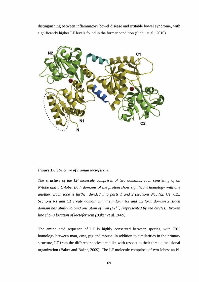
69
distinguishing between inflammatory bowel disease and irritable bowel syndrome, with
significantly higher LF levels found in the former condition (Sidhu et al., 2010).
Figure 1.6 Structure of human lactoferrin.
The structure of the LF molecule comprises of two domains, each consisting of an
N-lobe and a C-lobe. Both domains of the protein show significant homology with one
another. Each lobe is further divided into parts 1 and 2 (sections N1, N2, C1, C2).
Sections N1 and C1 create domain 1 and similarly N2 and C2 form domain 2. Each
domain has ability to bind one atom of iron (Fe3+
) (represented by red circles). Broken
line shows location of lactoferricin (Baker et al. 2009).
The amino acid sequence of LF is highly conserved between species, with 70%
homology between man, cow, pig and mouse. In addition to similarities in the primary
structure, LF from the different species are alike with respect to their three dimensional
organization (Baker and Baker, 2009). The LF molecule comprises of two lobes: an N-
70
lobe (residues 1-333) and a C-lobe (residues 345-692) with a hinge region (residues
334-344) between them, that accounts for the flexibility of LF molecule (Legrand et al.,
2008). Both parts of the protein show significant homology (Figure 1.6). Metal binding
by LF is dependent on simultaneous binding of two HCO3- (or one CO32-
) anions. Iron
sequestration by LF is a reversible process, hence the LF molecule can exist in Fe3+
free
(apo-LF; open) and bound (holo-LF; closed and resistant to proteolysis) state (Anderson
et al., 1989). In the native state LF is 15-20% saturated with iron. When the level of iron
binding is below 5% it is considered to be iron free or apo-LF (Steijns and van
Hooijdonk, 2000). However, iron transport is not considered to be major role of LF, as
there have been many reports demonstrating its importance to the immune system.
Interestingly, however, LF has been shown to have 260 times higher affinity for iron
than of blood serum TRF at physiological pH, and LF has ability to retain iron at much
lower pH than TRF, possibly to endure acidic environment in the stomach of newborns
(Steijns and van Hooijdonk, 2000).
1.14 Microbiocidal effects of lactoferrin
The first function ascribed to LF was its ability to inhibit bacterial growth (Oram and
Reiter, 1968). At first, the bacteriostatic activity of LF was attributed to the iron-
chelating properties of the protein. However, a study from Arnold et al. (1982)
suggested that the action of LF is more complex than simple iron ion depletion. This
study demonstrated that Streptococcus mutans could survive up to 72h in an iron free
environment but incubation with human apo-LF resulted in a loss of viability after 1h
(Arnold et al., 1982). It is recognized now that the mechanism of LF antimicrobial
action lies in its capacity to bind LPS. Lipopolysaccharide is the main component of the
outer membrane of Gram negative bacteria. It consists of antigen O, core polysaccharide
and lipid A, with LF binding the lipid A portion (Appelmelk et al., 1994). The N-
terminus of both bovine and human LF is highly cationic due to the presence of 4
arginines (Arg) (Gly1-Arg
2-Arg
3-Arg
4-Arg
5) which are necessary for an effective
binding of human LF to LPS, heparin and DNA (van Berkel et al., 1997).
As well as having antibacterial properties, LF has been shown to have an antiviral
potential. The ability of bovine LF to reduce viral infection rates has been observed for
a variety of viruses, including human cytomegalovirus, HSV 1 and 2, respiratory
71
syncytial virus and human immunodeficiency virus (HIV) (van der Strate et al., 2001).
In addition to antibacterial and antiviral properties, LF has some antifungal and
antiparasitic features. Human and bovine milk were found to inhibit in vitro growth of
Entamoeba histolytica (E.histolytica), an intracellular parasite that causes amoebiasis.
The antiparasitic effect was achieved due to the action of LF together with lysozyme
and IgA. Since E.histolytica requires iron for its growth, perhaps it is not surprising that
the greatest inhibition of parasitic growth was accomplished with the apo-LF form
(León-Sicairos et al., 2006). Additionally, binding of LF to human blood monocytes
increased both the number of parasites phagocytosed and the percentage of cells
involved in phagocytosis, in comparison with cells cultured without LF (Lima and
Kierszenbaum, 1985).
1.15 Immunomodulatory functions of lactoferrin
As described previously, LF has the ability to interact with a wide variety of
microorganisms, and thereby to inhibit their invasion of the host organism by impeding
their growth or blocking their entry into cells. In addition to the interactions with
pathogens, as described before, LF has immunomodulatory properties. Lactoferrin has
been shown to affect cell migration, cytokine and chemokine production and activation
of DC (Actor et al., 2009). Lactoferrin is present in neutrophil granules, and is a
member of a group of proteins called alarmins. Alarmins are part of the innate immune
system and can recruit innate immune cells into a site of inflammation and subsequently
activate them (Yang et al., 2009). A study by de la Rosa et al. (2008) demonstrated the
selective ability of a commercially produced, recombinant human LF produced in
Aspergillus niger var. awamori (trade name: talactoferrin-α; TLF) to trigger in vitro
chemotaxis of human peripheral monocytes, but not DC or neutrophils. However, the
same authors demonstrated that intraperitoneal administration of TLF in mice induced
accumulation of neutrophils and monocytes/macrophages (de la Rosa et al., 2008).
Bournazou et al. (2009) reported that in vitro human LF, independent of its iron
saturation status, inhibited migration of human neutrophils, isolated from peripheral
blood, towards various chemoattractants (N-Formylmethionyl-leucyl-phenylalanine,
C5a, IL-8), as well as neutrophil activation, as measured by CD11b expression.
Moreover, in vivo human LF decreased the number of accumulating neutrophils in a
mouse thioglycollate peritonitis model (Bournazou et al., 2010; Bournazou et al., 2009).
72
In another study from the same group, it was found that human LF had an ability to
inhibit in vitro migration of eosinophils isolated from human blood towards eotaxin
(Bournazou et al., 2010). Thus, the data available on LF and cell migration of different
cells types are conflicting. Possible reasons for these apparent discrepancies are the
different platforms and organisms that were used, e.g. in vivo in mice versus in vitro
using human cells. Additionally different LF molecules were used (recombinant versus
native LF).
In line with its role as an alarmin LF has been shown to promote APC maturation.
Talactoferrin-α triggered maturation of human monocyte derived DC, as assessed by
up-regulation of co-stimulatory markers (CD80, CD83), MHC II expression and
enhanced ability to trigger lymphocyte proliferation (de la Rosa et al., 2008). In a
separate study, TLF was shown to promote DC maturation as demonstrated by
increased expression of co-stimulatory molecules (CD80, CD83, CD86), HLADR, and
of chemokine receptors (CXCR4, CCR5, CCR6, CCR7). Treatment with TLF also
inhibited the ability of DC to process antigen and increased DC activation of T cells
(Spadaro et al., 2008). Both studies demonstrated an impact of LF on APC maturation.
Importantly, Spadaro et al.(2008) found that the action of TLF was not due to LPS
contamination (Spadaro et al., 2008). Lactoferrin has been reported to influence
chemokine and cytokine expression in both inhibitory and stimulatory ways. A study by
Crouch et al. (1992) showed that human LF inhibited IL-2, IL-1 and TNF-α secretion
from lymphocyte cultures (Crouch et al., 1992). Talactoferrin-α treatment of human
peripheral blood derived mature DC significantly increased the secretion of IL-8,
CXCL10 and decreased IL-6, IL-10 and CCL20 (Spadaro et al., 2008). Haversen et al.
(2002) investigated the action of human LF on the human monocytic cell line, THP-1,
and showed that LF inhibited TNF-α, IL-1β, IL-6 and IL-8 secretion from LPS
stimulated cells. The inhibitory activity of human LF was observed whether LF was
added to the culture 30 minutes before or after LPS introduction (Håversen et al., 2002).
The experimental approach used in this latter paper (Håversen et al., 2002) supported
the assumption that the effects of LF are not solely due to LPS sequestration, however,
with the relatively short exposure time (30 minutes) it could still be argued that at least
some of the observed inhibition was due to LPS binding. A more convincing
demonstration would require removal of free LPS from the system prior to the addition
of LF, or the use of an LPS-independent stimulus to promote THP-1 activation. In
73
contrast to results from Haversen et al. (2002), were findings from Curran et al. (2006),
which demonstrated in a murine macrophage cell line the ability of bovine LF to
stimulate TNF-α and IL-6 production at levels comparable to LPS stimulation.
Furthermore, LF was ineffective at inhibiting cytokine production in response to LPS
irrespective of the time of bovine LF addition to the culture (Curran et al., 2006). Thus,
data concerning LF influence on cytokine production are conflicting, and often
contradictory. The discrepancies between studies by Haversen et al. (2002) and Curran
et al. (2006) can be in part explained by the fact that both groups used LF preparations
of different origin (derived from human or bovine sources, respectively) and the
platforms used for investigation of LF action were also different (a human cell line was
used by Haversen et al. (2002) and a murine cell line together with primary murine cells
were utilized by Curran et al. (2006)). This suggested that despite structural and
functional similarities between human LF and bovine LF molecules their precise
mechanisms of action may be different and may also be cell specific.
1.15.1 The impact of lactoferrin on Langerhans’ cell migration
Langerhans’ cells are a fascinating subset of DC with multiple roles during steady state
and inflammation. As mentioned before, using cytokine neutralizing antibodies, LC
migration in response to allergens has been shown to be dependent on at least two
cytokine signals IL-1β and TNF-α (Cumberbatch et al., 1997b). In the murine epidermis
IL-1β is thought to be produced exclusively by LC (Enk et al., 1993), whereas TNF-α is
likely to be primarily a keratinocyte product (Enk and Katz, 1992a). In addition to
allergen-induced LC migration being mediated via these cytokines, the recombinant
cytokines themselves have been shown stimulate LC mobilization. Thus, intradermal
injection of TNF-α was also demonstrated to induce LC migration and DC
accumulation in the draining LN (Cumberbatch et al., 1997b). The effect of injection of
recombinant IL-1β was similar to that of TNF-α, however, with one important
difference: the kinetics of the response was somewhat slower. Thus, TNF-α induced LC
migration took place within 30 minutes, whereas the effects of IL-1β were not apparent
until 2h after exposure. Higher doses of IL-1β did not alter the kinetics of LC
mobilization. With respect to DC accumulation, TNF-α treatment augmented DC levels
in the draining LN as soon as 2h after its delivery. In contrast, IL-1β-induced DC
74
accumulation was apparent 4h after injection and within 17h both cytokines stimulated
similar levels DC in the LN (Cumberbatch et al., 1997a).
The authors concluded that TNF-α and IL-1β provide pivotal, but independent, signals
for the reduction of LC in the epidermis and the subsequent increase in DC in the
draining LN (Cumberbatch et al., 1997b). Given that oxazolone-induced migration was
dependent on signals from both IL-1β and TNF-α, both anti-IL-1β and anti-TNF-α
antibodies inhibit allergen induced migration. Loss of LC from the epidermis induced
by exogenous IL-1β is inhibited by anti-TNF-α antibody (and vice versa), as both
signals are considered to be required for LC mobilization. Administration of
recombinant exogenous IL-1β alone triggers LC migration because it induces the
production of TNF-α and delivery of the exogenous TNF-α alone is sufficient due to the
release of endogenous IL-1β.
Furthermore, it seems that very similar mechanisms regulate LC migration in humans.
Cumberbatch et al. (1999, 2003) investigated the influence of homologous recombinant
TNF-α and IL-1β on LC migration in human skin and found that intradermal delivery of
either cytokine caused the dose-dependent loss of LC from the epidermis (Cumberbatch
et al., 2003; Cumberbatch et al., 1999d). Rambukkana et al. (1996) demonstrated that in
short-term in vitro human skin explant cultures allergen treatment (DNFB, NiSO4 and
DNCB) reduced LC numbers and levels of IL-1β expression increased. Addition of anti-
IL-1β antibody to the culture resulted in diminished levels of LC migration, suggesting
that in humans, similar to mice, the initiation of allergen-induced LC migration depends
on a signal from IL-1β (Rambukkana et al., 1996). The studies described clearly
demonstrate that a regulation of LC migration is governed by the same mechanisms in
human and in mice, and that they depend on a local availability of TNF-α.
Therefore, this system was chosen by Cumberbatch et al. (2000) to examine the
influence of LF on allergen and cytokine induced LC migration and DC accumulation in
mice. Those investigations revealed, that intradermal delivery of mouse LF was able to
inhibit LC mobilization and LN DC accumulation, in response to exposure to
oxazolone, and to IL-1β, but not TNF-α injection (Cumberbatch et al., 2000b).
Moreover, it was established that that topically applied human native LF also was able
to inhibit LC migration in mice upon oxazolone stimulation and intradermal injection of
75
homologous IL-1β, but not TNF-α (Almond et al., 2013). Therefore, data suggested that
LF exerted its inhibitory influence on LC migration and DC accumulation in the LN by
blocking de novo production of TNF- α (Almond et al., 2013; Cumberbatch et al., 2003;
Cumberbatch et al., 2000b).
The studies in mice were followed up with experiments in healthy human volunteers.
Griffiths et al. (2001) tested the impact of LF on allergen, DPC, and TNF-α induced LC
mobilization. Results were similar to those obtained in mice. Topically applied
homologous, recombinant LF inhibited LC migration in response to DPC, but had no
effect on TNF-α induced LC mobilization. In addition to blocking the decrease in LC
numbers in the epidermis, LF reduced the clinical and histological signs of
inflammation in DPC treated sites (Griffiths et al., 2001). Furthermore, it was shown,
that topically applied LF blocked IL-1β triggered LC migration (which, like chemical
allergen-induced LC migration, is dependent on de novo production of TNF-α) and also
inhibited IL-1β-triggered TNF-α production in the skin. These results confirmed the
mechanisms of LC migration in man and mouse, and reinforced the view that LF acts
via inhibition of TNF-α production (Cumberbatch et al., 2003).
1.16 Lactoferrin receptors
An important step in elucidating either the immunostimulatory or suppressive
mechanisms of LF activity is to determine to what receptor it binds. Due to its cationic
nature, LF binds to many anionic molecules, and that has impeded the identification of
specific receptors. Many different receptors on various cell types have been identified to
date.
The first LF receptor (LFR) to be identified was isolated from human intestine
(Kawakami and Lönnerdal, 1991) and it was expressed in a variety of the adult and
foetal tissues with high levels found in adult heart and foetal small intestine (Suzuki et
al., 2001). This LFR was established to be species specific and had a molecular weight
of approximately 114kDa (Kawakami and Lönnerdal, 1991) and subsequently has been
shown to be a trimer or tetramer of 36kDa subunits which binds LF in Ca2+
dependent,
saturable fashion. Receptors for LF were also discovered in the liver. Two types of LFR
also have been found on rat hepatocytes: low affinity LFR that was Ca2+
independent,
76
and a high affinity Ca2+
dependent receptor (Bennatt and McAbee, 1997). The uptake of
bovine LF by rat hepatocytes is independent of iron status and is mediated by the high
affinity receptor via clathrin-dependent endocytosis. Subsequently, LFR is recycled
onto the cell surface (McAbee et al., 1993). Despite similarities (presence of low and
high affinity receptors; reliance on Ca2+
) between LFR in the intestine and liver, there is
no evidence so far that these receptors are equivalent. As LF is known to have
immunomodulatory effects in vivo (Troost et al., 2003) and in vitro (Guillén et al.,
2002), considerable efforts have been made to identify LFR on the cells of the immune
system. Human LF was found to bind to human peripheral blood adherent mononuclear
cells, and B cell, but not T-cell, enriched lymphocytes in Ca2+
dependent fashion
(Bennett and Davis, 1981). The THP-1 human monocytic cell line (Tsuchiya et al.,
1980), that upon PMA stimulation differentiates into macrophages, is used frequently as
a model for macrophage responses and has been utilized in a study in which human LF
was found to bind to THP-1 macrophages via two receptors: one high affinity and one
low affinity. The study showed, that iron status of human LF did not influence its
binding by THP-1 macrophages and that protein, as well as oligosaccharide, structures
of human LF participated in its recognition by LFR (Eda et al., 1996). After further
investigation, LFR on THP-1 macrophages was identified as nucleolin (Hirano et al.,
2005). Nucleolin is a 105kDa protein involved in interactions with RNA and proteins in
the nucleus, nuclear transport as well as viral infection of cells, i.e. with HIV (Ginisty et
al., 1999). In 2004 Legrand et al. (2004) published an extensive study that revealed
nucleolin as the LFR for native human LF on the human cancerous mammary gland cell
line, MDA-MB-231. They demonstrated that nucleolin binds human LF and undergoes
endocytosis via a clathrin dependent mechanism. When investigating binding sites for
native human LF Legrand et al. (2004) used mutant Chinese hamster ovary (CHO) cell
lines that lack expression of surface proteoglycans, to establish their role in human LF
binding. The wild type CHO cell line had two human LF binding sites: the higher
affinity receptor (nucleolin), and lower affinity binding site, corresponding to the
presence of proteoglycans. Utilizing the cell line CHO618 that lacks proteoglycan
expression, it was shown that both LF lobes, namely the C- and N-lobe, participated in
human LF and bovine LF binding to nucleolin, whereas, basic LF regions did not
contribute to nucleolin binding. In the CHO cell line nucleolin, as well as
proteoglycans, was shown to be necessary for human LF endocytosis (Legrand et al.,
2004b).
77
Identification of LFR has been a difficult process due to the nature of LF and its non
specific binding to proteoglycans. Despite these obstacles, researchers have been
successful in characterizing various LFR that are cell specific. The presence of both low
and high affinity binding sites for LF on different cell types and their dependency of the
high affinity sites on Ca2+
suggest similarities between the various LFR that have been
identified. However, these receptors are not identical as they have different molecular
weights.
In addition to proteins described primarily as LFR, it was demonstrated that TLR4
might be required for some of LF functions (Ando et al., 2010). Indeed, it has been
demonstrated recently that TLF triggers mouse and human DC maturation via
interaction with TLR 2 and 4. Requirement for TLR2 and 4 in TLF-induced process of
DC maturation was confirmed by utilization of small interfering (si) RNA and KO
mice. First, CD14+ cells isolated from human PBMC were incubated with either siRNA
against TLR2 and 4 or scrambled controls and further cultured with 1000μg/ml TLF.
Cells in which TLR2 and 4 were absent (siRNA group) exhibited decreased expression
of DC maturation markers (CD40, CD80 and CD83), whereas these were present in the
control group (scrambled; the same nucleotide content as siRNA group but in a random
order). Similarly, a crucial role for these two TLR was demonstrated in mice. When
stimulated with TLF bone marrow cells from double TLR2 and 4 KO mice expressed
lower levels of maturation markers compared with WT counterparts (Spadaro et al.,
2014).
1.17 Characterization of molecular mechanisms of LF activity
Efforts have been made to characterize changes caused by LF at a molecular level.
Haversen et al. (2002) showed that LF decreased TNF-α, IL-1β, IL-6 and IL-8 mRNA
levels. Electrophoretic mobility shift assays demonstrated diminished NF-κB binding to
the TNF-α promoter region in the presence of human LF (Håversen et al., 2002). The
action of bovine LF is TLR4 independent with respect to IL-6 production, as peritoneal
macrophages from wild type and TLR4 KO mice secreted IL-6 in response to bovine
LF (Curran et al., 2006). A recent study by Ando et al. (2010) showed that native, LPS
depleted human LF stimulated NF-κB binding to DNA in THP-1 cells via a canonical
pathway that involves phosphorylation of IκB kinase (IKK). In addition, they found that
78
TLR4 is indispensible for LF-mediated NF-κB activation, a result that contradicts the
previously mentioned study by Curran et al. (2006), that found macrophages deficient in
TLR4 exhibited enhanced IκB degradation (IκB degradation by IKK is necessary for
NF-κB activation and subsequent translocation into the nucleus). Collectively, these
results again demonstrate that the immunomodulatory effects described for LF can be
both stimulatory, as well as inhibitory, depending on the experimental system used. In
order to confirm the dual properties of LF Ando et al. (2010) digested human LF with
actinase E. That digestion resulted in a molecule with similar oligosaccharide content to
the naive molecule which retained the ability to activate NF-κB, whereas inhibition of
LPS dependent NF-κB activation was compromised. Conversely, when human LF was
digested with endo-β-galactosidase, the LPS inhibitory action of human LF was
preserved and NF-κB activation was abolished. Digestion of LF with endo-β-
galactosidase changed the sugar content of LF but conserved the polypeptide chain of
the parent molecule. Interestingly, bovine LF did not activate NF-κB, although it
strongly inhibited LPS triggered NF-κB activation (Ando et al., 2010) and bovine LF
and human LF express different glycoprofiles (Spik et al., 1988). Ando et al. (2010)
proposed, therefore, that the immunostimulatory function is attributable to carbohydrate
part of the human LF molecule, whereas the immunosuppressive feature is due to the
polypeptide moiety (Ando et al., 2010).
Another approach to elucidating the mechanism of action of LF involved the use of
specific inhibitors to block multiple signalling pathways and observing if they change
the effect of LF on cells. Spadaro et al. (2008) examined effects of multiple signalling
pathways inhibitors on human monocyte derived DC activation enhanced by TLF. They
found that SB203580, a selective p38 inhibitor, had strongest inhibitory action against
TLF induced CD83, CXCR4 and CC7 expression. SB203580 and LY294002, a PI3K
inhibitor, suppressed CXCL10 secretion from monocyte derived DC to a similar level of
80%. These results suggest that LF may signal via p38 (Spadaro et al., 2008). In further
experiments pertussis toxin was used to inhibit TLF induced migration of human
peripheral blood monocytes. Pertussis toxin blocks the assembly of Giα subunit with
membrane bound G-coupled receptors. These results proposed that TLF potentially
utilizes G-coupled receptor that still remains to be elucidated (de la Rosa et al., 2008).
Finally the impact of LF on activation of the NF-κB pathway was investigated. This
revealed that LF is responsible for inducting p65 subunit phosphorylation via interaction
79
with TNFR associated factor (TRAF) and is dependent on IKKα and IKKβ (Oh et al.,
2007).
Reports addressing the immunomodulatory function of LF are often conflicting and
suggest that dual role for LF exists in the immune system as both an immunostimulatory
and an immunosuppressive agent. Ando et al. (2010) partially addressed this issue by
demonstrating that differences in sugar content between human and bovine LF result in
these two molecules having opposing properties in the process of NF-κB activation
(Ando et al., 2010). However, the experiments performed by Ando et al. (2010), where
they demonstrate immunosuppressive mechanism of LF, utilized LPS, which raises the
question of whether LF is truly inhibiting NF-κB activation or merely reducing the
availability of the stimulus (LPS). The results to date of attempts to elucidate the
mechanism through which LF exerts its actions have created a complex view of LF.
Indeed, as LF has many functions, it is anticipated that they can be mediated via
different pathways.
In this laboratory the immunomodulatory effect of LF was thought to be mediated via
inhibition of de novo TNF-α production, as concluded from the studies described
previously. However, as highlighted in this section, there have been many LFR
identified which can lead to multiple effects of LF depending on the target cell.
Therefore, the exact mechanism of inhibition of oxazolone-induced LC migration by LF
is still uncertain.
1.18 Lactoferrin derived peptides
It is of interest to identify which part of the LF molecule is responsible for its
immunomodulatory effects on LC. In 1992 Bellamy et al. demonstrated the antibacterial
and bactericidal properties of human and bovine lactoferricin (LFcinH and LFcinB,
respectively), pepsin-derived peptides of LF (Bellamy et al., 1992). Human and bovine
lactoferricins have been extensively studied in respect to their functional similarity to
LF and have been found to possess many of the properties of intact LF molecule
(Gifford et al., 2005). LFcinB was demonstrated to inhibit angiogenesis dependent on
basic fibroblast and vascular endothelial growths factors (bFGF and VEGF,
respectively) via inhibition of bFGF and VEGF binding to heparin on the surface of
80
endothelial cells, suggesting a role for LFcinB as an anti-cancer treatment (Mader et al.,
2006).
Another LF-derived peptide is lactoferrampin (LFampin). Similarly to LFcinB it is
derived from the N1 domain of bovine LF, which consists residues 268-284 and
exhibits potent anti-candida and anti-bacterial properties similar to parent molecule (van
der Kraan et al., 2004). Another described LF derivative is a peptide, called
’LFchimera’, that constitutes of LFcinB 17-30 (N-FKCRRWQWRMKKLG-C), a
peptide derived from LFcinB, and LFampin 265-284 (N-
DLIWKLLSKAQEKFGKNKSR-C) coupled together by a lysine amino acid. All three
peptides showed the ability to inhibit the growth of strains of multidrug resistant micro-
organisms, obtained from the clinic: Escherichia coli (E. coli), Staphylococcus aureus
(S. aureus) (Flores-Villaseñor et al., 2010) and Pseudomonas aeruginosa (Xu et al.,
2010). All three bacterial species have a great relevance for long-term care as they
target most vulnerable subjects (burn victims and bed-bound patients) and are extremely
hard to eradicate.
Lactoferrin derived peptides have been extensively studied with respect to their
antimicrobial roles. However, much less is known about their abilities to modulate the
immune and inflammatory responses. Recent reports indicate that LFcinB acts as an
anticancer agent due to its interaction with cancer cells. LactoferricinB has been shown
to promote apoptosis in CCRF-CEM T-leukemia and Jurkat T-leukemia cell lines
(Furlong et al., 2008). Examples of LF derived peptides described before show that they
can have antimicrobial potential that is superior to that of the parent molecule.
Therefore, it is of interest to identify the domain of LF that is responsible for
immunomodulatory action of LF, as its effects may prove to be more pronounced than
that of native LF. The peptide derived from the N lobe of human LF
(GRRRRSVQWCA), was demonstrated to specifically inhibit the enzyme
myeloperoxidase in human PBMC and subsequently decrease level of ROS production
following cells exposure to LPS (van der Does et al., 2012). Its ability to inhibit
oxazolone induced LC mobilization was investigated in the studies described herein.
81
1.19 Hypothesis, aims and experimental approach
1.19.1 Hypothesis
Lactoferrin interacts with an unknown cutaneous cell or cell types and inhibits
oxazolone and IL-1β induced LC migration via inhibition of cytokine signals.
1.19.2 Aims
This thesis sought to determine the mechanism of action of LF in modification of LC
migration and inflammation.
1.19.3 Experimental approach
In order to examine the previously observed inhibitory effects of human LF on
oxazolone and IL-1β induced migration in the mouse, it was essential to characterize
cutaneous cytokine production at the level of message and protein following exposure
to oxazolone in the presence and in the absence of LF treatment. Further clues to the
effects of LF were gained by examining the effect of LF on oxazolone triggered changes
in the LN, including proliferation and cytokine production. It has been mentioned in the
previous sections that allergen triggered LC migration is dependent on signals from
TNF-α and IL-1β (Cumberbatch et al., 1997b). Additionally, IL-1α, but not IL-1β, has
been shown to be important in irritant-induced LC migration (Cumberbatch et al.,
2002a). Therefore, these three cytokines were considered important in the process of LC
migration. Consequently, cutaneous expression of selected genes was examined
following intradermal delivery of these cytokines in the presence and in the absence of
LF.
There are thousands of chemicals recognized for their ability to cause contact
hypersensitivity (De Groot, 2008). For the purposes of investigating the mechanisms of
ACD a few reference contact allergens are generally utilized in laboratory animals. The
immunomodulatory effects of LF in the skin of mouse and human were examined
previously using oxazolone and DPC, respectively. To broaden the knowledge about
mechanism of action of LF another contact allergen, DNCB, was employed. Additional
82
work compared LC mobilization, cutaneous gene expression and cytokine production
profiles induced by various contact allergens, respiratory allergen and an irritant. These
experiments were designed to identify similarities and differences between chemicals
and investigate their mechanism of action with particular emphasis on oxazolone and
DNCB.
In addition to in vivo examination of effects of LF, two in vitro systems were used. The
first approach aimed to investigate the mechanism of inhibition of TNF-α production by
LF. The focus on TNF-α was derived from in vivo experiments in mice, which
suggested that inhibitory effect of LF on oxazolone-induced LC mobilization was likely
to be mediated via inhibition of de novo TNF-α production (Almond et al., 2013;
Cumberbatch et al., 2003; Cumberbatch et al., 2000b). Additionally, work in humans
confirmed that LF decreased TNF-α level in suction fluid following delivery of IL-1β
(Cumberbatch et al., 2003). Therefore, a THP-1 cell line was utilized, which has been
shown to express LFR upon treatment with PMA (Eda et al., 1996) and to produce
TNF-α in a LPS-independent manner. The necessity for an LPS-independent stimulus
was required due to the fact that LF was known to potently bind LPS (Appelmelk et al.,
1994), therefore, it could decrease TNF-α secretion by THP-1 cells by simply
sequestering the stimulus, which would impede the examination of mechanism of
immunomodulation.
Another in vitro platform utilized keratinocyte cell lines (PAM 212 and HaCaT, mouse
and human cell lines, respectively) and primary mouse keratinocytes. It was suspected
that the production of TNF-α during LC mobilization was mediated by keratinocytes,
the most abundant cell type in the epidermis. Therefore, keratinocytes were considered
legitimate candidates for investigation of the immunomodulatory effects of LF. As well
as examining LF, other peptides with immunomodulatory potential in vivo, namely
thioredoxin and Pep-1, were considered.
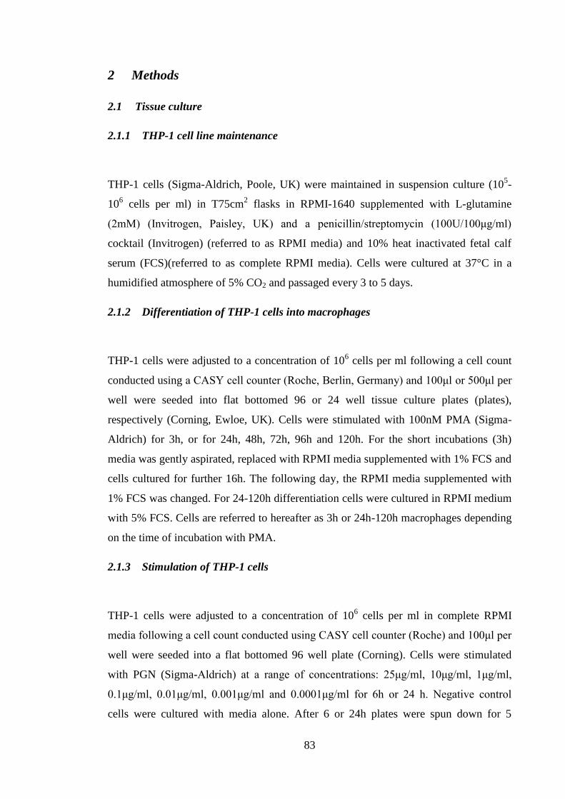
83
2 Methods
2.1 Tissue culture
2.1.1 THP-1 cell line maintenance
THP-1 cells (Sigma-Aldrich, Poole, UK) were maintained in suspension culture (105-
106 cells per ml) in T75cm
2 flasks in RPMI-1640 supplemented with L-glutamine
(2mM) (Invitrogen, Paisley, UK) and a penicillin/streptomycin (100U/100μg/ml)
cocktail (Invitrogen) (referred to as RPMI media) and 10% heat inactivated fetal calf
serum (FCS)(referred to as complete RPMI media). Cells were cultured at 37°C in a
humidified atmosphere of 5% CO2 and passaged every 3 to 5 days.
2.1.2 Differentiation of THP-1 cells into macrophages
THP-1 cells were adjusted to a concentration of 106 cells per ml following a cell count
conducted using a CASY cell counter (Roche, Berlin, Germany) and 100μl or 500μl per
well were seeded into flat bottomed 96 or 24 well tissue culture plates (plates),
respectively (Corning, Ewloe, UK). Cells were stimulated with 100nM PMA (Sigma-
Aldrich) for 3h, or for 24h, 48h, 72h, 96h and 120h. For the short incubations (3h)
media was gently aspirated, replaced with RPMI media supplemented with 1% FCS and
cells cultured for further 16h. The following day, the RPMI media supplemented with
1% FCS was changed. For 24-120h differentiation cells were cultured in RPMI medium
with 5% FCS. Cells are referred to hereafter as 3h or 24h-120h macrophages depending
on the time of incubation with PMA.
2.1.3 Stimulation of THP-1 cells
THP-1 cells were adjusted to a concentration of 106 cells per ml in complete RPMI
media following a cell count conducted using CASY cell counter (Roche) and 100μl per
well were seeded into a flat bottomed 96 well plate (Corning). Cells were stimulated
with PGN (Sigma-Aldrich) at a range of concentrations: 25μg/ml, 10μg/ml, 1μg/ml,
0.1μg/ml, 0.01μg/ml, 0.001μg/ml and 0.0001μg/ml for 6h or 24 h. Negative control
cells were cultured with media alone. After 6 or 24h plates were spun down for 5
84
minutes at 1200 revolutions per minute (rpm) at 4°C. Culture supernatants were
retained for analysis by enzyme-linked immunosorbent assay (ELISA). The cells were
lysed in lysis buffer (20mM Tris-HCL (Sigma-Aldrich), 137nM NaCl (Fisher Scientific,
Loughborough, UK) 20mM ethylenediaminetetraacetic acid (EDTA) (Sigma-Aldrich),
10% glycerol (Fisher Scientific), 0.5% Igepal (Sigma-Aldrich) and incubated on ice for
20 minutes. After centrifugation (5 minutes, 4°C), cell lysates were stored at -80°C until
analysis by ELISA.
2.1.4 Stimulation of THP-1 macrophages
Cells in TC plates were differentiated into THP-1 macrophages as described in the
section 2.1.2. Prior to use in the experiment, adherent cells in the plate were washed
with PBS, and subsequently cultured with a range of doses (0.001-25μg/ml) of PGN
alone. In some experiments cells were pre-treated for 1h with human native LF (Sigma-
Aldrich) at a range of concentrations: 100μg/ml, 50μg/ml, 20μg/ml, 10μg/ml, 2μg/ml,
0.2μg/ml, 0.02μg/ml, and 0.01μg/ml or with medium alone (0μg/ml LF). After 1h PGN
(25μg/ml) for 3h macrophages or 10μg/ml for stimulation of 24h-120h PMA activated
THP-1 macrophages, or media alone was added, and the cells were incubated for a
further 6h. After the incubation, cell lysates and supernatants were collected for ELISA
as described in the section 2.1.3.
Additional controls for cytokine inhibition were incorporated into some experiments:
dexamethasone (Sigma-Aldrich, formulated at 100nM in 0.005% dimethyl sulfoxide
[DMSO]), which was added to the culture at the same time as LF. As dexamethasone
was reconstituted in DMSO, the vehicle control of 0.005% DMSO was included.
2.1.5 HaCaT and PAM 212 cell line maintenance
HaCaT and PAM 212 cells were obtained from Professor Christopher E.M. Griffiths
(School of Translational Medicine, University of Manchester, Manchester, UK) and
Professor Alexander J. MacRobert (Institute of Biomedical Engineering, University
College London, London, UK), respectively. Cells were maintained as adherent cultures
in T75cm2 flasks in complete RPMI media. Every 3 to 5 days cells were passaged as
follows. Media was removed from the culture; cells were washed with RPMI media,
subsequently washed with 0.02% EDTA solution (Sigma-Aldrich) and treated with
85
10mg/ml trypsin (Invitrogen) for 20 minutes at 37°C before the addition of complete
RPMI media. Cells were enumerated by light microscopy following trypan blue staining
and reconstituted in complete RPMI media at 106 cells per ml.
2.1.5.1 HaCaT cell culture
Prior to use in the experiments cells were seeded at a concentration 105cells per well
into 12 well plates and cultured for 48h. Subsequently, cells were washed with RPMI
media and incubated with a range of PGN doses (0.01-25μg/ml) for 24h or with
10μg/ml for 4h. In addition to incubation with a TLR2 ligand, HaCaT cells were
cultured with cytokines. Firstly, HaCaT cells were incubated with a range of human
recombinant IL-1β doses (Insight Biotechnology, Wembley, UK) (0.01-100ng/ml) for
24h or 2h or with 2ng/ml of IL-1β for 2, 4, 24 or 48h. Cells were cultured also with
10ng/ml of human recombinant TNF-α (R&D Systems Europe Ltd., Abingdon, UK) for
1, 2, 4, 6, 10 or 24h. In some experiments, prior to treatment with cytokines, cells were
pre-treated for 2h with either a range (1-100μg/ml) of LF or BSA. Additional treatments
included 1μM of dexamethasone in 0.05% DMSO, 200μg/ml of Pep-1 peptide
(Pepceuticals, Enderby, UK; sequence: GAHWQFNALTVR) or 50μg/ml of thioredoxin
peptide (Pepcueticals, sequence: CGPCKMIKP) or a scrambled peptide (Pepcueticals,
sequence: IPCMPKCKG) with the same amino acid composition as thioredoxin peptide
but in a random order.
2.1.5.2 PAM 212 cell culture
Prior to use in experiments, cells were seeded at a concentration 105cells per well into
12 well plates and cultured for 48h. Subsequently, cells were washed with RPMI media
and incubated with a range of TLR ligands: PGN 25μg/ml, PolyI:C 25μg/ml, LPS
10μg/ml, Flagellin 83ng/ml, R848 500ng/ml, CpG 10μg/ml as well as PMA 1μg/ml for
24, 48 or 72h. Additionally PAM cells were cultured with homologous recombinant
cytokines, including a range (0.01-10ng/ml) of concentrations of IL-1β (R&D Systems)
for 24h or 2h or with 2ng/ml of IL-1β for 0.5, 2, 4 or 6h and a range (0.01-100ng/ml) of
concentrations of IL-1α (R&D Systems) for 2h. In some experiments, prior to treatment,
cells were pre-treated for 1 or 2h with either a range (1-100μg/ml) of LF or 100μg/ml of
BSA.
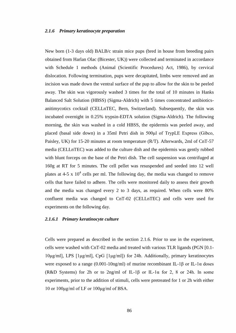
86
2.1.6 Primary keratinocyte preparation
New born (1-3 days old) BALB/c strain mice pups (bred in house from breeding pairs
obtained from Harlan Olac (Bicester, UK)) were collected and terminated in accordance
with Schedule 1 methods (Animal (Scientific Procedures) Act, 1986), by cervical
dislocation. Following termination, pups were decapitated, limbs were removed and an
incision was made down the ventral surface of the pup to allow for the skin to be peeled
away. The skin was vigorously washed 3 times for the total of 10 minutes in Hanks
Balanced Salt Solution (HBSS) (Sigma-Aldrich) with 5 times concentrated antibiotics-
anitimycotics cocktail (CELLnTEC, Bern, Switzerland). Subsequently, the skin was
incubated overnight in 0.25% trypsin-EDTA solution (Sigma-Aldrich). The following
morning, the skin was washed in a cold HBSS, the epidermis was peeled away, and
placed (basal side down) in a 35ml Petri dish in 500μl of TrypLE Express (Gibco,
Paisley, UK) for 15-20 minutes at room temperature (R/T). Afterwards, 2ml of CnT-57
media (CELLnTEC) was added to the culture dish and the epidermis was gently rubbed
with blunt forceps on the base of the Petri dish. The cell suspension was centrifuged at
160g at RT for 5 minutes. The cell pellet was resuspended and seeded into 12 well
plates at 4-5 x 10⁴ cells per ml. The following day, the media was changed to remove
cells that have failed to adhere. The cells were monitored daily to assess their growth
and the media was changed every 2 to 3 days, as required. When cells were 80%
confluent media was changed to CnT-02 (CELLnTEC) and cells were used for
experiments on the following day.
2.1.6.1 Primary keratinocyte culture
Cells were prepared as described in the section 2.1.6. Prior to use in the experiment,
cells were washed with CnT-02 media and treated with various TLR ligands (PGN [0.1-
10μg/ml], LPS [1μg/ml], CpG [1μg/ml]) for 24h. Additionally, primary keratinocytes
were exposed to a range (0.001-10ng/ml) of murine recombinant IL-1β or IL-1α doses
(R&D Systems) for 2h or to 2ng/ml of IL-1β or IL-1α for 2, 8 or 24h. In some
experiments, prior to the addition of stimuli, cells were pretreated for 1 or 2h with either
10 or 100μg/ml of LF or 100μg/ml of BSA.
87
2.2 Immunohistochemistry
Cells (HaCaT, PAM212, primary keratinocytes) were grown as described previously in
a 24 well plate (primary keratinocytes) or in a 24 well plate with a glass coverslip in
each well (HaCaT, PAM212). After 48h, cells were washed with a phosphate buffered
saline (PBS) and fixed with ice cold methanol (Fisher Scientific) for 10 minutes.
Subsequently, cells were washed with PBS and incubated with 0.2μg/ml rabbit anti-
keratin 14 (AF64) antibody (Covance, Leeds, UK) in 0.1% BSA in PBS at 37°C for 30
minutes. Afterwards, cells were washed again for 15 minutes with 0.05% Tween in PBS
and incubated overnight at 4°C with FITC-labelled goat anti-rabbit polyclonal antibody
at 10μg/ml. Then cells were washed and mounted onto a slide with VECASHIELD
mounting medium with 4',6-diamidino-2-phenylindole (DAPI) (Vector Laboratories,
Peterborough, UK).
2.3 Mycoplasma testing
Cell lines were routinely tested for the presence of mycoplasma infection using EZ-PCR
Mycoplasma Test Kit (Biological Industries, Kibbutz beit Haemek, Israel) according to
the supplied protocol. Briefly, cells were spun down for 5 minutes at 1000rpm at 4°C.
Afterwards, 2ml of supernatant was collected and spun for 10 minutes at 15.000rpm at
20°C, supernatant was gently aspirated and the remaining pellet was reconstituted in
20μl of buffer solution (supplied with the kit). After reconstitution, the sample was
heated for 3 minutes at 95°C. The sample was amplified by PCR. The reaction mixture
consisted of 15μl H2O, 3μl Reaction Mix (supplied with the kit) and 2μl sample (for
positive control: 1μl of template + 1μl of H2O). PCR run parameters are presented in the
Table 2.1.
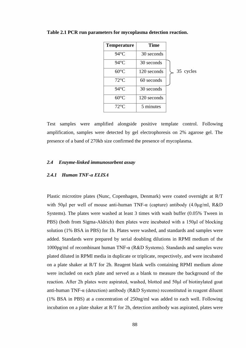
88
Table 2.1 PCR run parameters for mycoplasma detection reaction.
Temperature Time
94°C 30 seconds
94°C 30 seconds
60°C 120 seconds
72°C 60 seconds
94°C 30 seconds
60°C 120 seconds
72°C 5 minutes
Test samples were amplified alongside positive template control. Following
amplification, samples were detected by gel electrophoresis on 2% agarose gel. The
presence of a band of 270kb size confirmed the presence of mycoplasma.
2.4 Enzyme-linked immunosorbent assay
2.4.1 Human TNF-α ELISA
Plastic microtitre plates (Nunc, Copenhagen, Denmark) were coated overnight at R/T
with 50μl per well of mouse anti-human TNF-α (capture) antibody (4.0μg/ml, R&D
Systems). The plates were washed at least 3 times with wash buffer (0.05% Tween in
PBS) (both from Sigma-Aldrich) then plates were incubated with a 150μl of blocking
solution (1% BSA in PBS) for 1h. Plates were washed, and standards and samples were
added. Standards were prepared by serial doubling dilutions in RPMI medium of the
1000pg/ml of recombinant human TNF-α (R&D Systems). Standards and samples were
plated diluted in RPMI media in duplicate or triplicate, respectively, and were incubated
on a plate shaker at R/T for 2h. Reagent blank wells containing RPMI medium alone
were included on each plate and served as a blank to measure the background of the
reaction. After 2h plates were aspirated, washed, blotted and 50μl of biotinylated goat
anti-human TNF-α (detection) antibody (R&D Systems) reconstituted in reagent diluent
(1% BSA in PBS) at a concentration of 250ng/ml was added to each well. Following
incubation on a plate shaker at R/T for 2h, detection antibody was aspirated, plates were
35 cycles
89
washed and blotted and 50μl of streptavidin conjugated to horseradish-peroxidase
(Strep-HRP) (R&D Systems) was added per well. After a 30 minutes incubation with
the enzyme, the liquid was aspirated, plates were washed, blotted and 50μl of 3,3’,5,5’-
tetramethylbenzidine (TMB OneTM
; Invitrogen) was added, and the plates incubated for
20 minutes. Reaction between the enzyme and substrate (Strep-HRP and TMB) was
stopped by adding 50μl of 0.5M sulphuric acid to all wells. The optical density (OD)
reading of each well was determined at the wavelength of 450mm using an ELISA plate
reader (Titerek, Multiscan Plus). Data were analyzed using Graphpad (Graphpad
Software Inc, La Jola, USA). The lower limit of detection for human TNF-α ELISA was
16pg/ml.
2.4.2 Human IL-8 ELISA
Plastic microtitre plates (Nunc) were coated overnight at R/T with 50μl per well of
mouse anti-human IL-8 (capture) antibody (4.0μg/ml, R&D Systems). The plates were
washed at least 3 times with wash buffer (0.05% Tween in PBS) then plates were
incubated with a 150μl of blocking solution (1% BSA in PBS) for 1h. Plates were
washed as described before, and standards and samples were added. Standards were
prepared by serial doubling dilutions in RPMI medium of the 2000pg/ml of
recombinant human IL-8 (R&D Systems). Standards and samples were plated diluted in
RPMI media in duplicate or triplicate, respectively, and were incubated on a plate
shaker at R/T for 2h. Reagent blank wells containing RPMI medium alone were
included on each plate and served as a blank to measure the background of the reaction.
After 2h plates were aspirated, washed, blotted and 50μl of biotinylated goat anti-human
IL-8 (detection) antibody (R&D Systems) reconstituted in reagent diluent (0.1% BSA,
0.05% Tween 20 in Tris-buffered Saline [TBS]) at a concentration of 20ng/ml was
added to each well. Following incubation on a plate shaker at R/T for 2h, detection
antibody was aspirated, plates were washed and blotted and 50μl of Strep-HRP (R&D
Systems) was added per well. After a 30 minutes incubation with the enzyme, the liquid
was aspirated, plates were washed, blotted and 50μl TMB OneTM
(Invitrogen) was
added and the plates incubated for 20 minutes. Reaction between the enzyme and
substrate (Strep-HRP and TMB) was stopped by adding 50μl of 0.5M sulphuric acid to
all wells. The OD reading of each well was determined at the wavelength of 450mm
using an ELISA plate reader (Titerek, Multiscan Plus). Data were analyzed using
90
Graphpad (Graphpad Software Inc). The lower limit of detection for human IL-8 ELISA
was 32pg/ml.
2.4.3 Human IL-1β ELISA
Plastic microtitre plates (Nunc) were coated overnight at R/T with 50μl per well of
mouse anti-human IL-1β (capture) antibody (4.0μg/ml, R&D Systems). The plates were
washed at least 3 times with wash buffer (0.05% Tween in PBS then plates were
incubated with a 150μl of blocking solution (1% BSA in PBS) for 1h. Plates were
washed as described before, and standards and samples were added. Standards were
prepared by serial doubling dilutions in RPMI medium of the 250pg/ml of recombinant
human IL-1β (R&D Systems). Standards and samples were plated diluted in RPMI
media in duplicate or triplicate, respectively, and were incubated on a plate shaker at
R/T for 2h. Reagent blank wells containing RPMI medium alone were included on each
plate and served as a blank to measure the background of the reaction. After 2h plates
were aspirated, washed, blotted and 50μl of biotinylated goat anti-human IL-1β
(detection) antibody (R&D Systems) reconstituted in reagent diluent (1% BSA in PBS)
at a concentration of 200ng/ml was added to each well. Following incubation on a plate
shaker at R/T for 2h, detection antibody was aspirated, plates were washed and blotted
and 50μl of Strep-HRP (R&D Systems) was added per well. After a 30 minutes
incubation with the enzyme, the liquid was aspirated, plates were washed, blotted and
50μl of TMB OneTM
(Invitrogen) was added and the plates incubated for 20 minutes.
Reaction between the enzyme and substrate (Strep-HRP and TMB) was stopped by
adding 50μl of 0.5M sulphuric acid to all wells. The OD reading of each well was
determined at the wavelength of 450mm using an ELISA plate reader (Titerek,
Multiscan Plus). Data were analyzed using Graphpad (Graphpad Software Inc).The
lower limit of detection for human IL-1β ELISA was 3pg/ml.
2.4.4 Mouse TNF-α ELISA
Plastic microtitre plates (Nunc) were coated overnight at R/T with 50μl per well of goat
anti-mouse TNF-α (capture) antibody (0.8μg/ml, R&D Systems). The plates were
washed at least 3 times with wash buffer (0.05% Tween in PBS) then plates were
incubated with a 150μl of blocking solution (1% BSA in PBS) for 1h. Plates were
91
washed as described before, and standards and samples were added. Standards were
prepared by serial doubling dilutions in RPMI medium of the 2000pg/ml of
recombinant mouse TNF-α (R&D Systems). Standards and samples were plated diluted
in RPMI media in duplicate or triplicate, respectively, and were incubated on a plate
shaker at R/T for 2h. Reagent blank wells containing RPMI medium alone were
included on each plate and served as a blank to measure the background of the reaction.
After 2h plates were aspirated, washed, blotted and 50μl of biotinylated goat anti-mouse
TNF-α (detection) antibody (R&D Systems) reconstituted in reagent diluent (1% BSA
in PBS) at a concentration of 200ng/ml was added to each well. Following incubation
on a plate shaker at R/T for 2h, detection antibody was aspirated, plates were washed
and blotted and 50μl of Strep-HRP (R&D Systems) was added per well. After a 30
minutes incubation with the enzyme, the liquid was aspirated, plates were washed,
blotted and 50μl of TMB OneTM
(Invitrogen) was added and the plates incubated for 20
minutes. Reaction between the enzyme and substrate (Strep-HRP and TMB) was
stopped by adding 50μl of 0.5M sulphuric acid to all wells. The OD reading of each
well was determined at the wavelength of 450mm using an ELISA plate reader (Titerek,
Multiscan Plus). Data were analyzed using Graphpad (Graphpad Software Inc). The
lower limit of detection for mouse TNF-α ELISA was 32pg/ml.
2.4.5 Mouse IL-1β ELISA
Plastic microtitre plates (Nunc) were coated overnight at R/T with 50μl per well of rat
anti-mouse IL-1β (capture) antibody (4.0μg/ml, R&D Systems). The plates were
washed at least 3 times with wash buffer (0.05% Tween in PBS) then plates were
incubated with a 150μl of blocking solution (1% BSA in PBS) for 1h. Plates were
washed as described before, and standards and samples were added. Standards were
prepared by serial doubling dilutions in RPMI medium of the 1000pg/ml of
recombinant mouse IL-1β (R&D Systems). Standards and samples were plated diluted
in RPMI media in duplicate or triplicate, respectively, and were incubated on a plate
shaker at R/T for 2h. Reagent blank wells containing RPMI medium alone were
included on each plate and served as a blank to measure the background of the reaction.
After 2h plates were aspirated, washed, blotted and 50μl of biotinylated goat anti-mouse
IL-1β (detection) antibody (R&D Systems) reconstituted in reagent diluent (1% BSA in
PBS) at a concentration of 1.5μg/ml was added to each well. Following incubation on a
92
plate shaker at R/T for 2h, detection antibody was aspirated, plates were washed and
blotted and 50μl of Strep-HRP (R&D Systems) was added per well. After a 30 minutes
incubation with the enzyme, the liquid was aspirated, plates were washed, blotted and
50μl of 3TMB OneTM
(Invitrogen) was added and the plates incubated for 20 minutes.
Reaction between the enzyme and substrate (Strep-HRP and TMB) was stopped by
adding 50μl of 0.5M sulphuric acid to all wells. The OD reading of each well was
determined at the wavelength of 450mm using an ELISA plate reader (Titerek,
Multiscan Plus). Data were analyzed using Graphpad (Graphpad Software Inc). The
lower limit of detection for mouse IL-1β ELISA was 16pg/ml.
2.4.6 Mouse IL-1α ELISA
Plastic microtitre plates (Nunc) were coated overnight at R/T with 50μl per well of rat
anti-mouse IL-1α (capture) antibody (2.0μg/ml, R&D Systems). The plates were
washed at least 3 times with wash buffer (0.05% Tween in PBS) then plates were
incubated with a 150μl of blocking solution (1% BSA in PBS) for 1h. Plates were
washed as described before, and standards and samples were added. Standards were
prepared by serial doubling dilutions in RPMI medium of the 1000pg/ml of
recombinant mouse IL-1α (R&D Systems). Standards and samples were plated diluted
in RPMI media in duplicate or triplicate, respectively, and were incubated on a plate
shaker at R/T for 2h. Reagent blank wells containing RPMI medium alone rather than
cytokine were included on each plate and served as a blank to measure the background
of the reaction. After 2h plates were aspirated, washed, blotted and 50μl of biotinylated
goat anti-mouse IL-1α (detection) antibody (R&D Systems) reconstituted in reagent
diluent (1% BSA in PBS) at a concentration of 100ng/ml was added to each well.
Following incubation on a plate shaker at R/T for 2h, detection antibody was aspirated,
plates were washed and blotted and 50μl of Strep-HRP (R&D Systems) was added per
well. After a 30 minutes incubation with the enzyme, the liquid was aspirated, plates
were washed, blotted and 50μl of TMB OneTM
(Invitrogen) was added and the plates
incubated for 20 minutes. Reaction between the enzyme and substrate (Strep-HRP and
TMB) was stopped by adding 50μl of 0.5M sulphuric acid to all wells. The OD reading
of each well was determined at the wavelength of 450mm using an ELISA plate reader
(Titerek, Multiscan Plus). Data were analyzed using Graphpad (Graphpad Software
Inc). The lower limit of detection for mouse IL-1α ELISA was 16pg/ml.
93
2.4.7 Mouse IFN-γ ELISA
Plastic microtitre plates (Nunc) were coated overnight at R/T with 50μl per well of rat
anti-mouse IFN-γ (capture) antibody (4.0μg/ml, R&D Systems). The plates were
washed at least 3 times with wash buffer (0.05% Tween in (PBS) then plates were
incubated with a 150μl of blocking solution (1% BSA in PBS) for 1h. Plates were
washed as described before, and standards and samples were added. Standards were
prepared by serial doubling dilutions in RPMI medium of the 2000pg/ml of
recombinant mouse IFN-γ (R&D Systems). Standards and samples were plated diluted
in RPMI media in duplicate or triplicate, respectively, and were incubated on a plate
shaker at R/T for 2h. Reagent blank wells containing medium alone were included on
each plate and served as a blank to measure the background of the reaction. After 2h
plates were aspirated, washed, blotted and 50μl of biotinylated goat anti-mouse IFN-γ
(detection) antibody (R&D Systems) reconstituted in reagent diluent (0.1% BSA, 0.05%
Tween 20 in TBS) at a concentration of 400ng/ml was added to each well. Following
incubation on a plate shaker at R/T for 2h, detection antibody was aspirated, plates were
washed and blotted and 50μl of Strep-HRP (R&D Systems) was added per well. After a
30 minutes incubation with the enzyme, the liquid was aspirated, plates were washed,
blotted and 50μl of TMB OneTM
(Invitrogen) was added and the plates incubated for 20
minutes. Reaction between the enzyme and substrate (Strep-HRP and TMB) was
stopped by adding 50μl of 0.5M sulphuric acid to all wells. The OD reading of each
well was determined at the wavelength of 450mm using an ELISA plate reader (Titerek,
Multiscan Plus). Data were analyzed using Graphpad (Graphpad Software Inc). The
lower limit of detection for mouse IFN-γ ELISA was 32pg/ml.
2.4.8 Mouse IL-13 ELISA
Plastic microtitre plates (Nunc) were coated overnight at R/T with 50μl per well of rat
anti-mouse IL-13 (capture) antibody (4.0μg/ml, R&D Systems). The plates were
washed at least 3 times with wash buffer (0.05% Tween in PBS) then plates were
incubated with a 150μl of blocking solution (1% BSA in PBS) for 1h. Plates were
washed as described before, and standards and samples were added. Standards were
prepared by serial doubling dilutions in RPMI medium of the 4000pg/ml of
94
recombinant mouse IL-13 (R&D Systems). Standards and samples were plated diluted
in RPMI media in duplicate or triplicate, respectively, and were incubated on a plate
shaker at R/T for 2h. Reagent blank wells containing medium alone were included on
each plate and served as a blank to measure the background of the reaction. After 2h
plates were aspirated, washed, blotted and 50μl of biotinylated goat anti-mouse IL-13
(detection) antibody (R&D Systems) reconstituted in reagent diluent (1% BSA in PBS)
at a concentration of 200ng/ml was added to each well. Following incubation on a plate
shaker at R/T for 2h, detection antibody was aspirated, plates were washed and blotted
and 50μl of Strep-HRP (R&D Systems) was added per well. After a 30 minutes
incubation with the enzyme, the liquid was aspirated, plates were washed, blotted and
50μl of 3TMB OneTM
(Invitrogen) was added and the plates incubated for 20 minutes.
Reaction between the enzyme and substrate (Strep-HRP and TMB) was stopped by
adding 50μl of 0.5M sulphuric acid to all wells. The OD reading of each well was
determined at the wavelength of 450mm using an ELISA plate reader (Titerek,
Multiscan Plus). Data were analyzed using Graphpad (Graphpad Software Inc). Lower
limit of detection for mouse IL-13 ELISA was 64pg/ml.
2.4.9 Mouse IL-17A ELISA
Plastic microtitre plates (Nunc) were coated overnight at R/T with 50μl per well of rat
anti-mouse IL-17A (capture) antibody (2.0μg/ml, R&D Systems). The plates were
washed at least 3 times with wash buffer (0.05% Tween in PBS) then plates were
incubated with a 150μl of blocking solution (1% BSA in PBS) for 1h. Plates were
washed as described before, and standards and samples were added. Standards were
prepared by serial doubling dilutions in RPMI medium of the 1000pg/ml of
recombinant mouse IL-17A (R&D Systems). Standards and samples were plated diluted
in RPMI media in duplicate or triplicate, respectively, and were incubated on a plate
shaker at R/T for 2h. Reagent blank wells containing medium alone were included on
each plate and served as a blank to measure the background of the reaction. After 2h
plates were aspirated, washed, blotted and 50μl of biotinylated goat anti-mouse IL-17A
(detection) antibody (R&D Systems) reconstituted in reagent diluent (1% BSA in PBS)
at a concentration of 400ng/ml was added to each well. Following incubation on a plate
shaker at R/T for 2h, detection antibody was aspirated, plates were washed and blotted
and 50μl of Strep-HRP (R&D Systems) was added per well. After a 30 minutes
95
incubation with the enzyme, the liquid was aspirated, plates were washed, blotted and
50μl of TMB OneTM
(Invitrogen) was added and the plates incubated for 20 minutes.
Reaction between the enzyme and substrate (Strep-HRP and TMB) was stopped by
adding 50μl of 0.5M sulphuric acid to all wells. The OD reading of each well was
determined at the wavelength of 450mm using an ELISA plate reader (Titerek,
Multiscan Plus). Data were analyzed using Graphpad (Graphpad Software Inc). The
lower limit of detection for mouse IL-17A ELISA was 16pg/ml.
2.4.10 Mouse IL-17F ELISA
Plastic microtitre plates (Nunc) were coated overnight at R/T with 50μl per well of goat
anti-mouse IL-17F (capture) antibody (0.8μg/ml, R&D Systems). The plates were
washed at least 3 times with wash buffer (0.05% Tween in PBS) then plates were
incubated with a 150μl of blocking solution (1% BSA in PBS) for 1h. Plates were
washed as described before, and standards and samples were added. Standards were
prepared by serial doubling dilutions in RPMI medium of the 1500pg/ml of
recombinant mouse IL-17F (R&D Systems). Standards and samples were plated diluted
in RPMI media in duplicate or triplicate, respectively, and were incubated on a plate
shaker at R/T for 2h. Reagent blank wells containing medium alone rather than cytokine
were included on each plate and served as a blank to measure the background of the
reaction. After 2h plates were aspirated, washed, blotted and 50μl of biotinylated goat
anti-mouse IL-17F (detection) antibody (R&D Systems) reconstituted in reagent diluent
(1% BSA in PBS) at a concentration of 200ng/ml was added to each well. Following
incubation on a plate shaker at R/T for 2h, detection antibody was aspirated, plates were
washed and blotted and 50μl of Strep-HRP (R&D Systems) was added per well. After a
30 minutes incubation with the enzyme, the liquid was aspirated, plates were washed,
blotted and 50μl of TMB OneTM
(Invitrogen) was added and the plates incubated for 20
minutes. Reaction between the enzyme and substrate (Strep-HRP and TMB) was
stopped by adding 50μl of 0.5M sulphuric acid to all wells. The OD reading of each
well was determined at the wavelength of 450mm using an ELISA plate reader (Titerek,
Multiscan Plus). Data were analyzed using Graphpad (Graphpad Software Inc). The
lower limit of detection for mouse IL-17F ELISA was 23pg/ml.
96
2.4.11 Mouse CXCL9 ELISA
Plastic microtitre plates (Nunc) were coated overnight at R/T with 50μl per well of goat
anti-mouse CXCL9 (capture) antibody (0.8μg/ml, R&D Systems). The plates were
washed at least 3 times with wash buffer (0.05% Tween in PBS) then plates were
incubated with a 150μl of blocking solution (1% BSA in PBS) for 1h. Plates were
washed as described before, and standards and samples were added. Standards were
prepared by serial doubling dilutions in RPMI medium of the 1000pg/ml of
recombinant mouse CXCL9 (R&D Systems). Standards and samples were plated
diluted in RPMI media in duplicate or triplicate, respectively, and were incubated on a
plate shaker at R/T for 2h. Reagent blank wells containing RPMI medium alone were
included on each plate and served as a blank to measure the background of the reaction.
After 2h plates were aspirated, washed, blotted and 50μl of biotinylated goat anti-mouse
CXCL9 (detection) antibody (R&D Systems) reconstituted in reagent diluent (1% BSA
in PBS) at a concentration of 200ng/ml was added to each well. Following incubation
on a plate shaker at R/T for 2h, detection antibody was aspirated, plates were washed
and blotted and 50μl of Strep-HRP (R&D Systems) was added per well. After a 30
minutes incubation with the enzyme, the liquid was aspirated, plates were washed,
blotted and 50μl of TMB OneTM
(Invitrogen) was added and the plates incubated for 20
minutes. Reaction between the enzyme and substrate (Strep-HRP and TMB) was
stopped by adding 50μl of 0.5M sulphuric acid to all wells. The OD reading of each
well was determined at the wavelength of 450mm using an ELISA plate reader (Titerek,
Multiscan Plus). Data were analyzed using Graphpad (Graphpad Software Inc). The
lower limit of detection for mouse CXCL9 ELISA was 16pg/ml.
2.4.12 Mouse CXCL10 ELISA
Plastic microtitre plates (Nunc) were coated overnight at R/T with 50μl per well of rat
anti- mouse CXCL10 (capture) antibody (2.0μg/ml, R&D Systems). The plates were
washed at least 3 times with wash buffer (0.05% Tween in) then plates were incubated
with a 150μl of blocking solution (1% BSA in PBS) for 1h. Plates were washed as
described before, and standards and samples were added. Standards were prepared by
serial doubling dilutions in RPMI medium of the 4000pg/ml of recombinant mouse
97
CXCL10 (R&D Systems). Standards and samples were plated diluted in RPMI media in
duplicate or triplicate, respectively, and were incubated on a plate shaker at R/T for 2h.
Reagent blank wells containing RPMI medium alone were included on each plate and
served as a blank to measure the background of the reaction. After 2h plates were
aspirated, washed, blotted and 50μl of biotinylated goat anti-mouse CXCL10 (detection)
antibody (R&D Systems) reconstituted in reagent diluent (1% BSA in PBS) at a
concentration of 600ng/ml was added to each well. Following incubation on a plate
shaker at R/T for 2h, detection antibody was aspirated, plates were washed and blotted
and 50μl of Strep-HRP (R&D Systems) was added per well. After a 30 minutes
incubation with the enzyme, the liquid was aspirated, plates were washed, blotted and
50μl of TMB OneTM
(Invitrogen) was added and the plates incubated for 20 minutes.
Reaction between the enzyme and substrate (Strep-HRP and TMB) was stopped by
adding 50μl of 0.5M sulphuric acid to all wells. The OD reading of each well was
determined at the wavelength of 450mm using an ELISA plate reader (Titerek,
Multiscan Plus). Data were analyzed using Graphpad (Graphpad Software Inc). The
lower limit of detection for mouse CXCL10 ELISA was 62pg/ml.
2.5 Animal work
2.5.1 Animal source and maintenance
BALB/c and C57BL6 strain mice were obtained from Harlan Olac (Bicester, UK). Mice
deficient in TNFRII (TNFRII-/- strain mice; C57BL/6 strain background) (Peschon et
al., 1998) were a kind gift from J.J. Peschon, Department of Molecular Immunology,
Immunex Corp., Seattle, USA. Animals were 6-12 weeks old, with the exception of the
experiment presented in the Figure 6.10A where animals were 20 weeks old. Mice were
housed on sterilized wood bedding with materials provided for environmental
enrichment. Food (Special Diet Services, Witham, UK) and water were available ad
libitum. The ambient temperature was maintained at 21 +/- 2°C and relative humidity
was 55 +/- 10% with a 12 h light/dark cycle. All procedures were approved by the UK
Home Office and carried out in compliance with the UK Animals (Scientific
Procedures) Act, 1986 under a Home Office granted Project Licence.
98
2.5.2 Chemicals and exposure
Mice received 25μl of 0.5% or 3% oxazolone, 0.25% or 1% DNCB, 0.25% DNFB, 25%
TMA, 1% PPD, 1% DNTB all formulated in 4:1 acetone:olive oil (AOO); or 25% SLS
in dimethylformamide (DMF) (all from Sigma-Aldrich) to the dorsum of both ears. The
treatment time with chemicals was different depending on the endpoint that was tested.
The exposure to oxazolone, DNCB, DNFB, PPD, SLS and TMA for the purpose of LC
enumeration was mainly 4h, with the exception of TMA where exposure lasted 17h
(unless stated differently in the figure legend). The treatment with oxazolone and
DNCB for the explant experiments was either 2h or 6h (as stated in the figure legend).
The exposure of mice to chemicals prior to analysis of ears by quantitative (q)PCR was
for following times: oxazolone (0.5, 1, 2, 4, 6, 8, 16, 24, 72, 96 and 120h), DNCB (0.5,
1, 2, 4, 6, 8, 16 and 24h), DNTB and SLS (2, 4, 8 and 24h) and TMA (2, 4, 6, 8, 16 and
24h) ; after which time animals were sacrificed and ears were excised.
2.5.3 Exposure to lactoferrin
Native LF from human milk (Sigma-Aldrich) was reconstituted in PBS and added to
aqueous cream BP (Boots, Nottingham, UK) to achieve final concentration of (0.5 or
5μg). Cream containing LF or cream alone (30μl) was applied to the dorsum of ears of
mice.
2.5.4 Cytokine treatment
Mice received 30μl intradermal injections of recombinant murine TNF-α (50ng), IL-1β
(50ng) or IL-1α (50ng) (R&D Systems) formulated in 0.1% BSA/PBS (or 0.1%
BSA/PBS as vehicle control) to both ear pinnae using a fine gauge hypodermic needle.
Four h after injection animals were sacrificed and ears were excised for LC enumeration
or messenger (m) RNA extraction.
99
2.5.5 Antibody administration
Mice received 30μl intradermal injections of affinity purified goat anti-mouse anti-IL-
1β (3μg; R&D Systems) or rabbit anti-mouse anti-TNF-α (10μg; PeproTech, London,
UK) antibody to both ear pinnae using a fine gauge hypodermic needle. Normal goat
serum (NGS) and normal rabbit serum (NRS; 1 in 5 dilution in PBS) were used as
controls for anti-IL-1β and anti-TNF-α antibody, respectively. Two h following
antibody administration mice were treated with either oxazolone or DNCB.
2.6 Preparation of epidermal sheets and LC enumeration
Ears were removed and the dorsal halves were incubated for 1.5h in 0.02 mM EDTA in
PBS at 37oC. The epidermis was removed, washed in PBS and fixed in acetone at -20
oC
for 20 minutes. Epidermal sheets were stained for LC with 5μg/ml MHC II (FITC rat
anti-mouse IA-IE 2G9 [BD Biosciences, Oxford, UK]) diluted in 0.1% BSA/PBS for
1h. Epidermal sheets were mounted onto slides using VECTASHIELD (Vector
Laboratories Ltd, Peterborough, UK) and sealed with nail varnish. The frequencies of
stained cells were assessed by fluorescence microscopy using an eyepiece with a
calibrated grid (0.32 x 0.213) at x 40 magnification. Samples were blinded and for each
sheet 10 consecutive fields were counted in the central portion of each ear. Data are
expressed as the mean number of LC per mm2 per group.
2.7 Homogenization of ear tissue
Ears were collected and immediately immersed in RNAlater (Life Technologies) for a
minimum of 12h. Subsequently, ears were cut with a scalpel into approximately 1mm2
pieces and transferred into 350μl of lysis buffer with 1% of 2-mercaptoethanol (Life
technologies) and homogenized with a T10 basic ULTRA-TURRAX homogenizer
(IKA, Staufen,Germany) until most of the tissue was dispersed. Afterwards, samples
were centrifuged at 14.000rpm for 5 minutes at 4°C, lysate was collected and mRNA
was isolated as described in the section 2.9.
100
2.8 Preparation and analysis of lymph nodes for cytokine production
Draining LN were excised, pooled per animal and washed with PBS. Subsequently, they
were placed in an individual (one per animal) well of a 24 well TC plate in complete
RPMI media. Lymph nodes were disrupted and incubated with 500μg/ml collagenase
(Sigma-Aldrich) and 500μg/ml DNase (Sigma-Aldrich) for 30 minutes at R/T and
subsequently LN were mashed through 200μm metal gauze to obtain a single cell
suspension (enzymatic method). Alternatively, LN were disrupted and mashed though
metal gauze without enzyme treatment (mechanical method). Cells were counted using
a CASY cell counter (Roche) and adjusted to 107 cells per ml. Next, 1ml of cells was
seeded into 24 well plates and incubated for 120h. Following incubation supernatants
were collected, centrifuged stored at -80°C to be analyzed by ELISA.
2.8.1 Analysis of lymph node cell proliferation
Draining LN were excised and washed with PBS. Subsequently they were ruptured and
mashed through metal gauze to achieve a single cell suspension (mechanical method as
described in the section 2.8). Cells were enumerated using a CASY counter and 2x10⁶
cells per well were seeded into a 96 well TC plate in the presence of 296Bq of [3H]
thymidine (PerkinElmer, Cambridge, UK) for 24h. Following incubation, cells were
harvested onto glass microfiber filter mats (Whatman, Kent, UK) using a cell harvester
(Skatron, Norway). Filters were left to dry (minimum of 12h), and discs were placed in
10ml of scintillation fluid (Fisher Scientific). Radioactivity was measured by β-
scintillation counting (Packard Tri Carb 2500TR Scintillation Counter, PerkinElmer,
Inc., USA)
2.9 Isolation of messenger RNA
Messenger RNA was isolated using a PureLink RNA Mini Kit (Ambion, Paisley, UK)
according to manufacturer’s instructions. Cells were lysed using 350μl of lysis buffer
with 1% 2-mercaptoethanol. Cell or tissue lysates were collected and an equal amount
of 70% ethanol was added to the sample, and was subsequently loaded onto the spin
cartridge and centrifuged at 12.000g for 15 seconds (s). Next, 350μl of Wash Buffer I
101
was added and centrifuged at 12.000g for 15s. At this stage DNAse treatment was
performed using PureLink DNase Set (Ambion). According to manufacturer’s
instructions 10μl of PureLink DNase in 70μl of DNase I Reaction Buffer was added into
the spin cartridge’s membrane and incubated for 15 minutes at RT. Subsequently 350μl
Wash Buffer I was added and cartridges were centrifuged at 12.000g for 15s. The
collection tube with the flow-through was discarded, cartridges were inserted into a new
collection tube and the spin cartridge was washed twice with 500μl of Wash Buffer II.
Next, the spin cartridge on its own was spun down at 12.000g for 2 minutes; 55μl of
water was added into the spin cartridge and incubated for another 2 minutes. Following
incubation mRNA was eluted by centrifugation for 2 minutes at 12.000g. The
concentration and quality of the extracted mRNA was assessed by NanoDrop (Thermo
Scientific, Wilmington, USA). Subsequently, samples were stored at -80°C until further
use.
2.10 Synthesis of complementary DNA
Messenger RNA concentrations were adjusted by dilution in water and subsequently
samples were used to synthesize complementary (c) DNA, using High Capacity RNA-
to-cDNA Kit (Invitrogen). The kit was used according to the manufacturer’s
instructions. Samples were prepared as presented in the Table 2.2. Concurrently, control
samples to which no mRNA or no reverse transcriptase (RT) Enzyme Mix were added
were prepared; which served as no template control and no RT control samples,
respectively.
Table 2.2 Components of the cDNA synthesis reaction.
Component Amount (μl)
2✕ RT Buffer 10
20✕ RT Enzyme Mix 1
Nuclease-free H2O 3
mRNA Sample 6
Total per Reaction 20
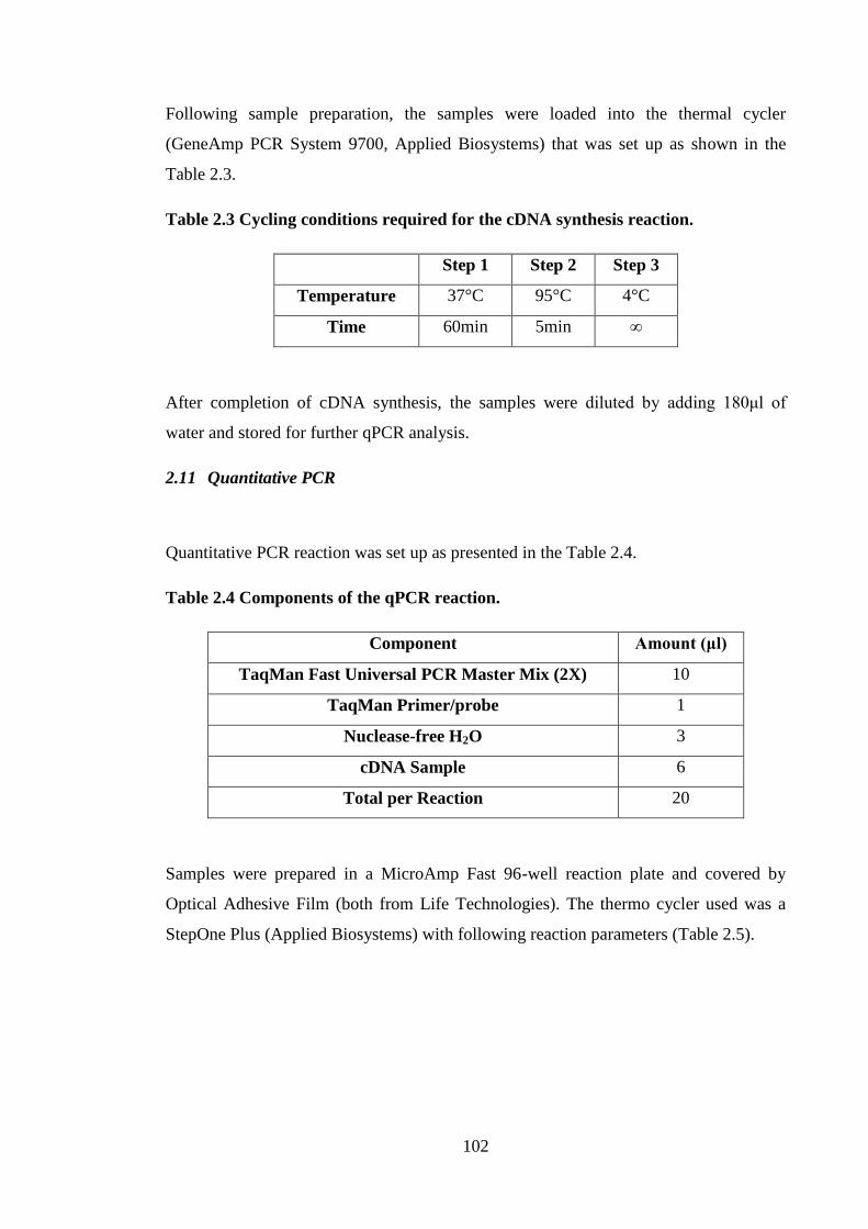
102
Following sample preparation, the samples were loaded into the thermal cycler
(GeneAmp PCR System 9700, Applied Biosystems) that was set up as shown in the
Table 2.3.
Table 2.3 Cycling conditions required for the cDNA synthesis reaction.
Step 1 Step 2 Step 3
Temperature 37°C 95°C 4°C
Time 60min 5min ∞
After completion of cDNA synthesis, the samples were diluted by adding 180μl of
water and stored for further qPCR analysis.
2.11 Quantitative PCR
Quantitative PCR reaction was set up as presented in the Table 2.4.
Table 2.4 Components of the qPCR reaction.
Component Amount (μl)
TaqMan Fast Universal PCR Master Mix (2X) 10
TaqMan Primer/probe 1
Nuclease-free H2O 3
cDNA Sample 6
Total per Reaction 20
Samples were prepared in a MicroAmp Fast 96-well reaction plate and covered by
Optical Adhesive Film (both from Life Technologies). The thermo cycler used was a
StepOne Plus (Applied Biosystems) with following reaction parameters (Table 2.5).
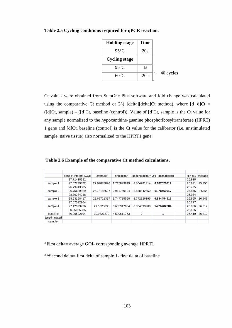
103
Table 2.5 Cycling conditions required for qPCR reaction.
Holding stage Time
95°C 20s
Cycling stage
95°C 1s
60°C 20s
Ct values were obtained from StepOne Plus software and fold change was calculated
using the comparative Ct method or 2^(–[delta][delta]Ct method), where [d][d]Ct =
([d]Ct, sample) – ([d]Ct, baseline (control)). Value of [d]Ct, sample is the Ct value for
any sample normalized to the hypoxanthine-guanine phosphoribosyltransferase (HPRT)
1 gene and [d]Ct, baseline (control) is the Ct value for the calibrator (i.e. unstimulated
sample, naive tissue) also normalized to the HPRT1 gene.
Table 2.6 Example of the comparative Ct method calculations.
*First delta= average GOI- corresponding average HPRT1
**Second delta= first delta of sample 1- first delta of baseline
gene of interest (GOI) average first delta* second delta** 2^(–[delta][delta]) HPRT1 average
27.71418381 25.918
sample 1 27.62739372 27.67078876 1.715829849 -2.804781914 6.987526812 25.991 25.955
26.79743385 25.795
sample 2 26.76629829 26.78186607 0.961769104 -3.558842659 11.78469617 25.845 25.82
28.76284218 26.934
sample 3 28.63158417 28.69721317 1.747785568 -2.772826195 6.834454513 26.965 26.949
27.57522964 26.777
sample 4 27.42993736 27.5025835 0.685917854 -3.834693909 14.26782884 26.856 26.817
30.95965385 26.405
baseline 30.90592194 30.9327879 4.520611763 0 1 26.419 26.412
(unstimulated
sample)
40 cycles
104
2.12 Analysis of cytokine production by explants
Ears were removed and the dorsal halves were isolated, as described previously in the
section 2.6. Dorsal halves were floated on 250μl of complete RPMI medium for 16h at
37oC in a humidified atmosphere of 5% CO2. Supernatants were harvested and analyzed
for TNF-α, IL-1α and IL-1β using DuoSet ELISA.
2.13 Statistical analyses
Statistical analysis was performed using Graphpad (Graphpad Software). Non-
parametric cytokine production data (ELISA) was analyzed by Mann-Whitney test or
Kruskal-Wallis test with Dunn post-test when comparing two or 3 or more groups,
respectively. Parametric ELISA data were analyzed by Student’s t test or one-way
ANOVA with Tukey’s multiple comparisons test when comparing two or 3 or more
groups, respectively. Where the values were below the limit of accurate detection, an
arbitrary value of half of the value of the lower limit of detection was assigned. The LC
migration data were analyzed by a Student’s t test, when comparing two groups or by
one-way ANOVA with Tukey’s multiple comparisons test when comparing three or
more groups. Non-parametric PCR data was analyzed by Mann-Whitney test or
Kruskal-Wallis test with Dunn post-test when comparing two or 3 or more groups,
respectively. Parametric PCR data were analyzed by Student’s t test or one-way
ANOVA with Tukey’s multiple comparisons test when comparing two or 3 or more
groups, respectively.
105
3 In vivo effects of lactoferrin
3.1 Introduction
Lactoferrin is an iron binding, mammalian protein present mainly in milk, but also in
serum, tears, saliva, and secondary neutrophil granules. Lactoferrin is involved in both
innate and acquired immunity and its roles are often contradictory. Indeed, it has been
described to be both “a shield and a sword” for an organism (Legrand and Mazurier,
2010). One of the first reports regarding the role of LF demonstrated that it was a potent
antimicrobial protein, and was able to exert bactericidal properties via iron sequestration
in vitro (Oram and Reiter, 1968). Lactoferrin was found to have a protective role in
vivo, whereby it was reported to protect mice from septic shock caused by exposure to a
lethal dose of E. coli (Zagulski et al., 1989). Transgenic mice that over-expressed
human LF were shown to display improved clearance of S. aureus. Spleen cells from
these mice infected with S.aureus exhibited higher levels of TNF-α and IFN-γ following
exposure to S.aureus (Toxic Shock Syndrome Toxin-1 [TSST-]1) antigen than did WT
controls. However, in non infected animals, stimulation of spleen cells with TSST-1
resulted in higher levels of TNF-α and IFN-γ production in WT mice (Guillén et al.,
2002). Also, a role for LF in humans was confirmed by Breton-Gorius et al. (1980), in a
case study of a patient with LF deficiency in secondary neutrophil granules, that
presented with recurrent infections starting at a very young age (Breton-Gorius et al.,
1980). It was demonstrated that during infection the concentration of LF in serum
increased, further confirming the role of this glycoprotein in immunity (Huang et al.,
2013; Muruganand et al., 2004). In addition to increased LF levels found in systemic
inflammation, it was demonstrated that levels of LF were elevated in a blister fluid
following local exposure to allergen (pollen or ragweed) of patients suffering from
seasonal rhinitis (Zweiman et al., 1990), suggesting that LF plays a role in the cutaneous
inflammation. In fact, it was found in this laboratory, that topical application of LF
inhibited LC migration triggered by exposure to contact allergen DPC or IL-1β.
Moreover, LF was reported to reduce the cutaneous inflammation associated with
topical exposure to DPC. There was a decrease in both erythema and cellular infiltration
at the site of contact (Griffiths et al., 2001). In addition, LF was demonstrated to exert
the same inhibitory effect on LC migration and DC accumulation in draining LN in
106
mice following treatment with another contact allergen, oxazolone (Cumberbatch et al.,
2000b). However, despite the established role of LF in the immune system its
mechanism of action is still not known fully. It was hypothesised that the mechanism by
which LF inhibited LC migration and DC accumulation was due to decreased de novo
production of TNF-α in the skin following treatment with allergen (Cumberbatch et al.,
2003). That conclusion was reached following results that showed that LF inhibited
migration that was dependent upon TNF-α, but was without effect on migration induced
by delivery of homologous, exogenous TNF-α. The cellular and molecular processes
through which LC are mobilized and migrate following exposure to contact allergens
are still not understood fully. Nor is it clear exactly how LF modulates LC migration.
The aim of this chapter is to describe in greater detail investigations designed to
characterize the biological effects of LF. To achieve this, the impact of LF on cytokine
production and expression in the skin of mice was assessed following allergen and
cytokine treatment. In addition, the role of LF on cytokine production and cell
proliferation in the draining LN was investigated.
3.2 Results
Lactoferrin has been demonstrated previously to affect the cells residing in the skin: LC
and DC, by impairing their migration into the draining LN (Cumberbatch et al., 2000b).
In the current investigations, topical exposure to LF inhibited LC migration in mice
induced by application of 0.5% oxazolone, confirming previous observations (Chapter
6). That finding provided reassurance that the batches of LF used here posessed the in
vivo biological activity reported previously. The purpose of the experiments described
herein was to examine the impact of in vivo topical exposure to LF on the other
parameters of the immune system activation caused by treatment with allergen, in order
to provide further insight regarding the potential mechanism of LF. Initially, the effects
of LF on allergen-induced changes in the skin, cytokines and other markers of
activation such as ICAM-1 expression, were measured, at both the protein and message
levels, as the skin is the site of first contact with LF. In subsequent experiments, the
potential downstream effects of LF treatment on the quality of immune response
provoked in the LN draining the site of topical exposure were examined, with respect to
overall immunogenicity (increased cellularity and proliferation) and expression of LN
cytokines. Finally, given that the stimulus of allergen is a relatively long lasting and
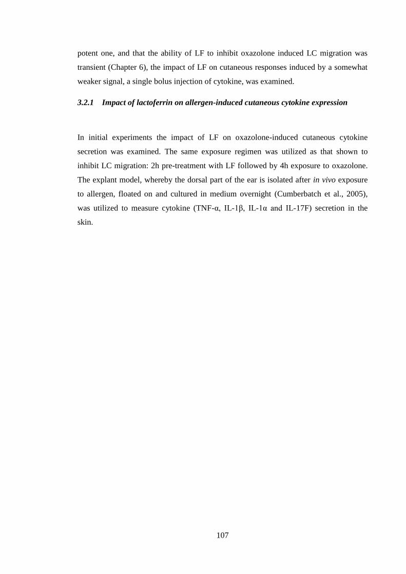
107
potent one, and that the ability of LF to inhibit oxazolone induced LC migration was
transient (Chapter 6), the impact of LF on cutaneous responses induced by a somewhat
weaker signal, a single bolus injection of cytokine, was examined.
3.2.1 Impact of lactoferrin on allergen-induced cutaneous cytokine expression
In initial experiments the impact of LF on oxazolone-induced cutaneous cytokine
secretion was examined. The same exposure regimen was utilized as that shown to
inhibit LC migration: 2h pre-treatment with LF followed by 4h exposure to oxazolone.
The explant model, whereby the dorsal part of the ear is isolated after in vivo exposure
to allergen, floated on and cultured in medium overnight (Cumberbatch et al., 2005),
was utilized to measure cytokine (TNF-α, IL-1β, IL-1α and IL-17F) secretion in the
skin.
108
TN
F-
[p
g/m
l]
N a iv e 0 L F
0
5 0
1 0 0
1 5 0
0 .5 % O x
IL-1
[
pg
/ml]
N a iv e 0 L F
0
1 0 0
2 0 0
3 0 0
4 0 0
0 .5 % O x
IL-1
[
pg
/ml]
N a iv e 0 L F
0
1 0 0 0
2 0 0 0
3 0 0 0
4 0 0 0
0 .5 % O x
IL-1
7F
[p
g/m
l]
N a iv e 0 L F
0
5 0 0
1 0 0 0
1 5 0 0
0 .5 % O x
A B
C D
Figure 3.1 The influence of LF on a cutaneous cytokine production following
treatment with oxazolone.
Young (6-8 weeks) BALB/c strain mice were treated with 0.5μg of LF in 30μl of aqueous
cream or cream alone on the dorsum of both ears. Two h later animals were challenged
on the same site with 25μl of 0.5% oxazolone (Ox) in AOO. After 4h animals were
sacrificed and ears were excised. Further controls were untreated (naive). Ears were
split into dorsal and ventral halves and dorsal halves were floated on 250μl of complete
RPMI media, and incubated for 16h. Following incubation, supernatants were
collected, and TNF-α (A), IL-1β (B), IL-1α (C) and IL-17F (D) production was
determined by ELISA. Dashed line represents the lower limit of detection for ELISA
(TNF-α (32pg/ml), IL-17F (23 pg/ml) and IL-1α and β (15 pg/ml). Graphs present data
(mean ± SEM) from a single experiment (n=1-4 ears). The statistical significance of
differences between cytokine levels in oxazolone-induced mice in the presence and in
the absence of LF was compared by a Mann-Whitney test; p>0.05.
109
All cytokines were produced in detectable levels from naive skin, with particularly
vigorous expression of IL-1 (~1000pg/ml) and relatively high, but variable, levels of
IL-17. Four h exposure to oxazolone did not affect TNF-α or IL-17F production (Figure
3.1A and 3.1D). On the other hand, an increase in the production of IL-1α and β was
observed. Baseline levels of IL-1α (1200pg/ml) were up-regulated to 2500pg/ml.
Release of IL-1β was much lower than that reported for IL-1α (100pg/ml in the absence
of allergen and 300pg/ml in the presence of allergen). Pre-treatment with LF was largely
without effect on oxazolone-induced cytokine expression. Although there was a slight
reduction in IL-1 this did not reach statistical significance. The high levels of cytokine
expressed in naive tissue were of some concern, and presumably reflected the trauma of
tissue isolation, particularly the separation of the dorsum from the ventral half of the
ear. Although only one time point was investigated, it seemed likely that this high
background level could obscure both allergen-induced effects and the impact of LF.
Therefore, it was decided to examine the effects of LF on cytokine mRNA levels in the
skin, as this measurement could be undertaken in the whole skin, processed
immediately after excision, without the need for traumatic separation of the dorsal
halves from the ventral halves.
3.2.2 Lactoferrin did not markedly affect baseline dermal cytokine expression
Initially, the impact of LF on baseline dermal cytokine gene expression was studied.
Mice were treated with LF and tissue was isolated 2, 4 and 6 h after the initiation of
exposure. Levels of cytokine (Il-1β, Tnf-α, Il-17F) and Icam-1 transcripts were
compared with naive (untreated) tissue (Figure 3.2). It was found that there was a small,
but transient increase in the expression of Tnf-, Il-1 and Icam-1 4h after treatment
with LF, which reached statistical significance only for Icam-1, and returned to basal
levels after 6h. Expression of Il-17F was unaffected. The impact was small at best (less
than 2 fold change) and it was not clear that such changes were due to LF or a result of
intra-experimental variation.
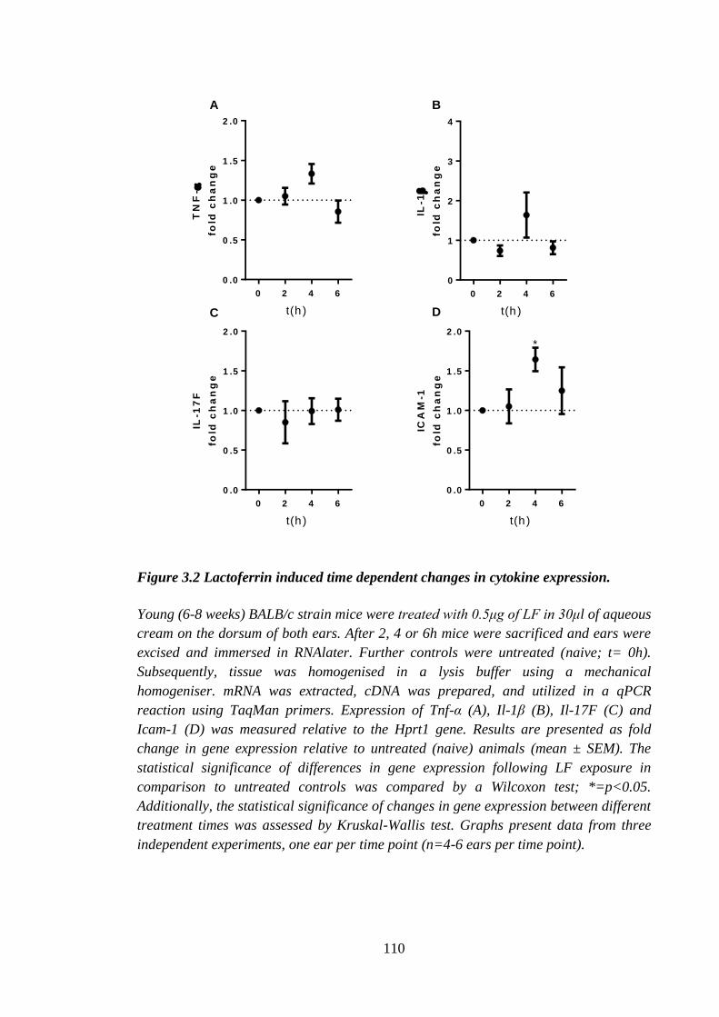
110
TN
F-
fold
ch
an
ge
0 2 4 6
0 .0
0 .5
1 .0
1 .5
2 .0
t(h )
IL-1
fold
ch
an
ge
0 2 4 6
0
1
2
3
4
t(h )
IL-1
7F
fold
ch
an
ge
0 2 4 6
0 .0
0 .5
1 .0
1 .5
2 .0
t(h )
0 2 4 6
0 .0
0 .5
1 .0
1 .5
2 .0
ICA
M-1
fold
ch
an
ge
t(h )
*
A B
C D
Figure 3.2 Lactoferrin induced time dependent changes in cytokine expression.
Young (6-8 weeks) BALB/c strain mice were treated with 0.5μg of LF in 30μl of aqueous
cream on the dorsum of both ears. After 2, 4 or 6h mice were sacrificed and ears were
excised and immersed in RNAlater. Further controls were untreated (naive; t= 0h).
Subsequently, tissue was homogenised in a lysis buffer using a mechanical
homogeniser. mRNA was extracted, cDNA was prepared, and utilized in a qPCR
reaction using TaqMan primers. Expression of Tnf-α (A), Il-1β (B), Il-17F (C) and
Icam-1 (D) was measured relative to the Hprt1 gene. Results are presented as fold
change in gene expression relative to untreated (naive) animals (mean ± SEM). The
statistical significance of differences in gene expression following LF exposure in
comparison to untreated controls was compared by a Wilcoxon test; *=p<0.05.
Additionally, the statistical significance of changes in gene expression between different
treatment times was assessed by Kruskal-Wallis test. Graphs present data from three
independent experiments, one ear per time point (n=4-6 ears per time point).
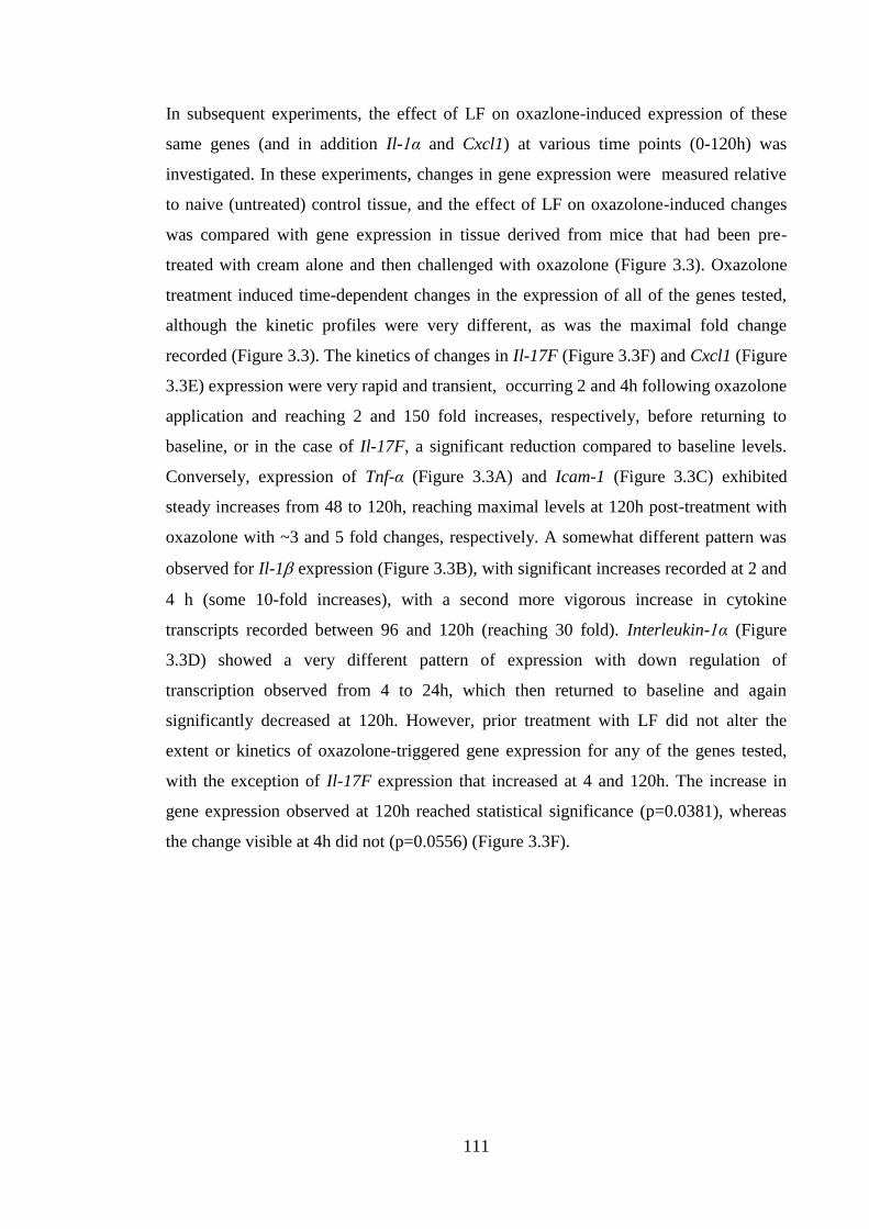
111
In subsequent experiments, the effect of LF on oxazlone-induced expression of these
same genes (and in addition Il-1α and Cxcl1) at various time points (0-120h) was
investigated. In these experiments, changes in gene expression were measured relative
to naive (untreated) control tissue, and the effect of LF on oxazolone-induced changes
was compared with gene expression in tissue derived from mice that had been pre-
treated with cream alone and then challenged with oxazolone (Figure 3.3). Oxazolone
treatment induced time-dependent changes in the expression of all of the genes tested,
although the kinetic profiles were very different, as was the maximal fold change
recorded (Figure 3.3). The kinetics of changes in Il-17F (Figure 3.3F) and Cxcl1 (Figure
3.3E) expression were very rapid and transient, occurring 2 and 4h following oxazolone
application and reaching 2 and 150 fold increases, respectively, before returning to
baseline, or in the case of Il-17F, a significant reduction compared to baseline levels.
Conversely, expression of Tnf-α (Figure 3.3A) and Icam-1 (Figure 3.3C) exhibited
steady increases from 48 to 120h, reaching maximal levels at 120h post-treatment with
oxazolone with ~3 and 5 fold changes, respectively. A somewhat different pattern was
observed for Il-1expression (Figure 3.3B), with significant increases recorded at 2 and
4 h (some 10-fold increases), with a second more vigorous increase in cytokine
transcripts recorded between 96 and 120h (reaching 30 fold). Interleukin-1α (Figure
3.3D) showed a very different pattern of expression with down regulation of
transcription observed from 4 to 24h, which then returned to baseline and again
significantly decreased at 120h. However, prior treatment with LF did not alter the
extent or kinetics of oxazolone-triggered gene expression for any of the genes tested,
with the exception of Il-17F expression that increased at 4 and 120h. The increase in
gene expression observed at 120h reached statistical significance (p=0.0381), whereas
the change visible at 4h did not (p=0.0556) (Figure 3.3F).
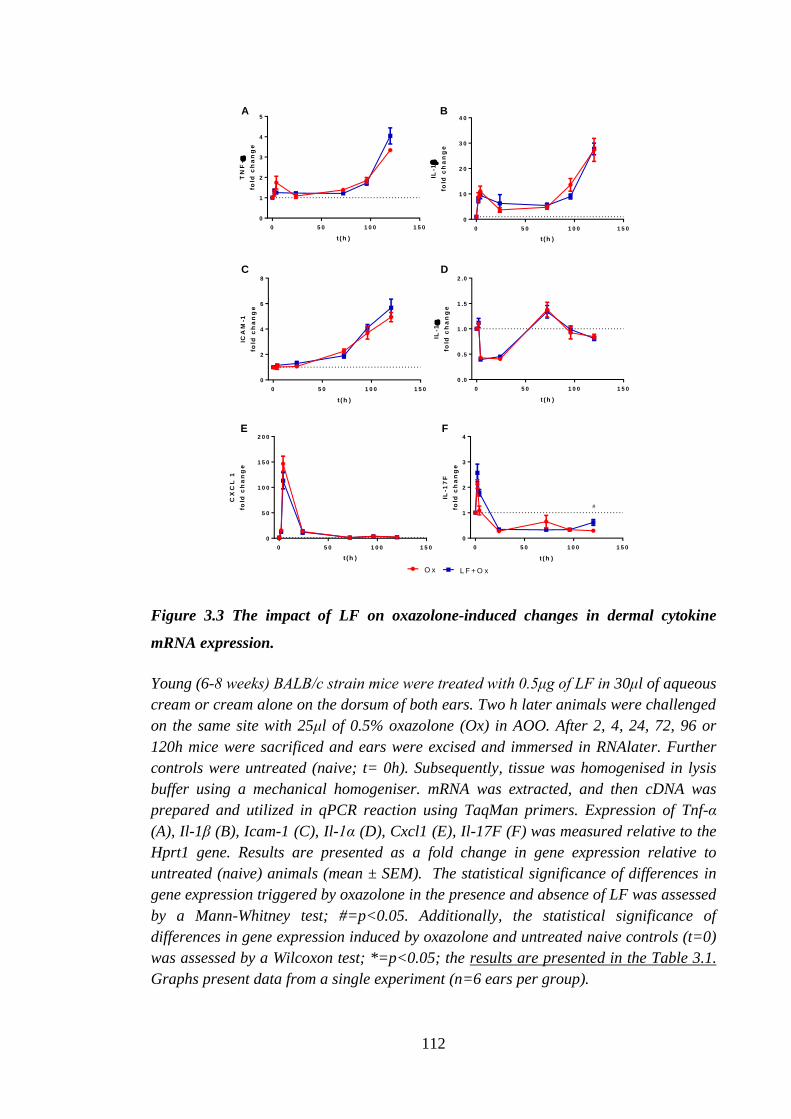
112
0 5 0 1 0 0 1 5 0
0
1
2
3
4
5
t (h )T
NF
-
fold
ch
an
ge
0 5 0 1 0 0 1 5 0
0
1 0
2 0
3 0
4 0
t (h )
IL-1
fold
ch
an
ge
0 5 0 1 0 0 1 5 0
0
2
4
6
8
t (h )
ICA
M-1
fold
ch
an
ge
0 5 0 1 0 0 1 5 0
0 .0
0 .5
1 .0
1 .5
2 .0
t (h )
IL-1
fold
ch
an
ge
0 5 0 1 0 0 1 5 0
0
5 0
1 0 0
1 5 0
2 0 0
t (h )
CX
CL
1
fold
ch
an
ge
0 5 0 1 0 0 1 5 0
0
1
2
3
4
t (h )
IL-1
7F
fold
ch
an
ge
#
O x L F + O x
A B
C D
E F
Figure 3.3 The impact of LF on oxazolone-induced changes in dermal cytokine
mRNA expression.
Young (6-8 weeks) BALB/c strain mice were treated with 0.5μg of LF in 30μl of aqueous
cream or cream alone on the dorsum of both ears. Two h later animals were challenged
on the same site with 25μl of 0.5% oxazolone (Ox) in AOO. After 2, 4, 24, 72, 96 or
120h mice were sacrificed and ears were excised and immersed in RNAlater. Further
controls were untreated (naive; t= 0h). Subsequently, tissue was homogenised in lysis
buffer using a mechanical homogeniser. mRNA was extracted, and then cDNA was
prepared and utilized in qPCR reaction using TaqMan primers. Expression of Tnf-α
(A), Il-1β (B), Icam-1 (C), Il-1α (D), Cxcl1 (E), Il-17F (F) was measured relative to the
Hprt1 gene. Results are presented as a fold change in gene expression relative to
untreated (naive) animals (mean ± SEM). The statistical significance of differences in
gene expression triggered by oxazolone in the presence and absence of LF was assessed
by a Mann-Whitney test; #=p<0.05. Additionally, the statistical significance of
differences in gene expression induced by oxazolone and untreated naive controls (t=0)
was assessed by a Wilcoxon test; *=p<0.05; the results are presented in the Table 3.1.
Graphs present data from a single experiment (n=6 ears per group).
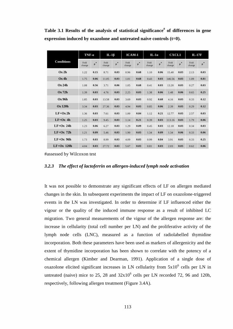
113
Table 3.1 Results of the analysis of statistical significance# of differences in gene
expression induced by oxazolone and untreated naive controls (t=0).
#assessed by Wilcoxon test
3.2.3 The effect of lactoferrin on allergen-induced lymph node activation
It was not possible to demonstrate any significant effects of LF on allergen mediated
changes in the skin. In subsequent experiments the impact of LF on oxazolone-triggered
events in the LN was investigated. In order to determine if LF influenced either the
vigour or the quality of the induced immune response as a result of inhibited LC
migration. Two general measurements of the vigour of the allergen response are: the
increase in cellularity (total cell number per LN) and the proliferative activity of the
lymph node cells (LNC), measured as a function of radiolabelled thymidine
incorporation. Both these parameters have been used as markers of allergenicity and the
extent of thymidine incorporation has been shown to correlate with the potency of a
chemical allergen (Kimber and Dearman, 1991). Application of a single dose of
oxazolone elicited significant increases in LN cellularity from 5x10⁶ cells per LN in
untreated (naive) mice to 25, 28 and 32x106 cells per LN recorded 72, 96 and 120h,
respectively, following allergen treatment (Figure 3.4A).
Conditions
TNF-α IL-1β ICAM-1 IL-1α CXCL1 IL-17F
Fold
changep
* Fold
changep
* Fold
changep
* Fold
changep
* Fold
changep
* Fold
changep
*
Ox 2h 1.22 0.15 8.71 0.03 0.96 0.68 1.10 0.06 15.40 0.03 2.13 0.03
Ox 4h 1.75 0.06 11.05 0.03 1.01 0.68 0.43 0.03 146.56 0.03 1.09 0.81
Ox 24h 1.08 0.56 3.71 0.06 1.05 0.68 0.41 0.03 13.20 0.03 0.27 0.03
Ox 72h 1.39 0.03 4.76 0.03 2.25 0.03 1.38 0.06 1.48 0.06 0.65 0.25
Ox 96h 1.85 0.03 13.58 0.03 3.69 0.03 0.92 0.68 4.16 0.03 0.33 0.12
Ox 120h 3.34 0.03 27.36 0.03 4.94 0.03 0.85 0.06 2.39 0.03 0.29 0.12
LF+Ox 2h 1.36 0.03 7.61 0.03 1.00 0.84 1.12 0.21 12.77 0.03 2.57 0.03
LF+Ox 4h 1.25 0.03 9.45 0.03 1.14 0.21 0.39 0.03 113.16 0.03 1.79 0.06
LF+Ox 24h 1.23 0.06 6.27 0.03 1.29 0.09 0.45 0.03 12.18 0.03 0.34 0.03
LF+Ox 72h 1.21 0.09 5.46 0.03 1.90 0.03 1.34 0.09 1.34 0.06 0.33 0.06
LF+Ox 96h 1.73 0.03 8.99 0.03 4.09 0.03 0.99 0.84 3.91 0.03 0.33 0.25
LF+Ox 120h 4.04 0.03 27.72 0.03 5.67 0.03 0.81 0.03 2.03 0.03 0.62 0.06
114
7 2 9 6 1 2 0
0
1 0
2 0
3 0
4 0
ce
lls
pe
r L
N [
x1
06
]
***
******
t (h )
7 2 9 6 1 2 0
0
2
4
6
8
1 0
DP
M (
x1
04
)
t(h )
**
***
N a ive O x L F 0 .5 g + O x
A B
Figure 3.4 Oxazolone-induced lymph node cell activation: the influence of LF.
Young (6-8 weeks) BALB/c strain mice were treated with 0.5μg of LF in 30μl of aqueous
cream or cream alone on the dorsum of both ears. Two h later animals were challenged
on the same site with 25μl of 0.5% oxazolone (Ox) in AOO. After 72, 96 or 120h,
animals alongside naive controls were sacrificed and auricular LN were excised.
Further controls were untreated (naive). Lymph nodes were disaggregated and mashed
through metal gauze to achieve a single cell suspension. Cells were enumerated using a
CASY counter (A) and 2x10⁶ cells per well were seeded into 96 well plate in the
presence of [3H] thymidine and cultured for 24h (B). Thymidine incorporation was
measured by β-scintillation counting and are displayed as dpm (disintegrations per
min)(B). Graphs present data (mean ± SEM) from a single experiment (n=3 animals per
group). The statistical significance of differences in LNC numbers (A) or LNC
proliferation (B) between animals treated with oxazolone and those from naive animals
was assessed by one-way ANOVA; **=p<0.01, ***=p<0.001. Moreover, statistical
significance of differences in (A) cellularity and (B) proliferation between oxazolone
exposed animals in the presence and in the absence of LF was assessed by a Mann-
Whitney test (p>0.05).
In addition, the ability of LNC to proliferate after oxazolone application was examined.
It was found that at all time points examined, 72, 96 and 120h, after treatment with
oxazolone LNC exhibited a significant increase in proliferation (19, 9 and 6 fold,
respectively), compared with untreated controls. However, a single application of LF
did not significantly impact on LN cellularity or on the proliferative activity of LNC
population. These data indicated that LF pre-treatment did not impact significantly on
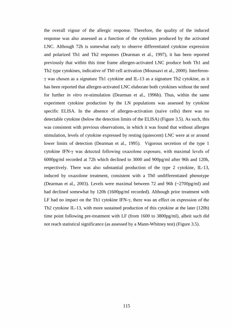
115
the overall vigour of the allergic response. Therefore, the quality of the induced
response was also assessed as a function of the cytokines produced by the activated
LNC. Although 72h is somewhat early to observe differentiated cytokine expression
and polarized Th1 and Th2 responses (Dearman et al., 1997), it has been reported
previously that within this time frame allergen-activated LNC produce both Th1 and
Th2 type cytokines, indicative of Th0 cell activation (Moussavi et al., 2000). Interferon-
γ was chosen as a signature Th1 cytokine and IL-13 as a signature Th2 cytokine, as it
has been reported that allergen-activated LNC elaborate both cytokines without the need
for further in vitro re-stimulation (Dearman et al., 1996b). Thus, within the same
experiment cytokine production by the LN populations was assessed by cytokine
specific ELISA. In the absence of allergen-activation (naive cells) there was no
detectable cytokine (below the detection limits of the ELISA) (Figure 3.5). As such, this
was consistent with previous observations, in which it was found that without allergen
stimulation, levels of cytokine expressed by resting (quiescent) LNC were at or around
lower limits of detection (Dearman et al., 1995). Vigorous secretion of the type 1
cytokine IFN-γ was detected following oxazolone exposure, with maximal levels of
6000pg/ml recorded at 72h which declined to 3000 and 900pg/ml after 96h and 120h,
respectively. There was also substantial production of the type 2 cytokine, IL-13,
induced by oxazolone treatment, consistent with a Th0 undifferentiated phenotype
(Dearman et al., 2003). Levels were maximal between 72 and 96h (~2700pg/ml) and
had declined somewhat by 120h (1600pg/ml recorded). Although prior treatment with
LF had no impact on the Th1 cytokine IFN-γ, there was an effect on expression of the
Th2 cytokine IL-13, with more sustained production of this cytokine at the later (120h)
time point following pre-treatment with LF (from 1600 to 3800pg/ml), albeit such did
not reach statistical significance (as assessed by a Mann-Whitney test) (Figure 3.5).
116
IFN
- [
pg
/ml]
7 2 h 9 6 h 1 2 0 h
0
2 0 0 0
4 0 0 0
6 0 0 0
8 0 0 0
1 0 0 0 0
IL-1
3 [
pg
/ml]
7 2 h 9 6 h 1 2 0 h
0
1 0 0 0
2 0 0 0
3 0 0 0
4 0 0 0
5 0 0 0
O x L F /O xN a iv e
IF N - IL -1 3
Figure 3.5 Impact of LF on allergen-actviated lymph node cell cytokine production.
Young (6-8 weeks) BALB/c strain mice were treated with 0.5μg of LF in 30μl of aqueous
cream or cream alone on the dorsum of both ears. Two h later animals were challenged
on the same site with 25μl of 0.5% oxazolone (Ox) in AOO. After 72, 96 or 120h,
animals were sacrificed and auricular LN were excised. Further controls were
untreated (naive). Lymph nodes were disaggregated and mashed through metal gauze
to achieve a single cell suspension. Cells were enumerated using a CASY counter and
107
cells were seeded into 24 well plates and cultured for a further 120h. Subsequently,
supernatants were collected and the IFN-γ and IL-13 content was determined by ELISA
(the lower limit of detection was 31 and 62 pg/ml for IFN-γ and IL-13, respectively).
Graphs present data (mean ± SEM) from a single experiment (n= 3 animals per group).
The statistical analysis of differences between cytokine production in the presence and
in the absence of LF was assessed by a Mann-Whitney test; p>0.05.
Thus, there was some indication that pre-treatment with LF may impact on the quality
of the response to oxazolone, with an increased bias towards the Th2 cytokine IL-13
recorded. In addition to examining the impact of LF on the Th1/Th2 skewing of the
LNC cytokine response that was observed some 3 to 5 days after the initiation of
exposure, the early (6 to 24h) cellular events in the LN were examined. It has been
shown previously that one of the earliest changes in the LN draining the site of allergen
application is the expression of the cytokine IL-17 by innate immune cells (T cells)
(PhD thesis Hayes, 2012). This production of IL-17 (both IL-17A and F cytokines)
precedes the marked increase in cellularity stimulated by contact allergen, being
detected after 6h of exposure. Mice were treated with oxazolone for either 6 or 24h, in
the presence or absence of prior application of LF. After LN isolation, the standard
method for preparation of a single cell suspension of LNC for subsequent
117
culture/analysis utilized in this laboratory has been mechanical disaggregation through
wire gauze. However, in experiments designed to look at the cellular phenotype of cells,
including those in which the observations of early IL-17 LN expression were recorded
(PhD thesis Hayes, 2012), single cell suspensions were prepared by enzymatic
digestion. Therefore, in these experiments, both methods of preparation of LNC were
compared. The LNC count increased from ~6x10⁶ cells per LN in naive animals to
approximately 9.5x10⁶ and 8x10
6 cells per LN in mice treated with oxazolone for 6h
using mechanical and enzymatic methods, respectively. Exposure to oxazolone for 24h
resulted in a further increase in LNC to 11x106 and 13.5x10
6 utilising mechanical and
enzymatic methods, respectively. Allergen induced increases were similar regardless of
the method of LN preparation. Application of LF was without marked effects on LN
cellularity, with the exception of 24h following oxazolone application and mechanical
digestion, where increased total cell numbers were recorded (Figure 3.6B). Enzymatic
preparation of LNC 24h after oxazolone application resulted in the lower number of
LNC in the presence of LF, however, such did not reach statistical significance.
Subsequently, the production by LNC of IL-17A and IL-17F was assessed. It was found
that when LNC were obtained using the mechanical method there was no detectable
production of IL-17 6h after oxazolone treatment (<15 and 23pg/ml, for IL-17A and IL-
17F, respectively; data not shown). When the enzymatic method was used 6h following
oxazolone application, IL-17 production increased from resting levels of ~50 and
500pg/ml for A and F isoforms to 300 and 1500pg/ml, respectively (Figure 3.6A), with
prior application of LF being without effect. Exposure to oxazolone for 24h resulted in
more vigorous IL-17 production. When mechanical disaggregation was used, 24h
exposure to oxazolone induced the release of 1200 and 3500pg/ml of IL-17 A and F,
respectively, by LNC, with levels being reduced somewhat following LF pre-treatment
(Figure 3.6B). In contrast, when enzymatic cell isolation was utilized for samples
treated with oxazolone for 24h, there was a striking, approximately 5 fold increase, in
levels of IL-17 produced (Figure 3.6C) in comparison with the level of production by
cells isolated using mechanical disaggregation (Figure 3.6B). Cells that were obtained
by enzymatic digestion produced 6000 and 30000pg/ml of IL-17 A and F, respectively,
and prior application LF did not markedly affect IL-17 release (Figure 3.6C).
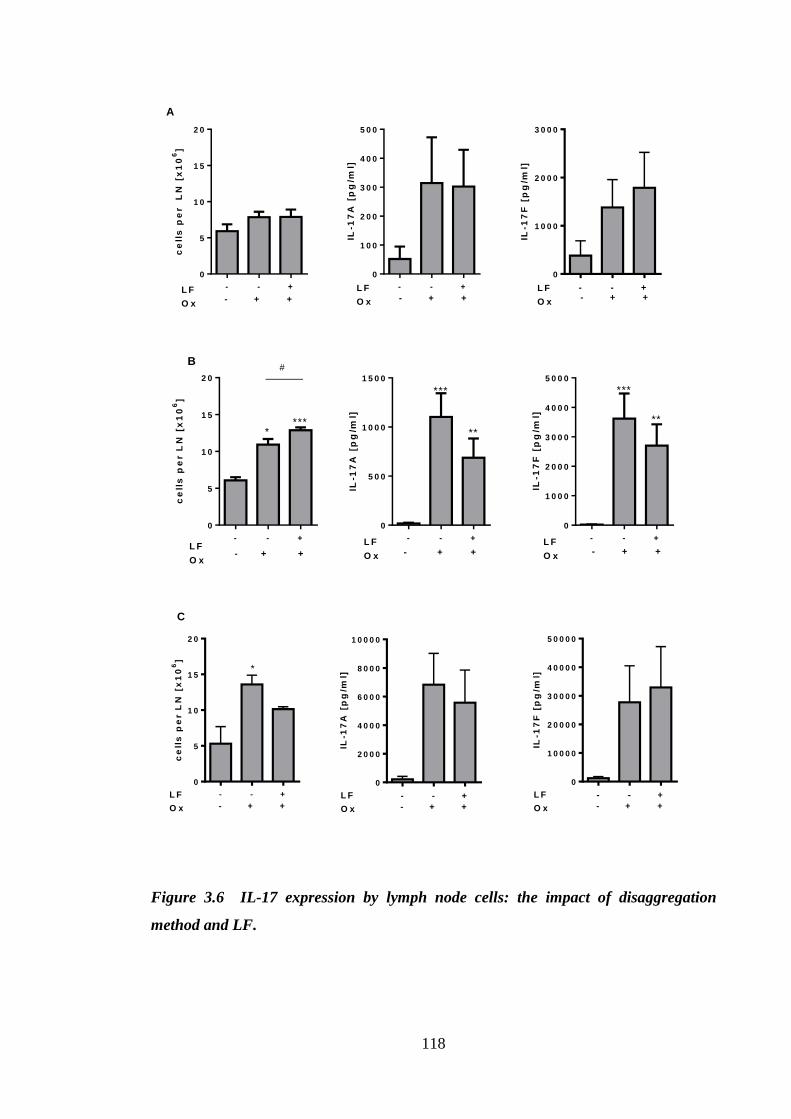
118
- - +
0
5
1 0
1 5
2 0
ce
lls
pe
r
LN
[x
10
6]
- + +L F
O x
- - +
0
1 0 0
2 0 0
3 0 0
4 0 0
5 0 0
IL-1
7A
[p
g/m
l]
L F
O x - + +- - +
0
1 0 0 0
2 0 0 0
3 0 0 0
IL-1
7F
[p
g/m
l]
- + +L F
O x
A
- - +
0
5
1 0
1 5
2 0
ce
lls
pe
r L
N [
x1
06
]
L F
O x - + +
****
#
- - +
0
5 0 0
1 0 0 0
1 5 0 0
IL-1
7A
[p
g/m
l]
L F
O x - + +
***
**
- - +
0
1 0 0 0
2 0 0 0
3 0 0 0
4 0 0 0
5 0 0 0
IL-1
7F
[p
g/m
l]L F
O x - + +
***
**
B
- - +
0
5
1 0
1 5
2 0
ce
lls
pe
r L
N [
x1
06
]
L F
O x - + +
*
- - +
0
2 0 0 0
4 0 0 0
6 0 0 0
8 0 0 0
1 0 0 0 0
IL-1
7A
[p
g/m
l]
L F
O x - + +
- - +
0
1 0 0 0 0
2 0 0 0 0
3 0 0 0 0
4 0 0 0 0
5 0 0 0 0
IL-1
7F
[p
g/m
l]
L F
O x - + +
C
Figure 3.6 IL-17 expression by lymph node cells: the impact of disaggregation
method and LF.
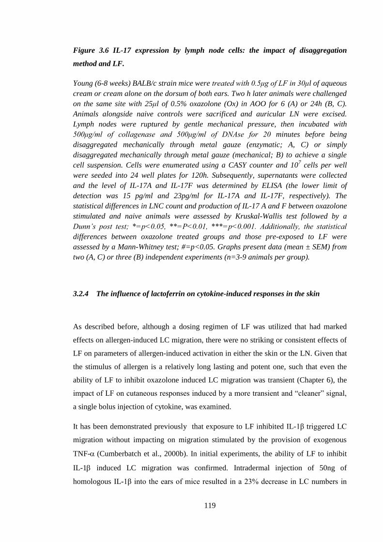
119
Figure 3.6 IL-17 expression by lymph node cells: the impact of disaggregation
method and LF.
Young (6-8 weeks) BALB/c strain mice were treated with 0.5μg of LF in 30μl of aqueous
cream or cream alone on the dorsum of both ears. Two h later animals were challenged
on the same site with 25μl of 0.5% oxazolone (Ox) in AOO for 6 (A) or 24h (B, C).
Animals alongside naive controls were sacrificed and auricular LN were excised.
Lymph nodes were ruptured by gentle mechanical pressure, then incubated with
500μg/ml of collagenase and 500μg/ml of DNAse for 20 minutes before being
disaggregated mechanically through metal gauze (enzymatic; A, C) or simply
disaggregated mechanically through metal gauze (mechanical; B) to achieve a single
cell suspension. Cells were enumerated using a CASY counter and 107 cells per well
were seeded into 24 well plates for 120h. Subsequently, supernatants were collected
and the level of IL-17A and IL-17F was determined by ELISA (the lower limit of
detection was 15 pg/ml and 23pg/ml for IL-17A and IL-17F, respectively). The
statistical differences in LNC count and production of IL-17 A and F between oxazolone
stimulated and naive animals were assessed by Kruskal-Wallis test followed by a
Dunn’s post test; *=p<0.05, **=P<0.01, ***=p<0.001. Additionally, the statistical
differences between oxazolone treated groups and those pre-exposed to LF were
assessed by a Mann-Whitney test; #=p<0.05. Graphs present data (mean ± SEM) from
two (A, C) or three (B) independent experiments (n=3-9 animals per group).
3.2.4 The influence of lactoferrin on cytokine-induced responses in the skin
As described before, although a dosing regimen of LF was utilized that had marked
effects on allergen-induced LC migration, there were no striking or consistent effects of
LF on parameters of allergen-induced activation in either the skin or the LN. Given that
the stimulus of allergen is a relatively long lasting and potent one, such that even the
ability of LF to inhibit oxazolone induced LC migration was transient (Chapter 6), the
impact of LF on cutaneous responses induced by a more transient and “cleaner” signal,
a single bolus injection of cytokine, was examined.
It has been demonstrated previously that exposure to LF inhibited IL-1β triggered LC
migration without impacting on migration stimulated by the provision of exogenous
TNF-(Cumberbatch et al., 2000b). In initial experiments, the ability of LF to inhibit
IL-1 induced LC migration was confirmed. Intradermal injection of 50ng of
homologous IL-1β into the ears of mice resulted in a 23% decrease in LC numbers in
120
comparison with untreated controls. Prior treatment with LF significantly decreased the
extent of LC migration to just 6% (Figure 3.7A). Exposure to IL-1β resulted in activated
LC phenotype characterized by increased expression of MHC II molecules (brighter cell
body appearance), more pronounced dendrites and enlarged cell body (Figure 3.7C)
when compared to LC from untreated mice (Figure 3.7B). Pre-treatment with LF
resulted in the change in cell morphology. Cells were larger, more dendritic and
resembled those exposed to DNCB, exposure to which was demonstrated to result in
morphology that was established to result mainly from high levels of IL-1β produced in
the skin (Figure 3.7D).
121
N a iv e 0 L F
0
2 0 0
4 0 0
6 0 0
8 0 0
1 0 0 0
1 2 0 0
LC
/ m
m2
5 0 n g
IL -1
***
A B
C D
Figure 3.7 Lactoferrin inhibited IL-1β mediated LC migration.
Young (6-8 weeks) BALB/c strain mice (n=3 per group, 6 ears per group) were treated
on the dorsum of both ears with either 30μl of 0.5μg of LF in aqueous cream (D) or
aqueous cream alone (C) for 2h. Subsequently, both groups received intradermal
injection of 50ng of homogenous IL-1β for further 4h. Treated animals alongside non-
treated controls (naive) (B) (n=3 per group; 6 ears per group) were sacrificed, ears
were excised and LC enumerated by fluorescence microscopy and expressed as the
mean frequency of LC per mm2 per group ± SEM (A). The statistical significance of
differences in LC numbers between untreated (naive) and cytokine exposed animals;
***=p<0.001 was assessed by one-way ANOVA. Representative micrographs of
epidermal sheets show LC morphology (B-D). Scale bar 100μm.
122
To examine further the potential mechanism by which LF inhibited IL-1β induced
migration, the same protocol was utilized as described in the previous experiment
(Figure 3.7), but instead of assessing the extent of LC migration, the level of cutaneous
expression of various genes was measured. Expression of the cytokines Tnf-, Il-1, Il-
1 and Il-17A was measured, as well as the chemokine Cxcl1 and the adhesion molecule
Icam-1, 4h after injection of cytokine, the same time point that was utilized for LC
migration. It was found that exposure to IL-1β resulted in up-regulation of all of the
genes tested with rank order of induction Cxcl1 < Il-1β < Il-17F < Icam-1 < Tnf-α
(Figure 3.8A-3.8E), with the exception of Il-1α, expression of which was found to be
decreased (Figure 3.8F) in comparison with expression in the untreated control (as
represented by a dashed line). Fold changes ranged from ~4-fold increase in Tnf-α to
250-fold increase in Cxcl1. Cytokine (IL-1)-induced changes in a cutaneous gene
expression were therefore much more vigorous than those induced by allergen,
particularly with respect to Il-1 (100-fold versus 12-fold), Icam-1 (40-fold versus 1-
fold) and Il-17F (30-fold versus 1-fold). Application of LF prior to exposure to IL-1β
resulted in the inhibition of Il-1β, Cxcl1 and Il-17F expression, however, only the
decrease in Il-17F transcript levels reached statistical significance. The effects on Il-1β
and Cxcl1 were to reduce levels by approximately 50% and 30%, respectively, whereas
the effect on Il-17F was even more striking, reducing levels from an average of ~30-
fold to less than 10-fold.
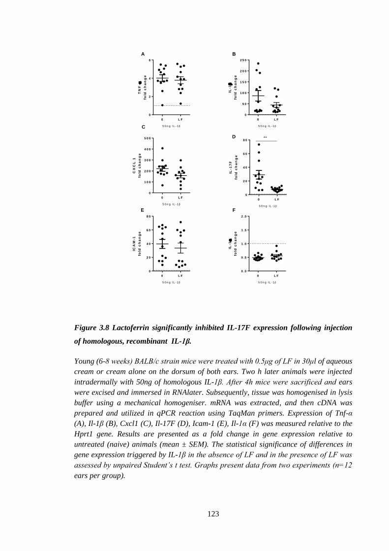
123
0 L F
0
2
4
6
TN
F-
fold
ch
an
ge
5 0 n g IL -1
0 L F
0
5 0
1 0 0
1 5 0
2 0 0
2 5 0
IL-1
fold
ch
an
ge
5 0 n g IL -1
0 L F
0
1 0 0
2 0 0
3 0 0
4 0 0
5 0 0
CX
CL
1
fold
ch
an
ge
5 0 n g IL -1
0 L F
0
2 0
4 0
6 0
8 0
IL-1
7F
fold
ch
an
ge
**
5 0 n g IL -1
0 L F
0
2 0
4 0
6 0
8 0
ICA
M-1
fold
ch
an
ge
5 0 n g IL -1
0 L F
0 .0
0 .5
1 .0
1 .5
2 .0
IL-1
fold
ch
an
ge
5 0 n g IL -1
A B
C
D
E F
Figure 3.8 Lactoferrin significantly inhibited IL-17F expression following injection
of homologous, recombinant IL-1β.
Young (6-8 weeks) BALB/c strain mice were treated with 0.5μg of LF in 30μl of aqueous
cream or cream alone on the dorsum of both ears. Two h later animals were injected
intradermally with 50ng of homologous IL-1β. After 4h mice were sacrificed and ears
were excised and immersed in RNAlater. Subsequently, tissue was homogenised in lysis
buffer using a mechanical homogeniser. mRNA was extracted, and then cDNA was
prepared and utilized in qPCR reaction using TaqMan primers. Expression of Tnf-α
(A), Il-1β (B), Cxcl1 (C), Il-17F (D), Icam-1 (E), Il-1α (F) was measured relative to the
Hprt1 gene. Results are presented as a fold change in gene expression relative to
untreated (naive) animals (mean ± SEM). The statistical significance of differences in
gene expression triggered by IL-1β in the absence of LF and in the presence of LF was
assessed by unpaired Student’s t test. Graphs present data from two experiments (n=12
ears per group).
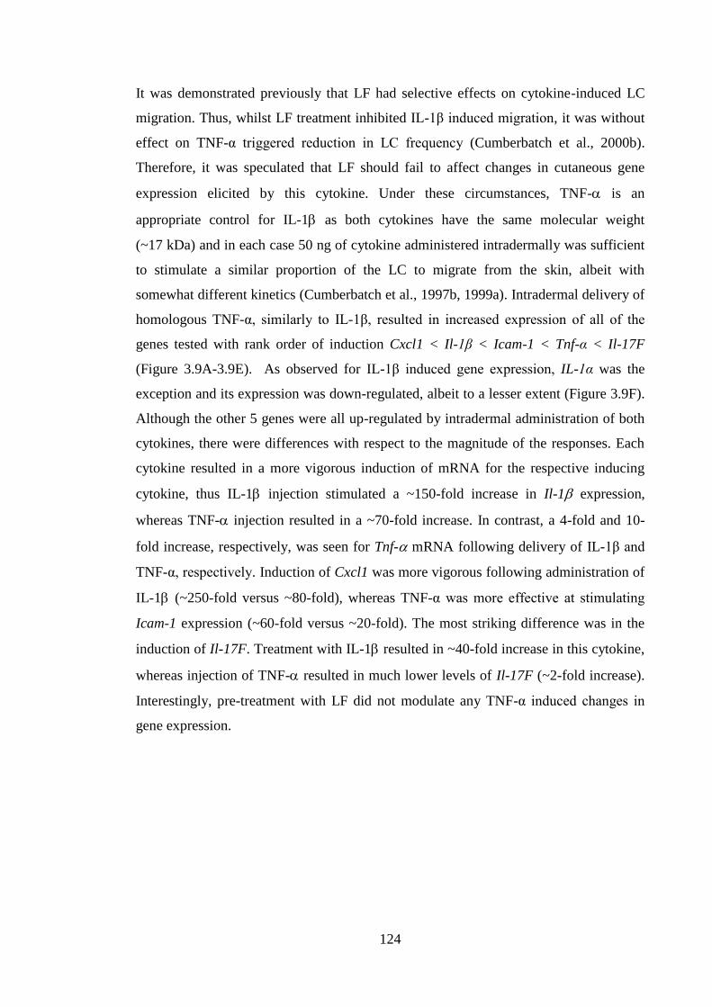
124
It was demonstrated previously that LF had selective effects on cytokine-induced LC
migration. Thus, whilst LF treatment inhibited IL-1β induced migration, it was without
effect on TNF-α triggered reduction in LC frequency (Cumberbatch et al., 2000b).
Therefore, it was speculated that LF should fail to affect changes in cutaneous gene
expression elicited by this cytokine. Under these circumstances, TNF- is an
appropriate control for IL-1 as both cytokines have the same molecular weight
(~17 kDa) and in each case 50 ng of cytokine administered intradermally was sufficient
to stimulate a similar proportion of the LC to migrate from the skin, albeit with
somewhat different kinetics (Cumberbatch et al., 1997b, 1999a). Intradermal delivery of
homologous TNF-α, similarly to IL-1β, resulted in increased expression of all of the
genes tested with rank order of induction Cxcl1 < Il-1β < Icam-1 < Tnf-α < Il-17F
(Figure 3.9A-3.9E). As observed for IL-1β induced gene expression, IL-1α was the
exception and its expression was down-regulated, albeit to a lesser extent (Figure 3.9F).
Although the other 5 genes were all up-regulated by intradermal administration of both
cytokines, there were differences with respect to the magnitude of the responses. Each
cytokine resulted in a more vigorous induction of mRNA for the respective inducing
cytokine, thus IL-1 injection stimulated a ~150-fold increase in Il-1 expression,
whereas TNF- injection resulted in a ~70-fold increase. In contrast, a 4-fold and 10-
fold increase, respectively, was seen for Tnf- mRNA following delivery of IL-1β and
TNF-α, respectively. Induction of Cxcl1 was more vigorous following administration of
IL-1 (~250-fold versus ~80-fold), whereas TNF-α was more effective at stimulating
Icam-1 expression (~60-fold versus ~20-fold). The most striking difference was in the
induction of Il-17F. Treatment with IL-1 resulted in ~40-fold increase in this cytokine,
whereas injection of TNF- resulted in much lower levels of Il-17F (~2-fold increase).
Interestingly, pre-treatment with LF did not modulate any TNF-α induced changes in
gene expression.
125
0 L F
0
5
1 0
1 5
TN
F-
fold
ch
an
ge
T N F - 5 0 n g
0 L F
0
5 0
1 0 0
1 5 0
IL-1
fold
ch
an
ge
T N F - 5 0 n g
0 L F
0
5 0
1 0 0
1 5 0
CX
CL
1
fold
ch
an
ge
T N F - 5 0 n g
0 L F
0
1
2
3
4
5
IL-1
7F
fold
ch
an
ge
T N F - 5 0 n g
0 L F
0
5 0
1 0 0
1 5 0
ICA
M-1
fold
ch
an
ge
T N F - 5 0 n g
0 L F
0 .0
0 .5
1 .0
1 .5
2 .0
IL-1
fold
ch
an
ge
T N F - 5 0 n g
A B
C D
E F
Figure 3.9 Lactoferrin did not affect cytokine expression following injection of
homologous, recombinant TNF-α.
Young (6-8 weeks) BALB/c strain mice were treated with 0.5μg of LF in 30μl of aqueous
cream or cream alone on the dorsum of both ears. Two h later animals were injected
intradermally with 50ng of homologous TNF-α. After 4h mice were sacrificed and ears
were excised and immersed in RNAlater. Subsequently, tissue was homogenised in a
lysis buffer using a mechanical homogeniser. mRNA was extracted, and then cDNA was
prepared and utilized in qPCR reaction using TaqMan primers. Expression of Tnf-α
(A), Il-1β (B), Cxcl1 (C), Il-17F (D), Icam-1 (E), Il-1α (F) was measured relative to the
Hprt1 gene. Results are presented as a fold change in gene expression relative to
untreated (naive) animals (mean ± SEM). The statistical significance of differences in
gene expression triggered by TNF-α in the absence of LF and in the presence of LF was
assessed by a Mann-Whitney test. Graphs present data from a single experiment (n=4-6
ears per group).
126
Finally, the impact of the other IL-1 family member, IL-1 on a cutaneous gene
expression, and the ability of LF to modulate IL-1 induced changes, has been
explored. Interleukin-1 has a very similar profile of biological activity to IL-1 and
acts through the same signalling receptor (the IL-1R1) (Jensen, 2010). However, the
release of these cytokines can be induced by different stimuli. Thus, whereas IL-1 has
been shown to play a role in allergen-induced LC migration (Cumberbatch et al.,
1997a), IL-1α has been demonstrated to be critical for irritant (SLS)-induced LC
migration (Cumberbatch et al., 2002a). Given the fact that the two molecules signal
through the same receptor, it was anticipated that administration of IL-1 would induce
a similar profile of gene expression changes to those observed for IL-1 and that LF
would display a similar ability to modulate these effects. The same protocol was used as
that utilized in the experiments presented in the Figures 3.8 and 3.9; intradermal
injection of 50ng of cytokine and isolation of tissue 4h later. Delivery of homologous
IL-1α resulted in up-regulation of the majority of the genes tested with rank order of
induction Cxcl1< Il-17F < Il-1β < Icam-1 < Tnf-α, similar to those changes induced by
IL-1β and TNF-α. Again, the only gene that was not up-regulated was Il-1α (Figure
3.10F). Although the rank order of the increases in gene expression were very similar to
those observed following administration of IL-1, there were some differences with
respect to the magnitude of the response. In particular, induction of Il-1 was
considerably less vigorous (~25-fold compared with ~80-fold) and increases in Il-17F
transcripts were also less marked. These differences may reflect subtle differences in
kinetics or inter-animal variations. In addition, it should be noted that due to failed
intradermal injections at some sites, the IL-1 control group only contains 3 replicates
rather than 6 replicates. Given the rather marked inter-animal variation in Il-1 and Il-
17F expression observed in the IL-1β treated animals (Figure 3.8), this reduction in
group size may well explain the lower levels of these cytokines recorded. Prior
treatment with LF was without effect on Il-1 and Il-17F mRNA expression, but did
result in a significant decrease in expression of Cxcl1 (Figure 3.10C). Interestingly,
contrary to the injection of IL-1β and TNF-α that resulted in the induction of their own
transcripts, delivery of IL-1α was not able to up-regulate expression of Il-1α within the
time frame investigated, suggesting a different mechanism for regulation of Il-1α
transcription than that of the other proinflammatory cytokines Il-1β and Tnf-α.
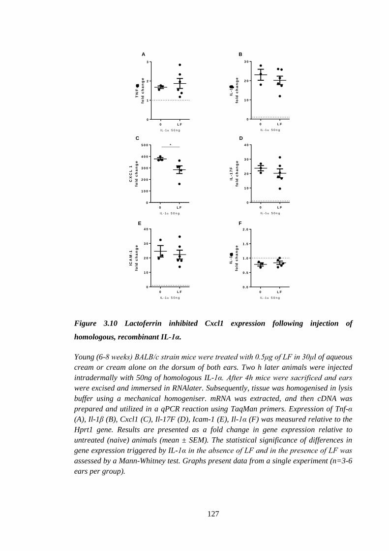
127
0 L F
0
1
2
3
TN
F-
fold
ch
an
ge
IL -1 5 0 n g
0 L F
0
1 0
2 0
3 0
IL-1
fold
ch
an
ge
IL -1 5 0 n g
0 L F
0
1 0 0
2 0 0
3 0 0
4 0 0
5 0 0
CX
CL
1
fold
ch
an
ge
IL -1 5 0 n g
*
0 L F
0
1 0
2 0
3 0
4 0
IL-1
7F
fold
ch
an
ge
IL -1 5 0 n g
0 L F
0
1 0
2 0
3 0
4 0
ICA
M-1
fold
ch
an
ge
IL -1 5 0 n g
0 L F
0 .0
0 .5
1 .0
1 .5
2 .0IL
-1
fold
ch
an
ge
IL -1 5 0 n g
A B
C D
E F
Figure 3.10 Lactoferrin inhibited Cxcl1 expression following injection of
homologous, recombinant IL-1α.
Young (6-8 weeks) BALB/c strain mice were treated with 0.5μg of LF in 30μl of aqueous
cream or cream alone on the dorsum of both ears. Two h later animals were injected
intradermally with 50ng of homologous IL-1α. After 4h mice were sacrificed and ears
were excised and immersed in RNAlater. Subsequently, tissue was homogenised in lysis
buffer using a mechanical homogeniser. mRNA was extracted, and then cDNA was
prepared and utilized in a qPCR reaction using TaqMan primers. Expression of Tnf-α
(A), Il-1β (B), Cxcl1 (C), Il-17F (D), Icam-1 (E), Il-1α (F) was measured relative to the
Hprt1 gene. Results are presented as a fold change in gene expression relative to
untreated (naive) animals (mean ± SEM). The statistical significance of differences in
gene expression triggered by IL-1α in the absence of LF and in the presence of LF was
assessed by a Mann-Whitney test. Graphs present data from a single experiment (n=3-6
ears per group).
128
3.3 Discussion
In this chapter, the aim was to examine further the mechanism by which LF inhibits
oxazolone-induced LC migration. The focus of the experiments described in this section
was on the events in the skin, as well as in the draining LN. Various endpoints were
measured to advance the understanding of the mode of action of LF.
Firstly, dermal cytokine (protein) production in ear tissue was assessed following
treatment with oxazolone in the presence or the absence of LF. It was previously
established that the process of LC migration is dependent on cytokine signals derived
from a variety of cells present in the cutaneous environment. These in turn affect the
expression of chemokines, receptors and proteases that lead to LC mobilisation
(Griffiths et al., 2005). Two key cytokines in the events of LC migration are TNF-α and
IL-1β (Cumberbatch et al., 1997b). However, IL-1α was also found to be important
(Cumberbatch et al., 2002a). In the current investigations, the effect of LF on
oxazolone-induced cytokine secretion in vivo was assessed using ear explant model. It
was demonstrated that there was a relatively high production of all of the measured
cytokines in naive samples. Exposure to oxazolone resulted in an up-regulation of IL-1α
and IL-1β protein expression, whereas levels of TNF-α and IL-17F remained similar to
those found in the naive samples. Prior treatment with LF was largely without effect on
cytokine production. There was a small reduction in IL-1β levels but such did not reach
statistical significance. Measurement of cytokine protein production in the skin, both in
mice and in humans, is a challenging task. There is no non-invasive method to obtain
human skin samples. Many studies utilize specimens from circumcisions, plastic
surgeries or punch biopsies which could be utilized later in Western blot (WB) analysis
of homogenised skin or assessed by IHC techniques. Alternatively, studies in humans
have utilized vacuum suction to create cutaneous blisters and subsequently analyze
blister fluid for the presence of various proteins; such blister fluid, which derives from
the local microenvironment, is thought to resemble serum in protein content. In fact, LF
was shown to inhibit TNF-α production in suction blister fluid following injection of
IL-1β (Cumberbatch et al., 2003). It is considerably more difficult to measure cytokine
production in mouse skin. There are reports, whereby dorsal skin biopsies were taken
from mice, homogenised and analyzed by either by ELISA or WB for cytokine
expression (Osborne-Hereford et al., 2008). However, the majority of murine skin is
129
covered in hair impeding access to the epidermis, and making sterilisation more
demanding, partially explaining why mouse ears are so often used in exploration of
cutaneous biology. Utilization of suction blister approach is not viable in mice, as
animal would have to be anesthetised, therefore increasing the amount of experimental
procedures, and further, the sample size would be minute. In our laboratory, however,
an explant model was utilized, whereby following treatment with allergen ears were
excised, divided into dorsal and ventral parts and dorsal part was floated onto the
culture media. Next, released proteins were measured in the culture media. However, in
addition to chemical and biological insults causing inflammatory cytokine production in
the skin, physical insults, including heat and pressure, could have the same effect
(Dearman et al., 2004; Schwacha et al., 2008). Therefore, the physical disruption of the
ear was thought to be responsible for relatively high levels of all of the cytokines
measured observed in naive samples. Indeed, it was reported by Cumberbatch et al.
(2005) that following separation of the ear there was high constitutive production of
IL-6 that was not further induced following exposure to 1% DNCB or 25% TMA
(Cumberbatch et al., 2005). Interleukin 6 is a pleiotropic cytokine that has been
demonstrated to be produced following induction with PAMP and DAMP by many cells
types including DC, keratinocytes and endothelial cells (Tanaka and Kishimoto, 2012).
It was demonstrated also that IL-6 plays a role in wound healing, as mice deficient in
IL-6 displayed delayed wound healing following punch biopsy wound, as demonstrated
by delayed wound closure and re-epithelialization (Lin et al., 2003).
It was reported that following treatment with an allergen there was an induction of
TNF-α protein in the skin (Flint et al., 1998; Kaplan et al., 2012). However, contrary to
those reports this was not observed in the current experiments. It is possible, that the
trauma of ear separation obscured oxazolone-induced cytokine production.
Additionally, in the current study only a single time point was utilized (4h). It is
possible that TNF-α production had already peaked at an earlier time point. It was
demonstrated previously that TNF-α levels in the serum of LPS injected rabbits peaked
2h after endotoxin exposure and the half life of the cytokine was established to be
relatively short and was 6-7 minutes (Beutler et al., 1985). Similarly, Oliver et al.
(1993) demonstrated that TNF-α production, following exposure of the whole blood to
LPS, also peaked after 2h, and the TNF-α half-life was approximately 18 minutes
(Oliver et al., 1993). These results suggested that in the current study oxazolone-
130
induced TNF-α production could have peaked before the samples were collected,
explaining the lack of oxazolone-induced, TNF-α production. Additionally, explant
samples were incubated ex vivo for 16h, in which time TNF-α molecule could have been
degraded. Half-lives of pro-forms of IL-1α and IL-1β were established to be 15h and 3h,
respectively, and their release was recorded to take place 12h and 2h after activation of
human monocytes with LPS (Hazuda et al., 1988). Long half-life of IL-1α might
partially explain high levels of this protein recorded in the current study. Overall, it
would have been helpful to perform a time course of cytokine production by skin
explants to assess the time dependent changes following exposure to allergen.
The baseline levels of measured cytokines were relatively high due to apparently
traumatic nature of sample preparation, and not markedly induced by oxazolone. It was
decided to investigate the baseline cytokine expression utilising a method that did not
require an excessive handling of the tissue. To achieve that qPCR was used, whereby
whole ear was used for mRNA isolation. It was found that treatment with LF on its own
did not modulate the expression of genes tested at any of the time points with the
exception of Icam-1 4h following treatment. In the skin ICAM-1 is constitutively
expressed by endothelial cells; additionally its expression can be induced by IFN-γ and
TNF-α in keratinocytes (Griffiths et al., 1989). Observed levels of Icam-1 expression
up-regulation were very low and most probably induced in endothelial cells as there was
no increase in Tnf-α expression preceding up-regulation in Icam-1 expression. Low
levels of Icam-1 expression were also demonstrated in LC cultured in vitro for 12 h and
interaction between ICAM-1 and LFA-1 was shown to be crucial for the ability of LC to
stimulate proliferation of allogeneic T cells (Simon et al., 1991). The effect of LF on
ICAM-1 expression was not without precedent. Indeed, LF was demonstrated to
regulate ICAM-1 expression in human umbilical vein endothelial cells (HUVEC)
following activation with TNF-α. Kim et al. (2012) reported that LF competed with
NF-ƘB for ICAM-1 promotor, therefore inhibiting its expression (Kim et al., 2012). In
contrast, this study found an increase in Icam-1 expression following exposure to LF.
However, it is possible that, as Kim et al. (2012) have shown, following binding of
ICAM-1 promoter and in the absence of TNF-α, LF triggered Icam-1 expression. As
reported previously, LF has dual influences on immune responses and it is plausible that
in the presence of stimuli (TNF-α) LF inhibits ICAM-1 expression, as reported by Kim
et al. (2012), but on its own, LF was able to increase the level of Icam-1 transcription.
131
However, there was no lasting effect of LF treatment in the steady state. Therefore, in
addition to examining expression of cytokines it would have been of interest to
investigate the induction of enzymes responsible for antioxidant defence, as oxidative
stress is known to be induced in the skin after exposure to allergens; that in turn
contributes to the danger signalling and subsequent activation of DC (Corsini et al.,
2013). Furthermore, it was observed before that LF was able to induce (by
approximately 2-fold) the steady state expression of genes involved in oxidative stress
management (GSH peroxidase 1 and 4, prostaglandin-endoperoxide synthase 1,
peroxiredoxins and superoxide dismutase 1) in the whole blood cells, following 2h
incubation with 10μg/ml of LF (Kruzel et al., 2013).
Next, the impact of LF on oxazolone-induced gene expression was investigated. As
previously stated, treatment with allergen has been shown to result in the induction of
epidermal TNF-α mRNA expression (Enk and Katz, 1992a). In the study by Enk and
Katz (1992a) there was a rapid induction of Tnf-α, Il-1α and Il-1β expression 1h after
exposure to 3% TNCB, which was still evident 24h after treatment. However, the
current study did not confirm these results. There are many reasons for these
discrepancies, such as different allergens and techniques used to detect expression. In
the Chapter 6 it is reported that there are differences between allergens with regards to
their ability to induce cytokine expression, therefore, it is possible that TNCB,
especially at a somewhat high concentration of 3%, was more efficient at inducing
TNF-α expression. Moreover, the technique utilized by Enk and Katz (1992) was
different. Briefly, they performed PCR on cDNA and obtained cytokine specific
products. Subsequently, PCR products were hybridised with cytokine-specific,
radioactive probes; later electorophoresis was completed and radioactivity was assessed.
That method was semi-quantitative, as the intensity of the band was assessed. It is
possible that by utilising that method a weak cytokine signal could have been amplified.
In the current study the induction of Tnf-α expression was minimal until 120h after
treatment with oxazolone. Similarly, expression of Il-1β was approximately 10 times
higher than that in untreated animals 4h following exposure to oxazolone, until it rose to
30 fold 5 days after treatment, which is likely to represent the beginning of an elicitation
response (Saint-Mezard et al., 2003). Conversely, no induction of Il-1α expression was
observed at any time point. It is surprising, as an induction of IL-1α protein was
observed 4h after allergen treatment. These results suggest that secretion of IL-1α in the
132
skin is not regulated at the level of transcription (Jensen, 2010). Expression of
chemokine Cxcl2 (Mip2) was measured by Katz and Enk (1992) and it was increased 4h
after application of TNCB. In the current study induction of chemokine CXCL1, which
together with CXCL2 signals via the CXCR2 receptor (Zaja-Milatovic and Richmond,
2008) was measured. In accordance with the results of Enk and Katz (1992), rapid
induction of Cxcl1 was observed that declined after 72h to levels 2 times higher than
those observed in naive controls. It was shown previously, utilising respiratory
sensitization of mice with OVA, that Cxcl1 expression was detected as soon as 1h
following exposure to OVA and peaked 3h after treatment. Subsequently, neutrophils
were detected in the lungs of sensitized mice after 3h and maximum neutrophil numbers
were detected 6h after exposure to OVA (Knott et al., 2001). Results by Knott et al.
(2001) suggested that CXCL1 is rapidly induced in allergic reactions and its primary
role is to recruit neutrophils to the site of allergen exposure. It was established
previously that neutrophils have a role in the challenge phase of hypersensitivity
(Dilulio et al., 1999), but not in the sensitization events, therefore, it would be of
particular interest to investigate the role of neutrophils in the primary phase of response
to allergen. Expression of Icam-1 was still increasing 120h after allergen exposure,
similar to expression of Tnf-α and Il-1β. Expression of ICAM-1 was demonstrated
previously 4h following challenge with oxazolone, as well as after intradermal cytokine
(TNF-α, IL-1α and IL-1β) delivery (McHale et al., 1999). Taken together results by
McHale et al. (1999) and presented in the current study suggested that induction of
Icam-1 expression begins following primary exposure to oxazolone and then it is further
induced after allergen challenge. It was demonstrated, as previously mentioned, that the
presence of ICAM-1 was shown to be important for LC-induced T cell proliferation
(Simon et al., 1991). Moreover, following challenge with DNFB, presence of ICAM-1
on endothelial cells was crucial for recruitment and activation of CD8+ T cells and their
subsequent production of IFN-γ and IL-17 (Kish et al., 2011). Finally, the expression of
Il-17F was rapidly induced 2h after treatment with oxazolone and afterwards it
decreased to a level below that present in untreated controls. Prior treatment with LF did
not modulate any of the genes at the time point when an effect on LC migration was
observed in vivo (4h). However, there was a statistically significant effect of LF
observed at 120h, whereby LF increased Il-17F expression following oxazolone
treatment accelerating the return of Il-17F expression levels to those found in untreated
133
controls. Although statistically significant, this result is not likely to be biologically
relevant.
Exposure to oxazolone revealed a differential cytokine patterns over time that help to
understand the immunological mechanisms of sensitization. Investigated kinetics
spanned 5 days and it could be speculated that increase in Tnf-α and Il-1β expression
observed from 4th
and 3rd
day, respectively, could be a result of the adaptive immune
response, whereby allergen was still present on the surface of the skin following initial
exposure and was providing an elicitation signal (Saint-Mezard et al., 2003). It was
demonstrated previously that IL-1β at the level of mRNA and protein was rapidly (15
and 30 minutes, respectively) detected in the skin, in both epidermis and dermis,
following exposure to oxazolone (Kermani et al., 2000) and preceded TNF-α production
in the skin, both in the form of mRNA and protein, which was observed after 30
minutes and 1h, respectively (Flint et al., 1998). Based upon these results, it can be
concluded that the increase in the expression of Tnf-α and Il-1β observed 72h after
initial exposure was due to the initiation of adaptive immune response (Asada et al.,
1997). Rapid up-regulation of expression of neutrophil chemoattractant CXCL1 was
observed 4h following oxazolone exposure and declined thereafter. Previous reports
suggested that CXCL1 expression occurs rapidly, within 30 minutes, following
challenge with 0.2% of DNFB of sensitised and non-sensitized skin of BALB/c strain
mice (Dilulio et al., 1999). The presence of neutrophils was demonstrated to be
necessary to achieve the optimum CHS. It was shown that neutrophils were crucial for
the recruitment of CD8+ T cells, as when CXCL1 was inhibited, the CHS reaction
decreased, but could be rescued by a transfer of hapten specific CD8+ T cells (Dilulio et
al., 1999). Similarly, it was established that in mice deficient in CXCR2 (CXCL1
receptor) CHS reaction to DNFB and oxazolone, as measured by ear swelling, was
decreased (Cattani et al., 2006).
Whilst there were no major changes in the cutaneous cytokine production and
expression recorded in the presence of LF, it was decided that the downstream events of
LNC proliferation and LN cytokine production would be investigated. It is known that
exposure to an allergen results in LN hypertrophy caused by DC and lymphocyte
migration (PhD thesis Hayes, 2012; Sikorski et al., 1996). A slight increase in LNC
number was observed as soon as 6h following oxazolone exposure; by 24h the increase
was statistically significant and it continued to increase until 120h. The data recorded in
134
this chapter were in agreement with previously published reports, whereby there was an
early allergen-induced increase in LN cellularity (PhD thesis Hayes, 2012). The rapid
augmentation of LN cellularity is a result of enhanced lymphocyte influx into the LN
and simultaneous restriction of their exit; process termed the LN shut down. It is
hypothesised that the main purpose of the LN shut down is to increase the probability of
CD4+ T cells to recognize the antigen that is presented by arriving DC from the skin
(McConnell et al., 1980). Furthermore, it was shown that there was a marked increase
in LNC proliferation following application of oxazolone in comparison with naive
animals. The proliferative response was the highest 72h after exposure to oxazolone and
deceased to levels similar to those observed in naive animals after 120h. Observed
changes to LNC proliferation reflect increased division of allergen specific T cells that
take place following encounter with DC carrying the hapten. A subsequent decrease in
the ability of cells to divide was driven by changing populations of LNC that after 120h
are characterized by a presence of non-proliferating memory T cells that will become
activated upon future exposure to the same allergen, as well as an increase in B cell
numbers (Sikorski et al., 1996). It was found that LF did not modulate cellularity or LN
proliferation at 72, 96 or 120h time points. A possible reason for that might be that
contact allergens are known to persist in the skin for up to 14 days after the first
exposure, and even single application of allergen (oxazolone or FITC) was able to result
in CHS reaction in mice (Saint-Mezard et al., 2003). In this section it was shown that
exposure to oxazolone over a somewhat longer time frame (72-120h) resulted in a
steady increase of Tnf-α, Il-1β and Icam-1 expression. Therefore, it is not surprising that
while LF did not affect cytokine expression after allergen treatment that it was also
without effect on secondary events in the LN. In the Chapter 6 it was presented also that
the effect of LF on LC migration was transient, therefore, suggesting that LF might be
metabolized or not able to overcome constant stimulation from allergen present in the
skin (Saint-Mezard et al., 2003).
Lymph node cells were reported to produce cytokines following exposure to allergens.
The type of cytokines produced depends on the type of sensitising chemical, which in
turn relies on the quality of immune response triggered by that substance (Dearman and
Kimber, 1999). Chemical allergens have been demonstrated to polarize immune
response, with contact and respiratory allergens resulting in Th1 and Th2-skewed
responses, respectively, and subsequent production of either IFN-γ or IL-4 and IL-13
135
(Dearman and Kimber, 1999). In this section it was found that following stimulation
with oxazolone for 72h to 120h LNC produced cytokines that were markers of both Th1
and Th2 immune response. The relatively short 5 day protocol that was utilized in the
current study was insufficient to result in full polarisation of immune response. Typical
polarisation protocols involve treatment with allergen on the flank on day 0 and day 5,
followed by allergen application to the ear on days 10, 11 and 12 and termination on
day 13 (Dearman and Kimber, 1999). There was no effect of LF on IFN-γ production by
LNC at any of the time points. In contrast, treatment with LF resulted in increased
production, albeit not statistically significant, of IL-13 at 120h. It was reported
previously that the application of LF 1h before the final exposure to respiratory allergen,
toluene diisocyanate, in a chronic exposure protocol, increased production of IL-4
(Zimecki et al., 2012). Accordingly, LF was demonstrated to inhibit antigen specific
proliferation of Th1, but not Th2, specific cell line (Zimecki et al., 1996). Inhibitory
effects on Th1 mediated response might explain an increase in Th2 involved cytokine
IL-4, and could partially be responsible for the increase in IL-13 production observed in
the current study, despite the lack of effect of LF on IFN-γ production by LNC.
Interestingly, while the production of IFN-γ decreased with time, levels of IL-13
remained comparable at all time points tested. The observed decrease in IFN-γ
production was consistent with the decrease in cell proliferation. Interferon γ was
demonstrated to be derived from CD8+ cells in the LN as well as in the skin (Xu et al.,
1996). It is possible that the observed decrease in LN cell proliferation and IFN-γ
production was caused by CD8+
cell migration to the skin, as it was reported previously
that 120h following initial exposure to oxazolone CD8+ cells and IFN-γ were detected
in the skin. Moreover, ear swelling was demonstrated to increase 120h after initial
exposure to allergen (Saint-Mezard et al., 2003). These reports are in accordance with
data from the current study, wherein expression of Tnf-α and Il-1β rose 120h following
exposure to oxazolone.
Subsequently, production of another cytokine, IL-17, was considered. Interleukin 17 (A
and F) belongs to a recently identified family of cytokines. They are produced by a
newly described subset of Th17 cells, but also by cells of the innate immune system:
DC, macrophages, NK cells and γδ T cells (Onishi and Gaffen, 2010); and is thought to
play a key role in the first line of host defence. Interleukin 17 has been identified as an
early marker of ACD (PhD thesis Hayes, 2012), and the current study supported that
136
observation. Production of IL-17A and F was observed in the LN 6h following exposure
to oxazolone. In our laboratory two LN preparation protocols were used; one that
utilized enzymes (collagenase and DNase) and another that did not utilize enzymes and
relied solely on mechanical disaggregation. It was found that different preparation
techniques affected IL-17 production by LN following exposure to allergen. Thus, early
(6h) IL-17 production was not observed when LN were prepared by mechanical
disruption alone, whereas there was a detectable production of IL-17 24h after allergen
treatment. However, levels of IL-17 observed 24h after oxazolone exposure from
mechanically prepared LN were almost 5 times lower than those from LN cultures
prepared with enzymes. It has to be noted that the early (6h) IL-17 production is
attributable to innate cells of the immune system, γδ T cells, as it is too early for
polarized Th or Tc responses to develop. In fact, γδ T cells have been shown to be the
cellular source of IL-17 in other independent experiments in this laboratory (PhD thesis
Hayes, 2012). γδ T cells comprise 3% of LN and spleen cells in rodents. The proportion
of these T cells is much higher in the sites of contact with the external environment and
γδ T cells can sometimes be the only T cells present in the intra-epidermal spaces. In
murine skin, γδ T cells are present in the epidermis as well as in the dermis, and they
participate in maintaining cutaneous homeostasis via secretion of chemokines and
cytokines (Macleod and Havran, 2011). In the mouse epidermis γδ T cells are found in
the proximity of LC (personal communication, Kieran Mellody). It was demonstrated
in our laboratory that IL-17 is produced by LNC as soon as 6h following exposure to
DNCB. It also was found that the main cell subset responsible for production of this
cytokine was γδ T cells (PhD thesis Hayes, 2012). It has been established that γδ T cells
produce IL-17 in response to IL-1β and IL-23 (Sutton et al., 2009). It is possible that
following allergen exposure, DC arriving in the LN were a source of IL-1β, therefore
stimulating IL-17 production by γδ T cells resident in the LN. It could be speculated
that the lack of IL-17 signal in the absence of enzymatic digestion of LN was due to γδ
T cells or DC being “trapped” within the LN capsule, and only upon treatment with
collagenase and DNase these cells were retrieved and able to receive or give a cytokine
(IL-1β) signal that stimulated IL-17 expression by γδ T cells. γδ T cells responsible for
IL-17 production in the LN were most likely LN resident or dermal γδ T cells but not
DETC. It was reported previously that dermal γδ T cells were producing IL-17 and
capable of migration (Gray et al., 2011; Nielsen et al., 2014), conversely, DETC were
137
able to migrate to the LN following exposure to DNFB but they did not produce IL-17A
or IFN-γ (Nielsen et al., 2014).
Treatment with LF prior to exposure to oxazolone had different effects depending on
the single cell preparation procedure. Thus, in the samples prepared via mechanical
disaggregation LF resulted in a statistically significant increase in the number of cells in
the LN. However, the opposite effect was observed in the samples prepared via
enzymatic method, albeit the effect was not statistically significant. Cumberbatch et al.
(2000) reported that LF inhibited DC accumulation in the LN 18h following oxazolone
treatment. In the aforementioned report mechanical method of LN preparation was
utilized. However, the inhibition of DC accumulation observed by Cumberbatch et
al.(2000) was not likely to have contributed to the change in the total LN cellularity, as
reported numbers of DC comprised only 0.1% of all LN cells (Cumberbatch et al.,
2000b). Opposing effects of LF on LN cellularity that depend on the method of single
cell suspension preparation were perplexing. Cytokine production results suggested that
there were marked differences in the effects of mechanical and enzymatic methods. It is
possible that LF induced increased level of an unknown cell subset migration to the LN,
and these cells were susceptible to enzyme-induced cell death and were not able to be
enumerated. However, during mechanical method of LN processing that same unknown
population could be enumerated and would account for the increased cellularity. Despite
the differences in LN cellularity, secretion of IL-17 was measured by standardised
number (107) of cells per well, allowing for comparisons between groups. To explain
the differences in cytokine levels between mechanical and enzymatic LNC retrieval, one
could speculate that it was possible that during a mechanical LN disaggregation some of
the migratory DC from the skin were present close to the LN capsule, as they were
inhibited in their migration by LF, and therefore were not completely retrieved during
mechanical disaggregation. As a consequence, they could have not supplied γδ T cells
with a signal to produce IL-17. In the situation where enzymes were utilized to achieve
single cell suspension, the inhibitory effect of LF on IL-17 production was no longer
visible. It is plausible that in the latter scenario all of the DC were “freed” and
subsequently able to interact with LN γδ T cells and trigger them to release IL-17 that
was no longer affected by LF.
It is not known which cell type LF is directly acting on in the skin, therefore, two
hypotheses are possible. Firstly, LF could impact on γδ T cells. It was demonstrated
138
previously that LF is capable of interacting with T cells (Legrand et al., 1997), and
expression of functional LFR was demonstrated by human γδ T cells, as measured by
binding of radiolabelled LF to decidual γδ T cells (Mincheva-Nilsson et al., 1997).
Interaction of γδ T cells with LF could be developmentally justified as both are present
in the epithelium and mucosal barriers (Girardi, 2006; Lönnerdal and Iyer, 1995).
Therefore, following application of LF to the skin it could inhibit TNF-α secretion by
DETC or dermal γδ T cells; further, LF could increase DETC migration to the LN,
which would explain increased cellularity in the LN and decreased IL-17 production [as
DETC do not produce IL-17 in the LN (Nielsen et al., 2014)]. Secondly, LF could affect
DC, LC and/or DC. It was demonstrated previously that LF had an effect on the extent
of DC activation and cytokine output (de la Rosa et al., 2008), and therefore could
modulate DC in a way that upon their arrival in the LN their ability to trigger IL-17
production was diminished. Alternatively, a combination of these two scenarios is
possible. Indeed, it was shown that human γδ T cells from circulating blood, activated
by stimulation phospho antigen, were able to induce DC maturation via TNF-α
production, as measured by an increase in CD86, CD83 and MHC II expression by DC.
Additionally γδ T cells increased IL-12 production by DC, which was dependent on
IFN-γ secretion by γδ T cells (Ismaili et al., 2002). Further interactions between two cell
types were demonstrated by Conti et al. (2005), whereby upon co-culture of DC and γδ
T cells, the latter increased expression of CD25 and CD69, as well as production of
TNF-α and IFN-γ (Conti et al., 2005). Results from human cells were confirmed by
Dieli et al. (2004) using murine γδ T cells and DC. They demonstrated that IFN-γ
production by γδ T cells was dependent upon DC-derived IL-12 (Dieli et al., 2004).
Reports of interaction between DC and γδ T cells are of specific interest in the immune
surveillance of the skin where these two cell types co-exist. It is possible that LF could
impact on one or both cell types in order to achieve inhibition of LC migration and
modulation of IL-17 production by LNC. It would be of particular importance to
perform studies where mouse skin was treated with radio-labelled LF to allow for
identification of the cell target of LF.
Lactoferrin was shown to decrease oxazolone triggered IL-17 production by LN cells.
There were no LF-induced changes to oxazolone provoked gene expression in the skin
or LNC proliferation. One of the obstacles in elucidating the mechanism of action of LF
was the unknown mode of oxazolone action in the skin. Indeed, despite many advances
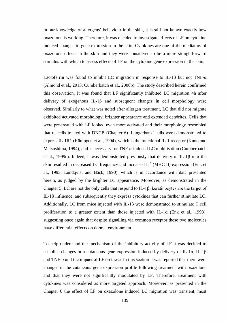
139
in our knowledge of allergens’ behaviour in the skin, it is still not known exactly how
oxazolone is working. Therefore, it was decided to investigate effects of LF on cytokine
induced changes to gene expression in the skin. Cytokines are one of the mediators of
oxazolone effects in the skin and they were considered to be a more straightforward
stimulus with which to assess effects of LF on the cytokine gene expression in the skin.
Lactoferrin was found to inhibit LC migration in response to IL-1β but not TNF-α
(Almond et al., 2013; Cumberbatch et al., 2000b). The study described herein confirmed
this observation. It was found that LF significantly inhibited LC migration 4h after
delivery of exogenous IL-1β and subsequent changes in cell morphology were
observed. Similarly to what was noted after allergen treatment, LC that did not migrate
exhibited activated morphology, brighter appearance and extended dendrites. Cells that
were pre-treated with LF looked even more activated and their morphology resembled
that of cells treated with DNCB (Chapter 6). Langerhans’ cells were demonstrated to
express IL-1R1 (Kämpgen et al., 1994), which is the functional IL-1 receptor (Kuno and
Matsushima, 1994), and is necessary for TNF-α-induced LC mobilisation (Cumberbatch
et al., 1999c). Indeed, it was demonstrated previously that delivery of IL-1β into the
skin resulted in decreased LC frequency and increased Ia+ (MHC II) expression (Enk et
al., 1993; Lundqvist and Bäck, 1990), which is in accordance with data presented
herein, as judged by the brighter LC appearance. Moreover, as demonstrated in the
Chapter 5, LC are not the only cells that respond to IL-1β; keratinocytes are the target of
IL-1β influence, and subsequently they express cytokines that can further stimulate LC.
Additionally, LC from mice injected with IL-1β were demonstrated to stimulate T cell
proliferation to a greater extent than those injected with IL-1α (Enk et al., 1993),
suggesting once again that despite signalling via common receptor these two molecules
have differential effects on dermal environment.
To help understand the mechanism of the inhibitory activity of LF it was decided to
establish changes in a cutaneous gene expression induced by delivery of IL-1α, IL-1β
and TNF-α and the impact of LF on these. In this section it was reported that there were
changes in the cutaneous gene expression profile following treatment with oxazolone
and that they were not significantly modulated by LF. Therefore, treatment with
cytokines was considered as more targeted approach. Moreover, as presented in the
Chapter 6 the effect of LF on oxazolone induced LC migration was transient, most
140
likely due to allergen persistence in the skin. Consequently, a more transient stimulant
was required to investigate the persistence of LF effects. However, only single time
point (4h) was examined following delivery of cytokine.
Delivery of IL-1β resulted in substantial up-regulation of cytokine expression. The
observed cytokine profile was more vigorous (higher extent of gene induction) than that
caused by oxazolone for all of the genes tested with the exception of Il-1α that was not
up-regulated following exposure to any of the cytokines or allergens utilized in these
investigations. In fact, expression of Il-1α was only ever modestly induced in the skin
(Chapter 6), whereas its expression and secretion in primary keratinocytes and in PAM
cells was readily up-regulated following exposure to both IL-1α and IL-1β. Interleukin
1α is constitutively expressed by PAM keratinocytes (Mee et al., 2005) and by
distressed dermal cells, as exemplified by ear explants in this chapter, suggesting that
regulation of IL-1α production is not controlled on the level of mRNA expression.
Moreover, IL-1α was demonstrated previously to be produced, but not released, by
murine keratinocytes, following exposure to contact allergens; levels of approximately
3000pg/ml were observed following stimulation with 10μg DNCB and 500μg
oxazolone for 24h (Corsini et al., 1998). Therefore, it was somewhat surprising that in
the whole ear tissue Il-1α expression was below the level present in naive animals
following stimulation with IL-1β, TNF-α and IL-1α. The lack of up-regulation of Il-1α
expression might suggest that mRNA for this cytokine is always present in the ear and
what is modulated is the extent of its translation. However, this theory is somewhat
contradicted by results from keratinocytes where cytokine stimulation (IL-1α and IL-1β)
resulted in up-regulation of Il-1α expression (Chapter 5). It is also plausible that the
resident immune cells in the ear provide negative feedback to keratinocytes, thereby
decreasing their transcription of Il-1α. It might potentially suggest that expression of Il-
1α is under stricter control than Il-1β.
These results suggest that IL-1α production is differentially regulated in the skin than Il-
1β with which it shares a common receptor, IL-1R1. In fact, it was demonstrated by Enk
et al. (1993) that while injection of anti-IL-1β antibody decreased TNCB induced ear
swelling, anti-IL-1α antibody was without pronounced effect (Enk et al., 1993).
Additionally, one might speculate that increasing intracellular levels of pro-IL-1α might
negatively regulate its own expression, as it was demonstrated previously that pro-IL-1α
141
is capable of intracrine signalling (Werman et al., 2004). Moreover, Boraschi et al.
(1990) demonstrated that IL-1α and IL-1β had the same stimulating ability on
thymocytes, as well as similar pyrogenic activity in rabbits. Conversely, it was
established that IL-1β, but not IL-1α, was able to enhance T cell dependent antibody
production by spleen cells after injection of sheep red blood cells (SRBC). Furthermore,
when IL-α and IL-1β were administered together, IL-1α could inhibit
immunostimulatory action of IL-1β (Boraschi et al., 1990). Similarly, Nakae et al.
(2001) showed decreased anti-SRBC antibody production in IL-1β deficient mice
(Nakae et al., 2001). These results shed a light on a dichotomy between two IL-1
isoforms, partially explaining their differential regulation in the skin observed in the
current investigations.
With the exception of Il-1α expression, injection of IL-1β resulted in up-regulation of
all of the tested genes. The highest induction was observed in Cxcl1 expression. As
discussed in the Chapter 5, treatment of primary keratinocytes and PAM cells with IL-
1β resulted in a rapid and considerable induction of Cxcl1 mRNA levels. CXCL1,
together with CXCL2, is responsible for neutrophil chemotaxis (Zaja-Milatovic and
Richmond, 2008). Kish et al. (2012) demonstrated a crucial role for IL-1β-induced
CXCL1 in recruitment of neutrophils to the allergen challenge site and subsequent
infiltration of CD8+ cells into the skin (Kish et al., 2012). In addition to up-regulating
other genes, IL-1β substantially induced its own expression. It was observed in the
Chapter 5 that upon incubation with IL-1β primary keratinocytes and HaCaT cells
moderately induced IL-1β mRNA levels, therefore suggesting that cells other than
keratinocytes were responsible for an induction of IL-1β observed in the ear.
Endothelial cells and/or fibroblasts potentially could be responsible for the high IL-1β
mRNA levels observed in the ear tissue.
In addition to measuring expression of cytokines, the level of induction of an adhesion
molecule was assessed. ICAM-1 is constitutively expressed by variety of cell types
present in the ear, including: endothelial cells, epithelial cells, fibroblasts, keratinocytes.
Moreover, ICAM-1 was demonstrated previously to be vigorously up-regulated
following stimulation with IL-1α, IL-1β and TNF-α; with the highest degree of
induction observed for combination of TNF-α and IFN-γ (Roebuck and Finnegan,
1999). Observed increase in ICAM-1 levels in the presence of inflammatory cytokines
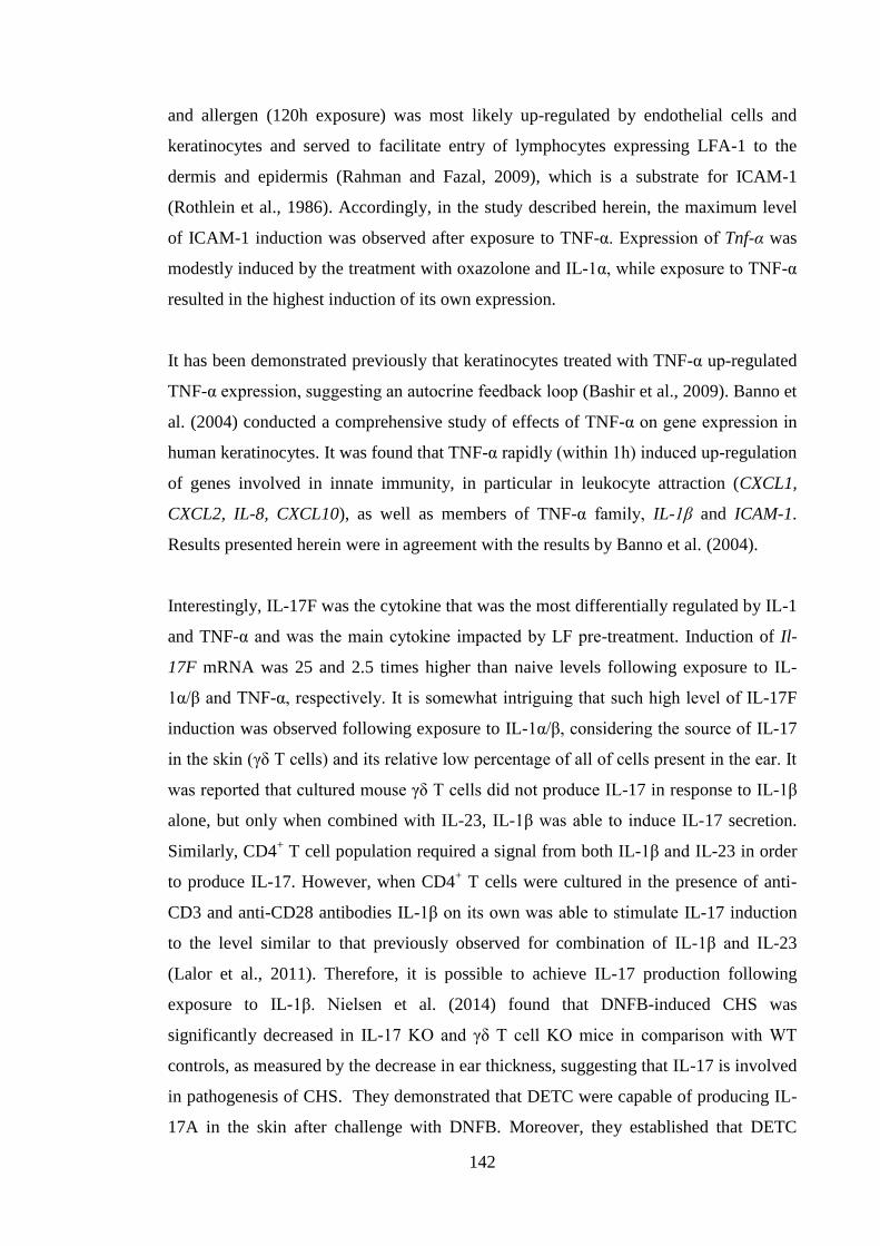
142
and allergen (120h exposure) was most likely up-regulated by endothelial cells and
keratinocytes and served to facilitate entry of lymphocytes expressing LFA-1 to the
dermis and epidermis (Rahman and Fazal, 2009), which is a substrate for ICAM-1
(Rothlein et al., 1986). Accordingly, in the study described herein, the maximum level
of ICAM-1 induction was observed after exposure to TNF-α. Expression of Tnf-α was
modestly induced by the treatment with oxazolone and IL-1α, while exposure to TNF-α
resulted in the highest induction of its own expression.
It has been demonstrated previously that keratinocytes treated with TNF-α up-regulated
TNF-α expression, suggesting an autocrine feedback loop (Bashir et al., 2009). Banno et
al. (2004) conducted a comprehensive study of effects of TNF-α on gene expression in
human keratinocytes. It was found that TNF-α rapidly (within 1h) induced up-regulation
of genes involved in innate immunity, in particular in leukocyte attraction (CXCL1,
CXCL2, IL-8, CXCL10), as well as members of TNF-α family, IL-1β and ICAM-1.
Results presented herein were in agreement with the results by Banno et al. (2004).
Interestingly, IL-17F was the cytokine that was the most differentially regulated by IL-1
and TNF-α and was the main cytokine impacted by LF pre-treatment. Induction of Il-
17F mRNA was 25 and 2.5 times higher than naive levels following exposure to IL-
1α/β and TNF-α, respectively. It is somewhat intriguing that such high level of IL-17F
induction was observed following exposure to IL-1α/β, considering the source of IL-17
in the skin (γδ T cells) and its relative low percentage of all of cells present in the ear. It
was reported that cultured mouse γδ T cells did not produce IL-17 in response to IL-1β
alone, but only when combined with IL-23, IL-1β was able to induce IL-17 secretion.
Similarly, CD4+ T cell population required a signal from both IL-1β and IL-23 in order
to produce IL-17. However, when CD4+ T cells were cultured in the presence of anti-
CD3 and anti-CD28 antibodies IL-1β on its own was able to stimulate IL-17 induction
to the level similar to that previously observed for combination of IL-1β and IL-23
(Lalor et al., 2011). Therefore, it is possible to achieve IL-17 production following
exposure to IL-1β. Nielsen et al. (2014) found that DNFB-induced CHS was
significantly decreased in IL-17 KO and γδ T cell KO mice in comparison with WT
controls, as measured by the decrease in ear thickness, suggesting that IL-17 is involved
in pathogenesis of CHS. They demonstrated that DETC were capable of producing IL-
17A in the skin after challenge with DNFB. Moreover, they established that DETC
143
migrated to the draining LN, but they did not produce IL-17. Production of IL-17A by
purified and cultured DETC was up-regulated by stimulation with IL-1β and further
increased when combination of IL-1β and anti-CD3 was used. The authors suggested
that the IL-1β that stimulated DETC to release IL-17A was derived from keratinocytes,
as demonstrated by DNBS-triggered IL-1β expression by PAM keratinocytes (Nielsen
et al., 2014). Study by Nielsen et al. (2014) only considered DETC cells as the source of
IL-17A.
In our laboratory production of IL-17 by skin explants following DNCB exposure was
identified previously to be derived from dermal γδ T cells. It was established that γδ T
cell KO animals did not produce IL-17 in response to DNCB. Conversely, AhR KO
mice, which lack DETC, but not dermal γδ T cell population, had the same IL-17
production following DNCB exposure as WT counterparts (PhD thesis Hayes, 2012). It
is possible that discrepancies between results of Nielsen and Hayes stemmed from the
fact that Hayes (2012) measured IL-17 production following single exposure to
allergen, whereas Nielsen et al. (2014) investigated IL-17 production during CHS, when
in addition to dermal γδ T cells, DETC could also exhibit IL-17 production (Nielsen et
al., 2014). It can be concluded that in the current study population of γδ T cells,
responsible for induction of IL-17, was the one resident in the dermis.
Interleukin-17 production in the skin was found following primary exposure to allergen
(DNCB) as demonstrated by Hayes (2012). It was detected during CHS reaction to
DNFB treatment (Nielsen et al., 2014), as well as produced by lymphocytes in response
to stimulation with IL-1β in combination with either IL-23 or anti-CD3 or anti-CD28
antibodies (Lalor et al., 2011). In the current study expression of Il-17F was detected
after exposure to allergen (oxazolone, DNCB [Chapter 6]), as well as following
injection of cytokine. In addition to previously mentioned γδ T cells, of both epidermal
and dermal origin, IL-17 was demonstrated to derive from mast cells and neutrophils in
the inflammatory models of skin inflammation, namely, application of LTB4 and tape-
stripping (Keijsers et al., 2014; Lin et al., 2011). It is plausible that in the experimental
system whereby IL-1α/β or TNF-α were injected, there was a recruitment of
neutrophils, due to high level of mRNA induction of CXCL1, that is known to be
responsible for neutrophil chemotaxis (Zaja-Milatovic and Richmond, 2008). Study by
Johnson et al. (2011) demonstrated that in the rat brain peak production of CXCL1 was
144
12h after exposure to soman, a toxic substance with impact on nervous system, and it
preceded neutrophil accumulation in the same parts of the brain where production of
CXCL1 was demonstrated (Johnson et al., 2011). In a study by de Filippo et al. (2013)
it was established that mast-cell derived CXCL1 was responsible for neutrophil
accumulation. When mice were injected inraperitoneally with LPS (that caused CXCL1
release from mast cells), neutrophils were detected as soon as 1h after endotoxin
delivery (De Filippo et al., 2013). Studies by De Filippo et al. (2013) and Johnson et al.
(2011) suggest that it is possible that increased levels of CXCL1 resulted in neutrophil
accumulation. Therefore, it is likely that observed increase in IL-17F expression might
be partially mediated by neutrophils.
Prior application of LF significantly inhibited IL-1β induced Il-17F expression and
decreased albeit not significantly Cxcl1 mRNA levels (p=0.0559). In contrast, exposure
to LF before injection of IL-1α significantly decreased Cxcl1 mRNA level, but was
without effect on Il-17F expression. The experiment wherein IL-1α was injected was
somewhat flawed, in that the control group (without LF) had only 3 data points due to
unsuccessful intradermal injections of cytokine. Nevertheless, that result was intriguing
as the extent of IL-1α and IL-1β induced expression was similar and both IL-1α and IL-
1β signal via the same receptor: IL-1R1 (Kuno and Matsushima, 1994). However, the
disparity between the effects of LF on changes mediated by two IL-1 isoforms could be
reconciled by reports that these two cytokines may have in fact differential functions in
immunity, despite utilising common receptor. Results demonstrated in this chapter
present an alternative way of thinking about the events surrounding LC migration and
the role that LF plays in its inhibition. It would have been interesting to assess if in vivo
LF could inhibit IL-1α induced LC migration. It was demonstrated previously that IL-
1α induced migration was inhibited by anti-TNF-α antibody (Cumberbatch et al.,
2002a), therefore suggesting that LF could potentially also be effective in inhibiting IL-
1α triggered decrease in LC numbers in the epidermis. However, in the light of results
presented in this chapter, it is possible that the mode of action of LF is not mediated by
inhibition of TNF-α signalling. No effect of LF was observed on TNF-α-induced gene
expression which was in accordance with results from LC migration studies, whereby
LF did not inhibit TNF-α triggered LC evacuation (Cumberbatch et al., 2000b; Almond
et al., 2013).
145
Lactoferrin is a pleiotropic iron-binding glycoprotein present in epithelial secretions and
secondary neutrophil granules. It was shown to be involved in cell proliferation,
differentiation, migration, wound healing, innate and adaptive immunity (Legrand and
Mazurier, 2010). In our laboratory LF was shown to inhibit LC migration and DC
accumulation following exposure to oxazolone (Almond et al., 2013; Cumberbatch et
al., 2000b), as well as to decrease the level of IL-1β-induced cutaneous TNF-α in the
suction blister fluid (Cumberbatch et al., 2003). The aim of experiments described in
this section was to elucidate the mechanism by which LF exerted these effects. It was
found that LF decreased mRNA levels of Cxcl1 and Il-17F induced by IL-1α and IL-1β,
respectively. It is interesting that these cytokines were not considered previously to be
involved in the process of LC migration. LF did not impact markedly on the secondary
events in the LN (cell proliferation, cytokine production) with the exception of IL-13
expression, suggesting that the quality of the induced immune response may be
impacted by LF. However, it is likely that the lack of an effect of LF on the vigour of
the response was due to the persistence of oxazolone in the skin and continuous
stimulation of dermal cytokine production. Langerhans’ cells migration is thought to
depend upon signals from TNF-α and IL-1β. Following receipt of these two signals LC
undergo series of changes that allow them first to leave the epidermis and subsequently
to arrive in the LN (Griffiths et al., 2005). The first step in LC migration is their
detachment from surrounding keratinocytes, and is achieved via a switch in chemokine
receptor expression. In the epidermis LC express CCR6 receptor for keratinocyte
derived CCL20 and E-cadherin that mediates their attachment to neighbouring
keratinocytes (Cumberbatch et al., 2000a). Following signal from TNF-α, LC decrease
expression of CCR6 and E-cadherin, facilitating their detachment from keratinocytes,
and up-regulate expression of CXCR4 receptor that drives LC chemotaxis towards the
dermis where production of CXCL12 occurs (Villablanca and Mora, 2008). Chemokine
receptor switch is a crucial step in LC migration and it is possible that in addition to
signals from TNF-α and IL-1β, cytokine IL-17 also participates in that process. Indeed,
the presence of IL-17 producing γδ T cells has been demonstrated in the epidermis
(Nielsen et al., 2014) as well as in the dermis (Sumaria et al., 2011). It was established
by many studies that IL-17 plays an important role during skin infections (Cho et al.,
2010) and diseases (Fischer-Stabauer et al., 2012), however, there are not many reports
relating to the role of IL-17 in LC migration. The published literature reveals that there
are several potential steps where LC mobilisation could be affected by IL-17. Effects of
146
IL-17A were assessed on fibroblast like cells called synoviocytes that are crucial for
pathogenesis of rheumatoid arthritis (RA). It was demonstrated by Hot et al. (2012) that
IL-17A up-regulated expression of CXCR4 on synoviocytes derived from patients with
RA. Additionally, they reported that a combination of IL-17A and TNF-α resulted in
even higher level of induction of CXCR4 (Hot et al., 2012). Data from Hot et al. (2012)
implicate IL-17 in regulation of LC migration via its effects on CXCR4 expression that
was demonstrated to be necessary for LC egress from the epidermis. Interleukin-17A
was confirmed to affect breast cancer metastasis to bone and lungs in mice by mediating
CXCL12 expression. It was established that upon systemic delivery of anti-IL-17A the
percentage of metastatic tumours was diminished. It was found that anti-IL-17A
antibody decreased CXCL12 levels in tissues that were targets for metastatic cells
(bone and lung) (Roy et al., 2014). A study by Roy et al. (2014) revealed another
potential connection between IL-17 and LC migration, whereby IL-17 is necessary for
CXCL12 production that subsequently mediates LC mobilisation into the dermis. Taken
together data from Roy et al. (2014) and Hot et al. (2012) suggest that IL-17 might
participate in CXCR4-CXCL12 signalling axis that is responsible for LC mobilisation
into the dermis. Additionally, it was established by Li et al. (2011) that IL-17A
facilitates migration of hepatic carcinoma cells in vitro via up-regulation of MMP2 and
MMP9 expression (Li et al., 2011). Both of these enzymes were demonstrated to be
necessary for LC migration, albeit at a later stage, i.e. mobilisation from the dermis to
lymphatic vessels. It is possible that this speculation is unlikely, as results implicating
IL-17 in CXCR4-CXCL12 signalling came from tissues other than the skin. However, it
has not been long since IL-17 has been identified as an important factor in cutaneous
immunology; therefore, its exact role is still under investigation.
In conclusion, data presented in this chapter suggests an alternative mechanism by
which LF inhibits oxazolone and IL-1β induced LC mobilisation. So far the focus has
been on TNF-α and IL-1β. However, results presented in this chapter imply that crucial
role might be played by CXCL1 and IL-17. Moreover, this data suggest that inhibitory
effects of LF on LC migration could be mediated via inhibition of these two signals.
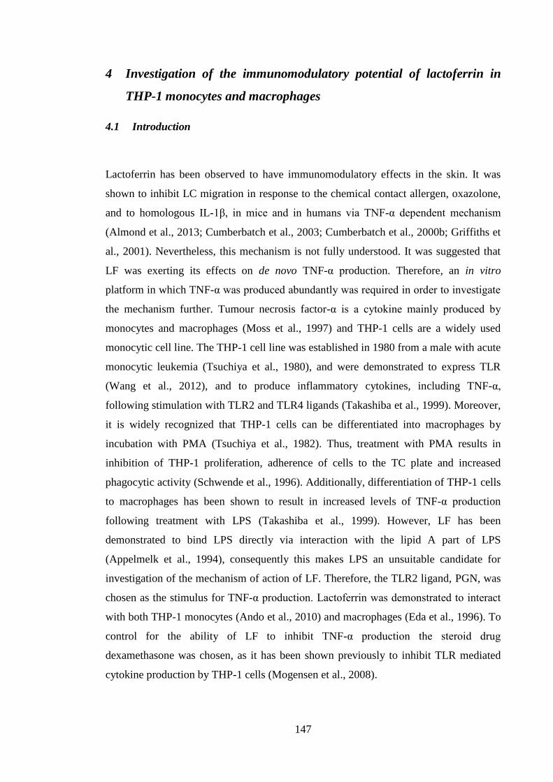
147
4 Investigation of the immunomodulatory potential of lactoferrin in
THP-1 monocytes and macrophages
4.1 Introduction
Lactoferrin has been observed to have immunomodulatory effects in the skin. It was
shown to inhibit LC migration in response to the chemical contact allergen, oxazolone,
and to homologous IL-1β, in mice and in humans via TNF-α dependent mechanism
(Almond et al., 2013; Cumberbatch et al., 2003; Cumberbatch et al., 2000b; Griffiths et
al., 2001). Nevertheless, this mechanism is not fully understood. It was suggested that
LF was exerting its effects on de novo TNF-α production. Therefore, an in vitro
platform in which TNF-α was produced abundantly was required in order to investigate
the mechanism further. Tumour necrosis factor-α is a cytokine mainly produced by
monocytes and macrophages (Moss et al., 1997) and THP-1 cells are a widely used
monocytic cell line. The THP-1 cell line was established in 1980 from a male with acute
monocytic leukemia (Tsuchiya et al., 1980), and were demonstrated to express TLR
(Wang et al., 2012), and to produce inflammatory cytokines, including TNF-α,
following stimulation with TLR2 and TLR4 ligands (Takashiba et al., 1999). Moreover,
it is widely recognized that THP-1 cells can be differentiated into macrophages by
incubation with PMA (Tsuchiya et al., 1982). Thus, treatment with PMA results in
inhibition of THP-1 proliferation, adherence of cells to the TC plate and increased
phagocytic activity (Schwende et al., 1996). Additionally, differentiation of THP-1 cells
to macrophages has been shown to result in increased levels of TNF-α production
following treatment with LPS (Takashiba et al., 1999). However, LF has been
demonstrated to bind LPS directly via interaction with the lipid A part of LPS
(Appelmelk et al., 1994), consequently this makes LPS an unsuitable candidate for
investigation of the mechanism of action of LF. Therefore, the TLR2 ligand, PGN, was
chosen as the stimulus for TNF-α production. Lactoferrin was demonstrated to interact
with both THP-1 monocytes (Ando et al., 2010) and macrophages (Eda et al., 1996). To
control for the ability of LF to inhibit TNF-α production the steroid drug
dexamethasone was chosen, as it has been shown previously to inhibit TLR mediated
cytokine production by THP-1 cells (Mogensen et al., 2008).
148
In order to understand fully the mechanism of action of LF it was crucial to identify a
receptor through which it was acting. To date many different receptors on various cell
types have been identified as LFR. Surface nucleolin has been shown to be responsible
for LF binding to and internalisation into THP-1 macrophages (Hirano et al., 2005).
Therefore, this receptor has been investigated as the most likely candidate.
The objective of the work presented in this chapter was to utilize THP-1 monocytes and
macrophages and to establish whether human native LF was able to modulate their
cytokine production in response to PGN. Additionally, the aim was to elucidate further
the mechanism of action of LF.
4.2 Results
4.2.1 Dose response of THP-1 monocytes to PGN
The first step in the investigations of the immunomodulatory mechanisms of LF was to
establish an in vitro system in which TNF-α was produced vigorously in response to an
LPS-independent stimulus. The human monocytic cell line THP-1 was selected to be
stimulated with PGN, a TLR2 ligand, for either 6 or 24h. Secretion of TNF-α and IL-8
was measured by ELISA at both time points. In the absence of PGN at both 6 and 24h
TNF-α production was below the limit of accurate detection of the ELISA (~20pg/ml).
Treatment with PGN resulted in detectable TNF-α production only for the highest dose
of the stimulant (25μg/ml) and was approximately 40pg/ml at both time points (Figure
4.1A and 4.1B). Secretion of IL-8 was considerably higher than that reported for TNF-
α, such that baseline expression was ~100-150pg/ml at both time points. Incubation
with PGN resulted in a dose dependent increase in IL-8 production with highest levels
(1.9 and 3.8ng/ml) observed after 24h for 10 and 25μg/ml of PGN, respectively (Figure
4.1D). Despite the relatively low levels of TNF-α produced by THP-1 monocytes
following PGN stimulation, the effect of pre-treatment with LF was examined.
Treatment with PGN resulted in secretion of ~100pg/ml of TNF-α and addition of LF
(2-100 μg/ml) was without effect on TNF-α production. However, prior incubation with
the positive control dexamethasone (100nM) inhibited TNF-α secretion by 81% (Figure
4.2).
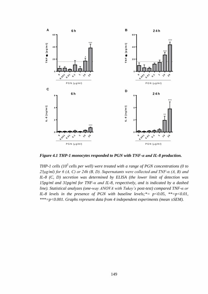
149
TN
F-
[p
g/m
l]
0
0.0
01
0.0
10.1 1
10
25
0
2 0
4 0
6 0
P G N (g /m l)
*
***
TN
F-
[p
g/m
l]
0
0.0
01
0.0
10.1 1
10
25
0
2 0
4 0
6 0
P G N (g /m l)
***
***
IL-8
[n
g/m
l]
0
0.0
01
0.0
10.1 1
10
25
0
2
4
6
P G N (g /m l)
***IL
-8 [
ng
/ml]
0
0.0
01
0.0
10.1 1
10
25
0
2
4
6
P G N (g /m l)
**
***
6 h 2 4 h
6 h 2 4 h
A B
CD
Figure 4.1 THP-1 monocytes responded to PGN with TNF-α and IL-8 production.
THP-1 cells (105
cells per well) were treated with a range of PGN concentrations (0 to
25μg/ml) for 6 (A, C) or 24h (B, D). Supernatants were collected and TNF-α (A, B) and
IL-8 (C, D) secretion was determined by ELISA (the lower limit of detection was
15pg/ml and 31pg/ml for TNF-α and IL-8, respectively, and is indicated by a dashed
line). Statistical analyzes (one-way ANOVA with Tukey’s post-test) compared TNF-α or
IL-8 levels in the presence of PGN with baseline levels;*= p<0.05, **=p<0.01,
***=p<0.001. Graphs represent data from 4 independent experiments (mean ±SEM).
150
TN
F-
[p
g/m
l]
0
DM
SO
Dex 1
00n
M 0 210
20
50
100
0
5 0
1 0 0
1 5 0
2 0 0
2 5 0
L F (g /m l)
Figure 4.2 Lactoferrin did not modulate PGN-induced TNF-α secretion by
monocytes.
THP-1 cells (105
cells per well) were pre-treated with a range of LF concentrations (0-
100 μg/ml) or 100nM dexamethasone (Dex) formulated in DMSO, or DMSO alone, for
1h before the addition of PGN (25μg/ml) for 6h. Control cells were untreated.
Supernatants were collected and TNF-α secretion was determined by ELISA (the lower
limit of detection 15pg/ml, indicated by a dashed line). Graphs represent data from 3
independent experiments (mean ± SEM).
4.2.2 PGN-activated THP-1 macrophages: dose response
Given the relatively low levels of TNF-α produced by PGN stimulated THP-1
monocytes and the lack of effect of LF, an experimental system in which more vigorous
TNF-α responses could be provoked was examined. Therefore, THP-1 monocytes were
differentiated into macrophages by 3h treatment with PMA. Figure 3.3 illustrates the
dose dependent increase in TNF-α secretion by THP-1 macrophages 6h (Figure 4.3A)
and 24h (Figure 4.3B) following treatment with PGN. Differentiation into macrophages
by PMA resulted in considerably higher baseline and PGN-induced cytokine production
compared with THP-1 monocytes. Thus, at 6h, baseline levels of approximately
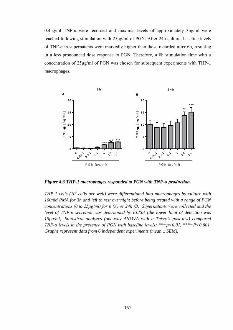
151
0.4ng/ml TNF-α were recorded and maximal levels of approximately 3ng/ml were
reached following stimulation with 25μg/ml of PGN. After 24h culture, baseline levels
of TNF-α in supernatants were markedly higher than those recorded after 6h, resulting
in a less pronounced dose response to PGN. Therefore, a 6h stimulation time with a
concentration of 25μg/ml of PGN was chosen for subsequent experiments with THP-1
macrophages.
TN
F-
[n
g/m
l]
0
0.0
01
0.0
10.1 1
10
25
0
5
1 0
1 5
2 0
P G N (g /m l)
****** ***
TN
F-
[n
g/m
l]
0
0.0
01
0.0
10.1 1
10
25
0
5
1 0
1 5
2 0
P G N (g /m l)
*****
6 h 2 4 h
A B
Figure 4.3 THP-1 macrophages responded to PGN with TNF-α production.
THP-1 cells (105
cells per well) were differentiated into macrophages by culture with
100nM PMA for 3h and left to rest overnight before being treated with a range of PGN
concentrations (0 to 25μg/ml) for 6 (A) or 24h (B). Supernatants were collected and the
level of TNF-α secretion was determined by ELISA (the lower limit of detection was
15pg/ml). Statistical analyzes (one-way ANOVA with a Tukey’s post-test) compared
TNF-α levels in the presence of PGN with baseline levels; **=p<0.01, ***=P<0.001.
Graphs represent data from 6 independent experiments (mean ± SEM).
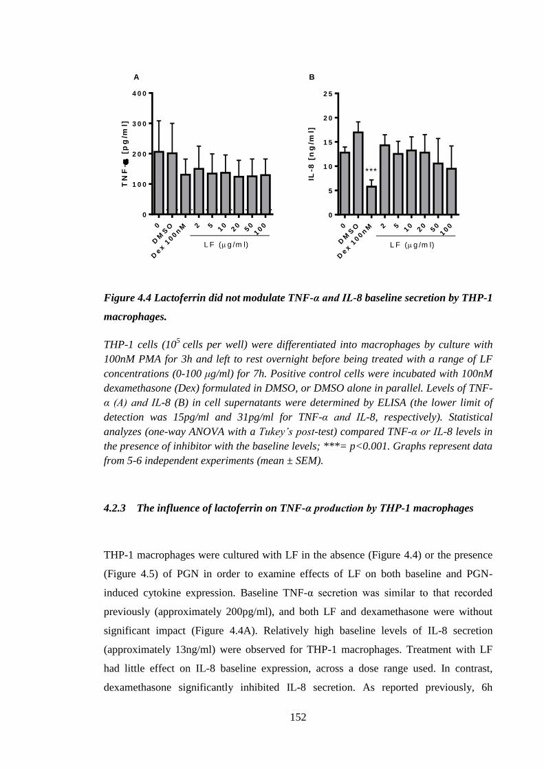
152
TN
F-
[p
g/m
l]
0
DM
SO
Dex 1
00n
M 2 510
20
50
100
0
1 0 0
2 0 0
3 0 0
4 0 0
L F (g /m l)
IL-8
[n
g/m
l]
0
DM
SO
Dex 1
00n
M 2 510
20
50
100
0
5
1 0
1 5
2 0
2 5
L F (g /m l)
***
A B
Figure 4.4 Lactoferrin did not modulate TNF-α and IL-8 baseline secretion by THP-1
macrophages.
THP-1 cells (105
cells per well) were differentiated into macrophages by culture with
100nM PMA for 3h and left to rest overnight before being treated with a range of LF
concentrations (0-100 μg/ml) for 7h. Positive control cells were incubated with 100nM
dexamethasone (Dex) formulated in DMSO, or DMSO alone in parallel. Levels of TNF-
α (A) and IL-8 (B) in cell supernatants were determined by ELISA (the lower limit of
detection was 15pg/ml and 31pg/ml for TNF-α and IL-8, respectively). Statistical
analyzes (one-way ANOVA with a Tukey’s post-test) compared TNF-α or IL-8 levels in
the presence of inhibitor with the baseline levels; ***= p<0.001. Graphs represent data
from 5-6 independent experiments (mean ± SEM).
4.2.3 The influence of lactoferrin on TNF-α production by THP-1 macrophages
THP-1 macrophages were cultured with LF in the absence (Figure 4.4) or the presence
(Figure 4.5) of PGN in order to examine effects of LF on both baseline and PGN-
induced cytokine expression. Baseline TNF-α secretion was similar to that recorded
previously (approximately 200pg/ml), and both LF and dexamethasone were without
significant impact (Figure 4.4A). Relatively high baseline levels of IL-8 secretion
(approximately 13ng/ml) were observed for THP-1 macrophages. Treatment with LF
had little effect on IL-8 baseline expression, across a dose range used. In contrast,
dexamethasone significantly inhibited IL-8 secretion. As reported previously, 6h
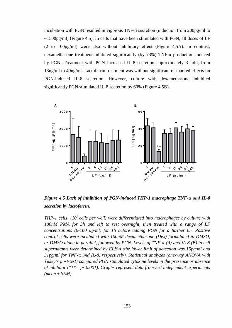
153
incubation with PGN resulted in vigorous TNF-α secretion (induction from 200pg/ml to
~1500pg/ml) (Figure 4.5). In cells that have been stimulated with PGN, all doses of LF
(2 to 100μg/ml) were also without inhibitory effect (Figure 4.5A). In contrast,
dexamethasone treatment inhibited significantly (by 73%) TNF-α production induced
by PGN. Treatment with PGN increased IL-8 secretion approximately 3 fold, from
13ng/ml to 40ng/ml. Lactoferrin treatment was without significant or marked effects on
PGN-induced IL-8 secretion. However, culture with dexamethasone inhibited
significantly PGN stimulated IL-8 secretion by 60% (Figure 4.5B).
TN
F-
[p
g/m
l]
0
DM
SO
Dex 1
00n
M 2 510
20
50
100
0
1 0 0 0
2 0 0 0
3 0 0 0
L F (g /m l)
IL-8
[n
g/m
l]
0
DM
SO
Dex 1
00n
M 2 510
20
50
100
0
2 0
4 0
6 0
L F (g /m l)
***
A B
Figure 4.5 Lack of inhibition of PGN-induced THP-1 macrophage TNF-α and IL-8
secretion by lactoferrin.
THP-1 cells (105
cells per well) were differentiated into macrophages by culture with
100nM PMA for 3h and left to rest overnight, then treated with a range of LF
concentrations (0-100 μg/ml) for 1h before adding PGN for a further 6h. Positive
control cells were incubated with 100nM dexamethasone (Dex) formulated in DMSO,
or DMSO alone in parallel, followed by PGN. Levels of TNF-α (A) and IL-8 (B) in cell
supernatants were determined by ELISA (the lower limit of detection was 15pg/ml and
31pg/ml for TNF-α and IL-8, respectively). Statistical analyzes (one-way ANOVA with
Tukey’s post-test) compared PGN stimulated cytokine levels in the presence or absence
of inhibitor (***= p<0.001). Graphs represent data from 5-6 independent experiments
(mean ± SEM).
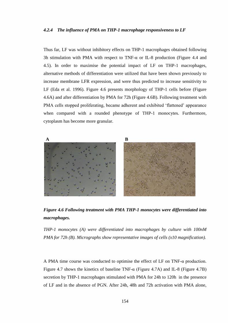
154
4.2.4 The influence of PMA on THP-1 macrophage responsiveness to LF
Thus far, LF was without inhibitory effects on THP-1 macrophages obtained following
3h stimulation with PMA with respect to TNF-α or IL-8 production (Figure 4.4 and
4.5). In order to maximise the potential impact of LF on THP-1 macrophages,
alternative methods of differentiation were utilized that have been shown previously to
increase membrane LFR expression, and were thus predicted to increase sensitivity to
LF (Eda et al. 1996). Figure 4.6 presents morphology of THP-1 cells before (Figure
4.6A) and after differentiation by PMA for 72h (Figure 4.6B). Following treatment with
PMA cells stopped proliferating, became adherent and exhibited ‘flattened’ appearance
when compared with a rounded phenotype of THP-1 monocytes. Furthermore,
cytoplasm has become more granular.
Figure 4.6 Following treatment with PMA THP-1 monocytes were differentiated into
macrophages.
THP-1 monocytes (A) were differentiated into macrophages by culture with 100nM
PMA for 72h (B). Micrographs show representative images of cells (x10 magnification).
A PMA time course was conducted to optimise the effect of LF on TNF-α production.
Figure 4.7 shows the kinetics of baseline TNF-α (Figure 4.7A) and IL-8 (Figure 4.7B)
secretion by THP-1 macrophages stimulated with PMA for 24h to 120h in the presence
of LF and in the absence of PGN. After 24h, 48h and 72h activation with PMA alone,
A B
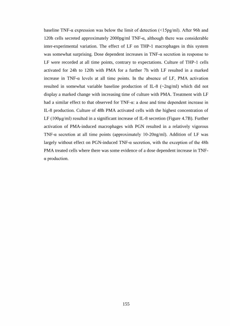
155
baseline TNF-α expression was below the limit of detection (<15pg/ml). After 96h and
120h cells secreted approximately 2000pg/ml TNF-α, although there was considerable
inter-experimental variation. The effect of LF on THP-1 macrophages in this system
was somewhat surprising. Dose dependent increases in TNF-α secretion in response to
LF were recorded at all time points, contrary to expectations. Culture of THP-1 cells
activated for 24h to 120h with PMA for a further 7h with LF resulted in a marked
increase in TNF-α levels at all time points. In the absence of LF, PMA activation
resulted in somewhat variable baseline production of IL-8 (~2ng/ml) which did not
display a marked change with increasing time of culture with PMA. Treatment with LF
had a similar effect to that observed for TNF-α: a dose and time dependent increase in
IL-8 production. Culture of 48h PMA activated cells with the highest concentration of
LF (100μg/ml) resulted in a significant increase of IL-8 secretion (Figure 4.7B). Further
activation of PMA-induced macrophages with PGN resulted in a relatively vigorous
TNF-α secretion at all time points (approximately 10-20ng/ml). Addition of LF was
largely without effect on PGN-induced TNF-α secretion, with the exception of the 48h
PMA treated cells where there was some evidence of a dose dependent increase in TNF-
α production.
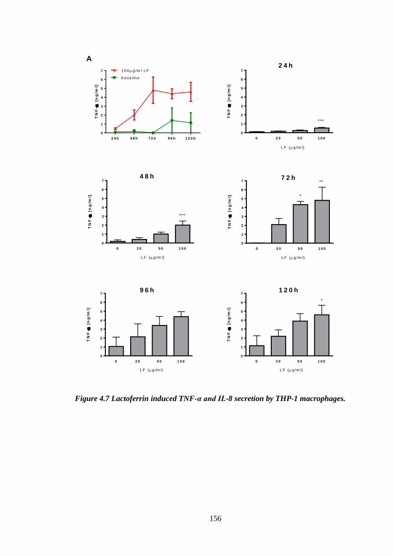
156
2 4 h 4 8 h 7 2 h 9 6 h 1 2 0 h
0
1
2
3
4
5
6
7
TN
F-
[n
g/m
l]
b a se lin e
1 0 0 g /m l L F
0 2 0 5 0 1 0 0
0
1
2
3
4
5
6
7
L F (g /m l)
***
2 4 h
TN
F-
[n
g/m
l]
0 2 0 5 0 1 0 0
0
1
2
3
4
5
6
7
***
4 8 h
TN
F-
[n
g/m
l]
L F (g /m l)
0 2 0 5 0 1 0 0
0
1
2
3
4
5
6
7
*
**7 2 h
TN
F-
[n
g/m
l]
L F (g /m l)
0 2 0 5 0 1 0 0
0
1
2
3
4
5
6
79 6 h
TN
F-
[n
g/m
l]
L F (g /m l)
0 2 0 5 0 1 0 0
0
1
2
3
4
5
6
7
*
1 2 0 h
TN
F-
[n
g/m
l]
L F (g /m l)
A
Figure 4.7 Lactoferrin induced TNF-α and IL-8 secretion by THP-1 macrophages.
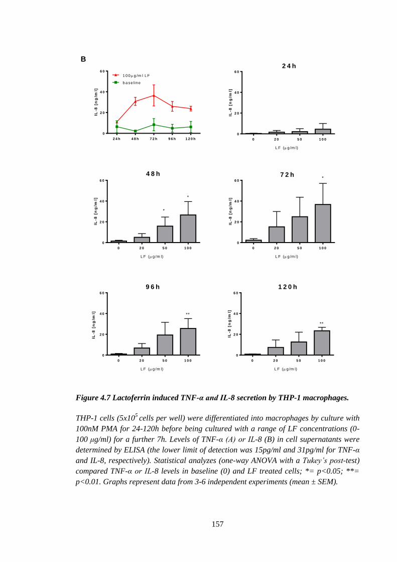
157
IL-8
[n
g/m
l]
0 2 0 5 0 1 0 0
0
2 0
4 0
6 0
L F (g /m l)
2 4 h
IL-8
[n
g/m
l]
0 2 0 5 0 1 0 0
0
2 0
4 0
6 0
L F (g /m l)
*
*
4 8 h
IL-8
[n
g/m
l]
0 2 0 5 0 1 0 0
0
2 0
4 0
6 0
L F (g /m l)
*7 2 h
0 2 0 5 0 1 0 0
0
2 0
4 0
6 0
L F (g /m l)
IL-8
[n
g/m
l] **
9 6 h
IL-8
[n
g/m
l]
0 2 0 5 0 1 0 0
0
2 0
4 0
6 0
L F (g /m l)
**
1 2 0 h
IL-8
[n
g/m
l]
2 4 h 4 8 h 7 2 h 9 6 h 1 2 0 h
0
2 0
4 0
6 0
b a se lin e
1 0 0 g /m l L F
B
Figure 4.7 Lactoferrin induced TNF-α and IL-8 secretion by THP-1 macrophages.
THP-1 cells (5x10⁵
cells per well) were differentiated into macrophages by culture with
100nM PMA for 24-120h before being cultured with a range of LF concentrations (0-
100 μg/ml) for a further 7h. Levels of TNF-α (A) or IL-8 (B) in cell supernatants were
determined by ELISA (the lower limit of detection was 15pg/ml and 31pg/ml for TNF-α
and IL-8, respectively). Statistical analyzes (one-way ANOVA with a Tukey’s post-test)
compared TNF-α or IL-8 levels in baseline (0) and LF treated cells; *= p<0.05; **=
p<0.01. Graphs represent data from 3-6 independent experiments (mean ± SEM).
158
2 4 h 4 8 h 7 2 h 9 6 h 1 2 0 h
0
1 0
2 0
3 0
4 0
TN
F-
[n
g/m
l]
b a se lin e
1 0 0 g /m l L F
0 2 0 5 0 1 0 0
0
1 0
2 0
3 0
4 0
5 0
L F ( g /m l)
2 4 h
TN
F-
[n
g/m
l]
0 2 0 5 0 1 0 0
0
1 0
2 0
3 0
4 0
5 0
L F ( g /m l)
*
4 8 h
TN
F-
[n
g/m
l]
0 2 0 5 0 1 0 0
0
1 0
2 0
3 0
4 0
5 0
L F ( g /m l)
7 2 h
TN
F-
[n
g/m
l]
0 2 0 5 0 1 0 0
0
1 0
2 0
3 0
4 0
5 0
L F ( g /m l)
9 6 h
TN
F-
[n
g/m
l]
0 2 0 5 0 1 0 0
0
1 0
2 0
3 0
4 0
5 0
L F ( g /m l)
1 2 0 h
TN
F-
[n
g/m
l]
Figure 4.8 Lactoferrin did not inhibit PGN-induced TNF-α production by THP-1
macrophages.
THP-1 cells (5x10⁵
cells per well) were differentiated into macrophages by culture with
100nM PMA for 24-120h before being treated with a range of LF concentrations (0-100
μg/ml) for 1h prior to addition of 10μg/ml of PGN for a further 6h. Levels of TNF-α in
cell supernatants were determined by ELISA (the lower limit of detection was 15pg/ml).
Statistical analysis (one-way ANOVA with a Tukey’s post-test) compared TNF-α levels
in baseline and LF treated cells;*=p<0.05. Graphs represent data from 3-6
independent experiments (mean ± SEM).
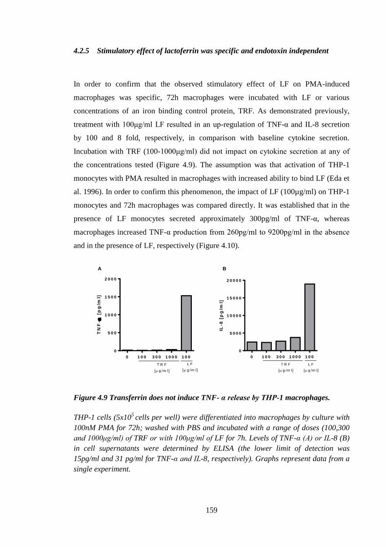
159
4.2.5 Stimulatory effect of lactoferrin was specific and endotoxin independent
In order to confirm that the observed stimulatory effect of LF on PMA-induced
macrophages was specific, 72h macrophages were incubated with LF or various
concentrations of an iron binding control protein, TRF. As demonstrated previously,
treatment with 100μg/ml LF resulted in an up-regulation of TNF-α and IL-8 secretion
by 100 and 8 fold, respectively, in comparison with baseline cytokine secretion.
Incubation with TRF (100-1000μg/ml) did not impact on cytokine secretion at any of
the concentrations tested (Figure 4.9). The assumption was that activation of THP-1
monocytes with PMA resulted in macrophages with increased ability to bind LF (Eda et
al. 1996). In order to confirm this phenomenon, the impact of LF (100µg/ml) on THP-1
monocytes and 72h macrophages was compared directly. It was established that in the
presence of LF monocytes secreted approximately 300pg/ml of TNF-α, whereas
macrophages increased TNF-α production from 260pg/ml to 9200pg/ml in the absence
and in the presence of LF, respectively (Figure 4.10).
0 1 0 0 3 0 0 1 0 0 0 1 0 0
0
5 0 0
1 0 0 0
1 5 0 0
2 0 0 0
T R F
[ g /m l]
L F
[ g /m l]
TN
F-
[p
g/m
l]
0 1 0 0 3 0 0 1 0 0 0 1 0 0
0
5 0 0 0
1 0 0 0 0
1 5 0 0 0
2 0 0 0 0
T R F
[ g /m l]
L F
[ g /m l]
IL-8
[p
g/m
l]
A B
Figure 4.9 Transferrin does not induce TNF- α release by THP-1 macrophages.
THP-1 cells (5x10⁵
cells per well) were differentiated into macrophages by culture with
100nM PMA for 72h; washed with PBS and incubated with a range of doses (100,300
and 1000μg/ml) of TRF or with 100μg/ml of LF for 7h. Levels of TNF-α (A) or IL-8 (B)
in cell supernatants were determined by ELISA (the lower limit of detection was
15pg/ml and 31 pg/ml for TNF-α and IL-8, respectively). Graphs represent data from a
single experiment.
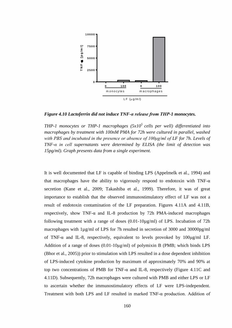
160
0 1 0 0 0 1 0 0
0
2 5 0 0
5 0 0 0
7 5 0 0
1 0 0 0 0
m o n o c y te s m a c ro p h a g e s
L F (g /m l)
TN
F-
[p
g/m
l]
Figure 4.10 Lactoferrin did not induce TNF-α release from THP-1 monocytes.
THP-1 monocytes or THP-1 macrophages (5x10⁵
cells per well) differentiated into
macrophages by treatment with 100nM PMA for 72h were cultured in parallel, washed
with PBS and incubated in the presence or absence of 100μg/ml of LF for 7h. Levels of
TNF-α in cell supernatants were determined by ELISA (the limit of detection was
15pg/ml). Graph presents data from a single experiment.
It is well documented that LF is capable of binding LPS (Appelmelk et al., 1994) and
that macrophages have the ability to vigorously respond to endotoxin with TNF-α
secretion (Kane et al., 2009; Takashiba et al., 1999). Therefore, it was of great
importance to establish that the observed immunostimulatory effect of LF was not a
result of endotoxin contamination of the LF preparation. Figures 4.11A and 4.11B,
respectively, show TNF-α and IL-8 production by 72h PMA-induced macrophages
following treatment with a range of doses (0.01-10µg/ml) of LPS. Incubation of 72h
macrophages with 1µg/ml of LPS for 7h resulted in secretion of 3000 and 30000pg/ml
of TNF-α and IL-8, respectively, equivalent to levels provoked by 100μg/ml LF.
Addition of a range of doses (0.01-10μg/ml) of polymixin B (PMB; which binds LPS
(Bhor et al., 2005)) prior to stimulation with LPS resulted in a dose dependent inhibition
of LPS-induced cytokine production by maximum of approximately 70% and 90% at
top two concentrations of PMB for TNF-α and IL-8, respectively (Figure 4.11C and
4.11D). Subsequently, 72h macrophages were cultured with PMB and either LPS or LF
to ascertain whether the immunostimulatory effects of LF were LPS-independent.
Treatment with both LPS and LF resulted in marked TNF-α production. Addition of
161
10μg/ml of PMB, as previously demonstrated, inhibited LPS-triggered cytokine
production but was without effect on LF-induced TNF-α secretion (Figure 4.11E),
demonstrating that the stimulatory effect of LF was independent of endotoxin. The
effect of LPS on 72h THP-1 macrophages was assessed in the single experiment.
Similarly, the impact of PMB on LPS-induced THP-1 macrophages was confirmed in a
single experiment. It is known that LPS is capable of dose-dependently stimulating
TNF-α release by macrophages (Lichtman et al., 1998; Song et al., 2000) and that PMB
is able to inhibit such induction (Cardoso et al., 2007). Therefore, it was deemed
suitable to perform these experiments once.
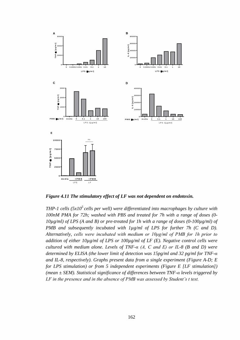
162
0 0 .0 0 0 1 0 .0 0 1 0 .0 1 0 .1 1 1 0
0
2 0 0 0
4 0 0 0
6 0 0 0
L P S [ g /m l]
TN
F-
[p
g/m
l]
0 0 .0 0 0 1 0 .0 0 1 0 .0 1 0 .1 1 1 0
0
2 0 0 0 0
4 0 0 0 0
6 0 0 0 0
8 0 0 0 0
IL-8
[p
g/m
l]
L P S [ g /m l]
m e d ia 0 0 .1 1 1 0 1 0 0
0
1 0 0 0
2 0 0 0
3 0 0 0
L P S 1 [g /m l]
P M B [ g /m l]
TN
F-
[p
g/m
l]
m e d ia 0 0 .1 1 1 0 1 0 0
0
1 0 0 0 0
2 0 0 0 0
3 0 0 0 0
4 0 0 0 0
P M B [ g /m l]
L P S 1 [g /m l]
IL-8
[p
g/m
l]
A B
C D
m e d ia + P M B + P M B
0
2 5 0 0
5 0 0 0
7 5 0 0
1 0 0 0 0
L P S L F
TN
F-
[p
g/m
l]
E
n s
Figure 4.11 The stimulatory effect of LF was not dependent on endotoxin.
THP-1 cells (5x10⁵
cells per well) were differentiated into macrophages by culture with
100nM PMA for 72h; washed with PBS and treated for 7h with a range of doses (0-
10µg/ml) of LPS (A and B) or pre-treated for 1h with a range of doses (0-100µg/ml) of
PMB and subsequently incubated with 1µg/ml of LPS for further 7h (C and D).
Alternatively, cells were incubated with medium or 10μg/ml of PMB for 1h prior to
addition of either 10µg/ml of LPS or 100µg/ml of LF (E). Negative control cells were
cultured with medium alone. Levels of TNF-α (A, C and E) or IL-8 (B and D) were
determined by ELISA (the lower limit of detection was 15pg/ml and 32 pg/ml for TNF-α
and IL-8, respectively). Graphs present data from a single experiment (Figure A-D; E
for LPS stimulation) or from 5 independent experiments (Figure E [LF stimulation])
(mean ± SEM). Statistical significance of differences between TNF-α levels triggered by
LF in the presence and in the absence of PMB was assessed by Student’s t test.
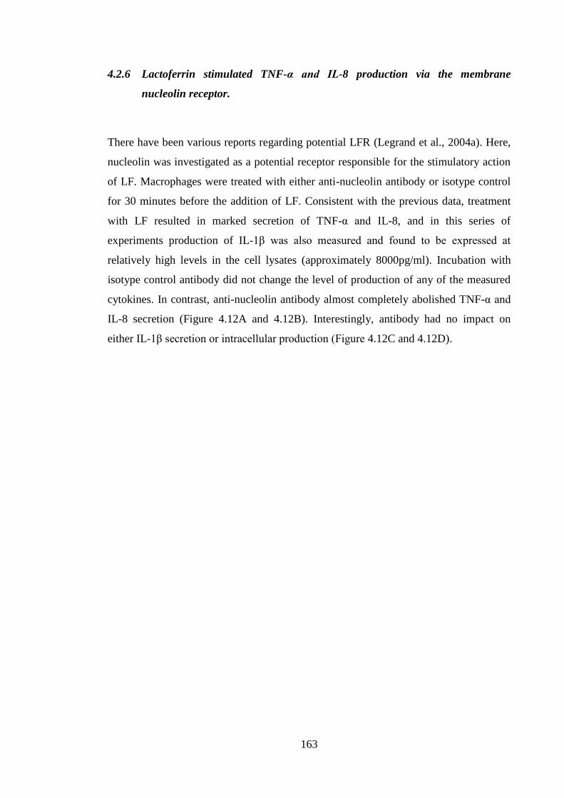
163
4.2.6 Lactoferrin stimulated TNF-α and IL-8 production via the membrane
nucleolin receptor.
There have been various reports regarding potential LFR (Legrand et al., 2004a). Here,
nucleolin was investigated as a potential receptor responsible for the stimulatory action
of LF. Macrophages were treated with either anti-nucleolin antibody or isotype control
for 30 minutes before the addition of LF. Consistent with the previous data, treatment
with LF resulted in marked secretion of TNF-α and IL-8, and in this series of
experiments production of IL-1β was also measured and found to be expressed at
relatively high levels in the cell lysates (approximately 8000pg/ml). Incubation with
isotype control antibody did not change the level of production of any of the measured
cytokines. In contrast, anti-nucleolin antibody almost completely abolished TNF-α and
IL-8 secretion (Figure 4.12A and 4.12B). Interestingly, antibody had no impact on
either IL-1β secretion or intracellular production (Figure 4.12C and 4.12D).
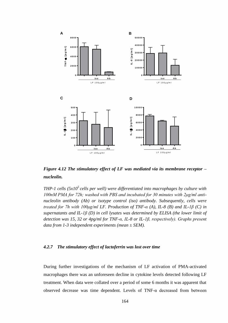
164
TN
F-
[p
g/m
l]
is o A b
0
2 0 0 0
4 0 0 0
6 0 0 0
8 0 0 0
L F 1 0 0 g /m l
IL-8
[p
g/m
l]
is o A b
0
1 0 0 0 0
2 0 0 0 0
3 0 0 0 0
4 0 0 0 0
5 0 0 0 0
L F 1 0 0 g /m l
IL-1
[
pg
/ml]
is o A b
0
1 0 0
2 0 0
3 0 0
4 0 0
5 0 0
L F 1 0 0 g /m l
IL-1
[
pg
/ml]
is o A b
0
2 0 0 0
4 0 0 0
6 0 0 0
8 0 0 0
1 0 0 0 0
L F 1 0 0 g /m l
A B
C D
Figure 4.12 The stimulatory effect of LF was mediated via its membrane receptor –
nucleolin.
THP-1 cells (5x10⁵
cells per well) were differentiated into macrophages by culture with
100nM PMA for 72h; washed with PBS and incubated for 30 minutes with 2μg/ml anti-
nucleolin antibody (Ab) or isotype control (iso) antibody. Subsequently, cells were
treated for 7h with 100μg/ml LF. Production of TNF-α (A), IL-8 (B) and IL-1β (C) in
supernatants and IL-1β (D) in cell lysates was determined by ELISA (the lower limit of
detection was 15, 32 or 4pg/ml for TNF-α, IL-8 or IL-1β, respectively). Graphs present
data from 1-3 independent experiments (mean ± SEM).
4.2.7 The stimulatory effect of lactoferrin was lost over time
During further investigations of the mechanism of LF activation of PMA-activated
macrophages there was an unforeseen decline in cytokine levels detected following LF
treatment. When data were collated over a period of some 6 months it was apparent that
observed decrease was time dependent. Levels of TNF-α decreased from between
165
14000pg/ml (18.07.2011) and 5000pg/ml (27.07-07.10.2011) to below 1000pg/ml
(14.10-07.11.2011) (Figure 4.13A). This was not due to a change in the preparation of
LF, which remained constant. The extent of TNF-α production by 72h macrophages
triggered by LF continued to decline, although the responsiveness of the cells to LPS
remained relatively stable (Figure 4.13B). Various sources of THP-1 cells and PMA
have been used in order to overcome the decline in sensitivity of the cells to LF. Cells
that were utilised were consistently free of mycoplasma infection. Additionally,
alternative ways of differentiation of THP-1 cells to macrophages (treatment with
vitamin D, IFN-γ, sodium butyrate) have been investigated (data not shown). However,
none of these approaches resulted in a THP-1 macrophage phenotype that was
responsive to LF. Therefore, due to the loss of LF stimulatory ability in 72h
macrophages no further experiments were performed in THP-1 cells to investigate the
mechanism of the stimulatory action of LF.
18.0
7
27.0
7
04.0
8
10.0
8
15.0
8
30.0
9
07.1
0
14.1
0
25.1
0
25.1
0
02.1
1
04.1
1
07.1
1
0
5 0 0 0
1 0 0 0 0
1 5 0 0 0
TN
F-
[p
g/m
l]
0 2 4 6 8
0
5 0 0
1 0 0 0
1 5 0 0
2 0 0 0
TN
F-
[p
g/m
l] b a se lin e
L F
L P S
BA
Figure 4.13 The immunostimulatory effect of lactoferrin was lost over time.
THP-1 cells (5x10⁵
cells per well) were differentiated into macrophages by culture with
100nM PMA for 72h; washed with PBS and treated for 7h with 100μg/ml LF (A) or with
100μg/ml LF and 1μg/ml LPS for 7h (B). Production of TNF-α in supernatants was
determined by ELISA (the lower limit of detection was 15pg/ml). Graphs present data
from 1-2 independent experiments performed over a period of some 4 months (mean ±
SEM).
166
4.3 Discussion
Lactoferrin is a naturally occurring iron-binding protein present in mammalian
secretions which has been known for decades for its antimicrobial properties (Stephens
et al., 1980). Recently, however, LF has been shown to have potential
immunomodulatory properties. Results to date from our laboratory support the
hypothesis that exogenous LF inhibits LC migration in vivo in response to the chemical
contact allergen oxazolone via effects on the availability of TNF-α in mice (Almond et
al., 2013; Cumberbatch et al., 2000b) and in humans (Griffiths et al., 2001). The aim of
this chapter was to determine the impact of LF on TNF-α production by THP-1 cells, in
order to elucidate further the mechanism of its action.
The first step was to establish an in vitro system in which TNF-α was produced
robustly. The human monocytic cell line THP-1 was the first cell line of choice for
examining the mechanism of action of LF (Tsuchiya et al., 1980). THP-1 cells have
been shown previously to produce TNF-α in response to TLR agonists, for example to:
LPS, liopotechoic acid, flagellin or PGN (Remer et al., 2006). As described previously,
one of the properties of LF is its ability to bind LPS (Appelmelk et al., 1994).
Therefore, in order to avoid inhibition via sequestration of the stimulus, LPS was not
deemed suitable for assessing the action of LF. Instead the TLR2 agonist, PGN, was
chosen as a trigger for TNF-α production. In the studies reported herein TNF-α
production by PGN-activated THP-1 monocytes occurred at a very low levels at both 6h
and 24h. Production of the chemokine IL-8 was considerably higher particularly
following 24h incubation with the highest concentration of PGN. Despite the low level
of TNF-α production the ability of LF to modulate PGN-induced TNF-α secretion was
investigated and it was found that LF did not exert inhibitory effects in that
experimental system.
The positive control for inhibition of cytokine production was dexamethasone, a
corticosteroid drug well known to down-regulate inflammatory cytokine production,
such as that produced by macrophages following LPS stimulation (Armstrong et al.,
2009). Dexamethasone almost completely abrogated TNF-α production by THP-1
monocytes following PGN treatment. Consequently, differentiation of THP-1 cells into
167
macrophages was explored using methods utilized by Zhou et al. (2010)(3h PMA
activation) and Eda et al. (1996)(24h-120h PMA activation). Differentiation of THP-1
cells into macrophages with PMA is a well recognized method (Tsuchiya et al., 1982).
Our laboratory routinely utilized the protocol of 3h PMA differentiation with
subsequent overnight incubation of the cells and that method was used first to obtain
macrophages for these investigations. As predicted, 3h THP-1 macrophages produced
higher levels of TNF-α than did THP-1 monocytes. Human peripheral blood monocytes
have been reported previously to produce approximately 3000pg/ml of TNF-α following
2h incubation with 10μg/ml of PGN (Bonnet et al., 2008). The result reported by
Bonnet et al. (2008) was similar to that observed in the current studies where THP-1
macrophages incubated for 3h with PMA followed by treatment with PGN (25μg/ml)
secreted approximately 2500pg/ml of TNF-α. Dexamethasone treatment resulted in
overall inhibition of both TNF-α and IL-8 production by 3h THP-1 macrophages with
the exception of baseline (in the absence of PGN) TNF-α secretion. Naitoh et al. (2001)
investigated the effect of PMA on TNF-α secretion by THP-1 monocytes and
macrophages and found that PMA (incubation for 24h with 50ng/ml of PMA) treatment
of THP-1 monocytes induced cytokine secretion (approximately 300pg/ml of TNF-α),
whereas, the same treatment did not induce detectable cytokine secretion by THP-1
macrophages (differentiated with 50ng/ml PMA for 12h). From the perspective of the
results reported by Naitoh et al (2001) it is difficult to determine whether the 3h PMA
activation method caused TNF-α secretion in the current study, however, it is consistent
with the absent or low baseline levels of TNF-α secreted by THP-1 macrophages
activated with PMA for 24h-120h. PMA-induced IL- 8 was reported previously to be
resistant to dexamethasone mediated inhibition (Anttila et al., 1992). In the current
investigations IL-8 secretion was susceptible to dexamethasone treatment. The
discrepancy could be explained by the fact that levels of IL-8 demonstrated by Antilla et
al. (1992) were much higher than those reported in the current study (100-300ng/ml
vs.10-20ng/ml), suggesting that there might be a limit to the ability of dexamethasone
with respect to inhibiting PMA-induced IL-8 production. Lactoferin had little or no
inhibitory effect on TNF-α secretion by THP-1 monocytes when it was added either 1h
before or 1h after (data not shown) addition of PGN to the culture.
In differentiated (3h PMA) THP-1 cells some modest effects of LF were observed with
decreased baseline (approximately 25% inhibition) and PGN-induced TNF-α secretion
168
(25% inhibition). In addition, doses of 2-100μg/ml LF had the same slight inhibitory
effect on secreted TNF-α. In parallel, experiments were conducted in which LF was
added to the culture 1h after PGN (total of PGN stimulation time remained 6h) in order
to examine if the time of introduction of LF to the culture influenced its effects (data not
shown). In these experiments the inhibitory effects of LF were comparable to those
achieved when LF was added 1h prior to PGN stimulation. In addition to inhibiting
TNF-α expression, LF reduced, although to lesser extent, IL-8 secretion. Similar to the
influence of LF on TNF-α, these effects were not dose dependent nor did they reach
statistical significance. Although the observed inhibitory effect of LF was not
statistically significant, it was consistent between experiments, different cytokines and
was in keeping with the in vivo data from LC migration experiments (Cumberbatch et
al., 2000b), and as such, was thought to be of physiological relevance. The data reported
herein represented the first step in examining the immunosuppressive function of LF in
an LPS independent system. In order to better characterize the effect of LF, experiments
were performed to titrate out the inhibitory effect of LF on TNF-α production (data not
shown). During these investigations it became apparent that the modest inhibition of
TNF-α production was no longer observed at concentrations of LF that previously
inhibited cytokine production. Thus, the effect of LF was not robust, and therefore this
was not an appropriate experimental system in which to examine the mechanism of the
effects of LF.
It has been shown previously that THP-1 cells differentiated into macrophages bound
the highest levels of radio-labelled LF when they were activated with PMA for 120h,
suggesting that there was a time dependent increase in expression of the receptor
following treatment with PMA (Eda et al., 1996). Given this information on LFR
expression in THP-1 macrophages, an alternative method of THP-1 differentiation was
utilized. A PMA time course was performed to establish if LF treatment at time points
that had been reported to show an increased expression of the receptor resulted in
augmented inhibitory activity of LF. When THP-1 macrophages produced after 24h to
120h stimulation with PMA were activated with PGN no inhibitory effect of LF on
TNF-α secretion was observed at any of the time points tested. Surprisingly, in 24h-
120h THP-1 macrophages incubated in the absence of PGN a dose dependent,
stimulatory effect of LF on TNF-α and IL-8 secretion was observed. Stimulation by LF
of cytokine production by macrophages has been reported previously by other authors
169
using LF from various species. For example, when the murine macrophage cell line
RAW 264.7 was incubated with bovine LF (100μg/ml) approximately 6000pg/ml of
TNF-α was produced (Curran et al., 2006), similar results to those observed in current
investigations. Thus, the same concentration of human LF (100μg/ml) stimulated TNF-
α secretion at similar levels by 72h, 96h and 120h THP-1 macrophages. Furthermore,
Sorimachi et al. (1997) used bovine LF to assess its effects on macrophages derived
from rat bone marrow. After 6h incubation with bovine LF release of approximately
1200 pg/ml of TNF-α and 3000 pg/ml of IL-8 was observed. Those levels were
somewhat lower and ten times less, respectively, than what was found in the current
investigations. However, these differences were likely to stem from the fact that the
concentration that utilized by Sorimachi et al. (1997) was 10μg/ml which is 10 times
less than what was used in the current study, as well as different cell type was used.
It was considered important to determine if observed activation of THP-1 macrophages
was in fact due to LF, rather than to a nonspecific effect of additional protein.
Therefore, TRF was used as a control. Lactoferrin and TRF both belong to the
transferrin family of proteins, have the same molecular weight (80kDa), share 61.4%
sequence homology and both are capable of binding two Fe3+
ions (Wally and
Buchanan, 2007). Transferrin is the primary iron carrier in the blood and has a role in
the innate immune response due to its involvement in iron metabolism (Johnson and
Wessling-Resnick, 2012). Treatment with TRF did not result in either TNF-α or IL-8
secretion, whereas incubation with LF triggered release of 1.5ng/ml of TNF-α and
19ng/ml of IL-8, implying that the observed stimulatory effect was attributable to a
specific property of LF. In addition, specificity of the LF effect was confirmed by
demonstrating that LF did not stimulate TNF-α release by THP-1 monocytes,
suggesting as Eda et al. (1996) had shown that the interaction of LF with THP-1
macrophages and subsequent TNF-α release, was a consequence of the interaction of LF
with its specific receptor, which was up-regulated by PMA activation.
Moreover, it was necessary to establish that observed immunostimulatory action of LF
was not due to endogenous LPS contamination of the protein preparation. Protein
preparations can contain endotoxin contamination (Magalhães et al., 2007) that can lead
to false positive results if the protein is not endotoxin depleted prior to use (Lin and
Kuo, 2010). In our laboratory commercially available proteins have been found to have
170
from 0.05 endotoxin units (EU)/mg to as high as 6000EU/mg. THP-1 monocytes have
been reported to respond to 200EU or less with cytokine secretion (personal
communication, Dr. Rebecca Dearman). Therefore, it was critical to establish that the
observed activation of TNF-α by LF was not mediated by contaminating LPS. Despite
the fact that previous batches of LF were known to have relatively low endotoxin levels
[10EU per mg of LF;(Almond et al., 2013)] it was decided to conduct an experiment in
which THP-1 macrophages were exposed to PMB, a specific LPS binding protein, prior
to addition of LF. It has been reported that THP-1 macrophages were sensitive even to
low levels of endotoxin. Takashiba et al. (1999) demonstrated that THP-1 cells
differentiated with 200nM PMA for 24h produced ~3ng/ml of TNF-α following 6h
incubation with 0.1μg/ml of LPS (equal to 1000EU). Similarly, in the current study
THP-1 macrophages vigorously responded to treatment with LPS with TNF-α and IL-8
production. Incubation of cells with PMB, prior to addition of LPS, dose dependently
inhibited cytokine release, confirming PMB as LPS inhibitor. Polymixin B has been
reported to efficiently inhibit stimulation of PBMC by contaminating endotoxin in the
preparation of recombinant proteins produced in E. coli (Cardoso et al., 2007). In
another study pre-incubation of murine macrophage cell line RAW264.7 for 30 minutes
with 10μg/ml of PMB abrogated release of TNF-α following stimulation with 0.2ng/ml
of LPS, as well as 1μg/ml of human recombinant Hsp70. In the absence of PMB, LPS
and human recombinant Hsp70 triggered release of approximately 6ng/ml of TNF-α
(Gao and Tsan, 2003). On the other hand, Tynan et al. (2012) reported that PMB
inadequately inhibited LPS-induced TNF-α secretion by bone marrow derived DC. At
1μg/ml of LPS, 10μg/ml of PMB did not have inhibitory effects. With decreasing
concentrations of LPS, the PMB effect was more pronounced and reached
approximately 90% inhibition when 1ng/ml of LPS was used (Tynan et al., 2012). In the
study reported herein, PMB inhibited LPS-induced TNF-α secretion by 80%, but was
without impact on LF-triggered TNF-α production. Similarly, Na et al. (2004) reported
that while PMB (at 50μg/ml) was able to abolish the ability of LPS to trigger nitric
oxide (NO) production by RAW 264.7 cells, it had only minimal effect on NO
production elicited by complex of bovine LF with LPS (Na et al., 2004). In the current
study PMB was used to prove independence of observed immunostimulatory effect of
human LF. However, the study by Na et al. (2004) suggested that use of PMB is not
suitable to confirm endotoxin independence of LF. It has to be noted, that the
concentration utilized in the current study was lower than that used by Na et al. (2004)
171
(10 vs. 50μg/ml). It was shown previously that at 50μg/ml PMB caused 21% release of
chromium from chromium labelled K562 human leukemia cells, in comparison with
cells treated with medium alone. Conversely, no chromium was released from cells
treated with 10μg/ml of PMB, implying that PMB at 50μg/ml was toxic to cells (Duwe
et al., 1986). The results of Duwe et al. (1986) also suggested that a fraction of NO
released by PMB treated RAW 264.7 cells in the study by Na et al.(2004) could have
come from dead or dying cells and not as a result of stimulation with un-purified bovine
LF. Taken together, it was concluded that in the study reported herein the observed
immunostimulatory potential of LF was not due to endotoxin contamination.
Having established the specificity of LF effects, it was decided to identify the receptor
through which LF was excreting its stimulatory action. There have been many proteins
that have been implicated acting as LFR in various cell types. It is logical from an
evolutionary point of view that there would be a LFR present in the intestine, as LF is
abundantly present in the breast milk (Suzuki et al., 2001). However, in addition to its
presence in milk, LF can also be found in serum and in the secondary neutrophil
granules (Wang et al., 1995), indicating its involvement in the immune response. PMA-
differentiated THP-1 macrophages were observed to bind LF (Eda et al., 1996),
oxidised erythrocytes (Beppu et al., 1996) and early apoptotic Jurkat cells via a protein
later identified as nucleolin (Hirano et al., 2005). With nucleolin identified as an LFR
present on THP-1 macrophages it was investigated whether anti-nucleolin antibody
could block the observed immunostimulatory effects of LF. It was found that both
TNF-α and IL-8 production was almost completely abrogated when cells were pre-
incubated with anti-nucleolin antibody. Conversely, neither secretion nor intra-cellular
production of IL-1β was affected. However, Ando et al. (2010) suggested that nucleolin
was an unlikely candidate for transducing the signal from LF as it lacks the cytoplasmic
domain, which would be responsible for such role. Nevertheless, it was possible that
nucleolin has a crucial role in TLR4-mediated signal transduction (Ando et al., 2010;
Curran et al., 2006), whereby its (nucleolin) blockage results in abolition of LF-
mediated TNF-α and IL-8 secretion. Lactoferrin, with other proteins secreted by
neutrophils, α-defensins and cathelicidins (LL-37), recently has been described as an
alarmin. Alarmins are proteins that in response to the external or internal injury are able
to recruit and activate cells of the immune system. Despite the lack of homology
between alarmins, they are all cationic proteins (Yang et al., 2009). One of the reasons
172
why anti-nucleolin antibody did not inhibit IL-1β production could have been that IL-1β
synthesis was achieved via an interaction of LF with P2X7 receptor and not nucleolin. It
was reported that another alarmin, LL-37, was capable of activating P2X7 receptor and
subsequent IL-1β production (Elssner et al., 2004). To date, there have been no reports
demonstrating that blocking nucleolin resulted in an inhibition of cytokine production.
However, Wang et al. (2011) have shown that the treatment of rat alveolar macrophages
with anti-nucleolin siRNA inhibited TNF-α and IL-6 production following treatment
with 1μg/ml of LPS. Results reported by Wang et al. (2011) may suggest that cytokine
inhibition observed in the current study was due to inhibition of LPS signalling,
furthermore indicating that the immunostimulatory effects of LF were in fact due to
endotoxin contamination. However, as it was demonstrated by experiments utilising
PMB, cytokine secretion following LF treatment in this study was endotoxin
independent.
It would have been of interest to see if treatment with nucleolin agonist other than LF
(eg. midkine, a low molecular weight protein involved in cell proliferation, migration
and organogenesis (Muramatsu, 2002)) would have resulted in a similar increase in
TNF-α production. Similarly, it would have been important to observe if a treatment
with nucleolin inhibitor, pseudo-peptide HB-19, would have resulted in abolition of the
stimulatory effects caused by LF (Nisole et al., 1999). However, none of these
experiments could take place due to the observed loss of responsiveness of THP-1
macrophages to stimulation with LF.
While performing routine experiments that were to further characterize LF-triggered
cytokine production, it was noticed that the stimulatory effect of LF on 72h THP-1
macrophages was diminishing. After collating the data, it was evident that the change in
responsiveness had happened over a period of a month (from 07 October to 07
November). New THP-1 cells were acquired, however, that did not make a difference.
Cells that were used were not infected with mycoplasma. Additionally, new batches of
PMA and LF were sourced. Lactoferrin, both from the previously used batch and from
the new stock, displayed the same level of biological activity in vivo, that is, both were
capable of inhibiting oxazolone-induced LC migration. Nevertheless, after investigating
every component of the system (cells, PMA and LF) the initial up-regulation of TNF-α
production by LF was still not observed. It would have been useful to utilise LF from
173
other commercial sources than the one that was used throughout current investigations
(Sigma Aldrich).
Investigation of the mechanism of action of LF started with the intention of elucidating
its inhibitory effect on TNF-α production and has finished by demonstrating specific,
stimulatory effects of the glycoprotein. Although at first this may seem contradictory,
there are in fact many reports to support both claims of LF immunosuppressive and
immunostimulatory action. Thus, injection of recombinant mouse LF into mice resulted
in myelopoesis that presented as increased levels of neutrophils, metamyelocytes and
myelocytes in circulating blood. This effect of LF could be perceived as an
inflammatory one, as higher levels of neutrophils appear when there is an infection, but
at the same time when LF is present it serves to restore homeostasis, therefore
reinforcing its role as both a stimulator and suppressant (Zimecki et al., 2013). Nishiya
et al. (1982) demonstrated that human LF exerted both stimulatory and inhibitory
effects on the same population of cells. The ability of LF to modulate cytotoxicity and
antibody-dependent cellular toxicity of human PBMC was tested. It was found that LF
increased the cytotoxicity of the adherent PBMC fraction against the K562 cell line at
concentration of 1μM, while the same concentration of the control protein TRF was
without effect. In contrast, when antibody-dependent cell-mediated cytotoxicity activity
of PBMC was measured against antibody coated liver cells, it was established that LF
dose dependently inhibited that effect, but again only in adherent fraction of PMBC
(Nishiya and Horwitz, 1982). Those results suggest that the role of LF on immune
system is complex and multifaceted. It is likely that its role depends on the underlying
condition of the organism.
Some of the discrepancies in the reports of LF functions might stem from the fact that
LF was found to exist in 3 isoforms that differ with regards to their RNase and iron
binding abilities. Affinity chromatography eluted 3 LF fractions that had two distinct
functions: LF α – classical iron binding form, LF β and γ – RNase, non-iron binding
forms eluted from Cibacron Blue Sepharose at 0.4 and 0.2M NaCl, respectively
(Furmanski et al., 1989). The concept of multiple LF isomers was further investigated
and this time 5 fractions were identified, with differential functions of DNase, RNase,
and ATPase as well as amylase and phosphatase (Kanyshkova et al., 2003). It was not
stated what the differences were, if any, between isoforms, other than differential
174
strength of binding to the Cibacron Blue Sepharose. Moreover, it was suggested that the
ability of LF to oligomerise might impact on its versatile functions. Indeed, it was
reported that status of LF in the solution depends on the LF concentration and the
presence of salts and/or LF substrates, like DNA or ATP (Nevinskii et al., 2009). Most
commonly found forms of LF are monomers and tetramers (Mantel et al., 1994).
In the current investigations there was no distinction between apo- and holo-forms of
LF and only native form of LF was used that was demonstrated previously to be
approximately 12-25% saturated with iron (Bezwoda and Mansoor, 1989). It was
demonstrated before that iron saturation status does not affect LF ability to inhibit LC
migration (Cumberbatch et al., 2000b). However, there were reports that iron saturation
status impacted on other aspects of LF function. Proliferation of Caco-2 cells (epithelial
colorectal carcinoma cells) was more strongly stimulated by apo-LF, but not by holo-
LF, despite both molecules signalling through the same receptor in a common clathrin-
dependent pathway (Buccigrossi et al., 2007; Jiang et al., 2011). These differences were
thought to stem from the fact that the apo-form of LF had a different conformation than
the holo-form. When iron is bound by LF it causes the molecule to become more
compact, therefore, potentially masking binding sites that could be recognized in the
apo- form of LF (Grossmann et al., 1992).
It was reported that human LF in THP-1 and mouse embryonic fibroblast cells activated
the canonical pathway of NF-ƘB, regardless of whether it was depleted of endotoxin
using PMB. Additionally, LF activated NF-ƘB in the presence of inhibitors of
transcription (actinomycin D) and translation (cyclohexamide) (Ando et al., 2010),
suggesting that the observed effect was solely due to LF and not secondary to TNF-α,
which was shown to be induced by LF and is known to potently induce NF-ƘB
canonical pathway (Zelová and Hošek, 2013). Ando et al. (2010) established that NF-
ƘB activation was due to the carbohydrate moiety of LF, as the effect was abolished
when LF was treated with endo-β-galactosidase that destabilises glycosylation chains.
However, the activation of NF-ƘB remained intact following incubation with a non
specific proteinase, actinase E. Moreover, Ando et al. (2010) have shown that bovine
LF was unable to trigger NF-ƘB signalling, whist inhibiting LPS-induced NF-ƘB
175
stimulation. That result contrasted with data gathered in the current investigations (data
not shown) and others (Sorimachi et al., 1997), where the treatment with bovine LF
resulted in cytokine production. Ando at el. (2010) concluded that the protein part of LF
molecule was responsible for the inhibitory actions of LF, whereas the carbohydrate
portion of the molecule evoked stimulatory effects.
Results presented in this chapter suggest dichotomy in the function of LF that is in
accordance with the literature. It is possible that the duality of LF action stems from its
structure, with protein and glycan parts of the molecule responsible for opposing
effects. Furthermore, various receptors utilized by LF could influence its function
(Losfeld et al., 2009; Spadaro et al., 2014; Suzuki et al., 2001). In addition, the
possibility of differential functions of a variety of LF isoforms makes it possible for LF
molecule to exert a plethora of different functions depending on the organisms’
requirements.
176
5 Effects of lactoferrin on keratinocytes
5.1 Introduction
The skin is the organism’s first line of defence. It is divided into 3 layers: epidermis,
dermis and hypodermis, that have distinct as well as complementary roles in
maintaining the safety of the organism (Nestle et al., 2009). The epidermis is the
outermost layer of the skin. It comprises of four layers (moving from the bottom
outwards): basal, spinous, granular and cornified. Cells (keratinocytes) in the basal layer
divide and move to the spinous layer where they change shape from columnar to
polygonal, and start expressing keratins (K1 and K10) distinct from those produced by
basal layer cells (K5 and K14) (Haines and Lane, 2012). Ultimately keratinocytes
become corneocytes in the cornified layer, whereby they lose all of their organelles,
become filled with keratin and provide the barrier function of the skin (Nestle et al.,
2009).
It has been established that following allergen treatment increased epidermal levels of
TNF-α and IL-1β are found (Enk and Katz, 1992a). Further, these two cytokine signals
were demonstrated to be necessary for LC migration (Cumberbatch et al., 1997b). It was
established that LC were the main source of IL-1β in the steady state epidermis (Enk et
al., 1993; Matsue et al., 1992b). Therefore, it was hypothesised that the TNF-α
necessary for LC migration was derived from keratinocytes. The fact that topical
exposure to LF inhibited LC migration induced by oxazolone and exogenous IL-1β, but
was without effect on LC mobilisation triggered by exogenous TNF-α (Cumberbatch et
al., 2000b), suggested that LF decreases the availability of endogenous TNF-α by
interaction with keratinocytes. In the series of experiments described in this chapter
three keratinocyte cell types have been utilized to investigate the immunomodulatory
effects of LF, primary mouse keratinocytes and the PAM 212 cell line derived from
BALB/c strain mice and human immortalized keratinocytes, the HaCaT cell line.
Cytokine production and expression was stimulated using the TLR2 ligand PGN,
similarly to experiments described in the Chapter 4. Additionally, keratinocytes were
stimulated with cytokines that have been shown to play important roles in LC
migration: TNF-α, IL-1α and IL-1β.
177
5.2 Results
5.2.1 PAM 212 and HaCaT cell lines and primary murine keratinocytes expressed
keratin-14
The first step in these investigations was to establish that the cells used were indeed
keratinocytes. Immunofluorescence staining was performed for the expression of K14,
present in basal keratinocytes of all stratified squamous epithelial tissues (Green et al.,
2003). Both murine and human cell lines (Figure 5.1A-5.1D) were cultured on a glass
coverslip, stained and mounted onto a slide. Primary murine cells failed to grow on a
glass surface; therefore, staining was performed in the culture plate. Due to differences
in the preparation of the samples different microscopes had to be utilized. This may
partially explain differences observed in image brightness between samples. Both PAM
and HaCaT cells grew to confluence on glass coverslips (as shown by DAPI staining),
and clear staining of K14 fibres in the cytoplasm was observed compared with the
isotype (negative) controls in both cell types. Comparatively little staining was observed
for primary keratinocytes, although the pattern was also cytoplasmic (Figure 5.1E,
5.1F). This differential staining appears to be due to the different preparation method
utilized for the primary keratinocytes, as when PAM cells were prepared in the same
way as primary murine keratinocytes, they also displayed considerably lower levels of
keratin staining (Figure 5.1G, 5.1H). Thus all three cell types expressed K14 to some
extent. Keratin 14 together with K5 is expressed by cells in the basal layer of epidermis.
These two keratins have been identified as markers of stratified squamous epithelial
cells (epidermis)(Nelson and Sun, 1983). Therefore, detection of K14 within the tested
cells confirmed their status as keratinocytes.
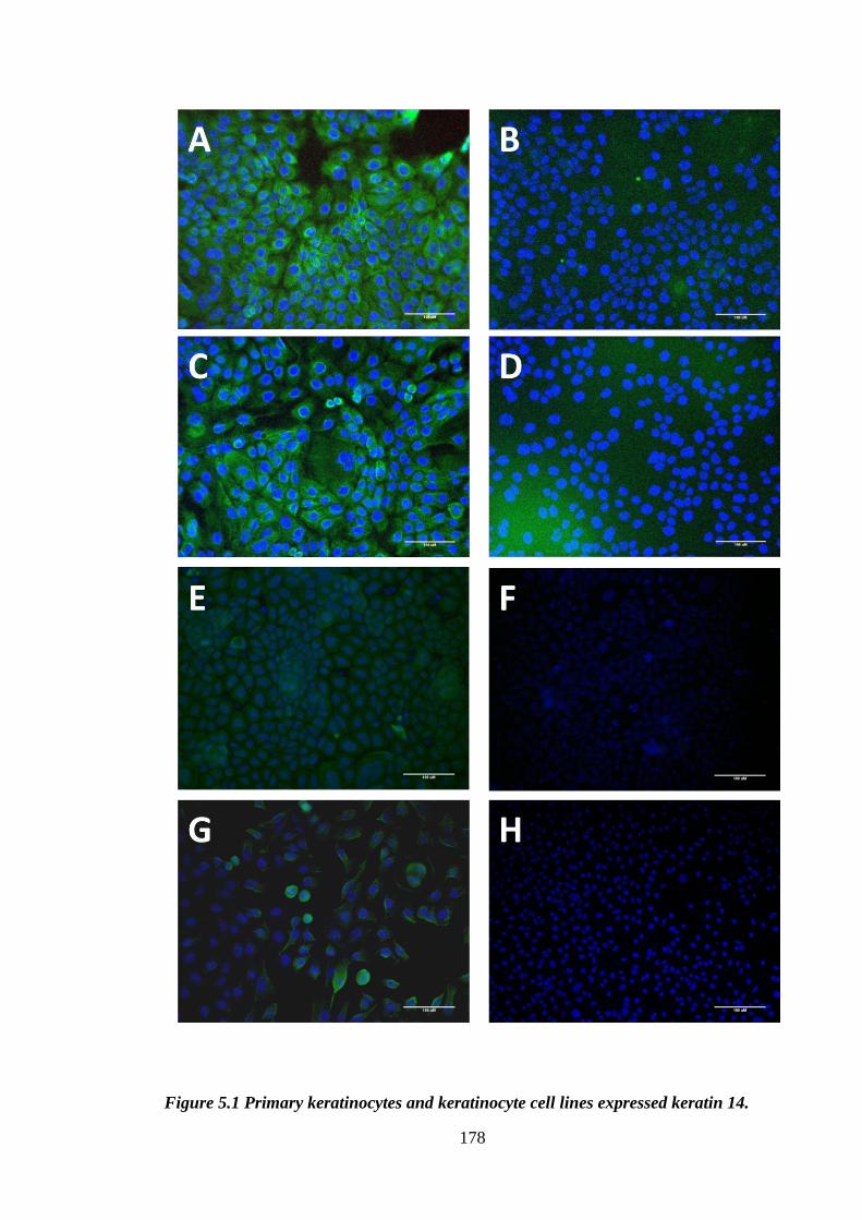
178
Figure 5.1 Primary keratinocytes and keratinocyte cell lines expressed keratin 14.

179
Figure 5.1 Primary keratinocytes and keratinocyte cell lines expressed keratin 14.
PAM cells (A, B, G, H), HaCaT cells (C, D) or primary murine keratinocytes (E, F)
were cultured for 48h on glass coverslips (A-D) or in tissue culture plates (E-H),
washed and fixed in ice cold methanol for 10 minutes, washed with PBS and stained
with anti-K14 antibody at 37°C for 30 minutes. Subsequently, cells were washed and
incubated with secondary goat anti-rabbit FITC conjugated antibody (1:50 dilution).
Next, cells were mounted onto a slide in VECTASHIELD mounting medium with DAPI.
Representative micrographs are shown of anti-keratin staining (A, C, E, G) as well as
respective isotype staining (B, D, F, H) overlaid with DAPI image (x20 magnification).
Scale bar 100μm.
Table 5.1 PAM 212 cells did not secrete TNF-α following incubation with a variety
of stimuli.
PAM cells were grown in complete RPMI medium. Cells were passaged every 2-3 days.
For use in the experiment cells were seeded at 105 cells per well in 12 well plates and
cultured for 48h to become confluent. Subsequently, cells were washed and incubated
with the listed stimuli for 24h. Following incubation supernatants were collected and
levels of cytokines and chemokines were determined by ELISA. * n.d- not done.
5.2.2 PAM cells did not respond to TLR ligands
Following on from the work conducted using THP-1 macrophages (Chapter 4) it was
decided to in vitro stimulate keratinocytes with PGN to elicit cytokine or chemokine
production. Lactoferrin has been shown to bind LPS potently, thereby excluding its use
as a stimulant in the current investigations, as described in the previous chapter. Primary
Cytokine measured
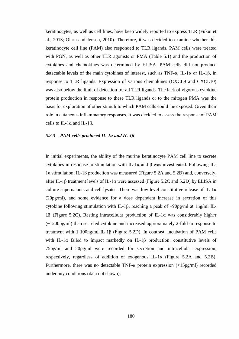
180
keratinocytes, as well as cell lines, have been widely reported to express TLR (Fukui et
al., 2013; Olaru and Jensen, 2010). Therefore, it was decided to examine whether this
keratinocyte cell line (PAM) also responded to TLR ligands. PAM cells were treated
with PGN, as well as other TLR agonists or PMA (Table 5.1) and the production of
cytokines and chemokines was determined by ELISA. PAM cells did not produce
detectable levels of the main cytokines of interest, such as TNF-α, IL-1α or IL-1β, in
response to TLR ligands. Expression of various chemokines (CXCL9 and CXCL10)
was also below the limit of detection for all TLR ligands. The lack of vigorous cytokine
protein production in response to these TLR ligands or to the mitogen PMA was the
basis for exploration of other stimuli to which PAM cells could be exposed. Given their
role in cutaneous inflammatory responses, it was decided to assess the response of PAM
cells to IL-1α and IL-1β.
5.2.3 PAM cells produced IL-1α and IL-1β
In initial experiments, the ability of the murine keratinocyte PAM cell line to secrete
cytokines in response to stimulation with IL-1α and β was investigated. Following IL-
1α stimulation, IL-1β production was measured (Figure 5.2A and 5.2B) and, conversely,
after IL-1β treatment levels of IL-1α were assessed (Figure 5.2C and 5.2D) by ELISA in
culture supernatants and cell lysates. There was low level constitutive release of IL-1α
(20pg/ml), and some evidence for a dose dependent increase in secretion of this
cytokine following stimulation with IL-1β, reaching a peak of ~90pg/ml at 1ng/ml IL-
1 (Figure 5.2C). Resting intracellular production of IL-1α was considerably higher
(~1200pg/ml) than secreted cytokine and increased approximately 2-fold in response to
treatment with 1-100ng/ml IL-1β (Figure 5.2D). In contrast, incubation of PAM cells
with IL-1α failed to impact markedly on IL-1β production: constitutive levels of
75pg/ml and 20pg/ml were recorded for secretion and intracellular expression,
respectively, regardless of addition of exogenous IL-1α (Figure 5.2A and 5.2B).
Furthermore, there was no detectable TNF-α protein expression (<15pg/ml) recorded
under any conditions (data not shown).
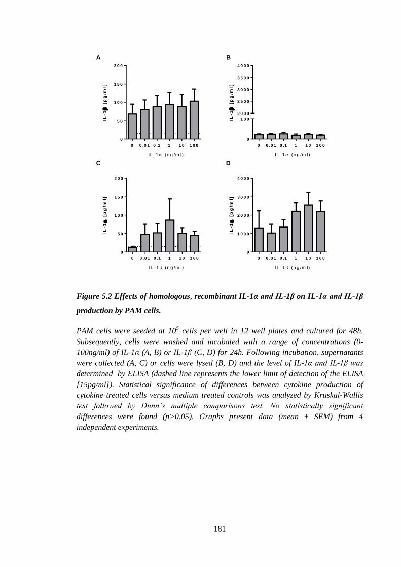
181
0 0 .0 1 0 .1 1 1 0 1 0 0
0
5 0
1 0 0
1 5 0
2 0 0
IL-1
[
pg
/ml]
IL -1 (n g /m l)
0 0 .0 1 0 .1 1 1 0 1 0 0
0
1 0 0
2 0 0 0
2 5 0 0
3 0 0 0
3 5 0 0
4 0 0 0
IL-1
[
pg
/ml]
IL -1 (n g /m l)
0 0 .0 1 0 .1 1 1 0 1 0 0
0
5 0
1 0 0
1 5 0
2 0 0
IL-1
[
pg
/ml]
IL -1 (n g /m l)
0 0 .0 1 0 .1 1 1 0 1 0 0
0
1 0 0 0
2 0 0 0
3 0 0 0
4 0 0 0
IL-1
[
pg
/ml]
IL -1 (n g /m l)
A B
C D
Figure 5.2 Effects of homologous, recombinant IL-1α and IL-1β on IL-1α and IL-1β
production by PAM cells.
PAM cells were seeded at 105 cells per well in 12 well plates and cultured for 48h.
Subsequently, cells were washed and incubated with a range of concentrations (0-
100ng/ml) of IL-1α (A, B) or IL-1β (C, D) for 24h. Following incubation, supernatants
were collected (A, C) or cells were lysed (B, D) and the level of IL-1α and IL-1β was
determined by ELISA (dashed line represents the lower limit of detection of the ELISA
[15pg/ml]). Statistical significance of differences between cytokine production of
cytokine treated cells versus medium treated controls was analyzed by Kruskal-Wallis
test followed by Dunn’s multiple comparisons test. No statistically significant
differences were found (p>0.05). Graphs present data (mean ± SEM) from 4
independent experiments.
182
5.2.4 PAM cells responded to stimulation with IL-1α and IL-1β
Due to the relatively low levels of IL-1α and β protein release by PAM cells, and
absence of TNF-α production, the next step in the current investigations was to examine
changes in mRNA levels for Tnf-α, Il-1β, Il-1α, Cxcl1 and Icam-1 expressed by PAM
cells following cytokine stimulation. Quantitative PCR was utilized to determine the
change in gene expression (Tnf-α, Il-1β, Il-1α, Cxcl1 and Icam-1) levels (relative to the
housekeeping Hprt1 gene). In the first instance, dose dependent changes were examined
following culture with homologous IL-1α and IL-1β. The 2h time point was chosen for
investigation of the dose dependent effects of IL-1α and β stimulation (Figure 5.3).
PAM keratinocytes responded to cytokine stimulation within this 2h time period and
IL-1α was the most potent stimulus for all of the genes tested. Incubation with IL-1α at
concentrations between 0.1 and 1ng/ml resulted in maximal augmentation of expression
for all genes (2, 19, 64, 200 and 290 fold change for Il-1β, Il-1α, Icam-1, Cxcl1 and
Tnf-α, respectively). PAM cells were somewhat less responsive to IL-1β, with peak
induction recorded at 1ng/ml. The rank order of triggered responses was the same for
IL-1α and IL-1β: TNF-α < CXCL1 < ICAM-1 < IL-1α < IL-1β. Interestingly, whereas
TNF-α protein was not detected following cytokine treatment, Tnf-α gene expression
was very markedly up-regulated by both cytokines, with a maximal induction observed
following incubation with 0.1ng/ml of IL-1α (290 fold change) and with 1ng/ml of
IL-1β (490 fold change).
183
0 5 1 0
0
2 0 0
4 0 0
6 0 0
IL -1 (n g /m l)
Fo
ld c
ha
ng
e
0 5 1 0
0
2 0 0
4 0 0
6 0 0
IL -1 (n g /m l)
Fo
ld c
ha
ng
e
A B
T N F - IL 1 C X C L 1 IL -1 IC A M -1
Figure 5.3 Homologous, recombinant IL-1α and IL-1β-induced dose dependent
cytokine and Icam-1 expression by PAM cells.
PAM cells were seeded at 105 cells per well in 12 well plates and cultured for 48h.
Subsequently cells were incubated with a range (0-10ng/ml) of concentrations of IL-1α
(A) or IL-1β (B) for 2h. After incubation cells were lysed, mRNA was extracted; cDNA
was prepared and utilized in a qPCR reaction using TaqMan primers. Expression of
Tnf-α, Cxcl1, Il-1α, Il-1β and Icam-1 was measured relative to the Hprt1 gene and
compared with the medium (0 ng/ml) treated sample. Graphs present data from a single
experiment.
Based on these dose response analyses, PAM cells were incubated with 2ng/ml of either
IL-1α or IL-1β for 0, 1, 2, 4, 6, 8 or 24h (Figure 5.4). The rank order of induction of the
molecules investigated was the same as that recorded for the dose response studies
(TNF-α < CXCL1 < ICAM-1 < IL-1α < IL-1β). Furthermore, the kinetics of cytokine-
induced expression of the molecules was also similar; peaking at 1 to 2 h and returning
to baseline levels within 8h. However, IL-1α was a more potent stimulator than was the
equivalent amount of IL-1β. High level expression of Tnf-α was induced by stimulation
with both IL-1α and β (316 and 100 increase in Tnf-α were observed following 1h
treatment with IL-1α and IL-1β, respectively). Whereas Il-1β exhibited the lowest levels
of up-regulation among the genes measured (maximal induction 1.9 fold increase
following 4h treatment with IL-1α). On the contrary, expression of Il-1α was rapidly
increased to 19 and 16 fold increase by both IL-1α and IL-1β, respectively, at
concentration of 1ng/ml.
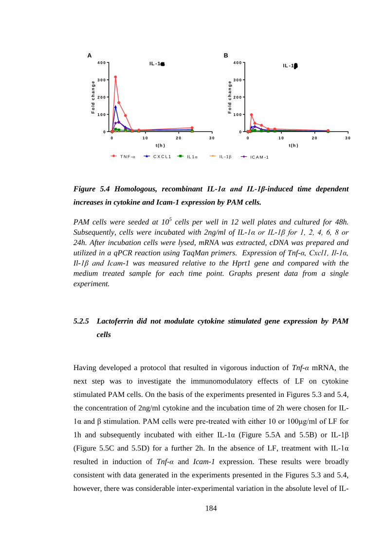
184
0 1 0 2 0 3 0
0
1 0 0
2 0 0
3 0 0
4 0 0
t (h )
Fo
ld c
ha
ng
e
IL -1
0 1 0 2 0 3 0
0
1 0 0
2 0 0
3 0 0
4 0 0
Fo
ld c
ha
ng
e
t ( h )
IL -1
A B
T N F - IL -1 IL 1 IC A M -1C X C L 1
Figure 5.4 Homologous, recombinant IL-1α and IL-1β-induced time dependent
increases in cytokine and Icam-1 expression by PAM cells.
PAM cells were seeded at 105 cells per well in 12 well plates and cultured for 48h.
Subsequently, cells were incubated with 2ng/ml of IL-1α or IL-1β for 1, 2, 4, 6, 8 or
24h. After incubation cells were lysed, mRNA was extracted, cDNA was prepared and
utilized in a qPCR reaction using TaqMan primers. Expression of Tnf-α, Cxcl1, Il-1α,
Il-1β and Icam-1 was measured relative to the Hprt1 gene and compared with the
medium treated sample for each time point. Graphs present data from a single
experiment.
5.2.5 Lactoferrin did not modulate cytokine stimulated gene expression by PAM
cells
Having developed a protocol that resulted in vigorous induction of Tnf-α mRNA, the
next step was to investigate the immunomodulatory effects of LF on cytokine
stimulated PAM cells. On the basis of the experiments presented in Figures 5.3 and 5.4,
the concentration of 2ng/ml cytokine and the incubation time of 2h were chosen for IL-
1α and β stimulation. PAM cells were pre-treated with either 10 or 100μg/ml of LF for
1h and subsequently incubated with either IL-1α (Figure 5.5A and 5.5B) or IL-1β
(Figure 5.5C and 5.5D) for a further 2h. In the absence of LF, treatment with IL-1α
resulted in induction of Tnf-α and Icam-1 expression. These results were broadly
consistent with data generated in the experiments presented in the Figures 5.3 and 5.4,
however, there was considerable inter-experimental variation in the absolute level of IL-
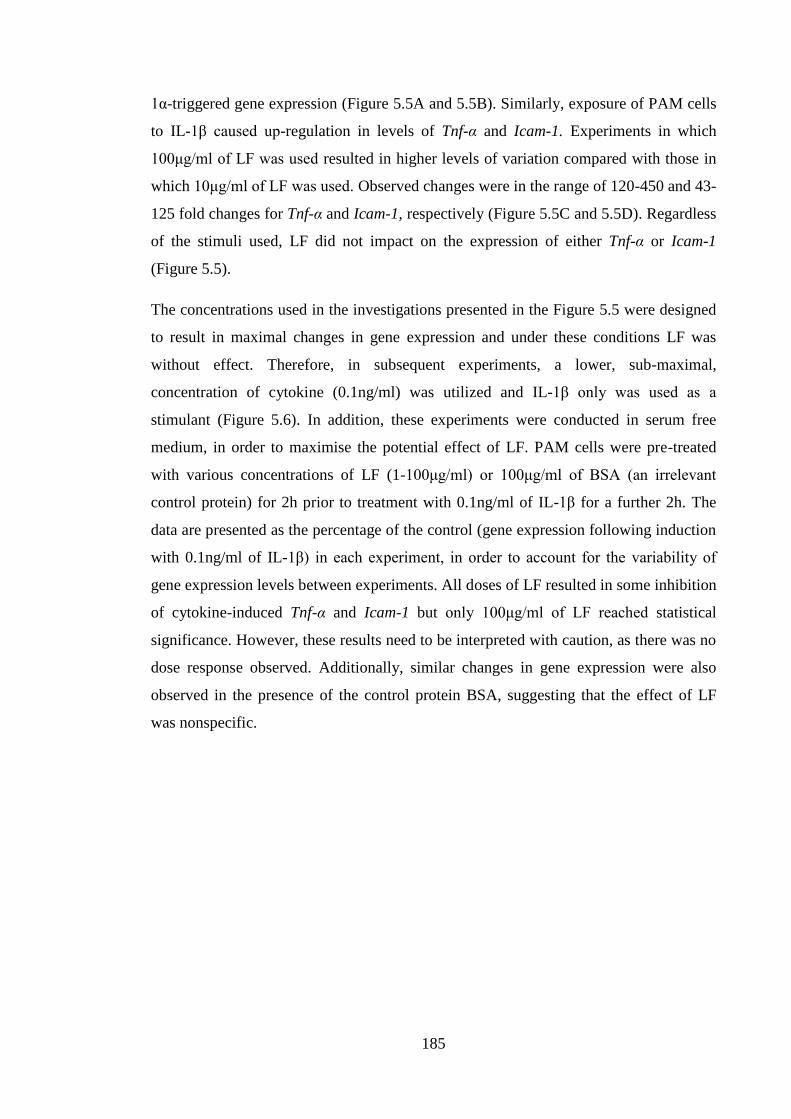
185
1α-triggered gene expression (Figure 5.5A and 5.5B). Similarly, exposure of PAM cells
to IL-1β caused up-regulation in levels of Tnf-α and Icam-1. Experiments in which
100μg/ml of LF was used resulted in higher levels of variation compared with those in
which 10μg/ml of LF was used. Observed changes were in the range of 120-450 and 43-
125 fold changes for Tnf-α and Icam-1, respectively (Figure 5.5C and 5.5D). Regardless
of the stimuli used, LF did not impact on the expression of either Tnf-α or Icam-1
(Figure 5.5).
The concentrations used in the investigations presented in the Figure 5.5 were designed
to result in maximal changes in gene expression and under these conditions LF was
without effect. Therefore, in subsequent experiments, a lower, sub-maximal,
concentration of cytokine (0.1ng/ml) was utilized and IL-1β only was used as a
stimulant (Figure 5.6). In addition, these experiments were conducted in serum free
medium, in order to maximise the potential effect of LF. PAM cells were pre-treated
with various concentrations of LF (1-100μg/ml) or 100μg/ml of BSA (an irrelevant
control protein) for 2h prior to treatment with 0.1ng/ml of IL-1β for a further 2h. The
data are presented as the percentage of the control (gene expression following induction
with 0.1ng/ml of IL-1β) in each experiment, in order to account for the variability of
gene expression levels between experiments. All doses of LF resulted in some inhibition
of cytokine-induced Tnf-α and Icam-1 but only 100μg/ml of LF reached statistical
significance. However, these results need to be interpreted with caution, as there was no
dose response observed. Additionally, similar changes in gene expression were also
observed in the presence of the control protein BSA, suggesting that the effect of LF
was nonspecific.
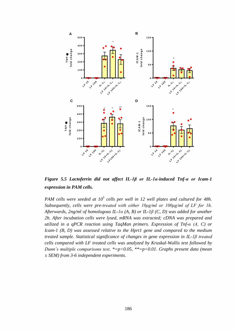
186
TN
F-
fold
ch
an
ge
LF
10
LF
100
IL-1
LF
10+IL
-1
LF
100+IL
-1
0
1 0 0
2 0 0
3 0 0
4 0 0
5 0 0
* * **
ICA
M-1
fold
ch
an
ge
LF
10
LF
100
IL-1
LF
10+IL
-1
LF
100+IL
-1
0
5 0
1 0 0
1 5 0
*
TN
F-
fold
ch
an
ge
LF
10
LF
100
IL-1
LF
10+IL
-1
LF
100+IL
-1
0
1 0 0
2 0 0
3 0 0
4 0 0
5 0 0
*
ICA
M-1
fold
ch
an
ge
LF
10
LF
100
IL-1
LF
10+IL
-1
LF
100+IL
-1
0
5 0
1 0 0
1 5 0
*
A B
C D
Figure 5.5 Lactoferrin did not affect IL-1β or IL-1α-induced Tnf-α or Icam-1
expression in PAM cells.
PAM cells were seeded at 105 cells per well in 12 well plates and cultured for 48h.
Subsequently, cells were pre-treated with either 10μg/ml or 100μg/ml of LF for 1h.
Afterwards, 2ng/ml of homologous IL-1α (A, B) or IL-1β (C, D) was added for another
2h. After incubation cells were lysed, mRNA was extracted; cDNA was prepared and
utilized in a qPCR reaction using TaqMan primers. Expression of Tnf-α (A, C) or
Icam-1 (B, D) was assessed relative to the Hprt1 gene and compared to the medium
treated sample. Statistical significance of changes in gene expression in IL-1β treated
cells compared with LF treated cells was analyzed by Kruskal-Wallis test followed by
Dunn’s multiple comparisons test. *=p<0.05, **=p<0.01. Graphs present data (mean
± SEM) from 3-6 independent experiments.
187
1 1 0 1 0 0 1 0 0
0
5 0
1 0 0
1 5 0
L F B S A
% c
on
tro
l
1 1 0 1 0 0 1 0 0
0
5 0
1 0 0
1 5 0
L F B S A
% c
on
tro
l* *
Figure 5.6 Lactoferrin significantly inhibited expression of Tnf-α and Icam-1
induced by lower concentration of IL-1β.
PAM cells were seeded at 105 cells per well in 12 well plates and cultured for 48h.
Subsequently, cells were washed and cultured in RPMI media. Cells were pre-treated
with 1, 10 or 100μg/ml LF or with 100μg/ml BSA for 2h. After 2h 0.1 ng/ml homologous
IL-1β was added for further 2h. After incubation cells were lysed, mRNA was extracted;
cDNA was prepared and utilized in a qPCR reaction using TaqMan primers.
Expression of Tnf-α (A) or Icam-1 (B) was assessed relative to the Hprt1 gene, and
compared with the medium treated sample. Data are presented as a percentage of the
control (0.1ng/ml of IL-1β). The statistical significance of changes in gene expression
between the different LF concentrations, compared with no LF (100%), was analysed
by a Kruskal-Wallis test followed by Dunn’s test; *=p<0.05. Graphs present data
(mean ± SEM) from 2-4 independent experiments
5.2.6 PGN induced TNF-α production by primary keratinocytes
Cell lines such as PAM may not display the characteristics of freshly isolated cells,
which might explain why these cells do not secrete significant amounts of TNF-α and
do not respond to LF. Therefore, in parallel studies the responses of primary
keratinocytes derived from new born BALB/c strain mice were investigated. Thus,
TNF-α, IL-1α and β secretion was measured following TLR2 ligand (PGN) stimulation
of primary keratinocyte cultures (Figure 5.7). Resting cells did not express detectable
levels of secreted TNF-α, although there was detectable constitutive expression of both
IL-1α and IL-1β (50 and 20pg/ml, respectively). Peptidoglycan treatment resulted in a
modest increase in TNF-α secretion (50pg/ml) (Figure 5.7A) but did not impact on
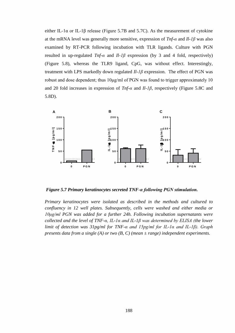
188
either IL-1α or IL-1β release (Figure 5.7B and 5.7C). As the measurement of cytokine
at the mRNA level was generally more sensitive, expression of Tnf-α and Il-1β was also
examined by RT-PCR following incubation with TLR ligands. Culture with PGN
resulted in up-regulated Tnf-α and Il-1β expression (by 3 and 4 fold, respectively)
(Figure 5.8), whereas the TLR9 ligand, CpG, was without effect. Interestingly,
treatment with LPS markedly down regulated Il-1β expression. The effect of PGN was
robust and dose dependent; thus 10μg/ml of PGN was found to trigger approximately 10
and 20 fold increases in expression of Tnf-α and Il-1β, respectively (Figure 5.8C and
5.8D).
0 P G N
0
5 0
1 0 0
1 5 0
2 0 0
TN
F-
[p
g/m
l]
0 P G N
0
5 0
1 0 0
1 5 0
2 0 0
IL-1
[
pg
/ml]
0 P G N
0
5 0
1 0 0
1 5 0
2 0 0
IL-1
[
pg
/ml]
A B C
Figure 5.7 Primary keratinocytes secreted TNF-α following PGN stimulation.
Primary keratinocytes were isolated as described in the methods and cultured to
confluency in 12 well plates. Subsequently, cells were washed and either media or
10μg/ml PGN was added for a further 24h. Following incubation supernatants were
collected and the level of TNF-α, IL-1α and IL-1β was determined by ELISA (the lower
limit of detection was 31pg/ml for TNF-α and 15pg/ml for IL-1α and IL-1β). Graph
presents data from a single (A) or two (B, C) (mean ± range) independent experiments.

189
P G N L P S C p G
0
1
2
3
4
TN
F-
fold
ch
an
ge
1 g /m l
P G N L P S C p G
0
1
2
3
4
IL-1
fold
ch
an
ge
1 g /m l
0 .1 1 1 0
0
5
1 0
1 5
TN
F-
Fo
ld c
ha
ng
e
P G N ( g /m l)
0 .1 1 1 0
0
1 0
2 0
3 0
4 0
IL-1
fold
ch
an
ge
P G N ( g /m l)
A B
C D
Figure 5.8 Primary murine keratinocytes expressed Tnf-α and Il-1β upon TLR2
ligand stimulation.
Primary keratinocytes were isolated as described in the methods and cultured to
confluency in 12 well plates. Subsequently, cells were washed and incubated with
1μg/ml of LPS, CpG, R848, PGN (A, B) or with a range of PGN doses (C, D) for 24h.
After incubation cells were lysed, mRNA was extracted; cDNA was prepared and
utilized in a qPCR reaction using TaqMan primers. Expression of Tnf-α and Il-1β was
measured relative to the Hprt1 gene and compared with the medium treated sample.
Graphs present data from a single experiment or 5 independent experiments (mean ±
SEM).
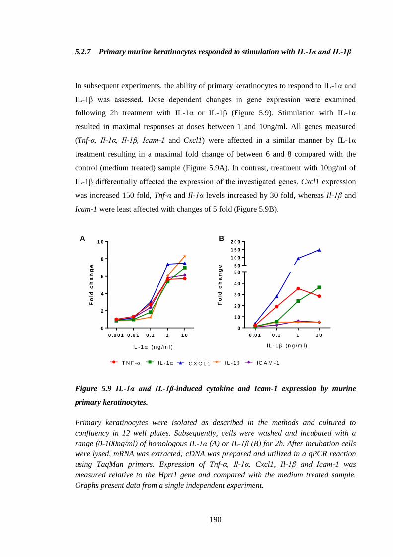
190
5.2.7 Primary murine keratinocytes responded to stimulation with IL-1α and IL-1β
In subsequent experiments, the ability of primary keratinocytes to respond to IL-1α and
IL-1β was assessed. Dose dependent changes in gene expression were examined
following 2h treatment with IL-1α or IL-1β (Figure 5.9). Stimulation with IL-1α
resulted in maximal responses at doses between 1 and 10ng/ml. All genes measured
(Tnf-α, Il-1α, Il-1β, Icam-1 and Cxcl1) were affected in a similar manner by IL-1α
treatment resulting in a maximal fold change of between 6 and 8 compared with the
control (medium treated) sample (Figure 5.9A). In contrast, treatment with 10ng/ml of
IL-1β differentially affected the expression of the investigated genes. Cxcl1 expression
was increased 150 fold, Tnf-α and Il-1α levels increased by 30 fold, whereas Il-1β and
Icam-1 were least affected with changes of 5 fold (Figure 5.9B).
0 .0 0 1 0 .0 1 0 .1 1 1 0
0
2
4
6
8
1 0
Fo
ld c
ha
ng
e
IL -1 (n g /m l)
0 .0 1 0 .1 1 1 0
0
1 0
2 0
3 0
4 0
5 0
5 0
1 0 0
1 5 0
2 0 0
IL -1 (n g /m l)
Fo
ld c
ha
ng
e
T N F - IL -1 C X C L 1 IL -1 IC A M -1
A B
Figure 5.9 IL-1α and IL-1β-induced cytokine and Icam-1 expression by murine
primary keratinocytes.
Primary keratinocytes were isolated as described in the methods and cultured to
confluency in 12 well plates. Subsequently, cells were washed and incubated with a
range (0-100ng/ml) of homologous IL-1α (A) or IL-1β (B) for 2h. After incubation cells
were lysed, mRNA was extracted; cDNA was prepared and utilized in a qPCR reaction
using TaqMan primers. Expression of Tnf-α, Il-1α, Cxcl1, Il-1β and Icam-1 was
measured relative to the Hprt1 gene and compared with the medium treated sample.
Graphs present data from a single independent experiment.
191
5.2.8 Lactoferrin did not modulate cytokine stimulated gene expression by primary
keratinocytes
Experiments depicted in the Figure 5.7 demonstrated that there were differences in the
vigour of the changes caused by stimulation with IL-1α and IL-1β. The concentration of
2ng/ml was chosen for both IL-1α and IL-1β to provide, respectively, vigorous and sub-
optimal levels of stimulation of primary keratinocytes. In that setting potential
immunomodulatory effects of LF were examined. Lactoferrin alone was without any
inhibitory effects on gene expression at any time point. There was considerable inter-
experimental variation in the cytokine-induced changes in gene expression, however,
some patterns emerged. Treatment with IL-1β (Figure 5.10E-5.10F) caused similar
effects on Il-1β and Icam-1 gene expression to those observed for IL-1α treatment
(Figure 5.10B-5.10C), both with respect to the kinetics and the extent of up-regulation.
The effects on Tnf-α expression were more variable, with most marked increases
recorded at 24h following treatment with IL-1β (Figure 5.10D).
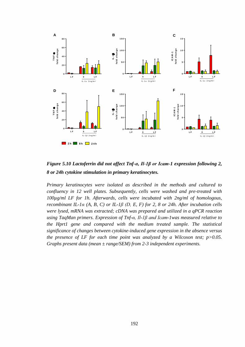
192
L F 0 L F
0
2 0
4 0
6 0
8 0
TN
F-
fold
ch
an
ge
IL -1 2 n g /m l
L F 0 L F
0
5 0
1 0 0
1 5 0
IL-1
fold
ch
an
ge
IL -1 2 n g /m l
L F 0 L F
0
5
1 0
1 5
ICA
M-1
fold
ch
an
ge
IL -1 2 n g /m l
L F 0 L F
0
2 0
4 0
6 0
8 0
TN
F-
fold
ch
an
ge
IL -1 2 n g /m l
L F 0 L F
0
5
1 0
1 5
ICA
M-1
fold
ch
an
ge
IL -1 2 n g /m l
2 h 8 h 2 4 h
L F 0 L F
0
5 0
1 0 0
1 5 0
IL-1
fold
ch
an
ge
IL -1 2 n g /m l
A B C
D E F
Figure 5.10 Lactoferrin did not affect Tnf-α, Il-1β or Icam-1 expression following 2,
8 or 24h cytokine stimulation in primary keratinocytes.
Primary keratinocytes were isolated as described in the methods and cultured to
confluency in 12 well plates. Subsequently, cells were washed and pre-treated with
100µg/ml LF for 1h. Afterwards, cells were incubated with 2ng/ml of homologous,
recombinant IL-1α (A, B, C) or IL-1β (D, E, F) for 2, 8 or 24h. After incubation cells
were lysed, mRNA was extracted; cDNA was prepared and utilized in a qPCR reaction
using TaqMan primers. Expression of Tnf-α, Il-1β and Icam-1was measured relative to
the Hprt1 gene and compared with the medium treated sample. The statistical
significance of changes between cytokine-induced gene expression in the absence versus
the presence of LF for each time point was analyzed by a Wilcoxon test; p>0.05.
Graphs present data (mean ± range/SEM) from 2-3 independent experiments.
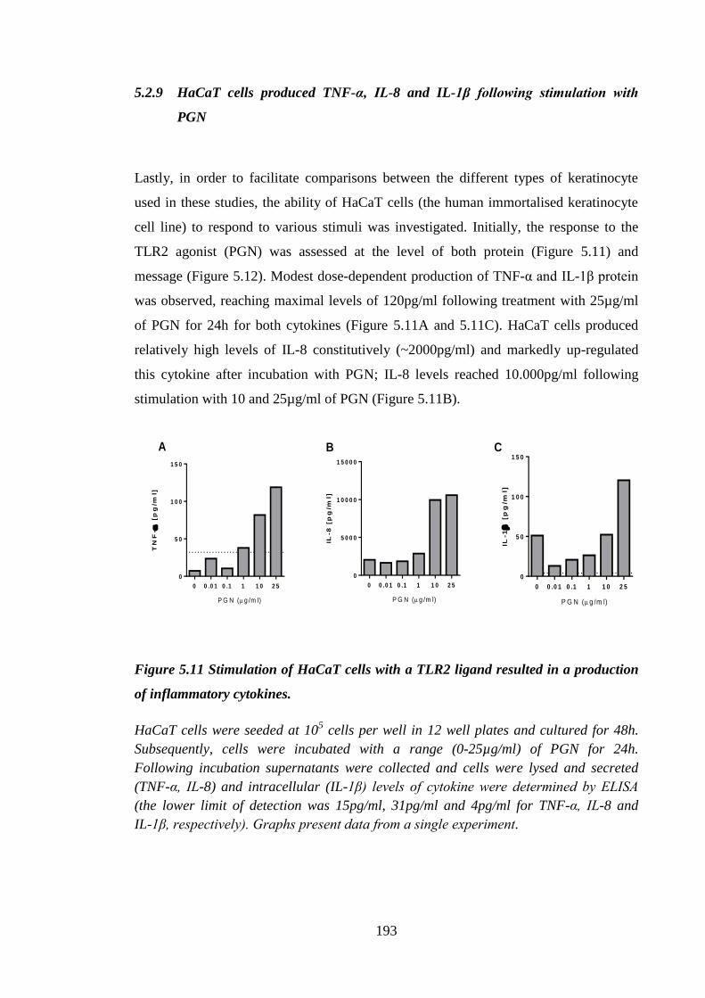
193
5.2.9 HaCaT cells produced TNF-α, IL-8 and IL-1β following stimulation with
PGN
Lastly, in order to facilitate comparisons between the different types of keratinocyte
used in these studies, the ability of HaCaT cells (the human immortalised keratinocyte
cell line) to respond to various stimuli was investigated. Initially, the response to the
TLR2 agonist (PGN) was assessed at the level of both protein (Figure 5.11) and
message (Figure 5.12). Modest dose-dependent production of TNF-α and IL-1β protein
was observed, reaching maximal levels of 120pg/ml following treatment with 25µg/ml
of PGN for 24h for both cytokines (Figure 5.11A and 5.11C). HaCaT cells produced
relatively high levels of IL-8 constitutively (~2000pg/ml) and markedly up-regulated
this cytokine after incubation with PGN; IL-8 levels reached 10.000pg/ml following
stimulation with 10 and 25µg/ml of PGN (Figure 5.11B).
TN
F-
[p
g/m
l]
0 0 .0 1 0 .1 1 1 0 2 5
0
5 0
1 0 0
1 5 0
P G N (g /m l)
IL-8
[p
g/m
l]
0 0 .0 1 0 .1 1 1 0 2 5
0
5 0 0 0
1 0 0 0 0
1 5 0 0 0
P G N (g /m l)
IL-1
[p
g/m
l]
0 0 .0 1 0 .1 1 1 0 2 5
0
5 0
1 0 0
1 5 0
P G N (g /m l)
A B C
Figure 5.11 Stimulation of HaCaT cells with a TLR2 ligand resulted in a production
of inflammatory cytokines.
HaCaT cells were seeded at 105 cells per well in 12 well plates and cultured for 48h.
Subsequently, cells were incubated with a range (0-25µg/ml) of PGN for 24h.
Following incubation supernatants were collected and cells were lysed and secreted
(TNF-α, IL-8) and intracellular (IL-1β) levels of cytokine were determined by ELISA
(the lower limit of detection was 15pg/ml, 31pg/ml and 4pg/ml for TNF-α, IL-8 and
IL-1β, respectively). Graphs present data from a single experiment.
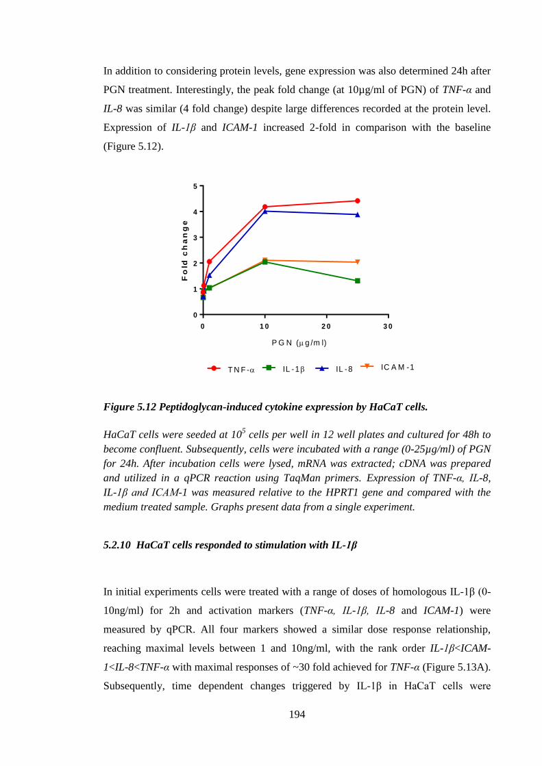
194
In addition to considering protein levels, gene expression was also determined 24h after
PGN treatment. Interestingly, the peak fold change (at 10µg/ml of PGN) of TNF-α and
IL-8 was similar (4 fold change) despite large differences recorded at the protein level.
Expression of IL-1β and ICAM-1 increased 2-fold in comparison with the baseline
(Figure 5.12).
0 1 0 2 0 3 0
0
1
2
3
4
5F
old
ch
an
ge
T N F - IL -8IL -1 IC A M -1
P G N (g /m l)
Figure 5.12 Peptidoglycan-induced cytokine expression by HaCaT cells.
HaCaT cells were seeded at 105 cells per well in 12 well plates and cultured for 48h to
become confluent. Subsequently, cells were incubated with a range (0-25µg/ml) of PGN
for 24h. After incubation cells were lysed, mRNA was extracted; cDNA was prepared
and utilized in a qPCR reaction using TaqMan primers. Expression of TNF-α, IL-8,
IL-1β and ICAM-1 was measured relative to the HPRT1 gene and compared with the
medium treated sample. Graphs present data from a single experiment.
5.2.10 HaCaT cells responded to stimulation with IL-1β
In initial experiments cells were treated with a range of doses of homologous IL-1β (0-
10ng/ml) for 2h and activation markers (TNF-α, IL-1β, IL-8 and ICAM-1) were
measured by qPCR. All four markers showed a similar dose response relationship,
reaching maximal levels between 1 and 10ng/ml, with the rank order IL-1β<ICAM-
1<IL-8<TNF-α with maximal responses of ~30 fold achieved for TNF-α (Figure 5.13A).
Subsequently, time dependent changes triggered by IL-1β in HaCaT cells were
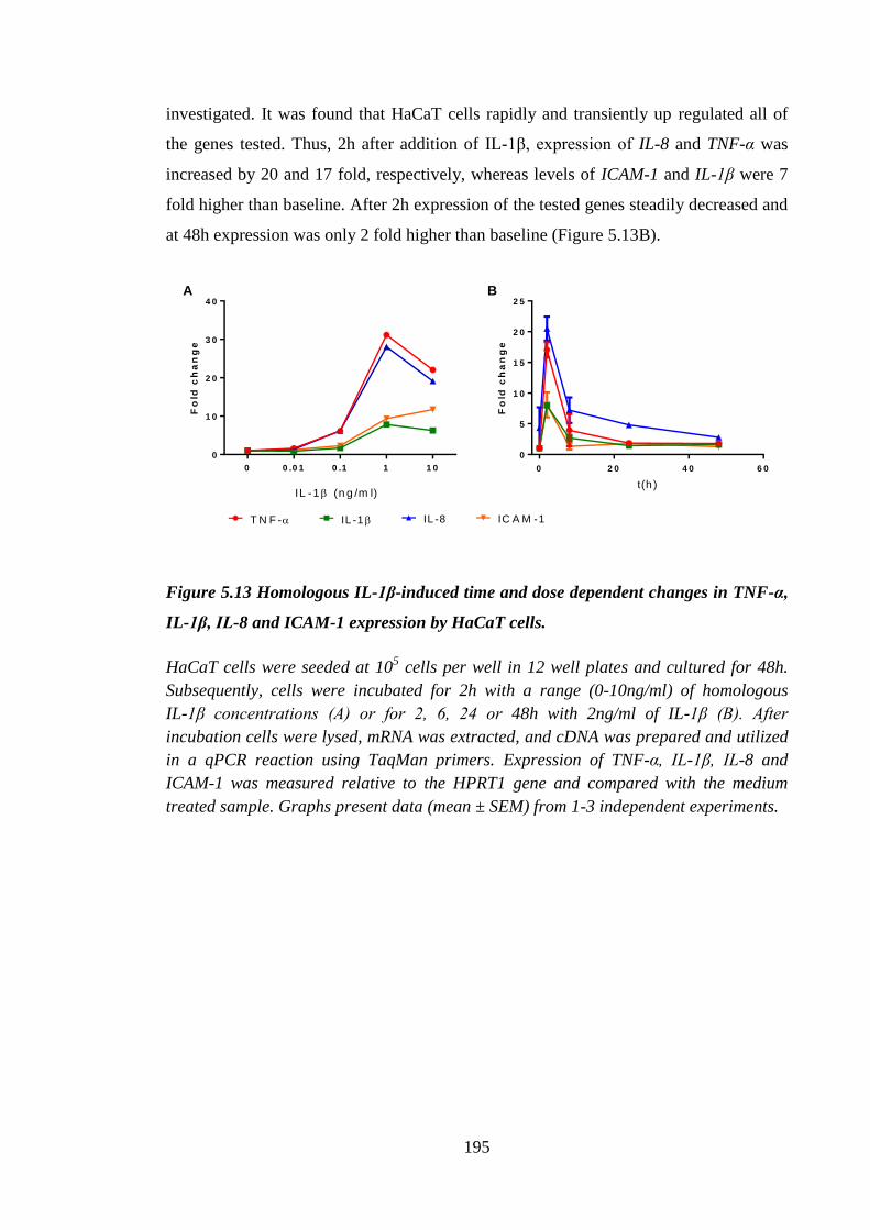
195
investigated. It was found that HaCaT cells rapidly and transiently up regulated all of
the genes tested. Thus, 2h after addition of IL-1β, expression of IL-8 and TNF-α was
increased by 20 and 17 fold, respectively, whereas levels of ICAM-1 and IL-1β were 7
fold higher than baseline. After 2h expression of the tested genes steadily decreased and
at 48h expression was only 2 fold higher than baseline (Figure 5.13B).
0 0 .0 1 0 .1 1 1 0
0
1 0
2 0
3 0
4 0
Fo
ld c
ha
ng
e
IL -1 (n g /m l)
0 2 0 4 0 6 0
0
5
1 0
1 5
2 0
2 5
Fo
ld c
ha
ng
e
t(h )
A B
T N F - IL -1 IL -8 IC A M -1
Figure 5.13 Homologous IL-1β-induced time and dose dependent changes in TNF-α,
IL-1β, IL-8 and ICAM-1 expression by HaCaT cells.
HaCaT cells were seeded at 105 cells per well in 12 well plates and cultured for 48h.
Subsequently, cells were incubated for 2h with a range (0-10ng/ml) of homologous
IL-1β concentrations (A) or for 2, 6, 24 or 48h with 2ng/ml of IL-1β (B). After
incubation cells were lysed, mRNA was extracted, and cDNA was prepared and utilized
in a qPCR reaction using TaqMan primers. Expression of TNF-α, IL-1β, IL-8 and
ICAM-1 was measured relative to the HPRT1 gene and compared with the medium
treated sample. Graphs present data (mean ± SEM) from 1-3 independent experiments.
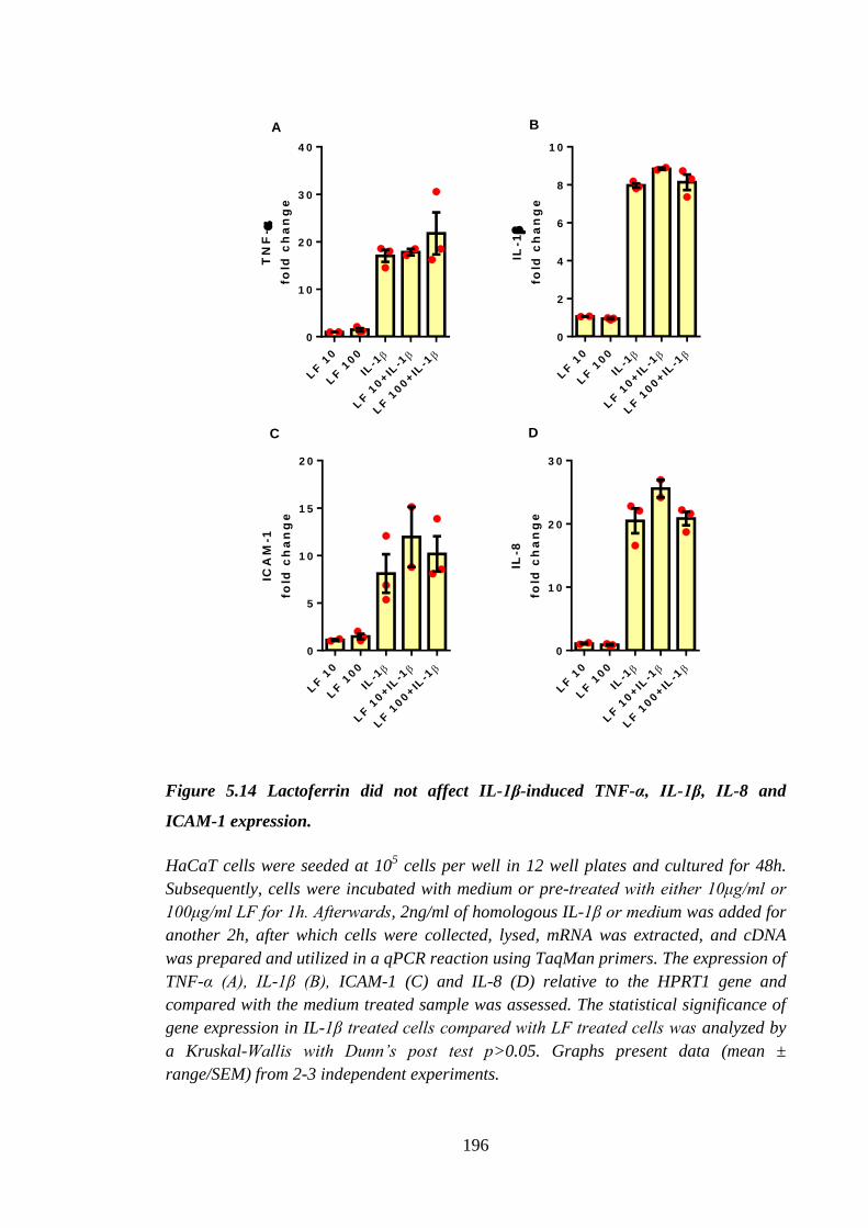
196
LF
10
LF
100
IL-1
LF
10+IL
-1
LF
100+IL
-1
0
1 0
2 0
3 0
4 0
TN
F-
fold
ch
an
ge
LF
10
LF
100
IL-1
LF
10+IL
-1
LF
100+IL
-1
0
2
4
6
8
1 0
IL-1
fold
ch
an
ge
LF
10
LF
100
IL-1
LF
10+IL
-1
LF
100+IL
-1
0
5
1 0
1 5
2 0
ICA
M-1
fold
ch
an
ge
LF
10
LF
100
IL-1
LF
10+IL
-1
LF
100+IL
-1
0
1 0
2 0
3 0
IL-8
fold
ch
an
ge
A B
C D
Figure 5.14 Lactoferrin did not affect IL-1β-induced TNF-α, IL-1β, IL-8 and
ICAM-1 expression.
HaCaT cells were seeded at 105 cells per well in 12 well plates and cultured for 48h.
Subsequently, cells were incubated with medium or pre-treated with either 10μg/ml or
100μg/ml LF for 1h. Afterwards, 2ng/ml of homologous IL-1β or medium was added for
another 2h, after which cells were collected, lysed, mRNA was extracted, and cDNA
was prepared and utilized in a qPCR reaction using TaqMan primers. The expression of
TNF-α (A), IL-1β (B), ICAM-1 (C) and IL-8 (D) relative to the HPRT1 gene and
compared with the medium treated sample was assessed. The statistical significance of
gene expression in IL-1β treated cells compared with LF treated cells was analyzed by
a Kruskal-Wallis with Dunn’s post test p>0.05. Graphs present data (mean ±
range/SEM) from 2-3 independent experiments.
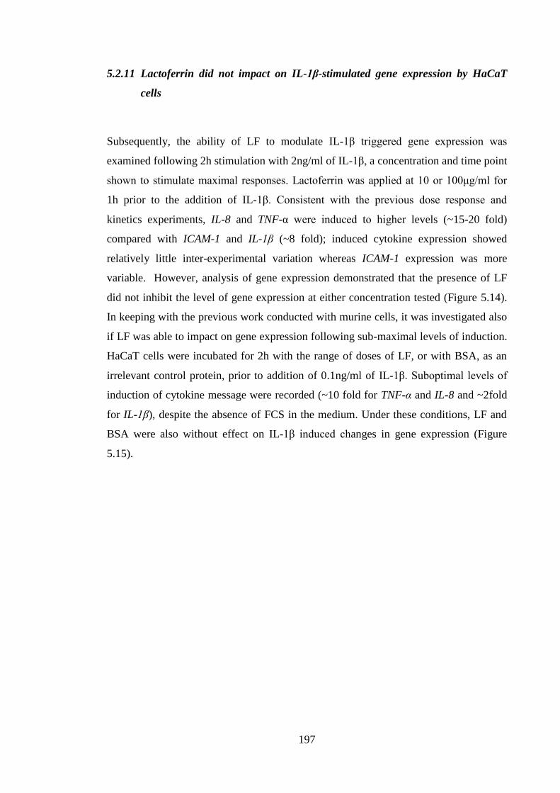
197
5.2.11 Lactoferrin did not impact on IL-1β-stimulated gene expression by HaCaT
cells
Subsequently, the ability of LF to modulate IL-1β triggered gene expression was
examined following 2h stimulation with 2ng/ml of IL-1β, a concentration and time point
shown to stimulate maximal responses. Lactoferrin was applied at 10 or 100μg/ml for
1h prior to the addition of IL-1β. Consistent with the previous dose response and
kinetics experiments, IL-8 and TNF-α were induced to higher levels (~15-20 fold)
compared with ICAM-1 and IL-1β (~8 fold); induced cytokine expression showed
relatively little inter-experimental variation whereas ICAM-1 expression was more
variable. However, analysis of gene expression demonstrated that the presence of LF
did not inhibit the level of gene expression at either concentration tested (Figure 5.14).
In keeping with the previous work conducted with murine cells, it was investigated also
if LF was able to impact on gene expression following sub-maximal levels of induction.
HaCaT cells were incubated for 2h with the range of doses of LF, or with BSA, as an
irrelevant control protein, prior to addition of 0.1ng/ml of IL-1β. Suboptimal levels of
induction of cytokine message were recorded (~10 fold for TNF-α and IL-8 and ~2fold
for IL-1β), despite the absence of FCS in the medium. Under these conditions, LF and
BSA were also without effect on IL-1β induced changes in gene expression (Figure
5.15).
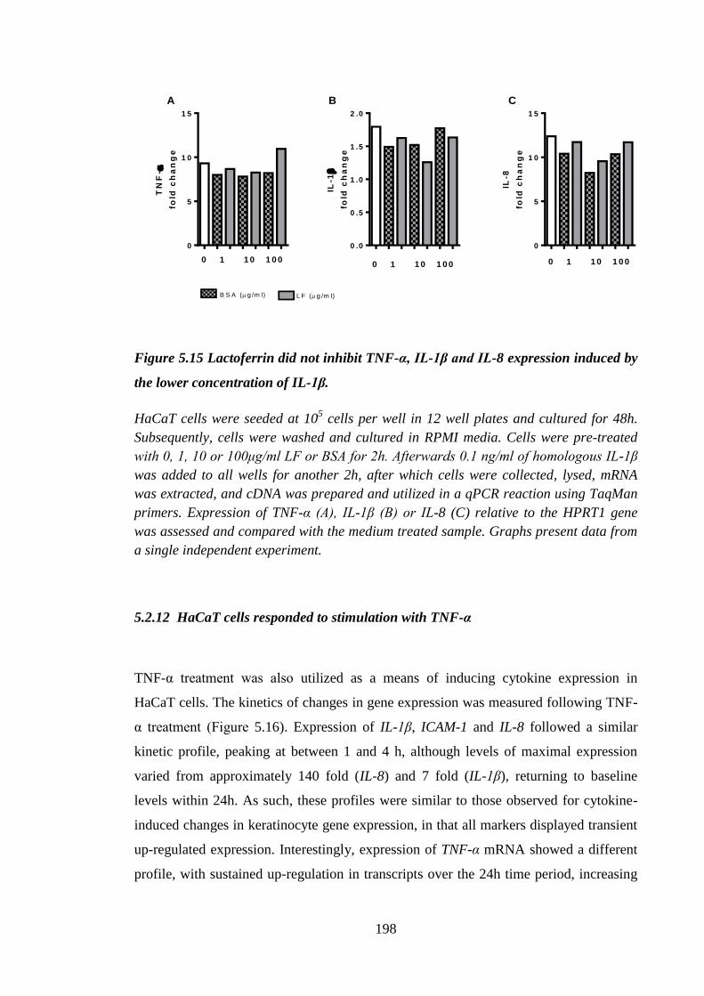
198
TN
F-
fold
ch
an
ge
0
5
1 0
1 5
1 1 0 1 0 00
IL-1
fold
ch
an
ge
0 .0
0 .5
1 .0
1 .5
2 .0
1 1 0 1 0 00
IL-8
fold
ch
an
ge
0
5
1 0
1 5
1 1 0 1 0 00
L F (g /m l)B S A (g /m l)
A B C
Figure 5.15 Lactoferrin did not inhibit TNF-α, IL-1β and IL-8 expression induced by
the lower concentration of IL-1β.
HaCaT cells were seeded at 105 cells per well in 12 well plates and cultured for 48h.
Subsequently, cells were washed and cultured in RPMI media. Cells were pre-treated
with 0, 1, 10 or 100μg/ml LF or BSA for 2h. Afterwards 0.1 ng/ml of homologous IL-1β
was added to all wells for another 2h, after which cells were collected, lysed, mRNA
was extracted, and cDNA was prepared and utilized in a qPCR reaction using TaqMan
primers. Expression of TNF-α (A), IL-1β (B) or IL-8 (C) relative to the HPRT1 gene
was assessed and compared with the medium treated sample. Graphs present data from
a single independent experiment.
5.2.12 HaCaT cells responded to stimulation with TNF-α
TNF-α treatment was also utilized as a means of inducing cytokine expression in
HaCaT cells. The kinetics of changes in gene expression was measured following TNF-
α treatment (Figure 5.16). Expression of IL-1β, ICAM-1 and IL-8 followed a similar
kinetic profile, peaking at between 1 and 4 h, although levels of maximal expression
varied from approximately 140 fold (IL-8) and 7 fold (IL-1β), returning to baseline
levels within 24h. As such, these profiles were similar to those observed for cytokine-
induced changes in keratinocyte gene expression, in that all markers displayed transient
up-regulated expression. Interestingly, expression of TNF-α mRNA showed a different
profile, with sustained up-regulation in transcripts over the 24h time period, increasing
199
steadily from 23 fold above baseline after 1h to a 114 fold increase 24h following TNF-
α stimulation.
0 1 0 2 0 3 0
0
5 0
1 0 0
1 5 0
2 0 0
t (h )
Fo
ld c
ha
ng
e
T N F - IL -1 IL -8 IC A M -1
Figure 5.16 Effects of homologous TNF-α on time dependent changes in TNF-α, IL-
1β, IL-8 and ICAM-1 mRNA expression by HaCaT cells.
HaCaT cells were seeded at 105 cells per well in 12 well plates and cultured for 48h.
Subsequently, cells were washed and cultured in RPMI media with 10ng TNF-α for 0, 1,
2, 4, 6, 10 or 24h. Following incubation cells were collected, lysed, mRNA was
extracted, and cDNA was prepared and utilized in a qPCR reaction using TaqMan
primers. Expression of TNF-α, IL-1β, IL-8 and ICAM-1 was measured relative to the
HPRT1 gene and compared with the medium treated sample. Graphs present data from
a single independent experiment.
5.2.13 Cytokine triggered gene expression by HaCaT cells was modulated by
dexamethasone and thioredoxin peptide
Given the relative insensitivity of the different keratinocyte cell types to the
immunomodulatory effects of LF, additional control inhibitory molecules were
employed as positive controls for the experimental system. These controls were the
potent anti-inflammatory steroid drug, dexamethasone (Remick et al., 1989), and
peptides: thioredoxin (Tian et al., 2013) and Pep-1 (Mummert et al., 2000). All of these
controls (with the exception of Pep-1) were shown previously to exert anti-
200
inflammatory actions in the skin in vivo as well as in vitro in keratinocyte cell culture
(Cumberbatch et al., 1999b; Tian et al., 2013). Given that gene expression following
treatment with TNF-α resulted in a more delayed induction of cytokine changes than
those observed for IL-1β, the 24h time point was chosen for both cytokines for further
investigations of these immunomodulators. In the presence of the control irrelevant
protein (BSA), there was a marked and relatively consistent TNF-α-induced up-
regulation in transcripts for TNF-α (~300 fold), IL-8 (~50 fold) and IL-1β (5 fold).
Similar effects were recorded for IL-1β-induced changes, although such were of a lower
order of magnitude (15 fold, 10 fold and 2 fold for TNF-α, IL-8 and IL-1β respectively)
(Figure 5.17), as the 24h time point was selected on the basis of optimal responses for
TNF-α. Culture with Pep-1 was without significant effect on any of the parameters
measured. In contrast, treatment with dexamethasone resulted in a marked down-
regulation of all cytokines induced by IL-1β (at least 50% inhibition) and of TNF-α-
induced TNF-α and IL-1β, although IL-8 was unaffected. It was not possible to test for
the statistical significance of these findings as only 2 independent experiments were
conducted with dexamethasone. Another peptide tested for its immunomodulatory
potential was one derived from thioredoxin (TRX). Cells were pre-treated with the TRX
peptide, or a scrambled peptide with the same amino acid content, but in a random
sequence, and then stimulated with either IL-1β (Figure 5.18) or TNF-α (Figure 5.19)
for a further 24h. There was some inter-experimental variation, but following pre-
treatment with scrambled peptide a broadly similar profile of IL-1β-induced gene
expression changes were observed to those recorded previously (Figure 5.17). In three
independent experiments, TRX was without marked effect on IL-1β-induced gene
expression (Figure 5.18). As reported previously, stimulation of HaCaT cells with TNF-
α resulted in more vigorous gene expression, particularly for TNF-α transcripts,
although there was some inter-experimental variation. TRX peptide was without effect
on IL-8 levels, but the other genes (TNF-α, IL-1β and ICAM-1) all showed a trend for
inhibition which reached statistical significance for TNF-α and IL-1β (Figure 5.19A and
5.19B).
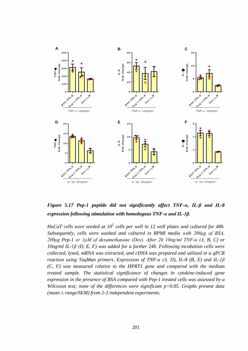
201
TN
F-
fold
ch
an
ge
BS
A 2
00
g
Pep
-1 2
00
g
Dex 1M
0
1 0 0
2 0 0
3 0 0
4 0 0
5 0 0
T N F - 1 0 n g /m l
IL-8
fold
ch
an
ge
BS
A 2
00
g
Pep
-1 2
00
g
Dex 1M
0
2 0
4 0
6 0
8 0
T N F - 1 0 n g /m l
IL-1
fold
ch
an
ge
BS
A 2
00
g
Pep
-1 2
00
g
Dex 1M
0
5
1 0
1 5
T N F - 1 0 n g /m l
TN
F-
fold
ch
an
ge
BS
A 2
00
g
Pep
-1 2
00
g
Dex 1M
0
5
1 0
1 5
2 0
IL -1 1 0 n g /m l
IL-8
fold
ch
an
ge
BS
A 2
00
g
Pep
-1 2
00
g
Dex 1M
0
5
1 0
1 5
IL -1 1 0 n g /m lIL
-1
fold
ch
an
ge
BS
A 2
00
g
Pep
-1 2
00
g
Dex 1M
0
1
2
3
IL -1 1 0 n g /m l
A B C
D E F
Figure 5.17 Pep-1 peptide did not significantly affect TNF-α, IL-β and IL-8
expression following stimulation with homologous TNF-α and IL-1β.
HaCaT cells were seeded at 105 cells per well in 12 well plates and cultured for 48h.
Subsequently, cells were washed and cultured in RPMI media with 200μg of BSA,
200μg Pep-1 or 1μM of dexamethasone (Dex). After 2h 10ng/ml TNF-α (A, B, C) or
10ng/ml IL-1β (D, E, F) was added for a further 24h. Following incubation cells were
collected, lysed, mRNA was extracted, and cDNA was prepared and utilized in a qPCR
reaction using TaqMan primers. Expression of TNF-α (A, D), IL-8 (B, E) and IL-1β
(C, F) was measured relative to the HPRT1 gene and compared with the medium
treated sample. The statistical significance of changes in cytokine-induced gene
expression in the presence of BSA compared with Pep-1 treated cells was assessed by a
Wilcoxon test; none of the differences were significant p>0.05. Graphs present data
(mean ± range/SEM) from 2-3 independent experiments.
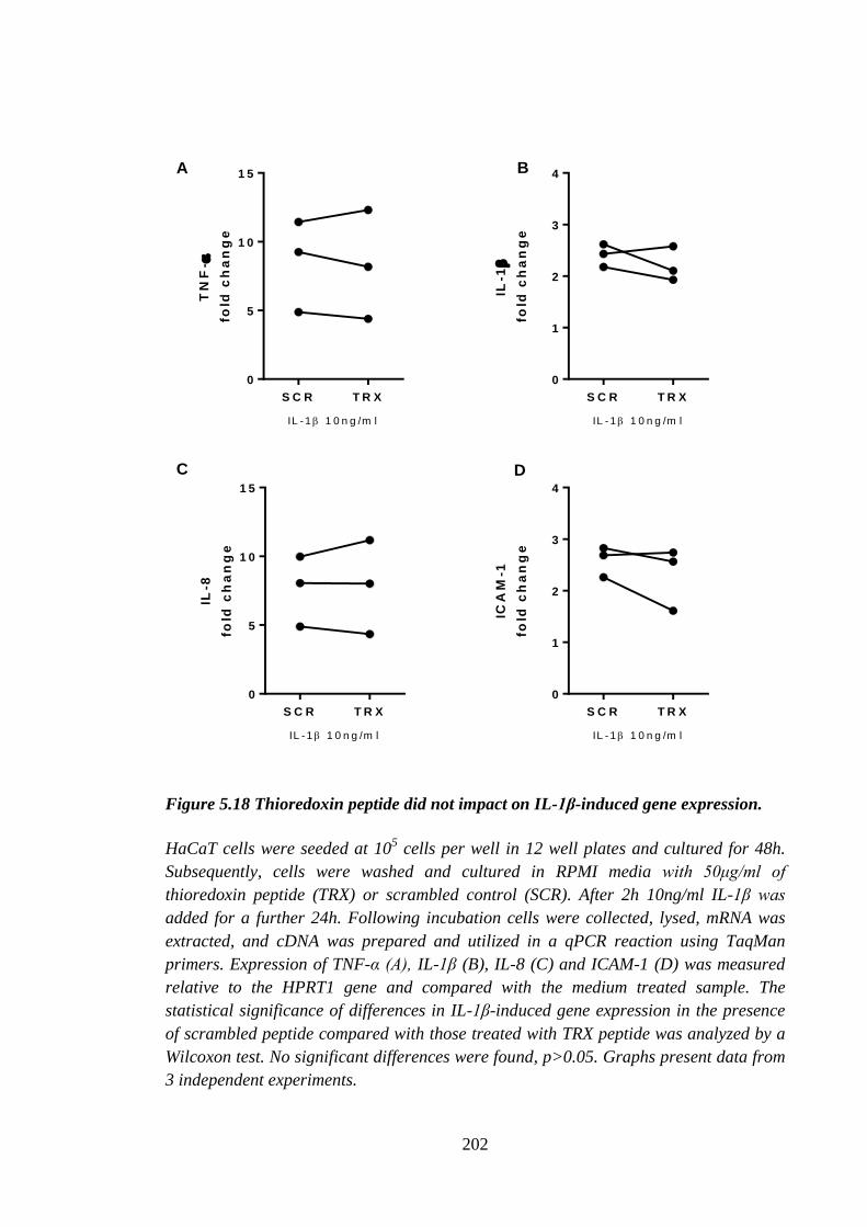
202
S C R T R X
0
5
1 0
1 5
TN
F-
fold
ch
an
ge
IL -1 1 0 n g /m l
S C R T R X
0
1
2
3
4
IL-1
fold
ch
an
ge
IL -1 1 0 n g /m l
S C R T R X
0
5
1 0
1 5
IL-8
fold
ch
an
ge
IL -1 1 0 n g /m l
S C R T R X
0
1
2
3
4IC
AM
-1
fold
ch
an
ge
IL -1 1 0 n g /m l
A B
C D
Figure 5.18 Thioredoxin peptide did not impact on IL-1β-induced gene expression.
HaCaT cells were seeded at 105 cells per well in 12 well plates and cultured for 48h.
Subsequently, cells were washed and cultured in RPMI media with 50μg/ml of
thioredoxin peptide (TRX) or scrambled control (SCR). After 2h 10ng/ml IL-1β was
added for a further 24h. Following incubation cells were collected, lysed, mRNA was
extracted, and cDNA was prepared and utilized in a qPCR reaction using TaqMan
primers. Expression of TNF-α (A), IL-1β (B), IL-8 (C) and ICAM-1 (D) was measured
relative to the HPRT1 gene and compared with the medium treated sample. The
statistical significance of differences in IL-1β-induced gene expression in the presence
of scrambled peptide compared with those treated with TRX peptide was analyzed by a
Wilcoxon test. No significant differences were found, p>0.05. Graphs present data from
3 independent experiments.
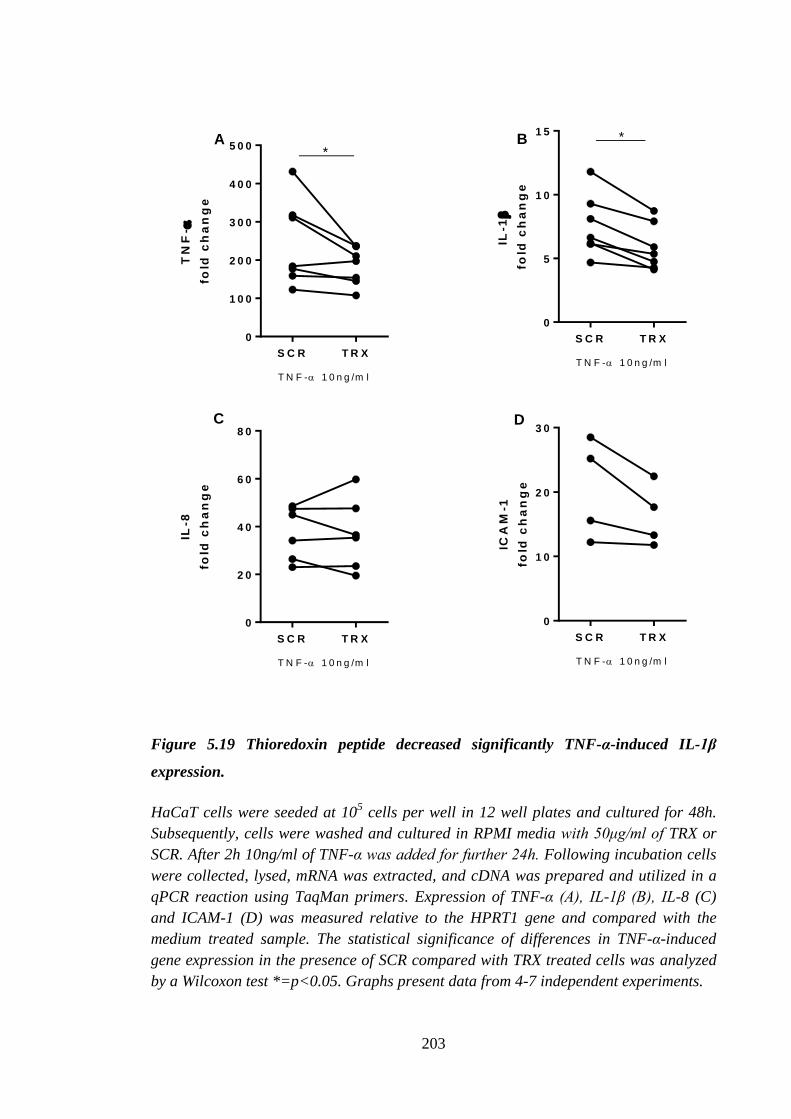
203
TN
F-
fold
ch
an
ge
S C R T R X
0
1 0 0
2 0 0
3 0 0
4 0 0
5 0 0
T N F - 1 0 n g /m l
*
IL-1
fold
ch
an
ge
S C R T R X
0
5
1 0
1 5 *
T N F - 1 0 n g /m l
IL-8
fold
ch
an
ge
S C R T R X
0
2 0
4 0
6 0
8 0
T N F - 1 0 n g /m l
ICA
M-1
fold
ch
an
ge
S C R T R X
0
1 0
2 0
3 0
T N F - 1 0 n g /m l
A B
C D
Figure 5.19 Thioredoxin peptide decreased significantly TNF-α-induced IL-1β
expression.
HaCaT cells were seeded at 105 cells per well in 12 well plates and cultured for 48h.
Subsequently, cells were washed and cultured in RPMI media with 50μg/ml of TRX or
SCR. After 2h 10ng/ml of TNF-α was added for further 24h. Following incubation cells
were collected, lysed, mRNA was extracted, and cDNA was prepared and utilized in a
qPCR reaction using TaqMan primers. Expression of TNF-α (A), IL-1β (B), IL-8 (C)
and ICAM-1 (D) was measured relative to the HPRT1 gene and compared with the
medium treated sample. The statistical significance of differences in TNF-α-induced
gene expression in the presence of SCR compared with TRX treated cells was analyzed
by a Wilcoxon test *=p<0.05. Graphs present data from 4-7 independent experiments.
204
5.3 Discussion
Keratinocytes are the main cellular component of the epidermis. They are
immunologically active cells capable of producing and responding to a plethora of
cytokines (Gutowska-Owsiak and Ogg, 2012). As such, they are important in the events
of LC migration and maturation and have been suggested to be a potential target for the
inhibitory action of LF on oxazolone and IL-1β-induced LC migration (Almond et al.,
2013; Cumberbatch et al., 2000b). Therefore, in the experiments described herein
keratinocytes from 3 different origins were utilized as a platform to investigate the
mechanism of LF immunomodulation. In the first instance all 3 cell types were
characterized in order to confirm their status as keratinocytes. The HaCaT cell line are
spontaneously transformed epithelial cells from human adult skin and they do not cause
malignant growths following their injection into experimental animals (Boukamp et al.,
1988). The PAM (212) cell line are also spontaneously transformed cells derived from
the BALB/c strain mouse (Yuspa et al., 1980). Lastly, primary murine keratinocytes
from new born BALB/c strain mice were utilized and they were obtained as described in
the Chapter 2. It was necessary to confirm that the process of culturing primary
keratinocytes yielded pure populations of cells of interest without any contaminating
cells. Similarly, in the case of cell lines, even though they previously have been
confirmed to express keratinocyte markers by other authors, it is not unheard of for cell
lines to become contaminated with a different cell line (e.g. HeLa) or to lose their
characteristics during long-term culture ((ICLAC), 2013). Therefore, cells were stained
for K14. Keratin 14 is a member of the keratin protein family, filament-forming proteins
found most abundantly in the skin as well as in other epithelial tissues. Keratin 14 forms
a dimer with K5 and is considered to be a marker for both non- and keritanised stratified
epithelia (Haines and Lane, 2012; Shetty and Gokul, 2012). The expression of both K5
and K14 is characteristic of the basal layer of the epidermis. During differentiation cells
begin to express K1 and K10 which are considered to be markers of keratinised
epidermis (Shetty and Gokul, 2012). It was found that all three types of cells expressed
K14, thereby confirming their status as keratinocytes. It was not surprising that these
cells were identified as those from basal layer, as these are the only proliferating cells
within the epidermis. In addition to confirming their status a keratinocyte cells lines,
PAM and HaCaT cells were routinely tested for the presence of mycoplasma infection.
205
All of the experiments presented in this chapter were performed with mycoplasma-free
cells.
In the first instance experiments were performed using all 3 types of keratinocytes to
identify a stimulus that would trigger TNF-α release. It was reported previously that
keratinocytes expressed TLR (Lebre et al., 2007; Mempel et al., 2003). Furthermore, it
was established that TNF-α was expressed by LC-depleted epidermal cells and that it
was induced following in vivo exposure to IL-1β and allergen (TNCB) (Enk et al.,
1993). Initially PAM cells were stimulated with ligands for TLR2, 3, 4, 5 and 9 as well
as PMA, and the production of cytokines and chemokines was measured at the level of
protein by ELISA. No detectable cytokine or chemokine production was found in
response to TLR ligands. Those results were somewhat surprising as it was reported that
primary human and mouse keratinocytes were the source of a plethora of cytokines
(Gröne, 2002; Matsue et al., 1992a). Conversely, a study by Segawa et al. (2014)
reported that PAM cells constitutively produced neither TNF-α nor IL-6 (Marthinuss et
al., 1995; Segawa et al., 2014), but abundant expression of IL-1α was detected by PCR
(Marthinuss et al., 1995). Of the three cell types employed in this chapter PAM cells
were the least commonly utilized and thus there were few published reports whereby
PAM cells were treated with TLR ligands.
Cell lines do not always fully represent characteristics of primary cells; therefore, the
next step was to investigate the effects of TLR agonists on primary murine
keratinocytes. Cells were obtained from new born BALB/c strain mice. Keratinocytes
were used during their first passage, in order to maintain their physiological phenotype,
consequently, they could not be expanded and their number depended on the litter size.
As a result, some of the experiments were performed only once and not always with all
the necessary controls. It was demonstrated that, unlike PAM cells, primary
keratinocytes secreted TNF-α, which was further induced following treatment with
PGN. Unstimulated murine epidermal cells (Matsue et al., 1992a), as well as primary
cultured keratinocytes (Yarosh et al., 2000), were demonstrated previously to express
TNF-α mRNA. Moreover, secretion of IL-1α and IL-1β was assessed and it was found
that while primary keratinocytes produced detectable levels of both cytokines further
induction was not observed following exposure to PGN. Expression of Tnf-α and Il-1β
mRNA was also up-regulated after PGN treatment. Incubation with LPS did not modify
Tnf-α expression and dramatically decreased mRNA levels of Il-1β. There was no gene
206
induction observed following CpG treatment. Lack of response to LPS and CpG was
unexpected as human primary keratinocytes were demonstrated to express functional
TLR4 and TLR9 receptors. Furthermore, exposure to 1μg/ml of LPS and CpG was
shown previously to cause chemokine secretion by primary human keratinocytes (Lebre
et al., 2007). Moreover, TLR4 was established as playing a key role in wound healing in
murine models, thereby suggesting the presence of TLR4 receptor in the skin.
Experiments with normal human epidermal keratinocytes confirmed that the presence of
TLR4 was responsible for cell migration and IL-1β production after scratch wounding
(Chen et al., 2013). In the current investigation, exposure to LPS did not result in
cytokine secretion (data not shown).
Finally, HaCaT cells (a human spontaneously transformed keratinocyte cell line) were
examined (Boukamp et al., 1988). HaCaT cells were found to express TLR2 at a lower
level than other keratinocyte cell lines: KERTr, HEK001, but at a level comparable
with that reported for primary keratinocytes (Olaru and Jensen, 2010). HaCaT cells
responded to treatment with PGN in a dose dependent manner with production of TNF-
α, IL-8 and IL-1β. This observation was in agreement with previously published data,
whereby HaCaT cells were shown to secrete TNF-α and IL-8 following incubation with
10μg/ml of another TLR2 ligand, zymosan (Lee et al., 2009). However, there was a
difference in the levels reported. In the study reported herein maximal release of
~100pg/ml of TNF-α and IL-1β and 10000pg/ml of IL-8 was observed 24h after
addition of 25μg/ml of PGN to the culture. In contrast, Lee et al. (2009) demonstrated
release of approximately 1800pg/ml of both TNF-α and IL-8. This big discrepancy in
secreted cytokine levels is somewhat surprising, especially as Lee et al. (2009) reported
similar levels of TNF-α and IL-8 while in the current investigations IL-8 has been
produced at much higher levels than TNF-α. Because of the relatively low levels of
TNF-α protein production it was decided that, as for PAM cells and keratinocytes, gene
expression by HaCaT cells would be investigated. Exposure of HaCaT cells to PGN for
24h even at the highest concentration (25μg/ml), resulted in a modest increase of
cytokines and adhesion molecule (<5 fold increase which is in agreement with study by
Olaru et al. (2010) that reported 4 fold increase in IL-8 expression) (Olaru and Jensen,
2010).
Overall, results from current investigations suggest that the PAM cells are not
representative of murine keratinocytes, in that they do not seem to express any TLR
207
receptors, as judged by the lack of their response to TLR agonists. It is possible that
these cells could respond to TLR ligands with induction of gene expression but such
was not measured here. On the other hand, primary murine keratinocytes and HaCaT
cells appear to be a more robust tool for studying response of keratinocytes to TLR
agonists, which is reflected with their widespread use in dermatology research,
particularly in the case of HaCaT cells.
Despite their inability to produce cytokines in response to TLR ligands there were
studies demonstrating that PAM cells were capable of cytokine expression and secretion
following incubation with other stimuli. Yonei et al. (2007) demonstrated dose
dependent induction of IL-10 and IL-1 following incubation with trichloroacetic acid, a
widely used peeling agent, as well as constitutive production of IFN-γ by PAM cells.
Additionally, PAM cells were shown to secrete IL-3 constitutively (Peterseim et al.,
1993), as well as in response to UV radiation (Gallo et al., 1991). Furthermore, PAM
cells were shown to express mRNA for IL-7, which was identified as a growth factor
for DETC. Moreover, incubation of 7-17 cells, a DETC cell line, with PAM cell
supernatants was shown to promote 7-17 DETC cell line proliferation, whereas addition
of anti-IL-7 antibody partially inhibited proliferation (Matsue et al., 1993a).
Furthermore, secretion of GM-CSF and M-CSF by PAM cells was reported
(Chodakewitz et al., 1990; Gallo et al., 1991). Although no detectable cytokine or
chemokine secretion was reported in the experiments conducted herein following
stimulation with TLR ligands, IL-1 release was observed following exposure to PMA
(Table 5.1). Indeed, it was established previously by McGuire et al. (1988), that the
treatment with PMA increased IL-1α mRNA levels, as assessed by Northern blot.
Another phorbol, 12-o-tetradecanoylphorbol-13-acetate (TPA), was used to induce
cytokine production by PAM cells and there was a small but significant up-regulation of
thymic stromal lymphopoietin (TSLP) production following 24h incubation with 30nM
TPA (Segawa et al., 2014). Furthermore, it was reported that PMA treatment (10ng/ml)
of PAM cells resulted in up-regulation of IL-10 (Enk and Katz, 1992b). Alas, secretion
of neither TSLP nor IL-10 was measured in the current study, although a wide range of
other cytokines and chemokines was investigated. Ansel et al. (1988) examined the
ability of PAM cells to produce IL-1. It was found that IL-1α was constitutively
expressed and further induced following LPS treatment and exposure to UV light.
Interleukin-1β was not detected in PAM cells or in primary keratinocytes derived from
208
new born mice (Ansel et al., 1988). The results reported by Ansel el al. (1988) were in
accordance with the results presented in the current chapter, whereby expression of IL-
1β by PAM cells was least induced after stimulation with either IL-1α or IL-1β,
however, IL-1β was detected in the culture supernatants albeit at a low level.
With the knowledge from the literature that PAM cells were capable of induction of
cytokines it was thought that they still could prove to be a useful tool for investigating
the immunomodulatory effects of LF. Therefore, it was decided to utilize cytokines IL-
1α and IL-1β as stimulants. It was demonstrated previously that in vivo injection of IL-
1β and, to lesser extent, IL-1α, resulted in TNF-α mRNA expression by murine
epidermal cells (Enk et al., 1993). However, when PAM cells were incubated with a
range of doses of IL-1α or IL-1β, secretion of TNF-α was not detected (data not shown).
Despite relatively high concentration of intracellular IL-1α much lower levels were
secreted in to the culture media in the investigations described herein. The difference
between the level of IL-1α in cell lysates and supernatants presumably stemmed from
the mechanism of IL-1 release, whereby a second signal is required for the processing
and release of IL-1α and IL-1β (Lie et al., 2012). Moreover, the ELISA DuoSet that was
utilized detected pro-IL-1α with the same specificity as it did the mature form of
cytokine (personal communication, Joseph Ainscough), therefore explaining the high
concentration of intracellular IL-1α. Conversely, overall levels of IL-1β were low and
were not modulated by the exposure to IL-1α. It was difficult to compare that result
with the literature, as there are few reports regarding production of IL-1β by PAM cells.
Ansel et al. (1988) reported that PAM cells did not express IL-1β mRNA in the steady
state or following LPS activation (Ansel et al., 1988). Presence of IL-1β was reported in
the epidermal cell suspension, however, only following allergen application and in the
presence of LC, indicating that in mouse epidermis the primary source of IL-1β were
LC (Enk et al., 1993; Enk and Katz, 1992a). The secretion of cytokines by PAM cells
under these conditions was not robust enough, particularly with respect to TNF-α,
therefore, it was decided that gene expression would be measured by utilising qPCR. In
the first instance, the response of PAM cells was measured following exposure to a
range of doses of either IL-1α or IL-1β for 2h. Gene expression was calculated using the
ddCt method, whereby a change in the gene expression was normalised against
expression of the housekeeping gene Hprt1 and further compared to an untreated
sample (Pfaffl, 2001). Results from experiments utilising qPCR, presented in the current
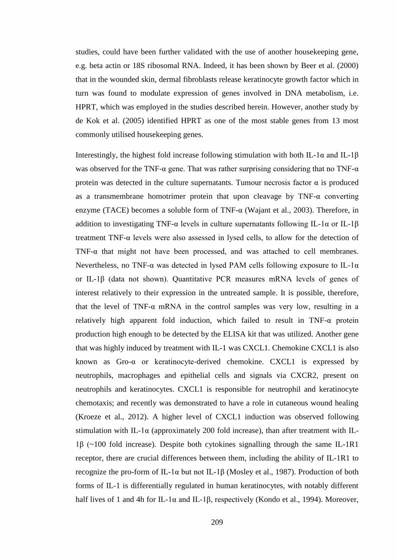
209
studies, could have been further validated with the use of another housekeeping gene,
e.g. beta actin or 18S ribosomal RNA. Indeed, it has been shown by Beer et al. (2000)
that in the wounded skin, dermal fibroblasts release keratinocyte growth factor which in
turn was found to modulate expression of genes involved in DNA metabolism, i.e.
HPRT, which was employed in the studies described herein. However, another study by
de Kok et al. (2005) identified HPRT as one of the most stable genes from 13 most
commonly utilised housekeeping genes.
Interestingly, the highest fold increase following stimulation with both IL-1α and IL-1β
was observed for the TNF-α gene. That was rather surprising considering that no TNF-α
protein was detected in the culture supernatants. Tumour necrosis factor α is produced
as a transmembrane homotrimer protein that upon cleavage by TNF-α converting
enzyme (TACE) becomes a soluble form of TNF-α (Wajant et al., 2003). Therefore, in
addition to investigating TNF-α levels in culture supernatants following IL-1α or IL-1β
treatment TNF-α levels were also assessed in lysed cells, to allow for the detection of
TNF-α that might not have been processed, and was attached to cell membranes.
Nevertheless, no TNF-α was detected in lysed PAM cells following exposure to IL-1α
or IL-1β (data not shown). Quantitative PCR measures mRNA levels of genes of
interest relatively to their expression in the untreated sample. It is possible, therefore,
that the level of TNF-α mRNA in the control samples was very low, resulting in a
relatively high apparent fold induction, which failed to result in TNF-α protein
production high enough to be detected by the ELISA kit that was utilized. Another gene
that was highly induced by treatment with IL-1 was CXCL1. Chemokine CXCL1 is also
known as Gro-α or keratinocyte-derived chemokine. CXCL1 is expressed by
neutrophils, macrophages and epithelial cells and signals via CXCR2, present on
neutrophils and keratinocytes. CXCL1 is responsible for neutrophil and keratinocyte
chemotaxis; and recently was demonstrated to have a role in cutaneous wound healing
(Kroeze et al., 2012). A higher level of CXCL1 induction was observed following
stimulation with IL-1α (approximately 200 fold increase), than after treatment with IL-
1β (~100 fold increase). Despite both cytokines signalling through the same IL-1R1
receptor, there are crucial differences between them, including the ability of IL-1R1 to
recognize the pro-form of IL-1α but not IL-1β (Mosley et al., 1987). Production of both
forms of IL-1 is differentially regulated in human keratinocytes, with notably different
half lives of 1 and 4h for IL-1α and IL-1β, respectively (Kondo et al., 1994). Moreover,
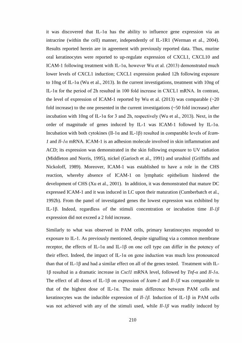
210
it was discovered that IL-1α has the ability to influence gene expression via an
intracrine (within the cell) manner, independently of IL-1R1 (Werman et al., 2004).
Results reported herein are in agreement with previously reported data. Thus, murine
oral keratinocytes were reported to up-regulate expression of CXCL1, CXCL10 and
ICAM-1 following treatment with IL-1α, however Wu et al. (2013) demonstrated much
lower levels of CXCL1 induction; CXCL1 expression peaked 12h following exposure
to 10ng of IL-1α (Wu et al., 2013). In the current investigations, treatment with 10ng of
IL-1α for the period of 2h resulted in 100 fold increase in CXCL1 mRNA. In contrast,
the level of expression of ICAM-1 reported by Wu et al. (2013) was comparable (~20
fold increase) to the one presented in the current investigations (~50 fold increase) after
incubation with 10ng of IL-1α for 3 and 2h, respectively (Wu et al., 2013). Next, in the
order of magnitude of genes induced by IL-1 was ICAM-1 followed by IL-1α.
Incubation with both cytokines (Il-1α and IL-1β) resulted in comparable levels of Icam-
1 and Il-1α mRNA. ICAM-1 is an adhesion molecule involved in skin inflammation and
ACD; its expression was demonstrated in the skin following exposure to UV radiation
(Middleton and Norris, 1995), nickel (Garioch et al., 1991) and urushiol (Griffiths and
Nickoloff, 1989). Moreover, ICAM-1 was established to have a role in the CHS
reaction, whereby absence of ICAM-1 on lymphatic epithelium hindered the
development of CHS (Xu et al., 2001). In addition, it was demonstrated that mature DC
expressed ICAM-1 and it was induced in LC upon their maturation (Cumberbatch et al.,
1992b). From the panel of investigated genes the lowest expression was exhibited by
IL-1β. Indeed, regardless of the stimuli concentration or incubation time Il-1β
expression did not exceed a 2 fold increase.
Similarly to what was observed in PAM cells, primary keratinocytes responded to
exposure to IL-1. As previously mentioned, despite signalling via a common membrane
receptor, the effects of IL-1α and IL-1β on one cell type can differ in the potency of
their effect. Indeed, the impact of IL-1α on gene induction was much less pronounced
than that of IL-1β and had a similar effect on all of the genes tested. Treatment with IL-
1β resulted in a dramatic increase in Cxcl1 mRNA level, followed by Tnf-α and Il-1α.
The effect of all doses of IL-1β on expression of Icam-1 and Il-1β was comparable to
that of the highest dose of IL-1α. The main difference between PAM cells and
keratinocytes was the inducible expression of Il-1β. Induction of IL-1β in PAM cells
was not achieved with any of the stimuli used, while Il-1β was readily induced by
211
primary keratinocytes. As mentioned before, IL-1β mRNA was detected in murine
epidermis but the signal was lost when LC were depleted, suggesting its absence from
keratinocytes (Enk et al., 1993; Enk and Katz, 1992a). It is known that both human and
murine keratinocytes are a source of IL-1α, but the presence of IL-1β is much debated
(Ansel et al., 1988; Kupper et al., 1986; Matsushima et al., 2010). Interleukin-1β mRNA
was detected in unstimulated human keratinocytes, but no protein was detected.
Conversely, IL-1α was detected in both message and protein form (Kupper et al., 1986).
Therefore, it was not surprising that relatively low levels of IL-1β protein were
produced by primary keratinocytes, and that IL-1β mRNA was induced by both PGN
and IL-1 stimulation. Interleukin-1β was recently detected in murine skin using
confocal microscopy and flow cytometry, wherein DsRed fluorescent protein was
expressed under an IL-1β promoter allowing visualisation of IL-1β in the steady state
and after induction with oxazolone. It was found that following exposure to oxazolone
epidermal cells that expressed IL-1β were mainly CD45+ (>85%) and the remaining
proportion (<15%) were keratinocytes. Further analysis of CD45+ cells showed that the
majority expressed CD11b and Gr-1, additionally there were populations present
expressing F4/80 and MHC II. Similarly, in the dermal cells allergen (oxazolone)
induced IL-1β expression was detected in granulocytes, monocytes/macrophages and
DC (Matsushima et al., 2010).
In HaCaT cells the IL-1β-induced pattern of gene expression was similar to that
triggered by PGN with TNF-α and IL-8 exhibiting the highest and similar levels of
induction followed by IL-1β and ICAM-1. Gene expression peaked 2h after addition of
IL-1β which was in accordance with the early maximal gene induction recorded for
PAM cells. Dose dependent gene expression reached its peak at 1ng/ml of IL-1β.
Exposure to another cytokine involved in LC migration, TNF-α, resulted in the rapid
up-regulation of ICAM-1 and IL-8 which was in agreement with the literature
(Lakshminarayanan et al., 1997). The induction of IL-1β was minimal, which was in
accordance with reports demonstrating that in the epidermis LC, and not keratinocytes,
were the main source of IL-1β (Enk et al., 1993). TNF-α was able to induce its own
expression with an interesting kinetic profile. Whereas induction of other measured
cytokines had returned to baseline levels within 24h following exposure to TNF-α, the
up-regulation of TNF-α was maintained up to 24h. This result was somewhat surprising.
Banno et al. (2004) demonstrated that TNF-α induced its own expression most
212
vigorously 1h after initial exposure and decreased following 24h, albeit not to the
baseline level (Banno et al., 2004), which contradicts results obtained in the study
reported herein.
Following the dose response and kinetic experiments, it was decided to utilize the
concentration of 2ng/ml of IL-1α and IL-1β for the induction of gene expression in
PAM cells and primary keratinocytes or 2ng/ml of IL-1β for HaCaT cells. Two h
incubation time, in which the immunomodulatory effects of LF were assessed, was
chosen for PAM cells and HaCaT cells, while 2, 8 and 24h time points were selected for
primary keratinocytes. There are reports demonstrating LF as a stimulatory and
inhibitory protein, therefore it was decided to utilize two concentrations of LF (10μg/ml
and 100μg/ml) in an attempt to cover the active range for PAM and HaCaT cell lines;
meanwhile just one concentration of LF (100μg/ml) was utilized with primary murine
keratinocytes. Exposure of PAM cells to both concentrations of LF did not result in
changes in either Tnf-α or Icam-1 expression. There was a considerable variability
between independent experiments potentially contributing to lack of visible effects of
LF. Attempts were made to optimise the potential activity of LF by utilising a
suboptimal concentration of IL-1β (0.1ng/ml) to ensure that TNF-α expression was not
maximal. Additionally, experiments were conducted in the absence of any foreign
protein (medium 0% FCS) to obviate any blocking effects of nonspecific protein.
However, only the highest concentration of LF (100μg/ml) significantly inhibited Tnf-α
and Icam-1 gene expression and this effect was similar to that observed in the presence
of the irrelevant control protein, BSA. Moreover, there was no dose dependent changes
present, suggesting that the observed inhibitory effect was not specific. The same was
true for primary keratinocytes. Inhibition of cytokine or adhesion molecule expression
was not observed at any time point for any stimulant. As reported previously in the
current investigations for PAM cells, LF on its own did not modulate the expression of
the investigated genes. Similarly, in the case of HaCaT cells, treatment with LF on its
own neither modulated baseline gene expression nor IL-1β-induced expression, a result
that was in accordance with the data gathered from PAM cells and keratinocytes.
Decreasing the concentration of IL-1β to suboptimal levels did not alter the inability of
LF to modulate gene expression by HaCaT cells.
Lack of inhibition of PGN and IL-1 induced keratinocyte gene expression by LF may
have many potential reasons. In the investigations described in the current chapter the
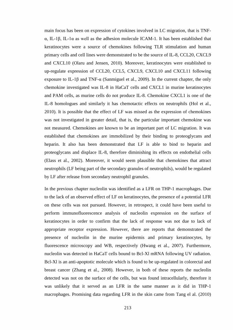
213
main focus has been on expression of cytokines involved in LC migration, that is TNF-
α, IL-1β, IL-1α as well as the adhesion molecule ICAM-1. It has been established that
keratinocytes were a source of chemokines following TLR stimulation and human
primary cells and cell lines were demonstrated to be the source of IL-8, CCL20, CXCL9
and CXCL10 (Olaru and Jensen, 2010). Moreover, keratinocytes were established to
up-regulate expression of CCL20, CCL5, CXCL9, CXCL10 and CXCL11 following
exposure to IL-1β and TNF-α (Sanmiguel et al., 2009). In the current chapter, the only
chemokine investigated was IL-8 in HaCaT cells and CXCL1 in murine keratinocytes
and PAM cells, as murine cells do not produce IL-8. Chemokine CXCL1 is one of the
IL-8 homologues and similarly it has chemotactic effects on neutrophils (Hol et al.,
2010). It is possible that the effect of LF was missed as the expression of chemokines
was not investigated in greater detail, that is, the particular important chemokine was
not measured. Chemokines are known to be an important part of LC migration. It was
established that chemokines are immobilized by their binding to proteoglycans and
heparin. It also has been demonstrated that LF is able to bind to heparin and
proteoglycans and displace IL-8, therefore diminishing its effects on endothelial cells
(Elass et al., 2002). Moreover, it would seem plausible that chemokines that attract
neutrophils (LF being part of the secondary granules of neutrophils), would be regulated
by LF after release from secondary neutrophil granules.
In the previous chapter nucleolin was identified as a LFR on THP-1 macrophages. Due
to the lack of an observed effect of LF on keratinocytes, the presence of a potential LFR
on these cells was not pursued. However, in retrospect, it could have been useful to
perform immunofluorescence analysis of nucleolin expression on the surface of
keratinocytes in order to confirm that the lack of response was not due to lack of
appropriate receptor expression. However, there are reports that demonstrated the
presence of nucleolin in the murine epidermis and primary keratinocytes, by
fluorescence microscopy and WB, respectively (Hwang et al., 2007). Furthermore,
nucleolin was detected in HaCaT cells bound to Bcl-Xl mRNA following UV radiation.
Bcl-Xl is an anti-apoptotic molecule which is found to be up-regulated in colorectal and
breast cancer (Zhang et al., 2008). However, in both of these reports the nucleolin
detected was not on the surface of the cells, but was found intracellularly, therefore it
was unlikely that it served as an LFR in the same manner as it did in THP-1
macrophages. Promising data regarding LFR in the skin came from Tang el al. (2010)
214
who demonstrated that LF-mediated keratinocyte migration and proliferation via LRP1
protein, suggesting that it was LFR present on keratinocytes (Tang et al., 2010). Indeed,
it was described previously that there exists more than one LFR on different cell types
and that LRP1 was one of the potential receptors. It is important to note that despite the
lack of striking inhibitory effects of LF in vitro the same batch of protein was
demonstrated to be biologically active in vivo.
Although it was not possible to demonstrate that cytokine induction by IL-1 was
modulated by LF it was decided to investigate the immunomodulatory potential of two
different peptides: TRX and Pep-1, which are involved in the protection against ROS
and inhibiting hyaluronic acid, respectively. These peptides, similarly to LF, were found
to inhibit LC migration following exposure to allergen (Dearman et al., 2010; Mummert
et al., 2000). Pep-1 is a 12 amino acid peptide that was developed as an inhibitor of HA
(Mummert et al., 2000), as well as a probe to detect HA in tissues (Zmolik and
Mummert, 2005). It was demonstrated that HA played a role in the events of LC
migration and maturation, whereby inhibition of HA with Pep-1 resulted in inhibition of
DNFB-stimulated LC migration and decreased expression of CD86, a marker of LC
maturation (Mummert et al., 2003). Moreover, it was found that HaCaT cells express
HA receptors (CD44, RHAMM, LYVE1, TLR4) and enzymes involved in the HA
synthesis and degradation. Furthermore, HA was found to facilitate the migration of
HaCaT cells following scratch injury (Colella et al., 2012). Additionally, human
epidermal keratinocytes were demonstrated to produce and secrete HA (Akiyama et al.,
1994). Therefore, it was decided to investigate, whether a specific HA inhibitor would
impact on cytokine (TNF-α and IL-1β) induced gene expression by HaCaT cells.
Thioredoxin and thioredoxin derived peptide, TRX, similarly to LF, were found to
inhibit oxazolone-induced LC migration, but thioredoxin was thought to influence IL-1β
(Dearman et al., 2010) and not TNF-α signalling (Cumberbatch et al., 2000b).
Consequently, it was of particular interest to test, whether Pep-1 and TRX peptides were
able to have any impact on keratinocytes in vitro, as LF was unable to have significant
impact on keratinocytes.
To test the immunomodulatory ability of TRX and Pep-1, gene expression by HaCaT
cells was induced by exposure to TNF-α and IL-1β. As there were differences between
TNF-α and IL-1β, with respect to peak response of time induced changes, the 24h time
215
point was chosen, to accommodate for that. Dexamethasone was used again as a
positive control for inhibition of cytokine induced gene expression. Prior exposure to
Pep-1 peptide did not result in a significant inhibition of gene expression regardless of
the stimuli used. Pep-1 was demonstrated previously to decrease the expression of
TNF-α and TLR4 mRNA in articular tissue derived from animals with collagen-induced
arthritis (Campo et al., 2013), suggesting its ability to exert inhibitory effects in vivo. It
is possible that the lack of effect of Pep-1 observed in the current study was due to the
fact that HA did not participate in TNF-α and IL-1β induced gene expression.
Hyaluronic acid is a large molecular weight polymer comprised of D-N
acetylglucosamine and D-glucuronic acid. It is expressed by all of the tissues in the
body and is a major component of ECM. Under physiological conditions HA has a
molecular weight of approximately 107 Da, however, in the event of inflammation or
injury, smaller fragments of HA are found. It was demonstrated that HA of different
size ranges exhibited distinct functions. High molecular weight HA displayed an anti-
inflammatory properties, whereas LMW HA had a pro-inflammatory potential (Petrey
and de la Motte, 2014). Therefore, it would be of interest to investigate whether gene
induction stimulated via oxidative stress, (which facilitates HA degradation to LMW
fragments (Esser et al., 2012)) e.g. DNCB treatment, was modulated by Pep-1 peptide.
Dexamethasone inhibited induction of TNF-α and IL-1β expression and had a
considerably less pronounced effect on expression of IL-8. Dexamethasone was
demonstrated previously to inhibit TNF-α and IL-1β induced IL-8 protein and mRNA
production in airway epithelial cells (Kwon et al., 1994). However, dexamethasone was
demonstrated to have a lesser effect on LPS-induced IL-8 expression by alveolar
macrophages (Armstrong et al., 2009), therefore showing that susceptibility of IL-8
production to dexamethasone could be cell specific.
Subsequently TRX, another immunomodulatory peptide, was investigated. Thioredoxin
is a small, 12kDa protein crucial in the protection against oxidative stress. It works by
catalyzing the reduction of disulfide bonds (Yoshihara et al., 2014). The 9mer peptide
derived from thioredoxin utilized in these experiments was demonstrated previously in
our laboratory to inhibit LC migration induced by oxazolone and TNF-α but not IL-1β
(Cumberbatch et al., 2009). That is, analogous with LF, thioredoxin was able to inhibit
LC migration, but the mechanism appears to be somewhat different, as IL-1β dependent
migration was blocked (TNF-α and oxazolone induced migration), whereas exogenous
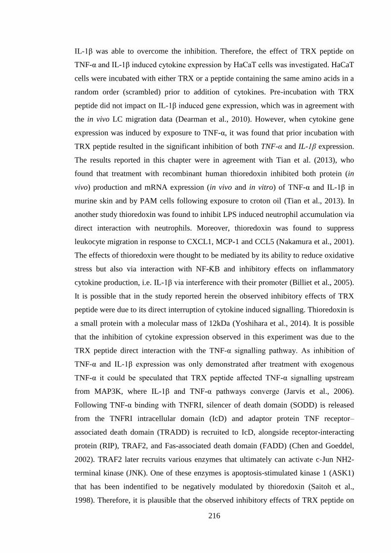
216
IL-1β was able to overcome the inhibition. Therefore, the effect of TRX peptide on
TNF-α and IL-1β induced cytokine expression by HaCaT cells was investigated. HaCaT
cells were incubated with either TRX or a peptide containing the same amino acids in a
random order (scrambled) prior to addition of cytokines. Pre-incubation with TRX
peptide did not impact on IL-1β induced gene expression, which was in agreement with
the in vivo LC migration data (Dearman et al., 2010). However, when cytokine gene
expression was induced by exposure to TNF-α, it was found that prior incubation with
TRX peptide resulted in the significant inhibition of both TNF-α and IL-1β expression.
The results reported in this chapter were in agreement with Tian et al. (2013), who
found that treatment with recombinant human thioredoxin inhibited both protein (in
vivo) production and mRNA expression (in vivo and in vitro) of TNF-α and IL-1β in
murine skin and by PAM cells following exposure to croton oil (Tian et al., 2013). In
another study thioredoxin was found to inhibit LPS induced neutrophil accumulation via
direct interaction with neutrophils. Moreover, thioredoxin was found to suppress
leukocyte migration in response to CXCL1, MCP-1 and CCL5 (Nakamura et al., 2001).
The effects of thioredoxin were thought to be mediated by its ability to reduce oxidative
stress but also via interaction with NF-ƘB and inhibitory effects on inflammatory
cytokine production, i.e. IL-1β via interference with their promoter (Billiet et al., 2005).
It is possible that in the study reported herein the observed inhibitory effects of TRX
peptide were due to its direct interruption of cytokine induced signalling. Thioredoxin is
a small protein with a molecular mass of 12kDa (Yoshihara et al., 2014). It is possible
that the inhibition of cytokine expression observed in this experiment was due to the
TRX peptide direct interaction with the TNF-α signalling pathway. As inhibition of
TNF-α and IL-1β expression was only demonstrated after treatment with exogenous
TNF-α it could be speculated that TRX peptide affected TNF-α signalling upstream
from MAP3K, where IL-1β and TNF-α pathways converge (Jarvis et al., 2006).
Following TNF-α binding with TNFRI, silencer of death domain (SODD) is released
from the TNFRI intracellular domain (IcD) and adaptor protein TNF receptor–
associated death domain (TRADD) is recruited to IcD, alongside receptor-interacting
protein (RIP), TRAF2, and Fas-associated death domain (FADD) (Chen and Goeddel,
2002). TRAF2 later recruits various enzymes that ultimately can activate c-Jun NH2-
terminal kinase (JNK). One of these enzymes is apoptosis-stimulated kinase 1 (ASK1)
that has been indentified to be negatively modulated by thioredoxin (Saitoh et al.,
1998). Therefore, it is plausible that the observed inhibitory effects of TRX peptide on
217
TNF-α stimulated induction of TNF-α and IL-1β expression were due to its interaction
with ASK1.
The impact of thioredoxin and LF on oxazolone-induced LC migration was found to be
similar: both compounds were able to prevent the decrease in LC numbers. The
difference between proteins was visible when cytokines were utilized: LF inhibited
IL-1β induced migration suggesting that it impacted on de novo TNF-α production
(Cumberbatch et al., 2000b), whereas the opposite was true for thioredoxin, it was
thought to influence IL-1β signalling (Dearman et al., 2010). It was somewhat
surprising that there was an observed effect of TRX, but not LF, in HaCaT cells. It has
been hypothesised that in the sequence of events of allergen (oxazolone) induced LC
migration, TNF-α was synthesized by keratinocytes, TNF-α in turn stimulated LC, via
TNFRII, to release IL-1β that was synthesized and present in LC in its pro-form.
Subsequently, secreted IL-1β acted via IL-1R1 on LC in an autocrine manner, supplying
the second signal for migration of LC. Therefore, it was rather surprising that in the
current investigation it was reported that TRX was able to inhibit TNF-α-induced IL-1β
expression by HaCaT keratinocytes, whereas LF failed to inhibit IL-1β-triggered TNF-α
expression.
The mechanism of action of LF (either stimulatory or inhibitory) is still largely
unknown, therefore it is difficult to speculate where it may exert its effects. It is possible
that the end points utilized in the experiments presented in this chapter were not ideal;
although such were based on known actions of LF in vivo. For example, the production
of chemokines may be the key target of LF, as it has been reported that keratinocytes
produce many chemokines in response to TLR stimulation (Olaru and Jensen, 2010).
Overall, keratinocytes did not prove a suitable model to investigate the
immunomodulatory effects of LF. It could be speculated that one cell system cannot be
representative of the complex connections found in the skin. Additionally, it indicates
that despite being the most abundant cell type in the epidermis, the keratinocyte may not
be the target cell for LF action.
218
6 Differential cytokine requirements for mobilization of LC induced by
oxazolone and DNCB
6.1 Introduction
The human immune system has evolved to recognize and eradicate external and internal
dangers. It is very complex machinery that requires meticulous control and tuning.
However, there are instances where the immune system reacts against the organism
itself (autoimmunity) or it mounts a disproportionate reaction against harmless
compounds (allergic reactions). Allergic contact dermatitis is a form of type IV
hypersensitivity (delayed) (DTH) reaction, where the subject has become sensitized to
a compound upon primary exposure, and the clinical manifestations are evident in
sensitised individuals 48h or more after secondary encounter (or challenge). Causative
agents are LMW molecules (usually <500Da). They are almost always unable to trigger
allergic reactions on their own, as it is only after covalently binding to proteins that they
become immunogenic. Over 4000 different chemical allergens have been demonstrated
to cause ACD in humans. Cross-sectional studies have shown that between 20 and 30%
of the population could be diagnosed with contact allergy to at least one substance
(Peiser et al., 2012; Uter et al., 2005). Common contact allergens include metals: nickel,
gold; cosmetics, e.g. balsam of Peru, fragrances, hair dyes (PPD) ; medications:
antibiotics and steroids (Fonacier et al., 2010).
Despite the prevalence of ACD, and the wide range of chemicals that cause this
problem, the exact mechanisms of action are incompletely understood. Considerable
efforts have been made to elucidate the sequence of events following initial skin
exposure to haptens. They have been shown to interact with nucleophilic amino acids,
including but not restricted to, cysteine and lysine. This property stems from their
electrophilic nature and was used for the development of alternative ways of identifying
contact allergens (Gerberick et al., 2004). It was demonstrated previously that
application of allergen results in inflammatory cytokine production in the skin (Enk and
Katz, 1992a), however, the exact proteins involved are still not characterized fully. One
of the allergens that has been studied most closely is the metal allergen, nickel. It was
found that in human skin nickel interacts with specific histidine residues within TLR4,
thus providing a direct danger signal (Schmidt et al., 2010). Moreover, other allergens
219
were found to trigger the generation of ROS in the skin that in turn cause the creation of
LMW HA fragments that serve as a secondary inflammation signal (Esser et al., 2012).
Application of the somewhat limited range of allergens that have been examined to
human and mouse skin universally results in cytokine production and LC migration.
Langerhans’ cells comprise 2-5% of all epidermal cells in adult mouse skin, and are the
only myeloid cells in the epidermis (Romani et al., 2010). They are positioned between
keratinocytes and serve as immune sentinels, detecting danger signals and subsequently
presenting antigens in the context of MHC class II (and class I) to naive T cells in the
draining LN (Toebak et al., 2009). Langerhans’ cells were discovered in 1868 by Paul
Langerhans and were originally thought to be a part of the nervous system (Ginhoux
and Merad, 2010). Further studies showed, however, that LC derive from the bone
marrow (Katz et al., 1979). The process of LC migration was considered to depend
universally on cytokine signals from TNF-α and IL-1. The data supporting this
conclusion came from the investigations in mice, mainly utilising oxazolone as a model
contact allergen (Cumberbatch et al., 2002a; Cumberbatch et al., 1997b). In addition,
LC migration has been investigated in response to UV radiation (Cooper et al., 1992),
yeast (Haley et al., 2012) and virus (Byrne et al., 2001) infection. Moreover,
involvement of TNF-α and IL-1 cytokines has been established both in vivo
(Cumberbatch et al., 1999d) and ex vivo (Stoitzner et al., 1999). However, there is a
limited amount of information regarding influence of contact allergens on LC migration
in humans. In our laboratory DPC has been used as a model allergen that triggered LC
mobilisation (Griffiths et al., 2001). Additionally, there are reports of DNCB use in
humans (Friedmann et al., 1983) and its impact on LC migration (Oakford et al., 2011).
In the investigations described here chemical allergens have been used as a tool with
which to examine the characteristics of LC biology, in particular the role of cytokines in
LC function and skin inflammation. In this chapter differential cytokine regulation of
LC migration between allergens is considered, with particular focus on two model
compounds: oxazolone and DNCB. It was established in a mouse model that LC
migration in response to the contact allergen oxazolone, two cytokine signals from
TNF-α and IL-1β were required for LC mobilisation (Cumberbatch et al., 1997b). In the
experiments described here the mechanisms of oxazolone- and DNCB-mediated LC
migration were investigated. The belief that TNF-α is an essential signal for LC
migration is challenged initially by differential inhibition of oxazolone and DNCB-
220
induced LC mobilisation by LF. It was described previously that LF inhibited oxazolone
triggered LC migration via inhibition of de novo TNF-α production in the skin.
Therefore, divergent effects of LF on oxazolone and DNCB-induced LC mobilisation
were investigated further using TNFRII KO mice that lack the sole TNF-α receptor that
is expressed by LC (Wang et al., 1996), and the administration of neutralising anti-
cytokine antibody. Lastly, an attempt was made to elucidate the mechanisms driving the
differences between allergens using of cytokine profiling. The production of cytokines,
chemokine (CXCL1) and adhesion molecule ICAM-1 by allergen-treated skin tissue
was examined on the level of protein secretion and gene expression.
6.2 Results
6.2.1 Contact allergens (oxazolone and DNCB) induced LC migration
Topical application of contact allergens oxazolone and DNCB has been shown
previously to induce immune responses and to result in LC migration (Cumberbatch et
al., 1997b; Cumberbatch et al., 1992a). In preliminary experiments the ability of
oxazolone and DNCB to induce LC migration was confirmed. In the steady state there
were approximately 1000 LC per mm2 in BALB/c strain mouse skin. The loss of
between 20 and 40% of LC is the maximum amount of cells that can migrate rapidly
from the epidermis regardless of the stimulus applied to the skin (Cumberbatch and
Kimber, 1990). Mice were topically exposed to a single dose of either allergen in AOO,
a vehicle that was shown previously to have good topical application characteristics for
organic chemicals (Heylings et al., 1996; Warbrick et al., 1999). In the current
investigations controls used in in vivo experiments were either AOO treated or untreated
(naive). Application of AOO for either 4 or 17h was shown previously not to impact on
LC numbers in comparison with untreated animals (Cumberbatch et al., 2005).
Similarly, AOO was without effect on DC accumulation in the LN when compared with
untreated mice (Cumberbatch et al., 2005).
Doses of allergens that were used had been demonstrated previously to result in
vigorous LNC proliferation to similar extents, and were therefore deemed to have
similar levels of allergenicity (Dearman and Kimber, 1991; Van Och et al., 2002).
Application of both allergens resulted in a decrease in LC numbers to approximately
800 LC per mm2, representing a 21% and a 24% decrease for DNCB and oxazolone,
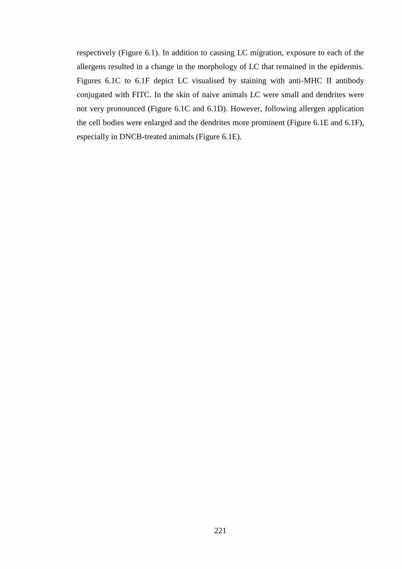
221
respectively (Figure 6.1). In addition to causing LC migration, exposure to each of the
allergens resulted in a change in the morphology of LC that remained in the epidermis.
Figures 6.1C to 6.1F depict LC visualised by staining with anti-MHC II antibody
conjugated with FITC. In the skin of naive animals LC were small and dendrites were
not very pronounced (Figure 6.1C and 6.1D). However, following allergen application
the cell bodies were enlarged and the dendrites more prominent (Figure 6.1E and 6.1F),
especially in DNCB-treated animals (Figure 6.1E).
222
Figure 6.1 Treatment with DNCB and oxazolone resulted in LC migration.
Young (6-8 weeks) BALB/c strain mice (n=3 per group, 6 ears per group) were treated
on the dorsum of both ears with 25μl of 1% DNCB or 0.5% oxazolone (Ox) in AOO for
4h. Treated animals, alongside untreated controls (naive) (n=3 per group; 6 ears per
group), were sacrificed, ears were excised and LC frequency was enumerated by
fluorescence microscopy and is expressed as the mean frequency of LC per mm2 per
group ± SEM (A, B). The statistical significance of differences between LC numbers in
untreated (naive) and allergen treated animals was assessed by Student’s t test;
**=p<0.01. Representative micrographs of epidermal sheets show LC morphology of
representative samples from naive (C, D) and DNCB (E) or oxazolone (F) treated
animals. x40 magnification.
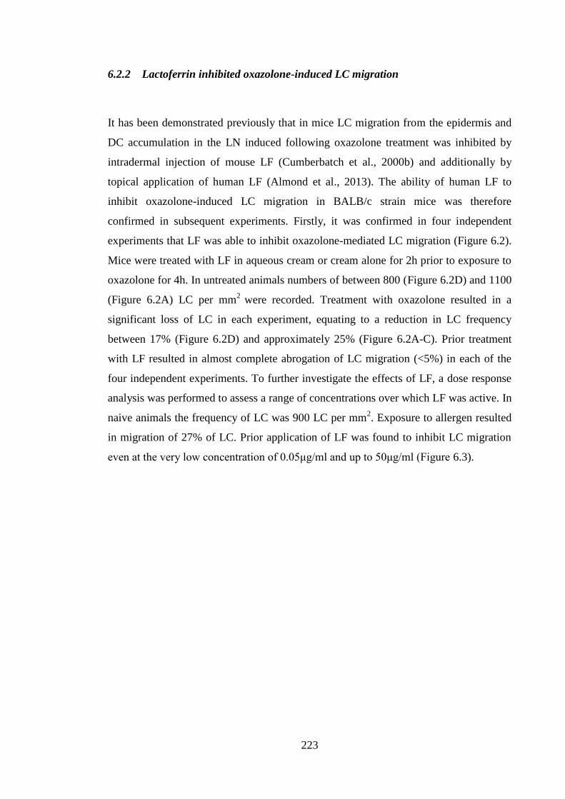
223
6.2.2 Lactoferrin inhibited oxazolone-induced LC migration
It has been demonstrated previously that in mice LC migration from the epidermis and
DC accumulation in the LN induced following oxazolone treatment was inhibited by
intradermal injection of mouse LF (Cumberbatch et al., 2000b) and additionally by
topical application of human LF (Almond et al., 2013). The ability of human LF to
inhibit oxazolone-induced LC migration in BALB/c strain mice was therefore
confirmed in subsequent experiments. Firstly, it was confirmed in four independent
experiments that LF was able to inhibit oxazolone-mediated LC migration (Figure 6.2).
Mice were treated with LF in aqueous cream or cream alone for 2h prior to exposure to
oxazolone for 4h. In untreated animals numbers of between 800 (Figure 6.2D) and 1100
(Figure 6.2A) LC per mm2
were recorded. Treatment with oxazolone resulted in a
significant loss of LC in each experiment, equating to a reduction in LC frequency
between 17% (Figure 6.2D) and approximately 25% (Figure 6.2A-C). Prior treatment
with LF resulted in almost complete abrogation of LC migration (<5%) in each of the
four independent experiments. To further investigate the effects of LF, a dose response
analysis was performed to assess a range of concentrations over which LF was active. In
naive animals the frequency of LC was 900 LC per mm2. Exposure to allergen resulted
in migration of 27% of LC. Prior application of LF was found to inhibit LC migration
even at the very low concentration of 0.05μg/ml and up to 50μg/ml (Figure 6.3).
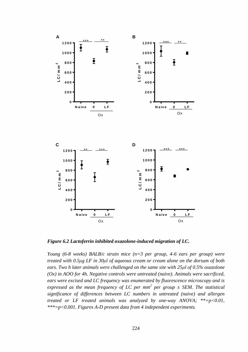
224
LC
/ m
m2
N a iv e 0 L F
0
2 0 0
4 0 0
6 0 0
8 0 0
1 0 0 0
1 2 0 0*** **
Ox
LC
/ m
m2
N a iv e 0 L F
0
2 0 0
4 0 0
6 0 0
8 0 0
1 0 0 0
1 2 0 0 *** **
Ox
LC
/ m
m2
N a iv e 0 L F
0
2 0 0
4 0 0
6 0 0
8 0 0
1 0 0 0
1 2 0 0 ** ***
Ox
LC
/ m
m2
N a iv e 0 L F
0
2 0 0
4 0 0
6 0 0
8 0 0
1 0 0 0
1 2 0 0 *** ***
Ox
A B
C D
Figure 6.2 Lactoferrin inhibited oxazolone-induced migration of LC.
Young (6-8 weeks) BALB/c strain mice (n=3 per group, 4-6 ears per group) were
treated with 0.5μg LF in 30μl of aqueous cream or cream alone on the dorsum of both
ears. Two h later animals were challenged on the same site with 25μl of 0.5% oxazolone
(Ox) in AOO for 4h. Negative controls were untreated (naive). Animals were sacrificed,
ears were excised and LC frequency was enumerated by fluorescence microscopy and is
expressed as the mean frequency of LC per mm2 per group ± SEM. The statistical
significance of differences between LC numbers in untreated (naive) and allergen
treated or LF treated animals was analyzed by one-way ANOVA; **=p<0.01,
***=p<0.001. Figures A-D present data from 4 independent experiments.
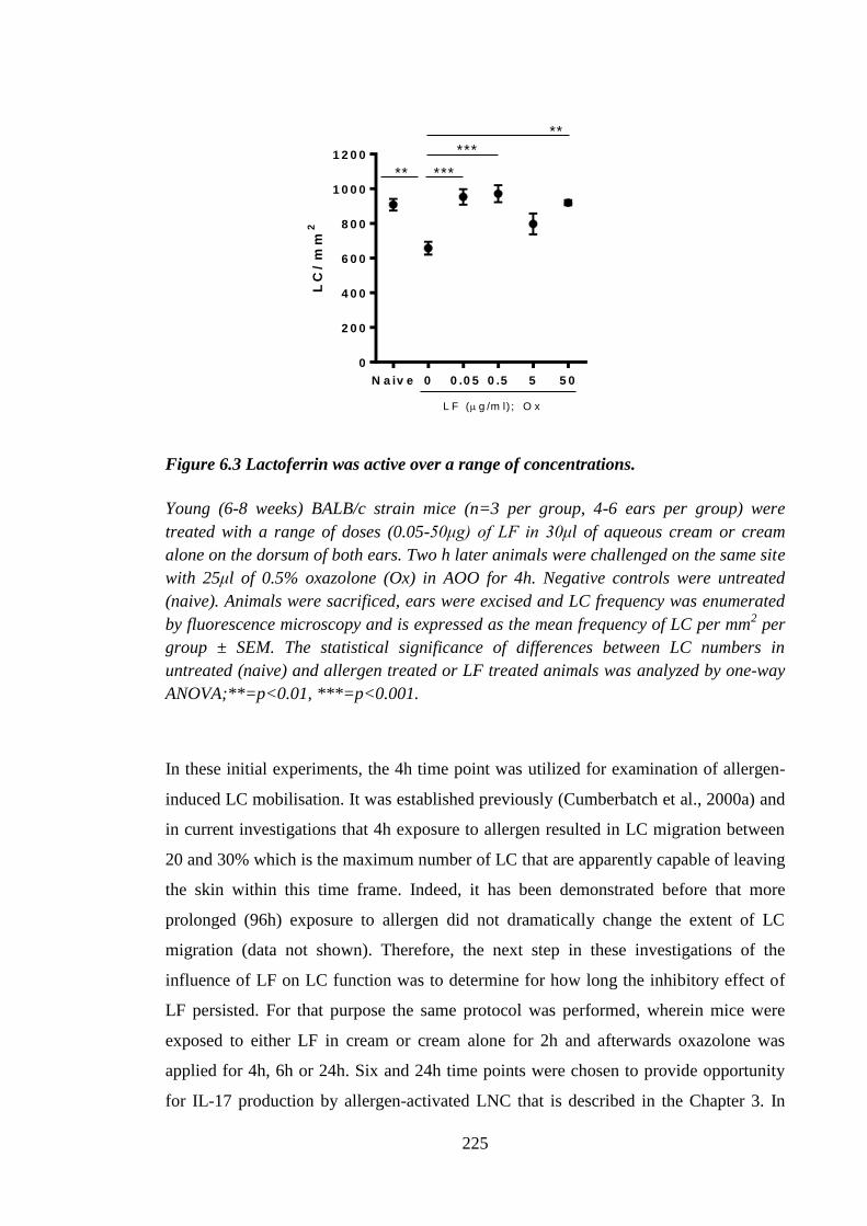
225
LC
/ m
m2
N a iv e 0 0 .0 5 0 .5 5 5 0
0
2 0 0
4 0 0
6 0 0
8 0 0
1 0 0 0
1 2 0 0
L F ( g /m l) ; O x
** ***
***
**
Figure 6.3 Lactoferrin was active over a range of concentrations.
Young (6-8 weeks) BALB/c strain mice (n=3 per group, 4-6 ears per group) were
treated with a range of doses (0.05-50μg) of LF in 30μl of aqueous cream or cream
alone on the dorsum of both ears. Two h later animals were challenged on the same site
with 25μl of 0.5% oxazolone (Ox) in AOO for 4h. Negative controls were untreated
(naive). Animals were sacrificed, ears were excised and LC frequency was enumerated
by fluorescence microscopy and is expressed as the mean frequency of LC per mm2 per
group ± SEM. The statistical significance of differences between LC numbers in
untreated (naive) and allergen treated or LF treated animals was analyzed by one-way
ANOVA;**=p<0.01, ***=p<0.001.
In these initial experiments, the 4h time point was utilized for examination of allergen-
induced LC mobilisation. It was established previously (Cumberbatch et al., 2000a) and
in current investigations that 4h exposure to allergen resulted in LC migration between
20 and 30% which is the maximum number of LC that are apparently capable of leaving
the skin within this time frame. Indeed, it has been demonstrated before that more
prolonged (96h) exposure to allergen did not dramatically change the extent of LC
migration (data not shown). Therefore, the next step in these investigations of the
influence of LF on LC function was to determine for how long the inhibitory effect of
LF persisted. For that purpose the same protocol was performed, wherein mice were
exposed to either LF in cream or cream alone for 2h and afterwards oxazolone was
applied for 4h, 6h or 24h. Six and 24h time points were chosen to provide opportunity
for IL-17 production by allergen-activated LNC that is described in the Chapter 3. In

226
accordance with previously reported data it was observed that in untreated (naive)
animals there were ~1100 LC per mm2. Exposure to oxazolone for 4h, 6h and 24h
resulted in a loss of LC of 24%, 26% and 17%, respectively. That is, the impact of
oxazolone on LC frequency persisted for some 24h. It was found that the effects of LF
were relatively transient; whilst migration was significantly reduced (3%) in the
presence of LF at 4h post-oxazolone treatment (Figure 6.4A), LC mobilisation increased
to 12% after 6h (Figure 6.4B) following oxazolone application and the inhibitory effect
of LF was no longer apparent after 24h (Figure 6.4C).
LC
/m
m2
N a iv e 0 L F
0
2 0 0
4 0 0
6 0 0
8 0 0
1 0 0 0
1 2 0 0
O x
*** **
4 h 6 h
LC
/ m
m2
N a iv e 0 L F
0
2 0 0
4 0 0
6 0 0
8 0 0
1 0 0 0
1 2 0 0
O x
*** **
**
LC
/m
m2
N a iv e 0 L F
0
2 0 0
4 0 0
6 0 0
8 0 0
1 0 0 0
1 2 0 0
O x
*
2 4 hA B C
Figure 6.4 The inhibitory effect of LF on oxazolone-induced LC migration was
transient.
Young (6-8 weeks) BALB/c mice (n=3 per group, 6 ears per group) were treated with
0.5μg of LF in 30μl of aqueous cream or cream alone on the dorsum of both ears. Two
h later animals were challenged on the same site with 25μl 0.5% of oxazolone (Ox) in
AOO for 4h (A), 6h (B) or 24h (C). Negative controls were untreated (naive). Animals
were sacrificed, ears were excised and LC frequency was enumerated by fluorescence
microscopy and is expressed as the mean frequency of LC per mm2 per group ± SEM.
The statistical significance of differences between LC numbers in untreated (naive) and
allergen treated or LF treated animals was analyzed by one-way ANOVA;
*=p<0.05,**=p<0.01, ***=p<0.001.
227
6.2.3 The N-lobe of lactoferrin was not responsible for inhibition of LC migration
In the current investigation human LF was used in the mouse model of allergen-induced
LC migration. Previous reports from our laboratory examined primarily homologous LF
with regards to its inhibitory effects on LC mobilisation (Cumberbatch et al., 2000b;
Griffiths et al., 2001). However, it has been established that there is a high degree of
homology between LF molecules from various species, with mouse and human LF
sharing 70% amino acid sequence homology (Baker and Baker, 2009). Among other
roles, LF is widely recognized as an anti-microbial protein (Dalmastri et al., 1988). It
has been documented that many of the anti-bacterial properties of LF can be attributed
to the N-lobe of the protein, which is known to bind LPS (Drago-Serrano et al., 2012).
It was of interest, therefore, to determine whether the observed inhibitory effect of the
LF on LC migration resided also in this part of the molecule. The commercially
available N-lobe that was used in the current investigations was a 14 amino acid peptide
which contained residues 231-245 of human LF protein. There is 67% identity between
murine and human N-lobe peptide (BLAST analysis of murine and human N-lobe).
Treatment with oxazolone resulted in LC migration, albeit to a lower extent than
previously reported (12%). Pre-treatment with LF was able to inhibit oxazolone-induced
LC mobilisation. However, even this relatively low level of LC migration was not
inhibited by prior application of N-lobe peptide, either at an equivalent concentration
(with respect to total protein, μg/ml) to that of the whole molecule or in marked excess
of that concentration, suggesting that this part of the molecule did not participate in the
modulation of LC mobilisation (Figure 6.5).
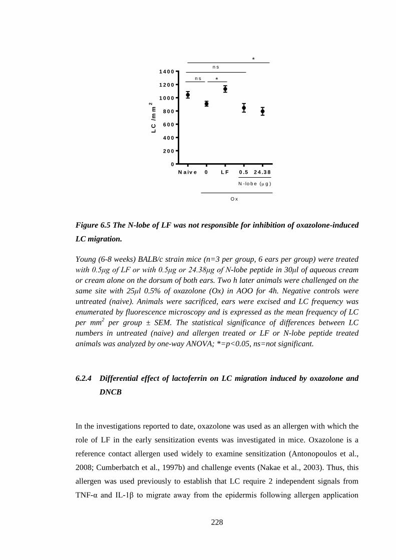
228
LC
/m
m2
N a iv e 0 L F 0 .5 2 4 .3 8
0
2 0 0
4 0 0
6 0 0
8 0 0
1 0 0 0
1 2 0 0
1 4 0 0
N - lo b e ( g )
O x
*n s
n s
*
Figure 6.5 The N-lobe of LF was not responsible for inhibition of oxazolone-induced
LC migration.
Young (6-8 weeks) BALB/c strain mice (n=3 per group, 6 ears per group) were treated
with 0.5μg of LF or with 0.5μg or 24.38μg of N-lobe peptide in 30μl of aqueous cream
or cream alone on the dorsum of both ears. Two h later animals were challenged on the
same site with 25μl 0.5% of oxazolone (Ox) in AOO for 4h. Negative controls were
untreated (naive). Animals were sacrificed, ears were excised and LC frequency was
enumerated by fluorescence microscopy and is expressed as the mean frequency of LC
per mm2 per group ± SEM. The statistical significance of differences between LC
numbers in untreated (naive) and allergen treated or LF or N-lobe peptide treated
animals was analyzed by one-way ANOVA; *=p<0.05, ns=not significant.
6.2.4 Differential effect of lactoferrin on LC migration induced by oxazolone and
DNCB
In the investigations reported to date, oxazolone was used as an allergen with which the
role of LF in the early sensitization events was investigated in mice. Oxazolone is a
reference contact allergen used widely to examine sensitization (Antonopoulos et al.,
2008; Cumberbatch et al., 1997b) and challenge events (Nakae et al., 2003). Thus, this
allergen was used previously to establish that LC require 2 independent signals from
TNF-α and IL-1β to migrate away from the epidermis following allergen application
229
(Cumberbatch et al., 1997b). 2,4-dinitrocholorobenzene is another chemically unrelated
potent contact allergen commonly used in laboratory investigations of CHS, but in
addition to its use in animal models, there is a wealth of data regarding the impact of
DNCB exposure in humans (Friedmann et al., 1983), as DNCB was used to treat
alopecia areata (Singh and Lavanya, 2010), warts (Kwok et al., 2012) and has been used
also by at least one research group as an experimental contact allergen in humans
(Friedmann et al., 1983). In subsequent experiments, DNCB was used as an additional
stimulant for LC migration and comparisons were drawn with oxazolone. The influence
of LF on DNCB-induced LC mobilisation was examined 4h (2 experiments), 6h (2
experiments) and 24h (1 experiment) after initiation of exposure and was compared with
concurrent treatment with oxazolone. In each experiment and at each time point
exposure to DNCB resulted in a significant decrease in LC numbers ranging from 14%
(Figure 6.6B) to 25% (Figure 6.6C-6.6E). The extent of LC loss was similar for DNCB
and oxazolone, with the exception of the experiment presented in Figure 6.6D where
DNCB triggered twice as many LC to migrate as did oxazolone (25% vs. 11%). As
previously demonstrated in the Figure 6.4, the inhibitory effect of LF on oxazolone-
induced LC migration was transient. Thus, in both experiments at the 4h time point, a
significant inhibition of LC migration was recorded following pre-treatment with LF
(4% and 1% migration). In contrast, it was found that DNCB-triggered LC migration
was not affected by LF at this time point (Figure 6.6A and 6.6B). As reported
previously (Figure 6.3), the effect of LF on oxazolone-induced mobilisation was
transient, such that at 6h and 24h after initiation of treatment, LF was unable to prevent
the migration of LC (14% and 21% migration after 6h and 17% migration following
24h). Interestingly, in both experiments at the 6h time point there was some evidence
for a degree of inhibition of DNCB-induced LC migration by LF. Thus, values recorded
were 14% and 17% mobilisation in the presence and 25% migration in the absence of
LF. However, only the experiment in the Figure 6.6C reached statistical significance.
There was no impact of LF on 24h DNCB-induced LC migration (Figure 6.6E; 24% of
LC were mobilized in the presence and absence of LF).
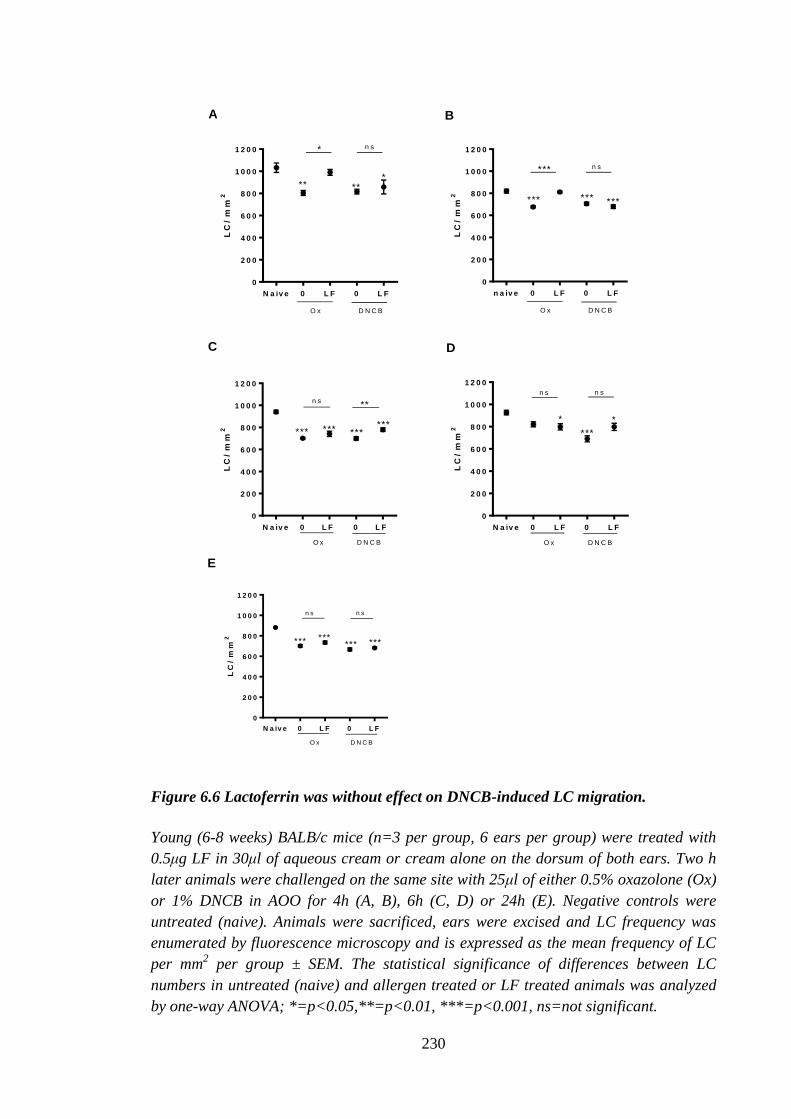
230
LC
/ m
m2
N a iv e 0 L F 0 L F
0
2 0 0
4 0 0
6 0 0
8 0 0
1 0 0 0
1 2 0 0
O x D N C B
** **
* n s
*
A
LC
/ m
m2
n a iv e 0 L F 0 L F
0
2 0 0
4 0 0
6 0 0
8 0 0
1 0 0 0
1 2 0 0
O x D N C B
***
*** n s
*** ***
LC
/ m
m2
N a iv e 0 L F 0 L F
0
2 0 0
4 0 0
6 0 0
8 0 0
1 0 0 0
1 2 0 0
O x D N C B
*** *** ******
n s**
LC
/ m
m2
N a iv e 0 L F 0 L F
0
2 0 0
4 0 0
6 0 0
8 0 0
1 0 0 0
1 2 0 0
O x D N C B
*
***
*
n s n s
B
C D
LC
/ m
m2
N a iv e 0 L F 0 L F
0
2 0 0
4 0 0
6 0 0
8 0 0
1 0 0 0
1 2 0 0
O x D N C B
****** *** ***
n s n s
E
Figure 6.6 Lactoferrin was without effect on DNCB-induced LC migration.
Young (6-8 weeks) BALB/c mice (n=3 per group, 6 ears per group) were treated with
0.5μg LF in 30μl of aqueous cream or cream alone on the dorsum of both ears. Two h
later animals were challenged on the same site with 25μl of either 0.5% oxazolone (Ox)
or 1% DNCB in AOO for 4h (A, B), 6h (C, D) or 24h (E). Negative controls were
untreated (naive). Animals were sacrificed, ears were excised and LC frequency was
enumerated by fluorescence microscopy and is expressed as the mean frequency of LC
per mm2 per group ± SEM. The statistical significance of differences between LC
numbers in untreated (naive) and allergen treated or LF treated animals was analyzed
by one-way ANOVA; *=p<0.05,**=p<0.01, ***=p<0.001, ns=not significant.
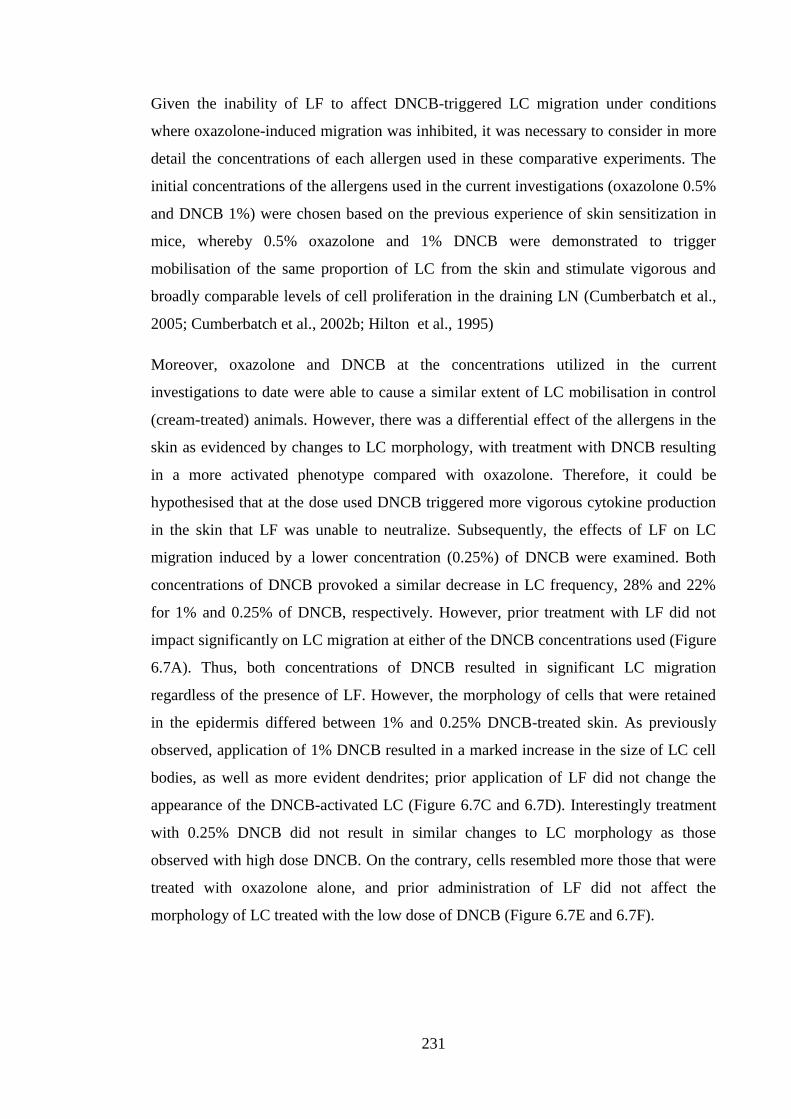
231
Given the inability of LF to affect DNCB-triggered LC migration under conditions
where oxazolone-induced migration was inhibited, it was necessary to consider in more
detail the concentrations of each allergen used in these comparative experiments. The
initial concentrations of the allergens used in the current investigations (oxazolone 0.5%
and DNCB 1%) were chosen based on the previous experience of skin sensitization in
mice, whereby 0.5% oxazolone and 1% DNCB were demonstrated to trigger
mobilisation of the same proportion of LC from the skin and stimulate vigorous and
broadly comparable levels of cell proliferation in the draining LN (Cumberbatch et al.,
2005; Cumberbatch et al., 2002b; Hilton et al., 1995)
Moreover, oxazolone and DNCB at the concentrations utilized in the current
investigations to date were able to cause a similar extent of LC mobilisation in control
(cream-treated) animals. However, there was a differential effect of the allergens in the
skin as evidenced by changes to LC morphology, with treatment with DNCB resulting
in a more activated phenotype compared with oxazolone. Therefore, it could be
hypothesised that at the dose used DNCB triggered more vigorous cytokine production
in the skin that LF was unable to neutralize. Subsequently, the effects of LF on LC
migration induced by a lower concentration (0.25%) of DNCB were examined. Both
concentrations of DNCB provoked a similar decrease in LC frequency, 28% and 22%
for 1% and 0.25% of DNCB, respectively. However, prior treatment with LF did not
impact significantly on LC migration at either of the DNCB concentrations used (Figure
6.7A). Thus, both concentrations of DNCB resulted in significant LC migration
regardless of the presence of LF. However, the morphology of cells that were retained
in the epidermis differed between 1% and 0.25% DNCB-treated skin. As previously
observed, application of 1% DNCB resulted in a marked increase in the size of LC cell
bodies, as well as more evident dendrites; prior application of LF did not change the
appearance of the DNCB-activated LC (Figure 6.7C and 6.7D). Interestingly treatment
with 0.25% DNCB did not result in similar changes to LC morphology as those
observed with high dose DNCB. On the contrary, cells resembled more those that were
treated with oxazolone alone, and prior administration of LF did not affect the
morphology of LC treated with the low dose of DNCB (Figure 6.7E and 6.7F).
232
Figure 6.7 Lactoferrin did not inhibit DNCB-induced migration of LC at low or high
doses of allergen.
Young (6-8 weeks) BALB/c strain mice (n=3, 5-6 ears per group) were treated with
0.5μg LF in 30μl of aqueous cream or cream alone on the dorsum of both ears. Two h
later animals were challenged on the same site with 25μl of either 1% DNCB or 0.25%
of DNCB in AOO for 4h. Negative controls were untreated (naive). Animals were
sacrificed, ears were excised and LC frequency was enumerated by fluorescence
microscopy and is expressed as the mean frequency of LC per mm2 per group ± SEM
(A). The statistical significance of differences between LC numbers in untreated (naive)
or allergen treated animals (with or without LF) was analyzed by one-way ANOVA;
***=p<0.001. Representative micrographs of epidermal sheets show LC morphology
from respective treatments (B-F). x40 magnification.
233
6.2.5 DNCB-mediated LC migration was IL-1β dependent and TNF-α independent
It has been demonstrated before that prior exposure to LF inhibited oxazolone-induced
LC migration, as well as that triggered by delivery of homologous IL-1β, but not TNF-α
(Cumberbatch et al., 2000b). It was suggested that LF inhibition of LC migration in
response to oxazolone treatment was dependent on reducing the availability of TNF-α.
Additionally, LF was shown to decrease the level of TNF-α in the blister fluid in suction
blisters induced in human volunteers following delivery of exogenous homologous IL-
1β (Cumberbatch et al., 2003). Moreover, it was established previously that LC
migration was dependent on two independent cytokine signals from TNF-α and IL-1β
(Cumberbatch et al., 1997b). Taken together, these data suggested that LF was capable
of inhibiting TNF-α dependent migration (oxazolone and IL-1β-induced migration),
possibly through inhibition of TNF-α production. Lactoferrin is unlikely to be blocking
TNF-α signalling (at the TNF-α receptor level), as LF is unable to inhibit migration
when exogenous TNF-α was provided (Almond et al., 2013; Cumberbatch et al.,
2000b). It was intriguing, therefore, that as demonstrated in the Figure 6.6 LF was
unable to inhibit DNCB-mediated LC migration, suggesting that such migration might
be TNF-α independent. This hypothesis was particularly interesting as LC migration
provoked by a variety of other stimuli, including: UV radiation (Moodycliffe et al.,
1994), physical trauma (Ghaznawie et al., 1999), irritants (Cumberbatch et al., 2002a)
and certain pathogens (Shankar et al., 1996), have been reported to be dependent upon
this cytokine. It was demonstrated previously that delivery of TNF-α resulted in LC
migration in mice (Cumberbatch et al., 1999a) and in humans (Cumberbatch et al.,
1999d). In addition, TNF-α was reported be necessary for DC accumulation in the LN
following treatment with the fluorescent allergen, FITC (Cumberbatch and Kimber,
1995).
The role of TNF-α and IL-1β cytokines in DNCB-triggered LC mobilisation was
investigated further. The initial approach chosen was to utilize neutralising antibodies
directed against either TNF-α or IL-1β and to compare their impact on LC migration
stimulated following application of oxazolone or DNCB. In the first instance a
polyclonal anti-TNF-α antibody was employed. Intradermal delivery of normal rabbit
serum did not impact on LC migration following allergen application, thus, levels of
migration of 18% and 22% were induced by oxazolone and DNCB, respectively, similar
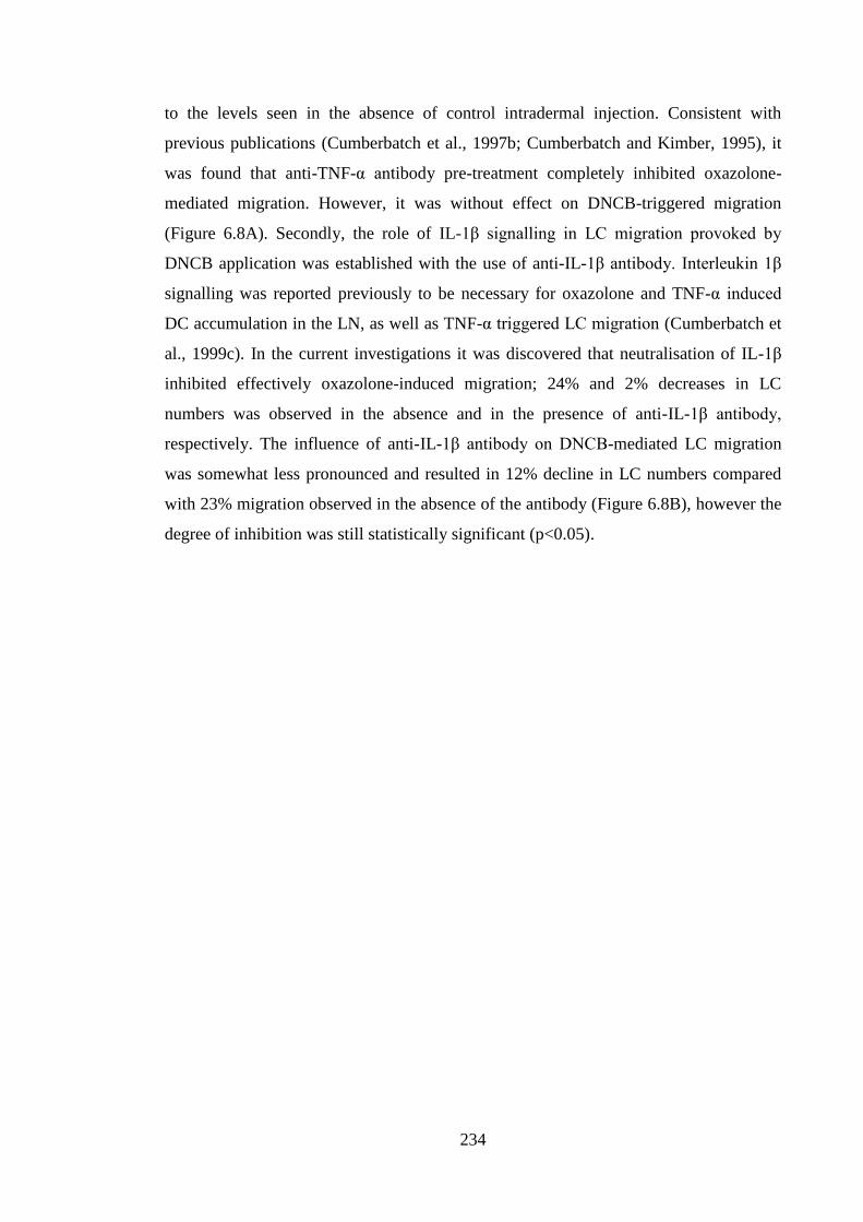
234
to the levels seen in the absence of control intradermal injection. Consistent with
previous publications (Cumberbatch et al., 1997b; Cumberbatch and Kimber, 1995), it
was found that anti-TNF-α antibody pre-treatment completely inhibited oxazolone-
mediated migration. However, it was without effect on DNCB-triggered migration
(Figure 6.8A). Secondly, the role of IL-1β signalling in LC migration provoked by
DNCB application was established with the use of anti-IL-1β antibody. Interleukin 1β
signalling was reported previously to be necessary for oxazolone and TNF-α induced
DC accumulation in the LN, as well as TNF-α triggered LC migration (Cumberbatch et
al., 1999c). In the current investigations it was discovered that neutralisation of IL-1β
inhibited effectively oxazolone-induced migration; 24% and 2% decreases in LC
numbers was observed in the absence and in the presence of anti-IL-1β antibody,
respectively. The influence of anti-IL-1β antibody on DNCB-mediated LC migration
was somewhat less pronounced and resulted in 12% decline in LC numbers compared
with 23% migration observed in the absence of the antibody (Figure 6.8B), however the
degree of inhibition was still statistically significant (p<0.05).
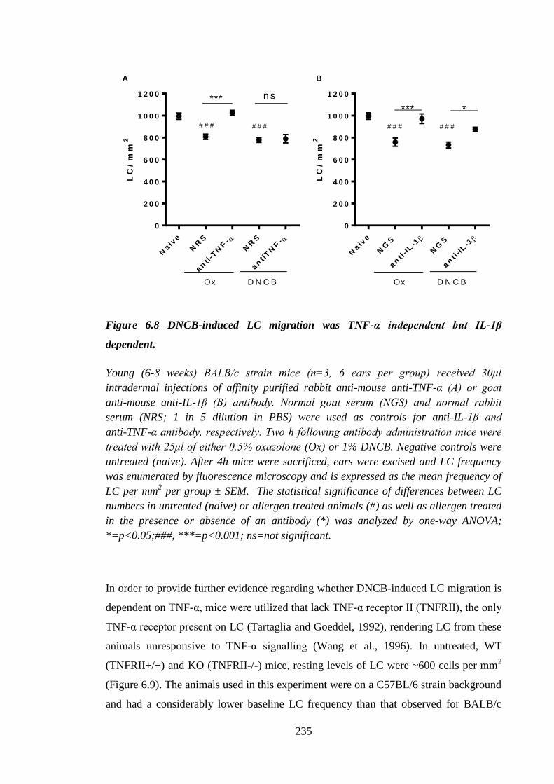
235
LC
/ m
m2
Naiv
e
NR
S
an
ti-T
NF
-
NR
S
an
tiT
NF
-
0
2 0 0
4 0 0
6 0 0
8 0 0
1 0 0 0
1 2 0 0
Ox D N C B
# # # # # #
n s***
LC
/ m
m2
Naiv
e
NG
S
an
t i- I
L-1
NG
S
an
t i- I
L-1
0
2 0 0
4 0 0
6 0 0
8 0 0
1 0 0 0
1 2 0 0
Ox D N C B
# # # # # #
****
A B
Figure 6.8 DNCB-induced LC migration was TNF-α independent but IL-1β
dependent.
Young (6-8 weeks) BALB/c strain mice (n=3, 6 ears per group) received 30μl
intradermal injections of affinity purified rabbit anti-mouse anti-TNF-α (A) or goat
anti-mouse anti-IL-1β (B) antibody. Normal goat serum (NGS) and normal rabbit
serum (NRS; 1 in 5 dilution in PBS) were used as controls for anti-IL-1β and
anti-TNF-α antibody, respectively. Two h following antibody administration mice were
treated with 25μl of either 0.5% oxazolone (Ox) or 1% DNCB. Negative controls were
untreated (naive). After 4h mice were sacrificed, ears were excised and LC frequency
was enumerated by fluorescence microscopy and is expressed as the mean frequency of
LC per mm2 per group ± SEM. The statistical significance of differences between LC
numbers in untreated (naive) or allergen treated animals (#) as well as allergen treated
in the presence or absence of an antibody (*) was analyzed by one-way ANOVA;
*=p<0.05;###, ***=p<0.001; ns=not significant.
In order to provide further evidence regarding whether DNCB-induced LC migration is
dependent on TNF-α, mice were utilized that lack TNF-α receptor II (TNFRII), the only
TNF-α receptor present on LC (Tartaglia and Goeddel, 1992), rendering LC from these
animals unresponsive to TNF-α signalling (Wang et al., 1996). In untreated, WT
(TNFRII+/+) and KO (TNFRII-/-) mice, resting levels of LC were ~600 cells per mm2
(Figure 6.9). The animals used in this experiment were on a C57BL/6 strain background
and had a considerably lower baseline LC frequency than that observed for BALB/c
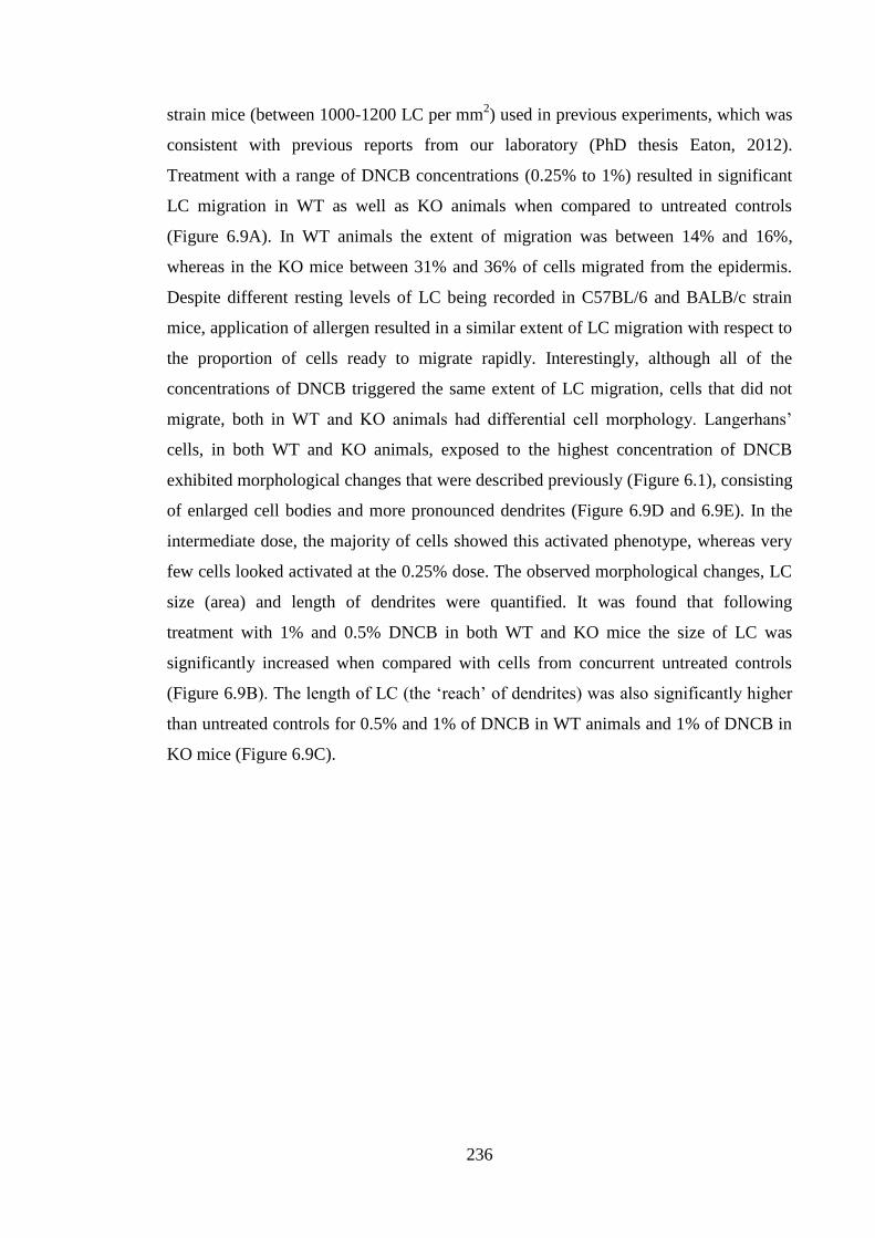
236
strain mice (between 1000-1200 LC per mm2) used in previous experiments, which was
consistent with previous reports from our laboratory (PhD thesis Eaton, 2012).
Treatment with a range of DNCB concentrations (0.25% to 1%) resulted in significant
LC migration in WT as well as KO animals when compared to untreated controls
(Figure 6.9A). In WT animals the extent of migration was between 14% and 16%,
whereas in the KO mice between 31% and 36% of cells migrated from the epidermis.
Despite different resting levels of LC being recorded in C57BL/6 and BALB/c strain
mice, application of allergen resulted in a similar extent of LC migration with respect to
the proportion of cells ready to migrate rapidly. Interestingly, although all of the
concentrations of DNCB triggered the same extent of LC migration, cells that did not
migrate, both in WT and KO animals had differential cell morphology. Langerhans’
cells, in both WT and KO animals, exposed to the highest concentration of DNCB
exhibited morphological changes that were described previously (Figure 6.1), consisting
of enlarged cell bodies and more pronounced dendrites (Figure 6.9D and 6.9E). In the
intermediate dose, the majority of cells showed this activated phenotype, whereas very
few cells looked activated at the 0.25% dose. The observed morphological changes, LC
size (area) and length of dendrites were quantified. It was found that following
treatment with 1% and 0.5% DNCB in both WT and KO mice the size of LC was
significantly increased when compared with cells from concurrent untreated controls
(Figure 6.9B). The length of LC (the ‘reach’ of dendrites) was also significantly higher
than untreated controls for 0.5% and 1% of DNCB in WT animals and 1% of DNCB in
KO mice (Figure 6.9C).
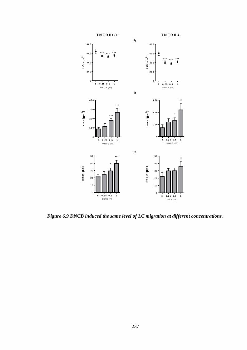
237
LC
/ m
m2
0 0 .2 5 0 .5 1
0
2 0 0
4 0 0
6 0 0
8 0 0
D N C B (% )
*** *** ***
LC
/ m
m2
0 0 .2 5 0 .5 1
0
2 0 0
4 0 0
6 0 0
8 0 0
D N C B (% )
*** *** ***
are
a [
m2
]
0 0 .2 5 0 .5 1
0
1 0 0
2 0 0
3 0 0
4 0 0
D N C B (% )
***
***
are
a [
m2
]
0 0 .2 5 0 .5 1
0
2 0 0
4 0 0
6 0 0
D N C B (% )
***
*
len
gth
[
m]
0 0 .2 5 0 .5 1
0
1 0
2 0
3 0
4 0
5 0
D N C B (% )
*
***
len
gth
[
m]
0 0 .2 5 0 .5 1
0
1 0
2 0
3 0
4 0
5 0
D N C B (% )
**
T N F R II+ /+ T N F R II- / -
A
B
C
Figure 6.9 DNCB induced the same level of LC migration at different concentrations.
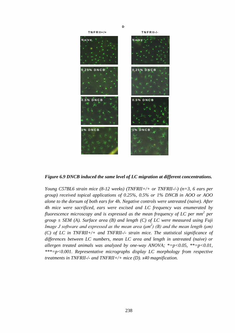
238
Figure 6.9 DNCB induced the same level of LC migration at different concentrations.
Young C57BL6 strain mice (8-12 weeks) (TNFRII+/+ or TNFRII-/-) (n=3, 6 ears per
group) received topical applications of 0.25%, 0.5% or 1% DNCB in AOO or AOO
alone to the dorsum of both ears for 4h. Negative controls were untreated (naive). After
4h mice were sacrificed, ears were excised and LC frequency was enumerated by
fluorescence microscopy and is expressed as the mean frequency of LC per mm2 per
group ± SEM (A). Surface area (B) and length (C) of LC were measured using Fuji
Image J software and expressed as the mean area (μm2) (B) and the mean length (μm)
(C) of LC in TNFRII+/+ and TNFRII-/- strain mice. The statistical significance of
differences between LC numbers, mean LC area and length in untreated (naive) or
allergen treated animals was analyzed by one-way ANOVA; *=p<0.05, **=p<0.01,
***=p<0.001. Representative micrographs display LC morphology from respective
treatments in TNFRII-/- and TNFRII+/+ mice (D). x40 magnification.
T N F R II+ /+ T N F R II- / -
N a iv e
0 .2 5 % D N C B
0 .5 % D N C B
1 % D N C B
N a iv e
0 .2 5 % D N C B
0 .5 % D N C B
1 % D N C B
D
239
6.2.6 Members of dinitrohalobenzene family induced LC migration in the absence
of TNF-α signalling
After establishing, using a variety of approaches, that DNCB can indeed cause LC
migration in the absence of TNF-α signalling, other stimuli that previously have been
shown to trigger LC migration were investigated with respect to their ability to cause
LC migration in the absence of TNFRII. Knockout mice and their WT counterparts
were treated with a number of different contact allergens (DNCB, DNFB, PPD), the
respiratory allergen (TMA) or the non-sensitizing skin irritant (SLS). With the
exception of PPD, all these stimuli have been shown previously to induce LC migration
in BALB/c strain mice (Antonopoulos et al., 2008; Cumberbatch et al., 2005;
Cumberbatch et al., 2002a). 2,4-dinitrofluorobenzene is a member of the same family of
chemicals (dinitrohalobenzenes) as DNCB, and similarly to DNCB, it is a contact
sensitizer (Watanabe et al., 2008). Paraphenylenediamine is a contact allergen found in
hair dyes, and as a known skin sensitizer, it was expected to cause LC migration (Pot et
al., 2013). Trimellitic anhydride is a recognized respiratory allergen (Dearman et al.,
1991; Grammer et al., 1997) and has been shown previously in mice to result in LC
migration following topical exposure, albeit with somewhat delayed kinetics
(Cumberbatch et al., 2005). Lastly, SLS is not an allergen, but a skin irritant,
nevertheless, when applied topically it was reported to trigger LC migration
(Cumberbatch et al., 2002a). The hypothesis was that when applied topically these
chemicals would not cause LC migration in TNFRII-/- mice. In the untreated (naive) or
vehicle (AOO) treated WT and KO mice resting LC levels were ~800 and ~600 cells
per mm2, respectively. In WT mice treatment with all of the stimuli resulted in LC
migration in the expected range of between 20 and 30% (Figure 6.10A). However, in
the KO animals only members of the dinitrohalobenzene family (DNCB and DNFB)
were capable of triggering significant LC migration resulting in LC loss comparable to
that reported for WT mice (between 20% and 37%) (Figure 6.10B). For the other
stimuli the loss of LC from the skin ranged between 2% and 4%; none of which reached
statistical significance. In the series of experiments reported in the Figure 6.10
oxazolone was not used as one of the stimuli, despite this chemical being used routinely
as a reference contact allergen in previous studies of the cytokine regulation of LC
migration (Cumberbatch et al., 2001). This was due to the fact that oxazolone failed to
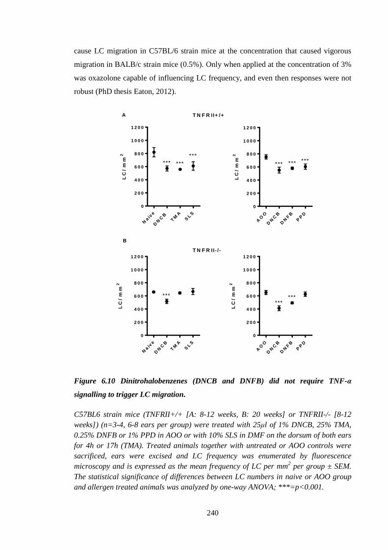
240
cause LC migration in C57BL/6 strain mice at the concentration that caused vigorous
migration in BALB/c strain mice (0.5%). Only when applied at the concentration of 3%
was oxazolone capable of influencing LC frequency, and even then responses were not
robust (PhD thesis Eaton, 2012).
LC
/ m
m2
Naiv
e
DN
CB
TM
AS
LS
0
2 0 0
4 0 0
6 0 0
8 0 0
1 0 0 0
1 2 0 0
*** ***
***
LC
/ m
m2
AO
O
DN
CB
DN
FB
PP
D
0
2 0 0
4 0 0
6 0 0
8 0 0
1 0 0 0
1 2 0 0
*** *** ***
LC
/ m
m2
Naiv
e
DN
CB
TM
AS
LS
0
2 0 0
4 0 0
6 0 0
8 0 0
1 0 0 0
1 2 0 0
***
LC
/ m
m2
AO
O
DN
CB
DN
FB
PP
D
0
2 0 0
4 0 0
6 0 0
8 0 0
1 0 0 0
1 2 0 0
******
A
B
T N F R II+ /+
T N F R II- / -
Figure 6.10 Dinitrohalobenzenes (DNCB and DNFB) did not require TNF-α
signalling to trigger LC migration.
C57BL6 strain mice (TNFRII+/+ [A: 8-12 weeks, B: 20 weeks] or TNFRII-/- [8-12
weeks]) (n=3-4, 6-8 ears per group) were treated with 25μl of 1% DNCB, 25% TMA,
0.25% DNFB or 1% PPD in AOO or with 10% SLS in DMF on the dorsum of both ears
for 4h or 17h (TMA). Treated animals together with untreated or AOO controls were
sacrificed, ears were excised and LC frequency was enumerated by fluorescence
microscopy and is expressed as the mean frequency of LC per mm2 per group ± SEM.
The statistical significance of differences between LC numbers in naive or AOO group
and allergen treated animals was analyzed by one-way ANOVA; ***=p<0.001.
241
6.2.7 Oxazolone and DNCB induced divergent cutaneous cytokine expression
profiles
In the current investigations it was found that there were differences between these two
allergens with regards to their ability to induce LC migration, particularly with respect
to cytokine requirements for LC migration. Therefore, it was of particular interest to
investigate the kinetics, vigour and quality of cutaneous cytokine mRNA profiles
induced by oxazolone and DNCB. It was decided that measurement of gene expression
in excised full thickness skin, sampled from the ear, would be most appropriate, as this
tissue could be quickly excised without further manipulation and fixed in RNAlater
immediately after excision, therefore, reducing the impact of trauma on gene expression
(as observed in the explant experiments; Figure 3.1).
The figure 6.11 presents a comparison between two allergens: with respect to the
expression of various IL-1 family members (IL-1α and IL-1β; IL-18 and IL-33), the
proflammatory cytokines TNF-α and IL-17F, the chemokine CXCL1 and the membrane
marker ICAM-1, as a downstream indicator of inflammation. Expression of these
molecules was assessed relative to baseline expression in control naive animals and a
single animal was used at each time point (0.5h to 24h). Exposure to both allergens
resulted in remarkably similar kinetic profiles for each molecule, although in many
cases, the extent of activation was markedly higher for the DNCB-treated skin. A
number of genes (TNF-α, IL-1β, CXCL1, ICAM-1 and IL-17F) were up-regulated upon
allergen exposure; all followed a similar, transient, profile, returning to baseline levels
within 24 h, with the exception of TNF-α, which peaked at approximately between 1
and 2h. Peak expression of markers was generally at 6h after initiation of exposure. In
contrast, other IL-1 family members (Il-1α, Il-18 and Il-33) all exhibited down-
regulation, also reaching maximal levels at around 6h and returning back to baseline
levels by 24h. As indicated previously, although the patterns of expression were very
similar for both allergens for all markers, there were some differences with respect to
the vigour of the reaction between DNCB and oxazolone. The most striking differences
were observed with respect to Il-1β and Cxcl1 expression. At peak expression (6h) the
fold increases recorded were 8 and 18 for oxazolone and 55 and 130-fold for DNCB, for
Il-1β and Cxcl1, respectively. DNCB-triggered Il-1β expression was higher than that
observed following oxazolone treatments at all times, whereas Cxcl1 expression was
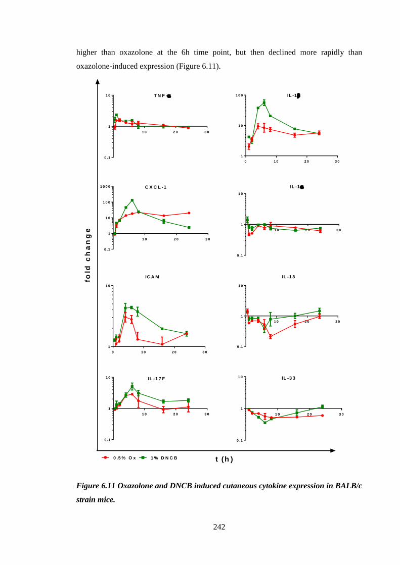
242
higher than oxazolone at the 6h time point, but then declined more rapidly than
oxazolone-induced expression (Figure 6.11).
1 0 2 0 3 0
0 .1
1
1 0 T N F -
0 1 0 2 0 3 0
1
1 0
1 0 0 IL -1
1 0 2 0 3 0
0 .1
1
1 0
1 0 0
1 0 0 0 C X C L -1
1 0 2 0 3 0
0 .1
1
1 0
IL -1
0 1 0 2 0 3 0
1
1 0
IC A M
1 0 2 0 3 0
0 .1
1
1 0
IL -1 8
1 0 2 0 3 0
0 .1
1
1 0 IL -1 7 F
1 0 2 0 3 0
0 .1
1
1 0 IL -3 3
fold
ch
an
ge
t (h )0 .5 % O x 1 % D N C B
Figure 6.11 Oxazolone and DNCB induced cutaneous cytokine expression in BALB/c
strain mice.
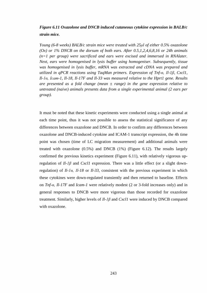
243
Figure 6.11 Oxazolone and DNCB induced cutaneous cytokine expression in BALB/c
strain mice.
Young (6-8 weeks) BALB/c strain mice were treated with 25μl of either 0.5% oxazolone
(Ox) or 1% DNCB on the dorsum of both ears. After 0.5,1,2,4,6,8,16 or 24h animals
(n=1 per group) were sacrificed and ears were excised and immersed in RNAlater.
Next, ears were homogenised in lysis buffer using homogeniser. Subsequently, tissue
was homogenised in lysis buffer, mRNA was extracted and cDNA was prepared and
utilized in qPCR reactions using TaqMan primers. Expression of Tnf-α, Il-1β, Cxcl1,
Il-1α, Icam-1, Il-18, Il-17F and Il-33 was measured relative to the Hprt1 gene. Results
are presented as a fold change (mean ± range) in the gene expression relative to
untreated (naive) animals presents data from a single experimental animal (2 ears per
group).
It must be noted that these kinetic experiments were conducted using a single animal at
each time point, thus it was not possible to assess the statistical significance of any
differences between oxazolone and DNCB. In order to confirm any differences between
oxazolone and DNCB-induced cytokine and ICAM-1 transcript expression, the 4h time
point was chosen (time of LC migration measurement) and additional animals were
treated with oxazolone (0.5%) and DNCB (1%) (Figure 6.12). The results largely
confirmed the previous kinetics experiment (Figure 6.11), with relatively vigorous up-
regulation of Il-1β and Cxcl1 expression. There was a little effect (or a slight down-
regulation) of Il-1α, Il-18 or Il-33, consistent with the previous experiment in which
these cytokines were down-regulated transiently and then returned to baseline. Effects
on Tnf-α, Il-17F and Icam-1 were relatively modest (2 or 3-fold increases only) and in
general responses to DNCB were more vigorous than those recorded for oxazolone
treatment. Similarly, higher levels of Il-1β and Cxcl1 were induced by DNCB compared
with oxazolone.
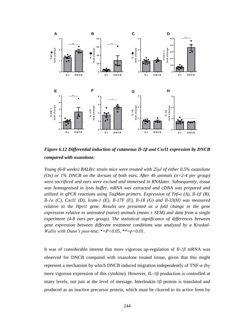
244
TN
F-
fold
ch
an
ge
O x D N C B
0
1
2
3
**
IL-1
fold
ch
an
ge
O x D N C B
0
2 0
4 0
6 0
8 0
**
IL-1
fold
ch
an
ge
O x D N C B
0
1
2
3
CX
CL
1
fold
ch
an
ge
O x D N C B
0
1 0
2 0
3 0
4 0
5 0 **
ICA
M-1
fold
ch
an
ge
O x D N C B
0
1
2
3
4
5 **
IL-1
7F
fold
ch
an
ge
O x D N C B
0
1
2
3
4
*
IL-1
8
fold
ch
an
ge
O x D N C B
0
1
2
3
IL-3
3
fold
ch
an
ge
O x D N C B
0
1
2
3
A B C D
E F G H
Figure 6.12 Differential induction of cutaneous Il-1β and Cxcl1 expression by DNCB
compared with oxazolone.
Young (6-8 weeks) BALB/c strain mice were treated with 25μl of either 0.5% oxazolone
(Ox) or 1% DNCB on the dorsum of both ears. After 4h animals (n=2-4 per group)
were sacrificed and ears were excised and immersed in RNAlater. Subsequently, tissue
was homogenised in lysis buffer, mRNA was extracted and cDNA was prepared and
utilized in qPCR reactions using TaqMan primers. Expression of Tnf-α (A), Il-1β (B),
Il-1α (C), Cxcl1 (D), Icam-1 (E), Il-17F (F), Il-18 (G) and Il-33(H) was measured
relative to the Hprt1 gene. Results are presented as a fold change in the gene
expression relative to untreated (naive) animals (mean ± SEM) and data from a single
experiment (4-8 ears per group). The statistical significance of differences between
gene expression between different treatment conditions was analyzed by a Kruskal-
Wallis with Dunn’s post-test; *=P<0.05, **=p<0.01.
It was of considerable interest that more vigorous up-regulation of Il-1β mRNA was
observed for DNCB compared with oxazolone treated tissue, given that this might
represent a mechanism by which DNCB induced migration independently of TNF-α (by
more vigorous expression of this cytokine). However, IL-1β production is controlled at
many levels, not just at the level of message. Interleukin-1β protein is translated and
produced as an inactive precursor protein, which must be cleaved to its active form by
245
the enzyme caspase 1, which also exists as an inactive precursor. In addition, it also is
regulated at the level of secretion, with a second signal required for the release of this
cytokine (Netea et al., 2010). Therefore, in addition to measuring this cytokine at the
level of transcription, it was important to measure the secretion of the IL-1β. Thus,
animals were treated with 0.5% oxazolone or 1% DNCB for either 2h or 6h (Figure
6.13). Ears were excised, explants prepared and floated on medium overnight, then
cytokines (TNF-α, IL-1α and β) that had been secreted into the supernatant were
analyzed by ELISA. Production of TNF-α in the absence of allergen treatment was
~100pg/ml. Application of allergen was largely without effect, apart from 2h exposure
to oxazolone where TNF-α levels of 200pg/ml, were recorded. High constitutive
production of IL-1α (~2000pg/ml) was observed and this was not modulated by
exposure to allergens. Baseline release of IL-1β was ~100pg/ml, and was up-regulated
slightly by treatment with oxazolone at both time points to approximately ~200pg/ml.
However, the most vigorous production of this cytokine was reported 6h after DNCB
application (approximately 5-fold increase) (Figure 6.13C).
TN
F-
[p
g/m
l]
N a iv e 2 h 6 h 2 h 6 h
0
1 0 0
2 0 0
3 0 0
4 0 0
5 0 0
0 .5 % O x 1 % D N C B
IL-1
[
pg
/ml]
N a iv e 2 h 6 h 2 h 6 h
0
2 0 0 0
4 0 0 0
6 0 0 0
8 0 0 0
0 .5 % O x 1 % D N C B
N a iv e 2 h 6 h 2 h 6 h
0
5 0 0
1 0 0 0
1 5 0 0
0 .5 % O x 1 % D N C B
IL-1
[
pg
/ml]
A B C
Figure 6.13 Allergen treatment provoked release of cytokines from the skin.
Young (6-8 weeks) BALB/c strain mice were treated with 25μl of either 0.5% oxazolone
(Ox) or 1% DNCB on the dorsum of both ears. After 2 or 6h (A, B, C; n=1 per group; 2
ears) treated animals and untreated controls (naive) were sacrificed and ears were
excised. Ears were split into dorsal and ventral halves and the dorsal part was floated
on 250μl of complete RPMI media and incubated for 16h. Following incubation
supernatants were collected and TNF-α (A), IL-1α (B) or IL-1β (C) production was
determined by ELISA. The dashed line represents the lower limit of detection for each
ELISA (TNF-α [32pg/ml], IL-1α and β [15pg/ml]). Graphs present mean ± range (for
n=1; 2 ears per group).
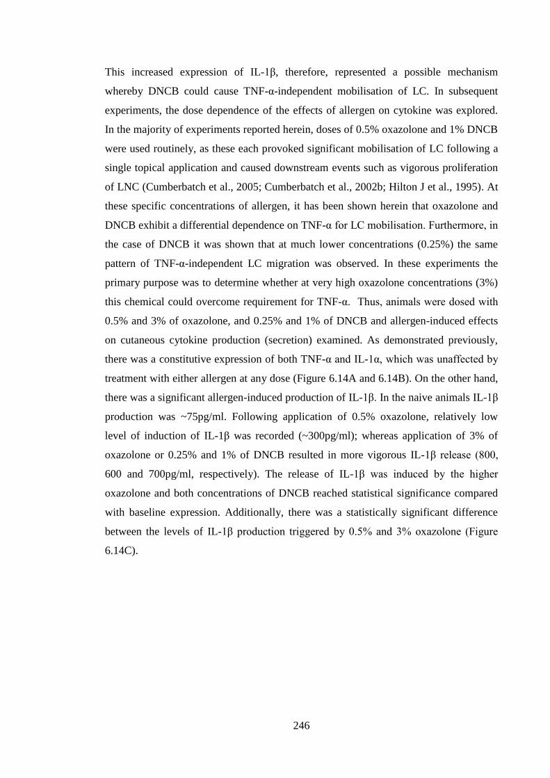
246
This increased expression of IL-1β, therefore, represented a possible mechanism
whereby DNCB could cause TNF-α-independent mobilisation of LC. In subsequent
experiments, the dose dependence of the effects of allergen on cytokine was explored.
In the majority of experiments reported herein, doses of 0.5% oxazolone and 1% DNCB
were used routinely, as these each provoked significant mobilisation of LC following a
single topical application and caused downstream events such as vigorous proliferation
of LNC (Cumberbatch et al., 2005; Cumberbatch et al., 2002b; Hilton J et al., 1995). At
these specific concentrations of allergen, it has been shown herein that oxazolone and
DNCB exhibit a differential dependence on TNF-α for LC mobilisation. Furthermore, in
the case of DNCB it was shown that at much lower concentrations (0.25%) the same
pattern of TNF-α-independent LC migration was observed. In these experiments the
primary purpose was to determine whether at very high oxazolone concentrations (3%)
this chemical could overcome requirement for TNF-α. Thus, animals were dosed with
0.5% and 3% of oxazolone, and 0.25% and 1% of DNCB and allergen-induced effects
on cutaneous cytokine production (secretion) examined. As demonstrated previously,
there was a constitutive expression of both TNF-α and IL-1α, which was unaffected by
treatment with either allergen at any dose (Figure 6.14A and 6.14B). On the other hand,
there was a significant allergen-induced production of IL-1β. In the naive animals IL-1β
production was ~75pg/ml. Following application of 0.5% oxazolone, relatively low
level of induction of IL-1β was recorded (~300pg/ml); whereas application of 3% of
oxazolone or 0.25% and 1% of DNCB resulted in more vigorous IL-1β release (800,
600 and 700pg/ml, respectively). The release of IL-1β was induced by the higher
oxazolone and both concentrations of DNCB reached statistical significance compared
with baseline expression. Additionally, there was a statistically significant difference
between the levels of IL-1β production triggered by 0.5% and 3% oxazolone (Figure
6.14C).
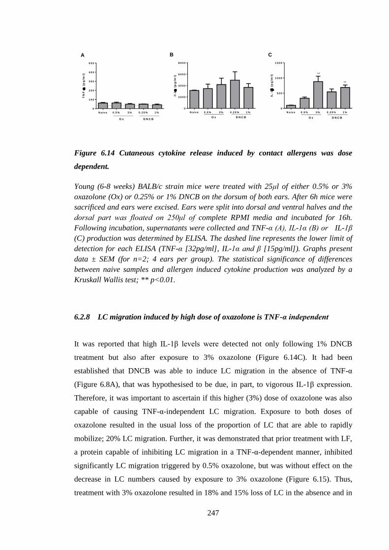
247
TN
F-
(p
g/m
l)
N a iv e 0 .5 % 3 % 0 .2 5 % 1 %
0
1 0 0
2 0 0
3 0 0
4 0 0
5 0 0
O x D N C B
Il-1
(
pg
/ml)
N a iv e 0 .5 % 3 % 0 .2 5 % 1 %
0
2 0 0 0
4 0 0 0
6 0 0 0
8 0 0 0
O x D N C B
IL-1
(
pg
/ml)
N a iv e 0 .5 % 3 % 0 .2 5 % 1 %
0
5 0 0
1 0 0 0
1 5 0 0
O x D N C B
**
**
A B C
Figure 6.14 Cutaneous cytokine release induced by contact allergens was dose
dependent.
Young (6-8 weeks) BALB/c strain mice were treated with 25μl of either 0.5% or 3%
oxazolone (Ox) or 0.25% or 1% DNCB on the dorsum of both ears. After 6h mice were
sacrificed and ears were excised. Ears were split into dorsal and ventral halves and the
dorsal part was floated on 250μl of complete RPMI media and incubated for 16h.
Following incubation, supernatants were collected and TNF-α (A), IL-1α (B) or IL-1β
(C) production was determined by ELISA. The dashed line represents the lower limit of
detection for each ELISA (TNF-α [32pg/ml], IL-1α and β [15pg/ml]). Graphs present
data ± SEM (for n=2; 4 ears per group). The statistical significance of differences
between naive samples and allergen induced cytokine production was analyzed by a
Kruskall Wallis test; ** p<0.01.
6.2.8 LC migration induced by high dose of oxazolone is TNF-α independent
It was reported that high IL-1β levels were detected not only following 1% DNCB
treatment but also after exposure to 3% oxazolone (Figure 6.14C). It had been
established that DNCB was able to induce LC migration in the absence of TNF-α
(Figure 6.8A), that was hypothesised to be due, in part, to vigorous IL-1β expression.
Therefore, it was important to ascertain if this higher (3%) dose of oxazolone was also
capable of causing TNF-α-independent LC migration. Exposure to both doses of
oxazolone resulted in the usual loss of the proportion of LC that are able to rapidly
mobilize; 20% LC migration. Further, it was demonstrated that prior treatment with LF,
a protein capable of inhibiting LC migration in a TNF-α-dependent manner, inhibited
significantly LC migration triggered by 0.5% oxazolone, but was without effect on the
decrease in LC numbers caused by exposure to 3% oxazolone (Figure 6.15). Thus,
treatment with 3% oxazolone resulted in 18% and 15% loss of LC in the absence and in
248
the presence of LF, respectively. Application of 0.5% oxazolone resulted in the change
in morphology as previously described (Figure 6.1C). However, treatment with 3%
oxazolone caused changes in LC phenotype that resembled those observed following
exposure to 1% DNCB, regardless of the presence of LF.
Figure 6.15 Lactoferrin did not inhibit LC migration caused by high concentration of
oxazolone.
Young (6-8 weeks) BALB/c strain mice (n=3 per group, 4-6 ears per group) were
treated with 5μg of LF in 30μl of aqueous cream or cream alone on the dorsum of both
ears. Two h later animals were challenged on the same site with 25μl of 0.5% or 3%
oxazolone (Ox) in AOO for 4h. Animals were sacrificed, ears were excised and LC
frequency was enumerated by fluorescence microscopy and is expressed as the mean
frequency of LC per mm2 per group ± SEM. The statistical significance of differences
between LC numbers in untreated (naive) and allergen treated and LF treated animals
was analyzed by one-way ANOVA; *=p<0.05, **=p<0.01. Representative micrographs
display LC morphology from respective treatments.
249
6.2.9 Chemicals provoked distinct cutaneous cytokine expression profiles
Finally, a series of experiments was conducted to explore the nature of cutaneous
cytokine expression following topical exposure to other types of chemical insult.
Animals were exposed to a respiratory allergen (TMA) (Grammer et al., 1997), DNTB a
chemical that has been reported previously to be a tolerogen and to failed to provoke LC
migration (Udey, 2012) or to a skin irritant, SLS (Agner et al., 1989), and time
dependent changes in cutaneous gene expression were examined.
Quantitative PCR was used to assess time dependent changes in cutaneous cytokine
(TNF-α, IL-1β, IL-1α, IL-33, IL-18), chemokine (CXCL1) and adhesion molecule
(ICAM-1) expression levels. It has been reported that chemical allergens form two
groups: those with the ability to cause ACD and a smaller group with the ability to
cause sensitization of the respiratory tract and occupational asthma or rhinitis (Kimber
et al., 2010). These different types of chemicals have been shown to polarize the
immune response to either a Th1 or a Th2 phenotype. Prolonged (2 week) exposure to
either contact or respiratory allergens results in cytokine production by LNC that is
characteristic of each group of allergens (Dearman and Kimber, 1999). The purpose of
the current series of experiments was to determine whether different classes of
chemical, including a respiratory allergen, exhibited specific patterns of cutaneous gene
expression following single dose exposure. Indeed, it was established previously that
exposure to respiratory allergen TMA resulted in delayed LC migration (17h) in
comparison with contact allergens (4h) via cutaneous production of IL-10
(Cumberbatch et al., 2005). Mice were exposed to TMA, DNTB or SLS, and time
dependent changes in skin gene expression were examined. It was demonstrated before
that exposure to TMA and SLS resulted in LC migration that was dependent upon TNF-
α signalling (Figure 6.10). Treatment with TMA was without effect on expression of
majority of markers throughout the time course (TNF-α, IL-1α, IL-33, IL-18, ICAM-1).
However, a marked up-regulation of Cxcl1 was recorded that peaked at 16h with a 15-
fold increase in comparison with naive tissue. Another up-regulated gene was Il-1β; its
expression did not increase until 24h after initiation of exposure and was approximately
10 times higher than that of untreated (naive) controls at this time point (Figure 6.16). It
is of interest that TMA induced LC migration takes place 17h following exposure
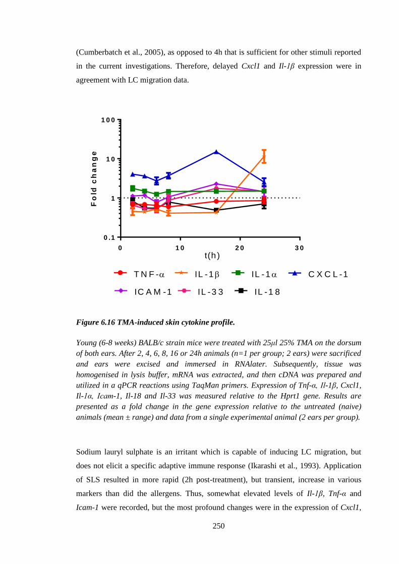
250
(Cumberbatch et al., 2005), as opposed to 4h that is sufficient for other stimuli reported
in the current investigations. Therefore, delayed Cxcl1 and Il-1β expression were in
agreement with LC migration data.
0 1 0 2 0 3 0
0 .1
1
1 0
1 0 0
Fo
ld c
ha
ng
e
T N F - IL -1IL -1 C X C L -1
IL -3 3 IL -1 8IC A M -1
t(h )
Figure 6.16 TMA-induced skin cytokine profile.
Young (6-8 weeks) BALB/c strain mice were treated with 25μl 25% TMA on the dorsum
of both ears. After 2, 4, 6, 8, 16 or 24h animals (n=1 per group; 2 ears) were sacrificed
and ears were excised and immersed in RNAlater. Subsequently, tissue was
homogenised in lysis buffer, mRNA was extracted, and then cDNA was prepared and
utilized in a qPCR reactions using TaqMan primers. Expression of Tnf-α, Il-1β, Cxcl1,
Il-1α, Icam-1, Il-18 and Il-33 was measured relative to the Hprt1 gene. Results are
presented as a fold change in the gene expression relative to the untreated (naive)
animals (mean ± range) and data from a single experimental animal (2 ears per group).
Sodium lauryl sulphate is an irritant which is capable of inducing LC migration, but
does not elicit a specific adaptive immune response (Ikarashi et al., 1993). Application
of SLS resulted in more rapid (2h post-treatment), but transient, increase in various
markers than did the allergens. Thus, somewhat elevated levels of Il-1β, Tnf-α and
Icam-1 were recorded, but the most profound changes were in the expression of Cxcl1,
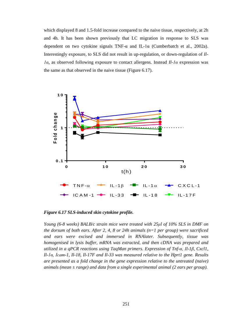
251
which displayed 8 and 1.5-fold increase compared to the naive tissue, respectively, at 2h
and 4h. It has been shown previously that LC migration in response to SLS was
dependent on two cytokine signals TNF-α and IL-1α (Cumberbatch et al., 2002a).
Interestingly exposure, to SLS did not result in up-regulation, or down-regulation of Il-
1α, as observed following exposure to contact allergens. Instead Il-1α expression was
the same as that observed in the naive tissue (Figure 6.17).
0 1 0 2 0 3 0
0 .1
1
1 0
Fo
ld c
ha
ng
e
T N F - IL -1IL -1
IL -1 8
C X C L -1
IL -3 3IC A M -1 IL -1 7 F
t(h )
Figure 6.17 SLS-induced skin cytokine profile.
Young (6-8 weeks) BALB/c strain mice were treated with 25μl of 10% SLS in DMF on
the dorsum of both ears. After 2, 4, 8 or 24h animals (n=1 per group) were sacrificed
and ears were excised and immersed in RNAlater. Subsequently, tissue was
homogenised in lysis buffer, mRNA was extracted, and then cDNA was prepared and
utilized in a qPCR reactions using TaqMan primers. Expression of Tnf-α, Il-1β, Cxcl1,
Il-1α, Icam-1, Il-18, Il-17F and Il-33 was measured relative to the Hprt1 gene. Results
are presented as a fold change in the gene expression relative to the untreated (naive)
animals (mean ± range) and data from a single experimental animal (2 ears per group).
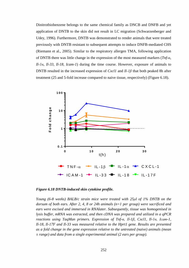
252
Dinitrothiobenzene belongs to the same chemical family as DNCB and DNFB and yet
application of DNTB to the skin did not result in LC migration (Schwarzenberger and
Udey, 1996). Furthermore, DNTB was demonstrated to render animals that were treated
previously with DNTB resistant to subsequent attempts to induce DNFB-mediated CHS
(Riemann et al., 2005). Similar to the respiratory allergen TMA, following application
of DNTB there was little change in the expression of the most measured markers (Tnf-α,
Il-1α, Il-33, Il-18, Icam-1) during the time course. However, exposure of animals to
DNTB resulted in the increased expression of Cxcl1 and Il-1β that both peaked 8h after
treatment (25 and 5-fold increase compared to naive tissue, respectively) (Figure 6.18).
0 1 0 2 0 3 0
0 .1
1
1 0
1 0 0
Fo
ld c
ha
ng
e
T N F - IL -1IL -1 C X C L -1
IL -3 3 IL -1 8IC A M -1 IL -1 7 F
t(h )
Figure 6.18 DNTB-induced skin cytokine profile.
Young (6-8 weeks) BALB/c strain mice were treated with 25μl of 1% DNTB on the
dorsum of both ears. After 2, 4, 8 or 24h animals (n=1 per group) were sacrificed and
ears were excised and immersed in RNAlater. Subsequently, tissue was homogenised in
lysis buffer, mRNA was extracted, and then cDNA was prepared and utilized in a qPCR
reactions using TaqMan primers. Expression of Tnf-α, Il-1β, Cxcl1, Il-1α, Icam-1,
Il-18, Il-17F and Il-33 was measured relative to the Hprt1 gene. Results are presented
as a fold change in the gene expression relative to the untreated (naive) animals (mean
± range) and data from a single experimental animal (2 ears per group).
253
6.3 Discussion
Following exposure to biological, chemical or physical stimuli (Byrne et al., 2001;
Ghaznawie et al., 1999; Lee et al., 2012) a proportion of LC has been shown to undergo
changes, that eventually lead them to migrate away from the epidermis via afferent
lymphatics to the LN (Cumberbatch et al., 2000a). In this chapter, the impact of
different classes of chemical (contact and respiratory allergens, as well as an irritant)
was assessed on the mechanism of LC migration and cutaneous cytokine production.
Previous data from our laboratory have shown that in naive male BALB/c strain mice,
resting LC frequencies of 800-1000 cells per mm2 and of 1200 cells per mm
2 in female
naїve mice were recorded (PhD thesis Eaton, 2012). Therefore, the frequencies of LC
observed in the current studies were in agreement with the previously presented data.
Langerhans’ cell numbers in C57BL/6 strain mice were considerably lower than those
observed in BALB/c strain mice and, regardless of gender of the mice, presented at
approximately 600-800 cells per mm2
which also was in accordance with the previous
data from our laboratory (PhD thesis Eaton, 2012).
Langerhans’ cell migration
following exposure to allergen has been reported to be between 20% and 30% of cells in
mice and in humans (Antonopoulos et al., 2008; Cumberbatch et al., 2001; Griffiths et
al., 2001), which coincides with the proportion of cells exhibiting decreased E-cadherin
expression following exposure to allergen (Schwarzenberger and Udey, 1996).
However, the exact mechanism that determines which cells will leave the epidermis and
which will stay, is not known. In accordance with the published data, in the current
study 4h exposure to oxazolone and DNCB resulted in LC mobilisation. Increase in the
time of exposure to allergen (up to 24h) did not induce further LC mobilisation and the
proportion of migrating cells remained between 20% and 30%; the proportion of LC
which is apparently the number of cells that are able to respond rapidly at any one time
to migratory stimuli.
Similarly, increasing or decreasing the concentration of oxazolone and DNCB,
respectively, did not change the proportion of migrating LC. In addition to triggering
LC migration, exposure to allergen resulted in changes to LC morphology. In the steady
state LC had relatively small cell bodies and visible dendrites that were not particularly
pronounced. However, following treatment with allergens, the cell bodies of cells that
254
did not migrate became larger and dendrites were more visible, particularly in the
animals treated with DNCB and DNFB, and high concentrations of oxazolone. In the
current investigations, LC were visualised by staining with FITC-conjugated MHC II
antibody, therefore, brighter appearance of LC could be attributable to a higher
expression of MHC II molecules. It has been established that in vivo (DNCB,
oxazolone, DNFB) and in vitro (DNFB) exposure of LC to contact allergens resulted in
decreased cell surface MHC II expression, but increased overall intracellular levels of
the molecule (Becker et al., 1992). Therefore, the observed increase in brightness of
MHC II expression following exposure to allergens was likely to be due to increased
intracellular expression. In the protocol used in the current study, prior to incubation
with the detection antibody, epidermal sheets were fixed with acetone, which acts as a
protein denaturing agent, therefore disturbing the integrity of cells and allowing anti-
MHC II antibody access inside the LC. Following exposure to lower concentrations of
DNCB (0.25%), LC presented a less activated phenotype, more resembling that
observed after oxazolone treatment. Divergent phenotypes observed following exposure
to various concentrations of allergens suggested that with decreasing the concentration
of allergen, the production of inflammatory mediators responsible for the phenotypic
changes is diminished. However, currently the direct factors responsible for these
morphological changes are not known. Indeed, the origins of changes to LC
morphology following exposure to allergens in general are not well characterized. Up-
regulation of MHC II by LC following exposure to allergen has been established
(Becker et al., 1992), however, changes to the LC cytoskeleton that would account for
the increased surface area of the cells, or more prominent dendrites have not been
described in the literature. Vimentin is an intermediate filament protein, like keratin, it
is a part of the cytoskeleton and participates in cellular adhesion and migration. Mice
that lack vimentin have impaired wound healing and disturbed leukocyte homing to the
LN (Ivaska et al., 2007). An attempt was made by Bacci and colleagues (1996) to
investigate the effects of UV radiation and TNF-α on vimentin expression by LC. It was
found that both stimulants significantly decreased vimentin expression, as assessed by a
decreased signal from anti-vimentin antibody. In addition, exposure to vinblastine, an
alkaloid that disrupts microtubules, resulted in down-regulation of vimentin expression
by LC. Vinblastine also triggered LC migration. These data suggested that stimuli that
cause LC migration (UV radiation and TNF-α) disrupt LC cytoskeleton, and conversely,
compounds that impact on cytoskeleton (vinblastine) result in LC mobilisation (Bacci et
255
al., 1996). Therefore, it could be concluded that treatment with allergens: oxazolone and
to larger extent with dinitrohalobenzenes (DNCB and DNFB) disrupts the cytoskeleton
causing the characteristic ‘flattened’ LC phenotype, although the specific mechanism is
not fully understood. Despite these indications of divergent phenotypes driven by
different allergens, the extent of migration triggered by both doses of DNCB and
oxazolone was similar. It was reported previously that intradermal injection of
homologous LF inhibited DC accumulation in the LN following exposure to oxazolone.
Further work demonstrated that LF also inhibited LC migration in response to
exogenous IL-1β, but not TNF-α, suggesting that LF exerted its action via inhibition of
TNF-α signalling (Cumberbatch et al., 2000b). Moreover, it was demonstrated that
topical application of homologous LF had similar effects in humans. Indeed, exposure
to LF was reported to inhibit LC migration in response to DPC, IL-1β, but not that
triggered by exogenously supplied TNF-α. Furthermore, Almond et al. (2013)
demonstrated that topical application of heterologous (human) LF of both native and
recombinant origin, inhibited LC migration in response to oxazolone in mice. In dose
response experiments it was found that the inhibitory effects of LF were retained even at
the low dose of 0.05μg. The standard amount utilized in the current and previous
experiments in mice was 0.5μg and in investigations using homologous murine LF the
application contained 0.75μg (Cumberbatch et al., 2000b). In experiments in humans,
50μl of a 0.04% LF solution (equating to 0.02μg of LF per treatment) was used
(Griffiths et al., 2001), suggesting that LF is indeed active in vivo even at a very low
concentration. The amount of LF used in experiments in human was based on the
quantity of that protein in the human tears, which has been established to be 2mg/ml
and consisted 25% of total amount of protein present in tears (Kijlstra et al., 1983). The
amount of LF used in the current study was equivalent of 6.25 pM and dose response
experiment established inhibitory role of LF even at 625 fM dose. Biological activity of
proteins at such low concentrations is characteristic for cytokines. Indeed, cytokines
utilized in the studies described herein were shown to be active at pico- and femto-
molar concentrations. In the Chapter 5 0.1ng (~5.7 fM) of IL-1α and β were able to
trigger TNF-α expression by PAM cells and in vivo effects were observed following
injection of 50ng (~2.8 pM) of IL-1α and β (Chapter 3).
The kinetics of the effect of LF also was examined. Whilst the effect of oxazolone on
LC migration was relatively similar at all time points, the inhibitory effect of LF was
256
transient. Significant inhibition of migration by LF was observed when mice were
exposed to oxazolone for 4h, however, inhibitory effect was diminished after 6h and it
was completely lost after 24h. One of the reasons for the inability of LF to exert its
inhibitory potential after more than 4h might be the fact that allergens have the ability to
persist in the skin for up to 14 days following first application (Saint-Mezard et al.,
2003). Indeed, it was demonstrated in the current studies, in the Chapter 3, that 120h
following initiation of oxazolone treatment the level of Tnf-α and Il-1β mRNA begun to
increase, suggesting that the local effects of topical oxazolone exposure, if not
oxazolone itself, were persistent. However, under the conditions examined, LF did not
modulate oxazolone-induced cutaneous cytokine expression or LN cell proliferation
(Chapter 3). Therefore, it was not entirely surprising that there was no long-lasting
effect of LF on LC migration. In the study reported by Cumberbatch et.al (2000) LF
inhibited oxazolone-triggered DC accumulation in the draining LN 18h following initial
exposure (Cumberbatch et al., 2000b). The different time point examined in the study
by Cumberbatch et al. (2002) is consistent with the arrival of dermal DC into the LN
which starts at 8h following exposure to allergen (Allan et al., 2006). In the study by
Griffiths et.al (2001) LF was found to exert its inhibitory potential on LC migration
measured 17h following treatment with DPC (Griffiths et al., 2001). The possible
explanation for the discrepancy between current study and that reported by Griffiths et
al. (2001) could be the differences between LF metabolism in human and mice or the
different allergen (DPC versus oxazolone). Moreover, LF utilized in the studies
described in this thesis was heterologous, which could have impacted on the rate of its
turnover. Furthermore, longer inhibitory effect of LF in humans might be due to the fact
that it is believed that LC mobilisation in the human skin is a slower process than that
observed in the murine equivalent, due to the thicker epidermis in human subjects
(personal communication, Dr. Rebecca Dearman).
Lactoferrin is produced as a glycosylated protein (Spik et al., 1988) and there have been
reports that attributed opposing functions, stimulatory and inhibitory to glycan and
protein moieties of LF, respectively (Ando et al., 2010). However, the ability to inhibit
oxazolone and DPC-induced LC migration is thought to be attributable to the protein
part of LF due to the following information. Firstly, recombinant human LF, produced
in Aspergillus niger var. awamori, was demonstrated to have an inhibitory effect in
human skin (Griffiths et al., 2001), and it was shown that this recombinant form of LF
257
had the same amino acid sequence, but a differential glycosylation pattern (Ward et al.,
1997). Furthermore, a native human LF, human recombinant LF produced in
Aspergillus and in rice, were all demonstrated to inhibit oxazolone induced LC
migration in mice, and similarly all of those proteins share identical amino acid
sequence, but a differential glyco-profile (Almond et al., 2013). And lastly, as
demonstrated by Almond et al. (2013) and in the current investigations, human LF
inhibited oxazolone-mediated LC migration in mice, and murine and human LF share
~70% amino acid homology (Baker, 1994). Therefore, the ability of the commercially
available LF peptide, N-lobe, to inhibit oxazolone-induced LC migration was examined.
There is 67% homology between mouse and human within corresponding 231-245
residues of LF. It was hypothesised that if the protein proportion of LF was responsible
for inhibitory effect of LF it could be due to the N-lobe, which was demonstrated
previously also to have immunosuppressive properties (Nibbering et al., 2001; van der
Does et al., 2012). To assess the ability of N-lobe to inhibit oxazolone-induced LC
migration the same amount of N-lobe and LF was used, as well as N-lobe in excess.
Prior application of the N-lobe failed to compromise oxazolone-induced LC migration,
suggesting that this part of LF was not responsible for inhibition of LC mobilisation
following exposure to oxazolone. It was reported previously, that in mice intra-
peritoneal administration of the N-lobe (10μg and 100μg/mouse) was capable of
approximately ~50% suppression of DTH reaction of footpad swelling following
challenge with SRBC, as well as inhibiting to a similar extent the frequency of SRBC-
positive plaque forming cells (Siemion et al., 1995). Thus, Siemion et al. (1995)
reported that the N-lobe was able to suppress adaptive immune response at both the
humoral and cellular levels. However, in the current investigations, the role of N-lobe in
modifying innate immune responses was assessed. The lack of effect of the N-lobe
under these conditions suggested that this peptide might interact primarily with T cells,
which are unlikely to be involved in the LC migration initiated following primary
exposure to allergen. These results implied also that another part of LF is responsible
for its inhibition of LC migration.
The immunomodulatory role of LF was investigated primarily utilising oxazolone.
Moreover, oxazolone has been used previously to elucidate which cytokine signals were
required for LC migration, namely TNF-α and IL-1β. Another popular contact allergen,
DNCB, has been used widely in our laboratory, primarily for the examination of
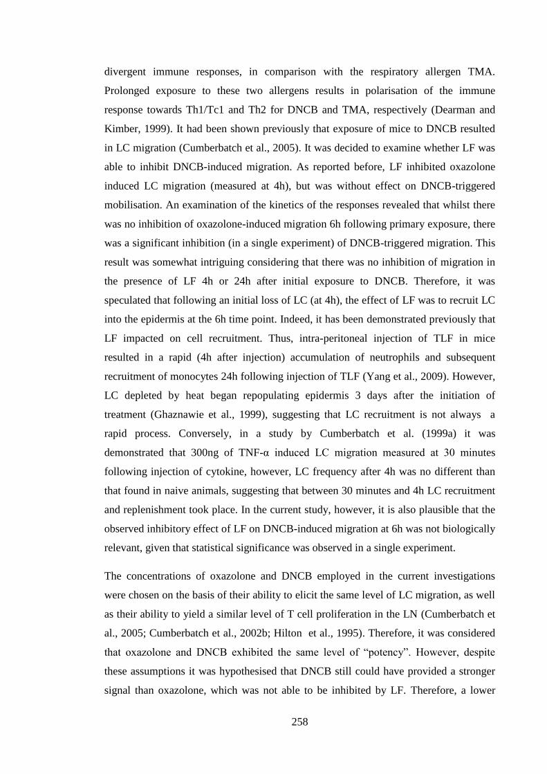
258
divergent immune responses, in comparison with the respiratory allergen TMA.
Prolonged exposure to these two allergens results in polarisation of the immune
response towards Th1/Tc1 and Th2 for DNCB and TMA, respectively (Dearman and
Kimber, 1999). It had been shown previously that exposure of mice to DNCB resulted
in LC migration (Cumberbatch et al., 2005). It was decided to examine whether LF was
able to inhibit DNCB-induced migration. As reported before, LF inhibited oxazolone
induced LC migration (measured at 4h), but was without effect on DNCB-triggered
mobilisation. An examination of the kinetics of the responses revealed that whilst there
was no inhibition of oxazolone-induced migration 6h following primary exposure, there
was a significant inhibition (in a single experiment) of DNCB-triggered migration. This
result was somewhat intriguing considering that there was no inhibition of migration in
the presence of LF 4h or 24h after initial exposure to DNCB. Therefore, it was
speculated that following an initial loss of LC (at 4h), the effect of LF was to recruit LC
into the epidermis at the 6h time point. Indeed, it has been demonstrated previously that
LF impacted on cell recruitment. Thus, intra-peritoneal injection of TLF in mice
resulted in a rapid (4h after injection) accumulation of neutrophils and subsequent
recruitment of monocytes 24h following injection of TLF (Yang et al., 2009). However,
LC depleted by heat began repopulating epidermis 3 days after the initiation of
treatment (Ghaznawie et al., 1999), suggesting that LC recruitment is not always a
rapid process. Conversely, in a study by Cumberbatch et al. (1999a) it was
demonstrated that 300ng of TNF-α induced LC migration measured at 30 minutes
following injection of cytokine, however, LC frequency after 4h was no different than
that found in naive animals, suggesting that between 30 minutes and 4h LC recruitment
and replenishment took place. In the current study, however, it is also plausible that the
observed inhibitory effect of LF on DNCB-induced migration at 6h was not biologically
relevant, given that statistical significance was observed in a single experiment.
The concentrations of oxazolone and DNCB employed in the current investigations
were chosen on the basis of their ability to elicit the same level of LC migration, as well
as their ability to yield a similar level of T cell proliferation in the LN (Cumberbatch et
al., 2005; Cumberbatch et al., 2002b; Hilton et al., 1995). Therefore, it was considered
that oxazolone and DNCB exhibited the same level of “potency”. However, despite
these assumptions it was hypothesised that DNCB still could have provided a stronger
signal than oxazolone, which was not able to be inhibited by LF. Therefore, a lower
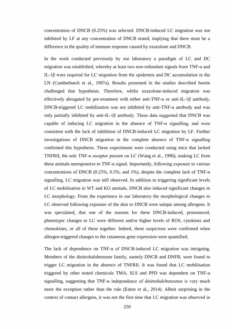
259
concentration of DNCB (0.25%) was selected. DNCB-induced LC migration was not
inhibited by LF at any concentration of DNCB tested, implying that there must be a
difference in the quality of immune response caused by oxazolone and DNCB.
In the work conducted previously by our laboratory a paradigm of LC and DC
migration was established, whereby at least two non-redundant signals from TNF-α and
IL-1β were required for LC migration from the epidermis and DC accumulation in the
LN (Cumberbatch et al., 1997a). Results presented in the studies described herein
challenged that hypothesis. Therefore, whilst oxazolone-induced migration was
effectively abrogated by pre-treatment with either anti-TNF-α or anti-IL-1β antibody,
DNCB-triggered LC mobilisation was not inhibited by anti-TNF-α antibody and was
only partially inhibited by anti-IL-1β antibody. These data suggested that DNCB was
capable of inducing LC migration in the absence of TNF-α signalling, and were
consistent with the lack of inhibition of DNCB-induced LC migration by LF. Further
investigations of DNCB migration in the complete absence of TNF-α signalling
confirmed this hypothesis. These experiments were conducted using mice that lacked
TNFRII, the sole TNF-α receptor present on LC (Wang et al., 1996), making LC from
these animals unresponsive to TNF-α signal. Importantly, following exposure to various
concentrations of DNCB (0.25%, 0.5%, and 1%), despite the complete lack of TNF-α
signalling, LC migration was still observed. In addition to triggering significant levels
of LC mobilisation in WT and KO animals, DNCB also induced significant changes in
LC morphology. From the experience in our laboratory the morphological changes to
LC observed following exposure of the skin to DNCB were unique among allergens. It
was speculated, that one of the reasons for these DNCB-induced, pronounced,
phenotypic changes to LC were different and/or higher levels of ROS, cytokines and
chemokines, or all of these together. Indeed, these suspicions were confirmed when
allergen-triggered changes to the cutaneous gene expression were quantified.
The lack of dependence on TNF-α of DNCB-induced LC migration was intriguing.
Members of the dinitrohalobenzene family, namely DNCB and DNFB, were found to
trigger LC migration in the absence of TNFRII. It was found that LC mobilisation
triggered by other tested chemicals TMA, SLS and PPD was dependent on TNF-α
signalling, suggesting that TNF-α independence of dinitrohalobenzenes is very much
more the exception rather than the rule (Eaton et al., 2014). Albeit surprising in the
context of contact allergens, it was not the first time that LC migration was observed in
260
the absence of TNF-α signalling. Byrne et al. (2001) reported that cutaneous West Nile
virus infection was able to trigger LC mobilisation in TNF-α deficient mice. However,
whilst West Nile virus-induced LC migration was independent of TNF-α, LC migration
from epidermis and accumulation in the LN was significantly inhibited by treatment
with anti-IL-1β antibody (Byrne et al., 2001), again consistent with the pattern for
dinitrohalobenzenes reported herein.
Thus, it has been established that DNCB and oxazolone exhibited differential cytokine
requirements with regards to LC migration. To gain more detailed understanding of the
processes that might be responsible for the divergence between these two allergens, the
cutaneous gene expression profiles triggered by allergen exposure were investigated
over the period of 24h. As previously described, qPCR was utilized to investigate the
changes in gene expression in comparison with untreated samples. It has been
established previously using PCR and liquid hybridization (whereby PCR products
underwent electrophoresis and subsequently were incubated with complementary
primers, tagged with radioactive phosphorus) that shortly (within 90 minutes) after
exposure to 3% TNCB expression of Tnf-α, Il-1β and Cxcl10 was observed (Enk and
Katz, 1992a). TNCB is widely used in the investigations of ACD, however, it is
impossible to utilize this chemical in our laboratory due to safety risks, as TNCB is an
explosive (Parihar et al., 1967). In the current investigations substantial induction of
Tnf-α expression was not observed. The discrepancy between two studies could stem
from the fact that different stimulants and detection methods were utilized. However,
rapid expression of IL-1β was demonstrated in the study reported herein. There were
marked differences observed between cytokine levels induced by oxazolone and DNCB.
Thus, it was established that DNCB induced significantly higher cutaneous expression
of Tnf-α, Il-1β, Cxcl1, Icam-1 and Il-17F, suggesting that these higher cytokine signals
might be responsible for the lack of DNCB dependence upon TNF-α. Consistent with
the gene expression data, it was found that DNCB triggered higher levels of IL-1β
protein release. To account for that fact that different concentrations of oxazolone and
DNCB were used (despite triggering the same extent of LC migration), the ability of
high and low concentrations of both oxazolone and DNCB to induce cytokine
production and release was considered. It was found that although there were no
differences in the release of TNF-α and IL-1α there was a significant divergence in the
level of IL-1β release. Interestingly, it was observed that exposure to a high (3%)
261
concentration of oxazolone resulted in a similar level of IL-1β release as that triggered
by both concentrations of DNCB. This result suggested two things. First, that IL-1β
might be the mediator of TNF-α independence of DNCB, due to its high levels in
DNCB-treated skin, and a close resemblance of LC morphology between DNCB and
IL-1β exposure. Secondly, it could be speculated that if IL-1β was responsible for TNF-
α independent DNCB-induced LC migration, therefore, a high (3%) concentration of
oxazolone, that induced similar vigorous IL-1β production, could also trigger TNF-α
independent LC migration. To test this hypothesis, LC migration was assessed
following exposure to 0.5% and 3% oxazolone in the presence and absence of LF. It
was found that pre-treatment with LF significantly inhibited oxazolone induced LC
migration (using the standard, low dose of oxazolone), which was demonstrated to be a
result of inhibition of de novo TNF-α production (Cumberbatch et al., 2000b). The dose
effect of oxazolone could not be reconciled on the basis of differential TNF-α
expression, although it should be noted that marked allergen-induced TNF-α release
was not observed during these experiments. Therefore, it was hypothesised, that the
failure of LF to inhibit LC migration provoked by the high concentration (3%) of
oxazolone was due to higher IL-1β levels present in the skin. These results implied that
when a high concentration of IL-1β is present, TNF-α signalling might be redundant for
LC mobilisation.
The C57BL/6 TNFRII KO strain mice could not be used to investigate the effect of 3%
oxazolone on TNF-α independent LC migration due to the inability of C57BL/6 strain
mice to respond robustly to oxazolone with LC migration (PhD thesis Eaton, 2012).
There have been numerous reports demonstrating differences between BALB/c and
C57BL/6 strain mice. Overall, is it considered that BALB/c and C57BL/6 strain mice
have a higher propensity to exhibit Th2 and Th1 driven conditions, respectively
(Watanabe et al., 2004). Oxazolone was identified previously as a contact allergen on
the basis of its failure to trigger IgE production following an appropriate dosing regimen
involving 2 exposures over a 14 day period (Dearman and Kimber, 1992). However, in
another study utilising Brown Norway rats, oxazolone was characterized as a potential
respiratory allergen capable of inducing IgE production and gene expression
comparable to that triggered by TMA (Kuper et al., 2011). Furthermore, oxazolone was
employed for development of colitis in C57BL/10 mice, which was driven by Th2
response and was dependent on NK-cell derived IL-13 (Heller et al., 2002). On the
262
contrary, another report established that oxazolone was unable to induce IgE production
and demonstrated that intranasal sensitization and challenge with oxazolone did not
result in allergic rhinitis (Farraj et al., 2004). There are no reports regarding oxazolone
allergy in humans, due to lack of significant exposure. Although there are anecdotal
reports of ACD to oxazolone among laboratory workers (personal communication, Prof.
Ian Kimber), however, it is difficult to assess what properties oxazolone-induced
sensitivity would have in humans. Nonetheless, despite some reports suggesting that
oxazolone could be considered as a respiratory allergen, prevalent consensus exists that
oxazolone is in fact a contact allergen.
Despite classification of oxazolone as a contact sensitizer, reports suggesting that
oxazolone is a respiratory sensitizer, and therefore would induce Th2-skewed immune
response, could partially explain why oxazolone is unable to induce sufficient LC
migration in C57BL/6 strain mice (PhD thesis Eaton, 2012), which are known to
preferentially mount Th1-dependent responses (Watanabe et al., 2004). In their study
Watanabe et al. (2004) investigated responses of macrophages derived from both mouse
strains and found that C57BL/6 strain mice produced higher levels of TNF-α and IL-12
in response to LPS than BALB/c strain mice, which in turn can lead to Th1 polarized
response in C57BL/6 strain mice (Watanabe et al., 2004). Conversely, oxazolone
readily triggered LC mobilisation in BALB/c strain mice, which were demonstrated to
be more prone to Th2 polarized responses (Watanabe et al., 2004). These two mouse
strains were demonstrated to differ with regards to TLR expression by their respective
DC, which explained their divergent susceptibility to Listeria monocytogenes infection
(Liu et al., 2002), and could potentially be the reason for differential responses to
oxazolone. Naive DC isolated from spleens of C57BL/6 and BALB/c strain mice
preferentially expressed TLR9 and TLR 2, 4 and 6, respectively (Liu et al., 2002). TLR
2 and 4 were shown previously to be required for the development of CHS reactions in
response to TNCB (Martin et al., 2008). Therefore, the higher degree of expression of
these TLR receptors on DC from BALB/c strain mice could explain the ability of LC
from these mice to readily migrate from the epidermis following exposure to oxazolone.
Another reason for the divergence between mice strains with respect to their response to
oxazolone might be the fact that C57BL/6 strain mice have naturally disrupted gene
encoding phospholipase A2 (PLA2) (Kennedy et al., 1995). Indeed, PLA2 was
demonstrated to be expressed in the skin of both mouse and human, and was implicated
263
in both maintaining the barrier function of epidermis via its involvement in fatty acid
metabolism, as well as in the inflammatory response due to participation in synthesis of
arachidonic acid (Ilic et al. 2014). In fact, it was reported that overexpression of human
PLA2 in mice resulted in a phenotype that corresponds with ACD (Sato et al., 2009).
Therefore, differences with respect to PLA2 expression might be another reason for
differential responses of BALB/c and C57BL/6 strain mice to oxazolone. Moreover,
PLA2 potentially might be involved in a signalling cascade triggered by oxazolone that
leads to LC mobilisation. Lastly, as oxazolone-induced LC migration is dependent on
TNF-α there is a possibility that there are differences between the two mice strains with
respect to their ability to produce and/or respond to TNF-α in the skin. However, data
published by Friedrich et al. (2014) showed that in the steady state the amount of TNF-α
present in the skin of BALB/c and C57BL/6 strain mice was comparable, ~12pg/mg of
protein. Significant increase in TNF-α level was observed in C57BL/6 but not in
BALB/c strain mice following exposure to UV light. However, detected levels of TNF-
α were below the limit of accurate detection of the ELISA kit that was utilized in the
study by Friedrich et al. (2014).
To summarise, it was demonstrated, in the current investigations that under conditions
where high levels of IL-1β were present LC migration could take place in the absence of
TNF-α signalling. Interestingly, Haley et al. (2007) demonstrated that mice that lack
MyD88 (an adaptor molecule for IL-1R1) still display the same level of LC migration in
the steady state, as measured by LC accumulation in the draining LN; as well as
following exposure to TRITC. Tetramethylrhodamine has been used, similarly to FITC,
as a stimulator of LC migration (Haley et al., 2012). It could be argued that in the study
by Haley et al. (2012), the trigger that stimulated LC migration was different to that
utilized in the current investigations, therefore partially accounting for differences.
Moreover, in the current study it was shown that presence of IL-1β was required, but it
is not known if this cytokine acts directly upon LC or if it induces the production of
another mediator, e.g. CXCL1. While the study presented herein was not in complete
agreement with evidence proposed by Haley et al. (2010), it was consistent with the
report by Antonopoulos et al. (2001) in which a critical role for caspase 1 was
demonstrated, both in the events of LC migration, and CHS measured by challenge-
induced changes in ear swelling (Antonopoulos et al., 2001). Caspase 1 is a cysteine
264
protease that participates in the conversion pro-IL-1β into its active form (Thornberry et
al., 1992).
One of the difficulties in interpreting the results from the current investigations in the
context of previously published data is that it is relatively rare for previous publications
to have employed both of these allergens. One such study that utilized both oxazolone
and DNCB was conducted by Traidl et al. (1999), wherein the dependence of CHS
reaction on IL-4 signalling triggered by both allergens was tested. They found that the
CHS reaction, as measured by challenge-induced changes in ear swelling, triggered by
DNCB was attenuated in IL-4 KO mice. Interestingly, when ear swelling was triggered
by oxazolone, there was no difference in the hypersensitivity manifestation between WT
and KO animals (Traidl et al., 1999), suggesting a differential reliance of these two
allergens on IL-4 signalling. In previous work, in two independent studies, another
difference between the two allergens was identified. The first study demonstrated that
the extent of the CHS reaction to oxazolone was similar in WT and IL-1β deficient mice
(Zheng et al., 1995). In the second study the same experiment was conducted by
Shornick et al. (1996), but on this occasion TNCB, a chemical different from DNCB by
one nitro group, was utilized. It was found that when low doses of TNCB (0.03%, 0.1%,
0.3%) were selected for sensitization, there were visible differences between WT and
IL-1β KO animals; namely, the CHS reaction was attenuated in the absence of IL-1β.
However, when the concentration of the sensitizing dose was increased to 1% or 3% the
divergence that was observed b previously etween IL-1β KO and WT mice was no
longer present (Shornick et al., 1996). That is, low dose of sensitizer was dependent
upon IL-1β, whereas, high dose was independent. These results from IL-1β deficient
mice are of particular interest, as evidence from the current study suggests that one of
the differences between oxazolone and DNCB might be their differential ability to
induce IL-1β production in the skin. While oxazolone induced LC mobilisation was
dependent on IL-1β, data presented by Zheng et al. (1995) proposed that IL-1β was not
essential for the development of CHS. However, it has to be noted that concentrations
used in that study were very high (4% and 1% of oxazolone for sensitization and
elicitation, respectively). Therefore, it is possible that whilst in the experiments reported
herein inhibition of oxazolone-induced LC migration was observed following anti-IL-1β
injection, if the higher concentration of oxazolone had been used, the inhibitory effect
of anti-IL-1β antibody might have been overcome. Conversely, the study by Shornick et
265
al. (1996) reported a necessity for IL-1β signalling to achieve an optimum CHS reaction
in response to TNCB. However, the need for IL-1β was overcome by the use of a higher
concentration of allergen, thereby allowing IL-1β KO mice to mount the same level of
CHS measured by footpad swelling. The approach taken by Shornick et al. (1996) was
similar to that used in the investigations described herein, whereby it was demonstrated
that the requirement for TNF-α signalling was no longer apparent when a higher
concentration of IL-1β was present. The data presented in this chapter, as well as that
reported by Shornick et al. (1996), suggest that there might be compensatory
mechanisms in place within the immune system that ascertain, that if the danger signal
is vigorous enough, there will be an appropriate response, even in the absence of one of
the key cytokines: IL-1β or TNF-α. These results also further stress the need to critically
assess the data derived from chemical allergens with respect to both the identity and the
dose of chemical allergen. It is clear that despite many similarities, major differences
exist in the quality of induced immune response.
It is clear from this study and from the literature that IL-1β and TNF-α play key, albeit
still not fully elucidated, roles in the mobilisation of LC and in CHS. An attempt to
investigate the exact roles of TNF-α and IL-1 in the events of CHS was made by (Nakae
et al., 2003). They found that IL-1 and TNF-α had distinct roles in the sensitization and
elicitation phases of CHS. It was found that there was a comparable suppression of CHS
responses to TNCB and oxazolone in both C57BL/6 and BALB/c strain mice that
lacked either IL-1α/β or TNF-α in comparison to WT counterparts. However, when
triple KO mice were utilized the suppression was even greater but not complete. Similar
observations were made when the presence of FITC positive cells was assessed in the
draining LN, representing presumably antigen-bearing DC. Thus, IL-1α/β or TNF-α KO
mice displayed decreased levels of CD11c+FITC
+ cells (approximately 26%) in
comparison to WT (44%), but when triple KO mice were utilized migration was almost
completely absent and antigen bearing DC levels were around 3%; therefore suggesting
the existence of compensatory mechanisms in the case of absence of one of the
cytokines. Additionally, there were differences between cytokines in relation to their
role in the T cell activation. When LN cells from sensitized animals were cultured with
haptenated spleen cells, a lower level of activation was observed in cells derived from
IL-1α/β KO mice, whereas cells isolated from TNF-α KO mice displayed levels of
activation comparable to those observed in WT animals (measured by radiolabelled
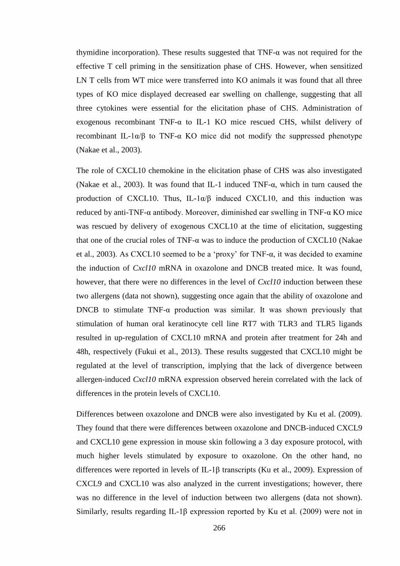
266
thymidine incorporation). These results suggested that TNF-α was not required for the
effective T cell priming in the sensitization phase of CHS. However, when sensitized
LN T cells from WT mice were transferred into KO animals it was found that all three
types of KO mice displayed decreased ear swelling on challenge, suggesting that all
three cytokines were essential for the elicitation phase of CHS. Administration of
exogenous recombinant TNF-α to IL-1 KO mice rescued CHS, whilst delivery of
recombinant IL-1α/β to TNF-α KO mice did not modify the suppressed phenotype
(Nakae et al., 2003).
The role of CXCL10 chemokine in the elicitation phase of CHS was also investigated
(Nakae et al., 2003). It was found that IL-1 induced TNF-α, which in turn caused the
production of CXCL10. Thus, IL-1α/β induced CXCL10, and this induction was
reduced by anti-TNF-α antibody. Moreover, diminished ear swelling in TNF-α KO mice
was rescued by delivery of exogenous CXCL10 at the time of elicitation, suggesting
that one of the crucial roles of TNF-α was to induce the production of CXCL10 (Nakae
et al., 2003). As CXCL10 seemed to be a ‘proxy’ for TNF-α, it was decided to examine
the induction of Cxcl10 mRNA in oxazolone and DNCB treated mice. It was found,
however, that there were no differences in the level of Cxcl10 induction between these
two allergens (data not shown), suggesting once again that the ability of oxazolone and
DNCB to stimulate TNF-α production was similar. It was shown previously that
stimulation of human oral keratinocyte cell line RT7 with TLR3 and TLR5 ligands
resulted in up-regulation of CXCL10 mRNA and protein after treatment for 24h and
48h, respectively (Fukui et al., 2013). These results suggested that CXCL10 might be
regulated at the level of transcription, implying that the lack of divergence between
allergen-induced Cxcl10 mRNA expression observed herein correlated with the lack of
differences in the protein levels of CXCL10.
Differences between oxazolone and DNCB were also investigated by Ku et al. (2009).
They found that there were differences between oxazolone and DNCB-induced CXCL9
and CXCL10 gene expression in mouse skin following a 3 day exposure protocol, with
much higher levels stimulated by exposure to oxazolone. On the other hand, no
differences were reported in levels of IL-1β transcripts (Ku et al., 2009). Expression of
CXCL9 and CXCL10 was also analyzed in the current investigations; however, there
was no difference in the level of induction between two allergens (data not shown).
Similarly, results regarding IL-1β expression reported by Ku et al. (2009) were not in
267
agreement with those demonstrated in the current study. These discrepancies were likely
to be caused by a different mouse strains utilized by Ku et al. (2009) and the current
study, which were CBA/N and BALB/c strain mice, respectively. In addition, the
exposure protocol utilized by Ku et al. (2009) and in the current studies was different. In
the studies described herein mice were exposed to allergen once for various times,
whereas in the study by Ku et al. (2009) animals were exposed to allergens 3 times on
day 0, 1 and 2 and samples were collected 24h after last exposure. Additionally, the
dose of oxazolone utilized in the experiments described here was lower (0.5%) than that
used by Ku et al. (2009) (1%), and as mentioned in the current studies and in the
investigations by Shornick et al. (1996), the dose of allergen exposure can dramatically
change the quality of immune response to allergens.
Langerhans’ cell migration in response to contact allergens was believed previously to
depend upon receipt of two signals by LC, TNF-α and IL-1β. Contact allergens were
considered to be one group of chemicals that have the ability to cause ACD but do not
generally display respiratory sensitising potential. Contact allergens are distinct from a
smaller group of chemicals, respiratory allergens that display a different clinical profile
and cause sensitization of the respiratory tract. These two groups of allergens (contact
and respiratory) exhibit differences with regards to the quality of immune response that
they trigger; namely Th1/Tc1 and Th2, respectively (Dearman and Kimber, 1992). The
study presented herein has exposed that there is a divergence within, the previously
considered to be uniform, contact allergen group that is exemplified by differential
effects on LC migration. These differences are probably due to a divergent pattern of
induction of gene expression elicited by chemicals that in turn might be a consequence
of the utilization of differential signalling pathways. The exact mode of action of
oxazolone and DNCB is still not known. One of the only allergens for which the exact
mechanism has been fully established is nickel. It was recently discovered, that in the
skin, it directly binds to TLR4 receptor within histidine residues 456 and 458, therefore,
inducing signalling cascade that results in sensitization (Schmidt et al., 2010).
The pattern of gene expression of some other chemicals that were employed in the
current study was investigated to ascertain if any conclusions could be drawn between
the pattern of cutaneous gene expression and ability to result in LC mobilisation. All of
the chemicals used, with the exception of DNTB, were demonstrated previously to
trigger LC migration. However, a recent study challenged the notion that DNTB does
268
not trigger LC migration. Gomez de Aguero et al., (2012) measured the number of
epidermal LC in the draining LN following exposure to DNTB. Mice used in their study
expressed enhanced green fluorescent protein (EGFP) under control of the langerin
promoter. Furthermore, DC were visualised by exposure of the skin to TRITC. Lymph
node cells were further stained with anti-CD103 and anti-CD207 antibodies to allow for
LC identification that would be EGFP, TRITC and CD207 positive and CD103
negative. By employing this approach de Aguero et al. (2012) demonstrated the
presence of low number of LC (1% of TRITC+ cells) in the draining LN 24h following
exposure to DNTB that increased 72h after (3% of TRITC+ cells), which corresponded
with the peak LC migration to the draining LN. Moreover, they reported that LC were
responsible for DNTB-induced protection against CHS after treatment with DNFB
(Gomez de Agüero et al., 2012). The main differences observed between chemicals
were with regards to Il-1β and Cxcl1 expression. Levels of Il-1β and Cxcl1 peaked
rapidly after exposure to SLS, which corresponds with the pattern of expression of these
genes observed following exposure to oxazolone and DNCB; with somewhat later peaks
recorded for DNTB and TMA.
Cytokine expression data in combination with the knowledge about LC migration
allows for speculation regarding a connection between the two processes. Indeed, early
Il-1β and Cxcl1 induction correlates with LC migration observed 4h after exposure to
SLS. However, it is not known if LC mobilisation could be observed sooner. TMA
triggered LC migration occurs 17h after exposure (Cumberbatch et al., 2005) and this
delayed migration correlated with delayed cytokine expression data. Intriguingly, there
was a clear division between the tempo of Cxcl1 and Il-1β signals for TMA which
peaked after 16h and 24h, respectively. Putting together the information regarding LC
migration and the kinetics of induction of cytokine production, it could be speculated
that LC migration following exposure to DNTB could potentially be expected
approximately 8h after initiation of treatment due to the induction of Cxcl1 and Il-1β.
The role of IL-1β was demonstrated previously to be crucial in mounting a complete
CHS in response to TNCB (Shornick et al., 2001; Shornick et al., 1996), most likely due
to the more recently discovered role of inflammasome as a master switch between
establishing tolerance and sensitization (Watanabe et al., 2008). The results obtained in
the current investigations allow speculation regarding the role of CXCL1 in the
sensitization process. Indeed, it has been demonstrated herein that CXCL1 was
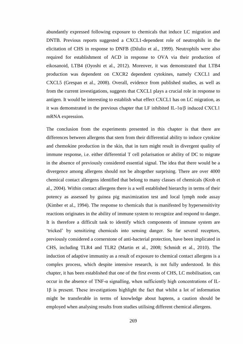
269
abundantly expressed following exposure to chemicals that induce LC migration and
DNTB. Previous reports suggested a CXCL1-dependent role of neutrophils in the
elicitation of CHS in response to DNFB (Dilulio et al., 1999). Neutrophils were also
required for establishment of ACD in response to OVA via their production of
eikosanoid, LTB4 (Oyoshi et al., 2012). Moreover, it was demonstrated that LTB4
production was dependent on CXCR2 dependent cytokines, namely CXCL1 and
CXCL5 (Grespan et al., 2008). Overall, evidence from published studies, as well as
from the current investigations, suggests that CXCL1 plays a crucial role in response to
antigen. It would be interesting to establish what effect CXCL1 has on LC migration, as
it was demonstrated in the previous chapter that LF inhibited IL-1α/β induced CXCL1
mRNA expression.
The conclusion from the experiments presented in this chapter is that there are
differences between allergens that stem from their differential ability to induce cytokine
and chemokine production in the skin, that in turn might result in divergent quality of
immune response, i.e. either differential T cell polarisation or ability of DC to migrate
in the absence of previously considered essential signal. The idea that there would be a
divergence among allergens should not be altogether surprising. There are over 4000
chemical contact allergens identified that belong to many classes of chemicals (Krob et
al., 2004). Within contact allergens there is a well established hierarchy in terms of their
potency as assessed by guinea pig maximization test and local lymph node assay
(Kimber et al., 1994). The response to chemicals that is manifested by hypersensitivity
reactions originates in the ability of immune system to recognize and respond to danger.
It is therefore a difficult task to identify which components of immune system are
‘tricked’ by sensitizing chemicals into sensing danger. So far several receptors,
previously considered a cornerstone of anti-bacterial protection, have been implicated in
CHS, including TLR4 and TLR2 (Martin et al., 2008; Schmidt et al., 2010). The
induction of adaptive immunity as a result of exposure to chemical contact allergens is a
complex process, which despite intensive research, is not fully understood. In this
chapter, it has been established that one of the first events of CHS, LC mobilisation, can
occur in the absence of TNF-α signalling, when sufficiently high concentrations of IL-
1β is present. These investigations highlight the fact that whilst a lot of information
might be transferable in terms of knowledge about haptens, a caution should be
employed when analysing results from studies utilising different chemical allergens.
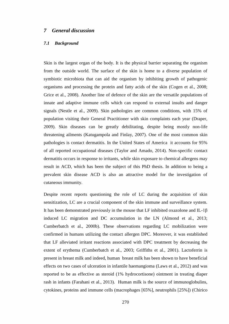
270
7 General discussion
7.1 Background
Skin is the largest organ of the body. It is the physical barrier separating the organism
from the outside world. The surface of the skin is home to a diverse population of
symbiotic microbiota that can aid the organism by inhibiting growth of pathogenic
organisms and processing the protein and fatty acids of the skin (Cogen et al., 2008;
Grice et al., 2008). Another line of defence of the skin are the versatile populations of
innate and adaptive immune cells which can respond to external insults and danger
signals (Nestle et al., 2009). Skin pathologies are common conditions, with 15% of
population visiting their General Practitioner with skin complaints each year (Draper,
2009). Skin diseases can be greatly debilitating, despite being mostly non-life
threatening ailments (Katugampola and Finlay, 2007). One of the most common skin
pathologies is contact dermatitis. In the United States of America it accounts for 95%
of all reported occupational diseases (Taylor and Amado, 2014). Non-specific contact
dermatitis occurs in response to irritants, while skin exposure to chemical allergens may
result in ACD, which has been the subject of this PhD thesis. In addition to being a
prevalent skin disease ACD is also an attractive model for the investigation of
cutaneous immunity.
Despite recent reports questioning the role of LC during the acquisition of skin
sensitization, LC are a crucial component of the skin immune and surveillance system.
It has been demonstrated previously in the mouse that LF inhibited oxazolone and IL-1β
induced LC migration and DC accumulation in the LN (Almond et al., 2013;
Cumberbatch et al., 2000b). These observations regarding LC mobilization were
confirmed in humans utilizing the contact allergen DPC. Moreover, it was established
that LF alleviated irritant reactions associated with DPC treatment by decreasing the
extent of erythema (Cumberbatch et al., 2003; Griffiths et al., 2001). Lactoferrin is
present in breast milk and indeed, human breast milk has been shown to have beneficial
effects on two cases of ulceration in infantile haemangioma (Laws et al., 2012) and was
reported to be as effective as steroid (1% hydrocortisone) ointment in treating diaper
rash in infants (Farahani et al., 2013). Human milk is the source of immunoglobulins,
cytokines, proteins and immune cells (macrophages [65%], neutrophils [25%]) (Chirico
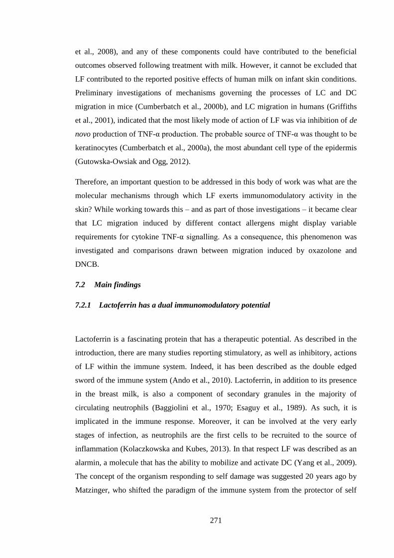
271
et al., 2008), and any of these components could have contributed to the beneficial
outcomes observed following treatment with milk. However, it cannot be excluded that
LF contributed to the reported positive effects of human milk on infant skin conditions.
Preliminary investigations of mechanisms governing the processes of LC and DC
migration in mice (Cumberbatch et al., 2000b), and LC migration in humans (Griffiths
et al., 2001), indicated that the most likely mode of action of LF was via inhibition of de
novo production of TNF-α production. The probable source of TNF-α was thought to be
keratinocytes (Cumberbatch et al., 2000a), the most abundant cell type of the epidermis
(Gutowska-Owsiak and Ogg, 2012).
Therefore, an important question to be addressed in this body of work was what are the
molecular mechanisms through which LF exerts immunomodulatory activity in the
skin? While working towards this – and as part of those investigations – it became clear
that LC migration induced by different contact allergens might display variable
requirements for cytokine TNF-α signalling. As a consequence, this phenomenon was
investigated and comparisons drawn between migration induced by oxazolone and
DNCB.
7.2 Main findings
7.2.1 Lactoferrin has a dual immunomodulatory potential
Lactoferrin is a fascinating protein that has a therapeutic potential. As described in the
introduction, there are many studies reporting stimulatory, as well as inhibitory, actions
of LF within the immune system. Indeed, it has been described as the double edged
sword of the immune system (Ando et al., 2010). Lactoferrin, in addition to its presence
in the breast milk, is also a component of secondary granules in the majority of
circulating neutrophils (Baggiolini et al., 1970; Esaguy et al., 1989). As such, it is
implicated in the immune response. Moreover, it can be involved at the very early
stages of infection, as neutrophils are the first cells to be recruited to the source of
inflammation (Kolaczkowska and Kubes, 2013). In that respect LF was described as an
alarmin, a molecule that has the ability to mobilize and activate DC (Yang et al., 2009).
The concept of the organism responding to self damage was suggested 20 years ago by
Matzinger, who shifted the paradigm of the immune system from the protector of self
272
from non-self to the defender of self from danger, regardless of its source (Matzinger,
1994).
In the study described herein and in other publications, LF has been shown to interact
with surface nucleolin. Among its other roles, surface nucleolin has been described as
the receptor for apoptotic cells (Hirano et al., 2005) and oxidized red blood cells (Miki
et al., 2007) via recognition of oxidized carbohydrate chains (Miki et al., 2014; Miki et
al., 2012). As such, nucleolin is a scavenger receptor implicated in eliminating danger
signals. It was observed in the investigations described herein that following exposure
to LF 72h THP-1 macrophages released TNF-α and IL-8 in a nucleolin-dependent
fashion. It has not been reported whether recognition of other substrates by surface
nucleolin is associated with inflammatory cytokine release. Rather, it was found that
phagocytosis of apoptotic cells had an anti-inflammatory potential via the release of
TGF-β and PGE2 by macrophages (Fadok et al., 1998). Further, apoptotic cells were
demonstrated to increase IL-10 and decrease TNF-α secretion by PBMC following 16h
incubation with LPS. Anti-CD36 antibody (CD36 is a phagocytic receptor) was found
to mimic the suppressive effect of apoptotic cells, whereas blocking of phagocytosis
decreased the inhibitory effects of apoptotic cells (Voll et al., 1997). Lactoferrin was
recognized by the same receptor as apoptotic cells; however, as described herein, its
effects were found to be immunostimulatory when incubated with 72h THP-1
macrophages. Conversely, administration of LF in vivo inhibited oxazolone-triggered
LC mobilization potentially via decrease of de novo TNF-α production. It is possible
that the fact that LF is found in the secondary neutrophil granules influences the way it
is recognized by the immune system and the way that cells respond to it. Indeed,
apoptotic cells release variety of ‘find me’ factors, including ATP and uridine-5'-
triphosphate (UTP) (Elliott et al., 2009), CX3CL1 (Truman et al., 2008) and LF
(Bournazou et al., 2009), that influence the movement of various types of cells
(Willems et al., 2014). Hence, in the in vivo model of oxazolone induced migration, the
presence of LF could be perceived as though neutrophils undergoing apoptosis are
present and thus the anti-inflammatory pathway is activated. In contrast, in the in vitro
THP-1 model the concentration of LF could have been too high to mimic apoptotic cells
and was recognized as a danger signal, to which macrophages responded with the
release of inflammatory mediators. Therefore, it could be speculated that function of LF
depends on both the context and concentration. Indeed, when high, pathologically
273
relevant, concentrations of LF (10-100μg/ml) are present, DC activation was
demonstrated (de la Rosa et al., 2008; Spadaro et al., 2008). Therefore, high LF
concentrations, which are symptomatic of neutrophil degranulation, aid the adaptive
immune response. At higher concentrations another ‘find me’ signal, ATP, is
considered to be pro-inflammatory (Kono and Rock, 2008), therefore, it is possible that
LF could work in a similar manner. High concentrations of LF could indicate to DC that
the situation is no longer suitable for phagocyte recruitment and instead initiation of
inflammation is required.
Expression of surface nucleolin by LC or keratinocytes has not been reported in the
literature to date. Consequently, it is difficult to speculate whether the effects of LF in
vivo are mediated via this receptor. A receptor for LF has been identified on activated
human PBMC including γδ T cells (Mincheva-Nilsson et al., 1997), which is consistent
with the data reported herein, that LF inhibited IL-1β induced IL-17F expression in the
skin, the latter cytokine probably being derived from γδ T cells (Gray et al., 2011; PhD
thesis Hayes, 2012; Nielsen et al., 2014).
7.2.2 Keratinocytes might not be the source of TNF-α during the process of LC
migration
Keratinocytes are the main cell population in the epidermis and have been demonstrated
to be a rich source of cytokines and chemokines (Gröne, 2002). It has been speculated
that during skin sensitization IL-1β and TNF-α cytokine signals that are required for LC
migration derive from LC and keratinocytes, respectively (Enk et al., 1993; Enk and
Katz, 1992a). Therefore, to determine whether LF could regulate TNF-α production by
keratinocytes, in vitro keratinocytes from three different sources were utilized to
measure the impact of LF on IL-1α and IL-1β induced gene expression. Keratinocytes
did not readily secrete TNF-α (PAM and HaCaT cells) or secreted only low amounts of
the cytokine (primary keratinocyes), suggesting that they might not be the primary
source of this cytokine in vivo. Tumour necrosis factor α was detected at higher levels in
skin explants following in vivo exposure to oxazolone, however, the cytokine was most
likely produced due to the trauma of explant preparation rather than in response to
allergen. Indeed, treatment with oxazolone did not result in TNF-α release above the
levels found in the naive ear explant samples. The expression of TNF-α mRNA by
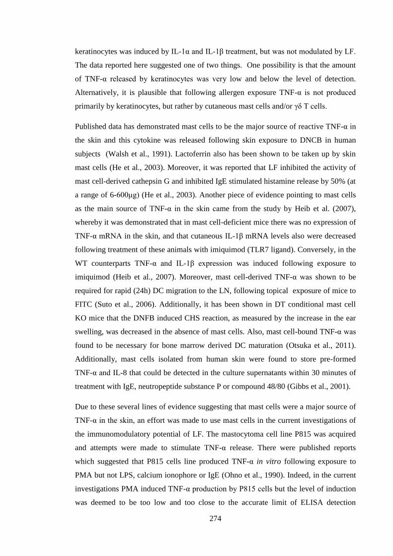
274
keratinocytes was induced by IL-1α and IL-1β treatment, but was not modulated by LF.
The data reported here suggested one of two things. One possibility is that the amount
of TNF-α released by keratinocytes was very low and below the level of detection.
Alternatively, it is plausible that following allergen exposure TNF-α is not produced
primarily by keratinocytes, but rather by cutaneous mast cells and/or γδ T cells.
Published data has demonstrated mast cells to be the major source of reactive TNF-α in
the skin and this cytokine was released following skin exposure to DNCB in human
subjects (Walsh et al., 1991). Lactoferrin also has been shown to be taken up by skin
mast cells (He et al., 2003). Moreover, it was reported that LF inhibited the activity of
mast cell-derived cathepsin G and inhibited IgE stimulated histamine release by 50% (at
a range of 6-600μg) (He et al., 2003). Another piece of evidence pointing to mast cells
as the main source of TNF-α in the skin came from the study by Heib et al. (2007),
whereby it was demonstrated that in mast cell-deficient mice there was no expression of
TNF-α mRNA in the skin, and that cutaneous IL-1β mRNA levels also were decreased
following treatment of these animals with imiquimod (TLR7 ligand). Conversely, in the
WT counterparts TNF-α and IL-1β expression was induced following exposure to
imiquimod (Heib et al., 2007). Moreover, mast cell-derived TNF-α was shown to be
required for rapid (24h) DC migration to the LN, following topical exposure of mice to
FITC (Suto et al., 2006). Additionally, it has been shown in DT conditional mast cell
KO mice that the DNFB induced CHS reaction, as measured by the increase in the ear
swelling, was decreased in the absence of mast cells. Also, mast cell-bound TNF-α was
found to be necessary for bone marrow derived DC maturation (Otsuka et al., 2011).
Additionally, mast cells isolated from human skin were found to store pre-formed
TNF-α and IL-8 that could be detected in the culture supernatants within 30 minutes of
treatment with IgE, neutropeptide substance P or compound 48/80 (Gibbs et al., 2001).
Due to these several lines of evidence suggesting that mast cells were a major source of
TNF-α in the skin, an effort was made to use mast cells in the current investigations of
the immunomodulatory potential of LF. The mastocytoma cell line P815 was acquired
and attempts were made to stimulate TNF-α release. There were published reports
which suggested that P815 cells line produced TNF-α in vitro following exposure to
PMA but not LPS, calcium ionophore or IgE (Ohno et al., 1990). Indeed, in the current
investigations PMA induced TNF-α production by P815 cells but the level of induction
was deemed to be too low and too close to the accurate limit of ELISA detection
275
(~50pg/ml) to be further modulated with LF (data not shown). In agreement with results
by Ohno et al. (1990), no TNF-α production was observed following IgE treatment.
Furthermore, there was no induction of TNF-α expression after treatment with IL-1α or
IL-1β.
It could be speculated that primary mast cells or bone marrow derived mast cells would
have been more efficient in their production of TNF-α, and therefore could have been a
better system in which potential of LF was investigated. As previously mentioned,
TNF-α is able to remain in a membrane bound form. Indeed, it was shown that direct
contact between mast cells and DC was required for DC maturation (Otsuka et al.,
2011). It is possible that TNF-α produced by P815 cell line was bound to the cell
membrane and not released into the culture medium, and could have been detected
using whole cell lysates in the ELISA analysis. Nevertheless, such experiments were
not conducted.
However, the suggestion that the TNF-α is pivotal in the events of LC migration might
come from mast cell stores is contradicted by time course experiments using exogenous
cytokine. Injection of recombinant TNF-α resulted in IL-1β dependent LC migration
within 30 minutes, suggesting that IL-1β was already present in the cell and did not
have to be synthesized. Whereas injection of recombinant IL-1β resulted in TNF-α-
dependent LC migration after 2h, suggesting that TNF-α may be produced de novo
(Cumberbatch et al., 1997a). Degranulation of mast cells, as observed by decreased
chymase staining in the skin, was one of the first changes observed after exposure to
allergen. In the human skin it was reported to take place as soon 1h after DNCB
application (Waldorf et al., 1991). However, it is possible that despite the rapid release
of mast cell contents, the 2h delay with respect to the initiation of LC migration
observed by Cumberbatch et al. (1997) was a result of the diffusion of TNF-α from the
dermis to the epidermis. Alternatively, release of TNF-α by mast cells in response to
exogenous IL-1β could be secondary to stimulation with a mediator induced by IL-1β
and not by IL-1β itself. Equally, there is some evidence which suggested that
keratinocytes were the source of TNF-α. In preliminary experiments, although
keratinocytes did not secrete TNF-α, high levels of TNF-α mRNA induction were
detected following stimulation in vitro with cytokines relevant in the events of LC
migration.
276
Murine epidermal γδ T cells have been shown to express TNF-α following activation
with a mitogen, concanavalin A (Matsue et al., 1993b). Similarly, human dermal γδ T
cells secreted large quantities of TNF-α following incubation with PMA and ionomycin
(Ebert, 2006). In the epidermis DETC cells are in close proximity to LC and through
production of TNF-α could efficiently activate adjacent LC. As mentioned previously,
TNF-α is found in both soluble and membrane bound forms. It has been found that
membrane bound form of TNF-α binds TNFRII more strongly than does the soluble
cytokine, and it has been suggested that full activation of TNFRII is achieved only via
interaction with membrane bound TNF-α (Faustman and Davis, 2010; Grell et al.,
1995). Langerhans’ cells were shown to express only TNFRII (Larregina et al., 1996).
Therefore, DETC would seem like a plausible source of TNF-α in the skin with regard
to the activation of LC.
7.2.3 Divergence in the contact allergen family revealed a crucial role of IL-1β
It has been shown before that chemical allergens and irritants induce various signals that
impact on LC mobilization. One of the types of materials is an irritant, SLS, a surfactant
commonly used in cleaning and hygiene products. Despite its inability to trigger a
specific immune response in the draining LN following skin exposure, treatment with
SLS results in LC migration. It was found using neutralizing anti-cytokine antibody that
in common with oxazolone, SLS-induced LC mobilization was dependent upon a signal
from TNF-α (Cumberbatch et al., 2002a). However, the requirement for the second
signal was different. Irritants were found to require a second stimulus from IL-1α
(Cumberbatch et al., 2002a), and not IL-1β, which was shown previously to be required
for allergen induced LC migration (Cumberbatch et al., 1997a). Cumberbatch and
colleagues have shown that SLS induced LC migration and DC accumulation in the LN
was not inhibited with anti-IL-1β antibody, whilst administration of anti-IL-1α antibody
effectively inhibited SLS-induced LC mobilization and DC accumulation. Conversely,
oxazolone induced changes in LC and DC populations were inhibited by anti-IL-1β, but
not anti-IL-1α, antibody (Cumberbatch et al., 2002a).
Another example of differential cytokine production in response to another chemical
that stimulates LC migration, is that observed following treatment with the respiratory
allergen, TMA, which has been shown to trigger delayed LC migration in comparison
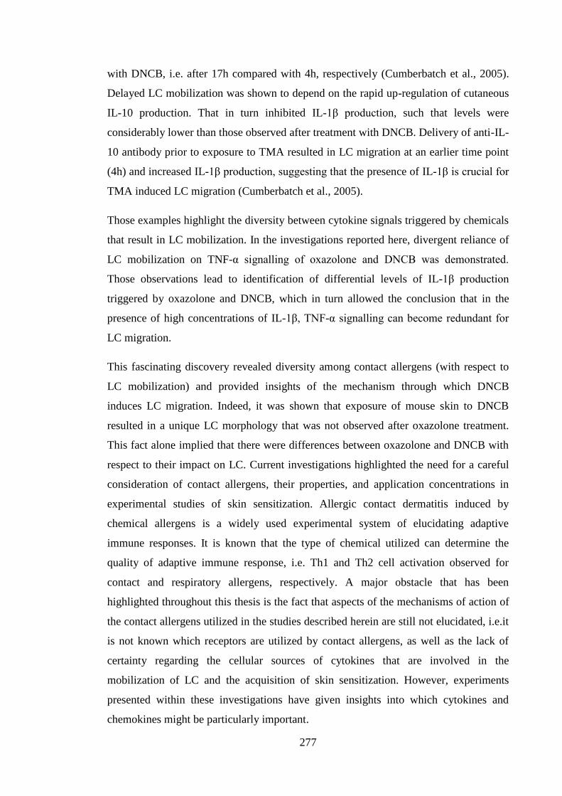
277
with DNCB, i.e. after 17h compared with 4h, respectively (Cumberbatch et al., 2005).
Delayed LC mobilization was shown to depend on the rapid up-regulation of cutaneous
IL-10 production. That in turn inhibited IL-1β production, such that levels were
considerably lower than those observed after treatment with DNCB. Delivery of anti-IL-
10 antibody prior to exposure to TMA resulted in LC migration at an earlier time point
(4h) and increased IL-1β production, suggesting that the presence of IL-1β is crucial for
TMA induced LC migration (Cumberbatch et al., 2005).
Those examples highlight the diversity between cytokine signals triggered by chemicals
that result in LC mobilization. In the investigations reported here, divergent reliance of
LC mobilization on TNF-α signalling of oxazolone and DNCB was demonstrated.
Those observations lead to identification of differential levels of IL-1β production
triggered by oxazolone and DNCB, which in turn allowed the conclusion that in the
presence of high concentrations of IL-1β, TNF-α signalling can become redundant for
LC migration.
This fascinating discovery revealed diversity among contact allergens (with respect to
LC mobilization) and provided insights of the mechanism through which DNCB
induces LC migration. Indeed, it was shown that exposure of mouse skin to DNCB
resulted in a unique LC morphology that was not observed after oxazolone treatment.
This fact alone implied that there were differences between oxazolone and DNCB with
respect to their impact on LC. Current investigations highlighted the need for a careful
consideration of contact allergens, their properties, and application concentrations in
experimental studies of skin sensitization. Allergic contact dermatitis induced by
chemical allergens is a widely used experimental system of elucidating adaptive
immune responses. It is known that the type of chemical utilized can determine the
quality of adaptive immune response, i.e. Th1 and Th2 cell activation observed for
contact and respiratory allergens, respectively. A major obstacle that has been
highlighted throughout this thesis is the fact that aspects of the mechanisms of action of
the contact allergens utilized in the studies described herein are still not elucidated, i.e.it
is not known which receptors are utilized by contact allergens, as well as the lack of
certainty regarding the cellular sources of cytokines that are involved in the
mobilization of LC and the acquisition of skin sensitization. However, experiments
presented within these investigations have given insights into which cytokines and
chemokines might be particularly important.
278
The data from current studies, together with published reports, point to IL-1β as a key
cytokine during skin sensitization. Indeed, the inflammasome has been described as a
master switch of sensitization on which the chemical sensitizing ability lies (Watanabe
et al., 2008).
There are two possible reasons for the divergence of cytokine requirements between
oxazolone and DNCB. Firstly, as it has been mentioned before with regards to different
cutaneous DC subsets, there can be a certain level of overlap of function between
various DC populations. Similarly, it is possible that in the presence of high levels of
IL-1β, or a potent stimulus which triggers high levels of IL-1β production, absence of
TNF-α is not an obstacle in the mobilization of LC. Secondly, chemical contact
allergens are thought to be misrecognized by the immune system as infectious
organisms. There is evidence that pathogens may exhibit different patterns of cytokine
requirements for LC migration. It has been shown that infection of the skin with a West
Nile virus resulted in migration of LC that was independent of TNF-α signalling but still
reliant on IL-1β (Byrne et al., 2001). Conversely, LC cells from mice unresponsive to
IL-1β (MyD88 KO) exhibited unaltered mobilization in response to C. albicans
infection (Haley et al., 2012). Migration of LC exposed to staphylococcal enterotoxin-A
was found to depend on signals from both IL-1α and TNF-α (Shankar et al., 1996). It is
possible that oxazolone and DNCB with their mechanisms of action resemble different
pathogens that exert divergent effects on cutaneous cytokine release and subsequently
differently affect migration of LC.
7.3 Future Aims
The aim of this PhD thesis was to investigate the molecular mechanisms of skin
sensitization and its immunomodulation by LF. Some progress has been made towards
that goal; however, there are still many questions that remain unanswered. One of the
main uncertainties highlighted by these investigations is the cellular source – or sources
– of TNF-α in the skin following exposure to allergen. It would be particularly
important to use IHC techniques to detect TNF-α protein. That would allow for
preserving the integrity of the tissue with respect to the relationship between different
cell types and the identification of the exact cellular source or sources of this cytokine.
Furthermore, IHC could be employed to investigate the fate of LF in the skin.
279
Additionally, in the follow up studies it would be of interest to identify whether LF
impacts on the CHS reaction as measured by changes in challenge-induced ear thickness
and cutaneous cellular infiltrate, and thus assessing its potential as a treatment for ACD.
Studies in human subjects have shown that LF has the ability to decrease the erythema
triggered by initial exposure to DPC (Griffiths et al., 2001). Moreover, due to its impact
on IL-17F expression, LF might potentially therapeutic potential for psoriasis, as IL-17
is considered to be an important cytokine in the pathogenesis of that disease (Golden et
al., 2013). Internet searches revealed that there are treatments available directed at
psoriasis patients, based on claims of LF activity, e.g. equilac, available in cream and
capsule forms, which contains LF from mare’s milk (Ecopharma, 2014). Indeed, a
recent publication reported improvement in psoriatic plaques following topical
treatment with bovine LF (Saraceno et al., 2014). However, no mechanistic information
is available to provide clues as to how exactly LF improved psoriatic plaques.
Lactoferrin has been shown to have a beneficial effect on the management of acne,
whereby oral delivery of bovine LF resulted in a decreased number of inflammatory
acne lesions in an open label (Mueller et al., 2011) and double blind (Kim et al., 2010)
trial. Inflammation of the skin, despite its low mortality rate, has a huge impact on the
wellbeing status of patients and can be gravely debilitating. The ability of LF to mitigate
cutaneous inflammation would be greatly beneficial.
The current investigations have highlighted further the differences between contact
allergens and revealed a novel TNF-α independent mechanism of LC mobilization. It
would have been of interest to conduct LC mobilization experiments utilising a wider
range of contact allergens, possibly employing IL-1β KO animals, to definitively assess
the role of this cytokine in the process of LC migration in response to allergens.
Similarly, the role of TNF-α, and not only of TNFRII, could be assessed using TNF-α
KO mice. Importantly, TNFRII KO mice not only lack TNFRII expression by LC but
also by other DC subsets and most likely γδ T cells. TNFRI is constitutively expressed
by nearly all cells, whereas TNFRII is an inducible receptor expressed by the cells of
myeloid lineage, peripheral T cells and macrophages. However, the most definitive
answers could be gained by deploying mice lacking both TNF-α and IL-1β to determine
the central roles of these two cytokines. It is plausible that in the situation when either
of these two cytokines was absent, compensatory mechanisms would be able to restore
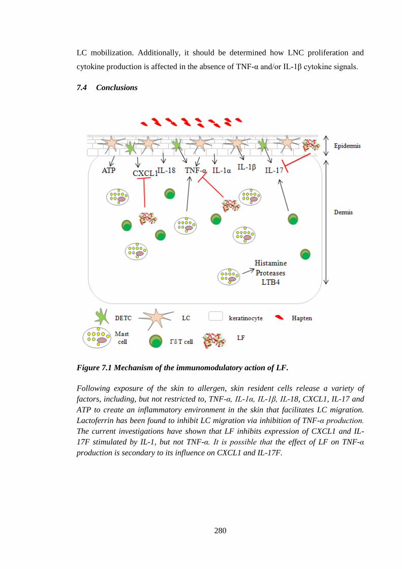
280
LC mobilization. Additionally, it should be determined how LNC proliferation and
cytokine production is affected in the absence of TNF-α and/or IL-1β cytokine signals.
7.4 Conclusions
Figure 7.1 Mechanism of the immunomodulatory action of LF.
Following exposure of the skin to allergen, skin resident cells release a variety of
factors, including, but not restricted to, TNF-α, IL-1α, IL-1β, IL-18, CXCL1, IL-17 and
ATP to create an inflammatory environment in the skin that facilitates LC migration.
Lactoferrin has been found to inhibit LC migration via inhibition of TNF-α production.
The current investigations have shown that LF inhibits expression of CXCL1 and IL-
17F stimulated by IL-1, but not TNF-α. It is possible that the effect of LF on TNF-α
production is secondary to its influence on CXCL1 and IL-17F.
281
The effect of the concentration of antigen on the dependency of various cell types and
cytokines to induce immune response has been considered previously. It has been said
by Paracelsus in 17th
century that the dose makes the poison. It is important not only in
the consideration of toxicology, but can be extrapolated to immunology. That simple
notion should be regarded especially now when there are so many known variables in
the system of cutaneous immune network. The advent of ever more accurate techniques
of detection of cells, as well as new methods for their in vivo visualization, e.g. Kaede
protein (Tomura and Kabashima, 2013) or intravital multiphoton imaging (Kabashima
and Egawa, 2014) makes immunology an exciting and ever expanding field. This PhD
thesis has established that immunomodulatory effects of LF in the skin might be
mediated by the inhibition of CXCL1 and IL-17F signalling. Moreover, it was found
that in certain circumstances LC migration is possible in the absence of TNF-α
signalling, which is potentially mediated by the presence of high levels of IL-1β. This
knowledge revealed a novel mechanism of LC mobilization and further highlighted the
importance of IL-1β cytokine in the cutaneous immunity.
282
8 References
I. C. L. A. C., 2013, Database of Cross-Contaminated or Misidentified Cell Lines.
Actor, J. K., S. A. Hwang, and M. L. Kruzel, 2009, Lactoferrin as a natural immune
modulator: Curr Pharm Des, v. 15, p. 1956-73.
Ade, N., F. Leon, M. Pallardy, J. L. Peiffer, S. Kerdine-Romer, M. H. Tissier, P. A.
Bonnet, I. Fabre, and J. C. Ourlin, 2009, HMOX1 and NQO1 genes are
upregulated in response to contact sensitizers in dendritic cells and THP-1 cell
line: role of the Keap1/Nrf2 pathway: Toxicol Sci, v. 107, p. 451-60.
Agner, T., J. Serup, V. Handlos, and W. Batsberg, 1989, Different skin irritation
abilities of different qualities of sodium lauryl sulphate: Contact Dermatitis, v.
21, p. 184-8.
Aiba, S., and S. I. Katz, 1990, Phenotypic and functional characteristics of in vivo-
activated Langerhans cells: J Immunol, v. 145, p. 2791-6.
Aiba, S., A. Terunuma, H. Manome, and H. Tagami, 1997, Dendritic cells differently
respond to haptens and irritants by their production of cytokines and expression
of co-stimulatory molecules: Eur J Immunol, v. 27, p. 3031-8.
Akiyama, H., M. Saito, G. Qiu, T. Toida, and T. Imanari, 1994, Analytical studies on
hyaluronic acid synthesis by normal human epidermal keratinocytes cultured in
a serum-free medium: Biol Pharm Bull, v. 17, p. 361-4.
Albanesi, C., 2010, Keratinocytes in allergic skin diseases: Curr Opin Allergy Clin
Immunol, v. 10, p. 452-6.
Albanesi, C., A. Cavani, and G. Girolomoni, 1998, Interferon-gamma-stimulated human
keratinocytes express the genes necessary for the production of peptide-loaded
MHC class II molecules: J Invest Dermatol, v. 110, p. 138-42.
Albanesi, C., A. Cavani, and G. Girolomoni, 1999, IL-17 is produced by nickel-specific
T lymphocytes and regulates ICAM-1 expression and chemokine production in
human keratinocytes: synergistic or antagonist effects with IFN-gamma and
TNF-alpha: J Immunol, v. 162, p. 494-502.
Allan, R. S., J. Waithman, S. Bedoui, C. M. Jones, J. A. Villadangos, Y. Zhan, A. M.
Lew, K. Shortman, W. R. Heath, and F. R. Carbone, 2006, Migratory dendritic
cells transfer antigen to a lymph node-resident dendritic cell population for
efficient CTL priming: Immunity, v. 25, p. 153-62.
Almond, R. J., B. F. Flanagan, A. Antonopoulos, S. M. Haslam, A. Dell, I. Kimber, and
R. J. Dearman, 2013, Differential immunogenicity and allergenicity of native
and recombinant human lactoferrins: role of glycosylation: Eur J Immunol, v.
43, p. 170-81.
Anderson, B. F., H. M. Baker, G. E. Norris, D. W. Rice, and E. N. Baker, 1989,
Structure of human lactoferrin: crystallographic structure analysis and
refinement at 2.8 A resolution: J Mol Biol, v. 209, p. 711-34.
Ando, K., K. Hasegawa, K. Shindo, T. Furusawa, T. Fujino, K. Kikugawa, H. Nakano,
O. Takeuchi, S. Akira, T. Akiyama, J. Gohda, J. Inoue, and M. Hayakawa, 2010,
Human lactoferrin activates NF-kappaB through the Toll-like receptor 4
pathway while it interferes with the lipopolysaccharide-stimulated TLR4
signaling: FEBS J, v. 277, p. 2051-66.
Ansel, J. C., T. A. Luger, D. Lowry, P. Perry, D. R. Roop, and J. D. Mountz, 1988, The
expression and modulation of IL-1 alpha in murine keratinocytes: J Immunol, v.
140, p. 2274-8.
283
Antonopoulos, C., M. Cumberbatch, R. J. Dearman, R. J. Daniel, I. Kimber, and R. W.
Groves, 2001, Functional caspase-1 is required for Langerhans cell migration
and optimal contact sensitization in mice: J Immunol, v. 166, p. 3672-7.
Antonopoulos, C., M. Cumberbatch, J. B. Mee, R. J. Dearman, X. Q. Wei, F. Y. Liew,
I. Kimber, and R. W. Groves, 2008, IL-18 is a key proximal mediator of contact
hypersensitivity and allergen-induced Langerhans cell migration in murine
epidermis: J Leukoc Biol, v. 83, p. 361-7.
Anttila, H. S., S. Reitamo, M. Ceska, and M. Hurme, 1992, Signal transduction
pathways leading to the production of IL-8 by human monocytes are
differentially regulated by dexamethasone: Clin Exp Immunol, v. 89, p. 509-12.
Appelmelk, B. J., Y. Q. An, M. Geerts, B. G. Thijs, H. A. de Boer, D. M. MacLaren, J.
de Graaff, and J. H. Nuijens, 1994, Lactoferrin is a lipid A-binding protein:
Infect Immun, v. 62, p. 2628-32.
Armstrong, J., C. Sargent, and D. Singh, 2009, Glucocorticoid sensitivity of
lipopolysaccharide-stimulated chronic obstructive pulmonary disease alveolar
macrophages: Clin Exp Immunol, v. 158, p. 74-83.
Arnold, R. R., J. E. Russell, W. J. Champion, M. Brewer, and J. J. Gauthier, 1982,
Bactericidal activity of human lactoferrin: differentiation from the stasis of iron
deprivation: Infect Immun, v. 35, p. 792-9.
Aruffo, A., I. Stamenkovic, M. Melnick, C. B. Underhill, and B. Seed, 1990, CD44 is
the principal cell surface receptor for hyaluronate: Cell, v. 61, p. 1303-13.
Asada, H., J. Linton, and S. I. Katz, 1997, Cytokine gene expression during the
elicitation phase of contact sensitivity: regulation by endogenous IL-4: J Invest
Dermatol, v. 108, p. 406-11.
Ashikaga, T., Y. Yoshida, M. Hirota, K. Yoneyama, H. Itagaki, H. Sakaguchi, M.
Miyazawa, Y. Ito, H. Suzuki, and H. Toyoda, 2006, Development of an in vitro
skin sensitization test using human cell lines: the human Cell Line Activation
Test (h-CLAT). I. Optimization of the h-CLAT protocol: Toxicol In Vitro, v. 20,
p. 767-73.
Bacci, S., T. Nakamura, and J. W. Streilein, 1996, Failed antigen presentation after
UVB radiation correlates with modifications of Langerhans cell cytoskeleton: J
Invest Dermatol, v. 107, p. 838-43.
Bach, J. F., and J. François Bach, 2003, Regulatory T cells under scrutiny: Nat Rev
Immunol, v. 3, p. 189-98.
Baggiolini, M., C. De Duve, P. L. Masson, and J. F. Heremans, 1970, Association of
lactoferrin with specific granules in rabbit heterophil leukocytes: J Exp Med, v.
131, p. 559-70.
Baker, 1994, STRUCTURE AND REACTIVITY OF TRANSFERRINS, ADVANCES
IN INORGANIC CHEMISTRY, p. 75.
Baker, E. N., and H. M. Baker, 2009, A structural framework for understanding the
multifunctional character of lactoferrin: Biochimie, v. 91, p. 3-10.
Banchereau, J., and R. M. Steinman, 1998, Dendritic cells and the control of immunity:
Nature, v. 392, p. 245-52.
Banno, T., A. Gazel, and M. Blumenberg, 2004, Effects of tumor necrosis factor-alpha
(TNF alpha) in epidermal keratinocytes revealed using global transcriptional
profiling: J Biol Chem, v. 279, p. 32633-42.
Bashir, M. M., M. R. Sharma, and V. P. Werth, 2009, UVB and proinflammatory
cytokines synergistically activate TNF-alpha production in keratinocytes
through enhanced gene transcription: J Invest Dermatol, v. 129, p. 994-1001.
284
Becker, D., M. Mohamadzadeh, K. Reske, and J. Knop, 1992, Increased level of
intracellular MHC class II molecules in murine Langerhans cells following in
vivo and in vitro administration of contact allergens: J Invest Dermatol, v. 99, p.
545-9.
Bedoui, S., P. G. Whitney, J. Waithman, L. Eidsmo, L. Wakim, I. Caminschi, R. S.
Allan, M. Wojtasiak, K. Shortman, F. R. Carbone, A. G. Brooks, and W. R.
Heath, 2009, Cross-presentation of viral and self antigens by skin-derived
CD103+ dendritic cells: Nat Immunol, v. 10, p. 488-95.
Beer, H. D., Gassmann, M. G., Munz, B., Steiling, H., Engelhardt, F., Bleuel, K.,
Werner, S., 2000, Expression and function of keratinocyte growth factor and
activin in skin morphogenesis and cutaneous wound repair: J Investig Dermatol
Symp Proc, v. 5, p. 34-9.
Bellamy, W., M. Takase, K. Yamauchi, H. Wakabayashi, K. Kawase, and M. Tomita,
1992, Identification of the bactericidal domain of lactoferrin: Biochim Biophys
Acta, v. 1121, p. 130-6.
Bennatt, D. J., and D. D. McAbee, 1997, Identification and isolation of a 45-kDa
calcium-dependent lactoferrin receptor from rat hepatocytes: Biochemistry, v.
36, p. 8359-66.
Bennett, C. L., E. van Rijn, S. Jung, K. Inaba, R. M. Steinman, M. L. Kapsenberg, and
B. E. Clausen, 2005, Inducible ablation of mouse Langerhans cells diminishes
but fails to abrogate contact hypersensitivity: J Cell Biol, v. 169, p. 569-76.
Bennett, R. M., and J. Davis, 1981, Lactoferrin binding to human peripheral blood cells:
an interaction with a B-enriched population of lymphocytes and a subpopulation
of adherent mononuclear cells: J Immunol, v. 127, p. 1211-6.
Beppu, M., S. Eda, M. Fujimaki, E. Hishiyama, and K. Kikugawa, 1996, Recognition of
poly-N-acetyllactosaminyl saccharide chains on iron-oxidized erythrocytes by
human monocytic leukemia cell line THP-1 differentiated into macrophages:
Biol Pharm Bull, v. 19, p. 188-94.
Bernstein, D. I., R. Patterson, and C. R. Zeiss, 1982, Clinical and immunologic
evaluation of trimellitic anhydride-and phthalic anhydride-exposed workers
using a questionnaire with comparative analysis of enzyme-linked
immunosorbent and radioimmunoassay studies: J Allergy Clin Immunol, v. 69,
p. 311-8.
Beutler, B. A., I. W. Milsark, and A. Cerami, 1985, Cachectin/tumor necrosis factor:
production, distribution, and metabolic fate in vivo: J Immunol, v. 135, p. 3972-
7.
Bezwoda, W. R., and N. Mansoor, 1989, Lactoferrin from human breast milk and from
neutrophil granulocytes. Comparative studies of isolation, quantitation,
characterization and iron binding properties: Biomed Chromatogr, v. 3, p. 121-6.
Bhor, V. M., C. J. Thomas, N. Surolia, and A. Surolia, 2005, Polymyxin B: an ode to an
old antidote for endotoxic shock: Mol Biosyst, v. 1, p. 213-22.
Biedermann, T., M. Kneilling, R. Mailhammer, K. Maier, C. A. Sander, G. Kollias, S.
L. Kunkel, L. Hültner, and M. Röcken, 2000, Mast cells control neutrophil
recruitment during T cell-mediated delayed-type hypersensitivity reactions
through tumor necrosis factor and macrophage inflammatory protein 2: J Exp
Med, v. 192, p. 1441-52.
Billiet, L., C. Furman, G. Larigauderie, C. Copin, K. Brand, J. C. Fruchart, and M.
Rouis, 2005, Extracellular human thioredoxin-1 inhibits lipopolysaccharide-
induced interleukin-1beta expression in human monocyte-derived macrophages:
J Biol Chem, v. 280, p. 40310-8.
285
Black, A. P., M. R. Ardern-Jones, V. Kasprowicz, P. Bowness, L. Jones, A. S. Bailey,
and G. S. Ogg, 2007, Human keratinocyte induction of rapid effector function in
antigen-specific memory CD4+ and CD8+ T cells: Eur J Immunol, v. 37, p.
1485-93.
Black, C. A., 1999, Delayed type hypersensitivity: current theories with an historic
perspective: Dermatol Online J, v. 5, p. 7.
Blauvelt, A., S. I. Katz, and M. C. Udey, 1995, Human Langerhans cells express E-
cadherin: J Invest Dermatol, v. 104, p. 293-6.
Bluestone, J. A., and A. K. Abbas, 2003, Natural versus adaptive regulatory T cells: Nat
Rev Immunol, v. 3, p. 253-7.
Boislève, F., S. Kerdine-Römer, N. Rougier-Larzat, and M. Pallardy, 2004, Nickel and
DNCB induce CCR7 expression on human dendritic cells through different
signalling pathways: role of TNF-alpha and MAPK: J Invest Dermatol, v. 123,
p. 494-502.
Boltjes, A., and F. van Wijk, 2014, Human dendritic cell functional specialization in
steady-state and inflammation: Front Immunol, v. 5, p. 131.
Bonnet, M. P., H. Beloeil, D. Benhamou, J. X. Mazoit, and K. Asehnoune, 2008, The
mu opioid receptor mediates morphine-induced tumor necrosis factor and
interleukin-6 inhibition in toll-like receptor 2-stimulated monocytes: Anesth
Analg, v. 106, p. 1142-9, table of contents.
Bonneville, M., C. Chavagnac, M. Vocanson, A. Rozieres, J. Benetiere, I. Pernet, A.
Denis, J. F. Nicolas, and A. Hennino, 2007, Skin contact irritation conditions the
development and severity of allergic contact dermatitis: J Invest Dermatol, v.
127, p. 1430-5.
Boraschi, D., L. Villa, G. Volpini, P. Bossù, S. Censini, P. Ghiara, G. Scapigliat, L.
Nencioni, M. Bartalini, and G. Matteucci, 1990, Differential activity of
interleukin 1 alpha and interleukin 1 beta in the stimulation of the immune
response in vivo: Eur J Immunol, v. 20, p. 317-21.
Borkowski, T. A., J. J. Letterio, A. G. Farr, and M. C. Udey, 1996, A role for
endogenous transforming growth factor beta 1 in Langerhans cell biology: the
skin of transforming growth factor beta 1 null mice is devoid of epidermal
Langerhans cells: J Exp Med, v. 184, p. 2417-22.
Borkowski, T. A., J. J. Letterio, C. L. Mackall, A. Saitoh, X. J. Wang, D. R. Roop, R. E.
Gress, and M. C. Udey, 1997, A role for TGFbeta1 in langerhans cell biology.
Further characterization of the epidermal Langerhans cell defect in TGFbeta1
null mice: J Clin Invest, v. 100, p. 575-81.
Bos, J. D., M. B. Teunissen, I. Cairo, S. R. Krieg, M. L. Kapsenberg, P. K. Das, and J.
Borst, 1990, T-cell receptor gamma delta bearing cells in normal human skin: J
Invest Dermatol, v. 94, p. 37-42.
Boukamp, P., R. T. Petrussevska, D. Breitkreutz, J. Hornung, A. Markham, and N. E.
Fusenig, 1988, Normal keratinization in a spontaneously immortalized
aneuploid human keratinocyte cell line: J Cell Biol, v. 106, p. 761-71.
Bour, H., E. Peyron, M. Gaucherand, J. L. Garrigue, C. Desvignes, D. Kaiserlian, J. P.
Revillard, and J. F. Nicolas, 1995, Major histocompatibility complex class I-
restricted CD8+ T cells and class II-restricted CD4+ T cells, respectively,
mediate and regulate contact sensitivity to dinitrofluorobenzene: Eur J Immunol,
v. 25, p. 3006-10.
Bournazou, I., K. J. Mackenzie, R. Duffin, A. G. Rossi, and C. D. Gregory, 2010,
Inhibition of eosinophil migration by lactoferrin: Immunol Cell Biol, v. 88, p.
220-3.
286
Bournazou, I., J. D. Pound, R. Duffin, S. Bournazos, L. A. Melville, S. B. Brown, A. G.
Rossi, and C. D. Gregory, 2009, Apoptotic human cells inhibit migration of
granulocytes via release of lactoferrin: J Clin Invest, v. 119, p. 20-32.
Breton-Gorius, J., D. Y. Mason, D. Buriot, J. L. Vilde, and C. Griscelli, 1980,
Lactoferrin deficiency as a consequence of a lack of specific granules in
neutrophils from a patient with recurrent infections. Detection by
immunoperoxidase staining for lactoferrin and cytochemical electron
microscopy: Am J Pathol, v. 99, p. 413-28.
Bryce, P. J., M. L. Miller, I. Miyajima, M. Tsai, S. J. Galli, and H. C. Oettgen, 2004,
Immune sensitization in the skin is enhanced by antigen-independent effects of
IgE: Immunity, v. 20, p. 381-92.
Buccigrossi, V., G. de Marco, E. Bruzzese, L. Ombrato, I. Bracale, G. Polito, and A.
Guarino, 2007, Lactoferrin induces concentration-dependent functional
modulation of intestinal proliferation and differentiation: Pediatr Res, v. 61, p.
410-4.
Bursch, L. S., L. Wang, B. Igyarto, A. Kissenpfennig, B. Malissen, D. H. Kaplan, and
K. A. Hogquist, 2007, Identification of a novel population of Langerin+
dendritic cells: J Exp Med, v. 204, p. 3147-56.
Byamba, D., T. G. Kim, D. H. Kim, J. H. Je, and M. G. Lee, 2010, The Roles of
Reactive Oxygen Species Produced by Contact Allergens and Irritants in
Monocyte-derived Dendritic Cells: Ann Dermatol, v. 22, p. 269-78.
Byrne, S. N., G. M. Halliday, L. J. Johnston, and N. J. King, 2001, Interleukin-1beta but
not tumor necrosis factor is involved in West Nile virus-induced Langerhans cell
migration from the skin in C57BL/6 mice: J Invest Dermatol, v. 117, p. 702-9.
Cai, Y., X. Shen, C. Ding, C. Qi, K. Li, X. Li, V. R. Jala, H. G. Zhang, T. Wang, J.
Zheng, and J. Yan, 2011, Pivotal role of dermal IL-17-producing γδ T cells in
skin inflammation: Immunity, v. 35, p. 596-610.
Campo, G. M., A. Avenoso, A. D'Ascola, G. Nastasi, A. Micali, D. Puzzolo, A. Pisani,
V. Prestipino, M. Scuruchi, A. Calatroni, and S. Campo, 2013, Combined
treatment with hyaluronan inhibitor Pep-1 and a selective adenosine A2 receptor
agonist reduces inflammation in experimental arthritis: Innate Immun, v. 19, p.
462-78.
Campos, R. A., M. Szczepanik, A. Itakura, M. Akahira-Azuma, S. Sidobre, M.
Kronenberg, and P. W. Askenase, 2003, Cutaneous immunization rapidly
activates liver invariant Valpha14 NKT cells stimulating B-1 B cells to initiate T
cell recruitment for elicitation of contact sensitivity: J Exp Med, v. 198, p. 1785-
96.
Cardoso, L. S., M. I. Araujo, A. M. Góes, L. G. Pacífico, R. R. Oliveira, and S. C.
Oliveira, 2007, Polymyxin B as inhibitor of LPS contamination of Schistosoma
mansoni recombinant proteins in human cytokine analysis: Microb Cell Fact, v.
6, p. 1.
Cattani, F., A. Gallese, M. Mosca, P. Buanne, L. Biordi, S. Francavilla, G. Coletti, L.
Pellegrini, G. Melillo, and R. Bertini, 2006, The role of CXCR2 activity in the
contact hypersensitivity response in mice: Eur Cytokine Netw, v. 17, p. 42-8.
Caux, C., C. Massacrier, B. Dubois, J. Valladeau, C. Dezutter-Dambuyant, I. Durand,
D. Schmitt, and S. Saeland, 1999, Respective involvement of TGF-beta and IL-4
in the development of Langerhans cells and non-Langerhans dendritic cells from
CD34+ progenitors: J Leukoc Biol, v. 66, p. 781-91.
287
Caux, C., C. Massacrier, B. Vanbervliet, B. Dubois, C. Van Kooten, I. Durand, and J.
Banchereau, 1994, Activation of human dendritic cells through CD40 cross-
linking: J Exp Med, v. 180, p. 1263-72.
Cavani, A., F. Nasorri, C. Prezzi, S. Sebastiani, C. Albanesi, and G. Girolomoni, 2000,
Human CD4+ T lymphocytes with remarkable regulatory functions on dendritic
cells and nickel-specific Th1 immune responses: J Invest Dermatol, v. 114, p.
295-302.
Cella, M., D. Scheidegger, K. Palmer-Lehmann, P. Lane, A. Lanzavecchia, and G.
Alber, 1996, Ligation of CD40 on dendritic cells triggers production of high
levels of interleukin-12 and enhances T cell stimulatory capacity: T-T help via
APC activation: J Exp Med, v. 184, p. 747-52.
Chen, G., and D. V. Goeddel, 2002, TNF-R1 signaling: a beautiful pathway: Science, v.
296, p. 1634-5.
Chen, L., S. Guo, M. J. Ranzer, and L. A. DiPietro, 2013, Toll-like receptor 4 has an
essential role in early skin wound healing: J Invest Dermatol, v. 133, p. 258-67.
Chia, A. J., C. E. Goldring, N. R. Kitteringham, S. Q. Wong, P. Morgan, and B. K.
Park, 2010, Differential effect of covalent protein modification and glutathione
depletion on the transcriptional response of Nrf2 and NF-kappaB: Biochem
Pharmacol, v. 80, p. 410-21.
Chirico, G., R. Marzollo, S. Cortinovis, C. Fonte, and A. Gasparoni, 2008, Antiinfective
properties of human milk: J Nutr, v. 138, p. 1801S-1806S.
Cho, J. S., E. M. Pietras, N. C. Garcia, R. I. Ramos, D. M. Farzam, H. R. Monroe, J. E.
Magorien, A. Blauvelt, J. K. Kolls, A. L. Cheung, G. Cheng, R. L. Modlin, and
L. S. Miller, 2010, IL-17 is essential for host defense against cutaneous
Staphylococcus aureus infection in mice: J Clin Invest, v. 120, p. 1762-73.
Chodaczek, G., A. Bacsi, N. Dharajiya, S. Sur, T. K. Hazra, and I. Boldogh, 2009,
Ragweed pollen-mediated IgE-independent release of biogenic amines from
mast cells via induction of mitochondrial dysfunction: Mol Immunol, v. 46, p.
2505-14.
Chodaczek, G., V. Papanna, M. A. Zal, and T. Zal, 2012, Body-barrier surveillance by
epidermal γδ TCRs: Nat Immunol, v. 13, p. 272-82.
Chodakewitz, J. A., J. Lacy, S. E. Edwards, N. Birchall, and D. L. Coleman, 1990,
Macrophage colony-stimulating factor production by murine and human
keratinocytes. Enhancement by bacterial lipopolysaccharide: J Immunol, v. 144,
p. 2190-6.
Chorro, L., A. Sarde, M. Li, K. J. Woollard, P. Chambon, B. Malissen, A.
Kissenpfennig, J. B. Barbaroux, R. Groves, and F. Geissmann, 2009,
Langerhans cell (LC) proliferation mediates neonatal development, homeostasis,
and inflammation-associated expansion of the epidermal LC network: J Exp
Med, v. 206, p. 3089-100.
Clarke, S. R., M. Barnden, C. Kurts, F. R. Carbone, J. F. Miller, and W. R. Heath, 2000,
Characterization of the ovalbumin-specific TCR transgenic line OT-I: MHC
elements for positive and negative selection: Immunol Cell Biol, v. 78, p. 110-7.
Coffman, R. L., 2006, Origins of the T(H)1-T(H)2 model: a personal perspective: Nat
Immunol, v. 7, p. 539-41.
Coffman, R. L., and J. Carty, 1986, A T cell activity that enhances polyclonal IgE
production and its inhibition by interferon-gamma: J Immunol, v. 136, p. 949-
54.
288
Coffman, R. L., D. A. Lebman, and B. Shrader, 1989, Transforming growth factor beta
specifically enhances IgA production by lipopolysaccharide-stimulated murine
B lymphocytes: J Exp Med, v. 170, p. 1039-44.
Coffman, R. L., J. Ohara, M. W. Bond, J. Carty, A. Zlotnik, and W. E. Paul, 1986, B
cell stimulatory factor-1 enhances the IgE response of lipopolysaccharide-
activated B cells: J Immunol, v. 136, p. 4538-41.
Cogen, A. L., V. Nizet, and R. L. Gallo, 2008, Skin microbiota: a source of disease or
defence?: Br J Dermatol, v. 158, p. 442-55.
Colella, G., A. Vicidomini, V. Soro, A. Lanza, and N. Cirillo, 2012, Molecular insights
into the effects of sodium hyaluronate preparations in keratinocytes: Clin Exp
Dermatol, v. 37, p. 516-20.
Conti, L., R. Casetti, M. Cardone, B. Varano, A. Martino, F. Belardelli, F. Poccia, and
S. Gessani, 2005, Reciprocal activating interaction between dendritic cells and
pamidronate-stimulated gammadelta T cells: role of CD86 and inflammatory
cytokines: J Immunol, v. 174, p. 252-60.
Cooper, K. D., L. Oberhelman, T. A. Hamilton, O. Baadsgaard, M. Terhune, G. LeVee,
T. Anderson, and H. Koren, 1992, UV exposure reduces immunization rates and
promotes tolerance to epicutaneous antigens in humans: relationship to dose,
CD1a-DR+ epidermal macrophage induction, and Langerhans cell depletion:
Proc Natl Acad Sci U S A, v. 89, p. 8497-501.
Corsini, E., V. Galbiati, D. Nikitovic, and A. M. Tsatsakis, 2013, Role of oxidative
stress in chemical allergens induced skin cells activation: Food Chem Toxicol, v.
61, p. 74-81.
Corsini, E., A. Primavera, M. Marinovich, and C. L. Galli, 1998, Selective induction of
cell-associated interleukin-1alpha in murine keratinocytes by chemical allergens:
Toxicology, v. 129, p. 193-200.
Crouch, S. P., K. J. Slater, and J. Fletcher, 1992, Regulation of cytokine release from
mononuclear cells by the iron-binding protein lactoferrin: Blood, v. 80, p. 235-
40.
Cumberbatch, M., M. Bhushan, R. J. Dearman, I. Kimber, and C. E. Griffiths, 2003, IL-
1beta-induced Langerhans' cell migration and TNF-alpha production in human
skin: regulation by lactoferrin: Clin Exp Immunol, v. 132, p. 352-9.
Cumberbatch, M., K. Clelland, R. J. Dearman, and I. Kimber, 2005, Impact of
cutaneous IL-10 on resident epidermal Langerhans' cells and the development of
polarized immune responses: J Immunol, v. 175, p. 43-50.
Cumberbatch, M., R. J. Dearman, C. Antonopoulos, R. W. Groves, and I. Kimber, 2001,
Interleukin (IL)-18 induces Langerhans cell migration by a tumour necrosis
factor-alpha- and IL-1beta-dependent mechanism: Immunology, v. 102, p. 323-
30.
Cumberbatch, M., R. J. Dearman, C. E. Griffiths, and I. Kimber, 2000a, Langerhans cell
migration: Clin Exp Dermatol, v. 25, p. 413-8.
Cumberbatch, M., R. J. Dearman, R. W. Groves, C. Antonopoulos, and I. Kimber,
2002a, Differential regulation of epidermal langerhans cell migration by
interleukins (IL)-1alpha and IL-1beta during irritant- and allergen-induced
cutaneous immune responses: Toxicol Appl Pharmacol, v. 182, p. 126-35.
Cumberbatch, M., R. J. Dearman, and I. Kimber, 1997a, Interleukin 1 beta and the
stimulation of Langerhans cell migration: comparisons with tumour necrosis
factor alpha: Arch Dermatol Res, v. 289, p. 277-84.
289
Cumberbatch, M., R. J. Dearman, and I. Kimber, 1997b, Langerhans cells require
signals from both tumour necrosis factor alpha and interleukin 1 beta for
migration: Adv Exp Med Biol, v. 417, p. 125-8.
Cumberbatch, M., R. J. Dearman, and I. Kimber, 1999a, Induction by tumour necrosis
factor alpha of dose-related changes in Langerhans cell frequency in mice: Arch
Dermatol Res, v. 291, p. 453-8.
Cumberbatch, M., R. J. Dearman, and I. Kimber, 1999b, Inhibition by dexamethasone
of Langerhans cell migration: influence of epidermal cytokine signals:
Immunopharmacology, v. 41, p. 235-43.
Cumberbatch, M., R. J. Dearman, and I. Kimber, 1999c, Langerhans cell migration in
mice requires intact type I interleukin 1 receptor (IL-1RI) function: Arch
Dermatol Res, v. 291, p. 357-61.
Cumberbatch, M., R. J. Dearman, and I. Kimber, 2002b, Influence of ageing on
Langerhans cell migration in mice: identification of a putative deficiency of
epidermal interleukin-1beta: Immunology, v. 105, p. 466-77.
Cumberbatch, M., R. J. Dearman, I. Kimber, and R. C. Viner, 2009, Anti-Inflammatory
Peptides.
Cumberbatch, M., R. J. Dearman, S. Uribe-Luna, D. R. Headon, P. P. Ward, O. M.
Conneely, and I. Kimber, 2000b, Regulation of epidermal Langerhans cell
migration by lactoferrin: Immunology, v. 100, p. 21-8.
Cumberbatch, M., S. J. Gould, S. W. Peters, D. A. Basketter, R. J. Dearman, and I.
Kimber, 1992a, Influence of topical exposure to chemical allergens on murine
Langerhans cells. Comparison of 2,4-dinitrochlorobenzene with trimellitic
anhydride: J Clin Lab Immunol, v. 37, p. 65-81.
Cumberbatch, M., C. E. Griffiths, S. C. Tucker, R. J. Dearman, and I. Kimber, 1999d,
Tumour necrosis factor-alpha induces Langerhans cell migration in humans: Br
J Dermatol, v. 141, p. 192-200.
Cumberbatch, M., and I. Kimber, 1990, Phenotypic characteristics of antigen-bearing
cells in the draining lymph nodes of contact sensitized mice: Immunology, v. 71,
p. 404-10.
Cumberbatch, M., and I. Kimber, 1995, Tumour necrosis factor-alpha is required for
accumulation of dendritic cells in draining lymph nodes and for optimal contact
sensitization: Immunology, v. 84, p. 31-5.
Cumberbatch, M., S. W. Peters, S. J. Gould, and I. Kimber, 1992b, Intercellular
adhesion molecule-1 (ICAM-1) expression by lymph node dendritic cells:
comparison with epidermal Langerhans cells: Immunol Lett, v. 32, p. 105-10.
Curran, C. S., K. P. Demick, and J. M. Mansfield, 2006, Lactoferrin activates
macrophages via TLR4-dependent and -independent signaling pathways: Cell
Immunol, v. 242, p. 23-30.
Dalmastri, C., P. Valenti, P. Visca, P. Vittorioso, and N. Orsi, 1988, Enhanced
antimicrobial activity of lactoferrin by binding to the bacterial surface:
Microbiologica, v. 11, p. 225-30.
Danks, J. M., O. Cromwell, J. A. Buckingham, A. J. Newman-Taylor, and R. J. Davies,
1981, Toluene-diisocyanate induced asthma: evaluation of antibodies in the
serum of affected workers against a tolyl mono-isocyanate protein conjugate:
Clin Allergy, v. 11, p. 161-8.
De Filippo, K., A. Dudeck, M. Hasenberg, E. Nye, N. van Rooijen, K. Hartmann, M.
Gunzer, A. Roers, and N. Hogg, 2013, Mast cell and macrophage chemokines
CXCL1/CXCL2 control the early stage of neutrophil recruitment during tissue
inflammation: Blood, v. 121, p. 4930-7.
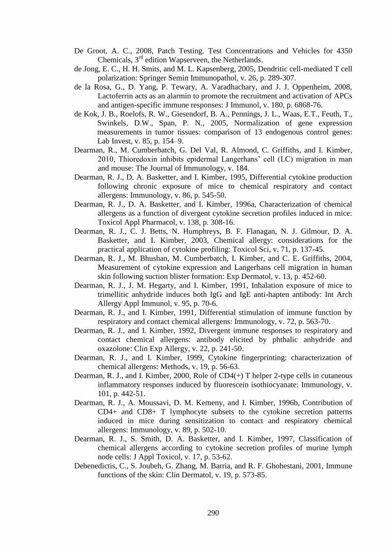
290
De Groot, A. C., 2008, Patch Testing. Test Concentrations and Vehicles for 4350
Chemicals, 3rd
edition Wapserveen, the Netherlands.
de Jong, E. C., H. H. Smits, and M. L. Kapsenberg, 2005, Dendritic cell-mediated T cell
polarization: Springer Semin Immunopathol, v. 26, p. 289-307.
de la Rosa, G., D. Yang, P. Tewary, A. Varadhachary, and J. J. Oppenheim, 2008,
Lactoferrin acts as an alarmin to promote the recruitment and activation of APCs
and antigen-specific immune responses: J Immunol, v. 180, p. 6868-76.
de Kok, J. B., Roelofs, R. W., Giesendorf, B. A., Pennings, J. L., Waas, E.T., Feuth, T.,
Swinkels, D.W., Span, P. N., 2005, Normalization of gene expression
measurements in tumor tissues: comparison of 13 endogenous control genes:
Lab Invest, v. 85, p. 154–9.
Dearman, R., M. Cumberbatch, G. Del Val, R. Almond, C. Griffiths, and I. Kimber,
2010, Thioredoxin inhibits epidermal Langerhans’ cell (LC) migration in man
and mouse: The Journal of Immunology, v. 184.
Dearman, R. J., D. A. Basketter, and I. Kimber, 1995, Differential cytokine production
following chronic exposure of mice to chemical respiratory and contact
allergens: Immunology, v. 86, p. 545-50.
Dearman, R. J., D. A. Basketter, and I. Kimber, 1996a, Characterization of chemical
allergens as a function of divergent cytokine secretion profiles induced in mice:
Toxicol Appl Pharmacol, v. 138, p. 308-16.
Dearman, R. J., C. J. Betts, N. Humphreys, B. F. Flanagan, N. J. Gilmour, D. A.
Basketter, and I. Kimber, 2003, Chemical allergy: considerations for the
practical application of cytokine profiling: Toxicol Sci, v. 71, p. 137-45.
Dearman, R. J., M. Bhushan, M. Cumberbatch, I. Kimber, and C. E. Griffiths, 2004,
Measurement of cytokine expression and Langerhans cell migration in human
skin following suction blister formation: Exp Dermatol, v. 13, p. 452-60.
Dearman, R. J., J. M. Hegarty, and I. Kimber, 1991, Inhalation exposure of mice to
trimellitic anhydride induces both IgG and IgE anti-hapten antibody: Int Arch
Allergy Appl Immunol, v. 95, p. 70-6.
Dearman, R. J., and I. Kimber, 1991, Differential stimulation of immune function by
respiratory and contact chemical allergens: Immunology, v. 72, p. 563-70.
Dearman, R. J., and I. Kimber, 1992, Divergent immune responses to respiratory and
contact chemical allergens: antibody elicited by phthalic anhydride and
oxazolone: Clin Exp Allergy, v. 22, p. 241-50.
Dearman, R. J., and I. Kimber, 1999, Cytokine fingerprinting: characterization of
chemical allergens: Methods, v. 19, p. 56-63.
Dearman, R. J., and I. Kimber, 2000, Role of CD4(+) T helper 2-type cells in cutaneous
inflammatory responses induced by fluorescein isothiocyanate: Immunology, v.
101, p. 442-51.
Dearman, R. J., A. Moussavi, D. M. Kemeny, and I. Kimber, 1996b, Contribution of
CD4+ and CD8+ T lymphocyte subsets to the cytokine secretion patterns
induced in mice during sensitization to contact and respiratory chemical
allergens: Immunology, v. 89, p. 502-10.
Dearman, R. J., S. Smith, D. A. Basketter, and I. Kimber, 1997, Classification of
chemical allergens according to cytokine secretion profiles of murine lymph
node cells: J Appl Toxicol, v. 17, p. 53-62.
Debenedictis, C., S. Joubeh, G. Zhang, M. Barria, and R. F. Ghohestani, 2001, Immune
functions of the skin: Clin Dermatol, v. 19, p. 573-85.
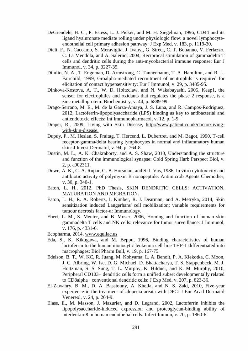
291
DeGrendele, H. C., P. Estess, L. J. Picker, and M. H. Siegelman, 1996, CD44 and its
ligand hyaluronate mediate rolling under physiologic flow: a novel lymphocyte-
endothelial cell primary adhesion pathway: J Exp Med, v. 183, p. 1119-30.
Dieli, F., N. Caccamo, S. Meraviglia, J. Ivanyi, G. Sireci, C. T. Bonanno, V. Ferlazzo,
C. La Mendola, and A. Salerno, 2004, Reciprocal stimulation of gammadelta T
cells and dendritic cells during the anti-mycobacterial immune response: Eur J
Immunol, v. 34, p. 3227-35.
Dilulio, N. A., T. Engeman, D. Armstrong, C. Tannenbaum, T. A. Hamilton, and R. L.
Fairchild, 1999, Groalpha-mediated recruitment of neutrophils is required for
elicitation of contact hypersensitivity: Eur J Immunol, v. 29, p. 3485-95.
Dinkova-Kostova, A. T., W. D. Holtzclaw, and N. Wakabayashi, 2005, Keap1, the
sensor for electrophiles and oxidants that regulates the phase 2 response, is a
zinc metalloprotein: Biochemistry, v. 44, p. 6889-99.
Drago-Serrano, M. E., M. de la Garza-Amaya, J. S. Luna, and R. Campos-Rodriguez,
2012, Lactoferrin-lipopolysaccharide (LPS) binding as key to antibacterial and
antiendotoxic effects: Int Immunopharmacol, v. 12, p. 1-9.
Draper, R., 2009, Living with Skin Disease, http://www.patient.co.uk/doctor/living-
with-skin-disease.
Dupuy, P., M. Heslan, S. Fraitag, T. Hercend, L. Dubertret, and M. Bagot, 1990, T-cell
receptor-gamma/delta bearing lymphocytes in normal and inflammatory human
skin: J Invest Dermatol, v. 94, p. 764-8.
Dustin, M. L., A. K. Chakraborty, and A. S. Shaw, 2010, Understanding the structure
and function of the immunological synapse: Cold Spring Harb Perspect Biol, v.
2, p. a002311.
Duwe, A. K., C. A. Rupar, G. B. Horsman, and S. I. Vas, 1986, In vitro cytotoxicity and
antibiotic activity of polymyxin B nonapeptide: Antimicrob Agents Chemother,
v. 30, p. 340-1.
Eaton, L. H., 2012, PhD Thesis, SKIN DENDRITIC CELLS: ACTIVATION,
MATURATION AND MIGRATION.
Eaton, L. H., R. A. Roberts, I. Kimber, R. J. Dearman, and A. Metryka, 2014, Skin
sensitization induced Langerhans' cell mobilization: variable requirements for
tumour necrosis factor-α: Immunology.
Ebert, L. M., S. Meuter, and B. Moser, 2006, Homing and function of human skin
gammadelta T cells and NK cells: relevance for tumor surveillance: J Immunol,
v. 176, p. 4331-6.
Ecopharma, 2014, www.equilac.us
Eda, S., K. Kikugawa, and M. Beppu, 1996, Binding characteristics of human
lactoferrin to the human monocytic leukemia cell line THP-1 differentiated into
macrophages: Biol Pharm Bull, v. 19, p. 167-75.
Edelson, B. T., W. KC, R. Juang, M. Kohyama, L. A. Benoit, P. A. Klekotka, C. Moon,
J. C. Albring, W. Ise, D. G. Michael, D. Bhattacharya, T. S. Stappenbeck, M. J.
Holtzman, S. S. Sung, T. L. Murphy, K. Hildner, and K. M. Murphy, 2010,
Peripheral CD103+ dendritic cells form a unified subset developmentally related
to CD8alpha+ conventional dendritic cells: J Exp Med, v. 207, p. 823-36.
El-Zawahry, B. M., D. A. Bassiouny, A. Khella, and N. S. Zaki, 2010, Five-year
experience in the treatment of alopecia areata with DPC: J Eur Acad Dermatol
Venereol, v. 24, p. 264-9.
Elass, E., M. Masson, J. Mazurier, and D. Legrand, 2002, Lactoferrin inhibits the
lipopolysaccharide-induced expression and proteoglycan-binding ability of
interleukin-8 in human endothelial cells: Infect Immun, v. 70, p. 1860-6.
292
Elliott, M. R., F. B. Chekeni, P. C. Trampont, E. R. Lazarowski, A. Kadl, S. F. Walk, D.
Park, R. I. Woodson, M. Ostankovich, P. Sharma, J. J. Lysiak, T. K. Harden, N.
Leitinger, and K. S. Ravichandran, 2009, Nucleotides released by apoptotic cells
act as a find-me signal to promote phagocytic clearance: Nature, v. 461, p. 282-
6.
Elssner, A., M. Duncan, M. Gavrilin, and M. D. Wewers, 2004, A novel P2X7 receptor
activator, the human cathelicidin-derived peptide LL37, induces IL-1 beta
processing and release: J Immunol, v. 172, p. 4987-94.
Emedicinehealth, 2014, Contact Dermatitis Symptoms and Signs,
http://www.emedicinehealth.com/contact_dermatitis/page3_em.htm#Contact%2
0Dermatitis%20Symptoms.
Enk, A. H., V. L. Angeloni, M. C. Udey, and S. I. Katz, 1993, An essential role for
Langerhans cell-derived IL-1 beta in the initiation of primary immune responses
in skin: J Immunol, v. 150, p. 3698-704.
Enk, A. H., and S. I. Katz, 1992a, Early molecular events in the induction phase of
contact sensitivity: Proc Natl Acad Sci U S A, v. 89, p. 1398-402.
Enk, A. H., and S. I. Katz, 1992b, Identification and induction of keratinocyte-derived
IL-10: J Immunol, v. 149, p. 92-5.
Esaguy, N., A. P. Aguas, and M. T. Silva, 1989, High-resolution localization of
lactoferrin in human neutrophils: labeling of secondary granules and cell
heterogeneity: J Leukoc Biol, v. 46, p. 51-62.
Esser, P. R., U. Wölfle, C. Dürr, F. D. von Loewenich, C. M. Schempp, M. A.
Freudenberg, T. Jakob, and S. F. Martin, 2012, Contact sensitizers induce skin
inflammation via ROS production and hyaluronic acid degradation: PLoS One,
v. 7, p. e41340.
Fadok, V. A., D. L. Bratton, A. Konowal, P. W. Freed, J. Y. Westcott, and P. M.
Henson, 1998, Macrophages that have ingested apoptotic cells in vitro inhibit
proinflammatory cytokine production through autocrine/paracrine mechanisms
involving TGF-beta, PGE2, and PAF: J Clin Invest, v. 101, p. 890-8.
Farahani, L. A., M. Ghobadzadeh, and P. Yousefi, 2013, Comparison of the effect of
human milk and topical hydrocortisone 1% on diaper dermatitis: Pediatr
Dermatol, v. 30, p. 725-9.
Farraj, A. K., J. R. Harkema, and N. E. Kaminski, 2004, Allergic rhinitis induced by
intranasal sensitization and challenge with trimellitic anhydride but not with
dinitrochlorobenzene or oxazolone in A/J mice: Toxicol Sci, v. 79, p. 315-25.
Faustman, D., and M. Davis, 2010, TNF receptor 2 pathway: drug target for
autoimmune diseases: Nat Rev Drug Discov, v. 9, p. 482-93.
Fischer-Stabauer, M., A. Boehner, S. Eyerich, T. Carbone, C. Traidl-Hoffmann, C. B.
Schmidt-Weber, A. Cavani, J. Ring, R. Hein, and K. Eyerich, 2012, Differential
in situ expression of IL-17 in skin diseases: Eur J Dermatol, v. 22, p. 781-4.
Flint, M. S., R. J. Dearman, I. Kimber, and S. A. Hotchkiss, 1998, Production and in
situ localization of cutaneous tumour necrosis factor alpha (TNF-alpha) and
interleukin 6 (IL-6) following skin sensitization: Cytokine, v. 10, p. 213-9.
Flores-Villaseñor, H., A. Canizalez-Román, M. Reyes-Lopez, K. Nazmi, M. de la
Garza, J. Zazueta-Beltrán, N. León-Sicairos, and J. G. Bolscher, 2010,
Bactericidal effect of bovine lactoferrin, LFcin, LFampin and LFchimera on
antibiotic-resistant Staphylococcus aureus and Escherichia coli: Biometals, v.
23, p. 569-78.
Fonacier, L. S., S. C. Dreskin, and D. Y. Leung, 2010, Allergic skin diseases: J Allergy
Clin Immunol, v. 125, p. S138-49.
293
Friedmann, P. S., C. Moss, S. Shuster, and J. M. Simpson, 1983, Quantitative
relationships between sensitizing dose of DNCB and reactivity in normal
subjects: Clin Exp Immunol, v. 53, p. 709-15.
Fukui, A., K. Ohta, H. Nishi, H. Shigeishi, K. Tobiume, M. Takechi, and N. Kamata,
2013, Interleukin-8 and CXCL10 expression in oral keratinocytes and
fibroblasts via Toll-like receptors: Microbiol Immunol, v. 57, p. 198-206.
Furlong, S. J., N. D. Ridgway, and D. W. Hoskin, 2008, Modulation of ceramide
metabolism in T-leukemia cell lines potentiates apoptosis induced by the
cationic antimicrobial peptide bovine lactoferricin: Int J Oncol, v. 32, p. 537-44.
Furmanski, P., Z. P. Li, M. B. Fortuna, C. V. Swamy, and M. R. Das, 1989, Multiple
molecular forms of human lactoferrin. Identification of a class of lactoferrins
that possess ribonuclease activity and lack iron-binding capacity: J Exp Med, v.
170, p. 415-29.
Gallo, R. L., R. Staszewski, D. N. Sauder, T. L. Knisely, and R. D. Granstein, 1991,
Regulation of GM-CSF and IL-3 production from the murine keratinocyte cell
line PAM 212 following exposure to ultraviolet radiation: J Invest Dermatol, v.
97, p. 203-9.
Gao, B., and M. F. Tsan, 2003, Endotoxin contamination in recombinant human heat
shock protein 70 (Hsp70) preparation is responsible for the induction of tumor
necrosis factor alpha release by murine macrophages: J Biol Chem, v. 278, p.
174-9.
Gao, F., J. R. Koenitzer, J. M. Tobolewski, D. Jiang, J. Liang, P. W. Noble, and T. D.
Oury, 2008, Extracellular superoxide dismutase inhibits inflammation by
preventing oxidative fragmentation of hyaluronan: J Biol Chem, v. 283, p. 6058-
66.
Garioch, J. J., R. M. Mackie, I. Campbell, and A. Forsyth, 1991, Keratinocyte
expression of intercellular adhesion molecule 1 (ICAM-1) correlated with
infiltration of lymphocyte function associated antigen 1 (LFA-1) positive cells in
evolving allergic contact dermatitis reactions: Histopathology, v. 19, p. 351-4.
Geijtenbeek, T. B., R. Torensma, S. J. van Vliet, G. C. van Duijnhoven, G. J. Adema,
Y. van Kooyk, and C. G. Figdor, 2000, Identification of DC-SIGN, a novel
dendritic cell-specific ICAM-3 receptor that supports primary immune
responses: Cell, v. 100, p. 575-85.
Geissmann, F., S. Jung, and D. R. Littman, 2003, Blood monocytes consist of two
principal subsets with distinct migratory properties: Immunity, v. 19, p. 71-82.
Gerberick, F., M. Aleksic, D. Basketter, S. Casati, A. T. Karlberg, P. Kern, I. Kimber, J.
P. Lepoittevin, A. Natsch, J. M. Ovigne, C. Rovida, H. Sakaguchi, and T.
Schultz, 2008, Chemical reactivity measurement and the predicitve
identification of skin sensitisers. The report and recommendations of ECVAM
Workshop 64: Altern Lab Anim, v. 36, p. 215-42.
Gerberick, G. F., J. D. Vassallo, R. E. Bailey, J. G. Chaney, S. W. Morrall, and J. P.
Lepoittevin, 2004, Development of a peptide reactivity assay for screening
contact allergens: Toxicol Sci, v. 81, p. 332-43.
Ghaznawie, M., J. M. Papadimitriou, and P. J. Heenan, 1999, The repopulation of
murine Langerhans cells after depletion by mild heat injury: Br J Dermatol, v.
141, p. 206-10.
Gibbs, B. F., J. Wierecky, P. Welker, B. M. Henz, H. H. Wolff, and J. Grabbe, 2001,
Human skin mast cells rapidly release preformed and newly generated TNF-
alpha and IL-8 following stimulation with anti-IgE and other secretagogues: Exp
Dermatol, v. 10, p. 312-20.
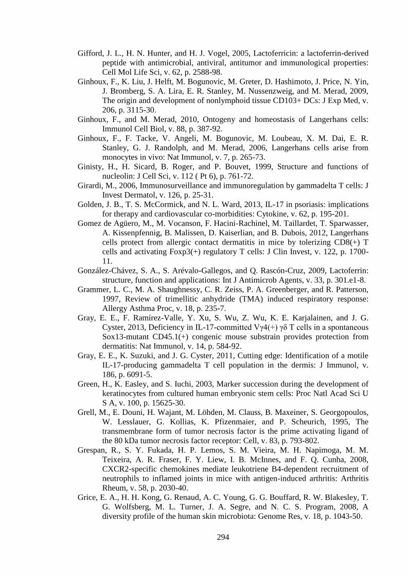
294
Gifford, J. L., H. N. Hunter, and H. J. Vogel, 2005, Lactoferricin: a lactoferrin-derived
peptide with antimicrobial, antiviral, antitumor and immunological properties:
Cell Mol Life Sci, v. 62, p. 2588-98.
Ginhoux, F., K. Liu, J. Helft, M. Bogunovic, M. Greter, D. Hashimoto, J. Price, N. Yin,
J. Bromberg, S. A. Lira, E. R. Stanley, M. Nussenzweig, and M. Merad, 2009,
The origin and development of nonlymphoid tissue CD103+ DCs: J Exp Med, v.
206, p. 3115-30.
Ginhoux, F., and M. Merad, 2010, Ontogeny and homeostasis of Langerhans cells:
Immunol Cell Biol, v. 88, p. 387-92.
Ginhoux, F., F. Tacke, V. Angeli, M. Bogunovic, M. Loubeau, X. M. Dai, E. R.
Stanley, G. J. Randolph, and M. Merad, 2006, Langerhans cells arise from
monocytes in vivo: Nat Immunol, v. 7, p. 265-73.
Ginisty, H., H. Sicard, B. Roger, and P. Bouvet, 1999, Structure and functions of
nucleolin: J Cell Sci, v. 112 ( Pt 6), p. 761-72.
Girardi, M., 2006, Immunosurveillance and immunoregulation by gammadelta T cells: J
Invest Dermatol, v. 126, p. 25-31.
Golden, J. B., T. S. McCormick, and N. L. Ward, 2013, IL-17 in psoriasis: implications
for therapy and cardiovascular co-morbidities: Cytokine, v. 62, p. 195-201.
Gomez de Agüero, M., M. Vocanson, F. Hacini-Rachinel, M. Taillardet, T. Sparwasser,
A. Kissenpfennig, B. Malissen, D. Kaiserlian, and B. Dubois, 2012, Langerhans
cells protect from allergic contact dermatitis in mice by tolerizing CD8(+) T
cells and activating Foxp3(+) regulatory T cells: J Clin Invest, v. 122, p. 1700-
11.
González-Chávez, S. A., S. Arévalo-Gallegos, and Q. Rascón-Cruz, 2009, Lactoferrin:
structure, function and applications: Int J Antimicrob Agents, v. 33, p. 301.e1-8.
Grammer, L. C., M. A. Shaughnessy, C. R. Zeiss, P. A. Greenberger, and R. Patterson,
1997, Review of trimellitic anhydride (TMA) induced respiratory response:
Allergy Asthma Proc, v. 18, p. 235-7.
Gray, E. E., F. Ramírez-Valle, Y. Xu, S. Wu, Z. Wu, K. E. Karjalainen, and J. G.
Cyster, 2013, Deficiency in IL-17-committed Vγ4(+) γδ T cells in a spontaneous
Sox13-mutant CD45.1(+) congenic mouse substrain provides protection from
dermatitis: Nat Immunol, v. 14, p. 584-92.
Gray, E. E., K. Suzuki, and J. G. Cyster, 2011, Cutting edge: Identification of a motile
IL-17-producing gammadelta T cell population in the dermis: J Immunol, v.
186, p. 6091-5.
Green, H., K. Easley, and S. Iuchi, 2003, Marker succession during the development of
keratinocytes from cultured human embryonic stem cells: Proc Natl Acad Sci U
S A, v. 100, p. 15625-30.
Grell, M., E. Douni, H. Wajant, M. Löhden, M. Clauss, B. Maxeiner, S. Georgopoulos,
W. Lesslauer, G. Kollias, K. Pfizenmaier, and P. Scheurich, 1995, The
transmembrane form of tumor necrosis factor is the prime activating ligand of
the 80 kDa tumor necrosis factor receptor: Cell, v. 83, p. 793-802.
Grespan, R., S. Y. Fukada, H. P. Lemos, S. M. Vieira, M. H. Napimoga, M. M.
Teixeira, A. R. Fraser, F. Y. Liew, I. B. McInnes, and F. Q. Cunha, 2008,
CXCR2-specific chemokines mediate leukotriene B4-dependent recruitment of
neutrophils to inflamed joints in mice with antigen-induced arthritis: Arthritis
Rheum, v. 58, p. 2030-40.
Grice, E. A., H. H. Kong, G. Renaud, A. C. Young, G. G. Bouffard, R. W. Blakesley, T.
G. Wolfsberg, M. L. Turner, J. A. Segre, and N. C. S. Program, 2008, A
diversity profile of the human skin microbiota: Genome Res, v. 18, p. 1043-50.
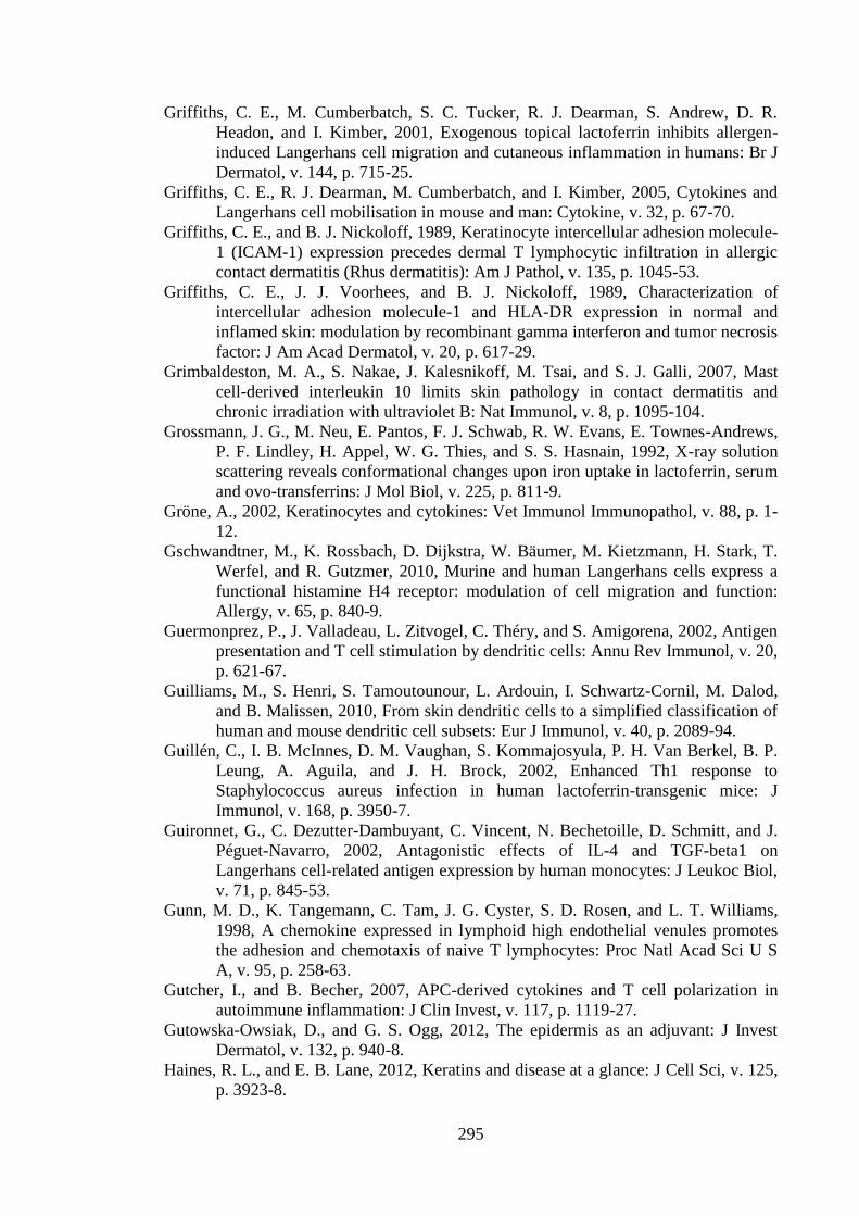
295
Griffiths, C. E., M. Cumberbatch, S. C. Tucker, R. J. Dearman, S. Andrew, D. R.
Headon, and I. Kimber, 2001, Exogenous topical lactoferrin inhibits allergen-
induced Langerhans cell migration and cutaneous inflammation in humans: Br J
Dermatol, v. 144, p. 715-25.
Griffiths, C. E., R. J. Dearman, M. Cumberbatch, and I. Kimber, 2005, Cytokines and
Langerhans cell mobilisation in mouse and man: Cytokine, v. 32, p. 67-70.
Griffiths, C. E., and B. J. Nickoloff, 1989, Keratinocyte intercellular adhesion molecule-
1 (ICAM-1) expression precedes dermal T lymphocytic infiltration in allergic
contact dermatitis (Rhus dermatitis): Am J Pathol, v. 135, p. 1045-53.
Griffiths, C. E., J. J. Voorhees, and B. J. Nickoloff, 1989, Characterization of
intercellular adhesion molecule-1 and HLA-DR expression in normal and
inflamed skin: modulation by recombinant gamma interferon and tumor necrosis
factor: J Am Acad Dermatol, v. 20, p. 617-29.
Grimbaldeston, M. A., S. Nakae, J. Kalesnikoff, M. Tsai, and S. J. Galli, 2007, Mast
cell-derived interleukin 10 limits skin pathology in contact dermatitis and
chronic irradiation with ultraviolet B: Nat Immunol, v. 8, p. 1095-104.
Grossmann, J. G., M. Neu, E. Pantos, F. J. Schwab, R. W. Evans, E. Townes-Andrews,
P. F. Lindley, H. Appel, W. G. Thies, and S. S. Hasnain, 1992, X-ray solution
scattering reveals conformational changes upon iron uptake in lactoferrin, serum
and ovo-transferrins: J Mol Biol, v. 225, p. 811-9.
Gröne, A., 2002, Keratinocytes and cytokines: Vet Immunol Immunopathol, v. 88, p. 1-
12.
Gschwandtner, M., K. Rossbach, D. Dijkstra, W. Bäumer, M. Kietzmann, H. Stark, T.
Werfel, and R. Gutzmer, 2010, Murine and human Langerhans cells express a
functional histamine H4 receptor: modulation of cell migration and function:
Allergy, v. 65, p. 840-9.
Guermonprez, P., J. Valladeau, L. Zitvogel, C. Théry, and S. Amigorena, 2002, Antigen
presentation and T cell stimulation by dendritic cells: Annu Rev Immunol, v. 20,
p. 621-67.
Guilliams, M., S. Henri, S. Tamoutounour, L. Ardouin, I. Schwartz-Cornil, M. Dalod,
and B. Malissen, 2010, From skin dendritic cells to a simplified classification of
human and mouse dendritic cell subsets: Eur J Immunol, v. 40, p. 2089-94.
Guillén, C., I. B. McInnes, D. M. Vaughan, S. Kommajosyula, P. H. Van Berkel, B. P.
Leung, A. Aguila, and J. H. Brock, 2002, Enhanced Th1 response to
Staphylococcus aureus infection in human lactoferrin-transgenic mice: J
Immunol, v. 168, p. 3950-7.
Guironnet, G., C. Dezutter-Dambuyant, C. Vincent, N. Bechetoille, D. Schmitt, and J.
Péguet-Navarro, 2002, Antagonistic effects of IL-4 and TGF-beta1 on
Langerhans cell-related antigen expression by human monocytes: J Leukoc Biol,
v. 71, p. 845-53.
Gunn, M. D., K. Tangemann, C. Tam, J. G. Cyster, S. D. Rosen, and L. T. Williams,
1998, A chemokine expressed in lymphoid high endothelial venules promotes
the adhesion and chemotaxis of naive T lymphocytes: Proc Natl Acad Sci U S
A, v. 95, p. 258-63.
Gutcher, I., and B. Becher, 2007, APC-derived cytokines and T cell polarization in
autoimmune inflammation: J Clin Invest, v. 117, p. 1119-27.
Gutowska-Owsiak, D., and G. S. Ogg, 2012, The epidermis as an adjuvant: J Invest
Dermatol, v. 132, p. 940-8.
Haines, R. L., and E. B. Lane, 2012, Keratins and disease at a glance: J Cell Sci, v. 125,
p. 3923-8.
296
Haley, K., B. Z. Igyártó, D. Ortner, A. Bobr, S. Kashem, D. Schenten, and D. H.
Kaplan, 2012, Langerhans cells require MyD88-dependent signals for Candida
albicans response but not for contact hypersensitivity or migration: J Immunol,
v. 188, p. 4334-9.
Haniffa, M., M. Collin, and F. Ginhoux, 2013, Ontogeny and functional specialization
of dendritic cells in human and mouse: Adv Immunol, v. 120, p. 1-49.
Haniffa, M., A. Shin, V. Bigley, N. McGovern, P. Teo, P. See, P. S. Wasan, X. N.
Wang, F. Malinarich, B. Malleret, A. Larbi, P. Tan, H. Zhao, M. Poidinger, S.
Pagan, S. Cookson, R. Dickinson, I. Dimmick, R. F. Jarrett, L. Renia, J. Tam, C.
Song, J. Connolly, J. K. Chan, A. Gehring, A. Bertoletti, M. Collin, and F.
Ginhoux, 2012, Human tissues contain CD141hi cross-presenting dendritic cells
with functional homology to mouse CD103+ nonlymphoid dendritic cells:
Immunity, v. 37, p. 60-73.
Havran, W. L., Y. H. Chien, and J. P. Allison, 1991, Recognition of self antigens by
skin-derived T cells with invariant gamma delta antigen receptors: Science, v.
252, p. 1430-2.
Hayes, M. D., 2012, PhD Thesis, THE ROLE OF INNATE AND ADAPTIVE IL-17
PRODUCING CELLS IN CHEMICAL ALLERGY.
Hazuda, D. J., J. C. Lee, and P. R. Young, 1988, The kinetics of interleukin 1 secretion
from activated monocytes. Differences between interleukin 1 alpha and
interleukin 1 beta: J Biol Chem, v. 263, p. 8473-9.
He, S., A. R. McEuen, S. A. Blewett, P. Li, M. G. Buckley, P. Leufkens, and A. F.
Walls, 2003, The inhibition of mast cell activation by neutrophil lactoferrin:
uptake by mast cells and interaction with tryptase, chymase and cathepsin G:
Biochem Pharmacol, v. 65, p. 1007-15.
Heath, W. R., and F. R. Carbone, 2013, The skin-resident and migratory immune
system in steady state and memory: innate lymphocytes, dendritic cells and T
cells: Nat Immunol, v. 14, p. 978-85.
Heib, V., M. Becker, T. Warger, G. Rechtsteiner, C. Tertilt, M. Klein, T. Bopp, C.
Taube, H. Schild, E. Schmitt, and M. Stassen, 2007, Mast cells are crucial for
early inflammation, migration of Langerhans cells, and CTL responses
following topical application of TLR7 ligand in mice: Blood, v. 110, p. 946-53.
Heilborn, J. D., M. F. Nilsson, G. Kratz, G. Weber, O. Sørensen, N. Borregaard, and M.
Ståhle-Bäckdahl, 2003, The cathelicidin anti-microbial peptide LL-37 is
involved in re-epithelialization of human skin wounds and is lacking in chronic
ulcer epithelium: J Invest Dermatol, v. 120, p. 379-89.
Helft, J., F. Ginhoux, M. Bogunovic, and M. Merad, 2010, Origin and functional
heterogeneity of non-lymphoid tissue dendritic cells in mice: Immunol Rev, v.
234, p. 55-75.
Heller, F., I. J. Fuss, E. E. Nieuwenhuis, R. S. Blumberg, and W. Strober, 2002,
Oxazolone colitis, a Th2 colitis model resembling ulcerative colitis, is mediated
by IL-13-producing NK-T cells: Immunity, v. 17, p. 629-38.
Heylings, J. R., H. M. Clowes, M. Cumberbatch, R. J. Dearman, I. Fielding, J. Hilton,
and I. Kimber, 1996, Sensitization to 2,4-dinitrochlorobenzene: influence of
vehicle on absorption and lymph node activation: Toxicology, v. 109, p. 57-65.
Hilton , Dearman RJ, Basketter DA, and K. I, 1995, Identification of chemical
respiratory allergens : dose response relationships in the mouse IgE test.:
Toxicology Methods., 50-51 p.
297
Hilton J, Dearman RJ, Basketter DA, and K. I, 1995, Identification of chemical
respiratory allergens : dose response relationships in the mouse IgE test.:
Toxicology Methods., 50-51 p.
Hirano, K., Y. Miki, Y. Hirai, R. Sato, T. Itoh, A. Hayashi, M. Yamanaka, S. Eda, and
M. Beppu, 2005, A multifunctional shuttling protein nucleolin is a macrophage
receptor for apoptotic cells: J Biol Chem, v. 280, p. 39284-93.
Hogg, N., R. Henderson, B. Leitinger, A. McDowall, J. Porter, and P. Stanley, 2002,
Mechanisms contributing to the activity of integrins on leukocytes: Immunol
Rev, v. 186, p. 164-71.
Hol, J., L. Wilhelmsen, and G. Haraldsen, 2010, The murine IL-8 homologues KC,
MIP-2, and LIX are found in endothelial cytoplasmic granules but not in
Weibel-Palade bodies: J Leukoc Biol, v. 87, p. 501-8.
Honda, T., G. Egawa, S. Grabbe, and K. Kabashima, 2013, Update of immune events in
the murine contact hypersensitivity model: toward the understanding of allergic
contact dermatitis: J Invest Dermatol, v. 133, p. 303-15.
Hopkins, J. E., D. J. Naisbitt, N. R. Kitteringham, R. J. Dearman, I. Kimber, and B. K.
Park, 2005, Selective haptenation of cellular or extracellular protein by chemical
allergens: association with cytokine polarization: Chem Res Toxicol, v. 18, p.
375-81.
Hot, A., S. Zrioual, V. Lenief, and P. Miossec, 2012, IL-17 and tumour necrosis factor α
combination induces a HIF-1α-dependent invasive phenotype in synoviocytes:
Ann Rheum Dis, v. 71, p. 1393-401.
Howell, M. D., 2007, The role of human beta defensins and cathelicidins in atopic
dermatitis: Curr Opin Allergy Clin Immunol, v. 7, p. 413-7.
HSE, 2013, Work-related skin disease in Great Britain, in H. a. S. E. UK, ed.,
www.hse.gov.uk/statistics/causdis/dermatitis/skin.pdf.
Huang, K., G. Du, C. Wei, S. Gu, and J. Tang, 2013, Elevated serum lactoferrin and
neopterin are associated with postoperative infectious complications in patients
with acute traumatic spinal cord injury: Arch Med Sci, v. 9, p. 865-71.
Hwang, J., A. Kalinin, M. Hwang, D. E. Anderson, M. J. Kim, O. Stojadinovic, M.
Tomic-Canic, S. H. Lee, and M. I. Morasso, 2007, Role of Scarf and its binding
target proteins in epidermal calcium homeostasis: J Biol Chem, v. 282, p.
18645-53.
Håversen, L., B. G. Ohlsson, M. Hahn-Zoric, L. A. Hanson, and I. Mattsby-Baltzer,
2002, Lactoferrin down-regulates the LPS-induced cytokine production in
monocytic cells via NF-kappa B: Cell Immunol, v. 220, p. 83-95.
Igyarto, B. Z., M. C. Jenison, J. C. Dudda, A. Roers, W. Müller, P. A. Koni, D. J.
Campbell, M. J. Shlomchik, and D. H. Kaplan, 2009, Langerhans cells suppress
contact hypersensitivity responses via cognate CD4 interaction and langerhans
cell-derived IL-10: J Immunol, v. 183, p. 5085-93.
Igyártó, B. Z., K. Haley, D. Ortner, A. Bobr, M. Gerami-Nejad, B. T. Edelson, S. M.
Zurawski, B. Malissen, G. Zurawski, J. Berman, and D. H. Kaplan, 2011, Skin-
resident murine dendritic cell subsets promote distinct and opposing antigen-
specific T helper cell responses: Immunity, v. 35, p. 260-72.
Igyártó, B. Z., and D. H. Kaplan, 2013, Antigen presentation by Langerhans cells: Curr
Opin Immunol, v. 25, p. 115-9.
Ikarashi, Y., T. Tsuchiya, and A. Nakamura, 1993, A sensitive mouse lymph node assay
with two application phases for detection of contact allergens: Arch Toxicol, v.
67, p. 629-36.
298
Ismaili, J., V. Olislagers, R. Poupot, J. J. Fournié, and M. Goldman, 2002, Human
gamma delta T cells induce dendritic cell maturation: Clin Immunol, v. 103, p.
296-302.
Ivaska, J., H. M. Pallari, J. Nevo, and J. E. Eriksson, 2007, Novel functions of vimentin
in cell adhesion, migration, and signaling: Exp Cell Res, v. 313, p. 2050-62.
Jain, J., C. Loh, and A. Rao, 1995, Transcriptional regulation of the IL-2 gene: Curr
Opin Immunol, v. 7, p. 333-42.
Jakob, T., and M. C. Udey, 1998, Regulation of E-cadherin-mediated adhesion in
Langerhans cell-like dendritic cells by inflammatory mediators that mobilize
Langerhans cells in vivo: J Immunol, v. 160, p. 4067-73.
Jakubzick, C., F. Tacke, F. Ginhoux, A. J. Wagers, N. van Rooijen, M. Mack, M.
Merad, and G. J. Randolph, 2008, Blood monocyte subsets differentially give
rise to CD103+ and CD103- pulmonary dendritic cell populations: J Immunol, v.
180, p. 3019-27.
Jameson, J., and W. L. Havran, 2007, Skin gammadelta T-cell functions in homeostasis
and wound healing: Immunol Rev, v. 215, p. 114-22.
Jarvis, M. A., J. A. Borton, A. M. Keech, J. Wong, W. J. Britt, B. E. Magun, and J. A.
Nelson, 2006, Human cytomegalovirus attenuates interleukin-1beta and tumor
necrosis factor alpha proinflammatory signaling by inhibition of NF-kappaB
activation: J Virol, v. 80, p. 5588-98.
Jenkins, M. K., P. S. Taylor, S. D. Norton, and K. B. Urdahl, 1991, CD28 delivers a
costimulatory signal involved in antigen-specific IL-2 production by human T
cells: J Immunol, v. 147, p. 2461-6.
Jenkinson, C., R. E. Jenkins, M. Aleksic, M. Pirmohamed, D. J. Naisbitt, and B. K.
Park, 2010, Characterization of p-phenylenediamine-albumin binding sites and
T-cell responses to hapten-modified protein: J Invest Dermatol, v. 130, p. 732-
42.
Jensen, L. E., 2010, Targeting the IL-1 family members in skin inflammation: Curr
Opin Investig Drugs, v. 11, p. 1211-20.
Jha, S., and J. P. Ting, 2009, Inflammasome-associated nucleotide-binding domain,
leucine-rich repeat proteins and inflammatory diseases: J Immunol, v. 183, p.
7623-9.
Jiang, R., V. Lopez, S. L. Kelleher, and B. Lönnerdal, 2011, Apo- and holo-lactoferrin
are both internalized by lactoferrin receptor via clathrin-mediated endocytosis
but differentially affect ERK-signaling and cell proliferation in Caco-2 cells: J
Cell Physiol, v. 226, p. 3022-31.
Joffre, O. P., E. Segura, A. Savina, and S. Amigorena, 2012, Cross-presentation by
dendritic cells: Nat Rev Immunol, v. 12, p. 557-69.
Johnson, E. A., T. L. Dao, M. A. Guignet, C. E. Geddes, A. I. Koemeter-Cox, and R. K.
Kan, 2011, Increased expression of the chemokines CXCL1 and MIP-1α by
resident brain cells precedes neutrophil infiltration in the brain following
prolonged soman-induced status epilepticus in rats: J Neuroinflammation, v. 8,
p. 41.
Johnson, E. E., and M. Wessling-Resnick, 2012, Iron metabolism and the innate
immune response to infection: Microbes Infect, v. 14, p. 207-16.
Joss, A., M. Akdis, A. Faith, K. Blaser, and C. A. Akdis, 2000, IL-10 directly acts on T
cells by specifically altering the CD28 co-stimulation pathway: Eur J Immunol,
v. 30, p. 1683-90.
299
Jux, B., S. Kadow, and C. Esser, 2009, Langerhans cell maturation and contact
hypersensitivity are impaired in aryl hydrocarbon receptor-null mice: J
Immunol, v. 182, p. 6709-17.
Kabashima, K., and G. Egawa, 2014, Intravital Multiphoton Imaging of Cutaneous
Immune Responses: J Invest Dermatol.
Kabashima, K., N. Shiraishi, K. Sugita, T. Mori, A. Onoue, M. Kobayashi, J. Sakabe, R.
Yoshiki, H. Tamamura, N. Fujii, K. Inaba, and Y. Tokura, 2007, CXCL12-
CXCR4 engagement is required for migration of cutaneous dendritic cells: Am J
Pathol, v. 171, p. 1249-57.
Kadow, S., B. Jux, S. P. Zahner, B. Wingerath, S. Chmill, B. E. Clausen, J. Hengstler,
and C. Esser, 2011, Aryl hydrocarbon receptor is critical for homeostasis of
invariant gammadelta T cells in the murine epidermis: J Immunol, v. 187, p.
3104-10.
Kaminski, M. J., P. R. Bergstresser, and A. Takashima, 1993, In vivo activation of
mouse dendritic epidermal T cells in sites of contact dermatitis: Eur J Immunol,
v. 23, p. 1715-8.
Kane, B., U. Kolsum, T. Southworth, J. Armstrong, A. Woodcock, and D. Singh, 2009,
The effects of smoking on the lipopolysaccharide response and glucocorticoid
sensitivity of alveolar macrophages of patients with asthma: Chest, v. 136, p.
163-70.
Kanitakis, J., 2002, Anatomy, histology and immunohistochemistry of normal human
skin: Eur J Dermatol, v. 12, p. 390-9; quiz 400-1.
Kanyshkova, T. G., S. E. Babina, D. V. Semenov, N. Isaeva, A. V. Vlassov, K. N.
Neustroev, A. A. Kul'minskaya, V. N. Buneva, and G. A. Nevinsky, 2003,
Multiple enzymic activities of human milk lactoferrin: Eur J Biochem, v. 270, p.
3353-61.
Kaplan, D. H., 2010, In vivo function of Langerhans cells and dermal dendritic cells:
Trends Immunol, v. 31, p. 446-51.
Kaplan, D. H., B. Z. Igyártó, and A. A. Gaspari, 2012, Early immune events in the
induction of allergic contact dermatitis: Nat Rev Immunol, v. 12, p. 114-24.
Kaplan, D. H., A. Kissenpfennig, and B. E. Clausen, 2008, Insights into Langerhans cell
function from Langerhans cell ablation models: Eur J Immunol, v. 38, p. 2369-
76.
Karlberg, A. T., M. A. Bergström, A. Börje, K. Luthman, and J. L. Nilsson, 2008,
Allergic contact dermatitis--formation, structural requirements, and reactivity of
skin sensitizers: Chem Res Toxicol, v. 21, p. 53-69.
Katugampola, R. P., and A. Y. Finlay, 2007, Impact of skin diseases on quality of life:
Eur J Dermatol, v. 17, p. 102-6.
Katz, S. I., K. Tamaki, and D. H. Sachs, 1979, Epidermal Langerhans cells are derived
from cells originating in bone marrow: Nature, v. 282, p. 324-6.
Kawakami, H., and B. Lönnerdal, 1991, Isolation and function of a receptor for human
lactoferrin in human fetal intestinal brush-border membranes: Am J Physiol, v.
261, p. G841-6.
Kehrl, J. H., L. M. Wakefield, A. B. Roberts, S. Jakowlew, M. Alvarez-Mon, R.
Derynck, M. B. Sporn, and A. S. Fauci, 1986, Production of transforming
growth factor beta by human T lymphocytes and its potential role in the
regulation of T cell growth: J Exp Med, v. 163, p. 1037-50.
Keijsers, R. R., A. G. Hendriks, P. E. van Erp, B. van Cranenbroek, P. C. van de
Kerkhof, H. J. Koenen, and I. Joosten, 2014, In Vivo Induction of Cutaneous
300
Inflammation Results in the Accumulation of Extracellular Trap-Forming
Neutrophils Expressing RORγt and IL-17: J Invest Dermatol, v. 134, p. 1276-84.
Kennedy, B. P., P. Payette, J. Mudgett, P. Vadas, W. Pruzanski, M. Kwan, C. Tang, D.
E. Rancourt, and W. A. Cromlish, 1995, A natural disruption of the secretory
group II phospholipase A2 gene in inbred mouse strains: J Biol Chem, v. 270, p.
22378-85.
Kermani, F., M. S. Flint, and S. A. Hotchkiss, 2000, Induction and localization of
cutaneous interleukin-1 beta mRNA during contact sensitization: Toxicol Appl
Pharmacol, v. 169, p. 231-7.
Kijlstra, A., S. H. Jeurissen, and K. M. Koning, 1983, Lactoferrin levels in normal
human tears: Br J Ophthalmol, v. 67, p. 199-202.
Kim, C. W., T. H. Lee, K. H. Park, S. Y. Choi, and J. Kim, 2012, Human lactoferrin
suppresses TNF-α-induced intercellular adhesion molecule-1 expression via
competition with NF-κB in endothelial cells: FEBS Lett, v. 586, p. 229-34.
Kim, J., Y. Ko, Y. K. Park, N. I. Kim, W. K. Ha, and Y. Cho, 2010, Dietary effect of
lactoferrin-enriched fermented milk on skin surface lipid and clinical
improvement of acne vulgaris: Nutrition, v. 26, p. 902-9.
Kim, Y., Y. S. Lee, J. H. Hahn, J. Choe, H. J. Kwon, J. Y. Ro, and D. Jeoung, 2008,
Hyaluronic acid targets CD44 and inhibits FcepsilonRI signaling involving
PKCdelta, Rac1, ROS, and MAPK to exert anti-allergic effect: Mol Immunol, v.
45, p. 2537-47.
Kimber, I., D. A. Basketter, and R. J. Dearman, 2010, Chemical allergens--what are the
issues?: Toxicology, v. 268, p. 139-42.
Kimber, I., and R. J. Dearman, 1991, Investigation of lymph node cell proliferation as a
possible immunological correlate of contact sensitizing potential: Food Chem
Toxicol, v. 29, p. 125-9.
Kimber, I., R. J. Dearman, E. W. Scholes, and D. A. Basketter, 1994, The local lymph
node assay: developments and applications: Toxicology, v. 93, p. 13-31.
Kingston, D., M. A. Schmid, N. Onai, A. Obata-Onai, D. Baumjohann, and M. G.
Manz, 2009, The concerted action of GM-CSF and Flt3-ligand on in vivo
dendritic cell homeostasis: Blood, v. 114, p. 835-43.
Kish, D. D., A. V. Gorbachev, and R. L. Fairchild, 2012, IL-1 receptor signaling is
required at multiple stages of sensitization and elicitation of the contact
hypersensitivity response: J Immunol, v. 188, p. 1761-71.
Kish, D. D., N. Volokh, W. M. Baldwin, and R. L. Fairchild, 2011, Hapten application
to the skin induces an inflammatory program directing hapten-primed effector
CD8 T cell interaction with hapten-presenting endothelial cells: J Immunol, v.
186, p. 2117-26.
Kissenpfennig, A., S. Henri, B. Dubois, C. Laplace-Builhé, P. Perrin, N. Romani, C. H.
Tripp, P. Douillard, L. Leserman, D. Kaiserlian, S. Saeland, J. Davoust, and B.
Malissen, 2005, Dynamics and function of Langerhans cells in vivo: dermal
dendritic cells colonize lymph node areas distinct from slower migrating
Langerhans cells: Immunity, v. 22, p. 643-54.
Klechevsky, E., R. Morita, M. Liu, Y. Cao, S. Coquery, L. Thompson-Snipes, F. Briere,
D. Chaussabel, G. Zurawski, A. K. Palucka, Y. Reiter, J. Banchereau, and H.
Ueno, 2008, Functional specializations of human epidermal Langerhans cells
and CD14+ dermal dendritic cells: Immunity, v. 29, p. 497-510.
Klekotka, P. A., L. Yang, and W. M. Yokoyama, 2010, Contrasting roles of the IL-1
and IL-18 receptors in MyD88-dependent contact hypersensitivity: J Invest
Dermatol, v. 130, p. 184-91.
301
Knott, P. G., P. R. Gater, P. J. Dunford, M. E. Fuentes, and C. P. Bertrand, 2001, Rapid
up-regulation of CXC chemokines in the airways after Ag-specific CD4+ T cell
activation: J Immunol, v. 166, p. 1233-40.
Kobayashi, M., S. Nunomura, Y. Gon, D. Endo, S. Kishiro, M. Fukunaga, Y. Kitahata,
T. Terui, and C. Ra, 2010, Abrogation of high-affinity IgE receptor-mediated
mast cell activation at the effector phase prevents contact hypersensitivity to
oxazolone: J Invest Dermatol, v. 130, p. 725-31.
Kobayashi, Y., 1997, Langerhans' cells produce type IV collagenase (MMP-9)
following epicutaneous stimulation with haptens: Immunology, v. 90, p. 496-
501.
Kolaczkowska, E., and P. Kubes, 2013, Neutrophil recruitment and function in health
and inflammation: Nat Rev Immunol, v. 13, p. 159-75.
Komori, H. K., D. A. Witherden, R. Kelly, K. Sendaydiego, J. M. Jameson, L. Teyton,
and W. L. Havran, 2012, Cutting edge: dendritic epidermal γδ T cell ligands are
rapidly and locally expressed by keratinocytes following cutaneous wounding: J
Immunol, v. 188, p. 2972-6.
Kondo, S., D. N. Sauder, T. Kono, K. A. Galley, and R. C. McKenzie, 1994,
Differential modulation of interleukin-1 alpha (IL-1 alpha) and interleukin-1
beta (IL-1 beta) in human epidermal keratinocytes by UVB: Exp Dermatol, v. 3,
p. 29-39.
Kono, H., and K. L. Rock, 2008, How dying cells alert the immune system to danger:
Nat Rev Immunol, v. 8, p. 279-89.
Korn, T., E. Bettelli, M. Oukka, and V. K. Kuchroo, 2009, IL-17 and Th17 Cells: Annu
Rev Immunol, v. 27, p. 485-517.
Krob, H. A., A. B. Fleischer, R. D'Agostino, C. L. Haverstock, and S. Feldman, 2004,
Prevalence and relevance of contact dermatitis allergens: a meta-analysis of 15
years of published T.R.U.E. test data: J Am Acad Dermatol, v. 51, p. 349-53.
Kroeze, K. L., M. A. Boink, S. C. Sampat-Sardjoepersad, T. Waaijman, R. J. Scheper,
and S. Gibbs, 2012, Autocrine regulation of re-epithelialization after wounding
by chemokine receptors CCR1, CCR10, CXCR1, CXCR2, and CXCR3: J Invest
Dermatol, v. 132, p. 216-25.
Kruzel, M. L., J. K. Actor, M. Zimecki, J. Wise, P. Płoszaj, S. Mirza, M. Kruzel, S. A.
Hwang, X. Ba, and I. Boldogh, 2013, Novel recombinant human lactoferrin:
differential activation of oxidative stress related gene expression: J Biotechnol,
v. 168, p. 666-75.
Ku, H. O., S. H. Jeong, H. G. Kang, H. M. Pyo, J. H. Cho, S. W. Son, S. M. Yun, and
D. Y. Ryu, 2009, Gene expression profiles and pathways in skin inflammation
induced by three different sensitizers and an irritant: Toxicol Lett, v. 190, p.
231-7.
Kuno, K., and K. Matsushima, 1994, The IL-1 receptor signaling pathway: J Leukoc
Biol, v. 56, p. 542-7.
Kuper, C. F., M. Radonjic, J. van Triel, R. Stierum, R. J. de Groot, and J. H. Arts, 2011,
Oxazolone (OXA) is a respiratory allergen in Brown Norway rats: Toxicology,
v. 290, p. 59-68.
Kupper, T. S., D. W. Ballard, A. O. Chua, J. S. McGuire, P. M. Flood, M. C. Horowitz,
R. Langdon, L. Lightfoot, and U. Gubler, 1986, Human keratinocytes contain
mRNA indistinguishable from monocyte interleukin 1 alpha and beta mRNA.
Keratinocyte epidermal cell-derived thymocyte-activating factor is identical to
interleukin 1: J Exp Med, v. 164, p. 2095-100.
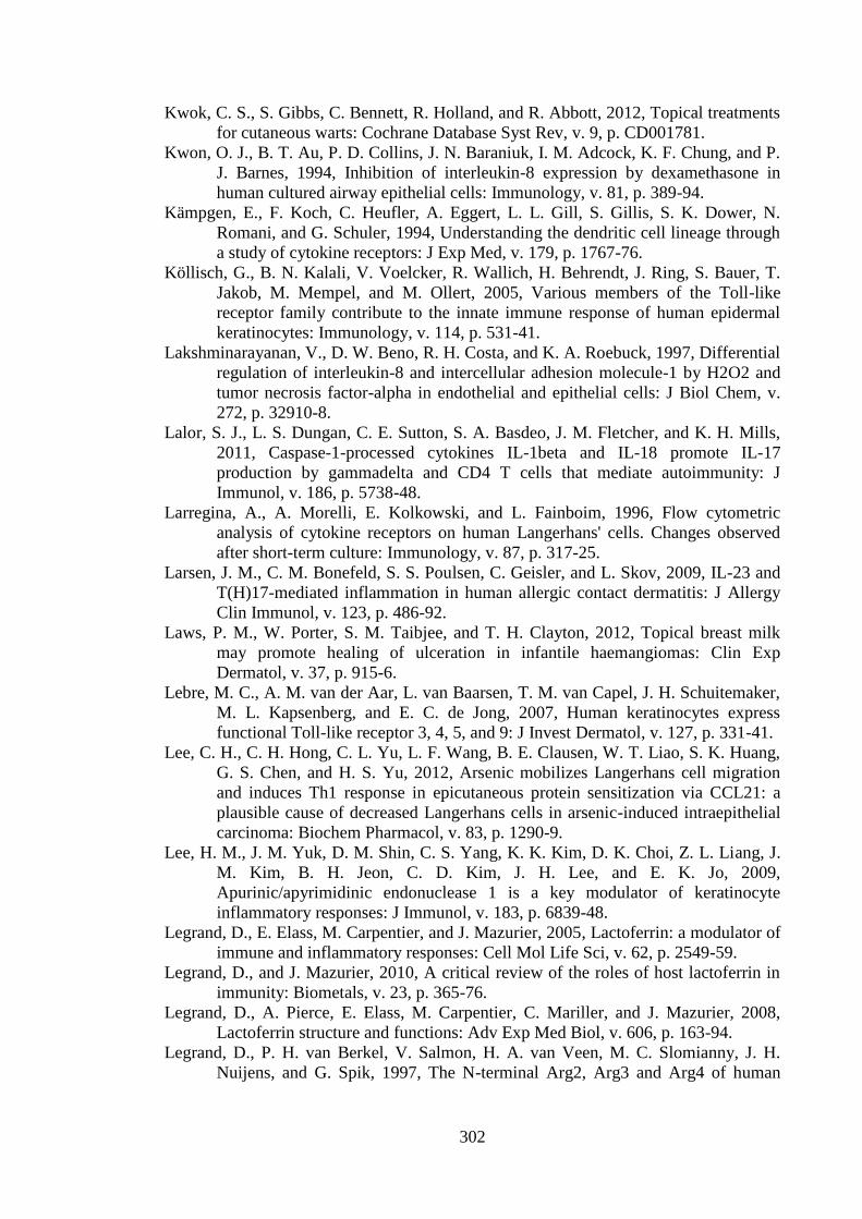
302
Kwok, C. S., S. Gibbs, C. Bennett, R. Holland, and R. Abbott, 2012, Topical treatments
for cutaneous warts: Cochrane Database Syst Rev, v. 9, p. CD001781.
Kwon, O. J., B. T. Au, P. D. Collins, J. N. Baraniuk, I. M. Adcock, K. F. Chung, and P.
J. Barnes, 1994, Inhibition of interleukin-8 expression by dexamethasone in
human cultured airway epithelial cells: Immunology, v. 81, p. 389-94.
Kämpgen, E., F. Koch, C. Heufler, A. Eggert, L. L. Gill, S. Gillis, S. K. Dower, N.
Romani, and G. Schuler, 1994, Understanding the dendritic cell lineage through
a study of cytokine receptors: J Exp Med, v. 179, p. 1767-76.
Köllisch, G., B. N. Kalali, V. Voelcker, R. Wallich, H. Behrendt, J. Ring, S. Bauer, T.
Jakob, M. Mempel, and M. Ollert, 2005, Various members of the Toll-like
receptor family contribute to the innate immune response of human epidermal
keratinocytes: Immunology, v. 114, p. 531-41.
Lakshminarayanan, V., D. W. Beno, R. H. Costa, and K. A. Roebuck, 1997, Differential
regulation of interleukin-8 and intercellular adhesion molecule-1 by H2O2 and
tumor necrosis factor-alpha in endothelial and epithelial cells: J Biol Chem, v.
272, p. 32910-8.
Lalor, S. J., L. S. Dungan, C. E. Sutton, S. A. Basdeo, J. M. Fletcher, and K. H. Mills,
2011, Caspase-1-processed cytokines IL-1beta and IL-18 promote IL-17
production by gammadelta and CD4 T cells that mediate autoimmunity: J
Immunol, v. 186, p. 5738-48.
Larregina, A., A. Morelli, E. Kolkowski, and L. Fainboim, 1996, Flow cytometric
analysis of cytokine receptors on human Langerhans' cells. Changes observed
after short-term culture: Immunology, v. 87, p. 317-25.
Larsen, J. M., C. M. Bonefeld, S. S. Poulsen, C. Geisler, and L. Skov, 2009, IL-23 and
T(H)17-mediated inflammation in human allergic contact dermatitis: J Allergy
Clin Immunol, v. 123, p. 486-92.
Laws, P. M., W. Porter, S. M. Taibjee, and T. H. Clayton, 2012, Topical breast milk
may promote healing of ulceration in infantile haemangiomas: Clin Exp
Dermatol, v. 37, p. 915-6.
Lebre, M. C., A. M. van der Aar, L. van Baarsen, T. M. van Capel, J. H. Schuitemaker,
M. L. Kapsenberg, and E. C. de Jong, 2007, Human keratinocytes express
functional Toll-like receptor 3, 4, 5, and 9: J Invest Dermatol, v. 127, p. 331-41.
Lee, C. H., C. H. Hong, C. L. Yu, L. F. Wang, B. E. Clausen, W. T. Liao, S. K. Huang,
G. S. Chen, and H. S. Yu, 2012, Arsenic mobilizes Langerhans cell migration
and induces Th1 response in epicutaneous protein sensitization via CCL21: a
plausible cause of decreased Langerhans cells in arsenic-induced intraepithelial
carcinoma: Biochem Pharmacol, v. 83, p. 1290-9.
Lee, H. M., J. M. Yuk, D. M. Shin, C. S. Yang, K. K. Kim, D. K. Choi, Z. L. Liang, J.
M. Kim, B. H. Jeon, C. D. Kim, J. H. Lee, and E. K. Jo, 2009,
Apurinic/apyrimidinic endonuclease 1 is a key modulator of keratinocyte
inflammatory responses: J Immunol, v. 183, p. 6839-48.
Legrand, D., E. Elass, M. Carpentier, and J. Mazurier, 2005, Lactoferrin: a modulator of
immune and inflammatory responses: Cell Mol Life Sci, v. 62, p. 2549-59.
Legrand, D., and J. Mazurier, 2010, A critical review of the roles of host lactoferrin in
immunity: Biometals, v. 23, p. 365-76.
Legrand, D., A. Pierce, E. Elass, M. Carpentier, C. Mariller, and J. Mazurier, 2008,
Lactoferrin structure and functions: Adv Exp Med Biol, v. 606, p. 163-94.
Legrand, D., P. H. van Berkel, V. Salmon, H. A. van Veen, M. C. Slomianny, J. H.
Nuijens, and G. Spik, 1997, The N-terminal Arg2, Arg3 and Arg4 of human
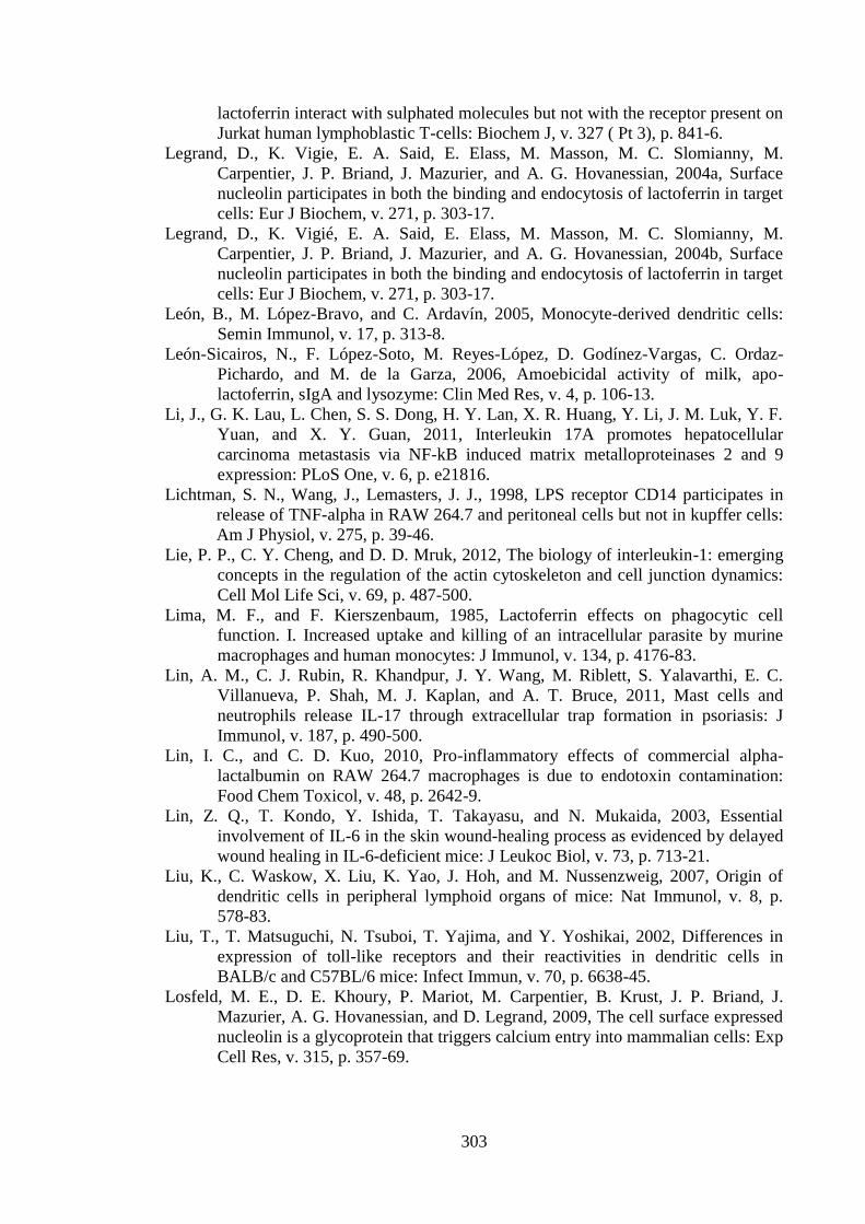
303
lactoferrin interact with sulphated molecules but not with the receptor present on
Jurkat human lymphoblastic T-cells: Biochem J, v. 327 ( Pt 3), p. 841-6.
Legrand, D., K. Vigie, E. A. Said, E. Elass, M. Masson, M. C. Slomianny, M.
Carpentier, J. P. Briand, J. Mazurier, and A. G. Hovanessian, 2004a, Surface
nucleolin participates in both the binding and endocytosis of lactoferrin in target
cells: Eur J Biochem, v. 271, p. 303-17.
Legrand, D., K. Vigié, E. A. Said, E. Elass, M. Masson, M. C. Slomianny, M.
Carpentier, J. P. Briand, J. Mazurier, and A. G. Hovanessian, 2004b, Surface
nucleolin participates in both the binding and endocytosis of lactoferrin in target
cells: Eur J Biochem, v. 271, p. 303-17.
León, B., M. López-Bravo, and C. Ardavín, 2005, Monocyte-derived dendritic cells:
Semin Immunol, v. 17, p. 313-8.
León-Sicairos, N., F. López-Soto, M. Reyes-López, D. Godínez-Vargas, C. Ordaz-
Pichardo, and M. de la Garza, 2006, Amoebicidal activity of milk, apo-
lactoferrin, sIgA and lysozyme: Clin Med Res, v. 4, p. 106-13.
Li, J., G. K. Lau, L. Chen, S. S. Dong, H. Y. Lan, X. R. Huang, Y. Li, J. M. Luk, Y. F.
Yuan, and X. Y. Guan, 2011, Interleukin 17A promotes hepatocellular
carcinoma metastasis via NF-kB induced matrix metalloproteinases 2 and 9
expression: PLoS One, v. 6, p. e21816.
Lichtman, S. N., Wang, J., Lemasters, J. J., 1998, LPS receptor CD14 participates in
release of TNF-alpha in RAW 264.7 and peritoneal cells but not in kupffer cells:
Am J Physiol, v. 275, p. 39-46.
Lie, P. P., C. Y. Cheng, and D. D. Mruk, 2012, The biology of interleukin-1: emerging
concepts in the regulation of the actin cytoskeleton and cell junction dynamics:
Cell Mol Life Sci, v. 69, p. 487-500.
Lima, M. F., and F. Kierszenbaum, 1985, Lactoferrin effects on phagocytic cell
function. I. Increased uptake and killing of an intracellular parasite by murine
macrophages and human monocytes: J Immunol, v. 134, p. 4176-83.
Lin, A. M., C. J. Rubin, R. Khandpur, J. Y. Wang, M. Riblett, S. Yalavarthi, E. C.
Villanueva, P. Shah, M. J. Kaplan, and A. T. Bruce, 2011, Mast cells and
neutrophils release IL-17 through extracellular trap formation in psoriasis: J
Immunol, v. 187, p. 490-500.
Lin, I. C., and C. D. Kuo, 2010, Pro-inflammatory effects of commercial alpha-
lactalbumin on RAW 264.7 macrophages is due to endotoxin contamination:
Food Chem Toxicol, v. 48, p. 2642-9.
Lin, Z. Q., T. Kondo, Y. Ishida, T. Takayasu, and N. Mukaida, 2003, Essential
involvement of IL-6 in the skin wound-healing process as evidenced by delayed
wound healing in IL-6-deficient mice: J Leukoc Biol, v. 73, p. 713-21.
Liu, K., C. Waskow, X. Liu, K. Yao, J. Hoh, and M. Nussenzweig, 2007, Origin of
dendritic cells in peripheral lymphoid organs of mice: Nat Immunol, v. 8, p.
578-83.
Liu, T., T. Matsuguchi, N. Tsuboi, T. Yajima, and Y. Yoshikai, 2002, Differences in
expression of toll-like receptors and their reactivities in dendritic cells in
BALB/c and C57BL/6 mice: Infect Immun, v. 70, p. 6638-45.
Losfeld, M. E., D. E. Khoury, P. Mariot, M. Carpentier, B. Krust, J. P. Briand, J.
Mazurier, A. G. Hovanessian, and D. Legrand, 2009, The cell surface expressed
nucleolin is a glycoprotein that triggers calcium entry into mammalian cells: Exp
Cell Res, v. 315, p. 357-69.
304
Lundqvist, E. N., and O. Bäck, 1990, Interleukin-1 decreases the number of Ia+
epidermal dendritic cells but increases their expression of Ia antigen: Acta Derm
Venereol, v. 70, p. 391-4.
Lönnerdal, B., and S. Iyer, 1995, Lactoferrin: molecular structure and biological
function: Annu Rev Nutr, v. 15, p. 93-110.
Macleod, A. S., and W. L. Havran, 2011, Functions of skin-resident γδ T cells: Cell Mol
Life Sci, v. 68, p. 2399-408.
Mader, J. S., D. Smyth, J. Marshall, and D. W. Hoskin, 2006, Bovine lactoferricin
inhibits basic fibroblast growth factor- and vascular endothelial growth
factor165-induced angiogenesis by competing for heparin-like binding sites on
endothelial cells: Am J Pathol, v. 169, p. 1753-66.
Magalhães, P. O., A. M. Lopes, P. G. Mazzola, C. Rangel-Yagui, T. C. Penna, and A.
Pessoa, 2007, Methods of endotoxin removal from biological preparations: a
review: J Pharm Pharm Sci, v. 10, p. 388-404.
Mangan, P. R., L. E. Harrington, D. B. O'Quinn, W. S. Helms, D. C. Bullard, C. O.
Elson, R. D. Hatton, S. M. Wahl, T. R. Schoeb, and C. T. Weaver, 2006,
Transforming growth factor-beta induces development of the T(H)17 lineage:
Nature, v. 441, p. 231-4.
Mantel, C., K. Miyazawa, and H. E. Broxmeyer, 1994, Physical characteristics and
polymerization during iron saturation of lactoferrin, a myelopoietic regulatory
molecule with suppressor activity: Adv Exp Med Biol, v. 357, p. 121-32.
Marthinuss, J., L. Lawrence, and M. Seiberg, 1995, Apoptosis in Pam212, an epidermal
keratinocyte cell line: a possible role for bcl-2 in epidermal differentiation: Cell
Growth Differ, v. 6, p. 239-50.
Martin, S. F., J. C. Dudda, E. Bachtanian, A. Lembo, S. Liller, C. Dürr, M. M.
Heimesaat, S. Bereswill, G. Fejer, R. Vassileva, T. Jakob, N. Freudenberg, C. C.
Termeer, C. Johner, C. Galanos, and M. A. Freudenberg, 2008, Toll-like
receptor and IL-12 signaling control susceptibility to contact hypersensitivity: J
Exp Med, v. 205, p. 2151-62.
Matsue, H., P. R. Bergstresser, and A. Takashima, 1993a, Keratinocyte-derived IL-7
serves as a growth factor for dendritic epidermal T cells in mice: J Immunol, v.
151, p. 6012-9.
Matsue, H., P. D. Cruz, P. R. Bergstresser, and A. Takashima, 1992a, Cytokine
expression by epidermal cell subpopulations: J Invest Dermatol, v. 99, p. 42S-
45S.
Matsue, H., P. D. Cruz, P. R. Bergstresser, and A. Takashima, 1992b, Langerhans cells
are the major source of mRNA for IL-1 beta and MIP-1 alpha among
unstimulated mouse epidermal cells: J Invest Dermatol, v. 99, p. 537-41.
Matsue, H., P. D. Cruz, P. R. Bergstresser, and A. Takashima, 1993b, Profiles of
cytokine mRNA expressed by dendritic epidermal T cells in mice: J Invest
Dermatol, v. 101, p. 537-42.
Matsue, H., D. Edelbaum, D. Shalhevet, N. Mizumoto, C. Yang, M. E. Mummert, J.
Oeda, H. Masayasu, and A. Takashima, 2003, Generation and function of
reactive oxygen species in dendritic cells during antigen presentation: J
Immunol, v. 171, p. 3010-8.
Matsushima, H., Y. Ogawa, T. Miyazaki, H. Tanaka, A. Nishibu, and A. Takashima,
2010, Intravital imaging of IL-1beta production in skin: J Invest Dermatol, v.
130, p. 1571-80.
Matzinger, P., 1994, Tolerance, danger, and the extended family: Annu Rev Immunol,
v. 12, p. 991-1045.
305
Mayumi, N., E. Watanabe, Y. Norose, E. Watari, S. Kawana, T. B. Geijtenbeek, and H.
Takahashi, 2013, E-cadherin interactions are required for Langerhans cell
differentiation: Eur J Immunol, v. 43, p. 270-80.
McAbee, D. D., W. Nowatzke, C. Oehler, M. Sitaram, E. Sbaschnig, J. T. Opferman, J.
Carr, and K. Esbensen, 1993, Endocytosis and degradation of bovine apo- and
holo-lactoferrin by isolated rat hepatocytes are mediated by recycling calcium-
dependent binding sites: Biochemistry, v. 32, p. 13749-60.
McConnell, I., J. Hopkins, and P. Lachmann, 1980, Lymphocyte traffic through lymph
nodes during cell shutdown: Ciba Found Symp, v. 71, p. 167-95.
McGuire, J., R. Langdon, N. Birchall, and T. Kupper, 1988, Interleukin-1 alpha mRNA
induced by cycloheximide PMA, and retinoic acid is reduced by dexamethasone
in PAM-212 keratinocytes: Ann N Y Acad Sci, v. 548, p. 283-90.
McHale, J. F., O. A. Harari, D. Marshall, and D. O. Haskard, 1999, Vascular endothelial
cell expression of ICAM-1 and VCAM-1 at the onset of eliciting contact
hypersensitivity in mice: evidence for a dominant role of TNF-alpha: J
Immunol, v. 162, p. 1648-55.
McKenna, H. J., K. L. Stocking, R. E. Miller, K. Brasel, T. De Smedt, E. Maraskovsky,
C. R. Maliszewski, D. H. Lynch, J. Smith, B. Pulendran, E. R. Roux, M. Teepe,
S. D. Lyman, and J. J. Peschon, 2000, Mice lacking flt3 ligand have deficient
hematopoiesis affecting hematopoietic progenitor cells, dendritic cells, and
natural killer cells: Blood, v. 95, p. 3489-97.
Mee, J. B., C. Antonopoulos, S. Poole, T. S. Kupper, and R. W. Groves, 2005, Counter-
regulation of interleukin-1alpha (IL-1alpha) and IL-1 receptor antagonist in
murine keratinocytes: J Invest Dermatol, v. 124, p. 1267-74.
Mempel, M., V. Voelcker, G. Köllisch, C. Plank, R. Rad, M. Gerhard, C. Schnopp, P.
Fraunberger, A. K. Walli, J. Ring, D. Abeck, and M. Ollert, 2003, Toll-like
receptor expression in human keratinocytes: nuclear factor kappaB controlled
gene activation by Staphylococcus aureus is toll-like receptor 2 but not toll-like
receptor 4 or platelet activating factor receptor dependent: J Invest Dermatol, v.
121, p. 1389-96.
Merad, M., F. Ginhoux, and M. Collin, 2008, Origin, homeostasis and function of
Langerhans cells and other langerin-expressing dendritic cells: Nat Rev
Immunol, v. 8, p. 935-47.
Merad, M., M. G. Manz, H. Karsunky, A. Wagers, W. Peters, I. Charo, I. L. Weissman,
J. G. Cyster, and E. G. Engleman, 2002, Langerhans cells renew in the skin
throughout life under steady-state conditions: Nat Immunol, v. 3, p. 1135-41.
Merad, M., P. Sathe, J. Helft, J. Miller, and A. Mortha, 2013, The dendritic cell lineage:
ontogeny and function of dendritic cells and their subsets in the steady state and
the inflamed setting: Annu Rev Immunol, v. 31, p. 563-604.
Middleton, M. H., and D. A. Norris, 1995, Cytokine-induced ICAM-1 expression in
human keratinocytes is highly variable in keratinocyte strains from different
donors: J Invest Dermatol, v. 104, p. 489-96.
Miki, Y., H. Dambara, Y. Tachibana, K. Hirano, M. Konishi, and M. Beppu, 2014,
Macrophage recognition of toxic advanced glycosylation end products through
the macrophage surface-receptor nucleolin: Biol Pharm Bull, v. 37, p. 588-96.
Miki, Y., K. Hirano, and M. Beppu, 2012, Macrophage recognition of thiol-group
oxidized cells: recognition of carbohydrate chains by macrophage surface
nucleolin as apoptotic cells: Biosci Biotechnol Biochem, v. 76, p. 2068-74.
Miki, Y., T. Tazawa, K. Hirano, H. Matsushima, S. Kumamoto, N. Hamasaki, T.
Yamaguchi, and M. Beppu, 2007, Clearance of oxidized erythrocytes by
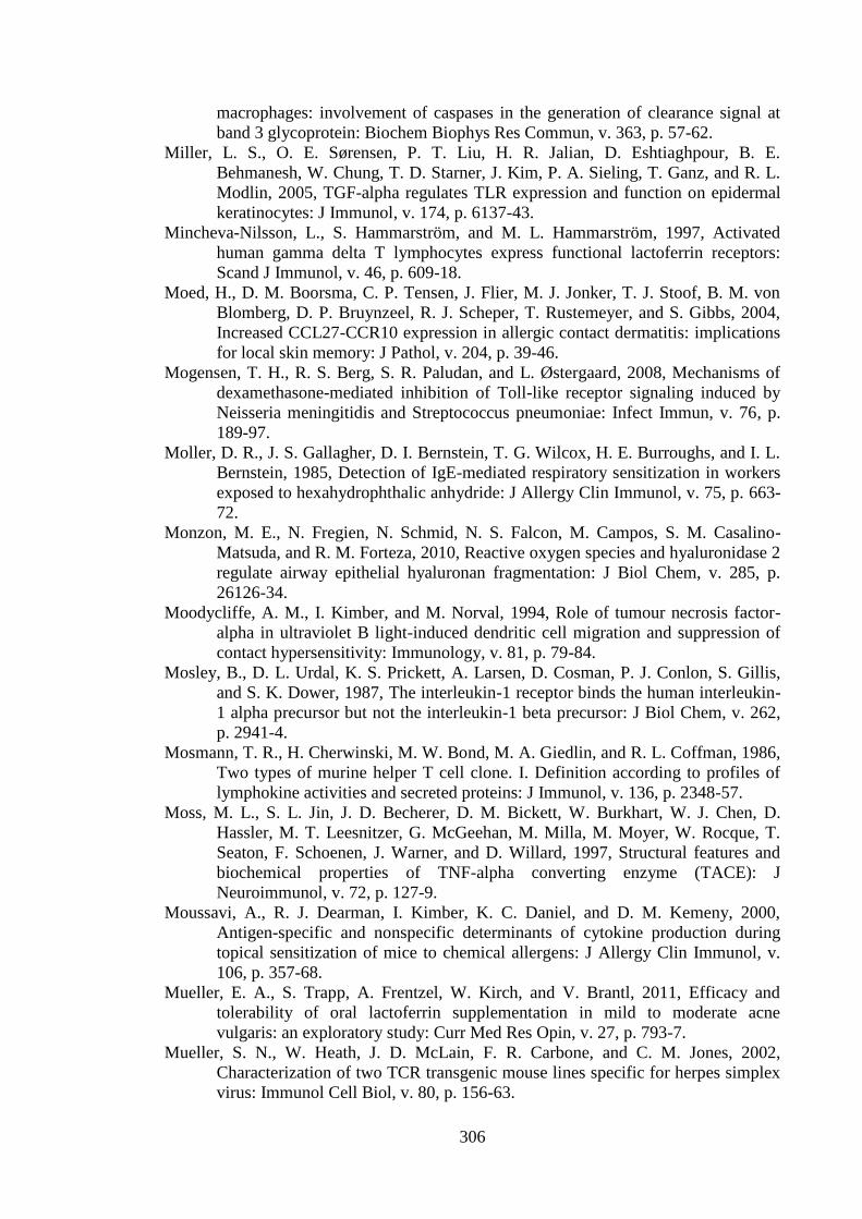
306
macrophages: involvement of caspases in the generation of clearance signal at
band 3 glycoprotein: Biochem Biophys Res Commun, v. 363, p. 57-62.
Miller, L. S., O. E. Sørensen, P. T. Liu, H. R. Jalian, D. Eshtiaghpour, B. E.
Behmanesh, W. Chung, T. D. Starner, J. Kim, P. A. Sieling, T. Ganz, and R. L.
Modlin, 2005, TGF-alpha regulates TLR expression and function on epidermal
keratinocytes: J Immunol, v. 174, p. 6137-43.
Mincheva-Nilsson, L., S. Hammarström, and M. L. Hammarström, 1997, Activated
human gamma delta T lymphocytes express functional lactoferrin receptors:
Scand J Immunol, v. 46, p. 609-18.
Moed, H., D. M. Boorsma, C. P. Tensen, J. Flier, M. J. Jonker, T. J. Stoof, B. M. von
Blomberg, D. P. Bruynzeel, R. J. Scheper, T. Rustemeyer, and S. Gibbs, 2004,
Increased CCL27-CCR10 expression in allergic contact dermatitis: implications
for local skin memory: J Pathol, v. 204, p. 39-46.
Mogensen, T. H., R. S. Berg, S. R. Paludan, and L. Østergaard, 2008, Mechanisms of
dexamethasone-mediated inhibition of Toll-like receptor signaling induced by
Neisseria meningitidis and Streptococcus pneumoniae: Infect Immun, v. 76, p.
189-97.
Moller, D. R., J. S. Gallagher, D. I. Bernstein, T. G. Wilcox, H. E. Burroughs, and I. L.
Bernstein, 1985, Detection of IgE-mediated respiratory sensitization in workers
exposed to hexahydrophthalic anhydride: J Allergy Clin Immunol, v. 75, p. 663-
72.
Monzon, M. E., N. Fregien, N. Schmid, N. S. Falcon, M. Campos, S. M. Casalino-
Matsuda, and R. M. Forteza, 2010, Reactive oxygen species and hyaluronidase 2
regulate airway epithelial hyaluronan fragmentation: J Biol Chem, v. 285, p.
26126-34.
Moodycliffe, A. M., I. Kimber, and M. Norval, 1994, Role of tumour necrosis factor-
alpha in ultraviolet B light-induced dendritic cell migration and suppression of
contact hypersensitivity: Immunology, v. 81, p. 79-84.
Mosley, B., D. L. Urdal, K. S. Prickett, A. Larsen, D. Cosman, P. J. Conlon, S. Gillis,
and S. K. Dower, 1987, The interleukin-1 receptor binds the human interleukin-
1 alpha precursor but not the interleukin-1 beta precursor: J Biol Chem, v. 262,
p. 2941-4.
Mosmann, T. R., H. Cherwinski, M. W. Bond, M. A. Giedlin, and R. L. Coffman, 1986,
Two types of murine helper T cell clone. I. Definition according to profiles of
lymphokine activities and secreted proteins: J Immunol, v. 136, p. 2348-57.
Moss, M. L., S. L. Jin, J. D. Becherer, D. M. Bickett, W. Burkhart, W. J. Chen, D.
Hassler, M. T. Leesnitzer, G. McGeehan, M. Milla, M. Moyer, W. Rocque, T.
Seaton, F. Schoenen, J. Warner, and D. Willard, 1997, Structural features and
biochemical properties of TNF-alpha converting enzyme (TACE): J
Neuroimmunol, v. 72, p. 127-9.
Moussavi, A., R. J. Dearman, I. Kimber, K. C. Daniel, and D. M. Kemeny, 2000,
Antigen-specific and nonspecific determinants of cytokine production during
topical sensitization of mice to chemical allergens: J Allergy Clin Immunol, v.
106, p. 357-68.
Mueller, E. A., S. Trapp, A. Frentzel, W. Kirch, and V. Brantl, 2011, Efficacy and
tolerability of oral lactoferrin supplementation in mild to moderate acne
vulgaris: an exploratory study: Curr Med Res Opin, v. 27, p. 793-7.
Mueller, S. N., W. Heath, J. D. McLain, F. R. Carbone, and C. M. Jones, 2002,
Characterization of two TCR transgenic mouse lines specific for herpes simplex
virus: Immunol Cell Biol, v. 80, p. 156-63.
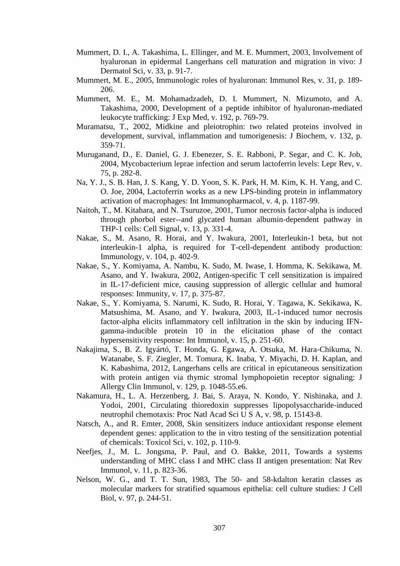
307
Mummert, D. I., A. Takashima, L. Ellinger, and M. E. Mummert, 2003, Involvement of
hyaluronan in epidermal Langerhans cell maturation and migration in vivo: J
Dermatol Sci, v. 33, p. 91-7.
Mummert, M. E., 2005, Immunologic roles of hyaluronan: Immunol Res, v. 31, p. 189-
206.
Mummert, M. E., M. Mohamadzadeh, D. I. Mummert, N. Mizumoto, and A.
Takashima, 2000, Development of a peptide inhibitor of hyaluronan-mediated
leukocyte trafficking: J Exp Med, v. 192, p. 769-79.
Muramatsu, T., 2002, Midkine and pleiotrophin: two related proteins involved in
development, survival, inflammation and tumorigenesis: J Biochem, v. 132, p.
359-71.
Muruganand, D., E. Daniel, G. J. Ebenezer, S. E. Rabboni, P. Segar, and C. K. Job,
2004, Mycobacterium leprae infection and serum lactoferrin levels: Lepr Rev, v.
75, p. 282-8.
Na, Y. J., S. B. Han, J. S. Kang, Y. D. Yoon, S. K. Park, H. M. Kim, K. H. Yang, and C.
O. Joe, 2004, Lactoferrin works as a new LPS-binding protein in inflammatory
activation of macrophages: Int Immunopharmacol, v. 4, p. 1187-99.
Naitoh, T., M. Kitahara, and N. Tsuruzoe, 2001, Tumor necrosis factor-alpha is induced
through phorbol ester--and glycated human albumin-dependent pathway in
THP-1 cells: Cell Signal, v. 13, p. 331-4.
Nakae, S., M. Asano, R. Horai, and Y. Iwakura, 2001, Interleukin-1 beta, but not
interleukin-1 alpha, is required for T-cell-dependent antibody production:
Immunology, v. 104, p. 402-9.
Nakae, S., Y. Komiyama, A. Nambu, K. Sudo, M. Iwase, I. Homma, K. Sekikawa, M.
Asano, and Y. Iwakura, 2002, Antigen-specific T cell sensitization is impaired
in IL-17-deficient mice, causing suppression of allergic cellular and humoral
responses: Immunity, v. 17, p. 375-87.
Nakae, S., Y. Komiyama, S. Narumi, K. Sudo, R. Horai, Y. Tagawa, K. Sekikawa, K.
Matsushima, M. Asano, and Y. Iwakura, 2003, IL-1-induced tumor necrosis
factor-alpha elicits inflammatory cell infiltration in the skin by inducing IFN-
gamma-inducible protein 10 in the elicitation phase of the contact
hypersensitivity response: Int Immunol, v. 15, p. 251-60.
Nakajima, S., B. Z. Igyártó, T. Honda, G. Egawa, A. Otsuka, M. Hara-Chikuma, N.
Watanabe, S. F. Ziegler, M. Tomura, K. Inaba, Y. Miyachi, D. H. Kaplan, and
K. Kabashima, 2012, Langerhans cells are critical in epicutaneous sensitization
with protein antigen via thymic stromal lymphopoietin receptor signaling: J
Allergy Clin Immunol, v. 129, p. 1048-55.e6.
Nakamura, H., L. A. Herzenberg, J. Bai, S. Araya, N. Kondo, Y. Nishinaka, and J.
Yodoi, 2001, Circulating thioredoxin suppresses lipopolysaccharide-induced
neutrophil chemotaxis: Proc Natl Acad Sci U S A, v. 98, p. 15143-8.
Natsch, A., and R. Emter, 2008, Skin sensitizers induce antioxidant response element
dependent genes: application to the in vitro testing of the sensitization potential
of chemicals: Toxicol Sci, v. 102, p. 110-9.
Neefjes, J., M. L. Jongsma, P. Paul, and O. Bakke, 2011, Towards a systems
understanding of MHC class I and MHC class II antigen presentation: Nat Rev
Immunol, v. 11, p. 823-36.
Nelson, W. G., and T. T. Sun, 1983, The 50- and 58-kdalton keratin classes as
molecular markers for stratified squamous epithelia: cell culture studies: J Cell
Biol, v. 97, p. 244-51.
308
Nestle, F. O., P. Di Meglio, J. Z. Qin, and B. J. Nickoloff, 2009, Skin immune sentinels
in health and disease: Nat Rev Immunol, v. 9, p. 679-91.
Netea, M. G., A. Simon, F. van de Veerdonk, B. J. Kullberg, J. W. Van der Meer, and
L. A. Joosten, 2010, IL-1beta processing in host defense: beyond the
inflammasomes: PLoS Pathog, v. 6, p. e1000661.
Nevinskii, A. G., S. E. Soboleva, F. V. Tuzikov, V. N. Buneva, and G. A. Nevinsky,
2009, DNA, oligosaccharides, and mononucleotides stimulate oligomerization
of human lactoferrin: J Mol Recognit, v. 22, p. 330-42.
Nibbering, P. H., E. Ravensbergen, M. M. Welling, L. A. van Berkel, P. H. van Berkel,
E. K. Pauwels, and J. H. Nuijens, 2001, Human lactoferrin and peptides derived
from its N terminus are highly effective against infections with antibiotic-
resistant bacteria: Infect Immun, v. 69, p. 1469-76.
Nickoloff, B. J., and L. A. Turka, 1994, Immunological functions of non-professional
antigen-presenting cells: new insights from studies of T-cell interactions with
keratinocytes: Immunol Today, v. 15, p. 464-9.
Nielsen, M. M., P. Lovato, A. S. MacLeod, D. A. Witherden, L. Skov, B. Dyring-
Andersen, S. Dabelsteen, A. Woetmann, N. Ødum, W. L. Havran, C. Geisler,
and C. M. Bonefeld, 2014, IL-1β-dependent activation of dendritic epidermal T
cells in contact hypersensitivity: J Immunol, v. 192, p. 2975-83.
Nieuwenhuis, E. E., S. Gillessen, R. J. Scheper, M. A. Exley, M. Taniguchi, S. P. Balk,
J. L. Strominger, G. Dranoff, R. S. Blumberg, and S. B. Wilson, 2005, CD1d
and CD1d-restricted iNKT-cells play a pivotal role in contact hypersensitivity:
Exp Dermatol, v. 14, p. 250-8.
Nishiya, K., and D. A. Horwitz, 1982, Contrasting effects of lactoferrin on human
lymphocyte and monocyte natural killer activity and antibody-dependent cell-
mediated cytotoxicity: J Immunol, v. 129, p. 2519-23.
Nisole, S., B. Krust, C. Callebaut, G. Guichard, S. Muller, J. P. Briand, and A. G.
Hovanessian, 1999, The anti-HIV pseudopeptide HB-19 forms a complex with
the cell-surface-expressed nucleolin independent of heparan sulfate
proteoglycans: J Biol Chem, v. 274, p. 27875-84.
Noordegraaf, M., V. Flacher, P. Stoitzner, and B. E. Clausen, 2010, Functional
redundancy of Langerhans cells and Langerin+ dermal dendritic cells in contact
hypersensitivity: J Invest Dermatol, v. 130, p. 2752-9.
Oakford, M. E., S. V. Dixon, S. August, C. Pickard, M. Ardern-Jones, P. Lackie, P. S.
Friedmann, and E. Healy, 2011, Migration of immunocytes across the basement
membrane in skin: the role of basement membrane pores: J Invest Dermatol, v.
131, p. 1950-3.
Oh, S. M., S. H. Lee, B. J. Lee, C. W. Pyo, N. K. Yoo, S. Y. Lee, J. Kim, and S. Y.
Choi, 2007, A distinct role of neutrophil lactoferrin in RelA/p65
phosphorylation on Ser536 by recruiting TNF receptor-associated factors to
IkappaB kinase signaling complex: J Immunol, v. 179, p. 5686-92.
Ohno, I., Y. Tanno, K. Yamauchi, and T. Takishima, 1990, Production of tumour
necrosis factor by mastocytoma P815 cells: Immunology, v. 69, p. 312-5.
Olaru, F., and L. E. Jensen, 2010, Chemokine expression by human keratinocyte cell
lines after activation of Toll-like receptors: Exp Dermatol, v. 19, p. e314-6.
Oliver, J., L. Bland, C. Oettinger, M. Arduino, S. McAllister, S. Aguero, and M. Favero,
1993, Cytokine kinetics in an in vitro whole blood model following an
endotoxin challenge.: Lymphokine Cytokine Res., v. 12, p. 6.
Onishi, R. M., and S. L. Gaffen, 2010, Interleukin-17 and its target genes: mechanisms
of interleukin-17 function in disease: Immunology, v. 129, p. 311-21.
309
Oram, J. D., and B. Reiter, 1968, Inhibition of bacteria by lactoferrin and other iron-
chelating agents: Biochim Biophys Acta, v. 170, p. 351-65.
Osborne-Hereford, A. V., S. W. Rogers, and L. C. Gahring, 2008, Neuronal nicotinic
alpha7 receptors modulate inflammatory cytokine production in the skin
following ultraviolet radiation: J Neuroimmunol, v. 193, p. 130-9.
Otsuka, A., M. Kubo, T. Honda, G. Egawa, S. Nakajima, H. Tanizaki, B. Kim, S.
Matsuoka, T. Watanabe, S. Nakae, Y. Miyachi, and K. Kabashima, 2011,
Requirement of interaction between mast cells and skin dendritic cells to
establish contact hypersensitivity: PLoS One, v. 6, p. e25538.
Ouwehand, K., S. J. Santegoets, D. P. Bruynzeel, R. J. Scheper, T. D. de Gruijl, and S.
Gibbs, 2008, CXCL12 is essential for migration of activated Langerhans cells
from epidermis to dermis: Eur J Immunol, v. 38, p. 3050-9.
Oyoshi, M. K., R. He, Y. Li, S. Mondal, J. Yoon, R. Afshar, M. Chen, D. M. Lee, H. R.
Luo, A. D. Luster, J. S. Cho, L. S. Miller, A. Larson, G. F. Murphy, and R. S.
Geha, 2012, Leukotriene B4-driven neutrophil recruitment to the skin is
essential for allergic skin inflammation: Immunity, v. 37, p. 747-58.
Parihar, D. B., S. P. Sharma, and K. K. Verma, 1967, Charge-transfer complexes of
2,4,6-trinitrophenyl-N-methylnitramine (tetryl) and 2,4-dinitrochlorobenzene
with some amines: J Chromatogr, v. 29, p. 258-62.
Peiser, M., T. Tralau, J. Heidler, A. M. Api, J. H. Arts, D. A. Basketter, J. English, T. L.
Diepgen, R. C. Fuhlbrigge, A. A. Gaspari, J. D. Johansen, A. T. Karlberg, I.
Kimber, J. P. Lepoittevin, M. Liebsch, H. I. Maibach, S. F. Martin, H. F. Merk,
T. Platzek, T. Rustemeyer, A. Schnuch, R. J. Vandebriel, I. R. White, and A.
Luch, 2012, Allergic contact dermatitis: epidemiology, molecular mechanisms,
in vitro methods and regulatory aspects. Current knowledge assembled at an
international workshop at BfR, Germany: Cell Mol Life Sci, v. 69, p. 763-81.
Peschon, J. J., D. S. Torrance, K. L. Stocking, M. B. Glaccum, C. Otten, C. R. Willis,
K. Charrier, P. J. Morrissey, C. B. Ware, and K. M. Mohler, 1998, TNF
receptor-deficient mice reveal divergent roles for p55 and p75 in several models
of inflammation: J Immunol, v. 160, p. 943-52.
Peterseim, U. M., S. N. Sarkar, and T. S. Kupper, 1993, Production of IL-3 by non-
transformed primary neonatal murine keratinocytes: evidence for constitutive
IL-3 gene expression in neonatal epidermis: Cytokine, v. 5, p. 240-9.
Petrey, A. C., and C. A. de la Motte, 2014, Hyaluronan, a Crucial Regulator of
Inflammation: Front Immunol, v. 5, p. 101.
Pfaffl, M. W., 2001, A new mathematical model for relative quantification in real-time
RT-PCR: Nucleic Acids Res, v. 29, p. e45.
Pivarcsi, A., L. Bodai, B. Réthi, A. Kenderessy-Szabó, A. Koreck, M. Széll, Z. Beer, Z.
Bata-Csörgoo, M. Magócsi, E. Rajnavölgyi, A. Dobozy, and L. Kemény, 2003,
Expression and function of Toll-like receptors 2 and 4 in human keratinocytes:
Int Immunol, v. 15, p. 721-30.
Polak, M. E., L. Newell, V. Y. Taraban, C. Pickard, E. Healy, P. S. Friedmann, A. Al-
Shamkhani, and M. R. Ardern-Jones, 2012, CD70-CD27 interaction augments
CD8+ T-cell activation by human epidermal Langerhans cells: J Invest
Dermatol, v. 132, p. 1636-44.
Pot, L. M., S. M. Scheitza, P. J. Coenraads, and B. Blömeke, 2013, Penetration and
haptenation of p-phenylenediamine: Contact Dermatitis, v. 68, p. 193-207.
Ptak, W., and P. W. Askenase, 1992, Gamma delta T cells assist alpha beta T cells in
adoptive transfer of contact sensitivity: J Immunol, v. 149, p. 3503-8.
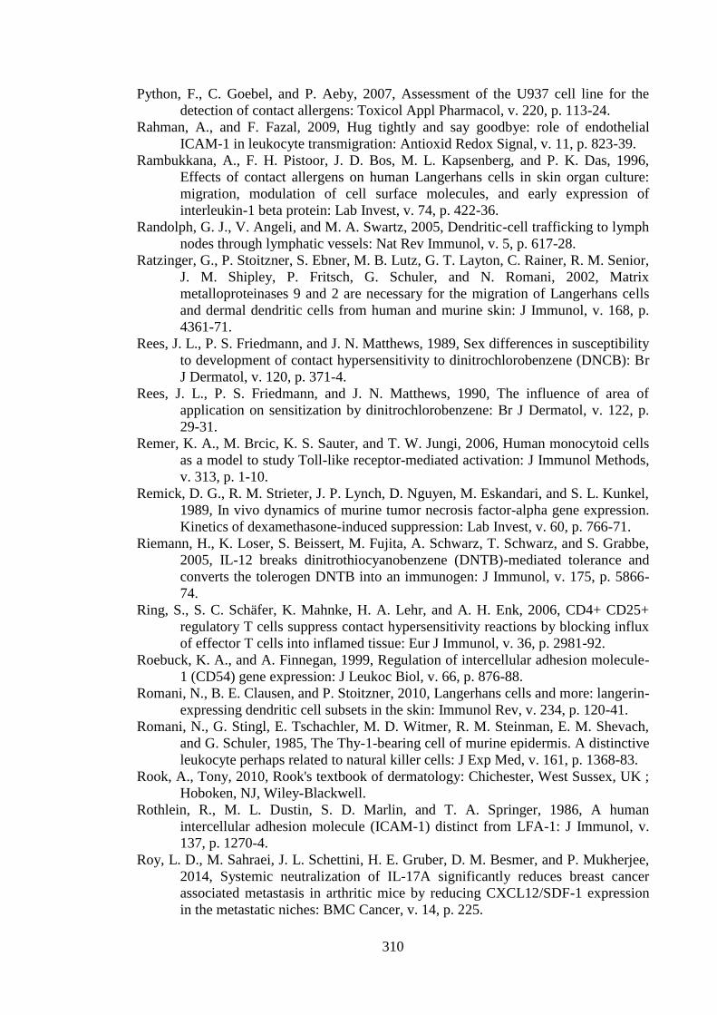
310
Python, F., C. Goebel, and P. Aeby, 2007, Assessment of the U937 cell line for the
detection of contact allergens: Toxicol Appl Pharmacol, v. 220, p. 113-24.
Rahman, A., and F. Fazal, 2009, Hug tightly and say goodbye: role of endothelial
ICAM-1 in leukocyte transmigration: Antioxid Redox Signal, v. 11, p. 823-39.
Rambukkana, A., F. H. Pistoor, J. D. Bos, M. L. Kapsenberg, and P. K. Das, 1996,
Effects of contact allergens on human Langerhans cells in skin organ culture:
migration, modulation of cell surface molecules, and early expression of
interleukin-1 beta protein: Lab Invest, v. 74, p. 422-36.
Randolph, G. J., V. Angeli, and M. A. Swartz, 2005, Dendritic-cell trafficking to lymph
nodes through lymphatic vessels: Nat Rev Immunol, v. 5, p. 617-28.
Ratzinger, G., P. Stoitzner, S. Ebner, M. B. Lutz, G. T. Layton, C. Rainer, R. M. Senior,
J. M. Shipley, P. Fritsch, G. Schuler, and N. Romani, 2002, Matrix
metalloproteinases 9 and 2 are necessary for the migration of Langerhans cells
and dermal dendritic cells from human and murine skin: J Immunol, v. 168, p.
4361-71.
Rees, J. L., P. S. Friedmann, and J. N. Matthews, 1989, Sex differences in susceptibility
to development of contact hypersensitivity to dinitrochlorobenzene (DNCB): Br
J Dermatol, v. 120, p. 371-4.
Rees, J. L., P. S. Friedmann, and J. N. Matthews, 1990, The influence of area of
application on sensitization by dinitrochlorobenzene: Br J Dermatol, v. 122, p.
29-31.
Remer, K. A., M. Brcic, K. S. Sauter, and T. W. Jungi, 2006, Human monocytoid cells
as a model to study Toll-like receptor-mediated activation: J Immunol Methods,
v. 313, p. 1-10.
Remick, D. G., R. M. Strieter, J. P. Lynch, D. Nguyen, M. Eskandari, and S. L. Kunkel,
1989, In vivo dynamics of murine tumor necrosis factor-alpha gene expression.
Kinetics of dexamethasone-induced suppression: Lab Invest, v. 60, p. 766-71.
Riemann, H., K. Loser, S. Beissert, M. Fujita, A. Schwarz, T. Schwarz, and S. Grabbe,
2005, IL-12 breaks dinitrothiocyanobenzene (DNTB)-mediated tolerance and
converts the tolerogen DNTB into an immunogen: J Immunol, v. 175, p. 5866-
74.
Ring, S., S. C. Schäfer, K. Mahnke, H. A. Lehr, and A. H. Enk, 2006, CD4+ CD25+
regulatory T cells suppress contact hypersensitivity reactions by blocking influx
of effector T cells into inflamed tissue: Eur J Immunol, v. 36, p. 2981-92.
Roebuck, K. A., and A. Finnegan, 1999, Regulation of intercellular adhesion molecule-
1 (CD54) gene expression: J Leukoc Biol, v. 66, p. 876-88.
Romani, N., B. E. Clausen, and P. Stoitzner, 2010, Langerhans cells and more: langerin-
expressing dendritic cell subsets in the skin: Immunol Rev, v. 234, p. 120-41.
Romani, N., G. Stingl, E. Tschachler, M. D. Witmer, R. M. Steinman, E. M. Shevach,
and G. Schuler, 1985, The Thy-1-bearing cell of murine epidermis. A distinctive
leukocyte perhaps related to natural killer cells: J Exp Med, v. 161, p. 1368-83.
Rook, A., Tony, 2010, Rook's textbook of dermatology: Chichester, West Sussex, UK ;
Hoboken, NJ, Wiley-Blackwell.
Rothlein, R., M. L. Dustin, S. D. Marlin, and T. A. Springer, 1986, A human
intercellular adhesion molecule (ICAM-1) distinct from LFA-1: J Immunol, v.
137, p. 1270-4.
Roy, L. D., M. Sahraei, J. L. Schettini, H. E. Gruber, D. M. Besmer, and P. Mukherjee,
2014, Systemic neutralization of IL-17A significantly reduces breast cancer
associated metastasis in arthritic mice by reducing CXCL12/SDF-1 expression
in the metastatic niches: BMC Cancer, v. 14, p. 225.
311
Saeki, H., A. M. Moore, M. J. Brown, and S. T. Hwang, 1999, Cutting edge: secondary
lymphoid-tissue chemokine (SLC) and CC chemokine receptor 7 (CCR7)
participate in the emigration pathway of mature dendritic cells from the skin to
regional lymph nodes: J Immunol, v. 162, p. 2472-5.
Saint-Mezard, P., M. Krasteva, C. Chavagnac, S. Bosset, H. Akiba, J. Kehren, J.
Kanitakis, D. Kaiserlian, J. F. Nicolas, and F. Berard, 2003, Afferent and
efferent phases of allergic contact dermatitis (ACD) can be induced after a single
skin contact with haptens: evidence using a mouse model of primary ACD: J
Invest Dermatol, v. 120, p. 641-7.
Saitoh, M., H. Nishitoh, M. Fujii, K. Takeda, K. Tobiume, Y. Sawada, M. Kawabata, K.
Miyazono, and H. Ichijo, 1998, Mammalian thioredoxin is a direct inhibitor of
apoptosis signal-regulating kinase (ASK) 1: EMBO J, v. 17, p. 2596-606.
Sallusto, F., M. Cella, C. Danieli, and A. Lanzavecchia, 1995, Dendritic cells use
macropinocytosis and the mannose receptor to concentrate macromolecules in
the major histocompatibility complex class II compartment: downregulation by
cytokines and bacterial products: J Exp Med, v. 182, p. 389-400.
Sallusto, F., and A. Lanzavecchia, 1994, Efficient presentation of soluble antigen by
cultured human dendritic cells is maintained by granulocyte/macrophage
colony-stimulating factor plus interleukin 4 and downregulated by tumor
necrosis factor alpha: J Exp Med, v. 179, p. 1109-18.
Sallusto, F., B. Palermo, D. Lenig, M. Miettinen, S. Matikainen, I. Julkunen, R. Forster,
R. Burgstahler, M. Lipp, and A. Lanzavecchia, 1999, Distinct patterns and
kinetics of chemokine production regulate dendritic cell function: Eur J
Immunol, v. 29, p. 1617-25.
Sallusto, F., P. Schaerli, P. Loetscher, C. Schaniel, D. Lenig, C. R. Mackay, S. Qin, and
A. Lanzavecchia, 1998, Rapid and coordinated switch in chemokine receptor
expression during dendritic cell maturation: Eur J Immunol, v. 28, p. 2760-9.
Sanmiguel, J. C., F. Olaru, J. Li, E. Mohr, and L. E. Jensen, 2009, Interleukin-1
regulates keratinocyte expression of T cell targeting chemokines through
interleukin-1 receptor associated kinase-1 (IRAK1) dependent and independent
pathways: Cell Signal, v. 21, p. 685-94.
Saraceno, R., T. Gramiccia, S. Chimenti, P. Valenti, M. Pietropaoli, and L. Bianchi,
2014, Topical lactoferrin can improve stable psoriatic plaque: G Ital Dermatol
Venereol, v. 149, p. 335-40.
Sato, H., Y. Taketomi, Y. Isogai, S. Masuda, T. Kobayashi, K. Yamamoto, and M.
Murakami, 2009, Group III secreted phospholipase A2 transgenic mice
spontaneously develop inflammation: Biochem J, v. 421, p. 17-27.
Sato, K., N. Yamashita, M. Baba, and T. Matsuyama, 2003, Regulatory dendritic cells
protect mice from murine acute graft-versus-host disease and leukemia relapse:
Immunity, v. 18, p. 367-79.
Schmidt, M., B. Raghavan, V. Müller, T. Vogl, G. Fejer, S. Tchaptchet, S. Keck, C.
Kalis, P. J. Nielsen, C. Galanos, J. Roth, A. Skerra, S. F. Martin, M. A.
Freudenberg, and M. Goebeler, 2010, Crucial role for human Toll-like receptor
4 in the development of contact allergy to nickel: Nat Immunol, v. 11, p. 814-9.
Schuler, G., and R. M. Steinman, 1985, Murine epidermal Langerhans cells mature into
potent immunostimulatory dendritic cells in vitro: J Exp Med, v. 161, p. 526-46.
Schwacha, M. G., E. Nickel, and T. Daniel, 2008, Burn injury-induced alterations in
wound inflammation and healing are associated with suppressed hypoxia
inducible factor-1alpha expression: Mol Med, v. 14, p. 628-33.
312
Schwarzenberger, K., and M. C. Udey, 1996, Contact allergens and epidermal
proinflammatory cytokines modulate Langerhans cell E-cadherin expression in
situ: J Invest Dermatol, v. 106, p. 553-8.
Schwende, H., E. Fitzke, P. Ambs, and P. Dieter, 1996, Differences in the state of
differentiation of THP-1 cells induced by phorbol ester and 1,25-
dihydroxyvitamin D3: J Leukoc Biol, v. 59, p. 555-61.
Segawa, R., S. Yamashita, N. Mizuno, M. Shiraki, T. Hatayama, N. Satou, M.
Hiratsuka, M. Hide, and N. Hirasawa, 2014, Identification of a cell line
producing high levels of TSLP: advantages for screening of anti-allergic drugs: J
Immunol Methods, v. 402, p. 9-14.
Seneschal, J., R. A. Clark, A. Gehad, C. M. Baecher-Allan, and T. S. Kupper, 2012,
Human epidermal Langerhans cells maintain immune homeostasis in skin by
activating skin resident regulatory T cells: Immunity, v. 36, p. 873-84.
Shankar, G., S. Pickard-Elias, and K. Burnham, 1996, Superantigen-induced
Langerhans cell depletion is mediated by epidermal cell-derived IL-1 alpha and
TNF alpha: Cell Immunol, v. 171, p. 240-5.
Sharp, L. L., J. M. Jameson, G. Cauvi, and W. L. Havran, 2005, Dendritic epidermal T
cells regulate skin homeostasis through local production of insulin-like growth
factor 1: Nat Immunol, v. 6, p. 73-9.
Shetty, S., and G. S, 2012, Keratinization and its disorders: Oman Med J, v. 27, p. 348-
57.
Shornick, L. P., A. K. Bisarya, and D. D. Chaplin, 2001, IL-1beta is essential for
langerhans cell activation and antigen delivery to the lymph nodes during
contact sensitization: evidence for a dermal source of IL-1beta: Cell Immunol, v.
211, p. 105-12.
Shornick, L. P., P. De Togni, S. Mariathasan, J. Goellner, J. Strauss-Schoenberger, R.
W. Karr, T. A. Ferguson, and D. D. Chaplin, 1996, Mice deficient in IL-1beta
manifest impaired contact hypersensitivity to trinitrochlorobenzone: J Exp Med,
v. 183, p. 1427-36.
Sidhu, R., P. Wilson, A. Wright, C. W. Yau, F. A. D'Cruz, L. Foye, S. Morley, A. J.
Lobo, M. E. McAlindon, and D. S. Sanders, 2010, Faecal lactoferrin--a novel
test to differentiate between the irritable and inflamed bowel?: Aliment
Pharmacol Ther, v. 31, p. 1365-70.
Siemion, I. Z., J. Sloń, and Z. Wieczorek, 1995, The immunosuppressive mini-domain
of human lactoferrin: J Pept Sci, v. 1, p. 295-302.
Sikorski, E. E., G. F. Gerberick, C. A. Ryan, C. M. Miller, and G. M. Ridder, 1996,
Phenotypic analysis of lymphocyte subpopulations in lymph nodes draining the
ear following exposure to contact allergens and irritants: Fundam Appl Toxicol,
v. 34, p. 25-35.
Simon, D., C. Aeberhard, Y. Erdemoglu, and H. U. Simon, 2014, Th17 cells and tissue
remodeling in atopic and contact dermatitis: Allergy, v. 69, p. 125-31.
Simon, J. C., P. D. Cruz, R. E. Tigelaar, R. D. Sontheimer, and P. R. Bergstresser, 1991,
Adhesion molecules CD11a, CD18, and ICAM-1 on human epidermal
Langerhans cells serve a functional role in the activation of alloreactive T cells:
J Invest Dermatol, v. 96, p. 148-51.
Simonsson, C., S. I. Andersson, A. L. Stenfeldt, J. Bergström, B. Bauer, C. A. Jonsson,
M. B. Ericson, and K. S. Broo, 2011, Caged fluorescent haptens reveal the
generation of cryptic epitopes in allergic contact dermatitis: J Invest Dermatol,
v. 131, p. 1486-93.
313
Singh, G., and M. Lavanya, 2010, Topical immunotherapy in alopecia areata: Int J
Trichology, v. 2, p. 36-9.
Smith, K. A., 1988, Interleukin-2: inception, impact, and implications: Science, v. 240,
p. 1169-76.
Snapper, C. M., and W. E. Paul, 1987, Interferon-gamma and B cell stimulatory factor-1
reciprocally regulate Ig isotype production: Science, v. 236, p. 944-7.
Song, M., Phelps, D. S., 2000, Comparison of SP-A and LPS effects on the THP-1
monocytic cell line: Am J Physiol Lung Cell Mol Physiol, v. 279, p. 110-7.
Sorimachi, K., K. Akimoto, Y. Hattori, T. Ieiri, and A. Niwa, 1997, Activation of
macrophages by lactoferrin: secretion of TNF-alpha, IL-8 and NO: Biochem
Mol Biol Int, v. 43, p. 79-87.
Spadaro, M., C. Caorsi, P. Ceruti, A. Varadhachary, G. Forni, F. Pericle, and M.
Giovarelli, 2008, Lactoferrin, a major defense protein of innate immunity, is a
novel maturation factor for human dendritic cells: FASEB J, v. 22, p. 2747-57.
Spadaro, M., M. Montone, M. Arigoni, D. Cantarella, G. Forni, F. Pericle, S. Pascolo,
R. A. Calogero, and F. Cavallo, 2014, Recombinant human lactoferrin induces
human and mouse dendritic cell maturation via Toll-like receptors 2 and 4:
FASEB J, v. 28, p. 416-29.
Spik, G., B. Coddeville, and J. Montreuil, 1988, Comparative study of the primary
structures of sero-, lacto- and ovotransferrin glycans from different species:
Biochimie, v. 70, p. 1459-69.
Steijns, J. M., and A. C. van Hooijdonk, 2000, Occurrence, structure, biochemical
properties and technological characteristics of lactoferrin: Br J Nutr, v. 84 Suppl
1, p. S11-7.
Steinbrink, K., M. Wölfl, H. Jonuleit, J. Knop, and A. H. Enk, 1997, Induction of
tolerance by IL-10-treated dendritic cells: J Immunol, v. 159, p. 4772-80.
Steiner, G., F. Koning, A. Elbe, E. Tschachler, W. M. Yokoyama, E. M. Shevach, G.
Stingl, and J. E. Coligan, 1988, Characterization of T cell receptors on resident
murine dendritic epidermal T cells: Eur J Immunol, v. 18, p. 1323-8.
Steinman, R. M., D. Hawiger, and M. C. Nussenzweig, 2003, Tolerogenic dendritic
cells: Annu Rev Immunol, v. 21, p. 685-711.
Stephens, S., J. M. Dolby, J. Montreuil, and G. Spik, 1980, Differences in inhibition of
the growth of commensal and enteropathogenic strains of Escherichia coli by
lactotransferrin and secretory immunoglobulin A isolated from human milk:
Immunology, v. 41, p. 597-603.
Stoitzner, P., K. Pfaller, H. Stössel, and N. Romani, 2002, A close-up view of migrating
Langerhans cells in the skin: J Invest Dermatol, v. 118, p. 117-25.
Stoitzner, P., C. H. Tripp, A. Eberhart, K. M. Price, J. Y. Jung, L. Bursch, F. Ronchese,
and N. Romani, 2006, Langerhans cells cross-present antigen derived from skin:
Proc Natl Acad Sci U S A, v. 103, p. 7783-8.
Stoitzner, P., M. Zanella, U. Ortner, M. Lukas, A. Tagwerker, K. Janke, M. B. Lutz, G.
Schuler, B. Echtenacher, B. Ryffel, F. Koch, and N. Romani, 1999, Migration of
langerhans cells and dermal dendritic cells in skin organ cultures: augmentation
by TNF-alpha and IL-1beta: J Leukoc Biol, v. 66, p. 462-70.
Sumaria, N., B. Roediger, L. G. Ng, J. Qin, R. Pinto, L. L. Cavanagh, E. Shklovskaya,
B. Fazekas de St Groth, J. A. Triccas, and W. Weninger, 2011, Cutaneous
immunosurveillance by self-renewing dermal gammadelta T cells: J Exp Med, v.
208, p. 505-18.
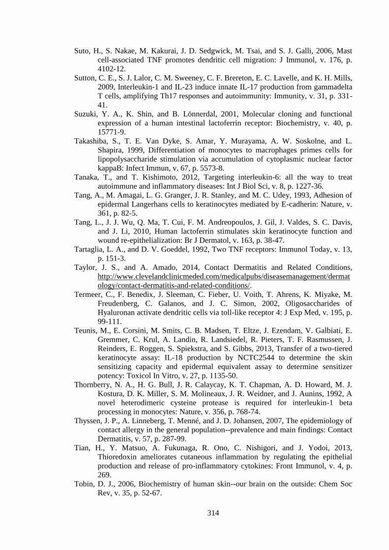
314
Suto, H., S. Nakae, M. Kakurai, J. D. Sedgwick, M. Tsai, and S. J. Galli, 2006, Mast
cell-associated TNF promotes dendritic cell migration: J Immunol, v. 176, p.
4102-12.
Sutton, C. E., S. J. Lalor, C. M. Sweeney, C. F. Brereton, E. C. Lavelle, and K. H. Mills,
2009, Interleukin-1 and IL-23 induce innate IL-17 production from gammadelta
T cells, amplifying Th17 responses and autoimmunity: Immunity, v. 31, p. 331-
41.
Suzuki, Y. A., K. Shin, and B. Lönnerdal, 2001, Molecular cloning and functional
expression of a human intestinal lactoferrin receptor: Biochemistry, v. 40, p.
15771-9.
Takashiba, S., T. E. Van Dyke, S. Amar, Y. Murayama, A. W. Soskolne, and L.
Shapira, 1999, Differentiation of monocytes to macrophages primes cells for
lipopolysaccharide stimulation via accumulation of cytoplasmic nuclear factor
kappaB: Infect Immun, v. 67, p. 5573-8.
Tanaka, T., and T. Kishimoto, 2012, Targeting interleukin-6: all the way to treat
autoimmune and inflammatory diseases: Int J Biol Sci, v. 8, p. 1227-36.
Tang, A., M. Amagai, L. G. Granger, J. R. Stanley, and M. C. Udey, 1993, Adhesion of
epidermal Langerhans cells to keratinocytes mediated by E-cadherin: Nature, v.
361, p. 82-5.
Tang, L., J. J. Wu, Q. Ma, T. Cui, F. M. Andreopoulos, J. Gil, J. Valdes, S. C. Davis,
and J. Li, 2010, Human lactoferrin stimulates skin keratinocyte function and
wound re-epithelialization: Br J Dermatol, v. 163, p. 38-47.
Tartaglia, L. A., and D. V. Goeddel, 1992, Two TNF receptors: Immunol Today, v. 13,
p. 151-3.
Taylor, J. S., and A. Amado, 2014, Contact Dermatitis and Related Conditions,
http://www.clevelandclinicmeded.com/medicalpubs/diseasemanagement/dermat
ology/contact-dermatitis-and-related-conditions/.
Termeer, C., F. Benedix, J. Sleeman, C. Fieber, U. Voith, T. Ahrens, K. Miyake, M.
Freudenberg, C. Galanos, and J. C. Simon, 2002, Oligosaccharides of
Hyaluronan activate dendritic cells via toll-like receptor 4: J Exp Med, v. 195, p.
99-111.
Teunis, M., E. Corsini, M. Smits, C. B. Madsen, T. Eltze, J. Ezendam, V. Galbiati, E.
Gremmer, C. Krul, A. Landin, R. Landsiedel, R. Pieters, T. F. Rasmussen, J.
Reinders, E. Roggen, S. Spiekstra, and S. Gibbs, 2013, Transfer of a two-tiered
keratinocyte assay: IL-18 production by NCTC2544 to determine the skin
sensitizing capacity and epidermal equivalent assay to determine sensitizer
potency: Toxicol In Vitro, v. 27, p. 1135-50.
Thornberry, N. A., H. G. Bull, J. R. Calaycay, K. T. Chapman, A. D. Howard, M. J.
Kostura, D. K. Miller, S. M. Molineaux, J. R. Weidner, and J. Aunins, 1992, A
novel heterodimeric cysteine protease is required for interleukin-1 beta
processing in monocytes: Nature, v. 356, p. 768-74.
Thyssen, J. P., A. Linneberg, T. Menné, and J. D. Johansen, 2007, The epidemiology of
contact allergy in the general population--prevalence and main findings: Contact
Dermatitis, v. 57, p. 287-99.
Tian, H., Y. Matsuo, A. Fukunaga, R. Ono, C. Nishigori, and J. Yodoi, 2013,
Thioredoxin ameliorates cutaneous inflammation by regulating the epithelial
production and release of pro-inflammatory cytokines: Front Immunol, v. 4, p.
269.
Tobin, D. J., 2006, Biochemistry of human skin--our brain on the outside: Chem Soc
Rev, v. 35, p. 52-67.
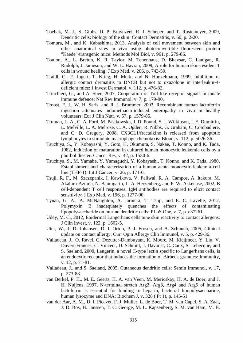
315
Toebak, M. J., S. Gibbs, D. P. Bruynzeel, R. J. Scheper, and T. Rustemeyer, 2009,
Dendritic cells: biology of the skin: Contact Dermatitis, v. 60, p. 2-20.
Tomura, M., and K. Kabashima, 2013, Analysis of cell movement between skin and
other anatomical sites in vivo using photoconvertible fluorescent protein
"Kaede"-transgenic mice: Methods Mol Biol, v. 961, p. 279-86.
Toulon, A., L. Breton, K. R. Taylor, M. Tenenhaus, D. Bhavsar, C. Lanigan, R.
Rudolph, J. Jameson, and W. L. Havran, 2009, A role for human skin-resident T
cells in wound healing: J Exp Med, v. 206, p. 743-50.
Traidl, C., F. Jugert, T. Krieg, H. Merk, and N. Hunzelmann, 1999, Inhibition of
allergic contact dermatitis to DNCB but not to oxazolone in interleukin-4-
deficient mice: J Invest Dermatol, v. 112, p. 476-82.
Trinchieri, G., and A. Sher, 2007, Cooperation of Toll-like receptor signals in innate
immune defence: Nat Rev Immunol, v. 7, p. 179-90.
Troost, F. J., W. H. Saris, and R. J. Brummer, 2003, Recombinant human lactoferrin
ingestion attenuates indomethacin-induced enteropathy in vivo in healthy
volunteers: Eur J Clin Nutr, v. 57, p. 1579-85.
Truman, L. A., C. A. Ford, M. Pasikowska, J. D. Pound, S. J. Wilkinson, I. E. Dumitriu,
L. Melville, L. A. Melrose, C. A. Ogden, R. Nibbs, G. Graham, C. Combadiere,
and C. D. Gregory, 2008, CX3CL1/fractalkine is released from apoptotic
lymphocytes to stimulate macrophage chemotaxis: Blood, v. 112, p. 5026-36.
Tsuchiya, S., Y. Kobayashi, Y. Goto, H. Okumura, S. Nakae, T. Konno, and K. Tada,
1982, Induction of maturation in cultured human monocytic leukemia cells by a
phorbol diester: Cancer Res, v. 42, p. 1530-6.
Tsuchiya, S., M. Yamabe, Y. Yamaguchi, Y. Kobayashi, T. Konno, and K. Tada, 1980,
Establishment and characterization of a human acute monocytic leukemia cell
line (THP-1): Int J Cancer, v. 26, p. 171-6.
Tsuji, R. F., M. Szczepanik, I. Kawikova, V. Paliwal, R. A. Campos, A. Itakura, M.
Akahira-Azuma, N. Baumgarth, L. A. Herzenberg, and P. W. Askenase, 2002, B
cell-dependent T cell responses: IgM antibodies are required to elicit contact
sensitivity: J Exp Med, v. 196, p. 1277-90.
Tynan, G. A., A. McNaughton, A. Jarnicki, T. Tsuji, and E. C. Lavelle, 2012,
Polymyxin B inadequately quenches the effects of contaminating
lipopolysaccharide on murine dendritic cells: PLoS One, v. 7, p. e37261.
Udey, M. C., 2012, Epidermal Langerhans cells tune skin reactivity to contact allergens:
J Clin Invest, v. 122, p. 1602-5.
Uter, W., J. D. Johansen, D. I. Orton, P. J. Frosch, and A. Schnuch, 2005, Clinical
update on contact allergy: Curr Opin Allergy Clin Immunol, v. 5, p. 429-36.
Valladeau, J., O. Ravel, C. Dezutter-Dambuyant, K. Moore, M. Kleijmeer, Y. Liu, V.
Duvert-Frances, C. Vincent, D. Schmitt, J. Davoust, C. Caux, S. Lebecque, and
S. Saeland, 2000, Langerin, a novel C-type lectin specific to Langerhans cells, is
an endocytic receptor that induces the formation of Birbeck granules: Immunity,
v. 12, p. 71-81.
Valladeau, J., and S. Saeland, 2005, Cutaneous dendritic cells: Semin Immunol, v. 17,
p. 273-83.
van Berkel, P. H., M. E. Geerts, H. A. van Veen, M. Mericskay, H. A. de Boer, and J.
H. Nuijens, 1997, N-terminal stretch Arg2, Arg3, Arg4 and Arg5 of human
lactoferrin is essential for binding to heparin, bacterial lipopolysaccharide,
human lysozyme and DNA: Biochem J, v. 328 ( Pt 1), p. 145-51.
van der Aar, A. M., D. I. Picavet, F. J. Muller, L. de Boer, T. M. van Capel, S. A. Zaat,
J. D. Bos, H. Janssen, T. C. George, M. L. Kapsenberg, S. M. van Ham, M. B.
316
Teunissen, and E. C. de Jong, 2013, Langerhans cells favor skin flora tolerance
through limited presentation of bacterial antigens and induction of regulatory T
cells: J Invest Dermatol, v. 133, p. 1240-9.
van der Does, A. M., P. J. Hensbergen, S. J. Bogaards, M. Cansoy, A. M. Deelder, H. C.
van Leeuwen, J. W. Drijfhout, J. T. van Dissel, and P. H. Nibbering, 2012, The
human lactoferrin-derived peptide hLF1-11 exerts immunomodulatory effects by
specific inhibition of myeloperoxidase activity: J Immunol, v. 188, p. 5012-9.
van der Kraan, M. I., J. Groenink, K. Nazmi, E. C. Veerman, J. G. Bolscher, and A. V.
Nieuw Amerongen, 2004, Lactoferrampin: a novel antimicrobial peptide in the
N1-domain of bovine lactoferrin: Peptides, v. 25, p. 177-83.
van der Strate, B. W., L. Beljaars, G. Molema, M. C. Harmsen, and D. K. Meijer, 2001,
Antiviral activities of lactoferrin: Antiviral Res, v. 52, p. 225-39.
Van Och, F. M., H. Van Loveren, W. H. De Jong, and R. J. Vandebriel, 2002, Cytokine
production induced by low-molecular-weight chemicals as a function of the
stimulation index in a modified local lymph node assay: an approach to
discriminate contact sensitizers from respiratory sensitizers: Toxicol Appl
Pharmacol, v. 184, p. 46-56.
Van Och, F. M., H. Van Loveren, J. C. Van Wolfswinkel, A. J. Machielsen, and R. J.
Vandebriel, 2005, Assessment of potency of allergenic activity of low molecular
weight compounds based on IL-1alpha and IL-18 production by a murine and
human keratinocyte cell line: Toxicology, v. 210, p. 95-109.
Villablanca, E. J., and J. R. Mora, 2008, A two-step model for Langerhans cell
migration to skin-draining LN: Eur J Immunol, v. 38, p. 2975-80.
Vocanson, M., A. Hennino, A. Rozières, G. Poyet, and J. F. Nicolas, 2009, Effector and
regulatory mechanisms in allergic contact dermatitis: Allergy, v. 64, p. 1699-
714.
Voelcker, V., C. Gebhardt, M. Averbeck, A. Saalbach, V. Wolf, F. Weih, J. Sleeman,
U. Anderegg, and J. Simon, 2008, Hyaluronan fragments induce cytokine and
metalloprotease upregulation in human melanoma cells in part by signalling via
TLR4: Exp Dermatol, v. 17, p. 100-7.
Voll, R. E., M. Herrmann, E. A. Roth, C. Stach, J. R. Kalden, and I. Girkontaite, 1997,
Immunosuppressive effects of apoptotic cells: Nature, v. 390, p. 350-1.
Vrieling, M., W. Santema, I. Van Rhijn, V. Rutten, and A. Koets, 2012, γδ T cell
homing to skin and migration to skin-draining lymph nodes is CCR7
independent: J Immunol, v. 188, p. 578-84.
Vukmanovic-Stejic, M., B. Vyas, P. Gorak-Stolinska, A. Noble, and D. M. Kemeny,
2000, Human Tc1 and Tc2/Tc0 CD8 T-cell clones display distinct cell surface
and functional phenotypes: Blood, v. 95, p. 231-40.
Wajant, H., K. Pfizenmaier, and P. Scheurich, 2003, Tumor necrosis factor signaling:
Cell Death Differ, v. 10, p. 45-65.
Waldorf, H. A., L. J. Walsh, N. M. Schechter, and G. F. Murphy, 1991, Early cellular
events in evolving cutaneous delayed hypersensitivity in humans: Am J Pathol,
v. 138, p. 477-86.
Wally, J., and S. K. Buchanan, 2007, A structural comparison of human serum
transferrin and human lactoferrin: Biometals, v. 20, p. 249-62.
Walsh, K. P., and K. H. Mills, 2013, Dendritic cells and other innate determinants of T
helper cell polarisation: Trends Immunol, v. 34, p. 521-30.
Walsh, L. J., G. Trinchieri, H. A. Waldorf, D. Whitaker, and G. F. Murphy, 1991,
Human dermal mast cells contain and release tumor necrosis factor alpha, which
317
induces endothelial leukocyte adhesion molecule 1: Proc Natl Acad Sci U S A,
v. 88, p. 4220-4.
Wang, B., S. Kondo, G. M. Shivji, H. Fujisawa, T. W. Mak, and D. N. Sauder, 1996,
Tumour necrosis factor receptor II (p75) signalling is required for the migration
of Langerhans' cells: Immunology, v. 88, p. 284-8.
Wang, D., K. M. Pabst, Y. Aida, and M. J. Pabst, 1995, Lipopolysaccharide-inactivating
activity of neutrophils is due to lactoferrin: J Leukoc Biol, v. 57, p. 865-74.
Wang, D., P. L. Xiao, H. X. Duan, M. Zhou, J. Liu, W. Li, K. L. Luo, J. J. Chen, and J.
Y. Hu, 2012, Peptidoglycans promotes human leukemic THP-1 cell apoptosis
and differentiation: Asian Pac J Cancer Prev, v. 13, p. 6409-13.
Wang, L., L. S. Bursch, A. Kissenpfennig, B. Malissen, S. C. Jameson, and K. A.
Hogquist, 2008, Langerin expressing cells promote skin immune responses
under defined conditions: J Immunol, v. 180, p. 4722-7.
Wang, Y., M. Mao, and J. C. Xu, 2011, Cell-surface nucleolin is involved in
lipopolysaccharide internalization and signalling in alveolar macrophages: Cell
Biol Int, v. 35, p. 677-85.
Warbrick, E. V., R. J. Dearman, D. A. Basketter, and I. Kimber, 1999, Influence of
application vehicle on skin sensitization to
methylchloroisothiazolinone/methylisothiazolinone: an analysis using the local
lymph node assay: Contact Dermatitis, v. 41, p. 325-9.
Ward, P. P., G. A. Cunningham, and O. M. Conneely, 1997, Commercial production of
lactoferrin, a multifunctional iron-binding glycoprotein: Biotechnol Genet Eng
Rev, v. 14, p. 303-19.
Watanabe, H., S. Gehrke, E. Contassot, S. Roques, J. Tschopp, P. S. Friedmann, L. E.
French, and O. Gaide, 2008, Danger signaling through the inflammasome acts as
a master switch between tolerance and sensitization: J Immunol, v. 180, p. 5826-
32.
Watanabe, H., K. Numata, T. Ito, K. Takagi, and A. Matsukawa, 2004, Innate immune
response in Th1- and Th2-dominant mouse strains: Shock, v. 22, p. 460-6.
Weber, F. C., P. R. Esser, T. Müller, J. Ganesan, P. Pellegatti, M. M. Simon, R. Zeiser,
M. Idzko, T. Jakob, and S. F. Martin, 2010, Lack of the purinergic receptor
P2X(7) results in resistance to contact hypersensitivity: J Exp Med, v. 207, p.
2609-19.
Werman, A., R. Werman-Venkert, R. White, J. K. Lee, B. Werman, Y. Krelin, E.
Voronov, C. A. Dinarello, and R. N. Apte, 2004, The precursor form of IL-
1alpha is an intracrine proinflammatory activator of transcription: Proc Natl
Acad Sci U S A, v. 101, p. 2434-9.
Willems, J. J., B. P. Arnold, and C. D. Gregory, 2014, Sinister self-sacrifice: the
contribution of apoptosis to malignancy: Front Immunol, v. 5, p. 299.
Willimann, K., D. F. Legler, M. Loetscher, R. S. Roos, M. B. Delgado, I. Clark-Lewis,
M. Baggiolini, and B. Moser, 1998, The chemokine SLC is expressed in T cell
areas of lymph nodes and mucosal lymphoid tissues and attracts activated T
cells via CCR7: Eur J Immunol, v. 28, p. 2025-34.
Woodland, D. L., and R. W. Dutton, 2003, Heterogeneity of CD4(+) and CD8(+) T
cells: Curr Opin Immunol, v. 15, p. 336-42.
Wu, T., R. Du, Y. Hong, L. Jia, Q. Zeng, and B. Cheng, 2013, IL-1 alpha regulates
CXCL1, CXCL10 and ICAM1 in network form in oral keratinocytes: Clin Lab,
v. 59, p. 1105-11.
Xu, G., W. Xiong, Q. Hu, P. Zuo, B. Shao, F. Lan, X. Lu, Y. Xu, and S. Xiong, 2010,
Lactoferrin-derived peptides and Lactoferricin chimera inhibit virulence factor
318
production and biofilm formation in Pseudomonas aeruginosa: J Appl
Microbiol, v. 109, p. 1311-8.
Xu, H., N. A. DiIulio, and R. L. Fairchild, 1996, T cell populations primed by hapten
sensitization in contact sensitivity are distinguished by polarized patterns of
cytokine production: interferon gamma-producing (Tc1) effector CD8+ T cells
and interleukin (Il) 4/Il-10-producing (Th2) negative regulatory CD4+ T cells: J
Exp Med, v. 183, p. 1001-12.
Xu, H., H. Guan, G. Zu, D. Bullard, J. Hanson, M. Slater, and C. A. Elmets, 2001, The
role of ICAM-1 molecule in the migration of Langerhans cells in the skin and
regional lymph node: Eur J Immunol, v. 31, p. 3085-93.
Yamane, H., and W. E. Paul, 2013, Early signaling events that underlie fate decisions of
naive CD4(+) T cells toward distinct T-helper cell subsets: Immunol Rev, v.
252, p. 12-23.
Yang, D., G. de la Rosa, P. Tewary, and J. J. Oppenheim, 2009, Alarmins link
neutrophils and dendritic cells: Trends Immunol, v. 30, p. 531-7.
Yarosh, D., D. Both, J. Kibitel, C. Anderson, C. Elmets, D. Brash, and D. Brown, 2000,
Regulation of TNFalpha production and release in human and mouse
keratinocytes and mouse skin after UV-B irradiation: Photodermatol
Photoimmunol Photomed, v. 16, p. 263-70.
Yokozeki, H., K. Watanabe, K. Igawa, Y. Miyazaki, I. Katayama, and K. Nishioka,
2001, Gammadelta T cells assist alphabeta T cells in the adoptive transfer of
contact hypersensitivity to para-phenylenediamine: Clin Exp Immunol, v. 125,
p. 351-9.
Yonei, N., N. Kanazawa, T. Ohtani, F. Furukawa, and Y. Yamamoto, 2007, Induction
of PDGF-B in TCA-treated epidermal keratinocytes: Arch Dermatol Res, v. 299,
p. 433-40.
Yoshihara, E., S. Masaki, Y. Matsuo, Z. Chen, H. Tian, and J. Yodoi, 2014,
Thioredoxin/Txnip: Redoxisome, as a Redox Switch for the Pathogenesis of
Diseases: Front Immunol, v. 4, p. 514.
Yuspa, S. H., P. Hawley-Nelson, B. Koehler, and J. R. Stanley, 1980, A survey of
transformation markers in differentiating epidermal cell lines in culture: Cancer
Res, v. 40, p. 4694-703.
Zagulski, T., P. Lipiński, A. Zagulska, S. Broniek, and Z. Jarzabek, 1989, Lactoferrin
can protect mice against a lethal dose of Escherichia coli in experimental
infection in vivo: Br J Exp Pathol, v. 70, p. 697-704.
Zaja-Milatovic, S., and A. Richmond, 2008, CXC chemokines and their receptors: a
case for a significant biological role in cutaneous wound healing: Histol
Histopathol, v. 23, p. 1399-407.
Zelová, H., and J. Hošek, 2013, TNF-α signalling and inflammation: interactions
between old acquaintances: Inflamm Res, v. 62, p. 641-51.
Zhang, J., G. Tsaprailis, and G. T. Bowden, 2008, Nucleolin stabilizes Bcl-X L
messenger RNA in response to UVA irradiation: Cancer Res, v. 68, p. 1046-54.
Zhao, Y., A. Balato, R. Fishelevich, A. Chapoval, D. L. Mann, and A. A. Gaspari, 2009,
Th17/Tc17 infiltration and associated cytokine gene expression in elicitation
phase of allergic contact dermatitis: Br J Dermatol, v. 161, p. 1301-6.
Zheng, H., D. Fletcher, W. Kozak, M. Jiang, K. J. Hofmann, C. A. Conn, D. Soszynski,
C. Grabiec, M. E. Trumbauer, and A. Shaw, 1995, Resistance to fever induction
and impaired acute-phase response in interleukin-1 beta-deficient mice:
Immunity, v. 3, p. 9-19.
319
Zhou, L., M. M. Chong, and D. R. Littman, 2009, Plasticity of CD4+ T cell lineage
differentiation: Immunity, v. 30, p. 646-55.
Zhou, R., A. Tardivel, B. Thorens, I. Choi, and J. Tschopp, 2010, Thioredoxin-
interacting protein links oxidative stress to inflammasome activation: Nat
Immunol, v. 11, p. 136-40.
Zimecki, M., J. Artym, M. Kocięba, K. Kaleta-Kuratewicz, P. Kuropka, J. Kuryszko,
and M. Kruzel, 2013, Homologous lactoferrin triggers mobilization of the
myelocytic lineage of bone marrow in experimental mice: Stem Cells Dev, v.
22, p. 3261-70.
Zimecki, M., J. Artym, M. Kocięba, and M. Kruzel, 2012, Effects of lactoferrin on
elicitation of the antigen-specific cellular and humoral cutaneous response in
mice: Postepy Hig Med Dosw (Online), v. 66, p. 16-22.
Zimecki, M., J. Mazurier, G. Spik, and J. A. Kapp, 1996, Lactoferrin inhibits
proliferative response and cytokine production of TH1 but not TH2 cell lines:
Arch Immunol Ther Exp (Warsz), v. 44, p. 51-6.
Zmolik, J. M., and M. E. Mummert, 2005, Pep-1 as a novel probe for the in situ
detection of hyaluronan: J Histochem Cytochem, v. 53, p. 745-51.
Zweiman, B., U. Kucich, M. Shalit, C. Von Allmen, A. Moskovitz, G. Weinbaum, and
P. C. Atkins, 1990, Release of lactoferrin and elastase in human allergic skin
reactions: J Immunol, v. 144, p. 3953-60.


