Embed Size (px)
Citation preview

SMA-Causing Missense Mutations in Survival motorneuron (Smn) Display a Wide Range of Phenotypes WhenModeled in DrosophilaKavita Praveen1¤, Ying Wen2, Kelsey M. Gray1, John J. Noto1, Akash R. Patlolla3, Gregory D. Van Duyne4,
A. Gregory Matera1,2,3,5*
1 Curriculum in Genetics and Molecular Biology, University of North Carolina, Chapel Hill, Chapel Hill, North Carolina, United States of America, 2 Integrative Program for
Biological and Genome Sciences, University of North Carolina, Chapel Hill, Chapel Hill, North Carolina, United States of America, 3 Department of Biology, University of
North Carolina, Chapel Hill, Chapel Hill, North Carolina, United States of America, 4 Department of Biochemistry & Biophysics, University of Pennsylvania, Perelman School
of Medicine, Philadelphia, Pennsylvania, United States of America, 5 Lineberger Comprehensive Cancer Center, University of North Carolina, Chapel Hill, Chapel Hill, North
Carolina, United States of America
Abstract
Mutations in the human survival motor neuron 1 (SMN) gene are the primary cause of spinal muscular atrophy (SMA), adevastating neuromuscular disorder. SMN protein has a well-characterized role in the biogenesis of small nuclearribonucleoproteins (snRNPs), core components of the spliceosome. Additional tissue-specific and global functions havebeen ascribed to SMN; however, their relevance to SMA pathology is poorly understood and controversial. Using Drosophilaas a model system, we created an allelic series of twelve Smn missense mutations, originally identified in human SMApatients. We show that animals expressing these SMA-causing mutations display a broad range of phenotypic severities,similar to the human disease. Furthermore, specific interactions with other proteins known to be important for SMN’s role inRNP assembly are conserved. Intragenic complementation analyses revealed that the three most severe mutations, all ofwhich map to the YG box self-oligomerization domain of SMN, display a stronger phenotype than the null allele and behavein a dominant fashion. In support of this finding, the severe YG box mutants are defective in self-interaction assays, yetmaintain their ability to heterodimerize with wild-type SMN. When expressed at high levels, wild-type SMN is able tosuppress the activity of the mutant protein. These results suggest that certain SMN mutants can sequester the wild-typeprotein into inactive complexes. Molecular modeling of the SMN YG box dimer provides a structural basis for this dominantphenotype. These data demonstrate that important structural and functional features of the SMN YG box are conservedbetween vertebrates and invertebrates, emphasizing the importance of self-interaction to the proper functioning of SMN.
Citation: Praveen K, Wen Y, Gray KM, Noto JJ, Patlolla AR, et al. (2014) SMA-Causing Missense Mutations in Survival motor neuron (Smn) Display a Wide Range ofPhenotypes When Modeled in Drosophila. PLoS Genet 10(8): e1004489. doi:10.1371/journal.pgen.1004489
Editor: Gregory A. Cox, The Jackson Laboratory, United States of America
Received January 22, 2014; Accepted May 19, 2014; Published August 21, 2014
Copyright: � 2014 Praveen et al. This is an open-access article distributed under the terms of the Creative Commons Attribution License, which permitsunrestricted use, distribution, and reproduction in any medium, provided the original author and source are credited.
Funding: This work was supported by NIH grant R01-NS41617 (to AGM). KP was supported in part by an American Heart Association predoctoral fellowship. KMGwas supported in part by an NSF predoctoral fellowship, DGE-1144081. The funders had no role in study design, data collection and analysis, decision to publish,or preparation of the manuscript.
Competing Interests: The authors have declared that no competing interests exist.
* Email: [email protected]
¤ Current address: Center for Human Disease Modeling, Duke University, Durham, North Carolina, United States of America
Introduction
Proximal spinal muscular atrophy (SMA) is a common
neuromuscular disorder, recognized as the most prevalent genetic
cause of early childhood mortality [1]. SMA is characterized by
degeneration of motor neurons in the anterior horn of the lower
spinal cord, and progressive symmetrical paralysis. Coupled with
this loss of motor function, SMA patients display severe atrophy of
the proximal muscles. The onset of symptoms and their severity
can vary, leading to an historical classification of SMA into three
distinct subtypes [1]. More recently, clinicians have recognized
that SMA is better characterized as a continuous spectrum
disorder, ranging from severe (prenatal onset) to nearly asymp-
tomatic [2].
Almost two decades ago, mutations in the survival motor neuron1 (SMN1) gene were shown to be causative for SMA [3]. The
disease typically results from homozygous deletion of SMN1;
however, a small fraction of SMA patients have lost one copy of
SMN1 and the remaining copy contains a point mutation [4]. The
best characterized function for the ubiquitously expressed SMN
protein is in the biogenesis of Sm-class small nuclear ribonucleo-
proteins (snRNPs), core factors of the spliceosome [5,6]. In
addition, SMN has been implicated in numerous other cellular
activities, including axonal transport, neuronal pathfinding,
formation and function of neuromuscular junctions, myoblast
fusion and maintenance of muscle architecture [4,7–11]. Despite
this multitude of putative functions attributed to SMN, or perhaps
because of it, the precise pathophysiological mechanisms that give
rise to SMA are the subject of considerable debate. Seemingly
straightforward questions of disease pathogenesis have yet to be
definitively answered. For example, is SMA caused by a cell-
autonomous reduction of SMN protein levels in motor neurons
[12–16] or is it a more systemic defect involving other cell types
[17–24]?
PLOS Genetics | www.plosgenetics.org 1 August 2014 | Volume 10 | Issue 8 | e1004489

Irrespective of the question of cellular autonomy, the molecularetiology of SMA also remains unclear. Is the neuromuscular
dysfunction seen in SMA patients caused by a loss of Sm-class
spliceosomal snRNPs, ultimately leading to defects in pre-mRNA
splicing? Or is it due to some non-canonical function of SMN
and/or snRNPs? Experiments using animal models of severe SMA
suggest that the pre-mRNA splicing defects observed in late-stage
SMA animals are indeed tissue-specific [25,26]. However, such
deficits are only detectable later in the disease course, after the
onset of neuromuscular dysfunction [27–29]. Complicating
matters, SMN-deficient animals are developmentally delayed or
arrested [22,29–34], making the selection of properly staged
control animals critical to the comparison of phenotypes.
Moreover, alternative splicing of the SMN gene duplicate
(SMN2) in humans or mouse models is another variable, creating
a feedback loop [35,36] that can negatively regulate SMN
expression. In the fruitfly, the vast majority of Smn pre-mRNA
transcripts are intronless (Flybase; [37]). Therefore, we set out to
create an allelic series of Drosophila SMA models wherein we
could specifically focus on SMN protein function, in the absence of
other complicating factors.
Results and Discussion
Generation of Drosophila models of SMA patientmutations
To identify which functions of SMN are critical to the pathology
of SMA, we aimed to create disease-relevant models that disrupt
subsets of SMN interactions. Point mutations are useful in this
context, as they can disrupt specific functions of multi-domain
proteins, leaving other functions unaffected. To date, twenty-five
different SMN1 point mutations have been identified in SMA
patients [4]. Many of these mutations are located at residues that
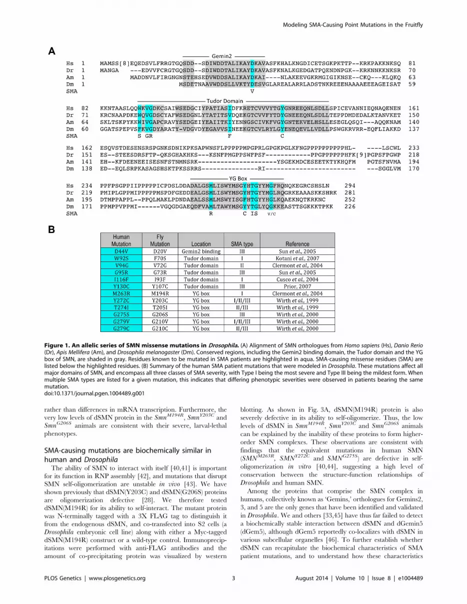
are conserved between humans and insects (Fig. 1A). In this
report, transgenic Drosophila bearing twelve of these SMA-causing
point mutations were generated (Fig. 1B). The salient features of
the Smn transgenic cassette have been previously described [28],
including a 3X FLAG tag that was inserted immediately
downstream of the ATG start codon. The transgenes were also
expressed under control of the native Smn promoter and 39
flanking sequences [28] and inserted at a landing site located
within band 86Fb on chromosome 3R using the PhiC31 integrase
system [38]. Note that all of the constructs (including an SmnWT
control) were injected directly into embryos heterozygous for the
SmnX7 microdeletion, a null mutation [39] that was recombined
with the appropriate PhiC31 landing site prior to injection (see
Methods for details). Note that the SmnX7 deletion removes the
promoter region and the entire Smn open reading frame, leaving
behind only 44 bp of the 39 UTR [39]. Thus, this mutant
produces no Smn mRNA.
As shown in Fig. 1, one of the mutations lies in the region
responsible for Gemin2 binding, five are located in the Tudor
domain, and six in the YG box oligomerization domain of SMN,
thus mirroring the distribution of point mutations identified in
SMA patients. For phenotypic analysis of the point mutants in our
model, we crossed the transgenic flies (SmnX7, Flag-SmnTg) with
an SmnX7 null allele to obtain flies that are homozygous null for
endogenous Smn and hemizygous for the Flag-Smn transgene. We
adopted this approach because human SMA patients that express
SMN1 missense mutations are also typically hemizygous and
because we observed an improvement in the viability of the
transgenic mutants when crossed to SmnX7, as compared to the
self-cross. This latter finding indicates the presence of recessive
alleles in the transgenic background that contribute to the
phenotype of the homozygotes.
SMA patient-derived mutations show a range ofphenotypic severities in Drosophila
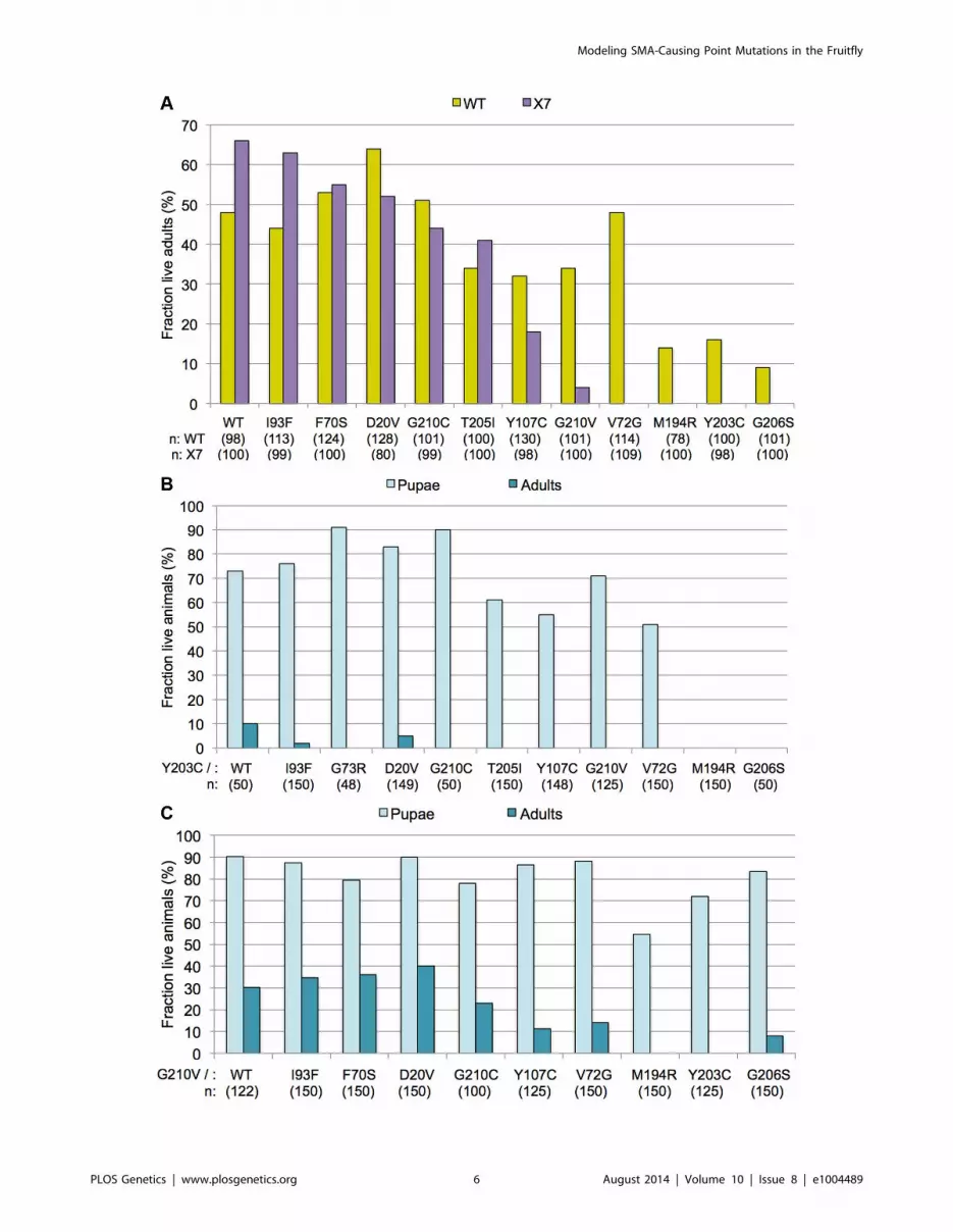
When expressed in an SmnX7/X7 null background, the hemizy-
gous Flag-SmnWT construct showed robust rescue of adult viability
(,67%, see Fig. 2A), consistent with our previous findings using
an SmnX7/D null background [28]. The degree of rescue achieved
by expressing each of the different SMA point mutations varied
(Fig. 2A). Three of the transgenes (SmnM194R, SmnY203C and
SmnG206S) failed to rescue the larval lethality of the null animals
(Fig. 2A). In contrast, a second group of point mutants rescued
adult viability to a degree roughly similar to that of the SmnWT
transgene, including: SmnD20V, SmnF70S and SmnG73R and
SmnI93F. A third group of mutants can be characterized as
having intermediate phenotypes: SmnV72G, SmnY107C, SmnT205I,
SmnG210C and SmnG210V. This last category can be categorized as
pupal-lethal, as the majority of these animals die prior to eclosion.
The SmnG210C mutation produced a much milder phenotype
(,45% rescue to adulthood) compared to the SmnG210V mutation
(,5% rescue) despite the fact that they are located at the same
residue. Interestingly, SmnV72G was the only mutation that rescued
the larval lethality of the null animals but then also resulted in
complete pupal lethality (Fig. 2A). Thus, when expressed in
Drosophila, disease-causing Smn point mutations display a range
in the age of symptomatic onset and in life-expectancy.
As shown previously [28], the amount of dSMN protein
expressed from transgenes driven by the native Smn promoter and
integrated at site 86Fb is several-fold lower than that of the
endogenous gene (Fig. 2B). However, we observed robust rescue of
the null phenotype in the presence of the SmnWT transgene, as
these animals are both viable and fertile. A majority of the SMA
alleles expressed equivalent levels of transgenic protein when
compared to SmnWT. The exceptions were SmnM194R, SmnY203C
and SmnG206S, which showed a significant reduction in dSMN
levels (Fig. 2B). Because the constructs were all inserted into the
identical genomic location, and this same level of expression was
observed in multiple independent transformants (Fig. S1), the
reduction is most likely due to instability of the mutant proteins
Author Summary
Spinal Muscular Atrophy (SMA) is a prevalent childhoodneuromuscular disease, which in its most common formcauses death by the age of two. One in fifty Americans is acarrier for SMA, making this genetic disease a serioushealth concern. SMA is caused by loss of functionmutations in the survival motor neuron 1 (SMN1) gene.SMN is an essential protein and has a well-characterizedfunction in the assembly of small nuclear ribonucleopro-teins (snRNPs), which are core components of thespliceosome. To elucidate the phenotypic consequencesof disrupting specific SMN protein interactions, we havegenerated a series of SMA-causing point mutations,modeled in Drosophila melanogaster. Using this system,we have shown that key aspects of SMN structure andfunction are conserved between humans and flies.Intragenic complementation analyses reveal the potentialfor dominant negative interactions between wild-type andmutant SMN subunits, highlighting the essential nature ofthe YG box in formation of higher-order SMN multimers.These results provide a basis for future studies investigat-ing therapy targeted at restoration of functional SMNoligomers.
Modeling SMA-Causing Point Mutations in the Fruitfly
PLOS Genetics | www.plosgenetics.org 2 August 2014 | Volume 10 | Issue 8 | e1004489
rather than differences in mRNA transcription. Furthermore, the
very low levels of dSMN protein in the SmnM194R, SmnY203C and
SmnG206S animals are consistent with their severe, larval-lethal
phenotypes.
SMA-causing mutations are biochemically similar inhuman and Drosophila
The ability of SMN to interact with itself [40,41] is important
for its function in RNP assembly [42], and mutations that disrupt
SMN self-oligomerization are unstable in vivo [43]. We have
shown previously that dSMN(Y203C) and dSMN(G206S) proteins
are oligomerization defective [28]. We therefore tested
dSMN(M194R) for its ability to self-interact. The mutant protein
was N-terminally tagged with a 3X FLAG tag to distinguish it
from the endogenous dSMN, and co-transfected into S2 cells (a
Drosophila embryonic cell line) along with either a Myc-tagged
dSMN(M194R) construct or a wild-type control. Immunoprecip-
itations were performed with anti-FLAG antibodies and the
amount of co-precipitating protein was visualized by western
blotting. As shown in Fig. 3A, dSMN(M194R) protein is also
severely defective in its ability to self-oligomerize. Thus, the low
levels of dSMN in SmnM194R, SmnY203C and SmnG206S animals
can be explained by the inability of these proteins to form higher-
order SMN complexes. These observations are consistent with
findings that the equivalent mutations in human SMN
(SMNM263R, SMNY272C and SMNG275S) are defective in self-
oligomerization in vitro [40,44], suggesting a high level of
conservation between the structure-function relationships of
Drosophila and human SMN.
Among the proteins that comprise the SMN complex in
humans, collectively known as ‘Gemins,’ orthologues for Gemins2,
3, and 5 are the only genes that have been identified and validated
in Drosophila. We and others [33,45] have thus far failed to detect
a biochemically stable interaction between dSMN and dGemin5
(dGem5), although dGem5 reportedly co-localizes with dSMN in
various subcellular organelles [46]. To further establish whether
dSMN can recapitulate the biochemical characteristics of SMA
patient mutations, and to understand how these characteristics
Figure 1. An allelic series of SMN missense mutations in Drosophila. (A) Alignment of SMN orthologues from Homo sapiens (Hs), Danio Rerio(Dr), Apis Mellifera (Am), and Drosophila melanogaster (Dm). Conserved regions, including the Gemin2 binding domain, the Tudor domain and the YGbox of SMN, are shaded in gray. Residues known to be mutated in SMA patients are highlighted in aqua. SMA-causing missense residues (SMA) arelisted below the highlighted residues. (B) Summary of the human SMA patient mutations that were modeled in Drosophila. These mutations affect allmajor domains of SMN, and encompass all three classes of SMA severity, with Type I being the most severe and Type III being the mildest form. Whenmultiple SMA types are listed for a given mutation, this indicates that differing phenotypic severities were observed in patients bearing the samemutation.doi:10.1371/journal.pgen.1004489.g001
Modeling SMA-Causing Point Mutations in the Fruitfly
PLOS Genetics | www.plosgenetics.org 3 August 2014 | Volume 10 | Issue 8 | e1004489

correlate with animal phenotypes, we analyzed the mutant dSMN
proteins for their ability to bind dGem2 and dGem3 using a co-
transfection assay.
The binding of dGem2 was reduced by a single Smn point
mutation. As shown in Fig. 3B, dGem2 binding to dSMN(D20V)
was reduced as compared to wild-type, but not eliminated. None
of the other dSMN mutants showed any decrease in binding to
dGem2 (Fig. 3B). This was not a surprising finding, as residue D20
lies within the known Gemin2 binding domain of SMN (Fig. 1A),
and is consistent with experiments on its human counterpart,
SMN(D44V) [47,48]. Interestingly, SmnD20V flies display a mild
phenotype, suggesting that the observed residual interaction with
dGem2 is sufficient for its function in vivo. Indeed, SMA patients
with the D44V mutation have been diagnosed with the mild form
of SMA, type III [49].
We also analyzed the interaction of various Flag-tagged dSMN
mutants with Myc-dGem3 by co-immunoprecipitation with anti-
Myc antibodies. As shown in Fig. 3C, we found that binding of
dGem3 was severely disrupted by two point mutations, SmnY203C
(SMNY272C in humans) and SmnG206S (SMNG275S). Consistent
with our findings, the human SMNY272C mutation was previously
shown to disrupt Gemin3 binding in vitro [40]. Notably, we found
that the SmnT205I (SMNT274I) mutation, which lies between
residues Y203 and G206, did not affect dSMN’s ability to interact
with dGem3 (Fig. 3C). This finding is consistent with previous
observations that SMNT274I is active in snRNP assembly, an
activity that also requires Gemin3 [42]. Strikingly, the phenotype
of the SmnT205I animals was much less severe than that of either
the SmnY203C or SmnG206S mutants (Fig. 2A). Given that
dSMN(T205I) and dSMN(G206S) proteins are both moderately
defective in self-oligomerization ([28] and Fig. 3A), we attribute
the more severe phenotype of the SmnG206S mutants to the
inability of dSMN(G206S) to interact with dGem3. This
interpretation is bolstered by the fact that null mutations in
Gemin3 were shown to destabilize dSMN in vivo [33]. In
summary, these results further demonstrate that important
biochemical properties of SMN are conserved between Drosophilaand humans [24,28,45].
Smn missense mutations display incomplete dominanceTo further characterize the Smn point mutants, we crossed each
of them to the wild-type (WT) rescue line, SmnWT. Since SMA
patients display a recessive mode of inheritance, we expected that
the SmnWT transgene would rescue organismal viability in the mis-
sense mutant backgrounds (genotype: SmnX7/X7,Flag-SmnWT/Mut)
to roughly the same extent as it did in the hemizygous rescue line
Figure 2. SMA patient-derived mutations show a range of life expectancies in Drosophila. (A) Viability analyses of SMA point mutations inDrosophila. SmnX7/TM6.tb GFP flies were crossed with each mutant line (SmnX7,SmnTg/TM6.tb GFP) and hemizygous first instar mutant larvae (SmnX7/X7,SmnTg/2 where Tg stands for transgene) were collected and followed through development. Oregon-R (OR) animals served as controls. The data foreach genotype are expressed as a fraction of pupae or adults over the total number of starting larvae (n), listed below each genotype. Expression ofthe WT transgene (SmnX7/X7,SmnWT/2) shows robust rescue of the null phenotype (,67% adults). The mutants show a range in severity, from verysevere (larval lethal), to intermediate (pupal lethal), to mild (adults). (B) Expression levels of Smn transgenes were examined by western blotting.Larval lysates from homozygous mutant lines (SmnX7/X7,SmnTg/Tg), were probed with anti-dSMN or anti-FLAG antibodies, as indicated. The slowermigrating bands represent the FLAG-tagged transgenic proteins and the faster migrating band corresponds to endogenous dSMN, present only inthe OR controls. The mutant transgenes show similar levels of dSMN protein compared to SmnWT animals, with the exception of SmnM194R, SmnY203C
and SmnG206S, which show a significant reduction. Tubulin was used as a loading control.doi:10.1371/journal.pgen.1004489.g002
Modeling SMA-Causing Point Mutations in the Fruitfly
PLOS Genetics | www.plosgenetics.org 4 August 2014 | Volume 10 | Issue 8 | e1004489

(SmnX7/X7,Flag-SmnWT/2). However, for many mutations we
observed an intermediate level of rescue, between that observed
when expressing the wild-type transgene alone versus that of the
mutant transgenes alone (Fig. 4A). This dominant phenotype was
most evident when the wild-type transgene was expressed in
combination with the three most severe YG box alleles (M194R,Y203C and G206S). For example SmnX7/X7,Flag-SmnWT/Y203C
animals displayed a much lower eclosion frequency (Fig. 4A) than
did either of the two controls, SmnX7/X7,Flag-SmnWT/2 (hemizy-
gotes) or SmnX7/X7,Flag-SmnWT/WT (homozygotes). Importantly,
expression of the wild-type Smn transgene in combination with the
M194R, Y203C or G206S point mutants rescued the larval
lethality associated with expression these alleles on their own
(Fig. 4A). Thus, these dominant phenotypes are intermediate in
their severity.
We note that SmnWT/WT homozygotes (obtained by an
intercross of two different founder lines) showed a somewhat
lower eclosion frequency (,50%) than did the hemizygotes
(,70%). These findings suggest that second-site recessive alleles
may contribute to the decreased fitness of the homozygotes and
that outcrossing to a ‘clean’ SmnX7 chromosome ameliorates these
effects. Moreover, the presence of second site recessives may also
affect the results of the intragenic complementation analyses
(Fig. 4A), as the transgenes were all inserted into the same genetic
background. However, when co-expressed with SmnWT, the
phenotypic severities observed for the point mutant lines
correlated with the degree of rescue when expressed alone. For
example, SmnG210V had a less severe phenotype (pupal lethal) than
SmnG206S (larval lethal), when expressed alone. When co-
expressed with the wild-type transgene, SmnG210V/WT flies also
showed a milder phenotype than did SmnG206S/WT (Fig. 4A).
There was one exception to this rule; SmnV72G displayed an early
pupal-lethal phenotype when expressed alone, but had an eclosion
frequency comparable to that of the mild mutations when co-
Figure 3. Interaction of SMN mutants with proteins involved in RNP assembly. (A) SmnM194R and SmnG206S are defective in a self-oligomerization assay. Note that oligomerization of SmnY203C and SmnT205I was tested previously [28]. Lysates were prepared from cells co-expressingFLAG- and Myc-tagged versions of dSMN mutants or WT controls. Co-immunoprecipitation was performed with anti-FLAG antibody followed bywestern analysis with anti-Myc antibody to visualize the amount of Myc-tagged dSMN that co-precipitated with the FLAG-tagged protein. (B)dSMN(D20V) protein shows defective binding to dGem2. Immunoprecipitation was performed with anti-FLAG antibody followed by western analysiswith anti-Myc antibody to visualize the amount of Myc-tagged dGem2. (C) dSMN(Y203C) and dSMN(G206S) are defective in their ability to binddGem3. Immunoprecipitation of Myc-dGem3 was followed by probing with anti-FLAG antibody to visualize Flag-dSMN.doi:10.1371/journal.pgen.1004489.g003
Modeling SMA-Causing Point Mutations in the Fruitfly
PLOS Genetics | www.plosgenetics.org 5 August 2014 | Volume 10 | Issue 8 | e1004489
Modeling SMA-Causing Point Mutations in the Fruitfly
PLOS Genetics | www.plosgenetics.org 6 August 2014 | Volume 10 | Issue 8 | e1004489

expressed with SmnWT. In summary, complementation crosses
with the mutant and wild-type Smn transgene display intermediate
phenotypes.
Using SmnY203C and SmnG210V as test cases, we carried out
additional complementation crosses to each of the other point
mutant lines. As shown in Fig. 4B, co-expression of SmnY203C with
the other transgenes had a negative effect on adult viability. When
crossed to Y203C, alleles that showed strong rescue of the null
phenotype when expressed alone (e.g. WT, D20V, G73R, or
I93F) either completely failed to eclose as adults or did so at very
low frequencies. However, this dominant effect was not fully
penetrant, as a majority of the trans-heterozygous animals
developed beyond larval stages and died as pupae (Fig. 4B). The
only exceptions were crosses between Y203C and the other two
severe mutations (M194R and G206S), which continued to die as
larvae. SmnG210V co-expression with the other transgenes had a
positive effect on pupation of flies expressing the severe mutations
(Fig. 4C). Overall, when crossed to G210V, adult viability
decreased. Crosses between G210V and either V72G or G206Swere the only exceptions, with a small fraction of flies eclosing.
This is in contrast to the pupal lethality observed when V72G was
expressed alone and the larval lethality of G206S. Thus the
intermediate phenotypes observed in these complementation
crosses provide a clear example of the genetic principle of
incomplete dominance.
Severe YG box mutants retain the ability to interact withwild-type SMN and display a more pronouncedphenotype than Smn null animals
The most severe Smn point mutations in our collection
(M194R, Y203C and G206S) all map to the YG box self-
oligomerization domain (Fig. 1A), which is the most well-
conserved region of SMN. Consistent with their YG box location,
these three alleles are defective in self-interaction assays (Fig. 3A)
and do not form stable SMN complexes in vivo (Fig. 2B). By such
criteria, these three alleles would normally be considered to be
protein nulls, yet their phenotype is actually more severe than that
of the null allele, SmnX7/X7. Indeed, these larvae all died by ,8–9
days after hatching, whereas ,20% of the null mutants were still
alive at this time point (Fig. 5A). The M194R, Y203C and G206Slarvae are similar in size to Smn null mutants and are noticeably
smaller and less active their SmnWT counterparts (Figs. 5B and
5C). These latter observations are consistent with previous
analyses of Smn null mutants [19,31,33]. The long-lived larval
phenotype displayed by the Smn null animals [28,33], but not by
the severe point mutants (Fig. 5A), suggests that zygotic expression
of the oligomerization-defective point mutations inhibits the
function of the endogenous Gemins and/or the maternally
contributed (wild-type) dSMN.
Despite the fact that the severe YG box mutants are unstable
and fail to self-interact, we reasoned that the dominant negative
phenotype seen upon co-expression of wild-type and mutant
dSMN suggests that these two proteins might form heterodimers.
We tested this idea by co-transfecting differentially tagged wild-
type and mutant dSMN constructs and measuring the amount of
co-precipitated protein. We assayed Y203C, G206S, G210V(Fig. 6A) along with a number of other mutant Flag-dSMN
constructs, and found that they were able to pull down wild-type
Myc-dSMN. Consistent with these findings, Martin et al. [44]
recently showed that human SMN YG box mutants M263R,Y272C and G275S (corresponding to M194R, Y203C and G206Sin flies) were completely unable to form dimers or higher-order
multimers in vitro, as measured by multi-angle light scattering
(SEC-MALS). In contrast, a relatively mild YG box mutation,
T274I (T205I in flies), was only partially defective in self-
interaction [28,40,44,50]. We conclude that the dominant
negative interaction of Smn alleles is not limited to zygotically
expressed protein, but extends to the maternal contribution as
well. Moreover, these findings show that important structural and
functional features of the SMN YG box are conserved between
vertebrates and invertebrates.
Overexpression of Flag-SmnWT suppresses SMNdominant negative interactions
It is important to note that the dominant effect of expressing the
M194R, Y203C and G206S transgenes was not observed when
these alleles were carried over a balancer or a wild-type third
chromosome. Smn transgenes integrated at the 86Fb landing site
express relatively low levels of Flag-dSMN, compared to the
endogenous gene located at 73A9 (Fig. 2B). One interpretation of
these results is that high levels of wild-type dSMN expression from
the endogenous gene can squelch the activity of the mutant
transgenic protein. Alternatively, the observed decrease in viability
of the heteroallelic Smn combinations could be due to second site
recessive mutations, as the transgenes were all inserted into the
same genetic background. To explicitly address this question, we
over-expressed wild-type Flag-dSMN from a transgene located on
the second chromosome, using the UAS-Gal4 system [51]. Next,
we measured eclosion frequencies of UAS-Flag-Smn,act5C-Gal4;SmnX7/X7,Flag-SmnWT/Y203C transgenic rescue animals versus
those that lack the act5C-Gal4 driver. As shown in Fig. 6B,
overexpression of WT Flag-dSMN increased the eclosion
frequency from ,6% to ,60%. Equivalent results were obtained
with a tubulin-Gal4 driver. Thus, we can fully rescue the
dominant negative effect of the heteroallelic SmnY203C/WT
transgenes by overexpressing WT dSMN, demonstrating that
the decrease in viability of these animals is not due to second site
recessives. We therefore conclude that severe Smn YG box
mutations can act in a dominant fashion, given that our
transgenic alleles are expressed at relatively low (but equivalent)
levels, the observed dominant behavior of the severe YG box
mutants could also be viewed as creating a haploinsufficient
condition, titrating away wildtype SMN monomers and/or
Gemin components. In either event, the ‘squelching’ experiment
(Fig. 6B) is important because it suggests that overexpression of
full-length SMN protein (e.g. from SMN2) may be an effec-
tive therapy for SMA patients bearing severe SMN1 point
mutations.
Figure 4. Incomplete dominance of Smn mutants. (A) Each of the Smn point mutant lines (SmnX7,SmnTg/TM6-tb.GFP) was crossed to the wild-type(WT) rescue line (SmnWT,SmnWT/TM6-tb.GFP). For most mutations, this resulted in an intermediate level of rescue, between that observed when expressingthe wild-type transgene alone versus that of the mutant transgenes alone. SmnX7/X7,SmnTg/WT flies (WT) are shown in yellow and SmnX7/X7,SmnTg/2 flies (X7)are shown in purple. Genotypes of the various transgenes are listed along the X-axis. Larval progeny were followed through development and the numberof eclosing adult flies was measured and expressed as a fraction of the total number of starting animals (n) for each genotype. (B) The SmnX7,SmnY203C
mutants were crossed with each of the other transgenic lines, both mutant (SmnX7,SmnTg) and wild-type (SmnX7,SmnWT). Larval progeny were followedthrough development and the number of pupae and adults was measured and expressed as a fraction of the total number of starting animals (n) for eachgenotype. (C) The SmnX7,SmnG210V mutants were crossed to the other alleles, as described in panel B.doi:10.1371/journal.pgen.1004489.g004
Modeling SMA-Causing Point Mutations in the Fruitfly
PLOS Genetics | www.plosgenetics.org 7 August 2014 | Volume 10 | Issue 8 | e1004489

Structural basis for the YG box dominant negativephenotype
Within the SMN YG box dimer [44], conserved tyrosine and
glycine residues are thought to form a network of intersubunit
interactions, wherein each tyrosine side chain packs against the
main chain atoms of the of the i+3 glycine on the opposing helix.
As illustrated in Fig. 7, molecular modeling of Drosophila SMN
dimers reveals that the Y203C mutation is predicted to disrupt this
intermolecular Y-G contact. Wild-type homodimers can form two
such interactions, whereas Y203C:WT heterodimers can form
only one of them; Y203C homodimers are incapable of making
these contacts (Fig. 7). Similarly, mutation of G206 to a bulkier
serine is predicted to have a disruptive effect. Most important, the
Y203-G206 contact occurs precisely at the point at which the two
Figure 5. Phenotypic characterization of three severe YG box mutants. (A) Viability of larvae hemizygous for the SmnM194R, SmnG206S andSmnY203C mutations was assayed. The graph tracks survival of approximately100 larvae (n) for each genotype (SmnX7/X7,SmnTg/2), along withhomozygous null mutant (SmnX7/SmnX7) and Oregon-R (OR) controls, over time. All three missense mutants die earlier than the null animals,suggesting a dominant negative effect. Note that by day 5 all of the wild-type control larvae have pupated. (B) Graph of average distance traveledover 20 s after stimulation with a needle (n.10 larvae were scored for each genotype). Larval movement was impaired for M194R, Y203C and G206Slarvae, relative to WT transgene controls (P,1.361025). (C) Graph of overall larval size, as measured by area (in mm2). For each genotype, n.24 larvaewere scored (P,1.861027). For illustration, an image of two representative larvae (SmnWT and SmnG206S) is shown.doi:10.1371/journal.pgen.1004489.g005
Modeling SMA-Causing Point Mutations in the Fruitfly
PLOS Genetics | www.plosgenetics.org 8 August 2014 | Volume 10 | Issue 8 | e1004489

helices cross, so this interaction is expected to be most critical for
stability of the SMN dimer.
Interestingly, the highly conserved threonine residue present
within the 203YxTG206 motif does not participate directly in the
dimer interface [44]. Consistent with this observation, we found
that the T205I mutant interacts with dGem3, whereas the Y203Cand G206S constructs do not. These results suggest that dGem3
interacts with a multimeric form of SMN.
Because the human SMN crystal structure [44] does not contain
any information regarding residues that are located upstream of
M263 (M194 in the fruitfly), we are currently unable to create an
accurate molecular model of the M194R mutation. However, it is
interesting to note that constructs containing the proximal N-
terminal region of human SMN (i.e. residues 252–294 instead of
263–294) have a strong tendency to form higher-order structures,
primarily tetramers and octamers [44]. Taken together with the
relative instability of dSMN(M194R) protein (Fig. 2B), its inability
to self-interact in cultured cells (Fig. 3A), and the severe larval-
lethal phenotype of this mutation (Fig. 4), these results support the
view that formation of higher-order multimers of SMN is
important for its function in vivo (Fig. 7).
Using the Drosophila system to assay SMN proteinfunction
Gaining crucial insight from simpler model organisms is a
proven strategy for unraveling complicated biological questions.
Using a single point mutation (SmnT205I), we recently showed
that the larval locomotion and adult viability defects associated
with loss of SMN can be uncoupled from the snRNP assembly
function of SMN [28]. More specifically, we found that complete
loss of SMN did not result in appreciable changes in the splicing
of minor intron genes [28,29], thus arguing for a non-splicing
mechanism for SMA etiology. In this report, we have conducted
genetic and biochemical characterization of eleven additional
Smn mutants. We show that this allelic series captures a range of
phenotypic severities, further suggesting a high level of conser-
Figure 6. Severe YG box mutants retain the ability to interact with wild-type SMN and overexpression of WT Flag-dSMN rescuestheir dominant negative effect. (A) Self-oligomerization defective YG box mutants are still able to interact with WT dSMN. Lysate from cells co-transfected with Myc tagged WT Smn and FLAG tagged mutant protein were immunoprecipitated with anti-FLAG antibody. The amount of co-precipitating Myc-WT dSMN was visualized by western blotting. A variety of FLAG-tagged dSMN mutants including Y203C, G206S and G210V wereable to pull down Myc-WT dSMN. (B) The wild-type dSMN protein was overexpressed in an SmnY203C/WT background using a UAS::Flag-SmnWT
transgene driven by Actin-5C::GAL4. Control larvae (UAS::SmnWT/CyoGFP; SmnX7,SmnWT/SmnX7,SmnY203C, labeled Ctrl) and overexpression larvae(UAS::SmnWT/Actin5C::Gal4; SmnX7,SmnWT/SmnX7,SmnY203C, labeled Ctrl+Act5C::WT) were followed over development and the numbers of pupae andadults were measured. Overexpression of dSMN ameliorates the dominant negative effect of the SmnY203C transgene by increasing the number ofeclosing adults from ,6% to ,60%.doi:10.1371/journal.pgen.1004489.g006
Modeling SMA-Causing Point Mutations in the Fruitfly
PLOS Genetics | www.plosgenetics.org 9 August 2014 | Volume 10 | Issue 8 | e1004489

vation between the human and fruitfly SMN proteins and related
pathways.
The complex organization and polymorphic nature of the two
human SMN genes complicates the analysis of SMA patient
phenotypes. Indeed, SMN2 copy number variation is the best
known modifier of SMA, potentially masking the phenotype of
SMN1 point mutants [4]. However, SMN2 copy number is not
always predictive of SMA disease severity [52]. Thus, developing
an animal model that is an accurate predictor of SMN protein
function is an important goal.
We find good correlation between the biochemical properties of
the YG box mutants and their phenotypic severities. Patients
bearing point mutations in SMN1 are relatively rare, and
genotypic information (SMN2 copy number) is often not available.
Such is the case for the only patient reported to bear a G275Smutation (G206S in flies), who presented with a mild form of SMA
but SMN2 copy number was not determined [53]. Biochemically
speaking, G275S should be a severe mutation, as this mutant is
unable to form oligomers [44]. Consistent with these findings,
G206S is a severe mutation in the fly and fails to bind Gemin3
Figure 7. SMNY203C is predicted to disrupt an intermolecular Y-G contact, weakening or preventing oligomerization. A combination ofexperimental analysis and structural modeling of Drosophila SMN dimers reveals that wild-type homodimers (left column) can make two Y-G contactsand are able to form higher-order structures (active SMN oligomers). In contrast, Y203C homodimers (right) are unable to make these contacts and donot dimerize (inactive SMN monomers). Interestingly, Y203C:WT heterodimers (middle) make only one Y-G contact but the loss of one interactiondoes not prevent dimerization. These findings suggest that the dominant negative activity of Y203C is due to an inability of the heterodimers to formactive, higher-order oligomers.doi:10.1371/journal.pgen.1004489.g007
Modeling SMA-Causing Point Mutations in the Fruitfly
PLOS Genetics | www.plosgenetics.org 10 August 2014 | Volume 10 | Issue 8 | e1004489

(this work). Similarly, four human patients bearing the T274Imutation presented with intermediate forms of SMA, yet they had
only a single copy of SMN2 [53,54]. Given that the T274I mutant
was shown to be active in Sm core assembly [42], the milder
human phenotype was perhaps to be expected. Concordantly, the
corresponding fly mutant, T205I, also displays an intermediate
phenotype ([28]; this work). The same is true for D44V (D20V),
which is located in the Gemin2 binding domain, and presents with
a relatively mild phenotype in both humans and flies.
In contrast, the phenotypes of the Tudor domain mutants were
harder to compare. Of the five mutations tested, two of them
(F70S and I93F) were not as severe as one would have predicted
from the human data (W92S, [55]; I116F, [56]). Using an S2 cell
co-transfection assay similar to that in Fig. 3, we screened the
entire panel of Smn mutants for their ability to bind to SmD1 and
found no appreciable differences. Previous results suggest that
Drosophila cells are less sensitive to the methylation status of Sm
proteins than are human cells [57–59]. Given that the Tudor
domain is thought to be a methyl-binding module, perhaps
Drosophila are more tolerant to mutations in this region of SMN.
However, we previously identified a synthetic lethal genetic
interaction between Smn and Dart5 (the fruitfly orthologue of
the arginine methyltransferase, PRMT5; see [59]), so methylation
of Sm proteins (or other SMN interactors) may not be completely
dispensable in flies. In the absence of more quantitative
biochemical assays of SMN function (particularly for the Tudor
domain mutants), we are currently unable to make good
correlations between genotype and phenotype. Thus, additional
efforts in this area will be needed.
Additional mutations, those that are patient-derived as well as
those that are predicted by ultrastructural studies, should greatly
aide future investigations of the SMN YG box oligomerization
motif by providing an all-important organismal readout. More-
over, additional phenotypic analyses (longevity, locomotion, flight,
neuromuscular development, etc.) particularly of adult animals,
should provide quantitative measures of differences between the
weaker Smn alleles. Correlating this information along with
proteomic and RNomic analyses of these alleles will provide
important data on SMN function and SMA etiology.
Materials and Methods
Fly stocksAll stocks were cultured on molasses and agar at room
temperature (2461uC) in half-pint bottles. Oregon-R was used
as the wild-type allele. The SmnX7 microdeletion allele was a gift
from S. Artavanis-Tsakonis (Harvard University, Cambridge,
USA). This deficiency removes the promoter and the entire SMN
coding region, leaving only the final 44 bp of the 39 UTR [39].
Larval size and locomotionControl and mutant larvae (73–77 hrs post egg-laying) were
imaged at 106magnification with a stereo dissection microscope
(Leica) at 106magnification. Images were captured with a digital
camera and larval outlines were traced. Total pixel area was
calculated using ImageJ software and measurements converted to
square millimeters. For larval locomotion, Smn control and
mutant larvae (73–77 hours post egg-laying) were placed on
1.5% agarose molasses plates and prodded with a needle to
stimulate movement. After 20 s an image was recorded (76magnification) and the tracks were traced and total distance
traveled was measured using ImageJ software. P-values were
generated using a 2-tailed student’s t-test, assuming unequal
variance.
Rescue constructsAs described in Praveen et al. [28], a ,3 kb fragment
containing the entire Smn coding region was inserted into the
pAttB vector [38]. A 36 FLAG tag was inserted immediately
downstream of the dSMN start codon. Point mutations were
introduced into this construct using Quickchange (Invitrogen) site-
directed mutagenesis according to manufacturer’s instructions.
The transgenes were injected directly into embryos heterozygous
for the SmnX7 microdeletion [39] that was recombined prior to
injection with the 86Fb PhiC31 landing site (Bloomington Stock
Center, IN, USA). The injections were performed by BestGene
Inc. (Chino Hills, CA).
Tissue culture and transfectionsS2 cell lines were obtained from the Drosophila Genome
Resource Center (Bloomington, IL). S2 cells were maintained in
Express Five SFM (Gibco) supplemented with 1% penicillin/
streptomycin and 9% L-glutamine. Cells were removed from the
flask using a cell scraper and passaged to maintain a density of
approximately 106–107 cells/mL. S2 cells were transferred to
SF900 SFM (Gibco) prior to transfection with Cellfectin II
(Invitrogen). Transfections were performed according to Cellfectin
II protocol in a final volume of 3 mL in a T-25 flask containing
56106 cells that were plated one hour before transfection. The
total amount of DNA used in transfections was 2.5 ug.
Immunoprecipitation and western blottingLarval lysates were prepared by crushing animals in lysis buffer
(50 mM Tris-HCl [pH 7.5], 150 mM NaCl, 1 mM EDTA, 1%
NP-40) with 16 protease inhibitor cocktail (Invitrogen) and
clearing the lysate by centrifugation at 13,000 RPM for 10 min
at 4uC. S2 cell lysates were prepared by suspending cells in lysis
buffer (50 mM Tris-HCl [pH 7.5], 150 mM NaCl, 1 mM EDTA,
1% NP-40) with 10% glycerol and 16 protease inhibitor cocktail
(Invitrogen) and disrupting cell membranes by pulling the
suspension through a 25 gauge needle (Becton Dickinson). The
lysate was then cleared by centrifugation at 13,000 RPM for 5 min
at 4uC. Western blotting on lysates was performed using standard
protocols. Rabbit anti-dSMN serum [28] was affinity purified. For
Western blotting, dilutions of 1 in 2,500 for the affinity purified
anti-dSMN, 1 in 10,000 for anti-a tubulin (Sigma), 1 in 10,000 for
monoclonal anti-FLAG (Sigma), 1 in 10,000 for polyclonal anti-
Myc and 1 in 5000 for monoclonal anti-Myc (Santa Cruz) were
used. Anti-FLAG antibody crosslinked to agarose beads (EZview
Red Anti-FLAG M2 affinity gel, Sigma) or anti-Myc antibody
crosslinked to agarose beads (Sigma) were used to immunopre-
cipitate FLAG and Myc tagged proteins from cells.
Structural modelingA structural model of a Drosophila SMN YG-box dimer was
generated by replacement of side chains in the Martin et al. [44]
structure (pdb code 4GLI) with those that differ in the DrosophilaSMN sequence. For each substitution, the new side chain rotamer
was adjusted to be identical to that found in the human structure.
No changes to the main chain conformation were made and no
steric clashes were observed in the final model.
Supporting Information
Figure S1 Flag-dSMN is expressed at similar levels in multiple
independent transformants when inserted at the same genomic
location. (A) Similar levels of Flag-dSMN expression are obtained
in multiple independent transformants of the same SMN missense
mutation inserted at the same genomic location (86Fb). Similar
Modeling SMA-Causing Point Mutations in the Fruitfly
PLOS Genetics | www.plosgenetics.org 11 August 2014 | Volume 10 | Issue 8 | e1004489
levels of expression between independent transformants are
observed with all mutations, regardless of the severity of the
phenotype in which they result. Lysates were prepared from flies
expressing Flag-tagged versions of dSMN mutants. Anti-FLAG
antibody was used to visualize the amount of Flag-dSMN. Tubulin
was used as a loading control. (B) Levels of dSMN from an SmnWT
transgene inserted at different genomic locations (86Fb, 65B2, and
68A4) vary depending on the insertion site. Independent
transformants of 65B2 and 68A4 insertions are shown. Higher
levels of dSMN expression are achieved using the 86Fb insertion
site.
(PDF)
Acknowledgments
We thank B. McCabe and A. Tsakonis for sharing fly lines and N. Shofer
for technical assistance.
Author Contributions
Conceived and designed the experiments: KP YW JJN AGM. Performed
the experiments: KP YW KMG JJN ARP GDVD. Analyzed the data: KP
YW KMG JJN ARP GDVD AGM. Contributed reagents/materials/
analysis tools: AGM GDVD. Wrote the paper: AGM KP KMG GDVD.
References
1. Pearn J (1980) Classification of spinal muscular atrophies. Lancet 1: 919–922.
2. Tiziano FD, Melki J, Simard LR (2013) Solving the puzzle of spinal muscularatrophy: what are the missing pieces? Am J Med Genet A 161A: 2836–2845.
3. Lefebvre S, Burglen L, Reboullet S, Clermont O, Burlet P, et al. (1995)Identification and characterization of a spinal muscular atrophy-determining
gene. Cell 80: 155–165.
4. Burghes AH, Beattie CE (2009) Spinal muscular atrophy: why do low levels of
survival motor neuron protein make motor neurons sick? Nat Rev Neurosci 10:597–609.
5. Fischer U, Englbrecht C, Chari A (2011) Biogenesis of spliceosomal smallnuclear ribonucleoproteins. Wiley Interdiscip Rev RNA 2: 718–731.
6. Matera AG, Wang Z (2014) A day in the life of the spliceosome. Nat Rev Mol
Cell Biol 15: 108–121.
7. Shababi M, Lorson CL, Rudnik-Schoneborn SS (2014) Spinal muscular
atrophy: a motor neuron disorder or a multi-organ disease? J Anat 224: 15–28.
8. Goulet BB, Kothary R, Parks RJ (2013) At the ‘‘junction’’ of spinal muscular
atrophy pathogenesis: the role of neuromuscular junction dysfunction in SMAdisease progression. Curr Mol Med 13: 1160–1174.
9. Fallini C, Bassell GJ, Rossoll W (2012) Spinal muscular atrophy: the role ofSMN in axonal mRNA regulation. Brain Res 1462: 81–92.
10. Praveen K, Wen Y, Gray KM, Van Duyne GD, Matera AG (2014) Pointmutations in fruitfly Survival Motor Neuron (Smn) recapitulate the full range of
phenotypic severity observed in human SMA patients. PLoS Genet in revision.
11. Hamilton G, Gillingwater TH (2013) Spinal muscular atrophy: going beyond the
motor neuron. Trends Mol Med 19: 40–50.
12. Golic KG (2013) RNA-guided nucleases: a new era for engineering the genomes
of model and nonmodel organisms. Genetics 195: 303–308.
13. Gogliotti RG, Quinlan KA, Barlow CB, Heier CR, Heckman CJ, et al. (2012)
Motor neuron rescue in spinal muscular atrophy mice demonstrates thatsensory-motor defects are a consequence, not a cause, of motor neuron
dysfunction. J Neurosci 32: 3818–3829.
14. Park GH, Maeno-Hikichi Y, Awano T, Landmesser LT, Monani UR (2010)Reduced survival of motor neuron (SMN) protein in motor neuronal progenitors
functions cell autonomously to cause spinal muscular atrophy in model miceexpressing the human centromeric (SMN2) gene. J Neurosci 30: 12005–12019.
15. Paez-Colasante X, Seaberg B, Martinez TL, Kong L, Sumner CJ, et al. (2013)Improvement of neuromuscular synaptic phenotypes without enhanced survival
and motor function in severe spinal muscular atrophy mice selectively rescued inmotor neurons. PLoS One 8: e75866.
16. Frugier T, Tiziano FD, Cifuentes-Diaz C, Miniou P, Roblot N, et al. (2000)Nuclear targeting defect of SMN lacking the C-terminus in a mouse model of
spinal muscular atrophy. Hum Mol Genet 9: 849–858.
17. Hua Y, Sahashi K, Rigo F, Hung G, Horev G, et al. (2011) Peripheral SMNrestoration is essential for long-term rescue of a severe spinal muscular atrophy
mouse model. Nature 478: 123–126.
18. Mentis GZ, Blivis D, Liu W, Drobac E, Crowder ME, et al. (2011) Early
functional impairment of sensory-motor connectivity in a mouse model of spinalmuscular atrophy. Neuron 69: 453–467.
19. Imlach WL, Beck ES, Choi BJ, Lotti F, Pellizzoni L, et al. (2012) SMN isrequired for sensory-motor circuit function in Drosophila. Cell 151: 427–439.
20. Hayhurst M, Wagner AK, Cerletti M, Wagers AJ, Rubin LL (2012) A cell-autonomous defect in skeletal muscle satellite cells expressing low levels of
survival of motor neuron protein. Dev Biol 368: 323–334.
21. Sahashi K, Ling KK, Hua Y, Wilkinson JE, Nomakuchi T, et al. (2013)
Pathological impact of SMN2 mis-splicing in adult SMA mice. EMBO Mol Med
5: 1586–1601.
22. Boyer JG, Murray LM, Scott K, De Repentigny Y, Renaud JM, et al. (2013)
Early onset muscle weakness and disruption of muscle proteins in mouse modelsof spinal muscular atrophy. Skelet Muscle 3: 24.
23. Walker MP, Rajendra TK, Saieva L, Fuentes JL, Pellizzoni L, et al. (2008) SMNcomplex localizes to the sarcomeric Z-disc and is a proteolytic target of calpain.
Hum Mol Genet 17: 3399–3410.
24. Rajendra TK, Gonsalvez GB, Walker MP, Shpargel KB, Salz HK, et al. (2007)
A Drosophila melanogaster model of spinal muscular atrophy reveals a functionfor SMN in striated muscle. J Cell Biol 176: 831–841.
25. Zhang Z, Lotti F, Dittmar K, Younis I, Wan L, et al. (2008) SMN deficiency
causes tissue-specific perturbations in the repertoire of snRNAs and widespreaddefects in splicing. Cell 133: 585–600.
26. Lotti F, Imlach WL, Saieva L, Beck ES, Hao le T, et al. (2012) An SMN-
dependent U12 splicing event essential for motor circuit function. Cell 151: 440–454.
27. Baumer D, Lee S, Nicholson G, Davies JL, Parkinson NJ, et al. (2009)Alternative splicing events are a late feature of pathology in a mouse model of
spinal muscular atrophy. PLoS Genet 5: e1000773.
28. Praveen K, Wen Y, Matera AG (2012) A Drosophila model of spinal muscularatrophy uncouples snRNP biogenesis functions of survival motor neuron from
locomotion and viability defects. Cell Rep 1: 624–631.
29. Garcia EL, Lu Z, Meers MP, Praveen K, Matera AG (2013) Developmental
arrest of Drosophila survival motor neuron (Smn) mutants accounts for
differences in expression of minor intron-containing genes. RNA 19: 1510–1516.
30. Schrank B, Gotz R, Gunnersen JM, Ure JM, Toyka KV, et al. (1997)
Inactivation of the survival motor neuron gene, a candidate gene for humanspinal muscular atrophy, leads to massive cell death in early mouse embryos.
Proc Natl Acad Sci U S A 94: 9920–9925.
31. Chan YB, Miguel-Aliaga I, Franks C, Thomas N, Trulzsch B, et al. (2003)Neuromuscular defects in a Drosophila survival motor neuron gene mutant.
Hum Mol Genet 12: 1367–1376.
32. Winkler C, Eggert C, Gradl D, Meister G, Giegerich M, et al. (2005) Reduced U
snRNP assembly causes motor axon degeneration in an animal model for spinal
muscular atrophy. Genes Dev 19: 2320–2330.
33. Shpargel KB, Praveen K, Rajendra TK, Matera AG (2009) Gemin3 is an
essential gene required for larval motor function and pupation in Drosophila.Mol Biol Cell 20: 90–101.
34. Bosch-Marce M, Wee CD, Martinez TL, Lipkes CE, Choe DW, et al. (2011)
Increased IGF-1 in muscle modulates the phenotype of severe SMA mice. HumMol Genet 20: 1844–1853.
35. Jodelka FM, Ebert AD, Duelli DM, Hastings ML (2010) A feedback loopregulates splicing of the spinal muscular atrophy-modifying gene, SMN2. Hum
Mol Genet 19: 4906–4917.
36. Ruggiu M, McGovern VL, Lotti F, Saieva L, Li DK, et al. (2012) A role forSMN exon 7 splicing in the selective vulnerability of motor neurons in spinal
muscular atrophy. Mol Cell Biol 32: 126–138.
37. Brown JB, Boley N, Eisman R, May GE, Stoiber MH, et al. (2014) Diversity and
dynamics of the Drosophila transcriptome. Nature.
38. Bischof J, Maeda RK, Hediger M, Karch F, Basler K (2007) An optimizedtransgenesis system for Drosophila using germ-line-specific phiC31 integrases.
Proc Natl Acad Sci U S A 104: 3312–3317.
39. Chang HC, Dimlich DN, Yokokura T, Mukherjee A, Kankel MW, et al. (2008)
Modeling spinal muscular atrophy in Drosophila. PLoS One 3: e3209.
40. Lorson CL, Strasswimmer J, Yao JM, Baleja JD, Hahnen E, et al. (1998) SMNoligomerization defect correlates with spinal muscular atrophy severity. Nat
Genet 19: 63–66.
41. Pellizzoni L, Charroux B, Dreyfuss G (1999) SMN mutants of spinal muscular
atrophy patients are defective in binding to snRNP proteins. Proc Natl Acad
Sci U S A 96: 11167–11172.
42. Shpargel KB, Matera AG (2005) Gemin proteins are required for efficient
assembly of Sm-class ribonucleoproteins. Proc Natl Acad Sci U S A 102:17372–17377.
43. Burnett BG, Munoz E, Tandon A, Kwon DY, Sumner CJ, et al. (2009)
Regulation of SMN protein stability. Mol Cell Biol 29: 1107–1115.
44. Martin R, Gupta K, Ninan NS, Perry K, Van Duyne GD (2012) The survival
motor neuron protein forms soluble glycine zipper oligomers. Structure 20:1929–1939.
45. Kroiss M, Schultz J, Wiesner J, Chari A, Sickmann A, et al. (2008) Evolution of
an RNP assembly system: a minimal SMN complex facilitates formation ofUsnRNPs in Drosophila melanogaster. Proc Natl Acad Sci U S A 105: 10045–
10050.
46. Cauchi RJ, Sanchez-Pulido L, Liu JL (2010) Drosophila SMN complex proteins
Gemin2, Gemin3, and Gemin5 are components of U bodies. Exp Cell Res 316:
2354–2364.
Modeling SMA-Causing Point Mutations in the Fruitfly
PLOS Genetics | www.plosgenetics.org 12 August 2014 | Volume 10 | Issue 8 | e1004489
47. Ogawa C, Usui K, Aoki M, Ito F, Itoh M, et al. (2007) Gemin2 plays an
important role in stabilizing the survival of motor neuron complex. J Biol Chem
282: 11122–11134.
48. Zhang R, So BR, Li P, Yong J, Glisovic T, et al. (2011) Structure of a key
intermediate of the SMN complex reveals Gemin2’s crucial function in snRNP
assembly. Cell 146: 384–395.
49. Sun Y, Grimmler M, Schwarzer V, Schoenen F, Fischer U, et al. (2005)
Molecular and functional analysis of intragenic SMN1 mutations in patients with
spinal muscular atrophy. Hum Mutat 25: 64–71.
50. Wirth B, Herz M, Wetter A, Moskau S, Hahnen E, et al. (1999) Quantitative
analysis of survival motor neuron copies: identification of subtle SMN1
mutations in patients with spinal muscular atrophy, genotype-phenotype
correlation, and implications for genetic counseling. Am J Hum Genet 64:
1340–1356.
51. Brand AH, Perrimon N (1993) Targeted gene expression as a means of altering
cell fates and generating dominant phenotypes. Development 118: 401–415.
52. Yamamoto T, Sato H, Lai PS, Nurputra DK, Harahap NI, et al. (2013)
Intragenic mutations in SMN1 may contribute more significantly to clinical
severity than SMN2 copy numbers in some spinal muscular atrophy (SMA)
patients. Brain Dev.
53. Wirth B (2000) An update of the mutation spectrum of the survival motor
neuron gene (SMN1) in autosomal recessive spinal muscular atrophy (SMA).Hum Mutat 15: 228–237.
54. Hahnen E, Schonling J, Rudnik-Schoneborn S, Raschke H, Zerres K, et al.
(1997) Missense mutations in exon 6 of the survival motor neuron gene inpatients with spinal muscular atrophy (SMA). Hum Mol Genet 6: 821–825.
55. Kotani T, Sutomo R, Sasongko TH, Sadewa AH, Gunadi, et al. (2007) A novelmutation at the N-terminal of SMN Tudor domain inhibits its interaction with
target proteins. J Neurol 254: 624–630.
56. Cusco I, Barcelo MJ, del Rio E, Baiget M, Tizzano EF (2004) Detection of novelmutations in the SMN Tudor domain in type I SMA patients. Neurology 63:
146–149.57. Gonsalvez GB, Rajendra TK, Tian L, Matera AG (2006) The Sm-protein
methyltransferase, dart5, is essential for germ-cell specification and maintenance.Curr Biol 16: 1077–1089.
58. Gonsalvez GB, Tian L, Ospina JK, Boisvert FM, Lamond AI, et al. (2007) Two
distinct arginine methyltransferases are required for biogenesis of Sm-classribonucleoproteins. J Cell Biol 178: 733–740.
59. Gonsalvez GB, Praveen K, Hicks AJ, Tian L, Matera AG (2008) Sm proteinmethylation is dispensable for snRNP assembly in Drosophila melanogaster.
RNA 14: 878–887.
Modeling SMA-Causing Point Mutations in the Fruitfly
PLOS Genetics | www.plosgenetics.org 13 August 2014 | Volume 10 | Issue 8 | e1004489


