Embed Size (px)
Citation preview

VASCULAR CELLKarlsson et al. Vascular Cell (2015) 7:5 DOI 10.1186/s13221-015-0030-2
RESEARCH Open Access
Solenopsin A and analogs exhibit ceramide-likebiological activityIsabella Karlsson1, Xin Zhou2, Raquela Thomas3, Allorie T Smith4, Michael Y Bonner1, Pooja Bakshi6, Ajay K Banga6,J Phillip Bowen6, Ghassan Qabaja7, Shavon L Ford7, Matthew D Ballard7, Kimberly S Petersen7, Xuechen Li5,Guangping Chen5, Besim Ogretmen3, Jin Zhang2, E Blake Watkins4, Rebecca S Arnold8 and Jack L Arbiser1,9*
Abstract
Background: (−)-Solenopsin A is a piperidine alkaloid that is a component of the venom of the fire ant Solenopsisinvicta. Previously, we have demonstrated that solenopsin exhibit anti-angiogenic activity and downregulatephosphoinositol-3 kinase (PI3K) in the p53 deficient renal cell carcinoma cell line 786-O. Solenopsin has structuralsimilarities to ceramide, a major endogenous regulator of cell signaling and cancer therapy induced apoptosis.
Methods: Different analogs of solenopsin were synthesized in order to explore structure-activity relationships. Theanti-proliferative effect of solenopsin and analogs was tested on six different cell lines, including three tumor celllines, two normal cutaneous cell lines, and one immortalized hyperproliferative cell line. FRET-based reporters wereused to study the affect of solenopsin and analogs on Akt activity and PDK1 activation and sucrose density gradientfractionation was performed to examine recruitment of PTEN to membrane rafts. Western-blotting was used toevaluate the affect of solenopsin and analogs on the Akt and the MAPK 44/42 pathways in three different tumorcell lines. Measurement of cellular oxygen consumption rate together with autophagy staining was performed tostudy mitochondrial function. Finally, the affect of solenopsin and analogs on ROS production was investigated.
Results: In this paper we demonstrate that solenopsin analogs with potent anti-proliferative effects can be synthesizedfrom inexpensive dimethylpyridines. To determine whether solenopsin and analogs act as ceramide analogs, weexamined the effect of solenopsin and analogs on two stereotypic sites of ceramide activity, namely at lipid raftsand mitochondria. We found that native solenopsin, (−)-solenopsin A, inhibits functional Akt activity and PDK1activation in lipid rafts in a similar fashion as ceramide. Both cis and trans analogs of solenopsin reduce mitochondrialoxygen consumption, increase reactive oxygen, and kill tumor cells with elevated levels of Akt phosphorylation.However, only solenopsin induces mitophagy, like ceramide.
Conclusions: The requirements for ceramide induced mitophagy and inhibition of Akt activity and PDK1activation in lipid rafts are under strict stereochemical control. The naturally occurring (−)-solenopsin A mimicsome of the functions of ceramide and may be therapeutically useful in the treatment of hyperproliferative andmalignant disorders of the skin, even in the presence of elevated levels of Akt.
Keywords: Solenopsin A, Ceramide, Akt, Mitophagy, Reactive oxygen
* Correspondence: [email protected] of Dermatology, Emory University School of Medicine, Atlanta,GA, USA9Atlanta Veterans Administration Hospital, and Winship Cancer Institute,Emory University, Atlanta, GA, USAFull list of author information is available at the end of the article
© 2015 Karlsson et al.; licensee BioMed Central. This is an Open Access article distributed under the terms of the CreativeCommons Attribution License (http://creativecommons.org/licenses/by/4.0), which permits unrestricted use, distribution, andreproduction in any medium, provided the original work is properly credited. The Creative Commons Public DomainDedication waiver (http://creativecommons.org/publicdomain/zero/1.0/) applies to the data made available in this article,unless otherwise stated.

Karlsson et al. Vascular Cell (2015) 7:5 Page 2 of 11
Background(−)-Solenopsin A is a piperidine alkaloid that is a com-ponent of the venom of the fire ant Solenopsis invicta.Previously, we demonstrated that solenopsin exhibitedanti-angiogenic activity and downregulated phosphoinositol-3 kinase (PI3K) in the p53 deficient renal cell carcinoma cellline 786-O [1]. Solenopsin structurally resembles ceramidesin chemical structure (Figure 1). Ceramides are fatty acidamides of sphingosine, which play a crucial role inhomeostasis of the skin and other organs [2-5]. Defectsin the production of ceramides may inhibit physiologiccell death, leading to the persistence of inflammatoryand neoplastic cells [5-7]. Large-scale extraction ofsolenopsin from fire ants is not feasible, and currentsynthetic routes are multistep routes with low yields.Thus, we devised a simple two-step synthesis of solenop-sin analogs starting from inexpensive dimethyl pyridine.We examined the effect of solenopsin and analogs on twostereotypic sites of ceramide activity, lipid rafts and mito-chondria [7,8]. We show that native (−)-solenopsin A hasceramide-like activity in lipid rafts and mitochondria.Finally, we show that solenopsin and analogs kill tumorcells regardless of phosphatase and tensin homolog(PTEN) status or Akt activation. Given that loss of
Figure 1 Compounds studied in this paper and synthetic procedures for pthe venom of the fire ant solenopsis invicta. (+)-Solenopsin A is the enantioresembles the structure of ceramides, which are fatty acid amides of sphinorgans. Solenopsin analogs S11-S14 were synthesized by deprotonation on-butyllithiium, followed by addition of alkyl bromides. Analog S15 was synreagent decylmagnesium bromide. The solenopsin analogs (S11-S15) were s
PTEN and elevation of Akt are major mechanisms ofresistance to chemotherapy [9,10], the use of solenopsinand analogs may be of great utility in treating hyper-proliferative skin disorders.
MethodsSynthesis(+)-Solenopsin A and (−)-solenopsin A were synthesizedas HCl salts as previously described [1]. CompoundS11-S14 were synthesized by deprotonation of 2,6-dimethylpyridine (S12-S14) or 2,4,6-trimethylpyridine(S11) by n-butyllithium, followed by addition of alkylbromides (Figure 1). S15 was synthesized by treatingpyridine-2-carboxaldehyde with the Grignard reagentdecylmagnesium bromide (Figure 1). The solenopsin ana-logs (S11-S15) were successfully obtained after hydrogen-ation of the various 2-alkylpyridines (Figure 1). Detailedsynthetic procedures and characterization data can befound in the Additional file 1.
Cells and culture conditionsIn this study eight different cell lines were used: humanA375 melanoma cells, human A2058 melanoma cells,immortalized murine endothelial SVR cells [11-13],
reparation of compounds S11-S15. (−)-Solenopsin A is a component ofmer of the naturally occurring solenopsin. The structure of solenopsingosine that play a crucial role in homeostasis of the skin and otherf 2,6-dimethylpyridine (S12-S14) or 2,4,6-trimethylpyridine (S11) bythesized by treating pyridine-2-carboxaldehyde with the Grignarduccessfully obtained after hydrogenation of the various 2-alkylpyridines.

Karlsson et al. Vascular Cell (2015) 7:5 Page 3 of 11
primary human melanocytes, primary human keratino-cyts, HaCaTs (immortalized human keratinocytes), murineembryonic NIH3T3 fibroblast cells, and human UM-SCC1A squamous carcinoma cells. All cell lines weregrown in DMEM with 10% fetal bovine serum, exceptfor primary keratinocytes which were grown in serumfree keratinocyte growth media and primary melanocytesthat were grown in complete melanocyte growth media
Proliferation studiesA375, A2058, SVR, primary melanocyte, primary keratino-cyte, and HaCaT cells were treated with test compoundsfor 24 hours, followed by cell counting with a CoulterCounter. All compounds were tested in quadruplicates.For A375s, A2058s, SVRs, and primary melanocytes50,000 cells/well were plated. Due to difficulty growingthe cells, primary keratinocytes and HaCaTs were platedat a concentration of 20,000 cells/well, and 15,000 cells/well respectively. All cells were treated with 20 μM of cer-amide C2. A375s, A2058s, and SVRs were treated with10 μM of (−)-solenopsin A, (+)-solenopsin A, or analogsS11-S15, whereas primary melanocytes, primary keratino-cytes, and HaCaTs were treated with 20 μM of solenopsinand analogs.
FRET-based reporter constructThe development of the biocensors Lyn-PARE andAktAR has been described previously [14,15]. Briefly,AktAR was generated by a fluorescent protein pair, ceru-lean and cpVE172, sandwiching a forkhead-associatedbinding domain (FHA1) and an Akt substrate domain(FOXO) [15]. Lyn-PARE was generated by sandwichingfull-length PDK1 between a FRET pair, ECFP (cyanfluorescent protein) and citrine (yellow fluorescent pro-tein) [16], and a motif generated from the Lyn-kinasegene was added to the 5’-end to target the construct toraft microdomains [14].
Cell transfection and imagingCell transfection and imaging was conducted as previ-ously described [14,15]. NIH3T3 cells were treated for1 h with DMSO solutions of ceramide C2 (50 μM),(+)-solenopsin A (10 and 20 μM), (−)-solenopsin A (10and 20 μM), and analogs S11-S15 (10 μM). A more de-tailed description of the experimental procedure can befound in the Additional file 1.
Sucrose density gradient fractionationCells were treated for 1 h with 20 μM DMSO solutionsof (+)-solenopsin A, (−)-solenopsin A, analogs (S12-S15), or 50 μM of ceramide. Lipid raft fractionation wasperformed with a 5-40% sucrose discontinuous gradientas previously described [17,18]. After ultracentrifugation,thirteen 385 μL fractions were collected, starting from
the top of the tube. Equal volumes of each fraction wereanalyzed by Western blot with rabbit polyclonal anti-bodies for caveolin-1 and PTEN. A more detailed de-scription of the experimental procedure can be found inthe Additional file 1.
Western blot analysisCells were grown in T-25 flasks until 80% confluentfollowed by treatment for 24 h with 10 μM DMSO solu-tions of (+)-solenopsin A, (−)-solenopsin A, or analogs(S11-S15). Sample aliquots normalized for protein quan-tities were size fractionated by 10% SDS-PAGE, and theproteins were transferred to a PVDF membrane. Theblots were incubated in blocking solution; TBS with 5%(wt/vol) powdered nonfat milk for 1 h at room temperature,followed by incubation over night with rabbit poly-clonal p-Akt S473, p-MAPK 44/42, and Β-actin.
Measurement of oxygen consumption rate (OCR)UM-SCC1A cells were plated 15,000 cells/well in 200 μlDMEM supplemented with 10% FBS and 1% penicillin-streptomycin in each well of a 96-well Seahorse plateand incubated overnight at 37°C with 5% CO2. Cellswere treated with 10 μM of compound or DMSO andincubated for 24 hours. OCR was measured as pmolesO2/minute using the Seahorse Biosciences instrumentper manufacturer’s instructions. Protein amounts in eachwell were quantified using the Thermo Scientific PierceProtein Assay, per manufacturer’s instructions.
Autophagosome stainingUM-SCC1A cells were treated with DMSO (control) or10 μM of (−)-solenopsin A or analog S14 for 18 h. Thecells were stained using a Cyto-ID Autophagy Stainingassay from Life Technologies.
Measurement of ROS with dihydroethidium (DHE)Briefly, A375 and SVR cells were treated for 24 h with10 μM DMSO solutions of (+)-solenopsin A, (−)-sole-nopsin A, or analogs (S11-S15). Cells were washed, pel-leted, suspended in 10 μM DHE and incubated for10 min. Thereafter cells were counted using a BectonDickinson FACScan flow cytometer. Mean values ofDHE fluorescence intensity were compared and all sam-ples were repeated in triplicate. A more detailed descrip-tion of the experimental procedure can be found in theAdditional file 1.
Determination of cytotoxicity by MTT assayMTT reagent was added to the tissue inserts after72 hours treatment with 10 μM of Solenopsin A and an-alogs S12 and S14 followed by incubation for 3 hours at37°C with 5% CO2. Thereafter, MTT was extracted fromthe tissues and the absorbance was measured at 570 nm.
Karlsson et al. Vascular Cell (2015) 7:5 Page 4 of 11
Cell viability was calculated using a spreadsheet providedby MatTek; viability of less than 50% was determined tobe irritant and cytotoxic. H&E staining of the tissues fromthe cultures inserts were also performed; see Additionalfile 1 for more detailed experimental procedures.
ResultsStructure-activity relationships of solenopsin and analogsDifferent analogs of solenopsin were synthesized (Figure 1)in order to explore structure-activity relationships. Threedifferent tumor cell lines, relevant for skin, were used toassess anti-proliferative potency: human A375 melanomacells, human A2058 melanoma cells, and murine SVRangiosarcoma cells. No significant difference between thenaturally occurring compound, (−)-solenopsin A, and itsenantiomer (+)-solenopsin A could be seen in any of thetumor cell lines (Figure 2). Both of these compounds havetrans geometry. The mixture of the two cis isomers ofsolenopsin, S12, show weaker anti-proliferative activitythan (−)-solenopsin A in all three cell lines (Figure 2), sug-gesting that the trans isomers are more potent than thecorresponding cis isomers. Elongation of the aliphatic sidechain with 8 carbons had a negative effect on potency, asanalogs S11 and S13 displayed a lower anti-proliferativeeffect than both solenopsin and S12. Also, the only com-pound that showed no anti-proliferative effect in any ofthe cell lines was S11, which in addition to having the lon-gest aliphatic side chain also has an extra methyl group onthe piperidine ring. Interestingly, analog S14, which has a4 carbon longer aliphatic side chain than solenopsin,turned out to be the most potent analog in human mel-anoma A375 cells and murine angiosarcoma SVR cells(Figure 2a and b). As S14 was significantly more potentthan S12 in all three cell lines, it would appear as if a15-carbon side chain is the optimum length for anti-proliferative activity. S15, which has the same length sidechain as solenopsin but lacks the methyl group and has anextra hydroxyl group, also displayed potent anti-proliferative effect in all three cell lines and in humanmelanoma A2058 cells it was the most potent analog.The effect of ceramide, solenopsin A, and analogs
S11-S15 was also assessed on two normal cutaneous celllines, namely primary melanocytes and primary keratino-cytes (Additional file 1: Figure S1). In addition, the activityof the compounds was assessed in HaCaTs, which areimmortalized hyperproliferative human keratinocytes(Additional file 1: Figure S1). The analogs S11 and S13,which were inactive in the tumor cell lines, did nothave any activity in these cell lines either. Ceramideonly showed activity in primary melanocytes and keratino-cytes (Additional file 1: Figure S1), but not in malignantA375s, A2058s, and SVRs, (Figure 2). Interestingly, HaCatcells, which represent premalignant keratinocytes, areresistant to ceramide, supporting our hypothesis that loss
of response to ceramide may represent an early event inskin carcinogenesis. Solenopsin A and the analogs S12,S14, and S15 had significant activity in all cell lines, in-cluding malignant and primary cell lines (Figure 2 andAdditional file 1: Figure S1). Solenopsin A and activeanalogs were shown to be non-toxic to reconstitutedskin equivalents (Additional file 1: Figure S2). Normalkeratinization was preserved as assessed by routinehistology (data not show).
Solenopsin inhibits functional Akt activity and PDK1activationCeramides are found in the cell membrane where theyact as signaling molecules and play a role in a variety ofphysiological conditions, such as: differentiation, prolifera-tion, programmed cell death, apoptosis etc. [19]. Theunderlying mechanisms are complex and the exact meansby which ceramides function as signaling molecules arenot clear. However, there are a number of studies thatshow that ceramide inhibit the PI3K/Akt pathway [14]. Todetermine whether solenopsin A and analogs have similarmodes of action as ceramide, we employed a FRET-basedAkt activity reporter (AktAR) and a PDK1 activation re-porter targeted to membrane rafts (Lyn-PARE) [14,15].The AktAR construct contains a binding domain (FHA1),a substrate domain (FOXO), and two fluorescent proteinsthat constitute a FRET pair. This reporter functions assurrogate substrate for Akt and phosphorylation of thesubstrate leads to a detectable change in FRET [15]. Lyn-PARE contains the full-length PDK1 protein flanked bytwo fluorescent proteins that constitute a FRET pair. Thisconstruct also contains a motif derived from Lyn-kinase,which targets it to membrane rafts. Activation of PDK1leads to a conformational change and thereby a detectablechange in FRET [14]. Previously, these reporters havebeen used to show that ceramide treatment inhibitsPDGF-induced Akt activity and activation of PDK1 inmembrane rafts [14]. At 20 μM concentrations both (+)-and (−)-solenopsin A inhibited Akt activity (AktAR) andPDK1 activation (Lyn-PARE) to similar extent as treat-ment with 50 μM of ceramide (Figure 3). At 10 μMconcentrations some inhibition of Akt activity andPDK1 activation could be seen for both (+)- and(−)-solenopsin A. At 10 μM S13 and possibly also S12displayed some inhibition of Akt activity and PDK1 ac-tivation. No significant inhibition could be seen for anyof the other solenopsin analogs (Figure 3).
Translocation of PTEN to membrane raftsTo examine whether solenopsin recruits PTEN tomembrane rafts, as ceramide does, sucrose density gradi-ent fractionation was performed followed by evalu-ation of the PTEN levels in each fraction. PTENnegatively regulates the PI3K/Akt signaling, which

Figure 2 Assessment of anti-proliferative activity for solenopsin and analogs S11-S15. The anti-proliferative effect of (+)-solenopsin A ((+) Sol. A),(−)-solenopsin A ((−) Sol. A), ceramide C2, and solenopsin analogs S11-S15 were evaluated in (a) A375 cells, (b) SVR cells, and (c) A2058 cells.50,000 cells/well were plated and treated for 24 h with each compound. The first DMSO bar in each chart serves as control for (+)-solenopsin A,(−)-solenopsin A, and ceramide C2. The second DMSO bar is the control for S11-S15. The displayed data are an average of four experiments ± s.d.
Karlsson et al. Vascular Cell (2015) 7:5 Page 5 of 11

Figure 3 Solenopsin A inhibits Akt activity (AktAR) and PDK1activation (Lyn-PARE). NIH3T3 cells were transfected with AktARor Lyn-PARE and serum starved for 24 h, followed by 1 h treatmentswith DMSO, ceramide C2, (+)-solenopsin A ((+) Sol. A), (−)-solenopsinA ((−) Sol. A) or solenopsin analogs S11-S15. To study Akt activityand PDK1 activation, 50 ng/mL of PDGF was added followed byimmediate imaging. FRET ratio of regions of interest at cell cytosol andat cell periphery representing the plasma membrane were used,respectively. Akt activity is measured by AktAR response (darkgrey columns) and PDK1 activation by Lyn-PARE response (lightgrey columns). All the ratios were normalized with the ratio before PDGFaddition. Data shown are an average of at least three experiments ± s.d.
Karlsson et al. Vascular Cell (2015) 7:5 Page 6 of 11
takes place in the lipid raft regions, by converting PIP3(phosphatidylinostol (3,4,5)-triphosphate) to PIP2 (phos-phatidylinositol (4,5)-biphosphate). As most PTEN isusually localized to nonraft regions [14,20,21], studiessuggest that ceramide inhibits PI3K/Akt signaling bytranslocating PTEN from nonraft regions into lipidrafts [15,22,23].As expected, the amounts of PTEN seem to be
higher in lipid raft fractions from cells treated withceramide (fraction 1–4) (Additional file 1: Figure S3).The compound that appeared to have the largest amountof PTEN in the raft fractions was (−)-solenopsin A(Additional file 1: Figure S3). (+)-solenopsin A andS12-treated cells showed similar amounts of PTEN inthe raft fractions as the ceramide treated cells (Additionalfile 1: Figure S3). The rest of the analogs (S13-S15) ap-peared to have similar or even lower amounts of PTEN inlipid rafts than the control (Additional file 1: Figure S3). Itis worth pointing out that for these experiments the use ofa loading control is not possible. Therefore, these resultsshould only be regarded as an indication of the com-pounds ability to recruit PTEN to membrane rafts. How-ever, the FRET-based analysis was largely consistent withthe PTEN localization as demonstrated by sucrose densityfractionation.
Solenopsin A and analogs effect on signaling pathways iscontext dependentA375 (human melanoma), SVR (murine angiosarcoma),and A2058 (human melanoma) cells treated with solenop-sin A and analogs were evaluated by Western-blottingwith p-Akt S473, p-MAPK 44/42, and B-actin (Additionalfile 1: Figure S4 and Additional file 1: Table S1). In A375human melanoma cells, an up-regulation of p-Akt S473and p-MAPK 44/42 could be seen for (+)- and (−)-sole-nopsin A, as well as for analogs S12-S15. In SVR murineangiosarcoma cells on the other hand, p-Akt S473 and p-MAPK 44/42 were down-regulated in all treatmentgroups, and especially in cells treated with analog S15.The results for the human melanoma A2058 cells weresimilar to A375 cells but not as pronounced, i.e. there is aslight up-regulation of p-Akt S473 and p-MAPK 44/42 insome of the treatment groups compared to the control.Both human melanoma cell lines (A375 and A2058) haveintact p53 and loss of p16ink4a [24], whereas the murineangiosarcoma cell line (SVR) has defective p53 functiondue to the presence of SV40 large T antigen [11]. Thismay account for the observed difference in cell signalingbetween these cell lines.
Solenopsin A and analogs affect mitochondrial functionAlthough solenopsin A recruits PTEN to lipid rafts(Additional file 1: Figure S3), it does not appear tobe enough to dephosphorylate Akt (Additional file 1:Figure S4) in the human melanoma cell lines A375 andA2058 (both are wild type p53) like it did in 786-O renalcell carcinoma cells (mutant p53) [1]. One explanationfor the anti-proliferative activity of solenopsin and ana-logs could be that they, like ceramide, also localize tomitochondria and increase the production of reactiveoxygen [25], thereby causing mitophagy and cell death.Cellular oxygen consumption rate (OCR) can be indicativeof mitochondrial function. To investigate if solenopsin Aand analogs alter mitochondrial function in human headand neck squamous carcinoma cells like ceramide [8], UM-SCC1A cells were treated with 10 μM of solenopsin andanalogs, followed by incubation for 24 hours. Cells treatedwith (+)- and (−)-solenopsin A, S12, S14, and S15 dis-played reduced OCR compared to control, while cellstreated with S13 have slightly reduced OCR and cellstreated with S11 show little change in OCR compared tothe control (Figure 4). To verify that mitophagy is thereason for the observed decrease in OCR, a Cyto-ID Au-tophagy Staining assay was used. The dye stains autopha-gosomes and co-localizes with LC3-II. UM-SCC1A cellswere treated with DMSO (control) and 10 μM of (−)-sole-nopsin A or analog S14 for 18 h. A significant increase influorescence could be seen for (−)-solenopsin A comparedto the control, which verifies that (−)-solenopsin A in-duces autophagy (Additional file 1: Figure S5). Analog

Figure 4 Solenopsin A and analogs reduce oxygen consumption rate. UM-SCC1A cells were plated 15,000/well and treated for 24 h with 10 μMof (+)-solenopsin A ((+) Sol. A), (−)-solenopsin A ((−) Sol. A), and solenopsin analogs S11-S15. Oxygen consumption rate (OCR) was measured aspmoles O2/minute using a Seahorse Biosciences instrument. Data shown are an average of three experiments ± s.d.
Karlsson et al. Vascular Cell (2015) 7:5 Page 7 of 11
S14, on the other hand, does not appear to induce autoph-agy; instead it looks as if S14 may even decrease basallevels of autophagy (Additional file 1: Figure S5).
Solenopsin A and analogs elevate ROS levelsCeramide is known to increase ROS production [25-27]and given that Akt phosphorylation was elevated bysolenopsin and analog treatment in human melanomacells, and that elevated phosphorylation of Akt is a com-mon response to superoxide, we examined levels ofsuperoxide. There was a marked increase in superoxidelevels, as measured by DHE fluorescence, in both A375human melanoma and SVR murine angiosarcoma cellstreated with the compounds (+)- and (−)-solenopsin A,S12, S14, and S15 ranging from 1.7-2.3 fold comparedto vehicle treated cells (Figure 5). Compounds S11, andS13 had no effect on superoxide levels (Figure 5).
DiscussionCeramides play a role in physiologic cell death, as theyare involved in removal of undesired cells, and thus limitboth inflammation and neoplasia [28,29]. Ceramideshave also been implicated in mediating cell death due tochemotherapy and radiation, and inability to generateceramides is linked to resistance to these treatments[7,28-30]. Thus, restoration of ceramide is a potentialanti-angiogenic and anti-inflammatory modality, but iscomplicated by difficult synthesis, low stability and rapidmetabolism. Therefore, analogs that do not suffer fromthese disadvantages could be therapeutically beneficial.We noted the similarity of solenopsin to ceramide andhypothesized that solenopsin and analogs might act asceramide-like agonists. We thus evaluated their ceramide-like activity at both the lipid membrane and mitochondria.
A major obstacle to the widespread use of solenopsinis obtaining sufficient quantities for preclinical andclinical studies. Extraction of solenopsin from ants isnot feasible, and therefore large scale synthesis is re-quired. All current synthetic methods suffer from reli-ance on expensive reagents and multiple steps [31-33]. Inthis paper, we demonstrate that solenopsin analogs canbe synthesized by lithiation of inexpensive industrialdimethylpyridines, followed by alkylation of the lithiatedpyridines with alkyl halides, which can be varied. Finally,the alkylated pyridine is hydrogenated to give the sole-nopsin analogs (Figure 1). Each of these steps is amen-able to scale up for industrial production [32,34]. In aprevious study by our group we showed that solenop-sin analogs with shorter aliphatic side chains lackedanti-proliferative activity in murine angiosarcoma SVRcells [1]. Here we demonstrate that solenopsin analogswith 8 carbon longer side chains (S11 and S13) alsolack significant anti-proliferative activity in murineSVR angiosarcoma and human melanoma (A375 andA2058) cells. On the other hand, S14, which has a4 carbon longer side chain, and S15, which has thesame length side-chain but contains a hydroxyl groupand lacks the methyl group, are equally as potent as(−)-solenopsin A in all three cell lines. Thus, optimallength of the aliphatic side chain appears to be associ-ated with high anti-proliferative activity.One of the modes of ceramide’s activity is downregula-
tion of Akt, a serine-threonine kinase that plays a centralrole in protecting tumor cells from apoptosis [35,36].Akt, which originally was discovered as a viral oncogene,has transforming activity in multiple cell types. PTEN isa tumor suppressor that inhibits Akt activation in lipidrafts. We used A375 human melanoma cells, which

Figure 5 Solenopsin A and analogs increase superoxide levels. Investigation of solenopsin and analogs S11-S14 effect on superoxide levels in(a) A375 cells, and (b) SVR cells. Cells were treated for 24 h with 10 μM of (+)-solenopsin A ((+) Sol. A), (−)-solenopsin A ((−) Sol. A), andsolenopsin analogs S11-S15. Cells were trypsined and incubated for 10 min in 10 μM dihydroethedium, followed by analysis with a FACScan flowcytometer. Data shown are an average of three experiments ± s.d.
Karlsson et al. Vascular Cell (2015) 7:5 Page 8 of 11
express wild type PTEN [37], to assess the effect of sole-nopsin A on PTEN localization. Indeed, solenopsin Adid appear to cause relocalization of PTEN to lipid rafts(Additional file 1: Figure S3). FRET-based reporters wereused to show that solenopsin A inhibits Akt activity(AktAR) and PDK1 activation (Lyn-PARE) in lipid raftsto the same extent as ceramide in NIH3T3 murine em-bryonic fibroblast cells (Figure 3). Unexpectedly, wefound that solenopsin A increases Akt phosphorylationin cells with wild type p53 (A375 and A2058), while itdecreases Akt activation in cells with defective p53 func-tion (SVR and 786-O) (Additional file 1: Figure S4), thusdemonstrating a context dependent effect on tumor cells[1,38]. Taken together it appears as if relocalization ofPTEN to lipid rafts is insufficient to block Akt activa-tion, at least in cells with wild type p53. In addition,solenopsin A is highly effective in killing A375 humanmelanoma cells, despite no reduction of Akt phosphoryl-ation. This implies that solenopsin’s killing of tumorcells with wild type PTEN may not depend exclusivelyon Akt deactivation, but may involve additional events,such as mitochondrial induced cell death. Indeed, inves-tigation of solenopsin and analogs affect on OCR-levelsrevealed a similar trend as the proliferation assays.
Solenopsin A and the analogs S12, S14 and S15 allmarkedly decreased the OCR levels (Figure 4) in UM-SCC1A cells. Interestingly, autophagy staining showedthat (−)-solenopsin A (trans geometry) but not S14 (cisgeometry) induced mitophagy (Additional file 1: FigureS5), indicating strict stereochemical requirements for in-duction of mitophagy. Thus, the trans geometry is notessential for anti-proliferative activity, although it ap-pears to be required for ceramide-like mitophagy activ-ity. Further, our study of superoxide levels displayed asimilar pattern as the proliferation and OCR assays, i.e.solenopsin A, S12, S14 and S15 substantially increasedsuperoxide, whereas S11 and S13 showed no effect (Fig-ure 5). Figure 6 is a suggested model, based on our re-sults, which show the effect of ceramide and solenopsinon cell function. The FDA requires all new topical andtransdermal products to be evaluated for skin irritationand sensitization [39]. A 3D cell culture of human skinkeratinocytes has been evaluated by European Center forthe Validation of Alternative Methods as an alternativeto the rabbit draize test, to avoid animal usage [40]. Inour study, solenopsin and analogs S12, S14 were foundto be non-irritant (Additional file 1: Figure S2). Thus,solenopsin and analogs have the potential for further
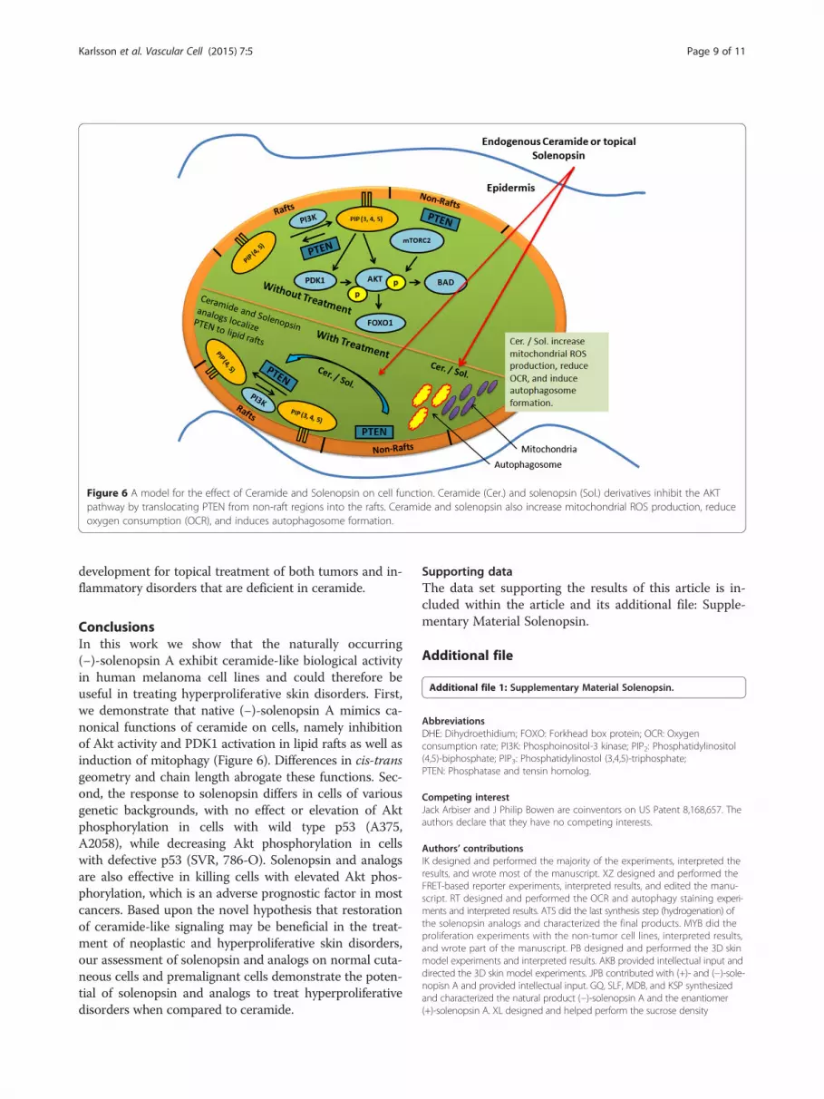
Figure 6 A model for the effect of Ceramide and Solenopsin on cell function. Ceramide (Cer.) and solenopsin (Sol.) derivatives inhibit the AKTpathway by translocating PTEN from non-raft regions into the rafts. Ceramide and solenopsin also increase mitochondrial ROS production, reduceoxygen consumption (OCR), and induces autophagosome formation.
Karlsson et al. Vascular Cell (2015) 7:5 Page 9 of 11
development for topical treatment of both tumors and in-flammatory disorders that are deficient in ceramide.
ConclusionsIn this work we show that the naturally occurring(−)-solenopsin A exhibit ceramide-like biological activityin human melanoma cell lines and could therefore beuseful in treating hyperproliferative skin disorders. First,we demonstrate that native (−)-solenopsin A mimics ca-nonical functions of ceramide on cells, namely inhibitionof Akt activity and PDK1 activation in lipid rafts as well asinduction of mitophagy (Figure 6). Differences in cis-transgeometry and chain length abrogate these functions. Sec-ond, the response to solenopsin differs in cells of variousgenetic backgrounds, with no effect or elevation of Aktphosphorylation in cells with wild type p53 (A375,A2058), while decreasing Akt phosphorylation in cellswith defective p53 (SVR, 786-O). Solenopsin and analogsare also effective in killing cells with elevated Akt phos-phorylation, which is an adverse prognostic factor in mostcancers. Based upon the novel hypothesis that restorationof ceramide-like signaling may be beneficial in the treat-ment of neoplastic and hyperproliferative skin disorders,our assessment of solenopsin and analogs on normal cuta-neous cells and premalignant cells demonstrate the poten-tial of solenopsin and analogs to treat hyperproliferativedisorders when compared to ceramide.
Supporting dataThe data set supporting the results of this article is in-cluded within the article and its additional file: Supple-mentary Material Solenopsin.
Additional file
Additional file 1: Supplementary Material Solenopsin.
AbbreviationsDHE: Dihydroethidium; FOXO: Forkhead box protein; OCR: Oxygenconsumption rate; PI3K: Phosphoinositol-3 kinase; PIP2: Phosphatidylinositol(4,5)-biphosphate; PIP3: Phosphatidylinostol (3,4,5)-triphosphate;PTEN: Phosphatase and tensin homolog.
Competing interestJack Arbiser and J Philip Bowen are coinventors on US Patent 8,168,657. Theauthors declare that they have no competing interests.
Authors’ contributionsIK designed and performed the majority of the experiments, interpreted theresults, and wrote most of the manuscript. XZ designed and performed theFRET-based reporter experiments, interpreted results, and edited the manu-script. RT designed and performed the OCR and autophagy staining experi-ments and interpreted results. ATS did the last synthesis step (hydrogenation) ofthe solenopsin analogs and characterized the final products. MYB did theproliferation experiments with the non-tumor cell lines, interpreted results,and wrote part of the manuscript. PB designed and performed the 3D skinmodel experiments and interpreted results. AKB provided intellectual input anddirected the 3D skin model experiments. JPB contributed with (+)- and (−)-sole-nopisn A and provided intellectual input. GQ, SLF, MDB, and KSP synthesizedand characterized the natural product (−)-solenopsin A and the enantiomer(+)-solenopsin A. XL designed and helped perform the sucrose density

Karlsson et al. Vascular Cell (2015) 7:5 Page 10 of 11
fractionation experiments. GC provided intellectual input and directed the su-crose density fractionation experiments. BO provided intellectual input and di-rected the OCR and autophagy staining experiments. JZ providedintellectual input, directed the FRET-based reporter experiments, andedited the manuscript. EBW provided intellectual input, direction in thesynthesis design, and edited the manuscript. RSA designed and performedthe ROS experiments and interpreted results. JLA formulated the researchideas, provided intellectual input, directed the research designs, providedresources, interpreted results, and wrote major parts of the manuscript. Allauthors read and approved the final manuscript.
AcknowledgementsThis work was funded by Dr. Arbiser's NIH R01 AR047901, the MargolisFoundation, Rabinowitch-Davis Foundations and Dr. Zhang's NIH R01 DK073368.
Author details1Department of Dermatology, Emory University School of Medicine, Atlanta,GA, USA. 2Department of Pharmacology and Molecular Sciences, The JohnsHopkins University School of Medicine, Baltimore, MD, USA. 3Department ofBiochemistry and Molecular Biology, Medical University of South Carolina,Charleston, SC, USA. 4Department of Pharmaceutical Sciences, School ofPharmacy, Union University, Jackson, TN, USA. 5Department of Physiologyand Renal Division, Emory University School of Medicine, Atlanta, GA, USA.6Center for Drug Design, Department of Pharmaceutical Sciences, College ofPharmacy, Mercer University, Atlanta, GA, USA. 7Department of Chemistry &Biochemistry, University of North Carolina Greensboro, Greensboro, NC, USA.8Department of Urology, Emory University School of Medicine, Atlanta, GA,USA. 9Atlanta Veterans Administration Hospital, and Winship Cancer Institute,Emory University, Atlanta, GA, USA.
Received: 17 April 2015 Accepted: 21 April 2015
References1. Arbiser JL, Kau T, Konar M, Narra K, Ramchandran R, Summers SA, et al.
Solenopsin, the alkaloidal component of the fire ant (Solenopsis invicta), is anaturally occurring inhibitor of phosphatidylinositol-3-kinase signaling andangiogenesis. Blood. 2007;109:560–5.
2. Rabionet M, Gorgas K, Sandhoff R. Ceramide synthesis in the epidermis.Biochim Biophys Acta. 1841;2014:422–34.
3. Mencarelli C, Martinez-Martinez P. Ceramide function in the brain: when aslight tilt is enough. Cell Mol Life Sci. 2013;70:181–203.
4. Garcia-Barros M, Coant N, Truman JP. Snider AJ. Sphingolipids in coloncancer. Biochim Biophys Acta: Hannun YA; 2013.
5. Furuya H, Shimizu Y, Kawamori T. Sphingolipids in cancer. Cancer MetastasisRev. 2011;30:567–76.
6. Gomez-Munoz A, Gangoiti P, Arana L, Ouro A, Rivera IG, Ordonez M, et al.New insights on the role of ceramide 1-phosphate in inflammation. BiochimBiophys Acta. 1831;2013:1060–6.
7. Ogretmen B, Hannun YA. Biologically active sphingolipids in cancerpathogenesis and treatment. Nat Rev Cancer. 2004;4:604–16.
8. Sentelle RD, Senkal CE, Jiang W, Ponnusamy S, Gencer S, Selvam SP, et al.Ceramide targets autophagosomes to mitochondria and induces lethalmitophagy. Nat Chem Biol. 2012;8:831–8.
9. Nagata Y, Lan KH, Zhou X, Tan M, Esteva FJ, Sahin AA, et al. PTEN activationcontributes to tumor inhibition by trastuzumab, and loss of PTEN predictstrastuzumab resistance in patients. Cancer Cell. 2004;6:117–27.
10. Clark AS, West K, Streicher S, Dennis PA. Constitutive and inducible Aktactivity promotes resistance to chemotherapy, trastuzumab, or tamoxifen inbreast cancer cells. Mol Cancer Ther. 2002;1:707–17.
11. Arbiser JL, Moses MA, Fernandez CA, Ghiso N, Cao Y, Klauber N, et al.Oncogenic H-ras stimulates tumor angiogenesis by two distinct pathways.Proc Natl Acad Sci U S A. 1997;94:861–6.
12. Arbiser JL, Klauber N, Rohan R, van Leeuwen R, Huang MT, Fisher C, et al.Curcumin is an in vivo inhibitor of angiogenesis. Mol Med. 1998;4:376–83.
13. Bai X, Cerimele F, Ushio-Fukai M, Waqas M, Campbell PM, Govindarajan B,et al. Honokiol, a small molecular weight natural product, inhibits angiogenesisin vitro and tumor growth in vivo. J Biol Chem. 2003;278:35501–7.
14. Gao X, Lowry PR, Zhou X, Depry C, Wei Z, Wong GW, et al. PI3K/Aktsignaling requires spatial compartmentalization in plasma membranemicrodomains. Proc Natl Acad Sci U S A. 2011;108:14509–14.
15. Gao X, Zhang H, Takahashi T, Hsieh J, Liao J, Steinberg GK, et al. The Aktsignaling pathway contributes to postconditioning's protection againststroke; the protection is associated with the MAPK and PKC pathways.J Neurochem. 2008;105:943–55.
16. Griesbeck O, Baird GS, Campbell RE, Zacharias DA, Tsien RY. Reducing theenvironmental sensitivity of yellow fluorescent protein. Mechanism andapplications. J Biol Chem. 2001;276:29188–94.
17. Huang H, Feng X, Zhuang J, Frohlich O, Klein JD, Cai H, et al. Internalizationof UT-A1 urea transporter is dynamin dependent and mediated by bothcaveolae- and clathrin-coated pit pathways. Am J Physiol Renal Physiol.2010;299:F1389–1395.
18. Chen G, Howe AG, Xu G, Frohlich O, Klein JD, Sands JM. Mature N-linkedglycans facilitate UT-A1 urea transporter lipid raft compartmentalization.FASEB J. 2011;25:4531–9.
19. Castro BM, Prieto M, Silva LC. Ceramide: a simple sphingolipid with uniquebiophysical properties. Prog Lipid Res. 2014;54:53–67.
20. Caselli A, Mazzinghi B, Camici G, Manao G, Ramponi G. Some protein tyrosinephosphatases target in part to lipid rafts and interact with caveolin-1. BiochemBiophys Res Commun. 2002;296:692–7.
21. Vazquez F, Ramaswamy S, Nakamura N, Sellers WR. Phosphorylation of thePTEN tail regulates protein stability and function. Mol Cell Biol.2000;20:5010–8.
22. Goswami R, Singh D, Phillips G, Kilkus J, Dawson G. Ceramide regulation ofthe tumor suppressor phosphatase PTEN in rafts isolated from neurotumorcell lines. J Neurosci Res. 2005;81:541–50.
23. Hajduch E, Turban S, Le Liepvre X, Le Lay S, Lipina C, Dimopoulos N, et al.Targeting of PKCzeta and PKB to caveolin-enriched microdomains represents acrucial step underpinning the disruption in PKB-directed signalling byceramide. Biochem J. 2008;410:369–79.
24. Tsao H, Zhang X, Fowlkes K, Haluska FG. Relative reciprocity of NRAS andPTEN/MMAC1 alterations in cutaneous melanoma cell lines. Cancer Res.2000;60:1800–4.
25. Kogot-Levin A, Saada A. Ceramide and the mitochondrial respiratory chain.Biochimie. 2014;100:88–94.
26. Lecour S, Van der Merwe E, Opie LH, Sack MN. Ceramide attenuates hypoxiccell death via reactive oxygen species signaling. J Cardiovasc Pharmacol.2006;47:158–63.
27. Li H, Junk P, Huwiler A, Burkhardt C, Wallerath T, Pfeilschifter J, et al. Dualeffect of ceramide on human endothelial cells: induction of oxidative stressand transcriptional upregulation of endothelial nitric oxide synthase.Circulation. 2002;106:2250–6.
28. Bose R, Verheij M, Haimovitz-Friedman A, Scotto K, Fuks Z, Kolesnick R.Ceramide synthase mediates daunorubicin-induced apoptosis: an alternativemechanism for generating death signals. Cell. 1995;82:405–14.
29. Kolesnick R, Fuks Z. Radiation and ceramide-induced apoptosis. Oncogene.2003;22:5897–906.
30. Beckham TH, Cheng JC, Marrison ST, Norris JS, Liu X. Interdiction ofsphingolipid metabolism to improve standard cancer therapies. Adv CancerRes. 2013;117:1–36.
31. Reding MT, Buchwald SL. Short Enantioselective Total Syntheses of thePiperidine Alkaloids (S)-Coniine and (2R,6R)-trans-Solenopsin A via CatalyticAsymmetric Imine Hydrosilylation. J Org Chem. 1998;63:6344–7.
32. Wilkinson TJ, Stehle NW, Beak P. Enantioselective syntheses of 2-alkyl- and2,6-dialkylpiperidine alkaloids: preparations of the hydrochlorides of (−)-coniine,(−)-solenopsin A, and (−)-dihydropinidine. Org Lett. 2000;2:155–8.
33. Amat M, Llor N, Hidalgo J, Escolano C, Bosch J. Enantioselective synthesis ofpiperidine, indolizidine, and quinolizidine alkaloids from a phenylglycinol-derived delta-lactam. J Org Chem. 2003;68:1919–28.
34. Macconnell JG, Blum MS, Fales HM. Alkaloid from fire ant venom:identification and synthesis. Science. 1970;168:840–1.
35. Yao R, Cooper GM. Requirement for phosphatidylinositol-3 kinase inthe prevention of apoptosis by nerve growth factor. Science.1995;267:2003–6.
36. Kennedy SG, Wagner AJ, Conzen SD, Jordan J, Bellacosa A, Tsichlis PN, et al.The PI 3-kinase/Akt signaling pathway delivers an anti-apoptotic signal.Genes Dev. 1997;11:701–13.
37. Tsao H, Goel V, Wu H, Yang G, Haluska FG. Genetic interaction betweenNRAS and BRAF mutations and PTEN/MMAC1 inactivation in melanoma.J Invest Dermatol. 2004;122:337–41.
38. Fried L, Arbiser JL. The reactive oxygen-driven tumor: relevance to melanoma.Pigment Cell Melanoma Res. 2008;21:117–22.

Karlsson et al. Vascular Cell (2015) 7:5 Page 11 of 11
39. Tan CH, Rasool S, Johnston GA. Contact dermatitis: allergic and irritant.Clin Dermatol. 2014;32:116–24.
40. Kandarova H, Klausner M, Kubilus J, Ayehunie S, Hayden P, Kaluzhny Y, et al.Update on validation status and industry utilization of normal human 3D(NHU-3D) animal alternative models. In: MatTek Corporation A, MA, USA,MatTek In Vitro Life Science Laboratories, Bratislava, Slovakia, editors.Presented at 8th World Congress on Alternative and Animal Use; Montreal,Canada. 2011.
Submit your next manuscript to BioMed Centraland take full advantage of:
• Convenient online submission
• Thorough peer review
• No space constraints or color figure charges
• Immediate publication on acceptance
• Inclusion in PubMed, CAS, Scopus and Google Scholar
• Research which is freely available for redistribution
Submit your manuscript at www.biomedcentral.com/submit


![EXHIBIT C [HOTEL LEASE]](https://img.pdfslide.net/doc/110x75/6326c5cde491bcb36c0af5de/exhibit-c-hotel-lease.jpg)


