Embed Size (px)
Citation preview
Somatic Sensations: I. General Organization, the
Tactile andPosition Senses
CLASSIFICATION OF SOMATIC SENSES
The somatic senses can be classified into three physiologic types:
(1) the mechanoreceptive somatic senses, which include both tactile and position sensations that are stimulated by mechanical displacement of some tissue of the body
(2) the thermoreceptive senses, which detect heat and cold
(3) the pain sense, which is activated by any factor that damages the tissues.
Other Classifications of Somatic Sensations:
Exteroreceptive sensations are those from the surface of the body
Proprioceptive sensations are those having to do with the physical state of the body, including position sensations, tendon and muscle sensations, pressure sensations from the bottom of the feet, and even the sensation of equilibrium (which is often considered a “special” sensation rather than a somatic sensation)
Visceral sensations are those from the viscera of the body; in using this term, one usually refers specifically to sensations from the internal organs
Deep sensations are those that come from deep tissues, such as from fasciae, muscles, and bone. These include mainly “deep” pressure, pain, and vibration
Interrelations Among the Tactile Sensations of Touch, Pressure, and Vibration
Although touch, pressure, and vibration are frequently classified as separate sensations, they are all detected by the same types of receptors.
There are three principal differences among them:
(1) touch sensation generally results from stimulation of tactile receptors in the skin or in tissues immediately beneath the skin
(2) pressure sensation generally results from deformation of deeper tissues
(3) vibration sensation results from rapidly repetitive sensorysignals, but some of the same types of receptors as those for touch and pressure are used
1. Free nerve endings• which are found everywhere in the skin and in many other
tissues, candetect touch and pressure.
• For instance, even light contact with the cornea of the eye, which contains no
other type of nerve ending besides free nerve endings, can nevertheless elicit touch and pressure sensations.
2. Meissner’s corpuscle• Touch receptor with great sensitivity • An elongated encapsulated nerve ending of a large (type
Ab) myelinated sensory nerve fiber. Inside the capsulation are many branching terminal nerve filaments.
• These corpuscles are present in the nonhairy parts of the skin and are particularly abundant in the fingertips, lips, and other areas of the skin where one’s ability to discern spatial locations of touch sensations is highly developed.
• Meissner’s corpuscles adapt in a fraction of a second
after they are stimulated, which means that they are particularly sensitive to movement of objects over the surface of the skin as well as to low frequency vibration.
TACTILE RECEPTORS
3. Expanded tip tactile receptors, one type of which is Merkel’s discs • found in fingertips and hairy parts of the skin • These receptors differ from Meissner’s corpuscles in
that they transmit an initially strong but partially adapting signal and then a continuing weaker signal that adapts only slowly. Therefore, they are responsible for giving steady-state signals that allow one to determine continuous touch of objects against the skin.
• Merkel’s discs are often grouped together in a receptor organ called the Iggo dome receptor, which projects upward against the underside of the epithelium of the skin This causes the epithelium at this point to protrude outward, thus creating a dome and constituting an extremely sensitive receptor.
• Also note that the entire group of Merkel’s discs is innervated by a single large myelinated nerve fiber (type Aβ).
• These receptors, along with the Meissner’s corpuscles, play extremely important roles in localizing touch sensations to specific surface areas of the body and in determining the texture of what is felt.
.5. Ruffini’s end-organs• located in the deeper layers of the skin and also in
still deeper internal tissues, which are multibranched, encapsulated endings
• These endings adapt very slowly and, therefore, are important for signaling continuous states of deformation of the tissues, such as heavy prolonged touch and pressure signals.
• They are also found in joint capsules and help to signal
the degree of joint rotation.
6. Pacinian corpuscles• lie both immediately beneath the skin and deep in the
fascial tissues of the body.
• They are stimulated only by rapid local compression of the tissues because they adapt in a few hundredths of a second. Therefore, they are particularly important for detecting tissue vibration or other rapid changes in the mechanical state of the tissues.
• Almost all sensory information from the somatic segments of the body enters the spinal cord through the dorsal roots of the spinal nerves.
• From the entry point, the sensory signals are carried
through one of two alternative sensory pathways: (1)the dorsal column–medial lemniscal system or (2) the anterolateral system
• These two systems come back together partially at the level of the thalamus
After the signals synapse and cross to the opposite side in the medulla, they continue upward through the brain stem to the thalamus by way of the medial lemniscus.
• Conversely, signals in the anterolateral system, immediately after entering the spinal cord from the dorsal spinal nerve roots, synapse in the dorsal horns of the spinal gray matter, then cross to the opposite side of the cord and ascend through the anterior and lateral white columns of the cord. They terminate at all levels of the lower brain stem and in the thalamus
Sensory Pathways for Transmitting Somatic Signals into the Central Nervous System
Detection of Vibration
All tactile receptors are involved in detection of vibration, although different receptors detect different frequencies of vibration.
Pacinian corpuscles can detect signal vibrations from 30 to 800 cycles per secondbecause they respond extremely rapidly to minute and rapid deformations of the tissues, and they also transmit their signals over type Aβ nerve fibers, which cantransmit as many as 1000 impulses per second.
Position Senses
The position senses are frequently also called proprioceptivesenses. They can be divided into two subtypes:
(1) static position sense, which means conscious perception of the orientation of the different parts of the body with respect to one another
(2)dynamic proprioception or rate of movement sense, also called kinesthesia

Somatic Sensations:II. Pain, Headache, and
Thermal Sensations• Many, if not most, ailments of the body cause pain• Furthermore, the ability to diagnose different diseases
depends to a great extent on a physician’s knowledge of the different qualities of pain
Pain Is a Protective Mechanism• Pain occurs whenever any tissues are being damaged, and it
causes the individual to react to remove the pain stimulus• Even such simple activities as sitting for a long time on
the ischia can cause tissue destruction because of lack of blood flow to the skin where it is compressed by the weight of the body. When the skin becomes painful as a result of the ischemia, the person normally shifts weight subconsciously.
• A person who has lost the pain sense, as after spinal cord injury, fails to feel the pain and, therefore, fails to shift. This soon results in total breakdown and desquamation of the skin at the areas of pressure.
Pain Receptors and Their Stimulation
Pain Receptors Are Free Nerve Endings
• The pain receptors in the skin and other tissues are all free nerve endings. They are widespread in the superficial layers of the skin as well as in certain internal tissues, such as the periosteum, the arterial walls, the joint surfaces, and the falx and tentorium in the cranial vault.
• Most other deep throbbing pain, nauseous pain, and chronic pain. This type of pain is usually associated with tissue destruction. It can lead to prolonged, unbearable suffering. It can occur both in the skin and in almost any deep tissue or organ.
Types of Pain and Their Qualities—Fast Pain and Slow Pain
• Fast pain is felt within about 0.1 second after a pain stimulus is applied, whereas slow pain begins only after 1 second or more and then increases slowly over many seconds and sometimes even minutes.
• Fast pain is also described by many alternative names, such as sharp pain,
pricking pain, acute pain, and electric pain. This type of pain is felt when a needle is stuck into the skin, when the skin is cut with a knife, or when the skin is acutely burned. It is also felt when the skin is subjected to electric shock. Fast-sharp pain is not felt in most deeper tissues of the body.
• Slow pain also goes by many names, such as slow burning pain, aching pain,throbbing pain, nauseous pain, and chronic pain.
• This type of pain is usually associated with tissue destruction. It can lead to prolonged, unbearable suffering. It can occur both in the skin and in almost any deep tissue or organ.
Three Types of Stimuli Excite Pain ReceptorsMechanical,Thermal, and Chemical• Pain can be elicited by multiple types of stimuli.
They are classified as mechanical, thermal, and chemical pain stimuli.
• In general, fast pain is elicited by the mechanical and thermal types of
stimuli, whereas slow pain can be elicited by all three types.
• The chemical substances are especially important in stimulating the slow, suffering type of pain that occurs after tissue injury
• Some of the chemicals that excite the chemical type of pain are bradykinin, serotonin, histamine, potassium ions, acids, acetylcholine, and proteolytic enzymes.
• In addition, prostaglandins and substance P enhance the sensitivity of pain endings but do not directly excite them.
• Nonadapting Nature of Pain Receptors• In contrast to most other sensory receptors of the body,
pain receptors adapt very little and sometimes not at all.
• In fact, under some conditions, excitation of pain fibers becomes progressively greater, especially so for slow-aching-nauseous pain, as the pain stimulus continues.
• This increase in sensitivity of the pain receptors is called hyperalgesia.
• One can readily understand the importance of this failure of pain receptors to adapt, because it allows the pain to keep the person apprised of a tissue-damaging stimulus as long as it persists
Rate of Tissue Damage as a Stimulus for Pain
• The average person begins to perceive pain when the skin is heated above 45°C
• This is also the temperature at which the tissues begin to be damaged by heat; indeed, the tissues are eventually destroyed if the temperature remains above this level indefinitely.
• Therefore, it is immediately apparent that pain resulting from heat is closely correlated with the rate at which damage to the tissues is occurring and not with the total damage that has already occurred.
• The intensity of pain is also closely correlated with the rate of tissue damage from causes other than heat, such as bacterial infection, tissue ischemia, tissue contusion, and so forth.
Tissue Ischemia as a Cause of Pain
When blood flow to a tissue is blocked, the tissue often becomes very painfulwithin a few minutes. The greater the rate of metabolism of the tissue, the more rapidly the pain appears.
For instance, if a blood pressure cuff is placed around the upper arm and inflated until the arterial blood flow ceases, exercise of the forearm muscles sometimes can cause muscle pain within 15 to 20 seconds. In the absence of muscle exercise, the pain may not appear for 3 to 4 minutes even though the muscle blood flow remains zero.
One of the suggested causes of pain during ischemia is accumulation of large amounts of lactic acid in the tissues, formed as a consequence of anaerobic metabolism (metabolism without oxygen).
It is also probable that other chemical agents, such as bradykinin andproteolytic enzymes, are formed in the tissues because of cell damage and that these, in addition to lactic acid, stimulate the pain nerve endings.
Muscle Spasm as a Cause of PainMuscle spasm is also a common cause of pain, and it is the basis of manyclinical pain syndromes.
This pain probably results partially from the direct effect of muscle spasm instimulating mechanosensitive pain receptors, but it might also result from the indirect effect of muscle spasm to compress the blood vessels and causeischemia.
Also, the spasm increases the rate of metabolism in the muscle tissue, thus making the relative ischemia even greater, creating ideal conditions for therelease of chemical pain-inducing substances.
Dual Pathways for Transmission of PainSignals into the Central Nervous System
Even though all pain receptors are free nerve endings, these endings use two separate pathways for transmitting pain signals into the central nervous system.
The two pathways mainly correspond to the two types of pain—a fast-sharp pain pathway and a slow-chronic pain pathway.
Peripheral Pain Fibers—“Fast” and “Slow” Fibers The fast sharp pain signals are elicited by either mechanical or thermal pain stimuli; they are transmitted in the peripheral nerves to the spinal cord by small type Aδ fibers at velocities between 6 and 30 m/sec.
Conversely, the slow-chronic type of pain is elicited mostly by chemical types of pain stimuli but sometimes by persisting mechanical or thermal stimuli.
This slow chronic pain is transmitted to the spinal cord by type C fibers at velocities between 0.5 and 2 m/sec.
Dual Pain Pathways in the Cord and Brain StemThe Neospinothalamic Tract and the
Paleospinothalamic Tract
On entering the spinal cord, the pain signals take twopathways to the brain, through : (1) the neospinothalamic tract (2) the paleospinothalamic tract
Neospinothalamic Tract for Fast Pain. The fast type Aδ pain fibers transmit mainly mechanical and acute thermal pain
Paleospinothalamic Pathway for Transmitting Slow-Chronic PainThe paleospinothalamic pathway is a much older system and transmits pain mainly from the peripheral slow-chronic type C pain fibers, although it does transmit some signals from type Aδ fibers as well
Pain Suppression (“Analgesia”) System in theBrain and Spinal Cord
The degree to which a person reacts to pain varies tremendously. This results partly from a capability of the brain itself to suppress input of pain signals to the nervous system by activating a pain control system, called an analgesia system.
The analgesia system consists of three major components: (1) The periaqueductal gray and periventricular areas of the mesencephalonand upper pons surround the aqueduct of Sylvius and portions of the third and fourth ventricles. Neurons from these areas send signals to: (2) the raphe magnus nucleus, a thin midline nucleus located inthe lower pons and upper medulla, and the nucleus reticularis paragigantocellularis, located laterally in the medulla. From these nuclei, second-order signals are transmitted down the dorsolateral columns in thespinal cord to: (3) a pain inhibitory complex located in the dorsal horns of the spinal cord. At this point, the analgesia signals can block the pain before it is relayedto the brain.
Inhibition of Pain Transmission by Simultaneous Tactile Sensory Signals
Another important event in the saga of pain control was the discovery that stimulation of large type Aβ sensory fibers from peripheral tactile receptors can depress transmission of pain signals from the same body area.
This presumably results from local lateral inhibition in the spinal cord. It explains why such simple maneuvers as rubbing the skin near painful areas is often effective in relieving pain.
And it probably also explains why liniments are often useful for pain relief.
This mechanism and the simultaneous psychogenic excitation of the central analgesia system are probably also the basis of pain relief by acupuncture.
Referred Pain
Often a person feels pain in a part of the body that is fairly remote from the tissue causing the pain. This is called referred pain.
For instance, pain in one of the visceral organs often is referred to an area on the body surface. Knowledge of the different types of referred pain is important in clinical diagnosis because in many visceral ailments the only clinical sign is referred pain.
Mechanism of Referred Pain:Branches of visceral pain fibers are shown to synapse in the spinal cord on the same second-order neurons (1 and 2) that receive pain signals from theskin. When the visceral pain fibers are stimulated, pain signals from the viscera are conducted through at least some of the same neurons that conduct pain signals from the skin, and the person has the feeling that the sensations originate in the skin itself.
Visceral PainIn clinical diagnosis, pain from the different viscera of the abdomen and chest is one of the few criteria that can be used for diagnosing visceral inflammation, visceral infectious disease, and other visceral ailments.
Often, the viscera have sensory receptors for no other modalities of sensation besides pain.
Also, visceral pain differs from surface pain in several important aspects. One of the most important differences between surface pain and visceral pain is that highly localized types of damage to the viscera seldom cause severe pain.
Conversely, any stimulus that causes diffuse stimulation of pain nerve endings throughout a viscus causes pain that can be severe. E.g. Ischemia caused by occluding the blood supply to a large area of gut stimulates many diffuse pain fibers at the same time and can result in extreme pain.
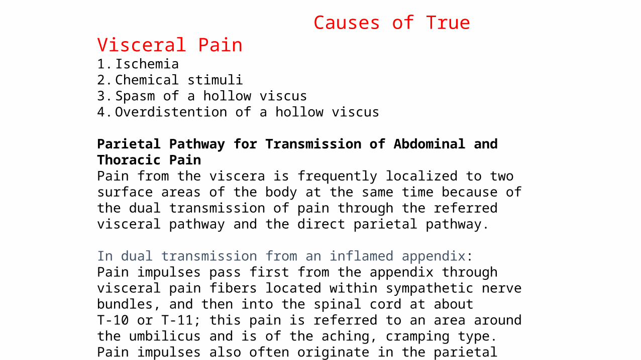
Causes of True Visceral Pain1.Ischemia2.Chemical stimuli3.Spasm of a hollow viscus4.Overdistention of a hollow viscus
Parietal Pathway for Transmission of Abdominal and Thoracic PainPain from the viscera is frequently localized to two surface areas of the body at the same time because of the dual transmission of pain through the referred visceral pathway and the direct parietal pathway. In dual transmission from an inflamed appendix: Pain impulses pass first from the appendix through visceral pain fibers located within sympathetic nerve bundles, and then into the spinal cord at aboutT-10 or T-11; this pain is referred to an area around the umbilicus and is of the aching, cramping type. Pain impulses also often originate in the parietal peritoneum where the inflamed appendix touches or is adherent to the abdominal wall. These cause pain of the sharp type directly over the irritated peritoneum in the right lower quadrant of the abdomen
Some Clinical Abnormalities of Pain and Other Sensations
HyperalgesiaA pain nervous pathway sometimes becomes excessively excitable; this gives rise to hyperalgesia, which means hypersensitivity to pain.
Possible causes of hyperalgesia:(1) excessive sensitivity of the pain receptorsthemselves, which is called primary hyperalgesia
(2) facilitation of sensory transmission, which is calledsecondary hyperalgesia
An example of primary hyperalgesia is the extreme sensitivity of sunburned skin, which results from sensitization of the skin pain endings by local tissue products from the burn—perhaps histamine, perhaps prostaglandins, perhaps others. Secondary hyperalgesia frequently results from lesions in the spinal cord or thethalamus.
HeadacheHeadaches are a type of pain referred to the surface of the head from deep head structures.
Some headaches result from pain stimuli arising inside the cranium, but others result from pain arising outside the cranium, such as from the nasal sinuses.
Areas of the Head to Which Intracranial Headache Is ReferredStimulation of pain receptors in the cerebral vault above the tentorium, including the upper surface of the tentorium itself, initiates pain impulses in the cerebral portion of the fifth nerve and, therefore, causes referredheadache to the front half of the head in the surface areas supplied by this somatosensory portion of the fifth cranial nerve
Headache Caused by Eye DisordersDifficulty in focusing one’s eyes clearly may cause excessive contraction ofthe eye ciliary muscles in an attempt to gain clear vision.
Even though these muscles are extremely small, it is believed that tonic contraction of them can cause retroorbital headache. Also, excessive attempts to focus the eyes can result in reflex spasm in various facial and extraocular muscles, which is a possible cause of headache.
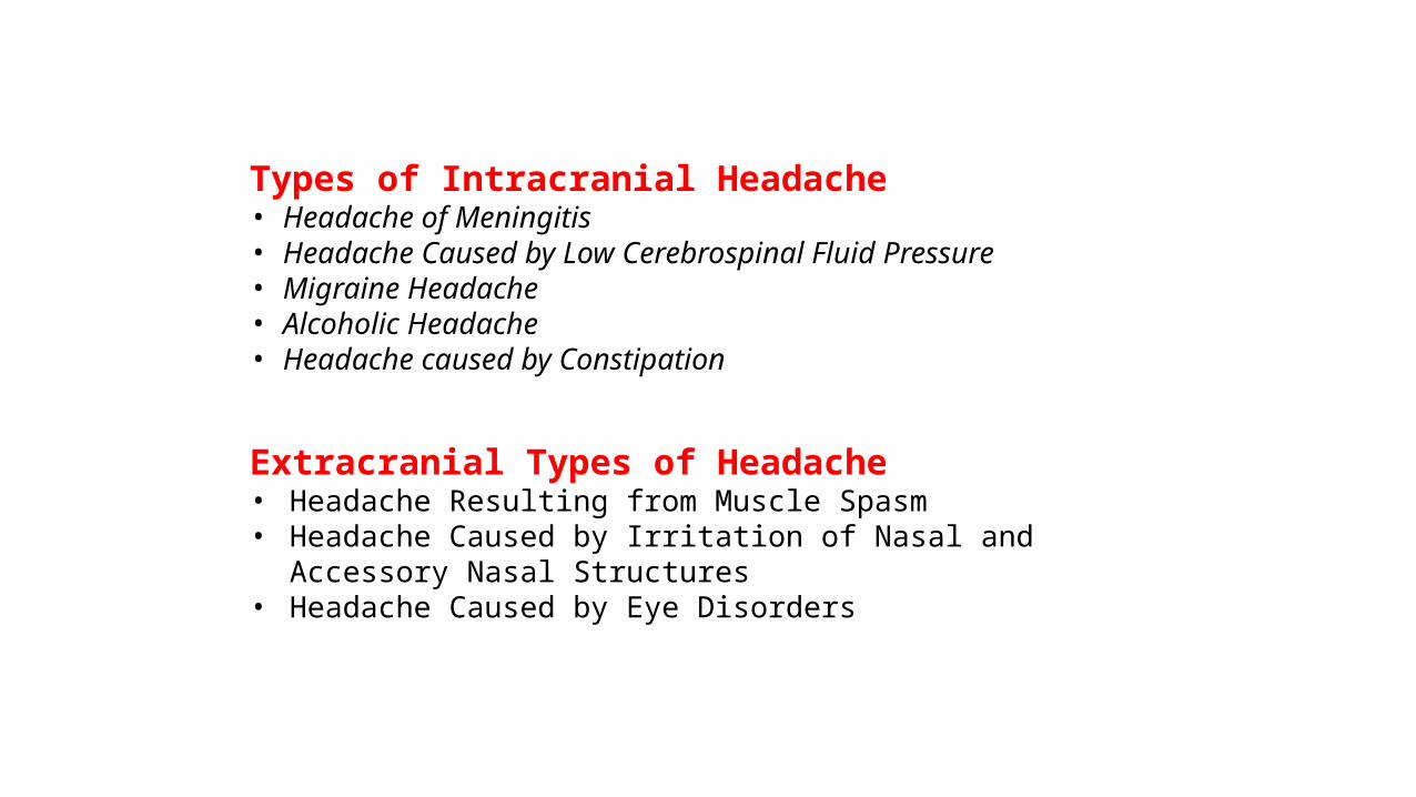
Types of Intracranial Headache• Headache of Meningitis• Headache Caused by Low Cerebrospinal Fluid Pressure• Migraine Headache• Alcoholic Headache• Headache caused by Constipation
Extracranial Types of Headache• Headache Resulting from Muscle Spasm• Headache Caused by Irritation of Nasal and
Accessory Nasal Structures• Headache Caused by Eye Disorders
Thermal Sensations
Thermal Receptors and Their Excitation
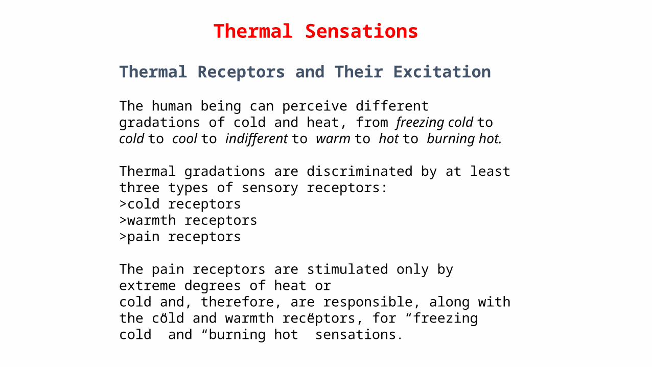
The human being can perceive different gradations of cold and heat, from freezing cold to cold to cool to indifferent to warm to hot to burning hot.
Thermal gradations are discriminated by at least three types of sensory receptors: >cold receptors>warmth receptors >pain receptors
The pain receptors are stimulated only by extreme degrees of heat orcold and, therefore, are responsible, along with the cold and warmth receptors, for “freezing cold” and “burning hot” sensations.
Stimulatory Effects of Rising and Falling Temperature—Adaptation of Thermal ReceptorsWhen a cold receptor is suddenly subjected to an abrupt fall in temperature, itbecomes strongly stimulated at first, but this stimulation fades rapidly during the first few seconds and progressively more slowly during the next 30 minutes or more.
In other words, the receptor “adapts” to a great extent, but never 100 per cent.
Thus, it is evident that the thermal senses respond markedly to changes in temperature, in addition to being able to respond to steady states of temperature.
This means that when the temperature of the skin is actively falling, a person feels much colder than when the temperature remains cold at the same level. Conversely, if the temperature is actively rising, the person feels much warmer than he or she would at the same temperature if it were constant. The response to changes in temperature explains the extreme degree of heat one feels on first entering a tub of hot water and the extreme degree of cold felt on going from aheated room to the out-of-doors on a cold day.
Mechanism of Stimulation ofThermal ReceptorsIt is believed that the cold and warmth receptors are stimulated by changes in their metabolic rates, and that these changes result from the fact that temperature alters the rate of intracellular chemical reactions more than twofold for each 10°C change.
In other words, thermal detection probably results not from direct physical effects of heat or cold on the nerve endings but from chemical stimulation of the endings as modified by temperature.
Stimulation of Thermal Receptors—Sensations of Cold, Cool, Indifferent, Warm, and Hot
Responses of four types of nerve fibers: (1)a pain fiber stimulated by cold (2) a cold fiber (3) a warmth fiber(4) A pain fiber stimulated by heat
Note especially that these fibers respond differently at different levels of temperature. For instance, in the very cold region, only the cold-pain fibers are stimulated (if the skin becomes even colder, so that it nearly freezes or actually does freeze, these fibers cannot be stimulated).
As the temperature rises to +10° to 15°C, the cold-pain impulses cease, but the cold receptors begin to be stimulated, reaching peak stimulation at about 24°C and fading out slightly above 40°C.
Above about 30°C, the warmth receptors begin to be stimulated, but thesealso fade out at about 49°C.
Finally, at around 45°C, the heat-pain fibers begin to be stimulated by heat and, paradoxically, some of the cold fibers begin to be stimulated again, possibly because of damage to the cold endings caused by the excessive heat.
Transmission of Thermal Signalsin the Nervous System
In general, thermal signals are transmitted in pathways parallel to those for pain signals. On entering the spinal cord, the signals travel for a few segmentsupward or downward in the tract of Lissauer and then terminate mainly in laminae I, II, and III of the dorsal horns—the same as for pain.
After a small amount of processing by one or more cord neurons, the signalsenter long, ascending thermal fibers that cross to the opposite anterolateral sensory tract and terminate in both : (1) the reticular areas of the brain stem (2) the ventrobasal complex of the thalamus.


