Embed Size (px)
Citation preview

European Journal of Neuroscience, Vol. 7, pp. 1154-1163, 1995 @ Europeun Neuroscience Association
C02 Stimulation of the Cornea: A Comparison Between Human Sensation and Nerve Activity in Polymodal Nociceptive Afferents of the Cat
Xiaojie Chen, Juana Gallar, Miguel A. Pozo', Manuel Baeza2 and Carlos Belrnonte Departamento de Fisiologia and lnstituto de Neurociencias, Universidad de Alicante, Apdo. 374, 03080 Alicante, Spain 'Present address: Departamento de Fisiologia, Facultad de Medicina, Universidad Complutense, Madrid, Spain *Servicio de Oftalmologia, Hospital General Universitario de Alicante, Universidad de Alicante, Alicante, Spain
Keywords: nociceptors, chemical pain, acid, pH, aesthesiometry
Abstract
Excitation of nociceptors by low pH has been proposed as a cause of pain following tissue injury. Here we have studied the effect of pH reductions caused by application of CO2 pulses to the cornea on the activity of corneal afferent nerves of the cat and on the magnitude of pain sensations in humans. Single-unit activity was recorded from corneal afferent fibres in anaesthetized cats. The corneal receptive field of A-delta or C polymodal nociceptive units was exposed for 30 s to a gas mixture with different concentrations of C02 in air (0, 35, 50, 65, 80 and 98.5%). Responses to C02 were evoked at a mean threshold concentration of 40 ? 3% C02. They consisted of a discharge of impulses that decayed gradually to a tonic level. In 15% of the units the initial burst was absent. The COP concentration and firing frequency data could be fitted to a power function with an exponent of 1.12. Pulses of C02 were also applied to the cornea of 16 human volunteers. Sensations experienced were measured by means of a visual analogue scale and a verbal descriptor scale. Flow was adjusted below the mechanical stimulation threshold (2.8 2 0.5 mg). When mixtures containing 10-90% C02 in 5% steps were applied as 3 s pulses, threshold sensation, described as a mild stinging pain, was evoked at 33.5 ? 4.0% Cop This sensation became overtly painful with higher C02 concentrations (47.5 2 3.6% COP). For the same subject the sensory threshold was remarkably constant, though it changed with longer exposure times. The relationship between COP concentration and magnitude of pain could be adjusted to a power function with a power exponent of 1.12. Curves of C02 concentration versus neural discharges in the cat and versus psychophysical sensation in humans were very similar. These results show that corneal polymodal nociceptors respond to protons, and encode changes in C02 concentration presumably reflecting pH changes. The same stimulus evokes corneal pain sensations in humans, thus opening the possibility of using C02 as an effective stimulus for corneal aesthesiometry.
Introduction
A reduction in the pH of the extracellular environment of sensory nerve terminals is an effective stimulus for the excitation of nociceptors (Lindahl, 1961; Belmonte et al., 1991; Steen et al., 1992). During tissue injury or ischaemia, local acidosis develops through metabolic proton production but also as a consequence of C02 accumulation (Roos and Boron, 1981). which leads to carbonic acid formation in hypoxic or damaged tissues. Local increase in Hf ion concentration observed in exudates has been proposed as one of the main causes of sustained nociceptive stimulation and therefore of pain from inflamed temtories (Lindahl, 1974; Steen et al., 1992).
About 70% of the total sensory supply of the cat's cornea is made of polymodal nociceptors (Belmonte and Giraldez, 198 1; Belmonte et al., 1991; Gallar et al., 1993). Corneal polymodal nociceptors are excited by mechanical, thermal and chemical stimuli and respond to acidic solutions (pH 7.04.5) with impulse frequencies that are
roughly proportional to extracellular pH values (Belmonte et al., 1991; Gallar et al., 1993).
Carbon dioxide acts as a pungent stimulus when applied to the nasal mucosa of human subjects (Cain and Murphy, 1980; Kobal and Hummel, 1988; Anton et al., 1992; Thiirauf et al., 1993). When the eye surface is exposed to a C02 environment, corneal pH decreases (Bonanno and Poke, 1987). This pH change is expected to excite corneal polymodal nociceptors in a manner proportional to pH reduction; however, activation by C 0 2 of corneal nociceptors has not been studied experimentally. Moreover, the possibility of evoking pain sensations by controlled acidic stimulation of the human cornea with COz has never been tested, although it is an alternative procedure to measure corneal sensitivity.
The aim of this study was to examine the effects of C02 pulses of variable concentrations on the neural activity of corneal polymodal
Correspondence 10: Professor Carlos Belmonte, as above
Received 22 September 1994, revised 12 December 1994, accepted 19 December 1994

C02 stimulation and corneal pain 1155
manipulator, at a distance of -5 mm. The flow of gas was adjusted with the regulator to a level such that no neural activity was evoked on the afferent nerves when the stimulation was made with air only. To determine whether the unit was sensitive to COz, a 5-30 s pulse of 98.5% C02 was applied initially. Then, gas mixtures of different COz concentration (5-30 s pulses) were applied in ascending andor descending order, with a 5-10 min interval between stimuli.
nociceptors of the cat and to compare their responses with the pain sensations evoked by the same stimuli applied to human volunteers. Some of the results have been published in abstract form (Chen et al., 1993).
Materials and methods
Electrophysiological experiments Experiments were performed on adult cats of both sexes anaesthetized with sodium pentobarbitone (Nembutal, 40 mgkg, i.p.). Animals were kept in an areflexic state by i.v. infusion of dilute Nembutal (5 mgkg) during the experiment. Rectal temperature and blood pressure were continuously monitored and maintained within physio- logical limits. At the end of the experiment, cats were killed with an overdose of anaesthetic.
Extracellular recordings were made from single corneal afferent fibres dissected from mixed ciliary nerves in the cat’s eye, through Ag-AgCI electrodes using conventional electrophysiological equip- ment, as described elsewhere (Belmonte et al., 1991; Pozo et al., 1992; Gallar et al., 1993). Mechanical stimulation of the cornea was performed with a Cochet-Bonnet aesthesiometer (Cochet and Bonnet, 1960) to determine force thresholds and receptive field borders. Sensitivity to COz was tested by applying a gas jet to the corneal surface containing COz at different concentrations (see below). Sensitivity to acid was also examined by topical application of 60 pl of a solution of 10 mM acetic acid in 155 mM NaCl (pH 3.25), to the unit’s receptive field. Thermal sensitivity was tested with an electronically controlled thermode (8 mm2 surface area) centred on the receptive field. The system allowed stepwise heating in 2°C steps, from an adapting 35°C temperature to a final value of 46-50°C. each step lasting 15 s. Stimuli were ended by active cooling. The cornea was kept moist throughout the experiment by repeated application of isotonic saline either in drops or with an ultrasonic vaporizer.
To measure conduction latency, suprathreshold electric shocks (0.1-0.5 ms, 0.5-3 mA) were applied to the receptive field andlor limbus area with a pair of silver electrodes spaced 3-5 mm apart. Conduction distance was estimated by placing an 8.0 gauge thread along the trajectory of the nerve.
Neural discharge, blood pressure and stimulating pulses were recorded on an FM magnetic tape and analysed off-line with a window discriminator, an analogue-to-digital converter and a computer. The response to COz was analysed by measuring the latency (delay between application of the COz and the first impulse given by the unit), time to peak (delay between the first impulse and the bin in which a maximal number of impulses was measured) and the following frequency values (impulses/s) during the C02 pulses, taking the beginning of the pulse as zero time: mean discharge rate during a definite period, and peak frequency (maximal firing frequency during one bin). Data are presented as mean 2 SEM. Both parametric (t-test) and non-parametric (Wilcoxon test) statistical tests were used.
Carbon dioxide stimulation
Animal experiments For electrophysiological experiments in cats, pressurized tanks con- taining calibrated gas mixtures of air and COz at different concentra- tions (0, 35, 50, 65, 80 and 98.5%) were used. Gas mixtures were humidified and carried through a plastic tube to a gas flow regulator and a three-way valve. One of the outputs of the valve ended in a glass tube ( 1 mm inner diameter) whose tip was placed perpendicularly to the corneal receptive field of the recorded unit by means of a
Human experiments In some human sensation experiments, the same calibrated gas mixtures employed in the electrophysiological studies were used (0, 35, 50, 65, 80 and 98.5% COz in air). In another group of individuals, variable COz concentrations were produced by mixing the content of two tanks filled with 98.5% COz and air respectively. A gas mixer (Witt Gasetechnik, Witt, Germany), previously calibrated with an infrared COz detector, was used to obtain the desired C 0 2 concentration at the gas output (lo-90% COz in 5 1 0 % steps).
Gas mixtures were carried through a gas-impermeable tube to a three-way solenoid valve (The Lee Co., Westbrook, CT) driven by a pulse generator and mounted on a lens-holding universal trial frame. One of the outlets of the valve was connected to a silicone tube. The end of this tube was attached to the centre of one eye-ring of the graduation frame (Fig. 1). By adjusting the position of the graduation frame, the tip of the gas tube was placed perpendicularly to the centre of the cornea, at a distance of 8-10 mm from the corneal surface. The second outlet of the valve diverted the gas away from the eye. Gas flowed continuously through this outlet, unless the electronic valve transiently changed the direction of the flow to the surface of the cornea, for a pre-established period (0.1-10 s) (Fig. 1). The pressure of the gas jet was adjusted by regulating the outflow of the gas mixture with a pressure regulator. The pressure of the gas jet was calibrated as follows. The output tube was connected in series with a pressure transducer (Statham P23XL) and was placed perpendic- ularly to the plate of a weighting balance, with the tip at a distance of 10 mm from the plate. The force exerted by increasing flow rates of gas was measured in milligrams and correlated with the
PRESSURE TRANSDUCER
VALVE
CORNEA
$d FIG. 1. Diagram of the experimental setup used for psychophysical measurements of corneal sensibility. Gas mixtures of variable CO? concentration and flow rate were obtained from a gas mixer and were applied to the eye through a tube whose end was placed at the desired distance from the centre of the cornea with an adjustable correcting lens frame. The direction of the gas flow was regulated with an electronic valve controlled by a square- wave pulse generator. The force of the gas jet was estimated with a pressure transducer placed in series with the direction of flow.

1156 CO2 stimulation and corneal pain
corresponding output voltages given by the transducer. The temper- ature of the gas at the output, measured with a thermode (Sensortek IT-23) connected to an electronic thermometer, was 24°C.
Psychophysical measurements Eighteen healthy volunteers (12 males and six females) with ages ranging from 23 to 37 years participated in this study. None of them wore contact lenses or had any ocular pathology. They gave informed consent to a protocol approved by our Institute and were free to interrupt the session at any time. The experimental protocol started with a general description of the experiment. Then the mechanical sensory threshold was measured with air puffs of 3 s duration at different pressures, ranging from I to 10 mg, in 0.2 mg steps applied in ascending and descending series. Every pressure pulse was applied twice with an interstimulus interval of 1 min. Five minutes after this test, the sensitivity to C 0 2 was explored. In eight subjects, COz mixtures contained in separate gas tanks were used. Pulses of 3 s duration and increasing concentrations (0, 35, 50, 65, 80 and 98.5%) were sequentially applied to the cornea at 1 min intervals, with the gas flow adjusted 0.5 mg below mechanical threshold. After a resting period of 5 min, the experiment was repeated but with COz mixtures presented in a decreasing concentration order. Also, pulses (35, 80 and 98.5% C02) of variable duration (1-10 s) were applied in a separate session ( 1 week apart). In ten subjects, the stimulating series consisted of pulses of variable C02 concentration (between 10 and 90% in 5-10% steps), applied randomly. This experiment was repeated at variable time intervals ( 1 day or 2 weeks). In five of these individuals, slit-lamp examination of the corneal epithelium was performed at the end of the experiment.
Pain measurement was performed using an electronic version of a ‘visual analogue scale’ (VAS) (Anton et al., 1992) and a ‘verbal descriptor scale’ (VDS) (Price, 1988). Subjects were seated comfort- ably in a chair, in front of a panel containing a vertical chain of 20 numbered LEDs whose colour varied from green (bottom, ‘no pain’) to red (top, tolerance level, ‘unbearable pain’). The subject could rate the intensity of the sensation by adjusting with a potentiometer the number of LEDs lighting up.
The subject also rated the sensation using verbal descriptors that were later given a numerical value from I to 5: ‘felt but not irritating’ (I); ‘irritating but tolerable’ (2); ‘painful’ (3); ‘very painful’ (4); ‘unbearably painful’ (5). At the end of the experiments, subjects were also asked to describe in their own words the sensation experienced.
In a separate experiment performed in three individuals, the cornea was anaesthetized with topical tetracaine. A 0.003 inch diameter thermocouple microprobe (Sensortek IT-23, time constant 5 ms) connected to an electronic thermometer (Sensortek, BAT-1 2) was placed on the corneal surface. Temperature was measured at the point of incidence of the gas jet, for the different gas pressures used in the various experimental protocols.
Psychophysical data were reported as mean 2 SEM. Visual analogue scale values were standardized by dividing individual responses to each C02 concentration by the mean response of the subject to all COz pulses applied in the test (Anton et al. 1992).
Results Electrophysiological experiments Sensory units selected for this study (n = 81). were thin myelinated (A-delta) or unmyelinated (C) afferents that responded to mechanical stimulation of the cornea as well as to the application to the corneal
surface of a 5-30 s pulse of 98.5% CO2. Seven of these units, in which thermal sensitivity was explored, also responded to heating of the receptive area. Table 1 summarizes the data on conduction velocity, mechanical threshold and receptive field size of the fibres in which these parameters could be accurately measured. Except when otherwise stated, no differences were found between A-delta and C units in the responses to C02 reported below. Therefore, the results are pooled from all myelinated and unmyelinated fibres.
Exposure of the cornea to 98.5% C02 for 5-30 s triggered two types of afferent discharges. In -85% of the units explored (48 of 57) responses appeared with a short latency ( I .9 5 0.2 s, n = 45) and consisted of a rapid discharge of impulses (mean peak frequency 9.6 2 0.8 impulses/s, n = 48) that decayed gradually to a steady- state tonic level when pulses longer than 5 s were applied (Fig. 2A). In another group of fibres the initial firing was absent; the impulse discharge appeared after a long latency (17.1 ? 2.3 s, n = 9). increased gradually to reach a plateau and decreased slowly at the end of the COz pulse (Fig. 2B). No correlation was found between these firing patterns in response to COz and other properties of the units (mechanical threshold or size of the receptive field); nevertheless, long-latency responses were observed in only one (out of fifteen) of the C fibres and in eight (out of 47) of the A-delta units.
Interruption of the C 0 2 stimulation of the cornea gradually arrested the discharge of the unit. Background activity, compared before and after COz stimulation, was not modified significantly by the stimulus (0.14 ? 0.07 impulses/s before versus 0.08 ? 0.03 after COz application, n = 14).
When calibrated gas mixtures from tanks containing 35, 50, 65, 80 and 98.5% COz in air were applied to the cornea, a correlation was found between COz concentration and mean tiring frequency of the fibres. Figure 3 shows the time course of the impulse discharge evoked by gas pulses of increasing COz concentration during 30 s in 16 units responding with a short latency; the concentration-response curve of this population of corneal units is shown with data on mean and peak frequencies in Figure 4A; the slope of the line of the concentration-response data in a log-log plot (equal to the exponent of the power function) was 1.12 (Fig. 4A, inset). The latency of the response to COz decreased proportionally to the concentration of
TABLE I . Functional properties of corneal units
Myelinated Unmyelinated
Conduction velocity ( m l s ) 6.1 ? 0.82 1.1 2 0.12 (n = 44)
(n = 58)
0.79 2 0.08 ( n = 45)
(n = 25)
(n = 23) Receptive field diameter (mm) 5.9 t 0.3 4.4 f 0.3
Mechanical response: mechanical threshold (mN) 0.77 t 0.13
(n = 20) Response to heat: thermal threshold (“C) 1 st heating 41.0 2 2.0 40.2 ? 0.5
2nd heating 43.0 ? 2.0 41.4 f 1.1
Chemical response (impulseds, 30 s) 10 mM acetic acid 0.4 t 0.06 0.2
98.5% COz 2.1 ? 0.3 2.9 f 0.3
(n = 2 )
(n = 2)
(n = 5 )
(n = 5 )
( 13/16 j* (l/l)a
(n = 44) (n = 9)
Data are mean 2 SEM. aNumber of responding unitdnumber of units explored.

C02 stimulation and corneal pain 1157
98.5% CO,
- t O L , I I I I I I I
0 5 10 15 20 25 30 35
Time (s)
FIG. 2. Mean frequency of discharge of two types of corneal polymodal afferents in response to a 98.5% C 0 2 pulse applied for 30 s to the receptive area. Average response (mean ? SEM) of fibres with an immediate response (A, n = 48) and of units with a delayed response (B, n = 9). Stimulus duration indicated by the upper bar. Insets show sample records of the impulse response of two A-delta units to a 98.5% C 0 2 pulse. Thick arrows signal the beginning of the stimulus. Time scale 4 s in A, 8 s in B.
C02, although the time to reach peak frequency value was independent of C 0 2 concentration (Fig. 4B).
The CO2 concentration needed to recruit the response of corneal units varied from 35 to 65% (mean 40.6 2 2.7%. n = 16). Maximal firing frequency was attained at 65% C02 in four of 16 units; three of them were unmyelinated. Also, C fibres were significantly slower in reaching their peak firing frequency (2.5 2 0.6 s, n = 8) than A-delta fibres (1.7 2 0.2 s, n = 39, P < 0.05). However, peak frequency values were similar in A-delta (2.4 2 0.3 impulses/s, n = 10) and in C fibres (2.9 2 0.4 impulses/s, n = 6).
Repeated application (three times) of a 98.5% C02 stimulus lasting 30 s to the receptive field of 17 units with an interval of 5-10 min between pulses did not change the mean frequency of the evoked impulse response. However, in four units in which the cycle of variable C02 concentrations was applied first in ascending and then in descending steps with a 5 min interval, comparatively lower mean frequency values were found for each concentration step during the descending series (data not shown).
Fibres responding with a long latency also encoded the C02 concentration, but maximal discharges were obtained at values of 65-80% C 0 2 . Figure 5 gives an example of the behaviour of one of these units.
Acetic acid (10 mM) dropped onto the corneal receptive field excited 16 of 18 corneal units that were also sensitive to 98.5% C02. As shown in Table 2, the mean discharge evoked by acetic acid was of lower frequency than the C02 response.
The effect of heating was tested in seven units sensitive to C02. All of them responded to temperatures above an average threshold value of 40.4 rt 0.6"C. The mean frequency increase during a stepwise temperature elevation of 10°C was 0.8 2 0.3 impulses/s.
35% co,
a 6 1
50% CO,
65% CO,
80% CO,
- wbmhiww 0
98.5% co, a
2 0
0 5 10 15 20 25 30
T i m e ( s )
FIG. 3. Mean frequency of discharge of corneal polymodal units (mean t SEM; n = 16) responding with a short latency to the application of C02 mixtures at the concentrations given on the right for a 30s period (upper bar).
Repetition of the heating cycle after a 5 min pause, performed in six fibres, reduced the response in five of them (to 0.5 ? 0.2 impulsesls) and augmented mean frequency in the remaining unit. Pulses of increasing C02 concentration applied to these fibres following the second heating cycle evoked an impulse response whose mean discharge rate was 35 2 12% of preheating frequency values (n = 6).
Psychophysical experiments
Threshold to stimulation with air Air pulses applied to the cornea were detected and reported as 'felt but not irritating' when a critical flow rate was attained. The threshold for this response, measured at the centre of the cornea, ranged from 1 to 4 mg, with an average value of 2.8 2 0.5 mg (mean rt SEM, n = 13). Changes in corneal surface temperature evoked by air pulses were small (<O.I"C) and did not vary appreciably with flow intensity at subthreshold or threshold level or with the type of gas mixture used (air or C 0 2 ) .

1158 COz stimulation and corneal pain
n v)
A \
E . > 5 r = t
a .-
30 50 100
C I I
a r I
B
0 L I I I I
25 50 75 ~
100
CO, concentrat ion (%)
FIG. 4. Response of corneal polymodal units to increasing COz concentrations. (A) Mean frequency of discharge during 5 s (filled circles) and 30 s (open circles) pulses of increasing C 0 2 concentration in 16 polymodal units. Inset shows log-log representation of the mean firing frequency versus C02 concentration data of these units. (B) Mean latency of presentation of the first spike (filled squares) and mean latency of the peak frequency (time to peak, open squares) at increasing C02 concentrations. Data for the 16 units shown in A (mean 2 SEM).
Threshold to stimulation with C 0 2
In eight subjects, experiments were made with mixtures of separate tanks, using 3 s pulses and a gas flow rate just below mechanical threshold. All subjects detected the COz stimulus at concentration that varied between 35 and 50% COz (mean 38.7 5 2.5%). A more accurate measurement of threshold concentration was made in a group of ten subjects to whom air mixtures containing 10-90% COz in 5% steps were randomly presented. The threshold sensation experienced (‘felt but not irritating’) was always reported as having an unpleasant tone and was usually described as a mild stinging sensation and occasionally as a pricking sensation. Presentation of stimuli of higher CO2 concentration elicited an unambiguous sensation of irritation (‘irritating but tolerable’), that regularly evoked blinking. Detection and irritation levels varied widely among individuals. This is shown in Figure 6A, in which individual values are represented. In three of these cases, the first sensation reported was already one of unambiguous irritation. Mean values were 33.5 2 4.02% COz for detection and 47.5 2 3.6% for imtation threshold (n = 10). Figure
I I 12
35% co,
4
a
’: 1 98.5% CO,
0 5 10 15 20 25 30
T i m e ( s )
FIG. 5. Impulse density histogram of a corneal polymodal afferent with long latency in response to C02 pulses of increasing concentrations, applied for 30 s (upper bar) to the receptive area.
TABLE 2. Characteristics of the response of corneal polymodal units to both chemical stimuli, C02 and acetic acid
~ ~~
98.5% C02 10 mM acetic acid
Latency (s) 3.9 2 1.6 5.0 2 1.8 Time-to-peak (s) 2.7 2 0.4 1.3 ? O.la Peak frequency (impulses/s) 10.1 2 1.8 3.0 2 0.6” Mean discharge rate (impulses/s) (0-10 s) 4.5 2 0.9 0.8 2 0.2a (0-30 s) 2.4 2 0.4 0.4 ? 0.1”
( I 7/1 7)b (14/l 7)b
”P < 0.01. bNumber of responding unitslnumber of explored units.
6B shows the percentage of subjects that experienced these sensations for each concentration tested.
Repetition of the test 24 h later, performed in five subjects, did not change appreciably the individual threshold values (Fig. 7A). Reported values for imtation tended to be higher after previous
C 0 2 stimulation and corneal pain 1159
...... :jr ,___,
...... -
2.5 *- 2.0 I?
1.5
3 1.0
N
c
m
0 c 0.5
- - - - -
B 100 r I : A
G 7 0 c 60 8 50 1 40 : 30
20
v
N 10 8 0
._ 0 Slope 0 89. r=O 946 0 Slope 0 99. r=O 997 0 Slope 0 89. r=O 946 0 Slope 0 99. r=O 997
C 0 c
c 0 al
r 25
0 82
0 0 , 1 1 1
0 20 40 60 80 100 = 10 100
CO, concentration (%) CO, concentration (%) I I I I I
0 25 50 75 100
CO, concentration (%) Detection Irritation
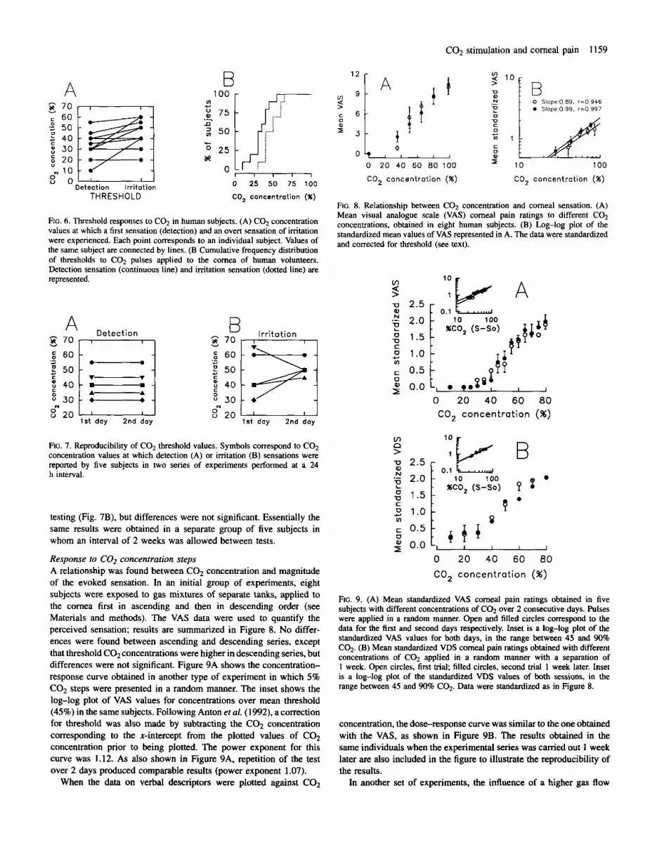
THRESH 0 LD FIG. 8. Relationship between CO2 concentration and corneal sensation. (A) Mean visual analogue scale (VAS) corneal pain ratings to different CO? concentrations, obtained in eight human subjects. (B) Log-log plot of the standardized mean values of VAS represented in A. The data were standardized and corrected for threshold (see text).
FIG. 6. Threshold responses to CO2 in human subjects. (A) C02 concentration values at which a first sensation (detection) and an overt sensation of irritation were experienced. Each point corresponds to an individual subject. Values of the same subject are connected by lines. (B Cumulative frequency distribution of thresholds to C02 pulses applied to the cornea of human volunteers. Detection sensation (continuous line) and irritation sensation (dotted line) are represented. v) s
0.1 u R 4 6 10 100 &-J %CO, (S-So)
1 .o 0.5 - 5 0.0 t, ,.p I I
0 20 40 60 80 CO, concentration (%)
8 3 0
1st day 2nd day 8 20
1st day 2nd day < 20
v,
> n B FIG. 7. Reproducibility of C 0 2 threshold values. Symbols correspond to CO2 concentration values at which detection (A) or irritation (B) sensations were reported by five subjects in two series of experiments performed at a 24 h interval. 0.1 L
10 100 %CO, (S-So)
Q testing (Fig. 7B), but differences were not significant. Essentially the same results were obtained in a separate group of five subjects in whom an interval of 2 weeks was allowed between tests.
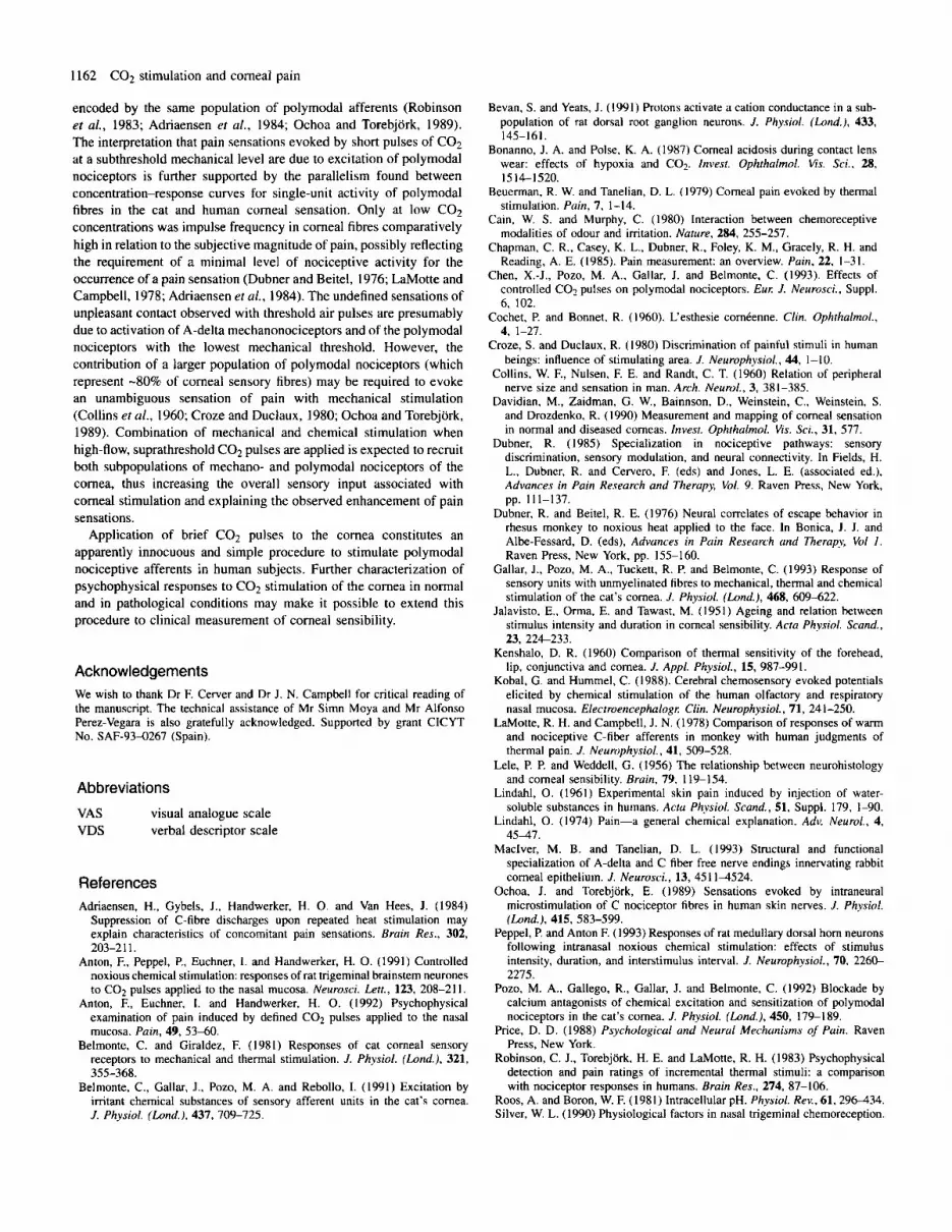
Response to CO, concentration steps A relationship was found between C 0 2 concentration and magnitude of the evoked sensation. In an initial group of experiments, eight subjects were exposed to gas mixtures of separate tanks, applied to the cornea first in ascending and then in descending order (see Materials and methods). The VAS data were used to quantify the perceived sensation; results are summarized in Figure 8. No differ- ences were found between ascending and descending series, except that threshold C 0 2 concentrations were higher in descending series, but differences were not significant. Figure 9A shows the concentration- response curve obtained in another type of experiment in which 5% C02 steps were presented in a random manner. The inset shows the log-log plot of VAS values for concentrations over mean threshold (45%) in the same subjects. Following Anton et al. (1992), a correction for threshold was also made by subtracting the C02 concentration corresponding to the x-intercept from the plotted values of C02 concentration prior to being plotted. The power exponent for this curve was 1.12. As also shown in Figure 9A, repetition of the test over 2 days produced comparable results (power exponent 1.07).
When the data on verbal descriptors were plotted against COz
0 20 40 60 80
CO, concentration (%)
FIG. 9. (A) Mean standardized VAS corneal pain ratings obtained in five subjects with different concentrations of COz over 2 consecutive days. Pulses were applied in a random manner. Open and filled circles correspond to the data for the first and second days respectively. Inset is a log-log plot of the standardized VAS values for both days, in the range between 45 and 90% CO2. (B) Mean standardized VDS corneal pain ratings obtained with different concentrations of CO2 applied in a random manner with a separation of 1 week. Open circles, first trial; filled circles, second trial 1 week later. Inset is a log-log plot of the standardized VDS values of both sessions, in the range between 45 and 90% COP Data were standardized as in Figure 8.
concentration, the dose-response curve was similar to the one obtained with the VAS, as shown in Figure 9B. The results obtained in the same individuals when the experimental series was canied out 1 week later are also included in the figure to illustrate the reproducibility of the results.
In another set of experiments, the influence of a higher gas flow

11 60 COz stimulation and corneal pain
3 -
2 -
1 -
-
v,
> S O Q)
n
z
u L, I I I I
0 20 40 60 80 cO, concentrat ion (%)
FIG. 10. Changes in the intensity of experienced sensation (VDS) induced by pulses of increasing COz concentration applied at low, subthreshold flow (open circles) and at high flow (filled circles) to five human subjects. Inset is a semi-log plot of the same data. Data are mean 2 SEM.
(suprathreshold for mechanical stimulation) on the dose-response curve was analysed. Figure 10 shows the dose-response curves obtained in five individuals, when gas flow was adjusted just below mechanical threshold (open circles) or 0.25 mg over mechanical threshold (black circles). The inset plots the data on the perceived magnitude of the sensation on a linear scale against C 0 2 concentration (logarithmic scale). Sensations experienced for a given COz concentra- tion were greater when a higher gas flow was employed.
Duration of COz pulses Figure IIA shows the results obtained in eight subjects to whom pulses of increasing duration (0.1, 0.5, 1, 3, 5 and 10 s) were applied sequentially using three C 0 2 concentrations (35, 80 and 98.5%). The 35% concentration was subthreshold unless the cornea was exposed for 1-3 s; for longer durations a proportional increase in the intensity of the sensation experienced was detected. When the same series of stimuli was repeated 5 min later with higher C02 concentrations (80 and 98.5%), the minimal time required to evoke a sensation was shorter and the intensity of the reported pain for a given duration greater. The data for 10 s pulses are not plotted since most individuals blinked before the end of the stimulus. In some cases, a sensation of mild ocular discomfort remained for several minutes after application of long-lasting pulses, but no alterations in the corneal epithelium could be detected by slit-lamp examination performed in five of these subjects while the after-sensations were still present. Repetition in six subjects of the series of variable-duration stimuli, using C 0 2 pulses of decreasing duration (from 10 to 0.1 s), gave comparable results (Fig. 11B).
Discussion Sensitivity of corneal fibres to CO, The present study confirms that a significant proportion of corneal sensory afferents are chemosensitive (Belmonte et al., 199 1; Gallar et al., 1993) and demonstrate that they respond to pH changes caused by C02 application. Responses of nociceptive fibres to C02 have been reported in the skin of rats in vitro (Steen et al., 1992) and in second-order nociceptive neurons innervating the rat olfactory mucosa (Anton et al., 1991; Peppel and Anton, 1993). No major differences in COz sensitivity were found between A-delta and C corneal
Stimulus duration (s) Stimulus duration ( s )
FIG. 1 I . Changes in the intensity of experienced sensations induced by pulses of 35% CO2 (open circles), 80% CO1 (filled circles) and 98.5% COz (open triangles) of variable duration in eight human volunteers. In each series, successive pulses were applied at I rnin intervals. Tests with variable concentrations were performed leaving a 5 min pause between trials. Duration changes were applied in increasing (A) or decreasing (B) order. Mean visual analogue scale (VAS) values are standardized by dividing individual responses to each CO2 concentration by the mean response of the subject to all the C 0 2 pulses applied. Bars are SEM.
nociceptive units, supporting the notion that fibre diameter does not determine differences in transduction properties among corneal polymodal nociceptors (Gallar et al., 1993). Unmyelinated fibres had smaller receptive areas and longer response latencies than A-delta units, possibly reflecting a different branching pattern within the cornea for both types of fibres (MacIver and Tanelian, 1993).
Most corneal units responded to long COz pulses with a transient frequency peak and a sustained discharge at a lower frequency. In dorsal root ganglion neurons of the rat presumed as nociceptive, Bevan and Yeats (1991) reported that changes of pH in the bathing medium elicited a transient, rapidly inactivating sodium current followed by a second phase of inward current with a much slower time course. This sustained inward current evoked by protons was due to an increase in non-selective cation conductance; it developed after the first current had decayed substantially, could be sustained for minutes with variable desensitization, and increased in size with increasing acidity. In the in vitro skin preparation, Steen et al. (1992) emphasized the analogy in time course and amplitude between impulse discharges evoked by C 0 2 in nociceptive terminals and membrane current changes induced by protons in the cell soma. A similar parallelism was also noticed here. This observation lends support to the hypothesis that depolarization by pH changes of the cell body and of peripheral terminals occurs through similar ionic mechanisms .
Corneal units that respond to mechanical stimuli and acid are also sensitive to other imtant chemicals and to heat; thus, they have been classified as polymodal nociceptors (Belmonte et al., 1991; Gallar et al., 1993). In the present experiments, heat sensitivity was only tested in a reduced number of the units that had previously been activated by C02. Nevertheless, their sensitivity to acid stimulation strongly suggests that they are corneal polymodal nociceptors. Carbon dioxide excited corneal units more vigorously and persistently than acetic acid (Belmonte et al., 1991; Gallar et al., 1993). Presumably, protons released by a single administration of acetic acid are rapidly buffered, thus limiting the intensity and duration of their stimulatory action. In contrast, sustained exposure to COz causes continuous formation of carbonic acid and a more widespread and sustained reduction of corneal pH (Bonanno and Poke, 1987). This will also recruit fibres deeply located in the corneal epithelium. Furthermore,

C 0 2 stimulation and corneal pain 1161
the possibility that changes in intracellular pH contribute to the impulse discharge elicited by C02, as suggested by Steen et al. (1992), has to be considered.
About 15% of corneal fibres started to fire with CO2 after a long latency (several seconds), poorly encoding COz concentration. An explanation for the delayed firing would be that the nerve endings of these fibres were deeply located in the cornea. However, this appears unlikely because their latency of response was not greatly influenced by C02 concentration; furthermore, peak discharge rates were attained with submaximal C 0 2 concentrations, thus speaking against the idea that a pH gradient needs to be built up through the corneal surface to excite these units. The possibility exists that long-latency units were corneal mechanonociceptive fibres which are insensitive to external application of acetic acid, heat and irritant chemicals (Belmonte et al., 1991). but may be depolarized by the intracellular pH changes induced by sustained exposure to C02. In the skin in vitro, some high-threshold mechanosensitive fibres (functionally analogous to mechanonociceptors of the cornea) responded irregularly to long-lasting pH changes (Steen et al., 1992).
Relationship between C02 Concentration of stimulating pulses and impulse activity of corneal fibres In a previous paper (Belmonte et al., 1991), a correlation between the pH of the stimulating solution and the mean frequency of discharge of corneal nociceptive fibres was reported. Likewise, increasing C 0 2 concentrations applied to the cornea elicited a parallel elevation in firing frequency. The log-log plot, which reflects the relationship between stimulus intensity and magnitude of the response, had a power exponent of 1.1, which is slightly lower than the value found for polymodal nociceptors of the monkey’s skin in response to noxious heat (Dubner, 1985) but within the range obtained from the rat ethmoid nerve with different irritant compounds applied to the nasal mucosa (Silver, 1990). These data indicate that corneal polymodal nociceptors, like other nociceptive trigeminal neurons (Anton et al., 1991; Peppel and Anton, 1993), effectively encode changes in C 0 2 concentration, presumably reflecting variations in corneal pH induced by COz exposure. In addition, when C02 concentration was increased a larger number of corneal units was recruited, thus contributing to the augmentation of the sensory input associated with corneal stimula- tion by low pH values.
Sensations induced by stimulation of the cornea with C02 in human subjects The question of whether different forms of cornea-stimulating energy evoke different qualities of corneal sensation has been a matter of discussion for almost a century. Mechanical stimulation of the human cornea usually causes a sharp sensation of pain; however, reports of ‘touch’ sensations aroused by low-intensity stimulation of the cornea can be found in the literature (Lele and Weddell, 1956). In contrast, psychophysical experiments by Kenshalo ( I 960) and Beuerman and Tanelian (1979) established that thermal stimuli elicited only sensations of irritation or pain. To our knowledge, no systematic studies of corneal sensations evoked by chemical stimulation have been performed by previous workers.
In the present experiments involving human subjects, the mechan- ical threshold for corneal sensation was determined using air puffs of increasing flow. The threshold force value found (-3 mg) is similar to that obtained by previous authors in young normal adults (Davidian et al., 1990). Temperature changes detected at the corneal surface by application of subthreshold or threshold pulses of air or COz were negligible, thus suggesting that corneal indentation rather than cooling
was the effective stimulus for the corneal sensations evoked by our gas jet. Threshold mechanical sensations were not identified as openly unpleasant by our subjects, in agreement with previous observations where an air puff was applied to the cornea (Jalavisto et al., 1951; Lele and Weddell, 1956; Davidian et al., 1990). In contrast, threshold C 0 2 stimuli presented at subthreshold mechanical level aroused a sensation of different quality, always including a component of unpleasantness. Nonetheless, most individuals were able to distinguish between ‘detection’ and ‘imtation’ according to the characteristics of sensation experienced. The nmow limits of the ‘prepain’ range for noxious stimulation with COz have been already signalled in psychophysical experiments where COz was applied to the nasal mucosa (Anton et al., 1992). Mean threshold values for pain and imtation sensation found in the nasal mucosa and the cornea (-47% C02) were strikingly similar (Anton et al., 1992). As could be expected, threshold values in our experiments were fairly variable among individuals (Chapman et al., 1985), but remained constant and reproducible for the same subject under controlled conditions of stimulus duration and flow of C02.
Relationship between intensity and duration of stimulus and magnitude of sensation In this study, pain sensations of increasing magnitude were reported with higher C02 concentrations. Results obtained with two different scaling techniques (VAS and VDS) were rather similar and can be fitted to a straight line in a log-log plot, reflecting that the intensity of pain sensation is a power function of stimulus magnitude (Stevens, 1975). The power exponent found in our study, -1.1, was obtained after applying Ekman’s correction to the data (Anton et al., 1992). This exponent is within the limits of those obtained for C 0 2 stimulation of the nasal mucosa (Stevens and Cain, 1986; Anton et al., 1992) and is consistent with the protective role attributed to the corneal nociceptive system.
As increasing C 0 2 concentration results in a local pH drop in the cornea, it is not surprising that suprathreshold stimuli of longer duration evoked more intense sensations. This was also true for C 0 2 concentrations that were subthreshold for short pulses, but were above threshold for longer presentation times. Presumably, with low C 0 2 mixtures a certain time was required to build up the proton concentra- tion necessary to excite polymodal nociceptive units.
When the flow rate of the stimulating C02 mixture was increased above threshold mechanical level, the intensity function curve was shifted to the left. This may be due to a faster and more pronounced pH drop andor to the summation of sensation evoked by mechanical stimulation. If mechanical stimulation contributes to the overall perceived sensation, the parallelism between the two curves, except at high C 0 2 concentrations, would indicate that interaction between chemical and mechanical sensations was small (Cain and Murphy, 1980).
Correspondence between sensory judgements in humans and single-unit activity in corneal nociceptive fibres of the cat Stimulation of the human cornea with C02 elicited sensations of imtation that appear to be quite similar to those described with the application of heat (Kenshalo, 1960; Beuerman and Tanelian, 1979). In the cat’s cornea, short-lasting pH reductions induced by COz or acetic acid appear to activate selectively the same group of A-delta and C-polymodal nociceptors that respond to heating of the corneal surface over 39°C (Belmonte and Giraldez, 1981; Belmonte et at., 199 1 ; Gallar et al., 1993). Thus, it can be assumed that in the cornea, as in the skin and other tissues, thermal and chemical pain are
I162 C02 stimulation and corneal pain
encoded by the same population of polymodal afferents (Robinson er al., 1983; Adriaensen er al., 1984; Ochoa and Torebjork, 1989). The interpretation that pain sensations evoked by short pulses of C02 at a subthreshold mechanical level are due to excitation of polymodal nociceptors is further supported by the parallelism found between concentration-response curves for single-unit activity of polymodal fibres in the cat and human corneal sensation. Only at low COz concentrations was impulse frequency in corneal fibres comparatively high in relation to the subjective magnitude of pain, possibly reflecting the requirement of a minimal level of nociceptive activity for the occurrence of a pain sensation (Dubner and Beitel, 1976; LaMotte and Campbell, 1978; Adriaensen et al., 1984). The undefined sensations of unpleasant contact observed with threshold air pulses are presumably due to activation of A-delta mechanonociceptors and of the polymodal nociceptors with the lowest mechanical threshold. However, the contribution of a larger population of polymodal nociceptors (which represent -80% of corneal sensory fibres) may be required to evoke an unambiguous sensation of pain with mechanical stimulation (Collins er al., 1960; Croze and Duclaux, 1980; Ochoa and Torebjork, 1989). Combination of mechanical and chemical stimulation when high-flow, suprathreshold C02 pulses are applied is expected to recruit both subpopulations of mechano- and polymodal nociceptors of the cornea, thus increasing the overall sensory input associated with corneal stimulation and explaining the observed enhancement of pain sensations.
Application of brief COz pulses to the cornea constitutes an apparently innocuous and simple procedure to stimulate polymodal nociceptive afferents in human subjects. Further characterization of psychophysical responses to COz stimulation of the cornea in normal and in pathological conditions may make it possible to extend this procedure to clinical measurement of corneal sensibility.
Acknowledgements We wish to thank Dr F. Cerver and Dr J. N. Campbell for critical reading of the manuscript. The technical assistance of Mr Simn Moya and Mr Alfonso Perez-Vegara is also gratefully acknowledged. Supported by grant CICYT No. SAF-93-0267 (Spain).
Abbreviations
VAS visual analogue scale VDS verbal descriptor scale
References Adriaensen, H., Gybels, J., Handwerker, H. 0. and Van Hees, J. (1984)
Suppression of C-fibre discharges upon repeated heat stimulation may explain characteristics of concomitant pain sensations. Bruin Res., 302,
Anton, F., Peppel, P., Euchner, I. and Handwerker, H. 0. (1991) Controlled noxious chemical stimulation: responses of rat trigeminal brainstem neurones to C02 pulses applied to the nasal mucosa. Neurosci. Lett., 123, 208-21 I .
Anton, F., Euchner, 1. and Handwerker, H. 0. (1992) Psychophysical examination of pain induced by defined C02 pulses applied to the nasal mucosa. Pain, 49, 5 3 4 0 .
Belmonte, C. and Giraldez, F. (1981) Responses of cat corneal sensory receptors to mechanical and thermal stimulation. J . Physiol. (Lond.). 321,
Belmonte, C., Gallar, J., Pozo, M. A. and Rebollo, 1. (1991) Excitation by irritant chemical substances of sensory afferent units in the cat’s cornea. J. Physiol. (Lond.), 437, 709-725.
203-2 1 1.
355-368.
Bevan, S. and Yeats, J. (1991) Protons activate a cation conductance in a sub- population of rat dorsal root ganglion neurons. J . Physiol. (Land.), 433, 145-161.
Bonanno, J. A. and Poke, K. A. (1987) Corneal acidosis during contact lens wear: effects of hypoxia and C02. Invest. Ophthalmol. Vis. Sci.. 28, 1514-1520.
Beuerman, R. W. and Tanelian, D. L. (1979) Corneal pain evoked by thermal stimulation. Pain, 7. 1-14.
Cain, W. S. and Murphy, C. (1980) Interaction between chemoreceptive modalities of odour and irritation. Nature, 284, 255-257.
Chapman, C. R., Casey, K. L., Dubner, R., Foley, K. M., Gracely, R. H. and Reading, A. E. (1985). Pain measurement: an overview. Pain, 22, 1-31.
Chen, X.-J., Pozo, M. A,, Gallar, J. and Belmonte, C. (1993). Effects of controlled COz pulses on polymodal nociceptors. Eu,: J . Neurosci., Suppl. 6, 102.
Cochet, P. and Bonnet. R. (1960). L‘esthesie corntenne. Clin. Ophthalmol., 4, 1-27.
Croze, S. and Duclaux, R. (1980) Discrimination of painful stimuli in human beings: influence of stimulating area. J. Neurophysiol., 44, 1-10,
Collins, W. F., Nulsen, F. E. and Randt, C. T. (1960) Relation of peripheral nerve size and sensation in man. Arch. Neurol., 3, 381-385.
Davidian, M., Zaidman, G. W., Bainnson, D., Weinstein, C., Weinstein, S. and Drozdenko, R. (1990) Measurement and mapping of corneal sensation in normal and diseased corneas. Invest. Ophrhalmol. Vis. Sci., 31, 577.
Dubner, R. ( 1985) Specialization in nociceptive pathways: sensory discrimination, sensory modulation, and neural connectivity. In Fields, H. L., Dubner, R. and Cervero, F. (eds) and Jones, L. E. (associated ed.), Advances in Pain Research and Therupy, Vol. 9. Raven Press, New York,
Dubner, R. and Beitel, R. E. (1976) Neural correlates of escape behavior in rhesus monkey to noxious heat applied to the face. In Bonica, J. J. and Albe-Fessard, D. (eds), Advances in Pain Research and Therupy, Vol I . Raven Press, New York, pp. 155-1 60.
Gallar, J., Pozo, M. A,, Tuckett, R. P. and Belmonte, C. (1993) Response of sensory units with unmyelinated fibres to mechanical, thermal and chemical stimulation of the cat’s cornea. J . Physiol. (Lond.), 468, 609-622.
Jalavisto, E., Orma, E. and Tawast, M. (1951) Ageing and relation between stimulus intensity and duration in corneal sensibility. Acru Phvsiol. Scand., 23, 22&233.
Kenshalo, D. R. (1960) Comparison of thermal sensitivity of the forehead, lip, conjunctiva and cornea. J . Appl. Physiol., 15, 987-991.
Kobal, G. and Hummel, C. (1988). Cerebral chemosensory evoked potentials elicited by chemical stimulation of the human olfactory and respiratory nasal mucosa. Electroencephalog,: Clin. Neurophysiol., 71, 241-250.
LaMotte, R. H. and Campbell, J. N. (1978) Comparison of responses of warm and nociceptive C-fiber afferents in monkey with human judgments of thermal pain. J . Neurophysiol., 41, 509-528.
Lele, P. P. and Weddell, G. (1956) The relationship between neurohistology and corneal sensibility. Brain, 79, 119-154.
Lindahl, 0. (1961) Experimental skin pain induced by injection of water- soluble substances in humans. Acru Physiol. Scand., 51, Suppl. 179, 1-90.
Lindahl, 0. (1974) Pain-a general chemical explanation. Adv. Neurol., 4, 4 5 4 7 .
MacIver, M. 9. and Tanelian, D. L. (1993) Structural and functional specialization of A-delta and C fiber free nerve endings innervating rabbit corneal epithelium. J . Neurosci., 13, 45 114524.
Ochoa, J. and Torebjork, E. (1989) Sensations evoked by intraneural microstirnulation of C nociceptor fibres in human skin nerves. J . Physiol. (Land.), 415, 583-599.
Peppel, P. and Anton F. (1993) Responses of rat medullary dorsal horn neurons following intranasal noxious chemical stimulation: effects of stimulus intensity, duration, and interstimulus interval. J. Neurophysiol., 70, 226C- 2275.
Pozo, M. A,, Gallego, R., Gallar, J. and Belmonte, C. (1992) Blockade by calcium antagonists of chemical excitation and sensitization of polymodal nociceptors in the cat’s cornea. J . Physiol. (Lond.), 450, 179-189.
Price, D. D. (1988) Psychological and Neural Mechanisms of Pain. Raven Press, New York.
Robinson, C. J., Torebjork, H. E. and LaMotte, R. H. (1983) Psychophysical detection and pain ratings of incremental thermal stimuli: a comparison with nociceptor responses in humans. Bruin Res., 274, 87-106.
Roos, A. and Boron, W. F. (1981) Intracellular pH. Physiol. Rev., 61,296434. Silver, W. L. (1990) Physiological factors in nasal trigeminal chemoreception.
pp. 111-137.

C 0 2 stimulation and corneal pain 1163
In Green, B. G., Mason, J. R. and Kare, M. R. (eds), Chemical Senses. Vol. 2: Irrirurion. Marcell Dekker, New York, pp. 21-41,
Protons selectively induce lasting excitation and sensitization to mechanical stimulation of nociceptors in the rat skin. J. Neurosci., 12, 86-95.
Stevens, J. C. and Cain, W. S. (1986) Aging and the perception of nasal irritation. Physiol. Behav., 37, 323-328.
Stevens, S. S. (1975) fsychophysics. John Wiley, New York. Tanelian, D. L. and Beuerman, R. W. (1983). Responses of rabbit corneal
Steen, K. H., Reeh, P. W., Anton, F. and Handwerker, H. 0. (1992) nociceptors to mechanical and thermal stimulation. Exp. Neurol., 84, 165-178.
Thiirauf, N., Hummel, T., Kettenmann, B. and Kobal, G. (1993). Nociceptive and reflexive responses recorded from the human nasal mucosa. Brain Res., 629, 293-299.


