Embed Size (px)
Citation preview

14470892-6638/98/0012-1447/$02.25 Q FASEB
/ 383a no09 Mp 1447 Wednesday Sep 23 02:11 PM LP–FASEB no09
Spatially controlled cell engineering on biodegradablepolymer surfaces
NIKIN PATEL, ROBERT PADERA,* GILES H. W. SANDERS, SCOTT M. CANNIZZARO,*MARTYN C. DAVIES, ROBERT LANGER,* CLIVE J. ROBERTS, SAUL J. B. TENDLER,PHILIP M. WILLIAMS, AND KEVIN M. SHAKESHEFF1
Laboratory of Biophysics and Surface Analysis, School of Pharmaceutical Sciences, The University ofNottingham, Nottingham NG7 2RD, United Kingdom; and *Department of Chemical Engineering,Massachusetts Institute of Technology, E25–342, Cambridge Massachusetts 02139, USA
ABSTRACT Controlling receptor-mediated inter-actions between cells and template surfaces is a cen-tral principle in many tissue engineering procedures(1–3). Biomaterial surfaces engineered to presentcell adhesion ligands undergo integrin-mediated mo-lecular interactions with cells (1, 4, 5), stimulating cellspreading, and differentiation (6–8). This provides amechanism for mimicking natural cell-to-matrix in-teractions. Further sophistication in the control ofcell interactions can be achieved by fabricating sur-faces on which the spatial distribution of ligands isrestricted to micron-scale pattern features (9–14).Patterning technology promises to facilitate spatiallycontrolled tissue engineering with applications in theregeneration of highly organized tissues. These newapplications require the formation of ligand patternson biocompatible and biodegradable templates,which control tissue regeneration processes, beforeremoval by metabolism. We have developed amethod of generating micron-scale patterns of anybiotinylated ligand on the surface of a biodegradableblock copolymer, polylactide-poly(ethylene glycol).The technique achieves control of biomolecule de-position with nanometer precision. Spatial controlover cell development has been observed when usingthese templates to culture bovine aortic endothelialcells and PC12 nerve cells. Furthermore, neurite ex-tension on the biodegradable polymer surface is di-rected by pattern features composed of peptides con-taining the IKVAV sequence (15, 16), suggesting thatdirectional control over nerve regeneration on bio-degradable biomaterials can be achieved.—Patel, N.,Padera, R., Sanders, G. H. W., Cannizzaro, S. M., Da-vies, M. C., Langer, R., Roberts, C. J., Tendler,S. J. B., Williams, P. M., and Shakesheff, K. M. Spa-tially controlled cell engineering on biodegradablepolymer surfaces. FASEB J. 12, 1447–1454 (1998)
Key Words: tissue engineering · neurite extension · integrins· patterning
THE ELUCIDATION OF biomolecular pathways that de-termine cell behavior during tissue development has
created the possibility of bioengineering complex tissuestructures (17–19). This bioengineering is dependenton the generation of cellular microenvironments thatmimic those encountered during natural development(1, 20, 21). For example, biodegradable polymer sur-faces can be engineered to present peptides containingthe amino acid sequence RGD (1, 22–24). This se-quence binds to integrin receptors on cell surfaces, in-ducing cell spreading and intracellular signaling, hencemimicking cell-to-extracellular matrix interactions (4–6). The design of these biomimetic surfaces has beenused within the field of tissue engineering to controlcell behavior of many tissue types, including cartilageand liver tissues (25, 26).
An important challenge in tissue engineering is toapply the principles of molecular biology to controlthe spatial organization of cells. This is vital in thebioengineering of tissues that require precisely de-fined cellular architectures. For example, the func-tioning of tissues such as nerves and blood vessels isdependent on the controlled orientation of cells. Formany tissue types, the spatial organization of cells isrequired to ensure that cell-to-cell interactions occur(18, 27, 28). These interactions are essential in cellphenotype preservation (29).
Achieving spatially controlled cell engineering re-quires parallel development in molecular biologyand biomimetic materials technology. In the field ofmolecular biology, there has been a continuing re-finement in the our understanding of ligand-to-celladhesion receptor interactions. This has generated alarge number of cell adhesion motifs that can controlcell adhesion behavior via specific receptor interac-tions (30, 31). In the design of biomimetic materials,the aim is to use these motifs to promote cell inter-actions with synthetic materials. Most biomimetic ma-terials for cell engineering have been based on bio-degradable polymers, which are removed from theengineered tissues by hydrolysis and dissolution ofbreakdown products (32). The major technological
1 Correspondence. E-mail: [email protected]
1448 Vol. 12 November 1998 The FASEB Journal PATEL ET AL.
/ 383a no09 Mp 1448 Wednesday Sep 23 02:11 PM LP–FASEB no09
challenge in the fabrication of these biomimetic ma-terials is the development of techniques of engineer-ing biodegradable polymer surfaces (1, 33). This sur-face engineering has been used to immobilize motifshomogeneously over surfaces. However, to fabricatebiomimetic materials for spatially controlled cell en-gineering, it is necessary to restrict motif immobili-zation to predefined micron-scale patterns.
Recent advances in patterning technology havegenerated a range of techniques with which biomo-lecules can be immobilized on surfaces with micron-scale precision. These techniques include litho-graphic methods (34) that use patterned masks torestrict the location of interactions between a beamof light, ions or electrons, and a surface (12) andmicro-contact printing techniques (35). Most pat-terning techniques have been developed on a smallset of materials with chemical modifications that re-quire highly specialized surface reactions. This canrestrict the types of ligands and surfaces that can bepatterned. The objective of the current work was todevelop a generic technology that can form patternsof any ligand onto a biodegradable polymer surface.
As with many aspects of tissue engineering re-search, this work requires a multidisciplinary strategythat combines molecular biology with biodegradablepolymer synthesis, surface engineering and analysis,and patterning technology. Our method generatesmicron-scale patterns of any biotinylated ligand onthe surface of a biodegradable block copolymer,polylactide-poly(ethylene glycol) (PLA-PEG).2 Nano-meter control over the location of biomolecule hasbeen achieved. This control has been confirmed bymolecular resolution of protein molecules on the pat-terned surfaces using atomic force microscopy(AFM). Spatial control over cell development hasbeen observed when using these templates to culturebovine aortic endothelial cells and PC12 nerve cells.In addition, directional control over neurite exten-sion from PC12 cells has been achieved on patternscomposed of peptides containing the IKVAV se-quence (15, 16) through precise control of the lo-cation of receptor-mediated interactions.
MATERIALS AND METHODS
PLA-PEG-biotin synthesis
a-Amine v-hydroxy PEG (Shearwater Polymers, Inc.; averagemol wt, 3.8k) was stirred overnight with NHS-biotin (Fluka,Milan, Italy) and triethylamine in methylene chloride and ace-
2 Abbreviations: AFM, atomic force microscopy; av-R, rho-damine-labeled avidin; BAECs, bovine aortic endothelial cells;DMEM, Dulbecco’s modified Eagle’s medium; NGF, nervegrowth factor; NMR, nuclear magnetic resonance; PBS, phos-phate-buffered saline; PDMS, poly(dimethyl siloxane); PLA-PEG, polylactide-poly(ethylene glycol).
tonitrile, at room temperature, under argon. The biotinylatedPEG was then isolated by vacuum filtration and dried azeo-tropically from toluene. Then recrystallized (l-)lactide was pol-ymerized from the v-hydroxy PEG-biotin end by refluxing intoluene, using stannous 2-ethylhexanoate as a catalyst. PLA-PEG-biotin was precipitated from a methylene chloride solu-tion by the addition of cold ether.
1H-NMR (nuclear magnetic resonance) at each stage con-firmed the attachment of biotin to the PEG chain. Specifically,attachment of biotin-NHS to the end group amine of a-aminev-hydroxy PEG to form an amide bond was confirmed by shiftof the free amine protons to an amide proton at 7.8 ppm andthe appearance of a triplet (methylene from biotin arm alphato the amide) at 2.05 ppm. The proton signals from the bi-cyclic biotin structure owing to the (2)methine protons (4.3and 4.2 ppm) and urea protons (6.45 and 6.35 ppm) can beseen throughout the synthesis of PLA-PEG-biotin: the biotinstructure remains intact and is not damaged from the lactidepolymerization onto HO-PEG-biotin.
The molecular weight of the PLA-PEG-biotin was deter-mined by 1H-NMR, using the PEG signal as a reference. Gelpermeation chromatography revealed one peak indicative ofpure material. PLA molecular weight for this study was 9.2k.
Mold fabrication
A patterned poly(dimethyl siloxane) (PDMS) mold wasformed by curing its prepolymer (Sylgard 184, Dow Corning)on a patterned master prepared photolithographically by ex-posing and developing a photoresist pattern on gallium/ar-senide wafers (36). The PDMS mold bearing the negative pat-tern of the master was peeled off and washed repeatedly withethanol, hexane, and de-ionized water. After drying under ar-gon, the mold was placed onto an epitaxially grown gold sur-face (see Fig. 2) (37). The capillaries were rendered hydro-philic by plasma treatment with O2 (load coil power A 200 W)for 1 s (Bio-Rad RF Plasma Barrel Etcher)
Avidin pattern formation
Within 1 min of plasma treatment, the mold was placed ontoa film of the PLA-PEG-biotin. The polymer film was formedfrom a 1 mg/mL solution of the polymer in trifluoroethanol,drop cast onto a polystyrene substrate, and dried under vac-uum. A 500 mg/ml solution of rhodamine-labeled avidin (av-R) (Sigma, Dorset, U.K.) was prepared using distilled water.Approximately 1 mL of this av-R solution was dropped ontothe PLA-PEG-biotin surface. The drop was positioned so thatthe av-R solution wetted an end of the mold where the capil-laries were open. After 1 h of contact, the av-R solution wasremoved by blotting and replaced with approximately 20 mlof distilled water; after 5 min, the water was removed. Thiswashing procedure was repeated five times. Then the samplewas immersed in water and the mold removed by peeling thePDMS and PLA-PEG-biotin surfaces apart. The sample waswashed with an additional 100 mL of water.
Atomic force microscopy
Images were obtained using a Digital Instruments (Santa Bar-bara, Calif.) multimode scanning probe microscope with aDigital Instruments Nanoscope IIIa controller. Images wereacquired in tapping mode using sharpened silicon nitride tipswith cantilever resonance frequencies in the range of 307–375kHz. Simultaneous topographic and phase images were ob-tained at a scan rate of 1 Hz.
Cell culture
All reagents were obtained from Sigma unless otherwisenoted. Bovine aortic endothelial cells (BAECs) were main-

SPATIALLY CONTROLLED TISSUE ENGINEERING 1449
/ 383a no09 Mp 1449 Wednesday Sep 23 02:12 PM LP–FASEB no09
Figure 1. Schematic representation of the surface engineeringof PLA-PEG-biotin. Biotin moieties presented at the polymersurface are used to immobilize tetrameric avidin molecules.Free biotin binding sites on the avidin molecules are in turnused to anchor biotinylated ligands. All steps in the surfaceengineering are performed in aqueous environments.
Figure 2. A) The microfluidic patterning technique requirestreatment of the PDMS mold with an O2 plasma. This treat-ment increases the hydrophilicity of any PDMS surface that isnot protected by the gold surface. Transferring the treatedmold to the PLA-PEG-biotin surfaces produces capillaries withhydrophilic walls. Avidin solution flow across the PLA-PEG-biotin surface is restricted to the capillary regions by the hy-drophobic regions of the mold base. B) Schematicrepresentation of the microfluidic patterning technique.
tained in low glucose Dulbecco’s modified Eagle’s medium(DMEM) supplemented with 10% fetal bovine serum, 0.5%penicillin, 0.5% streptomycin, and 1% L-glutamine in a hu-midified incubator at 377C and 5% CO2. Cells were passagedby trypsinization before reaching confluence, usually everyfifth day. Fresh media were added every other day. PC12 cells(ATCC CRL-1721) were grown in suspension culture in F-12Kmedia supplemented with 15% horse serum, 2.5% fetal bovineserum, 0.5% penicillin, 0.5% streptomycin, and 1% L-gluta-mine in a humidified incubator at 377C and 5% CO2. Cellswere passaged 1:5 every third day
Cell spreading experiment
The biotinylated polypeptides were synthesized and theirsequences confirmed at the MIT Biopolymer ProcessingLaboratory. The samples were sterilized under UV light for10 min. A 1 mg/mL solution of peptide (biotin-G11GRGDSfor endothelial cell experiments, biotin-G5CSRARKQAA-SIKVAVSADR for PC-12 cell experiments) was added to thetissue culture plate and allowed to incubate at 377C on ashaker plate for 30 min. The samples were washed threetimes with sterile phosphate-buffered saline (PBS) prior toseeding with cells.
BAECs between passages 7 and 9 were removed from tissueculture flasks by trypsinization, pelleted by centrifugation, re-suspended, washed three times, and diluted to the appropri-
ate concentration in serum-free DMEM. PC12 cells wereprimed with culture medium containing 50 ng/mL 7S nervegrowth factor (NGF) 48 h prior to the experiment. The cellswere pelleted by centrifugation, washed three times with se-rum-free F-12K medium, passed through a 22-gauge needle toobtain a single cell suspension, and diluted to the appropriateconcentration with serum-free F-12K medium supplementedwith 50 ng/mL 7S NGF. For both cell types, approximately10,000 cells/cm2 were added to each sample and the platesreturned to the incubator for the duration of the experiment(48 h). At the end point of the experiment, the samples werewashed gently with PBS to remove unattached cells and visu-alized with a Nikon Diaphot TMD inverted microscopeequipped with a Hitachi HV-C20 high-resolution CCD videocamera using phase contrast objectives. Images were digitizedusing NIH Image (v1.61) image analysis software. In addition,some BAEC samples were fixed in 10% neutral buffered for-malin for 10 min, washed with water, and stained with hema-toxylin for visualization.
1450 Vol. 12 November 1998 The FASEB Journal PATEL ET AL.
/ 383a no09 Mp 1450 Wednesday Sep 23 02:12 PM LP–FASEB no09
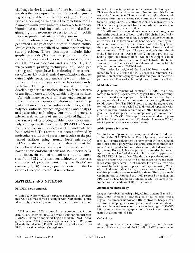
Figure 3. A) Fluorescence microscopy image of 12 mm-wide lines of av-R on a PLA-PEG-biotin surface. The image was recordedat 101 magnification. Inset shows a region of the patterned surface at 601 magnification. B) Fluorescence microscopy imageof 30, 50, 70, and 70 mm-wide lines of av-R on a PLA-PEG-biotin surface (101) magnification. C) Phase detection AFM image ofa line confirming the generation of a sharp pattern edge. D) Tapping mode AFM image of boundary between avidin coveredlines and PLA-PEG-biotin only gap regions. The inset shows a 100 nm1 100 nm region of the line displaying individual moleculesof the avidin.
RESULTS AND DISCUSSION
To generate a generic patterning technology for usein biomimetic materials design, we used avidin–bio-tin interaction as a functionality for the ligand im-mobilization. We introduced a biotin molecule as theend group of the PEG block of the PLA-PEG copol-ymer to produce the biodegradable molecule shownbelow (termed PLA-PEG-biotin).
HN NH
SO O
O
OO
PLA PEG Biotin
x
H N
y
HN NH
SO O
O
OO
PLA PEG Biotin
x
H N
y
As shown schematically in Fig. 1, when films of PLA-
PEG-biotin are exposed to aqueous solutions of av-idin, the polymer surface immobilizes avidin mole-cules. Each avidin molecule can then act as a bridgebetween the biotinylated polymer and a biotinylatedligand. PLA-PEG was chosen as the foundation forthe surface engineering because this polymer hasbecome established as a material that possesses ex-cellent bulk and surface properties for biomaterialapplications (38). In addition, the PEG chains act asflexible, hydrophilic spacers for the biotin moieties.The preservation of the biotin structure during thesynthesis of PLA-PEG-biotin was verified by 1H-NMR,and the avidin binding ability of the tethered biotinwas confirmed by surface plasmon resonance anal-ysis (39).
A microfluidic network system (40, 41) was used toform a pattern of avidin molecules on PLA-PEG-bio-tin film surfaces. The pattern was composed of avidinfunctionalized lines, with widths of 70, 50, 30, or 12mm, separated by 250 mm gaps that were free of theprotein. To achieve pattern formation, an aqueous

SPATIALLY CONTROLLED TISSUE ENGINEERING 1451
/ 383a no09 Mp 1451 Wednesday Sep 23 02:12 PM LP–FASEB no09
Figure 4. Spatially controlled ad-hesion and spreading of bovineaortic endothelial cells on 70 and50 mm-wide lines containing RGDpeptides. Panels A, B, D are trans-mission images. Panel C is a phasecontrast image.
solution of avidin was allowed to flow through capil-laries, created by placing a PDMS mold (see Fig. 2)on PLA-PEG-biotin surfaces. The mold must protectthe polymer surface from the avidin solution over thegap regions while allowing the avidin solution to flowover the polymer in the capillaries. In its native state,the hydrophobicity of the PDMS ensured that regionsof the polymer surface in contact with the mold basewere protected from the avidin solution. However,this hydrophobicity also inhibited solution flowthrough the capillaries, so an oxygen plasma tech-nique was developed to render the capillary walls hy-drophilic while maintaining the hydrophobicity ofthe mold base (Fig. 2A). Base regions of the moldwere shielded from the activity of the plasma by con-formal contact with an epitaxially grown gold surface.The mold was then transferred to the PLA-PEG-bio-tin surface. Contact angle analysis confirmed the dif-ferential hydrophobicity/hydrophilicity of shieldedand unshielded regions of the mold. Shieldedregions displayed contact angles of 1057 with water,whereas unshielded regions were saturated with wa-ter drops. Further evidence of the successful treat-ment of the capillary walls was provided by the rapidflow of water through the capillaries.
The flow of the avidin solution through the hydro-philic capillaries was monitored by fluorescence mi-
croscopy using avidin labeled with rhodamine (av-R).After 1 h of exposure of PLA-PEG-biotin to the av-Rsolution, the sample was washed thoroughly and themold was removed by peeling. Resulting patterns ofav-R on the PLA-PEG-biotin surface are shown in Fig.3A,B. The presence of a sharp boundary between av-idin-immobilized and protein-free regions has beenconfirmed by phase detection AFM (Fig. 3C), inwhich differences in viscoelastic and adhesive prop-erties of avidin and PLA-PEG-biotin generate imagecontrast (42).
The thickness of the avidin layer deposited on thePLA-PEG-biotin was measured by tapping modeAFM. The AFM image in Fig. 3D shows the topogra-phy of a PLA-PEG-biotin surface at a protein bound-ary. The edge of the line was resolved, and cross-sec-tional analysis recorded a step height of less than 5nm. Given that the dimensions of the avidin moleculehave been estimated as 5.6 nm 1 5.0 nm 1 4.0 nmby X-ray crystallography (43), a step height of 5 nmis indicative of a monolayer coverage. The inset im-age shows a 100 nm 1 100 nm scan of the channelon which molecular-resolution of the protein hasbeen achieved. On some areas of the avidin channel,protein aggregates are evident. These aggregateswere resistant to washing with water. The AFM imagein Fig. 3D also demonstrates the exceptional control

1452 Vol. 12 November 1998 The FASEB Journal PATEL ET AL.
/ 383a no09 Mp 1452 Wednesday Sep 23 02:12 PM LP–FASEB no09
Figure 5. Spatially controlled ad-hesion and spreading of PC12cells on lines containing IKVAVpeptides recorded by phase con-trast light microscopy. A) Lowmagnification image showing thepreferential adhesion of PC12cells to the 70 and 50 mm-widelines. B, C) Images showing neu-rite extension and joining be-tween individual PC12 cells andcell clusters. White markers indi-cate the boundaries of the 70 mmlines. No neurites were observedto extend from any PC12 cells thatadhered on nonpatterned regions.D) The path of the neurite extend-ing up the left-hand boundary ofthe line is altered by the IKVAV pep-tide pattern restricting the neuriteto the line.
of avidin distribution along the channel edge. Lateraldeviations of this edge from a straight line are small.The largest lateral deviation on this image is 30 nm,equivalent to approximately 6 avidin molecules. Mostdeviations were found to be less than 20 nm inlength.
The micron-scale patterns of avidin on the PLA-PEG-biotin surfaces were then used to immobilizebiotinylated peptides containing either the fibronec-tin fragment RGD or the laminin fragment IKVAV.Initial cell culture studies were performed on theRGD-presenting patterns, using BAECs to prove thatthe patterns could spatially organize the adhesion ofan anchorage-dependent cell type. As shown in Fig.4, the BAECs adhered and spread on the RGD-func-tionalized lines but did not adhere to unfunctional-ized areas between the lines. Complete cell coverageof the 70 and 50 mm width lines was achieved, butlittle cell adhesion occurred to the 30 or 12 mm-widelines.
Having proved that patterns on the biodegradablesurface could be used in spatially controlled cell en-gineering, we investigated the control of a cell devel-opmental process, namely, directed neurite exten-sion. Directed neurite extension occurs duringperipheral nerve regeneration, and it is known that
one component of this extension is integrin-to-lami-nin interactions (15, 44–47). PC12 cells were used inthis study because of their known laminin-inducedneurite extension. As shown in Fig. 5, PC12 cellsshowed selective adherence to the IKVAV function-alized lines, with only a small degree of cell adhesionbetween the lines (Fig. 5). In addition, no cell ad-hesion was observed on negative control samples con-sisting of PLA-PEG-biotin-avidin patterns in theabsence of the biotinylated IKVAV sequence. Direc-tionally controlled neurite outgrowth was stimulatedby the IKVAV micropattern, with neurites extendingbetween groups of cells often hundreds of micronsapart (Fig. 5B, C). The extent of control over neuritegrowth was demonstrated by the morphology ofmany neurites that approached the boundary be-tween the functionalized and unfunctionalized sur-faces, but were always effectively restricted fromcrossing the interface (Fig. 5D).
CONCLUSIONS
The mimicking of biological microenvironments byusing biomolecular ligands immobilized on the sur-faces of biodegradable polymer templates is a major

SPATIALLY CONTROLLED TISSUE ENGINEERING 1453
/ 383a no09 Mp 1453 Wednesday Sep 23 02:12 PM LP–FASEB no09
tool in cell engineering. As the level of sophisticationof this mimicking increases, driven by the desire tobioengineer complex human tissues, there is a needto control not only the type of receptor-mediated in-teractions but also the location of template-to-cell in-teractions. This spatial control of cell engineering isrequired to direct cell growth for cell types that re-quire precise spatial development to function (e.g.,nerve cells). In addition, spatial control provides amechanism for influencing cell-to-cell interactions.
The patterning technique described in this paperprovides a generic technology by which any biotiny-lated ligand can be patterned onto the surface of abiodegradable polymer. The polymer structure canbe tailored to meet the requirements of specificbioengineering applications. The ability to patternsuch a wide range of ligands opens new possibilitiesin spatially controlled cell engineering, because theability to control cell organization on the templatesbecomes limited by our understanding of the molec-ular biology of ligand-to-receptor binding and its in-fluence on cellular development, not by surface en-gineering constraints.
Advances in cell patterning have recently been re-ported, primarily those on gold surfaces patternedusing molecular self-assembly (11, 13). These studiesutilize the ability to precisely control the patterningprocedures with nanometer precision of molecularimmobilization. If the field of tissue engineering is toexploit these fundamental studies, it is necessary toachieve similar patterning precision on biodegrada-ble polymer templates. The detailed surface analysisof our patterned surfaces has revealed that our tech-nique retains the precision of ligand immobilization.
Finally, the ability to direct neurite extension onthe patterned templates demonstrates that naturalbiological organizational principles can be mimickedsuccessfully. Research on the mechanisms of periph-eral and central nerve regeneration continues to re-veal mechanisms by which medical interventions canaugment natural regenerative processes (47–51). Pat-terned templates that determine the nature and locationof neurite-to-matrix interactions offer the potential torefine our ability to guide regeneration.
The authors thank G. Laws and J. R. Middleton for theirassistance in the preparation of the patterned master. M.C.D.,S.J.B.T., and C.J.R. acknowledge Eli Lilly for the funding of astudentship to N.P.; G.H.W.S. acknowledges the EPSRC forpostdoctoral funding; S.M.C. acknowledges support from anNIH postdoctoral fellowship. K.M.S. is an EPSRC AdvancedResearch Fellow and wishes to acknowledge the support of theInternational Committee Fund. S.J.B.T. is a Nuffield Foun-dation Science Research Fellow. R.S.L. acknowledges the Na-tional Science Foundation for support.
REFERENCES
1. Hubbell, J. A. (1995) Biomaterials in tissue engineering. Bio-Tech-nology 13, 565–576
2. Lopina, S. T., Wu, G., Merrill, E. W., and Griffith-Cima, L. (1996)Hepatocyte culture on carbohydrate-modified star polyethyleneoxide hydrogels. Biomaterials 17, 559–569
3. Han, D. K., and Hubbell, J. A. (1996) Lactide-basedpoly(ethylene glycol) polymer networks for scaffolds in tissueengineering. Macromolecules 29, 5233–5235
4. Ruoslahti, E. (1996) RGD and other recognition sequences orintegrins. Annu. Rev. Cell Dev. Biol., 12, 697–715
5. Massia, S. P., and Hubbell, J. A. (1991) An RGD spacing of 440nm is sufficient for integrin a-V-b-3-mediated fibroblast spread-ing and 140 nm for focal contact and stress fiber formation. J.Cell Biol. 114, 1089–1100
6. Humphries, M. (1996) Integrin activation: the link between li-gand binding and signal transduction. Curr. Opin. Cell Biol. 8,632–640
7. Schlaepfer, D. D., Hanks, S. K., Hunter, T., and van der Geer, P.(1994) Integrin-mediated signal transduction linked to Ras path-way by GRB binding to focal adhesion kinase. Nature (London)372, 786–791
8. Palecek, S. P., Loftus, J. C., Ginsberg, M. H., Lauffenburger,D. A., and Horwitz, A. F. (1997) Integrin-ligand binding prop-erties govern cell migration speed through cell-substratum ad-hesiveness. Nature (London) 385, 537–540
9. Lhoest, J.-B., Detrait, E., Dewez, J.-L., Van den Bosch de Aguilar,P., and Bertrand, P. (1996) A new plasma-based method to pro-mote cell adhesion on micrometric tracks on polystyrene sub-strates. J. Biomater. Sci. Polym. Ed. 7, 1039–1054
10. Hickman, J. J., Bhatia, S. K., Quong, J. N., Shoen, P., Stenger,D. A., Pike, C. J., and Cotman, C. W. (1994) Rational patterndesign for in vitro cellular networks using surface photochemis-try. J. Vacuum Sci. Technol. 12, 607–616
11. Mrksich, M., Chen, C. S., Xia, Y., Dike, L. E., Ingber, D. E., andWhitesides, G. M. (1996) Controlling cell attachment on con-toured surfaces with self-assembled monolayers of alkanethio-lates on gold. Proc. Natl. Acad. Sci. USA 93, 10775–10778
12. Lee, J.-S., Kaibara, M., Iwaki, M., Sasabe, H., Suszuki, Y., andKusakabe, M. (1993) Selective adhesion and proliferation of cellson ion-implanted polymer domains. Biomaterials 14, 958–960
13. Chen, C. S., Mrksich, M., Huang, S., Whitesides, G. M., and Ing-ber, D. E. (1997) Geometric control of cell life and death. Science276, 1425–1428
14. Spargo, B. J., Testoff, M. A., Nielsen, T. B., Stenger, D. A., Hick-man, J. J., and Rudolph, A. S. (1994) Spatially controlled adhe-sion, spreading, and differentiation of endothelial cells on self-assembled molecular monolayers. Proc. Natl. Acad. Sci. USA 91,11070–11074
15. Ranieri, J. P., Bellamkonda, R., Bekos, E. J., Vargo, T. G., Gar-della, J. A., and Aebischer, P. (1995) Neuronal cell attachmentto fluorinated ethylene propylene films with covalently immo-bilized laminin oligopeptides YIGSR and IKVAV. J. Biomed. Mater.Res. 29, 779–785
16. Ranieri, J. P., Bellamkonda, R., Bekos, E. J., Gardella, J. A., Jr.,Mathieu, H. J., Ruiz, L., and Aebischer P. (1994) Spatial controlof neuronal cell attachment and differentiation on covalentlypatterned laminin oligopeptides substrates. Int. J. Dev. Neurosci.12, 725–735
17. Minuth, W. W., Sittinger, M., and Kloth, S. (1998) Tissue engi-neering generation of differentiated artificial tissue for biomed-ical applications. Cell Tissue Res. 291, 1–11
18. Larue, L., Antos, C., Butz, S., Huber, O., Delmas, V., Dominis,M., and Kemler, R. (1996) A role for cadherins in tissue forma-tion. Development 122, 3185–3194
19. Michalopoulos G. K., and DeFrances M. C. (1997) Liver regen-eration. Science 276, 60–66
20. Cima, L. G., and Langer, R. (1993) Engineering human tissue.Chem. Engin. Prog., June, 46–54
21. Langer, R., and Vacanti, J. P. (1993) Tissue engineering. Science260, 920–926
22. Drumheller. P. D., and Hubbell, J. A. (1994) Polymer networkswith grafted cell-adhesion peptides for highly biospecific cell ad-hesive substrates. Anal. Biochem. 222, 380–388
23. Cook, A. D., Hrkach, J. S., Gao, N. N., Johnson, I. M., Pajvani,U. B., Cannizzaro, S. M., and Langer, R. (1997) Characterizationand development of RGD-peptide-modified poly(lactic acid-co-lysine) as an interactive, resorbable biomaterial. J. Biomed. Mater.Res. 35, 513–523

1454 Vol. 12 November 1998 The FASEB Journal PATEL ET AL.
/ 383a no09 Mp 1454 Wednesday Sep 23 02:12 PM LP–FASEB no09
24. Ho, S. P., and Britton, D. H. O. (1994) Arg-gly-Asp peptides inpolyurethanes: design, synthesis and characterization. Adv. Ma-ter. 6, 130–130
25. Cao, Y. L., Vacanti, J. P., Paige, K. T., Upton, J., and Vacanti,C. A. (1997) Transplantation of chondrocytes utilizing a poly-mer-cell construct to produce tissue-engineered cartilage in theshape of a human ear. Plast. Reconstr. Surg. 100, 297–302
26. Mooney, D. J., Sano, K., Kaufmann, P. M., Majahod, K., Schloo,B., and Vacanti, J. P. (1997) Long-term engraftment of hepato-cytes transplanted on biodegradable polymer sponges. J. Biomed.Mater. Res. 37, 413–420
27. Shibuya, Y., Mizoguchi, A., Takeichi, M., Shimada, K., and Ide,C. (1995) Localization of N-cadherin in the normal and regen-erating nerve fibres of the chicken peripheral nervous system.Neuroscience 67, 253–261
28. Geiger, B., and Ayalon, O. (1992) Cadherins. Annu. Rev. Cell Biol.8, 307–332
29. Ben-Ze’ev, A., Robinson, G. S., Bucher, N. L. R., and Farmer,S. R. (1988) Cell-cell and cell-matrix interactions differentiallyregulate the expression of hepatic and cytoskeletal genes in pri-mary cultures of rat hepatocytes. Proc. Natl. Acad. Sci. USA 85,2161–2165
30. Yamada, K. M. (1991) Adhesive recognition sequences. J. Biol.Chem. 266, 12809–12812
31. Pasqualini, R., and Ruoslahti, E. (1996) Organ targeting In vivousing phage display peptide libraries. Nature (London) 380, 364–366
32. Langer, R. (1995) 1994 Whittaker Lecture: Polymers for drugdelivery and tissue engineering. Ann. Biomed. Engin. 23, 101–111
33. Ratner, B. D. (1996) The engineering of biomaterials exhibitingrecognition and specificity. J. Mol. Recognit. 9, 617–625
34. Proceedings of the 40th International Conference on Electron,Ion and Photon Beam Technology and Nanofabrication. (1996)J. Vacuum Sci. Technol., Vol. 7
35. Zhao, X. M., Xia, Y., and Whitesides, G. M. (1997) Soft litho-graphic methods for nano-fabrication. J. Mater. Chem. 7, 1069–1074
36. Kumar, A., Biebuyck, H. A., and Whitesides, G. M. (1994) Pat-terning of self-assembled monolayers: applications in materialsscience. Langmuir 10, 1498–1511
37. DeRose, J. A., Thundat, T., Nagahara, L. A., and Lindsay, S. M.(1991) Gold grown epitaxially on mica—conditions for largearea flat faces. Surface Sci. 256, 102–108
38. Li, S. M., Rashkov, I., Espartero, J. L., Manolova, N., and Vert,M. (1996) Synthesis, characterization, and hydrolytic degrada-tion of PLA/PEO/PLA triblock copolymers with long poly(l-lac-tic acid) blocks. Macromolecules 29, 57–62
39. Cannizzaro, S. M., Padera, R. F., Langer, R., Rogers, R., Black,F. E., Davies, M. C., Tendler, S. J. B., and Shakesheff, K. M.(1998) A novel biotinylated degradable polymer for cell-inter-active applications. Biotechnol. Bioengin. In press
40. Kim, E., Xia, Y., and Whitesides, G. M. (1995) Polymer micro-structures formed by molding in capillaries. Nature (London) 376,581–584
41. Delamarche, E., Schmid, H., Michel, B., and Briebuyck, H.(1997) Stability of molded polydimethylsiloxane microstructu-res. Adv. Mater. 9, 741–746
42. Tamayo, J., and Garcia, R. (1996) Deformation, contact time andphase contrast in tapping mode scanning force microscopy.Langmuir 12, 4430–4435
43. Pugliese, L., Coda, A., Malcovati, M., and Bolognesi, M. (1993)3-Dimensional structure of the tetragonal crystal form of egg-white avdin in its functional complex with biotin at a 2.7 ang-strom resolution. J. Mol. Biol. 231, 698–710
44. Taskinen, H.-S., Heino, J., and Roytta, M. (1995) The dynamicsof b1 integrin expression during peripheral nerve regeneration.Acta Neuropathol. 89, 144–151
45. Pollock, M. (1995) Nerve regeneration. Curr. Opin. Neurol. 8,354–358
46. Ide, C. (1996) Peripheral nerve regeneration. Neurosci. Res. 25,101–121
47. Brecknell, J. E., and Fawcett, J. W. (1996) Axonal regeneration.Biol. Rev. 71, 227–255
48. Ochi, M., Wakasa, M., Ikuta Y., and Kwong, W. H. (1994) Nerveregeneration in predegenerated basal lamina graft: the effect ofduration on axonal extension. Exp. Neurol. 128, 216–225
49. Evans, P. J., Midha, R., and Mackinnon, S. E. (1994) The pe-ripheral nerve allograft: a comprehensive review of regenerationand neuroimmunology. Prog. Neurobiol. 43, 187–233
50. Decherchi, P., and Gauthier, P. (1996) In vitro pre-degeneratednerve autografts support CNS axonal regeneration. Brain Res.726, 181–188
51. Nicholls, J., and Saunders, N. (1996) Regeneration of immaturemammalian spinal cord after injury. Trends Neurosci. 19, 229–234
Received for publication March 27, 1998.Revised for publication June 17, 1998.


