Embed Size (px)
Citation preview

Speckle Tracking Ultrasound for Assessment of the RelativeMotion of Flexor Tendon and Subsynovial Connective Tissue inthe Human Carpal Tunnel
Yuichi Yoshii, M.D, Hector R. Villarraga, M.D.*, Jacqueline Henderson, Ph.D, Chunfeng Zhao,M.D, Kai-Nan An, Ph.D, and Peter C. Amadio, M.DOrthopedic Biomechanics Laboratory, Division of Orthopedic Research, Mayo Clinic, Rochester,Minnesota 55905* Division of Cardiovascular Diseases, Mayo Clinic, Rochester, Minnesota 55905
AbstractThe objective of this study was to compare tissue Doppler imaging and speckle tracking ultrasoundto assess the relative motion of flexor tendon and surrounding subsynovial connective tissue (SSCT).
Twenty normal human wrists were imaged with an ultrasound scanner. The two ultrasound methodsmeasured the excursion and maximum velocity of the tendon and SSCT while subjects gripped threedifferent sized acrylic tubes, and these were correlated with tendon excursions estimated from fingerjoint angle changes. The maximum velocity ratio (=SSCT/tendon velocity) and the shear index (=[(Tendon excursion−SSCT excursion)/Tendon excursion]×100%) were calculated.
The intraclass correlation coefficient was higher for joint angle/speckle tracking tendon excursion(0.642) than for joint angle/tissue Doppler excursion (0.377). The speckle tracking method couldalso discriminate differences in maximum velocity ratio and shear index for different tube sizes.
We conclude that speckle tracking may be useful in assessing the relative motion of tendon andSSCT.
KeywordsCarpal Tunnel; Subsynovial Connective Tissue (SSCT); Speckle Tracking; Velocity Vector Imaging(VVI); Shear Index; Relative Motion; Ultrasound
INTRODUCTIONCarpal tunnel syndrome (CTS), a pressure induced neuropathy of the median nerve at the wrist,is one of the most common clinical problems facing hand surgeons. Within the carpal tunnel,which is functionally a closed compartment (Cobb et al. 1995), there are flexor tendons, themedian nerve, and a specially adapted paratenon, the subsynovial connective tissue (SSCT)(Guimberteau 2001; Ettema et al. 2004). The SSCT mediates movement between the flexor
Corresponding Author: Peter C. Amadio, M.D., Mayo Clinic, Orthopedic Biomechanics Laboratory, 200 First Street SW, Rochester,MN 55905, Tel: 507-538-1717, Fax: 507-284-5392, [email protected]'s Disclaimer: This is a PDF file of an unedited manuscript that has been accepted for publication. As a service to our customerswe are providing this early version of the manuscript. The manuscript will undergo copyediting, typesetting, and review of the resultingproof before it is published in its final citable form. Please note that during the production process errors may be discovered which couldaffect the content, and all legal disclaimers that apply to the journal pertain.
NIH Public AccessAuthor ManuscriptUltrasound Med Biol. Author manuscript; available in PMC 2010 December 1.
Published in final edited form as:Ultrasound Med Biol. 2009 December ; 35(12): 1973–1981. doi:10.1016/j.ultrasmedbio.2009.07.004.
NIH
-PA Author Manuscript
NIH
-PA Author Manuscript
NIH
-PA Author Manuscript

tendons and median nerve. As a framework for blood and lymph vessels, the SSCT plays afundamental role in the nutrition of the structures embedded in it (Guimberteau 2001). Itsmechanical significance relates to its effect on kinematics within the carpal tunnel(Guimberteau 2001). Previous studies have shown that SSCT motion characteristics (Ettemaet al. 2008) and thickness (Ettema et al. 2006) are different in patients with carpal tunnelsyndrome when compared to unaffected individuals. However, these observations have thusfar required surgical exposure of the carpal tunnel and are not useful for the assessment of earlychanges in the SSCT in individuals affected by, or at risk for, CTS. A method that could assessSSCT function and anatomy in carpal tunnel would be helpful in strengthening the chain ofevidence linking SSCT mechanics to CTS.
The use of diagnostic ultrasonography is attractive although other diagnostic studies are morecommonly used in confirming the diagnosis of CTS and in excluding other pathologies (Buch-Jaeger and Foucher 1994; Bordalo-Rodrigues et al. 2004). The principal advantages ofultrasonography are its low cost, short study time, non-invasiveness, and the possibility ofdynamic imaging. A pilot study has explored the ability of ultrasonography to visualize theSSCT in relation to the middle finger flexor digitorum superficialis (FDS) tendon, toqualitatively characterize local anatomy, and to analyze the SSCT thickness in vitro(Ettema etal. 2006). Another study quantitatively analyzed SSCT motion with tissue Doppler ultrasoundby measuring its maximum flexion and extension velocity in relation to the middle finger FDStendon velocity (Oh et al. 2007). Although tissue Doppler imaging could identify and trackSSCT motion separately from that of its associated tendons, in these studies, both noted thatthe usefulness of Doppler imaging is limited by its angle dependence. This is particularly anissue for structures that move in three dimensions, such as the tendons in the carpal tunnel.
Speckle tracking is a new, angle independent, ultrasound method that calculates and displaysregional movement from routine black and white echo images in terms of velocity anddirection. We wished to test the hypothesis that an image analysis approach based on speckletracking could evaluate the relative motion and the velocity of the flexor tendons and SSCT inroutine grayscale ultrasonographic carpal tunnel images. Thus, the purpose of this study wasto compare between tissue Doppler derived techniques and a new ultrasound method based onspeckle tracking to assess the relative motion of the flexor tendon and surrounding subsynovialconnective tissue (SSCT) in the carpal tunnel in normal human subjects.
METHODSThis study protocol was approved by our Institutional Review Board (IRB). Ten normalvolunteers (six male, four female), with a mean age of 33.6 +/− 6.5 years, were recruited. Theparticipants were excluded if they reported a history of carpal tunnel syndrome, cervicalradiculopathy, rheumatoid arthritis, osteoarthritis, degenerative joint disease, flexor tendinitis,gout, hemodialysis, obesity, sarcoidosis, peripheral nerve disease, amyloidosis, or traumaticinjuries to the ipsilateral arm.
Participants were given a brief description of the purpose of the research and the testingprocedures during the initial contact. Since the IRB waived written consent for this minimalrisk study, verbal consent was obtained from all who participated in the study. After givinginformed consent, the ultrasound measurements were performed. Each subject was imagedlying supine with the shoulder abducted to 45 degrees, with the elbow fully extended and theforearm supinated. The forearm of the examinee was fastened on a custom-made table withthe wrist in the neutral position. An ultrasound scanner (Acuson Sequoia C512, SiemensMedical Solutions, Malvern, PA) equipped with a model 15L8 linear array transducer was setto a depth of 20mm with a 14MHz image acquisition frequency. Ultrasound evaluation wasperformed by an orthopedic surgeon trained in pertinent measurements of velocity vector
Yoshii et al. Page 2
Ultrasound Med Biol. Author manuscript; available in PMC 2010 December 1.
NIH
-PA Author Manuscript
NIH
-PA Author Manuscript
NIH
-PA Author Manuscript

imaging (VVI, Siemens Medical Solutions USA, Inc., PA, USA), and tissue Doppler imagingby an experienced sonographic technician and a cardiologist with expertise in sonography.
Image Acquisition ProcedureA mark was made on the skin surface at three levels: the wrist flexion crease, the hook of thehamate and the distal edge of the transverse carpal ligament. The levels of the hook of hamateand the distal edge of the transverse carpal ligament were identified by palpation and confirmedby ultrasonographic imaging. The wrist crease level was chosen for imaging, since this locationavoids undesirable physical contact of the flexing fingers with the transducer and allows bettercontrol of the angle between the ultrasound beam and the structures of interest.
The transducer was placed just proximal to the wrist flexion crease with a custom holder inorder to maintain a fixed position. The middle finger FDS tendon was identified by cross-sectional and longitudinal ultrasonographic imaging as the patient voluntarily flexed andextended the middle finger while simultaneously holding the other fingers extended. Thismaneuver isolates the FDS, and blocks the motion of the middle finger flexor profundus (FDP)tendon. Since tendons are fibrillar in morphology, we were able to recognize the flexor tendonas a moving structure with a multitude of parallel striations. Then, the surrounding soft tissueand the immobile transverse carpal ligament were identified more palmarly.
To evaluate the discrimination sensitivity of our ultrasound methods, small, medium and largeacrylic tubes, 3.8, 5.1, and 6.4 cm (1.5, 2.0 and 2.5 inch) in diameter, respectively, were usedto limit tendon excursion. The participants were asked to flex and extend with four fingers(index, middle, ring, little) from full finger extension to the maximum flexion possible whengripping the various tubes. The participants were asked to move consistently and repeatedly,with a metronome marking a beat of 0.8 Hz for each direction (flexion or extension) of motion.Before data collection, the participants practiced the motion with the examiner. (Figure 1)
For the VVI method, the image acquisition frame rate was maintained at 70Hz, and the imagecompression was set to low. The image was recorded for three flexion-extension cycles foreach tube size. Using the cine-loop function, the image was reduced to 37% of real time motionand recorded. This play speed maximized the recording frame rate and was the slowest limitto include one full motion cycle in the recording frame. Three complete motion cycles wererecorded for each subject for each tube.
The tissue Doppler method has been described previously (Ettema et al. 2006). In brief, thevelocity signals were obtained placing a pulsed wave cursor (a Doppler sampling window 1mm long) over the sampling area of interest during finger motion. The angle correction wasoptimized to a cosine of 60 degrees. After recording the tendon motion cycles, the cursor wasmoved on to the SSCT. After confirming that tendon velocity was relatively constant betweenruns and that the Doppler shifts of SSCT and tendon were similar for similar velocities, theDoppler velocity spectra corresponding to the tendon and SSCT were recorded to capture threecomplete motion cycles for each subject for each tube. Both of the image acquisition procedureswere performed in both right and left wrists for each subject, giving a total of 20 wrists foranalysis.
Data AnalysisFor the speckle tracking measurement, the images were analyzed with Syngo VVI software(Siemens Medical Solutions USA, Inc., PA, USA)(Chen et al. 2007). After importing theimages into the software, one cycle of motion was reviewed. Using the period selector mode,the timing bars were set to the beginning and end of one motion. By a point-click approach,three markers were placed on the FDS tendon tissue speckles, perpendicular to the tendon
Yoshii et al. Page 3
Ultrasound Med Biol. Author manuscript; available in PMC 2010 December 1.
NIH
-PA Author Manuscript
NIH
-PA Author Manuscript
NIH
-PA Author Manuscript

motion direction, with a distance between the two furthest markers of approximately onemillimeter. Based on a previous cadaver study (Ettema et al. 2006), the SSCT was defined asthe highly echoic layer at the border of the tendon. Since the SSCT is normally thinner thanone millimeter, the three markers were placed in following positions: one at the border betweenthe tendon and the highly echogenic layer; one in the highly echogenic layer; and one at theouter border of the highly echogenic layer. This marker placement resembled the Doppler gate.Since this software tracks the area bounded by the applied markers, these markers wereconsidered as capturing a representative segment of SSCT.
The analysis was performed using the software’s generic curve mode. Better tracking wasidentified by a clear difference in the color which characterized the motion direction (Figure2. Supplementary materials show examples of tracking each tissue). Based on this colordifferentiation, the best tracking point was selected for data analysis. The velocity and straintime series data were calculated by the software and were saved in Excel (Microsoft, Redmond,WA) format. The maximum velocity for both flexion and extension were measured. In addition,as described below, the tendon and SSCT excursions were calculated based on the area underthe velocity/time series data. This image analyzing process took about 1 hour for each tubesize.
For the tissue Doppler measurement, a detailed plot of target tissue velocity (or Doppler shiftfrequency) against time was displayed in spectral Doppler mode (Cigali et al. 1996; Hough etal. 2000). In this mode, Doppler velocity spectra of the three flexion and extension motionswere analyzed for the motions of the tendon and corresponding SSCT. Doppler velocity spectrawere interactively outlined, and by placing the caliper at the peak of the Doppler signal, themachine calculated the maximum velocity. The maximum velocities for flexion and extensionwere defined by the highest and the lowest points, respectively, of the Doppler spectra. Inaddition, since the Doppler signal represents the velocity-time series, the velocity time integralwas defined as the excursion (Hough et al. 2000). This required measuring the area under theDoppler velocity curve using Image J Software (National Institute of Mental Health, MD, USA)for both tendon and SSCT excursion. (Figure 3)
The averages of maximum velocities and excursions for three cycles for both tendon and SSCTmotions for each tube size were used for further analysis. The ratio of the SSCT maximumvelocity relative to tendon maximum velocity was calculated as the maximum velocity ratio.In addition, a shear index, which was defined by the following equation (Yoshii et al. 2008),was calculated:
Tendon Excursion Estimation from Joint Angle MeasurementTo estimate the ability of the ultrasound methods to accurately measure tendon excursion, thetendon excursions were calculated from the known relationship between FDS tendon excursionand joint rotation (An et al. 1983). With the participants holding the tubes used in the ultrasoundexamination, the angles of the proximal interphalangeal (PIP) and metacarpophalangeal (MP)joints were measured at the dorsal aspect of the joints with a protractor. The distalinterphalangeal (DIP) joint motion was not measured, since the FDS does not act on the DIPjoint. The ultrasound analyst was blinded to the angle measurement result. Based on the workof An et al.(An et al. 1983), we used 0.62 cm and 1.19 cm as the FDS moment arms for thePIP and MP joints, respectively, to calculate the tendon excursion. Thus, the equation for thetendon excursion was defined as:
Yoshii et al. Page 4
Ultrasound Med Biol. Author manuscript; available in PMC 2010 December 1.
NIH
-PA Author Manuscript
NIH
-PA Author Manuscript
NIH
-PA Author Manuscript

Statistical AnalysisThe intraclass correlation coefficient, was used to evaluate the correlation of the tendonexcursion measurement from each of the two ultrasonographic methods and the estimationderived from the measurement of the change in joint angle. For the error analysis, the excursionfrom the joint angle rotation was considered the gold standard, and the difference of tendonexcursion between each ultrasonographic method and the excursion from joint angle rotationwere measured. A two-way analysis of variance (ANOVA), followed by Scheffe’s post hoctest, was used to compare the differences in shear index and maximum velocity ratio fordifferent tube sizes within the same measurement method. For the maximum velocity ratio,the differences in same direction motion (flexion or extension) were compared. The resultswere expressed as mean ± standard deviation (SD). P-values of less than 0.05 were consideredstatistically significant. All analyses were performed by SAS/STAT version 9.1.3 software(SAS institute Inc., Cary, NC).
RESULTSSummary results of the relationship between the excursions from the ultrasound measurementsand the excursions from the joint angle calculations are shown in Figure 4. The excursionsfrom the speckle tracking measurements were underestimated when compared to thecorresponding excursions calculated from the joint angles. There was also a trend tooverestimate the excursions from the tissue Doppler derived measurement compared to theexcursions derived from the joint angle calculations. The intraclass correlation coefficientbetween the excursions from the joint angle calculations and the speckle trackingmeasurements was 0.642. The intraclass correlation coefficient between the excursions fromthe joint angle calculations and the tissue Doppler measurements was 0.377. The meandifference between the excursions from the joint angle calculations and the speckle trackingmeasurements was 1.07±0.27 cm. The mean difference between the excursions from the jointangle calculations and the tissue Doppler measurements was 0.44±0.59 cm.
Summary results for maximum velocity ratio are shown in Figure 5. For the speckle trackingmeasurements, the maximum velocity ratio was smaller for the small size tube and larger forthe large size tube, with a significant difference in the maximum velocity ratio between thesmall size tube and large size tube in the extension direction of motion (P<0.01). There werealso significant differences between small tube and large tube (P<0.01), and between mediumtube and large tube (P<0.05) in the flexion direction. For the tissue Doppler measurements,there was no significant difference in the maximum velocity ratio between tube sizes in eitherdirection of motion.
Summary results for shear index are shown in Figure 6. For the speckle tracking measurements,the shear indices were 39.4% (+/−13.3%), 31.3% (+/−9.9%), and 22.3% (+/−9.4%) for thesmall, medium and large size tubes, respectively. There were significant differences in shearindex between each tube size for the speckle tracking method (P<0.01). For the tissue Dopplermeasurement, the shear indices were 9.8% (+/−12.8%), 13.1% (+/−14.2%), and 2.1% (+/−14.6%) for the small, medium and large size tubes, respectively. There was no significantdifference in the shear index between tube sizes for the tissue Doppler measurements.
Yoshii et al. Page 5
Ultrasound Med Biol. Author manuscript; available in PMC 2010 December 1.
NIH
-PA Author Manuscript
NIH
-PA Author Manuscript
NIH
-PA Author Manuscript

DISCUSSIONThis was a preliminary study using speckle tracking ultrasound to estimate the relative motionof structures in the human carpal tunnel. We demonstrated that an ultrasound method basedon speckle tracking had the potential to evaluate tendon and SSCT motion. In addition, wefound that the maximum velocity ratio and shear index were distinguishable for speckletracking ultrasound. Although other methods have been used to measure tendon motion, suchas fluoroscopy or direct intraoperative inspection, ultrasound is the most common andinexpensive method, and it is also non-invasive.
In recent years, tissue Doppler strain and strain rate imaging have been used generally toquantify tissue function. However, as a one dimensional method, tissue Doppler can onlyquantify the axial component of motion in an angle dependent manner. Once the angle betweenthe ultrasonic beam and the tissue is beyond a certain range, the Doppler measurement losesits validity (Castro et al. 2000; Steinman et al. 2001; Galderisi et al. 2006).
Speckle tracking is based on two dimensional gray scale images and thus is angle independentin principle. The general principle of this technique relies on the tracking of acoustic signals(speckles) in the tissues from frame to frame throughout the motion cycle with an optimizedpattern-matching algorithm. Because the speckles consist of a certain number of two-dimensional pixels, the tracking has good stability from one frame to the next. Byreconstructing the deformation and motion of the speckles, the motion of fluid and tissue canbe analyzed (Chen, et al. 2007). Speckle tracking is a mature method for echocardiographicanalysis (Pirat et al. 2006; Chen et al. 2007; Zhang et al. 2008). The accuracy of speckle trackingand its clinical utility for the assessment of cardiac function has been demonstrated in severalstudies (Helle-Valle et al. 2005; Notomi et al. 2005).
In this study, we wished to analyze the motions of flexor tendons and their surrounding tissueusing speckle tracking. Our goal was to measure simultaneously the motion of two separatestructures, the tendon and SSCT, within the same cycle of motion and from the same acquiredimage, something that is not technically possible with the Doppler method. Because the speckletracking software was originally developed for the evaluation of cardiac function, though, itwas a challenge to use it for tendon motion analysis. However, by recording the reduced playspeed image for one cycle of tendon motion, the speckle tracking method appropriatelyestimated tendon motion, and, indeed, did so with better correlation to an independentestimation of tendon excursion than tissue Doppler.
The difference between the excursions from the joint angle calculations and the speckletracking measurements was larger than between the excursions from the joint angle calculationsand the tissue Doppler measurements. However, the correlation coefficient of the excursionbetween the excursions from the joint angle calculations and the speckle trackingmeasurements was better than between the excursions from the joint angle calculations andthe tissue Doppler measurements. This suggests that the speckle tracking was superior to detectthe magnitude of the tendon excursion, but not as precise to measure excursion.
The maximum velocity ratio and shear index represent different aspects of tendon motion.While the maximum velocity ratio represents a specific instant in the motion cycle, the shearindex summarizes the whole cycle of motion. The maximum velocity ratio in the speckletracking method was lower for the small tube and higher for the large tube. Given a fixed cycletime, since the tendons needed to move a larger distance to grip the small tube it is logical thattendon velocity should be greater for the small tube than for the large tube. However, a previouscadaver study has shown that higher overall velocities are associated with lower maximumvelocity ratios for tendon and SSCT (Oh et al. 2007). Our findings are consistent with thisreport.
Yoshii et al. Page 6
Ultrasound Med Biol. Author manuscript; available in PMC 2010 December 1.
NIH
-PA Author Manuscript
NIH
-PA Author Manuscript
NIH
-PA Author Manuscript

The shear index represents the SSCT displacement relative to flexor tendon displacement. Anydelay in the initiation of SSCT motion or decrease in SSCT velocity will lead to an increasein the shear index. In this study, motion around the small tube was associated with a highershear index. This can be explained by a longer tendon excursion relative to SSCT excursion,producing a larger absolute difference between tendon and SSCT excursion. In a previousstudy, this shear index was investigated with a fluoroscopic assessment of the relative motionof FDS tendon and SSCT (Yoshii et al. 2008). That study showed that the shear indexconsistently increased until 70% of full tendon motion was achieved. In this study, excursionwith the small size tube produced less than 70% of full tendon excursion, as derived from thejoint angle measurements. Thus, the difference we observed in shear index for different tubesizes is consistent with data on shear index obtained from other methods.
Although ultrasound has been used to diagnose CTS, based on differences in static images ofnerve morphology (Wong et al. 2002; Ziswiler et al. 2005; Klauser et al. 2009), less use hasbeen made of its potential value in dynamically assessing tendon mechanics, andpathomechanics, within the carpal tunnel. By identifying movements in which the differencebetween tendon and SSCT motion is greatest, the speckle tracking method used in this studymay provide a way to assess the presence or risk of SSCT shear injury within the carpal tunnel,and lead to a better understanding of the role of SSCT shear in CTS pathogenesis. In the future,by comparing the shear index and maximum velocity ratios between normal subjects and CTSpatients, it may be possible to establish a new, non-invasive, inexpensive, and quantitativemethod to aid in the diagnosis of an early stage of CTS, or even a predisposition for CTS, inwhich the SSCT is structurally abnormal but the nerve is not. We are currently planning to testthis method in patients with established CTS, in addition to recruiting more normal subjectsto refine the methods described above.
There are several limitations in our study. First, we did not directly measure tendon excursionin vivo. We did find that there was an underestimation of tendon excursion with the speckletracking method and an overestimation with the tissue Doppler imaging method compared tothe implied excursion derived from joint angle measurements. The underestimation for thespeckle tracking method may be due to slight differences in the alignment of the direction oftendon motion and the transducer. If there is a mismatch between the direction of tendon motionand the transducer, the tendon becomes faint on the monitor. In addition, in subjects with thicksubcutaneous tissues, the image will also become faint. In such cases, the tracking maymomentarily fail, resulting in an underestimation of the tendon excursion. For the Dopplermethod, the velocity is defined by the Doppler shift and the cosine of the angle between theultrasound beam and the direction of the tendon motion. Thus, if the tendon motion angle tothe ultrasound beam is smaller than the estimated angle correction, the Doppler shift will appearto be greater than it actually is, and the tendon velocity will be overestimated. These factorsshould be taken into consideration in future studies. Second, although the motions of flexortendon and SSCT are three-dimensional, speckle tracking can only offer the two-dimensionalinformation in one scan. However, the alternative, tissue Doppler, can only assess onedimension. Third, we did not adjust the moment arms based on the actual measurements of thespecific hands in our study, but instead used mean values from a previous study (An et al.1983). However, we believe that it is unlikely that the mean moment arm in our subjects wouldhave varied significantly from the mean values reported previously. An et al. have shown thatthese moment arms are closely correlated with tendon excursion in normal hands, Moreover,to measure tendon excursion directly, we would have needed to surgically expose the tendons,which is ethically contraindicated in these normal subjects. A fourth limitation is that the resultsare operator dependent, specifically with regard to transducer placement. Again, however, thisis more so for tissue Doppler imaging, which is highly dependent also on the angle of the cursorto the spectral steer of the cursor relative to the moving structure. For this reason, it may takelonger to acquire appropriate images for the analysis than with a static ultrasonogram. In this
Yoshii et al. Page 7
Ultrasound Med Biol. Author manuscript; available in PMC 2010 December 1.
NIH
-PA Author Manuscript
NIH
-PA Author Manuscript
NIH
-PA Author Manuscript

study, the transducer was held in position with a custom fixture. Once the transducer was placedin position, the examiner only focused on the cycle of the motion on the screen. This may helpto minimize operator dependency. Fifth, while the differences in correlation coefficient favoredspeckle tracking, the correlations were only moderate. However, it is important to note thatspeckle tracking is an evolving technology, while tissue Doppler is a mature technology. Thus,we believe that the “upside” is greater for speckle tracking than it is for tissue Doppler. Finally,because this study was a pilot in vivo study, the number of participants was small and we didnot assess inter-examiner or intra-examiner differences. These remain for future investigations.However, we found there is a potential to estimate the shear between the tissues with speckletracking ultrasound. This seems to be a unique aspect of ultrasound imaging, which isimpossible with other clinical imaging methods. We hope to modify this method for clinicaluse.
CONCLUSIONIn conclusion, speckle tracking appears to be a potentially useful method to evaluate the relativemotion of flexor tendon and SSCT in the carpal tunnel. In addition, we found that the maximumvelocity ratio and shear index were distinguishable by this method. These may be useful indicesto evaluate the pathomechanics of flexor tendons and SSCT in the carpal tunnel. We believethat further studies of this method would be worthwhile, both to refine the methods, and tocompare images in subjects with and without carpal tunnel syndrome.
Supplementary MaterialRefer to Web version on PubMed Central for supplementary material.
AcknowledgmentsThe project was supported by NIAMS Grant (AR49823) from NIH and CTSA Grant (RR024152) from NCRR. Theauthors would like to thank Mr. Stephen Cha for help with the statistical analysis.
ReferencesAn KN, Ueba Y, Chao EY, Cooney WP, Linscheid RL. Tendon excursion and moment arm of index
finger muscles. J Biomech 1983;16:419–425. [PubMed: 6619158]Bordalo-Rodrigues MP, Amin P, Rosenberg ZS. MR imaging of common entrapment neuropathies at
the wrist. Magn Reson Imaging Clin N Am 2004;12:265–279. [PubMed: 15172386]Buch-Jaeger N, Foucher G. Correlation of clinical signs with nerve conduction tests in the diagnosis of
carpal tunnel syndrome. J Hand Surg Br 1994;19:720–724. [PubMed: 7706873]Castro PL, Greenberg NL, Drinko J, Garcia MJ, Thomas JD. Potential pitfalls of strain rate imaging:
angle dependency. Biomed Sci Instrum 2000;36:197–202. [PubMed: 10834232]Chen J, Cao T, Duan Y, Yuan L, Yang Y. Velocity vector imaging in assessing the regional systolic
function of patients with post myocardial infarction. Echocardiography 2007;24:940–945. [PubMed:17894572]
Cigali BS, Buyruk HM, Snijders CJ, Lameris JS, Holland WP, Mesut R, Stam HJ. Measurement of tendonexcursion velocity with colour Doppler imaging: a preliminary study on flexor pollicis longus muscle.Eur J Radiol 1996;23:217–221. [PubMed: 9003928]
Cobb TK, Cooney WP, An KN. Pressure dynamics of the carpal tunnel and flexor compartment of theforearm. J Hand Surg Am 1995;20:193–198. [PubMed: 7775750]
Ettema AM, Amadio PC, Zhao C, Wold LE, An KN. A histological and immunohistochemical study ofthe subsynovial connective tissue in idiopathic carpal tunnel syndrome. J Bone Joint Surg Am2004;86:1458–1466. [PubMed: 15252093]
Yoshii et al. Page 8
Ultrasound Med Biol. Author manuscript; available in PMC 2010 December 1.
NIH
-PA Author Manuscript
NIH
-PA Author Manuscript
NIH
-PA Author Manuscript

Ettema AM, Amadio PC, Zhao C, Wold LE, O’Byrne MM, Moran SL, An KN. Changes in the functionalstructure of the tenosynovium in idiopathic carpal tunnel syndrome: a scanning electron microscopestudy. Plast Reconstr Surg 2006a;118:1413–1422. [PubMed: 17051112]
Ettema AM, Belohlavek M, Zhao C, Oh SH, Amadio PC, An KN. High-resolution ultrasound analysisof subsynovial connective tissue in human cadaver carpal tunnel. J Orthop Res 2006b;24:2011–2020.[PubMed: 16894606]
Ettema AM, An KN, Zhao C, O’Byrne MM, Amadio PC. Flexor tendon and synovial gliding duringsimultaneous and single digit flexion in idiopathic carpal tunnel syndrome. J Biomech 2008;41:292–298. [PubMed: 17953971]
Galderisi M, Olibet M, Sidiropulos M, Innelli P, D’Errico A, de Divitiis O. Currently available technologyfor echocardiographic assessment of left ventricular function. Expert Rev Med Devices 2006;3:207–214. [PubMed: 16515387]
Guimberteau, JC. New ideas in hand flexor tendon surgery. Institut Aquitain De La Main; 2001. Thesliding system. Vascularized flexor tendon transfers.
Helle-Valle T, Crosby J, Edvardsen T, Lyseggen E, Amundsen BH, Smith HJ, Rosen BD, Lima JA, TorpH, Ihlen H, Smiseth OA. New noninvasive method for assessment of left ventricular rotation: speckletracking echocardiography. Circulation 2005;112:3149–3156. [PubMed: 16286606]
Hough AD, Moore AP, Jone MP. Peripheral nerve motion measurement with spectral Dopplersonography: a reliability study. J Hand Surg Br 2000;25:585–589. [PubMed: 11106525]
Klauser AS, Halpern EJ, De Zordo T, Feuchtner GM, Arora R, Gruber J, Marinoli C, Loscher WN. Carpaltunnel syndrome assessment with US: value of additional cross-sectional area measurements of themedian nerve in patients versus healthy volunteers. Radiology 2009;250:171–7. [PubMed:19037017]
Notomi Y, Lysyansky P, Setser RM, Shiota T, Popovi ZB, Martin-Miklovic MG, Weaver JA, OryszakSJ, Greenberg NL, White RD, Thomas JD. Measurement of ventricular torsion by two-dimensionalultrasound speckle tracking imaging. J Am Coll Cardiol 2005;21:2034–2041. [PubMed: 15963406]
Oh S, Belohlavek M, Zhao C, Osamura N, Zobitz ME, An KN, Amadio PC. Detection of differentialgliding characteristics of the flexor digitorum superficialis tendon and subsynovial connective tissueusing color Doppler sonographic imaging. J Ultrasound Med 2007;26:149–155. [PubMed:17255175]
Pirat B, McCulloch ML, Zoghbi WA. Evaluation of global and regional right ventricular systolic functionin patients with pulmonary hypertension using a novel speckle tracking method. Am J Cardiol2006;98:699–704. [PubMed: 16923465]
Steinman AH, Tavakkoli J, Myers JG Jr, Cobbold RS, Johnston KW. Sources of error in maximumvelocity estimation using linear phased-array Doppler systems with steady flow. Ultrasound MedBiol 2001;27:655–664. [PubMed: 11397530]
Wong SM, Griffith JF, Hui AC, Tang A, Wong KS. Discriminatory sonographic criteria for the diagnosisof carpal tunnel syndrome. Arthritis Rheum 2002;46:1914–1921. [PubMed: 12124876]
Yoshii Y, Zhao C, Zhao KD, Zobitz ME, An KN, Amadio PC. The effect of wrist position on the relativemotion of tendon, nerve, and subsynovial connective tissue within the carpal tunnel in a humancadaver model. J Orthop Res 2008;26:1153–1158. [PubMed: 18383182]
Zhang H, Zhu T, Tian X, Zhou X, Li J, Wei Z, Zhu Y, Qian Y, Zhang J. Quantitative EchocardiographicAssessment of Myocardial Acceleration in Normal Left Ventricle by Using Velocity Vector Imaging.J Am Soc Echocardiogr 2008;21:813–817. [PubMed: 18313263]
Ziswiler HR, Reichenbach S, Vögelin E, Bachmann LM, Villiger PM, Jüni P. Diagnostic value ofsonography in patients with suspected carpal tunnel syndrome: a prospective study. Arthritis Rheum2005;52:304–311. [PubMed: 15641050]
Yoshii et al. Page 9
Ultrasound Med Biol. Author manuscript; available in PMC 2010 December 1.
NIH
-PA Author Manuscript
NIH
-PA Author Manuscript
NIH
-PA Author Manuscript

Figure 1.Picture of the ultrasound exam set up 1) transducer, 2) acrylic tube, 3) transducer holder, 4)adjustable arm.
Yoshii et al. Page 10
Ultrasound Med Biol. Author manuscript; available in PMC 2010 December 1.
NIH
-PA Author Manuscript
NIH
-PA Author Manuscript
NIH
-PA Author Manuscript
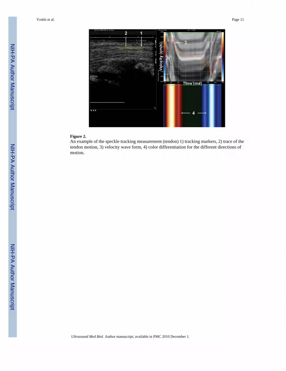
Figure 2.An example of the speckle tracking measurement (tendon) 1) tracking markers, 2) trace of thetendon motion, 3) velocity wave form, 4) color differentiation for the different directions ofmotion.
Yoshii et al. Page 11
Ultrasound Med Biol. Author manuscript; available in PMC 2010 December 1.
NIH
-PA Author Manuscript
NIH
-PA Author Manuscript
NIH
-PA Author Manuscript

Figure 3.An example of the tissue Doppler imaging measurement (flexor tendon) 1) pulsed wave cursor,line shows angle correction, 2) the maximum velocity for flexion, 3) the area under the dottedline was measured for the excursion.
Yoshii et al. Page 12
Ultrasound Med Biol. Author manuscript; available in PMC 2010 December 1.
NIH
-PA Author Manuscript
NIH
-PA Author Manuscript
NIH
-PA Author Manuscript

Figure 4.The relationship between the excursion derived from change in joint angle and the ultrasoundmeasurements A) tendon excursions from joint angle and speckle tracking method, B) tendonexcursions from joint angle and tissue Doppler imaging method.
Yoshii et al. Page 13
Ultrasound Med Biol. Author manuscript; available in PMC 2010 December 1.
NIH
-PA Author Manuscript
NIH
-PA Author Manuscript
NIH
-PA Author Manuscript

Figure 5.Maximum velocity ratio a) speckle tracking method, b) tissue Doppler imaging method. Pairsof values that are statistically different from each other are denoted (*), (**), (+) (*, +: P<0.01,**: P<0.05).
Yoshii et al. Page 14
Ultrasound Med Biol. Author manuscript; available in PMC 2010 December 1.
NIH
-PA Author Manuscript
NIH
-PA Author Manuscript
NIH
-PA Author Manuscript

Figure 6.Shear Index. Pairs of values that are statistically different from each other are denoted (*), (**),(+) (P<0.01).
Yoshii et al. Page 15
Ultrasound Med Biol. Author manuscript; available in PMC 2010 December 1.
NIH
-PA Author Manuscript
NIH
-PA Author Manuscript
NIH
-PA Author Manuscript


