Embed Size (px)
Citation preview

Tissue Stretch Induces Nuclear Remodeling in Connective TissueFibroblasts
Helene M. Langevin*,‖, Kirsten N. Storch*, Robert R. Snapp§, Nicole A. Bouffard*, Gary J.Badger¶, Alan K. Howe#, and Douglas J. Taatjes†*Department of Neurology, University of Vermont College of Medicine 89 Beaumont Ave. Burlington,VT 05405, USA‖ Department of Orthopaedics & Rehabilitation, University of Vermont College of Medicine 89Beaumont Ave. Burlington, VT 05405, USA§ Department of Computer Science, University of Vermont College of Medicine 89 Beaumont Ave.Burlington, VT 05405, USA¶ Department of Medical Biostatistics, University of Vermont College of Medicine 89 Beaumont Ave.Burlington, VT 05405, USA# Department of Pharmacology, Vermont Cancer Center, University of Vermont College of Medicine89 Beaumont Ave. Burlington, VT 05405, USA†Department of Pathology University of Vermont College of Medicine 89 Beaumont Ave. Burlington,VT 05405, USA
AbstractStudies in cultured cells have shown that nuclear shape is an important factor influencing nuclearfunction, and that mechanical forces applied to the cell can directly affect nuclear shape. In a previousstudy, we demonstrated that stretching of whole mouse subcutaneous tissue causes dynamiccytoskeletal remodeling with perinuclear redistribution of α-actin in fibroblasts within the tissue. Wehave further shown that the nuclei of these fibroblasts have deep invaginations containing α-actin.In the current study, we hypothesized that tissue stretch would cause nuclear remodeling with areduced amount of nuclear invagination, measurable as a change in nuclear concavity. Subcutaneousareolar connective tissue samples were excised from 28 mice and randomized to either tissue stretchor no stretch for 30 minutes, then examined with histochemistry and confocal microscopy. Instretched tissue (vs. non-stretched), fibroblast nuclei had a larger cross sectional area (p<.001),smaller thickness (p<.03) in the plane of the tissue, and smaller relative concavity (p<.005) indicatingan increase in nuclear convexity. The stretch-induced loss of invaginations may have importantinfluences on gene expression, RNA trafficking and/or cell differentiation.
KeywordsCytoskeleton; subcutaneous; nucleus; mechanotransduction; invagination
Corresponding Author: Helene M. Langevin, Department of Neurology, University of Vermont, [email protected], tel:802-656-1001, fax: 802-656-8704.Disclosure: Helene M. Langevin is a partner of Stromatec, Inc.
NIH Public AccessAuthor ManuscriptHistochem Cell Biol. Author manuscript; available in PMC 2011 April 1.
Published in final edited form as:Histochem Cell Biol. 2010 April ; 133(4): 405–415. doi:10.1007/s00418-010-0680-3.
NIH
-PA Author Manuscript
NIH
-PA Author Manuscript
NIH
-PA Author Manuscript

IntroductionThe cell nucleus is increasingly recognized as a highly dynamic organelle whose function isintrinsically linked to its structural organization (Dundr and Misteli, 2001). Nuclear shape hasbeen shown to influence fundamental aspects of nuclear function including chromatinremodeling and gene transcription (Dalby et al., 2007; Itano et al., 2003; Lammerding et al.,2004; Thomas et al., 2002). One of the most dramatic changes in nuclear shape is the foldingand unfolding of nuclear invaginations. These structures are thought to play a role in celldifferentiation as well as chromatin organization, nucleocytoplasmic transport, calciumsignaling and RNA trafficking (Abe et al., 2004; Bloom et al., 1996; Echevarria et al., 2003;Fricker et al., 1997; Johnson et al., 2003). There is also evidence that the commitment of a cellto proliferation or differentiation can be influenced by the overall shape of the cell, in additionto the shape of its nucleus (Ben-Ze'ev et al., 1980; McBeath et al., 2004). A proposedmechanism underlying this phenomenon is that cell shape affects nuclear shape which in turnaffects chromatin remodeling and gene expression (Ingber et al., 1987; Maniotis et al., 1997).
Studies in cultured cells and whole tissue have shown that the shape, position and orientationof the nucleus as well as the organization of the nuclear lamina can be influenced by mechanicalforces applied to the cell (Bloom et al., 1996; Flaherty et al., 1972; Gieni and Hendzel, 2007;Guilak, 1995; Hu et al., 2005; Huang et al., 2007; Maniotis et al., 1997; Philip and Dahl,2008). Changes in nuclear shape in response to an applied force can results from passiveviscoelastic deformation (Guilak et al., 2000; Rowat et al., 2006; Vaziri and Mofrad, 2007) aswell as active cytoskeletally-mediated structural remodeling (Deguchi et al., 2005). We havepreviously shown that the nuclei of fibroblasts within mouse subcutaneous “loose” areolarconnective tissue have deep nuclear invaginations containing cytoplasm that are associatedwith concavity of the nuclear surface (Storch et al., 2007). We also have shown that stretchingof the whole tissue causes these fibroblasts to respond rapidly (i.e. within minutes) withextensive cell spreading, lamellipodia formation and perinuclear actin redistribution (Langevinet al., 2005; Storch et al., 2007). The current study was designed to test the hypothesis thattissue stretch results in nuclear remodeling measurable as a change in the degree of nuclearconcavity.
Materials and MethodsThe experimental protocols used in these experiments were approved by the University ofVermont IACUC Committee. Two complementary groups of experiments were performedusing different tissue fixation methods: 3% paraformaldehyde (PFA) was used in experimentsexamining nuclear concavity, while 95% ethanol was used in experiments examining cell crosssectional area, nuclear orientation and nuclear eccentricity. Although both fixatives yieldedsimilar results with respect to the basic difference in nuclear shape between stretched and non-stretched tissue (Figure 1), each fixative had distinct advantages and limitations: PFA producedless tissue shrinking and better preservation of nuclear morphological detail but did not allowpreservation of tissue orientation. Ethanol fixation yielded optimal visualization andmeasurement of cell body shape (Langevin et al., 2004) and allowed preservation of tissueorientation during tissue dissection (see method below) but produced more tissue shrinking(nuclear cross sectional area was ∼30% smaller with ethanol compared with PFA fixation).
Effect of tissue stretch on nuclear cross sectional area and concavityWe performed a set of experiments in which we examined the effect of tissue stretch on nuclearshape in high magnification confocal microscopy images. Tissue samples from 28 mice wereexcised and randomized to either stretch (n = 14) or no stretch (n = 14) ex vivo for 30 minutesfollowed by tissue fixation in 3% PFA and staining of the nucleus with DAPI.
Langevin et al. Page 2
Histochem Cell Biol. Author manuscript; available in PMC 2011 April 1.
NIH
-PA Author Manuscript
NIH
-PA Author Manuscript
NIH
-PA Author Manuscript

Nuclear cross sectional area, orientation and eccentricity with immediate (2 minutes) vs.sustained (30 minutes) stretch, and after the release of stretch
To investigate whether the observed stretch-induced changes in nuclear shape are primarilypassive (either from direct stretching of the nucleus or compression of the nucleus fromthinning of the stretched tissue), as opposed to active remodeling, we measured nuclear crosssectional area, eccentricity and orientation after 2 minutes (N=3 mice) and 30 minutes (N=3mice) of stretch ex vivo, as well as in tissue stretched for 30 minutes, then released for 2 minutes(N=3 mice) and 10 minutes (N=3 mice). Samples were fixed in 95% ethanol for 1 hour.Subcutaneous tissue samples were dissected on three sides and separated from thesubcutaneous muscle, leaving a partially attached tissue flap that was allowed to rehydrate inPBS containing 1% BSA overnight. A glass slide was then placed under the sample and thefourth side was cut while preserving the orientation of samples relative to the direction ofstretch. Fixation was followed by staining with DAPI.
Effect of pharmacological inhibitor10 μM Rho kinase inhibitor Y27632 (BioMol, Philadelphia, PA) was used to investigate therole of Rho-dependent cytoskeletal remodeling in stretch-induced change in nuclear shape.Tissue samples were cut in half then randomized to stretch vs. no stretch. Each sample wasincubated for 10 minutes with the inhibitor, then stretched (or not stretched) for 30 minutes.Samples were then fixed in 3% PFA for 1 hour as above, then stained with DAPI.
Relationship between cell cross sectional area and nuclear cross sectional areaWe examined the relationship between cell cross sectional area and nuclear cross-sectionalarea in a set of confocal laser microscopy images obtained in a previously published study inwhich 23 male C57Black6 mice weighing 19-21 g were randomized to either stretch or nostretch for 10, 60 or 120 minutes ex vivo (Langevin et al., 2005). Tissue stretch was followedby tissue fixation in 95% ethanol and histochemical staining with phalloidin for visualizationof cell bodies and SYTOX green nucleic acid stain for visualization of the nucleus. Cell andnuclear cross sectional area were measured as described below.
Stretched and non-stretched subcutaneous tissue sample preparationIn all experiments, tissue samples were harvested from the abdomen immediately after deathin C57BL/6 male mice (19-21g). Whole skin flaps (8 cm × 3 cm) containing dermis,subcutaneous muscle and subcutaneous tissue were dissected away from the abdominal wallmusculature, excised, and placed between stainless steel grips submerged in HEPES-physiological saline solution (HEPES-PSS) (141.8 mM NaCl, 4.7 mM KCl, 1.7 mMMgSO4, 0.39 mM EDTA, 2.8 mM CaCl2, 10.0 mM HEPES, 1.2 mM KH2PO4, 5.0 mM glucose,pH 7.4) at 37°C as previously described (Langevin et al., 2005). Tissue grips were connectedto a 500 g capacity strain gauge transducer. Samples randomized to stretch were elongated ata rate of 1 mm/sec by advancing a micrometer connected to the distal tissue grip until a loadof 0.02 N (corresponding to 15-25% tissue strain relative to non-stretched length) wasregistered, then maintained at that length for the prescribed incubation time. Non-stretchedsamples were incubated in grips for 30 minutes at the length corresponding to the length of thetissue laying flat. At the end of incubation, samples were fixed in 3% PBS or 95% ethanol atthe stretched (or unstretched) length. After fixation, three samples of areolar connective tissuewere dissected for each animal. Samples located between the superficial (subcutaneous) fasciaand the deep (muscular) fascia were dissected (sample dimensions 20-30 mm in width, 60-80mm in length and 20-50 μm in thickness), mounted on glass slides and rinsed in PBS/1.0%BSA/0.1% Triton × 100.
Langevin et al. Page 3
Histochem Cell Biol. Author manuscript; available in PMC 2011 April 1.
NIH
-PA Author Manuscript
NIH
-PA Author Manuscript
NIH
-PA Author Manuscript

Histochemical and immunohistochemical stainingUnless stated otherwise, histochemical staining of the nucleus was performed with 4′,6-diamidino-2-phenylindole, dihydrochloride (DAPI) nucleic acid stain (Invitrogen, Carlsbad,CA) at a dilution of 1:1000 for five minutes at room temperature. Samples were counterstainedwith Texas Red conjugated Phalloidin, a specific stain for polymerized actin, (Invitrogen,Carlsbad, CA) at a 1:25 dilution for 40 minutes at 4°C for visualization of cell bodies. A subsetof PFA-fixed samples were also stained for α-actin using indirect immunohistochemistry.Samples were first incubated with a mouse monoclonal anti-α-smooth muscle actin primaryantibody (Sigma, St. Louis, MO) at 1:100 dilution followed by an Alexa 488-conjugatedsecondary goat anti-mouse antibody (Invitrogen Corporation, Carlsbad, CA) at 1:200 dilutionas previously described (Langevin et al., 2006). To reduce background staining resulting fromusing a mouse primary antibody, samples were incubated with Mouse-On-Mouse (M.O.M)blocking reagents (Vector Laboratories, Burlingame, CA) according to the manufacturers'directions. Samples were overlaid with a glass coverslip using 50% glycerol in PBS with 1%N-propylgallate as a mounting medium.
Confocal scanning laser microscopyTissue samples were imaged with a Zeiss LSM 510 META confocal scanning laser microscope.Four to six fields per sample were first imaged at 63× by an operator unaware of the studycondition (stretch vs. no stretch). One to two cells per field were then imaged at highermagnification using a PlanApochromat 100× (1.4 N.A.) oil immersion lens, focusing directlyon the nuclei using a zoom factor of two times. Thus, a total of 12-15 cells were imaged at bothmagnifications per animal. Z-stacks were acquired such that the entire thickness of each nucleuswas captured. Each optical section had an optical thickness of 0.7 μm for all laser lines and a0.33 μm inter image interval.
Morphometric analysisNuclear cross sectional area and relative concavity—The shape of each nucleus,projected onto the plane of the tissue, was measured in relation to its convex hull, the smallestconvex region that encloses the nuclear projection. (Recall that a region R is said to be convexif and only if every line segment XY lies completely within R for every X and Y in R.) Lettinga denote the nuclear cross sectional area (defined as the area of the nuclear projection onto theplane of the tissue), and h the area of its convex hull, we define the area of the deficit as d=h-a, which estimates the sum of the invaginated regions. We then define the relative concavityas c=d/h. Consequently, a convex nucleus --- one without invaginations or indentations ---results in a relative concavity of 0.
It is necessary to preprocess each image stack in a consistent manner before the relativeconcavity can be measured. Image stacks were imported into a custom made image analysisprogram. The nuclear cross sectional area was estimated from the 2-d projections of the imagestacks onto the plane of the tissue. Projected images were thresholded for DAPI by aninvestigator unaware of the study condition. For each image a threshold level was chosen thatoptimally revealed the nuclear boundary in the presence of image noise. A closure operationwas then performed in order to fill isolated interior pixels that were below the threshold (Figure2A). Each active pixel was then assigned to a connected component following a sequentiallabeling algorithm (Horn, 1986). The convex hull for the largest connected component wasthen computed using a randomized incremental algorithm (Berg, 1997). Initially, three activepixels were chosen, at random, from the largest component to form a nondegenerate triangle(having a nonzero area), defining the vertices of the initial “convex polygon”. One by one,each remaining active pixel in the component was chosen in random order. Pixels falling withinthe incremental convex polygon were ignored. Whenever a pixel fell outside of the incrementalconvex polygon it was added as a new vertex, replacing the vertices that fall within the interior
Langevin et al. Page 4
Histochem Cell Biol. Author manuscript; available in PMC 2011 April 1.
NIH
-PA Author Manuscript
NIH
-PA Author Manuscript
NIH
-PA Author Manuscript

of the enlarged convex polygon. (Figure 2 A,B). Letting a denote the area of the largestcomponent, and h, the area of its convex hull, we compute the relative concavity of the nuclearcross section as c = (h – a)/h = d/h. (Figure 2C). Measurably concave regions ranged fromshallow depressions to deep intranuclear invaginations.
Nuclear thickness—For measurement of nuclear thickness, image stacks were importedinto the image analysis software package MetaMorph (Molecular Devices, Downington, PA).Nuclear thickness was estimated by counting the number of individual optical sections in whicha nucleus appeared, multiplied by the optical inter image interval (0.33 μm).
Nuclear volume—For measurement of nuclear volume, image stacks were imported into theanalysis software package Volocity (Improvision, Lexington, MA) to calculate voxel basednuclear volume estimations. In this program, the images of nuclei were cropped by thresholdingon DAPI staining. To account for variations in staining levels between samples, a threshold ofaverage intensity +1.0σ was used to select the entire nucleus, then the “fill holes” option waschosen where objects less than 1 μm3 in size were excluded to reduce noise.
Nuclear eccentricity and orientationThe elongation of each nuclear area is described in terms of the first and second centralmoments of the locations of the pixels from the corresponding connected component: the two-dimensional mean (μ) estimates the location of the nuclear center, while the 2×2 covariancematrix (S) describes the second-order spread about the center. Then the eigenvalues (λ1, λ2)and eigenvectors (v1, v2) of the covariance matrix (S) are computed algebraically, such thatSvi = λi vi, for i = 1, and 2, with λ1 ≥ λ2≥ 0. We define the orientation of the nucleus as theangle between the dominant eigenvector (v1) and the horizontal axis, and the eccentricity asthe square root of the eigenvalue ratio, i.e., √ λ1/ λ2 (Pratt, 1991).
Cell body cross sectional areaCell body cross sectional area was measured in six fields per sample using MetaMorph softwarefor morphometric analysis as previously described (Langevin et al., 2005).
Statistical Methods—Differences between stretched and non-stretched tissue with respectto nuclear outcome measures were evaluated using analyses of variance corresponding to anested design. The replicate observations derived from the 8-12 cells examined per animalwere considered a nested factor within mouse. The F-tests corresponding to testing differencesbetween experimental conditions (i.e. stretch and no stretch) utilized across-animal variabilitywithin each condition (df = 26), not the variability associated with replicate cells within eachanimal, as the appropriate error term. Analyses were performed using SAS Version 8, PROCMIXED (SAS Institute, Cary, NC). Statistical significance was determined based on α = 0.05.
In the additional analysis of data from previously published experiments, the relationshipbetween cell cross-sectional area and nuclear cross-sectional area at different time points instretched and non-stretched samples was examined using correlation analyses.
ResultsFibroblast nuclei within non-stretched areolar connective tissue were smaller and more concavecompared with those in stretched tissue, which tended to be round or oval with a smoothcircumference (Figure 3). Morphometric measurements showed that fibroblast nuclei instretched tissue, compared with non-stretched tissue had significantly greater nuclear crosssectional area (mean ± SE 109 ± 3 μm2 vs. 93 ± 3 μm2, F1,26=13.9, p = .001) (Figure 4A) anddecreased thickness (3.28 ± 0.12 μm vs. 3.63 ± 0.09 μm, F1,2 6= 5.4, p = .03) (Figure 4C).
Langevin et al. Page 5
Histochem Cell Biol. Author manuscript; available in PMC 2011 April 1.
NIH
-PA Author Manuscript
NIH
-PA Author Manuscript
NIH
-PA Author Manuscript

Nuclear volume showed a trend similar to that of cross sectional area, i.e. became on averagelarger in stretched compared with non-stretched tissue but this difference was not statisticallysignificant (365 ± 12 μm3 vs. 333 ± 14 μm3 F1,26 = 4.2, p= .05) (Figure 4B). Fibroblast nucleiwithin stretched tissue also were less concave, having a smaller relative concavity than thenuclei of non-stretched tissue (0.02 ± 0.0017 vs. 0.03 ± 0.002 for stretch and no stretchrespectively, F1,26 = 9.6, p = .005) (Figure 4D).
There was no significant difference in cellular or nuclear cross sectional area between stretchedand non-stretched tissue after 2 minutes of stretch (Figure 5). In tissue that was stretched for30 minutes and then either released for 2 or 10 minutes, or not released, significant differenceswere observed between conditions for cell cross sectional area, but not nuclear cross sectionalarea (F2,6=18.9, p<.003) (Figure 5). Cell cross sectional area was significantly reduced after 2minutes (p=.003) and 10 minutes (p=.001) of release (Fisher's LSD). Although there was atrend toward decreased nuclear cross sectional area in released tissue, this did not reachstatistical significance (p=.42 for 2 minutes and p=.07 for 10 minutes). There were nosignificant differences in nuclear orientation or eccentricity within the image plane in tissuestretched vs. non-stretched for 2 or 30 minutes (data not shown).
Nuclear cross-sectional area was generally moderately correlated with cell cross-sectional areain both stretched and non-stretched conditions at 10 minutes, 60 minutes and 120 minutes ofincubation (illustrated for 120 minutes in Figure 6) suggesting that the stretch-induced changein cell shape may be related to the change in nuclear shape. This is further suggested by theobservation that the pattern of α-actin changed both within and outside the nuclear domain inresponse to tissue stretch (Figures 7). In non-stretched tissue, α-actin was present throughoutthe cytoplasm as well as within deep nuclear invaginations (Figure 8) as previously reported(Storch et al., 2007). In contrast, in stretched tissue, α-actin immunoreactivity was redistributedpredominantly around the nucleus and was not detectable within the nuclear domain (Figure9). In order to further investigate a possible link between cytoskeletal and nuclear remodeling,we examined the shape of fibroblast nuclei in the presence of an inhibitor of Rho kinase, whichregulates cytoskeletal actin bundle contractility (Burridge and Wennerberg, 2004;Ridley andHall, 1992;Ridley et al., 1992). In the presence of the Rho kinase inhibitor, nuclei had a similarmorphology with and without stretch (Figure 10). Mean±SE nuclear cross sectional area was74.6±9.5 μ2 with stretch and 77.4±5.4 μ2 without stretch (based on N=11-14 cells percondition). This suggests that Rho- dependent cytoskeletal mechanisms are required for changein nuclear shape in response to tissue stretch.
DiscussionStretching of connective tissue for 30 minutes caused a change in the shape of fibroblast nucleiwhich were wider, flatter and smoother (less concave) than those within non-stretched tissue.Our confocal images show that nuclei in non-stretched tissue had indentations ranging fromsmall wrinkles to deep invaginations, all of which contributed to greater nuclear concavity(Figure 2). Tissue stretch was associated with both loss of deep invaginations, as well assuperficial smoothing of the nuclear surface. The potential consequences of stretch-inducednuclear remodeling and loss of nuclear concavity are far reaching, since mounting evidencesuggests that cell and nuclear shape can influence cell differentiation, chromatin structure, andhistone acetylation (Chen et al., 1997;Dalby, 2005;Kim et al., 2005). Indeed, directtransmission of forces through the cytoplasmic and nuclear cytoskeleton via changes in celland nuclear shape has been proposed as a source of specific coupling between tissue mechanicalforces and the genome (Gieni and Hendzel, 2007). In addition, because nuclear invaginationsbring cytoplasmic space closer to the interior of nuclear domain, these structures are thoughtto be an important conduit for nucleocytoplasmic transport (Bourgeois et al., 1979;Dupuy-Coin et al., 1986).
Langevin et al. Page 6
Histochem Cell Biol. Author manuscript; available in PMC 2011 April 1.
NIH
-PA Author Manuscript
NIH
-PA Author Manuscript
NIH
-PA Author Manuscript

The change in nuclear shape and smoothing of the nuclear surface could result from directviscoelastic deformation (from in-plane tensile forces and/or perpendicular compressive forcesdue to thinning of the tissue during stretching) or could represent a more complex phenomenoninvolving active reorganization of nuclear structure in response to tissue stretch. Micropipetteaspiration experiments on isolated nuclei have shown that the nucleus reversibly deformswithin seconds in response to a direct mechanical input (Guilak et al., 2000; Rowat et al.,2006; Vaziri and Mofrad, 2007). Recent rheological models based on soft glassy materials alsosuggest that nuclear stiffness can continue to change over longer time scales in response tomechanical deformation, and cytoskeletal remodeling has been suggested as a possibleexplanation for this type of biomechanical behavior (Dahl et al., 2005; Mandadapu et al.,2008). In a study of cartilage compressed for 30 minutes ex vivo, chondrocyte nuclei becameflatter in response to tissue compression and this effect was inhibited by pretreatment withcytochalasin-D, suggesting that the actin cytoskeleton plays a role linking extracellular matrixcompression and nuclear deformation (Guilak, 1995). In cultured endothelial cells, prolongedshear stress for 24 hours was shown to cause nuclear morphological and stiffness changes thatpersisted after isolation of the nuclei and were also prevented by cytochalasin-D, againsuggesting structural remodeling (Deguchi et al., 2005). In the current study, the delayed onsetof the change in nuclear morphology (more than 2 minutes) and the persistence ofmorphological changes after the release of tissue stretch suggest that the changes in nuclearshape observed in this study involve structural remodeling, rather than direct stretching orcompression of the nuclei. The lack of change in nuclear eccentricity or orientation furthersuggests that the changes in nuclear shape observed in this study are not the direct result oftissue stretching. The change in cell shape and redistribution of perinuclear actin observed inresponse to tissue stretch shows that extensive cytoskeletal reorganization is taking place inresponse to tissue stretch. The lack of stretch-induced change in nuclear shape in the presenceof Rho kinase inhibition further suggests that actin-based cytoskeletal mechanisms are involvedin nuclear remodeling in response to tissue stretch.
A potential limitation of our study is that analyzing confocal image data in two dimensionsmay have underestimated nuclear concavity oriented perpendicular to the tissue that would notbe visible in a two dimensional image stack projection. It is possible that out-of-plane changesin cell and nuclear orientation occur in response to mechanical forces (i.e., the cell and nucleuscould rotate relative to the plane of the tissue or the nucleus could rotate relative to a morestationary cell body) (Gieni and Hendzel, 2007). We however consider it unlikely thatdifferences in nuclear shape and relative concavity between stretched and non-stretched tissuewere entirely due to rotation of nuclear invaginations out of the tissue plane. Because the nucleiof the fibroblasts are shaped like disks, we would expect that the aspect ratio (defined as thenuclear thickness divided by the square root of the nuclear cross sectional area) would increaseas a result of rotation out of the tissue plane. However, the mean ± SE aspect ratio was 0.39 ±0.014 for non-stretched tissue and 0.32 ± 0.016 for stretched tissue. This decrease in nuclearaspect ratio with stretch strongly suggests that nuclear deformation predominated over nuclearrotation.
In summary, stretching of whole mouse subcutaneous tissue resulted in a change in fibroblastnuclear shape with a loss of nuclear concavity. These results in whole tissue support a growingliterature indicating that the mechanical environment of the cell has a profound influence onthe cell nucleus.
AcknowledgmentsThe authors thank Drs. Nicholas Heintz and William C. Eanrshaw for helpful discussions. This work was funded bythe National Institutes of Health Center for Complementary and Alternative Medicine research Grant RO1-AT01121and by National Institutes of Health Grant P20 RR16435 from the Center of Biomedical Research Excellence Programof the National Center for Research Resources. Its contents are solely the responsibility of the authors and do not
Langevin et al. Page 7
Histochem Cell Biol. Author manuscript; available in PMC 2011 April 1.
NIH
-PA Author Manuscript
NIH
-PA Author Manuscript
NIH
-PA Author Manuscript

necessarily represent the official views of the National Center for Complementary and Alternative Medicine, NationalInstitutes of Health.
ReferencesAbe T, Takano K, Suzuki A, Shimada Y, Inagaki M, Sato N, Obinata T, Endo T. Myocyte differentiation
generates nuclear invaginations traversed by myofibrils associating with sarcomeric protein mRNAs.J Cell Sci 2004;117:6523–6534. [PubMed: 15572409]
Ben-Ze'ev A, Farmer SR, Penman S. Protein synthesis requires cell-surface contact while nuclear eventsrespond to cell shape in anchorage-dependent fibroblasts. Cell 1980;21:365–372. [PubMed: 6157481]
Berg, Md. Computational geometry : algorithms and applications. Springer; Berlin; New York: 1997.Bloom S, Lockard VG, Bloom M. Intermediate filament-mediated stretch-induced changes in chromatin:
a hypothesis for growth initiation in cardiac myocytes. J Mol Cell Cardiol 1996;28:2123–2127.[PubMed: 8930807]
Bourgeois CA, Hemon D, Bouteille M. Structural relationship between the nucleolus and the nuclearenvelope. J Ultrastruct Res 1979;68:328–340. [PubMed: 490761]
Burridge K, Wennerberg K. Rho and Rac take center stage. Cell 2004;116:167–179. [PubMed: 14744429]Chen CS, Mrksich M, Huang S, Whitesides GM, Ingber DE. Geometric control of cell life and death.
Science 1997;276:1425–1428. [PubMed: 9162012]Dahl KN, Engler AJ, Pajerowski JD, Discher DE. Power-law rheology of isolated nuclei with deformation
mapping of nuclear substructures. Biophys J 2005;89:2855–2864. [PubMed: 16055543]Dalby MJ. Topographically induced direct cell mechanotransduction. Medical engineering & physics
2005;27:730–742. [PubMed: 15921949]Dalby MJ, Gadegaard N, Herzyk P, Sutherland D, Agheli H, Wilkinson CD, Curtis AS.
Nanomechanotransduction and interphase nuclear organization influence on genomic control. J CellBiochem 2007;102:1234–1244. [PubMed: 17427951]
Deguchi S, Maeda K, Ohashi T, Sato M. Flow-induced hardening of endothelial nucleus as an intracellularstress-bearing organelle. J Biomech 2005;38:1751–1759. [PubMed: 16005465]
Dundr M, Misteli T. Functional architecture in the cell nucleus. Biochem J 2001;356:297–310. [PubMed:11368755]
Dupuy-Coin AM, Moens P, Bouteille M. Three-dimensional analysis of given cell structures: nucleolus,nucleoskeleton and nuclear inclusions. Methods Achiev Exp Pathol 1986;12:1–25. [PubMed:3007927]
Echevarria W, Leite MF, Guerra MT, Zipfel WR, Nathanson MH. Regulation of calcium signals in thenucleus by a nucleoplasmic reticulum. Nat Cell Biol 2003;5:440–446. [PubMed: 12717445]
Flaherty JT, Pierce JE, Ferrans VJ, Patel DJ, Tucker WK, Fry DL. Endothelial nuclear patterns in thecanine arterial tree with particular reference to hemodynamic events. Circ Res 1972;30:23–33.[PubMed: 5007525]
Fricker M, Hollinshead M, White N, Vaux D. Interphase nuclei of many mammalian cell types containdeep, dynamic, tubular membrane-bound invaginations of the nuclear envelope. J Cell Biol1997;136:531–544. [PubMed: 9024685]
Gieni RS, Hendzel MJ. Mechanotransduction from the ECM to the genome: Are the pieces now in place.J Cell Biochem. 2007
Guilak F. Compression-induced changes in the shape and volume of the chondrocyte nucleus. J Biomech1995;28:1529–1541. [PubMed: 8666592]
Guilak F, Tedrow JR, Burgkart R. Viscoelastic properties of the cell nucleus. Biochem Biophys ResCommun 2000;269:781–786. [PubMed: 10720492]
Horn, B. Robot vision. MIT Press; McGraw-Hill; Cambridge, Mass. New York: 1986.Hu S, Chen J, Butler JP, Wang N. Prestress mediates force propagation into the nucleus. Biochem Biophys
Res Commun 2005;329:423–428. [PubMed: 15737604]Huang HY, Liao J, Sacks MS. In-situ deformation of the aortic valve interstitial cell nucleus under
diastolic loading. J Biomech Eng 2007;129:880–889. [PubMed: 18067392]
Langevin et al. Page 8
Histochem Cell Biol. Author manuscript; available in PMC 2011 April 1.
NIH
-PA Author Manuscript
NIH
-PA Author Manuscript
NIH
-PA Author Manuscript

Ingber DE, Madri JA, Folkman J. Endothelial growth factors and extracellular matrix regulate DNAsynthesis through modulation of cell and nuclear expansion. In Vitro Cell Dev Biol 1987;23:387–394. [PubMed: 2438264]
Itano N, Okamoto S, Zhang D, Lipton SA, Ruoslahti E. Cell spreading controls endoplasmic and nuclearcalcium: a physical gene regulation pathway from the cell surface to the nucleus. Proc Natl Acad SciU S A 2003;100:5181–5186. [PubMed: 12702768]
Johnson N, Krebs M, Boudreau R, Giorgi G, LeGros M, Larabell C. Actin-filled nuclear invaginationsindicate degree of cell de-differentiation. Differentiation 2003;71:414–424. [PubMed: 12969334]
Kim YB, Yu J, Lee SY, Lee MS, Ko SG, Ye SK, Jong HS, Kim TY, Bang YJ, Lee JW. Cell adhesionstatus-dependent histone acetylation is regulated through intracellular contractility-related signalingactivities. J Biol Chem 2005;280:28357–28364. [PubMed: 15961394]
Lammerding J, Schulze PC, Takahashi T, Kozlov S, Sullivan T, Kamm RD, Stewart CL, Lee RT. LaminA/C deficiency causes defective nuclear mechanics and mechanotransduction. J Clin Invest2004;113:370–378. [PubMed: 14755334]
Langevin HM, Bouffard NA, Badger GJ, Iatridis JC, Howe AK. Dynamic fibroblast cytoskeletal responseto subcutaneous tissue stretch ex vivo and in vivo. Am J Physiol Cell Physiol 2005;288:C747–756.[PubMed: 15496476]
Langevin HM, Cornbrooks CJ, Taatjes DJ. Fibroblasts form a body-wide cellular network. HistochemCell Biol 2004;122:7–15. [PubMed: 15221410]
Langevin HM, Storch KN, Cipolla MJ, White SL, Buttolph TR, Taatjes DJ. Fibroblast spreading inducedby connective tissue stretch involves intracellular redistribution of alpha- and beta-actin. HistochemCell Biol 2006;125:487–495. [PubMed: 16416024]
Mandadapu KK, Govindjee S, Mofrad MR. On the cytoskeleton and soft glassy rheology. J Biomech2008;41:1467–1478. [PubMed: 18402964]
Maniotis AJ, Chen CS, Ingber DE. Demonstration of mechanical connections between integrins,cytoskeletal filaments, and nucleoplasm that stabilize nuclear structure. Proc Natl Acad Sci U S A1997;94:849–854. [PubMed: 9023345]
McBeath R, Pirone DM, Nelson CM, Bhadriraju K, Chen CS. Cell shape, cytoskeletal tension, and RhoAregulate stem cell lineage commitment. Dev Cell 2004;6:483–495. [PubMed: 15068789]
Philip JT, Dahl KN. Nuclear mechanotransduction: response of the lamina to extracellular stress withimplications in aging. J Biomech 2008;41:3164–3170. [PubMed: 18945430]
Pratt, WK. Digital Image Processing. John Wiley & Sons; NY: 1991. p. 636-644.Ridley AJ, Hall A. The small GTP-binding protein rho regulates the assembly of focal adhesions and
actin stress fibers in response to growth factors. Cell 1992;70:389–399. [PubMed: 1643657]Ridley AJ, Paterson HF, Johnston CL, Diekmann D, Hall A. The small GTP-binding protein rac regulates
growth factor-induced membrane ruffling. Cell 1992;70:401–410. [PubMed: 1643658]Rowat AC, Lammerding J, Ipsen JH. Mechanical properties of the cell nucleus and the effect of emerin
deficiency. Biophys J 2006;91:4649–4664. [PubMed: 16997877]Storch KN, Taatjes DJ, Bouffard NA, Locknar S, Bishop NM, Langevin HM. Alpha smooth muscle actin
distribution in cytoplasm and nuclear invaginations of connective tissue fibroblasts. Histochem CellBiol 2007;127:523–530. [PubMed: 17310383]
Thomas CH, Collier JH, Sfeir CS, Healy KE. Engineering gene expression and protein synthesis bymodulation of nuclear shape. Proc Natl Acad Sci U S A 2002;99:1972–1977. [PubMed: 11842191]
Vaziri A, Mofrad MR. Mechanics and deformation of the nucleus in micropipette aspiration experiment.J Biomech 2007;40:2053–2062. [PubMed: 17112531]
Langevin et al. Page 9
Histochem Cell Biol. Author manuscript; available in PMC 2011 April 1.
NIH
-PA Author Manuscript
NIH
-PA Author Manuscript
NIH
-PA Author Manuscript

Fig. 1.Morphological appearance of fibroblast nuclei in non-stretched and stretched mousesubcutaneous areolar connective tissue (30 minutes ex vivo) fixed with 95% ethanol (A,C) vs.3% PFA(B,D) and stained with DAPI. Images are composite projections of stacks containing20 optical sections taken at a 0.33 μm inter-image interval.
Langevin et al. Page 10
Histochem Cell Biol. Author manuscript; available in PMC 2011 April 1.
NIH
-PA Author Manuscript
NIH
-PA Author Manuscript
NIH
-PA Author Manuscript

Fig. 2.(A,B) Illustration of method used to analyze image stacks for nuclear cross sectional area(shaded area in A) and nuclear convex hull area (area inside line in A and B); (C) Schematicdrawing of the areas used to compute the relative concavity. In panel C, a (gray region)represents nuclear cross sectional area; h represents the area of its convex hull (the interior ofthe dark contour); d=h-a (white region) represents the deficit and estimates the sum ofinvaginated areas.
Langevin et al. Page 11
Histochem Cell Biol. Author manuscript; available in PMC 2011 April 1.
NIH
-PA Author Manuscript
NIH
-PA Author Manuscript
NIH
-PA Author Manuscript

Fig. 3.Example of high magnification image of fibroblast nuclei within non-stretched (A) andstretched (B) mouse subcutaneous areolar connective tissue (30 minutes ex vivo). Whole tissuesamples were fixed in 3% PFA, stained with DAPI nucleic acid stain and imaged with confocalmicroscopy. Each image represents a single optical section of 0.7 μm thickness.
Langevin et al. Page 12
Histochem Cell Biol. Author manuscript; available in PMC 2011 April 1.
NIH
-PA Author Manuscript
NIH
-PA Author Manuscript
NIH
-PA Author Manuscript
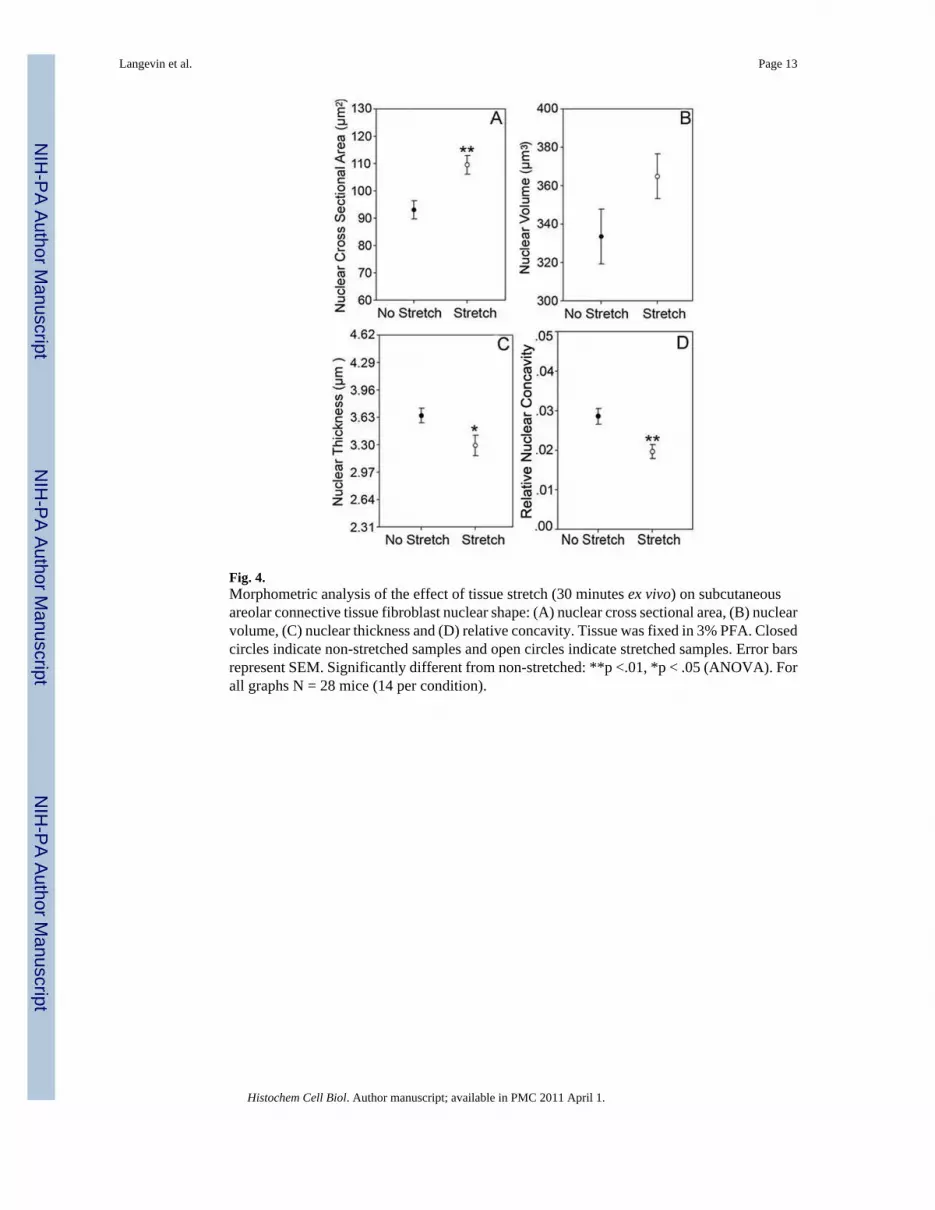
Fig. 4.Morphometric analysis of the effect of tissue stretch (30 minutes ex vivo) on subcutaneousareolar connective tissue fibroblast nuclear shape: (A) nuclear cross sectional area, (B) nuclearvolume, (C) nuclear thickness and (D) relative concavity. Tissue was fixed in 3% PFA. Closedcircles indicate non-stretched samples and open circles indicate stretched samples. Error barsrepresent SEM. Significantly different from non-stretched: **p <.01, *p < .05 (ANOVA). Forall graphs N = 28 mice (14 per condition).
Langevin et al. Page 13
Histochem Cell Biol. Author manuscript; available in PMC 2011 April 1.
NIH
-PA Author Manuscript
NIH
-PA Author Manuscript
NIH
-PA Author Manuscript

Fig. 5.Effect of tissue stretch for 2 minutes vs. 30 minutes ex vivo, as well as following release ofstretch for 2 minutes vs. 10 minutes on cell cross sectional area (A) and nuclear cross sectionalarea (B). Tissue samples were fixed in 95% ethanol. Error bars represent SEM. *Significantlydifferent from non-stretched (p < .05 paired t-test). Means sharing a common letter are notsignificantly different (Fisher's LSD, p<.05). N = 12 mice (3 per condition).
Langevin et al. Page 14
Histochem Cell Biol. Author manuscript; available in PMC 2011 April 1.
NIH
-PA Author Manuscript
NIH
-PA Author Manuscript
NIH
-PA Author Manuscript

Fig. 6.Relationship between fibroblast cell body cross sectional and nuclear cross sectional area instretched and non-stretched tissue incubated for 120 minutes. Circles indicate individual cellsin stretched (open circles) and non-stretched (closed circles) tissue. Linear regression lines areshown for stretched (dashed lines) and non-stretched (solid lines) tissue samples.
Langevin et al. Page 15
Histochem Cell Biol. Author manuscript; available in PMC 2011 April 1.
NIH
-PA Author Manuscript
NIH
-PA Author Manuscript
NIH
-PA Author Manuscript

Fig. 7.Fibroblasts within non-stretched (A,B,C) and stretched (D,E,F) mouse subcutaneous areolarconnective tissue (30 minutes ex vivo) stained with phalloidin (red, A,D),immunohistochemical staining for α-actin (green, A,B,D,E) and DAPI nucleic acid stain (white,C,F) imaged with confocal laser scanning microscopy using a PlanApochromat 63× (NA=1.4)oil immersion lens. Arrowheads (B,C) indicate a nuclear invagination containing α-actin.Images are projections of eight optical sections. Scale bars 5 μm.
Langevin et al. Page 16
Histochem Cell Biol. Author manuscript; available in PMC 2011 April 1.
NIH
-PA Author Manuscript
NIH
-PA Author Manuscript
NIH
-PA Author Manuscript

Fig. 8.Serial confocal microscopy images showing a fibroblast within non-stretched mousesubcutaneous areolar connective tissue (30 minutes ex vivo) stained for α-actin (green) andDAPI (blue). The middle two rows of optical sections shows presence of α-actin positivestaining in nuclear invaginations within the nuclear domain. Optical sections represent a 0.33μm inter-image interval. Scale bar 5 μm.
Langevin et al. Page 17
Histochem Cell Biol. Author manuscript; available in PMC 2011 April 1.
NIH
-PA Author Manuscript
NIH
-PA Author Manuscript
NIH
-PA Author Manuscript

Fig. 9.Serial confocal microscopy images showing a fibroblast within stretched mouse subcutaneousareolar connective tissue (30 minutes ex vivo) stained for α-actin (green) and DAPI (blue). Themiddle row of optical sections shows the absence of α-actin positive staining in the nucleardomain. Optical sections represent a 0.33 μm inter-image interval. Scale bar 5 μm.
Langevin et al. Page 18
Histochem Cell Biol. Author manuscript; available in PMC 2011 April 1.
NIH
-PA Author Manuscript
NIH
-PA Author Manuscript
NIH
-PA Author Manuscript

Fig. 10.Fibroblast nuclear morphology in mouse subcutaneous areolar connective tissue incubated exvivo for 30 minutes with and without stretch in the presence of an inhibitor of Rho kinase.Mean±SE nuclear cross sectional area was 74.6±9.5 μ2 with stretch and 77.4±5.4 μ2 withoutstretch (based on N=11-14 cells per condition). Scale bars, 10μm.
Langevin et al. Page 19
Histochem Cell Biol. Author manuscript; available in PMC 2011 April 1.
NIH
-PA Author Manuscript
NIH
-PA Author Manuscript
NIH
-PA Author Manuscript


