Embed Size (px)
Citation preview
1
Sphingolipids in the Pathobiology of Acute Lung Injury
Viswanathan Natarajan1,2,3,¶, Steven M. Dudek2,3, Jeffrey R. Jacobson2,3, Liliana Moreno-
Vinasco2,3, Long Shuang Huang1,3, Taimur Abassi2,3, Biji Mathew2,3, Yutong Zhao4, Lichun Wang2,3, Robert Bittman5, Ralph Weichselbaum6 , Evgeny Berdyshev2,3, Joe G.N. Garcia2,3
1Department of Pharmacology, 2Department of Medicine and 3Institute for Personalized Respiratory Medicine, University of Illinois, Chicago, IL 60612; 4Department of Medicine, University of Pittsburg, Pittsburg, PA; 5Department of Chemistry and Biochemistry, Queens College of City University of New York, Flushing, NY 11367, and 6Department of Radiation Oncology, University of Chicago, Chicago, IL 60637 Running Title: S1P signaling in lung injury Key Words: Sphingolipids; S1P; S1P Receptors; Sphingosine Kinase; S1P Lyase; Sepsis; Radiation; Lung Injury; Inflammation Address Correspondence to: Viswanathan Natarajan PhD University of Illinois COMRB Building, Room # 3137 909, South Wolcott Avenue Chicago, IL 60612 Tel: 312-355-5896 Fax: 312-355-1225 Email: [email protected] Author Disclosure: None of the authors has a financial relationship with a commercial entity that has an interest in the subject of this manuscript. Acknowledgments: The authors thank Dr. Prasad Kanteti for helpful comments and proof reading of the manuscript. This work was supported by National Institutes of Health grant PPGs HL58064 and HL98050 to V.N. J.G.N.G, S.M.D, and J.R.J.

2
ABSTRACT
Acute lung injury (ALI), due to sepsis or excessive mechanical ventilator stress, and sub-
acute lung injury, due to ionizing radiation (RILI), share profound increases in vascular
permeability as a key element and common pathway driving increased morbidity and
mortality. Unfortunately, despite advances in understanding of lung pathophysiology,
specific ALI or RILI therapies currently do not exist, including for alleviation of unremitting
pulmonary leak, a defining feature of these illnesses. There is a critical need for new
mechanical insights which can lead to novel strategies, biomarkers, and therapies to
reduce lung injury. Sphingosine 1-phosphate (S1P) is a naturally occurring bioactive
sphingolipid that acts extracellularly via its G-protein coupled S1P1-5 as well as
intracellularly on various targets. S1P-mediated cellular responses are further regulated by
S1P synthesis catalyzed by sphingosine kinases 1 and 2 and degradation mediated by
lipid phosphate phosphatases, S1P phosphatases, and S1P lyase. We and others have
demonstrated that S1P is a potent angiogenic factor that enhances lung endothelial cell
integrity and inhibits vascular permeability and alveolar flooding in pre-clinical animal
models of ALI. In addition to S1P, S1P analogs such as FTY720, FTY720-P, and FTY-720
phosphonates exhibit therapeutic potential in murine models of lung injury. This
translational review summarizes the roles of S1P, S1P analogs, S1P metabolizing
enzymes, and S1P receptors in the pathophysiology of lung injury with particular emphasis
on development of potential novel biomarkers and S1P-based therapies for ALI and RILI.

3
Introduction
Acute and sub-acute inflammatory lung injuries are common and devastating disorders
resulting from insults such as sepsis, ventilator-induced lung injury, ischemia/reperfusion,
hyperoxia, and radiation therapy for thoracic malignancies. Unfortunately, despite recent
advances in our understanding of the mechanisms and pathophysiology of acute lung
injury (ALI), mortality rates remain very high (30-50%) with paucity of of specific therapies
for the treatment of ALI (1,2). The only therapy for radiation-induced pneumonitis is based
on long-term treatment with a high dose of corticosteroids; however, it is encumbered by
severe side effects and relatively low efficacy (3). There is, therefore, an urgent need for
new mechanistic insights into the pathophysiology of ALI which are likely to reveal new
potential therapeutic targets, discover novel biomarkers, and develop highly efficacious
targeted therapies that will effectively reduce the morbidity and mortality associated with
acute and sub-acute lung injury.
“Sphingosin,” first described by J. L. W. Thudichum in 1884, derived its name from
the Greek word “Sphinx” meaning enigmatic; and encompasses many compounds
commonly referred to as sphingoid bases (4). Since their initial description, sphingoid
bases have been found to be critical structural components of biological membranes and
highly essential bioactive lipids that regulate diverse signaling pathways. Aberrant
regulation of the sphingoid bases is known to contribute to a variety of pathologies that
underlie cancer, inflammation, injury, edema, and infections (5-9). Of the several hundreds
of sphingoid bases described to date, at least six, namely sphingomyelin (SM),
sphingosine (Sph), sph-1-phosphate (S1P), ceramide, ceramide 1-phosphate (Cer1P), and
sphingosylphosphorylcholine (lyso-SM), are considered key signaling and regulatory

4
bioactive lipids (10). Furthermore, the importance of these lipids in human health and
disease is underscored by the exponentially increasing number of publications that define
key role for these lipids in the areas of basic biology, and translational and clinical
research.
Among the various organs, the lung is intensely investigated to better understand
the role of S1P, their receptors and metabolizing enzymes in cellular functions under
physiological as well as pathological conditions such as acute and sub-acute lung injury,
pulmonary barrier dysfunction/edema, emphysema, and airway inflammation (5,6,11). In
view of the complexity of sphingolipid metabolism and the inter-conversion of bioactive
sphingolipids, and the varied expression and differential function of their multiple G-protein
coupled receptors, there is a need to comprehensively review literature that addresses the
pathways that regulate sphingolipid metabolism and mechanisms of action in ALI and RILI.
This review addresses the regulatory mechanisms underlying S1P generation and
signaling in the context of lung inflammation and injury; especially in conditions such as
sepsis- and radiation-induced lung injury with an emphasis on genomic, lipidomic, and
metabolomic approaches. In addition to S1P, the roles of Sph and S1P analogs such as
FTY720, (S)-FTY720 phosphate (FTY720-P), and FTY720 phosphonates as novel
therapeutic agents for acute lung injury will be discussed.
Sphingolipid metabolism in mammalian cells
Sphingomyelin (SM), the major sphingolipid of biological membranes, is synthesized de
novo from serine and palmitoyl CoA, which undergo condensation catalyzed by serine
palmitoyltransferase (SPT) to from 3-keto-dihydroSph, which is reduced to dihydroSph,
5
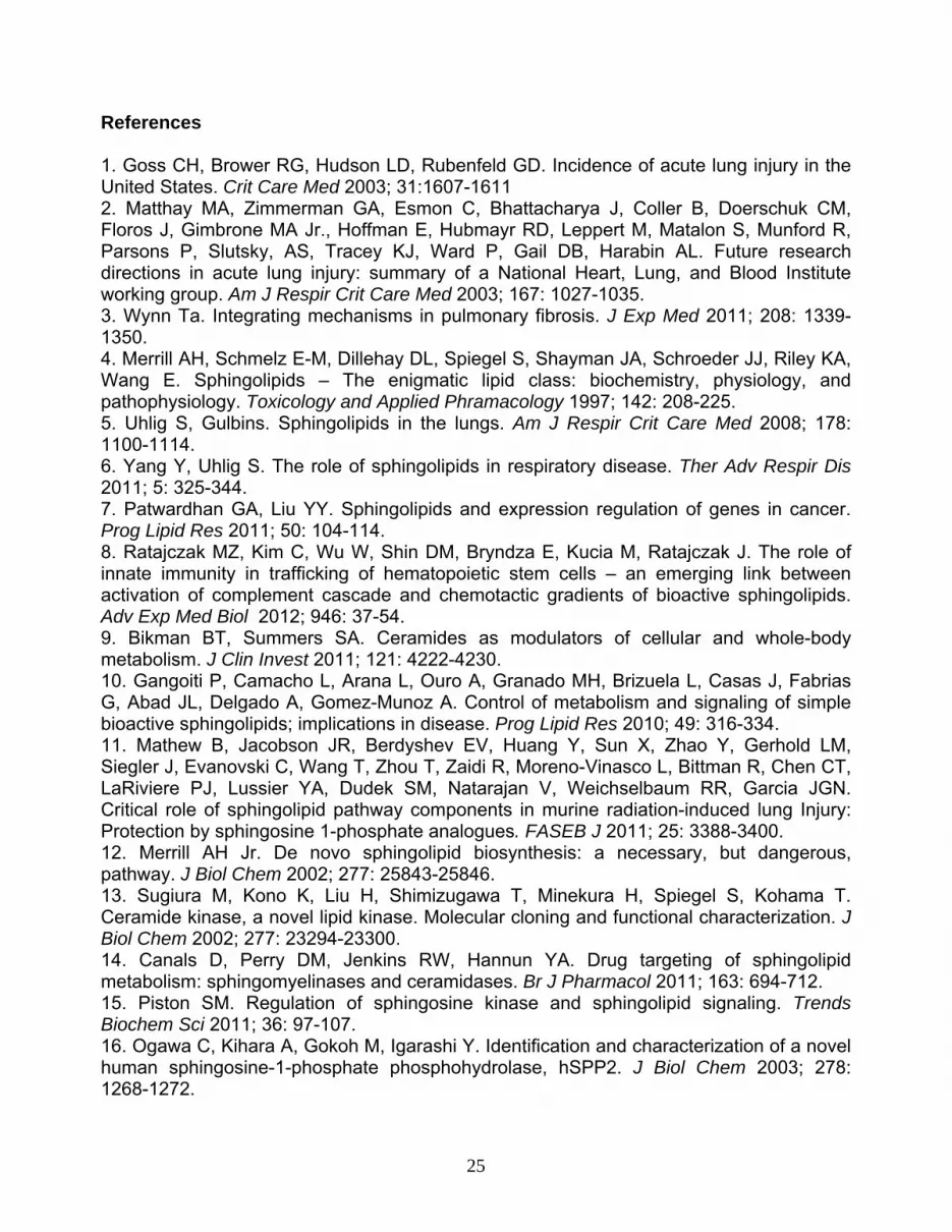
followed by ceramide synthase mediated N-acylation to dihydroceramide, and subsequent
desaturation to ceramide (12). Ceramide is then channeled to complex sphingolipids such
as SM or hydrolyzed to Sph through the action of ceramidases or glycosylated to form
glycosphingolipids (Fig. 1). Ceramide is also phosphorylated by ceramide kinase to Cer1P
(13) [Nati -2002 is not a recent reference]. Mammalian cells do not convert dihydroSph to
Sph; however, Sph is generated from ceramide(s) by ceramidases (14). Sph is
phosphorylated by two Sph kinases, SphK1 and/or SphK2, to S1P (15), and S1P is
dephosphorylated to Sph through the action of specific S1P phosphatases 1 and 2
(S1PPases) (16) or through non-specific lipid phosphate phosphatases (LPPs) (17,18).
Alternatively, S1P is irreversibly cleaved by S1P lyase (S1PL), a pyridoxal phosphate
dependent enzyme, to ethanolamine phosphate and trans-2-hexadecenal. Subsequently,
trans-2-hexadecenal is oxidized by fatty aldehyde dehydrogenase to trans-2-hexadecenoic
acid, which is recycled into glycerolipid or sphingolipid metabolic pathways while
ethanolamine phosphate is utilized for the biosynthesis of ethanolamine phospholipids
(Fig. 1) (19-21). Additionally, in response to TNF-α and other agonists, SM is hydrolyzed
to ceramide(s) of variable N-acyl chain length by one of the three (acid, neutral, alkaline)
sphingomyelinases (SMases) (Fig. 2) (22). Ceramide acts intracellularly and functions as a
second messenger by modulating ceramide-activated protein phosphatases and kinases
(23,24). Thus, the complexity in sphingolipid metabolism enables cells to orchestrate
cellular responses by regulating the inter-conversions via the anabolic and catabolic
enzymes that regulate their intracellular levels and spatio-temporal distribution.
Sphingosine 1-phosphate in vascular permeability

6
S1P is present in plasma and tissues, and the levels of S1P are 3 to 4 times higher in
serum than in plasma (25). The source of plasma S1P is controversial. The initial notion
that platelets are a major source of circulating S1P may be erroneous as erythrocytes (26),
hematopoietic cells (27), and vascular endothelial cells ECs (28) are now known to also
contribute to plasma S1P levels. S1P, initially identified as a mitogen for fibroblasts (29), is
a potent angiogenic factor and plays an essential role in vessel maturation, vascular
permeability, trafficking of T- and B-lymphocytes, and dendritic cells, reproduction, and
central nervous system development (30,31). The ability of platelets to decrease
endothelial barrier permeability (32) may be mediated by S1P stored within the platelets
(33). Pioneering studies identified S1P as a major barrier-protective agent responsible for
maintenance of vascular barrier integrity in vitro and in vivo (33-36). Exogenous addition of
S1P to human and bovine lung ECs increased transendothelial monolayer resistance
(TER) with the barrier-enhancing effect of S1P rapid, dose-dependent, and mediated via
S1P1 (34). Agonists of S1P receptors such as SEW2871, FTY720-P, and FTY720
phosphonates were also effective in enhancing endothelial barrier function (37).
Interestingly, intracellularly released S1P from caged S1P also rapidly and significantly
enhanced the endothelial barrier function (38). However, the above effect of intracellular
S1P was independent of S1P1, but similar to exogenous S1P,in requiring Rac1 (38). This
study delineated an important function for intracellular S1P and enzymes involved in S1P
accumulation in cells in regulating barrier integrity. In fact, the extracellular action of S1P
on endothelial barrier enhancement was dependent on intracellular S1P generation as
blocking or down-regulation of sphingosine kinase (SphK) 1 activity or its forced
expression attenuated S1P-induced barrier enhancement (Natarajan V., unpublished

7
data). In addition to in vitro barrier protective effects, a barrier-regulatory role for S1P in
murine and canine models of ALI was demonstrated. In an isolated perfused murine lung
model, S1P infusion (1 μM) resulted in a significant decrease in the rate of edema
formation without a change in pulmonary artery pressure (39). Similarly, LPS-challenged
mice with lung or renal injury, the magnitude of pulmonary edema was significantly
attenuated by intravenous administration of S1P (39). The ability of S1P to confer
protection against sepsis-induced barrier dysfunction was also confirmed in a canine
model, wherein infusion of S1P reduced bronchoalveolar lavage (BAL) fluid protein
accumulation and alveolar edema as measured by computed tomography (35).
Mechanism(s) of S1P-mediated barrier protection
The mechanism(s) of S1P-mediated regulation of vascular permeability is yet to be fully
defined; however, these events involve activation of its G-protein coupled S1P1 and other
S1P-receptors with subsequent downstream activation of Rac1, cytoskeletal
reorganization, adherens and tight junction assembly, and focal adhesion formation (Fig.
3) (31,34,40). As caged S1P-mediated barrier enhancement is Rac1 dependent (38), it is
possible that S1P may directly interact or bind to Rac1 and induce dissociation of Rho
GDP dissociation inhibitor (RhoGDI) from Rac1 for activation and redistribution to the cell
periphery (41). This suggests that the action of S1P could be similar to another acidic
phospholipid, phosphatidic acid (PA), generated by the phospholipase D signaling
pathway, wherein PA acts as a membrane anchor of Rac1 by interacting with the polybasic
motif in the carboxyl-terminal of Rac1, as shown in OVCAR-3 cells (42).

8
Sphingosine kinases and S1P lyase in sepsis-induced lung injury
The S1P-induced protection of endothelial barrier function in LPS-induced lung injury
suggests a role for S1P-metabolizing enzymes in the lung injury and repair. Circulating and
cellular S1P levels are regulated by its synthesis and catabolism (15,30). The availability of
Sph is the rate-limiting step in the intracellular generation of S1P, and Sph is derived either
from ceramides through ceramidases or from circulating plasma S1P through ecto-LPPs
(9,17,18). Recent studies showed that human lung ECs have the ability to utilize
exogenously added S1P to generate intracellular S1P by lipid phosphate phosphatases
(18). In addition to these two pathways, S1P can also be generated in plasma by
lysophospholipase D/autotaxin-mediated hydrolysis of sphingosylphosphorylcholine (44);
however, it is unclear if this pathway is a major source of plasma S1P. Thus, targeting
SphKs, S1PPases, LPPs, and S1PL represent novel therapeutic approaches with the
potential to minimize or ameliorate lung inflammation and injury.
Role of Sph kinases 1 and 2 in acute- and sub-acute lung injury
The role of SphKs in lung inflammation and injury is somewhat controversial. Loss of
SphK1 or SphK2 expression in mice had no significant effect on inflammatory responses,
and normal neutrophil function was observed in SphK1 and SphK2 knockout mice;
however, accelerated bacterial lung infection in SphK2, but not SphK1, knockout mice as
compared to wild-type controls was observed (45). Inhibition of SphK1 expression using
anti-sense or a SphK inhibitor such as N,N-dimethyl-Sph attenuated neutrophil activation,
chemotaxis and lung permeability (46), and disruption of SphK1 gene in mice had no effect

9
on lymphocyte trafficking and lymphocyte distribution (47). LPS challenge of C57BL/6 wild
type mice differentially up-regulated SphK1 and SphK2 expression levels; however,
SphK1-/- mice were more susceptible to LPS-induced lung injury compared to wild type
mice while over-expression of SphK1 (wild type) delivered by adenoviral vector to lungs
protected SphK1-/- mice from lung injury and blunted the severity of the LPS response (48).
SphK1 was up-regulated in stimulated human phagocytes and in peritoneal phagocytes of
patients with severe sepsis, and inhibition of SphK1 with siRNA protected mice against
sepsis-induced pro-inflammatory responses (49). In this study, LPS challenge or cecal
ligation puncture increased mortality rate in SphK1-/- mice; yet it was suggested that this
phenomenon represents an “adaptive compensation during development” that contradicts
several earlier studies demonstrating an anti-inflammatory role for Sphk1 in ALI (49). As
Sphk1 regulates S1P production, macrophages with increased expression of Sphk1 should
generate higher levels of S1P; however, the reported results in Sphk1 knockdown
experiments are quite the opposite (49). Recently, a novel antimycobacterial role for Sphk1
was reported in macrophages wherein SphK1 knockdown increased sensitivity to
Mycobacterium smegmatis infection (50). In phagocytes, SphK1 regulated C5L2 and CD88
expression and dampened inflammatory responses to endotoxin (51). Therefore, additional
ALI models are necessary to determine whether SphK1 has a protective or pro-
inflammatory role in lung injury and inflammation. In contrast to the LPS-induced lung
injury model, SphK1 deficiency protected mice from hyperoxia-induced lung inflammation
and injury (Natarajan et al., unpublished data). Thus, the role of SphK1 in lung
inflammation and injury might depend on the type of insult and degree of oxidative stress

10
as increased S1P modulates reactive oxygen species (ROS) generation via NADPH
oxidase activation..
In addition to ALI, SphK1 seems to play a role in sub-ALI such as radiation-induced
lung injury (RILI). Thoracic radiation of mice (25 Gy) enhanced expression of SphK1 and 2
in mouse lungs at 6 weeks and increased the ratio of ceramide to S1P and dihydro-S1P in
plasma, bronchoalveolar lavage (BAL) fluid, and lung tissue (11). SphK1-/- mice exhibited
higher susceptibility to RILI at 6 weeks, indicating a protective role for SphK1 against RILI
(11). However, pretreatment with myriocin, an inhibitor of SPT, decreased inflammation
and fibrogenesis at 18 weeks post-irradiation (52). Inhibition of SPT also decreased
radiation-induced SphK activity in the lung, which modulated the levels of S1P/dihydro-
S1P in lung tissue and circulation (52). Interestingly, simvastatin, a 3-hydroxy-3-
methylglutaryl-CoA reductase inhibitor, attenuated RILI in the mouse model by modulating
the expression of SphK and S1PL proteins and the levels of ceramide to S1P/dihydroS1P
in lung tissues (53). These results support the notion that S1P and S1P-metabolizing
enzymes are potential therapeutic targets against RILI.
S1PL deficiency protects against LPS-induced ALI
Current evidence suggests a role for S1PL in normal development, reproduction, cell
survival, cancer, and immunity (19). S1PL function seems to be critical for mammalian
survival as S1P lyase knockout (Sgpl1-/-) mice do not survive beyond a couple of weeks
after birth and exhibit vascular abnormalities (54). S1PL deficiency elevated S1P, Sph,
ceramide, and SM levels in the serum and liver (55), and produced a pro-inflammatory
response by impairing neutrophil trafficking (56). In Sgpl1+/- mice, LPS challenge increased

11
lethality, serum levels of TNF-α, MCP-1, and IL-6, and sphingoid bases compared to
Sgpl1+/+ mice (57), suggesting a detrimental effect of high-circulating S1P levels. Genetic
and chemical inhibition of S1PL with the inhibitors 2-acetyl-4(5)-[1R,2S,3R,4-
tetrahydroxybutyl]-imidazole (THI) and LX2931 increased circulating and tissue S1P levels,
reduced peripheral lymphocytes, and alleviated inflammatory responses in animal models
of autoimmunity (58). Complete deficiency of S1PL in mice resulted in lesions in the lung,
heart, urinary system, and bone as well as T-cell depletion in the blood, thymus, spleen,
and lymph nodes attributed to very high circulating S1P levels (58). However, partial
restoration of S1PL activity in humanized knock-in mice harboring one (Sgpl1H/-) or two
alles (Sgpl1H/H) of human Sgpl1 offered protection from the lethal lymphoid lesions that
developed in Sgpl1-/- mice (58).
A novel role for S1PL and intracellularly generated S1P in protecting against LPS-
induced ALI has been recently demonstrated in vivo and in vitro (59). Intratracheal
instillation of LPS (5 mg/kg) to mice enhanced lung S1PL expression, decreased S1P
levels in lung tissue, and induced lung injury. Sgpl1+/- mice exhibited increased S1P levels
in lung tissue and BAL fluid, with reduced lung injury and inflammation in response to LPS
challenge. Furthermore, reduction of S1PL activity by oral administration of THI showed a
direct correlation between elevated S1P levels in lung tissue and BAL fluid and reduced
levels of neutrophils and IL-6 in mice administered with LPS intratracheally (59). In vitro
treatment of human lung micro vascular ECs with LPS resulted in reduced levels of
intracellular S1P and increased mRNA and protein expression of S1PL. Down-regulation
of S1PL expression by siRNA increased S1P levels in the cells and medium and
attenuated LPS-mediated phosphorylation of p38 MAPK and I-kB; and decreased IL-6

12
secretion, while over-expression of S1PL enhanced LPS-induced phosphorylation of p38
MAPK and I-kB, and enhanced IL-6 secretion. S1PL siRNA was more effective than
exogenous S1P in attenuating LPS-induced IL-6 secretion. Furthermore, S1PL siRNA
attenuated LPS-induced endothelial barrier disruption by inducing activation and
redistribution of Rac-1 to the cell periphery. There is some controversy regarding the role
of S1PL-generated metabolites, namely ethanolamine phosphate and trans-2-hexadecenal
in regulating physiological responses such as mitogenesis, inflammation, and apoptosis. In
mouse F9 embryonic carcinoma and HeLa cells, over-expression of SphK1 enhanced
DNA synthesis; however, in Sgpl1-/- null cells or Sgpl1-/- null cells over-expressing SphK1
there was no effect on mitogenesis, suggesting that the products of S1PL pathway, not
S1P itself, stimulated mitogenesis (60,61). Addition of trans-2-hexadecenal to HEK293,
NIH 3T3, and HeLa cells induced cytoskeletal reorganization and apoptosis that was
dependent on JNK activation and c-Jun phosphorylation (62). In a recent study, trans-2-
hexadecenal was shown to react readily with deoxyguanosine and DNA to form aldehyde-
derived DNA adducts that may have potentially mutagenic consequences or trigger a
previously unrecognized DNA damage response in living cells (63). As both ethanolamine
phosphate and trans-2-hexadecenal were added exogenously, it is unclear if intracellularly
generated metabolites of S1P by S1PL exhibit similar physiological responses.
SM synthase 2 and ALI
Sphingomyelin synthase (SMS) 1 and 2 catalyze transfer of phosphorylcholine from
phosphatidylcholine to ceramide and generate SM, a major component of all mammalian
cell membranes. Recent studies have demonstrated SMS2 deficiency attenuates NF-kB

13
signaling, thereby suggesting a role for ceramide in NF-kB signal transduction (64).
However, ceramide also inhibits NF-kB activation (65) indicating a differential role for
ceramide in NF-kB activation in different cell types. SMS2-/- mice treated with LPS showed
decreased inflammation, cytokine induction, and lung injury compared to SMS2+/+ wild type
control mice (66). Furthermore, SMS2 depletion attenuated LPS-induced p38 MAPK and
JNK activation and transcriptional activity of NF-kB in human pulmonary artery endothelial
cells, suggesting that blocking SM synthesis regulates endotoxin-induced inflammatory
responses in a murine model of ALI. However, the effect of SMS2 knockdown on S1P,
sphingosine and ceramide levels in lung tissue and plasma was not determined (66).
Limitations of S1P therapy in ALI
Although S1P infusion has proven to be beneficial against LPS-induced lung injury in
animal models (35,39), it is an endogenous bioactive lipid that has pleotropic effects; and
some of these effects are likely to limit its usefulness in minimizing ALI. For example,
intravascular administration of S1P decreases the severity of ALI; however, intratracheal
administration can produce pulmonary edema through disruption of the
epithelial/endothelial barrier via ligation of S1P1 or S1P3 (107). In human lung ECs, high
concentrations of S1P (>10 µM) can disrupt EC monolayer integrity in vitro through ligation
of S1P3 and subsequent activation of Rho, suggesting a limited therapeutic window for
S1P in barrier enhancement (31). S1P also exhibits well-described cardiac toxicity
(bradycardia) through activation of S1P3 in the heart (67), directly stimulates contraction of
human airway and bronchial smooth muscle cells (68), and increases airway hyper-

14
responsiveness in allergen-challenged mice (69), suggesting a potential for S1P to
exacerbate airway obstruction in asthmatics.
FTY720 and FTY720-P as barrier protective and anti-inflammatory agents
Given the limitations of S1P as a therapeutic agent in ALI, there has been considerable
interest in the biologic effects of a structurally similar compound, 2-amino-2-(2-[4-
octylphenyl]ethyl)-1,3-propanediol (FTY720) (Fig. 4), a synthetic derivative of the fungal
metabolite myriocin. FTY720 has attracted a great deal of clinical interest as an
immunosuppressive agent, and in fact was approved by FDA in 2010 for treatment of
multiple sclerosis (70). In addition to its immuno-modulatory effects, FTY720 also
decreased vascular permeability both in vivo (39,71), and in vitro (72). For example, a
single i.p. injection of FTY720 significantly attenuated murine pulmonary injury after LPS
administration (35). Interestingly, while lower doses of FTY720 (0.01-1 M) enhanced
endothelial barrier function in HUVECs, higher concentrations of FTY720 (10-100 M)
induced irreversible barrier breakdown and apoptosis (73). Similarly, low concentrations of
FTY720 (0.1 mg/kg) reduced lung permeability in mechanically ventilated mice; however,
higher concentrations (2 mg/kg) increased pulmonary leak and apoptosis in ventilated
mice without affecting permeability in non-ventilated mice (73). As non-phosphorylated
FTY720 has a low affinity for S1P receptors (74), current concepts related to FTY720
mode of action invoke phosphorylation of FTY720 in situ by SphK2 to give the S1P analog
(S)-FTY720-P (75), thereby enhancing the affinity for S1P family of receptors, particularly
S1P1 and S1P3. The phosphorylation of FTY720 to FTY720-P occurs rapidly both in vitro
and in vivo (75), however, ~25% of FTY720 still remains in the non-phosphorylated state in

15
patients (76). Furthermore, FTY720-P reversed VEGF-induced transcellular permeability in
murine embryonic ECs (72). These investigations point out that FTY720-mediated
protection against EC barrier dysfunction and lung inflammation is complex, dose
sensitive, and appears to involve both non-phosphorylated and phosphorylated forms of
FTY720.
Mechanism(s) of barrier regulation by FTY720 and FTY720-P
The mechanism(s) of FTY720 and FTY720-P action in preventing vascular leak remains
unclear. FTY720, unlike S1P, appears to internalize and down regulate S1PR1 signaling
instead of activating this pathway. In contrast to S1P, FTY720 enhances EC barrier
function without rapidly increasing intracellular calcium, cortical actin structure, or requiring
expression of proteins integral to the generation of the cortical actin ring (Rac, cortactin)
(71). Furthermore, these studies suggest that FTY720 enhances EC barrier function via a
novel S1P1-independent mechanism since abrogating S1P1 expression using specific
siRNA failed to block the above effect, while embryonic EC cultured from S1PR1-/- mice
retained the ability to mount a FTY720-induced barrier enhancing response (71). Since
pertussis toxin completely abolished the above effect (71), Gi-protein coupled receptors
(GPCR) appear to play a key role in the above FTY720-conferred EC barrier
enhancement. GPCR is a large and diverse family of receptors with varying homologies to
the S1P receptors that could potentially participate in transducing FTY720 responses (77).
For example, FTY720 interacts with the cannabinoid family of GPCRs (78), but the primary
cannabinoid receptors, CB1 and CB2, are not involved in the EC barrier-enhancing
response (71). In addition, FTY720 (but not S1P) inhibits S1PL (79), cytosolic

16
phospholipase A2 (80), and ceramide synthases (81,82)), and activates protein
phosphatase 2A (83). In human lung ECs, FTY720 increased c-Abl tyrosine kinase activity
and c-Abl siRNA-attenuated FTY720-dependent barrier enhancement (Fig. 5) (84).
However, FTY720 increased protein phosphatase 2A expression failed to alter FTY720-
induced barrier enhancement (84). FTY720 also up-regulated the expression of EC
junctional proteins β-catenin and ZO-1 (72) and promoted adherens junction assembly
(85); however, ECs treated with specific siRNAs against claudin-5 or ZO-1/ZO-2 did not
alter FTY720-induced barrier enhancement, suggesting that adherens junction or tight
junction proteins are not involved in FTY720-induced barrier enhancement (84). A new
paradigm has developed in which FTY720 appears to function as an S1P1 antagonist and
exerts its observed inhibitory effects on lymphocyte circulation (86). In contrast to FTY720,
FTY720-P increased [Ca2+]i in ECs, induced cortical actin distribution to the cell periphery,
and focal adhesion activity via Rac1 (Fig. 5). Further evidence in support of FTY720-
induced down regulation of S1P1 comes from the ability of FTY720-P to induce
ubiquitination and proteosomal degradation of S1P1 in cultured ECs to a greater extent
than S1P (87).
Limitations of FTY720 and FTY720-P as therapeutic agents in ALI
Similar to S1P, FTY720 also exhibits physiological responses that may limit its therapeutic
utility in patients with life-threatening inflammatory diseases such as ALI. Its
immunosuppressant effects may be detrimental to patients with ALI, many of whom have
sepsis as a triggering event (88). FTY720 induces bradycardia through its effect on S1P3,
which is similar to that seen with S1P in animal models and has been confirmed in patients

17
with ALI (88). Further, in a recent multiple sclerosis clinical trial, FTY720 increased rates of
dyspnea and decreased lung function (lowered forced expiratory volume 1) (89), perhaps
mediated through mechanisms similar to those seen in S1P-induced contraction of
bronchial smooth muscle in mice (68). Importantly, FTY720-P induced ubiquitination and
proteosomal degradation of S1P1 in cultured ECs (85) and HEK293 cells (90). Prolonged
exposure of FTY720 resulted in the down regulation of EC surface expression of S1P1 and
decreased response to S1P (91). Administration of FTY720 (0.5-5.0 mg/kg) to wild type
C57BL/6 mice induced a dose-dependent S1P1 degradation and an increase in vascular
permeability (92). This in vivo barrier-disruptive effect of FTY720 is in contrast to its
barrier-protective effect observed in vitro (37,71,84), suggesting differential responses in
mouse and human endothelium. However, the in vivo effect in mice point out a direct link
between S1P1 degradation and vascular leak, which may account for the recent report of
increased lung injury and mortality observed in bleomycin-injured mice receiving prolonged
FTY720 treatment (92). In summary, these studies suggest that FTY720 itself is unlikely to
be an optimal therapeutic agent for ALI.
FTY720 phosphonates and endothelial barrier function
As a result of these limitations of FTY720 and FTY720-P, there is significant interest in
FTY720-P analogs and related compounds that may have fewer side effects. Several
groups have synthesized multiple derivatives of FTY720-P, including phosphonates
(74,93), phosphothioates (94), 4(5)-phenylimidazole-containing (95) and conformationally
constrained analogs (96), primarily for the purposes of characterizing their S1P-receptor
affinity and their ability to induce lymphopenia (97). A number of novel analogs of FTY720-

18
P, namely the (R)- and (S)-enantiomers of FTY720 phosphonates (Fig. 4) (98) have been
partially evaluated in vitro and in vivo for protection against endothelial barrier dysfunction
and pulmonary leak in three murine models of lung injury. The (R)- and (S)-enantiomers of
FTY720 phosphonate and of their unsaturated derivatives, FTY720-vinylphosphonate,, in
contrast to the (R)- and (S)-enantiomers of the FTY720 regioisomers (in which the
positions of the amino group and one of the hydroxyl groups are reversed), enhanced
endothelial barrier integrity in human lung ECs (37). Consistent with these in vitro
responses, in a murine model of lung injury, (S)-FTY720 vinylphosphonate significantly
reduced LPS-induced vascular leak and infiltration of leukocytes into the alveolar space
(37). Similarly, prolonged administration of (S)-FTY720 vinylphosphonate significantly
improved survival of bleomycin-induced mortality in mice (99). In a preclinical model of
RILI, (S)-FTY720 vinylphosphonate, but not FTY720, conferred protection against
radiation-induced pulmonary leak and inflammation (11). In human pulmonary artery
smooth muscle, breast cancer and androgen-independent prostate cancer cells (S)-
FTY720 vinylphosphonate regulated SphK1 activity via induction of proteosomal
degradation of SphK1 (100). Furthermore, (S)-FTY720 vinylphosphonate did not activate
any of the S1P1-5 whereas the R enantiomer was an agonist of S1P1 and a partial
antagonist of S1P2 and S1P5 (101).
Mechanism(s) of barrier protection by FTY720 phosphonates and limitations of
utility in ALI.
Compared to S1P and FTY720, very little is known about the potential mechanisms
underlying barrier protection promoted by FTY720 phosphonates. In vitro, (S)-FTY720

19
phosphonate maintained basal expression of S1P1 in contrast to the significant reduction
(>50%) induced by S1P or FTY720 treatment (90), suggesting inhibition of ubiquitination-
mediated degradation of S1P1 by (S)-FTY720-phosphonate (99). Furthermore, recruitment
of β-arrestin to S1P1 by (S)-FTY720 phosphonate was significantly lower that by S1P or
FTY720 treatment, indicating prolonged signaling through S1P1 by (S)-FTY720
phosphonate (99).
As stated above, FTY720 is a FDA-approved drug for oral treatment of MS. In
contrast to FTY720, studies with FTY720 phosphonates in pre-clinical animal models are
limited and its metabolism in vivo is unknown. Further studies on its cytotoxicity,
pharmacokinetics, and efficacy in animal models are necessary to advance these
phosphonate analogs to phase 1 trial in humans.
S1P receptors in lung injury and barrier regulation
S1P elicits its cellular effects through a family of G protein-coupled S1PR1-5 receptors,
formerly known as endothelial differentiation gene (Edg) receptors, that are expressed in
various cell types including endothelial cells (102). Although these receptors share
significant homology and overlap in their biological functions, distinct receptor sub-type
spatial distribution, coupling to different G-proteins, and differences in receptor-complex
internalization and recycling may provide specificity to each of the S1P receptors (Fig. 6).
Deletion of S1P1 in mice is embryonically lethal (E12.5 and E14.5) (103), while S1PR2 and
S1PR3 deletions appear to have no discernible effect on normal phenotype (104).
However, S1P1/S1P2 double knockout and S1P1/S1P2/S1P3 triple null embryos showed a
more severe vascular phenotype than S1P1 knockout embryos, suggesting that the three

20
S1P receptors cooperate to promote vascular development during embryonic
angiogenesis (105). There is overwhelming evidence for the role of S1P/S1P1 in
preventing vascular leakage induced by many edemagenic agents including LPS in the
lung (35,39,73,90, 92,106,107). Consistent with the barrier-protective role of S1P1,
pretreatment of wild type mice with an S1P1 inverse agonist, SB-649146, or use of S1P1+/-
mice, exhibited reduced S1P/SEW-2871-induced barrier protection after LPS challenge
(107). Similar to the protective role of S1P1, S1P2-null mice and mice with reduced
expression of S1P3 generated by treating with specific siRNA against S1P3 also offered
significant protection against LPS-induced barrier disruption and leak compared to wild
type mice (107). However, S1P2 seems to mediate enhanced vascular permeability in
newborn mice exposed to a hypoxia-induced model of retinopathy (108) and a hydrogen
peroxide-induced model of barrier dysfunction (104). In RILI, the roles of S1P2 and S1P3 in
barrier regulation appear to be conflicting as observed in a preclinical model of LPS-
induced ALI. In a murine RILI model, knock down or reduced expression of S1P1, S1P2,
and S1P3 increased susceptibility to lung injury (11). These results suggest a differential
role for S1P1-3 in the above two models of lung injury due to differential transduction of
signals via multimeric G-proteins (Fig. 7). In addition to genetic engineering of S1P
receptors, S1P-receptor agonists and antagonists could be useful to study the roles of
S1P1-5 in lung inflammation and injury. However, many of the existing agonists such as
SEW2871 and AUY954 for S1P1 exhibit poor water solubility, thereby limiting their use in
animal models of lung inflammation and injury. A systematic study of S1P receptor-
mediated signaling and the study of intracellular targets that regulate S1P levels in

21
vascular cells may provide greater insight into the mechanisms underlying barrier function
disruption seen in acute- and sub-acute- lung injury.
Functional polymorphism of sphingolipid targets and association with inflammatory
lung disorders.
Association studies on functional consequences of single nucleotide polymorphisms
(SNPs) in S1P receptors, SphKs, S1PL, and sphingomyelinase that are linked to acute-
and sub-acute lung injury/inflammation are limited. Our genomic and genetic analyses
focused on both African descent and European descent populations with sepsis/ALI and
asthma. We explored the contribution of polymorphisms in the S1PR1 gene to asthma
susceptibility via a combination of gene resequencing for SNP discovery, case-control
association, functional evaluation of associated SNPs, and protein immunochemistry
studies. Direct DNA sequencing identified multiple novel S1PR1 variants with a promoter
SNP rs2038366 (21557G/T) associated with asthma and ALI in European descent
individuals (109-112). In African descent individuals an association was found for both
asthma and severe asthma for intronic SNP rs3753194 (c.21641170A/G) and for promoter
SNP rs59317557 (2532C/G) with severe asthma. Alleles of the promoter SNPs rs2038366
(21557G/T) and rs59317557 (2532C/G) influenced the activity of a luciferase S1PR1
reporter vector in transfected endothelial cells. These data provide strong support for a role
for S1PR1 gene variants in asthma susceptibility and severity. Similarly, preliminary
studies revealed S1P3 promoter SNPs rs7022797 (-1899 T/G) and rs11137480 (-1785
G/C) in European descent Americans conferred decreased susceptibility of both severe
sepsis and sepsis-induced ALI. Furthermore, S1P3 promoter SNP -1899G, and -1785C or

22
both significantly decreased luciferase promoter activity triggered by TNF-α induced
binding of transcriptional factors CDX1 and EBF1 to S1PR3 promoter (109-112). Thus,
multiple SNPs in S1P3 alter promoter activity and confer susceptibility to sepsis/ALI in
multiethnic population. These initial polymorphism studies suggest a potential association
between the sphingolipid pathway genes and susceptibility to sepsis, ALI and asthma in
African Americans and European Americans that need to be validated with larger groups
of control and patient populations (109-112).
S1P homeostasis in ALI
S1P levels in circulation are higher compared to intracellular concentrations and tightly
regulated by synthesis, secretion, and uptake by different cell types including endothelial
cells. Most likely, circulating S1P helps to maintain endothelial barrier integrity under basal
conditions. However, pathological conditions such as sepsis may alter S1P homeostasis
and offset the critical balance from tight endothelial junctions to barrier dysregulation. In a
murine model of ALI, intratracheal instillation of LPS (5 mg/kg body weight; 24 h) reduced
S1P levels in lung tissue (S1P, fmol/nmol lipid P: Vehicle, 296 + 24; LPS (i.t.), 138 + 16) as
well as in plasma (S1P, fmol/nmol lipid P: Vehicle, 1126 + 36; LPS (i.t.), 825 + 29) that
paralleled increased expression of S1PL in lungs and lung inflammation and injury (59).
Blocking S1PL activity by administration of THI in mice increased S1P levels in lungs
without altering plasma levels and reduced LPS-induced lung inflammation (59). In a
radiation model of RILI, the ceramide/S1P ratios in BAL, plasma, and lung tissues were
significantly increased and remained elevated as RILI progressed throughout the 12 week
period of RILI assessment (11). Intriguingly, plasma levels of S1P from patients with ALI

23
and sepsis were significantly decreased compared to controls; however, there were no
significant differences between African American and Caucasian populations in terms of
association of ALI or sepsis with circulating plasma S1P levels (Garcia et al., unpublished
data). Analysis of S1P levels from control, sepsis, and sepsis-induced ALI patients
revealed a potential correlation between lung injury/edema to decrease in circulating
plasma S1P levels (111). Whether lower plasma S1P levels serve as a potential biomarker
in sepsis and ALI pathologies remains to be confirmed.
Conclusions and future directions
Acute and sub-acute lung injuries share profound increases in vascular permeability as a
key element and common pathway driving increased morbidity and mortality in these
disorders. Recent studies suggest that the bioactive sphingolipid S1P and its receptors,
and enzymes of S1P metabolism are important modulators of lung injury and inflammation.
S1P is a potent angiogenic factor, enhances EC integrity in vitro, and is a robust in vivo
modulator of vascular permeability and alveolar flooding induced by endotoxemia. S1P
levels are regulated by its synthesis catalyzed by SphKs and degradation mediated by
S1PL, S1PPases, and LPPs. In a LPS-induced murine model of lung injury inflammation,
down-regulation of SphK1 potentiated the ALI while knock down of S1PL offered partial
protection, suggesting that S1PL may be a potential therapeutic target in ameliorating
sepsis-induced pulmonary edema. S1P-mediated endothelial responses are driven via G-
protein coupled S1P receptors, and current studies using knock out mice and S1P-
receptor antagonists show that S1P1 is the primary S1P receptor effecting EC barrier
protection while S1P3 and to a lesser extent S1P2 are barrier disruptive. Although S1P has

24
potential beneficial effect in restoring endothelial barrier integrity and suppressing
pulmonary leak due to sepsis, there are limitations to use of S1P as a therapeutic agent in
a clinical setting. Among S1P analogs evaluated for their efficacy in barrier protection
against acute and sub-acute lung injury animal models, (S)- FTY720 phosphonate
produced rapid and increased endothelial barrier function in vitro and decreased LPS- and
radiation-induced lung permeability in vivo. Association studies in Caucasian and African
American ALI cohorts show novel SNPs in S1P receptors and S1P-metabolizing enzymes.
Future studies on functional polymorphisms in sphingolipids pathway genes should provide
new approaches in drug development against ALI.
Although targeting of S1P receptors and metabolizing enzymes is promising in pre-
clinical animal models of sepsis-induced lung injury, the outcome of S1P targeting to
minimize alveolar flooding and pulmonary leak in ALI patients needs to be evaluated.
Development of specific small molecule agonists and antagonists of S1P receptors and
S1P metabolizing enzymes is critical to modulate the endothelial barrier dysfunction. This
would require not only development of selective and potent inhibitors of S1P receptors and
metabolizing enzymes with minimal cytotoxicity but pin-point targeting of these agents to
specific cell type(s) in the lung. Attempts to target S1P receptor(s) and metabolizing
enzyme(s) simultaneously should provide a synergistic approach in conferring protection
against ALI. Furthermore, identification of novel S1P-signaling biomarkers and systems
biology approach will greatly facilitate the development of novel S1P-based therapies for
patients with severe inflammatory lung injury.
25
References
1. Goss CH, Brower RG, Hudson LD, Rubenfeld GD. Incidence of acute lung injury in the United States. Crit Care Med 2003; 31:1607-1611 2. Matthay MA, Zimmerman GA, Esmon C, Bhattacharya J, Coller B, Doerschuk CM, Floros J, Gimbrone MA Jr., Hoffman E, Hubmayr RD, Leppert M, Matalon S, Munford R, Parsons P, Slutsky, AS, Tracey KJ, Ward P, Gail DB, Harabin AL. Future research directions in acute lung injury: summary of a National Heart, Lung, and Blood Institute working group. Am J Respir Crit Care Med 2003; 167: 1027-1035. 3. Wynn Ta. Integrating mechanisms in pulmonary fibrosis. J Exp Med 2011; 208: 1339-1350. 4. Merrill AH, Schmelz E-M, Dillehay DL, Spiegel S, Shayman JA, Schroeder JJ, Riley KA, Wang E. Sphingolipids – The enigmatic lipid class: biochemistry, physiology, and pathophysiology. Toxicology and Applied Phramacology 1997; 142: 208-225. 5. Uhlig S, Gulbins. Sphingolipids in the lungs. Am J Respir Crit Care Med 2008; 178: 1100-1114. 6. Yang Y, Uhlig S. The role of sphingolipids in respiratory disease. Ther Adv Respir Dis 2011; 5: 325-344. 7. Patwardhan GA, Liu YY. Sphingolipids and expression regulation of genes in cancer. Prog Lipid Res 2011; 50: 104-114. 8. Ratajczak MZ, Kim C, Wu W, Shin DM, Bryndza E, Kucia M, Ratajczak J. The role of innate immunity in trafficking of hematopoietic stem cells – an emerging link between activation of complement cascade and chemotactic gradients of bioactive sphingolipids. Adv Exp Med Biol 2012; 946: 37-54. 9. Bikman BT, Summers SA. Ceramides as modulators of cellular and whole-body metabolism. J Clin Invest 2011; 121: 4222-4230. 10. Gangoiti P, Camacho L, Arana L, Ouro A, Granado MH, Brizuela L, Casas J, Fabrias G, Abad JL, Delgado A, Gomez-Munoz A. Control of metabolism and signaling of simple bioactive sphingolipids; implications in disease. Prog Lipid Res 2010; 49: 316-334. 11. Mathew B, Jacobson JR, Berdyshev EV, Huang Y, Sun X, Zhao Y, Gerhold LM, Siegler J, Evanovski C, Wang T, Zhou T, Zaidi R, Moreno-Vinasco L, Bittman R, Chen CT, LaRiviere PJ, Lussier YA, Dudek SM, Natarajan V, Weichselbaum RR, Garcia JGN. Critical role of sphingolipid pathway components in murine radiation-induced lung Injury: Protection by sphingosine 1-phosphate analogues. FASEB J 2011; 25: 3388-3400. 12. Merrill AH Jr. De novo sphingolipid biosynthesis: a necessary, but dangerous, pathway. J Biol Chem 2002; 277: 25843-25846. 13. Sugiura M, Kono K, Liu H, Shimizugawa T, Minekura H, Spiegel S, Kohama T. Ceramide kinase, a novel lipid kinase. Molecular cloning and functional characterization. J Biol Chem 2002; 277: 23294-23300. 14. Canals D, Perry DM, Jenkins RW, Hannun YA. Drug targeting of sphingolipid metabolism: sphingomyelinases and ceramidases. Br J Pharmacol 2011; 163: 694-712. 15. Piston SM. Regulation of sphingosine kinase and sphingolipid signaling. Trends Biochem Sci 2011; 36: 97-107. 16. Ogawa C, Kihara A, Gokoh M, Igarashi Y. Identification and characterization of a novel human sphingosine-1-phosphate phosphohydrolase, hSPP2. J Biol Chem 2003; 278: 1268-1272.

26
17. Brindley DN, Pilquil C. Lipid phosphate phosphatases and signaling. J Lipid Res 2009; 50 Suppl: S225-S230. 18. Zhao Y, Usatyuk PV, Gorshkova I, He D, Watkins T, Garcia JGN, Saatian B, Brindley DN, Bittman R, Berdyshev EV, Natarajan V. Regulation of intracellular generation of sphingosine-1-phosphate by lipid phosphate phosphatase-1 and sphingosine kinase 1 in human lung endothelial cells. J Biol Chem 2007; 282: 14165-14177. 19. Bandhuvula P, Saba JD. Sphingosine-1-phosphate lyase in immunity and cancer: silencing the siren. Trends Mol Med 2007; 13: 210-217. 20. Berdyshev EV, Gorshkova I, Usatyuk P, Kalari S, Zhao Y, Pyne NJ, Pyne S, Sabbadini RA, Garcia JGN, Natarajan V. Intracellular S1P generation is essential for S1P-induced motility of human lung endothelial cells: role of sphingosine kinase 1 and S1P lyase. PLoS One 2011; 6: e16571. 21. Kariya Y, Kihara A, Ikeda M, Kikuchi F, Nakamura S, Hashimoto S, Choi CH, Lee YM, Igarashi Y. Products by the sphingosine kinase/sphingosine 1-phosphate (S1P) lyase pathway but not s1p stimulate mitogenesis. Genes Cells 2005;10:605-615. 22.Perrotta C, Clementi E. Biological roles of acid and neutral sphingomyelinases and their regulation by nitric oxide. Physiology(Bethesda) 2010; 25: 64-71. 23. Dobrowsky RT, Kamibayashi C, Mumby MC, Hannun YA. Ceramide activates heterotrimeric protein phosphatase 2A. J Biol Chem 1993; 268: 15523-15530. 24. Westwick JK, Bielawska AE, Dbaibo G, Hannun YA, Brenner DA. Ceramide activates the stress-activated protein kinases. J Biol Chem 1995; 270: 22689-22692. 25. Yatomi Y. Plasma sphingosine-1-phosphate metabolism and analysis. Biochim Biophys Acta 2008; 1780: 606-611. 26. Hanel P, Andreani P, Graler MH. Erythrocytes store and release sphingosine 1-phosphate in blood. FASEB J 2007; 21: 1202-1209. 27. Tani M, sano T, Ito M, Igarashi Y. Mechanisms of sphingosine and sphingosine 1-phosphate generation in human platelets. J Lipid Res 2005; 46: 2458-2467. 28. Venkataraman K, Lee YM, Michaud J, Thangada S, Ai Y, Bonkovsky HL, Parikh NS, Habrukowich C, Hla T. Vascular endothelium as a contributor of plasma sphingosine 1-phosphate. Circ Res 2008; 102: 669-676. 29. Chung T, Crilly KS, Anderson WH, Mukherjee JJ, Kiss Z. ATP-dependent choline phosphate-induced mitogenesis in fibroblasts involves activation of pp70 S6 kinase and phosphatidylinositol 3’-kinase through an extracellular site. Synergistic mitogenic effects of choline phosphate and sphingosine 1-phosphate. J Biol Chem 1997; 272: 3064-3072. 30. Pyne S, Pyne NJ. Sphingosine 1-phosphate signalling in mammalian cells. Biochem J 2000; 349: 385-402. 31. Wang L, Dudek SM. Regulation of vascular permeability by sphingosine 1-phosphate. Microvasc Res 2009; 77: 39-45. 32. Gimbrone MA Jr, Aster RH, Cotran RS, Corkerry J, Jandl JH, Folkman J. Preservation of vascular integrity in organs perfused in vitro with a platelet-rich medium. Nature 1969; 222: 33-36. 33. Schaphorst KL, Chiang E, Jacobs KN, Zaiman A, Natarajan V, Wigley F, Garcia JG. Role of sphingosine 1-phosphate in the enhancement of endothelial barrier integrity by platelet-released products. Am J Physiol Lung Cell Mol Physiol 2003; 285: L258-L267.
27
34. Garcia JG, Liu F, Verin AD, Birukova A, Dechert MA, Gerthoffer WT, Bamburg JR, English D. Sphingosine 1-phosphate promotes endothelial barrier integrity by Edg-dependent cytoskeletal rearrangement. J Clin Invest 2001; 108: 689-701. 35. McVerry BJ, Peng X, Hassoun PM, Sammani S, Simon BA, Garcia JG. Sphingosine 1-phosphate reduces leak in murine and canine models of acute lung injury. Am J Respir Crit Care Med 2004; 170: 987-993. 36. Okazaki M, Kreisel F, Richardson SB, Kreisel D, Krupnick AS, Patterson GA, Gelman AE. Sphingosine 1-phosphate inhibits ischemia reperfusion injury following experimental lung transplantation. Am J Transplant 2007; 7: 751-758. 37. Camp SM, Bittman R, Chiang ET, Moreno-Vinasco L, Mirzapoiazova T, Sammani S, Lu X, Sun C, Harbeck M, Roe M, Natarajan V, Gracia JG, Dudek SM. Synthetic analogs of FTY720 [2-amino-2-(2-[4-octylphenyl]ethyl)-1,3-propanediol] differentially regulate pulmonary vascular permeability in vivo and in vitro. J Pharmacol Exp Ther 2009; 331: 54- 38. Usatyuk PV, He D, Bindokas V, Berdyshev EV, Garcia JGN, Natarajan V. Photolysis of intracellular caged sphingosine-1-phosphate causes G-protein dependent and independent activation of cell signaling pathways in endothelial cells. Am. J. Physiology, Lung Cell Mol Physiol 2011; 300: L840-850. 39. Peng X, Hassoun PM, Sammani S, McVerry BJ, Burne MJ, Rabb H, Pearse D, Tuder RM, Garcia JG. Protective effects of sphingosine 1-phosphate in murine endotoxin-induced inflammatory lung injury. Am J Respir Crit Care Med 2004; 169: 1245-1251. 40. Jacobson JR, Garcia JG. Novel therapies for microvascular permeability in sepsis. Curr Drug Targets 2007; 8: 509-514. 41.Ugolev Y, Berdichevsky Y, Weinbaum C, Edgar P. Dissociation of Rac1(GDP)oRhoGDI complexes by the cooperative action of anionic liposomes containing phosphatidylinositol 3,4,5-trisphosphate, Rac guanine nucleotide exchange factor, and GTP. J Biol Chem 2008; 283: 22257-22271. 42. Chae YC, Kim JH, Kim KL, Kim HW, Lee HY, Heo WD, Meyer T, Suh P-G, Ryu SH. Phospholipase D activity regulates integrin-mediated cell spreading and migration by inducing GTP-Rac translocation to the plasma membrane. Mol Biol Cell 2008; 19: 3111-3123. 43. Van Veldhoven PP. Sphingosine-1-phosphate lyase. Methods Enzymol 2000; 311:244-254. 44. Nakanaga K, Hama K, Aoki J. Autotaxin – an LPA producing enzyme with diverse functions. J Biochem 2010; 148: 13-24. 45. Zemann B, Urtz N, Reuschel R, Mechtcheriakova D, Bornancin F, Badegruber, R., Baumruker, T., and Billich, A. Normal neutrophil functions in sphingosine kinase type 1 and 2 knockout mice. 2007. Immunol Lett, 109, 56-63. 46. Vlasenko LP, Melendez AJ. A critical role for sphingosine kinase in anaphylatoxin-induced neutropenia, peritonitis, and cytokine production in vivo. J Immunol 2005; 174: 6456-6461. 47. Allende ML, Sasaki T, Kawai H, Olivera A, Mi Y, van Echten-Deckert G, Hajdu R, Rosenbach M, Keohane CA, Mandala S, Spiegel S, Proia RL. Mice deficient in sphingosine kinase 1 are rendered lymphopenic by FTY720. J Biol Chem 2004; 279: 52487-52492.
28
48. Wadgaonkar R, Patel V, Grinkina N, Romano C. Liu J, Zhao Y, Sammani S, Garcia JGN, Natarajan V. Differential regulation of sphingosine kinases 1 and 2 in lung injury. Am J Physiol Lung Cell Mol Physiol. 2009; 296, L603-L613. 49. Puneet P, Yap CT, Wong L, Lam Y, Koh DR, Moochhala S, Pfeilschifter J, Huwiler A, Melendez AJ. SphK1 regulates pro-inflammatory responses associated with endotoxin and polymicrobial sepsis. Science 2010; 328: 1290-1294. 50. Prakash H, Luth A, Grinkina N, Holzer D, Wadgaonkar R, Gonzalez AP, Anes E, Kleuser B. Sphingosine kinase-1 (SphK-1) regulates mycobacterium smegmatis infection in macrophages. PLoS ONE 2010; 5: e10657. 51. Bachmaier K, Guzman E, Kawamura T, Gao X, Malik AB. Sphingosine kinase 1 mediation of expression of the anaphylatoxin receptor C5L2 dampens the inflammatory response to endotoxin. PLoS ONE 2012; 7: e30742. 52. Gorshkova I, Zhou T, Mathew B, Jacobson JR, Takekoshi D, Bhattacharya P, Smith B, Aydogan B, Weichselbaum RR, Natarajan V, Garcia JGN, and Berdyshev EV. Inhibition of serine palmitoyltransferase delays the onset of radiation-induced pulmonary fibrosis through the negative regulation of sphingosine kinase-1 expression. J Lipid Res 2011; 53: 1553-1568. 53. Mathew B, Huang Y, Jacobson JR, Berdyshev E, Gerhold LM, Wang T, Moreno-Vinasco L, Lang G, Zhao Y, Chen CT, Lariviere PJ, Mauceri H, Sammani S, Husain AN, Dudek SM, Natarajan V, Lussier YA, Weichselbaum, RR, and Garcia JGN Simvastatin attenuates radiation-induced murine lung injury and Dysregulated lung gene expression. Am J Respire Cell Mol Biol 2011;, 44: 415-422. 54. Schmahl J, Raymond CS, Soriano P. PDGF signaling specificity is mediated through multiple immediate early genes. Nat Genet 2007; 39: 52-60. 55. Bektas M, Allende ML, Lee BG, Chen W, Amar MJ, Remaley AT, Saba JD, Proia RL. Sphingosine 1-phosphate lyase deficiency disrupts lipid homeostasis in liver. J Biol Chem 2010; 285: 10880-10889. 56. Allende ML, Bektas M, Lee BG, Bonifacino E, Kang J, Tuymetova G, Chen W, Saba JD, Proia RL. Sphingosine 1-phosphate deficiency produces a pro-inflammatory response while impairing neutrophil trafficking. J Biol Chem 2011; 286: 7348-7358. 57. Oravecz T, Donoviel MS, Anderson SJ, Carson K, Sun W, Swaffield J, Liu Q, Kimball SD, Piggott JR, Zambrowicz BP, Sanda AT, Alexander C, Augeri DJ. Genetic and chemical inhibition of sphingosine 1-phosphate lyase results in peripheral lymphopenia and alleviates disease development in animal models of inflammation and autoimmunity. Blood 2007; 110: Abstract 2292. 58. Vogel P, Donoviel MS, Read R, Hansen GM, Hazlewood J, Anderson SJ, Sun W, Swaffield J, Oravecz T. Incomplete inhibition of sphingosine 1-phosphate lyase modulates immune system function yet prevents early lethality and non-lymphoid lesions. PLoS ONE 2009; 4: e4112. 59. Zhao Y, Gorshkova IA, Berdyshev E, Ma W, He D, Su Y, Usatyuk PV, Pendyala S, Fu P, Oskouian B, Saba JD, Garcia JGN, Natarajan V. Protection of LPS-induced murine acute lung injury and inflammation by sphingosine-1-phosphate lyase suppression. Am J Respir Cell Mol Biol 2012;, 45: 426-435. 60. Kariya Y, Kihara A, Ikeda M, Kikuchi F, Nakamura S, Hashimoto S, Choi C H, Lee Y M, Igarashi Y. Products by the sphingosine kinase/sphingosine 1-phosphate (S1P) lyase pathway but not S1P stimulate mitogenesis. Genes Cells 2005; 10: 605-615.

29
61. Kano-Sueoka T, Cohen DM, Yamaizumi Z, Nishimura S, Mori M, Fujiki H. Phosphoethanolamine as a growth factor of mammary carcinoma cell line of rat. Proc Natl Acad Sci USA 1979; 76: 5741-5744. 62. Kumar A, Byun H-S, Bittman R, Saba JD. The sphingolipid degradation product trans-2-hexadecenal induces cytoskeletal reorganization and apoptosis in a JNK-dependent manner. Cell Signal 2011; 23: 1144-1152. 63. Upadhyaya P, Kumar A, Byun H-S, Bittman R, Saba JD. The sphingolipid degradation product trans-2-hexadecenal forms adducts with DNA. Biochem Biophys Res Commun 2012; 424: 18-21. 64. HailemariamTK, Huan C, Li J, Roman C, Kalbfeisch M, Bui HH, Peake DA, Kuo M-S, Cao G, Wadgaonkar R, Jiang X-C. Sphingomyelin synthase 2 deficiency attenuates NF-kB activation. Arterioscler Thromb Vasc Biol 2008; 28: 1519-1526. 65. Gamard CJ, Dbaibo GS, Liu B, Obeid LM, Hannun YA. Selective involvement of ceramide in cytokine-induced apoptosis. Ceramide inhibits phorbol ester activation of nuclear factor kappaB. J Biol Chem 1997; 272: 16474-16481. 66. Gowda S, Yeang C, Wadgaonkar S, Anjum F, Grinkina N, Cutaia M, Jiang X-C, Wadgaonkar R. Sphingomyelin synthase 2 (SMS2) deficiency attenuates LPS-induced lung injury. Am J Physiol Lung Cell Mol Physiol 2010; 300: L430-L440. 67. Forrest M, Sun SY, Hajdu R, Bergstrom J, Card D, Doherty G, Hale J, Keohane C, Meyers C, Milligan J, Mills S, Nomura N, Rosen H, Rosenbach M, Shei GJ, Singer II, Tian M, West S, White V, Xie J, Proia RL, Mandala S. Immune cell regulation and cardiovascular effects of sphingosine 1-phosphate receptor agonists in rodents are mediated via distinct receptor subtypes. J Pharmacol Exp Ther 2004; 309: 758-768. 68. Rosenfeldt HM, Amrani Y, Watterson KR, Murthy KS, Panettieri RA Jr, Spiegel S. Sphingosine-1-phosphate stimulates contraction of human airway smooth muscle cells. FASEB J 2003; 17: 1789-1799. 69. Roviezzo F, Lorenzo A, Bucci M, Brancaleone V, Vellecco V, De Nardo M, Orlotti D, De Palma R, Rossi F, D’Agostino B, Cirino G. Sphingosine-1-phosphate/sphingosine kinase pathway is involved in mouse airway hyperresponsiveness. Am J Respir Cell Mol Biol 2007; 36: 757-762. 70. Strader CR, Pearce CJ, Oberlies NH. Fingolimod (FTY720): a recently approved multiple sclerosis drug based on a fungal secondary metabolite. J Nat Prod 2011; 74: 900-907. 116. 71. Sanchez T, Estrada-Hernandez T, Paik JH, Wu MT, Venkataraman K, Brinkmann V, Claffey K, Hla T. Phosphorylation and action of the immunomodulator FTY720 inhibits vascular endothelial cell growth factor-induced vascular permeability. J Biol Chem 2003; 278: 47281-47290. 72. Dudek SM, Camp SM, Chiang ET, Singleton PA, Usatyuk PV, Zhao Y, Natarajan V, Garcia JG. Pulmonary endothelial cell barrier enhancement by FTY720 does not require the S1P1 receptor. Cell Signal 2007; 19: 1754-1764. 73. Muller HC, Hocke AC, Hellwig K, Gutbier B, Peters H, Schnorock SM, Tschernig T, Schmiedl A, Hippenstiel S, N’Guessan PD, Rossean S, Suttrop N, Witzenrath M. The sphingosine-1-phosphate receptor agonist FTY720 dose dependently affected endothelial integrity in vitro and aggravated ventilator-induced lung injury in mice. Pulm Pharmacol Ther 2011; 24: 377-385.

30
74. Mandala S, Hajdu R, Bergstrom J, Quackenbush E, Xie J, Milligan J, Thornton R, Shei GJ, Card D, Keohane C, Rosenbach M, Hale J, Lynch CL, Rupprecht K, Parsons W, Rosen H. Alteration of lymphocyte trafficking by sphingosine-1-phosphate receptor agonists. Science 2002; 296: 346-349. 75. Billich A, Bornancin F, Devay P, Mechtcheriakova D, Urtz N, Baumruker T. Phosphorylation of the immunomodulatory drug FTY720 by sphingosine kinases. J Biol Chem 2003; 278: 47408-47415. 76. Kovarik JM, Schmouder RL, Slade AJ. Overview of FTY720 clinical pharmacokinetics and pharmacology. Ther Drug Monit 2004; 26: 585-587. 77. Takeda S, Kadowaki S, Haga T, Takaesu H, Mitaku S. Identification of G protein-coupled receptor genes from the human genome sequence. FEBS Lett 2002; 520: 97-101. 78. Paugh SW, Cassidy MP, He H, Milstien S, Sim-Selley LJ, Spiegel S, Selley DE. Sphingosine and its analog, the immunosuppressant 2-amino-2-(2-[4-octylphenyl]ethyl)-1,3-propanediol, interact with the CB1 cannabinoid receptor. Mol Pharmacol 2006; 70: 41-50. 79. Bandhuvula P, Tam YY, Oskouian B, Saba JD. The immune modulator FTY720 inhibits sphingosine-1-phosphate lyase activity. J Biol Chem 2005; 280: 33697-33700. 80. Payne SG, Oskeritzian CA, Griffiths R, Subramanian P, Barbour SE, Chalfant CE, Milstien S, Spiegel S. The immunosuppressant drug FTY720 inhibits cytosolic phospholipase A2 independently of sphingosine-1-phosphate receptors. Blood 2007; 109: 1077-1085. 81. Berdyshev EV, Gorshkova I, Skobeleva A, Bittman R, Lu X, Dudek SM, Mirzapoiazova T, Garcia JG, Natarajan V. FTY720 inhibits ceramide synthases and upregulates dihydrosphingosine-1-phosphate formation in human lung endothelial cells. J Biol Chem, 2009; 284: 5467-5477. 82. Lahiri S, Park H, Laviad EL, Lu X, Bittman R, Futerman AH. Ceramide synthesis is modulated by the sphingosine analog FTY720 via a mixture of uncompetitive and noncompetitive inhibition in an acyl-CoA chain length-dependent manner. J Biol Chem 2009; 284: 16090-16098. 83. Roberts KG, Smith AM, McDougal F, Carpenter H, Horan M, Neviani P, Powell JA, Thomas D, Guthridge MA, Perrotti D, Sim ATR, Ashman LK, Verrills NM. Essential requirement for PP2A inhibition by the Oncogenic receptor c-KIT suggests PP2A reactivation as a strategy to treat c-KIT+ cancers. Cancer Res 2010; 70: 5438-5447. 84. Wang L, Chiang ET, Simmons JT, Garcia JGN, Dudek SM. FTY720-induced human pulmonary endothelial barrier enhancement is mediated by c-Abl. Eur Respir J 2011; 38: 78-88. 85. Singer, II, Tian M, Wickham LA, Lin J, Matheravidathu SS, Forrest MJ, Mandala S, Quackenbush EJ. Sphingosine-1-phosphate agonists increase macrophage homing, lymphocyte contacts, and endothelial junctional complex formation in murine lymph nodes. J Immunol 2005; 175: 7151-7161. 86. Wei SH, Rosen H, Matheu MP, Sanna MG, Wang S-K, Jo E, Wong C-H, Parker I, Cahalan MD. Sphingosine 1-phosphate type 1 receptor agonism inhibits transendothelial migration of medullary T cells to lymphatic sinuses. Nat Immunol 2005; 6: 1228-1235. 87. Oo ML, Thangada S, Wu M-T, Liu CH, Macdonald TL, Lynch KR, Lin C-Y, Hla T. Immunosuppressive and anti-angiogenic sphingosine 1-phosphate receptor-1 agonists

31
induce ubiquitinylation and proteasomal degradation of the receptor. J Biol Chem 2007; 282: 9082-9089. 88. Wheeler AP, Bernard GR. Acute lung injury and the acute respiratory distress syndrome: a clinical review. Lancet 2007; 369: 1553-1564. 89. Kappos L, Antel J, Comi G, Montalban X, O’Connor P, Polman CH, Haas T, Korn AA, Karlsson G, Radue EW., Oral fingolimod (FTY720) for relapsing multiple sclerosis. N Engl J Med 2006; 355: 1124-1140. 90. Oo ML, Chang S-H, Thangada S, Wu M-T, Rezaul K, Blaho V, Hwang S-I, Han DK, Hla T. Engagement of S1P1-degradative mechanisms leads to vascular leak in mice. J Clin Invest 2011; 121: 2290-2300. 91. Krump-Konvalinkova V, Chwalla I, Siess W. FTY720 inhibits S1P-mediated endothelial healing: relationship to S1P1-receptor surface expression. Biochem Biophys Res Commun 2008; 370: 603-608. 92. Shea BS, Brooks SF, Fontaine BA, Chun J, Luster AD, Tager AM. , Prolonged S1P1 Agonist Exposure Exacerbates Vascular Leak, Fibrosis, and Mortality after Lung Injury. Am J Respir Cell Mol Biol. 2010: 43: 662-673. 93. Hale JJ, Neway W, Mills SG, Hajdu R, Keohane CA, Rosenbach M, Milligan J, Shei G-J, Chrebt G, Bergstrom J, Card D, Koo GC, Koprak SL, Jackson JJ, Rosen H, Mandala S. Potent S1P receptor agonists replicate the pharmacologic actions of the novel immune modulator FTY720. Bioorg Med Chem Lett 2004; 14: 3351-3355. 94. Foss FW, Clemens JJ, Davis MD, Snyder AH, Zigler MA, Lynch KR, Macdonald TL. Synthesis, stability, and implications of phosphothioate agonists of sphingosine-1-phosphate receptors. Bioorg Med Chem Lett 2005; 15: 4470-4474. 95. Clemens JJ, Davis MD, Lynch KR, Macdonald TL. Synthesis of 4(5)-phenylimidazole-based analogues of sphingosine-1-phosphate and FTY720: discovery of potent S1P1 receptor agonists. Bioorg Med Chem Lett 2005; 15: 3568-3572. 96. Zhu R, Snyder AH, Kharel Y, Schaffter L, Sun Q, Kennedy PC, Lynch KR, Macdonald TL. Asymmetric synthesis of conformationally constrained fingolimod analogues--discovery of an orally active sphingosine 1-phosphate receptor type-1 agonist and receptor type-3 antagonist. J Med Chem 2007; 50: 6428-6435. 97. Tarrason G, Auli M, Mustafa S, Dolgachev MT, Prats N, Dominguez M, Lopez R, Aguilar N, Calbet M, Pont M, Milligan G, Kunkel SL, Godessart N. The sphingosine-1-phosphate receptor-1 antagonist, W146, causes early and short-lasting peripheral blood lymphopenia in mice. Int Immunopharmacol 2011; 11: 1773-1779. 98. Lu X, Sun C, Valentine WJ, E S, Liu J, Tigyi G, Bittman R. Chiral vinylphosphonate and phosphonate analogues of the immunosuppressive agent FTY720. J Org Chem 2009; 74: 3192-3195. 99. Wang L, Sammani S, Moreno-Vinasco L, Camp SM, Bittman R, Garcia JGN, Dudek SM. FTY720 S-phosphonate exhibits superior barrier promoting properties to S1P and FTY720 in vitro and in vivo. Am J Respir Crit Care Med 2011; 183: A1954. 100. Tonelli F, Lim KG, Loveridge C, Long J, Piston SM, Tigyi G, Bittman R, Pyne S, Pyne NJ. FTY720 and (S)-FTY720 vinylphosphonate inhibit sphingosine kinase 1 and promote its proteosomal degradation in human pulmonary artery smooth muscle, breast cancer and androgen-independent prostate cancer. Cell Signal 2010; 22: 1536-1542. 101. Valentine WJ, Kiss GN, Liu J, Shuyu E, Gotoh M, Murakami-Murofushi K, Pham TC, Baker DL, Parrill AL, Lu X, Sun C, Bittman R, Pyne NJ, Tigyi G. (S)-FTY720-

32
vinylphosphonate, an analogue of the immunosuppressive agent FTY720, is a pan-antagonist of sphingosine 1-phosphate GPCR signaling and inhibits autotaxin activity. Cell Signal 2010; 22: 1543-1553. 102. Hla T. Sphingosine 1-phosphate receptors. Prostaglandins & Other Lipid Mediators 2001; 64: 135-142. 103. Liu Y, Wada R, Yamashita T, Mi Y, Deng CX, Hobson JP, Rosenfeldt HM, Nava VE, Chae SS, Lee MJ, Liu CH, Hla T, Spiegel S, Proia RL. Edg-1, the G protein-coupled receptor for sphingosine 1-phosphate, is essential for vascular maturation. J Clin Invest 2000; 106: 951-961. 104. Kono M, Allende ML, Proia RL. Sphingosine 1-phosphate regulation of mammalian development. Biochim Biophys Acta 2008; 1781; 435-441. 105. Kono M, Mi Y, Liu Y, Sasaki T, Allende ML, Wu Y-P, Yamashita T, Proia RL. The sphingosine-1-phosphate receptors S1P1, S1P2 , and S1P3 function coordinately during embryonic angiogenesis. J Biol Chem 2004; 279: 29367-29373. 106. Sanchez T, Skoura A, Wu MT, Casserly B, Harrington EO, Hla T. Induction of vascular permeability by the sphingosine-1-phosphate receptor-2 (S1P2R) and its downstream effectors ROCK and PTEN. Arterioscler Thromb Vasc Biol 2007; 27: 1312-1318. 107. Sammani S, Moreno-Vinasco L, Mirzapoiazova T, Singleton PA, Chiang ET, Evenoski CL, Wang T, Mathew B, Husain A, Moitra J, Sun X, Nunez L, Jacobson JR, Dudek SM, Natarajan V, Garcia JG. Differential effects of sphingosine 1-phosphate receptors on airway and vascular barrier function in murine lung. Am J Respir Cell Mol Biol 2010; 43: 394-402. 108. Skoura A, Sanchez T, Claffey K, Mandala SM, Proia RL, Hla T. essential role of sphingosine 1-phopsphate receptor 2 in pathological angiogenesis of the mouse retina. J Clin Invest 2007; 117: 2506-2516. 109. Sun X, Ma S-F, Wade MS, Moitra J, Garcia JGN. Two functional promoter variants of sphingosine-1-phosphate receptor 3 gene decrease human susceptibility to severe sepsis-asociated acute lung injury. Am J Respir Cell Mol Biol 2011; 183: A1345. 110. Ma S-F, Wade MS, Hysi P, Yates CR, Meduri GU, Zhao Y, Garcia JG, Natarajan V. Sphingosine kinase 1 gene polymorphisms are associated with acute lung injury in African and European Americans. Am J Respir Cell Mol Biol 2011; 183: A3875. 111. Petrache I, Goya J, Rush NI, Van DeMark BS, Kamocki K, Petrusca DN, Twigg III HL, Garcia JGN, Ma S, Natarajan V, Shen C, Berdyshev E. Ceramide and Sphingosine 1-phosphate (S1P) levels in patient with COPD. . Am J Respir Cell Mol Biol 2010; 181; A1539. 112. Sun S, S Ma, M Wade, C Flores, M Pino-Yanes, J Moitra, C Ober, R Kittles, Husain A, Ford J, Garcia, JGN. Functional variants of the sphingosine-1-phosphate receptor 1 gene associate with asthma susceptibility. J Allergy Clin Immunol 2010; 126: 241-249.

33
Figure Legends
Figure 1: Metabolism of sphingolipids in mammalian cells. Key enzymatic steps on
the biosynthesis and degradation of sphingoid bases and recycling of trans-2-hexadecenal
and ethanolamine phosphate from sphingolipids into glycerophospholipids are
summarized. SPT, serine palmitoyltransferase; SMase, sphingomyelinase; S1P,
sphingosine 1-phosphate; SphK, sphingosine kinase; SPP, S1P phosphatases; LPP, lipid
phosphate phosphatase
Figure 2: Intracellular generation and catabolism of S1P. Ceramide generated by
agonist-dependent hydrolysis of SM by SMase is converted to sphingosine by
ceramidases. Sphingosine is phosphorylated by SphK1 and/or SphK2 to S1P, and either
transported outside the cell or catabolized to trans-2-hexadecenal and ethanolamine
phosphate by S1P lyase. SMase, sphingomyelinase; S1P, sphingosine 1-phosphate
Figure 3: Regulation of endothelial barrier function by S1P. S1P binding to G-protein
coupled S1P1 activates Rac1 and induces a series of signaling cascades including
cytoskeletal reorganization, assembly of adherens junction and tight junction proteins, and
formation of focal adhesions that act together to enhance endothelial barrier function.
However, cleavage of the protease-activated receptor (PAR-1) by thrombin induces actin
stress fiber formation, disrupts assembly of adherens junctions, tight junction and focal
adhesion proteins, resulting in barrier disruption. LIM, LIM kinase; PAK, p21 activated
kinase-1; ZO, zona occludins; JAM, junctional adhesion molecule; PXN, paxillin; GIT, G-
protein coupled receptor kinase-interacting protein; FAK, focal adhesion kinase

34
Figure 4: Structures of FTY720 and FTY720 analogs. Shown are chemical structures of
FTY720, (R) and (S) stereoisomers of FTY720 phosphate, FTY720 phosphonate, and
FTY720 vinylphosphonate.
Figure 5: Comparative signaling pathways involved in endothelial cell barrier
enhancement by FTY720 and FTY720-P. Both FTY720 and FTY720-P potently increase
EC barrier function in vitro when concentrations of <10 µM are used (higher concentrations
or prolonged stimulation over hours to days may disrupt barrier function), but multiple
aspects differ in the signaling pathways involved. FTY720-P rapidly induces a series of
events similar to S1P to enhance barrier function, including S1P1 ligation, Gi-coupled
signaling, lipid raft membrane platforms, increased intracellular Ca2+, Rac1 activation, and
dynamic actin changes, producing increased cortical actin linked to adherens junction and
focal adhesion complex formation and stabilization. However, FTY720-P also induces
ubiquitination and subsequent proteosomal degradation of barrier-promoting S1P1,
eventually leading to increased permeability after prolonged exposure. EC barrier
enhancement by FTY720 is slower in onset and may involve an alternative, but not yet
identified, G-protein coupled receptor (GPCR) in addition to S1P1.. Similar to FTY720-P,
FTY720-induced barrier enhancement requires Gi and lipid raft coupled signaling.
However, no significant Ca2+ increase is observed in pulmonary EC, nor does dramatic
cytoskeletal rearrangement or cortical actin formation occur during the time frame
associated with maximal barrier effects. Tyrosine kinase activity is involved, with recent
work indicating that c-Abl and FAK signaling are necessary for optimal barrier
enhancement. Focal adhesion complexes also appear to participate in this process after

35
FTY720. EC, endothelial cell; Ub, ubiquitination; PXN, paxillin; FAK, focal adhesion
kinase; GIT, G-protein coupled receptor kinase interactor-1; Tyr, tyrosine
Figure 6: Potential role of S1P receptors in cellular and biological processes.
Extracellular S1P signals via G-protein coupled S1P receptors and regulate a number of
cellular and biological processes such as barrier integrity, barrier disruption, inflammation,
migration and angiogenesis in mammalian cells. S1PR, S1P receptor
Figure 7: Comparative signaling pathways involved in endothelial barrier integrity
and dysfunction by S1P via S1P1 and S1P3. S1P enhances endothelial barrier function
that includes S1P1 ligation, Gi-coupled signaling, lipid raft membrane platforms, increased
intracellular Ca2+, Rac1 activation, Tiam 1, PAK1 and PI3K recruitment to lipid rafts, and
dynamic actin changes, producing increased cortical actin linked to adherens junction and
focal adhesion complex formation and stabilization. However, ligation of S1P to S1P3
enhances RhoGEF recruitment to lipid rafts, and Rho activation leading to cytoskeletal
reorganization, decreased cortical actin and barrier dysfunction. S1P, Sphingosine 1-
phosphate; Tiam 1, T-cell lymphoma Invasion and Metastasis Factor 1; PAK, p-21
activated kinase; GEF, guanine nucleotide exchange factor; MYPT1, myosin phosphatase
targeting subunit; PI3K, phosphatidylinositol 3 kinase

Figure 1

Figure 2

Figure 3

FTY720 Phosphate FTY720 Phosphonate FTY720 Enephosphonate
(R)
(S)
FTY720
Figure 4

Figure 5

Figure 6

Actin
S1PS1P
PAK
Rac1Tiam1P13K
Cortactin
MyosinActin
MLCKMLCK
Increased Cortical Actin
S1PS1P
Rho
S1P3 S1P1
Myosin ActinMLCK
Increased Stress Fibers
RhoGEF
Ca2
Ca2
MYPT1Rho
Kinase
Actin/Myosin Contraction
Barrier Dysfunction Barrier Integrity
Cellular Adhesion
Figure 7