Embed Size (px)
Citation preview
Please cite this article in press as: Onoufriadis et al., Splice-Site Mutations in the Axonemal Outer Dynein Arm Docking Complex GeneCCDC114 Cause Prima..., The American Journal of Human Genetics (2013), http://dx.doi.org/10.1016/j.ajhg.2012.11.002
REPORT
Splice-Site Mutations in the AxonemalOuter Dynein Arm Docking ComplexGene CCDC114 Cause Primary Ciliary Dyskinesia
Alexandros Onoufriadis,1,10 Tamara Paff,2,3,4,10 Dinu Antony,1 Amelia Shoemark,5 Dimitra Micha,2
Bertus Kuyt,2 Miriam Schmidts,1 Stavroula Petridi,1 Jeanette E. Dankert-Roelse,6 Eric G. Haarman,3
Johannes M.A. Daniels,4 Richard D. Emes,7 Robert Wilson,8 Claire Hogg,5 Peter J. Scambler,1
Eddie M.K. Chung,9 UK10K,11 Gerard Pals,2,* and Hannah M. Mitchison1,*
Defects in motile cilia and sperm flagella cause primary ciliary dyskinesia (PCD), characterized by chronic airway disease, infertility, and
left-right laterality disturbances, usually as a result of loss of the outer dynein arms (ODAs) that power cilia/flagella beating. Here, we
identify loss-of-function mutations in CCDC114 causing PCD with laterality malformations involving complex heart defects.
CCDC114 is homologous to DCC2, an ODA microtubule-docking complex component of the biflagellate alga Chlamydomonas. We
show that CCDC114 localizes along the entire length of human cilia and that its deficiency causes a complete absence of ciliary
ODAs, resulting in immotile cilia. Thus, CCDC114 is an essential ciliary protein required for microtubular attachment of ODAs in
the axoneme. Fertility is apparently not greatly affected by CCDC114 deficiency, and qPCR shows that this may explained by low tran-
script expression in testis compared to ciliated respiratory epithelium. One CCDC114 mutation, c.742G>A, dating back to at least the
1400s, presents an important diagnostic and therapeutic target in the isolated Dutch Volendam population.
Motile cilia are found on the epithelial surface of the upper
and lower respiratory airway systems, the brain ependyma,
and fallopian tubes. Their core structure (axoneme), shared
with sperm flagella, comprises nine peripheral outer
doublet microtubules surrounding a central microtubular
pair (‘‘9þ2’’ arrangement), except in the case of motile
embryonic node cilia that lack the central pair (‘‘9þ0’’).
Microtubule-associated protein complexes are attached
along its length at regularly repeating intervals, which
contribute to axonemal stability and the coordinated
beating movement of cilia/flagella. These include paired
inner and outer dynein arms (IDA andODA), dyneinmotor
protein complexes that provide the ATP-driven force for
self-propagating axonemal beating,1 in addition to radial
spoke complexes and nexin-dynein regulatory complexes.
In the biflagellate alga Chlamydomonas, a well-established
model organism for human ciliary motility research
because of its highly similar axonemal structure, the outer
dynein arms are preassembled in the cytoplasm, trans-
ported to the axoneme, and then attached to the axonemal
microtubules via outer dynein arm docking complexes.2,3
Primary ciliary dyskinesia (PCD [MIM 244400]) is a reces-
sively inherited ciliary disorder affecting an estimated 1 per
15,000–30,000 live births,4–6 with an increased disease
frequency in some isolated and inbred populations.7,8 In
1Molecular Medicine Unit and Birth Defects Research Centre, Institute of Child
of Clinical Genetics, VU University Medical Center, PO Box 7057, 1077 MC
University Medical Center, PO Box 7057, 1077 MC Amsterdam, the Netherla
PO Box 7057, 1077 MC Amsterdam, the Netherlands; 5Department of Paediatr
SW3 6NP, UK; 6Department of Pediatrics, Atrium Medical Center, PO Box 444
Science, University of Nottingham, Sutton Bonington Campus, Leicestershire
and Harefield NHS Trust, London SW3 6NP, UK; 9General and Adolescent Pa
London WC1E 6DE, UK10These authors contributed equally to this work11A full list of UK10K RARE Consortium members may be found in the Suppl
*Correspondence: [email protected] (G.P.), [email protected] (H.M.M.)
http://dx.doi.org/10.1016/j.ajhg.2012.11.002. �2013 by The American Societ
The
AJHG 1
PCD, abnormal cilia/flagella motility leads to a number of
symptoms. Ineffective mucociliary clearance caused by
respiratory epithelial cilia dysmotility gives rise to chronic,
destructive upper and lower airway disease manifesting
with recurrent respiratory infections, chronic sinusitis,
and otitis media, usually evident from the first year of life
and progressing to permanent lung damage (bronchiec-
tasis).4,9 Individuals affected by PCD are often subfertile
and occasionally manifest hydrocephalus, and their left-
right axis determination is randomized with about half
having situs abnormalities (Kartagener syndrome [com-
bined PCD and situs inversus] [MIM 270100]) resulting
from embryonic node cilia dysfunction during develop-
ment.10,11 This causes complex malformations in ~6% of
cases, often associated with congenital heart disease.12–14
PCD is genetically heterogeneous and associated with
a variety of axonemal ultrastructural defects. Mutations
causing PCD have been defined in 17 genes, in addition
to RPGR (MIM 312610), which causes syndromic
disease.15 Loss of the outer dynein arms is the most
common ciliary defect observed in PCD (>65% of cases),
caused by mutations in ODA components (DNAH5 [MIM
603335], DNAI1 [MIM 6043661], DNAI2 [MIM 605483],
DNAL1 [MIM 602135], TXNDC3 [MIM 607421])16–20 or
in genes encoding proteins involved in ODA assembly
Health, University College London, LondonWC1N 1EH, UK; 2Department
Amsterdam, the Netherlands; 3Department of Padiatric Pulmonology, VU
nds; 4Department of Pulmonary Diseases, VU University Medical Center,
ic Respiratory Medicine, Royal Brompton and Harefield NHS Trust, London
6, 6401 CX Heerlen, the Netherlands; 7School of Veterinary Medicine and
LE12 5RD, UK; 8Host Defence Unit, Respiratory Medicine, Royal Brompton
ediatric Unit, University College London (UCL) Institute of Child Health,
emental Data
y of Human Genetics. All rights reserved.
American Journal of Human Genetics 92, 1–11, January 10, 2013 1
307
Please cite this article in press as: Onoufriadis et al., Splice-Site Mutations in the Axonemal Outer Dynein Arm Docking Complex GeneCCDC114 Cause Prima..., The American Journal of Human Genetics (2013), http://dx.doi.org/10.1016/j.ajhg.2012.11.002
and stability causing accompanying inner dynein arm
defects (LRRC50/DNAAF1 [MIM 613190], KTU/DNAAF2
[MIM 612517], DNAAF3 [MIM 614566], CCDC103 [MIM
614677], HEATR2 [MIM 614864], LRRC6 [MIM
614930]).3,21–26 An exception is DNAH11 (MIM 603339),
which encodes an ODA protein but is associated with
a normal ultrastructure.27,28 Mutations have also been re-
ported in radial spoke genes (RSPH4A [MIM 612647] and
RSPH9 [MIM 612648]),29 nexin-dynein regulatory
complex genes (CCDC39 [MIM 613798] and CCDC40
[MIM 613799]),30,31 and central pair apparatus genes
(HYDIN [MIM 610812]).32
Here, we first sought to identify the genetic defect in
PCD-affected families from Volendam, a fishing village in
North Holland that has been genetically isolated for
geographic and religious reasons since the 15th century.33
This genetic bottleneck effect has increased by 50- or
100-fold the risk of PCD to at least 1 per 400, as shown
by the fact that we have recorded >56 individuals (among
the current population of approximately 22,000) affected
by PCD in Volendam who are registered at family physi-
cians. The carrier frequency of the mutation in this popu-
lation can thus be estimated at 1 in 10. For genetic studies,
signed and informed consent was obtained from all partic-
ipants according to protocols approved by the institutional
ethics review boards. We used genomic DNA isolated from
peripheral blood samples from a total of eight Volendam
families. PCD-01 is a large multigeneration family with
eight affected individuals that was shown from genealog-
ical studies via available church records to originate from
three ancestral marriages, with extensive inbreeding
throughout the subsequent generations (Figure 1A). The
seven other families included eight affected individuals
(PCD-02 to PCD-08, Figure 1A). These families were not
aware of immediate blood connections to each other, but
surnames were shared among the family of PCD-01 III:8
and three of the smaller families, suggesting that historical
relationships do exist.
All 16 individuals affected with PCD from Volendam
share a similar disease course including typical PCD symp-
toms of early neonatal respiratory symptoms (cough,
increased mucus production, and shortness of breath),
pneumonia, and/or atelectasis (partial lung collapse)
(Table 1). During the course of disease, these individuals
variously manifested with otitis media, chronic respiratory
infections, chronic cough, and pneumonia. This induces
haemoptysis and requires hospital visits because of infec-
tions with a variety of pathogens, including Pseudomonas
aeruginosa. Six affected individuals from Volendam (38%)
have situs-related abnormalities, either complete left-right
organ reversal or isolated thoracic/abdominal complica-
tions, with complex heart malformations in two cases
(Table 1). Where information is available, all affected indi-
viduals had documented bronchiectasis or the early signs
of it (Table 1, Figure 1B). The high disease incidence in
Volendam is intriguing because infertility is often associ-
ated with PCD. It is therefore notable that five affected
2 The American Journal of Human Genetics 92, 1–11, January 10, 201
AJHG 1307
individuals from Volendam had children, with offspring
that included affected individuals in two cases (PCD-01
II:4 and II:7) (Table 1). Fertility problems were not reported
by any Volendam families. One male affected individual
homozygous for the mutation with children underwent
fertility testing in the past, but it showed a normal sperm
count and motility; paternity was confirmed by marker
analysis (Powerplex system, Promega).
We performed exome sequencing at the Wellcome Trust
Sanger Institute (Cambridge, UK) as part of the UK10K
project in two distantly related affected individuals from
the extended Volendam pedigree: PCD-01 III:3 and PCD-
01 III:8 (Figure 1). Approximately 3 mg of genomic DNA
was sheared to 100–400 bp by sonication (Covaris). Frag-
ments were subjected to Illumina paired-end DNA library
preparationandenriched for target sequences (AgilentSure-
Select All Exon 50 Mb kit), which were sequenced with
75 bp paired-end reads on the HiSeq platform (Illumina).
Sequencing reads that failed QC were removed with the
IlluminaGA Pipeline, and the rest were aligned to the refer-
ence human genome (GRCh37) by BWA (v0.5.9-r16).
GATK (v1.1.5) was used to realign around known indels
from the 1000 Genomes project34 and recalibrate base
quality scores. Alignments for a single sample were merged
and duplicates marked. Variants were called per-sample by
both SAMtools (v0.1.17) and GATK UnifiedGenotyper
(v1.1.5), filtered on variant quality metrics separately,
and the resulting data sets were merged. More than
6.60 Gb of sequence was generated per sample, such that
>77% of the target exome in both cases was present at
greater than 20-fold coverage (Table S1 available online).
Analysis of the exome variant profiles was performed
with EVAR software tool v.0.2.2 beta. We filtered variants
for novelty by comparing them to 181 UK10K non-PCD
exomes and by excluding those that were present in the
1000 Genomes Project polymorphism database with
a minor allele frequency >0.005.34 Because the Volendam
population is isolated and the PCD-01 III:3 individual is
the offspring of a consanguineous marriage, we followed
a model of rare autosomal-recessive inheritance. Therefore
we focused on homozygous nonsynonymous and splice-
site substitutions and indels that were shared by both
members of the extended pedigree. This revealed
CCDC114 (RefSeq accession number NM_144577.3) as
the only gene harboring low-frequency variants meeting
this criteria that were compatible with recessive inheri-
tance (Table S2).
CCDC114, located on chromosome 19q13.3, repre-
sented an excellent functional candidate, being the
human gene orthologous to Chlamydomonas DCC2, which
encodes an axonemal outer dynein armmicrotubule-dock-
ing complex subunit.35 Furthermore, in situ hybridization
images of mouse embryos generated as part of the Eurex-
press project and available within the Mouse Genome
Informatics pages showed a strong pattern of gene expres-
sion in motile ciliated tissues, including the nasal cavity
epithelium and brain ventricles.36 Both affected Volendam
3
Figure 1. Segregation Analysis of CCDC114 Mutations(A) Pedigree structure of Volendam families PCD-01–PCD-08 showing the segregation of the c.742G>Amutation and of UK family PCD-09 (boxed) showing segregation of the c.486þ1G>Amutation. The genealogy of PCD-01 is derived from available church records. Not allascertained individuals have been shown in the pedigrees, for reasons of space. Filled symbols indicate affected individuals, clearsymbols indicate unaffected individuals, gray indicates affected individuals for whom samples could not be obtained, diamonds anddashed symbols indicate confirmed older individuals where samples are unavailable. Asterisks indicate situs abnormalities were reported.(B) High-resolution computed tomography (HRCT) chest scan of an affected Volendam individual showing bronchiectasis of the rightand left lower lobes of the lung.
Please cite this article in press as: Onoufriadis et al., Splice-Site Mutations in the Axonemal Outer Dynein Arm Docking Complex GeneCCDC114 Cause Prima..., The American Journal of Human Genetics (2013), http://dx.doi.org/10.1016/j.ajhg.2012.11.002
individuals were homozygous for a c.742G>A substitution
affecting the final G nucleotide of CCDC114 exon 7, one of
the consensus splice donor bases essential to the mRNA
The
AJHG 1
splicing machinery. This base change is therefore pre-
dicted to cause a frameshift in the CCDC114 protein
resulting from loss of the conserved donor splice site.
American Journal of Human Genetics 92, 1–11, January 10, 2013 3
307
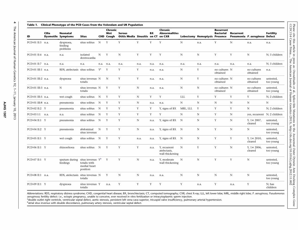
Table 1. Clinical Phenotype of the PCD Cases from the Volendam and UK Population
IDCiliaDysmotile
NeonatalSymptoms Situs CHD
ChronicWetCough
SerousOtitis Media Sinusitis
BXon CT
ChronicAbnormalitieson CXR Lobectomy Hemoptysis
RecurrentBacterialPresence
RecurrentPneumonia P. aeruginosa
FertilityDefect
PCD-01 II:3 n.a. dyspnoea,feedingproblems
situs solitus N Y Y Y Y Y N n.a. Y N n.a. n.a.
PCD-01 II:4 n.a. n.a. isolateddextrocardia
N Y N Y Y Y N N Y Y N N; 3 children
PCD-01 II:7 n.a. n.a. n.a. n.a. n.a. n.a. n.a. n.a. n.a. n.a. n.a. n.a. n.a. n.a. N; 3 children
PCD-01 III:1 n.a. RDS, atelectasis situs solitus Ya Y Y Y n.a. n.a. N Y no culturesobtained
N no culturesobtained
n.a.
PCD-01 III:2 n.a. dyspnoea situs inversustotalis
N N Y Y n.a. n.a. N Y no culturesobtained
N no culturesobtained
untested,too young
PCD-01 III:3 n.a. N situs inversustotalis
N Y Y N n.a. n.a. N N no culturesobtained
N no culturesobtained
untested,too young
PCD-01 III:5 n.a. wet cough situs solitus N Y Y N Y Y LLL Y Y Y Y N; 2 children
PCD-01 III:8 n.a. pneumonia situs solitus N Y Y N n.a. n.a. N N N N N N
PCD-02 II:2 Y pneumonia situs solitus N Y Y Y Y Y, signs of BX MRL, LLL Y Y Y N N; 2 children
PCD-03 I:1 n.a. n.a. situs solitus N Y Y Y Y Y N N Y N yes, recurrent N; 2 children
PCD-04 II:1 Y pneumonia situs solitus N Y Y N n.a. Y, signs of BX N N Y N Y, 13 2007,cleared
untested,too young
PCD-04 II:2 Y pneumonia abdominalsitus inversus
N Y Y N n.a. Y, signs of BX N N Y N N untested,too young
PCD-05 II:1 Y wet cough situs solitus N Y Y n.a. n.a. Y, signs of BX N N Y Y Y, 13 2010,cleared
untested,too young
PCD-06 II:1 Y rhinorrhoea situs solitus N Y Y Y n.a. Y, recurrentatelectasis,wall thickening
N Y Y N Y, 13 2006,cleared
untested,too young
PCD-07 II:1 Y sputum duringfeedings
situs inversustotalis withmedial heartposition
Yb Y Y N n.a. Y, moderatewall thickening
N N Y Y N untested,too young
PCD-08 II:1 n.a. RDS, atelectasis situs inversustotalis
N Y N N n.a. n.a. N N N N N untested,too young
PCD-09 II:1 Y dyspnoea situs inversustotalis
Y n.a. Y Y Y Y Y n.a. Y n.a. Y N; haschildren
Abbreviations: RDS, respiratory distress syndrome; CHD, congenital heart disease; BX, bronchiectasis; CT, computed tomography; CXR, chest X-ray; LLL, left lower lobe; MRL, middle right lobe; P. aeruginosa, Pseudomonasaeruginosa; fertility defect: i.e., ectopic pregnancy, unable to conceive, ever received in vitro fertilization or intracytoplasmic sperm injection.adouble outlet right ventricle, ventricular septal defect, aortic stenosis, persistent left vena cava superior, tricuspid valve insufficiency, pulmonary arterial hypertension.batrial situs inversus with double discordance, pulmonary artery stenosis, ventricular septal defect.
4TheAmerica
nJournalofHumanGenetics
92,1–11,January
10,2013
AJHG
1307
Please
citeth
isarticle
inpress
as:Onoufriad
iset
al.,Sp
lice-SiteMutatio
nsin
theAxonem
alOuter
Dynein
Arm
Dock
ingComplex
Gen
eCCDC114Cau
sePrim
a...,TheAmerican
Journ
alofHuman
Gen
etics(2013),http
://dx.doi.o
rg/10.1016/j.ajh
g.2012.11.002
Please cite this article in press as: Onoufriadis et al., Splice-Site Mutations in the Axonemal Outer Dynein Arm Docking Complex GeneCCDC114 Cause Prima..., The American Journal of Human Genetics (2013), http://dx.doi.org/10.1016/j.ajhg.2012.11.002
The c.742G>A substitution is also predicted to create
a missense change p.Ala248Thr, but the p.Ala248 amino
acid is not well conserved across species, and the missense
change was predicted to be nondeleterious to protein
structure according to programs that assess nonsynony-
mous SNPs (Polyphen-2, SIFT). We therefore concluded
that the putative splicing defect predicted by this substitu-
tion was the more likely mutation mechanism.
The c.742G>A variant was confirmed by capillary
sequencing (Figure S1). Segregation analysis of the
c.742G>A substitution in all available members of the
PCD-01 pedigree confirmed the recessive inheritance of
the variant (Figure 1A). The same variant was then
confirmed to segregate with disease in all available Volen-
dam pedigrees, PCD-02 to PCD-08 (Figure 1A). In total,
all 16 affected Volendam individuals carry the c.742G>A
variant as a homozygous change. This variant is reported
in dbSNP v135 to be present at a very low frequency in
heterozygous state in European descent controls from the
NHLBI Exome Variant Server (rs147718607). The A allele
is present at a frequency of less than 1 in 3,200 alleles,
well below DNA polymorphism levels because the homo-
zygous genotype would be extremely rare; this variant
frequency would give a prevalence of homozygous cases
of less than 1 in 40,000,000 (107).
We proceeded to sequence the CCDC114 exons and
flanking intronic regions in a larger cohort of 44 individ-
uals affected with PCD resulting from ODA and combined
ODA/IDA defects (primers listed in Table S3). Signed and
informed consent was obtained from all participants
according to protocols approved by the institutional ethics
review boards. Mutational analysis resulted in the identifi-
cation of an additional homozygous splice-site variant,
c.486þ1G>A, in one UK family, PCD-09 (Figure S1). This
substitution affects the CCDC114 exon 5 consensus splice
donor site, predicted to cause a frameshift in the CCDC114
protein product. This individual’s disease was consistent
with the Volendam cases, with typical features of PCD
including bronchiectasis requiring lobectomy (Table 1).
The affected individual PCD-09 II:1 has situs inversus
with congenital heart disease and had children with no
reported fertility problems. Segregation analysis in PCD-
09 family members confirmed a consistent recessive
pattern of inheritance (Figure 1A). The c.486þ1G>A
variant was absent from all the control sequence databases
(dbSNP v135, 1000 Genomes Project, NHLBI Exome
Variant Server).
We next used RNA isolated from ciliated cells of affected
individuals and controls to assess the functional impact of
the two CCDC114 splice donor site mutations. Nasal
brushings or curette biopsies were obtained from PCD-02
II:2 (c.742G>A homozygote) and PCD-09 II:1 (c.486þ1G>A homozygote) and healthy volunteers. For PCD-02
II:2, the RNA was isolated from the cells after culture in
standard conditions37 and for PCD-09 II:1 the RNAwas iso-
lated from noncultured cells. RNA was extracted from the
cells via TRIzol (Invitrogen) or the Quick RNA Miniprep
The
AJHG 1
Kit (Zymogen), and first-strand complementary DNA was
synthesized with random nonamers (Sigma-Aldrich) or
oligo-d(T)20 primer (Invitrogen) andOmniscript transcrip-
tase (QIAGEN) or Superscript II reverse transcriptase (Invi-
trogen). PCR amplification was carried out with primers in
exons 6 and 8 of CCDC114 in PCD-02 II:2 and in exons 2
and 5 of CCDC114 in PCD-09 II:1, in parallel to amplifica-
tion of the same samples with the control housekeeping
genes GAPDH (MIM 138400) and ACTB (MIM 102630),
respectively. The RT-PCR primers are listed in Table S4.
A larger RT-PCR product size was amplified from
the PCD-02 II:2 c.742G>A Volendam individual,
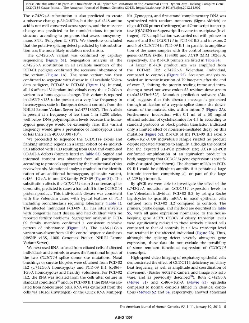
compared to controls (Figure S2). Sequence analysis re-
vealed an intronic insertion of 79 basepairs after the end
of exon 7, shifting the protein reading frame and intro-
ducing a novel nonsense codon 52 residues downstream
(p.Ala248Thrfs52*). Mutation prediction software (Ala-
mut) suggests that this aberrant message is generated
through utilization of a cryptic splice donor site down-
stream of the mutated site, within intron 7 (Figure 2A).
Furthermore, incubation with 0.1 ml of a 50 mg/ml
ethanol solution of cycloheximide for 4.5 hr according to
standard protocols to block protein translation indicated
only a limited effect of nonsense-mediated decay on this
mutation (Figure S2). RT-PCR of the PCD-09 II:1 exon 5
c.486þ1G>A UK individual’s sample yielded no product
despite repeated attempts to amplify, although the control
had the expected RT-PCR product size; ACTB RT-PCR
confirmed amplification of an equivalent product in
both, suggesting that CCDC114 gene expression is specifi-
cally disrupted (not shown). The aberrant mRNA in PCD-
09 II:1 could be difficult to amplify if it contains a large
intronic insertion comprising all or part of the large
(5,229 bp) intron 5.
By qPCR we were able to investigate the effect of the
c.742G>A mutation on CCDC114 expression levels in
the Volendam individual, PCD-02 II:2, by using a Roche
Lightcycler to quantify mRNA in nasal epithelial cells
cultured from PCD-02 II:2 compared to controls. The
primers, probe design, and method are described in Table
S5, with all gene expression normalized to the house-
keeping gene ACTB. CCDC114 ciliary transcript levels
were significantly reduced in these actively ciliated cells
compared to that of controls, but a low transcript level
was retained in the affected individual (Figure 2B). Thus,
although the splicing defect severely abrogates gene
expression, these data do not exclude the possibility
of some remnant functional expression of CCDC114
transcripts.
High-speed video imaging of respiratory epithelial cells
demonstrated the effect of CCDC114 deficiency on ciliary
beat frequency, as well as amplitude and coordination of
movement (Bassler A602F-2 camera and Image Pro soft-
ware, and as previously described38). Both c.742G>A
(Movie S1) and c.486þ1G>A (Movie S3) epithelia
compared to normal controls filmed in identical condi-
tions (Movies S2 and S4, respectively) showed abnormal
American Journal of Human Genetics 92, 1–11, January 10, 2013 5
307
Figure 2. CCDC114 Splice-Site Mutations Causing Primary Ciliary Dyskinesia(A) Effect of the c.742G>A Volendam mutation on splicing. The upper panels show the location of the mutation in genomic DNAsequence chromatograms and the splice-site prediction effect according to Alamut. Alamut uses the four different splice prediction soft-ware programs listed on the left. In comparison of the reference sequence from a control individual (top) against the mutant genomicDNA (bottom), the software predicts loss of the splice donor site and presence of a cryptic splice site 79 bp into the intron. The bottompanel shows the sequence of cDNA from a person who is homozygous for the mutation, isolated from ciliary cells and amplified via
6 The American Journal of Human Genetics 92, 1–11, January 10, 2013
AJHG 1307
Please cite this article in press as: Onoufriadis et al., Splice-Site Mutations in the Axonemal Outer Dynein Arm Docking Complex GeneCCDC114 Cause Prima..., The American Journal of Human Genetics (2013), http://dx.doi.org/10.1016/j.ajhg.2012.11.002
Please cite this article in press as: Onoufriadis et al., Splice-Site Mutations in the Axonemal Outer Dynein Arm Docking Complex GeneCCDC114 Cause Prima..., The American Journal of Human Genetics (2013), http://dx.doi.org/10.1016/j.ajhg.2012.11.002
ciliary motility comprising large areas of static cilia, with
occasionally 1–2 cilia having a twitching or flickering
movement that was stiff, slow, and ineffective for mucus
transport across the epithelial surface. This is consistent
with findings in other PCD individuals lacking the ODAs
(with or without accompanying IDA loss).39,40 Transmis-
sion electron microscopy of respiratory cilia cross-sections
showed that all CCDC114 mutant samples shared
a common ciliary ultrastructural defect, a loss of the outer
dynein arms (ODA) (Figure 3A). This is consistent with
EM findings in the Chlamydomonas strain oda1 carrying
null mutations in the CCDC114 ortholog DCC2.41
Interestingly, despite this lack of ODAs, the flagella of the
Chlamydomonas oda1 strain retain some ability to beat;
however, they beat slowly andwithout the correct effective
waveform.42 This species difference when ODA compo-
nents are deficient has been reported before.2
The Chlamydomonas ortholog of CCDC114 (DCC2/
ODA1) is a component of the ODA docking complex
(ODA-DC) required for the assembly of ODAs onto the
flagella peripheral doublet microtubules.35 In Chlamydo-
monas, ODA-DCs are transported and assembled onto the
peripheral microtubules independently from the ODAs
that attach to them, and the ODAs cannot attach in their
absence.43 In oda1 DCC2-null mutant strains, ODA-DCs
are not assembled onto the axoneme’s microtubules, and
consequently neither are the ODAs.43 We modeled the
comparative protein structure of CCDC114 to investigate
the potential functional impact of the identified splice-
site mutations. CCDC114, like Chlamydomonas DCC2,
has three coiled-coil domains (Figure 2C). Mutations in
coiled-coil domain proteins are already associated with
PCD, playing an important role in axonemal organization
and cilia ultrastructure.24,30,31 Coiled-coils were proposed
as likely to be important for interactions between DCC2
and other docking complex subunits, and the domain
between the second and third DCC2 coiled-coil domain
was also proposed to participate in protein-protein interac-
tions.35 A conserved structural maintenance of chromo-
somes (SMC) domain was also detected in CCDC114,
similar to those identified to play a role in microtubule-
based ciliary transport processes in the PCD-associated
proteins CCDC39 and CCDC40.30 The c.742G>A and
c.486þ1G>A mutations would lead to the lack of either
primers in exons 6 and 8. An intronic insertion of 79 basepairs is presetion substitution site (green arrow) and the presumed intronic cryptiof the regular splice donor site. The inclusion of 79 bases leads to a franovel amino acids, at the in-frame TAA codon indicated by the red b(B) Relative expression levels (normalized to ACTB) of CCDC114, CClial cells from controls or from Volendam PCD-02 II:2, assessed by qPexpressed at higher levels in cilia-producing cells compared to testisexpression in cilia-producing cells. In addition, CCDC114 and DNAHvidual compared to control. The means 5 SEM from triplicate repea(C) Location of the Volendam and UK splice-site mutations in the intprotein shown below. Black boxes indicate coding exons, white boxesdetected by Paircoil2 run with a minimumwindow size of 28. Homolof chromosomes protein) domain in CCDC114 indicated by the bluphodiesterase domain indicated by the orange box (PRK12704).
The
AJHG 1
one or two critical coiled-coil domains, with apparently
similarly deleterious consequences (Figure 2C).
To further investigate CCDC114 function, we analyzed
protein localization by high-resolution immunofluores-
cence microscopy in ciliated epithelial cells. In controls,
CCDC114 antisera decorates the full length of the cilia
(Figure 3B), suggesting that its putative role in tethering
of outer dynein arms is required along the entire axoneme.
Two different classes of ODAs have been defined by Flie-
gauf et al. at the distal (DNAH5-positive, DNAH9-positive)
and proximal (DNAH5-positive, DNAH9-negative) ends
of cilia.40 CCDC114 appears to be a component of ODA
docking complexes capable of interacting with both ODA
types. In contrast, the individuals carrying c.742G>A
and c.486þ1G>A mutations had severely reduced levels
of CCDC114 along their entire cilia (Figure 3B). By using
well-established diagnostic markers of axoneme integrity
developed by the Omran lab,22 we confirmed by staining
with the ODA component DNAH5 that the ODAs along
the cilia length are absent in c.742G>A and c.486þ1G>A
cells, whereas DNALI1 staining confirmed that the IDAs
are present and undisturbed (Figures S3 and S4). It is not
known whether human ODA-DCs and ODAs are trans-
ported by the same or different mechanisms to the
axonemes, but in Chlamydomonas they can be assembled
separately in the cytoplasm, and ODAs are assembled
in the cytoplasm even without the ODA-DC being
present.43 However, the cells deficient for CCDC114
arising from either mutation did not show any noticeable
cytoplasmic accumulation of ODAs by DNAH5 staining,
indicating a possible species difference in these pathways
(Figures S3 and S4). We investigated the expression levels
of DNAH5 mRNA by qPCR in cultured ciliated epithelial
cells from the Volendam individual PCD-02 II:2 (Roche
Lightcycler, Table S5), normalizing to the housekeeping
gene ACTB. There was a reduction in DNAH5 levels in
PCD-02 II:2 compared to control, although this was
less marked than the reduced CCDC114 ciliary expres-
sion (Figure 2B). These results are in agreement with
the lowered CCDC114 protein expression seen via
immunofluorescence; however, the lack of DNAH5 immu-
nofluorescence may reflect enhanced degradation of the
DNAH5 protein rather than a lack of its accumulation
(Figures 3 and S3).
nt in the c.742G>A individual’s cDNA, located between themuta-c splice site (pink arrow). The sequence shows no indication of usemeshift and a premature stop codon in exon 8 after addition of 52ox with arrow.
DC63, and DNAH5 in mRNA from testis and cultured nasal epithe-CR with a Roche Lightcycler as described in Table S5. CCDC114 iswhereas CCDC63 is expressed highly in testis with no detectable5 levels are both reduced in cilia from the Volendam affected indi-t experiments are shown.ron-exon structure shown above, and in a model of the CCDC114noncoding exons. The green boxes indicate coiled-coil domains asogy was also detected identifying an SMC (structural maintenancee box (SMC_prok_B TIGR02168) and a putative prokaryotic phos-
American Journal of Human Genetics 92, 1–11, January 10, 2013 7
307
Figure 3. CCDC114 Splice-Site Mutations Are Associated with Ciliary Axoneme Defects(A) Transmission electronmicrographs of cross-sections of respiratory epithelial cell cilia demonstrates loss of outer dynein arms in boththe PCD-02 II:2 and PCD-09 II:1 individuals carrying the c.742G>A and c.486þ1G>A splice donor mutations, respectively. All nineperipheral doublets showed loss and reduction of the outer dynein arms (arrows) compared to controls. Scale bar represents 100 nm.(B) Subcellular localization of CCDC114 protein (green) in respiratory epithelial cells via a rabbit polyclonal antibody (SigmaHPA042524). In healthy individuals (top), CCDC114 is localized along the length of the axoneme of the ciliated cells, whereas inboth PCD-02 II:2 and PCD-09 II:1, CCDC114 is markedly reduced (middle and bottom). Axoneme-specific anti-acetylated-a-tubulinantibody (Sigma) was used as a control to stain the entire axoneme (red). DNA (blue) was stained with DAPI (Invitrogen). Scale barsrepresent 10 mm.
Please cite this article in press as: Onoufriadis et al., Splice-Site Mutations in the Axonemal Outer Dynein Arm Docking Complex GeneCCDC114 Cause Prima..., The American Journal of Human Genetics (2013), http://dx.doi.org/10.1016/j.ajhg.2012.11.002
Our data suggest that a single ancestral CCDC114 muta-
tion, c.742G>A, underlies all Volendam PCD cases, most
probably spread by genetic bottleneck founder effect.
This village was founded in 1462 by 20 families who estab-
lished a settlement after the nearby town of Edam dug
a new exit to its sea harbor and dammed up the old exit.
8 The American Journal of Human Genetics 92, 1–11, January 10, 201
AJHG 1307
These families settled on the ‘‘filling dam’’ land or ‘‘Vollen-
dam’’ and because of their isolated site, the church refor-
mation in the late 16th century passed them without
effect. The major religion remains Roman Catholic and
even after their geographic isolation has lessened, their
religious and social distinctions have kept the Volendam
3
Please cite this article in press as: Onoufriadis et al., Splice-Site Mutations in the Axonemal Outer Dynein Arm Docking Complex GeneCCDC114 Cause Prima..., The American Journal of Human Genetics (2013), http://dx.doi.org/10.1016/j.ajhg.2012.11.002
population very isolated into the modern era. A review
of the unfiltered UK10K whole-exome sequence data, to
derive the available SNPs across the CCDC114 locus, shows
that the two distantly related individuals (PCD-01 III:3 and
III:8) carrying the c.742G>A mutation share a 2 megabase
haplotype (not shown). This supports the idea that the
Volendammutation was spread within this inbred popula-
tion from one original founding ancestor, and its small cor-
responding haplotype size explains why a single locus was
missed in past linkage mapping. In the large PCD-01 pedi-
gree (Figure 1), common ancestors are found six to seven
generations back, dating to the early 1800s. However,
because not all the Volendam families could be connected
and this small common haplotype was found to carry the
mutation, presumably reducedby ancientmeiotic recombi-
nation events, this suggests a more advanced age for the
shared mutation than the founding of the Volendam
village. According to the geneticmaps ofGenethon,Marsh-
field, and DeCode, this 2 Mb region on chromosome
19q13.33 spanning CCDC114 corresponds to a genetic
distance of 3.4–4.6 centiMorgans. Te Meerman et al.44
have shown that aroundanewmutation, ameanhaplotype
sharing lengthof 5 cM is reached after ~70 generations. This
preceeds the founding of the village of Volendam, which
occurred an estimated 22 generations ago. Consequently,
most likely, two or more carriers with the CCDC114 muta-
tion were present among the original founding families of
Volendam. The finding of four heterozygous European
carriers in the NHLBI Exome Variant Server supports the
hypothesis that this variant arose prior to the founding of
Volendam and was brought in by original settlers.
We found that fertility was not greatly affected among
individuals carrying CCDC114 mutations. The reasons for
this are not clear, but there may be some functional redun-
dancy of CCDC114 in sperm. In Chlamydomonas the ODA-
DC consists of the CCDC114 ortholog DCC2/ODA1 and
two other proteins, DCC1/ODA3 and DLE3/ODA14.35
However, the human ODA-DC seems to be differently
structured, because no definitive human homolog can be
found for DCC1/ODA3 or DLE3/ODA14. Furthermore,
there is a second human protein apart from CCDC114
with significant homology to DCC2: CCDC63, which is
26% identical to CCDC114. CCDC63 is also 21% identical
to theChlamydomonasODA5protein that is associatedwith
the axoneme and is required for outer dynein arm assembly
but independent from theODAs andODA-DCs.45Whether
CCDC63 plays an orthologous role to DCC2/CCDC114 or
to ODA5 is not yet clear,2,46 but CCDC63 represents an
excellent candidate gene for an overlapping phenotype to
that associated with CCDC114 mutations. The relative
levels of CCDC114 and CCDC63 proteins in the axoneme
of sperm is not well understood, but available evidence
from public expression databases such as Unigene suggests
that theCCDC63 transcript ismore highly sperm specific in
its expression than CCDC114, and thus it is not impossible
that CCDC114 function could be partially replaced by
CCDC63 in sperm.
The
AJHG 1
To test thishypothesis,weusedqPCR (Table S5) onmRNA
from testis (Life Technologies), the source of sperm cells
(used because in sperm there is no active transcription),
and from cultured nasal epithelial cells that were actively
producing cilia. The nasal cells were derived both from
controls and from the Volendam individual PCD-02 II:2.
Ahigh expression ofCCDC63was detected in control testis,
with no detectable expression in control cilia-producing
cells, even after adding 10 cycles to the qPCR, whereas
CCDC114 is expressed at >100 times higher levels in cilia-
producing cells compared to testis (Figure 2B). Without
a testis biopsy from an affected person, we cannot exclude
the possibility that affected individuals could retain some
testis expression of CCDC114; however, we can conclude
that the level of CCDC63 expression in control testis is
comparable to CCDC114 expression in control ciliary cells
and >100 times higher than CCDC114 in testis.
In summary, we report mutations within conserved
CCDC114 splice donor sites affecting a total of 17 individ-
uals with PCD, all homozygous for either c.742G>A or
c.486þ1G>A substitutions, conferring PCD with outer
dynein arm loss, cilia immotility, and laterality defects
including complex cardiac malformations. Recent large-
scale studies show the importance of this phenotype, esti-
mating that 65%–67% of PCD cases have outer dynein arm
deficiencies,47,48 either alone (33%–43%) or with other
structures involved. We reveal that CCDC114 has a highly
conserved role in ODA microtubular attachment, with
a likely role as an integral protein of the ODA-DC, the
loss of which prevents ODAs from binding onto axonemal
microtubules. We identified a difference in relative expres-
sion levels of CCDC114 that might suggest it has a more
prominent role in cilia compared to testis. Identification
of the Volendam founder mutation c.742G>A highlights
CCDC114 as an important target for future therapeutic
intervention, particularly in this at-risk population that
has a high prevalence of PCD.
Supplemental Data
Supplemental Data include the UK10K Consortium author list,
four figures, five tables, and four movies and can be found with
this article online at http://www.cell.com/AJHG/.
Acknowledgments
We would like to thank all the PCD families for their participation
in the study, Fiona Copeland, and the U.K. PCD Family Support
Group. We thank Maggie Meeks and R. Mark Gardiner for patient
recruitment and their past involvement in the project. We thank
Paul Griffin for electron microscopy processing. We thank all the
participants of the UK10K RARE group, as listed in the Supple-
mental Data file, which is part of theUK10KConsortium, in partic-
ularMatthewHurles, Saeed Al Turki, and Philip Beales. The UK10K
project is funded by the Wellcome Trust (award WT091310). T.P.,
D.M., B.K., andG.P. were supported by the Dutch patient organiza-
tion PCD Belangengroep, funded by ‘‘It Krystteam’’ (Friesland).
P.J.S. is supported by the Wellcome Trust and British Heart
American Journal of Human Genetics 92, 1–11, January 10, 2013 9
307
Please cite this article in press as: Onoufriadis et al., Splice-Site Mutations in the Axonemal Outer Dynein Arm Docking Complex GeneCCDC114 Cause Prima..., The American Journal of Human Genetics (2013), http://dx.doi.org/10.1016/j.ajhg.2012.11.002
Foundation. M.S. is supported by an Action Medical Research UK
Clinical Training Fellowship. A.O., D.A., M.S., S.P., E.M.K.C., and
H.M.M. are supported by the Milena Carvajal Pro-Kartagener
Foundation, Action Medical Research UK, the Henry Smith
Charity, and Newlife Foundation for Disabled Children UK.
Received: August 4, 2012
Revised: August 27, 2012
Accepted: November 1, 2012
Published: December 20, 2012
Web Resources
The URLs for data presented herein are as follows:
1000 Genomes, http://browser.1000genomes.org/index.html
BLAST, http://blast.ncbi.nlm.nih.gov/Blast.cgi
CDD, http://www.ncbi.nlm.nih.gov/sites/entrez?db¼cdd
dbSNP, http://www.ncbi.nlm.nih.gov/projects/SNP/
Mouse Genome Informatics, http://www.informatics.jax.org/
NHLBI Exome Variant Server/Sequencing Project (ESP), http://evs.
gs.washington.edu/EVS/
Online Mendelian Inheritance in Man (OMIM), http://www.
omim.org/
Paircoil2, http://www.groups.csail.mit.edu/cb/paircoil2/
SMART, http://www.smart.embl-heidelberg.de/
STRING 9.0, http://www.string-db.org/
UK10K Consortium, http://www.uk10k.org/
References
1. Mitchison, T.J., andMitchison, H.M. (2010). Cell biology: how
cilia beat. Nature 463, 308–309.
2. Pazour, G.J., Agrin, N., Walker, B.L., and Witman, G.B. (2006).
Identification of predicted human outer dynein arm genes:
candidates for primary ciliary dyskinesia genes. J. Med. Genet.
43, 62–73.
3. Mitchison, H.M., Schmidts, M., Loges, N.T., Freshour, J., Drit-
soula, A., Hirst, R.A., O’Callaghan, C., Blau, H., Al Dabbagh,
M., Olbrich, H., et al. (2012). Mutations in axonemal dynein
assembly factor DNAAF3 cause primary ciliary dyskinesia.
Nat. Genet. 44, 381–389, S1–S2.
4. Barbato, A., Frischer, T., Kuehni, C.E., Snijders, D., Azevedo, I.,
Baktai, G., Bartoloni, L., Eber, E., Escribano, A., Haarman, E.,
et al. (2009). Primary ciliary dyskinesia: a consensus statement
on diagnostic and treatment approaches in children. Eur.
Respir. J. 34, 1264–1276.
5. Bush, A., Chodhari, R., Collins, N., Copeland, F., Hall, P., Har-
court, J., Hariri, M., Hogg, C., Lucas, J., Mitchison, H.M., et al.
(2007). Primary ciliary dyskinesia: current state of the art.
Arch. Dis. Child. 92, 1136–1140.
6. Afzelius, B.A. (1998). Genetics and pulmonary medicine. 6.
Immotile cilia syndrome: past, present, and prospects for the
future. Thorax 53, 894–897.
7. Jeganathan, D., Chodhari, R., Meeks, M., Faeroe, O., Smyth,
D., Nielsen, K., Amirav, I., Luder, A.S., Bisgaard, H., Gardiner,
R.M., et al. (2004). Loci for primary ciliary dyskinesia map to
chromosome 16p12.1-12.2 and 15q13.1-15.1 in Faroe Islands
and Israeli Druze genetic isolates. J. Med. Genet. 41, 233–240.
8. O’Callaghan, C., Chetcuti, P., and Moya, E. (2010). High
prevalence of primary ciliary dyskinesia in a British Asian pop-
ulation. Arch. Dis. Child. 95, 51–52.
10 The American Journal of Human Genetics 92, 1–11, January 10, 20
AJHG 1307
9. Coren, M.E., Meeks, M., Morrison, I., Buchdahl, R.M., and
Bush, A. (2002). Primary ciliary dyskinesia: age at diagnosis
and symptom history. Acta Paediatr. 91, 667–669.
10. Kosaki, K., Ikeda, K., Miyakoshi, K., Ueno,M., Kosaki, R., Taka-
hashi, D., Tanaka, M., Torikata, C., Yoshimura, Y., and Takaha-
shi, T. (2004). Absent inner dynein arms in a fetus with
familial hydrocephalus-situs abnormality. Am. J. Med. Genet.
A. 129A, 308–311.
11. Ibanez-Tallon, I., Heintz, N., and Omran, H. (2003). To beat or
not to beat: roles of cilia in development and disease. Hum.
Mol. Genet. 12(Spec No 1), R27–R35.
12. Bush, A., Cole, P., Hariri, M., Mackay, I., Phillips, G., O’Calla-
ghan, C., Wilson, R., and Warner, J.O. (1998). Primary ciliary
dyskinesia: diagnosis and standards of care. Eur. Respir. J. 12,
982–988.
13. Nakhleh, N., Francis, R., Giese, R.A., Tian, X., Li, Y., Zariwala,
M.A., Yagi, H., Khalifa, O., Kureshi, S., Chatterjee, B., et al.
(2012). High prevalence of respiratory ciliary dysfunction in
congenital heart disease patients with heterotaxy. Circulation
125, 2232–2242.
14. Kennedy, M.P., Omran, H., Leigh, M.W., Dell, S., Morgan, L.,
Molina, P.L., Robinson, B.V., Minnix, S.L., Olbrich, H.,
Severin, T., et al. (2007). Congenital heart disease and other
heterotaxic defects in a large cohort of patients with primary
ciliary dyskinesia. Circulation 115, 2814–2821.
15. Moore, A., Escudier, E., Roger, G., Tamalet, A., Pelosse, B.,
Marlin, S., Clement, A., Geremek, M., Delaisi, B., Bridoux,
A.M., et al. (2006). RPGR ismutated in patients with a complex
X linked phenotype combining primary ciliary dyskinesia and
retinitis pigmentosa. J. Med. Genet. 43, 326–333.
16. Olbrich, H., Haffner, K., Kispert, A., Volkel, A., Volz, A.,
Sasmaz, G., Reinhardt, R., Hennig, S., Lehrach, H., Konietzko,
N., et al. (2002). Mutations in DNAH5 cause primary ciliary
dyskinesia and randomization of left-right asymmetry. Nat.
Genet. 30, 143–144.
17. Loges, N.T., Olbrich, H., Fenske, L., Mussaffi, H., Horvath, J.,
Fliegauf, M., Kuhl, H., Baktai, G., Peterffy, E., Chodhari, R.,
et al. (2008). DNAI2 mutations cause primary ciliary dyski-
nesia with defects in the outer dynein arm. Am. J. Hum.
Genet. 83, 547–558.
18. Pennarun, G., Escudier, E., Chapelin, C., Bridoux, A.M.,
Cacheux, V., Roger, G., Clement, A., Goossens, M., Amselem,
S., and Duriez, B. (1999). Loss-of-function mutations in
a human gene related to Chlamydomonas reinhardtii dynein
IC78 result in primary ciliary dyskinesia. Am. J. Hum. Genet.
65, 1508–1519.
19. Mazor, M., Alkrinawi, S., Chalifa-Caspi, V., Manor, E., Shef-
field, V.C., Aviram, M., and Parvari, R. (2011). Primary ciliary
dyskinesia caused by homozygous mutation in DNAL1, en-
coding dynein light chain 1. Am. J. Hum. Genet. 88, 599–607.
20. Duriez, B., Duquesnoy, P., Escudier, E., Bridoux, A.M., Escalier,
D.,Rayet, I.,Marcos,E.,Vojtek,A.M.,Bercher, J.F., andAmselem,
S. (2007). A common variant in combination with a nonsense
mutation in amember of the thioredoxin family causesprimary
ciliary dyskinesia. Proc. Natl. Acad. Sci. USA 104, 3336–3341.
21. Omran, H., Kobayashi, D., Olbrich, H., Tsukahara, T., Loges,
N.T., Hagiwara, H., Zhang, Q., Leblond, G., O’Toole, E.,
Hara, C., et al. (2008). Ktu/PF13 is required for cytoplasmic
pre-assembly of axonemal dyneins. Nature 456, 611–616.
22. Loges, N.T., Olbrich, H., Becker-Heck, A., Haffner, K., Heer, A.,
Reinhard, C., Schmidts, M., Kispert, A., Zariwala, M.A., Leigh,
M.W., et al. (2009). Deletions and point mutations of LRRC50
13
Please cite this article in press as: Onoufriadis et al., Splice-Site Mutations in the Axonemal Outer Dynein Arm Docking Complex GeneCCDC114 Cause Prima..., The American Journal of Human Genetics (2013), http://dx.doi.org/10.1016/j.ajhg.2012.11.002
cause primary ciliary dyskinesia due to dynein arm defects.
Am. J. Hum. Genet. 85, 883–889.
23. Duquesnoy, P., Escudier, E., Vincensini, L., Freshour, J., Bri-
doux, A.M., Coste, A., Deschildre, A., de Blic, J., Legendre,
M., Montantin, G., et al. (2009). Loss-of-function mutations
in the human ortholog of Chlamydomonas reinhardtii ODA7
disrupt dynein arm assembly and cause primary ciliary dyski-
nesia. Am. J. Hum. Genet. 85, 890–896.
24. Panizzi, J.R., Becker-Heck, A., Castleman, V.H., Al-Mutairi,
D.A., Liu, Y., Loges, N.T., Pathak, N., Austin-Tse, C., Sheridan,
E., Schmidts, M., et al. (2012). CCDC103 mutations cause
primary ciliary dyskinesia by disrupting assembly of ciliary
dynein arms. Nat. Genet. 44, 714–719.
25. Horani, A., Druley, T.E., Zariwala, M.A., Patel, A.C., Levinson,
B.T., Van Arendonk, L.G., Thornton, K.C., Giacalone, J.C., Al-
bee, A.J., Wilson, K.S., et al. (2012). Whole-exome capture and
sequencing identifies HEATR2 mutation as a cause of primary
ciliary dyskinesia. Am. J. Hum. Genet. 91, 685–693.
26. Kott, E., Duquesnoy, P., Copin, B., Legendre, M., Dastot-Le
Moal, F., Montantin, G., Jeanson, L., Tamalet, A., Papon,
J.-F., Siffroi, J.-P., et al. (2012). Loss-of-function mutations in
LRRC6, a gene essential for proper axonemal assembly of
inner and outer dynein arms, cause primary ciliary dyskinesia.
Am. J. Hum. Genet. 91, 958–964.
27. Bartoloni, L., Blouin, J.L., Pan, Y., Gehrig, C., Maiti, A.K.,
Scamuffa, N., Rossier, C., Jorissen, M., Armengot, M., Meeks,
M., et al. (2002). Mutations in the DNAH11 (axonemal heavy
chain dynein type 11) gene cause one form of situs inversus
totalis and most likely primary ciliary dyskinesia. Proc. Natl.
Acad. Sci. USA 99, 10282–10286.
28. Knowles, M.R., Leigh, M.W., Carson, J.L., Davis, S.D., Dell,
S.D., Ferkol, T.W., Olivier, K.N., Sagel, S.D., Rosenfeld, M.,
Burns, K.A., et al.; Genetic Disorders of Mucociliary Clearance
Consortium. (2012). Mutations of DNAH11 in patients with
primary ciliary dyskinesia with normal ciliary ultrastructure.
Thorax 67, 433–441.
29. Castleman, V.H., Romio, L., Chodhari, R., Hirst, R.A., de Cas-
tro, S.C., Parker, K.A., Ybot-Gonzalez, P., Emes, R.D., Wilson,
S.W., Wallis, C., et al. (2009). Mutations in radial spoke head
protein genes RSPH9 and RSPH4A cause primary ciliary dyski-
nesia with central-microtubular-pair abnormalities. Am. J.
Hum. Genet. 84, 197–209.
30. Merveille, A.C., Davis, E.E., Becker-Heck, A., Legendre, M.,
Amirav, I., Bataille, G., Belmont, J., Beydon, N., Billen, F.,
Clement, A., et al. (2011). CCDC39 is required for assembly
of inner dynein arms and the dynein regulatory complex
and for normal ciliary motility in humans and dogs. Nat.
Genet. 43, 72–78.
31. Becker-Heck, A., Zohn, I.E., Okabe, N., Pollock, A., Lenhart,
K.B., Sullivan-Brown, J., McSheene, J., Loges, N.T., Olbrich,
H., Haeffner, K., et al. (2011). The coiled-coil domain contain-
ing protein CCDC40 is essential for motile cilia function and
left-right axis formation. Nat. Genet. 43, 79–84.
32. Olbrich, H., Schmidts, M., Werner, C., Onoufriadis, A., Loges,
N.T., Raidt, J., Banki, N.F., Shoemark, A., Burgoyne, T., Al
Turki, S., et al. (2012). Recessive HYDIN mutations cause
primary ciliary dyskinesia without randomization of left-right
body asymmetry. Am. J. Hum. Genet. 91, 672–684.
33. Madan, K., Pieters, M.H., Kuyt, L.P., van Asperen, C.J., de
Pater, J.M., Hamers, A.J., Gerssen-Schoorl, K.B., Hustinx,
T.W., Breed, A.S., Van Hemel, J.O., et al. (1990). Paracentric
The A
AJHG 1
inversion inv(11)(q21q23) in The Netherlands. Hum. Genet.
85, 15–20.
34. The 1000 Genomes Project Consortium. (2010). A map of
human genome variation from population-scale sequencing.
Nature 467, 1061–1073.
35. Takada, S., Wilkerson, C.G., Wakabayashi, K., Kamiya, R., and
Witman, G.B. (2002). The outer dynein arm-docking
complex: composition and characterization of a subunit
(oda1) necessary for outer arm assembly. Mol. Biol. Cell 13,
1015–1029.
36. Diez-Roux, G., Banfi, S., Sultan, M., Geffers, L., Anand, S.,
Rozado, D., Magen, A., Canidio, E., Pagani, M., Peluso, I.,
et al. (2011). A high-resolution anatomical atlas of the tran-
scriptome in the mouse embryo. PLoS Biol. 9, e1000582.
37. Jorissen, M., Willems, T., and Van der Schueren, B. (2000).
Ciliary function analysis for the diagnosis of primary ciliary
dyskinesia: advantages of ciliogenesis in culture. Acta Otolar-
yngol. 120, 291–295.
38. Shoemark, A., Ozerovitch, L., andWilson, R. (2007). Aetiology
in adult patients with bronchiectasis. Respir. Med. 101, 1163–
1170.
39. Chilvers, M.A., Rutman, A., and O’Callaghan, C. (2003).
Ciliary beat pattern is associated with specific ultrastructural
defects in primary ciliary dyskinesia. J. Allergy Clin. Immunol.
112, 518–524.
40. Fliegauf, M., Olbrich, H., Horvath, J., Wildhaber, J.H., Zari-
wala, M.A., Kennedy, M., Knowles, M.R., and Omran, H.
(2005). Mislocalization of DNAH5 and DNAH9 in respiratory
cells from patients with primary ciliary dyskinesia. Am. J.
Respir. Crit. Care Med. 171, 1343–1349.
41. Kamiya, R. (1988). Mutations at twelve independent loci
result in absence of outer dynein arms in Chylamydomonas
reinhardtii. J. Cell Biol. 107, 2253–2258.
42. Kamiya, R., and Okamoto, M. (1985). A mutant of Chlamydo-
monas reinhardtii that lacks the flagellar outer dynein arm but
can swim. J. Cell Sci. 74, 181–191.
43. Wakabayashi, K., Takada, S., Witman, G.B., and Kamiya, R.
(2001). Transport and arrangement of the outer-dynein-
arm docking complex in the flagella of Chlamydomonas
mutants that lack outer dynein arms. Cell Motil. Cytoskeleton
48, 277–286.
44. te Meerman, G.J., and Van der Meulen, M.A. (1997). Genomic
sharing surrounding alleles identical by descent: effects of
genetic drift and population growth. Genet. Epidemiol. 14,
1125–1130.
45. Wirschell, M., Pazour, G., Yoda, A., Hirono, M., Kamiya, R.,
and Witman, G.B. (2004). Oda5p, a novel axonemal protein
required for assembly of the outer dynein arm and an associ-
ated adenylate kinase. Mol. Biol. Cell 15, 2729–2741.
46. Hom, E.F., Witman, G.B., Harris, E.H., Dutcher, S.K., Kamiya,
R., Mitchell, D.R., Pazour, G.J., Porter, M.E., Sale, W.S., Wir-
schell, M., et al. (2011). A unified taxonomy for ciliary
dyneins. Cytoskeleton (Hoboken) 68, 555–565.
47. Papon, J.F., Coste, A., Roudot-Thoraval, F., Boucherat, M.,
Roger, G., Tamalet, A., Vojtek, A.M., Amselem, S., and Escud-
ier, E. (2010). A 20-year experience of electron microscopy
in the diagnosis of primary ciliary dyskinesia. Eur. Respir. J.
35, 1057–1063.
48. Shoemark, A., Dixon, M., Corrin, B., and Dewar, A. (2012).
Twenty-year review of quantitative transmission electron
microscopy for the diagnosis of primary ciliary dyskinesia. J.
Clin. Pathol. 65, 267–271.
merican Journal of Human Genetics 92, 1–11, January 10, 2013 11
307


