Embed Size (px)
Citation preview
Review of Palaeobotany and Palynology 156 (2009) 211–232
Contents lists available at ScienceDirect
Review of Palaeobotany and Palynology
j ourna l homepage: www.e lsev ie r.com/ locate / revpa lbo
Sporoderm development in Trevesia burckii (Araliaceae). I. Tetrad period:Further evidence for the participation of self-assembly processes☆
Nina Gabarayeva a,⁎, Valentina Grigorjeva a, John R. Rowley b, Alan R. Hemsley c
a Komarov Botanical Institute, Popov st., 2, 197376, St.-Petersburg, Russiab Botanical Institute, Stockholm University, Stockholm, SE-106 91, Swedenc Department of Earth Sciences, Cardiff University, Park Place, Cardiff CF10 3YE, Wales, UK
☆ Nature need not cram genetic code with detail, pr(adapted from “The Fractal Geometry of Nature” by B.B.⁎ Corresponding author.
E-mail address: [email protected] (N. Gabarayeva).
0034-6667/$ – see front matter © 2008 Elsevier B.V. Adoi:10.1016/j.revpalbo.2008.12.001
a b s t r a c t
a r t i c l e i n f oArticle history:
The developmental events Received 22 February 2008Received in revised form 28 November 2008Accepted 2 December 2008Available online 7 December 2008Keywords:microstructurepattern formationself-assemblycolloidstensegrityECM-glycocalyxpollen wall developmentcallose envelopeTrevesia
in the periplasmic space and cytoplasm of microspores, and in the tapetum ofTrevesia burckii during the tetrad period have been traced in detail during microspore ontogeny from thesporogenous cell stage to the late tetrad stage and the initiation of the foot layer. The data obtained providesupport to two of our previously proposed hypotheses: (1) the glycocalyx (scaffolding primexine matrix) is acolloidal system; (2) the involvement of processes of self-assembly of a number of colloidal micellar systemsto the exine development. The main structures of the reticulate ectexine up to the establishment of the footlayer are columellae and tectumwhich evidently form on a base of spherical and cylindrical transitive micellemesophases after sporopollenin accumulation. The importance of the callose envelope surroundingmicrospores for exine development is discussed in the light of recent findings. Two possible pathways ofpattern determination are possible. One suggests the role of the plasma membrane in pattern imprinting andthe corresponding necessity of the transfer of 2-D information to 3-D. Our current supposition is that theother, self-assembly physical phenomena, cellular tensegrity, also participate in the process of theestablishment of a wide-spread reticulate exine pattern which appears as the result of an interplay of themicrospore cytoskeletal prestress and the resistance from the ECM (exocellular matrix=glycocalyx)adhesive sites. More and more information is appearing that provides evidence for the importance ofmechanical forces and physico-chemical regularities in the living world.
© 2008 Elsevier B.V. All rights reserved.
1. Introduction
In spite of the fact that a great many recent studies have beenundertaken on spore and pollen wall development, our knowledge ofexine patterning is still obscure. However, if we ever hope to unravelthis enigma, we will not manage this without developmental insightswhich provide a route to the understanding of the processes involvedin generating characteristic patterns of pollen or spore wall structureand ornamentation— the patterning, originating during developmentin response to the subtle variations of the conditions within theplasma membrane glycocalyx (Blackmore et al., 2007).
The question arises as to where such a key exists? The oftenspecies-specific nature of exine patterns suggests a genomic key. Butas more facts accumulate, the more suspicions appear that somethinginterferes with the precise and straightforward work of the genome.This “something” is, in the opinion of a number of authors, the inter-
edetermined by self-assemblyMandelbrot (1982).
ll rights reserved.
ference of self-assembly processes which are capable of distorting theregular work of the genome, making the results unpredictable(Heslop-Harrison, 1972; Gerasimova-Navashina, 1973; Sheldon andDickinson, 1983; Gabarayeva, 1990; van Uffelen, 1991; Hemsley et al.,1992; Gabarayeva, 1993; Southworth and Jernstedt, 1995). It is amatter of course that the input of the genome is large: without doubt,the genome determines the exact chemical composition of all thesubstances in the periplasmic space and their concentration, the restof constructive process is picked up by physico-chemical self-assembly (Gabarayeva and Hemsley, 2006). This is the point wherepossible distortion of initial genomic instruction could happen: thenon-linear character of self-assembly. The suspicions come alsofrom other sources: the striking similarity of lifeless patterns(e.g. technology organometallic covers — see Fig. 1 in Hemsley andGabarayeva, 2007) and exine microarchitecture is really confusing.The meaningful title of Hemsley et al's. paper (2004) includes thephrase “soft and sticky development” which points to the participa-tion of “soft matter” in exine development. The term “soft matter”denotes materials that are easily deformed by external stress, andencompasses liquid crystals, polymers, surfactants and colloids —
particles dispersed within another medium (van Blaaderen, 2006).

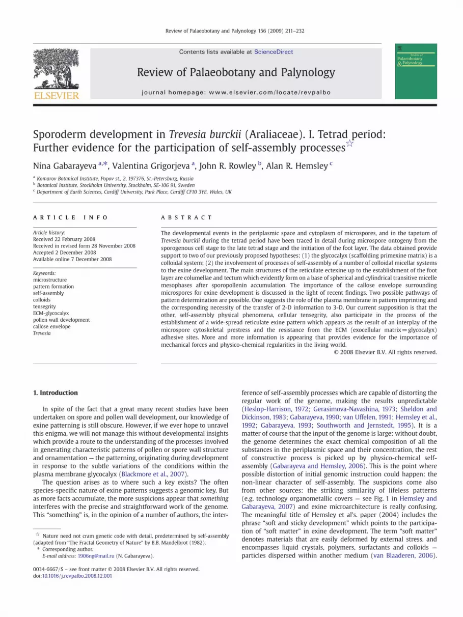
Fig. 1. Transitive stages of micelles and micelle aggregations (adapted from Hamley (2000) and Fridrichsberg (1995) which appear in solution as the concentration of surfactantincreases. Sketches b, c, d, e show sequential micelle mesophases formed in a water-based medium (so-called direct micelles). Sketches b', c', d', e show corresponding micellemesophases in a lipid-based medium (so-called reverse micelles). Note that stage e is the same for both sequences. a — true solution of surfactant molecules, with their hydrophilichead and hydrophobic tails. b— spherical micelle, with hydrophilic heads outside and hydrophobic tails hidden inside, formed at critical micelle concentration. c— cylindrical micelleformed at higher surfactant concentration. d — hexagonally packed cylindrical micelles spontaneously form a layer — “middle” mesophase. e — lamellar “neat” micelles in parallelarrangement, formed by bilayers of “middle” mesophase. Arrowheads show typical gaps between bilayers. b' — reverse spherical micelle, with hydrophobic tails sticking out andhydrophilic heads inside. c' — reverse cylindrical micelle. d' — layer of reverse hexagonally packed cylindrical micelles (reverse “middle” mesophase). Note that if a water-basedmedium changes to a lipid-based one, “direct” micelles undergo inversion. Note also the opposite course of events (see arrows).
212 N. Gabarayeva et al. / Review of Palaeobotany and Palynology 156 (2009) 211–232
Their basic constituents have sizes of between several nanometres andseveral micrometres, and have the potential to self-organize, formingbeautiful 3-D structures — complex colloids on scales up to amicrometre. Interestingly, the thickness of the exine in most speciesis about 1mm. These structures, obtained experimentally by a numberof authors (see review of van Blaaderen, 2006), are whimsical: “ice-cream cones”, the structures, indistinguishable from polyporatepollen grains, have “dome-like” and “raspberry-like” structures. Theinnovation is that these structures are being formed with a secondstage of self-organization. The particles are first formed as soft-matterscales, and then built to a far more intricate structure, allowingunprecedented control over the 3-D organization. Raspberry-likemimics were obtained from polystyrene latex dispersions in experi-ments modeling exine microarchitecture (Griffiths and Hemsley,2001). The most important conclusion is, however, that complexshape is no prerequisite for complex interaction. Another importantfeature of soft-matter components is that these structures can berecovered in the process of drying of emulsion droplets. It ismeaningful that the early surmise of Rowley (Rowley and Rowley,1998) was that late exine development (especially concerning pollenwall ornamentation) proceeds very quickly in desiccating conditionsbefore pollen dispersion. Some soft-matter structures were fabricatedby deposition of silica on liquid crystals formed by surfactants (Zoldesiand Imhof, cited by van Blaaderen, 2006). It is significant that Tryonand Lugardon (1978) revealed silicon and calcium depositions in themegaspores of Selaginella by X-ray spectrography and discussedmineral deposition in relation to the occurrence of this material in
other spores and the general role of these elements in other plantstructures.
In our previous papers we suggested a hypothesis for theexplanation of several stages of spore/microspore wall developmenton the basis of physical–chemical self-assembly processes unfolding incolloidal micelle systems in the periplasmic space (Gabarayeva andHemsley, 2006; Hemsley and Gabarayeva, 2007). At other stages directgenomic control prevails (Gabarayeva and Hemsley, 2006 — see Fig. 6in that paper). The scheme of an elementarymicelle system, with theirreversable mesophases, is summarized in Fig. 1. Our objective in thisstudy was to show in detail the exine development in Trevesia burckiiup to the late tetrad stage, and to confirm this hypothesis, while in thispaper we concentrate on the physico-chemical underlying cause. Wealso suggest the involvement of some other self-assembling mechan-isms (tensegrity, prestress) for exine pattern establishment.
The Trevesia species have been under consideration by severalauthors (Xu Ting-Yu, 1982; Grushvitsky et al., 1984; Shang Chih-Beiand Callen, 1988).
The abbreviations used in this paper are as follows:
CSK cytoskeleton3-D three-dimensionalECM extra (= exocellular) matrixMF microfilamentMT microtubuleSAPs sporopollenin acceptor particlesSP sporopollenin

213N. Gabarayeva et al. / Review of Palaeobotany and Palynology 156 (2009) 211–232
2. Materials and methods
Flower buds of Trevesia burckii Boerl. (Araliaceae) were collectedfrom plants (inventory number 259740) in the greenhouses of theKomarov Botanical Institute, St.-Petersburg, over a period of five years.Fragments of stamens were fixed in 3% glutaraldehyde and 2.5%sucrose in 0.1 M cacodolate buffer (pH 7.3, 20 °C, 24 h), with theaddition of 1% of lanthanum nitrate. The material was post-fixed with2% osmium tetroxide (pH 8.0, 20 °C, 2 h). After acetone dehydrationthe samples were embedded in a mixture of Epon and Araldite.Ultrathin sections were cut with a diamond knife and stained with asaturated solution of uranyl acetate in ethanol and 0.2% lead citrate.Sections were examined with a Hitachi H-600 TEM.
3. Results
3.1. Pretetrad period
Sporogenous cells with angular outlines fill the loculus of theanther and are surrounded with tapetal cells (Plate I, 1). The latterhave very dense cytoplasm and show darker contrast than sporogen-ous cells. Large nuclei with one or two nucleoli occupy the central partin the cytoplasm of the sporogenous cells where numerous smallvacuoles are observed. Cytomictic channels connect the cytoplasm ofthe adjacent tapetal cells (Plate I, 1, arrows). One is shown in Plate I, 2(arrow). Later, at the stage of microspore mother cells covered with athin callose envelope (Plate I, 3), some tapetal cells developoutgrowths and penetrate between adjacent microspore mothercells, whereas other tapetal cells remain in position. The tapetalcells are in a state of hyperactivity and contain huge osmiophilicplastids and ER dilations. After disintegration of the nuclear envelopemeiosis occurs. A metaphase is shown in Plate I, 4. On meiosiscompletion, the tetrad period of microspore development begins.
3.2. Tetrad period
3.2.1. Very young tetrad stageThe onset of this post-meiotic stage is marked by the appearance of
a thick callose envelope around each young microspore and the tetradas a whole (Plate I, 5). The profile of the plasma membrane is rathereven and lacks any signs of the cell surface coating (glycocalyx) at thispoint in ontogeny (Plate I, 6). This changes when, some time later, theprofile of the plasma membrane becomes wavy and dark-contrastedglobules appear in the periplasmic space on the surface of plasmamembrane (Plate II, 1). The surface of tapetal cells also is coveredwith the same material (Plate II, 2), a factor critical in the develop-mental sequence. At higher magnification some of the osmiophilicglobules show small ‘cogs’ on their surface (Plate II, 3). Endocytoticchannels and vacuoles, with engulfed portions of the osmiophilicmaterial covering the plasma membrane, are observed in the cyto-plasm at this stage (Plate II, 4). The radial extension of the periplasmicspace at sites of the plasma membrane invaginations is about 500 nm.This is indicative of very active behaviour of the plasma membrane inthe process of endocytosis. The invasive tapetal cells also bearosmiophilic droplets on their surface (Plate II, 5). Somewhat later adiversity of structures are seen in the periplasmic space: globules ofdifferent size; rod-like or rather tiny tubule-like structures (evidentlyattached at the base to the plasma membrane), globules in asso-ciation with tiny tubules which have a form resembling tadpoles; andeven structures that appear like some kind of laminated fern leaves(Plate II, 6).
All these structures disappear by the next stage, (probably beingengulfed in the process of endocytosis) and relinquish their place inthe periplasmic space to the real glycocalyx (i.e. these stages havebeen transitory, preparatory, pre-exine prior to initiation of the nextontogenetic event — exine establishment).
3.2.2. Young tetrad stageA survey of a part of a tetrad is shown in Plate III, 1. The plasma
membrane of the microspores at this stage has a rather even profileand is covered with a thin layer of the glycocalyx, its radial extent isabout 100 nm. The glycocalyx layer consists mainly of rounded unitswith a dark-contrasted core and light-contrasted halo, in addition torare radially oriented rods (Plate III, 2–3). The glycocalyx material isdelivered into the periplasmic space by Golgi vesicles. Indeed,microspore cytoplasm is full of Golgi vesicles and also contains coatedvesicles (Plate III, 3). At the next ontogenetic step the plasmamembrane is again strongly invaginated, and the radius of the peri-plasmic space is up to 250 nm (Plate III, 4). Two generations of theglycocalyx are seen within the periplasmic space: (1) a fine network-like pattern, with the addition of membrane-like structures, and (2) adark-contrasted layer covering the plasma membrane which consistsof rounded units with an osmiophilic core and a less contrasted halo(Plate III, 4). Here and there rounded units are seen in radial piles (thecentral arrow in Plate III, 4) or show short branches (the left arrow inPlate III, 4). The cytoplasm is overcrowded with ribosomes andcontains dilated cysternae of ER, that are mainly RER with darkcontrasted contents (Plate III, 4). More distinctly, clusters of roundedunits, and somewhat later their radially oriented associations, areobserved (Plate III, 5). These are shown in more detail in Plate III, 6.Dilated ER, Golgi stacks and Golgi vesicles are seen in the peripheralcytoplasm of the microspore (Plate III, 5).
3.2.3. The completion of the young tetrad microspore stageThe microspore plasma membrane profile becomes periodically
invaginated, with increased radius of the periplasmic space ininvaginations of up to 500 nm (Plate IV, 1, 2). This stage is a transitoryone before the next middle tetrad stage with its regularly invaginatedplasmamembrane. Images in Plate IV, 3 and 4most probably show theincomplete process of pre-patterning of the future reticulate exinepattern. Invagination sites, as in the previous stage, contain twosuccessively formed glycocalyx generations: (1) a fine network,situated distally in the periplasmic space (closer to the callose —
marked by stars), and (2) more proximal areas of darker materialwhich are represented by radially-oriented, extended units attachedto the plasma membrane (Plate IV, 3, 4, marked by asterisks). Theseextended rod-like units are probably in the process of construction ofthe rounded units with a dark core and light halo (the left invaginationarea in Plate IV, 3). The rounded units at this stage are of smallerdiameter than those at previous stage (Plate III, 5) and are a newfeature of the periplasmic space. It is probable that the previousgeneration of rounded units was engulfed and seen in the cytoplasmas a cluster (Plate IV, 4). Close observation also reveals rod-like unitson the top of evaginated parts of the plasma membrane. This, andaspects of the next development stage (Plate V), lead to the conclusionthat large invaginated areas such as those marked by asterisks inPlate IV, 3 and 4, will be later raised in development. The microsporecytoplasm is packed with active Golgi stacks and their vesicles andcontains cysternae of RER and small vacuoles.
3.2.4. Middle tetrad stagePlate V, 1 shows the border of the anther with a row of tapetal cells
and an adjacent tetrad. The surface of the tetrad microspore is deeplyand regularly invaginated. At higher magnification (Plate V, 2)procolumellae are distinctly seen on the tops of the evaginated sites.The maximum radius of the glycocalyx in areas of invaginations isabout 750 nm. In the periplasmic space both generations of theglycocalyx, (observed at the previous stage in Plate IV, 3–4), are seenclearly and marked correspondingly by stars and asterisks, but thesecond generation (marked by asterisks) has lost its connection withthe plasmamembrane inside invaginated areas andwill grow nomorein ontogeny, whereas on the tops of evaginations this latter glycocalyxgeneration experiences intensive growth from the side of plasma

214 N. Gabarayeva et al. / Review of Palaeobotany and Palynology 156 (2009) 211–232
membrane resulting in the appearance of procolumellae. In detail,procolumellae at this stage are bundles of radially-oriented extendedtubular units, with a dark core and light halo, the free ends of whichare seen at high magnification in Plate V, 3. Above each bundle(procolumella) an electron-transparent zone is observed. Its transpar-ency makes the appearance of free ends of the radial units especiallydistinctive. The microspore cytoplasm is full of ribosomes andcontains small vacuoles, tubular ER and low-differentiated mitochon-dria, whereas the cytoplasm of tapetal cells contains stacked RER,plastids with osmiophilic inclusions, large vacuoles, pro-orbiculesinside small plasma membrane invaginations and young orbiculesoutside the tapetal cell surface (Plate V, 1).
The form of the microspores inside the callose envelope at thisstage is very characteristic and reminiscent of a ball covered withblunt spines (Plate VI, 1). The optimal fixation of the material permitsobservation of procolumella substructure; plenty of coils or circleswith a dark core, arranged in radial rows (for this see the centralprocolumellae in Plate VI, 2 and a fragment of procolumella on the leftof it). Tiny branches of the free ends of the radial units, forming theprocolumellae, are especially distinctive above the left procolumella(Plate VI, 2) by virtue of the transparent zones above the procolu-mellar bundles. Detailed examination of the structure of the firstglycocalyx generation (marked by a star in Plate VI, 2) also show radialarrangement of units of the fine network. Moreover, Plate VI, 4 clearlyshows that radial units of the second, high contrasted glycocalyxgeneration are continuous with units of low-contrasted first glycoca-lyx generation. The important feature of procolumella units is thatthese units evidently extend through the plasma membrane into themicrospore cytoplasm (Plate VI, 3). This is especially evident in thearea of the cytoplasm above the oval vacuole. Another significantfeature is that the plasma membrane shows strong contrast imme-
Plate I. Pretetrad and very young microspore tetrad stages. (see on page 215)
1. Sporogenous (SC) and tapetal (Ta) cells in the anther loculus. Sporogenous cellsCytomictic channels (arrows) connect tapetal cells. Scale bar=2 μm.
2. Cytomictic channel (arrow) between adjacent tapetal cells. Scale bar=1 μm.3. The tapetal cell with an outgrowth intrudes between two microspore mother4. Metaphase in microspore mother cell. Arrow shows an invading tapetal cell. S5. Very young tetrad stage. Microspores are covered with a thick callose envelop6. A fragment of a very young tetrad microspore. The plasma membrane is smoo
Ca=callose, Cy=cytoplasm, N=nucleus, PM=plasma membrane.
Plate II. Very young microspore tetrad stage in progress. (see on page 216)
1. Osmiophilic globules (arrows) in the periplasmic space between the plasma m2. Osmiophilic concretions (arrows) on the surface of a tapetal cell (a survey).3. Highly magnified dark globules in the periplasmic space (arrows), some of th4. Endocytotic vacuole (asterisk) with engulfed portion of the microspore surfac5. Osmiophilic globules (arrows) on the surface of invading tapetal cells (Ta).6. A set of structures in the periplasmic space: globules of different size, spira
membrane, globules in association with tiny tubules or spirals (arrows); unusmesophases of micelles and their transitory forms.
Ca=callose, Cy=cytoplasm, P=periplasmic space. Scale bars=1 μm.
Plate III. Young microspore tetrad stage. The beginning of the glycocalyx development. (se
1. A survey of a part of the tetrad. The cell surface coating — the glycocalyx —
2, 3. The glycocalyx layer consists of roundish units with a dark core and lightcytoplasm (arrowhead in 3). Scale bars=0.5 μm.
4. The plasmamembrane is intensively invaginated. Two generations of the glyca dark contrasted core and less contrasted halo (arrows). Some of roundisharrow). Membrane-like structure in the periplasmic space (arrowhead). Plewith osmiophilic contents. Scale bar=0.5 μm.
5. Clusters of roundish units on the surface of the plasma membrane (arrowsdictyosomes (D) and their vesicles (GV), ribosomes and small vacuoles. Sca
6. Magnified fragment of 5 showing clusters of roundish units with a dark cthese units are interpreted as being spherical micelles which are on tbar=0.1 μm.
Ca=callose, CV=coated vesicle, N=nucleus, PS=periplasmic space.
diately at the base of procolumellae (Plate VI, 2–5). Detail of thesubstructure of the procolumella is clear in Plate VI, 4. The section isslightly oblique and distinctly shows the separate units of this clusterand even allows us to calculate an approximate number of unitsalongside the diameter of procolumella: 5 or 6. It is clear from this thatthe total number of tubular units in one columella is roughly from20 upto 40. The diameter of each is 50–70 nm and, what is significant, theunits have a light core and a dark “wrapper” (Plate VI, 4).
Gradually the profile of the plasma membrane becomes rathereven. Procolumellae have grown and almost reached the calloseborder. They acquire differential contrast, with highlights on the outersurface (Plate VI, 5).
3.2.5. Late tetrad stagePlate VII, 1–2 show that the situation in the periplasmic space has
changed dramatically from the previous stage. Distinct youngcolumellae are observed on the surface of microspores in contrast tothe previous stage (Plate VI, 1). This critical change is evidentlyassociated with the beginning of sporopollenin (SP) accumulation. Aweak tendency for differential contrast of the procolumellae (whichhas been observed at the previous stage, see Plate VI, 5) at this stageincreases by virtue of initial SP accumulations and “hollow”
columellae appear because of sharp difference in contrast betweenouter “wall” of the columellae and their core part (Plate VII, 3). Theform of the columellae varies from cylindrical to X-like. It should bestressed that the term “hollow” is used only as a description — toemphasize uneven electron contrast of columellae. Actually, the corepart of every cylindrical or X-like columella is filled up with the sameradial units as their outer part. The detailed structure of a columella isseen in detail in Plate VII, 4 (arrow), where radial units demonstrate aspiral or circled substructure, the coils having light cores and dark
are angular. The cytoplasm of tapetal cells is more dense than that of sporogenous ones.
cells (MMC). The latter are enveloped with a thin callose layer. Scale bar=2 μm.cale bar=2 μm.e. Large nuclei occupy the central part of the microspores. Scale bar=2 μm.th and lacks any signs of a cell surface coating. Scale bar=1 μm.
embrane of the microspore and callose envelope of a tetrad (a survey).
em have a jagged surface.e coating. Osmiophilic globules on the plasma membrane (arrows).
l-like or tubule-like tiny structures (arrowheads), attached at the base to the plasmaual branching structures (a pair of arrows). All these structures are evidently different
e on page 217)
is very thin. Scale bar=1 μm.halo (arrows), with addition of radially oriented rod-like units. Coated vesicle in the
ocalyx are observed: fine network (star) and osmiophilic clusters of roundish units withunits look branched (the left arrow), others are arranged into radial piles (the middlenty of ribosomes in the cytoplasm and dilated cysternae of endoplasmic reticulum (ER)
). Network-like glycocalyx (star). The cytoplasm is full of dilated cysternae (ER), activele bar=1 μm.ontrasted core and light halo, some of which are arranged into radial piles. Note thathe point of switching over to cylindrical micelles (radially oriented piles). Scale

Plate I (see caption on page 214).
215N. Gabarayeva et al. / Review of Palaeobotany and Palynology 156 (2009) 211–232

Plate II (see caption on page 214).
216 N. Gabarayeva et al. / Review of Palaeobotany and Palynology 156 (2009) 211–232

Plate III (see caption on page 214).
217N. Gabarayeva et al. / Review of Palaeobotany and Palynology 156 (2009) 211–232

218 N. Gabarayeva et al. / Review of Palaeobotany and Palynology 156 (2009) 211–232

219N. Gabarayeva et al. / Review of Palaeobotany and Palynology 156 (2009) 211–232
binder elements. Cross sections through the columellae show rings(that means that they are really cylindrical or close to cylindricalstructures with light-contrasted inner part) not pairs of columellae,situated close to each other (Plate VII, 4, arrowheads; Plate VIII).Central longitudinal sections through columellae shows that at thisontogenetic moment their radial extent is equal to that of theperiplasmic space and is about 330 nm (Plate VIII, 1). The profile of theplasmamembrane has become evenwith rare invaginations. One suchconcave area is seen in tangential section through the periplasmicspace where columellae have been cut at different planes of sec-tion (Plate VIII, 2) and demonstrates different forms: cylindrical-like,X-like, rings and circles (the latter correspond to sections of X-likecolumellae in the region of “neck”) Images in Plate VIII, 3 and 4correspond mainly to cross-sectioned columellae.
In the periplasmic space both generations of the glycocalyx persist(Plates VII and VIII). At this stage the initiation of the tectum takesplace (Plate VII), and the ground for the future foot layer appears asadditional glycocalyx units on the surface of the plasma membrane.These new components have a rounded form with a dark core andlight halo and are distributed, one by one or in clusters (Plate VIII, 1, 2and 4). The cytoplasm contains ER cysternae, active dictyosomes andtheir vesicles, rare coated vesicles, vacuoles, and under-differentiatedmitochondria. Lipid-like droplets are often found in association withthe base of columellae.
4. Discussion
4.1. Introduction to gametophyte phase
The stage of sporogenous cells is preparatory for a very importantevent in plant life— the reduction division, or meiosis. In most speciesof seed plants meiosis is a synchronous process, and this synchroniza-tion is coordinated by means of cytomictic channels betweensporogenous cells. A sporopolleninous sheet usually exists betweenthe endothecial cells and each tapetal cell, surrounding them from theouter (distal) side. Rowley has called these individual sporopolleninpartial enclosures of tapetal cells “tapetal markers” (1982, 1988, 1999,2000), because they really mark the initial position and size of everytapetal cell. The tapetal markers can be of use in tracing the initialposition of tapetal cells and their initial form and size. These initialparameters of tapetal cells can change through microspore ontogeny,as was shown for Nymphaea colorata (Rowley et al., 1992a,b).Outgrowths of some tapetal cells into the anther loculus also occurin Trevesia at the microspore mother cell stage, although others retaintheir palisade position.
4.2. Prelude to exine initiation
One of the main difficulties in ontogenetic studies (if there is notan in vitro study, undertaken with pollen or stamen culture) is thedetermination of the sequence of stages. The formation of structure isa spatio-temporal process (Sattler, 1992), and an erroneous inter-pretation of the sequence would mislead the interpretation of dataand observations. Many repeated fixations (for TEM) are needed toensure that the sequence of stages corresponds to reality. Discretestages in continuous development are surely imaginary things, but
Plate IV. The completion of the young tetrad microspore stage.
1. Microspore of a tetrad (a survey). Its plasma membrane is periodically inva2. Magnified part of the microspore shown in 1. Arrows point to invaginative3, 4. Details of a microspore surface. The plasma membrane is essentially and pe
the glycocalyx in the periplasmic space: a fine network (stars) and more con(asterisks). Rod-like units, or tufts, are probably cylindrical micelles, selfsmall evaginated sites, hence previously invaginated areas have undergon(shown in III, 5) are probably partly engulfed in the process of endocytosis
Ca=callose, Cy=cytoplasm, D=dictyosome, or Golgi stack, N=nucleus, RER
they are useful to order our knowledge. Especially difficult is therecognition of stages in the tetrad period, where no stable criteria existfor stage definition.
As shown in the Results above, several transitory, preparatorystages of exine establishment follow each other after meiosis and theappearance of microspore tetrads with their thick callose envelope.The main location of these events is the periplasmic space betweenthe plasma membrane and the callose envelope.
4.2.1. Is callose essential for exine development?This crucial question has been asked many times by many authors,
and always with ambiguous conclusions. The consensus (for instance,Dunbar, 1973; Barnes and Blackmore, 1986; Blackmore and Barnes,1990; Hesse, 1995; Blackmore et al., 2007), with the addition of recentdata on callose-deficient mutants (see below), suggests that callose isnecessary for normal exine development. This optimistic picture isspoiled by the fact that most spore plants manage very well withoutcallose around their tetrads and tetraspores (for a review seeGabarayeva and Hemsley, 2006), and heterosporous leptosporangiateferns have gone so far as to allow themselves to manage without thetetrad period entirely (Lugardon, 1990). In fairness, it should beremembered that most pollen walls are much more complicated intheir microarchitecture than most spore walls (exceptions, as usual,exist) and, as we concluded earlier (Gabarayeva and Hemsley, 2006),sporopollenin accumulation in spore walls is carried out sponta-neously, without special receptor sites in the spore surface coating(glycocalyx). Therefore, both features, the absence of callose and theabsence of specifically distributed sporopollenin receptors, arecoupled, hence a priori a relative simplicity of sporoderm structure inspore plants.
In the anther locules of several cytoplasmic male-sterile petunialines, callose wall dissolution occurs earlier than normal as a result ofthe premature appearance of callase activity (Izhar and Frankel,1971 — cited by Worrall et al., 1992). It has been suggested that themistiming of callose wall degradation may be a primary cause ofmale sterility in these lines. Worrall et al. (1992) have mimicked thisaspect in transgenic tobacco by engineering the secretion of amodified β-1,3-glucanase from the tapetum prior to the appearanceof callase activity in the locule. The results obtained demonstratedthat premature dissolution of the microsporocyte callose wall wassufficient to cause male sterility in transgenic tobacco. The authorsconclude (Worrall et al., 1992) that the aberrant microspore cell wallof transgenic plants (noncompressed laminations, with globularsporopollenin-like deposits above them) suggests the callose wallis required for the correct formation and surface patterning of themicrospore exine.
In Arabidopsis at least 12 genes have been identified that areinvolved in encoding callose synthase (CalS). It was demonstrated(Dong et al., 2005) that one of these genes, CalS5, encoded a callosesynthasewhich is responsible for the synthesis of callose of meiocytes,tetrads and microspores of tetrads. Knockout mutations of this generesulted in a severe reduction in fertility. Callose deposition in thismutant was almost completely lacking. As a result, the exine was notformed properly, affecting the bacculae and tectum structure. Thesedata suggest that callose has a vital significance in the establishment aproperly sculptured exine.
ginated (arrows).sites.riodically invaginated. Inside these vast invaginated areas, there are two generations oftrasted, extended rod-like units, oriented radially and attached to the plasma membrane-arranged from spherical micelles. The same rod-like units are seen on the tops ofe elevation at these points. The surface spherical micelles of the previous generationand seen in clusters in the cytoplasm (SM in IV, 4).
=rough endoplasmic reticulum, SM=spherical micelles, V=vacuole. Scale bars=1 μm.

220 N. Gabarayeva et al. / Review of Palaeobotany and Palynology 156 (2009) 211–232

221N. Gabarayeva et al. / Review of Palaeobotany and Palynology 156 (2009) 211–232
A striking case has been investigated by Anger and Weber (2006).These authors demonstrated the absence of callose in the pollenmother cells and tetrads of Arum alpinum resulting in no primexinematrix (glycocalyx) and no ectexine. The thick endexine forms, but isfollowed only by the appearance of polysaccharidic spines which donot resist acetolysis. It should be stressed that a non-callosic spacebetween microspores in the tetrads exists, but it does not duplicatethe role of callose. Hence, a narrow gap between plasma membraneand any enveloping “jacket” is not sufficient for normal exinedevelopment, demonstrating that some special character of calloseis crucial. Another exciting point about microspore pattern formationin Arum alpinum is that the outlines of spines are literally determinedby invaginations of an amoeboid tapetal plasma membrane (AngerandWeber, 2006). These invaginations serve as a mould (template), aprinciple considered to be rather widespread during pollen develop-ment (Heslop-Harrison, 1968; Dickinson, 1976a, 1982; Gabarayeva,2000). In essence, similar to the “principle of molding” is a “principleof alternativity” (the inverse of a template) in which some structuresprevent the deposition of others (Gabarayeva, 2000; Gabarayeva andGrigorjeva, 2003).
What is so special about callose that makes it essential? Calloseforms a semi-solid, elastic “jacket” around microspores and sets up aconstraint in the periplasmic space; this constraint is most probablyimportant for the establishment of the exine framework, glycocalyx,or primexine matrix (see below). But why exactly callose? Why not,in particular, a cellulose envelope which already covers the tetradas a whole from the time of microspore mother cell stage? The callose(β-1,3-glucane), existing in a water-based medium of thecal fluid,swells and represents a kind of homopolymer colloid (Hamm et al.,2004). Being a swelling colloid (a gel—see Ball, 1994), the calloseenvelope may behave actively in respect to the adjacent macro-molecular layer (glycocalyx), promoting a structure-forming processat the interface. In this respect, experimental work on simulation ofemulsification of an incompatible polymer interface (which has beenundertaken with technical aims) is very important (Hamm et al.,2004). The authors have prepared a trilayer system of three differentpolymers: amorphous homopolymer layer, liquid crystal (nematic)homopolymer layer, and an amorphous-nematic copolymer at theinterface between them. The latter is usually used (in materialstechnology) to increase the compatibility between the two formerlayers. As a result, the homopolymers first induce a lamellar (“neat”micellar) structure in the interfacial copolymer which, provingunstable, changes towards interwoven bicontinuous micelles whichthen fuse with both homopolymer blocks providing a mesoscopicanchoring of the interface (the pattern of which resembles columel-late-tectate exine patterning). This simulation provides an idea ofwhat is observed in the periplasmic space: a combination of callose(amorphous homopolymer), glycocalyx (nematic or smectic crystals)and emergent primexine pattern occurring as interfacial micellarmesophases of three components (namely incompatible polymers(amorphous callose and nematic liquid-crystalled glycocalyx) andmicroemulsions of sporopollenin precursors).
The initial notion of the callose envelope as an impermeablebarrier was questioned long ago (Rowley and Dunbar, 1970;Mascarenhas, 1975) and more recently (Gabarayeva, 1992; Scott,
Plate V. Middle tetrad stage.
1. A survey: tapetals cells (Ta) and a microspore (M) in an adjacent tetrad. The sinvaginations (small arrows) and orbicules alongside the border of the tapeta
2. Procolumellae on the top of the evaginated sites (arrows). They are clusters ofcylindrical micelles. Two generations of the glycocalyx in the periplasmic spacplasma membrane. Scale bar=0.5 μm.
3. Magnified part of 2. Substructure of procolumellae units–tufts is evident: tlipophilic interior and hydrophilic surface of cylindrical micelles. Electron(arrowheads). First (star) and second (asterisk) glycocalyx generations, the la
Ca=callose, N=nucleus, P=plastid, PM=plasma membrane, PS=periplasm
1994), as it was shown experimentally that a number of substancespass through callose (lanthanum nitrate — Gabarayeva and Rowley,1994), and even large ions such as colloidal iron (Rowley and Dunbar,1970; Rowley et al., 2003) and high molecular weight substances likecerium ions/cerium perhydroxide precipitate (Rodríguez-García andMajewska-Sawka, 1992) can pass through this layer.
4.2.2. Periplasmic space: arena for dynamic colloidal eventsThe gap between the plasma membrane of a microspore and the
internal surface of the callose cavity containing the microspore, theperiplasmic space is, essentially, where all major events connectedwith exine development occur. In spite of the fact that the bulk of SPaccumulates in the post tetrad period, and endexine (as a rule) andintine also appear after release of microspores from the tetrads, themain design of exine pattern (which makes pollen grains of eachspecies what they are) occurs in the tetrad period in this narrowperiplasmic space. Strictly speaking, immediately after the completionof meiosis and the formation of tetrads, there is no gap betweencallose and microspore surface. Then a gap appears, and its widthincreases gradually through the tetrad period, reaching severalhundred nm (up to 700–800 nm in Cabomba aquatica — Gabarayevaet al., 2003) by the end of the tetrad period. It should be stressed thatthe width of the periplasmic space is not constant alongside thesurface of a microspore as the profile of the plasma membrane isalways wavy, independent of any microarchitecture of the final exinepattern. However, in the case of a future reticulate pattern, invagina-tions of the plasma membrane are especially deep and regular.
The periplasmic space is filled with a water-based medium. Thisis a natural consequence of the permeability of the tetrad calloseenvelope which separates the microspores from the water-basedmedium of locular fluid, containing polysaccharides and proteins,during the tetrad period (Clément et al., 1998). Meanwhile, Golgivesicles deliver the first glycocalyx components (glycoproteins —
Rowley, 1971; Pettitt and Jermy, 1974; Rowley and Dahl, 1977; Pettitt,1979) into the periplasmic space. Lipopolysaccharides are alsodetected in the periplasmic space as constituents of the glycocalyx(Rowley, 1975) and, up to middle tetrad stage, lipoid SP precursorsalso appear in the same volume. All this makes the periplasmic spacean arena for colloidal relationships. The suggestion that the glycocalyxhas a colloidal nature and is established by self-assembly has beenoffered (Gabarayeva, 1990, 1993, 2000). In reality, most solutions inliving creatures are colloidal, pure solutions being rather rare things.The cytoplasm itself is a colloidal solution, which can turn from gel tosolid state and back (remember the pseudopodia of Amoeba).
First accumulations of Trevesia burckii, visible by TEM inside theperiplasmic space and on the plasma membrane, are dark contrastedglobules. Similar globules have been observed at this initial tetradstage in many species (Cosmos bipinnatus—Dickinson,1976a,b; Hesse,1985; Triticum aestivum — El-Ghazaly and Jensen, 1987; Poinciana —
Skvarla and Rowley,1987; Eucommia ulmoides— Rowley et al.,1992a,b;Michelia fuscata, Magnolia delavayi — Gabarayeva, 1991; Stangeriaeriopus — Gabarayeva and Grigorjeva, 2002, Caesalpinia japonica —
Takahashi, 1993; Nymphaeae colorata— Gabarayeva and Rowley, 1994;Anaxagorea brevipes — Gabarayeva, 1995; Nymphaea mexicana —
Gabarayeva and El-Ghazaly, 1997; Illicium floridanum — Gabarayeva
urface of the microspore is deeply invaginated. Proorbicules inside plasma membranel cells (large arrows). Scale bar=1 μm.radially oriented extended units (tufts) of 70 nm in diameter which are most probablee: the first one (star) and the second (asterisks), the latter has lost the contact with the
he units have dark-contrasted cores and weak-contrasted halos, these correspond totransparent zones above procolumellae allow observation of the free ends of unitstter appear as contrasted ribbons parallel to the plasma membrane. Scale bar=0.1 μm.
ic space, RER=rough endoplasmic reticulum, V=vacuole.

222 N. Gabarayeva et al. / Review of Palaeobotany and Palynology 156 (2009) 211–232

223N. Gabarayeva et al. / Review of Palaeobotany and Palynology 156 (2009) 211–232
and Grigorjeva, 2003; see also a review in Gabarayeva, 2000). InTrevesia, as well as in Stangeria, tiny “cogs” are discernible on thesurface of these globules. The fact that such osmiophilic globules areobserved also on the surface of tapetal cells (Plate II, 5), and that thefirst newcomers in the microspore periplasmic space (glycoproteins)are diphilic, surface active substances (surfactants), the dark globuleson the microspore plasma membrane are most probably sphericalmicelles. These are the first mesophase, self-assembling in a surfactantsolution of increasing concentration (Fig. 1 in Introduction), of acaptured lipophilic substance of tapetal/microspore origin in theprocess of so-called solubilization (Mittal and Mukerjee, 1977), their“jagged” surface being the hydrophilic heads of the glycoproteinmacromolecules. Significant images (Plate II, 6) assist in the under-standing of what happens next. A mixture of dark globules, tinytubules or rods 40 nm in diameter, numerous chimerical dark globulesin connection with tiny tubules-rods resembling tadpoles, and evenbranched tree-like structures all appear. We think that, at thisontogenetic moment, a kind of diffuse glycocalyx occurs, in whichthe distribution of the material is uneven through the width of theperiplasmic space. As a result, “hybrid” micelles appear; sphericalat the plasma membrane, with rudiments of cylindrical micellesprotruding from the former; tree-like images may show semi-developed cylindrical micelles (tufts, by Rowley and Flynn, 1968;Rowley, 1990) with their loops of binder spirals, cut transversally. It isperhaps meaningful that 15 years ago, studying exine development inNymphaea colorata, we observed numerous tadpole-like and tree-likestructures in the periplasmic space at the early and middle tetradstage and supplied micrographs with sketches to facilitate theirinterpretation. We also observed, in periplasmic space, short lamellaewith a central white line (Gabarayeva and Rowley, 1994). Nowwe suggest that all these types, seen in periplasm of Nymphaea andTrevesia, are different mesophases of a micelle system, existing in theperiplasmic space: spherical, cylindrical, lamellar (so-called “neat”)micelles with a typical gap between laminae, and cylindrical micellesin the process of self-arrangement (tadpole-like images).
The interpretation is that a lipoidal osmiophilic substance from thetapetum passes through the callose to the periplasmic space of themicrospores (or is derived from the microspore cytoplasm) andundergoes solubilization within the spherical and cylindrical micellesof the glycocalyx; then, in the course of endocytosis (which alternatesor is concurrent with a exocytotic wave of glycoprotein excretion), itfurther passes into the microspore cytoplasm for re-cycling.
The following structures, which come forward to the periplasmicarena (Plates III and IV), are all the same familiar members of transi-tional micellar mesophases. These include roundish entities with adark core and light contrasted halo (spherical micelles, Plate III, 2, 3),contrasted lines (membranes in self-assembling process, Plate III, 4),and fine networks (material of the first generation of the glycocalyx,which represents a kind of molecular and supramolecular “soup” ofspherical and cylindrical micelles, their remnants and their progeni-tors, depending on the width of the periplasmic space in the loci. Thiswidth is variable because of the dynamic-invaginative character of theplasma membrane (plasma membrane micromovements — Gabar-ayeva, 2000; glycocalyx generations — Gabarayeva and Grigorjeva,2002). The colloidal mixture constantly suffers variable constraint,
Plate VI. Middle tetrad stage.
1. Microspore in the form of a ball with blunt spines (survey). Scale bar=1 μm.2. Substructure of procolumellae seen as coils of radially oriented tufts, united into
ends of these tuft-micelles are distinctive on the tops of procolumellae on the3. Radially oriented tuft-units of procolumellae (arrows) go through the plasma4. The initial generation of the glycocalyx (star) shows radial arrangement of un
generation (asterisk) are continuous with initial units. The plasma membrane iScale bar=0.5 μm.
5. The profile of the plasma membrane is flatter than in earlier stage. Procolumellcontrast: highlights are seen on the outer surface. Scale bar=0.5 μm.
Ca=callose, Cy=cytoplasm, PS=periplasmic space. Stars=first glycocalyx g
which is the result of the variable volume of the periplasmic space. Aninterplay of contractile microfilaments (MFs), attached to the plasmamembrane, might be the cause of this variability. Finally, darkcontrasted units on the plasma membrane are clusters of sphericalmicelles (Plate III, 4, 5), the latter gradually self-arrange intocylindrical ones, attached to the plasma membrane (Plate IV, 3, 4).Clusters of roundish particles in the cytoplasm (apparently sphericalmicelles, seen in Plate IV, 4) may consist of those spherical units,which have been engulfed in the process of endocytosis.
These pre-exine stages have been transitive, preparatory oneswhich then pass initiative to the next ontogenetic events of exineestablishment.
4.2.3. Plasma membrane, its glycocalyx and tensegrity
4.2.3.1. Plasma membrane. A transitional stage with shallow plasmamembrane invaginations (shown in Plate IV) is accentuated in thenext stage which actually shows reticulate pattern of the primexineapparently before SP accumulation (Plate V). Indeed, the plasmamembrane is deeply and regularly invaginated. The glycocalyx on thetops of undulated plasmamembrane is prominent as bunches of radialcylindrical micelle-tufts. This differs from the portions of theglycocalyx inside the invaginations, where the glycocalyx is detachedfrom the plasma membrane and never develops further. Regularlyconvoluted plasma membrane is a general developmental feature ofall species with a reticulate exine pattern. With the attainment of themiddle tetrad stage, all of the main events in the establishment of afuture exine pattern have occurred. What, however, is the mechanismwhich forced the plasma membrane to take such a spatial form? Whyare some portions of the glycocalyx (on the top sites) promoted ingrowth, whereas others are stopped? Before suggesting the answer, itwould be reasonable to review, in short, what is known about theplasma membrane, its extracellular matrix (glycocalyx), and thecytoskeleton–cell-wall continuum.
Membranes represent the main structural component for thecomplex architecture of biological systems. Membranes composed ofamphiphilic molecules are highly flexible surfaces that determine thearchitecture of biological systems and provide basic structuralelements for complex fluids such as bicontinuous structures, includingunusual sponge-like phases. Physical theories describe the conforma-tional behaviour of membranes such as preferred shapes, shapetransformations and shape fluctuations, adhesion and unbinding ofmembranes (Lipowsky, 1991).
Lipid bilayers often form layered structures: multilamellar vesicles(spheres), cylindricals or oriented stacks. Other possible structuresare 3-D periodic surfaces which divide a space into two interwovenlabyrinths or subvolumes, giving rise to a bicontinuous phase. Suchsemi-crystalline states of bilayers are known for amphiphilicmolecules and, if with minimal interface and equal subvolumes, arecalled periodical minimal surfaces of Schwarz (Scriven, 1977). In plantcells such structures are often seen in the cytoplasm as prolamellarbodies (Gunning and Steer, 1986), as chain-mail aggregates of ER(Gabarayeva,1986,1987) or as prolamellar bodies in plastids (Careddaet al., 1999). There is little doubt that granae of thylakoids in plastidsare organised on the basis of lamellar micelles. The association of
bundles on the surface of plasmamembrane evaginations, is evident (arrows). The freeelectron transparent background. Scale bar=0.5 μm.membrane into the cytoplasm (arrowhead). Scale bar=0.5 μm.its of the fine network. Moreover, radial units of the second, high contrasted glycocalyxs strongly contrasted exactly at the base of procolumellae, shown also well in 2, 3 and 5.
ae have grown and almost reached the callose border (arrows). They acquire differential
eneration, asterisks=second glycocalyx generation.

224 N. Gabarayeva et al. / Review of Palaeobotany and Palynology 156 (2009) 211–232

225N. Gabarayeva et al. / Review of Palaeobotany and Palynology 156 (2009) 211–232
spherical and lamellar (“neat”) micelles have been most probablydescribed in plastids as so-called membrane-particle associations(Dickinson and Willson, 1983), where the lipid fraction andcarbohydrates meet, and were observed in plastids of tetradmicrospores of Anaxagorea (Gabarayeva, 1995).
Surfactant molecules in mixtures of immiscible fluids oftenassemble into membranes. Biomembranes are typically fluid, whichmeans that the molecules can diffuse rapidly within the membrane.Fluid membranes could be highly convoluted or crumpled. Biomem-branes often contain two-dimensional protein networks, such as aspectrin network on the plasma membrane of red blood cells; thesenetworks can be regarded as “fishnets” of fixed connectivity. Themeshsize of protein networks is about 100 nm (Elgsaeter et al., 1986).
There can be two states for several fluid membranes in a system:exhibiting non-binding and adhesive. The latter takes place when twoor more membranes stick together. The transition between these twostates is temperature-dependent. Non-binding and adhesion transi-tions of fluid membranes have been found in experiments with sugar-lipid membranes, where these membranes were unbound and exhibitstring undulations, appearing in light microscope as a thick fuzzy line(but as a bundle of separated membranes in TEM) at a definitetemperature. If the temperature decreases, the membranes suddenlyform an adhesive state, sticking together along vast regions andleaving some fragments in an unbound state, where membranesdisperse as a fan (Lipowsky, 1991). All this fits very well with experi-mental results of Rowley et al. (2003) on transfer of material throughexine: large transitory non-apertural bulges of the lamellar endexineof living Epilobiummicrospores closewithin 10min, if themicrosporeshave been put in cold (4 °C) phosphate buffer and observed in lightmicroscope. Rowley concluded long ago (1976) that the exine wassufficiently plastic during pollen development for great alterations inshape. Now we understand this phenomenon on a physico-chemicallevel such that the fluidity of biological membranes makes themplastic and subject to adhesion or separation, dependent upondifferent factors, including temperature.
The point of interest focusses, as earlier (Heslop-Harrison, 1972;Gerasimova-Navashina, 1973), on the mechanism of exine patterndetermination (Gabarayeva, 1990; van Uffelen, 1991; Hemsley et al.,1992; Collinson et al., 1993; Gabarayeva, 1993; Hemsley et al., 1994;Southworth and Jernstedt, 1995; Hemsley et al., 1996, Hemsley et al.,1998, 2000; Borsh and Wilde, 2000; Gabarayeva, 2000; Griffiths andHemsley, 2001; Hemsley et al., 2004; Gabarayeva and Hemsley, 2006;Hemsley and Gabarayeva, 2007). In species with a reticulate exine theinitial undulating conformation of the plasma membrane plays,without doubt, an important role in the pattern establishment (see,for example, Takashashi, 1986, 1991 — although the author did notmention in which way). Such movements of the plasma membrane(macromovements — Gabarayeva, 2000), once having occurred, thenstop, and the plasmamembrane retains its wavy profile for some time,usually through the tetrad period. But what is the reason for theappearance of the plasmamembrane invaginations? Are these depres-sions formed as the result of passive pushing inside of the areas of theplasmalemma in sites where the glycocalyx grows and increases involume? If so, these sites should be predetermined by the plasma
Plate VII. Late tetrad stage. The beginning of sporopollenin accumulation.
1–2. Surveys of tetrads with well-pronounced columellae, some of which look “
3. Longitudinal section through young “hollow” columellae showing their cylinspiral coils around one of the columellae (arrowhead) of which a detail is s
4. A section through differently bent columellae: longitudinally sectioned(arrowheads). The inner, core part of the columellae is filled with the sacolumellae is prominent: radial units demonstrate spiral or circular substrucevidently cylindrical micelles. The image of the columella with large spiral cbundle of cylindrical micelles into one huge supramicelle. Stars in 3 and 4 shbar=0.25 μm.
Ca=callose, D=dictyosome, ER=endoplasmic reticulum, GV=Golgi vesic
membrane itself. Or do these invaginations appear as a consequence ofpulling inside by contractile microfilaments (MFs), anchored to thenucleus envelope? This question is perhaps reminiscent of the well-known unresolved question about the primacy of hens or eggs. Itcould be a case of coupled mechanisms, or coupled modes of onemechanism, providing the intrinsically robust morphogenesis (Good-win et al., 1993). The spatial and temporal correlations were describedbetween the distributional pattern of cortical microtubules (MTs) andcoarsely reticulate exine patterns in Vigna vexillata (Muñoz et al.,1995). These results did not concur with earlier studies on Lilium(Dickinson and Sheldon, 1984; Sheldon and Dickinson, 1986), wherethe authors concluded that MTs did not correlate with exine pattern indeveloping microspores. Working on microspore development ofVigna unguiculata, Southworth and Jernstedt (1995) concluded thatmicrotubules did not determine exine pattern, on the contrary, thedeveloping pattern rearranged microtubules. The authors believedthat the pattern formed was a response to tencile and rigid propertiesof the cytoskeleton (tensegrity — see below) and to osmotic pressurein the microspore, balanced against the pressure and volume ofnewly secreted matrix. This model for pollen wall patterning includesa self-patterning idea and suggests that semi-stiff MTs bend or moveto areas of plasma membrane protrusions, allowing greater indenta-tion of the plasma membrane in other regions (Southworth andJernstedt, 1995). But then the question arises: what causes the initialindentations?
Sheldon and Dickinson (1983) suggested purely physical phenom-ena in establishment of exine patterning, with the participation ofcoated vesicles, inserted randomly into the plasma membrane, wherethe deposited protein, being hydrophilic, initially floats as circularplates within the fluid lipid part of the plasma membrane. Whensufficiently accumulated, these plates become tightly-packed and self-assemble into a hexagonal pattern (like bubbles within a foam) andlater the pattern would become stenciled on the plasma membraneand baccules form along lines specified by the interfaces between theplates. This hypothesis seems to us very tempting because of itsphysico-chemical simplicity and its principle similarity to colloidmicelle behaviour, the more so because hexagonality is a constantpattern feature (Scott, 1994), inherent in one or another stage of thetetrad microspore and not restricted to reticulate exine patterns(Gabarayeva, 1993, 2000). One circumstance however, seems confus-ing and that is the rarity of coated vesicle images observed inmicrospore development.
Perhaps significantly, Robenek (1980), studying isolated protoplastsof the callus in Skimmia japonica by the method of freeze-aching andTEM, discovered that while protoplasts began to regenerate theirenvelopes an identical pattern of particles were observed on bothplasma membrane and ER cysternae (which were in contact witheach other), and these patterns were mostly hexagonal. The author(Robenek, 1980) supposed that these particles were protein orlipoprotein molecules, and they appeared to transfer from the surfaceof ER to the plasma membrane into the special hybrid lipid membranebilayer. Three points seems to be important from this work; the idea ofimprinting of a pattern, the hexagonal (reticulate) nature of the pattern,and the expected chemical composition of particles forming the pattern.
hollow” (arrows). Scale bars=2 μm.drical or X-like form and a very uneven accumulation of sporopollenin (arrows). Largehown in the inset (white arrowheads). Scale bar=0.5 μm.columella (arrow) and columellae which are transversely sectioned or nearly so
me radial units as the outer part, but almost lacks contrast. The fine substructure ofture, the coils having light cores and dark binder elements. These substructural units areoils on the surface (3, inset, white arrowheads) suggests a possible re-arrangement of aow first generation of the glycocalyx; white asterisks show the second generation. Scale
les, N=nucleus, PM=plasma membrane, PS=periplasmic space.

226 N. Gabarayeva et al. / Review of Palaeobotany and Palynology 156 (2009) 211–232

227N. Gabarayeva et al. / Review of Palaeobotany and Palynology 156 (2009) 211–232
In earlier papers (Gabarayeva, 1990, 1993, 2000) two pathways forexine pattern determination have been suggested: (1) the introduc-tion of a ready template for the future pattern into the periplasmicspace for the glycocalyx (primexine matrix) formation by imprinta-tion of the sporophytic information from the plasma membrane (thelatter receives it during contact time with ER and/or othercytoplasmic organelles, or bears the code for the pattern formationfrom the time of premeiosis); (2) the disordered, random introductionof enzymes — catalysts of SP polymerisation (SAPs) into self-assembling 3-D network of the glycocalyx, with subsequent self-insertion of SAPs, resulting from the balance of weak and strongmolecular SAP-glycocalyx and SAP–SAP interactions. As we see it now,the first pathway suggests the conversion of 2-D information (codedon the surface of the plasmamembrane) into 3-D one. Such a problemexceeds the boundaries of the purely biological field and appeals togeometry, but could be suggested as unfolding of a “flat” pattern to a“volumetric”, with the obligatory presence of a temporal parameter. Asuggestion is found in Blackmore et al. (2007) who has advanced anopinion that it is the plasma membrane that should be regarded asmediating a pattern. The latter will be generated if the process takesplace at different times in different places on the microspore surface(italics are ours). The second suggestion, based mainly on physico-chemical regularities, was developed and extended, accenting theproperties of SP emulsions (Hemsley et al., 1992; Collinson et al., 1993;Hemsley et al., 1994), confirmed by experimental modelling of exineself-assembly (Hemsley et al., 1996, 1998; Griffiths and Hemsley,2001; Hemsley et al., 2004) and recently proposed as a model of exinedevelopment as a micelle system (Gabarayeva and Hemsley, 2006;Hemsley and Gabarayeva, 2007).
To our satisfaction, we have found confirmation of our ideas in therecent paper of Nishikawa et al. (2005) whoworkedwith alleles of theArabidopsis CALS5 gene, dissecting the role of callose synthase inpollination. Unlike strong CALS5 alleles (cals5-1 and cals5-2) thatcaused pollen degeneration and sterility (Dong et al., 2005),Nishikawa et al. (2005) identified fertile mutants cals5-3, cals5-4and cals5-5. A strong defect shows cals5-3: the tetrads completely lacka peripheral callose layer and have little callose between microspores;the peripheral callose layer is also missing from cals5-4 and cals5-5,but they have callose between microspores. All three mutants haveaberrantly patterned exine. Regretfully, the authors give illustrationsonly of mature pollen grains, so it is impossible to see developmentalstages of the pollen wall and judge the degree of primexine matrixdisturbance. Most interesting are the conclusions of these authors thatboth the extracellular callose layer and the intracellular factors whichnucleate SP deposition are required for exine formation. Callose couldtrap primexine subunits, increasing their local concentration, thenthese subunits self-assemble into a scaffold, nucleated by intracellularcomponents for SP accumulation. The possibility that callose providesa physical support for primexine assembly, is also suggested. Theseconclusions are very close to what we proposed in “micelle language”i.e. a gradual increasing of glycoprotein concentration in the peri-plasmic space, leading to appearance of several successive meso-phases of micelles, resulting in self-assembly of the glycocalyx(primexine matrix), and subsequent distribution of SAPs — the sites
Plate VIII. Late tetrad stage. The beginning of sporopollenin accumulation (cross sections o
1. A central radial section through columellae. These have a cylindrical form. NewLipid droplet at the base of a columella (black asterisk). First generation of th
2. Tangentially sectioned concave area through the periplasmic space where colcylindrical-like (black arrow), X-like (white arrow), rings (double arrowhead)the region of “neck”. First (stars) and second (white asterisks) generation of tbar=0.3 μm.
3. Cross-sectioned columellae looking like rings (arrows). Scale bar=0.3 μm.4. Young columellae, sectioned in different planes: longitudinally sectioned (arro
new glycocalyx units on the plasma membrane. Stars and asterisks in 3 and 4
Ca=callose, CV=coated vesicle, Cy=cytoplasm, PM=plasma membrane, PS
of SP nucleation (Gabarayeva and Hemsley, 2006; Hemsley andGabarayeva, 2007; data in this paper). As for the physical support,exerted by callose for primexine assembly, it has aspects in commonwith tensegrity ideas (see below). However, one of the authorsconclusion (Nishikawa et al., 2005) sounds more than strange; theyinferred that a structured exine is not required for pollen develop-ment, viability or fertility (on the basis that the examined mutantsdevelop pollen tubes). It is possible that such a conclusion has arisenfrom the fact that the authors studied pollen–stigma adhesion in self-pollinated pistils: pollen without an exine will develop in self-pollinating pistils, but lack of a wall is lethal as soon as pollen isreleased beyond the flower. If a structured exine were not required forpollen development, it would never be preserved in evolution.
4.2.3.2. Glycocalyx. The plant cell wall (especially, to our mind,microspore glycocalyx, or primexine matrix) can be consideredhomologous to an animal cell extra-cellular matrix (ECM) and inthis context it would be relevant to regard the idea of thecytoskeleton–cell-wall continuum (Wyatt and Carpita, 1993) as areal system, involved in the appearance of periodic plasma membraneinvaginations and the location of procolumellar bundles of theglycocalyx on top of protrusions. (Wyatt and Carpita reasonably stressthat the term “extracellular matrix” should be replaced with the term“exocellular matrix”, because the cell surface coating— glycocalyx— isan integral part of the cell and not something outside the cell). TheECM of animal cells is composed of glycoprotein, proteoglycans andglycosaminoglycans (Adams and Watt, 1993), and the ECM (glycoca-lyx) composition of plant cells, in particular of microspores, is similar:mainly glycoprotein, with the addition of lipopolysaccharides andlipids (Rowley, 1971; Pettitt and Jermy, 1974; Pettitt, 1979). The ECMplays a role in regulating the differentiated phenotype of cells, and canalso act as a physical barrier or selective filter to soluble molecules.The authors (Wyatt and Carpita, 1993) conceive the cytoskeleton andECM as an interactive scaffold for the perception and transduction ofpositional information and suggest that a cytoskeleton–ECM con-tinuum may be a common feature for all eukaryotic cells — plants,fungi and animals. Current knowledge points to an interaction of thecytoskeleton and ECM via trans-membrane proteins of the plasmamembrane. For example, the pattern of association of corticalmicrotubules with the plasma membrane reflects the pattern ofcellulose deposition in both primary and secondary walls. This isbecause inhibition of cellulose biosynthesis prevents normal function-ing of actin filaments, etc. (see Wyatt and Carpita, 1993 and literatureincluded). We have observed MTs and MFs during exine developmentin many species (despite not having used special methods todistinguish them), for example, in Anaxagorea brevipes and Illiciumfloridanum (Gabarayeva, 1995; Gabarayeva and Grigorjeva, 2003).Radially oriented MF are distinctly seen running down from theplasma membrane portion, underlining procolumella, to the cyto-plasm in Poinciana microspores (Skvarla and Rowley, 1987, Fig. 16)and in Plate I, 3 in Gabarayeva et al. (submitted to the same issue— thesecond part of Trevesia exine development). In Trevesia exineontogeny, fragments of MTs are clearly seen in Fig. VI, 3 (arrowhead),and procolumella (arrow) appear rooted in the cytoplasm via the
n the whole).
ly formed glycocalyx units on the surface of the plasma membrane (white arrowheads).e glycocalyx (star); second generation (white asterisk). Scale bar=0.5 μm.umellae had been cut at different planes of section and demonstrate different images:and circles (black arrowheads), the latter correspond to sections of X-like columellae inhe glycocalyx. New glycocalyx units at the plasma membrane (white arrowhead). Scale
w), bent (double arrowhead) and cross sectioned (arrowheads). Small arrows point toshow first and second generations of the glycocalyx. Scale bar=0.3 μm.
=periplasmic space.

228 N. Gabarayeva et al. / Review of Palaeobotany and Palynology 156 (2009) 211–232
plasma membrane, a common observation in ontogenetic studies.Careful examination allows us to distinguish traces of cytoskeletalbundles through Trevesia microspore cytoplasm, but they are notprominent.
Another important feature of the cytoskeleton–ECM continuum isadhesion, a physical attachment of the plasma membrane to the wall(in our case— to the glycocalyx). Local adhesion (or its absence) playsan important role in functioning of cytoskeleton–EMC continuum. Itwas shown that extracellularmatrix receptor, integrin β1 induces focaladhesion formation and supports a force-dependent stiffeningresponse, whereas non-adhesion receptors did not (Wang et al.,1993); tensegrity models mimic this response, suggesting thatintegrins act as mechanoreceptors and transmit mechanical signalsto the cytoskeleton. As described in Results, from the beginning of themiddle tetrad stage (Plate V) the second generation of the glycocalyx(marked elsewhere by asterisks) looses contact with the plasmamembrane and becomes detached from it in invaginated sites,between procolumellae-bearing protrusions. From this point inontogeny no new portions of the glycocalyx appear in theseinvaginative sites, and no sporopollenin (SP) accumulates throughthese “rejected” glycocalyx areas. Whether it is the stopping of theglycocalyx material delivery that is the reason for the conservation ofthe previously-formed glycocalyx layer or, on the contrary, the lost ofthe connection between the glycocalyx and the plasma membraneprevents further building-up the glycocalyx, is not clear. In any case,self-assembly of the glycocalyx cylindrical micelles ceases in areasinside plasmalemma depressions, whereas at pinnacles it continues.
The images shown in Plate VI, 2 and 4 deserve special attention.They clearly show that the first and second glycocalyx generations(marked by stars and asterisks) represent a continuum of radiallyoriented cylindrical micelles (tufts). Moreover, there is little differencebetween procolumellar units and those between them, other than inthe degree of apparent contrast. Another important point is that radialcylindrical micelles (tufts), gathered in ensembles on pinnacles of theplasma membrane protrusions, initially have dark-contrasted coresand light halos (see, for instance, Plates V, 3 and VI, 2). Somewhatlater, some of them have light cores and dark halos (Plate VI, 4) andthis feature becomes universal up to the end of the tetrad period. Thiswide-spread phenomenon — the reversal of the contrast, describedearlier (Dunbar and Rowley, 1984; Rowley and Claugher, 1996;Gabarayeva et al., 1998) — is most probably connected with inversionof so-called direct micelles (hydrophobic core, hydrophilic surface-binder— see scheme in Fig. 1, d) to reverse micelles (hydrophilic core,hydrophobic surface-binder — see Fig. 1, d'). Such a “turning insideout” occurs because of gradual change of chemical content of thesupporting medium in the periplasmic space as lipoid SP precursorsenter inside the periplasmic liquid.
4.2.3.3. Tensegrity. The tensegrity paradigm demands special con-sideration. The term (tensional integrity) has come from architecture,where it means a property of framed structures, based on the balanceof continuous tension and discontinuous compression in combinationsuch that every element of the structure works to maximumeffectiveness and economy. It was R. Buckminster Fuller (1965) whofirst spoke of tensegrity. His student, a sculptor K. Snelson, for the firsttime constructed a tangible model based upon this system of tensileorganization. Such architectural constructions are comprised of anassemblage of compression-resistant struts (e.g., soda straws, bones,wood dowels) that do not physically touch one another but areinterconnected by a continuous series of tension elements (e.g., elasticthreads, muscles, monofilament lines), so they are pulled up and openrather than compressed in place (Ingber and Jamieson, 1985). Thesetensegrity models if pressed to a substrate and flattened literally jumpup from the surface and restore their form when free. Amazingly,these models predict precisely a wide variety of CSK patterns,including “geodomes”. The correspondence between tensegrity
models and hand-drawn depictions of published light and TEMmicrographs, showing the triangulated arrangement of MFs within aCSK geodome, is striking (Ingber, 1993). If such a model wereanchored at multiple points to a malleable substratum, it wouldspontaneously retract, pull its attachments together and compress theunderlying foundation into folds (Ingber and Jamieson, 1985; Ingber,1993). The authors (Sims et al., 1992; Ingber, 1993), working with acell culture, concluded that living cells acted in a nearly identicalmanner. They flattened when attached to a highly adhesive substratebut detached and became rounded when their ECM anchors wereenzymatically removed. They physically pulled elastic substrata into“compression wrinkles”, and spontaneously contracted malleable ECMgels (Ingber, 1993 and literature cited). We used italics in the previousphrase to emphasise a striking similarity of changes described andthose we observed in developing Trevesia microspores around themiddle tetrad stage, i.e. a deeply, periodically invaginated microsporesurfacewith a contracted, wrinkled glycocalyx (ECM gel), non-adhesive(detached) plasma membrane depressions and adhesive pinnacles(Plate V, 2, 3). This partly “rejected” glycocalyx–ECM is distinctly seenat low magnification (Plate V, 1; Plate VI, 1), being separated from themicrospore plasma membrane by an electron transparent gap. Thenucleus, to some extent, mimics the microspore cell form (reminis-cent of a hedgehog which has bristled-up). Such forms of tetradmicrospore, typical for all species with reticulate exines, might occuras an interplay of osmotic pressure (in the locked space inside therather rigid callose jacket) and the tensegrity forces, innate to any cell.Large-scale changes in cell and nuclear shape result from the action ofmechanical tension that is generated within the cytoskeleton via anactomyosin filament sliding mechanism and transmitted acrossintegrin receptors of the plasma membrane. It is physically resistedby immobilized adhesion sites within the extra(exo)cellular matrix(Sims et al., 1992). These authors, working with an endothelial cellculture, have found that rapid and coordinated changes of cell,cytoskeletal, and nuclear form result when the cellular force balance isaltered. They used a variety of methods to shift forces from the ECMonto internal cytoskeleton struts (by enzymatic removal of adhesionsbetween ECM and plasma membrane integrines), and by increasingcytoskeletal tension above normal level (by adding ATP). The authorsobserved similar (but different speed) changes in both cases (theextended cell processes pulled back, following cytoplasmic andnuclear retraction) and found that cytoskeletal tension must beapplied to integrins to produce cell and nuclear retraction. All of thesefindings support the concept that integrins of the plasma membraneand ECM normally control both cell and nuclear shape by physicallyresisting cytoskeletal tension (Ingber, Jamieson, 1985; Sims et al.,1992). It is highly possible that other cytoskeletal elements, such asMTs and intermediate filaments (IFs), also play a role in maintenanceof force balance. Recent works provide strong evidence to support theuse of tensegrity by cells (see reviews by Ingber, 2003a,b). Tensegrityincludes two key determinants: architecture (3-D geodome construc-tion) and pre-stress. The former triangulate their structural membersand orient them along geodesics (minimal paths); the latter holdtheir joints in position as the result of “pre-existing tensile stress”(pre-stress). The cellular tensegrity model proposes that the wholecell is a pre-stressed tensegrity structure. In other words, cellsgenerate their own internal tension or pre-stress in the actincytoskeleton (MFs pull plasma membrane inside), which is balancedby internal microtubule struts (MTs do not allow plasmamembrane tosag inside) and external ECM adhesions (these focal adhesionsrepresent discrete points of cytoskeletal insertion on the ECManalogous to muscle-insertion sites on bones (Ingber, 2003a). It isshown experimentally that when the ECM is carefully removed from acell, it partially retracts. Cells cannot preserve their spread form in theabsence of their ECM adhesions (which are clustered integrinreceptors and cytoskeleton-coupling proteins — see literature citedin Ingber, 2003a). Such experiments have shown that to support cell

229N. Gabarayeva et al. / Review of Palaeobotany and Palynology 156 (2009) 211–232
spreading, isolated regions of the extracellular matrix, locatedbetween focal adhesions, must resist local compression produced bythe shortening of each internal stress fiber. It is for this reason thatcells can pull a flexible substrate up into “compression wrinkles”between their localized adhesions (Harris et al., 1980). Cells mustcontain some internal elements that resist inward-directed cytoske-letal forces in order to extend outward and this is a key feature oftensegrity architecture. Radially oriented MTs play this role in cells.Moreover, for tissue morphogenesis, it has been shown (Ingber, 1993)that thinning of the ECM scaffold will result in an increase in itsmechanical compliance and cause a local cell distortion through theaction of tractional forces exerted by surrounding cells. The con-sequence is an invaginated region (Ingber, 2003b). The samephenomenon would occur on the cell level. Thinning of the ECMwould result in an increase of its compliance and cause a localdepression because of the action of inward-directed forces imposed bycontractile MFs on the plasma membrane in a pre-stressed cell.
It is highly probable that analogous processes takes place inmicrospore tetrad development. Once balanced at early tetrad stage,when the “bare protoplast” of a microspore suffers pressure inside thesemi-rigid callose jacket, the cell takes a specific concave–convex form(Plate I, 5; Plate III, 1). At this initial stage microspores most probablyare not pre-stressed because they are restricted and supported fromall sides by the callose envelope. However, the cellular force balancebecomes altered and shifted as soon as the glycocalyx (ECM) appears(Plate III, 2–4): The ECM molecules probably produce cell shapechanges via binding to the cell surface by integrin receptors whichspan the plasma membrane and interconnect with actin MFs insidethe cell. As a result of the shifted balance and interplay between theoppositely directed forces of inward-directed MFs pulling andoutward-directed resistance of ECM–glycocalyx, the form of the cellsurface changes such that the microspore no longer occupies thewhole space inside the callose. Unexpectedly, the absolute volume of amicrospore in a tetrad decreases, as demonstrated in a stereologystudy (Gabarayeva and Grigorjeva, 2002). This effect is clearly seen inTrevesia in Plate V, 1. As soon as a microspore “feels free” of the callosesubstrate, it retracts even more (Plate III, 4), possibly because free orpartly detached cells exhibit retraction (see above). By this ontoge-netic point, or slightly later, new and different glycocalyx accumula-tions (initial cylindrical micelles) appear locally on the plasmamembrane (shown by asterisks in Plate IV) which introduce new,most powerful sites of resistance to microspore pre-stressed MFs and,to some extent, overcome it. As a result, these sites become pinnaclesof the plasma membrane and focal adhesions of the ECM–glycocalyx.Then, thinned regions of the extracellular matrix (glycocalyx, whichhas ceased to grow) located between focal adhesions at the plasmamembrane pinnacles (Plate V), being more compliant, cannot resistin full measure the inwardly-directed tractional forces of the pre-stressed MFs, and thus the plasma membrane invaginations and ECM-compressionwrinkles. Therefore, the cell itself takes on the form of anangled entity, the bristled hedgehog referred to above (Plate V, 1; VI,1) because of unsupported portions of the plasma membrane (seen asinvaginations) with the detached glycocalyx (ECM) between pinna-cles of the CSK, distributed underneath the plasma lemma protru-sions, and adherent to the glycocalyx. Indirect confirmation of thetensegrity-caused appearance of the intensive, regularly invaginatedmicrospore surface in the tetrad period (which prepatterns the futurereticulate exine) is the fact that only species with a reticulate patternhave, in their ontogeny, a period (about middle-late tetrad)characterized by a glycocalyx arrested in growth and thereforethinned in invaginations and promoted in growth on pinnacles. Ourreconsideration of our pollen wall ontogeny studies on this specialfeature confirms the fact (see, for example, data on Borago —
Gabarayeva et al., 1998; on Illicium and Schisandra — Gabarayeva,Grigorjeva, 2003). Bissel et al. (1982) suggested that ECM, as amultivalent ligand with a distinct periodicity, may cluster surface
growth factor receptors or trans-membrane molecules which in turnalter intracellular filament nucleation sites and polymerization (theconcept of “extended cytoskeleton”). Thus, one of the possibilities isthat the rough design of the future exine pattern comes from theplasma membrane with its trans-membrane integrins.
Alternatively, it could be that the trigger mechanism works “fromthe other end”: the contracting MFs, attached to the plasmamembrane at many points, pull it inside, bringing about theappearance of plasma membrane invaginations and folding of theglycocalyx. This mechanism suggests gene-controlled contraction ofMFs (induced by ATP?). Interestingly, at late tetrad stage in Stangeriaeriopusmicrospore development, when the distal pole of a microsporebegins to invaginate and the form of the microspore changes fromroundish to concave, this process coincides in time and topographywith the formation of many invaginations of the plasma membrane(Gabarayeva and Grigorjeva, 2002). These regular and deep invagina-tions are strangely similar to those typical for middle tetrad period ofTrevesia microspores. Our current understanding of this invaginativeprocess in Stangeria microspores suggests the involvement ofcontractile MFs in the appearance of their specific concave form.Direct support for MFs participating in the changing of microsporewall layer form during development comes from the necessity to use aspecial relaxation solution (EGTA, MgSO4, NH4Cl which preventscontraction of MFs — Huang and Pitelka, 1973) to keep bulges(irregular channels) in the endexine open during fixation for TEMpreparation, otherwise the bulges would be closed (Rowley andSkvarla, 2006).
Paxson-Sowders et al. (1997) showed that in the dex1 mutant, atthe early tetrad stage, a normal undulating pattern of the plasmamembrane is not observed. SP becomes randomly deposited on theplasma membrane in the form of large masses, and the exine surfacelacks a normal reticulate pattern. The authors concluded that themutation blocked the normal invagination of the plasma membrane.Later, Paxson-Sowders et al. (2001) isolated in this mutant the novelplant protein, DEX1, and using freezing methods, showed not only thatplasma membrane invaginations were absent, but also that theprimexine matrix was distorted and lacked fibrillar component. Theauthors predicted that the DEX1 protein was a membrane-associatedprotein that may span the plasma membrane and contains calcium-binding zones. These findings are in good accordance with ourunderstanding of tensegrity. If the primexine matrix is aberrant incomparison with normal (the latter always has thinned regionsalternating with thick ones in prepatterned glycocalyx of species withreticulate exine), then no compliant regions (which cannot resist toinward-directed tractional forces of prestressed MFs) occur. As a result,no invaginations would form. On the other hand, the DEX1 protein isprobably an integral protein. It is known that ECM (glycocalyx)molecules produce cell shape changes through their binding to cellsurface integrin receptors, the latter span the plasma membrane andinterconnect with actin MFs inside the cell. Because the DEX1 protein,and probably its distribution through the plasmamembrane, is differentfrom that operated in wild type it results in a shifted balance andinterplay between theoppositely directed forces of inward-directedMFspulling and the outward-directed resistance of ECM–glycocalyx, hencethe aberrant microspore form. If the glycocalyx (primexine matrix) isdistorted, no regular SP accumulation would be expected.
4.2.4. “Hollow” columellaeThe differential contrast of the columellae seen at the end ofmiddle
tetrad stage, with slight accent on the outer surface (Plate VI, 5),drastically increases by the late tetrad stage, when the firstaccumulation of SP takes place, so that columellae appear as “hollow”
structures in longitudinal (Plate VII, 3, 4) and cross (Plate VIII)sections. At first sight, such images seem to be striking and unknownin developmental palynology, but this initial impression is soonreplaced by the familiar motif of hollow structures (without quotation

230 N. Gabarayeva et al. / Review of Palaeobotany and Palynology 156 (2009) 211–232
marks). These are seen inwork on the chemical degradation of matureexines of several species (Blackmore and Claugher, 1987; Blackmore,1990) and SEM-traced ontogeny of Tragopogon microspores (Black-more and Barnes, 1987). The authors, using oxidation by potassiumpermanganate applied after conventional acetolysis procedure,observed in the region of ectexine a series of interconnected hollowspaces delimited by a boundary layer. The interpretation was that thisskeletal ectexine represented a remnant of the primexine correspond-ing to the receptive sites that first define the form of ectexinousstructural elements (Blackmore and Barnes, 1987). In immaturemicrospores of Echinops, the primexine consisted of a system ofhollow tubes, corresponding to columellae in the mature wall.Blackmore (1990) has interpreted these as receptive sites of theprimexine. Our data on the effect of 4-methylmorpholine N-oxidemonohydrate (MMNO. H2O), a potent solvent for polysaccharides, onmature exines of a number of species showed comparable results,especially prominent with Betula pollen where hollow columellaeappear after the treatment (Rowley et al., 2001, Figs. 7–9). Thediameter of the columellae is large (about 250 nm) and comparablewith those of Trevesia (350–400 nm). Another destruction experi-ment, using oxidation of mature exines with potassium permanganateafter acetolysis, demonstrated the appearance of hollow columellae inLavatera pollen walls and hollow alveolae in Stangeria alveolateexines, erosion increasing with the duration of treatment (Gabarayevaet al., 2003). Our interpretation was that the core subunits of tufts,which didn't resist oxidation, were sites of accumulation of receptor-independent SP (for the latter term see Rowley and Claugher, 1991).
All these findings clearly show that “hollow” columellae, observedin Trevesia late tetrad stage, are that very ontogenetic step which isusually missed in ontogeny studies — most probably because of itsbriefness. We think that the outer parts of the primexine “hollow”
columellae correspond to the receptive sites for receptor-dependentSP that first define the form of ectexinous structural elements (the“boundary layer” of Blackmore and Barnes, 1987).
Thus cylindrical micelles inside composite procolumellae atfirst do not accumulate SP (Plate VII, 4, arrow), then gradually do so(Plate VIII, 1) until only narrow SP-free channels remain.
Images like those shown in Plate VII, 3, arrowhead and inset, wherea gigantic spiral twists round the procolumella, may demonstrate thatseveral micelles– tufts, being packed together, undergo re-distributionof their subunits. Their cores become united, and their binder unitsform one huge common spiral around the whole procolumella.The same gigantic spirals around the columellae were observed inCabomba exine (Gabarayeva et al., 2003b). In this case a new arrange-ment of united tufts–micelles would correspond to the appearance ofone cylindrical super-micelle. This suggestion receives support fromthe fact that “when two or more similar micelles encounter each otherthey may merge” (Ball, 1994). In such a case, what we see as a“hollow” columella is an inverted super-micelle, with a hydrophilicinterior allowing passage of water-soluble nutrients from the tapetum— a microchannel, and a hydrophobic surface.
4.2.5. Summarizing remarks on tetrad periodOur results and interpretations can be compared with results on
male-sterile mutants of Arabidopsis thaliana (Ariizumi et al., 2004,2005). A novel mutant nef1 exists which completely lacks exine(Ariizumi et al., 2004). Only a coarse, sponge-like primexine matrixappears in this mutant microspore, predestined by very shallowplasma membrane invaginations. There is no procolumellae and nonormal SP accumulation. Instead, the whole microspore becomessurrounded with scale-like agglutinations of SP. Lipid analysisrevealed that the total lipid content was lower than that in the wildtype, and that the mutant has a defect in fatty acid and/or lipidbiosynthesis in the plastids of the tapetum (Ariizumi et al., 2004). Allthis developmental collapse is seen in the light of our understandingas follows: (1) lack of lipids from the early tetrad stage would
destabilise the colloidal composition in the periplasmic space,preventing normal formation of the micellar glycocalyx and wouldresult in the appearance of a coarse, disordered framework; (2) theprimexine matrix (glycocalyx) would lack lipopolysaccharides — aconstituent of the glycocalyx (Rowley, 1975); (3) indistinct, shallowand irregular invaginations of the plasma membrane (which areprobably the consequence of unbalanced tensegrity resulting from adiscord in CSK net and/or disorder in integrin–MF connection — themore so that NEF1 protein is predicted to be an integral membraneprotein which maintains the integrity of the cell envelope) wouldlead to aberrant architecture of the glycocalyx (unlike the normalreticulate prepattern, typical for a future reticulate exine); (4) initialdisorder in the glycocalyx tuft–micelle arrangement would lead tothe absence or abnormal distribution of SP receptors, and a defect infatty acid and/or lipid biosynthesis could result in the lack ofreceptor-dependent SP, the consequence of which would be theabsence of procolumellae at the middle tetrad stage and thedisorderly appearance of receptor-independent SP agglutinationsaround microspores and in the anther loculus; (5) microsporeslacking in the normal primexine with its plasmodesmata-likecylindrical micelles–tufts would be incapable of nutrient absorptionand therefore inviable. The other Arabidopsis male-sterile mutant,isolated by T-DNA tagging screening, did not have a normal thickprimexine. Instead, a moderately electron-dense layer is formedaround the microspores. Later, no reticulate exine is formed, but SPaccumulates in the form of an exine-like coarse structure. Theauthors (Ariizumi et al., 2005) concluded that the electron-denselayer was an aberrant primexine which partly played its role in SPdeposition. We suggest that in this mutant the distribution of SAPswas also disturbed or absent, resulting in spontaneous, receptor-independent SP accumulation.
The tetrad period is, in essence, themost important one in ectexinedevelopment: all the main events have already occurred, and theexine “design” is set. Further SP accumulation during the post-tetradperiod is only a quantitative increase of its mass, derived from purephysico-chemical processes, i.e. self-assembly of colloidal emulsions.Self-assembly also directs the formation of the second exine layer —the endexine.
4.3. Partial conclusions
The study of pollen wall development in Trevesia burckii hasrevealed the following sequence of events and their interpretations:
1. The early post-meiotic tetrad stages show mainly globularaccumulations in the periplasmic space, between callose and theplasma membrane, at some transitive moments with addition oftubule and branching structures, often conjugated with globules.These are most probably spherical micelles, with the addition ofcylindrical ones, and, in a way, hybrid micelles at the transitionfrom spherical to cylindrical, which capture lipophilic substances oftapetal or/and microspore origin.
2. The next structures in the periplasmic “arena” are clusters ofroundish entities with a dark core and light contrasted halo,fragments of membranes, and a fine network— a kind of molecularand supramolecular “soup” of spherical micelles, their remnantsand their progenitors.
3. Clusters of roundish entities are gradually replaced by radiallyoriented tuft-units. These evidently correspond to clusters ofspherical micelles which self-arrange into cylindrical ones attachedto the plasmamembrane. The latter becomes regularly invaginated.This stage is a conventional junction between transitive prepara-tory stages and the establishment of the glycocalyx (primexinematrix), a scaffolding for exine.
4. New data obtained in studies with mutants, confirm previousopinions on the importance of callose for the exine pattern

231N. Gabarayeva et al. / Review of Palaeobotany and Palynology 156 (2009) 211–232
formation. A callose jacket around a microspore not only restricts aspecial narrow slit (an arena for most important processes of thetetrad period), imposing necessary constraints in the periplasmicspace and promoting the increase of glycoprotein concentrationand micelle formation, but it may behave actively in respect to theadjacent macromolecular layer (glycocalyx), promoting structure-forming process at the interface.
5. At middle tetrad stage the plasmamembrane becomes deeply andregularly invaginated, with the glycocalyx occurring as bunches ofradially oriented cylindrical micelle–tufts on pinnacles (procolu-mellae), capable of further growth. However, within invaginationsthe glycocalyx is detached from the plasma membrane and neverundergoes further development. The microspores acquire atypical hedgehog-like form. The supposition is that tensegrityprinciples are involved in the change of the tetrad microsporeform in species with a reticulate pattern. A hypothesis for asequence of tensegrity events is proposed, starting with theprinciple of cell pre-stress.
6. The plasma membrane itself may be a major factor in patterndetermination. If it really bears a 2-dimensional sketch of thefuture pattern from the premeiotic sporophytic stage, a specialmechanism must exist to convert 2-dimensional information into3-dimensional (ontogenetic time will be the third parameter). Itmight also be that the plasmamembrane's integrins, being the sitesof adhesion between exocellular matrix and elements of thecytoskeleton, play a very important role in pattern determination(for instance, if they bear SAPs or promote their distribution).
7. Sporopollenin accumulation starts from the outer “walls” ofprocolumellae and progresses centripetally. Therefore the imagessuggest “hollow” columellae. Such an initial outline of the futureectexine pattern (boundary layer, in terms of S. Blackmore) is atypical feature of the middle tetrad stage. This phenomenon is ingood accordance with experiment data, revealing the extremeresistance of these sites to oxidation—most probably because theyare locations of sporopollenin polymerisation promoters(enzymes?), the sporopollenin-acceptor (-attractor?) particles(SAPs, by J. Rowley). Successive massive receptor-independentsporopollenin accumulations in the post-tetrad period are lessresistant to oxidation.
4.4. General conclusions
1. This study confirms the initially suggested hypothesis (Gabarayevaand Hemsley, 2006; Hemsley and Gabarayeva, 2007) aboutconsiderable participation of self-assembling micelle transitivemesophases to construction of the framework of exine —
glycocalyx; direct genomic control is linked mainly to the locationof sporopollenin receptors through this 3-D framework.
2. Tensegrity regularities, being self-assembling, are also suggested tobe involved in the establishment of the exine, timed for the middletetrad stage.
Acknowledgements
This study was supported by RFBR grant No. 08-04-00498 to NinaGabarayeva. We thank Bruce Sampson for reading and improvingthe language. We also are thankful to Peter Cinnman for the as-sistance with the TEM Hitachi-H-600, and to Nikolay Arnautov forthe opportunity to use the material from the green-houses for thisstudy.
References
Adams, J.C., Watt, F.M., 1993. Regulation of development and differentiation by theextracellular matrix. Development 117, 1183–1198.
Anger, E., Weber, M., 2006. Pollen wall formation in Arum alpinum. Ann. Bot. 97,239–244.
Ariizumi, T., Hatakeyama, K., Hinata, K., Inatsugi, R., Nishida, I., Sato, S., Kato, T., Tabata,S., Toriyama, K., 2004. Disruption of the novel plant protein NEF1 affects lipidaccumulation in the plastids of the tapetum and exine formation of pollen, resultingin male sterility in Arabidopsis thaliana. Plant J. 39, 170–181.
Ariizumi, T., Hatakeyama, K., Hinata, K., Sato, S., Kato, T., Tabata, S., Toriyama, K., 2005.The HKM gene, which is identical to the MS1 gene of Arabidopsis thaliana, isessential for primexine formation and exine pattern formation. Sex. Plant Reprod.18, 1–7.
Ball, P., 1994. Designing the Molecular World. Princeton University Press, Princeton,pp. 216–255.
Barnes, S.H., Blackmore, S., 1986. Some functional features in pollen development. In:Blackmore, S., Ferguson, I.K. (Eds.), Pollen and Spores: Form and Function.Academic Press, London, pp. 71–80.
Bissel, M.J., Hall, H.G., Parry, G., 1982. How does extra-cellular matrix direct geneexpression? Theor. Biol. 99, 31–68.
Blackmore, S., 1990. Sporoderm homologies and morphogenesis in land plants, with adiscussion on Echinops sphaerocephala (Compositae). Plant Syst. Evol. 5, 1–12.
Blackmore, S., Barnes, S.H., 1987. Pollen wall morphogenesis in Tragopogon porrifolius L.(Compositae: Lactuceae) and its taxonomic significance. Rev. Palaeobot. Palynol. 52,233–246.
Blackmore, S., Barnes, S.H., 1990. Comparative studies of mature and developing pollengrains. In: Claugher, D. (Ed.), Scanning Electron Microscopy in Taxonomy andFunctional Morphology. Systematics Association Special, vol. 41. Clarendon Press,Oxford, pp. 1–21.
Blackmore, S., Claugher, D., 1987. Observations an the substructural organization ofthe eine in Fagus sylvatica L. (Fagaceae) and Scorzonera hispanica L. (Compositae:Lactuceae). Rev. Palaeobot. Palynol. 53, 175–184.
Blackmore, S., Wortley, A.H., Skvarla, J.J., Rowley, J.R., 2007. Pollen wall development inflowering plants. New Phytol. 174, 483–498.
Borsh, T., Wilde, V., 2000. Pollen variability within species, populations, and individuals,with particular reference to Nelumbo. In: Harley, M.M., Morton, C.M., Blackmore,S. (Eds.), Pollen and Spores: Morphology and Biology. Royal Bot. Gardens, Kew,pp. 285–299.
Buckminster Fuller, R., 1965. Conceptuality of fundamental structures. In: Kepes, G.(Ed.), Structure in art and in science. Braziller, New York, pp. 66–87.
Caredda, A., Devaux, P., Sangwan, R., Clément, C., 1999. Differential development ofplastids during microspore embryogenesis in barley. Protoplasma 208, 248–256.
Clément, C., Laporte, P., Audran, J.C., 1998. The loculus content and tapetum duringpollen development in Lilium. Sex. Plant Reprod. 11, 94–106.
Collinson, M.E., Hemsley, A.R., Taylor, W.A., 1993. Sporopollenin exhibiting colloidalorganization in spore walls. Grana, Suppl. 1, 31–39.
Dickinson, H.G., 1976a. The deposition of acetolysis-resistant polymers during theformation of pollen. Pollen Spores 18, 321–334.
Dickinson, H.G., 1976b. Common factors in exine deposition. In: Ferguson, I.K.,Muller, J. (Eds.), The evolutionary significance of the exine. Academic Press,London, pp. 67–89.
Dickinson, H.G., 1982. The development of pollen. Rev. Cytol. Biol. Veg., Bot. 5 (1), 5–19.Dickinson, H.G., Sheldon, J.M., 1984. A radial system of microtubules extending between
the nuclear envelope and the plasma membrane during early mail haplophase inflowering plants. Planta 161, 86–90.
Dickinson, H.G., Willson, C., 1983. Two stages in the redifferentiation of amyloplasts inthe microspores of Lilium. Ann. Bot. 52, 803–810.
Dong, X.Y., Hong, Z.L., Sivaramakrichnan, M., Mahfouz, M., Verma, M.P.S., 2005. Callosesynthase (CalS5) is required for exine formation during microgametogenesis andfor pollen viability in Arabidopsis. Plant J. 42, 315–328.
Dunbar, A., 1973. Pollen development in the Eleocharis palustris group (Cyperaceae):1. Ultrastructure and ontogeny. Bot. Not. 126, 197–254.
Dunbar, A., Rowley, J.R., 1984. Betula pollen development before and after dormancy:exine and intine. Pollen Spores 26, 299–338.
El-Ghazaly, G., Jensen, W.A., 1987. Development of wheat (Triticum aestivum) pollen.II. Histochemical differentiation of wall and Ubish bodies during development.Am. J. Bot. 74, 1396–1418.
Elgsaeter, A., Stokker, B.T., Mikkelsen, A., Branton, D., 1986. The molecular basis oferythrocyte shape. Science 234, 1217–1223.
Fridrichsberg, D.A., 1995. Colloidal chemistry. Publish House “Chemistry”, St.-Petersburg.Gabarayeva, N.I., 1986. The development of the exine inMichelia fuscata (Magnoliaceae)
in connection with the changes in cytoplasmic organelles of microspores andtapetum. Bot. Z. 71, 311–322 (In Russian, with English summary).
Gabarayeva, N.I., 1987. Ultrastructure and sporoderm development in Manglietiatenuipes (Magnoliaceae) in the tetrad period: the establishment of primexine inconnection with functioning of cytoplasmic organelles. Bot. Z. (Leningrad) 72,281–290 (In Russian, with English summary).
Gabarayeva, N.I., 1990. Hypothetical ways of exine structure determination. Bot. Z.(Leningrad) 75, 1353–1362 (In Russian, with English summary).
Gabarayeva,N.I.,1991. Patternsofdevelopment inprimitiveangiospermpollen. In:Blackmore,S., Barnes, S.H. (Eds.), Pollen and Spores. Clarendon Press, Oxford, pp. 257–268.
Gabarayeva, N.I., 1992. Sporoderm development in Asimina triloba (Annonaceae). I. Thedevelopmental events before callose dissolution. Grana 31, 213–222.
Gabarayeva, N.I., 1993. Hypothetical ways of exine pattern determination. Grana 33(suppl. 2), 54–59.
Gabarayeva, N.I., 1995. Pollen wall and tapetum development in Anaxagorea brevipes(Annonaceae): sporoderm substructure, cytoskeleton, sporopollenin precursorparticles, and the endexine problem. Rev. Palaeobot. Palynol. 85, 123–152.
Gabarayeva, N.I., 2000. Principles and recurrent themes in sporoderm development.In: Harley, M.M., Morton, C.M., Blackmore, S. (Eds.), Pollen and Spores: Morphologyand Biology. Royal Bot Gardens, Kew, pp. 1–17.

232 N. Gabarayeva et al. / Review of Palaeobotany and Palynology 156 (2009) 211–232
Gabarayeva, N.I., Rowley, J.R., 1994. Exine development in Nymphaea colorata(Nymphaeaceae). Nord. J. Bot. 14, 671–691.
Gabarayeva, N.I., El-Ghazaly, G., 1997. Sporoderm development in Nymphaea mexicana(Nymphaeaceae). Plant Syst. Evol. 204, 1–19.
Gabarayeva, N.I., Grigorjeva, V.V., 2002. Exine development in Stangeria eriopus(Stangeriaceae): ultrastructure and substructure, sporopollenin accumulation,the equivocal character of the aperture, and stereology of microspore organelles.Rev. Palaeobot. Palynol. 122, 185–218.
Gabarayeva, N.I., Grigorjeva, V.V., 2003. Comparative study of the pollen walldevelopment in Illicium floridanum (Illiciaceae) and Schisandra chinensis (Schisan-draceae). Taiwania 48, 147–167.
Gabarayeva, N.I., Hemsley, A.R., 2006. Merging concepts: the role of self-assembly in thedevelopment of pollen wall structure. Rev. Palaeobot. Palynol. 138, 121–139.
Gabarayeva, N.I., Rowley, J.R., Skvarla, J.J., 1998. Exine development in Borago(Boraginaceae). 1. Microspore tetrad period. Taiwania 43, 203–214.
Gabarayeva, N.I., Grigorjeva, V.V., Rowley, J.R., 2003. Sporoderm ontogeny in Cabombaaquatica (Cabombaceae). Rev. Palaeobot. Palynol. 127, 147–173.
Gerasimova-Navashina, E.N., 1973. Physicochemical nature of primexine formation ofangiosperm pollen grains. In: Kovarski, A. (Ed.), Embryology of angiosperms.Kishinev, pp. 57–70.
Griffiths, P.C., Hemsley, A.R., 2001. Raspberries and muffins — mimicking biologicalpattern formation. Colloids Surf., B Biointerfaces 25, 163–170.
Goodwin, B., Kauffman, S., Murray, J.D., 1993. Is morphogenesis an intrinsically robustprocess? J. Theor. Biol. 163, 135–144.
Grushvitsky, I.V., Skvortsova, N.T., Arkhangelsky, D.B., Chistyakova, L.D., 1984. Thespecies of the genus Trevesia (Araliaceae) in the Flora of Vietnam. Bot. Z. 69,1019–1029 (in Russian, with English summary).
Gunning, B.E.S., Steer, M.W., 1986. Plant cell biology. An ultrastructural approach.University College Dublin, Dublin.
Hamley, I., 2000. Introduction to soft matter. Polymers, colloids, amphiphiles and liquidcrystals. John Wiley, Chichester.
Hamm, M., Goldbeck-Wood, G., Fraaije, J.G.E.M., Zvelindowsky, A.V., 2004. Nematic-amorphous polymer interfaces in the presence of a compatibilizer. J. Chem. Phys.121, 4430–4440.
Harris, A.K., Wild, P., Stopak, D., 1980. Silicone rubber substrata: a new wrinkle in thestudy of cell locomotion. Science 208, 177–180.
Hemsley, A.R., Gabarayeva, N.I., 2007. Exine development: the importance of lookingthrough a colloid chemistry “window”. Plant Syst. Evol. 263, 25–49.
Hemsley, A.R., Collinson, M.E., Brain, A.P.R., 1992. Colloidal crystal-like structure ofsporopollenin in the megaspore walls of recent Selaginella and similar fossil spores.Bot. J. Linn. Soc. 108, 307–320.
Hemsley, A.R., Collinson, M.E., Kovach, W.L., Vincent, B., Williams, T., 1994. The role ofself-assembly in biological systems: evidence from iridescent colloidal sporopolle-nin in Selaginella megaspore walls. Phil. Trans. R. Soc. Lond. B 345, 163–173.
Hemsley, A.R., Jenkins, P.D., Collinson, M.E., Vincent, B., 1996. Experimental modelling ofexine self-assembly. Bot. J. Linn. Soc. 121, 177–187.
Hemsley, A.R., Vincent, B., Collinson, M.E., Griffiths, P.C., 1998. Simulated self-assemblyof spore exines. Ann. Bot. 82, 105–109.
Hemsley, A.R., Collinson, M.E., Vicent, B., Griffiths, P.C., Jenkins, P.D., 2000. Self-assembly ofcolloidal units in exine development. In: Harley, M.M., Morton, C.M., Blackmore, S.(Eds.), Pollen and Spores: Morphology and Biology. Royal Bot. Gardens, Kew, pp.31–44.
Hemsley, A.R., Lewis, J., Griffiths, P.C., 2004. Soft and sticky development: someunderlying reasons for microarchitectural pattern convergence. Rev. Palaeobot.Palaynol. 130, 105–119.
Heslop-Harrison, J., 1968. Tapetal origin of pollen coat substances in Lilium. New Phytol.67, 779–786.
Heslop-Harrison, J., 1972. Pattern in plant cell walls: morphogenesis in miniature. Proc.Royal Inst. Great Britain, vol. 45, pp. 335–351.
Hesse, M., 1985. Hemispherical surface processes of exine and orbicules in Calluna(Ericaceae). Grana 24, 93–98.
Hesse, M., 1995. Cytology and morphogenesis of pollen and spores. Prog. Bot. 56,33–55.
Huang, B., Pitelka, D.R., 1973. The contractile process in the Ciliate, Stentor coeruleus. I.The role of microtubules and filaments. J. Cell Biol. 57, 704–728.
Ingber, D., 1993. Cellular tensegrity: defining new rules of biological design that governthe cytoskeleton. J. Cell Sci. 104, 613–627.
Ingber, D.E., 2003a. Tensegrity I. Cell structure and hierarchical systems biology. J. CellSci. 116, 1157–1173.
Ingber, D.E., 2003b. Tensegrity II. How structural networks influence cellularinformation processing networks. J. Cell Sci. 116, 1397–1408.
Ingber, D.E., Jamieson, J.D., 1985. Cells as tensegrity structures: architectural regulationof histodifferentiation by physical forces transduced over basement membrane. In:Andersson, L.C., Gahmberg, C.G., Ekblom, P. (Eds.), Gene expression during normaland malignant differentiation. Academic Press, Orlando, FL, pp. 13–32.
Lipowsky, R., 1991. The conformation of membranes. Nature 349, 475–481.Lugardon, B., 1990. Pteridophyte sporogenesis: a survey of spore wall ontogeny and fine
structure in a polyphyletic plant group. In: Blackmore, S., Knox, R.B. (Eds.),Microspores: Evolution and Ontogeny. Academic Press, London, pp. 95–120.
Mascarenhas, J.P., 1975. The biochemistry of angiosperm pollen development. Bot. Rev.41, 259–314.
Mittal, K.L., Mukerjee, P.,1977.Wideworld ofmicelles. In:Mittal, K.L. (Ed.), Micellization,Solubilization, and Microemulsions, vol. 1,2. Plenum Press, NY, pp. 11–31.
Muñoz, C.A., Webster, B.D., Jernstedt, J.A., 1995. Spatial congruence between exinepattern, microtubules and endomembranes in Vigna pollen. Sex. Plant Reprod. 8,147–151.
Nishikawa, S., Zinkl, G.M., Swanson, R.J., Maruyama, D., Preuss, D., 2005. Callose (β- 1, 3glucan) is essential for Arabidopsis pollen wall patterning, but not tube growth.BMC Plant Biol. 5, 22–30.
Paxson-Sowders, D.M., Owen, H.A., Makaroff, C.A., 1997. A comparative ultrastructuralanalysis of exine pattern development in wild-type Arabidopsis and a mutantdefective in pattern formation. Protoplasma 198, 53–65.
Paxson-Sowders, D.M., Dodrill, C.J., Owen, H.A., Makaroff, C.A., 2001. DEX1, a novelplant protein, is required for exine pattern formation during pollen development inArabidopsis. Plant Physiol. 127, 1739–1749.
Pettitt, J.M., 1979. Ultrastructure and cytochemistry of spore wall morphogenesis. In:Dyer, A.F. (Ed.), The experimental biology of ferns. Acad. Press, London, pp.211–252.
Pettitt, J.M., Jermy, A.C., 1974. The surface coats on spores. Biol. J. Linn. Soc. 6, 245–257.Robenek, H., 1980. Relationships between the endoplasmic reticulum and the plasma
membrane of protoplasts of Scimmia japonica Thunb. Biol. Zent.bl. 99, 13–23.Rodríguez-García, M.I., Majewska-Sawka, A., 1992. Is the special callose wall of
microsporocytes an impermeable barrier? J. Exp. Bot. 43, 1659–1663.Rowley, J.R., 1971. Implications on the nature of sporopollenin based upon pollen
development. In: Brooks, J., Grant, P.R., Muir, M., van Gijzel, P., Shaw, G. (Eds.),Sporopollenin. Academic Press, London, pp. 174–219.
Rowley, J.R., 1975. Lipopolysaccharides embedded within the exine of pollen grains.In: Bailey, G.W. (Ed.), 33rd Ann. Proc. Electron Microscopy Soc. Amer. Las Vegas,Nevada, pp. 572–573.
Rowley, J.R., 1990. The fundamental structure of the pollen exine. Plant Syst. Evol.,Suppl. 5, 13–29.
Rowley, J.R., Flynn, J.J., 1968. Tubular fibrils and the ontogeny of the yellow water lilypollen grain. Cell Biol. 39, 159.
Rowley, J.R., Dunbar, A., 1970. Transfer of colloid iron from sporophyte to gametophyte.Pollen Spores 12, 305–328.
Rowley, J.R., Dahl, A.O., 1977. Pollen development in Artemisia vulgaris with specialreference to glycocalyx material. Pollen Spores 19, 169–284.
Rowley, J.R., Claugher, D., 1991. Receptor-independent sporopollenin. Bot. Acta 104,316–323.
Rowley, J.R., Claugher, D., 1996. Structure of the exine of Epilobium angustifolium(Onagraceae). Grana 35, 79–86.
Rowley, J.R., Rowley, J.S., 1998. Stain reversal in pollen exines. In: Narendra, M.D. (Ed.),Current concepts in pollen-spore and biopollination research. Research Periodicalsand Book Publishing House, India, pp. 223–232.
Rowley, J.R., Skvarla, J.J., 2006. Pollen development in Epilobium (Onagraceae): latemicrospore stages (a review). Rev. Palaeobot. Palynol. 140, 91–112.
Rowley, J.R., Gabarayeva, N.I., Walles, B., 1992a. Cyclic invasion of tapetal cells into loculiduring microspore development in Nymphaea colorata (Nymphaeaceae). Amer. J.Bot. 79, 801–808.
Rowley, J.R., Skvarla, J.J., Pettitt, J.M., 1992b. Pollen wall development in Eucommiaulmoides (Eucommiaceae). Rev. Palaeobot. Palynol. 70, 297–323.
Rowley, J.R., Gabarayeva, N.I., Skvarla, J.J., El-Ghazaly, G., 2001. The effect of 4-methylmorpholine N-oxide monohydrate (MMNO. H2O) on pollen and sporeexines. Taiwania 46, 246–273.
Rowley, J.R., Skvarla, J.J., El-Ghazaly, G., 2003. Transfer of material through themicrospore exine — from the loculus into the cytoplasm. Can. J. Bot. 81, 1070–1082.
Sattler, R.,1992. Towards amore dynamic plantmorphology. Acta Biotheor. 38, 303–315.Scott, R.J.,1994. Pollenexine— thesporopolleninenigmaandthephysics ofpattern. In: Scott,
R.J., Stead,M.A. (Eds.),Molecular and Cellular Aspects of Plant Reproduction. Society forExperimental Biology Seminar Series, vol. 55. Cambridge Univ. Press, pp. 49–81.
Scriven, L.E., 1977. Equilibrium bicontinuous structures. In: Mittal, K.L. (Ed.), Micelliza-tion, Solubilization, and Microemulsions, vol. 1,2. Plenum Press, N.-Y., pp. 548–567.
Skvarla, J.J., Rowley, J.R., 1987. Ontogeny of pollen in Poinciana (Leguminosae).I. Development of exine template. Rev. Palaeobot. Palynol. 50, 239–311.
Sheldon, J.M., Dickinson, H.G., 1983. Determination of patterning in the pollen wall ofLilium henryi. J. Cell Sci. 63, 191–208.
Sheldon, J.M., Dickinson, H.G., 1986. Pollen wall formation in Lilium: the effect ofchaotropic agents, and the organisation of the microtubular cytoskeleton duringpattern development. Planta 168, 1–23.
Sims, J.R., Karp, S., Ingber, D.E., 1992. Altering the cellular mechanical force balanceresults in integrated changes in cell, cytoskeletal and nuclear shape. J. Cell Sci. 103,1215–1222.
Southworth, D., Jernstedt, J.A., 1995. Pollen exine development precedes microtubulerearrangement in Vigna unguiculata (Fabaceae): a model for pollenwall patterning.Protoplasma 187, 79–87.
Takahashi, M., 1993. Exine initiation and substructure in pollen of Caesalpinia japonica(Leguminosae: Caesalpinioidea). Am. J. Bot. 80, 192–197.
Tryon, A.F., Lugardon, B., 1978. Wall structure and mineral content in Selaginella spores.Pollen Spores 20, 315–340.
van Blaaderen, A., 2006. Colloids get complex. Nature 439, 545–546.van Uffelen, G.A., 1991. The control of spore wall formation. In: Blackmore, S., Barnes, S.H.
(Eds.), Pollen and spores: Patterns of Diversification. Clarendon Press, Oxford, pp.89–102.
Wang, N., Butler, J.P., Ingber, D.E., 1993. Mechanotransduction across the cell surface andthrough the cytoskeleton. Science 260, 1124–1127.
Worrall, D., Hird, D.L., Hodge, R., Paul, W., Draper, J., Scott, R., 1992. Prematuredissolution of the microsporocyte callose wall causes male sterility in transgenictobacco. Plant Cell 4, 759–771.
Wyatt, S.E., Carpita, N.C., 1993. The plant cytoskeleton–cell-wall continuum. Trends CellBiol. 3, 413–417.
Xu, Ting-Yu, 1982. Angiosperm pollen flora of tropic and subtropic China. Inst. Bot.South China, pp. 17–428.


