Embed Size (px)
Citation preview

Arctoa (2015) 24: 419-430
doi: 10.15298/arctoa.24.34
1 – Moscow State University, Biological Faculty, Higher Plant Dept., Moscow 119234 Russia – Россия 119234, Москва, Московскийуниверситет, Биологический факультет, каф. высших растений; e-mail: [email protected]
SPORODERM ULTRASTRUCTURE OF OEDIPODIUM GRIFFITHIANUM
(OEDIPODIOPSIDA, BRYOPHYTA)
СТРУКТУРА СПОРОДЕРМЫ OEDIPODIUM GRIFFITHIANUM
(OEDIPODIOPSIDA, BRYOPHYTA)
SVETLANA V. POLEVOVA1
СВЕТЛАНА В. ПОЛЕВОВА1
Abstract
Spores of Oedipodium griffithianum are studied under SEM and TEM, revealing a unique combina-
tion of characters: distinct trilete laesura, distal surface densely covered by club-shaped papillae often
fused by their distal parts, perine mostly eroded and fallen off in the mature spores, and layer between
exine and intine strongly variable in size and texture between distal and proximal hemisphere. This layer
is homogeneous or occasionally totally absent at distal pole, lamellose in equatorial region in sections of
fully developed spores, while indistinctly lamellose to homogeneous in a slightly premature spores; in the
proximal hemisphere and in laesura it is thick and has complex structure. In somewhat premature spores
an electron dense perine is observed upon exine, but it seems to easily fall off during spore maturation, so
fully mature spores almost lack perine like in Bruchia brevifolia. Oedipodium is similar to Sphagnopsida
in distinct laesura, unstable perine and complex multilaminate innermost layer of exine, a remnant of
tripartite lamella.
Резюме
Споры Oedipodium griffithianum изучены с помощью сканирующего и трансмиссионного
электронных микроскопов, что позволило выявить уникальную комбинацию признаков, вклю-
чающую: хорошо выраженную трилетную лезуру на проксимальной полусфере; столбиковидную
скульптуру дистальной полусферы, вершины столбиков часто сливаются в небольшие фрагменты
сетчатой скульптуры; периспорий нестойкий, легко стирающийся по мере высыпания спор из
коробочки, представлен электронно-темными гранулами, изменчивыми по размерам и форме;
срединный слой (производное трехчастной пластинки, TPL) между экзоспорием и эндоспорием,
который слабо представлен на дистальной полусфере, где он гомогенный, хорошо выражен в
месте перехода на проксимальную полусферу. Здесь срединный слой имеет четкую мульти-
ламеллятную структуру, а далее, на проксимальной стороне он становится сильно утолщенным,
размыто-волокнистым и особо сложно устроенным под лезурой. У недоразвитых спор срединный
слой в экваториальной части может не иметь отчетливых ламелл, а периспорий может быть
богато представлен особенно на проксимальной полусфере. Споры Oedipodium напоминают споры
Sphagnopsida хорошо развитой лезурой, нестойким периспорием и сложным ламеллатным
строением внутреннего слоя экзины, производного TPL. С другой стороны, по участию в
формировании скульптуры экзоспория и отсутствию периспория в зрелом состоянии Oedipodium
имеет сходство с видами рода Bruchia.
KEYWORDS: exine, intine, perine, tripartite lamellae, spore wall, Oedipodium, Oedipodiopsida, TEM,
SEM, mosses
INTRODUCTION
Moss spore studies with light microscopy and SEM
are fairly numerous. Many taxonomic revisions nowa-
days use SEM, and in a number of genera spore charac-
ters are useful for taxonomy. Especially well-known and
thoroughly studied are families Encalyptaceae (Horton,
1983), Polytrichaceae (Smith, 1971), and Bruchiaceae
(McClymoth, 1955). The greater variation occurs in ac-
rocarpous mosses, however in Hypnales the thorough
studies in e.g. Plagiotheciaceae (Ireland, 1987) and
Entodontaceae (Kungu et al., 2007) also found correla-
tion between spore surface sculpture and taxonomy. These
studies however do not cover all the families, as alete
spores in many families are fairly uniform and have lim-
ited diagnostic value. Likely for this reason, comprehen-
sive spore atlases are few. They include only regional
species from Europe (Boros et al., 1993) and China
(Zhang & Wu, 2005), and even then include not all the
genera. No worldwide review of moss spores of all the
families has been published so far.
420 S.V. POLEVOVA
Ultrastructural studies of moss spores using TEM were
started before SEM technique became widely available
(McClymoth & Larson, 1964), but the published results
are much fewer and many groups have not been studied
at all. The reason for that is likely the more difficult spec-
imen preparation, requiring more complicated methods
and less diverse structure, making results not so straight-
forward for discussion. However the study of the spore
wall structure proved its usefulness both for systematic
studies, as well as for understanding of its development
(Brown & Lemmon, 1980, 1981, 1984, 1988, 1990;
Brown et al., 1982a,b; Estébanez et al., 1997, Carrion
et. al., 1990, 1995, Estébanez et. al., 1997, 2006; Filina
& Filin, 1984, 1985; Luizi-Ponzo & Barth, 1998; Luizi-
Ponzo & Melhem, 2006; Mueller, 1974; Rernzaglia et
al., 1997). Brown et al. (2015) provided especially use-
ful overview of the spore ultrastructure of the basal groups
of mosses, liverworts, and hornworts. They included,
among others, the genus Oedipodium, which was also
simultaneously studied by me, as a moss recently as-
sumed as having a basal and intriguing phylogenetic
position. I am presenting here my observations which
are in general congruent with data published in this
paper, although expanding data on variation of its ul-
trastructure, mostly due to material used for Brown et
al. (2015) observation was likely slightly premature
when compared with ours.
The genus Oedipodium includes one species, O. grif-
fithianum (Dicks.) Schwägr., with wide and strongly dis-
junctive distribution (Ignatov et al., 2006). It has been
placed at first with Tayloria in Tayloriaceae of the order
Splachnales (Schimper, 1860), but later segregated in
monotypic family of the order Funariales (Schimper,
1876) and placed most commonly near Splachnaceae
(Brotherus, 1924) or even within the latter family (Sav-
icz-Lyubitskaya & Smirnova, 1970) which was at that
time universally accepted as a member of Funariales. The
placement in Splachnaceae was likely due to superficial
similarity to some species of Taylora in obtuse leaves
and long hypophysis of the capsule.
Already the first molecular studies found this place-
ment to be erroneous. Its position was revealed among the
most basal mosses of subclasses Takakiopsida, Sphagnop-
sida, Andreaopsida and Andreaobryopsida and the most
basal group of peristomate mosses of Polytrichopsida (New-
ton et al., 2000; Cox et al., 2004, Tsubota et al., 2004).
The sporophyte development of Oedipodium has been
studied by Shimamura & Deguchi (2008), who showed
that its structure do not contradict the hypothesis of the
primarily peristome absence, not its reduction as was
thought before.
This fact deserved the segregation of Oedipodium in
a separate subclass Oedipodiopsida, with a position in
moss system previous to the Polytrichopsida (Goffinet et
al., 2009; Frey & Stech, 2009). Ligrone & Ducket (2011)
however challenged such placement basing on the pla-
centa study, which indicates more similarity with Tet-
raphidales than with Polytrichales. It is worth mention-
ing that the similarity in the protonemal leaf structure
between Tetraphis and Oedipodium has been outlined by
Correns (1899).
MATERIAL AND METHODS
The study was based on two specimens of O. grif-
fithianum. The first one was collected in the Russian Far
East, in alpine belt of the Tardoki-Yani Mountain by V.A.
Bakalin in the late August 2013 and still not completely
dried (for herbarium) collection was put in a refrigerator
with +4°C. Illustrations based on this specimen are in
Figs. 1-12, 14-17, 23-24.
Second specimen was collected in 2014 in September
in Olkhovaya Mountian in Primorsky Territory by V.E.
Fedosov, and delivered in perfectly living condition. Cap-
sules were opened likely long ago and almost empty, but
at its bottom spores were observed lying near spore sac
wall, allowing comparing surface of the latter with that
of spore surface. Illustrations of this second specimen
are in Figs. 13, 18-22, 25-29. Some differences were found
between these somewhat premature spores from capsule
bottom and fully mature spores in the first specimen, so
below their differences are specially discussed.
Specimens for SEM studies were coated by gold and
studied under JSM-6380LA SEM (JEOL, Japan). TEM
specimen preparation included wetting in cacodylate
buffer for 1 hour, fixation in 2% glutaraldehyde (on the
same cacodylate buffer for 1 hour at room temperature,
washed in buffer and postfixed in 1% osmium tetroxide
for 2 hours, room temperature. Then spores were dehy-
drated in ethanol series to 96%, moved to pure, acetone,
acetone-epon and then epon-mix medium for 24 hours.
After that, polymerization was conducted for 5 days at
+62°C. The sectioning was done with a Leica-5 ultratome,
for sections 50 nm thick. Specimens partly underwent
contrasting with the uranyl acetate and lead citrate by
the protocol at http://www.2spi.com/catalog/chem/
lead_cit-addinfo.html, partly were studied without any
additional treatment.
Sections were studied under JEM-1011 TEM (Jeol,
Japan) at 80 kV and a CCD GATAN ES500W under con-
trol Digital Micrograph GATAN in Laboratory of elec-
tron microscopy at the Biological faculty of Lomonosov
Moscow State University. The terminology follows Brown
& Lemmon (1990) and Brown et al. (2015).
RESULTS
General morphology of mature spores
Spores are trilete, convex-hemispheric and trihedral,
round in polar view, hemispheric to convex-hemispheric
in equatorial view, 24–25 μm in polar axis and 29–35
μm in equatorial diameter. Laesurae are straight, with
labrum and never reach the sporoderm thickness; suture
is 13.3–16.7 μm long. Sporoderm thickened by wall
sculpture on distal hemisphere and thickened under laesu-
ra on proximal hemisphere (Figs. 1-4).

421Sporoderm ultrastructure of Oedipodium griffithiaum (Oedipodiopsida)
5
43
21
L
10 m20 m
10 m10 m
7
2 m
6Figs. 1-7. SEM micrographs of Oedipodium griffithianum. 1:
spores in tetrads; 2: two spores of disintegrated tetrad, still adjoin-
ing by their equatorial edge (above); 3: spore proximal hemisphere
showing micro-waved sculpture, small globular orbicules (Ubish
bodies) and trilete laesura; 4: spore distal hemisphere, clavate sculp-
ture and large globular orbicules (Ubish bodies) are visible; the
conglomerate heads of clavae are fused in reticulum at places; 5:
sculpture of distal spore surface, showing scabrate-microverrucate
tops of clubs coalesced in the reticulum; 6: equatorial region, a
view from proximal side, showing edge of area covered by clavae
and end of triradiate laesura, enlarged from Fig. 3; 7: sculpture of
spore surface at transition from distal side to equatorial region.

422 S.V. POLEVOVA
5 m
����������
8
11
I
TPL unseenE2
E1
P
E2
E2
O
I
E2
O
E1
0.5 m
E2
E1
O
TPL I
12
14
99a
TPL
5 m
TPL unseen
0.5 m
PE1
E2
I
E2
E1
0.2 m
10
E2
TPL
I
E2
13
423Sporoderm ultrastructure of Oedipodium griffithiaum (Oedipodiopsida)
Spores appeared to be difficult for impregnation, thus
the spore content was observed only partly. At the same
time invagination of laesure was commonly observed
(Figs. 8-9).
Sporoderm ultrasculpture
Distal hemisphere is covered by clavate ornamenta-
tion and bacula. The sporoderm surface and the top of
clubs are scabrate-microverrucate, while their lateral sur-
face is smooth. The height of clubs vary from 2.3 to 3.5
μm, while bacula often intermingled with them are short-
er, to ca. 1.5 μm. The diameter of the club heads is 1.5–
2.0 μm in average, while the distance between them is 3-
4 μm, so larger heads of the clubs are partly fused form-
ing a reticulum (Figs. 2, 4, 5, 9, 11). Ultrasculpture type
is changing abruptly to finely wavy one at the transition
to the proximal hemisphere (Figs. 2, 3, 6).
Sporoderm ultrastructure
In general, Sporoderm of Oedipodium consists of a two-
layered exine (exosporium), intine (endosporium), separat-
ed along most of spore surface by a well-differentited inner-
most layer of exine, called here TPL-layer, the derivate of
tripartite lamellae, the structure crucial for exine formation
in mosses (Brown & Lemmon, 1990; Brown et al., 2015).
As spore walls mature, the lamellae are cemented with
sporopollenin and obscured, but a distinctive multilamel-
late layer is often seen in the innermost exine. TPL recently
has been studied at developmental level (Wallace, 2013).
Outside the sporopollenin wall a layer of perine oc-
curs at places, more pronounced in spores from capsule
bottom, apparently somewhat premature ones.
Exine in most cases is stratified into the outer exine
(E1) and inner exine (E2). These two layers are differenti-
ated mainly in electron-density: the inner exine is lighter
inside and gradually changed to somewhat darker, so at
the border of the inner and outer exine the outer exine
appears to be lighter than inner exine, although in aver-
age both layers of exine can be of about the same color
(Figs. 12-13). However the outer exine is darker (Fig. 26).
Within laesura exine structure can be even more compli-
cated, as in the peripheral zone of inner exine two slightly
differentiated layers may be recognized, being differenti-
ated in electron density and in fine texture (Fig. 26).
Outer exine is also grading in color, being lighter in
its innermost part, which additionally contrasting bor-
der with the inner exine (Figs. 12, 13, 16). Inner exine is
fairly homogeneous in most places (Figs. 11, 15, 17),
and forms sculpture of clavae and bacula on the distal
hemisphere (Fig. 11). The outer exine has at places ap-
parent stratification in color (Figs. 16), however in most
cases is electron grey, grading to lighter zone towards
the border with inner exine (Figs. 14, 15, 22). Upon the
heads of sculpture elements outer exine may be thinning
up to the total absence (Figs. 11, 27).
The layer between intine and exine in the distal hemi-
sphere is thin, dark and homogeneous and occasionally
totally absent, in the equatorial region it is widened, dis-
tinctly multilamellar to almost homogeneous, while in
proximal part of spore and in laesura this median layer
has complex structure. The transitions of this median
layer from one variant of structure to another was ob-
served in numerous spore sections, ensuring that the struc-
ture of the common origin is at hand and it is called and
denoted in figures as TPL layer following Brown & Lem-
mon (1990) and Brown et al. (2015).
Electron-dark perine coats exine, but in Oedipodium
it is quite unevenly developed. It forms more or less solid
layer in a quite few places (Fig. 18), more commonly it is
thin and fragmentary (Fig. 16), mixed with electron-light
material (Fig. 20), strongly eroded and appeared as un-
connected immediately to exine (Fig. 15) or almost ab-
sent (Figs. 9, 12, 14). It is granulose, with electron-dark
granules of 0.1 μm or smaller. In the capsule bottom,
perine upon the spore wall (Fig. 28) is quite similar to
the electron dense layer upon the spore sac (Fig. 29), just
near the corresponding spore (cf. Fig. 27), which indi-
cates a putatively common origin of this material.
Distal hemisphere and equatorial region. The spore
outer surface is moderately smooth (Figs. 12, 14) to at
places wavy and channeled (Fig. 10). It is covered by elec-
tron-light spheroidal granules of orbicules (Ubish bodis)
partly mixed with electron-dark, or occasionally only mod-
erately electron-dark microgranules of perine. In mature
spore perine is poorly represented on distal hemisphere,
often almost absent, while in the capsule bottom, likely a
somewhat premature spores, the poorly structured mass is
seen between clavae of the distal hemisphere (Figs. 11,
25) and close to equatorial region the perine layer is some-
times quite apparent (Fig. 15).
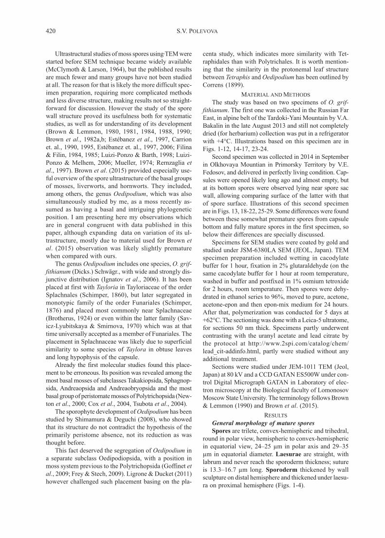
Figs. 8-14. Spores of Oedipodium griffithianum with details of sporoderm ultrastructure on the distal hemisphere, TEM (9-14)
and LCSM (8). 8: spore section in distal/proximal direсtion, showing in orange color fluorescence of cellulose in intine stained by
berberine, in contrast exine is dark brown; clavate bacula cover all surface of the distal hemisphere; the intine is thickened on the
proximal hemisphere; the middle layer is arrowed on the proximal hemisphere; 9: total spore section in parallel to proximal side,
which is somewhat invaginated, thus the laesura near proximal pole is cut (in the middle of the figure); 10: part of sporoderm
between bacula in a spore younger than one in Fig. 14; the filled with the electron-dark substance, narrow channels pierce the
outer exine, it is possible that this exine layer is formed by granule aggregation; 11: ultrastructure of distal sporoderm, showing
thin, electron-light intine (I), an almost unseen TPL layer, darked outer exine (E1), lighter inner exine (E2) and electron-dark
micro-granules of perine (P) on club tops; 12: distal sporoderm between clavae, with weak differentiation into E2 and E1, and
orbicules (O); TPL-layer is seen as dark line between exine and intine. 13: distal sporoderm in base of clavae, TPL unseen, E1
somewhat striolate, E2 homogenous, thin electron-light intine (I), with surface covered by orbicules (O) and mass of fine particules
of perine (P); 14: ultrastructure of distal sporoderm: thin, spotted, electron-light intine, the electron-dark TPL-layer, homogenous
inner exine, the thin electron-lighter outer exine with orbicules on its surface are visible;

424 S.V. POLEVOVA
0.5 m
15
E2
E1
ITPL
P
TPL
E2
E1
TPL
P0.5 m
20
1918
16
17
1 m
E2
I
TPL
E2
I
TPL
E2
ITPL
E2
I
TPL
E1
E1
E1
E1
P
P
P
P
I
0.5 m
1 mequato
rial -
- pro
ximal
I
TPL
E2
proximal -- equatorial

425Sporoderm ultrastructure of Oedipodium griffithiaum (Oedipodiopsida)
Thin electron-dark TPL-layer of to ca. 0.05 μm thick
occurs between the exine and the intine in distal hemi-
sphere in most spore sections (Figs. 12, 14), although
sometimes it is totally indiscernible (Figs. 11, 13). TPL-
layer is more apparent towards the equatorial region
where it changes into thicker and multilamellar, espe-
cially distinct at the bend to the proximal hemisphere
(Fig. 15). The multilaminar structure, however, is not
apparent in all sections: Figs. 16, 17 illustrate rather elec-
tron light TPL with non contrast lamellae while no trac-
es of lamellae are seen in Fig. 19. Further on from equa-
tor to proximal side, such TPL appears to be more homo-
geneous than intine, which in the distal hemisphere and
in transition from equator to proximal hemisphere, is
often distinctly fibrillose.
The total exine thickness is 0.58–0.67 μm between
the sculpture elements on the distal hemisphere. The outer
exine ranges from 0.12 to 0.17 μm, and the inner exine
being 0.42–0.52 μm between the sculpture elements on
the distal hemisphere. The intine thickness on the distal
hemisphere varies at 0.08–0.21 μm.
Proximal hemisphere. Sporoderm has the same lay-
ers without sculpture of clavae and bacula. Perine is also
fragmentary in fully mature spores (e.g. Fig. 24), but is
much better developed in spores from capsule bottom
(e.g. in Figs. 18, 20). The outer exine in such places has
more rough surface than in other places and somewhat
narrow channeled (Fig. 24 and 24a).
The total exine thickness (E1+E2) is 0.31–0.55 μm
between laesurae, thinning to 0.21–0.24 μm on the laesura
side. The outer exine is 0.10–0.18 μm and almost con-
stant throughout proximal hemisphere, while the inner
exine varies from 0.11–0.15 μm on the laesurae side and
to 0.21–0.42 μm in between laesurae. The TPL-layer be-
tween exine and intine is very different in thickness and
structure (see below). The intine ranges in thickness from
0.10–0.17 μm at equatorial zone to 0.54–1.52 μm on the
proximal pole.
Sporoderm ultrastructure of laesura is formed of thin
outer exine, thin inner exine, thick TPL-layer and in-
tine. Both exine layers are as apparent as in distal hemi-
sphere, becoming abruptly thin in the laesura centre (Figs.
21-24). A narrow canal in exine provides a contact be-
tween the intermediate layer and environment (Fig. 24a).
TPL-layer being often distinctly multilamellar in the
equatorial zone, abruptly changes towards of the proxi-
mal pole: apparent lamellae are disappearing, although a
weakly discernible lamellose patterns can be seen (Fig.
20). The overall color of the layer is quite similar to those
of intine or only slightly darker, however the wavy texture
allows delimitation of the TPL layer and intine without
difficulty. In many cases (e.g. Fig. 15), the TPL-layer has
“hatched” appearance, due to short irregularly arranged
dark lamella clusters spreading among the light matrix.
These clusters being darker provide a wavy appearance of
the layer, due to darker clusters of lamellae are arranged
at a narrow angle, 20-30°, with the inner exine border.
Ring-like structures are visible on the transverse cross-
sections of TPL-layer (middle layer) in laesura (Fig. 21-
23). It is probable that these structures looking like a
ring or slits on cross-sections are tubulose being formed
by lamellae. However details of the connections between
sharp continuous lamellae in the spore equatorial zone
and structures within the median part of laesura require
special studies.
DISCUSSION
Spores of Oedipodium are somewhat larger than av-
erage in mosses, considering that in many families 20
μm is the maximal spore size. In the moss spore atlas of
Boros et al. (1993), the mean size is below 20 μm in 151
species out of 210 species described.
The reason for such an upper limit is likely related to
the peristome, an organ specifically designed for opti-
mizing moss spore release. The ventral trabeculae on the
inner side of peristome teeth are spaced usually at 15-20
μm, which is the average cell size in moss sporophyte.
As they work as the mechanism carrying spores outside,
it can be assumed that the spore size in peristomate mosses
is under the pressure of natural selection, which adjusts
spore size to the distance between ventral trabecule. A
partial proof of such a correlation was published by Hut-
tunen et al. (2004) for pleurocarpous mosses, where the
enlarged spore size is associated with the peristome re-
duction/modification.
Thus 24–35 μm spore size in Oedipodium well coin-
cides with eperistomate Sphagnum, 15–41 μm, Andre-
aea, (10–)20–50(–110) μm; Andreaeobryum, (50–)90–
100(–120) μm, and Takakia, 25–36 μm.
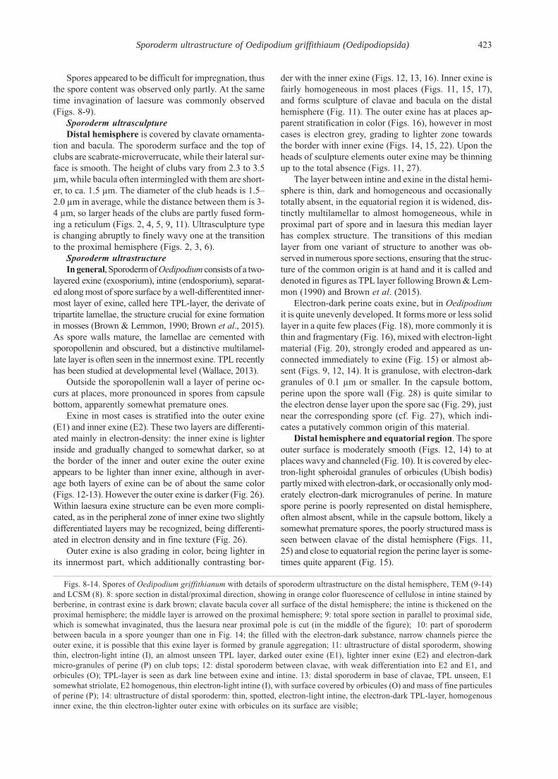
Figs. 15-20. Details of equatorial and proximal sporoderm ultrastructure in Oedipodium griffithianum, TEM. 15: Equatorial
area with transition to proximal hemisphere: perine is fragmentary, outer exine is layered, while the inner one is homogeneous;
distinctly lamellar at equator TPL-layer is broadened towards the proximal pole, with short dark ‘hachures’; 16: ultrastructure of
equatorial sporoderm, magnified: electron-grey intine, the lamellate more electron-dark middle layer, homogenous inner exine,
the thin more electron-light outer exine, and the microgranulate, electron-dark perine are visible; 17: section of equatorial sporoderm,
showing spotted, two exine layers with diffuse perine material, multilaminar TPL at the equator, continued into oblique-hatched
layer between exine and intine, thus treated homologous to TPL, although quite distinct structurally. 18: proximal surface between
laesulae, where the perine is remained, exine layers are homogeneous, broadened TPL-layer with ‘cloudy’ heterogeneity, and
fibrillose intine. 19: section of a collapsed spore where both proximal (above left) and distal (below right) surfaces are seen; note
thicker inner exine on distal side (outer exine is not shown on distal hemisphere), but much thicker TPL-layer and intine on
proximal side. 20: proximal surface section, from plane surface to laesura; note well retained perine, two-layered exine, and much
broadened TPL-layer and intine towards laesure; TPL includes darker and short ‘hatchures’ and few light lamellae, similar to
those in equatorial zone; intine is finely fibrillose and indistinctly two-layered.

426 S.V. POLEVOVA
23
E2
E1
P
E1
E2
�����
1 m
21
25
27 28 29
L
P
TPL
E1E2
����������
�����
�����
SSW
E2E2
E2
B
E1B
0.5 m
1 m
2 m
0.2 m
24
E1E2
P
P0.5 m
E1
E2
P 0.2 m
24a
22
TPL
P
E1
E2
TPL
I
1 m
26
E2E1
P
TPL

427Sporoderm ultrastructure of Oedipodium griffithiaum (Oedipodiopsida)
Even more apparent (although never statistically sup-
ported) is the trend to large and heavily ornamented spores
in eperisomate ephemeral acrocarpous mosses, e.g. Phy-
scomitrium, Physcomitrella, Weissia, Bruchia,
Ephemerum, Archidium. It seems that the advantages of
spore enlargements are almost universal, as there are a
rather few groups where gymnostomous sporophytes are
associated with the spores less than 20 μm.
In the spore wall structure, Oedipodium has a number
of characters that are rare in other mosses. These are: (1)
clear trilete laesura; (2) the lack of perine in the fully ma-
ture spores; (3) the column-like sculpture on the distal
hemisphere contrastingly different from the proximal side
which is only slightly wavy and the fusion of clavae heads
with reticulum formation on distal spore surface.
(1) The clear trilete laesura occurs in many groups
of hepatics and hornworts (Boros et al., 1993, Brown &
Lemmon, 1990; Brown et al., 2015 ), characterized by a
rather passive spore spreading. A clear trilete laesura is
never observed in mosses with well-developed peristomes,
being restricted to basal lineages, which are primarily
eperistomate: Sphagnum, Takakia, Oedipodium. However
the facts that some rather advanced groups, like Hedwi-
giaceae, Helicophyllaceae and Rhacocarpaceae may de-
velop at least a superficially very similar structure indi-
cate that this developmental pathway still remains. Note
however that the laesura in Hedwigia is much less dis-
tinctly armed compared with that in e.g. Sphagnum
(Brown et al., 1982a,b), as well as in Oedipodium.
(2) An unusual character of Oedipodium spores is the
absence of perine in the fully mature spores, although
slightly premature ones are covered by electron-dark gran-
ulose mass. The perine presence was indicated and illus-
trated for Oedipodium by Brown et al. (2015), who like-
ly described somewhat premature spores (given that
spores were still in tetrads and clavate elements were
lower, more scattered and not fused by their heads). Perine
forms spore surface sculpture in almost all reports of
mosses, with the only exception of Bruchia brevifolia
(McClymoth & Larson, 1964), however Rushing (1985)
supposed that in other species of this genus with ca. 25
species, spore walls may be formed with the more contri-
bution of perine.
Among the other moss genera studied with TEM for
the spore wall structure, a series of proportion on exine
and perine contribution to spore wall formation occur.
Most mosses have distinct perine sitting on the smooth
surface of exine. Loose and fragmentary deposition of
perine is typical to basal lineages of mosses including
Sphagnopsida, Andreaeopsida and Oedipodiopsida, al-
though it varies from species to species (Table 1).
There are also examples where the base of sculpture
elements is formed by electronically transparent layer of
exine (Trematodon longicollis, Ditrichum ssp., Polytri-
chum commune), while the main part of papillae are even-
ly dark to almost black, thus composed of material re-
ferred to for this reason as perine. Further, the spore wall
in Astomum phascoides has a sculpture formed both by
exine and perine, while in Ephemerum spinulosum perine
forms a thin (but continuous) layer upon sculpture formed
principally by exine.
The perine particles in mosses are usually larger than
observed here in Oedipodium, except Andreaea (Brown
& Lemmon, 1984).
The record of the electron-light perine (Estébanez et
al., 1997) likely corresponds to orbicules (Ubish bodies)
that are seen also on the sporoderm of Oedipodium, as
well as of Bruchia brevifolia (Fig. 6 in McClymont &
Larson, 1964). However the problem of referring to these
electron-light structures as perine require a specific study
of spore development.
(3) The column-like clavae on the distal spore surface
is not a unique feature in mosses, but the fusion by their
distal parts has never been reported except for Oedipodi-
Figs. 21-29. Details of laesura and inner wall of spore sac ultrastructure of Oedipodium griffithianum, TEM. 21: Transversal
section of laesura, showing greatly thickened middle layer, TPL intruding in laesura, which raised over the proximal surface (cf.
Fig. 3); the uniform two layered exine covers the middle layer on and between laesurae, the micro-granulate moderately electron-
dark perine remains between laesurae; 22: inner part of laesura with granulose perine and homogeneous inner and outer exine;
intine thickened immediately under laesure, while TPL-layer is much broadened, filling the main body of inner part of laesura;
hachures in TPL is directed towards the top of laesura; 23: upper part of laesura beside proximal pole; perine granulose, inner and
outer exine homogeneous; TPL included dark structures directed towards the laesura top; ring-like structure in TPL is arrowed; 24:
outer parts of ultrastructure of laesura nearly a place of convergence of two rays (cf. Fig. 3), showing narrow hole in two-layered
exine on the laesura center (cf. Figs. 3) and the loose micro-granulose perine; the thickened TPL layer contacts the spore environ-
ment, likely obtaining liquid water and conducting it through the intine and to a protoplast; 24a: close up of 24: note porose exine
near hollow, that are likely also may contribute to water uptake; 25: distal sporoderm of spore at open capsule bottom; the bacula
are composed of the homogeneous inner exine and heterogeneous outer exine, some bacteria are located between bacula; 26:
transverse section of laesura top, somewhat beside from the proximal pole; exine fully covers the surface and the main body is
filled by TPL-layer. 27: Spore sac wall (SSW) with nearby spore distal surface, in open capsule bottom; numerous bacteria are
located between the spore sac and spore; electron-dark micro-granules covers bacula, and especially space in between them, as
well as spore sac wall (cf. Figs. 25, 28-29); 28: part of outer exine with the electron-dark microgranules of perine; 29: part of spore
sac wall with the electron-dark microgranules similar to those in Fig. 28; сell wall and intracellular structures of spore sac not
visible; it is possible this granules are composed of the same material as the perine. Abbreviations: B: bacteria, C: clava, Е1: outer
exine, Е2: inner exine, I: intine, O: orbicule (Ubish body), ML: middle layer, Р: perine, PM: plasma membrane; SSW: spore sac
wall.

428 S.V. POLEVOVA
Tab
le 1
. A
com
par
ison o
f sp
oro
der
m l
ayer
s in
moss
es
Syst
emat
ic p
osi
tion
Sp
ecie
sP
erin
eE
xin
eL
ayer
bet
wee
n e
xin
eIn
tine
and i
nti
ne
Sphag
nopsi
da:
Sphag
nac
eae
Sphagnum
les
curi
i (B
row
n e
t al.
, 1982a,
b)
Loose
and f
ragm
enta
ryS
culp
ture
form
ing
Lam
ella
r ex
ine
AH
om
ogen
eous
or
not
spec
ifie
d
Sphag
nopsi
da:
Sphag
nac
eae
Sphagnum
capil
lifo
lium
(F
ilin
a &
Fil
in,1
985)
Loose
and f
ragm
enta
ryS
culp
ture
form
ing
Lam
ella
r ex
ine
Hom
ogen
eous
or
not
spec
ifie
d
Tak
akio
psi
da:
Tak
akia
ceae
:Ta
kaki
a c
erato
phyl
la (
Ren
zagli
a et
al.
, 1997;
Scu
lptu
re f
orm
ing
Thin
, e
lect
ron-l
ight
Lam
ella
r ex
ine
Hom
ogen
eous
or
not
spec
ifie
d
Bro
wn e
t al.
, 2015 )
Andre
aeopsi
da:
Andre
aeac
eae
An
dre
aea
ro
thii
(B
row
n &
Lem
mon,
1984)
Loose
and f
ragm
enta
ryL
oose
Not
obse
rved
Hom
ogen
eous
or
not
spec
ifie
d
Andre
aeopsi
da:
Andre
aeac
eae
An
dre
aea
ru
pes
tris
(F
ilin
a &
Fil
in,1
984)
Ab
sen
tL
oose
Not
obse
rved
Fib
rill
ose
Oed
ipodio
psi
da:
Oed
ipodia
ceae
Oed
ipodiu
m g
riff
ithia
num
(B
row
n e
t al.
, 2015)
Pre
sen
tS
culp
ture
form
ing
Lam
ella
r T
PL
at
pla
ces
Fib
rill
ose
Oed
ipodio
psi
da:
Oed
ipodia
ceae
Oed
ipodiu
m g
riff
ithia
num
(pre
sent
pap
er)
Loose
and f
ragm
enta
ryS
culp
ture
form
ing
Lam
ella
r, h
om
ogen
eous
Fib
rill
ose
to n
ot
obse
rved
at
pla
ces
Poly
tric
hopsi
da:
Poly
tric
hac
eae
Po
lytr
ich
um
co
mm
un
e (M
cCly
mont
& L
arso
n,
1964)
Scu
lptu
re f
orm
ing
Form
ing b
ases
of
?H
om
ogen
eous
or
not
spec
ifie
d
sculp
ture
ele
men
ts
Bry
opsi
da:
Funar
iace
aeP
hys
com
itri
um
turb
inatu
m (
McC
lym
ont
& L
arso
n,
1964)
Scu
lptu
re f
orm
ing
Th
in?
Hom
ogen
eous
or
not
spec
ifie
d
Bry
opsi
da:
Funar
iace
aeP
hys
com
itre
lla p
ate
ns
(Sch
uet
te e
t al.
, 2009)
Scu
lptu
re f
orm
ing
Tw
o l
ayer
edF
ibri
llat
e-g
ran
ula
teF
ibri
llat
e in
aper
ture
reg
ion
Bry
opsi
da:
Enca
lypta
ceae
Enca
lypta
rhabdoca
rpa (
McC
lym
ont
& L
arso
n,
1964)
Scu
lptu
re f
orm
ing
Th
in?
Hom
ogen
eous
or
not
spec
ifie
d
Bry
opsi
da:
Arc
hid
iace
aeA
rchid
ium
alt
ernif
oli
um
(M
cCly
mont
& L
arso
n,
1964)
Scu
lptu
re f
orm
ing
Th
in?
Lam
ella
r in
aper
ture
reg
ion
Bry
opsi
da:
Gri
mm
iace
aeG
rim
mia
ssp
. (E
steb
anez
et
al.
, 1997)
Scu
lptu
re f
orm
ing
Tw
o l
ayer
edL
amel
lar
in d
ista
lD
iffe
rent in
tine
in d
iffe
rent sp
ecie
s
hem
isp
her
e
Bry
opsi
da
Hel
icophyll
acea
eH
elic
ophyl
lum
torq
uatu
m (
Luiz
i-P
onzo
& M
elhem
, 2006)
Scu
lptu
re f
orm
ing
Th
in?
Lam
ella
te i
n a
per
ture
reg
ion
Bry
opsi
da
Pty
chom
itri
acea
eP
tych
om
itri
um
ssp
. (E
steb
anez
et.
al.
, 2006)
Scu
lptu
re f
orm
ing
Thin
or
thic
kL
amel
lar
or
non-l
amel
larB
ilay
ered
in d
iffe
rent
spec
ies
in d
iffe
rent
spec
ies
Bry
opsi
da
Bru
chia
ceae
Bru
chia
bre
vifo
lia (
McC
lym
ont
& L
arso
n,
1964)
Alm
ost
abse
nt
Scu
lptu
re f
orm
ing
Op
aqu
eH
om
ogen
eous
or
not
spec
ifie
d
Bry
opsi
da
Bru
chia
ceae
Tre
mato
don l
ongic
oll
is (
Bro
wn &
Lem
mon,
1981)
Scu
lptu
re f
orm
ing
Form
ing b
ases
of
Lam
ella
rF
ibri
llose
and l
amel
lar
sculp
ture
ele
men
ts
Bry
opsi
da
Fis
siden
tace
aeF
issi
den
s li
mbat
us
(Muel
lier
, 1974)
Scu
lptu
re f
orm
ing
Th
inE
lect
ron-g
ray
Hom
ogen
eous
or
not
spec
ifie
d
Bry
opsi
da
Dit
rich
acea
eD
itri
chum
pal
lidum
(B
row
n a
nd L
emm
on,
1980)
Scu
lptu
re f
orm
ing
Form
ing b
ases
of
Ele
ctro
n-d
ark i
nH
om
ogen
eous
or
not
spec
ifie
d
sculp
ture
ele
men
tspro
xim
al h
emis
pher
e
Bry
opsi
da:
Ephem
erac
eae
Ephem
erum
spin
ulo
sum
(M
cCly
mont
& L
arso
n,
1964)
Th
inS
culp
ture
form
ing
Op
aqu
eH
om
ogen
eous
or
not
spec
ifie
d
(or
Pott
iace
ae s
.l.)
Bry
opsi
da
Pott
iace
aeA
stom
um
phasc
oid
es (
McC
lym
ont
& L
arso
n,
1964)
Scu
lptu
re f
orm
ing
Form
ing b
ases
of
Op
aqu
eH
om
ogen
eous
or
not
spec
ifie
d
sculp
ture
ele
men
ts
Bry
opsi
da:
Pott
iace
aeP
ha
scu
m c
usp
ida
tum
(C
arri
on e
t al.
, 1990)
Scu
lptu
re f
orm
ing
Th
inS
epar
atin
g l
ayer
in
Hom
ogen
eous,
gra
y
pro
xim
al h
emis
pher
e
Bry
opsi
da:
Pott
iace
aeP
ha
scu
m c
usp
ida
tum
(M
cCly
mont
& L
arso
n,
1964)
Scu
lptu
re f
orm
ing
Th
in?
Hom
ogen
eous,
lig
ht
Bry
opsi
da:
Pott
iace
aeP
tery
goneu
rum
ssp
. (C
arri
on e
t. a
l.,
1995)
Scu
lptu
re f
orm
ing
Th
inS
epar
atin
g l
ayer
in
Fib
rill
ose
pro
xim
al h
emis
pher
e
Bry
opsi
da:
Pott
iace
aeW
eiss
ia v
irid
ula
(M
cCly
mont
& L
arso
n,
1964)
Scu
lptu
re f
orm
ing
Th
inO
paq
ue
Hom
ogen
eous
or
not
spec
ifie
d
Bry
opsi
da:
Ort
hotr
ichac
eae
Ort
hotr
ichum
(M
edin
e &
Est
eban
ez,
2014)
Scu
lptu
re f
orm
ing
Thin
, w
ith i
nner
Expan
ded
and w
ith
Bil
ayer
ed
lam
ella
ela
byri
nth
-lik
e in
trusi
ons

429Sporoderm ultrastructure of Oedipodium griffithiaum (Oedipodiopsida)
um. At the same time it appears to be quite similar to ect-
exine of angiosperms (Hesse et al. 2009). A somewhat sim-
ilar pattern has also been described in Haplomitrium
(Brown & Lemmon, 1986; Brown et al., 2015) and Apo-
treubia (Brown & Lemmon, 1990; Brown et al., 2015).
Contribution of spore wall layers in surface sculp-
turing
Perine is a layer forming the surface ornamenta-
tion in most mosses. More rarely exine participates in
the spore surface ornamentation, but most commonly
only slightly participates in forming of basal parts of
sculpture elements, e.g. in Astomum phascoides, Trem-
atodon longicollis, Ditrichum spp., Polytrichum com-
mune. Two latter species are rather ephemeral haplol-
epideous mosses of Dicranales (incl. Pottiales). A con-
siderable contribution of exine to the sculputre ele-
ment formation has been observed in Bruchia brevifo-
lia and Ephemerum spinulosum. Although not specially
studied, exine is the layer most likely responsible for
stellate ‘armature’ on spores of Encalypta sect. Cilia-
tae, in a way similar to hepatic genus Fossombronia
(Brown & Lemmon, 1993).
The layer between exine and intine treated here as TPL-
layer (Figs. 12-29) was recognized in many mosses, al-
though its terminology varies, as shown in Table 1. The
reason for various evaluation is likely, at least partly, re-
lated to the variation within the individual spores and also
to the stage of development. Oedipodium shows almost
all known states of this character, although some being
expressed only at specific places and moreover appear not
in all spores. Equatorial region with the maximally devel-
oped lamellar structure formed by 5 alternations of dark
and light zones (Figs. 15-17) is approaching to Sphag-
num, where however multilamellar layer is performed along
almost whole border of exine and intine. Lamellar struc-
ture is poorly expressed in the same place in spores from
capsule bottom (Figs. 18-19), however their putatively
premature state is hardly a reason. Brown et al. (2015)
illustrated a very distinct multilamellar structure in Oedi-
podium, in spores which are assumed to be somewhat pre-
mature (by reasons explained above).
In the aperture region, there are a number of com-
mon trends in mosses, including thinning of perine, and
more rarely thinning of exine, like it is seen in Oedipo-
dium laesura near proximal pole (Fig. 24). Splayed TPL
fills most volume of laesura, forming the aperture plug
and favoring sporeling. Electron-dark ‘hachures’ are ar-
ranged along the proximal spore wall in the proximal
hemisphere outside laesura and radially within laesura
itself (Fig. 22). The short lamellar plates are arranged in
direction from the pore where water uptake by spore is
possible. The obtained images allow suggestion that the
lamellae of the TPL-layer in proximity to aperture are
functioning as a wicks or even at places tubes, accelerat-
ing water conduction from the aperture to other spore
regions (cf. Figs. 15 & 20 with Figs. 21-23).
ACKNOWLEDGEMENTS
I am grateful to V.A. Bakalin and V.E. Fedosov for
providing plant material for this study, and to R.C. Brown,
V.F. Tarasevich and M.S. Ignatov for helpful comments
on the manuscript. SEM and TEM studied were con-
ducted in UFC MSU under support of MESRF. The work
was partly supported by the RSF grant 14-50-00029.
LITERATURE CITED
BOROS, A., M. JARAI-KOMLODI, Z. TOTH & S. NILSSON. 1993.An atlas of recent European bryophyte spores. – Akademic Press,Budapest. 321 S.
BROTHERUS, V. F. 1924. Musci. – In: Engler, A. & K. Prantl (eds.).Die Natürlichen Pflanzenfamilien ed. 2, 10, Berlin: Duncker and Hum-blot: 143–478.
BROWN, R.C. & B.E. LEMMON. 1980. Ultrastructure of sporogenesisin a moss, Ditrichum pallidum. III. Spore wall formation. – AmericanJournal of Botany 67: 918–934.
BROWN, R.C. & B.E. LEMMON. 1981. Aperture development in sporesof the moss Trematodon longicollis Mx. – Protoplasma 106: 273–287.
BROWN, R.C. & B.E. LEMMON. 1984. Spore wall development in An-dreaea (Musci: Andreaeopsida). – American Journal of Botany 71:412–420.
BROWN, R.C. & B.E. LEMMON. 1986. Spore wall development in theliverwort, Haplomitrium hookeri. – Canadian Journal of Botany 64:1174–1182.
BROWN, R.C. & B.E. LEMMON. 1988. Sporogenesis in bryophytes. –Advances in Bryology 3: 159–223.
BROWN, R.C. & B.E. LEMMON. 1990. Sporogenesis in bryophytes. –In: Blackmore S., S.H. Barnes (eds.) Pollen and spores: patterns ofdiversification. Systematics Association Special Volume No. 44. Ox-ford: Oxford Science Publications, 9–24.
BROWN, R.C. & B.E. LEMMON. 1993. Spore wall development in theliverwort Fossombronia wondraczekii (Corda) Dum. – Journal of theHattori Botanical Laboratory 74: 83–94.
BROWN, R.C., B.E. LEMMON & Z.B. CAROTHERS. 1982a. Sporewall development in Sphagnum lescurii. – Canadian Journal of Bot-any 60: 2394–2409.
BROWN, R.C., B.E. LEMMON & Z.B. CAROTHERS. 1982b. Sporewall ultrastructure of Sphagnum lescurii Sull. – Review of Palaeobot-any and Palynology 32: 99–107.
BROWN, R.C., B.E. LEMMON, M. SHIMAMURA, J.C. VILLARREAL& K.S. RENZAGLIA. 2015. Spores of relictual bryophytes: Diverseadaptations to life on land. – Review of Palaeobotany and Palynology216: 1–17.
CARRIÓN, J.S., J. GUERRA & R.M. ROS. 1990. Spore morphology ofthe European species of Phascum Hedw. (Pottiaceae, Musci) and itsclosest species. – Nova Hedwigia 51: 411–433.
CARRIÓN, J.S., M.J. CANO & J. GUERRA. 1995. Spore morphologyin the moss genus Pterygoneurum Jur. (Pottiaceae). – Nova Hedwigia61: 481–496.
CORRENS, C. 1899. Untersuchungen über die Vermehrung der Laub-moose durch Brutorgane und Stecklinge. – Jena, Verlag von GustavFischer. 742 pp.
COX, C.J., B. GOFFINET, A.J. SHAW & S.B. BOLES. 2004. Phyloge-netic relationships among the mosses based on heterogeneous Bayesiananalysis of multiple genes from multiple genomic compartments. – Sys-tematic Botany 29: 234–250.
ESTÉBANEZ, B., C. ALFAYATE & E. RON. 1997. Observations on sporeultrastructure in six species of Grimmia (Bryopsida). – Grana 36: 347–357.
ESTÉBANEZ, B., T. YAMAGUCHI & H. DEGUCHI. 2006. Ultrastruc-ture of the spore in four Japanese species of Ptychomitrium Fürnr. (Mus-ci). – Grana 45: 61–70.

430 S.V. POLEVOVA
[FILINA, N.I. & V.R. FILIN] ФИЛИНА Н.И., В.Р. ФИЛИН. 1984.
Развитие и строение спородермы Andreaea rupestris Hedw. (An-
dreaeaceae, Musci). – [The structure and development of the sporo-
derm in Andreaea rupestris Hedw. (Andreaeaceae, Musci)]. Вестник
Московского Университета. Сер.16, Биология [Vestnik Mosk-
ovskogo Universiteta, Series 16, Biology] 89: 86–100.
[FILINA, N.I. & V.R. FILIN] ФИЛИНА Н.И., В.Р. ФИЛИН. 1985.
Развитие и строение спородермы у Sphagnum capillifolium (Ehrh.)
Hedw. (Sphagnaceae, Musci). – [Ultrastructure and development of sporo-
derm in Sphagnum capillifolium (Ehrh.) Hedw. (Sphagnaceae, Musci]
Вестник Московского Университета. Сер.16, Биология [Vestnik
Moskovskogo Universiteta, Series 16, Biology] 16 (1): 51–60.
FREY, W. & M. STECH. 2009. Bryophyta (Musci, mosses). – In: Frey,W. (ed.), Syllabus of plant families A. Engler’s Syllabus der Pflan-zenfamilien. Part 3. Bryophytes and seedless vascular plants. 13thed. Gebr. Borntraeger Verlagsbuchhandlung, Stuttgart, Germany:116–257.
GOFFINET, B., W.R. BUCK & A.J. SHAW. 2009. Morphology, anato-my, and classification of the Bryophyta. – In: B. Goffinet & A.J. Shaw(eds.). Bryophyte biology, 2nd edn. Cambridge: Cambridge Univer-sity Press:55–138.
HESSE, M., H. HALBRITTER, R. ZETTER, M. WEBER, R. BUCH-NER, R.A. FROSCH-RADIVO & S. ULRICH. 2009. Pollen Termi-nology. – Wien, Springer Verlag, 261 pp.
HORTON, D.G. 1983. A revision of the Encalyptaceae (Musci), with par-ticular reference to the North American taxa. Part 2. – Journal of theHattori Botanical Laboratory 54: 353–532.
HUTTUNEN, S., M. S. IGNATOV, K. MÜLLER & D. QUANDT. 2004.Phylogeny and evolution of epiphytism in the three moss families Me-teoriaceae, Brachytheciaceae and Lembophyllaceae. – Monographs inSystematic Botany from the Missouri Bot. Garden 98: 328-361.
IGNATOV, M.S., E.A. IGNATOVA & V.YA. CHERDANTSEVA. 2006.Oedipodium griffithianum (Dicks.) Schwägr. (Oedipodiopsida, Bryo-phyta), a new species and class for Russia. – Arctoa 15: 211-214.
IRELAND, R. R. 1987. Scanning electron microscope study of the sporesof the North American species of Plagiothecium. – Memoirs of theNew York Botanical Garden 45: 95–110.
KUNGU, E. M., R. LONGTON & L. BONNER. 2007. Character reduc-tion and peristome morphology in Entodontaceae: constraints on an in-formation source. – In: Newton, A.E. & R.S. Tangney (eds.). Pleuro-carpous mosses: systematics and evolution. Systematic AssociationSpecial Volume 71: 247–268.
LIGRONE, R. & J.G. DUCKETT. 2011. Morphology versus moleculesin moss phylogeny: new insights (or controversies) from placental andvascular anatomy in Oedipodium griffithianum. – Plant Systematicsand Evolution 296: 275–282.
LUIZI-PONZO, A.P. & O.M. BARTH. 1998. Spore morphology of someBruchiaceae species (Bryophyta) from Brazil. – Grana 37:222–227.
LUIZI-PONZO, A.P. & T.S.A. MELHEM. 2006. Spore morphology andultrastructure of the tropical moss Helicophyllum torquatum (Hook.)Brid. (Helicophyllaceae) in relation to systematics and evolution. – Cryp-togamie Bryologie 27: 413–420.
McCLYMOTH, J.W. 1955. Spore studies in the Musci with the specialreference to the genus Bruchia. – Bryologist 58: 287–306.
McCLYMONT, J.W. & D.A. LARSON. 1964. An electron-microscopicstudy of spore wall structure in the Musci. – American Journal of Bot-any 51: 195–200.
MEDINA, N.G., B. ESTÉBANEZ. 2014. Does spore ultrastructure mir-ror different dispersal strategies in mosses? A study of seven IberianOrthotrichum species. – PLoS ONE 9(11): e112867. doi:10.1371/journal.pone.0112867.
MUELLER, D.M.J. 1974. Spore wall formation and chloroplast develop-ment during sporogenesis in the moss Fissidens limbatus. – AmericanJournal of Botany 61: 525–534.
NEWTON, A.E., C.J. COX, J.G. DUCKETT, J.A. WHEELER, B. GOFFI-NET, T.A. J. HEDDERSON & B.D. MISHLER. 2000. Evolution ofthe major moss lineages: phylogenetic analyses based on multiple genesequences and morphology. – Bryologist 103: 187–211.
RENZAGLIA, K.S., K.D. McFARLAND & D.K. SMITH. 1997. Anato-my and ultrastructure of the sporophyte of Takakia ceratophylla (Bryo-phyta). – American Journal of Botany 84(10): 1337–1350.
RUSHING, A.E. 1985. Spore morphology in the genus Bruchia Schwaegr.(Misci). – American Journal of Botany 72: 75–85.
SHIMAMURA, M. & H. DEGUCHI. 2008. Sporophyte anatomy of Oedi-podium griffithianum (Oedipodiaceae). – In: Mohamed, H. B.B. Baki,A. Nasrulhaq-Boyce et al. (eds.) Bryology in the New Millennium,Kuala-Lumpur: 319–325.
[SAVICZ-LYUBITSKAYA, L.I. & Z.N. SMIRNOVA] САВИЧ-ЛЮ-
БИЦКАЯ, Л.И., З.Н. СМИРНОВА. 1970. Определитель листосте-
бельных мхов СССР. Верхоплодные мхи. – [Handbook of mosses of
the USSR. The acrocarpous mosses] Л., Наука [Leningrad, Nauka],
822 pp.
SCHIMPER, W. P. 1860. Synopsis Muscorum Europaeorum, praemissaintroductione de elementis bryologicis tractante. – Stuttgartiae. Sump-tibus Librariae E. Schweizerbart, CLIX+735 pp.+8 Tabl.
SCHIMPER, W. P. 1876. Synopsis Muscorum Europaeorum, praemissaintroductione de elementis bryologicis tractante. Ed. 2. – Stuttgartiae.Sumptibus Librariae E. Schweizerbart (E. Koch), 886 pp.
SCHUETTE, S., A.J. WOOD, M. GEISLER, J. GEISLER-LEE, R. LI-GRONE & K.S. RENZAGLIA. 2009. Novel localization of callose inthe spores of Physcomitrella patens and phylogenomics of the callosesynthase gene family. – Annals of Botany 103: 749–756.
SMITH, G.L. 1971. Conspectus of the genera of Polytrichaceae. – Mem-oirs of the New York Botanical Garden 21(3): 1–83.
TSUBOTA, H., E. DE LUNA, D. GONZÁLEZ, M.S. IGNATOV & H.DEGUCHI. 2004. Molecular phylogenetics and ordinal relationshipsbased on analyses of a large-scale data set of 600 rbcL sequences ofmosses. – Hikobia 14: 149–170.
WALLACE, S. 2013 Evolutionary development of the plant spore andpollen wall. – PhD Thesis, University of Sheffield, 157 pp.
ZHANG, YU-LONG & WU PENG-CHEN. 2005. Spore morphology ofChinese bryophytes. – Beijing, Qingdao Publ. House 339+178 plates[in Chinese].