Embed Size (px)
Citation preview

E X P E R I M E N T A L C E L L R E S E A R C H 3 1 4 ( 2 0 0 8 ) 6 0 3 – 6 1 5
ava i l ab l e a t www.sc i enced i rec t . com
www.e l sev i e r. com/ loca te /yexc r
Research Article
Stem molecular signature of adipose-derived stromal cells
Daniele Peronia, Ilaria Scambia, Annalisa Pasinib, Veronica Lisib, Francesco Bifarib,Mauro Kramperab, Gino Rigottic, Andrea Sbarbatia, Mirco Galièa,⁎aDepartment of Morphological and Biomedical Sciences, Section of Anatomy and Histology, University of Verona, ItalybDepartment of Clinical and Experimental Medicine, Section of Haematology, University of Verona, ItalycPlastic and Reconstructive Surgery (II Division), City Hospital, Verona, Italy
A R T I C L E I N F O R M A T I O N
⁎ Corresponding author. Fax: +39 045 8027163E-mail address: [email protected] (
0014-4827/$ – see front matter © 2007 Elsevidoi:10.1016/j.yexcr.2007.10.007
A B S T R A C T
Article Chronology:Received 27 June 2007Revised version received10 September 2007Accepted 6 October 2007Available online 17 October 2007
It has recently been shown that adipose tissue is an abundant and easily accessiblesource of stromal progenitor cells (ADSCs, adipose-derived stromal cells), resemblingthe mesenchymal stem cells (MSCs) obtained from adult bone marrow. However, theidentification of these two lineages is still controversial and even the stem cell natureof ADSCs is doubted. In this study, we examined the “stemness” transcriptional profileof ADSCs and BM-MSCs, with two aims: (1) to compare their “stem cell molecularsignature” and (2) to dissect their constitutive expression pattern for moleculesinvolved in tissue development, homeostasis and repair. As well as severalmolecules involved in matrix remodeling and adult tissue angiogenesis and repair,we detected the expression of genes UTF-1, Nodal, and Snail2, which are known to beexpressed by embryonic stem cells but have been never described in other stemlineages. In addition, for the first time we described the transcriptional profile ofhuman BM-MSCs and ADSCs for the CD44 splice variants, which are determinant incell trafficking during embryonic development, in adult tissue homeostasis and also intumor dissemination.
Thus, our findings strongly support a close relationship between ADSCs and BM-MSCs, suggest an unexpected similarity between MSCs and embryonic stem cells, andpossibly support the potential therapeutic application of ADSCs.
© 2007 Elsevier Inc. All rights reserved.
Keywords:Mesenchymal stem cellsAdipose tissueUTF-1NodalSnail2CD44 splice variants
Introduction
Despite a common immunophenotype [1], the identification ofadipose-derived mesenchymal stem cells with bone-marrow-derived mesenchymal stem cells (BM-MSCs) is still controver-sial, and even their stem cell nature is doubted; for this reason,the first cell type is frequently referred to by the term
.M. Galiè).
er Inc. All rights reserved
“adipose-derived stromal cells” (ADSCs). However, ADSCsshare important biological properties with BM-MSCs, sincethey can be induced to myogenic [2], cardiomyogenic [3],epithelial [4], endothelial [5,6] and neurogenic [7] differentia-tion. In addition, both cell typesmay release trophicmediatorssuch as pro-angiogenic and anti-inflammatory factors [8]. Thefunctional analogy with BM-MSCs would seem to encourage
.

604 E X P E R I M E N T A L C E L L R E S E A R C H 3 1 4 ( 2 0 0 8 ) 6 0 3 – 6 1 5
the use of ADSCs for tissue engineering and cell therapystrategies. At the same time, clarification of their nature isrequired through the definition of a “molecular signature”based on stem-cell-related genes, such as markers of stemlineages and genes regulating developmental and regenera-tive processes. A large body of literature has identified thegrowth factors, adhesion molecules, cytokines/chemokinesand extracellular matrix proteins that drive embryonic devel-opment. Some of these molecules are known to orchestratehomeostasis and regenerative processes such as tissue turn-over, revascularization of ischemic regions, and woundhealing, or pathological processes such as inflammation andtumor dissemination. Several studies have addressed themolecular characterization of BM-MSCs and ADSCs [9–12], butlittle is known about their expression pattern for stemness-related molecules. The gene expression profile for thesemolecules is predictive of the therapeutic potential of ADSCsand provides a molecular basis for the clinical benefitsrecently reported in preliminary studies in murine models ofhuman diseases [13,14].
The capacity to regulate its own motility in response tosurrounding stimuli is an intrinsic aspect of stem cell pheno-type, which must be understood in view of ADSC-basedtherapeutic applications [8]. An important adhesion moleculethat plays a crucial role in cell migration/invasion programs isCD44 [15]. In addition to the standard CD44 (CD44s), manyisoforms generated by the retention of alternative exonsduring pre-mRNA splicing have been identified. While CD44sis expressed inmostmammalian cells and tissues, the splicingvariants are restricted to certain cytotypes of ectodermalorigin, such as keratinocytes and basal cells of squamous andglandular epithelia, or to certain physiological or pathologicalconditions, such as embryonic epithelial–stromal interaction[16], activation of T lymphocytes [17] and dissemination ofcancer cells [18,19]. All these phenomena are based oninvasive behavior and capacity to interact with the surround-ing microenvironment [20]. Although MSCs are known tosplice out CD44 alternative exons, the expression pattern ofCD44 isoforms in human ADSCs (hADSCs) and BM-MSCs(hBM-MSCs) has never been investigated.
Here we provide a high-throughput transcriptional ana-lysis of ADSCs, with two aims: (1) to compare the ‘stem cell’molecular signature of ADSCs with that of other stem cellpopulations, such as BM-MSCs, and (2) to dissect their
Fig. 1 – Cytofluorimetric identification of MSCs. Human ADSCs anbasis of the their surface immunophenotypical profile.
constitutive expression pattern for molecules involved intissue development, homeostasis and repair.
Materials and methods
Cell isolation
hADSCs and mADSCsHuman ADSCs were obtained from lipoaspirates of healthydonors after informed consent. Murine ADSCs were iso-lated from inguinal adipose tissues of C57BL/6 mice. Forcell isolation, we used the following standard procedure:the extracellular matrix was digested at 37 °C in HBSS with1 mg/ml collagenase type I (GIBCO life technology) and 2%BSA. After incubation, digestion enzyme activity wasneutralized with Dulbecco's modified Eagle medium(DMEM) containing 20% fetal bovine serum (FBS), andcentrifuged at 1200×g for 10 min to obtain a high-densitypellet, which constitutes the stromal vascular fraction(SVF). It was then resuspended in 160 mM NH4Cl andincubated at room temperature for 10 min to lysecontaminating red blood cells. The SVF was collected bycentrifugation and filtered through a 70-μm nylon mesh toremove cell debris.
SVF was cultured in 25-cm2 flasks (BD Falcon™, BectonDickinson, Milan, Italy) at a concentration of 1×10⁵ cells/cm2,using DMEM with high glucose concentration, GLUTAMAXI™, 20% heat-inactivated fetal calf serum (FCS), 100 U/mlpenicillin and 100 μg/ml streptomycin (all from GibcoBRL/Life Technologies, Milan, Italy).
hBM-MSCsHuman BM-MSCs were obtained from BM aspirates ofhealthy donors, after informed consent. BM mononuclearcells were isolated using density gradient centrifugation(Lymphoprep, Nycomed Pharm, Oslo, Norway), as previouslydescribed [21].
Cell culture
SVF and BM-MSC cultures were incubated at 37 °C in a 5% CO2
atmosphere. After 72 h, non-adherent cells were removed.When 70–80% confluent, adherent cells were trypsinized (0.05%
d BM-MSCs (a) andmurine ADSCs (b) were recognized on the

Table 1 – Stem cell related genes constitutively expressed in human ADSCs and BM-MSCs
Position Gene symbol Description BM-MSC ADSC
4 ACTG2 Actin, gamma 2, smooth muscle, enteric 0.39 0.355 ACVR1 Activin A receptor, type I 0.63 0.6210 BDNF Brain-derived neurotrophic factor 0.42 0.2623 BMPR2 Bone morphogenetic protein receptor, type II (serine/threonine kinase) 0.59 0.4328 CTNNB1 Catenin (cadherin-associated protein), beta 1, 88 kDa 0.40 0.3030 CCNE1 Cyclin E1 0.40 0.2933 CD24 CD24 antigen (small cell lung carcinoma cluster 4 antigen) 1.00 0.9235 CD44 CD44 antigen (homing function and Indian blood group system) 0.63 0.4636 CD9 CD9 antigen (p24) 0.30 0.3337 CDH1 Cadherin 1, type 1, E-cadherin (epithelial) 0.54 0.4844 CDKN1A Cyclin-dependent kinase inhibitor 1A (p21, Cip1) 0.30 0.3145 CDKN1B Cyclin-dependent kinase inhibitor 1B (p27, Kip1) 0.49 0.4946 CDKN2A Cyclin-dependent kinase inhibitor 2A (melanoma, p16, inhibits CDK4) 0.29 0.3047 CDKN2D Cyclin-dependent kinase inhibitor 2D (p19, inhibits CDK4) 0.49 0.4852 COL6A2 Collagen, type VI, alpha 2 0.39 0.4053 CST3 Cystatin C (amyloid angiopathy and cerebral hemorrhage) 1.02 0.9254 CXCL12/SDF-1 Chemokine (C–X–C motif) ligand 12, SDF-1 (stromal cell-derived factor 1) 0.51 0.4062 EGF Epidermal growth factor (beta-urogastrone) 0.28 0.2963 EGFR Epidermal growth factor receptor (erythroblastic leukemia viral
(v-erb-b) oncogene homolog, avian)0.37 0.42
69 FGF1 Fibroblast growth factor 1 (acidic) 0.28 0.3474 FGF16 Fibroblast growth factor 16 0.29 0.3078 FGF2 Fibroblast growth factor 2 (basic) 0.54 0.5484 FGF5 Fibroblast growth factor 5 0.34 0.3385 FGF6 Fibroblast growth factor 6 0.32 0.35105 GATA4 GATA binding protein 4 0.34 0.28120 GJB5 Gap junction protein, beta 5 (connexin 31.1) 0.30 0.23121 HSPA9B Heat shock 70-kDa protein 9B (mortalin-2) 0.56 0.38122 ICAM-1 Intercellular adhesion molecule 1 (CD54), human rhinovirus receptor 0.23 0.27126 IGF2 Insulin-like growth factor 2 (somatomedin A) 0.30 0.35128 IL6 Interleukin 6 (interferon, beta 2) 0.45 0.50134 INS Insulin 0.68 0.62135 INSRR Insulin receptor-related receptor 0.37 0.37141 ITGA5 Integrin, alpha 5 (fibronectin receptor, alpha polypeptide) 0.85 0.54144 ITGA8 Integrin, alpha 8 0.23 0.30148 ITGAV Integrin, alpha V (vitronectin receptor, alpha polypeptide, antigen CD51) 0.30 0.31150 ITGB1 Integrin, beta 1 (fibronectin receptor, beta polypeptide, antigen
CD29 includes MDF2, MSK12)0.51 0.50
154 ITGB5 Integrin, beta 5 0.42 0.31166 MDM2 Mdm2, transformed 3T3 cell double minute 2, p53 binding protein (mouse) 0.30 0.41179 NODAL Nodal homolog (mouse) 0.39 0.38180 NOG Noggin 0.39 0.42191 PAX6 Paired box gene 6 (aniridia, keratitis) 0.28 0.32192 PDGFA Platelet-derived growth factor alpha polypeptide 0.27 0.32200 POU5F1 POU domain, class 5, transcription factor 1 0.57 0.38210 PUM2 Pumilio homolog 2 (Drosophila) 0.31 0.33221 SNAI2 Snail homolog 2 (Drosophila) 0.31 0.28224 SOX13 SRY (sex determining region Y)-box 13 0.31 0.34226 SOX17 SRY (sex determining region Y)-box 17 0.31 0.25227 SOX18 SRY (sex determining region Y)-box 18 1.00 0.92228 SOX2 SRY (sex determining region Y)-box 2 0.34 0.30229 SOX3 SRY (sex determining region Y)-box 3 0.38 0.38243 THY1 Thy-1 cell surface antigen 0.40 0.37245 TNC Tenascin C (hexabrachion) 0.33 0.31248 UTF1 Undifferentiated embryonic cell transcription factor 1 0.66 0.52249 VCAM-1 Vascular cell adhesion molecule 1 0.26 0.28250 VEGF Vascular endothelial growth factor 0.32 0.29257 WNT5B Wingless-type MMTV integration site family, member 5B 0.30 0.26258 WNT6 Wingless-type MMTV integration site family, member 6 0.79 0.69260 WNT7B Wingless-type MMTV integration site family, member 7B 0.31 0.27261 WNT8A Wingless-type MMTV integration site family, member 8A 0.86 0.76262 ZFP42 Zinc finger protein 42 0.35 0.29
605E X P E R I M E N T A L C E L L R E S E A R C H 3 1 4 ( 2 0 0 8 ) 6 0 3 – 6 1 5

Fig. 2 – Stem-related transcriptional profile of hADSCs and hBM-MSCs. (a) Gene arrays are represented as the average of thearrays of long-term and short-term cultures of hADSCs and BM-MSCs, respectively. (b) The scatterplot shows that none of thegenes was overexpressed more than 2-fold (green lines) in one lineage as compared to the other.
Fig. 3 – Confirmation of expression of the selected genes byRT-PCR. mRNA from BM-MSCs and ADSCs subjected toretrotranscription as described in Materials and methods.PCR amplification with specific primers confirmed theexpression of genes implicated in matrix remodeling, cellmigration (Integrin αV, Integrin β5, Integrin β1, CD44s) and inembryogenesis (Pou5f1, UTF-1, Nodal, Snail2).
606 E X P E R I M E N T A L C E L L R E S E A R C H 3 1 4 ( 2 0 0 8 ) 6 0 3 – 6 1 5
trypsin at 37 °C for 5 min, GibcoBRL/Life Technologies),harvested and washed with medium to remove trypsin, andexpanded in larger flasks. A homogenous cell population ofADSCs or BM-MSCs was obtained after 2 to 3 weeks of culture.Cells at early (2 to 5, short-term cultures) and late (5 to 20, long-termcultures) passage inculturewereused for theexperiments.
HADSCs and hBM-MSCs were identified on the basis oftheir expression of CD105 (endoglin), CD73, CD29, CD44and CD90, and the lack of hematopoietic (CD45, CD14,CD34) and endothelial cell markers (CD31), all assessedusing cytofluorimetric analysis, as previously described [21](Fig. 1a).
The identification of mADSCs was based on the expressionof CD106 and the lack of hematopoietic (CD45, CD14, CD34)and endothelial cell (CD31) marker expression, as previouslydescribed [22] (Fig. 1b).
Both ADSCs and BM-MSCs were cultured in DMEM contain-ing 20% fetal bovine serum(FBS). Cultures were carried on inparallel by using the same FBS lot.

607E X P E R I M E N T A L C E L L R E S E A R C H 3 1 4 ( 2 0 0 8 ) 6 0 3 – 6 1 5
CFU-F assay
Three aliquots of 1×10⁵ SVF cells for each of 7 patients wereseeded in three 10-cm-diameter cell culture dishes, and after10 days of culture the number of adherent cells was estimatedby colony forming unit fibroblast (CFU-F) count. The culturelayer was fixed in 4% formalin for 15 min and than stained byGIEMSA. Only the coloniesmacroscopically visible as blue spotwere counted.
Microarray analysis
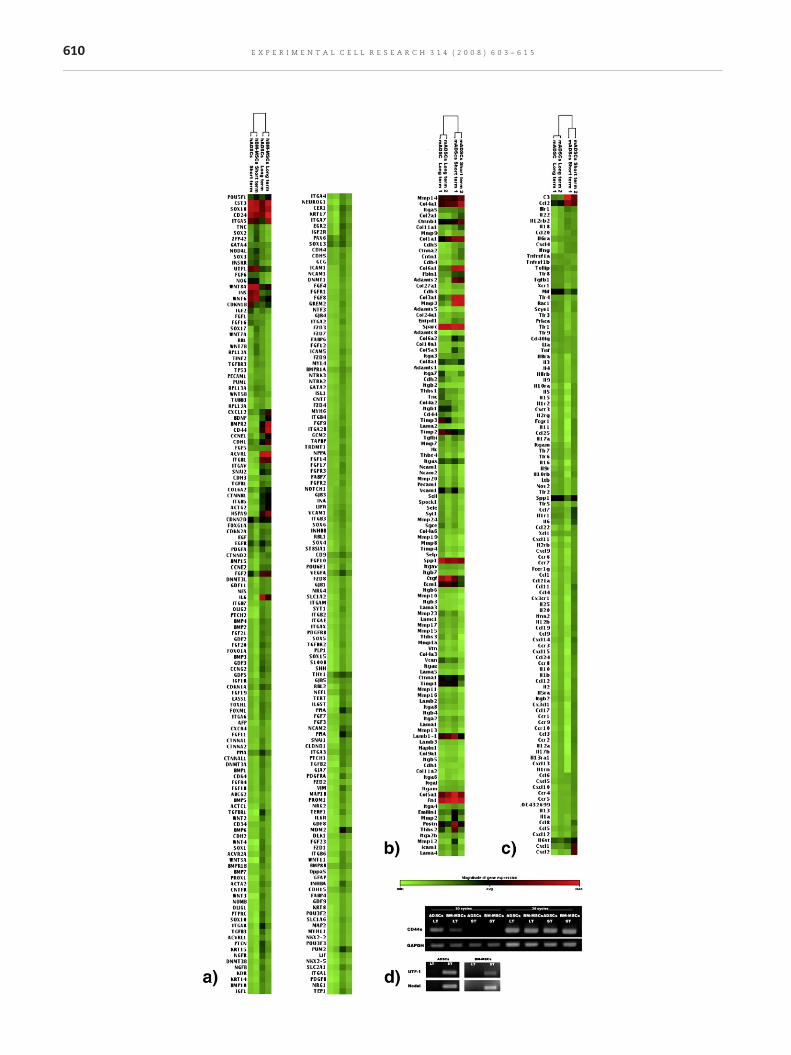
SamplesSince several authors have reported some transcriptionalchanges upon in vitro culture of MSCs [23,24], transcrip-tional analysis was performed on short-term and long-termcultured human ADSCs and BM-MSCs. The expressionvalues were reported as the average of the values obtainedfrom different time-cultured samples of the same lineage(Table 1).
Fig. 4 – Microarray-based transcriptional analysis of C57BL/6 ADGene arrays are represented as the average of the arrays of long
Stemness-related gene array was performed on short-termand long-term cultures of one lineage of hADSCs and onelineage of BM-MSCs. HADSCs and hBM-MSCs were obtainedfrom different patients. RT-PCRs were performed using RNAfrom different lineages with respect to that used for arrays.
Murine arrays were performed in duplicate using short-and long-term cultures of two distinct lineages of mADSCs,each obtained from a different animal.
Experimental procedureThe different types of mRNA extracted from each cellpopulation were used as templates to generate a cDNAlibrary. cRNA labeled with UTP–biotin (Enzo Roche MolecularBiochemicals, Mannheim, Germany) was retrotranscriptedand purified. UTP–biotin–cRNA from human MSCs washybridized on Gene Arrays containing probes specific forhuman genes related to stem phenotype, adhesion moleculesand cytokines/chemokines (from SuperArray® BioscienceCorporation, Frederick, MD, USA; Cat. nos. HS-601.2). TheUTP-biotin-cRNA from murine MSCs was hybridized in Gene
SCs for adhesion molecules and inflammatory cytokines. (a)-term and short-term cultured cells, respectively.
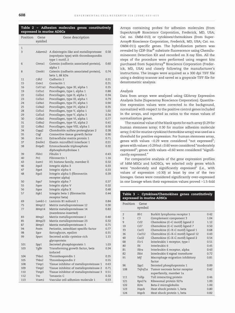
Table 2 – Adhesion molecules genes constitutivelyexpressed in murine ADSCs
Position Genesymbol
Gene description
13 Adamts2 A disintegrin-like and metalloprotease
(reprolysin type) with thrombospondintype 1 motif, 2
0.58
6 Ctnna1 Catenin (cadherin associated protein),alpha 1
0.60
8 Ctnnb1 Catenin (cadherin associated protein),beta 1, 88 kDa
0.74
11 Cdh2 Cadherin 2 0.3115 Cntn1 Contactin 1 0.3116 Col11a1 Procollagen, type XI, alpha 1 0.3519 Col1a1 Procollagen, type I, alpha 1 0.8822 Col2a1 Procollagen, type II, alpha 1 0.3423 Col3a1 Procollagen, type III, alpha 1 0.7924 Col4a1 Procollagen, type IV, alpha 1 0.9025 Col4a2 Procollagen, type IV, alpha 2 0.3528 Col5a1 Procollagen, type V, alpha 1 1.0229 Col5a3 Procollagen, type V, alpha 3 0.3430 Col6a1 Procollagen, type VI, alpha 1 0.7731 Col6a2 Procollagen, type VI, alpha 2 0.4132 Col8a1 Procollagen, type VIII, alpha 1 0.5034 Cspg2 Chondroitin sulfate proteoglycan 2 0.3835 Ctgf Connective tissue growth factor 0.9036 Ecm1 Extracellular matrix protein 1 0.7137 Emilin1 Elastin microfibril interfacer 1 0.5138 Entpd1 Ectonucleoside triphosphate
diphosphohydrolase 10.32
39 Fbln1 Fibulin 1 0.4340 Fn1 Fibronectin 1 1.1643 Icam1 H1 histone family, member 0 0.3246 Itga3 Integrin alpha 3 0.3347 Itga4 Integrin alpha 4 0.3348 Itga5 Integrin alpha 5 (fibronectin
receptor alpha)0.39
50 Itga7 Integrin alpha 7 0.3755 Itgav Integrin alpha V 0.3256 Itgax Integrin alpha X 0.4857 Itgb1 Integrin beta 1 (fibronectin
receptor beta)0.44
69 Lamb1-1 Laminin B1 subunit 1 0.8475 Mmp12 Matrix metalloproteinase 12 0.3077 Mmp14 Matrix metalloproteinase 14
(membrane-inserted)0.82
83 Mmp2 Matrix metalloproteinase 2 0.4085 Mmp23 Matrix metalloproteinase 23 0.3287 Mmp3 Matrix metalloproteinase 3 0.7594 Postn Periostin, osteoblast specific factor 0.7798 Sgce Sarcoglycan, epsilon 0.3199 Sparc Secreted acidic cysteine rich
glycoprotein1.15
101 Spp1 Secreted phosphoprotein 1 1.03103 Tgfbi Transforming growth factor, beta
induced0.34
104 Thbs1 Thrombospondin 1 0.35105 Thbs2 Thrombospondin 2 0.55108 Timp1 Tissue inhibitor of metalloproteinase 1 0.63109 Timp2 Tissue inhibitor of metalloproteinase 2 0.71110 Timp3 Tissue inhibitor of metalloproteinase 3 0.51112 Tnc Tenascin C 0.32113 Vcam1 Vascular cell adhesion molecule 1 0.53
608 E X P E R I M E N T A L C E L L R E S E A R C H 3 1 4 ( 2 0 0 8 ) 6 0 3 – 6 1 5
Arrays containing probes for adhesion molecules (fromSuperArray® Bioscience Corporation, Frederick, MD, USA;Cat. no. OMM-013) or cytokines/chemokines (from Super-Array® Bioscience Corporation, Frederick, MD, USA; Cat. no.OMM-011) specific genes. The hybridization pattern wasrevealed by CDP-Star® substrate fluorescence using Chemilu-minescent Detection Kit and recorded on X-ray film. All thesteps of the procedure were performed using reagent kitspurchased from SuperArray® Bioscience Corporation (Freder-ick, MD, USA) and closely following the manufacturer'sinstructions. The images were acquired as a 300 dpi TIFF fileusing a desktop scanner and saved as a grayscale TIFF file fordensitometric analysis.
AnalysisData from arrays were analyzed using GEArray ExpressionAnalysis Suite (Superarray Bioscience Corporation). Quantita-tive expression values were corrected to the background,normalized with respect to the positive control genes includedin the arrays, and reported as ratios to the mean values ofnormalization genes.
Themaximal value of theblank spots for each array (0.29 forhuman stemness array; 0.30 for murine adhesion moleculearray; 0.42 formurine cytokine/chemokine array)wasused as athreshold for positive expression. For human stemness array,genes with values b0.29 were considered “not expressed”;geneswith values≥0.29 but ≤0.60were considered “moderatelyexpressed”; genes with values =0.60 were considered “signifi-cantly expressed.”
For comparative analysis of the gene expression profilesof hBM-MSCs and hADSCs, we selected only genes whichwere “moderately and significantly expressed” (i.e., withvalues of expression ≥0.30) at least by one of the twolineages. Genes were considered significantly over-expressedin one lineage when their expression values proved N1.5-fold
Table 3 – Cytokines/Chemokines genes constitutivelyexpressed in murine ADSCs
Position Genesymbol
2 Blr1 Burkitt lymphoma receptor 1 0.423 C3 Complement component 3 1.049 Ccl2 Chemokine (C–C motif) ligand 2 1.1219 Ccl7 Chemokine (C–C motif) ligand 7 0.5033 Cxcl1 Chemokine (C–X–C motif) ligand 1 0.6836 Cxcl12 Chemokine (C–X–C motif) ligand 12 0.4340 Cxcl2 Chemokine (C–X–C motif) ligand 2 0.5468 Il1r1 Interleukin 1 receptor, type I 0.5180 Il6 Interleukin 6 0.4581 Il6ra Interleukin 6 receptor, alpha 0.4382 Il6st Interleukin 6 signal transducer 0.7291 Mif Macrophage migration inhibitory
factor0.81
96 Spp1 Secreted phosphoprotein 1 0.89108 Tnfrsf1a Tumor necrosis factor receptor
superfamily, member 1a0.42
111 Tollip Toll interacting protein 0.46121 Rps27a Ribosomal protein S27a 1.00122 B2m Beta-2 microglobulin 1.00123 Hspcb Heat shock protein 1, beta 0.80124 Hspcb Heat shock protein 1, beta 0.82

Table 4 – Culture-associated transcriptional changes forstemness-related genes in human cells
Symbol Description Ratio
Genes over-expressed in short-term vs. long-term human ADSCsCDKN1B Cyclin-dependent kinase inhibitor 1B
(p27, Kip1)1.88
INS Insulin 2.18ITGA5 Integrin, alpha 5 (fibronectin receptor,
alpha polypeptide)1.63
NOG Noggin 2.00UTF1 Undifferentiated embryonic cell
transcription factor 11.66
WNT6 Wingless-type MMTV integration sitefamily, member 6
1.77
WNT8A Wingless-type MMTV integration sitefamily, member 8A
1.93
Genes under-expressed in short-term vs. long-term human ADSCsACTG2 ATP-binding cassette, sub-family G (WHITE),
member 20.38
ACVR1 Activin A receptor, type I 0.18BMPR2 Bone morphogenetic protein receptor, type II
(serine/threonine kinase)0.29
CTNNB1 Catenin (cadherin-associated protein), beta 1,88 kDa
0.39
CCNE1 Cyclin E1 0.62CD44 CD44 molecule (Indian blood group) 0.30CD9 CD9 molecule 0.66CDH1 Cadherin 1, type 1, E-cadherin (epithelial) 0.63COL6A2 Collagen, type VI, alpha 2 0.65HSPA9 Heat shock 70-kDa protein 9 (mortalin) 0.28IL6 Interleukin 6 (interferon, beta 2) 0.23ITGA8 Integrin, alpha 8 0.39ITGB1 Integrin, beta 1 (fibronectin receptor, beta
polypeptide, antigen CD29 includes MDF2,MSK12)
0.23
ITGB5 Integrin, beta 5 0.44MDM2 Mdm2, transformed 3T3 cell double
minute 2, p53 binding protein (mouse)0.20
PTEN Phosphatase and tensin homolog(mutated in multiple advancedcancers 1)
0.43
PUM2 Pumilio homolog 2 (Drosophila) 0.33SNAI2 Snail homolog 2 (Drosophila) 0.27TGFBR1 Transforming growth factor,
beta receptor I (activin A receptortype II-like kinase, 53 kDa)
0.26
VEGFA Vascular endothelial growth factor A 0.54
Genes over-expressed in short-term vs. long-term human BM-MSCsFGF16 Fibroblast growth factor 16 1.52FGF6 Fibroblast growth factor 6 1.79INS Insulin 2.07INSRR Insulin receptor-related receptor 1.93NODAL Nodal homolog (mouse) 1.81NOG Noggin 2.59SOX3 SRY (sex determining region Y)-box 3
1.52UTF1 Undifferentiated embryonic cell transcription
factor 12.05
WNT6 Wingless-type MMTV integration site family,member 6
1.56
WNT7A Wingless-type MMTV integration site family,member 7A
1.67
Table 4 (continued)
Symbol Description Ratio
Genes under-expressed in short-term vs. long-term human BM-MSCsACTG2 Actin, gamma 2, smooth muscle, enteric 0.31ACVR1 Activin A receptor, type I 0.17BDNF Brain-derived neurotrophic factor 0.32BMPR2 Bone morphogenetic protein receptor, type II
(serine/threonine kinase)0.18
CTNNB1 Catenin (cadherin-associated protein), beta 1,88 kDa
0.27
CTNND2 Catenin (cadherin-associated protein), delta 2(neural plakophilin-related arm-repeat protein)
0.52
CCNE1 Cyclin E1 0.31CCNE2 Cyclin E2 0.44CD44 CD44 molecule (Indian blood group) 0.22CDH1 CD44 molecule (Indian blood group) 0.40CDKN1A Cyclin-dependent kinase inhibitor 1A
(p21, Cip1)0.58
CDKN2A Cyclin-dependent kinase inhibitor 2A(melanoma, p16, inhibits CDK4)
0.55
COL6A2 Collagen, type VI, alpha 2 0.47CXCL12 Chemokine (C–X–C motif) ligand 12
(stromal cell-derived factor 1)0.38
FGF2 Fibroblast growth factor 2 (basic) 0.43HSPA9 Heat shock 70-kDa protein 9 (mortalin) 0.25IL6 Interleukin 6 (interferon, beta 2) 0.22ITGAV Integrin, alpha V (vitronectin receptor,
alpha polypeptide, antigen CD51)0.45
ITGB1 Integrin, beta 1 (fibronectin receptor, betapolypeptide, antigen CD29 includesMDF2, MSK12)
0.29
ITGB5 Integrin, beta 5 0.41MDM2 Mdm2, transformed 3T3 cell double
minute 2, p53 binding protein (mouse)0.40
NCAM2 Neural cell adhesion molecule 2 0.50PUM2 Pumilio homolog 2 (Drosophila) 0.63SNAI2 Snail homolog 2 (Drosophila) 0.30
609E X P E R I M E N T A L C E L L R E S E A R C H 3 1 4 ( 2 0 0 8 ) 6 0 3 – 6 1 5
higher than the expression of the same gene in the otherlineage. On the contrary, genes were considered significantlyunder-expressed in one lineage when their expressionvalues were b0.75-fold that of the expression of the samegene in the other lineage.
RT-PCR
The expression of genes of interest was confirmed by RT-PCR.One hundred nanograms of mRNA from each cell populationunderwent reverse transcription (RT) in a reaction volume of20 μl and in the presence of random primers (Invitrogen). Onemicroliter of cDNA was then amplified by PCR, using theReactionReady HotStart PCR Mix and the appropriate primersets, both purchased from Superarray. Gene-specific primersused in this study were as follows: Nodal (PPH01944A9), UTF-1(PPH02391A), Snail2 (Slug) (PPH02475A), Integrin β5 (PPH00634A),Integrin αV (PPH00628A), and Integrin β1 (PPH00650A).
The PCR conditions were as follows: an initial denaturationof 15 min at 94 °C, 35 cycles (or 40 for Nodal and UTF-1) at 94 °Cfor 15 s, 59 °C for 30 s, 72 °C for 30 s, and a final extension of5 min at 72 °C.
Pou5f1 was amplified by using the primers: Fw—5′-GACAAC AAT GAG AAC CTT CAG GAG A-3′; Rw—5′-CTG GCG CCGGTT ACA GAA CCA-3′. The mixture was first heated at 94 °C
610 E X P E R I M E N T A L C E L L R E S E A R C H 3 1 4 ( 2 0 0 8 ) 6 0 3 – 6 1 5

611E X P E R I M E N T A L C E L L R E S E A R C H 3 1 4 ( 2 0 0 8 ) 6 0 3 – 6 1 5
for 2 min. Amplification was performed for 35 cycles at 94 °Cfor 45 s, 55 °C for 30 s and 72 °C for 90 s, followed by 72 °C for5 min.
PCR products were further analyzed on 2% agarose gels andphotographed with a CCD camera.
Analysis of CD44 splice variants by exon-specific PCR
To assess the expression of different isoforms of CD44 inMSCs,the mRNA was retrotranscripted as described above. Onemicroliter of resulting cDNA was subjected to PCR using Taqbuffer 1× (Promega), 1.5 mM MgCl2, 0.2 mM dNTPs, 1 U of Taqpolymerase (Promega) and 0.4 μM of each specific primer. Thestandard CD44 (CD44s) was detected using primers corre-sponding to 5′ (hSF 5′-AAGACATCTACCCCAGCAAC-3′) and 3′(hSR 5′-CCAAGATGATCAGCCATTCTGG-3′) constant domains.The variant isoforms were amplified using a set of 5′ primersthat specifically match to variable exons, and the common 3′primer hSR. The exon-specific primer sequences were thesame as used by Van Weering et al. [25]: pv2 5′-GATGAGCAC-TAGTGCTACAG-3′; pv3I 5′-ACGTCTTCAAATACCATCTC-3′;pv3II 5′-TGGGAGCCAAATGAAGAAAA-3′; pv4 5′-TCAACCA-CACCACGGGCTTT-3′; pv5 5′-GTAGACAGAAATGGCACCAC-3′;pv6 5′-GAGGCAACTCCTAGTAGTAC-3′; pv7 5′-CAGCCT-CAGCTCATACCAGC-3′; pv8 5′-TCCAGTCATAGTACAACGCT-3′; pv9 5′-CAGAGCTTCTCTACATCACA-3′; pv10 5′-GGTGGA-AGAAGAGACCCAAA-3′.
We omitted the exon v1 analysis because it is not expressedin human cells. Furthermore, we used two different primerpairs for exon v3 because it displays two alternative splicingforms (CD44v3I and CD44v3II).
PCR conditions were as follows: 94 °C for 2 min, 55 °C for2 min, 72 °C for 2 min (1×); 94 °C for 45 s, 55 °C for 1 min, 72 °Cfor 1 min (35×); 72 °C for 5 min. Amplification products wereresolved on 2% agarose gels.
Statistical analysis
To statistically evaluate the similarity of molecular signaturesin ADSCs and BM-MSCs we calculated the correlation index.Pb0.05 was taken to be statistically significant.
Results
Quantitative estimate of CFU-F population in humanlipoaspirates
On the basis of the CFU-F assays, we estimated that 1 ml ofhuman lipoaspirate contained at least 7.3×10³±6.3×10³ Cellscapable of forming fibroblast-like colonies, corresponding to0.5–5% of the total SVF population (4.04±3.26 on average).Despite the high individual variability observed, which is to be
Fig. 5 – Transcriptional changes with culture. In order to assesstranscriptome of short- and long-term cultured cells. Hierarchical(a), adhesionmolecules (b) and cytokines/chemokines (c) inmurincells at the same time points of culture than between cells at difconfirmed the increased expression of CD44 in long-term (LT) thresulted undetectable by RT- PCR in short-term cultured cells.
expected in human patients, this quantitative estimationdemonstrated that SFV of human lipoaspirates contains anamount of CFU-F which is 400 times greater than that of bonemarrow, where CFU frequency has been estimated at around 1per 105 cells (corresponding to a few hundred MSCs per cubiccentimeter of marrow) [26].
Human ADSCs and BM-MSCs have analogous transcriptionalphenotypes
Transcriptional analysis of hBM-MSCs and hADSCs revealed avirtually identical profile of genes identifying the stem cellphenotype (Fig. 2a). The list of genes included in the analysisand the quantification of their transcriptional expression aredetailed in the Supplementary Data.
The correlation coefficient between hADSCs and hBM-MSCs was 0.93 (Pb0.01), indicating that the gene expressionprofiles were very similar. None of the genes was over-expressed more than 2-fold in one lineage compared to theother (Fig. 2b).
Taking into account only the “expressed” genes (seeMaterials and methods), the only gene that displayed asignificantly different expression (N1.5-fold) in the two kindsof MSCs was brain-derived neurotrophic factor (BDNF). It wasmoderately expressed in hBM-MSCs (0.42) but was notdetected in hADSCs.
The consistency of the results obtained using differentsources and donors suggests that the transcriptional pheno-type described here was not due to individual variability andcan be generalized.
The ADSC molecular signature includes genes typical of otherstem lineages
Interestingly, both hBM-MSCs and hADSCs from differentpatients and at different culture time points expressedmolecules that are typical of embryonic phenotype, such asOct4, and that have never been described in other stemlineages so far, i.e., undifferentiation transcription factor (UTF-1)and Nodal (Figs. 2a and 3; Table 1). These molecules are crucialfor the migration of embryonic precursors during develop-ment; in addition, they are necessary to maintain theundifferentiated status of embryonic stem cells in culture.
Regenerative profile of hBM-MSCs and hADSCs
The genes that were constitutively expressed in both hBM-MSCs and hADSCs are listed in Table 1. This list includedgenes that are involved in tissue development, homeostasisand repair, such as adhesionmolecules (integrin α5, integrin αV,Integrin β1, Integrin β5), cytokines/chemokines (Cxcl 12/SDF-1,Interleukin 6), cyclin-dependent kinase inhibitors (CDK 1A/p21Cip1, CDK 1B/p27Kip1, CDK 2A/p16, CDK 2D/p19), growth
culture-associated transcriptional changes we compareclustergrams for stemness-related molecules in human cellse cells demonstrated an higher statistical correlation betweenferent time points of culture. Semi-quantitative RT-PCR (d)an in short-term (ST) cultured human cells. UTF-1 and Nodal
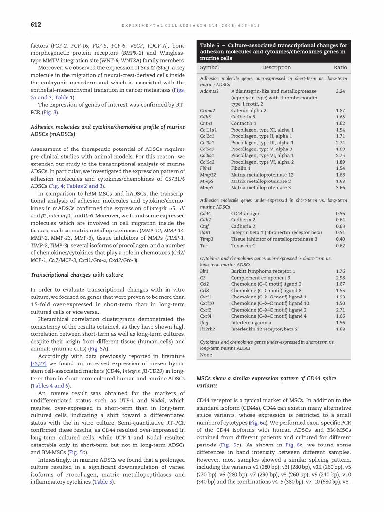
Table 5 – Culture-associated transcriptional changes foradhesion molecules and cytokines/chemokines genes inmurine cells
Symbol Description Ratio
Adhesion molecule genes over-expressed in short-term vs. long-termmurine ADSCsAdamts2 A disintegrin-like and metalloprotease
(reprolysin type) with thrombospondintype 1 motif, 2
3.24
Ctnna2 Catenin alpha 2 1.87Cdh5 Cadherin 5 1.68Cntn1 Contactin 1 1.62Col11a1 Procollagen, type XI, alpha 1 1.54Col2a1 Procollagen, type II, alpha 1 1.71Col3a1 Procollagen, type III, alpha 1 2.74Col5a3 Procollagen, type V, alpha 3 1.89Col6a1 Procollagen, type VI, alpha 1 2.75Col6a2 Procollagen, type VI, alpha 2 1.89Fbln1 Fibulin 1 1.54Mmp12 Matrix metalloproteinase 12 1.68Mmp2 Matrix metalloproteinase 2 1.63Mmp3 Matrix metalloproteinase 3 3.66
Adhesion molecule genes under-expressed in short-term vs. long-termmurine ADSCsCd44 CD44 antigen 0.56Cdh2 Cadherin 2 0.64Ctgf Cadherin 2 0.63Itgb1 Integrin beta 1 (fibronectin receptor beta) 0.51Timp3 Tissue inhibitor of metalloproteinase 3 0.40Tnc Tenascin C 0.62
Cytokines and chemokines genes over-expressed in short-term vs.long-term murine ADSCsBlr1 Burkitt lymphoma receptor 1 1.76C3 Complement component 3 2.98Ccl2 Chemokine (C–C motif) ligand 2 1.67Ccl8 Chemokine (C–C motif) ligand 8 1.55Cxcl1 Chemokine (C–X–C motif) ligand 1 1.93Cxcl10 Chemokine (C–X–C motif) ligand 10 1.50Cxcl2 Chemokine (C–X–C motif) ligand 2 2.71Cxcl4 Chemokine (C–X–C motif) ligand 4 1.66Ifng Interferon gamma 1.56Il12rb2 Interleukin 12 receptor, beta 2 1.68
Cytokines and chemokines genes under-expressed in short-term vs.long-term murine ADSCsNone
612 E X P E R I M E N T A L C E L L R E S E A R C H 3 1 4 ( 2 0 0 8 ) 6 0 3 – 6 1 5
factors (FGF-2, FGF-16, FGF-5, FGF-6, VEGF, PDGF-A), bonemorphogenetic protein receptors (BMPR-2) and Wingless-type MMTV integration site (WNT-6,WNT8A) family members.
Moreover, we observed the expression of Snail2 (Slug), a keymolecule in the migration of neural-crest-derived cells insidethe embryonic mesoderm and which is associated with theepithelial–mesenchymal transition in cancer metastasis (Figs.2a and 3; Table 1).
The expression of genes of interest was confirmed by RT-PCR (Fig. 3).
Adhesion molecules and cytokine/chemokine profile of murineADSCs (mADSCs)
Assessment of the therapeutic potential of ADSCs requirespre-clinical studies with animal models. For this reason, weextended our study to the transcriptional analysis of murineADSCs. In particular, we investigated the expression pattern ofadhesion molecules and cytokines/chemokines of C57BL/6ADSCs (Fig. 4; Tables 2 and 3).
In comparison to hBM-MSCs and hADSCs, the transcrip-tional analysis of adhesion molecules and cytokine/chemo-kines in mADSCs confirmed the expression of integrin α5, αVand β1, catenin β1, and IL-6. Moreover,we found someexpressedmolecules which are involved in cell migration inside thetissues, such as matrix metalloproteinases (MMP-12, MMP-14,MMP-2, MMP-23, MMP-3), tissue inhibitors of MMPs (TIMP-1,TIMP-2, TIMP-3), several isoforms of procollagen, and a numberof chemokines/cytokines that play a role in chemotaxis (Ccl2/MCP-1, Ccl7/MCP-3, Cxcl1/Gro-α, Cxcl2/Gro-β).
Transcriptional changes with culture
In order to evaluate transcriptional changes with in vitroculture, we focused on genes that were proven to bemore than1.5-fold over-expressed in short-term than in long-termcultured cells or vice versa.
Hierarchical correlation clustergrams demonstrated theconsistency of the results obtained, as they have shown highcorrelation between short-term as well as long-term cultures,despite their origin from different tissue (human cells) andanimals (murine cells) (Fig. 5A).
Accordingly with data previously reported in literature[23,27] we found an increased expression of mesenchymalstem cell-associated markers (CD44, Integrin β1/CD29) in long-term than in short-term cultured human and murine ADSCs(Tables 4 and 5).
An inverse result was obtained for the markers ofundifferentiated status such as UTF-1 and Nodal, whichresulted over-expressed in short-term than in long-termcultured cells, indicating a shift toward a differentiatedstatus with the in vitro culture. Semi-quantitative RT-PCRconfirmed these results, as CD44 resulted over-expressed inlong-term cultured cells, while UTF-1 and Nodal resulteddetectable only in short-term but not in long-term ADSCsand BM-MSCs (Fig. 5b).
Interestingly, in murine ADSCs we found that a prolongedculture resulted in a significant downregulation of variedisoforms of Procollagen, matrix metallopeptidases andinflammatory cytokines (Table 5).
MSCs show a similar expression pattern of CD44 splicevariants
CD44 receptor is a typical marker of MSCs. In addition to thestandard isoform (CD44s), CD44 can exist in many alternativesplice variants, whose expression is restricted to a smallnumber of cytotypes (Fig. 6a).We performed exon-specific PCRof the CD44 isoforms with human ADSCs and BM-MSCsobtained from different patients and cultured for differentperiods (Fig. 6b). As shown in Fig 6c, we found somedifferences in band intensity between different samples.However, most samples showed a similar splicing pattern,including the variants v2 (280 bp), v3I (280 bp), v3II (260 bp), v5(270 bp), v6 (280 bp), v7 (290 bp), v8 (260 bp), v9 (240 bp), v10(340 bp) and the combinations v4–5 (380 bp), v7–10 (680 bp), v8–

Fig. 6 – Expression profile of CD44 isoforms. (a) The CD44 pre-mRNA contains at least 20 exons, ten of which are constitutivelyexpressed and encode the standard isoform (CD44s). The remaining exons (v1–v10) can be alternatively spliced to generate over700 different isoforms (CD44v). Exon-specific RT-PCR (b) demonstrated that ADSCs and BM-MSCs show a similar CD44expression pattern (c).
613E X P E R I M E N T A L C E L L R E S E A R C H 3 1 4 ( 2 0 0 8 ) 6 0 3 – 6 1 5
9 (340 bp), v8–10 (540 bp) and 9–10 (430 bp). Furthermore, aftershort-term culture ADSCs have additional splicing variants incomparison with long-term cultures, including v5–10 (930 bp),and v6–10 (810 bp).
Discussion
In this study we showed that BM-MSCs and ADSCs exhibita virtually identical transcriptional profile for genes relatedto the stem cell phenotype, supporting the characteriza-tion of ADSCs as a peripheral lineage of MSCs. Interest-ingly, we found that BM-MSCs and ADSCs express variousgenes shared by embryonic stem cells. Along with Pou5f1(Oct4), which has been described to be expressed in MSCs[28], we found UTF-1, a common marker of embryonicstem cells, and Nodal which represents key signal in theembryonic stem cell system. UTF-1 and Nodal mRNAs arenormally found in embryonic stem lineages and in germline tissues, but their expression is rapidly downregulatedafter cell fate determination, so that their disappearancerepresents an early sign of the differentiation process [29].Our study is the first to show the expression of UTF-1 andNodal transcripts, in adult stem cells also, thus supportinga close relationship between MSCs and embryonic stemcells.
The molecular analogy with BM-MSCs and even embryo-nic stem cells supports the use of ADSCs for diversetherapeutic applications, such as plastic surgery, tissuerepair and revascularization of ischemic tissues, and pro-vides a molecular basis for the clinical benefits recentlyreported in preliminary studies on murine models of humanpathologies [13]. A large body of literature concerns the
mitogenic, angiogenic and differentiative signals involved inthese mechanisms. We have shown here that differentmolecules involved in these signaling pathways are con-stitutively expressed in ADSCs as well as in BM-MSCs.Besides basic FGF (FGF2), VEGF and PDGF-A, whose involve-ment in the recruitment of endothelial cell precursors iswell established, we found the Cxcl12/SDF-1 gene expressed.The chemokine Cxcl12/SDF-1 has recently been shown toregulate the trafficking of hematopoietic stem cells (HSCs)from the bone marrow to peripheral blood and to enhancepost-ischemia angiogenesis [30]. In addition, Cxcl12/SDF-1 isinvolved in embryonic development, as it is a crucialregulator of the early and late phases of embryogenesis[31,32] and it plays a role in the migration of germ cellprecursors [33].
In addition to multilineage plasticity and pro-angiogenicpotential, the capacity to home in on their putative nichethrough tissue barriers is an intrinsic aspect of stem cellphenotype. Therefore, identification of molecules involved incell mobilization during embryonic development, inflamma-tion, wound healing and tumor dissemination is useful inpredicting the therapeutic potential of ADSCs [8,14] and thepossible risks associated with their use. In our study ofmADSCs, we observed the expression of several matrixmetalloproteinases (MMP-12, MMP-14, MMP-2, MMP-23,MMP-3) and tissue inhibitors of MMPs (TIMP-1, TIMP-2, TIMP-3) which contribute to physiological remodeling of theextracellular matrix. In particular, MMP-2 and TIMP-2 areessential for the invasive capacity of BM-MSCs [34]. Amongthe invasion/migration molecules detected, Snail2 (Slug) isalso of interest. Snail2 is a zinc finger transcription factorwhich plays a prominent role in embryonic developmentthrough the regulation of neural crest migration [35]. It also

614 E X P E R I M E N T A L C E L L R E S E A R C H 3 1 4 ( 2 0 0 8 ) 6 0 3 – 6 1 5
has an important role in pathological conditions since it canconfer motile phenotype to neoplastic cells [36,37]. Itsconstitutive expression in MSCs suggests that Snail2 is akey regulator of the physiological properties of this stem cellpopulations, as well as of the predisposition of stem cells tospontaneous transformation [38].
Like Snail2, CD44 splice variants (CD44v) are widely studiedfor their role in cell growth and trafficking in both physiolo-gical and pathological conditions. Although CD44 representsone of the main identifying markers of the MSCs, and its rolein MSCs migration has been well-documented [39], theexpression pattern of CD44 splice variants in ADSCs hasnever been investigated so far. Our results demonstrate thatCD44 is alternatively spliced in BM-MSCs and ADSCs, leadingto similar expression patterns in the two cell types, which donot vary significantly during in vitro expansion. Thus, theheterogeneous expression of CD44 isoformsmay contribute tothe plasticity of MSCs and their interaction with the micro-environment, which are both typical aspects of stem cellphenotype.
Taken together, our results support the strong similarity ofADSCs and BM-MSCs, suggesting that ADSCs may be aspromising as BM-MSCs for various therapeutic applications,such as hematopoietic stem cell transplantation [40,41], tissueregeneration [42], treatment of severe acute graft versus hostdisease [43] and autoimmune disorders [44].
However, we found that prolonged in vitro culture sig-nificantly alters the transcriptional phenotype of both ADSCsand BM-MSCs. In accordance with data reported by otherauthors we shown an increased expression of mesenchymalstem-associated adhesion molecules, such as CD44 and inte-grinB1/CD29 both in human than inmurine cells. Moreover, wefound the drastic downregulation of extracellular matrix-associated molecules (varied procollagen isoforms, MMPs andTIMPs). The culture-associated changes in adhesion moleculeprofile reasonably reflect the adaptation to the new micro-environment. It should be a relevant insight to assess if thesechanges are reversible or if they stably affects the stem cellphysiology. Of notice, we found also the downregulation ofembryonic markers, such as UTF1 and Nodal, with culture.
Acknowledgments
We thank Dr. Paolo Farace for the graphic elaboration of genearray images. This work was supported by Cassa di Risparmiodi Verona, Vicenza, Belluno e Ancona.
Appendix A. Supplementary data
Supplementary data associated with this article can be found,in the online version, at doi:10.1016/j.yexcr.2007.10.007.
R E F E R E N C E S
[1] P.A. Zuk, M. Zhu, P. Ashjian, D.A. De Ugarte, J.I. Huang,H. Mizuno, Z.C. Alfonso, J.K. Fraser, P. Benhaim, M.H. Hedrick,
Human adipose tissue is a source of multipotent stem cells,Mol. Biol. Cell 13 (2002) 4279–4295.
[2] H. Mizuno, P.A. Zuk, M. Zhu, H.P. Lorenz, P. Benhaim,M.H. Hedrick, Myogenic differentiation by humanprocessed lipoaspirate cells, Plast. Reconstr. Surg. 109(2002) 199–209.
[3] K.G. Gaustad, A.C. Boquest, B.E. Anderson, A.M. Gerdes,P. Collas, Differentiation of human adipose tissue stem cellsusing extracts of rat cardiomyocytes, Biochem. Biophys. Res.Commun. 314 (2004) 420–427.
[4] M. Brzoska, H. Geiger, S. Gauer, P. Baer, Epithelialdifferentiation of human adipose tissue-derived adult stemcells, Biochem. Biophys. Res. Commun. 330 (2005) 142–150.
[5] V. Planat-Benard, J.S. Silvestre, B. Cousin, M. Andre, M.Nibbelink, R. Tamarat, M. Clergue, C. Manneville,C. Saillan-Barreau, M. Duriez, A. Tedgui, B. Levy, L. Penicaud,L. Casteilla, Plasticity of human adipose lineage cells towardendothelial cells: physiological and therapeutic perspectives,Circulation 109 (2004) 656–663.
[6] Y. Cao, Z. Sun, L. Liao, Y. Meng, Q. Han, R.C. Zhao, Humanadipose tissue-derived stem cells differentiate intoendothelial cells in vitro and improve postnatalneovascularization in vivo, Biochem. Biophys. Res. Commun.332 (2005) 370–379.
[7] K.M. Safford, K.C. Hicok, S.D. Safford, Y.D. Halvorsen, W.O.Wilkison, J.M. Gimble, H.E. Rice, Neurogenic differentiation ofmurine and human adipose-derived stromal cells, Biochem.Biophys. Res. Commun. 294 (2002) 371–379.
[8] A.I. Caplan, J.E. Dennis, Mesenchymal stem cells as trophicmediators, J. Cell Biochem. 98 (2006) 1076–1084.
[9] T.M. Liu, M. Martina, D.W. Hutmacher, J.H. Hui, E.H. Lee,B. Lim, Identification of common pathways mediatingdifferentiation of bone marrow- and adipose tissue-derivedhuman mesenchymal stem cells into three mesenchymallineages, Stem Cells 25 (2007) 750–760.
[10] A.J. Katz, A. Tholpady, S.S. Tholpady, H. Shang, R.C. Ogle, Cellsurface and transcriptional characterization of humanadipose-derived adherent stromal (hADAS) cells, Stem Cells23 (2005) 412–423.
[11] A.C. Boquest, A. Shahdadfar, K. Fronsdal, O. Sigurjonsson,S.H. Tunheim, P. Collas, J.E. Brinchmann, Isolation andtranscription profiling of purified uncultured human stromalstem cells: alteration of gene expression after in vitro cellculture, Mol. Biol. Cell 16 (2005) 1131–1141.
[12] S. Gronthos, D.M. Franklin, H.A. Leddy, P.G. Robey, R.W.Storms, J.M. Gimble, Surface protein characterization ofhuman adipose tissue-derived stromal cells, J. Cell Physiol.189 (2001) 54–63.
[13] E. Zappia, S. Casazza, E. Pedemonte, F. Benvenuto, I. Bonanni,E. Gerdoni, D. Giunti, A. Ceravolo, F. Cazzanti, F. Frassoni,G. Mancardi, A. Uccelli, Mesenchymal stem cells ameliorateexperimental autoimmune encephalomyelitis inducingT-cell anergy, Blood 106 (2005) 1755–1761.
[14] Y. Miyahara, N. Nagaya, M. Kataoka, B. Yanagawa, K. Tanaka,H. Hao, K. Ishino, H. Ishida, T. Shimizu, K. Kangawa, S. Sano,T. Okano, S. Kitamura, H. Mori, Monolayered mesenchymalstem cells repair scarred myocardium after myocardialinfarction, Nat. Med. 12 (2006) 459–465.
[15] H. Ponta, L. Sherman, P.A. Herrlich, CD44: from adhesionmolecules to signalling regulators, Nat. Rev., Mol. Cell Biol. 4(2003) 33–45.
[16] D.Wainwright, L. Sherman, J. Sleeman, H. Ponta, P. Herrlich, Asplice variant of CD44 expressed in the rat apical ectodermalridge contributes to limb outgrowth, Ann. N.Y. Acad. Sci. 785(1996) 345–349.
[17] R. Arch, K. Wirth, M. Hofmann, H. Ponta, S. Matzku,P. Herrlich, M. Zoller, Participation in normal immuneresponses of a metastasis-inducing splice variant of CD44,Science 257 (1992) 682–685.

615E X P E R I M E N T A L C E L L R E S E A R C H 3 1 4 ( 2 0 0 8 ) 6 0 3 – 6 1 5
[18] U. Gunthert, M. Hofmann, W. Rudy, S. Reber, M. Zoller,I. Haussmann, S. Matzku, A. Wenzel, H. Ponta, P. Herrlich, Anew variant of glycoprotein CD44 confers metastaticpotential to rat carcinoma cells, Cell 65 (1991) 13–24.
[19] D. Naor, R.V. Sionov, D. Ish-Shalom, CD44: structure, function,and association with the malignant process, Adv. Cancer Res.71 (1997) 241–319.
[20] C.R. Mackay, H.J. Terpe, R. Stauder, W.L. Marston, H. Stark,U. Gunthert, Expression and modulation of CD44 variantisoforms in humans, J. Cell Biol. 124 (1994) 71–82.
[21] M. Krampera, A. Pasini, A. Rigo, M.T. Scupoli, C. Tecchio,G. Malpeli, A. Scarpa, F. Dazzi, G. Pizzolo, F. Vinante, HB-EGF/HER-1 signaling in bone marrow mesenchymal stem cells:inducing cell expansion and reversibly preventingmultilineage differentiation, Blood 106 (2005) 59–66.
[22] M. Krampera, S. Glennie, J. Dyson, D. Scott, R. Laylor, E.Simpson, F. Dazzi, Bone marrow mesenchymal stem cellsinhibit the response of naive and memory antigen-specific Tcells to their cognate peptide, Blood 101 (2003) 3722–3729.
[23] J.B. Mitchell, K. McIntosh, S. Zvonic, S. Garrett, Z.E. Floyd,A.Kloster,Y.DiHalvorsen,R.W.Storms, B.Goh,G.Kilroy,X.Wu,J.M. Gimble, Immunophenotype of human adipose-derivedcells: temporal changes in stromal-associated and stemcell-associated markers, Stem Cells 24 (2006) 376–385.
[24] K. McIntosh, S. Zvonic, S. Garrett, J.B. Mitchell, Z.E. Floyd,L. Hammill, A. Kloster, Y. Di Halvorsen, J.P. Ting, R.W. Storms,B. Goh, G. Kilroy, X. Wu, J.M. Gimble, The immunogenicity ofhuman adipose-derived cells: temporal changes in vitro,Stem Cells 24 (2006) 1246–1253.
[25] D.H. van Weering, P.D. Baas, J.L. Bos, A PCR-based method forthe analysis of human CD44 splice products, PCR MethodsAppl. 3 (1993) 100–106.
[26] H. Castro-Malaspina, W. Ebell, S. Wang, Human bone marrowfibroblast colony-forming units (CFU-F), Prog. Clin. Biol. Res.154 (1984) 209–236.
[27] K. Yoshimura, T. Shigeura, D. Matsumoto, T. Sato, Y. Takaki,E. Aiba-Kojima, K. Sato, K. Inoue, T. Nagase, I. Koshima,K. Gonda, Characterization of freshly isolated and culturedcells derived from the fatty and fluid portions of liposuctionaspirates, J. Cell Physiol. 208 (2006) 64–76.
[28] S.J. Greco, K. Liu, P. Rameshwar, Functional similaritiesamong genes regulated by OCT4 in humanmesenchymal andembryonic stem cells, Stem Cells (in press).
[29] R. Brandenberger, H. Wei, S. Zhang, S. Lei, J. Murage, G.J. Fisk,Y. Li, C. Xu, R. Fang, K. Guegler, M.S. Rao, R. Mandalam,J. Lebkowski, L.W. Stanton, Transcriptome characterizationelucidates signaling networks that control human ES cellgrowth and differentiation, Nat. Biotechnol. 22 (2004) 707–716.
[30] D.J. Ceradini, A.R. Kulkarni, M.J. Callaghan, O.M. Tepper,N. Bastidas, M.E. Kleinman, J.M. Capla, R.D. Galiano, J.P.Levine, G.C. Gurtner, Progenitor cell trafficking is regulated byhypoxic gradients through HIF-1 induction of SDF-1, Nat.Med. 10 (2004) 858–864.
[31] K.E. McGrath, A.D. Koniski, K.M. Maltby, J.K. McGann, J. Palis,Embryonic expression and function of the chemokine SDF-1and its receptor, CXCR4, Dev. Biol. 213 (1999) 442–456.
[32] Y.R. Zou, A.H. Kottmann, M. Kuroda, I. Taniuchi, D.R. Littman,Function of the chemokine receptor CXCR4 inhaematopoiesis and in cerebellar development, Nature 393(1998) 595–599.
[33] E. Raz, Guidance of primordial germ cell migration, Curr.Opin. Cell Biol. 16 (2004) 169–173.
[34] C. Ries, V. Egea, M. Karow, H. Kolb, M. Jochum, P. Neth, MMP-2,MT1-MMP, and TIMP-2 are essential for the invasive capacityof human mesenchymal stem cells: differential regulation byinflammatory cytokines, Blood 109 (2007) 4055–4063.
[35] M. Cheung, M.C. Chaboissier, A. Mynett, E. Hirst, A. Schedl,J. Briscoe, The transcriptional control of trunk neural crestinduction, survival, and delamination, Dev. Cell 8 (2005)179–192.
[36] W.S. Wu, S. Heinrichs, D. Xu, S.P. Garrison, G.P. Zambetti, J.M.Adams, A.T. Look, Slug antagonizes p53-mediated apoptosisof hematopoietic progenitors by repressing puma, Cell 123(2005) 641–653.
[37] A. Inoue, M.G. Seidel, W.Wu, S. Kamizono, A.A. Ferrando, R.T.Bronson, H. Iwasaki, K. Akashi, A. Morimoto, J.K. Hitzler, T.I.Pestina, C.W. Jackson, R. Tanaka, M.J. Chong, P.J. McKinnon,T. Inukai, G.C. Grosveld, A.T. Look, Slug, a highly conservedzinc finger transcriptional repressor, protects hematopoieticprogenitor cells from radiation-induced apoptosis in vivo,Cancer Cells 2 (2002) 279–288.
[38] D. Rubio, J. Garcia-Castro, M.C. Martin, F.R. de la, J.C. Cigudosa,A.C. Lloyd, A. Bernad, Spontaneous human adult stem celltransformation, Cancer Res. 65 (2005) 3035–3039.
[39] H. Zhu, N. Mitsuhashi, A. Klein, L.W. Barsky, K.Weinberg, M.L.Barr, A. Demetriou, G.D. Wu, The role of the hyaluronanreceptor CD44 in mesenchymal stem cell migration in theextracellular matrix, Stem Cells 24 (2006) 928–935.
[40] A. Bacigalupo, Mesenchymal stem cells and haematopoieticstem cell transplantation, Best. Pract. Res. Clin. Haematol. 17(2004) 387–399.
[41] K. Le Blanc, O. Ringden, Use of mesenchymal stem cells forthe prevention of immune complications of hematopoieticstem cell transplantation, Haematologica 90 (2005) 438a.
[42] P.V. Giannoudis, I. Pountos, Tissue regeneration. The past, thepresent and the future, Injury 36 (Suppl 4) (2005) S2–S5.
[43] K. Le Blanc, I. Rasmusson, B. Sundberg, C. Gotherstrom,M. Hassan, M. Uzunel, O. Ringden, Treatment of severe acutegraft-versus-host disease with third party haploidenticalmesenchymal stem cells, Lancet 363 (2004)1439–1441.
[44] E. Zappia, S. Casazza, E. Pedemonte, F. Benvenuto, I. Bonanni,E. Gerdoni, D. Giunti, A. Ceravolo, F. Cazzanti, F. Frassoni,G. Mancardi, A. Uccelli, Mesenchymal stem cells ameliorateexperimental autoimmune encephalomyelitis inducingT-cell anergy, Blood 106 (2005) 1755–1761.


