Embed Size (px)
Citation preview

doi:10.1016/j.jmb.2005.03.052 J. Mol. Biol. (2005) 348, 1139–1151
Structural Insight into the Dioxygenation of NitroareneCompounds: the Crystal Structure of NitrobenzeneDioxygenase
Rosmarie Friemann1*, Maja M. Ivkovic-Jensen2, Daniel J. Lessner2
Chi-Li Yu2, David T. Gibson2, Rebecca E. Parales3, Hans Eklund1 andS. Ramaswamy4
1Department of MolecularBiology, Swedish University ofAgricultural Sciences, UppsalaBiomedical Center, Box 590S-751 24 Uppsala, Sweden
2Department of Microbiologyand Center for Biocatalysis andBioprocessing, The University ofIowa, Iowa City, IA 52242USA
3Section of MicrobiologyUniversity of California, DavisCA 95616, USA.
4Department of BiochemistryThe University of Iowa, IowaCity, IA 52242, USA
0022-2836/$ - see front matter q 2005 E
Abbreviations used: 2,4-DNT, 2,4nitrotoluene; 2NTDO, 2-nitrotoluenadvanced photon source; BphA1A2dioxygenase; DNTDO, 2,4-dinitrotoESRF, European synchrotron radiatnitrobenzene; NBDO, nitrobenzenenaphthalene dioxygenase; RDO, Riedioxygenase; RMS, root-mean-squaE-mail address of the correspond
Nitroaromatic compounds are used extensively in many industrialprocesses and have been released into the environment where they areconsidered environmental pollutants. Nitroaromatic compounds, ingeneral, are resistant to oxidative attack due to the electron-withdrawingnature of the nitro groups and the stability of the benzene ring. However,the bacterium Comamonas sp. strain JS765 can grow with nitrobenzene as asole source of carbon, nitrogen and energy. Biodegradation is initiated bythe nitrobenzene dioxygenase (NBDO) system. We have determined thestructure of NBDO, which has a hetero-hexameric structure similar to thatof several other Rieske non-heme iron dioxygenases. The catalytic subunitcontains a Rieske iron-sulfur center and an active-site mononuclear ironatom. The structures of complexes with substrates nitrobenzene and3-nitrotoluene reveal the structural basis for its activity with nitroarenes.The substrate pocket contains an asparagine residue that forms a hydrogenbond to the nitro-group of the substrate, and orients the substrate inrelation to the active-site mononuclear iron atom, positioning the moleculefor oxidation at the nitro-substituted carbon.
q 2005 Elsevier Ltd. All rights reserved.
Keywords: nitrobenzene dioxygenase; crystal structure; nitroarene; Rieskenon-heme dioxygenase; substrate specificity
*Corresponding authorIntroduction
Most nitroaromatic compounds are synthetic andused widely as industrial feedstocks, including theproduction of pesticides, dyes, and explosives.1
Nitroaromatic compounds, most of which areknown to be toxic, mutagenic and possibly carcino-genic, have contaminated the environment due toimproper storage, use, and disposal, and areconsidered serious environmental pollutants.2–5
Nitroaromatic compounds, in general, are resistant
lsevier Ltd. All rights reserve
-dinitrotoluene; NT,e dioxygenase; APS,, biphenylluene dioxygenase;ion facility; NB,dioxygenase; NDO,ske non-heme ironre.ing author:
to oxidative attack, due to the electron-withdrawingnature of the nitro groups and the stability of thebenzene ring. Because of their stability, thesecompounds are very resistant to biological andchemical degradation.6
At present, bacteria capable of degrading nitro-arene compounds have been isolated from onlysites that have been exposed to nitroarene contami-nation, suggesting a direct selective pressure isrequired for their evolution.6 Only two pathwayshave been described for the biodegradation ofnitrobenzene. The first is a reductive pathwaywhere Pseudomonas pseudoalcaligenes JS45 andPseudomonas putida HS12 can grow with nitro-benzene as the sole source of carbon, nitrogen andenergy.7–12
The second pathway for nitrobenzenedegradation in Comamonas sp. JS765, which growson nitrobenzene as a sole source of carbon, nitrogen,and energy, is an oxidative route. This straincontains a multicomponent nitrobenzene dioxygen-ase (NBDO) system.13
d.

Figure 1. Dioxygenation of nitro-benzene catalyzed by NBDO.
1140 The Crystal Structure of Nitrobenzene Dioxygenase
The NBDO system belongs to the naphthalenefamily of Rieske non-heme iron dioxygenases(RDOs).14 These multicomponent enzyme systemsadd both atoms of dioxygen to aromatic nuclei toform arene cis-dihydrodiols. In the course of thereaction, one dioxygen, two electrons and twoprotons are consumed. An iron-sulfur center-containing flavoprotein reductase, and a Rieske[2Fe–2S] ferredoxin, transport electrons to thedioxygenase, which catalyzes the reaction. NBDOis able to add both atoms of dioxygen to a nitroarenenucleus, resulting in a dihydroxy intermediate thatundergoes spontaneous rearrangement to form acatechol, with the accompanying release of nitrite(Figure 1).15
One of the most thoroughly studied terminaldioxygenases is naphthalene dioxygenase (NDO),for which three-dimensional structures exist for thefree enzyme,16 and for complexes along the reactionpathway.17 It is an a3b3 hetero-hexamer, where thecatalytic a subunits contain a Rieske iron-sulfurcluster and mononuclear iron at the active site. Anelectron is transferred from the Rieske [2Fe–2S]ferredoxin to the Rieske iron-sulfur cluster of thedioxygenase. This electron is then transported fromthe iron-sulfur cluster of the dioxygenase in one
Table 1. Data collection and refinement statistics
Native
Beamline ESRF, ID14-4Resolutiona (A) 52.7–1.2 (1.26–1.20)Space group P63Unit cell parametersaZb (A) 121.59c (A) 84.36Total observations 1,353,502Unique observations 207,117Rmerge
b 9.0 (61.3)Redundancy 6.5 (4.5)I/s(I) 4.1 (1.4)Completeness (%) 95.0 (91.8)Rfactor/Rfree
c (%) 15.5/17.2
RMSD from idealityBond angles (deg.) 1.270Bond lengths (A) 0.007
Average B-factor (A2)Protein 14.9Substrate[2Fe-2S] 12.6Fe 16.0Solvent 28.4
a Number in parentheses indicate the outer-resolution shell.b RmergeZ[Shkl Si jIKhIij/ShklSIjIj!100].c Rfactor/RfreeZShklsFojKjFcs/ShkljFoj, where Fo and Fc are the obs
subunit in the a-trimer to the active site in theneighboring subunit. The dioxygenase reactiontakes place only when the Rieske center and theactive-site iron are in the reduced form as demon-strated for NDO.18 In the presence of the substrate,oxygen binds at the active site iron and stereo-specifically oxidizes the aromatic substrate into acis-dihydrodiol.19
Besides NBDO and NDO, the naphthalene familyof Rieske non-heme dioxygenases also contains therelated enzymes 2-nitrotoluene dioxygenase(2NTDO)20 and 2,4-dinitrotoluene dioxygenase(DNTDO).21 These four enzymes share approxi-mately 80% sequence identity with each other. Themost critical difference between NDO and thenitroarene dioxygenases is the ability to oxidizethe aromatic ring of nitroarene compounds, result-ing in the elimination of the nitro group. NDO andother aromatic hydrocarbon dioxygenases are notcapable of catalyzing this reaction. NBDO is,however, the only member of the naphthalenefamily of RDOs that can oxidize all of the isomersof mono- and dinitrotoluenes with the concurrentrelease of nitrite.
In order to understand the structural basis for themechanism and substrate specificity for NBDO, we
Nitrobenzene 3-Nitrotoluene
MaxII, 711 MaxII, 71127.63–1.55 (1.63–1.55) 60.86–1.5 (1.58–1.50)
P63 P63
121.53 121.3584.18 84.16509,325 364,35999,964 108,450
7.9 (50.6) 7.9 (32.9)5.1 (4.3) 3.3 (3.1)6.7 (1.4) 6.7 (2.7)98.5 (99.7) 98.7 (97.6)16.8/19.2 16.9/19.1
1.306 1.3010.010 0.009
16.6 15.030.2 30.314.8 13.017.3 18.529.6 28.2
erved and calculated structure factors, respectively.

Figure 2. The overall structure of NBDO. The Rieske [2Fe–2S] and the mononuclear iron are shown in CPK modelrepresentation (iron is red and sulfur is orange). (a) Top view of the a3b3 hexamer. The a subunits are colored in beige,blue and pink. (b) Side view of one ab heterodimer. The a subunit is colored beige, the b subunit is brown. Romannumbers indicate structural differences between NBDO (colored beige and brown), NDO (turquoise) and BphA1A2(cerise) which are shown in (c). The picture was made using PyMOL (http://www.pymol.org).
The Crystal Structure of Nitrobenzene Dioxygenase 1141
have determined its structure in ligand-free formand in complexes with substrates nitrobenzene and3-nitrotoluene.
Results and Discussion
Structure determination
The crystal structure of NBDO was solved bymolecular replacement using the coordinates of thefully reduced NDO (PDB code 1O7W). The crystalshave one ab heterodimer in the asymmetric unit,corresponding to a solvent content of 50% (v/v).The structure has been refined with good stereo-chemistry to a final R-factor of 15.5% (Rfree 17.2%) to1.2 A resolution (Table 1). There are twoRamachandran outliers in the b subunit, His188and Glu149. Both residues are involved in subunit
interactions and have well-defined electrondensity. All residues except for the first two of theN terminus and last eight of the C terminus of thea subunit could be located in the electron densitymap.To determine the redox state of the Rieske center,
the absorption spectra of crystals were recordedbefore and after data collection (results not shown).Spectra of the enzyme solution used for crystal-lization and of the obtained crystals correspondedto the Rieske center in the oxidized state.22 Spectrarecorded of crystals after data collection clearlyindicate the reduced state of the cluster, suggestingthat X-ray radiation has reduced the Rieske cluster.The oxidation state of the mononuclear iron couldnot be determined through absorbance measure-ments, since the absorption spectra of the Rieskecluster interfere with the absorption of the mono-nuclear iron. No further characterization has been

Figure 3. Sequence alignment of the (a) a subunit and (b) b subunit fromNBDO, 2NTDO, DNTDO andNDO. Residuesnot conserved in all sequences are shown against a gray background. Residues at the active site are boxed, where non-conserved amino acids in the active site are shown in red. Residues coordinating the mononuclear iron and the Rieskecenter are in bold. The secondary structure elements for NBDO and NDO are given above the sequences. The Rieske
1142 The Crystal Structure of Nitrobenzene Dioxygenase

The Crystal Structure of Nitrobenzene Dioxygenase 1143
done to determine the oxidation state of themononuclear iron.
Overall structure
The overall structure of NBDO is illustrated inFigure 2. NBDO is a mushroom-shaped a3b3hexamer, as seen in NDO16 and biphenyldioxygenase (BphA1A2).23 The cap and stem arecomposed of the a3 and b3 subunits, respectively.
The structure of the b subunit
The b subunit consists of a long, twisted, six-stranded mixed b-sheet. The pleated sheet has threehelices on the concave side and one N-terminalhelix on the convex side. The general three-dimensional structure of the b subunit is similarto a number of functionally unrelated proteins:the association domain of Ca2C/calmodulin-dependent protein kinase II,24 scytalone dehydra-tase,25 nuclear transport factor 2,26 D5-3-ketosteroidisomerase,27 the N-terminal domain of penicillin-binding protein 2a,28 limonene-1,2-epoxidehydrolase29 and the C-terminal domain of orangecarotenoid protein.30 The dioxygenase b subunitdiffers from the others mainly in the N-terminalhelical extension and the long loop between b1 andb2 that interacts with the Rieske domain of the asubunit. Also, in contrast to other enzymes in thesuperfamily, the cavity in the center of the cone-shaped structure is blocked by the C terminus thatis anchored in the center of the structure byconserved arginine residues.
Two hetero-atom densities were observed in the bsubunit. Phased anomalous difference maps calcu-lated from single wavelength anomalous diffraction(SAD) data collected on the nickel and iron edgeclearly indicate the presence of two partiallyoccupied nickel atoms from the crystallizationsolution (results not shown). The first nickel atombinds between the b-sheet and the N-terminal helix.The second nickel atom is located between a3 andthe N-terminal amino acid residues of the neighbor-ing b subunit. Both nickel atoms have an octahedralcoordination where the first nickel atom is coordi-nated by the NE atom of His14, by one carboxyloxygen atom each from Glu18 and Glu160, andthree water molecules. The second nickel atom iscoordinated by two oxygen atoms of an ethyleneglycol molecule from the cryoprotectant solution,the NE atom of His56 and three water molecules.The nickel atoms are probably an artifact of thecrystallization and have no biological function.
Several studies have addressed whether the b
domain of the a subunit is shown in gray, the catalytic domaibelow the sequences and refers to NBDO and NDO, respectNBDO (accession no. AAL76202) sequence and the sequence(AAA92141) are 95%, 85% and 81%, respectively. The sequenceAAL76203) and 2NTDO (AAB40384), DNTDO (AAB09767) anThe picture was made using Alscript.56
subunit affects substrate specificity and regio-selectivity. The b subunit of biphenyl and toluatedioxygenases has been shown to affect thesubstrate specificity.31,32 Conversely, studies ofbenzene, 2-nitrotoluene, tetrachlorobenzene and2,4-dinitrotoluene dioxygenases indicate that the bsubunit is essential for activity but the a subunitalone controls the substrate specificity and regio-selectivity.33–37 As for NDO and BphA1A2, the bsubunit of NBDO does not interact with any residuearound the active site. The b subunit of NBDO ismore than 10 A from the active-site iron atom,suggesting that its main function is structural.
The structure of the a subunit
The a subunit has a Rieske domain (residues38–158) that contains a Rieske [2Fe–2S] center, and acatalytic domain (residues 1–37, 159–447) thatcontains mononuclear iron at the active site.The Rieske domain is dominated by three
antiparallel b-sheets, and is very similar to thecorresponding domains of NDO and biphenyldioxygenase as well as to the biphenyl dioxygenaseferredoxin,38 the Rieske subunit of arseniteoxidase39 and the Rieske domains of cytochromebc1
40,41 and cytochrome b6f.42 The Rieske [2Fe–2S]
cluster is coordinated by Cys79, His81 from the typeI b-turn between strand b5-b6 and Cys99 andHis102 from the type I b-hairpin between strandsb7 and b8. These residues are absolutely conservedwithin RDOs.The catalytic domain consists of a nine-stranded
antiparallel b-sheet surrounded by 12 a helices. Thestructure of the domain belongs to the helix-gripfold43 and resembles birch pollen allergen proteinBetv1,44 the multifunctional phosphatidylinositoltransfer protein45, steroidogenic acute regulatoryprotein related lipid transfer (START) domainMLN6446 and the cholesterol-regulated STARTprotein 4 (StarD4).44 What is common amongthese proteins is an anti-parallel b-sheet withseven strands in the strand order 2345671 and acommon long helix. This helix, a11 in NBDO,contains one of the ligands to the active-site iron,Asp360, and is broken in the middle. Functionally,there is some resemblance between the a subunit ofNBDO and the other helix grip fold structures, inthat there is a hydrophobic cavity between theb-sheet and the long helix. This cavity binds ahydrophobic molecule, like aromatic hydrocarbonsfor NBDO, cholesterol in StarD4 and MLN6. Thecavities are, however, different in size and shape.The mononuclear iron at the active site is
coordinated by one carboxyl oxygen atom of
n is in black. The sequence numbering is given above andively. The sequence identities between the a subunits ofs of 2NTDO (AAB40383), DNTDO (AAB09766) and NDOidentities between the b subunits of NBDO (accession no.d NDO (AAA92142) are 96%, 92% and 78%, respectively.

1144 The Crystal Structure of Nitrobenzene Dioxygenase
Asp360, the NE atoms of His206 and His213, andtwo water molecules. These residues are absolutelyconserved within the RDOs. A hydrogen bondingnetwork, through the conserved Asp203, connectsthe Rieske center of one a subunit and themononuclear iron at the active site in the neighbor-ing a subunit. The distance between the metalcenters measures 12 A. In the Rieske center, His102is hydrogen bonded to one oxygen atom of theAsp203 side-chain. The carbonyl oxygen atom ofAsp203 is hydrogen bonded to His206, which is aligand to the active-site iron atom.
Figure 4. Binding of (a) NB and (b) 3NTat the active siteof NBDO. The FobsKFcalc maps are computed before NBand 3NT were modeled. 2FobsKFcalc and FobsKFcalc isshown in blue and green, respectively. The hydrogenbonding distance from the oxygen atom of the nitrogroup closest to the iron to the side-chain of Asn258 is3.1 A. The 2FobsKFcalc is contoured at 1.0 s. The picturewas made using PyMOL (http://www.pymol.org).
Comparison with other Rieske non-hemedioxygenases
NBDO and NDO share 80% sequence identity(Figure 3), and the structures are very similar.Superposition of the ab heterodimers (623 Ca
atoms) gives an RMS deviation of 0.67 A (a subunit;RMS deviation of 0.66 for 430 Ca atoms, b subunit;RMS deviation of 0.66 for 193 Ca atoms). There areonly minor structural differences between theenzymes. The type II b-hairpin between sheetb5-b6 of the b subunit has a conformation slightlydifferent from that in NDO (Figure 2(c), I). This partwas shown to be flexible, since it occupies differentconformations in the structures solved for NDO.17
The only significant structural difference waslocated in the a subunit at the entrance of the activesite in the loops between a8-b13 and b21-a12(Figure 2(c), III).
BphA1A2 belongs to the toluene/biphenyl dioxy-genase family and shares 29% sequence identitywith NBDO. A total of 551 Ca atoms can besuperimposed with an RMS deviation of 1.57 A(a subunit; RMS deviation of 1.61 A for 396 Ca
atoms, b subunit; RMS deviation of 1.46 A for 161Ca atoms). There are two main structural differ-ences in the b subunits of NBDO and BphA1A2. Thetype II b-hairpin between sheet b5-b6 in NBDO isreplaced by a type III b-turn in BphA1A2. Thesecond half of the loop between b1 and b2 stacksagainst the Rieske domain of the a subunit inNBDO (Figure 2(c) III). This part of the loop inBphA1A2 has a different structure and is moredirected towards the b subunit with alteredinteractions with the Rieske domain. The part ofthe structure that differed most between NBDO andNDO differs also in BphA1A2. The loop between a8and b13 in NBDO is flexible in BphA1A2 andcannot be compared (Figure 2(c), IV). The loopbetween b14 and a9 in NBDO takes a slightlydifferent path in BphA1A2 and contains a shorta-helix (Figure 2(c), IV). These loops are believed toact as lids covering the channel to the active site.The RMS deviation of the a subunits between NDO,NBDO and BphA1A2 when these loops are omittedare 0.47 A for 407 Ca atoms and 1.54 A for 357 Ca
atoms, respectively. This suggests that the corestructures are highly conserved in this family ofenzymes.

The Crystal Structure of Nitrobenzene Dioxygenase 1145
Structures of NBDO in complex withnitrobenzene and 3-nitrotoluene
Structures of NBDO co-crystallized with twodifferent nitroaromatic compounds, nitrobenzene(NB) and 3-nitrotoluene (3NT) were refined to 1.5 Aand 1.55 A resolution to final R-factors of 16.8%(RfreeZ19.2%) and 15.4% (RfreeZ19.1%), respect-ively (Table 1). The nitroaromatic compounds couldbe identified clearly in the difference electrondensity maps in the active site (Figure 4). Theoxygen atoms of the electron-withdrawing nitrogroup are poorly defined in the electron densitymaps and have higher temperature factors than thecarbon atoms. This suggests that the nitro groupcould have been reduced to aniline by X-rays insome of its binding sites in the crystal, and reflects amixture of aniline and nitrobenzene.
Seventeen residues in the catalytic domain inthe a subunit line the flat and oval-shapedsubstrate pocket. The majority of these residues
Table 2. Oxidation products formed by NBDO,57 2NTDO,33 D
are hydrophobic, providing an appropriateenvironment for the binding of aromatic sub-strates. NB and 3NT bind in the same fashionand interact closely with the hydrophobicPhe200, Val207, Phe293 and Leu305. The methylgroup of 3NT points towards the entrance of theactive site and interacts with Gly204, Val207,Phe251, Phe293 and Asn295. In addition to thearomatic groups, there is a polar residue that isnot present in NDO or in BphA1A2, Asn258,which is located about 7 A from the active-siteiron atom. The oxygen atom of the nitro groupclosest to the iron atom forms a hydrogen bondto the side-chain of Asn258.The benzene rings of NB and 3NT are positioned
about 5 A away from the mononuclear iron. Thecarbon atoms at positions 1 and 6, which undergothe attack of the dioxygen, are located closest to themononuclear iron with the distances 4.7 A and4.4 A, respectively. The carbon atoms of naphtha-lene in NDO that are attacked by dioxygen are
NTDO,21 and NDO21,58,59

Table 3. Amino acid differences at the active site of NBDO, 2NTDO, NDO and DNTDO
Amino acid position NBDO 2NTDO NDO DNTDO
204/206/209 Gly Ile Ala Ile251/253/256 Phe Thr Leu Ser258/260/263 Asn Asn Val Val293/295/298 Phe Ile His Gln350/352/355 Ile Ile Phe Thr308/310/313 Ser Ser Ser Ala207/209/212 Val Val Val Ile
Positions in NBDO and 2NTDO are indicated by the first number, NDO for the second number and DNTDO by the last number.
1146 The Crystal Structure of Nitrobenzene Dioxygenase
located at a similar distance from the iron (4.6 A and4.3 A).
Substrate specificity of NBDO and NDO
NBDO and NDO, for which we have the three-dimensional structures, can both oxidize naphtha-lene to naphthalene cis-dihydrodiol (Table 2).NBDO forms predominantly diols from nitro-toluenes and small amounts of nitrobenzyl alcohols.The production of alcohols is probably due toerroneous positioning of the substrate relative to thereacting dioxygen species. NDO, on the other hand,is able to form nitrobenzyl alcohols only fromnitrotoluenes (Table 2).
Among the 17 residues that line the active site inNBDO, only five are different from those in NDO(Table 3) and are responsible for changing thesubstrate specificity toward nitroaromatic com-pounds. Considering that the overall structuresare very similar, the basis for their differentsubstrate specificities must reside with one ormore of these five residues. The residues areGly204 (Ala206 in NDO), Phe251 (Leu253), Asn258(Val260), Phe293 (His295) and Ile350 (Phe352)(Table 3). Most of these change the shape of thesubstrate cleft, which influences the position of thesubstrate relative to the active-site iron atom(Figure 5). The most important difference is thepresence of Asn258 in NBDO, which can form ahydrogen bond to the nitro group of the nitroarenesubstrates. An Asn258Val mutant changes thesubstrate specificity of NBDO such that a nitro-benzyl alcohol is the major product instead of acatechol for 3NT and NB (Kou-San Ju and RebeccaParales, personal communication). In NDO, His295is the only residue that can hydrogen bond to anoxygen atom of the nitro group. However, anitrobenzene molecule cannot bind in NDO as itdoes in NBDO, because Phe352 is larger than thecorresponding Ile in NBDO and will hinder correctpositioning relative to the active site. Nitrobenzenehydrogen bonded to His295 in NDO would notbind in a productive mode.
The proximity of the methyl and the nitro-groupsof 2NT makes it an interesting case. If 2NT bindswith the nitro-group interacting with Asn258 inNBDO, then the aromatic ring is exposed and theproduct resulting from the reaction will be 3-methylcatechol (Table 2). Binding of 2NT with the methyl
group positioned towards the mononuclear ironwill result in the formation of 2-nitrobenzyl alcohol.Both of these binding modes must be equallypossible. NDO has been shown to catalyze a varietyof reactions; dihydroxylation, mono-hydroxylation,demethylation, sulfoxidation etc.47 For NBDO, onlydihydroxylation and mono-hydroxylation have sofar been demonstrated. Most of the structural workon these dioxygenases has focused on dihydroxyla-tion reactions. The structure of NBDO gives the firstclues on how substrates that are monohydroxylatedcould bind in the active site.
Substrate specificity of NBDO and nitrotoluenedioxygenases
Two other enzymes of the naphthalene family ofRDOs that are active on nitroarene compounds are2-nitrotoluene (2NTDO) and 2,4-dinitrotoluenedioxygenases (DNTDO). Together with NDO andNBDO, they share approximately 80% sequenceidentity (Figure 3). Their overall structures shouldthus be similar, and the differences in substratespecificity should be due to differences of theresidues that line the substrate cleft.
In order to evaluate the structure of the activesites of 2NTDO and DNTDO models have beenmade based on the structures of NBDO and NDO(Figure 5). 2NTDO has an Asn (Table 3) in theposition corresponding to Asn258 in NBDO thatprobably hydrogen bonds to the oxygen atom of thenitro group as seen in NBDO. A difference in thesubstrate cleft compared to NBDO is that instead ofPhe293, there is an Ile in the corresponding positionin 2NTDO that should provide space for the methylgroup of 2NT and should result in a higherpercentage of dihydroxylated product formed(Table 2). 2NTDO and NBDO are the only enzymesof this series that are capable of oxidizing nitro-benzene, and this is probably due to the presence ofAsn258.
2,4-DNT is the largest substrate of the three nitrocompounds, and the substrate pocket in DNTDOmust be able to accommodate the methyl group andprovide hydrogen bonds to the two nitro groups.Two residues, Thr350 and Gln293 (NBDO number-ing), can hydrogen bond to an oxygen atom of thetwo different nitro groups. DNTDO and NBDO arethe only ones able to oxidize 2,4-dinitrotoluene. Thereason that 2NTDO cannot do this is probably steric

Figure 5. (a) The active site of NBDO (colored beige) with 3NT (orange) bound and NDO (blue) with naphthalenebound (dark blue). Models of the active sites of (b) 2NTDO and (c) DNTDO. Only the residues coordinating themononuclear iron and non-conserved residues in the active site are represented. Residue Ser308 occupies two differentconformations in the NBDO structure. The picture was made using PyMOL (http://www.pymol.org).
The Crystal Structure of Nitrobenzene Dioxygenase 1147
hindrance because of the Ile instead of Gly inposition 204 in NBDO.
Substrate-free NBDO, conformational changeson substrate binding
The structure of NBDO in the absence ofsubstrate was determined at 1.2 A resolution. The
substrate pocket still contained some density for anexogenous ligand that was interpreted to be anethylene glycol molecule from the cryo-solution.After refinement, the ethylene glycol molecule hadhigh B-factors, indicating partial occupancy.Contrary to results with NDO, there is a confor-
mational change upon substrate binding such thatthe active-site iron atom and the coordinating
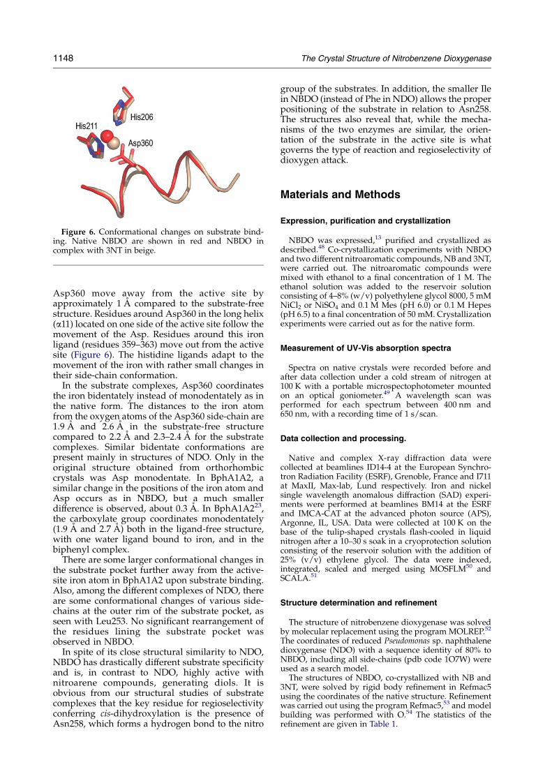
Figure 6. Conformational changes on substrate bind-ing. Native NBDO are shown in red and NBDO incomplex with 3NT in beige.
1148 The Crystal Structure of Nitrobenzene Dioxygenase
Asp360 move away from the active site byapproximately 1 A compared to the substrate-freestructure. Residues around Asp360 in the long helix(a11) located on one side of the active site follow themovement of the Asp. Residues around this ironligand (residues 359–363) move out from the activesite (Figure 6). The histidine ligands adapt to themovement of the iron with rather small changes intheir side-chain conformation.
In the substrate complexes, Asp360 coordinatesthe iron bidentately instead of monodentately as inthe native form. The distances to the iron atomfrom the oxygen atoms of the Asp360 side-chain are1.9 A and 2.6 A in the substrate-free structurecompared to 2.2 A and 2.3–2.4 A for the substratecomplexes. Similar bidentate conformations arepresent mainly in structures of NDO. Only in theoriginal structure obtained from orthorhombiccrystals was Asp monodentate. In BphA1A2, asimilar change in the positions of the iron atom andAsp occurs as in NBDO, but a much smallerdifference is observed, about 0.3 A. In BphA1A223,the carboxylate group coordinates monodentately(1.9 A and 2.7 A) both in the ligand-free structure,with one water ligand bound to iron, and in thebiphenyl complex.
There are some larger conformational changes inthe substrate pocket further away from the active-site iron atom in BphA1A2 upon substrate binding.Also, among the different complexes of NDO, thereare some conformational changes of various side-chains at the outer rim of the substrate pocket, asseen with Leu253. No significant rearrangement ofthe residues lining the substrate pocket wasobserved in NBDO.
In spite of its close structural similarity to NDO,NBDO has drastically different substrate specificityand is, in contrast to NDO, highly active withnitroarene compounds, generating diols. It isobvious from our structural studies of substratecomplexes that the key residue for regioselectivityconferring cis-dihydroxylation is the presence ofAsn258, which forms a hydrogen bond to the nitro
group of the substrates. In addition, the smaller Ilein NBDO (instead of Phe in NDO) allows the properpositioning of the substrate in relation to Asn258.The structures also reveal that, while the mecha-nisms of the two enzymes are similar, the orien-tation of the substrate in the active site is whatgoverns the type of reaction and regioselectivity ofdioxygen attack.
Materials and Methods
Expression, purification and crystallization
NBDO was expressed,13 purified and crystallized asdescribed.48 Co-crystallization experiments with NBDOand two different nitroaromatic compounds, NB and 3NT,were carried out. The nitroaromatic compounds weremixed with ethanol to a final concentration of 1 M. Theethanol solution was added to the reservoir solutionconsisting of 4–8% (w/v) polyethylene glycol 8000, 5 mMNiCl2 or NiSO4 and 0.1 M Mes (pH 6.0) or 0.1 M Hepes(pH 6.5) to a final concentration of 50 mM. Crystallizationexperiments were carried out as for the native form.
Measurement of UV-Vis absorption spectra
Spectra on native crystals were recorded before andafter data collection under a cold stream of nitrogen at100 K with a portable microspectophotometer mountedon an optical goniometer.49 A wavelength scan wasperformed for each spectrum between 400 nm and650 nm, with a recording time of 1 s/scan.
Data collection and processing.
Native and complex X-ray diffraction data werecollected at beamlines ID14-4 at the European Synchro-tron Radiation Facility (ESRF), Grenoble, France and I711at MaxII, Max-lab, Lund respectively. Iron and nickelsingle wavelength anomalous diffraction (SAD) experi-ments were performed at beamlines BM14 at the ESRFand IMCA-CAT at the advanced photon source (APS),Argonne, IL, USA. Data were collected at 100 K on thebase of the tulip-shaped crystals flash-cooled in liquidnitrogen after a 10–30 s soak in a cryoprotection solutionconsisting of the reservoir solution with the addition of25% (v/v) ethylene glycol. The data were indexed,integrated, scaled and merged using MOSFLM50 andSCALA.51
Structure determination and refinement
The structure of nitrobenzene dioxygenase was solvedby molecular replacement using the program MOLREP.52
The coordinates of reduced Pseudomonas sp. naphthalenedioxygenase (NDO) with a sequence identity of 80% toNBDO, including all side-chains (pdb code 1O7W) wereused as a search model.The structures of NBDO, co-crystallized with NB and
3NT, were solved by rigid body refinement in Refmac5using the coordinates of the native structure. Refinementwas carried out using the program Refmac5,53 and modelbuilding was performed with O.54 The statistics of therefinement are given in Table 1.

The Crystal Structure of Nitrobenzene Dioxygenase 1149
Protein Data Bank accession numbers
Coordinates and structure factors of the native formand complexes with NB and 3NT have been deposited inthe RCSB Protein Data Bank under accession codes 2BMO2BMQ and 2BMR.55
Acknowledgements
We thank J. Hajdu and G. Carlsson for help withthe microspectrophotometer. This work was sup-ported by the Swedish Research Council (to H.E.)and NIH grant GM062904-03 (to S.R.). We wouldlike to acknowledge the staff of the beamlines BM14and ID14-4 at ESRF, 711 at Max-lab and IMCA-CATat APS. Use of the IMCA-CAT beamline 17-ID at theAPS was supported by the companies of theIndustrial Macromolecular Crystallography Associ-ation through a contract with Illinois Institute ofTechnology.
References
1. Hartter, D. R. (1985). The use and importance ofnitroaromatic chemicals in the chemical industry. InToxicity of Nitroaromatic Compounds. Chemical IndustryInstitute of Toxicology Series (Rickert, D. E., ed.), pp.1–14, Hemisphere, Washington, DC.
2. Bruning, T., Thier, R. & Bolt, H. M. (2002). Nephro-toxicity and nephrocarcinogenicity of dinitrotoluene:new aspects to be considered. Rev. Environ. Health, 17,163–172.
3. Couch, D. B., Allen, P. F. & Abernethy, D. J. (1981). Themutagenicity of dinitrotoluenes in Salmonellatyphimurium. Mutat. Res. 90, 373–383.
4. Mirsalis, J. C. & Butterworth, B. E. (1982). Induction ofunscheduled DNA synthesis in rat hepatocytesfollowing in vivo treatment with dinitrotoluene.Carcinogenesis, 3, 241–245.
5. Tokiwa, H. & Ohnishi, Y. (1986). Mutagenicity andcarcinogenicity of nitroarenes and their sources in theenvironment. Crit. Rev. Toxicol. 17, 23–60.
6. Spain, J. C. (1995). Biodegradation of nitroaromaticcompounds. Annu. Rev. Microbiol. 49, 523–555.
7. He, Z. & Spain, J. C. (1997). Studies of the catabolicpathway of degradation of nitrobenzene by Pseudo-monas pseudoalcaligenes JS45: removal of the aminogroup from 2-aminomuconic semialdehyde. Appl.Environ. Microbiol. 63, 4839–4843.
8. He, Z., Davis, J. K. & Spain, J. C. (1998). Purification,characterization, and sequence analysis of 2-amino-muconic 6-semialdehyde dehydrogenase fromPseudomonas pseudoalcaligenes JS45. J. Bacteriol. 180,4591–4595.
9. He, Z. & Spain, J. C. (1998). A novel 2-aminomuconatedeaminase in the nitrobenzene degradation pathwayof Pseudomonas pseudoalcaligenes JS45. J. Bacteriol. 180,2502–2506.
10. Nishino, S. F. & Spain, J. C. (1993). Degradation ofnitrobenzene by a Pseudomonas pseudoalcaligenes. Appl.Environ. Microbiol. 59, 2520–2525.
11. Park, H. S. & Kim, H. S. (2000). Identification and
characterization of the nitrobenzene catabolic plas-mids pNB1 and pNB2 in Pseudomonas putida HS12.J. Bacteriol. 182, 573–580.
12. Somerville, C. C., Nishino, S. F. & Spain, J. C. (1995).Purification and characterization of nitrobenzenenitroreductase from Pseudomonas pseudoalcaligenesJS45. J. Bacteriol. 177, 3837–3842.
13. Lessner, D. J., Johnson, G. R., Parales, R. E., Spain, J. C.& Gibson, D. T. (2002). Molecular characterization andsubstrate specificity of nitrobenzene dioxygenasefrom Comamonas sp. strain JS765. Appl. Environ.Microbiol. 68, 634–641.
14. Gibson, D. T. & Parales, R. E. (2000). Aromatichydrocarbon dioxygenases in environmental bio-technology. Curr. Opin. Biotechnol. 11, 236–243.
15. Nishino, S. F. & Spain, J. C. (1995). Oxidativepathway for the biodegradation of nitrobenzene byComamonas sp. strain JS765.Appl. Environ.Microbiol. 61,2308–2313.
16. Kauppi, B., Lee, K., Carredano, E., Parales, R. E.,Gibson, D. T., Eklund, H. & Ramaswamy, S. (1998).Structure of an aromatic-ring-hydroxylating dioxy-genase–naphthalene 1,2-dioxygenase. Structure, 6,571–586.
17. Karlsson, A., Parales, J. V., Parales, R. E., Gibson, D. T.,Eklund, H. & Ramaswamy, S. (2003). Crystal structureof naphthalene dioxygenase: side-on binding ofdioxygen to iron. Science, 299, 1039–1042.
18. Wolfe, M. D., Parales, J. V., Gibson, D. T. & Lipscomb,J. D. (2001). Single turnover chemistry and regulationof O2 activation by the oxygenase component ofnaphthalene 1,2-dioxygenase. J. Biol. Chem. 276,1945–1953.
19. Gibson, D. T., Resnick, S. M., Lee, K., Brand, J. M.,Torok, D. S., Wackett, L. P. et al. (1995). Desaturation,dioxygenation, and monooxygenation reactions cata-lyzed by naphthalene dioxygenase from Pseudomonassp. strain 9816-4. J. Bacteriol. 177, 2615–2621.
20. Parales, J. V., Kumar, A., Parales, R. E. & Gibson, D. T.(1996). Cloning and sequencing of the genes encoding2-nitrotoluene dioxygenase from Pseudomonas sp.JS42. Gene, 181, 57–61.
21. Suen, W. C., Haigler, B. E. & Spain, J. C. (1996).2,4-Dinitrotoluene dioxygenase from Burkholderia sp.strain DNT: similarity to naphthalene dioxygenase.J. Bacteriol. 178, 4926–4934.
22. Karlsson, A., Parales, J. V., Parales, R. E., Gibson, D. T.,Eklund, H. & Ramaswamy, S. (2000). The reduction ofthe Rieske iron-sulfur cluster in naphthalene dioxy-genase by X-rays. J. Inorg. Biochem. 78, 83–87.
23. Furusawa, Y., Nagarajan, V., Tanokura, M., Masai, E.,Fukuda, M. & Senda, T. (2004). Crystal structure of theterminal oxygenase component of biphenyl dioxy-genase derived from Rhodococcus sp. strain RHA1.J. Mol. Biol. 342, 1041–1052.
24. Hoelz, A., Nairn, A. C. & Kuriyan, J. (2003). Crystalstructure of a tetradecameric assembly of the associ-ation domain of Ca2C/calmodulin-dependent kinaseII. Mol. Cell, 11, 1241–1251.
25. Lundqvist, T., Rice, J., Hodge, C. N., Basarab, G. S.,Pierce, J. & Lindqvist, Y. (1994). Crystal structure ofscytalone dehydratase: a disease determinant of therice pathogen,Magnaporthe grisea. Structure, 2, 937–944.
26. Bullock, T. L., Clarkson, W. D., Kent, H. M. & Stewart,M. (1996). The 1.6 A resolution crystal structure ofnuclear transport factor 2 (NTF2). J. Mol. Biol. 260,422–431.
27. Kim, S. W., Cha, S. S., Cho, H. S., Kim, J. S., Ha, N. C.,Cho, M. J. et al. (1997). High-resolution crystal

1150 The Crystal Structure of Nitrobenzene Dioxygenase
structures of D5-3-ketosteroid isomerase with andwithout a reaction intermediate analogue. Biochemistry,36, 14030–14036.
28. Lim, D. & Strynadka, N. C. (2002). Structural basis forthe beta lactam resistance of PBP2a from methicillin-resistant Staphylococcus aureus. Nature Struct. Biol. 9,870–876.
29. Arand, M., Hallberg, B. M., Zou, J., Bergfors, T.,Oesch, F., van der Werf, M. J. et al. (2003). Structureof Rhodococcus erythropolis limonene-1,2-epoxidehydrolase reveals a novel active site. EMBO J. 22,2583–2592.
30. Kerfeld, C. A., Sawaya, M. R., Brahmandam, V.,Cascio, D., Ho, K. K., Trevithick-Sutton, C. C. et al.(2003). The crystal structure of a cyanobacterial water-soluble carotenoid binding protein. Structure (Camb.),11, 55–65.
31. Hirose, J., Suyama, A., Hayashida, S. & Furukawa, K.(1994). Construction of hybrid biphenyl (bph) andtoluene (tod) genes for functional analysis of aromaticring dioxygenases. Gene, 138, 27–33.
32. Harayama, S., Rekik, M. & Timmis, K. N. (1986).Genetic analysis of a relaxed substrate specificityaromatic ring dioxygenase, toluate 1,2-dioxygenase,encoded by TOL plasmid pWW0 of Pseudomonasputida. Mol. Gen. Genet. 202, 226–234.
33. Parales, J. V., Parales, R. E., Resnick, S. M. & Gibson,D. T. (1998). Enzyme specificity of 2-nitrotoluene2,3-dioxygenase from Pseudomonas sp. strain JS42is determined by the C-terminal region of the alphasubunit of the oxygenase component. J. Bacteriol. 180,1194–1199.
34. Tan, H. M. & Cheong, C. M. (1994). Substitution ofthe ISP alpha subunit of biphenyl dioxygenasefrom Pseudomonas results in a modification of theenzyme activity. Biochem. Biophys. Res. Commun. 204,912–917.
35. Jiang, H., Parales, R. E. & Gibson, D. T. (1999). Thealpha subunit of toluene dioxygenase from Pseudo-monas putida F1 can accept electrons from reducedFerredoxinTOL but is catalytically inactive in theabsence of the beta subunit. Appl. Environ. Microbiol.65, 315–318.
36. Parales, R. E., Emig, M. D., Lynch, N. A. & Gibson,D. T. (1998). Substrate specificities of hybrid naphtha-lene and 2,4-dinitrotoluene dioxygenase enzymesystems. J. Bacteriol. 180, 2337–2344.
37. Beil, S., Mason, J. R., Timmis, K. N. & Pieper, D. H.(1998). Identification of chlorobenzene dioxy-genase sequence elements involved in dechlori-nation of 1,2,4,5-tetrachlorobenzene. J. Bacteriol. 180,5520–5528.
38. Colbert, C. L., Couture, M. M., Eltis, L. D. & Bolin, J. T.(2000). A cluster exposed: structure of the Rieskeferredoxin from biphenyl dioxygenase and the redoxproperties of Rieske Fe-S proteins. Struct. Fold. Des. 8,1267–1278.
39. Ellis, P. J., Conrads, T., Hille, R. & Kuhn, P. (2001).Crystal structure of the 100 kDa arsenite oxidase fromAlcaligenes faecalis in two crystal forms at 1.64 A and2.03 A. Structure (Camb.), 9, 125–132.
40. Iwata, S., Saynovits, M., Link, T. A. & Michel, H.(1996). Structure of a water soluble fragment of the’Rieske’ iron-sulfur protein of the bovine heartmitochondrial cytochrome bc1 complex determinedby MAD phasing at 1.5 A resolution. Structure, 4,567–579.
41. Iwata, S., Lee, J. W., Okada, K., Lee, J. K., Iwata, M.,
Rasmussen, B. et al. (1998). Complete structure of the11-subunit bovine mitochondrial cytochrome bc1complex. Science, 281, 64–71.
42. Carrell, C. J., Zhang, H., Cramer, W. A. & Smith, J. L.(1997). Biological identity and diversity in photo-synthesis and respiration: structure of the lumen-sidedomain of the chloroplast Rieske protein. Structure, 5,1613–1625.
43. Iyer, L. M., Koonin, E. V. & Aravind, L. (2001).Adaptations of the helix-grip fold for ligand bindingand catalysis in the START domain superfamily.Proteins: Struct. Funct. Genet. 43, 134–144.
44. Romanowski, M. J., Soccio, R. E., Breslow, J. L. &Burley, S. K. (2002). Crystal structure of the Musmusculus cholesterol-regulated START protein 4(StarD4) containing a StAR-related lipid transferdomain. Proc. Natl Acad. Sci. USA, 99, 6949–6954.
45. Yoder, M. D., Thomas, L. M., Tremblay, J. M., Oliver,R. L., Yarbrough, L. R. & Helmkamp, G. M., Jr (2001).Structure of a multifunctional protein. Mammalianphosphatidylinositol transfer protein complexed withphosphatidylcholine. J. Biol. Chem. 276, 9246–9252.
46. Tsujishita, Y. &Hurley, J. H. (2000). Structure and lipidtransport mechanism of a StAR-related domain.Nature Struct. Biol. 7, 408–414.
47. Resnick, S. M., Lee, K. & Gibson, D. T. (1996). Diversereactions catalyzed by naphthalene dioxygenase fromPseudomonas sp strain ncib 9816. J. Ind. Microbiol.Biotechnol. 17, 438–457.
48. Parales, R. E., Huang, R., Yu, C.-L., Parales, J., Lee,F. K. N., Lessner, D. J. et al. (2005). Purification,characterization, and crystallization of the com-ponents of the nitrobenzene and 2-nitrotoluenedioxygenase enzyme systems. Appl. Environ.Microbiol. in the press.
49. Hadfield, A. & Hajdu, J. (1993). A fast and portablemicrospectrophotometer for protein crystallography.J. Appl. Crystallog. 26, 839–842.
50. Powell, H. R. (1999). The Rossmann Fourier auto-indexing algorithm in MOSFLM. Acta Crystallog. sect.D, 55, 1690–1695.
51. Collaborative Computational Project, Number 4.(1994). The CCP4 suite: programs for protein crystal-lography. Acta Crystallog. sect. D, 50, 760–763.
52. Vagin, A. & Teplyakov, A. (1997). MOLREP: anautomated program for molecular replacement.J. Appl. Crystallog. 30, 1022–1025.
53. Murshudov, G. N. (1997). Refinement of macro-molecular structures by the maximum-likelihoodmethod. Acta Crystallog. sect. D, 53, 240–255.
54. Jones, T. A., Zou, J. Y., Cowan, S. W. & Kjeldgaard(1991). Improved methods for building proteinmodels in electron density maps and the location oferrors in these models. Acta. Crystallogr. sect. A, 47,110–119.
55. Berman, H. M., Westbrook, J., Feng, Z., Gilliland, G.,Bhat, T. N., Weissig, H., Shindyalov, I. N. & Bourne,P. E. (2000). The Protein Data Bank.Nucl. Acids Res. 28,235–242.
56. Barton, G. J. (1993). ALSCRIPT a tool to formatemultiple sequence alignment. Protein Engng. 6, 37–40.
57. Lessner, D. J., Johnson, G. R., Parales, R. E., Spain, J. C.& Gibson, D. T. (2002). Molecular characterization andsubstrate specificity of nitrobenzene dioxygenasefrom Comamonas sp. strain JS765. Appl. Environ.Microbiol. 68, 634–641.
58. Jeffrey, A. M., Yeh, H. J., Jerina, D. M., Patel, T. R.,

The Crystal Structure of Nitrobenzene Dioxygenase 1151
Davey, J. F. & Gibson, D. T. (1975). Initial reactions inthe oxidation of naphthalene by Pseudomonas putida.Biochemistry, 14, 575–584.
59. Jerina, D. M., Daly, J. W., Jeffrey, A. M. & Gibson, D. T.
(1971). Cis-1,2-dihydroxy-1,2-dihydronaphthalene: abacterial metabolite from naphthalene. Arch. Biochem.Biophys. 142, 394–396.
Edited by R. Huber
(Received 5 November 2004; received in revised form 11 March 2005; accepted 17 March 2005)


