Embed Size (px)
Citation preview

Seediscussions,stats,andauthorprofilesforthispublicationat:https://www.researchgate.net/publication/236911431
Structure-guideddiscoveryofthemetabolitecarboxy-SAMthatmodulatestRNAfunction
ARTICLEinNATURE·MAY2013
ImpactFactor:41.46·DOI:10.1038/nature12180·Source:PubMed
CITATIONS
23
READS
16
12AUTHORS,INCLUDING:
HuiXiao
YeshivaUniversity
46PUBLICATIONS696CITATIONS
SEEPROFILE
NawarAlObaidi
AlbertEinsteinCollegeofMedicine
10PUBLICATIONS33CITATIONS
SEEPROFILE
MatthewPJacobson
UniversityofCalifornia,SanFrancisco
160PUBLICATIONS6,137CITATIONS
SEEPROFILE
Young-SamLee
JohnsHopkinsUniversity
14PUBLICATIONS889CITATIONS
SEEPROFILE
Availablefrom:HuiXiao
Retrievedon:03February2016

Structure-guided discovery of carboxy-SAM as a novelmetabolite modulating tRNA function
Jungwook Kim1,*, Hui Xiao2, Jeffrey B. Bonanno1, Chakrapani Kalyanaraman3, ShoshanaBrown4, Xiangying Tang1, Nawar F. Al-Obaidi1, Yury Patskovsky1, Patricia C. Babbitt4,Matthew P. Jacobson3, Young-Sam Lee5, and Steven C. Almo1,6,*
1Department of Biochemistry, Albert Einstein College of Medicine, Bronx, NY 10461, USA.2Department of Pathology, Albert Einstein College of Medicine, Bronx, NY 10461, USA.3Department of Pharmaceutical Chemistry, University of California at San Francisco, SanFrancisco, CA 94158, USA.4Department of Bioengineering and Therapeutic Sciences, University of California at SanFrancisco, San Francisco, CA 94158, USA.5Department of Biology, Johns Hopkins University, Baltimore, MD 21218, USA.6Department of Physiology & Biophysics, Albert Einstein College of Medicine, Bronx, NY 10461,USA.
AbstractIdentifying novel metabolites and characterizing their biological functions are major challenges ofthe post-genomic era. X-ray crystallography can reveal unanticipated ligands which persistthrough purification and crystallization. These adventitious protein:ligand complexes provideinsights into new activities, pathways and regulatory mechanisms. We describe a new metabolite,carboxy-S-adenosylmethionine (Cx-SAM), its biosynthetic pathway and its role in tRNAmodification. The structure of CmoA, a member of the SAM-dependent methyltransferasesuperfamily, revealed a ligand in the catalytic site consistent with Cx-SAM. Mechanistic analysesdemonstrated an unprecedented role for prephenate as the carboxyl donor and the involvement ofa unique ylide intermediate as the carboxyl acceptor in the CmoA-mediated conversion of SAM toCx-SAM. A second member of the SAM-dependent methyltransferase superfamily, CmoB,recognizes Cx-SAM and acts as a carboxymethyltransferase to convert 5-hydroxyuridine (ho5U)into 5-oxyacetyl uridine (cmo5U) at the wobble position of multiple tRNAs in Gram negativebacteria1, resulting in expanded codon-recognition properties2,3. CmoA and CmoB represent thefirst documented synthase and transferase for Cx-SAM. These findings reveal new functionaldiversity in the SAM-dependent methyltransferase superfamily and expand the metabolic andbiological contributions of SAM-based biochemistry. These discoveries highlight the value of
Correspondence and requests for materials should be addressed to J.K. ([email protected]) or S.C.A.([email protected]).
Supplementary Information is linked to the online version of the paper at www.nature.com/nature.
Author Contributions J.K. performed cloning, protein purification, crystallography, and functional assay. H.X. performed MSanalysis of the in vitro assay. Y.S.L. performed LC-MS analysis of the CmoA-bound ligand and chemical synthesis of Cx-SAM. XTperformed the NMR experiments. N.F.A.-O. performed thermal denaturation studies. C.K. and M.P.J. performed computationalmodeling. S.B. and P.C.B. performed the bioinformatics analysis. J.B.B. and Y.P. assisted in crystallographic validation and analyzedcrystallographic ligand binding results, respectively. J.K. and S.C.A. designed the study, analyzed the data and wrote the manuscript.
Atomic coordinates and structure factors for the reported crystal structure are deposited in the Protein Data Bank under the accessioncode 4GEK.
The authors declare no competing financial interests.
NIH Public AccessAuthor ManuscriptNature. Author manuscript; available in PMC 2014 January 18.
Published in final edited form as:Nature. 2013 June 6; 498(7452): 123–126. doi:10.1038/nature12180.
NIH
-PA Author Manuscript
NIH
-PA Author Manuscript
NIH
-PA Author Manuscript

structural genomics approaches for identifying ligands in the context of their physiologicallyrelevant macromolecular binding partners and for aiding in functional assignment.
tRNAs contain numerous post-transcriptional modifications, with nearly 100 distinctmodifications reported1. Nucleotides at the wobble position are the most frequent targets forsuch modifications, as they confer efficient and accurate pairing between anticodons andcognate codon sequences. For example, wobble uridines in Gram negative bacteria are oftenmodified at C5 to 5-oxyacetyl uridine (cmo5U) (Fig. 1a), which allows multiple tRNAs todecode four of their respective degenerate codons3. This expanded recognition results fromstructural and tautomeric constraints imposed by the 5-oxyacetyl modification4.
Mutagenesis studies established that genes responsible for chorismate biosynthesis arenecessary for cmo5U formation and it was demonstrated that one carbon atom of theoxyacetyl group originates from SAM5,6. Gene disruption studies established that twomembers of the SAM-dependent methyltransferase superfamily (SDMT), CmoA and CmoB,were required for cmo5U formation2. Inactivation of cmoA resulted in formation ofincompletely modified tRNAs, with hydroxy uridine (ho5U) and methoxy uridine (mo5U)observed instead of cmo5U. In cmoB-defective mutants, only ho5U was detected. Despitethese observations, the roles of CmoA and CmoB in the transformation of ho5U to cmo5Uremain unclear. Wobble uridine hydroxylation is dependent on an unidentified enzyme (Fig.1a).
The New York Structural Genomics Research Consortium (NYSGRC) determined thestructure of Escherichia coli CmoA, which revealed unexpected electron density features atthe predicted SAM binding site. When SAM was modeled, residual electron densitysuggestive of a carboxylate group was observed adjacent to the S-methyl group. Refinementof this structure at 1.50Å supports a covalent linkage between the S-methyl group and theputative carboxylate, consistent with the previously unknown metabolite carboxy-SAM (Cx-SAM; (2S)-4-[{[(2S,3S,4R,5R)-5-(6-amino-9H-purin-9-yl)-3,4-dihydroxy-tetrahydrofuran-2-yl]methyl}(carboxylatomethyl)sulfonio]-2-ammoniobutanoate) (Fig. 2and Supplementary Fig. 1). Liquid chromatography–mass spectrometry (LC-MS) analysis ofpurified CmoA, unambiguously confirmed Cx-SAM as the CmoA-bound ligand whichpersisted through purification and crystallization (Fig. 3 and Supplementary Fig. 2).
The overall structure of E. coli CmoA is similar to that reported for the Haemophilusinfluenzae ortholog (67% sequence identity; PDB entry 1IM8; RMSD of 0.51Å for 222equivalent Cα atoms)7. Retrospective refinement of the H. influenza CmoA structurerevealed electron density features consistent with Cx-SAM and contacts similar to thoseobserved in the E. coli enzyme. These observations support the existence of conservedpathways involving Cx-SAM in the Gram negative bacteria.
The S-carboxymethyl group of Cx-SAM in the E. coli CmoA catalytic site forms a bidentatepolar interaction with the side-chain guanidinium of Arg-199, which is invariant amongCmoA orthologs. The 2’- and 3’-hydroxyl groups of Cx-SAM form hydrogen bonds withthe side-chain of Asp-89; the equivalent residue in all other SAM-dependentmethyltransferases is aspartate or glutamate. Other highly conserved residues among CmoAorthologs are contributed from helices α1, α6 and α7 (Supplementary Fig. 3), which appearcrucial for substrate binding and are not present in other members of the SDMT superfamily.The Cx-SAM binding pocket is largely hydrophobic, with no functionality capable ofdeprotonating the S-methyl group of SAM closer than 4.6Å. Adjacent to the Cx-SAMbinding site is a partially hydrophobic cavity which is the likely binding site for anadditional ligand/substrate (see below).
Kim et al. Page 2
Nature. Author manuscript; available in PMC 2014 January 18.
NIH
-PA Author Manuscript
NIH
-PA Author Manuscript
NIH
-PA Author Manuscript

Given data implicating chorismate, or a related metabolite, in wobble uridine oxyacetylationin Gram negative bacteria5,6, chorismate was examined as the potential carboxyl donor.Chorismate supported the CmoA-mediated formation of Cx-SAM as evidenced by LC-MS(Fig. 3b) and MS/MS (Supplementary Fig. 4). In addition to Cx-SAM, phenylpyruvate wasproduced with similar kinetics (Fig. 3c and Supplementary Fig. 5). The lack of a facilepathway for the direct conversion of chorismate to phenylpyruvate suggested thatchorismate undergoes a rearrangement prior to CmoA-catalyzed Cx-SAM formation.Among the several biologically characterized products of chorismate, only prephenatepossesses a scaffold consistent with the observed LC-MS and MS/MS fragmentation data.
When tested for in vitro production of Cx-SAM, prephenate was a more efficient substratethan chorismate (Fig. 3d) and the lag in Cx-SAM production observed with chorismate wasabsent. This behavior is consistent with the slow non-enzymatic conversion of chorismate toprephenate, which is utilized by CmoA for Cx-SAM formation (Supplementary Fig. 6).Furthermore, production of phenylpyruvate from prephenate was unambiguously confirmedby NMR and LC-MS of the in vitro assay solution (Supplementary Figs 7 and 8). Theseactivities are CmoA-specific as CmoB does not show any Cx-SAM synthase activity (datanot shown). We propose that prephenate is the biologically relevant substrate and the sourceof the carboxylate in the CmoA-catalyzed reaction.
These results are consistent with the CmoA-catalyzed decarboxylation and concomitant lossof hydroxide from prephenate to yield phenylpyruvate, water and carbon dioxide, which isthe source of the carboxylate functionality in Cx-SAM (Fig. 1b). In support of thishypothesis, uniformly 13C-labeled chorismate ([U-13C] chorismate) transferred 13C-carbondioxide to the product, Cx-SAM (Supplementary Fig. 9), unambiguously demonstrating thatprephenate is the carboxyl donor in the CmoA-catalyzed formation of Cx-SAM andvalidating the overall reaction proposed for CmoA. To our knowledge, this is the firstexample of prephenate serving as the carboxyl group donor in a carboxytransfer reaction.
There is precedence for decarboxylation of prephenate, as prephenate dehydratase (PDT)catalyzes the decarboxylation of prephenate with concomitant loss of hydroxide to generatephenylpyruvate in a fashion similar to that suggested for CmoA8. In the PDT-catalyzedreaction, elimination of the hydroxyl group as water is facilitated by the participation ofThr172 as a general acid to protonate the leaving hydroxide group. The threonine side-chainis not sufficiently acidic to directly protonate the prephenate hydroxyl (i.e., the conjugateacid of the prephenate hydroxyl is expected to behave similarly to an alcohol, with a pKa of~-2, while the pKa of threonine is ~16 in water); thus, other mechanisms must be operativeas the major driving force for this reaction. Hilvert and Cleland proposed that geometricdistortion drives decarboxylation and the favorable energetics associated with aromatizationreduces the bond order of the hydroxyl, shifting its reactivity towards that of hydroxide andallowing for efficient general acid catalysis8. Similar considerations are relevant to theCmoA-catalyzed reaction.
Computational docking of prephenate in the CmoA catalytic site suggests a pose in whichthe carboxylate and hydroxyl groups of prephenate sandwich the S-methyl group of SAM,consistent with the observed transcarboxylation reaction (Fig. 2 and Supplementary Fig. 10).Trievel recently highlighted the importance of carbon-oxygen (CH-O) hydrogen bonds inthe recognition and presentation of the S-methyl group in canonical SN2-based SAM-dependent methyltransfer reactions9. In the prephenate-bound model of CmoA (Fig. 2 andSupplementary Fig. 10), the hydroxyl oxygen of prephenate and the S-methyl carbon ofSAM form a potential 3.4Å CH…O hydrogen bond. Notably, the prephenate hydroxyl ispoorly positioned for an in-line SN2 attack on the S-methyl group, consistent with the lackof prephenate methylation. Instead, this arrangement suggests a substrate-assisted
Kim et al. Page 3
Nature. Author manuscript; available in PMC 2014 January 18.
NIH
-PA Author Manuscript
NIH
-PA Author Manuscript
NIH
-PA Author Manuscript

mechanism in which the departing prephenate OH group abstracts a proton from the S-methyl group of SAM, generating water and the nucleophilic ylide (stabilized carbanion),which is carboxylated by the carbon dioxide (Fig. 1b). Importantly, the pKa of thetrimethylsulfonium cation (a model of the S-methyl group in SAM) has been reported as18.9 and 18.2 in water and DMSO10,11, respectively. These values are similar to that of thegeneral acid (Thr172) in the PDT-catalyzed reaction and are consistent with elimination ofhydroxide from prephenate as water and formation of the reactive ylide intermediate.
Because of its importance in the mechanism, we sought direct evidence for the formation ofthe sulfonium ylide. Presentation of CmoA with triply-deuterated SAM and prephenateresults in formation of doubly-deuterated SAM, consistent with formation of the ylideintermediate by deuteron abstraction and regeneration of SAM by protonation (Fig. 3f). Inthe absence of prephenate, only triply-deuterated SAM was observed. In the absence ofCmoA, no exchange was observed in mixtures of [2H3-methyl]-SAM and prephenate.Partitioning of the ylide between product formation (Cx-SAM) and return to substrate(SAM) was quantified by determining the ratio between solvent-exchanged SAM and Cx-SAM: 92.0:8.0 and 97.3:2.7 at pH 7.3 and 8.5, respectively, providing compelling evidencethat the postulated ylide intermediate is on the reaction coordinate for Cx-SAM formation.
The most common biological fate of the SAM S-methyl group is intermolecular transfercatalyzed by methyltransferases, including epigenetic marking of DNA and histonetargets12,13, and a wide range of small molecule transformations14,15. By analogy, wehypothesized that Cx-SAM is utilized in a CmoB-catalyzed transcarboxymethylationreaction in the biosynthesis of cmo5U. Total RNA16 and purified tRNAs2 from cmoB-deficient cells (i.e., ho5U-containing RNA) were used as substrates in in vitro assays withprephenate, SAM and CmoA, with or without recombinant CmoB. After the transferreaction, RNAs were treated with P1 nuclease and the resulting 5’-nucleotidemonophosphates analyzed by MS (Fig. 4). CmoB catalyzed carboxymethyl transfer from insitu-generated Cx-SAM to ho5U-containing RNAs, as a species with mass corresponding tooxyacetyl-5-uridine-5’-monophosphate (cmo5UMP) was clearly detected. CmoA alone doesnot exhibit carboxymethyltransferase activity. Therefore, we conclude that Cx-SAM is thesubstrate for the CmoB-dependent transcarboxymethylation of ho5U-containing tRNAs(Fig. 1c).
In most Gram-negative species, cmoA and cmoB are co-conserved and immediately adjacentto each other in the genome, supporting the demonstrated functional relationship. Mostimportantly, the unique in vitro activities assigned to CmoA and CmoB are fully consistentwith all reported genetics findings relevant to cmo5U modification2,5,6. Our own geneticsand mutagenesis studies add further support, as plasmid-based expression of wild-typeCmoA restores production of cmo5U in CmoA-deficient (ΔcmoA) E. coli, while the D89Lmutant lacking in vitro biochemical activity failed to complement in vivo (SupplementaryFig. 11). Finally, in vivo- and in vitro-generated Cx-SAM-bound CmoA exhibitedcomparable behavior in the carboxymethylation of ho5U-containing RNAs, supporting therelevance of our in vitro studies (see Supplementary Fig. 12).
Elaboration of the S-methyl group of SAM with a variety of functional groups has beenpursued by chemical biologists to support studies including the generation of modifiednucleic acid17,18 and protein19 substrates, as well as genome-wide assignments of proteinmethyltransferase targets19. It is notable that Cx-SAM represents the first SAM derivativedemonstrated to have been shaped by evolution for a biologically meaningful function. Theelectrophilic properties of the SAM sulfonium center have been exploited to realizeexpanded functional diversity, as exemplified by Cx-SAM, via two unique activities withinthe SDMT superfamily (Supplementary Fig. 13a). Sequence analysis supports the existence
Kim et al. Page 4
Nature. Author manuscript; available in PMC 2014 January 18.
NIH
-PA Author Manuscript
NIH
-PA Author Manuscript
NIH
-PA Author Manuscript

of CmoA orthologs throughout the Gram negative proteobacteria, as well as in theVerrucomicrobia and some Cyanobacteria (Supplementary Fig. 13b). It remains to bediscovered how widespread these mechanisms are and whether there exist additionalbiologically relevant SAM-analogues.
The fortuitous identification of a bound ligand in a crystal structure (such as Cx-SAM inCmoA) is by no means unusual. We estimate that ~3–5% of all structures determined by theNYSGRC contain organic ligands derived from the expression host (typically E. coli),which persisted through purification and crystallization. Frequently this can be anticipated;e.g., finding NAD/NADH bound to targets annotated as oxidoreductases, or PLP inannotated aminotransferases. However, unanticipated ligands are also identified, includingnucleotides, amino acids, carbohydrates and lipids bound to proteins from a range ofbacterial species, providing clues regarding catalytic activity and biological function (seeexamples in Supplementary Fig. 14).
In summary, direct structural observation identified the novel metabolite Cx-SAM, leadingto the discovery of unique Cx-SAM synthase and carboxymethyltransferase activitiesinvolved in tRNA wobble base modification. These findings reveal new functional diversityin the SDMT superfamily, expand the metabolic and biological contributions for SAM-based biochemistry and presage the discovery of new metabolites and biological processes.This work highlights the power of structural genomics approaches for the discovery of newmetabolites, pathways and biology.
Full MethodsCloning and Protein Purification
The cmoA gene was amplified from genomic DNA of E.coli BL21 by PCR, cloned into LIC-pET46a (Novagen) and verified by the DNA sequence analysis (Genewiz). E. coli BL21(DE3) cells (Invitrogen) were transformed with vectors harboring the cmoA gene, grown inLB containing 100 mg/mL ampicillin at 37°C and induced with 0.5 mM IPTG at an OD600of ~1. Cells were incubated overnight at 25°C and harvested by centrifugation. Cell pelletswere resuspended with Bugbuster (Novagen) at room temperature for 30 min, the lysatescentrifuged at 41,400g for 30 min and the supernatants applied to Ni-agarose (Qiagene)columns pre-equilibrated with buffer A (20 mM HEPES, pH 7.5 and 150 mM KCl). Therecombinant protein was eluted with 150 mM imidazole in buffer A and further purified bysize exclusion chromatography on a HiLoad Superdex 200 column (GE) equilibrated withbuffer A. Final purity was over 95% as verified by SDS-PAGE analysis. For themeasurement of OD280 of the purified CmoA, the enzyme was denatured to separate SAM/Cx-SAM from the protein by multiple rounds of mixing with 8M guanidine chloridesolution followed by spin-filtration. An extinction coefficient of ε280 = 18.7 cm−1mM−1 wasused to calculate the yield of the nucleoside-free CmoA as calculated from the amino acidsequence. The cmoB gene was amplified from genomic DNA of E. coli BL21 by PCR andcloned into LIC-pET30a (Novagen). The purification of E. coli CmoB was identical to thatof CmoA, except for the use of kanamycin as the selectable marker. In addition, the affinitytag was removed by thrombin (Novagen) cleavage after elution of the recombinant CmoBfrom the Ni-agarose resin. The yield was quantitated using an extinction coefficient of ε280= 72.5cm−1mM−1, as calculated from the amino acid sequence.
The D89L mutant of CmoA was generated by QuickChange (Stratagene) with primers 5’-TTGCAAAATTATTGCCATCCTCAACTCCCCGGCGATGATT-3’ and 5’-AATCATCGCCGGGGAGTTGAGGATGGCAATAATTTTGCAA-3’, and the plasmid ofthe wild-type CmoA as the template for PCR. Purification of the D89L mutant was similarto that of the wild type described above; some purifications included the addition of anion
Kim et al. Page 5
Nature. Author manuscript; available in PMC 2014 January 18.
NIH
-PA Author Manuscript
NIH
-PA Author Manuscript
NIH
-PA Author Manuscript

exchange and gel filtration chromatography steps. For anion exchange separation, a MonoQcolumn (GE) was equilibrated with Buffer A (20 mM Tris-HCl, pH 8.5 and 150 mM KCl)and 1 mL sample was loaded. A linear gradient of Buffer B (20 mM Tris-HCl, pH=8.5 and 1M NaCl) was used to elute bound protein. Wild-type CmoA eluted as a single peak onMonoQ, while the D89L mutant exhibited two peaks. Eluted proteins were analyzed bySDS-PAGE, pooled, concentrated and loaded on a Superdex75 column (GE), equilibratedwith Buffer A.
Crystallization and Structural Determination of CmoAPurified CmoA was crystallized by sitting drop vapor diffusion at 21°C by mixing 1 µL ofthe protein with 1 µL of reservoir solution (0.2 M Li2SO4, 0.1 M Bis-Tris:HCl, pH 5.5, and25% PEG3350) and equilibrating over 0.1 mL of reservoir solution. X-ray data werecollected on an ADSC QUANTUM 315 CCD detector at the NSLS beam line X29A andprocessed with HKL300020. Diffraction data from CmoA crystals were collected at 100K,and at a wavelength λ = 0.9790Å, which were consistent with space group, P21212 (a =65.32, b = 78.68, c = 92.37Å), with two molecules per asymmetric unit. Molecularreplacement was performed using the structure of H. influenzae YecO (pdb code 1IM8) asthe search model with MOLREP21. Subsequent model building and refinement wereperformed with Coot22 and REFMAC523. The final model was refined to 1.50Å with Rwork= 0.17 and Rfree = 0.20 (Supplementary Table I). All residues are in allowed regions and nooutliers are found in a Ramachandran plot.
Time Course Assay of Cx-SAM ProductionThe time-dependent formation of carboxyl-SAM was monitored using [14C-methyl]-SAMwith either prephenate or chorismate. The assay mixture contains 20 mM sodium phosphate,pH 6.8, 0.2 mM [14C-methyl]-SAM (Perkin Elmer), and either 0.2 mM chorismate orprephenate. The assay was initiated by adding purified 2 µM CmoA to the assay mixture of20 µL total volume of. A 2 µL aliquot was periodically withdrawn and mixed with an equalvolume of 0.1 M HCl to quench the reaction, then 1 µL was spotted on TLC plate. The TLCwas developed with buffer composed of ammonium sulfate/sodium acetate, pH 6.0/iso-propanol (79/19/2). The plate was air-dried and exposed to a phosphor screen imager (GE)for 2 days. The image plate was scanned with Molecular Dynamics Storm 860PhosphorImager System with ImageQuant software.
Time Course Assay of Phenylpyruvate ProductionPhenylpyruvate formation was monitored at λ = 320 nm as described previously24. Theassay mixture contained 20 mM sodium phosphate, pH 6.8 and 0.2 mM prephenate, with orwithout 0.2 mM SAM in a total volume of 0.5 mL. The reaction was initiated by adding 2µM CmoA to the assay solution. A 70 µL aliquot of the reaction mixture was periodicallywithdrawn and added to 30 µL of 5 M NaOH. The absorbance at λ = 320 nm was measuredand the net OD320 was recorded by subtracting residual absorbance arising fromcontaminating phenylpyruvate. The net absorbance was converted to the concentration ofphenylpyruvate using a standard curve prepared with commercially obtained phenylpyruvate(Sigma-Aldrich). Non-enzymatic turn-over of prephenate to phenylpyruvate was measuredwith a sample composed of 20 mM sodium phosphate, pH 6.8, 0.2 mM prephenate, and 0.2mM SAM in 0.5 mL, without the addition of the enzyme.
Verification of Cx-SAM Co-purified with Recombinant CmoA by LC-MSA 10 µL recombinant protein solution was diluted with 10 µL water and then with 190 µL ofmethanol. The mixture was centrifuged at room temperature for 10 min (16,000g), and thesupernatant was used for the analysis. For each injection, an 80 µL aliquot was subjected to
Kim et al. Page 6
Nature. Author manuscript; available in PMC 2014 January 18.
NIH
-PA Author Manuscript
NIH
-PA Author Manuscript
NIH
-PA Author Manuscript

liquid chromatography–mass spectrometry analysis (Agilent 1200 HPLC coupled withAgilent 6210 AccurateMass electronspray mass spectrometer; ESI positive ion modedetection, 4 GHz, m/z range from 50–1200; Phenomenex Luna NH2 column 5 µm bead size,100 Å pore size, 150×2 mm) using a gradient system described in literature25. Data wereanalyzed using Agilent Mass Hunter software package.
MS Analysis of CmoA Assay10 mM prephenate (or 10 mM chorismate initially) was incubated with 10 mM SAM and 10µM CmoA in a total of 0.5 mL solution at room temperature overnight. An aliquot of 10 µLof the reaction mixture was mixed with 100 µL methanol, which was then infused into a 12TAgilent IonSpec FT-ICR-MS (Agilent Technologies, Inc., CA). Cx-SAM (m/z = 443.1373)was monitored in positive mode, and phenylpyruvate (m/z = 163.0404) was monitored innegative mode. The Agilent 12T QFT-ICR routinely provides better than 5 ppm massaccuracy with external calibration.
Solvent Isotope Exchange of Deuterated SAMThe assay solution was prepared by mixing 10 mM Tris, pH 8.0, 0.5 mM [2H3-methyl]SAM (CDNisotope, Quebec), 0.5 mM prephenate and 10 µM CmoA in 0.5 mL solution. Thereaction was incubated for 4 hr at room temperature and was quenched by filtering theenzyme with a spin column (MWCO = 10 kD). The sample was then analyzed by massspectrometry as described above. To examine whether solvent proton exchange at S-methylof SAM is prephenate-dependent manner, a sample without prephenate was prepared andanalyzed in an identical fashion.
Assay of CmoB ReactionCarboxymethyltransfer activity of CmoB was examined with a solution containing 50 mMTris, pH 8.0, 4 mM MgCl2, 1 mM prephenate, 1 mM SAM, 1 µM CmoA and total RNAextracted from cmoB-mutant E. coli cells as described before16, or purified tRNAs2, wherethe total volume of the assay is 50 µL. The reaction was initiated by adding 6 µM CmoB andincubated at the room temperature for 2 hr. One unit of P1 nuclease (US Biological) wasadded to the assay solution and incubated at 65°C for 1 hr to convert polynucleotides into5’-nucleotide monophosphates. The P1 nuclease-treated sample was mixed with 100 µLmethanol and vortexed before centrifugation at 13,200 RPM for 2 min. An aliquot ofsupernatant was injected to a 12T Agilent IonSpec FT-ICR-MS and analyzed in negativemode.
Computational ligand dockingTo create a model of the substrate prephenate bound to CmoA, we first removed thecarboxylate group from the product Cx-SAM to create SAM in the CmoA catalytic site. Theresulting complex was subjected to a protein preparation protocol, during which hydrogenatoms were added; protonation states of His residues were examined and adjusted ifnecessary; side-chains of Thr, Tyr and Asn residues were optimized for hydrogen bondinginteractions; and the entire structure was finally energy minimized such that heavy atompositions remained within 0.3Å of the starting coordinates.
Initial attempts to dock the substrate prephenate to this resulting model failed due toinadequate space for the ligand. We hypothesized that charged residues in the binding siterequired conformational changes to accommodate the ligand. Specifically, in the Cx-SAM-bound structure, Arg199 formed a salt-bridge interaction with the carboxylate group of Cx-SAM (Fig. 2c), and other charged residues in the active site such as Lys165 and Glu203either pointed toward the solvent or blocked portions of the active site. Therefore, we used
Kim et al. Page 7
Nature. Author manuscript; available in PMC 2014 January 18.
NIH
-PA Author Manuscript
NIH
-PA Author Manuscript
NIH
-PA Author Manuscript
an induced fit docking procedure, in which side-chains of residues that are within 5Å of thedocked prephenate pose were treated as conformationally flexible26. Induced fit dockinguses a combination of a molecular-mechanics energy function and an empirical scoringfunction-based energy to rank the ligand poses. We re-ranked the induced fit docking posesusing a molecular-mechanics based energy function that has been used successfully in manyapplications of metabolite docking. The lowest energy binding pose thus identified is shownin Fig. 2d.
Thermal stability of wild-type and D89L mutant CmoAThe fluorescence monitored thermal denaturation of wild-type and mutant CmoA wasperformed using a 7900HT RT-PCR system (Applied Biosystems). Briefly, 20 µL of eachprotein at 10 µM concentration was mixed with 0.5 µL of 200× Sypro orange solution andpipetted into separate wells of a 384-well PCR plate. After centrifugation to remove airbubbles, the plate is loaded into the PCR machine and the temperature ramped from 20 to99°C, in 1°C increments with a dwell time of 6 s. The negative first derivative of thefluorescence change (−dRFU/dT) for each protein is plotted against temperature, and themelting temperature is defined as the minimum in the −dRFU/dT curve. The wild type fromMonoQ exhibited a melting temperature (Tm) of 55.6 ± 0.1°C, while the two D89L mutantfractions displayed Tms that were 2–3°C lower. The behavior the wild-type and mutantspecies show that they are full folded under the conditions (i.e., temperature) used in vitroactivity assays; the lack of activity exhibited by the D89L mutant is thus the consequence ofa catalytic defect and not due to issues related to thermodynamic stability.
Chemical Synthesis of Cx-SAM3.0 mg of S-adenosyl-L-homocystein (SAH) was dissolved in 0.5 mL 150 mM ammoniumbicarbonate. To this solution, 2-iodoacetatic acid (100 mg) was added. The mixture wasincubated at 37°C for 12 hr with constant agitation. The progress of the reaction wasmonitored by thin layer chromatography (SiO2) using a solvent system composed ofmethanol:aqueous 1.5 N ammonium bicarbonate (10:1 vol./vol.). Rf= 0.6 and 0.3 for SAHand Cx-SAM, respectively. After the reaction was completed, 12 mL methanol was addedand the mixture incubated at 4°C overnight. Precipitates were collected by centrifugation at4°C (2,000g for 30 min), washed twice with ice-cold methanol and dissolved in 0.10 mLdeionized water. The product was purified using an HILIC as described above.Concentration of Cx-SAM was determined spectroscopically, assuming an extinctioncoefficient of SAM (ε260 = 154 cm−1mM−1).
Assay of Non-Enzymatic Formation of Prephenate from ChorismateThe rate of conversion from chorismate to prephenate in the absence of CmoA wasmeasured in vitro24. The assay solution contained 10 mM sodium phosphate (pH 6.8), 0.2mM SAM, and 0.2 mM chorismate in 0.5 mL. An aliquot of 80 µL was withdrawnperiodically and added to 5 µL of 4.5 M HCl. The mixture was then incubated at 37°C for 15min and combined with 15 µL of 12 M NaOH before the absorbance at 320 nm wasmeasured to determine phenylpyruvate.
NMR Analysis of the CmoA Assay MixtureTo define the nature of prephenate-derived product subsequent to donation of thecarboxylate, the in vitro assay was scaled up using 0.5 mM prephenate, 0.5 mM SAM, and10 µM CmoA in 10 mL total volume, so as to maximize the yield of products for NMRanalysis. The reaction was incubated at room temperature for 8hrs, and the enzyme filteredusing a spin column (MWCO 10 kD) before lyophilization. The lyophilized sample was
Kim et al. Page 8
Nature. Author manuscript; available in PMC 2014 January 18.
NIH
-PA Author Manuscript
NIH
-PA Author Manuscript
NIH
-PA Author Manuscript

dissolved in 0.6 mL D2O (Cambridge Isotope Laboratory, MA) and 1H-resonance data wascollected with a Bruker DRX-300 NMR spectrometer.
Partitioning of the Ylide IntermediateThe pH dependent ylide partitioning assay was performed with 0.2 mM prephenate, 0.05mM [2H3-methyl] SAM in either 10 mM ammonium acetate, pH 7.3, or 10 mM ammoniumbicarbonate, pH 8.5. The assay was initiated by adding 2 µM CmoA, with incubation atroom temperature for 2 hr before being analyzed by mass spectrometry as described below.Total amount [2H3-methyl]- and [2H2
1H-methyl]-SAM remaining after the reaction wasdetermined by adding a known amount of unlabeled SAM to the assay mixture as an internalstandard. The concentration of Cx-SAM was determined by subtracting the remaining SAMafter the reaction from the initial quantity added. The amount of solvent exchanged ([2H2
1H-methyl]) and non-exchanged ([2H3-methyl]) SAM were calculated from the relativeamplitude of corresponding MS peaks and the total SAM concentration. Based on theabsolute concentrations of [2H2
1H-methyl]-SAM and Cx-SAM the partitioning of the ylideintermediate back to reactant and forward to product was calculated (e.g., partitioning backto SAM is calculated as ([2H2
1H-methyl]-SAM)/([2H21H-methyl]-SAM + Cx-SAM)).
Verification of Enzymatic Formation of Phenylpyruvate Using LC-MSA Shimadzu HPLC, with two LC-20AD pumps, was used to generate a gradient with 50 µL/min flow rate. Solvent A was 5% acetonitrile in H2O and 0.1% formic acid while solvent Bconsisted of 95% acetonitrile in H2O and 0.1% formic acid. 50 µL of assay sample, whichwas used for NMR analysis above, was loaded onto a 1.0 × 50 mm C18 column(Phenomenex, CA). After desalting with 5 solvent B for 5 min, bound phenylpyruvic acidwas eluted with a 30 min gradient composed of 5% to 95% B. The effluent was directlydelivered into the 12T QFT-ICR-MS (Agilent Technologies, Inc., CA) for mass analysis.
Preparation of [U-13C] chorismateProduction and purification of chorismate using Aerobacter aerogenes 62-1 followed themethods developed previously27,28. A. aerogenes 62-1 was generously provided by Drs.Jared Parker and Christopher T. Walsh at Harvard Medical School. Overnight culture (1mL) of A. aerogenes 62-1 was added to 50 mL Medium A, and incubated at 30°C untilOD600 reached ~1. Cells were pelleted at 3,000g and washed with 25 mL Medium B whichdid not contain glucose. After pelleting once again, cells were resuspended in 25 mLMedium B with 0.2 g of [U-13C] glucose (purchased from Cambridge Isotope Laboratory).Cells were grown at 30°C for 15 hr for the production of labeled chorismate, harvested bycentrifugation and discarded. The supernatant was filtered and loaded on Hypercarb HPLCcolumn (10×100 mm) equilibrated in 10 mM ammonium acetate, pH 9.9. Chorismate waseluted with a linear gradient of acetonitrile and identified by monitoring OD275 of eachfraction. 13C-chorismate was confirmed by mass spectrometry (13C10H10O6Na, observed m/z = 259.0707, calculated m/z = 259.0711). Pooled chorismate was lyophilized, resupendedin water and quantitated by UV absorption at 275 nm (ε275 nm = 2630 M−1 cm−1)28.
Assay of CmoA with [U-13C] chorismateThe assay mixture (0.1 mL) contained 20 mM sodium phosphate, pH 6.8, 0.2 mM SAM, 0.2mM [U-13C] chorismate, and 2 µM CmoA. The reaction was incubated overnight at theroom temperature before analysis by mass spectrometry.
cmoA ComplementationFor the complementation assay, the cmoA gene was inserted between KpnI and HindIII sitesin the pQE30a (Qiagen) expression vector. cmoA-deficient E. coli cells (from KEIO
Kim et al. Page 9
Nature. Author manuscript; available in PMC 2014 January 18.
NIH
-PA Author Manuscript
NIH
-PA Author Manuscript
NIH
-PA Author Manuscript

collection) were transformed with either empty vector (pQE30a), plasmid bearing wild-typecmoA or plasmid bearing biochemically inactive D89L cmoA. Transformed cells weretypically grown in 50 mL LB media at 37°C and induced with 0.5 mM IPTG at an OD600 of~1. Cells were incubated overnight at 25°C and harvested by centrifugation. Total RNA wasextracted and treated with one unit of P1 nuclease at 65°C for 1 hr. The hydrolyzednucleotide samples were analyzed using LC/MS (Waters Symmetry C18 Column, 100Å, 3.5µm, 2.1 mm × 150 mm coupled to 12T Agilent IonSpec FT-ICR-MS) in negative mode. Alinear gradient of 20% to 95% acetonitrile and 0.1% formic acid was used over 15 min. Theidentification of cmo5UMP demonstrates the in vivo formation of cmo5U at the wobbleposition.
Cmo5UMP assay using in vivo- and in vitro-generated Cx-SAM13C-CmoA was purified from cells grown in M9 minimal media containing 13C-glucose asthe sole carbon source. The media contained 1× M9 salts (Sigma), 2 mM MgSO4, 0.2 mMCaCl2, and 0.4% [U-13C]-glucose (Cambridge Isotope Laboratory) in 0.5 L. These growthconditions yield 13C-Cx-SAM bound CmoA, which was purified as described above.Notably, the occupancy of 13C-Cx-SAM within 13C-CmoA was nearly 100% as determinedby MS analysis, which is higher than that typically observed in samples prepared from cellsgrown in LB media (see Supplementary Fig. 2). 10 µM 13C-Cx-SAM-CmoA complex wasadded to an assay mixture containing 20 mM ammonium acetate, pH 7.3, 4 mM MgCl2, 0.2mM 12C-prephenate, 0.2 mM 12C-SAM, 20 µM CmoB, and total RNA isolated from CmoB-deficient cells. The assay solution was incubated at room temperature for 30 min, quenchedwith 2 units of P1 nuclease and incubated at 65°C for 1 hr. Cmo5UMP formation wasanalyzed using LC/MS as described above (see Supplementary Fig. 12).
Network AnalysisBLAST29 e-values for sequences in the Pfam30 Methyltransf_18 family were obtained fromthe Structure Function Linkage Database (SFLD)31. SFLD BLAST searches are performedby comparing each sequence in a superfamily against each other. For efficiency, searchesare performed by BLASTing bundles of 100 query sequences against all other superfamilysequences. Results are post processed to obtain the equivalent blast2seq e-value(independent of database size) based on bit score. Cytoscape32 networks were created fromthese BLAST results at several different e-value cutoffs, and using either the full sequenceset or the subset of sequences most closely related to E. coli CmoA (based on BLAST e-value). Tools used for visualization of protein networks were created by the UCSF Resourcefor Biocomputing, Visualization, and Informatics and are available from the Resource(http://www.rbvi.ucsf.edu). Each node in the network represents a single sequence in thePfam Methyltransf_18 family (or a subset thereof) and each edge represents the pairwiseconnection with the most significant BLAST E-value (better than the cut-off) connecting thetwo sequences. Connections between nodes are only shown if the e-value of the best Blasthit between two sequences is at least as good as the specified e-value cutoff. Lengths ofedges are not meaningful except that sequences in tightly clustered groups are relativelymore similar to each other than sequences with few connections. The nodes were arrangedusing the yFiles organic layout provided with Cytoscape version 2.8. Annotationinformation retrieved from Swissprot33 (functional annotation) and NCBI34,35 (phylum),and calculated via a MUSCLE36 multiple sequence alignment (conservation of R199 in E.coli CmoA) was associated with each node as applicable.
Supplementary MaterialRefer to Web version on PubMed Central for supplementary material.
Kim et al. Page 10
Nature. Author manuscript; available in PMC 2014 January 18.
NIH
-PA Author Manuscript
NIH
-PA Author Manuscript
NIH
-PA Author Manuscript

AcknowledgmentsWe thank J. Parker and C.T. Walsh for providing the Aerobacter aerogenes 62-1strain. We are indebted to V.Schramm and J. Gerlt for critical discussions and reading of manuscript. This work was supported by NIH grantsGM094662 (to S.C.A.), GM093342 (to S.C.A., M.P.J. and P.C.B.) and the Albert Einstein Cancer Center. Thispublication was made possible by the Center for Synchrotron Biosciences grant, P30-EB-009998, from the NationalInstitute of Biomedical Imaging and Bioengineering (NIBIB). Use of the National Synchrotron Light Source,Brookhaven National Laboratory, was supported by the U.S. Department of Energy, Office of Science, Office ofBasic Energy Sciences, under Contract No. DE-AC02-98CH10886.
References1. Czerwoniec A, et al. MODOMICS: a database of RNA modification pathways. 2008 update.
Nucleic Acids Res. 2009; 37:D118–D121. [PubMed: 18854352]
2. Nasvall SJ, Chen P, Bjork GR. The modified wobble nucleoside uridine-5-oxyacetic acid intRNAPro(cmo5UGG) promotes reading of all four proline codons in vivo. Rna. 2004; 10:1662–1673. [PubMed: 15383682]
3. Nasvall SJ, Chen P, Bjork GR. The wobble hypothesis revisited: uridine-5-oxyacetic acid is criticalfor reading of G-ending codons. Rna. 2007; 13:2151–2164. [PubMed: 17942742]
4. Weixlbaumer A, et al. Mechanism for expanding the decoding capacity of transfer RNAs bymodification of uridines. Nat Struct Mol Biol. 2007; 14:498–502. [PubMed: 17496902]
5. Bjork GR. A novel link between the biosynthesis of aromatic amino acids and transfer RNAmodification in Escherichia coli. J Mol Biol. 1980; 140:391–410. [PubMed: 6160251]
6. Hagervall TG, Jonsson YH, Edmonds CG, McCloskey JA, Bjork GR. Chorismic acid, a keymetabolite in modification of tRNA. J Bacteriol. 1990; 172:252–259. [PubMed: 2104604]
7. Lim K, et al. Crystal structure of YecO from Haemophilus influenzae (HI0319) reveals amethyltransferase fold and a bound S-adenosylhomocysteine. Proteins. 2001; 45:397–407.[PubMed: 11746687]
8. Van Vleet J, Kleeb A, Kast P, Hilvert D, Cleland WW. 13C isotope effect on the reaction catalyzedby prephenate dehydratase. Biochim Biophys Acta. 2010; 1804:752–754. [PubMed: 19948253]
9. Horowitz S, Yesselman JD, Al-Hashimi HM, Trievel RC. Direct evidence for methyl groupcoordination by carbon-oxygen hydrogen bonds in the lysine methyltransferase SET7/9. J BiolChem. 2011; 286:18658–18663. [PubMed: 21454678]
10. Crosby J, Stirling CJM. Elimination and addition reactions. Part XIX. Elimination of phenoxidefrom [small beta]-substituted ethyl phenyl ethers: the nature of activation in 1,2-elimination.Journal of the Chemical Society B: Physical Organic. 1970:671–679.
11. Bordwell FG. Equilibrium acidities in dimethyl sulfoxide solution. Accounts Chem Res. 1988;21:456–463.
12. Arrowsmith CH, Bountra C, Fish PV, Lee K, Schapira M. Epigenetic protein families: a newfrontier for drug discovery. Nat Rev Drug Discov. 2012; 11:384–400. [PubMed: 22498752]
13. Cedar H, Bergman Y. Linking DNA methylation and histone modification: patterns andparadigms. Nat Rev Genet. 2009; 10:295–304. [PubMed: 19308066]
14. Luka Z, Mudd SH, Wagner C. Glycine N-methyltransferase and regulation of S-adenosylmethionine levels. J Biol Chem. 2009; 284:22507–22511. [PubMed: 19483083]
15. Vevodova J, et al. Structure/function studies on a S-adenosyl-L-methionine-dependenturoporphyrinogen III C methyltransferase (SUMT), a key regulatory enzyme of tetrapyrrolebiosynthesis. J Mol Biol. 2004; 344:419–433. [PubMed: 15522295]
16. Kowtoniuk WE, Shen Y, Heemstra JM, Agarwal I, Liu DR. A chemical screen for biological smallmolecule-RNA conjugates reveals CoA-linked RNA. Proc Natl Acad Sci U S A. 2009; 106:7768–7773. [PubMed: 19416889]
17. Dalhoff C, Lukinavicius G, Klimasauskas S, Weinhold E. Direct transfer of extended groups fromsynthetic cofactors by DNA methyltransferases. Nat Chem Biol. 2006; 2:31–32. [PubMed:16408089]
Kim et al. Page 11
Nature. Author manuscript; available in PMC 2014 January 18.
NIH
-PA Author Manuscript
NIH
-PA Author Manuscript
NIH
-PA Author Manuscript

18. Dalhoff C, Lukinavicius G, Klimasauskas S, Weinhold E. Synthesis of S-adenosyl-L-methionineanalogs and their use for sequence-specific transalkylation of DNA by methyltransferases. NatProtoc. 2006; 1:1879–1886. [PubMed: 17487172]
19. Binda O, et al. A chemical method for labeling lysine methyltransferase substrates. Chembiochem.2011; 12:330–334. [PubMed: 21243721]
Methods References20. Minor W, Cymborowski M, Otwinowski Z, Chruszcz M. HKL-3000: the integration of data
reduction and structure solution--from diffraction images to an initial model in minutes. ActaCrystallogr D Biol Crystallogr. 2006; 62:859–866. [PubMed: 16855301]
21. Lebedev AA, Vagin AA, Murshudov GN. Model preparation in MOLREP and examples of modelimprovement using X-ray data. Acta Crystallogr D Biol Crystallogr. 2008; 64:33–39. [PubMed:18094465]
22. Emsley P, Cowtan K. Coot: model-building tools for molecular graphics. Acta Crystallogr D BiolCrystallogr. 2004; 60:2126–2132. [PubMed: 15572765]
23. Murshudov GN, Vagin AA, Dodson EJ. Refinement of macromolecular structures by themaximum-likelihood method. Acta Crystallogr D Biol Crystallogr. 1997; 53:240–255. [PubMed:15299926]
24. Dopheide TA, Crewther P, Davidson BE. Chorismate mutase-prephenate dehydratase fromEscherichia coli K-12. II. Kinetic properties. J Biol Chem. 1972; 247:4447–4452. [PubMed:4261395]
25. Lorenz MA, Burant CF, Kennedy RT. Reducing time and increasing sensitivity in samplepreparation for adherent mammalian cell metabolomics. Anal Chem. 2011; 83:3406–3414.[PubMed: 21456517]
26. Kalyanaraman C, Bernacki K, Jacobson MP. Virtual screening against highly charged active sites:identifying substrates of alpha-beta barrel enzymes. Biochemistry. 2005; 44:2059–2071. [PubMed:15697231]
27. Gibson F. Chorismic acid: purification and some chemical and physical studies. Biochem J. 1964;90:256–261. [PubMed: 5834235]
28. Parker JB, Walsh CT. Olefin isomerization regiochemistries during tandem action of BacA andBacB on prephenate in bacilysin biosynthesis. Biochemistry. 2012; 51:3241–3251. [PubMed:22483065]
29. Altschul SF, et al. Gapped BLAST and PSI-BLAST: a new generation of protein database searchprograms. Nucleic Acids Res. 1997; 25:3389–3402. [PubMed: 9254694]
30. Punta M, et al. The Pfam protein families database. Nucleic Acids Res. 40:D290–D301. 10.1093/nar/gkr1065. [PubMed: 22127870]
31. Pegg SC, et al. Leveraging enzyme structure-function relationships for functional inference andexperimental design: the structure-function linkage database. Biochemistry. 2006; 45:2545–2555.[PubMed: 16489747]
32. Smoot ME, Ono K, Ruscheinski J, Wang PL, Ideker T. Cytoscape 2.8: new features for dataintegration and network visualization. Bioinformatics. 27:431–432. 10.1093/bioinformatics/btq675. [PubMed: 21149340]
33. Consortium U. Reorganizing the protein space at the Universal Protein Resource (UniProt).Nucleic Acids Res. 2012; 40:D71–D75. [PubMed: 22102590]
34. Benson DA, Karsch-Mizrachi I, Lipman DJ, Ostell J, Sayers EW. GenBank. Nucleic Acids Res.2009; 37:D26–D31. 10.1093/nar/gkn723. [PubMed: 18940867]
35. Sayers EW, et al. Database resources of the National Center for Biotechnology Information.Nucleic Acids Res. 2009; 37:D5–D15. 10.1093/nar/gkn741. [PubMed: 18940862]
36. Edgar RC. MUSCLE: a multiple sequence alignment method with reduced time and spacecomplexity. BMC Bioinformatics. 2004; 5:113. [PubMed: 15318951]
Kim et al. Page 12
Nature. Author manuscript; available in PMC 2014 January 18.
NIH
-PA Author Manuscript
NIH
-PA Author Manuscript
NIH
-PA Author Manuscript

Figure 1. Proposed chemical mechanism for the biosynthesis of cmo5Ua, Previously identified biosynthetic pathway for cmo5U at wobble uridines. First, thewobble uridine is converted to ho5U by an unknown mechanism, followed by the action ofCmoA and CmoB. b, Mechanism for CmoA-catalyzed Cx-SAM formation from SAM andprephenate. c, Mechanism for CmoB-catalyzed formation of cmo5U from ho5U and Cx-SAM.
Kim et al. Page 13
Nature. Author manuscript; available in PMC 2014 January 18.
NIH
-PA Author Manuscript
NIH
-PA Author Manuscript
NIH
-PA Author Manuscript

Figure 2. Structure of the CmoA:Cx-SAM complexa, Overall dimeric structure of E. coli CmoA, with α-helices, β-sheets and loops colored red,yellow and green, respectively. Cx-SAM and Arg-199 are represented as sticks. b, Fo-Fcdifference Fourier synthesis, calculated at 1.5 Å resolution with the ligand omitted,contoured at 5σ around the modeled Cx-SAM ligand. c, Catalytic site of CmoA. Proteincarbon atoms are colored green and Cx-SAM carbon atoms gray. Oxygen and nitrogenatoms are colored red and blue, respectively. Ionic interactions between Cx-SAM and theside-chain of Arg-199 are depicted as dashed lines (distances in Ångstroms). d,Computationally-predicted pose of prephenate in the CmoA catalytic site.
Kim et al. Page 14
Nature. Author manuscript; available in PMC 2014 January 18.
NIH
-PA Author Manuscript
NIH
-PA Author Manuscript
NIH
-PA Author Manuscript

Figure 3. Identification of low molecular weight compounds associated with CmoA-mediated Cx-SAM productiona, ESI-TOF mass spectra of a Cx-SAM standard (top) and the low molecular weightcompound copurifying with CmoA (bottom). Peak m/z: 443.1345 and 443.1359 (errors:−0.9 and +2.3 ppm) for Cx-SAM standard and the compound that copurified withrecombinant CmoA, respectively. b, Detection of Cx-SAM by in an in vitro assaycontaining SAM, chorismate and CmoA. c, Detection of phenylpyruvate (C9H7O3)formation in the assay mixture by MS in negative mode (m/z = 163.0402 observed;163.0395 calculated). d, Time course of Cx-SAM production in an in vitro assay of CmoA.The assay solution contained 20 mM sodium phosphate pH 6.8, 0.2 mM [14C-methyl]-SAM,
Kim et al. Page 15
Nature. Author manuscript; available in PMC 2014 January 18.
NIH
-PA Author Manuscript
NIH
-PA Author Manuscript
NIH
-PA Author Manuscript

0.2 mM prephenate or chorismate, and 2 µM CmoA. Error bars represent the standarddeviation of three data sets. e, Time course of the phenylpyruvate formation fromprephenate. The assay mixture contained 20 mM sodium phosphate, pH 6.8, 0.2 mMprephenate, 0.2 mM SAM (open circles and inverted triangles), and 2 µM CmoA (opencircles and filled circles). Error bars represent the standard deviation of three data sets. f,Solvent proton exchange of [2H3-methyl]-SAM catalyzed by CmoA. The sample contained10 mM Tris, pH 8.0, 0.5 mM [2H3-methyl]-SAM and 10 µM CmoA, with or without 0.5mM prephenate. The reaction was performed at room temperature for 4 hr. In the presenceof prephenate, doubly deuterated SAM (calculated m/z = 401.1576) was observed.
Kim et al. Page 16
Nature. Author manuscript; available in PMC 2014 January 18.
NIH
-PA Author Manuscript
NIH
-PA Author Manuscript
NIH
-PA Author Manuscript
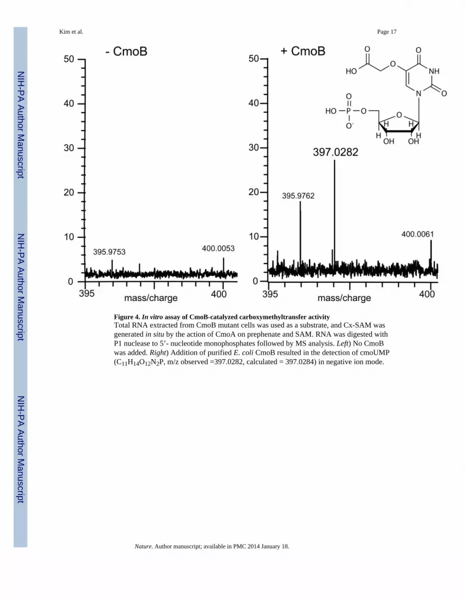
Figure 4. In vitro assay of CmoB-catalyzed carboxymethyltransfer activityTotal RNA extracted from CmoB mutant cells was used as a substrate, and Cx-SAM wasgenerated in situ by the action of CmoA on prephenate and SAM. RNA was digested withP1 nuclease to 5’- nucleotide monophosphates followed by MS analysis. Left) No CmoBwas added. Right) Addition of purified E. coli CmoB resulted in the detection of cmoUMP(C11H14O12N2P, m/z observed =397.0282, calculated = 397.0284) in negative ion mode.
Kim et al. Page 17
Nature. Author manuscript; available in PMC 2014 January 18.
NIH
-PA Author Manuscript
NIH
-PA Author Manuscript
NIH
-PA Author Manuscript


