Embed Size (px)
Citation preview
B I O C H E M I S T R Y
Studies of the Composition and Structure of Serum Lipoproteins. Separation and Characterization of Phospholipid -Protein Residues Obtained by Partial Delipidization of Very Low Density Lipoproteins of Human Serum*
Anders Gustafson,t Petar Alaupovic, and Robert H. Furman
BSTRACT: The partial delipidization of human serum a- and P-lipoproteins by n-heptane resulted in phos- pholipid-protein residues characterized by a single protein moiety, apolipoprotein A and apolipoprotein B, respectively. The very low density (VLD) lipoproteins (Sr >20) yielded, upon partial delipidization, a mixture of three phospholipid-protein residues. These were separated by preparative Pevikon zone electrophoresis and ultracentrifugation. The VLD phospholipid-protein residues and corresponding protein moieties were characterized by determination of sedimentation coeffi-
D ifferences in the terminal amino acids (Avigan et al., 1956; Shore, 1957; Rodbell, 1958), immuno- chemical specificities (Scanu et al., 1958b), and metabolic pathways (Gitlin et al., 1958) indicate that low-density @-LP)' and high-density (a-LP) human serum lipo- proteins contain single, distinct protein moieties. How- ever, similar studies of various ultracentrifugally iso- lated fractions of very low density (VLD) lipoproteins have provided only limited and inconclusive informa- tion concerning the distribution and type of their pro- tein moieties. It has been suggested that the chylo- microns (Sf >400) contain two (Scanu and Page, 1959) or three (Rodbell and Fredrickson, 1959) proteins, one of which is closely related to that of a-LP, and that the Sr 100-400 VLD-lipoproteins probably contain only one
* From the Cardiovascular Section, Oklahoma Medical Re- search Foundation, and Departments of Biochemistry and Medi- cine, University of Oklahoma School of Medicine, Oklahoma City, Oklahoma. Received Jury 22, 1965; revised October 14, 1965. Supported in part by grants from the U. S. Public Health Service (HE-6221 and HE-2528), the American Heart Associa- tion, and the Oklahoma Heart Association.
t Cardiovascular Research Trainee, supported by a U. S. Public Health Service grant (HTS-5403). Present address: Sahlgrenska sjukhuset, The University of Goteborg, Goteborg, Sweden.
1 Abbreviations used: Sr, flotation rate, expressed as Svedbergs (10-3 cm sec/dyne per g) of flotation in NaCl medium of density 1.063 g/ml at 26"; VLD-lipoproteins, very low density lipopro- teins, lipoproteins of density <1.006 g!ml and Sr >20; P-LP, P-lipoproteins, low-density lipoproteins, lipoproteins of density 1.006-1.063 g/ml and Sr &20; a-LP, a-lipoproteins, high-density lipoproteins, lipoproteins of density 1.063-1.210 g/ml. 632
cient, diffusion coefficient, hydrated density, N-terminal amino acids, by peptide patterns, and by immuno- chemical specificity. The protein moieties of two of the three VLD phospholipid-protein residues were the same as apolipoproteins A and B. The third phospho- lipid-protein residue contained another protein, desig- nated apolipoprotein C. It has been hypothesized that apolipoprotein C, due to its high phospholipid binding capacity, plays a major role in maintaining the struc- tural stability of protein-poor very low density lipo- protein particles.
protein (Levine et al., 1955; Briner et al., 1959) similar to, or the same as, that of P-LP. It has also been sug- gested, however, on the basis of the N-terminal amino acid analyses, that the VLD-lipoproteins (Sf >20) may contain as many as four distinct proteins (Rodbell, 1958; Shore and Shore, 1962a; Bobbitt and Levy, 1963).
So far, a lipid-free, water-soluble protein moiety has been obtained only from a-LP (Scanu et a[. , 1958a; Shore and Shore, 1962b) after ethanol-diethyl ether extraction. The application of the same delipidization procedure to PGP and VLD-lipoproteins failed to yield water-soluble proteins (Scanu et al., 1958b; Scanu and Page, 1959). On the other hand, the extraction of VLD-lipoproteins by diethyl ether (Avigan, 1957 ; Scanu and Page, 1959; Hayashi et al., 1959; Grundy et al., 1961) did yield soluble fractions. The relatively poor yields of lipid-protein residues obtained by this technique, however, made further studies on the frac- tionation and identification of modified VLD-lipo- proteins and their protein moieties difficult.
Since the high lipid content of the five VLD-lipo- protein subfractions separated at arbitrarily chosen flotation rates by sequential preparative ultracentrifuga- tion (Gustafson et a[., 1965) interfered considerably with attempts to isolate single homogeneous VLD- lipoproteins, the entire VLD-lipoprotein fraction (Sf >20) was partially delipidized by a new procedure (Gustafson, 1964) resulting in a mixture of phospho- lipid-protein residues (Gustafson et al., 1964). The fractionation and characterization of these phospho- lipid-protein residues and their protein components ir; the basis of this report.
A N D E R S GUSTAFSON, P E T A R ALAUPOVIC, A N D R O B E R T H. F U R M A N
V O L . 5, N O . 2, F E B R U A R Y 1 9 6 6
Experimental Section
Blood samples were obtained from healthy subjects (for the isolation of a- and PZP) and from subjects with hyperlipemia (hyperglyceridemia) of various types, except that no subjects with hyperchylomicronemia ("dietary fat-induced lipemia") were included. All subjects exhibited prompt and marked increase in plasma free fatty acid levels following heparin injection. Blood was obtained after the subjects had fasted over- night, in order to minimize the possibility of chylo- microns being present. The blood was allowed to clot and serum recovered by low-speed centrifugation.
Isolation of Lipoproteins. Serum lipoproteins were obtained by preparative ultracentrifugation in the No. 40 rotor of the Spinco Model L ultracentrifuge at 4". The VLD-lipoproteins (S, >20) were obtained by layering samples of hyperglyceridemic serum under equal volumes of d 1.0055 g/ml buffer solution (1.42 g of anhydrous disodium phosphate, 7.27 g of NaCI, and 0.1 g of disodium EDTA in 1 1. of solution adjusted to pH 7.0 with 1 N HC1) and centrifuging for 22 hr at 40,000 rpm (105,OOOg). p-LP and a-LP were isolated from normal sera at solvent densities of 1.063 and 1.210 g/ml, respectively, according to the the technique described by Bragdon et al. (1956). All lipoprotein frac- tions were washed twice by recentrifugation to remove contaminating proteins, including albumin. Cellulose acetate electrophoresis was employed to verify the absence of contaminating proteins.
Partial Delipidization. The a-, /?-, and VLD-lipo- protein fractions were dialyzed for 48 hr at 4" against four changes of distilled water containing 0.01% di- sodium EDTA, pH 7.0, lyophilized in the presence of insoluble potato starch powder (J. T. Baker Chemical Co.) and subjected to partial delipidization by n-heptane according to the method of Gustafson (1964). The sequential removal of lipids was followed routinely by thin layer chromatography, and the absence of neutral lipids in the heptane extracts was used as the criterion for discontinuing further delipidization. The phospho- lipid-protein residues were recovered from the starch by extraction with 0.3 M borate buffer, pH 8.2, or with 0.05 M phosphate buffer, pH 7.0.
Separation of Phospholipid-Protein Residues. The mixture of phospholipid-protein residues obtained by partial delipidization of the VLD-lipoproteins was sub- jected to horizontal electrophoresis on a "Pevikon C-870" (Stockholm Superfosfat AB, Stockholm, Swe- den) block (Muller-Eberhard, 1960), 50 X 15 cm, em- ploying 0.3 M borate buffer, pH 8.2, at a voltage gradient of 4.2 v/cm for 36 hr at an ambient temperature of 4". The block was cut into 70 segments, each 0.5 cm, and the phospholipid-protein residues were eluted from each segment by repeated extractions with 0.15 M NaCl containing 0.01 disodium EDTA, pH 8.0 (total volume approximately 10 ml). The eluate from each segment was subjected to double diffusion in agar gel employing antibodies to human serum, and the ab- sorbancy of each eluate determined at 280 mp. The immunochemical properties and protein concentrations
of the eluates defined two main electrophoretic zones, I and 11. Zone I eluates contained a single phospho- lipid-protein residue, zone I1 eluates contained two. The latter were separated by preparative ultracentrifugation in a NaBr solution of density 1.105 g/ml for 44 hr at 40,000 rpm (105,OOOg). All fractions thus purified were concentrated by dialysis against a 30 % aqueous solution of Dextran 80 (AB Pharmacia, Uppsala, Sweden) at 4".
Electrophoresis. Starch gel electrophoresis was per- formed according to Smithies (1955) utilizing a dis- continuous buffer system, pH 8.2 (Poulik, 1957), and cellulose acetate electrophoresis according to Kohn (1958) employing barbital buffer, pH 8.6, ionic strength 0.075. Ponceau 3R (Allied Chemical Corp., New York) and Amido Schwartz 10B (Bayer) were used to stain proteins on cellulose acetate strips and starch gel slices, respectively.
Immunochemical Methods. The phospholipid-protein residues were studied by immunoelectrophoresis (Wil- liams and Grabar, 1955) and by double diffusion (Ouchterlony, 1953) in agar gels employing barbital buffer, pH 8.6, ionic strength 0.050. Horse serum con- taining antibodies to human a- and &lipoproteins (Behringwerke, Marburg an der Lahn, Germany), horse serum containing antibodies to human serum albumin, and rabbit serum containing antibodies to human 7 s y-globulin (Mann Research Laboratories, New York) were used. The antibodies to a- and 6- lipoprotein present in the horse serum obtained from Behringwerke gave single precipitin lines with the a- and PLP , respectively, and showed no reaction with albumin. Horse serum containing antibodies to whole human serum proteins (MaM Research Laboratories) gave precipitin lines with both a- and p-LP.
Ultracentrifugal Analyses. Ultracentrifugal analyses of phospholipid-protein residues were carried out in a Spinco model E ultracentrifuge equipped with a phase- plate schlieren diaphragm and an automatic temperature control unit. Plate measurements were made with a Nikon microcomparator having a sensitivity of 0.001 mm. Sedimentation coefficients were determined at constant temperature (25-26 ") employing rotor speeds of 52,640 or 59,780 rpm. Solutions of the phospho- lipid-protein residues in 0.15 M NaCl (d 1.0055 g/ml) were spun in single-sector cells. The observed sedimen- tation coefficients were corrected to values in water at 20" by the usual methods (Schachman, 1957). The sedimentation coefficients at infinite dilution, &,, were extrapolated from s2a.w values obtained at three dif- ferent protein concentrations.
Hydrated densities were determined by measuring the sedimentation coefficients at several NaCl solution densities and extrapolating (a plot of 17s vs. p, where 9 is the solvent viscosity relative to that of water, S the observed sedimentation coefficient, and p the solution density) to the solution density of zero sedimentation (Shore and Shore, 1962a). The values for the densities and relative viscosities of solutions were taken from the International Critical Tables.
The apparent diffusion coefficients of the phospho- lipid-protein residues were evaluated in 0.15 M NaCl 633
P H O S P H O L I P I D - P R O T E I N R E S I D U E S O F S E R U M V L D - L I P O P R O T E I N S
B 1 0 C H E h.11 S ' I 'K Y
- - - , . , , . , , , , , , , , , , . , . .. . . .. . . . . . . .. . .. . . . . . , , , , .. , . . . . . .. . . . , . , . ... . , .. , . . .. .. , .. .
I 2 3 4 5
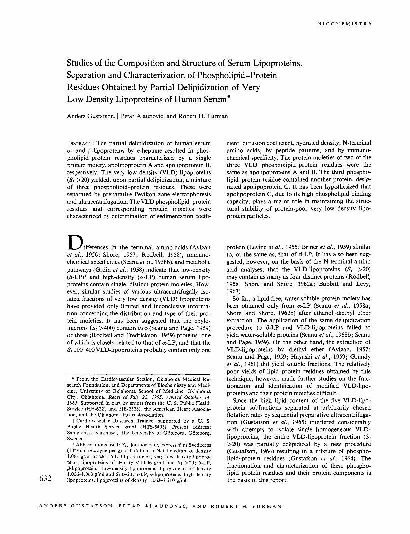
FIGURE 1 : Starch gel electropherogram of phospholipid- protein residues (protein stain). 1, whole serum; 2, partially delipidized VLD-lipoproteins (Sr >20) ; 3, 14s residue; 4, 4s residue; 5,7S residue.
solutions, at rotor speed of 12,590 rpm, by employing the height-area method. Diffusion coefficients, cor- rected to zero time, were then converted to standard conditions, D20,w, in the usual manner (Schachman, 1957). The average molecular weights of phospholipid- protein residues were calculated by utilizing the Sved- berg equation ( M = RTs/D(l - vp). The values for the apparent partial specific volume, 7, were obtained from the relation v = l/hydrated density.
N-Terminal Amino Acid Analj,sis. Dialyzed, concen- trated solutions of phospholipid-protein residues were extracted exhaustively by chloroform-methanol (2 : 1, v/v) or by ethanol-diethyl ether (3 : 1, v/v). The precipi- tated proteins were then dinitrophenylated according to the procedure of Fraenkel-Conrat et ul. (1955). The dinitrophenyl (DNP) proteins were washed with water, ethanol, and diethyl ether, dried in a desiccator over- night, and hydrolyzed by refluxing in redistilled 5.7 N HC1 for 10 hr. The ether-soluble DNP-amino acids were separated by paper chromatography (Blackburn and Lowther, 1951), employing as solvent t-amyl alcohol saturated with phthalate buffer. DNP-aspartic and DNP-glutamic acids were separated at pH 5 and' DNP-threonine and DNP-serine at pH 6.
Proteolysis of Proteins and Two-Dimensional Separa- lion of Peptides. The lipid-free proteins were digested for 2 hr at 28 O with trypsin (crystalline, salt-free, Boeh- ringer Mannheim Corp.) in a solution of 2 M urea con- taining 0.3 M ammonium carbonate, pH 8.4. The en- zyme:substrate ratio was 1 :40. The digestion with pepsin (twice crystallized, Worthington) was carried out with the same enzymesubstrate ratio in a solution adjusted to pH 2.0 by 0.1 N HC1 for 24 hr at 28". The trypsin digests were placed on a Dowex-50 column (100-200 mesh, H+ form), and the urea was eluted with deionized, glass-distilled water. Peptides were eluted with 4 M ",OH and, after lyophilization of eluates, redissolved in water (50 p1 of water/mg of peptides). The pepsin digests were dried in vacuo over PeOs and NaOH and the residual peptides redissolved in water (50 pl of water/mg of peptides). Peptide patterns of trypsin and pepsin digests were obtained by two- dimensional paper chromatography and electrophoresis 634
according to the technique of Katz et al. (1959). The peptide solutions were applied to Whatman No. 3 paper and subjected, in the first dimension, to descending chromatography in 1-butanol-acetic acid-water (4 : 1 : 5) . After drying, high-voltage electrophoresis was per- formed in the second dimension in a buffer composed of pyridine-acetic acid-water (1 : 10 : 189), pH 3.5, at 2000 v for 50 min. The peptides were stained by dipping the papers in 0.05% ninhydrin in ethanol and heating to 70" for 20 min.
Lipid Analyses. Concentrated or lyophilized salt-free samples of phospholipid-protein residues of VLD-, /3-, and a-lipoproteins were extracted exhaustively using chloroform-methanol (2: 1, v/v) or ethanol- diethyl ether (3 : 1, v/v). The precipitated proteins were removed by low-speed centrifugation, washed with diethyl ether, and dried in vacuo over Pro6. Lipid ex- tracts were washed according to the procedure of Folch et ul. (1957), dried with anhydrous Na2S04, evaporated to dryness in vacuo under nitrogen, redissolved in chloro- form, and aliquots taken for phospholipid analysis. Lipid phosphorus was determined by the wet digestion procedure of Youngburg and Youngburg (1930) with phosphorus measured by the method of Fiske and Subbarow (1925). The factor 25 was used to convert lipid phosphorus to phospholipid.
Quantitative analysis of individual phospholipids was performed by two-dimensional thin layer chromatog- raphy. Glass plates (20 X 20 cm) were coated with silica gel G (E. Merck, Darmstadt, West Germany) by use of the Desaga applicator (Stahl, E., 1962), and the chloroform extracts (20-25 pg of P) were applied with micropipets. The plates were developed in the first dimension with chloroform-methanol-water (65 : 25 : 4, v/v) and in the second dimension with chloroform- methanol-acetic acid-water (65 :25 : 8 :4, v/v). The separation and detection of phospholipids was followed by spraying individual plates with molybdenum blue reagent (Dittmer and Lester, 1964), ninhydrin, or Dragendorff reagent (Wagner et al., 1961). The phos- pholipid spots were detected routinely with iodine vapors, scraped off the plates, and transferred to diges- tion tubes. Phosphorus was determined by the micro- method of'Gerlach and Deuticke (1963).
Protein Analysis. Protein was determined by a slight modification of the Lowry method (Lowry et al., 1951), as previously described (Gustafson et al., 1965).
Results
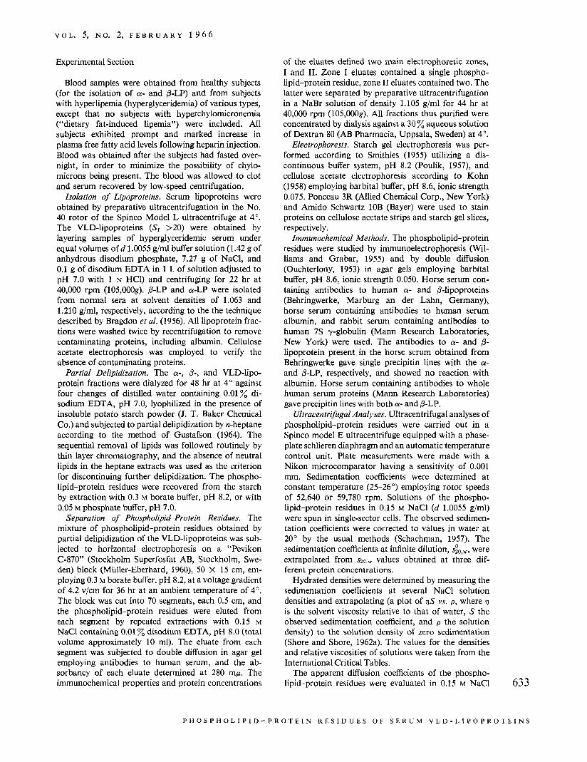
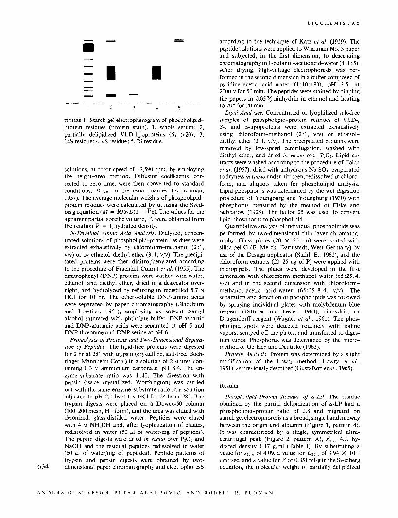
Phospholipid-Protein Residue of CY-LP. The residue obtained by the partial delipidization of CY-LP had a phospholipid-protein ratio of 0.8 and migrated on starch gel electrophoresis as a broad, single band midway between the origin and albumin (Figure 1, pattern 4). It was characterized by a single, symmetrical ultra- centrifugal peak (Figure 2, pattern A), s~,,, 4.3, hy- drated density 1.17 g/ml (Table I). By substituting a value for spa,,, of 4.09, a value for D20,w of 3.94 X lo-' cmZ/sec, and a value for v of 0.851 ml/g in the Svedberg equation, the molecular weight of partially delipidized
A N D C K S C i U S T A F b O N , PL: I A K A L A U I ' O V I C ' , A N D K O U b K 1 I I . F U K , M A N
V O L . 5, N O . 2, F E B R U A R Y 1 9 6 6
r,
A .--_-- min 16 32 8
Bll* 16 42 58
T i n 16 32 8 16 16 24
FIGURE 2: Ultracentrifugal schlieren patterns of phospholipid-protein residues. A, partially delipidized a-LP, 4 s residue; €3, partially delipidized p-LP, the major peak is the 14s residue; C, partially delipidized VLD-lipoproteins (Sr > 20); D, 4s residue from VLD-lipoproteins; E, the major peak is the 14s residue from VLD-lipoproteins; F, 7s residue from VLD-lipoproteins. All samples were run in 0.15 M NaCI, density 1.0055 g/ml. The interval (in minutes) after rotor attained a speed of 52,640 rpm for A, B, C, and D, and a speed of 59,780 rpm for E and F, is indicated below each frame.
~~~~~
TABLE I : Physical-Chemical Properties of Phospholipid-Protein Residues.
Source
Phospho- Phospho- lipid- lipid- Hydrated
D ~ ~ . ~ x 107 MOI w t Lipoprotein Protein Protein Density - Fraction Residue Ratio &w ( S ) (g/ml) V,, (ml/g) S P O . ~ (S) (cm2/sec) X 10-5
a-LP 4s 0 . 8 4 . 3 1.17 0.851 4.09 3.94 1.72 p-LP 14s 0 .7 14.5 1.18 . . . . . . . . . . . . VLD-LP 4s 0 . 9 4 .5 1.17 0.851 . . . . . . . . . VLD-LP 14s 0 . 7 14.0 1 .13 0.885 . . . . . . . . . VLD-LP 7s 2 .4 6 . 9 1.09 0.913 5.43 1.79 8.34
a-LP was calculated to be 172,000. The 4s residue gave a single precipitin line with antibodies to a-lipoprotein or human serum. The protein moiety of the completely delipidized 4s residue contained aspartic acid as the major N-terminal amino acid (21.8-23.7 kmoles/g of protein), with occasional traces of threonine (Table 11).
Phospholipid-Protein Residue of p-LP. The ultra- centrifugal pattern (Figure 2, pattern B) of the phospho- lipid-protein residue obtained from p-LP (Sf 0-20) revealed a major sedimenting peak, s&w 14.5. The 14s residue frequently contained an additional faster moving minor boundary with a sedimentation coefficient of approximately 20s. Due to its extraordinary tendency toward denaturation and aggregation, particularly at high salt concentrations, only limited information about the physical-chemical characteristics of the phospho- lipid-protein residue of p-LP was obtained. For this reason, the value of 1.18 g/ml for the hydrated density of this residue, calculated from measurements at low densities (1.005-1.010 g/ml), may be only an approxi-
mate value. The 14s residue migrated on starch gel electrophoresis (Figure 1, pattern 3) slightly ahead of intact p-LP, and gave a single precipitin line with anti+- lipoprotein serum. Glutamic acid was the major N- terminal amino acid of the protein moiety (4.5 pmoles/g of protein) with serine, threonine, and alanine as oc- casional minor contaminants (Table 11).
Phospholipid-Protein Residues of VLD-Lipoproteins (Sr >20). The partial delipidization of VLD-lipoproteins (Sf >20) isolated from hyperglyceridemic sera resulted in the formation of three phospholipid-protein residues. The ultracentrifugal pattern of a typical experiment showed initially two major boundaries (Figure 2, pat- tern C, 42 min). The slower sedimenting boundary subsequently resolved into two distinct peaks with approximate sedimentation coefficients of 4 S and 7 S. The mixture of residues was resolved by starch gel electrophoresis into three distinct zones (Figure 1 , pat- tern 2) and yielded three precipitin lines (Table 11) by immunoelectrophoresis and double diffusion with 635
P € l O S P H O L l P I D - P R O I L I N R E S I D U E S O F S E K U M V L D - L I P O P R O T E I N S
B I O C H E M I S T R Y
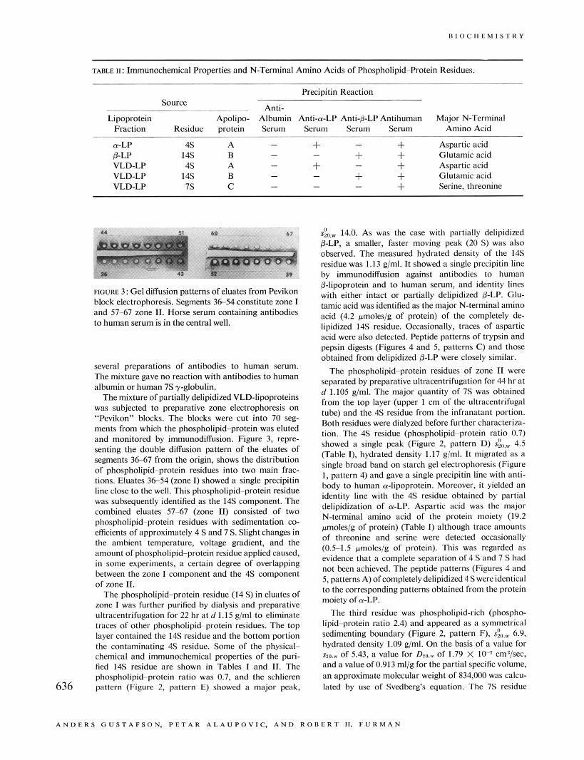
TABLE 11: Immunochemical Properties and N-Terminal Amino Acids of Phospholipid-Protein Residues.
Precipitin Reaction Source ~
~ Anti- Lipoprotein Apolipo- Albumin Anti-a-LP Anti-p-LP Antihuman Major N-Terminal
Fraction Residue protein Serum Serum Serum Serum Amino Acid
a-LP 4 s A - + - + Aspartic acid p-LP 14s B - - + + Glutamic acid VLD-LP 4 s A - + - + Aspartic acid VLD-LP 14s B - - + + Glutamic acid VLD-LP 7 s C - - - + Serine, threonine
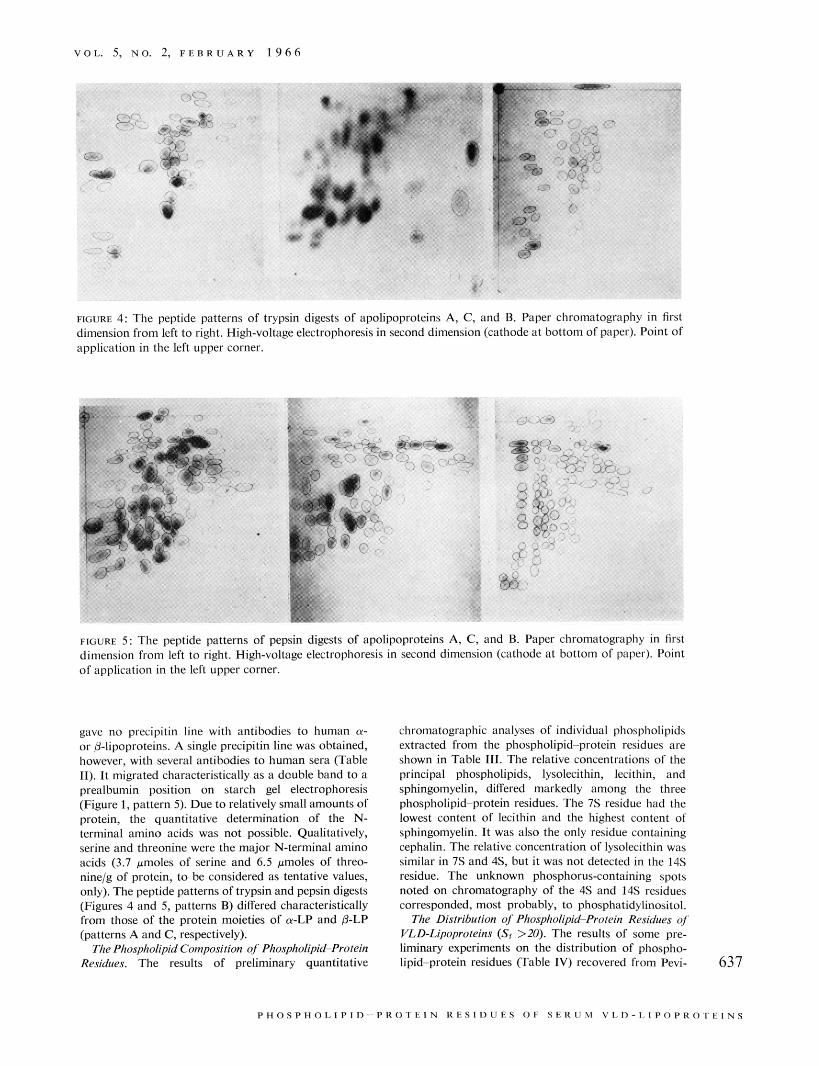
FIGURE 3: Gel diffusion patterns of eluates from Pevikon block electrophoresis. Segments 3 6 5 4 constitute zone I and 57-67 zone 11. Horse serum containing antibodies to human Serum is in the central well.
several preparations of antibodies to human serum. The mixture gave no reaction with antibodies to human albumin or human 7 s y-globulin.
The mixture of partially delipidized VLD-lipoproteins was subjected to preparative zone electrophoresis on “Pevikon” blocks. The blocks were cut into 70 seg- ments from which the phospholipid-protein was eluted and monitored by immunodiffusion. Figure 3, repre- senting the double diffusion pattern of the eluates of segments 3667 from the origin, shows the distribution of phospholipid-protein residues into two main frac- tions. Eluates 36-54 (zone I) showed a single precipitin line close to the well. This phospholipid-protein residue was subsequently identified as the 14s component. The combined eluates 57-67 (zone 11) consisted of two phospholipid-protein residues with sedimentation co- efficients of approximately 4 S and 7 S. Slight changes in the ambient temperature, voltage gradient, and the amount of phospholipid-protein residue applied caused, in some experiments, a certain degree of overlapping between the zone I component and the 4s component of zone 11.
The phospholipid-protein residue (14 S) in eluates of zone I was further purified by dialysis and preparative ultracentrifugation for 22 hr at d 1.15 gjml to eliminate traces of other phospholipid-protein residues. The top layer contained the 14s residue and the bottom portion the contaminating 4s residue. Some of the physical- chemical and immunochemical properties of the puri- fied 14s residue are shown in Tables I and 11. The phospholipid-protein ratio was 0.7, and the schlieren pattern (Figure 2, pattern E) showed a major peak, 636
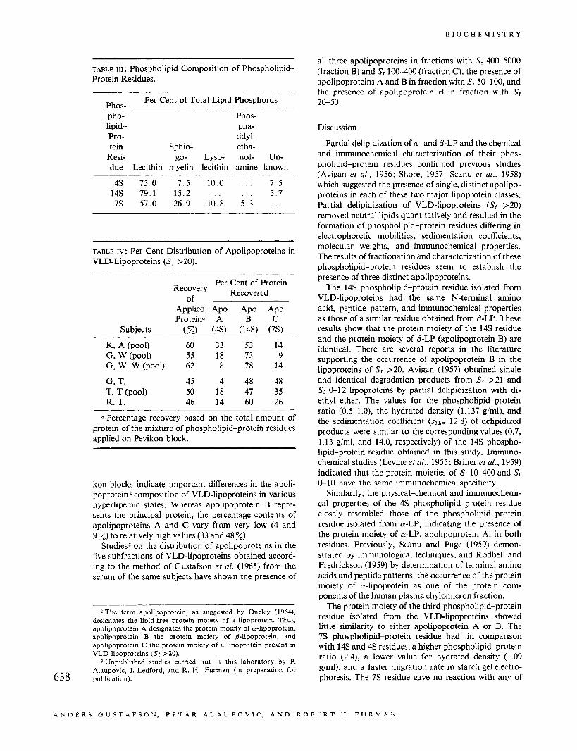
s:~., 14.0. As was the case with partially delipidized p-LP, a smaller, faster moving peak (20 S) was also observed. The measured hydrated density of the 14s residue was 1.13 g/ml. It showed a single precipitin line by immunodiffusion against antibodies to human 0-lipoprotein and to human serum, and identity lines with either intact or partially delipidized BLP. Glu- tamic acid was identified as the major N-terminal amino acid (4.2 pmoles/g of protein) of the completely de- lipidized 14s residue. Occasionally, traces of aspartic acid were also detected. Peptide patterns of trypsin and pepsin digests (Figures 4 and 5, patterns C) and those obtained from delipidized 0-LP were closely similar.
The phospholipid-protein residues of zone I1 were separated by preparative ultracentrifugation for 44 hr at d 1.105 g/ml. The major quantity of 7 s was obtained from the top layer (upper 1 cm of the ultracentrifugal tube) and the 4s residue from the infranatant portion. Both residues were dialyzed before further characteriza- tion. The 4s residue (phospholipid-protein ratio 0.7) showed a single peak (Figure 2, pattern D) 4.5 (Table I), hydrated density 1.17 gjml. It migrated as a single broad band on starch gel electrophoresis (Figure 1, pattern 4) and gave a single precipitin line with anti- body to human or-lipoprotein. Moreover, it yielded an identity line with the 4 s residue obtained by partial delipidization of or-LP. Aspartic acid was the major N-terminal amino acid of the protein moiety (19.2 pmolesjg of protein) (Table I) although trace amounts of threonine and serine were detected occasionally (O.Sl.5 pmolesjg of protein). This was regarded as evidence that a complete separation of 4 S and 7 S had not been achieved. The peptide patterns (Figures 4 and 5, patterns A) of completely delipidized 4 S were identical to the corresponding patterns obtained from the protein moiety of a-LP.
The third residue was phospholipid-rich (phospho- lipid-protein ratio 2.4) and appeared as a symmetrical sedimenting boundary (Figure 2, pattern F), sio,, 6.9, hydrated density 1.09 gjml. On the basis of a value for s30.r of 5.43, a value for D20.w of 1.79 X lo-’ cm2/sec, and a value of 0.913 ml/g for the partial specific volume, an approximate molecular weight of 834,000 was calcu- lated by use of Svedberg’s equation. The 7s residue
A N D E R S G U S T A F S O N , P E T A R A L A U P O V I C , A N D R O ~ E R T H. F U R M A N
V O L . 5, NO. 2, P E B R U A R Y 1 9 6 6
FIGURE 4: The peptide patterns of trypsin digests of apolipoproteins A, C, and B. Paper chromatography in first dimension from left to right, High-voltage electrophoresis in second dimension (cathode at bottom of paper). Point of application in the left upper corner.
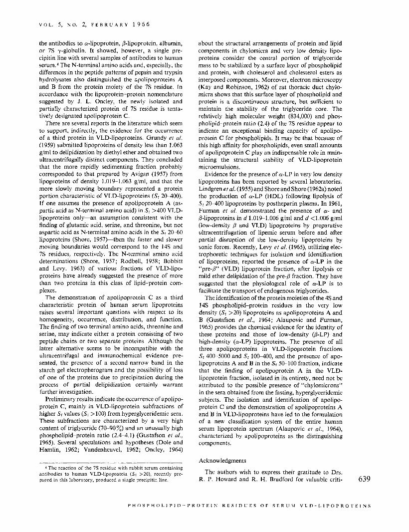
FIGURE 5 : The peptide patterns of pepsin digests of apolipoproteins A, C, and B. Paper chromatography in first dimension from left to right. High-voltage electrophoresis in second dimension (cathode at bottom of paper). Point of application in the left upper comer.
gave no precipitin line with antibodies to human a- or 0-lipoproteins. A single precipitin line was obtained, however, with several antibodies to human sera (Table 11). It migrated characteristically as a double hand to a prealbumin position on starch gel electrophoresis (Figure 1, pattern 5). Due to relatively small amounts of protein, the quantitative determination of the N- terminal amino acids was not possible. Qualitatively, serine and threonine were the major N-terminal amino acids (3.7 ,.moles of serine and 6.5 unoles of threo- nine/g of protein, to he considered as tentative values, only). The peptide patterns of trypsin and pepsin digests (Figures 4 and 5, patterns B) differed characteristically from those of the protein moieties of a-LP and 0-LP (patterns A and C, respectively).
The Phospholipid Composition of Phospholipid-Protein Residues. The results of preliminary quantitative
chromatographic analyses of individual phospholipids extracted from the phospholipid-protein residues are shown in Table 111. The relative concentrations of the principal phospholipids, lysolecithin, lecithin, and sphingomyelin, differed markedly among the three phospholipid-protein residues. The 'IS residue had the lowest content of lecithin and the highest content of sphingomyelin. It was also the only residue containing cephalin. The relative concentration of lysolecithin was similar in 'IS and 4S, hut it was not detected in the 14s residue. The unknown phosphorus-containing spots noted on chromatography of the 4s and 14s residues corresponded, most probably, to phosphatidylinositol.
The Distribution of Phospholipid-Protein Residues of VLD-Lipoproteins (Sr >XI). The results of some pre- liminary experiments on the distribution of phospho- lipid-protein residues (Table IV) recovered from Pevi- 631
P H O S P H O L I P I D - P R O T E I N R E S I D U E S O F S E R U M V L D - L I P O P R O T E I N S
B I O C H E M I S T R Y
TABLE 111 : Phospholipid Composition of Phospholipid- Protein Residues.
Per Cent of Total Lipid Phosphorus Phos- pho- Phos- lipid- pha- Pro- tidyl- tein Sphin- etha-
Resi- go- Lyso- nol- Un- due Lecithin myelin lecithin amine known
4s 75.0 7 .5 10.0 . . . 7 .5 14s 79.1 15.2 . . . . . . 5 . 7 7s 57.0 26.9 10.8 5 . 3 . . .
TABLE IV: Per Cent Distribution of Apolipoproteins in VLD-Lipoproteins (Sf >20).
Per Cent of Protein Recovered Recovery
Of
Applied Apo Apo Apo Proteina A B C
Subjects (%> ( 4 9 (14s) (7s)
G, W (pool) 55 18 73 9 K, A (pool) 60 33 53 14
G, W, W (pool) 62 8 78 14
G. T. 45 4 48 48 T, T (Pool) 50 18 47 35 R. T. 46 14 60 26
a Percentage recovery based on the total amount of protein of the mixture of phospholipid-protein residues applied on Pevikon block.
kon-blocks indicate important differences in the apoli- poprotein2 composition of VLD-lipoproteins in various hyperlipemic states. Whereas apolipoprotein B repre- sents the principal protein, the percentage contents of apolipoproteins A and C vary from very low (4 and 9 %) to relatively high values (33 and 48 %).
Studies3 on the distribution of apolipoproteins in the five subfractions of VLD-lipoproteins obtained accord- ing to the method of Gustafson et a/. (1965) from the serum of the same subjects have shown the presence of
2 The term apolipoprotein, as suggested by Oncley (1964), designates the lipid-free protein moiety of a lipoprotein. Thus, apolipoprotein A designates the protein moiety of a-lipoprotein, apolipoprotein B the protein moiety of @-lipoprotein, and apolipoprotein C the protein moiety of a lipoprotein present in VLD-lipoproteins (Sr >20).
3 Unpublished studies carried out in this laboratory by P. Alaupovic, J. Ledford, and R. H. Furman (in preparation for
638 publication).
all three apolipoproteins in fractions with Sf 400-5000 (fraction B) and Sr 100-400 (fraction C), the presence of apolipoproteins A and B in fraction with Sr 50-100, and the presence of apolipoprotein B in fraction with Sr 20-50.
Discussion
Partial delipidization of a- and p-LP and the chemical and immunochemical characterization of their phos- pholipid-protein residues confirmed previous studies (Avigan et a/., 1956; Shore, 1957; Scanu et al., 1958) which suggested the presence of single, distinct apolipo- proteins in each of these two major lipoprotein classes. Partial delipidization of VLD-lipoproteins (Sf >20) removed neutral lipids quantitatively and resulted in the formation of phospholipid-protein residues differing in electrophoretic mobilities, sedimentation coefficients, molecular weights, and immunochemical properties. The results of fractionation and characterization of these phospholipid-protein residues seem to establish the presence of three distinct apolipoproteins.
The 14s phospholipid-protein residue isolated from VLD-lipoproteins had the same N-terminal amino acid, peptide pattern, and immunochemical properties as those of a similar residue obtained from p-LP. These results show that the protein moiety of the 14s residue and the protein moiety of PGP (apolipoprotein B) are identical. There are several reports in the literature supporting the occurrence of apolipoprotein B in the lipoproteins of Sf >20. Avigan (1957) obtained single and identical degradation products from Sf >21 and Sf 0-1 2 lipoproteins by partial delipidization with di- ethyl ether. The values for the phospholipid-protein ratio (0.5-1.0), the hydrated density (1.137 g/ml), and the sedimentation coefficient ( ~ ~ 0 . ~ 12.8) of delipidized products were similar to the corresponding values (0.7, 1.13 g/ml, and 14.0, respectively) of the 14s phospho- lipid-protein residue obtained in this study. Immuno- chemical studies (Levine er al., 1955; Briner ef a/., 1959) indicated that the protein moieties of Sf 10-400 and Sr 0-10 have the same immunochemical specificity.
Similarily, the physical-chemical and immunochemi- cal properties of the 4s phospholipid-protein residue closely resembled those of the phospholipid-protein residue isolated from a-LP, indicating the presence of the protein moiety of a-LP, apolipoprotein A, in both residues. Previously, Scanu and Page (1959) demon- strated by immunological techniques, and Rodbell and Fredrickson (1959) by determination of terminal amino acids and peptide patterns, the occurrence of the protein moiety of a-lipoprotein as one of the protein com- ponents of the human plasma chylomicron fraction.
The protein moiety of the third phospholipid-protein residue isolated from the VLD-lipoproteins showed little similarity to either apolipoprotein A or B. The 7 s phospholipid-protein residue had, in comparison with 14s and 4s residues, a higher phospholipid-protein ratio (2.4), a lower value for hydrated density (1.09 g/ml), and a faster migration rate in starch gel electro- phoresis. The 7s residue gave no reaction with any of
A N D E R S G U S T A F S O N , P E T A R A L A U P O V I C , A N D R O R E R T H. F I J R M A N
V O L . 5, N O . 2, F E B R U A R Y 1 9 6 6
the antibodies to a-lipoprotein, 0-lipoprotein, albumin, or 7 s y-globulin. It showed, however, a single pre- cipitin line with several samples of antibodies to human serum. The N-terminal amino acids and, especially, the differences in the peptide patterns’ of pepsin and trypsin hydrolysates also distinguished the ;polipoproteins A and B from the protein moiety of the 7s residue. In accordance with the lipoprotein-protein nomenclature suggested by J. L. Oncley, the newly isolated and partially characterized protein of 7s residue is tenta- tively designated apolipoprotein C.
There are several reports in the literature which seem to support, indirectly, the evidence for the occurrence of a third protein in VLD-lipoproteins. Grundy et a/. (1959) submitted lipoproteins of density less than 1.063 g/ml to delipidization by diethyl ether and obtained two ultracentrifugally distinct components. They concluded that the more rapidly sedimenting fraction probably corresponded to that prepared by Avigan (1957) from lipoproteins of density 1.019-1.063 g/ml, and that the more slowly moving boundary represented a protein portion characteristic of VLD-lipoproteins (Si 20-400). If one assumes the presence of apolipoprotein A (as- partic acid as N-terminal amino acid) in Sf >400 VLD- lipoproteins only-an assumption consistent with the finding of glutamic acid, serine, and threonine, but not aspartic acid as N-terminal amino acids in the Sf 20-60 lipoproteins (Shore, 1957bthen the faster and slower moving boundaries would correspond to the 14s and 7s residues, respectively. The N-terminal amino acid determinations (Shore, 1957; Rodbell, 1958; Bobbitt and Levy, 1963) of various fractions of VLD-lipo- proteins have already suggested the presence of more than two proteins in this class of lipid-protein com- plexes.
The demonstration of apolipoprotein C as a third characteristic protein of human serum lipoproteins raises several important questions with respect to its homogeneity, occurrence, distribution, and function. The finding of two terminal amino acids, threonine and serine, may indicate either a protein consisting of two peptide chains or two separate proteins. Although the latter alternative seems to be incompatible with the ultracentrifugal and immunochemical evidence pre- sented, the presence of a second narrow band in the starch gel electropherogram and the possibility of loss of one of the proteins due to precipitation during the process of partial delipidization certainly warrant further investigation.
Preliminary results indicate the occurrence of apolipo- protein C, mainly in VLD-lipoprotein subfractions of higher Sr values (Sf >loo) from hyperglyceridemic sera. These subfractions are characterized by a very high content of triglyceride (70-90%) and an unusually high phospholipid-protein ratio (2.4-4.1) (Gustafson et al., 1965). Several speculations and hypotheses (Dole and Hamlin, 1962; Vandenheuvel, 1962; Oncley, 1964)
4 The reaction of the 7s residue with rabbit serum containing antibodies to human VLD-lipoprotein (Sf > 20), recently pre- pared in this laboratory, produced a single precipitin line.
about the structural arrangements of protein and lipid components in chylomicra and very low density lipo- proteins consider the central portion of triglyceride mass to be stabilized by a surface layer of phospholipid and protein, with cholesterol and cholesterol esters as interposed components. Moreover, electron microscopy (Kay and Robinson, 1962) of rat thoracic duct chylo- micra shows that this surface layer of phospholipid and protein is a discontinuous structure, but sufficient to maintain the stability of the triglyceride core. The relatively high molecular weight (834,000) and phos- pholipid-protein ratio (2.4) of the 7s residue appear to indicate an exceptional binding capacity of apolipo- protein C for phospholipids. It may be that because of this high affinity for phospholipids, even small amounts of apolipoprotein C play an indispensable role in main- taining the structural stability of VLD-lipoprotein microemulsions.
Evidence for the presence of a-LP in very low density lipoproteins has been reported by several laboratories. Lindgren etal. (1955) and Shore and Shore (1962a) noted the production of a-LP (HDL) following lipolysis of St 20-400 lipoproteins by postheparin plasma. In 1961, Furman et al. demonstrated the presence of a- and @-lipoproteins in d 1.01%1.006 g/ml and d <1.006 g/ml (low-density p and VLD) lipoproteins by preparative ultracentrifugation of lipemic serum before and after partial disruption of the low-density lipoproteins by sonic forces. Recently, Levy et a/. (1965), utilizing elec- trophoretic techniques for isolation and identification of lipoproteins, reported the presence of a-LP in the “pre-0’’ (VLD) lipoprotein fraction, after lipolysis or mild ether delipidation of the pre-0 fraction. They have suggested that the physiologcal role of a-LP is to facilitate the transport of endogenous triglycerides.
The identification of the protein moieties of the 4s and 14s phospholipid-protein residues in the very low density (Sf >20) lipoproteins as apolipoproteins A and B (Gustafson et al., 1964; Alaupovic and Furman, 1965) provides the chemical evidence for the identity of these proteins and those of low-density (@-LP) and high-density (a-LP) lipoproteins. The presence of all three apolipoproteins in VLD-lipoprotein fractions Sr 400-5000 and Sf 100-400, and the presence of apo- lipoproteins A and B in the Sf 50-100 fraction, indicate that the finding of apolipoprotein A in the VLD- lipoprotein fraction, isolated in its entirety, need not be attributed to the possible presence of “chylomicrons” in the sera obtained from the fasting, hyperglyceridemic subjects. The isolation and identification of apolipo- protein C and the demonstration of apolipoproteins A and B in VLD-lipoproteins have led to the formulation of a new classification system of the entire human serum lipoprotein spectrum (Alaupovic et al., 1964), characterized by apolipoproteins as the distinguishing components.
Acknowledgments
The authors wish to express their gratitude to Drs. R. P. Howard and R. H. Bradford for valuable criti- 639
P H 0 S P C 1 0 L I P I D --- P R 0 T E I N R E S I D LJ E S 0 F S F R IJ M V L D - L I P O P R 0 T E I N S
B I O C H E M I S T R Y
cisms, to S. Walraven, M. Sullivan, and A. W. Fryer for capable technical assistance, and to Mrs. M. Farmer for assistance in the preparation and typing of the manuscript.
References
Alaupovic, P., and Furman, R. H. (1965), J . Am. Oil
Alaupovic, P., Gustafson, A., Sanbar, S. S., and Fur-
Avigan, J. (1957), J . Biol. Chern. 226,957. Avigan, J., Redfield, R., and Steinberg, D. (1956),
Biochim. Biophys. Acta 20, 557. Blackburn, S., and Lowther, A. G. (1951), Biochem. J .
48,126. Bobbitt, J. L., and Levy, R. S. (1963), Abstracts, 145th
National Meeting of the American Chemical Society, New York, N. Y., Sept.
Bragdon, J. B., Havel, R. J., and Boyle, E. (1956), J. Lab. Cfin. Med. 48, 36.
Briner, W. W., Riddle, J. W., and Cornwell, D. G. (1959), J. Exptl. Med. 110, 113.
Dittmer, J. C., and Lester, R. L. (1964), J. Lipid Res. 5, 126.
Dole, V. P., and Hamlin, J . T., I11 (1962), Physiol. Rev. 42, 674.
Fiske, C. H., and Subbarow, Y. (1925), J. Biof. Clrem. 66, 375.
Folch, J., Lees, M., and Sloane-Stanley, G. H. (1957), J. Biof. Chem. 226, 497.
Fraenkel-Conrat, H., Harris, J. I., and Levy, A. L. (1955), Methods Biochem. Anafy. 2, 359.
Furman, R. H., Howard, R. P., Lakshmi, K . , and Norcia, L. N. (1961), Am. J . Clin. Nutr. 9, 73.
Gerlach, E., and Deuticke, B. (1963), Biochem. 2. 337, 477.
Gitlin, D., Cornwell, D. G., Nakasato, D., Oncley, J. L., Hughes, W. L., Jr., and Janeway, C. A. (1958), J . Clin. Invest. 37, 172.
Grundy, S., Dobson, H. L., and Griffin, A. C. (1959), Proc. SOC. Exptl. Biof. Med. 100, 704.
Grundy, S. M., Dobson, H. L., Kitzmiller, G. E., and Griffin, A. C. (1961), Am. .I. Physiol. 200, 1307.
Gustafson, A. (1964), Biochinz. Biophys. Acta 84, 223. Gustafson, A., Alaupovic, P., and Furman, R. H.
Gustafson, A., Alaupovic, P., and Furman, R. H.
Chemists’ SOC. 42, 140A.
man, R. H. (1964), Circufation 30, S ~ p p l . I I I , l .
(1964), Biochim. Biophys. Acta 84, 767.
(1965), Biochemistry 4, 596.
Am. Chem. SOC. 81, 3793.
(1959), J. Biol. Chem. 234, 2897.
Physiof. 47, 258.
Hayashi, S . , Lindgren, F., and Nichols, A. (1959), J.
Katz, A. M., Dreyer. W. J., and Anfinsen, C. B.
Kay, D., and Robinson, D. S. (1962), Quart. J . Exptf.
Kohn, J. (1958), Cfin. Chim. Acta 3, 450. Levine, L., Kauffman, D. L., and Brown, R. K. (1955),
Levy, R. I., Lees, R. S., and Fredrickson, D. S. (1965),
Lindgren, F. T., Nichols, A. V., and Freeman, N. K.
Lowry, 0. H., Rosebrough, N. J . , Farr, A. L., and
Muller-Eberhard, H. J. (1960), Scand. J . Cfin. Lab.
Oncley, J. L. (1964), Proc. Intern. Sym. Lipid Transport,
Ouchterlony, 0. (1953), Acta Path. Microbiof. Scand.
Poulik, M. D. (1957), Nature 180, 1477. Rodbell, M. (1958), Science 127, 701. Rodbell, M. R., and Fredrickson, D. S. (1959), J. Biof.
Scanu, A., Lewis, L. A,, and Bumpus, F. M. (1958a),
Scanu, A., Lewis, L. A., and Page, I. H. (1958b), J.
Scanu, A., and Page, I. H. (1959), J. Exptf. Med. 109.
Schachman, K. H. (1957), Methods Enzymof. 4, 32. Shore, B. S. (1957), Arch. Biochem. 71, 1. Shore, B., and Shore, V. (1962a), J. Atheroscfer. Res. 2,
Shore, B., and Shore, V. (1962b), Biochem. Biophys.
Smithies, 0. (1955), Biochern. J. 61, 629. Stahl, E. (1962), Dunnschicht-Chromatographie, Berlin,
Vandenheuvel, F. A. (1962), Can. J . Biochem. Physiof.
Wagner, H., Horhammer, L., and Wolff, P. (1961),
Williams, C. A., Jr., and Grabar, P. (1955), J . Zmmunol.
Youngburg, G. E., and Youngburg, M. V. (1930), J.
J. Expf l . Med. 102, 105.
J . Cfin. Invest. 44, 1068.
(1955), J . Phys. Chem. 59, 930.
Randall, R. J. (1951), J. Biol. Chem. 193, 265.
Invest. 12, 33.
70.
32,231.
Chem. 234, 562.
Arch. Biochem. Biophys. 74, 390.
Exptl. Med. 108, 185.
239.
104.
Res. Commun. 9, 455.
Springer-Verlag, p 5.
40,1299.
Biochem. Z. 334, 175.
74,158.
Lab. Cfin. Med. 16, 158.
640
A N D E R S G U S T A F S O N , P E T A R A L A U P O V I C , A N D R O B E R T H. F U R M A N


